Note: Descriptions are shown in the official language in which they were submitted.
CA 03005434 2018-05-15
WO 2017/087486
PCT/US2016/062225
METHODS AND COMPOSITIONS FOR TREATING
DISORDERS AND DISEASES USING SURVIVAL MOTOR
NEURON (SMN) PROTEIN
CROSS-REFERENCE TO RELATED APPLICATIONS
This application claims benefit of U.S. Provisional Application No.
62/255,721, filed
November 16, 2015, incorporated herein by reference in its entirety.
BACKGROUND
Aging-related muscle wasting and weakness (sarcopenia) is an important problem
of an
increasingly aging society (Faulkner et al. 2007; Manini et al. 2007). Though
sarcopenia is a
multifactorial phenomenon, a number of studies have indicated the presence of
marked
denervated and atrophied muscle fibers (Chai et al. 2011; Valdez et al. 2010;
Tomlinson et al.
1977; Oda et al. 1984; Kawamura et al. 1977). Indeed, using electromyographic
techniques that
allow longitudinal monitoring of motor unit function in the mouse in vivo
(Arnold et al. 2014), a
reduction in the number of functional motor neurons innervating the hind limb
muscles have
been identified as an early feature in aging mice. The motor unit is comprised
of a single motor
neuron and the muscle fibers it innervates. Motor unit synaptic connectivity
is maintained by
trophic support from various compartments (Fu et al. 2008; Koliatsos et al.
1993; Ikeda et al.
1995; Kablar et al. 2005), and it is suggested that the maintenance of motor
neuron connectivity
and repair of neuromuscular junctions (NMJs) is critical in aging.
Peripheral nerves are commonly injured from trauma including automobile
accidents,
motorcycle accidents, surgeries, knife and projectile wounds and birth
injuries to both the child
and mother. Common surgical causes of nerve injury include prostatectomy and
mastectomy.
Other common injuries during surgery are the result of long-term limb
positioning or inevitable
or accidental nerve compression. Following nerve injury there is a loss of
sensation and/or
function in the regions of the body innervated by the damaged nerve. For
example, following
nerve injury from prostatectomy there is commonly erectile dysfunction.
Following mastectomy
there is often loss of proper function of the upper extremity and/or scapula.
Furthei more,
following birth injury or other trauma with damage to the brachial plexus
there is dysfunction in
the ipsilateral limb. What is needed are methods and compositions related to
treating sarcopenia
and nerve injury in a subject in need thereof.
1
CA 03005434 2018-05-15
WO 2017/087486
PCT/US2016/062225
SUMMARY
Disclosed herein is a method of reducing sarcopenia in an individual, the
method
comprising: identifying an individual with sarcopenia, an individual with
symptoms of
sarcopenia, or an individual at risk for developing sarcopenia, wherein the
subject is 35 years old
or older; and administering to the individual a Survival Motor Neuron (SMN)
protein -increasing
substance, thereby reducing sarcopenia, sarcopenia symptoms, or the risk of
sarcopenia in the
individual.
Also disclosed is a method of treating an individual with nerve damage, the
method
comprising: identifying a subject with nerve damage; and administering to the
subject a Survival
Motor Neuron (SMN) protein-increasing substance, thereby reducing nerve damage
and/or
improving nerve function in the individual.
The details of one or more embodiments of the invention are set forth in the
accompa-
nying drawings and the description below. Other features, objects, and
advantages of the
invention will be apparent from the description and drawings, and from the
claims.
BRIEF DESCRIPTION OF THE DRAWINGS
The accompanying figures, which are incorporated in and constitute a part of
this
specification, illustrate several aspects described below.
FIGS. 1A-1D show electromyographic data in cohorts of male C57BL/6J mice at
different ages. Overview of electromyographic findings in C57BL/6J male mice
at 6 (n=10), 10
(n=10), 13 (n=10), and 24 months (n=6) of age is shown. For MUNE, CMAF', and
SMUP,
comparison between 10 m and 24 month old mice was performed. Single fiber EMG
was
perfoi __ med in two cohorts of animals at 6 and 14 month old animals. A. MUNE
(number of total
functional motor units) is diminished in 24 month old mice (224 36; p=0.027)
compared with 10
month old mice (341 29; p=0.027). B. When compared with CMAP response in 10
month old
mice (50.7mV 3.9), a reduction in 24 month old mice (38.2mV 3.8; p=0.034) is
noted. C.
Similarly, an increase in the single motor unit potential (SMUP) amplitude is
seen in 24 month
old mice (360 V 38) but this is not statistically significant ( p=0.144)
compared with 10 month
old mice (282 V 23). D. Alteration in NMJ transmission (increased jitter) are
noted in 14
month old (jitter=14.7 1.1 is, n=22 individual synapses, obtained from 2 mice)
versus 6 month
old mice (jitter=10.8 1.4 [is, n=18 individual synapses, obtained from 2 mice)
(p=0.043) (Data
shown as mean standard error of the mean) (ns, not significant; *, p<0.05).
FIGS. 2A-2B show real-time PCR utilizing enriched motor neuron (MN) samples
laser
capture microdissection (LCNI) (Ruggio et al. 2012). Figure 2A shows images of
motor neuron
2
CA 03005434 2018-05-15
WO 2017/087486
PCT/US2016/062225
samples both pre-LCM and post-LCM. Figure 2B shows mRNA levels of motor neuron
and non-
motor neuron samples.
FIGS. 3A-3B show sciatic CMAP and MUNE recordings in wild type FVBN (WT, blue
line) and transgenic FVBN mice with SMN protein overexpression (SMN
overexpression, red
line) following sciatic nerve crush at ¨3 month old (time 0 weeks=measurement
just prior to
crush). A. CMAP amplitudes are increased in SMN overexpressing transgenic mice
(n=6)
compared with wild type mice (n=4) at 4, 6, 7, and 8 weeks (p<0.05). B.
Similarly MUNE is
increased in SMN overexpressing transgenic mice compared to wild type mice at
4, 6, 7, and 8
weeks post-crush. (P<0.05). (Data shown as mean standard error).
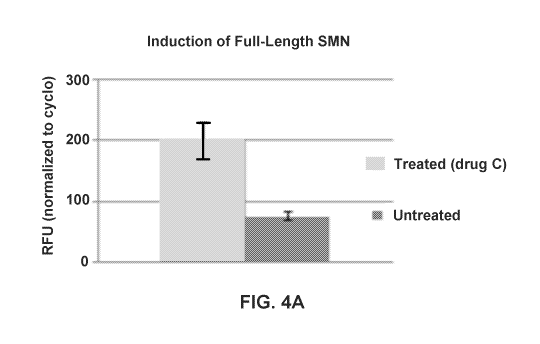
FIGS. 4A-4B show SMN induction. A. Induction of full-length SMN transcripts
(from
SMN2 transgene) after treatment with Drug C (digital PCR normalized to
cyclophilin) (p<0.01).
B. Structure of SMN-inducing Drug C.
FIG. 5 shows aged mice with SMN overexpression demonstrate improved
neuromuscular
function compared with aged control mice.
FIG. 6 shows mice with SMN overexpression demonstrate improved repair after
nerve
injury.
DETAILED DESCRIPTION
Definitions
The articles "a" and "an" are used herein to refer to one or to more than one
(i.e., to at
least one) of the grammatical object of the article. By way of example, "an
element" means one
element or more than one element.
"About" as used herein when referring to a measurable value such as an amount,
a
temporal duration, and the like, is meant to encompass variations of ±20%
or ±10%, more
preferably ±5%, even more preferably ±1%, and still more preferably .+-
Ø1% from the
specified value, as such variations are appropriate to perform the disclosed
methods.
A "prophylactic" treatment is a treatment administered to a subject who does
not exhibit
signs of a disease or exhibits only early signs for the purpose of decreasing
the risk of developing
pathology. The compounds of the invention may be given as a prophylactic
treatment to reduce
the likelihood of developing a pathology or to minimize the severity of the
pathology, if
developed.
A "therapeutic" treatment is a treatment administered to a subject who
exhibits signs or
symptoms of pathology for the purpose of diminishing or eliminating those
signs or symptoms.
3
CA 03005434 2018-05-15
WO 2017/087486
PCT/US2016/062225
The signs or symptoms may be biochemical, cellular, histological, functional,
subjective or
objective.
By "Survival Motor Neuron (SMN) -increasing substance" is meant any substance
that
increases the amount of SMN in an individual. Examples include, but are not
limited to,
compounds, compositions, anti sense oligonucleotides, long non coding RNAs or
treatment
therapies. "SMN-increasing substance" includes diverse classes of substances
that can be used
to increase SMN levels. Determining which compounds are able to increase SMN
levels can be
accomplished by those of skill in the art. Examples of such substances are
described herein.
An "analogue," "analog" or "derivative," which are used interchangeably,
refers to a
compound, e.g., a peptide or polypeptide, substantially similar in structure
and having the same
biological activity, albeit in certain instances to a differing degree, to a
naturally-occurring
molecule. Analogs differ in the composition of their amino acid sequences
compared to the
naturally-occurring polypeptide from which the analog is derived, based on one
or more
mutations involving (i) deletion of one or more amino acid residues at one or
more tel mini of the
polypeptide and/or one or more internal regions of the naturally-occurring
polypeptide sequence,
(ii) insertion or addition of one or more amino acids at one or more termini
(typically an
"addition" analog) of the polypeptide and/or one or more internal regions
(typically an
"insertion" analog) of the naturally-occurring polypeptide sequence or (iii)
substitution of one or
more amino acids for other amino acids in the naturally-occurring polypeptide
sequence.
The term "abnormal" when used in the context of organisms, tissues, cells or
components
thereof, refers to those organisms, tissues, cells or components thereof that
differ in at least one
observable or detectable characteristic (e.g., age, treatment, time of day,
etc.) from those
organisms, tissues, cells or components thereof that display the "normal"
(expected) respective
characteristic. Characteristics which are normal or expected for one cell or
tissue type, might be
abnormal for a different cell or tissue type.
As used herein, to "alleviate" a disease means to reduce the frequency or
severity of at
least one sign or symptom of a disease or disorder.
An "effective amount" as used herein, means an amount which provides a
therapeutic or
prophylactic benefit.
As used herein, the terms "therapy" or "therapeutic regimen" refer to those
activities
taken to alleviate or alter a disorder or disease state, e.g., a course of
treatment intended to reduce
or eliminate at least one sign or symptom of a disease or disorder using
pharmacological,
surgical, dietary and/or other techniques. A therapeutic regimen may include a
prescribed dosage
of one or more drugs or surgery. Therapies will most often be beneficial and
reduce or eliminate
4
CA 03005434 2018-05-15
WO 2017/087486
PCT/US2016/062225
at least one sign or symptom of the disorder or disease state, but in some
instances the effect of a
therapy will have non-desirable or side-effects. The effect of therapy will
also be impacted by the
physiological state of the subject, e.g., age, gender, genetics, weight, other
disease conditions,
etc.
The term "therapeutically effective amount" refers to the amount of the
subject
compound that will elicit the biological or medical response of a tissue,
system, or subject that is
being sought by the researcher, veterinarian, medical doctor or other
clinician. The term
"therapeutically effective amount" includes that amount of a compound that,
when administered,
is sufficient to prevent development of, or alleviate to some extent, one or
more of the signs or
symptoms of the disorder or disease being treated. The therapeutically
effective amount will vary
depending on the compound, the disease and its severity and the age, weight,
etc., of the subject
to be treated.
To "treat" a disease as the term is used herein, means to reduce the frequency
or severity
of at least one sign or symptom of a disease or disorder experienced by a
subject.
As used herein, the term "cell" is herein used in its broadest sense in the
art, referring to a
structural unit of a tissue present in a multicellular organism, which is
capable of self-replicating,
has genetic information and a mechanism for expressing it, and is surrounded
by a membrane
structure that isolates the living body from the outside. Cells used herein
may be either naturally-
occurring cells or artificially modified cells (e.g., fusion cells,
genetically modified cells, etc.), as
long as the cell has a chemical receptor or is capable of having such a
nucleic acid molecule
introduced therein. Examples of cell sources include, but are not limited to,
a single-cell culture;
the embryo, blood, or a body tissue of a normally- grown transgenic animal, a
mixture of cells
derived from normally-grown cell lines, and the like. In some preferred
embodiments, a cell
which is easily transformed or transfected is used.
As used herein, the term "tissue" refers to an aggregate of cells having
substantially the
same function and/or form in a multi-cellular organism. "Tissue" is typically
an aggregate of
cells of the same origin, but may be an aggregate of cells of different
origins as long as the cells
have the same function and/or form. Typically, a tissue constitutes a part of
an organ. Animal
tissues are separated into epithelial tissue, connective tissue, muscular
tissue, nervous tissue, and
the like, on a morphological, functional, or developmental basis.
As used herein, the telin "isolated" means that naturally accompanying
material is at least
reduced, or preferably substantially completely eliminated, in normal
circumstances. Therefore,
the telin "isolated cell" refers to a cell substantially free from other
accompanying substances
(e.g., other cells, proteins, nucleic acids, etc.) in natural circumstances.
The tenn "isolated" in
5
CA 03005434 2018-05-15
WO 2017/087486
PCT/US2016/062225
relation to nucleic acids or polypeptides means that, for example, the nucleic
acids or the
polypeptides are substantially free from cellular substances or culture media
when they are
produced by recombinant DNA techniques; or precursory chemical substances or
other chemical
substances when they are subsequently chemically synthesized.
As used herein, the term "gene" refers to an element defining a genetic trait.
A gene is
typically arranged in a given sequence on a chromosome. A gene which defines
the primary
structure of a protein is called a structural gene. A gene which regulates the
expression of a
structural gene is called a regulatory gene (e.g., promoter). As used herein,
"gene" may refer to a
"polynucleotide", "oligonucleotide", "nucleic acid", and a "nucleic acid
molecule."
As used herein, "gene product" includes a "polynucleotide", "oligonucleotide",
a
"nucleic acid" and a "nucleic acid molecule" and/or "protein", "polypeptide",
"oligopeptide" and
a "peptide", which are subsequent expression products of a gene. Those skilled
in the art
understand what a gene product is, according to the context used with
embodiments of the
present invention. Accordingly, gene used herein usually includes not only
double-stranded
DNA but also each single-stranded DNA, such as sense strand and antisense
strand constituting
thereof. Therefore, in embodiments of the present invention, the genes can
include any of
double-stranded DNA including human genome DNA, and single-stranded DNA (sense
strand)
including cDNA, as well as a single stranded DNA (anti sense) having a
sequence
complementary to the sense strand, as well as fragments thereof
The terms "polynucleotide", "oligonucleotide", "nucleic acid molecule" and
"nucleic
acid" as used herein have the same meaning and refer to a nucleotide polymer
having any length.
This term also includes an "oligonucleotide derivative" or a "polynucleotide
derivative". An
"oligonucleotide derivative" or a "polynucleotide derivative" includes a
nucleotide derivative, or
refers to an oligonucleotide or a polynucleotide having linkages between
nucleotides different
from typical linkages, which are interchangeably used.
As used herein, the term "fragment" with respect to a polypeptide or
polynucleotide
refers to a polypeptide or polynucleotide having a sequence length ranging
from 1 to n-1 with
respect to the full length of the reference polypeptide or polynucleotide (of
length n). The length
of the fragment can be appropriately changed depending on the purpose. For
example, in the case
of polypeptides, the lower limit of the length of the fragment includes 3, 4,
5, 6, 7, 8, 9, 10, 15,
20, 25, 30, 40, 50 or more nucleotides. Lengths represented by integers which
are not herein
specified (e.g., 1 1 and the like) can be appropriate as a lower limit. For
example, in the case of
polynucleotides, the lower limit of the length of the fragment includes 5, 6,
7, 8, 9, 10, 15, 20,
25, 30, 40, 50, 75, 100 or more nucleotides. Lengths represented by integers
which are not herein
6
CA 03005434 2018-05-15
WO 2017/087486
PCT/US2016/062225
specified (e.g., 1 1 and the like) may be appropriate as a lower limit. As
used herein, the length
of polypeptides or polynucleotides can be represented by the number of amino
acids or nucleic
acids, respectively. However, the above-described numbers are not absolute.
The above-
described numbers, as the upper or lower limits, are intended to include some
greater or smaller
numbers (e.g., . 10%), as long as the same function is maintained. In
embodiments of the
present invention, it is understood that any fragment can be used as long as
the fragment
functions as possessing transposition activity.
A "control" is an alternative subject or sample used in an experiment for
comparison
purpose. A control can be "positive" or "negative". For example, where the
purpose of the
experiment is to determine a correlation of an altered expression level of a
gene with a particular
type of pathology, it is generally preferable to use a positive control (a
subject or a sample from a
subject, carrying such alteration and exhibiting symptoms characteristic of
that disease), and a
negative control (a subject or a sample from a subject lacking the altered
expression and clinical
symptom of that disease).
"Differentially expressed" as applied to a gene, refers to the differential
production of the
mRNA transcribed from the gene or the protein product encoded by the gene. A
differentially
expressed gene may be overexpressed or underexpressed as compared to the
expression level of
a normal or control cell. In one aspect, it refers to a differential that is
at least 1.5 times, or at
least 2.5 times, or alternatively at least 5 times, or alternatively at least
10 times higher or lower
than the expression level detected in a control sample. The term
"differentially expressed" also
refers to nucleotide sequences in a cell or tissue which are expressed where
silent in a control
cell or not expressed where expressed in a control cell.
As used herein, the teini "modulate" means to vary the amount or intensity of
an effect or
outcome, e.g., to enhance, augment, diminish or reduce.
As used herein the term "ameliorate" is synonymous with "alleviate" and means
to
reduce or lighten. For example one may ameliorate the symptoms of sarcopenia
by making them
more bearable.
The present invention provides compounds which are in prodrug form. The teini
"prodrug" is intended to encompass compounds that, under physiological
conditions, are
converted into the therapeutically active agents of the present invention. A
common method for
making a prodrug is to include selected moieties that are hydrolyzed under
physiological
conditions to reveal the desired molecule. In other embodiments, the prodrug
is converted by an
enzymatic activity of the host animal. Additionally, prodrugs can be converted
to the compounds
of the present invention by chemical or biochemical methods in an ex vivo
environment. For
7
CA 03005434 2018-05-15
WO 2017/087486
PCT/US2016/062225
example, prodrugs can be slowly converted to the compounds of the present
invention when
placed in a transdeunal patch reservoir with a suitable enzyme or chemical
reagent.
Ranges: throughout this disclosure, various aspects of the invention can be
presented in a
range format. It should be understood that the description in range format is
merely for
convenience and brevity and should not be construed as an inflexible
limitation on the scope of
the invention. Accordingly, the description of a range should be considered to
have specifically
disclosed all the possible subranges as well as individual numerical values
within that range. For
example, description of a range such as from 1 to 6 should be considered to
have specifically
disclosed subranges such as from 1 to 3, from 1 to 4, from 1 to 5, from 2 to
4, from 2 to 6, from 3
to 6 etc., as well as individual numbers within that range, for example, 1, 2,
2.7, 3, 4, 5, 5.3, and
6. This applies regardless of the breadth of the range.
According to the methods taught herein, the subject is administered an
effective amount
of the agent. The tei __ ins effective amount and effective dosage are used
interchangeably. The
term effective amount is defined as any amount necessary to produce a desired
physiologic
response. Effective amounts and schedules for administering the agent may be
determined
empirically, and making such determinations is within the skill in the art.
The dosage ranges for
administration are those large enough to produce the desired effect in which
one or more
symptoms of the disease or disorder are affected (e.g., reduced or delayed).
The dosage should
not be so large as to cause substantial adverse side effects, such as unwanted
cross-reactions,
anaphylactic reactions, and the like. Generally, the dosage will vary with the
age, condition, sex,
type of disease, the extent of the disease or disorder, route of
administration, or whether other
drugs are included in the regimen, and can be determined by one of skill in
the art. The dosage
can be adjusted by the individual physician in the event of any
contraindications. Dosages can
vary, and can be administered in one or more dose administrations daily, for
one or several days.
Guidance can be found in the literature for appropriate dosages for given
classes of
pharmaceutical products.
As used herein the terms treatment, treat, or treating refers to a method of
reducing the
effects of a disease or condition or symptom of the disease or condition. Thus
in the disclosed
method, treatment can refer to a 10%, 20%, 30%, 40%, 50%, 60%, 70%, 80%, 90%,
or 1009/0
reduction in the severity of an established disease or condition or symptom of
the disease or
condition. For example, a method for treating a disease is considered to be a
treatment if there is
a 10% reduction in one or more symptoms of the disease in a subject as
compared to a control.
Thus the reduction can be a 10%, 20%, 30%, 40%, 50%, 60%, 70%, 80%, 90%, 100%,
or any
percent reduction in between 10% and 100% as compared to native or control
levels. It is
8
CA 03005434 2018-05-15
WO 2017/087486
PCT/US2016/062225
understood that treatment does not necessarily refer to a cure or complete
ablation of the disease,
condition, or symptoms of the disease or condition.
As used herein, the terms prevent, preventing, and prevention of a disease or
disorder
refers to an action, for example, administration of a therapeutic agent, that
occurs before or at
about the same time a subject begins to show one or more symptoms of the
disease or disorder,
which inhibits or delays onset or exacerbation of one or more symptoms of the
disease or
disorder. As used herein, references to decreasing, reducing, or inhibiting
include a change of
10%, 20%, 30%, 40%, 50%, 60%, 70%, 80%, 90% or greater as compared to a
control level.
Such telins can include but do not necessarily include complete elimination.
SMN-Increasing Substances and Methods of Use
Sarcopenia, the age-related wasting and loss of strength, is an important
neuromuscular
problem of aging. It affects up to 50% of individuals by the 8th decade, and
can lead to impaired
mobility, loss of independence, and increased risk of mortality. The
neuromuscular system is
comprised of groups of muscle fibers innervated by a single alpha motor
neuron, motor axon,
and synapses, tei _____________________________________________________ Hied a
motor unit. The normal development and maintenance of the motor unit
is dependent on trophic interactions between muscle and motor neurons. Losses
of muscle fiber,
neuromuscular junction (NMJ), and motor neuron function have all been
identified as potentially
important factors in sarcopenia, but the influence of neural factors on loss
of muscle function
with aging has received less attention. Electromyographic measures in vivo
enable longitudinal
quantification of the functional output from a muscle group, determination of
the number of
functional motor neurons, and assessment of NMJ integrity.
Prominent functional loss from the motor neuron pool associated with changes
in NMJ
transmission have been found in aging mice. Importantly these findings are
noted at earlier ages
than features of muscle loss, which shows that motor neuron dysfunction and
loss of connectivity
are important and early consequences of aging. The reduced ability of motor
neurons to repair
and maintain effective synaptic connectivity is an important factor underlying
the development
of sarcopenia. High expression of SMN protein in motor neurons is required for
NMJ formation
and maintenance during both development and regeneration, and SMN expression
in motor
neurons can be insufficient for motor unit repair and maintenance during
aging.
Increased SMN expression improves nerve regeneration in mice following sciatic
nerve
injury, and SMN overexpression can reduce aging-related motor unit losses and
improve
regeneration and maintenance at the NMJ.
9
CA 03005434 2018-05-15
WO 2017/087486 PCT/US2016/062225
In humans, SMN protein is encoded by two genes, SMN/ and SAIN2 (Lefebvre et
al.
1995). These genes differ by a single nucleotide which results in SMN2 exon7
being skipped in
the majority of transcripts. The loss of the amino acids encoded by exon7
results in an SMN
protein that does not oligomerize and gets rapidly degraded (Gennarelli et al.
1995; Lorson et al.
1998; Lorson et al. 2000; Burnett et al. 2009). In the autosomal recessive
disorder, spinal
muscular atrophy (SMA), SMN1 is lost or mutated and S'MN2 is retained which
results in
insufficient SMN for motor neurons and developmental NMJ maturation (Lefebvre
et al. 1995;
Burghes et al. 2009; Kariya et al. 2008). In contrast, SMN reduction induced
in adult mice (after
NMJ maturation) results in no marked abnormalities, but if adult mice with
reduced SMN
undergo sciatic nerve injury there is a marked defect in repair (Kariya et al.
2014).
The individual being treated can be of any age. Specifically, the individual
can be 30, 35,
40, 45, 50, 55, 60, 65, 70, 75, 80, 85, or 90 years old, or younger, older, or
any amount in
between. For example, the subject can be 35 years old or older. In one
embodiment, the subject
has not been diagnosed with spinal muscular atrophy (SMA). In another
embodiment, the
subject has been tested for SMA and it has been determined that the subject
does not have SMA.
SMA differs from sarcopenia and nerve injuries in multiple ways. Spinal
muscular atrophy
(SMA) is a neurological disorder characterized by loss of function of the
anterior horn cells in
the spinal cord that results from reduced levels of SMN protein as a result of
homozygous
mutationof the SMN1 gene. .
Sarcopenia
Disclosed herein is a method of reducing sarcopenia in an individual, the
method
comprising: identifying an individual with sarcopenia, an individual with
symptoms of
sarcopenia, or an individual at risk for developing sarcopenia, wherein the
subject is 35 years old
or older; and administering to the individual a Survival Motor Neuron (SMN) -
increasing
substance, thereby reducing sarcopenia, sarcopenia symptoms, or the risk of
sarcopenia in the
individual.
Sarcopenia has been defined by the prior art as the appendicular skeletal
muscle mass
(kg/height2 (m2)) being less than two standard deviations below the mean of a
young reference
group (i.e., the t-score). A t-score is detei mined by measuring the axial
skeletal muscle mass of a
patient, typically by dxa (i.e., dual energy xray absorptiometry) or a similar
and reproducible
measure. The measurement of axial skeletal muscle mass can be used to follow
the progress of
the patient to determine if treatment is slowing, preventing, or reversing
muscle mass decline.
CA 03005434 2018-05-15
WO 2017/087486
PCT/US2016/062225
Another type of patient that would benefit from the present invention is one
that has
suffered some loss of muscle mass, but who does not suffer from a condition
that interferes with
acts of daily living and/or prevents the subject from living an independent
life (e.g., a patient
who might soon need assisted living).
An individual can be diagnosed as having sarcopenia in a number of different
ways. For
example, sarcopenia can be measured using DXA (discussed above). DXA can be
measured in
combination with measuring gait speed (walking speed) (Muscaritoli et al.
(2010) Clinical
Nutrition 29(2):154-9). DXA measures lean body mass in reference to a normal
population. A
diagnostic definition that measures muscle strength and physical performance
can also be used.
Examples of such definitions have been developed by those of skill in the art.
Treating sarcopenia includes slowing its progression, stopping its
progression, and/or
partially reversing it. An example of slowing the progression of sarcopenia is
to change the
length of time a patient would go from a t-score of¨i.5 to ¨2 (e.g., if such a
progression would
normally take 5 years, then treating as used herein could slow this change to
10 years). Examples
of partial reversal include reducing a t-score 0.1, 0.2, 0.3, 0.4, 0.5, 0.6,
0.7, 0.8, 0.9, 1.0 or more
units (e.g., moving from a t-score of ¨2 to a t-score of ¨1.9, ¨1.8, ¨1.7,
¨1.6, ¨1.5, ¨1.4, ¨1.3,
¨1.2, ¨1.1, etc.). Treating sarcopenia can include inhibiting muscle
catabolism and/or increasing
muscle anabolism in a subject having or at risk of developing sarcopenia. It
can also include
improving the muscle: fat ratio in a subject having or at risk of developing
sarcopenia.
Treating sarcopenia can be measured by improving the gait of a subject having
or at risk
of developing sarcopenia. For example, improving the gait of the subject can
comprise
increasing stride length, reducing stride frequency, reducing stance width
variability or a
combination thereof. It can also include improving muscle functionality of a
subject having or at
risk of developing sarcopenia. The improvement in muscle functionality can be
demonstrated by
a reduction in the time required to complete a timed get-up-and-go test. It
can also be
demonstrated by a reduction in the time required to complete a timed stand
test.
Treating sarcopenia also includes delaying the onset of sarcopenia. For
example, if a
typical male age 50 would begin to see signs of sarcopenia by age 55,
treatment according to the
present invention could delay the onset 1, 2, 3, 4, 5, 6, 7, 8, 9, 10, or more
years. Thus, treating
sarcopenia would include treating patients who have not yet been diagnosed
with sarcopenia, but
who would be vulnerable or expected to be vulnerable to developing sarcopenia.
Patients who
are vulnerable or expected to be vulnerable also include (a) patients using
glucocorticoid
steroids, (b) patients with chronic infections, (c) patients with chronic
inflammatory conditions
(e.g., inflammatory bowel disease), and (d) patients with cancer.
11
CA 03005434 2018-05-15
WO 2017/087486
PCT/US2016/062225
Nerve Injury
Injuries to peripheral nerves can be caused by trauma, surgery, cancer and by
congenital
anomalies. Injuries to peripheral nerves can be also caused by radiation
therapy, chemotherapy,
metabolic/endocrine complications, inflammatory and autoimmune diseases,
vitamin
deficiencies, infectious diseases, toxic causes, accidental exposure to
organic metals and heavy
metals, drugs, amputations and disease or condition relating to a loss of
motor or sensory nerve
function. Nerve injury or lesion may include nerve transection, crush,
compression, stretch,
laceration (sharps or bone fragments), ischemia and blast. In addition, nerve
injury or lesion may
result from damage or disruption of the neuronal axons. Injuries to peripheral
nerves can be also
caused by radiation therapy, chemotherapy, metabolic/endocrine complications,
inflammatory
and autoimmune diseases, vitamin deficiencies, infectious diseases, toxic
causes, accidental
exposure to organic metals and heavy metals, drugs, amputations and disease or
condition
relating to a loss of motor or sensory nerve function. Nerve injury or lesion
may include nerve
transection, crush, compression, stretch, laceration (sharps or bone
fragments), ischemia and
blast. In addition, nerve injury or lesion may result from damage or
disruption of the neuronal
axons.
Disclosed herein are methods of treating an individual with nerve damage, the
method
comprising: identifying a subject with nerve damage; and administering to the
subject a Survival
Motor Neuron (SMN) protein-increasing substance, thereby reducing nerve damage
and/or
improving nerve function in the individual. The nerve damage can be caused by
an injury, for
example, such as a peripheral nerve injury. The nerve damage can also be
caused by a
mechanical traumatic brain, spinal cord or nerve tissue injury.
Increasing SMN Production
The SMN1 and SMN2 genes lie within the telomeric and centromeric halves,
respectively,
of a large, inverted duplication on chromosome 5q13. These genes share more
than 99%
nucleotide identity, and both are capable of encoding SMN (a 294-amino acid
RNA-binding
protein). Absence of SMN/ is partially compensated for by SMN2, which produces
enough
SMN protein to allow for relatively normal development in cell types other
than motor neurons.
However, SMN2 cannot fully compensate for loss of SMNI because, although SMN2
is
transcribed at a level comparable to that of SMN1, a large majority of SMN2
transcripts lack
exon 7, resulting in production of a truncated, less stable SMN protein
(Lefebvre et al., 1995;
Kashima et al., 2007; Lefebvre et al. 1997; Coovert et al. 1997).
12
CA 03005434 2018-05-15
WO 2017/087486
PCT/US2016/062225
Disclosed herein are a variety of methods and compositions for increasing SMN
production. While examples of such are given herein, it is noted that these
examples are not
intended to be limiting, and that the invention disclosed relates to any
method or composition
that increases SMN production, which is used to treat or prevent sarcopenia
and/or nerve
damage. For example, PCT Application W02009146033, which is incorporated
herein in its
entirety, discloses various methods and compositions for increasing SMN
production. However,
the present disclosure is not limited to these examples.
Disclosed herein are SMN-increasing substances that increase the expression or
activity
of an SMN agonist (e.g. Pumilio homolog 1, eIF-4E, MAP1B, Rholõ type II BMP
receptor, type
II TGF-beta receptor, R- SMAD protein, FGF-2 or FGF-3 receptor, RAS, and MAP
kinase) or
another protein or gene product that regulates the expression or activity of
the SMN agonist, such
as a transcription factor or other protein that acts upstream of the SMN
agonist in a particular
signaling cascade. In one embodiment, the agent is a small molecule compound
that directly or
indirectly increases the expression and/or activity of the SMN agonist. These
agents (SMN-
increasing substances) can be used to treat or prevent sarcopenia and/or nerve
damage, and are
discussed in more detail herein (W02009146033A3, incorporated by reference
herein for its
teaching concerning SMN increasing substances).
The present invention also encompasses a method of treating or preventing
sarcopenia
and/or nerve damage in a subject in need thereof comprising administering to
the subject an
SMN-increasing substance that decreases the expression or activity of a SMN
antagonist. As
used herein, a "SMN antagonist" is a gene or protein that negatively regulates
SMN function. A
SMN antagonist can also refer to a gene or protein that acts to interfere or
compete for binding
with SMN target proteins. SMN antagonists include, but are not limited to,
Fmrl, Moesin, slik,
SMAD6, and SMAD7. In some embodiments, an agent that decreases the expression
or activity
of a SMN antagonist is a small molecule compound that directly or indirectly
decreases the
expression and/or activity of the SMN antagonist. In other embodiments, an
agent that decreases
the expression or activity of a SMN antagonist is an antibody or fragment
thereof that binds to
the SMN antagonist and prevents its interaction with other proteins and/or
inhibits its activity.
In certain embodiments, an agent that decreases the expression or activity of
a SMN
antagonist is an antisense nucleic acid targeted to a sequence of the SMN
antagonist. Suitable
antisense nucleic acids can comprise ribonucleotides or deoxyribonucleotides
and preferably,
have at least one chemical modification. Such modifications include without
limitation locked
nucleic acids, peptide nucleic acids, sugar modifications, such as 2'-0-alkyl
(e.g. 2'-0-methyl, 2'-
0-methoxyethyl), 2'-fluoro, and 4' thio modifications, and backbone
modifications, such as one
13
CA 03005434 2018-05-15
WO 2017/087486
PCT/US2016/062225
or more phosphorothioate, morpholino, or phosphonocarboxylate linkages (see,
for example,
U.S. Patent Nos. 6,693,187 and 7,067,641, which are herein incorporated by
reference in their
entireties). Other modifications of antisense nucleic acids to enhance
stability and improve
efficacy, such as those described in U.S. Patent No. 6,838,283, which is
herein incorporated by
reference in its entirety, are known in the art and are suitable for use in
the methods of the
invention. Preferable antisense nucleic acids useful for inhibiting the
expression and/or activity
of a SMN antagonist are about 20 to about 200 nucleotides in length. Antisense
nucleic acids can
comprise a sequence that is at least partially complementary (e.g. at least
about 75%, 80%, 85%,
90%, 95%, 96%, 97%, 98%, or 99% complementary) to a gene sequence for a SMN
antagonist
or portion thereof. In one embodiment, the antisense nucleic acid comprises a
sequence that is
100% complementary to a gene sequence for a SMN antagonist or portion thereof.
The antisense
nucleic acid can target either a coding or non-coding region of the SMN
antagonist gene. In
some embodiments, the antisense nucleic acid targets an mRNA transcript from
the SMN
antagonist gene.
In other embodiments, an agent that decreases the expression or activity of a
SMN
antagonist is an inhibitory RNA molecule targeted to a sequence of the SMN
antagonist. The
inhibitory RNA molecule may be a double-stranded, small interfering RNA
(siRNA) or a short
hairpin RNA molecule (shRNA) comprising a stem-loop structure or a ribozyme.
The double-
stranded regions of the inhibitory RNA molecule may comprise a sequence that
is at least
partially identical and partially complementary, e.g. about 75%, 80%, 85%,
90%, 95%, 96%,
97%, 98%, or 99% identical and complementary, to a coding or non-coding region
of a gene
sequence for a SMN antagonist. In one embodiment, the double-stranded regions
of the
inhibitory RNA molecule may contain 100% identity and complementarity to the
gene sequence
for a SMN antagonist. In another embodiment, the inhibitory RNA molecule
targets an mRNA
transcript from the SMN antagonist gene.
The antisense nucleic acid or inhibitory RNA molecule targeted to a SMN
antagonist can
be encoded on an expression construct as described herein. In one embodiment,
the antisense
nucleic acid or inhibitory RNA molecule is under the control of a tissue-
specific promoter. In a
preferred embodiment, the tissue-specific promoter is a muscle-specific
promoter. In another
preferred embodiment, the tissue-specific promoter is a neuron-specific
promoter.
SMN levels can be increased by preventing skipping of exon 7 of SMN2
(W02001066129 Al, hereby incorporated by reference in its entirety for its
disclosure of
preventing skipping of exon 7 of SMN2). Accordingly, the present invention
provides a
substance which is capable of preventing the skipping (exclusion) of exon 7 of
the SMN2 gene.
14
CA 03005434 2018-05-15
WO 2017/087486
PCT/US2016/062225
Thus, the substance is suitable for the use as a therapeutic agent in treating
or preventing
sarcopenia and/or nerve damage.
The present invention also provides a process for changing the pre-mRNA
processing
relating to the SMN gene of a mammalian cell, which process comprises exposing
the cell to a
substance, which is capable of controlling the inclusion of exon 7 of the SMN2
gene, and thereby
treating or preventing sarcopenia and/or nerve damage. The present invention
also provides a
mammalian host cell which is stably transfected with a DNA encoding a
polypeptide which is
capable of at least partially preventing the skipping (exclusion) of exon 7 of
the SMN2 gene,
thereby treating or preventing sarcopenia and/or nerve damage. The transfected
mammalian host
cell, which preferably originates from a human individual to be treated and/or
from a cultured
human cell or cell line, is useful in gene therapy to treat or prevent
sarcopenia and/or nerve
damage.
Also disclosed are methods of increasing SMN levels by altering SMN2 splicing
(PCT
Application W02010120820 Al, hereby incorporated in its entirety for its
disclosure concerning
altered SMN2 splicing and antisense compounds targeted to ,SMN2). The present
invention is
directed to antisense compounds targeted to and hybridizable with a nucleic
acid molecule
encoding SMN2. Antisense compounds can be used to target intron 7 of SMN2,
which
compounds modulate splicing of SMN2 pre-mRNAs. In one embodiment, modulation
of splicing
results in an increase in exon 7 inclusion. In another embodiment, modulation
of splicing results
in a decrease in exon 7 inclusion. Disclosed herein are antisense compounds 16
to 30 nucleotides
in length targeted to intron 7 of SMN2, wherein the compounds comprise 2'-0-
methoxyethyl
sugar modifications, for example. Therefore, disclosed are methods of
increasing SMN
production, and thereby treating or preventing sarcopenia and/or nerve damage
by using 2'-0-
methoxyethyl (2'1\40E) chemistry. The antisense compounds can be targeted to
cis splicing
regulatory elements. Regulatory elements include exonic splicing enhancers,
exonic splicing
silencers, intronic splicing enhancers and intronic splicing silencers. Exonic
and intronic splicing
silencers are preferred targets.
Also provided are methods for modulating splicing of SMN2 mRNA in a cell,
tissue or
organ, thereby treating or preventing sarcopenia and/or nerve damage in a
subject. In one
embodiment, modulation of splicing is exon inclusion. In another embodiment,
modulation of
splicing is exon skipping. In one aspect, the compound is targeted to an
intronic splicing silencer
element. In another aspect, the compound is targeted to an exonic splicing
silencer element.
These are discussed in further detail below. Also disclosed is the use of an
antisense
oligonucleotide for the preparation of a medicament for modulating splicing of
an SMN2 pre-
CA 03005434 2018-05-15
WO 2017/087486
PCT/US2016/062225
mRNA. In one aspect, modulation of splicing results in an increase in exon 7
inclusion. Use of
an antisense oligonucleotide provided herein for the treatment or prevention
or sarcopenia and/or
nerve damage is further provided.
Also disclosed herein is a method of increasing SMN production by blocking
long
noncoding RNAs. Small molecules that activate the SMN promoter also act on
SMN]. The
down-regulation of long noncoding RNA which regulate SMN level (in particular
in neurons)
activates both SMN/ and SM1T2, and gives much higher SMN expression.
Also disclosed are methods for treating or preventing sarcopenia and/or nerve
injury
using the polycomb repressive complex 2 (PRC2)-interacting RNAs to increase or
enhance
production of SMN/ or SMN (W02013173638 Al, hereby incorporated by reference
in its
entirety for its disclosure concerning long noncoding RNAs and SMN). Polycomb
repressive
complex 2 (PRC2) is a histone methyltransferase and a known epigenetic
regulator involved in
silencing of genomic regions through methylation of histone H3. Among other
functions, PRC2
interacts with long noncoding RNAs (IncRNAs), such as RepA, Xist, and Tsix, to
catalyze
trimethylation of histone H3-lysine27. PRC2 contains four subunits, Eed,
Suz12, RbAp48, and
Ezh2. Single stranded oligonucleotides that bind to PRC2-associated regions of
RNAs (e.g.,
IncRNAs) which can arise from within a genomic region that encompasses or that
is in
functional proximity to the SMN/ or SMN2 gene can induce or enhance expression
of SMN] or
SMN2. This upregulation can result from inhibition of PRC2 mediated repression
of SMN1 or
SMN2.
Disclosed herein are methods of increasing SMN2 production by administering
aryl
substituted thiazol-2-yl-piperidines or related compounds (PCT Application
W02011130515 Al,
hereby incorporated by reference in its entirety for its disclosure concerning
increasing SMN2
production using aryl substituted thiazol-2-yl-piperidines). For example,
compounds and
pharmaceutically acceptable salts of Formula I and Foimula II are provided
herein.
R3 R3
--N 7-1¨\
X¨R4 A X¨R4
As
R7
Formula I Formula II
Compounds of Formula III, IV, and V are also provided herein.
16
CA 03005434 2018-05-15
WO 2017/087486 PCT/US2016/062225
R3
RptNN
\1/4/-1
N R,
Ri"" S Ri=-= S S
Foi ________________ mul a III Formula IV Formula V
These compounds are subformulae of Formula I in which:
X is CH, A is CRi and B is CR2 (Formula III);
X is CH, A is CRi and B is N (Formula IV); and
X is CH, A is CR2 and B is CRi (Formula V)
The variables in Formula III, IV, and V may carry the definitions set forth
for Formula I
or any of the definitions set forth below.
Compounds of Formula VI, VII, VIII, IX, and X are also provided herein.
R3 R3
R2 N R4 N N R4
R4
¨N
R7 R7 R7 Re
Formula VI Formula VII Formula
VIII
R3 R2 R3
N ............. N R4N
RI ) 1).¨R4
R7 R7
Formula IX Formula X
Compounds of subformulae VI to X are subformulae of Formula II in which: X is
CH, A
is R2, B is Rh D is N, and E is N (Formula VI),
X is CH, A is N, B is Ri, D is N, and E is N (Formula VII);
X is CH, A is N, B is Ri, D is N, and E is CR6 (Foimula VIII);
X is CH, A is N, B is Ri, D is CR5, and E is N (Formula IX); and X is CH, A is
Ri, B is
R2, D is N, and E is N (Formula X).
Also disclosed are method of increasing SMN2 production by administering
ubiquitin
carboxyl-teiminal hydrolase Li (UCHL I) regulators or related compounds
Disclosed herein is a
method of treating or preventing sarcopenia and/or nerve damage comprising
regulating the
17
CA 03005434 2018-05-15
WO 2017/087486
PCT/US2016/062225
expression level of survival of motor neuron 1 (SMN1) comprising administering
to a subject in
need thereof a therapeutically effective amount of ubiquitin carboxyl-terminal
hydrolase Li
(UCHL1) regulator and a pharmaceutically acceptable carrier. The protein
expression level of
SMN/ of the present invention is reduced by ubiquitin carboxyl-terminal
hydrolase Li (UCHL1)
regulator, wherein the ubiquitin carboxyl-terminal hydrolase Li (UCHL1)
reduces the
expression level of SMN/ by increasing the level of ubiquitinated SMN1.
Disclosed herein are tricyclo-DNA (tc-DNA) antisense nucleotides that are
effective in
facilitating exon skipping during pre-mRNA processing, in masking intronic
silencer sequences
and/or stem-loop sequences in pre-mRNA, and in targeting the RNase-mediated
destruction of
mRNA. Described herein are tc-DNA antisense nucleotides that may be used in
methods for the
treatment or prevention of sarcopenia and/or nerve damage by skipping mutated
exons, such as
masking an intronic silencing sequence and/or a terminal stem- loop sequence
within an SMN2
gene to yield modified functional SMN2 protein, including an amino acid
sequence encoded by
exon 7, which is capable of at least partially complementing a non- functional
SMN1 protein.
(See U.S. Patent Application U520120149756, herein incorporated by reference
in its entirety
for its teaching concerning tricyclo-DNA).
SMN production can also be increased by binding the intronic inhibitory
sequence
element, named ISS-Ni (for "intronic splicing silencer"), located in the SMN2
gene (U.S. Patent
8,586,559, hereby incorporated by reference in its entirety for disclosing ISS-
Ni as it relates to
SMN). The compositions and methods of the instant invention include
oligonucleotide reagents
(e.g., oligoribonucleotides) that effectively target the SMN2 ISS-Ni site in
the SMN2 pre-mRNA,
thereby modulating the splicing of SMN2 pre-mRNA to include exon 7 in the
processed
transcript. The ISS-Ni blocking agents of the invention cause elevated
expression of SMN
protein, thus compensating for the loss of SMN protein expression.
Also disclosed herein is the use of human SMN-like protein (HSLP), the
polynucleotides
encoding HSLP, and the use of these compositions and variants thereof for the
treatment or
prevention of sarcopenia and/or nerve injury (U.S. Patent 6,130,064, hereby
incorporated by
reference in its entirety for its disclosure concerning HSLP).
SMN levels can be increased through gene therapy. Any gene delivery method
known to
those of skill in the art can be used with the methods disclosed herein. A
"gene delivery vehicle"
is defined as any molecule that can carry inserted polynucleotides into a host
cell. Examples of
gene delivery vehicles are liposomes, biocompatible polymers, including
natural polymers and
synthetic polymers; lipoproteins; polypeptides; polysaccharides; artificial
viral envelopes;
recombinant yeast cells, metal particles; and bacteria or viruses,
baculovirus, adenovirus and
18
CA 03005434 2018-05-15
WO 2017/087486
PCT/US2016/062225
retrovirus, bacteriophage, cosmid, plasmid, fungal vectors and other
recombination vehicles
typically used in the art which have been described for expression in a
variety of eukaryotic and
prokaryotic hosts and may be used for gene therapy as well as for simple
protein expression.
Specifically, disclosed is the use of an adeno-associated virus, particularly
AAV9.
A "viral vector" is defined as a recombinantly produced virus or viral
particle that
comprises a polynucleotide to be delivered into a host cell, either in vivo,
ex vivo or in vitro.
Examples of viral vectors include retroviral vectors, adenovirus vectors,
adeno-associated virus
vectors, alphavirus vectors and the like. Alphavirus vectors, such as Semliki
Forest virus-based
vectors and Sindbis virus- based vectors, have also been developed for use in
gene therapy and
immunotherapy. (Schlesinger and Dubensky (1999) Curr. Opin. Biotechnol. 5:434-
439 and Ying
et al. (1999) Nat. Med. 5(7):823-827). In aspects where gene transfer is
mediated by a retroviral
vector, a vector construct refers to the polynucleotide comprising the
retroviral genome or part
thereof and a therapeutic gene. As used herein, "retroviral mediated gene
transfer" or "retroviral
transduction" carries the same meaning and refers to the process by which a
gene or nucleic acid
sequences are stably transferred into the host cell by virtue of the virus
entering the cell and
integrating its genome into the host cell genome. The virus can enter the host
cell via its normal
mechanism of infection or be modified such that it binds to a different host
cell surface receptor
or ligand to enter the cell. As used herein, "retroviral vector" refers to a
viral particle capable of
introducing exogenous nucleic acid into a cell through a viral or viral-like
entry mechanism.
In aspects where gene transfer is mediated by a DNA viral vector, such as an
adenovirus
(Ad) or adeno-associated virus (AAV), a vector construct refers to the
polynucleotide comprising
the viral genome or part thereof and a transgene. Wild-type AAV has high
infectivity and
specificity integrating into the host cell's genome.
Vectors that contain both a promoter and a cloning site into which a
polynucleotide can
be operatively linked are well known in the art. Such vectors are capable of
transcribing RNA in
vitro or in vivo and are commercially available from sources such as
Stratagene (La Jolla, CA)
and Promega Biotech (Madison, W1). In order to optimize expression and/or in
vitro
transcription, it may be necessary to remove, add or alter 5' and/or 3'
untranslated portions of the
clones to eliminate extra, potential inappropriate alternative translation
initiation codons or other
sequences that may interfere with or reduce expression, either at the level of
transcription or
translation. Alternatively, consensus ribosome binding sites can be inserted
immediately 5' of the
start codon to enhance expression.
Gene delivery vehicles also include several non-viral vectors, including
DNA/liposome
complexes, recombinant yeast cells and targeted viral protein-DNA complexes.
Liposomes that
19
CA 03005434 2018-05-15
WO 2017/087486
PCT/US2016/062225
also comprise a targeting antibody or fragment thereof can be used in the
methods of this
invention. To enhance delivery to a cell, the nucleic acid or proteins of this
invention can be
conjugated to antibodies or binding fragment(s) thereof which bind cell
surface antigens, e.g.,
TCR, CD3 or CD4.
Preferably, a pharmaceutically effective amount of an agent for treating or
preventing
sarcopenia or nerve injury is administered to the subject (e.g. human
subject). As used herein, the
term "pharmaceutically effective amount" means an amount that improves one or
more
symptoms.
Formulation of an agent described herein for treatment purposes comprises
combining
pharmaceutically effective amounts of the agent of the invention with
pharmaceutically
acceptable diluents, preservatives, solubilizers, emulsifiers, adjuvants
and/or carriers. Such
compositions include diluents of various buffer content (e.g., Tris- HC1,
acetate, phosphate), pH
and ionic strength; additives such as detergents and solubilizing agents
(e.g., Tween 80,
Polysorbate 80), anti-oxidants (e.g., ascorbic acid, sodium metabisulfite),
preservatives (e.g.,
Thimerosol, benzyl alcohol) and bulking substances (e.g., lactose, mannitol);
incorporation of the
material into particulate preparations of polymeric compounds such as
polylactic acid,
polyglycolic acid, etc. or into liposomes. Protein agents of the invention may
be produced as
fusion proteins to modulate or extend the half- life of the protein. Such
fusion proteins may
include human serum albumin, transferrin, other serum proteins, etc. Such
compositions may
influence the physical state, stability, rate of in vivo release, and rate of
in vivo clearance of the
present compounds. See, e.g., Remington's Pharmaceutical Sciences, 18th Ed.
(1990, Mack
Publishing Co., Easton, Pa. 18042) pages 1435-1712. The compositions may be
prepared in
liquid form, or may be in dried powder, such as lyophilized form. Implantable
sustained release
formulations are also contemplated. Preferably, pharmaceutical compositions
will be prepared in
a form appropriate for the intended application and be essentially free of
pyrogens, as well as
other impurities that could be haimful to humans or animals.
Colloidal dispersion systems, such as macromolecule complexes, nanocapsules,
microspheres, beads, and lipid-based systems including oil-in-water emulsions,
micelles, mixed
micelles, and liposomes, can be used as delivery vehicles for the therapeutic
agents described
herein, especially for nucleic acid-based therapeutic agents (e.g. expression
vectors, antisense
nucleic acids, and inhibitory RNA molecules). Commercially available fat
emulsions that are
especially suitable for delivering the nucleic acid agents of the invention to
tissues, such as
skeletal muscle tissue, include Intralipide, Liposyne, Liposyng II, Liposyne
III, Nutrilipid,
and other similar lipid emulsions. A preferred colloidal system for use as a
delivery vehicle in
CA 03005434 2018-05-15
WO 2017/087486
PCT/US2016/062225
vivo is a liposome (i.e., an artificial membrane vesicle). The preparation and
use of such systems
is well known in the art. Exemplary formulations are also disclosed in US
5,981,505; US
6,217,900; US 6,383,512; US 5,783,565; US 7,202,227; US 6,379,965; US
6,127,170; US
5,837,533; US 6,747,014; and WO 03/093449, which are herein incorporated by
reference in
their entireties.
Administration of the agents according to the methods of the present invention
may be
via any common route so long as the target tissue (e.g. skeletal muscle, motor
neurons) is
available via that route. This includes oral, nasal, or buccal. Alternatively,
administration may be
by intradermal, subcutaneous, intramuscular, intraperitoneal, intrathecal,
intraventricular,
intraparenchymal, intraarterial or intravenous injection, or by direct
injection into skeletal muscle
tissue or motor neurons. The therapeutic agents described herein would
noimally be
administered as pharmaceutically acceptable compositions, as described herein.
The agents may
also be administered parenterally or intraperitoneally. By way of
illustration, solutions of the
therapeutic agents as free base or pharmacologically acceptable salts can be
prepared in water
suitably mixed with a surfactant, such as hydroxypropylcellulose. Dispersions
can also be
prepared in glycerol, liquid polyethylene glycols, and mixtures thereof and in
oils. Under
ordinary conditions of storage and use, these preparations generally contain a
preservative to
prevent the growth of microorganisms.
The pharmaceutical forms suitable for injectable use include, for example,
sterile aqueous
solutions or dispersions and sterile powders for the extemporaneous
preparation of sterile
injectable solutions or dispersions. Generally, these preparations are sterile
and fluid to the extent
that easy injectability exists. Preparations should be stable under the
conditions of manufacture,
storage, and administration (depot delivery) and should be preserved against
the contaminating
action of microorganisms, such as bacteria and fungi. Appropriate solvents or
dispersion media
may contain, for example, water, ethanol, polyol (for example, glycerol,
propylene glycol, and
liquid polyethylene glycol, and the like), suitable mixtures thereof, and
vegetable oils. The
proper fluidity can be maintained, for example, by the use of a coating, such
as lecithin, by the
maintenance of the required particle size in the case of dispersion and by the
use of surfactants.
The prevention of the action of microorganisms can be brought about by various
antibacterial an
antifungal agents, for example, parabens, chlorobutanol, phenol, sorbic acid,
thimerosal, and the
like. In many cases, it will be preferable to include isotonic agents, for
example, sugars or
sodium chloride. Prolonged absorption of the injectable compositions can be
brought about by
the use in the compositions of agents delaying absorption, for example,
aluminum monostearate
and gelatin.
21
CA 03005434 2018-05-15
WO 2017/087486
PCT/US2016/062225
Sterile injectable solutions may be prepared by incorporating the therapeutic
agents in an
appropriate amount into a solvent along with any other ingredients (for
example as enumerated
above) as desired, followed by filtered sterilization. Generally, dispersions
are prepared by
incorporating the various sterilized active ingredients into a sterile vehicle
which contains the
basic dispersion medium and the desired other ingredients, e.g., as enumerated
above. In the case
of sterile powders for the preparation of sterile injectable solutions, the
preferred methods of
preparation include vacuum- drying and freeze-drying techniques which yield a
powder of the
active ingredient(s) plus any additional desired ingredient from a previously
sterile-filtered
solution thereof. Upon formulation, solutions are preferably administered in a
manner
compatible with the dosage formulation and in such amount as is
therapeutically effective. The
formulations may easily be administered in a variety of dosage forms such as
injectable
solutions, drug release capsules and the like. For parenteral administration
in an aqueous
solution, for example, the solution generally is suitably buffered and the
liquid diluent first
rendered isotonic for example with sufficient saline or glucose. Such aqueous
solutions may be
used, for example, for intrathecal, intravenous, intramuscular, subcutaneous
and intraperitoneal
administration. Preferably, sterile aqueous media are employed as is known to
those of skill in
the art, particularly in light of the present disclosure. Some variation in
dosage will necessarily
occur depending on the stage of disease to be treated and individual
characteristics of the subject
to be treated {e.g. size, age, overall health, etc.). The person responsible
for administration will,
in any event, deteimine the appropriate dose for the individual subject.
Moreover, for human
administration, preparations should meet sterility, pyrogenicity, general
safety and purity
standards as required by FDA Office of Biologies standards.
Any SMN-increasing agents disclosed herein can be co-administered with another
SMN-
increasing agent. In other words, one, two, three, or more of SMN-increasing
agents or methods
of treatment can be administered simultaneously, or before or after each
other. Also disclosed are
methods comprising administering an SMN-increasing agent as well as another
method or
composition known for treating or preventing sarcopenia. For example,
compositions known to
treat or prevent sarcopenia include, but are not limited to, testosterone,
estrogen, growth
hormone, creatine, or beta-alanine.
The subject can also be advised concerning diet and exercise. For example,
disclosed
herein are methods of treating or preventing sarcopenia and/or nerve damage in
an individual,
comprising providing an SMN-increasing substance, as well as providing infoi
____ illation regarding
exercise and nutrition.
22
CA 03005434 2018-05-15
WO 2017/087486
PCT/US2016/062225
Also disclosed are methods comprising administering an SMN-increasing agent as
well
as another method or composition known for treating nerve injury. For example,
compositions
known to treat nerve injury include, but are not limited to, neuregulin,
tegaserod, or follistatin.
Also disclosed is inhibiting myostatin, or administering a TNF-a inhibitor.
A number of embodiments of the invention have been described. Nevertheless, it
will be
understood that various modifications may be made without departing from the
spirit and scope
of the invention. Accordingly, other embodiments are within the scope of the
following claims.
EXAMPLES
Example 1: Loss of Motor Unit Function During Aging and SMN Overexpress ion
Electrotnyographic studies and aging-related changes
Muscle wasting by ¨2 years has been shown to occur in aging mice, and this
loss of
muscle mass appears to develop sometime after 15 months of age (Sayer etal.
2013;
Shavlakadze et al. 2010). Prominent morphological alterations at the NMJ have
also been
identified in aged mice, but reports vary regarding the extent of histological
loss of alpha motor
neurons in aging mice. (Chai et al. 2011; Sayer et al. 2013; Jang et al.
2010). In 27 month old
mice, in which overt motor neuron cell body loss was not identified, fiber
type grouping
consistent with loss of motor neurons (and compensatory reinnervation) was
identified when
compared with young adult mice (Chai et al. 2011). In various hind limb
muscles of 2 year old
mice, up to 20% of synapses may be fully denervated (not including partially
denervated or
morphologically altered synapses) (Chai et al. 2011; Valdez et al. 2010; Wang
et al. 2005). Loss
of muscle mass and strength are consequences of the functional loss of motor
units and motor
unit connectivity (with or without histological loss of the cell body of the
motor neuron). This
shows the value of a functional measure of the entire motor unit, rather than
isolated histological
analyses at the junction and motor neuron. Loss of motor neuron function can
be central to
sarcopenia, and aging-related weakness and muscle wasting emerge as the
ability of the motor
unit pool to compensate for these losses becomes insufficient. This can be
related to insufficient
numbers of functional motor neurons or intrinsic failure of individual motor
neurons to maintain
synaptic connections. Loss of motor unit function can occur much earlier than
previously
realized (presented later in Figure 1), which have gone unnoticed due to the
ability of the mouse
to undergo significant compensation through collateral sprouting. Therefore
many of the
previously observed changes at the synapse and in muscle can reflect secondary
changes of
earlier degenerative events that occur during aging.
23
CA 03005434 2018-05-15
WO 2017/087486
PCT/US2016/062225
Electromyographic studies can be utilized to observe the aging-related changes
that occur
in a longitudinal fashion. Identification of the early events can help to
understand the primary
and secondary changes that occur with aging over time. Of particular
importance is the fact that
these measures are modified from similar measurements that can be obtained in
clinical studies.
Therefore the ability to readily translate findings from animal studies is
significantly increased.
Compound muscle action potential (CMAP) amplitude is an assay of the
functional output of the
motor unit pool, regardless of whether functional loss occurs at the muscle
fiber, synapse, motor
axon, or motor neuron level. Motor unit number estimation (MUNE) provides an
estimation of
the number of functional motor units/neurons/motor axons. CMAP responses
following
repetitive nerve stimulation (RNS) can be recorded to assess sufficiency of
NMJ transmission.
Single fiber electromyography is utilized to assess NMJ transmission at single
synapses.
SFEMG findings that signify reduced safety factor (jitter) or insufficient
safety factor to reach
threshold (blocking) are the most sensitive parameters of abnormal NMJ
transmission in vivo
(Stalberg et al. 1997; Juel 2012). These electromyographic studies are
utilized to understand the
function of the entire motor unit, which are then correlated with other force
measures in vivo and
histological measures. Electromyographic studies have been performed from
cohorts of
C57BL/6J male mice at 3, 6, 10, 14, and 24 months of age. Prominent functional
loss from the
motor neuron pool innervating the hind limb muscles have been shown in aging
mice, and these
findings begin to emerge at ¨13 months (Figure 1). By 24 months motor unit
number estimation
(MUNE) is significantly reduced by ¨35% compared with 10 month old mice
consistent with
loss of motor neuron function (Figure 1A). These findings with MUNE are
consistent with
recently published results of motor axon counts in aged mice showing 35% loss
at the Li ventral
root (Valdez et al. 2010). Importantly, MUNE has advantages over anatomical
counts due to the
ability of MUNE to estimate the number of functional motor units/neurons,
rather than presence
or absence of motor neuron cell body or ventral root counts (i.e. histological
loss). Single motor
unit potential amplitude (or SMUP) is an assay of the output of a single motor
unit or neuron.
SMUP amplitude is increased by ¨28% in 2 year old mice (Figure 1B) consistent
with
compensatory reinnervation in response to denervation. Nevertheless, CMAP
amplitude is not
maintained in 24 month old mice and is reduced by ¨25% compared with 10 month
old mice
(Figure 1C). Therefore the reduced CMAP in the 24 month old animals identifies
the inability
of the remaining motor neurons to maintain a normal functional output from the
muscle. NMJ
transmission recordings utilizing axonal stimulation-SFEMG from the lateral
gastrocnemius
muscle demonstrate increased jitter in 14 month old mice 14.7 1.1 las (n=22
synapses/ single
muscle fibers) compared with 6 month old mice is 10.8 1.4 .is (18
synapses/single muscle
24
CA 03005434 2018-05-15
WO 2017/087486
PCT/US2016/062225
fibers) (p=0.04) (Figure 1D). Repetitive nerve stimulation (RNS) demonstrate
CMAP decrement
(abnormal NMJ transmission) in 50% (n=6) of 24 month old mice compared with no
mice
(n=10) at 10 months (p=0.036) also supporting functional changes at the NMJ.
Importantly
changes of motor neuron failure and reinnervation are occurring in these mice
(reduced MUNE
and increase SMUP size noted above), therefore the NMJ defects noted can be
secondary to
failure at the motor neuron or ineffective formation and maintenance of the
NMJ.
SMN2 Mouse Models
SMN protein is critical to motor neuron function and survival and low levels
lead to
motor neuron degeneration (Burghes et al. 2009; Arnold et al. 2013). If SMN
reduction occurs
after maturation of the NMJ, mice have no marked defects in early adulthood
but during aging
develop worsening NMJ defects and have impaired ability to reform effective
synapses
following nerve injuries (Kariya 2014). SMN can have an important role in
aging and
maintenance of the functional connectivity of the motor neuron at the synapse.
SMA mouse
models with the human SMN2 transgene and knockout of the mouse Sinn gene (and
therefore
low levels of SMN protein) have been generated to investigate spinal muscular
atrophy (Monami
et al. 2000). Additionally, high copy SMN2 mice (both 8 copy and 16 copy) were
generated to
study the effects of high SMN levels (Monani et al. 2000) and have been
maintained on a FVB
background in the colony for over 15 years. Different breeding strategies can
be utilized to
generate mice with varying levels of SMN.
In humans, sarcopenia is linked to not only loss of function and independence
of
activities of daily living but also an increased risk of early mortality
(Sayer et al. 2013; Landi et
al. 2013; Batsis et al. 2014; Atkins et al. 2014). It was shown that mice
harboring high copy
numbers of the SMN2 transgene (on a FVB background) live longer than wild type
FVBN mice.
A median survival of 591 days and 760 days in male and female FVB mice,
respectively, was
previously shown (Yuan et al. 2009). Whereas mice in the colony with 16 copies
of the SMN2
transgene (and homozygous null alleles for the mouse Sinn gene) on a FVB
background have a
median survival of 900 days for males (n=12) and 945 days for females (n=6).
This shows that
SMN protein levels modulate the effects of aging.
Wild-type FVB mice are compared to mice with 8 copies of the SMN2 transgene
and
mice with 16 copies of the SMN2 transgene (both lacking a functional mouse
Sinn gene). Mice
are analyzed longitudinally with non-invasive measures of motor unit function
(electromyographic and force-described below). These longitudinal measures are
compared with
cohorts of aged mice for endpoint measures of SMN mRNA transcript levels
(enriched motor
CA 03005434 2018-05-15
WO 2017/087486
PCT/US2016/062225
neuron samples via laser capture microdissection) as well as pathological
correlates at the ventral
root, NMJ and muscle (described herein). In this manner, it is determined
which degenerative
changes are occurring and the timing and severity of these changes.
Furthermore,
responsiveness of these changes to high levels of SMN expression in transgenic
animals are
determined. This detei mines that SMN levels can modulate the effects of
aging on the motor
unit.
A. Mouse Groups
= Wild-type FVB mice
= 8-copy SMN2 mice (homozygous null for mouse Sinn) with wild-type levels
of SMN
from SMN2 transgene
= 16-copy SMN2 (homozygous null for mouse Sinn) with 2X wild-type levels of
SMN from
SMN2 transgene
For each of these groups, 5 males/5 females are included for longitudinal
electromyographic and force measures and 3 males/3 females are included at
each endpoint
assessment of histology and SMN levels.
B. Longitudinal electromyographic studies
= CMAP (output of motor unit pool supplying a muscle) CMAP responses are
recorded
from the triceps surae muscles to assess total electromyographic output from
the triceps
surae muscles (Duque et al. 2014; Arnold et al. 2014).
= MUNE (# of functional motor neurons innervating a muscle) MUNE of the
sciatic nerve
is utilized to estimate the number of functional motor units innervating the
triceps surae
muscles (Duque et al. 2014; Arnold et al. 2014).
= Repetitive Nerve Stimulation (RNS) and single fiber EMG (SFEMG) (NMJ
transmission)
Two methods are utilized to assess NMJ transmission. RNS is
a clinical
electromyographic measure that involves recording the CMAP response following
a train
of stimuli. When NMJ transmission is sufficient, CMAP amplitude remains
stable, but if
endplate potentials are of insufficient amplitude to reach threshold for
muscle fiber action
potential generation, CMAP amplitude decrements (Arnold 2014). Single fiber
EMG
(SFEMG) is extracellular recording technique that utilizes a combination of
narrow filter
settings and small electrode surface to allow recording of single muscle fiber
action
potentials following axonal stimulation. SFEMG is the most sensitive measure
of
26
CA 03005434 2018-05-15
WO 2017/087486
PCT/US2016/062225
disruption of NMJ transmission in vivo, and it can be performed in human
studies and
animal models (Gooch et al. 2001; Meekins et al. 2007).
C. Force measurements in vivo:
Assessment of plantar flexion force (twitch force, maximum tetanic force, and
force-
frequency curve) is performed utilizing a whole mouse testing system with a
force
transducer and nerve stimulation in vivo. Force measurements are obtained in
vivo for
the benefits of being a physiological comparison to the electromyographic
recordings.
Force measurements are compared with muscle size to determine how motor unit
changes
relate to muscle quality, a important factor in aged muscle in humans (Newman
et al.
2003; Newman et al. 2006; Misic et al. 2007; Kennis et al. 2014; Goodpaster et
al. 2006.)
D. SMN transcript levels:
Enriched motor neuron samples are obtained via laser capture microdissection
(Figure 2)
and assessed by droplet digital PCR (similar to our work in both mouse and the
pig
(Ruggiu et al. 2012; Duque et al. 2014) to measure SMN transcript levels in
order to
understand how these levels correspond to function of the motor unit and
synaptic
maintenance during aging.
E. Endpoint morphological measurements.
= Muscle: Some histopathological features noted in muscle during aging
recapitulate
findings expected in denervation (e.g. type II fiber atrophy, grouped atrophy,
angular
atrophic fiber) as compared with features such as muscle fibers with central
nuclei which
corresponds with muscle fiber degeneration/regeneration (Carlson 1993). The
histopathological response of muscle during aging is determined, and these
findings are
compared to the functional status of the motor unit. Tibialis anterior and
gastrocnemius
muscle characteristics of fiber size, type, nuclei location, and wet muscle
weights are
quantified.
= L4 Ventral Root Counts: L4 ventral root motor axon counts are performed
to determine
the presence of motor axonal loss in the lower limb. L4 supplies both the
tibialis anterior
and gastrocnemius (Mohan et al. 2014), and L4 (and L3) spinal ventral roots
consistently
contribution to the sciatic nerve in the mouse, while L5 is less consistent
(Rigaud 2008).
27
CA 03005434 2018-05-15
WO 2017/087486
PCT/US2016/062225
= NMJ Innervation and Neurofilament Accumulation: Innervation status of
NMJ's are
quantified in the gastrocnemius and tibialis anterior. Due to its relevance to
a SMN
deficient state, splenius capitis, a posterior neck muscle that has shown
particular
susceptibility in SMN deficiency (Ling et al. 2012), is studied. Presynaptic
neurofilament
accumulation, also a prominent feature in SMA patients and mouse models (with
low
levels of SMN), is assessed with anti-neurofilament staining (Ling et al.
2012).
Linear mixed effects models are used to analyze electromyographic results
(Arnold
2014). The rate of change for each genotype of mice is analyzed. The analysis
of variance
(ANOVA) with factors of age and genotype is conducted to pairwise compare the
endpoints
measures between genotypes within each age group (10, 18, 26m) and between age
groups for
each genotype. The association of morphological and SMN transcript with
electromyographic
measures is evaluated by regression models. Including 10 mice (5 male/ 5
female) gives greater
than 80% power to detect a 1.2-fold difference in the number of functional
motor units, with
coefficient of variation (CV)=20% (Figure 1) at a=0.025 for 2 primary
contrasts (8, 16 copies
SMN2 vs WT). Increased power for the longitudinal study is due to the
correlation of repeat
measures in same animal. For the cohort of endpoints aging mice, n=6 (3
males/3 females) gives
enough power to detect a 1.5 fold difference. Power calculations were
performed using PASS 12
(NCSS, LLC; Kaysville, Utah). Data analysis can be done by using SAS software
(SAS, Inc.,
Cary, NC).
These studies provide insight to the components of the motor unit (muscle
fiber, neuron,
synapse) that are the earliest to show dysfunction, so that the compensatory
changes of the motor
unit following these early changes can be tracked. Functional,
electrophysiological, and
morphological measure determination allow a comprehensive look at the
neuromuscular system
with aging. Cross-sectional data shows that drop out of motor unit and
synaptic function begins
to occur between 10 and 13 months of age. Loss of motor neuron function can
precede features
of muscle wasting in aged mice. Loss of motor neuron function and associated
secondary NMJ
defects can occur simultaneously prior to losses in muscle fiber function due
to enlargement of
the single motor unit size. Alternatively, the earliest defects can occur at
the synapse and loss of
motor unit connectivity can be a later consequence. SMN overexpression in
transgenic animals
with 16 copies of SMN2 (and homozygous null mouse Sinn alleles) can result in
improved motor
neuron and synaptic function during aging. Mice with 8 copies of SMN2 (and
homozygous null
mouse Sinn alleles) and wild-type FVB mice can have similar features of aging-
related motor
unit degeneration over time (due to similar levels of SMN protein). Fully
developed adult mice
28
CA 03005434 2018-05-15
WO 2017/087486
PCT/US2016/062225
are assessed prior to any onset of electromyographic abnormalities (10 months)
and these
animals can be followed longitudinally until overt muscle atrophy is expected
(24 months). This
can capture mice pre-symptomatically and after onset of muscle loss. A gene
therapy approach
can also be used to transfer the SW/ gene (Foust et al. 2010; Bevan et al.
2010) for testing
SMN protein overexpression in other congenic strains. Additionally, telomerase
deficient mice
(Terc") can be used. Third generation Terc" mice have features of sarcopenia
associated with
reduced MUNE counts at 8 months are (173 44) compared with wild-type C57B6/J
mice at 10
months age (341 29). These mice can be utilized to study the effects of aging
on neuromuscular
function.
Example 2: SMN overexpression improves motor axon connectivity following
injury
Axonal sprouting and NMJ formation following nerve injury are less effective
in aged
mice, and aging motor neurons are unable develop as extensive innervation
territory or output
(Tanaka et al. 1991; Yuan et al 2012). Sciatic nerve crush studies resulted in
an injury associated
with complete recovery of CMAP response (functional output) by 80-100 days in
wild type
FVBN mice, but MUNE demonstrates incomplete motor unit repair with reduced
numbers of
functional motor neurons/units (Figure 3A and B). Therefore the nerve crush
technique models a
situation of complete recovery of functional output, but incomplete
regeneration of innervating
motor units. This system can be utilized to understand compensatory nerve
repair activity and to
test therapeutic targets to improve nerve repair activity.
The data disclosed herein shows that transgenic mice with SMN overexpression
have
improved regenerative capacity following nerve injury compared with wild type
mice.
Importantly, the data show that increased SIVIN protein expression improves
synaptic formation
following nerve crush (Figure 3). The early rate of repair activity in SMN
overexpression mice
(week 0 to week 2, which is dictated primarily by axonal sprouting) is similar
to WT mice. In
contrast, there is significant divergence between 2 weeks and 6 weeks post-
crush, and this is
consistent with the expected timing of NMJ formation/maturation. These
findings are consistent
with the prior work by Monani et al (analyzing NMJ formation histologically)
in mice with wild
type and deficient levels of SMN (Kariya et al. 2014). This repair activity
can be critical during
aging to maintain motor neuron function and NMJ connectivity. High levels of
SMN protein can
reduce degenerative susceptibility of the motor unit at the NMJ during injury
and lessen the loss
of regenerative capacity in aging mice.
Comparison is made between untreated young and old mice. Additionally, mice
are
treated with viral mediated gene transfer to assess the ability of SMN protein
overexpression to
29
CA 03005434 2018-05-15
WO 2017/087486
PCT/US2016/062225
improve axonal sprouting and synaptic formation following nerve crush injury
in both young and
aging animals. Therefore aging-related defects in repair activity (following
nerve crush (Fahim et
al. 1991; Landis et al. 2012)) can be corrected with SMN overexpression.
Testing motor neuron repair activity following SMN overexpression
Young adult mice (6 months)
= 8 males/8 females (AAV-SMN)
= 8 males/8 females (AAV-empty vector)
Aged mice (24 months)
= 8 males/8 females (AAV-SMN)
= 8 males/8 females (AAV-empty vector)
A. Double Sciatic Nerve Crush:
Sciatic crush is performed in both young adult (at 6 months) and aged mice (at
24
months).The sciatic nerve are crushed utilizing a standardized technique
(Bauder et al. 2012).
Using hemostatic forceps the sciatic nerve is crushed for 15 seconds at 3
clicks of the
hemostatic forceps. Then using the forceps the same crush site is crushed a
second time for
15 seconds at 3 clicks force. Carbon can be applied to the second forceps to
mark and
therefore identify the lesion site for endpoint histology.
B. SAN Overexpression:
There are two treatment cohorts in each age group: treatment with either scAAV-
SMN or
empty vector.
C. Longitudinal and Endpoint Analysis:
The rate and extent of the recovery of motor neuron and synaptic function is
assessed with
longitudinal electromyographic and endpoint histopathology measures. For the
entailed
studies, 5 males and 5 females (10 mice) in each group are followed
longitudinally for the
entire 10 weeks post-crush and then undergo endpoint analysis. At four weeks
crush, 3
males and 3 females (6 mice) in each group are euthanized for endpoint
analysis.
10 and 6 mice/group are used for longitudinal and endpoints measures study,
respectively. Longitudinal study is analyzed by mixed effect model and
endpoints measures are
CA 03005434 2018-05-15
WO 2017/087486
PCT/US2016/062225
analyzed by ANOVA. The rate of the change in electromyographic or endpoint
measures are
compared between groups (SMN vs empty vector) for aged or young mice, and
between age
group (aged vs young) for the mice with SMN overexpressed.
Regeneration of motor neuron and synaptic function can be impaired in placebo-
treated
aged animals (reduced number of functional motor neurons/units and impaired
synaptic
function). This loss of repair activity in aging mice can improved with
increased SMN
expression. In these treated animals, el ectromyographic measures demonstrate
increased
numbers of functioning motor units and improved synaptic transmission.
Similarly
morphological and force measures are improved. SMN expression increases during
nerve
regeneration (following nerve crush) (Kariya et al. 2014). SMN expression in
untreated animals
(following nerve crush and during nerve regeneration) can be diminished in
aged compared to
young mice.
Example 3: SMN overexpression improves muscle size and function in aged,
sarcopenic
mice
Progressive changes of synaptic disruption and denervation are noted during
the normal
aging process in both humans and mice (Delbono 2003). Interestingly, these
changes of
denervation are absent in some neck muscles of the mouse (Li et al. 2011), but
in leg muscles
which are important for weight bearing and mobility (in mouse and humans)
findings of
denervation have consistently been noted (Chai et al. 2011; Valdez et al.
2010). Age-related
alterations at the NMJ can originate from motor neuron loss, muscle fiber
degeneration, and
primary NMJ deficits. Synaptic signaling from denervated muscle fibers is
critical during
synaptic repair following muscle fiber injury (Doherty et al. 1993), but the
origins of synaptic
failure during aging have been unknown. Additionally, prior studies
demonstrated a less robust
response to partial denervation, which can be one important aspect of aging-
related muscle
weakness and wasting (Fahim et al. 1993; Jacob et al. 1990). These findings
show the
importance of denervation in the loss of function of aging individuals, and
that denervation can
be related to impaired maintenance of synaptic input to muscle fibers.
SMN expression is increased during both synaptic formation and maturation
during
development and during repair following an injury (Kariya et al. 2014).
Interestingly, SMN
expression is also increased during exercise (Biondi et al. 2008). A mouse
model of SMA
demonstrated increased SMN expression, preserved motor neuron counts, and
prolonged survival
following treadmill running exercise (Biondi et al. 2008). Thus, SMN protein
appears to have
31
CA 03005434 2018-05-15
WO 2017/087486
PCT/US2016/062225
significant importance with synaptic formation and can be regulated with
increased synaptic
activity.
Drug compounds (such as Drug C, shown in Figure 4) that efficiently stimulate
full-
length SMN from SMN2 (Figure 4A and B) are disclosed herein. Mice homozygous
for a mouse
Snm knockout allele that harbor 8 copies of a SMN2 transgene which are
phenotypical normal
and have SMN levels similar to wild-type mice (Monani et al. 2000). Thus these
mice can be
treated with SMN-inducing compounds that act on SMN2 to increase full-length
SMN protein to
test the effect of SMN overexpression on aging-related loss of muscle mass and
function.
SMN protein can be used to correct muscle wasting in aging mice. Utilizing
mice with
the SMN2 transgene (8 copies) SMN protein expression (from the SMN2 transgene)
is increased
utilizing compounds that increase full-length SMN production from SMN2. Mice
are monitored
longitudinally with electromyographic recordings and force measurements in
vivo and with
endpoint histology. Muscle mass and function improvement is determined, and
the mechanism
of this improvement (i.e. improved synaptic transmission, increased number of
functional motor
neurons, or increased muscle size or function), can be assessed.
A. SMN-Overexpression:
16 mice (8 male/8 female) are treated with vehicle (placebo) and compared with
16 mice
(8 male/8 female) treated with an SMN-inducing compound (such as Drug C shown
in
Figure 4) to increase SMN protein expression. Treatment occurs after
development of
sarcopenia (24 months).
B. Longitudinal and Endpoint Analysis:
To measure the longitudinal effects on motor unit function, longitudinal
electromyographic and force measures in vivo are perfoi _______________ flied
(as described herein) at 18, 24, and
27 months. Endpoint morphological assessment is performed to assess full-
length SMN
transcript levels, NMJ innervation status, muscle fiber size, and ventral root
counts at 27 months.
Treatment Groups at 24 months
(After onset of muscle loss)
= 16 mice (8 male/8 female)
(Drug C)
= 16 mice (8 male/8 female)
32
CA 03005434 2018-05-15
WO 2017/087486
PCT/US2016/062225
(Placebo/vehicle)
SMN overexpression, after onset of sarcopenia in 24 month-old mice can result
in
improved motor neuron connectivity and this can lead to improved muscle mass
and function.
SMN2 containing mice are used to assess small molecule SMN induction as a
therapeutic
strategy in sarcopenia. An alternative strategy for testing the effect of SMN
overexpression in
mice after onset of sarcopenia is gene transfer for induction of SMN
overexpression rather than
SMN2 containing mice and treatment with SMN-inducing compounds.
Unless defined otherwise, all technical and scientific terms used herein have
the same
meanings as commonly understood by one of skill in the art to which the
disclosed invention
belongs. Publications cited herein and the materials for which they are cited
are specifically
incorporated by reference.
Those skilled in the art will recognize, or be able to ascertain using no more
than routine
experimentation, many equivalents to the specific embodiments of the invention
described
herein. Such equivalents are intended to be encompassed by the following
claims.
REFERENCES
1. Faulkner JA, Larkin LM, Claflin DR, Brooks SV Age-related changes in the
structure
and function of skeletal muscles. Clinical and experimental pharmacology &
physiology. Nov
2007;34(11):1091-1096.
2. Manini TM, Visser M, Won-Park S, et al. Knee extension strength
cutpoints for
maintaining mobility. Journal of the American Geriatrics Society. Mar
2007;55(3):451-457.
3. Chai RJ, Vukovic J, Dunlop S, Grounds MD, Shavlakadze T. Striking
denervation of
neuromuscular junctions without lumbar motoneuron loss in geriatric mouse
muscle. PloS one.
2011;6(12):e28090.
4. Valdez G, Tapia JC, Kang H, et al. Attenuation of age-related changes in
mouse
neuromuscular synapses by caloric restriction and exercise. Proceedings of the
National
Academy of Sciences of the United States of America. Aug 17 2010;107(33):14863-
14868.
5. Tomlinson BE, Irving D. The numbers of limb motor neurons in the human
lumbosacral
cord throughout life. Journal of the neurological sciences. Nov 1977;34(2):213-
219.
6. Oda K. Age changes of motor innervation and acetylcholine receptor
distribution on
human skeletal muscle fibres. Journal of the neurological sciences. Nov-Dec
1984;66(2-3):327-
338.
7. Kawamura Y, O'Brien P, Okazaki H, Dyck PJ. Lumbar motoneurons of man II:
the
number and diameter distribution of large- and inteiniediate-diameter cytons
in "motoneuron
columns" of spinal cord of man. Journal of neuropathology and experimental
neurology. Sep-Oct
1977;36(5):861-870.
8. Arnold WD, Porensky PN, McGovern VL, et al. Electrophysiological
Biomarkers in
Spinal Muscular Atrophy: Preclinical Proof of Concept. Annals of clinical and
translational
neurology. Jan 1 2014;1(1):34-44.
33
CA 03005434 2018-05-15
WO 2017/087486
PCT/US2016/062225
9. Fu AKY, Cheung ZH, Ip NY. [beta]-catenin in reverse action. Nat
Neurosci. 03//print
2008; 11(3):244-246.
10. Koliatsos VE, Clatterbuck RE, Winslow JW, Cayouette MR, Prices DL.
Evidence that
brain-derived neurotrophic factor is a trophic factor for motor neurons in
vivo. Neuron. 3//
1993;10(3):359-367.
11. Ikeda K, Wong V, Holmlund TH, et al. Histometric effects of ciliary
neurotrophic factor
in wobbler mouse motor neuron disease. Annals of neurology. Jan 1995;37(1):47-
54.
12. Kablar B, Belliveau AC. Presence of neurotrophic factors in skeletal
muscle correlates
with survival of spinal cord motor neurons. Developmental dynamics : an
official publication of
the American Association of Anatomists. Nov 2005;234(3):659-669.
13. Fahim MA. Morphological correlates of physiological responses in
partially denervated
mouse muscle during aging. International Journal of Developmental
Neuroscience. 6//
1993;11(3):303-310.
14. Verdu E, Buti M, Navarro X. The effect of aging on efferent nerve
fibers regeneration in
mice. Brain Res. Oct 23 1995;696(1-2):76-82.
15. Tanaka K, Webster HD. Myelinated fiber regeneration after crush injury
is retarded in
sciatic nerves of aging mice. The Journal of comparative neurology. Jun 8
1991;308(2):180-187.
16. Yuan Q, Su H, Guo J, et al. Decreased c-Jun expression correlates with
impaired spinal
motoneuron regeneration in aged mice following sciatic nerve crush.
Experimental gerontology.
Apr 2012;47(4):329-336.
17. Kariya S, Obis T, Garone C, et al. Requirement of enhanced Survival
Motoneuron protein
imposed during neuromuscular junction maturation. The Journal of clinical
investigation. Feb 3
2014; 124(2):785-800.
18. Lefebvre S, Burglen L, Reboullet S, et al. Identification and
characterization of a spinal
muscular atrophy-determining gene. Cell. Jan 13 1995;80(1):155-165.
19. Gennarelli M, Lucarelli M, Capon F, et al. Survival motor neuron gene
transcript analysis
in muscles from spinal muscular atrophy patients. Biochem Biophys Res Commun.
Aug 4
1995;213(1):342-348.
20. Lorson CL, Strasswimmer J, Yao JM, et al. SMN oligomerization defect
correlates with
spinal muscular atrophy severity. Nat Genet. May 1998;19(1):63-66.
21. Lorson CL, Androphy EJ. An exonic enhancer is required for inclusion of
an essential
exon in the SMA-determining gene SMN. Hum Mol Genet. Jan 22 2000;9(2):259-265.
22. Burnett BG, Munoz E, Tandon A, Kwon DY, Sumner CJ, Fischbeck KH.
Regulation of
SMN protein stability. Molecular and cellular biology. Mar 2009;29(5):1107-
1115.
23. Ruggiu M, McGovern VL, Lotti F, et al. A role for SMN exon 7 splicing
in the selective
vulnerability of motor neurons in spinal muscular atrophy. Molecular &
Cellular Biology.
2012;32(1): 126-138.
24. Burghes AH, Beattie CE. Spinal muscular atrophy: why do low levels of
survival motor
neuron protein make motor neurons sick?. [Review] [171 refs]. Nature Reviews
Neuroscience.
2009; 10(8):597-609.
25. Kariya S, Park GH, Maeno-Hikichi Y, et al. Reduced SMN protein impairs
maturation of
the neuromuscular junctions in mouse models of spinal muscular atrophy. Hum
Mol Genet. Aug
15 2008;17(16):2552-2569.
26. Monani UR, Sendtner M, Coovert DD, et al. The human centromeric
survival motor
neuron gene (SMN2) rescues embryonic lethality in Smn-/- mice and results in a
mouse with
spinal muscular atrophy. Human Molecular Genetics. February 12, 2000
2000;9(3):333-339.
27. Foust KD, Wang X, McGovern VL, et al. Rescue of the spinal muscular
atrophy
phenotype in a mouse model by early postnatal delivery of SMN. Nat Biotech.
2010;28(3):271-
274.
34
CA 03005434 2018-05-15
WO 2017/087486
PCT/US2016/062225
28. Arnold WD, Kassar D, Kissel JT. Spinal muscular atrophy: Diagnosis and
management in
a new therapeutic era. Muscle Nerve. Oct 24 2014.
29. Arnold WD, Burghes AH. Spinal muscular atrophy: The development and
implementation of potential treatments. Annals of neurology. Aug 12 2013.
30. Kallman DA, Plato CC, Tobin JD. The role of muscle loss in the age-
related decline of
grip strength: cross-sectional and longitudinal perspectives. Journal of
gerontology. May
1990;45(3):M82-88.
31. Melton LJ, 3rd, Khosla S, Crowson CS, O'Connor MK, O'Fallon WM, Riggs
BL.
Epidemiology of sarcopenia. Journal of the American Geriatrics Society. Jun
2000;48(6):625-
630.
32. Goodpaster BH, Park SW, Harris TB, et al. The loss of skeletal muscle
strength, mass,
and quality in older adults: The Health, Aging and Body Composition Study. J
Gerentol Med Sci.
2006;61:1059-1064.
33. Delmonico MJ, Harris TB, Visser M, et al. Longitudinal study of muscle
strength, quality,
and adipose tissue infiltration. Am J Clin Nutr. Dec 2009;90(6):1579-1585.
34. Newman AB, Haggerty CL, Goodpaster B, et al. Strength and muscle
quality in a well-
functioning cohort of older adults: the Health, Aging and Body Composition
Study. Journal of
the American Geriatrics Society. Mar 2003;51(3):323-330.
35. Newman AB, Kupelian V, Visser M, et al. Strength, but not muscle mass,
is associated
with mortality in the health, aging and body composition study cohort. The
journals of
gerontology. Series A, Biological sciences and medical sciences. Jan
2006;61(1):72-77.
36. Misic MM, Rosengren KS, Woods JA, Evans EM. Muscle quality, aerobic
fitness and fat
mass predict lower-extremity physical function in community-dwelling older
adults.
Gerontology. 2007;53(5):260-266.
37. Li J GT, Arnold WD, Rosen GD,Zaworski PG, Rutkove SB. A comparison of
three
electrophysiological methods for the assessment of disease status in a mild
spinal muscular
atrophy mouse model. PloS one. 2014;"In Press".
38. Duque SI, Arnold WD, Odermatt P, et al. A large animal model of
Spinal Muscular
Atrophy and correction of phenotype. Annals of neurology. Dec 16 2014.
39. Gooch CL, Mosier DR. Stimulated single fiber electromyography in the
mouse:
techniques and nonnative data. Muscle Nerve. Jul 2001;24(7):941-945.
40. Meekins GD, Carter GT, Emery MJ, Weiss MD. Axonal degeneration in
the Trembler-j
mouse demonstrated by stimulated single-fiber electromyography. Muscle Nerve.
Jul
2007;36(1):81-86.
41. Bevan AK, Hutchinson KR, Foust KD, et al. Early heart failure in the
SMNDelta7 model
of spinal muscular atrophy and correction by postnatal scAAV9-SMN delivery.
Hum Mol Genet.
Oct 15 2010;19(20):3895-3905.
42. Bevan AK, Duque S, Foust KD, et al. Systemic gene delivery in large
species for
targeting spinal cord, brain, and peripheral tissues for pediatric disorders.
Molecular therapy : the
journal of the American Society of Gene Therapy. Nov 2011;19(11):1971-1980.
43. Sayer AA, Robinson SM, Patel HP, Shavlakadze T, Cooper C, Grounds MD.
New
horizons in the pathogenesis, diagnosis and management of sarcopenia. Age and
Ageing. March
1,2013 2013;42(2):145-150.
44. Shavlakadze T, McGeachie J, Grounds MID. Delayed but excellent myogenic
stem cell
response of regenerating geriatric skeletal muscles in mice. Biogerontology.
Jun 2010;11(3):363-
376.
45. Jang YC, Lustgarten MS, Liu Y, et al. Increased superoxide in vivo
accelerates age-
associated muscle atrophy through mitochondria' dysfunction and neuromuscular
junction
degeneration. FASEB journal : official publication of the Federation of
American Societies for
Experimental Biology. May 2010;24(5):1376-1390.
CA 03005434 2018-05-15
WO 2017/087486
PCT/US2016/062225
46. Wang Z-M, Zheng Z, Messi ML, Delbono 0. Extension and magnitude of
denervation in
skeletal muscle from ageing mice. The Journal of physiology. 2005;565(3):757-
764.
47. Stalberg E, Trontelj JV. The study of normal and abnormal neuromuscular
transmission
with single fibre electromyography. Journal of neuroscience methods. Jun 27
1997;74(2):145-
154.
48. Juel VC. Evaluation of neuromuscular junction disorders in the
electromyography
laboratory. Neurologic clinics. May 2012;30(2):621-639.
49. Hsieh-Li HM, Chang J-G, Jong Y-J, et al. A mouse model for spinal
muscular atrophy.
Nat Genet. 2000;24(1):66-70.
50. Landi F, Cruz-Jentoft AJ, Liperoti R, et al. Sarcopenia and mortality
risk in frail older
persons aged 80 years and older: results from ilSIRENTE study. Age Ageing. Mar
2013;42(2):203-209.
51. Batsis JA, Mackenzie TA, Barre LK, Lopez-Jimenez F, Bartels SJ.
Sarcopenia,
sarcopenic obesity and mortality in older adults: results from the National
Health and Nutrition
Examination Survey III. European journal of clinical nutrition. Sep
2014;68(9):1001-1007.
52. Atkins JL, Whincup PH, Morris RW, Lennon LT, Papacosta 0, Wannamethee
SG.
Sarcopenic obesity and risk of cardiovascular disease and mortality: a
population-based cohort
study of older men. Journal of the American Geriatrics Society. Feb
2014;62(2):253-260.
53. Yuan R, Tsaih S-W, Petkova SB, et al. Aging in inbred strains of mice:
study design and
interim report on median lifespans and circulating IGF1 levels. Aging Cell.
2009;8(3):277-287.
54. Kennis E, Verschueren S, Van Roie E, Thomis M, Lefevre J, Delecluse C.
Longitudinal
impact of aging on muscle quality in middle-aged men. Age (Dordrecht,
Netherlands).
2014;36(4):9689.
55. Goodpaster BH, Park SW, Harris TB, et al. The loss of skeletal muscle
strength, mass,
and quality in older adults: the health, aging and body composition study. The
journals of
gerontology. Series A, Biological sciences and medical sciences. Oct
2006;61(10):1059-1064.
56. Carlson BM. The regeneration of skeletal muscle. A review. The American
journal of
anatomy. Jun 1973;137(2):119-149.
57. Mohan R, Tosolini AP, Morris R. Targeting the motor end plates in the
mouse hindlimb
gives access to a greater number of spinal cord motor neurons: An approach to
maximize
retrograde transport. Neuroscience. 8/22/ 2014;274(0):318-330.
58. Rigaud M, Gemes G, Barabas ME, et al. Species and strain differences in
rodent sciatic
nerve anatomy: implications for studies of neuropathic pain. Pain. May
2008;136(1-2):188-201.
59. Ling KKY, Gibbs RM, Feng Z, Ko C-P. Severe neuromuscular denervation of
clinically
relevant muscles in a mouse model of spinal muscular atrophy. Human Molecular
Genetics.
January 1,2012 2012;21(1):185-195.
60. Ling KK, Gibbs RM, Feng Z, Ko CP. Severe neuromuscular denervation of
clinically
relevant muscles in a mouse model of spinal muscular atrophy. Hum Mol Genet.
Jan 1
2012;21(1): 185-195.
61. Jacob JM, Robbins N. Age differences in morphology of reinnervation of
partially
denervated mouse muscle. J Neurosci. May 1990;10(5):1530-1540.
62. Bauder AR, Ferguson TA. Reproducible mouse sciatic nerve crush and
subsequent
assessment of regeneration by whole mount muscle analysis. Journal of
visualized experiments :
JoVE. 2012(60).
63. Landis SC, Amara SG, Asadullah K, et al. A call for transparent
reporting to optimize the
predictive value of preclinical research. Nature. 10/11/print
2012;490(7419):187-191.
64. Delbono 0. Neural control of aging skeletal muscle. Aging cell. Feb
2003;2(1):21-29.
65. Li Y, Lee Yi, Thompson WJ. Changes in Aging Mouse Neuromuscular
Junctions Are
Explained by Degeneration and Regeneration of Muscle Fiber Segments at the
Synapse. The
Journal of Neuroscience. October 19, 2011 2011;31(42):14910-14919.
36
CA 03005434 2018-05-15
WO 2017/087486
PCT/US2016/062225
66. van Mier P, Lichtman J. Regenerating muscle fibers induce directional
sprouting from
nearby nerve terminals: studies in living mice. The Journal of Neuroscience.
September 1, 1994
1994; 14(9):5672-5686.
67. Biondi 0, Grondard C, Lecolle S, et al. Exercise-induced activation of
NMDA receptor
promotes motor unit development and survival in a type 2 spinal muscular
atrophy model mouse.
J Neurosci. Jan 23 2008;28(4):953-962.
68. Doherty TJ, Vandervoort AA, Brown WF. Effects of ageing on the motor
unit: a brief
review. Canadian journal of applied physiology = Revue canadienne de
physiologie appliquee.
Dec 1993;18(4):331-358.
69. Campbell NIJ, McComas AJ, Petito F. Physiological changes in ageing
muscles. Journal
of Neurology, Neurosurgery & Psychiatry. April 1, 1973 1973;36(2):174-182.
70. Larsson L, Ansved T. Effects of ageing on the motor unit. Progress in
neurobiology. Apr
1995;45(5):397-458.
71. Mittal KR, Logmani FH. Age-related reduction in 8th cervical ventral
nerve root
myelinated fiber diameters and numbers in man. Journal of gerontology. Jan
1987;42(1):8-10.
72. Aagaard P, Suetta C, Caserotti P, Magnusson SP, Kjaer M. Role of the
nervous system in
sarcopenia and muscle atrophy with aging: strength training as a
countermeasure. Scandinavian
journal of medicine & science in sports. Feb 2010;20(1):49-64.
73. Sugarman EA, Nagan N, Zhu H, et al. Pan-ethnic carrier screening and
prenatal diagnosis
for spinal muscular atrophy: clinical laboratory analysis of >72,400
specimens. European journal
of human genetics : EJHG. Jan 2012;20(1):27-32.
74. McAndrew PE, Parsons DW, Simard LR, et al. Identification of proximal
spinal muscular
atrophy carriers and patients by analysis of SMNT and SMNC gene copy number.
American
journal of human genetics. Jun 1997;60(6):1411-1422.
75. Naryshkin NA, Weetall M, Dakka A, et al. SMN2 splicing modifiers
improve motor
function and longevity in mice with spinal muscular atrophy. Science (New
York, N.Y.). August
8, 2014 2014;345(6197):688-693.
37