Note: Descriptions are shown in the official language in which they were submitted.
CA 03005657 2018-05-16
WO 2017/100000 PCT/US2016/063440
NNW SEQUENTIAL FLUID CHARACTERIZATION
This application claims the benefit of US Provisional Patent Application No.
62/259,347,
filed November 24, 2015, incorporated herein by reference.
BACKGROUND
Core analysis methods such as Routine Core Analysis ("RCA") or Mercury
injection
Capillary Pressure ("MICP") can be used to determine porosity and water
saturation in
conventional oil drilling processes. Both methods, however, demonstrate
problems when.
measuring reservoir porosity and fluid saturation in tight rock or
unconventional plays. To
address these problems, the Gas Research Institute ("GRI") developed a core
analysis method
that measures total water and includes the step of crushing sample before
cleaning.
In the GRI core analysis, intact rock undergoes mercury immersion to determine
bulk
volume. To estimate bulk volume from as-received bulk density and obtain oil
volume,
formation rock is then crushed and cleaned with toluene over a period of one
to two weeks. The
crushed rock is then dried and flooded with helium. With more surface area
exposed through
crushing, porosity measurements can provide an estimate of bulk volume
hydrogen ("B VH").
Recently, GRI. core analysis of unconventional plays has shown to provide
inconsistencies. Therefore, while the GRI core analysis has become a primary
core analysis of
unconventional resources, there remain drawbacks to using this methodology for
formation
analysis. First, only total porosity and water saturation are quantified.
Hydrocarbon saturation
or movable components are not. Hence, the volume of hydrogen and other
components must be
determined through a series of mass balance equations and are not measured.
Second, toluene is
used to clean crushed sample and does not remove all hydrocarbons. This
processing leads to
inaccurate measurements as well total porosity and water saturation
measurements that often do
not match actual well performance. Porosity of formations can be
underestimated as high as 100
percent. Third, attempts to standardize the GRI core analysis have failed.
Accordingly, a need exists for methods to determine reservoir porosity, fluid
saturation
(water and other -fluid components) and grain density in conventional and
unconventional plays
that avoid procedural and operational inconsistencies of current core
analysis.
1
CA 03005657 2018-05-16
=
WO 2017/100000 PCT/US2016/063440
SUMMARY OF THE INVENTION
Methods are provided for determining location of hydrocarbon in unconventional
plays
comprising the steps of: (a) receiving a sample from a reservoir; (b)
performing an NMR.
measurement on the formation sample to acquire a first NMR data set; (c)
drying the sample to
form a dried sample; (d) performing an NMR. measurement on the dried sample to
acquire a
second NMR data set: (e) saturating the dried sample with a fluid to form a
saturated sample; (0
performing an NMR measurement on the saturated sample to acquire a third NMR
data set; and
(g) analyzing the first -NMR data set, the second NMR. data set and the third
NMR data set to
obtain at least one formation property or one component, wherein the formation
property or the
component is used to locate an oil or gas reservoir or well, and/or complete
the well. The
formation property or component can be total porosity, moveable fluid
porosity, capillary bound
fluid porosity, clay bound fluid porosity, residual hydrocarbon, and/or heavy
hydrocarbon. In
the present methods, the first NlVIR data set can be an as received sample
matrix. The second
NMR data set can be a dry sample matrix, and the "as received" sample matrix
minus the "dry"
sample matrix represents one or more of the following: capillary bound fluid,
clay bound water,
residual hydrocarbon and/or capillary bound porosity. Furthermore, in the
present methods, the
third NMR. data set can be a saturation sample matrix and the saturation
sample matrix minus the
dry sample matrix can be used to identify and quantify mobile hydrocarbon.
In addition, methods of sequential fluid characterization provided herein can
comprise the
steps of: obtaining a sample from a reservoir; acquiring a first set of high
resolution NMR data
on the sample; dt),,ing the sample to produce dried sample; acquiring a second
set of high
resolution NMR data on the dried sample; determining residual hydrocarbon,
heavy hydrocarbon
and capillary bound porosity based on the first and the second sets of NAIR
data; and
commencing oil and/or gas production based on the determination of the heavy
hydrocarbon
and/or the residual hydrocarbon.
Furthermore, the methods of sequential fluid characterization can comprise the
steps of:
obtaining a sample from a reservoir; drying the sample to produce dried
sample; acquiring a first
set of high resolution NMR data on the dried sample; saturating the sample
with a fluid;
acquiring a second set of high resolution NMR data on the saturated sample;
and determining
movable porosity based on the first and the second sets of NMR. data; and
commencing oil or gas
production based on the determination of the heavy hydrocarbon and/or the
residual
2
CA 03005657 2018-05-16
WO 2017/100000 PCT/US2016/063440
hydrocarbon. In addition, methods of sequential fluid characterization
comprising the steps of:
obtaining a sample from a reservoir; acquiring a first set of high resolution
NMR data on the
sample; drying the sample to produce dried sample; acquiring a second set of
high resolution
NMR data on the dried sample; determining movable porosity based on the first
and the second
sets of NMR data; and commencing oil or gas production based on the
determination of the
heavy hydrocarbon and/or the residual hydrocarbon.
DESCRIPTION OF THE DRAWINGS
Figure 1 is a schematic illustrating the spinning of atomic particles as well
as the altering
of the spin of the particles when they are polarized.
Figure 2 is an illustration showing the components visible and not visible to
oil field
NMR. TOC, water, and hydrocarbon all contain hydrogen and are therefore
visible in the oil
field NMR spectrum. The rock matrix, in general, does not contain hydrogen and
is not visible to
the oil field NMR spectrum.
Figure 3 is a schematic illustrating the effect of inter-echo spacing (1E) on
interpreted
NMR porosity.
Figure 4 shows the polarization that causes the alignment of the particles as
a magnetic
field is applied which is recorded as Ti..
Figure 5 shows a response of hydrogen bearing components in a reservoir from
the Ti
and T2 decays.
Figure 6 is a schematic of the exemplary steps that can be used in the present
analysis.
Figure 7 is a flow chart of general methods steps for core analysis provided
herein.
Figure 8 shows an exemplary NMR system.
Figure 9 is a graphic depiction of boiling versus number of carbon atoms
contained in the
sample.
Figure 10 shows results of sample having areas "cooked off' by the drying
process and
then saturated with brine. Brine did not enter the area of low T2 times
providing assurance that
area contained heavy hydrocarbon.
Figure 11 shows high resolution NIVIR results where various properties can be
determined.
Figures 12A1, 12A2, and 12A3, 12B1, 12B2, 12B3, 12C1, 12C2, and 12C3 are two
and
three dimensional ("31)") plots that show high resolution NMR results of
sample as received -
3
CA 03005657 2018-05-16
= =
WO 2017/100000 PCT/US2016/063440
after drying (Figures 12A1, 12A2, and 12A3), after brine imbibition-after
drying (Figures 12B1,
12B2, and 12133) and after imbibition-as received (Figures 12C1, 12C2, and
12C3) and as
applied in sequential fluid characterization described herein.
Figures 13A1 and 13A2, 13B1 and 13B2 and 13C1 and 13 C2 are two dimensional
("21)") and 31) representations of the "as received" sample matrix minus the
"dry" sample matrix
using SFC matrix processing which show capillary bound fluid removed after
heating used to
determine capillary bound porosity.
Figures 14A, 14B and 14C are 2D and 3D representations identified residual and
moveable hydrocarbon as processed from SFC methodology herein showing movable
porosity as
equal to brine imbibition and residual hydrocarbon.
Figures 15A, 15B, 15C, 15D, and 15F are 2D and 3D representations showing a
clear
correlation to clay from lithologic determinations to SFC clay bound water,
meaning associated
clay bound water can be clearly identified using SFC matrix methodology.
Figure 16 is paleogeographic interpretation of the Western interior Seaway
during the
Cretaceous.
Figure 17 is a schematic illustration showing the time stratigraphic
corTelations between.
the productive Niobrara benches in the DJ basin to the Sand Wash basin in
western Colorado.
Figures 18A, 1813 and 18C are images taken from the Tow Creek bench in the
Sand
Wash basin.
Figure 19 is a combined SEM and EDS image of the same thin section of Figure
17. The
combined imaging using Qemsca.n, illustrated that the pellets that contained
the significant micro
pores were composed primarily of calcite. The image also illustrated the
significant amount of
organics by the black coloring.
Figure 20 provides initial results from the conventional NMR with only T2
relaxation
times run at .2 ms echo and on the 2 M1-12 machine.
Figure 21 is a schematic illustrating the percent of the signal captured from
a NMIt
measurement with changing echo time. Note the wireline tool has an echo of .4
ms which
captures approximately 40% of the signal. Using high resolution MIR with an
echo time of .1
ms allows one to capture close to 80% of the spectrum. As described herein, it
should be noted
most nano pores will occur in the early decay times which will only be visible
at low Th.
4
CA 03005657 2018-05-16
WO 2017/100000 PCT/US2016/063440
Figure 22 shows initial results of the high resolution NMR on the Tow Creek
sample with
identified regions on the T1/T2 map. Initial results yielded 14% porosity and
a high content of
heavy hydrocarbon.
Figure 23 shows high resolution NMR results for an over-mature Marcellus
sample. A
significant decrease in the heavy hydrocarbon amount was observed. The lack of
repeatability
gave credence to the theory that early time arrivals are not noise.
Figure 24 shows high resolution NMR results for a tight, clay devoid Berea
sample. The
results came back as .5% porosity and no early time arrival signal. This
illustrated the early time
arrivals are likely a heavy hydrocarbon and not noise. If the early time
arrivals were noise, it
would have been visible in a low porosity sample.
Figure 25 shows high resolution NMR results of artificially maturing a
Marcellus sample.
The results showed a disappearance of porosity signal from the region circled
in red as the
sample was matured. This indicated what exist in the early times is being
cracked during
in
Figure 26 shows three high resolution NMR results used in the SFC methodology
taken
from the Tow Creek sample. NMR Data Set 1 or as received, yielded a porosity
of 14 %. NMR
Data Set 2 or dried sample, measured a porosity of 10.7 %. NMR Data Set 3 or
Brine Saturated,
was determined to have a minimum porosity of 17.1 %.
Figure 27 is a flow chart representing the acquisition of the three data NMR
sets used in
the SFC methodology and then the data processing of the NMR data set in order
to determine
formation properties including fluid components and porosity of the reservoir.
Figure 28 is a graph illustrating the change in OGIP between plays using GRI
and SFC
methodologies. The line on the bar graph represents the Bcf/ft of each play,
illustrating the
relative richness per foot of hydrocarbon plays.
Figure 29 shows the variation of boiling point of water in relation to
salinity.
Figure 30 shows various three dimensional plots as used in the step of
analyzing the
NMR data.
DETAILED DESCRIPTION
Provided herein are methods for characterizing pore fluids and nano-pores by
measuring
hydrogen with high resolution nuclear magnetic resonance ("NMR"). The present
methods
provide a step by step analysis for fluid typing and allow for maturity
determinations without
CA 03005657 2018-05-16
=
=
WO 2017/100000 PCT/US2016/063440
Vitrinite reflectance. As noted above, a key problem in traditional core
analysis is the cleaning
step. This is particularly prevalent in small pore throat core analysis.
Furthermore, standard
.NMR techniques used in logging are aimed at matching downhole NMR logs and
not measuring
porosity or fluid components. At standard resolution, NMR cannot resolve
constituents of
porosity. On the other hand, as described herein, we have shown that high
resolution NMR not
only reduces data acquisition times to 0.1 milliseconds, but provides accurate
porosity values and
fluid component identification.
As such, provided herein are methods of core analysis which utilize high
resolution
nuclear magnetic resonance to analyze formations and reservoirs for porosity
and formation fluid
components. The present methods can be used in drilling and drilling
operations, and in locating
oil and gas reserves and/or reservoir evaluations and management. High
resolution NMR is un-
affected by cleaning efficiencies. Hence, the present methods and systems are
useful for
overcoming discrepancies between petrophysical measurements and production
performance and
to avoid them.
Oil and natural gas reserves are an amount of technically and economically
recoverable
oil and/or natural gas. Reserves may be for a well, for a reservoir, for a
field, for a nation or
even for the world. Different classifications of reserves are related to their
degree of certainty.
For example, the total estimated amount of oil in an oil reservoir including
both producible and
non-producible oil, referred to as oil "in place." However, because of
reservoir characteristics
and limitations in petroleum extraction technologies only a fraction of this
oil can be brought to
the surface - and it is only this producible fraction that is considered to be
reserves. The ratio of
reserves to the total amount of oil in a particular reservoir is called the
recovery factor.
Determining a recovery factor for a given field depends on several features of
the operation,
including method of recovery used and technological developments. To assess
the level of
hydrocarbon in a reserve can affect the recovery factor and the estimated
valve of the reserve.
The present methods and associated systems are sometimes generally referred to
herein
sometimes as Sequential Fluid Characterization ("SFC"). Sequential fluid
characterization can
be utilized to determine a target well and well locations. These systems and
methods also useful
to determine location(s) for well completion. The present methods and systems
are further
useful to calculate the number of barrels of oil and/or cubic feet of
hydrocarbon gas in a reserve
and can be applied in assessments of the financial value of a reserve.
6
CA 03005657 2018-05-16
WO 2017/100000 PCT/US2016/063440
SFC has several advantages over prior art core sample analysis (also referred
to herein as
formation sample analysis or reservoir sample analysis). First, these methods
rely on NMR.
measurements of the hydrogen atoms, without involving other nuclei such as
carbon. The NMR
signal of carbon is weak and, as such measurements of hydrogen provide for a
faster analysis
than compared to techniques that measure carbon. Second, the present methods
allow for the use
of standard equipment, as opposed to the multi-frequency probes that are
needed for
measurements that require two different nuclei, such as 1H-13C. Third, unlike
prior art methods,
the present methods are capable of distinguishing movable and bound fractions
of water (i.e.
capillary bound, clay bound and moveable water) from total water and can
differentiate
hydrocarbon materials based on viscosity and molecular structure in order to
distinguish such
materials. Fourth, the present methods can utilize bulk measurements such as
provided by with
Fourier transform infrared spectroscopy ("FTIR"). Fifth, the present methods
provide for
quantification of porosity and fluid components and offer field development
efficiencies unlike
prior art methods. Six, the present methodologies are nondestructive, non-
invasive, and do not
necessitate the crushing of the sample or separation of the organics from the
sample.
According to one aspect, the core analysis described herein includes the step
of
measuring formation properties using high resolution nuclear magnetic
resonance ("NMR")
spectroscopy to acquire NMR. data. The acquired NMR data is then analyzed to
determine one
or more properties of a formation. The NMR data acquired include Ti and/or T2
relaxation times.
In addition, the sequential fluid characterization systems and methods are
capable of detecting
=NlvIR signals with echo times of less than 200 microseconds.
Formation properties measured in the present systems and methods include: (1)
total
porosity; (2) porosity for each of moveable fluid: (i) capillary bound fluid,
(ii) clay bound fluid
and (iii) heavy hydrocarbon; (3) total organic carbon content; and (4) water
saturation. As used
herein, capillary bound porosity measure fluids including water held within
pores by capillary
forces. The capillary bound fluid is non-moveable and generally occupies pore
linings. Clay
bound fluid or clay bound water means and includes water or other fluid within
a clay lattice or
near the surface within the electrical double' layer that does not move when
fluid is flowed
through the rock. Essentially, the fluid/water fills pore space within day.
Clay bound porosity is
a. representation or measurement of the amount of clay bound fluid. :Moveable
fluid porosity is a
representation or measurement of porosity of the fluids not bound by capillary
forces and can
7
CA 03005657 2018-05-16
WO 2017/100000 PCT/US2016/063440
include hydrocarbons and water. Residual hydrocarbon porosity is a measurement
of residual
hydrocarbon that remains in the pore space due to drop in relative
permeability to hydrocarbon
and indicates the hydrocarbons still in the sample. The term heavy hydrocarbon
represents
hydrocarbons having high viscosity (tar like or bitumen) that are typically
not moveable. Heavy
hydrocarbons are bound in micro pores. Moveable fluids can include water,
hydrocarbon and
any other fluid or phase that can exist in pores, the composition of which is
not relevant in the
determinations made by SFC, because moveable fluid represents porosity.
Generally, in NMR. measurement, a magnetic field is applied to the nuclei of
the atoms,
not the electrons. Subatomic particles can be imagined to be spinning on an
axis. See e.g., Figure
1. When a NMR measurement is performed, the spin of these subatomic particles
is altered
thereby changing the net spin of the nuclei. The polarization of these nuclei
and their relaxation
in relation to spinning is what is measured by the NMR system and each element
has a specific
spectrum that correlates to its spin.
While NMR spectroscopy can be used to measure the spin of most atoms, its use
in
connection with geological formations has only been for conventional plays.
Hydrogen is a
component found in most fluids (i.e., hydrocarbon and water) contained within
the geological
formation. NMR can observe components that only contain hydrogen, but cannot
observe or
"see" other components of porosity that do not contain hydrogen (i.e. carbon
dioxide (CO2)). So
while components of a geological formation containing hydrogen will be visible
to the NMR
system, solid rock matrix and dry clay will not. Figure 2.
Hence, only components of a reservoir containing hydrogen are visible to the
NMR
spectrometer with the exception to the solid rock matrix and dry clay. In
order to create the spin
necessary for NMR spectroscopy, a magnetic moment needs to be created around
the sample.
The magnetic moment is the force the magnet can exert on electric currents and
torque that a
magnetic field will exert on the object (protons). Curie's Law calculates the
magnetic moment
per unit volume in oil field NMR. spectroscopy and approximates the number of
hydrogen that
can be then translated to porosity using proper calibration as shown in
Equation 1 below:
Mo = NBo(72112I(I-4-1))/(3(47t2)k.17)
(Equation 1)
where N=the number of protons under observation per unit volume, 7 is the
gyromagnetic
ratio for the proton under observation, h is Planck's constant, 1 is the spin
quantum number of the
8
CA 03005657 2018-05-16
WO 2017/100000 PCT/US2016/063440
nucleus under observation, k is Boltzman's constant and T is the absolute
temperature (Kelvin).
Mo is therefore proportional to the number protons (for e.g., hydrogens) and
the applied magnetic
field Bo. The presence of external strong permanent magnetic field (Bo)
polarizes hydrogen
atoms (protons) in the rock, which is called Ti polarization or longitudinal
relaxation (Equation
1). Hydrogen atoms in different fluids polarize at different Ti times and if
the wait time ("TW")
is long enough total T1 polarization also corresponds to total porosity
(Figure 3)
After complete polarization, a 900 pulse is applied to hydrogen protons to tip
them in a
transverse plane. This tipping is accomplished by applying an oscillating
magnetic field ("Br)
perpendicular to Bo. When oscillating field B1 is turned off, the proton
population begins to
diphase and the signal decays quickly in a transverse plane as protons lose
phase coherency. This
decay of signal in transverse plane is termed as T2 relaxation (or transverse
relaxation) (Figure
4).
The dephasing and difficulty in measurement can be reversed by applying series
of 180
pulses after initial 90" tipping pulse of an oscillating field at certain time
intervals. After the
pulses are applied, proton re-phasing occurs and a detectable signal is
generated at a receiver
coil, which is called spin echo. As time over which an oscillating field is
applied (T) the de-
phasing time is then equal to re-phasing time and spin echo peak occurs at
time 2-c which is
defined as Inter-Echo spacing ("TE") (Figure 3). Figure 4 shows the
polarization that causes the
alignment of the particles as a magnetic field is applied which is recorded as
T1. T1 is the
amount of time it takes to polarize protons in a longitudinal plane. The
amount of time it takes
for the protons to relax back to a stable state in a transverse plane is T2
relaxation. Each
component of the reservoir fluid is visible to the NMR polarizes and relaxes
at known and
distinctive paths (Coates and others, 1999)
Each fluid that contains hydrogen has characteristic response to an applied
magnetic field
in the way it polarizes or decays (Figure 5) which mainly depends upon three
factors: (1) the size
of pore these fluids residing in (2) the chemical composition and viscosity of
fluid; and (3) the
diffusivity of fluids. A. borehole fluid response to MYER Ti-T2 (polarization
and relaxation), for
example, is illustrated in Figure 5. Note the viscosity change of hydrocarbons
with changing T2
times. Ozen, A. E., et al., 77/12 NAIR Surface Relaxation Ratio /hr
Hydrocarbons and Brines in
Contact with Mature Organic-Shale Reservoir Rocks, Petrophysics Vol. 54, p. 11-
19 (2013);
Jiang, T., et at., Integrated Petrophysical Interpretation of the Eagle Ford
Shale with I-D and 2-
9
CA 03005657 2018-05-16
WO 2017/100000 PCT/US2016/063440
D Nuclear Magnetic Resonance (AMR, 54th SPWLA Annual Logging Symposium, June
2013;
:Kausik et at, NMI? Relawmen)) in Shale and Implications Pr Logging, 56th
Annual SPWLA.
Logging Symposium) July 2015; and Mansoor, PA., et al., Characterizing Light
Versus Bound
Hydrocarbon in ,Shalre Reservoir by integrating New Two-Dimensional AMR and
Advanced
Spectroscopy .1tleasurements, URTEC 2016 San Antonio.
For subject methods, after the protons have been completely polarized by the
applied.
magnetic field, an NMR measurement can be made by exciting the aligned protons
away from
complete polarization. This is done with an RF pulse (also called an
oscillating field or the Bi
field) produced by a probe. The probe is usually a coil of wire although other
geometries may be
used and the geometry is not of particular importance to the measurement. The
maximum
measured signal is obtained when the nuclei are rotated 90 away from the
applied magnetic
field. A pulse that rotates the magnetization 90" is referred to as a 90'
pulse. The rotation
caused by the RF pulse is a combination of power and duration of the RF pulse
and varies from
equipment to equipment. The magnetic moments of the nuclei precess around the
applied
magnetic field and are referred to as "spins". The rate of precession is a
combination of the type
of nuclei under observation, the applied magnetic field, and the nuclear and
electric interactions
the nuclei in the sample may be undergoing. This precession induces a voltage
in the coil (i.e. a
change of magnetic field will induce a change of current in a coil, leading to
a voltage) which
can be used to determine porosity. There may be a dead period after an RF
pulse where
magnetization cannot be measured because there is also "ringing" in the coil
due to the produced
:RF pulse. The length of this dead period is also equipment dependent.
There are two ways NMR signals relax after excitation: Ti and T2 relaxation.
Ti (also
called spin-lattice relaxation or longitudinal relaxation) describes the time
it takes for the
magnetic moment of nuclei to align along the applied magnetic field after
first being placed in
the magnetic field or the time required to regain longitudinal magnetization
following an RF
pulse. Ti is determined by interactions between the resonating protons and
their environment
("lattice") that allow the energy absorbed by the protons during resonance to
be dispersed in the
lattice. Different fluids, such as water, oil and gas, will each have
different Ti relaxation times
as shown in Figure 4.
12 relaxation (also known as spin-spin relaxation or transverse relaxation) is
a measure of
how long the precessing spins take to go from a coherent state to a disordered
state. T2 decay is
CA 03005657 2018-05-16
WO 2017/100000 PCT/US2016/063440
due to magnetic interactions that occur between the spins, each other and
their environment. In
contrast to Ti interactions, T2 interactions involve a phase change and thus a
loss of coherence
between spins rather than a transfer of energy. Initially after excitation,
all the spins precess in.
unison around the applied magnetic field. As time goes on, the spins interact
with each other and
their environment and therefore get out of sync. The time it takes for the
spins to lose their
coherency and lose order in their precession around the applied magnetic field
is the 12 time. Ti
and T2 relaxation events occur simultaneously but Ti must be longer than or
equal to the T2 time.
In some cases, such as in solids, the Ti time is significantly longer than the
T2 time.
There is also an additional type of T2 relaxation called 12. relaxation. This
is additional
dephasing of the spins due to time invariant magnetic field inhomogenieties
caused by things
such as magnet inhomogeneity, dipolar coupling, and chemical shift. Unlike 12,
12* relaxation is
reversible and can be reversed with a range of different pulse sequences such
as a spin echo or a
solid echo.
When fluids are inside a porous material, such as a sample (also referred to
herein
sometimes as a "core sample;" a "formation sample" or a "reservoir sample"),
the relaxation time
of the bulk fluid is enhanced by contact with pore surfaces. This will hold
true for both Ti and 12
relaxation processes. In smaller pores, the fluid will encounter the pore
surface more frequently
than in larger pores. This means that fluid inside small pores will have a
bulk relaxation time
that is faster than that of fluid inside large pores. The general basic
relation to correlate for the
measured Ti or 12 is given by:
1/11,2 ¨ pi,2(S/V)
where S and V are pore surface and volume, Ti.,2 is the relaxation time for
longitudinal and
transverse magnetization respectively and 1)1,2 is the surface relaxtivity.
Surface relaxivity corresponds to how effective the surface is in enhancing
the relaxation
of the fluid and depends on the lithology of the formation and the amount of
paramagnetics in
the samples. In addition, the 12 relaxation will have additional influence
from the presence of
internal gradients as defined by the following equation:
1/12 = 1/T2 Bulk + p2(S/V) D(iG-te)2/12
where 1, is the gyromagnetic ratio for the nuclei under observation, D is the
diffusion coefficient,
G is the constant magnetic field gradient, and te is the inter-echo spacing,
Figure 5 depicts an
ideal borehole fluid response to NIVIR 11-12 times.
11
CA 03005657 2018-05-16
A
WO 2017/100000 PCT/US2016/063440
Thus, each fluid that contains hydrogen will have a characteristic response to
a magnetic
field in the way it polarizes or decays, as shown in Figure 3, and which will
mainly depend upon
three factors: 1) size of pores the fluids reside in; 2) the chemical
composition and viscosity of
the fluids; and, 3) the diffusivity of fluids. Diffusivity is the rate at
which particles/fluid can
spread with in the pore space. It is a normal concern in larger pore systems
because fluids have
more freedom to move within the pore space. In tighter pores (nano or Micro)
particles and fluid
have a low degree of freedom to diffuse. Therefore, in the present methods, a
range of pore sizes
are not needed as diffusivity is based on Te and gas. We are imbibing with
water, and potential
effects of diffusivity are minimized. For this reason, a diffusivity
measurement is not necessary
in connection with the present sequential fluid characterization methods and
systems. Similarly,
the present methods and systems do not require knowledge of chemical
composition of fluids.
To perform the technique of the present disclosure at least two .NMR
measurements are
required and depending on the formation property, three sets of NMR
measurement are utilized.
A first NMR measurement is performed on a sample (also referred to as a
sample, a formation
sample and/or a reservoir sample) as received. These NMR measurements
collectively referred
to herein sometimes as "as received" .NMR data. A second NMR measurement is
taken after the
sample has been dried. These NMR measurements (after drying) collectively
referred to
sometimes as "dry" NMR. data. A third NMR measurement is taken after the dried
sample has
been saturated with a fluid, such as brine. These NMR measurements (after
saturation)
collectively referred to sometimes as "saturated" NMR data. Each NMR
measurement records a
response. For example, Ti relaxation or 12 relaxation of the constituents
containing hydrogen in.
the sample to a given pulse sequence is recorded. Examples of pulse sequences
include, but are
not limited to: an inversion recovery sequence; a saturation recovery
sequence; Carr-Purcell-
(CPMG) pulse echo train; a spin echo pulse; a solid echo pulse; a solid echo
train;
a free induction decay pulse sequence; a diffusion measurement; a quantum
filter measurement
sequence; an internal gradient measurement sequence and combinations thereof
The standard pulse sequence for measuring Ti relaxation is an inversion
recovery
sequence. Inversion recovery starts with an initial 180' pulse to invert the
magnetization to lie
along the negative Z-axis. The magnetization is allowed to relax for a time Ti-
tau (r). Then a
90' pulse is used to place the magnetization in the XY plane. The inversion
recovery may be
measured from the free induction decay, or off a spin echo or a solid echo. If
the signal is
12
CA 03005657 2018-05-16
WO 2017/100000 PCT/US2016/063440
measured off of a free induction decay, the NNIR signal is recorded after the
dead time following
the 900 pulse. If the signal is measured off of a spin echo, a 180" pulse will
be performed at a
time tau (r) following the 90" pulse and the signal is measured from the
resulting echo that
occurs at a time tau (r) after the 180' pulse. If the signal is measured off
of a solid echo, another
90 pulse will be performed at a time tau (t) following the first 90" pulse
and the signal is
measured from the resulting echo that occurs at a time tau (t) after the
second 90 pulse. The
time Ti-tau (t) is varied from short to long values to adequately measure the
range of possible Ti
relaxation times for the constituents in the sample.
Another method, called saturation recovery, may also be used to measure Ti
relaxation.
A saturation recovery sequence is characterized by numerous 90' degree pulses
in short
succession followed by a wait time to allow Ti relaxation. The number of 90'
pulses needed to
adequately saturate the system is determined empirically for a given sample.
The series of 90'
pulses is performed to saturate the magnetization of the entire sample to an
excited state. A
period of time Ti-tau (..r) is allowed to pass to allow the excited
magnetization to relax via Ti
relaxation. After the time Ti-tau (a), another 90 pulse is applied. The
saturation recovery signal
can be measured from the free induction decay, or off a spin echo or a solid
echo as described
above.
T2 relaxation may be measured using the Carr-Purcell-Meiboom-Gill ("CPMG")
sequence. This sequence uses an initial 90 pulse to excite the sample. After
the spins begin to
dephase, the spins will start to interact with each other and their
environment to lose coherency
in their precession. Some part of this loss of unison is random, while another
part of this loss is
due to local magnetic field inhomogenieties. The loss of coherency due to time
independent
magnetic field inhomogeneity is called T2 relaxation. Because the local
variants in the field
inhomogeneity are not random, they can be refocused. To refocus, a 180' pulse
is applied at a
time tau (T) after the initial 90 pulse. The spins will then start refocusing
until they regain
coherency at a time tau (T) after the 180 pulse to produce what is referred
to as a spin echo.
However, some of the signal intensity is lost due to the random true,
underlying T2 relaxation.
After a spin echo is produced, the magnetization begins to dephase again. The
magnetization
can be refocused again by applying yet another 180 pulse at time 2 x tau (T).
This can be
repeated until the magnetization has completely decayed away. By measuring the
intensity of
13
CA 03005657 2018-05-16
WO 2017/100000 PCT/US2016/063440
each spin echo and applying an inversion, the raw data can be converted to a
distribution of 12
times for the hydrogen-containing constituents in the sample.
There is another method of measuring 12 where only a single 1800 pulse is used
and the
time between the initial 90' pulse and the 180 pulse is varied. When there is
molecular
diffusion through magnetic gradients, this will lead to loss in addition to
the underlying 12
magnetization. These magnetic gradients can arise from gradients in the
applied magnetic field
or due to magnetic susceptibility differences between the sample and
saturating fluids. While
some applications make use of this effect, in general the pulse sequence echo
times will be kept
as short as possible to avoid this influence on the measured signal.
A solid echo sequence is another way magnetization that is lost may be
refocused. This
sequence uses an initial 900 pulse to excite the sample. After the spins begin
to dephase, the
spins will start to interact with each other and their environment to lose
coherency in their
precession. If the loss of coherency is due to homonuclear dipolar coupling,
they can be
refocused with a solid echo. To do this, a 90" pulse is applied at a time tau
(T) after the initial
90 pulse. The spins that have dephased due to homonuclear dipolar coupling
will start to
refocus until they regain coherency at a time tau (t) after the 90 pulse to
produce what is
referred to as a solid echo. However, some of the signal intensity is lost due
to the random true,
underlying 12 relaxation. In addition, the solid echo is only able to
completely refocus
magnetization dephased due to homonuclear dipolar coupling for isolated spin
pairs. The solid
echo will be less effective at refocusing magnetization lost due to
homonuclear dipolar for three
or more coupled spins. After an echo is produced, the magnetization begins to
dephase again.
By applying yet another 90' pulse at time 2 x tau, the magnetization can be
refocused again.
This can be repeated until the magnetization has completely decayed away,
creating a solid echo
train. With the solid echo train sequence, multiple solid echoes are performed
in succession
instead of just one. By measuring the intensity of each solid echo and
applying an inversion, the
raw data can be converted to a distribution of12 times for the constituents in
the sample.
Another NMR pulse sequence includes application of a free induction decay
pulse
sequence as discussed above. A free induction decay pulse sequence is
characterized by
measurement of the NMR signal after the dead time following a single 90
pulse. Alternatively,
the NMR pulse sequence may include application of a quantum filter measurement
sequence.
Common quantum filter measurements are the double quantum filter and the
triple quantum
14
CA 03005657 2018-05-16
WO 2017/100000 PCT/US2016/063440
filter. These are used to filter out all signals except those arising from a
double quantum
coherence or triple quantum coherence, respectively. In practice, quantum
filters can be
designed to filter for signal from a quantum coherence of any given rank. A
double quantum
filter begins with a 90' pulse to place the magnetization in the XY plane. The
system is allowed
to evolve for a time tau (r) in the presence of dipolar and j-couplings, which
will lead to the
creation of double quantum coherences. After a time tau (r), a second 90'
pulse is applied. A
180' pulse may be inserted at time tau (02 to refocus dephasing but it is not
required. Finally, a
third 90' pulse places the magnetization into the XY plane for measurement of
the NMR signal.
This measurement is repeated several times and the resulting signals are added
together. The
phase of the pulses are altered between the measurements such that when the
signals are added
together, only signals arising from the double quantum coherences remain and
other signals
cancel each other out. For other types of quantum filters, the angles of the
pulses may be
different.
A further NMR pulse sequence includes application of an internal gradient
measurement
sequence. An internal gradient measurement sequence involves application of a
90 pulse to
place the magnetization into the XY plane. After the 90 pulse, there is a
fixed period of time.
Following this fixed period of time, the NMR signal is measured. During the
fixed period of
time, there are a varied number of 180' pulses that are applied, from as low
one to as many as
possible that can be performed with the equipment at hand. The decrease in
signal as the number
of 180' pulses decreases is used to encode for the internal gradients that are
present.
13esides Ti and T2 relaxation times, NMR pulse sequences may be used to
measure the
diffusion coefficient of the hydrogen-containing constituents of the sample.
There are generally
three ways to measure the diffusion coefficient. The first is a pulsed field
gradient spin echo
sequence which uses a 90' pulse to excite the sample. A transient magnetic
field gradient is then
applied to the system for a tim.e 6 to impart a phase shift to the precessing
spins. The gradient is
turned off and a period of time is allowed to pass to allow the spin bearing
molecules to diffuse.
A 180' pulse is then applied and again, a period of time is allowed to pass to
allow the spin-
bearing molecules to diffuse. A transient magnetic field gradient is applied
to the sample for a
time 6 to again impart a phase shift to the precessing spins. The second
gradient will serve to
refocus the magnetization from spin bearing molecules that have not moved
since the first
transient gradient, but will not completely refocus the magnetization from
spin-bearing
CA 03005657 2018-05-16
WO 2017/100000 PCT/US2016/063440
molecules that have moved since the first. This is repeated with an increased
gradient strength to
increase attenuation of signal. The decrease in signal as the transient
gradient is applied is used
to encode for the diffusion coefficient of the hydrogen-containing
constituents in the sample.
The second way to measure diffusion coefficients is with the pulsed field
gradient
stimulated echo sequence. In this sequence, a 90' pulse is used to excite the
sample. A transient
magnetic field gradient is then applied to the sample for a time 8 to impart a
phase shift to the
precessing spins. The gradient is turned off, a brief period of time is
allowed to pass, and a 90
pulse is applied to restore the magnetization along the Z-axis. Again, a
period of time is allowed
to pass to allow the spin-bearing molecules to diffuse and another a 90 pulse
is applied to return
the magnetization to the 'KY. plane. A transient magnetic field gradient is
applied to the system
for a time 8 to again impart a phase shift to the precessing spins. The second
gradient will serve
to refocus the magnetization from spin bearing molecules that have not moved
since the first
transient gradient, but will not complete refocus the magnetization from spin-
bearing molecules
that have moved since the first. This is repeated with an increased gradient
strength to increase
attenuation of signal. The decrease in signal as the transient gradient is
applied is used to encode
for the diffusion coefficients of the constituents in the sample.
The third way to measure diffusion coefficients is the constant gradient
variable echo
spacing sequence. Here, a constant gradient is applied during the course of
the measurement. A
90 pulse is used to excite the sample. There is a wait time tau (T) and a 180
pulse is used to
refocus the magnetization. The value of tau (T) is varied and the decrease in
signal with
increased tau (T) is measured to determine the diffusion coefficient of the
hydrogen-containing
constituents in the sample.
We have concluded that a reservoir volumetric can be analyzed through NMR data
obtained via high resolution NMR in order to determine one or more properties
of formations
which can be representative of the reservoir from which a sample was obtained.
As described
below, the present methods include sample acquisition, NMR measurement and an
NMR data
analysis.
As exemplified in Figure 6, a core sample is obtained from a formation or a
reservoir and
can be at irreducible saturation. In the present methods, one can use
cuttings, crushed rock
samples, or larger cores such as whole cores. NMR machines can utilize samples
similar in size
to a one inch core plug. The process of NMR is non-destructive.
16
CA 03005657 2018-05-16
WO 2017/100000 PCT/US2016/063440
In the next step, core sample is then dried between about 100 C and 150 C,
between
about 105 C to 125 C, or between about 110 C to 125 C for at least 24 hours,
between about
36 hours and 48 hours, or approximately 48 hours. The temperature of the
drying step of the
present methods depends on the boiling point of fluid which can be scaled as
shown in Figure 29.
Figure 29 shows the variation of boiling point of water in relation to
salinity and the lower end of
the temperature of Step 2 of sequential fluid characterization workflow where
core sample is
heated to drive out water. The temperature can be as low as 100 C.
Additionally, boiling points
of fluids vary in solutions of NaC1 to MgC12 and other compounds
In addition, oil plays contain a higher hydrocarbons (C5.4" and above). Gas
plays contain
lower hydrocarbons (C4 and below). As shown in Figure 9, the boiling point of
fluids
contained in formation is based on salinity and the number of carbon atoms of
the compounds.
For example, C8 hydrocarbons cannot be removed by heating. Figure 9 filrther
depicts the upper
end of the temperature used the drying step of sequential fluid
characterization described herein,
where we heat the sample to drive off water. Higher temperatures can be used
to drive off the
water, but not convert the kerogen into hydrocarbon. As noted above,
temperature varies for gas
samples from 100 C to 125 X', and for oil samples from 100 'V to 150 C.
As described in Example I below, in order to determine whether a region of
formation is
occupied by water, sample should be dried to remove water and light oil. For
example, as shown
on the NMR spectra of Figure 22, a larger area in the early 12 time did not
leave. Therefore, it
was concluded that the component was heavy hydrocarbon having a high boiling
point.
A first set of NMR data is then acquired from the dried core sample with a
high
resolution NMR spectrometer. As used herein, data is generally referred to
herein as NMR
spectra, NMR data, or an NMR data set, each term are used herein
interchangeably. For
example, two-dimensional NMR spectra are rectangular arrays of real numbers
and are
commonly regarded as digitized images to be analyzed visually. See e.g.,
Havel, Timothy F., et
al., Matrix Decompositions of Two-Dimensional Nuclear Magnetic Resonance
Spectra, Proc.
Natl. Acad. Sci. USA Vol. 91, 7962-7966 (1994). However, one can treat the NMR
spectra as
mathematical matrices where linear algebra techniques are used to extract
valuable information
from them.
To obtain NMR data, useful NMR spectrometers can include ben.chtop NMR
spectrometers currently available from a variety of sources including
Magritek, Thermofisher
17
CA 03005657 2018-05-16
WO 2017/100000 PCT/US2016/063440
and AZO Materials. Because magnetic field strength determines resolution, NMR
spectrometers
can have a very strong, liquid helium-cooled superconducting magnet and be
large and
expensive. Useful core NMR machines include benchtop NMR machines that operate
at
frequencies from 2-24 MHz. These types of machines include NMR machines from
Oxford
Instruments, Magritek, and other manufacturers. 'Recently Oxford linstruinents
has released a
multi-frequency core NMR machine referred to as IMACore, that is also useful
in connection
with present systems and methods. In addition most table top core NMR machines
are calibrated
only to measure hydrogen in the core NMR machine. Those table tops can be re-
calibrated to
measure other elements besides hydrogen. The higher resolution machines like
IMACore, can
also measure other elements.
The core sample is then imbibed in brine for up to week, but no less than one
day to
produce a saturated core sample and at minimal pressure, i.e., 200 psi to
formation pressure.
Alternatives to brine include, but are not limited to, saturation of core
sample in Decan Cu),
helium, neon, argon, methane or fresh water. As shown in Figure 10, core
sample was saturated
with brine in areas "cooked off' by the drying process plus some more. Brine
does not enter
areas of low T2 times which then indicates that area contains heavy
hydrocarbon. The area of
brine imbibition plus residual hydrocarbon equates to movable porosity. The
final weight and
volume of the plug are then captured. NMR. data is then acquired from the
saturated core
sample.
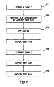
Figure 7 provides a general flow chart depicting the steps of the present
methodology. In
step 500, the sample is retrieved from a reservoir or formation. A formation
or geological
formation is the fundamental unit of lithostratigraphy and comprises a certain
number of rock
strata that have a comparable lith.ology, facies or other similar properties.
Understanding the
geology of a reservoir is essential to its development, production and
management and includes
understanding the external geology, that is, what created the hydrocarbon
trap, and internal
geology of the reservoir or the nature of the rocks in which the hydrocarbons
exist.
The sample that is retrieved is in its native state and can be retrieved from
a shale
formation using a wireline tool string or is a cuttings sample. In some
embodiments, the sample
may be in the shape of a regular cylinder while in other embodiments, the
sample may be an
irregular shaped sample.
18
CA 03005657 2018-05-16
WO 2017/100000 PCT/US2016/063440
At step 502, Mat_ spectroscopy of the sample is performed to acquire a first
NMR data
set. Step 502 begins by placing the sample in proximity to a magnet to apply a
magnetic field.
This may include, but is not limited to, placing the sample inside the magnet,
next to the magnet
or having the magnet placed in the sample in the case of an inside out probe.
The sample may be placed in proximity to the magnet for a period of time
sufficient to
allow the magnetic moment to come to equilibrium aligned along the applied
magnetic field.
This period of time can about 3 times the maximum Ti of the sample. In other
cases, the period
of time may be equal to about 10 times the maximum Ti of the sample. In many
samples, a
period of time of about 10 seconds to 20 seconds may be sufficient.
The NMR system may also be tuned and matched to ensure the best excitation, as
the
excitation frequency and reflected power of the probe may shift. This is
performed by running
and adjusting tuning capacitors until the produced signal by the probe is
optimized. Tuning is
complete when the frequency produced by the probe is within an acceptable
frequency distance
from the main resonance frequency. Matching is complete when the reflected
power is adjusted
to an acceptable minimum level. Pulses at about 900 and 180 are then
optimized for the
particular system. of magnet, probe, and sample. The combination of pulse
length and pulse
power that produces the maximum signal is established as the 90 pulse. The
combination of
pulse length and power that produces a minimum signal is established as the
180 pulse.
Typically when making a measurement, either the power is kept fixed and the
length of the pulse
is varied to get the different pulses, or the length of the pulse is kept
constant and the power is
varied. In certain embodiments, a 180' pulse that is twice the power of a 90
pulse is employed,
as this keeps the bandwidth used to excite the sample constant and is less
likely to introduce
issues given the broad line width which often occurs in samples. If other
pulse values are needed,
45 , 30 , etc. the necessary pulse length or power is calculated from the
empirically obtained 90
and 180 pulses.
As noted above, the NMR measurement is performed using a high resolution MAR
system. The NMR system applies a magnetic field to the sample. The NMR. system
then applies
a series of RF pulses according to a pulse sequence. The magnetic field that
is applied to the
sample is at least about 0.2 Tesla, which generates associated Larmor
frequencies of at least 8
MI-lz. In other embodiments, the frequency is at least about 10 MHZ or at
least about 20 MHz.
The pulse sequences generate NMR signals within the sample and the NMR system
detects the
19
CA 03005657 2018-05-16
WO 2017/100000 PCT/US2016/063440
NMR signals between or after the RF pulses. The high resolution NMR system
detects NMR
signals with echo times of less than about 200 microseconds. In further
embodiments, the high
resolution 'NMI{ system detects NMR signals with echo times of less than or
equal to about 1.50
microseconds or less than or equal to about 100 microseconds. Also, the NMR
system may have
a dead time of less than or equal to 50 microseconds. The "dead time" is the
time interval
defined by (i) the end of a RF pulse and (ii) the time when the NMR system
detects NMR
signals. By detecting short echo times and using shoit dead times, the NMR
system is able
detect a broader range of NMR signals from the hydrogen-containing components
present in the
sample.
A series of RE pulses with intermittent delays according to a pulse sequence,
such as
CPMG or inversion recovery sequence, are applied to the sample in order to
measure the T2
and/or Ti relaxation times from the time-domain decay or recovery of the
signal. The delay from
pulse to data acquisition may range from about 1 to about 50 milliseconds
after the start of pulse
scheme that acquires the relaxation decay or recovery curve; or from about 16
to about 20
milliseconds after the start of the pulse scheme; or from about 19
milliseconds after the start of
the pulse scheme. In some embodiments, the signal is used in a raw form,
without the use of
chemical shifts and without converting data into the frequency domain by
Fourier transform or
other means. Measuring and acquisition can be performed by, at least,
partially suppressing the
water or hydrocarbon signal prior to the beginning of the pulse sequence used
to record the
relaxation times.
After the first NMR data is obtained, the sample is removed from the NMR
system and
dried in step 504, The sample can be dried using any conventional method, such
as placing the
sample in an oven. The sample may be dried at any temperature, such as at a
temperature of at
least about 100 C, for example between about 110 C - 115 C. The sample may be
dried for a
period of time at least about 4 hours, or at least about 8 hours, or at least
about 16 hours, or at
least about 24 hours or even at least about 48 hours.
The dried sample is then placed within the NMR spectrometer (not shown) and an
NMR
measurement of the dried sample is performed as described above in step 502 to
acquire a second
NMR data set. After the second NMR data set has been obtained, the dried
sample is removed
from the 'MIR spectrometer and saturated with a fluid, such as brine, in step
506. The brine can
have a salinity similar to the salinity of the reservoir from which the sample
was obtained.
CA 03005657 2018-05-16
WO 2017/100000 PCT/US2016/063440
Once the dried sample has been saturated with brine, it can be placed within
the NMR
system and a third NNW. data set is obtained such as described above in step
50:2. The three
'NEVER data sets are then analyzed to determine at least one formation
property, such as by
analyzing the exponentially decaying NMR signal in the time-domain using
single- or multi-
exponential analysis, and comparing differences in the relaxation times Ti
and/or T2 for the
hydrogen-containing components in the samples.
Having the first, second and third NMR data sets, Ti and T2 relaxation time
spectrums,
which include amplitude versus Ti and T2 relaxation times, are determined from
the first, second
and third NMR data sets. Ti ¨ T2 plots for each NMR data set can be generated
and analyzed to
determine total porosity, and the porosity's for each of moveable water,
capillary bound water,
clay bound water, heavy hydrocarbons and light hydrocarbons.
:En the present systems and methods, :MIR data is used to determine the
following
formation properties: (1) total porosity; (2) porosity for each of: (a)
moveable fluid, (b) capillary
bound fluid, (c) clay bound fluid and (d) heavy hydrocarbons; (3) total
organic hydrogen content;
and (4) water saturation.
Figures 30A, 30B and 30C include a cross plot as a T1/T2 plot with porosity
shaded with
the color bar, and a XYZ plot with X axis of T2, Y axis of Ti, and Z axis of
porosity measured
with NMR. The NMR data also presented in a three dimensional ("3D") matrix.
Figures 30A,
30B and 30C show the three sets of NMR data as used in sequential fluid
characterization.
Figure 30A shows a first set of NMR data or "As received" data that can
quantify the following
formation properties: (i) capillary bound water; (ii.) clay bound water; (iii)
heavy hydrocarbons;
and (iv) residual hydrocarbon. Figure 30B shows a second set of NMR data or
"dry" data (where
the step of drying removed most water) that can be used to quantify (i)
capillary bound water; (ii)
clay bound water; and (iii) residual hydrocarbons. Figure 30 C shows a third
set of NMR data or
"saturated data" (where saturation quantifies movable porosity lost during
storage) that
quantifies mobile hydrocarbon.
More specifically, Figures 12A1, 12A2, 12A3, 12B1, 12B2, 12B3, 12C1, 12C2, and
12C3 collectively depict how NMR data is inputted into a SIT workflow. NMR raw
data
matrices: an "as received" matrix; a "dry" matrix; and a "saturated" matrix
are processed using
mathematical matrices as follows: 1) As Received matrix minus Dry matrix; 2)
Saturated matrix
minus Dry matrix; and 3) Saturated matrix minus As Received matrix. Processing
of the
21
CA 03005657 2018-05-16
=
WO 2017/100000 PCT/US2016/063440
matrices in the SFC workflow in such a manner separates NAIR raw data and
provides the
components and the properties of the formation and/or the reservoir which
include, but are not
limited to, capillary bound fluid, clay bound fluid, residual hydrocarbon,
heavy hydrocarbon, and
moveable fluid porosity. Ozen, A. E. et al., TIM IVA/IR Surface Relaxation
Ratio ibr
Hydrocarbons and Brines in Contact with Mature Organic-Shale Reservoir Rocks,
Petrophysics
Vol. 54, 11-19 (2013); Jiang, T., et al., Integrated Petrophysical
Interpretation of the Eagle Ford
Shale with .I-D and 2-D Nuclear Magnetic Resonance (NMR), 54th SPWLA Annual
Logging
Symposium, June 22 ,2013; Mansoor, R. A., et al., Characterizing Light versus
Bound
Hydrocarbon in Shale Reservoir by integrating New Two-Dimensional .NAIR and
Advanced
Spectroscopy Measurements, URTEC 2016 San Antonio.
The sequential fluid characterization system provided herein includes a high
resolution
-NMR spectrometer and an analysis system that includes a processor and non-
transitory,
computer-readable medium. The processor, the non-transitory, computer-readable
medium or
combinations thereof may comprise code. The sequential fluid characterization
system can also
include a graphical processing unit (GPLT) and a graphical user interface
(GUI). The code is
configured to calculate and distinguish SFC components in a sample. This is
accomplished by
using matrix math of 3D plots including T1,T2 and porosity.
As shown in Figure 8, a sequential fluid characterization system can further
comprise a
wireline tool string 604 that is deployed in a well 606 via a wireline truck
608. The wireline tool
604 is a downhole tool and is configured to remove a sample 602 from a
formation 610 using,
for example, a coring device. In another embodiment, the sample is a cuttings
sample. Cuttings
samples are pieces of formation that are cut away from the formation by a
drill bit during a
drilling operation and are retrieved from drilling mud that circulates to the
surface. This
disclosure is not limited to analysis of any particular type or form of sample
or retrieval system
used to obtain samples.
Once the sample 602 is obtained, it is transported to a surface facility 612
equipped to
carry out additional processing of the sample (for e.g., drying the sample and
saturating the
sample) and analyze NMR data. The surface facility 612 is located at the well
606, such as in a
truck or a cabin. In other instances, the surface facility 612 is located in a
location remote from
the well 606, such as in a laboratory. The analysis system 616 includes a
processor and non-
transitory, computer-readable medium. The processor, and the non-transitory,
computer-
22
CA 03005657 2018-05-16
=
WO 2017/100000 PCT/US2016/063440
readable medium, and/or combinations thereof may comprise computer code. The
analysis
system may also include a graphical processing unit (GPU) and a graphical user
interface (GUI),
such as a monitor, a touch screen, a mouse, a keyboard and/or a joystick. The
GUI allows an
operator to control and communicate with the NMR spectrometer 614. For
example, the NMR
spectrometer can detect MYER signals with echo times of less than 200
microseconds, for
example less than or equal to 100 ms. The NMR spectrometer 614 is used to
perform a NMR
measurement on the sample and to obtain NMR data sets. The NMR data sets are
communicated
to the analysis system. The SFC system utilizes NMR data to generate a
parameter, for example
a Ti and a T2 relaxation time spectrum and to determine different formation
properties.
The sample can be taken from a shale formation and analyzed with the subject
methods.
Shale formations are composed of fine-grained sedimentary rock. Some shale
formations are
rich in organic material and may be source rock for hydrocarbon reservoirs. In
some cases, the
shale formations also contain oil and gas.
EXAMPLE I
The Niobrara formation was deposited in the late Cretaceous in the
epicontenintal
Western Interior Seaway. Figure 16. This formation can be highly productive in
the DJ basin on
the eastern side of the Cretaceous seaway and is described as a Chalk and Marl
with
distinguishing benches composed of coccolith rich fecal pellets and pelagic
clays (see Stout, L.
"Carbon Isotope Chemostratigraphy of the Niobrara formation, Denver Basin,
CO.", Colorado
School of Mines Master's Thesis. (2012)). The Niobrara formation continued
deposition with
similar benches across the Cretaceous seaway to the western slope in what is
now called the
Sand Wash basin. Figure 17. See Finn, T. M., et at., Niobrara Total Petroleum
5:ystem in the
Southwestern Wyoming Province: USGS Petroleum Systems and Geologic Assessment
of Oil and
Gas in the Southwestern Wyoming Province, 141yoming, Colorado, and Utah,
Chapter 6 (2005)).
The Niobrara in the DJ and Sand Wash basin can be informally divided into
seven benches that
alternate between chalk and marl. In the Sand Wash basin the three prospective
units are the
Buck Peak, Tow Creek, and Wolf Mountain marl's. The Tow Creek bench was the
focus of the
study, as it is primarily composed of beds of organic material and pellets.
The pellets were
differentiated easily in thin sections as light and dark, as shown in Figures
11A, 11B and 11C.
Determining the pellets composition, thin sections were analyzed using
Qemscan. Qemscan is a
23
CA 03005657 2018-05-16
WO 2017/100000 PCT/US2016/063440
combination of SEM imaging with Energy Dispersive X-Ray Spectroscopy ("EDS")
as shown in
Figure 19. The results provided a lithological and digenetic interpretation of
the sample that was
not possible using standard thin section analysis. The Qemscan analysis
determined the light
pellets were primarily calcite while the dark pellets were organic rich.
Further analysis indicated
the importance of the light pellets to the reservoir viability of the Tow
Creek as seen in the thin
section analysis using epiflorescene. The light blue florescence 810 in the
light pellets represents
micro-porosity, which was pervasive in the Tow Creek bench.
As shown in Figure 18A, a thin section photomicrograph taken from the Tow
Creek
bench in the Sand Wash basin. Figure 18B represents a zoomed image of the same
thin section
seen on the left to illustrate the two types of pellets, light and dark. As
shown in Figure 18C, the
light pellets when viewed with epi-florescence showed significant micro pore
development. The
light pellets can make up close to 30 to 40% of the field of view in Tow Creek
thin sections.
Problem
A. Sand Wash well was drilled and completed in the Tow Creek bench of Niobrara
with
reasonable success in the black oil window (@40API and Ro 0.85). Approximately
600 feet of
core was taken with analysis being performed every ten feet which included
porosity and
saturation determinations using the GRI methodology. Interestingly, all
samples (60 in total)
came back with porosity in dynamic range of 4.5-6.5%. The samples taken from
the landing
zone yielded total water saturation ("SWT") of approximately 65%. This high
SWT
measurement from GM yielded a bulk volume hydrocarbon ("MTH") of 1.5-2%. The
well did
not produce any water during production. Considering that the IIVH was only
1.5-2%, it
indicated that in order to match production to reservoir quality, either frac
height must be
anomalously high (greater than 200 ft), or the recovery factor in black oil
window is substantially
higher than thought (greater than -40-50%).
Many issues were considered in attempting to resolve the odd correlation of
low BVH
and high SWT to production. One possibility considered was that the sample was
not properly
cleaned for the GRI procedure. This is a common problem when attempting GRI
methodology
as the proxy for a cleaned sample is when the toluene stops visibly changing
color. Even though
the sample was crushed to expose increased surface area in a low permeability
rock, it's thought
that the toluene did not infiltrate the rock entirely with a qualitative view
of discoloration. This
24
CA 03005657 2018-05-16
WO 2017/100000 PCT/US2016/063440
possibility was further amplified since significant micro-pore development was
observed in light
pellets (Figure 18C) which were usually encased or surrounded by organics as
seen from the
Qemscan analysis (Figure 19). Considering these observations, if the sample
was not cleaned
properly, porosity would be significantly under predicted.
An alternative possibility was that since water saturation is a relative term,
the 65% water
measured from GRI methodology could be bound and the formation would only
produce free
water. To confirm was difficult since GRI methodology cannot determine free,
irreducible, and
structural water. Another issue with GRI methodology is the lack of
standardization between
core analysis vendors. Therefore, to attempt to investigate the initial
hypothesis it was
determined to pursue conventional core NMR since it can separate fluids and is
independent of
cleaning.
Initial NMR Analysis
NMR data of sample is capable of identifying the correct porosity as well as
the
associated water (including structural or free water). A Tow Creek sample was
run using an
industry standard procedure. Only T2 measurements were acquired using a 0.2 ms
inter echo
time (TE) and 2 MHz equipment. Measured porosity was 7.5% which was about 1%
higher than
the porosity that was measured according to GRI methodology. This change was
not substantial
and still did not resolve the prior mentioned problems of an anomalously high
frac height and
recovery factor.
Figure 20 shows the initial results from the conventional NMR with only T2
relaxation
times run at .2 ms echo and on the 2 MHz machine. The result yielded a total
porosity of 7.5%,
which was not a significant increase to what was previously measured from GM
methodology.
Figure 21 is a schematic illustrating the percent of the signal captured from
a NMR measurement
with changing echo time. Note the wireline tool has an echo of .4 ms which
captures
approximately 40% of the signal. Using high resolution NMR with an echo time
of .1 ms allows
one to capture close to 80% of the spectrum. It should be noted most nano
pores will occur in the
early decay times which will only be visible at low TE.
The difference between NMR logging (logging tools) and .N1VER sequential fluid
characterization as described herein are frequency and resolution of NIvIR.
NMR logging tools
operate at 1 to 2 .Kfiz and a max of 2MHZ. In the present systems and methods,
.NMR operates at
a frequency greater than 2 MHz. In addition, resolution (as defined by Te or
echo spacing) of
CA 03005657 2018-05-16
WO 2017/100000 PCT/US2016/063440
logging tools is typically at a minimum between about 0.6 to 0.3 ms.
Resolution of NMR as
applied in the present methods and systems is less than or equal to .2 ms and
can be a.s low as
0.05ms. Hence, in sequential fluid characterization resolution is improved by
7-8 times over that
of logging tools. Furthermore, signal to noise ratio for NMR of the present
methods and systems
is in excess of 100, which is close to 10 times better. Figure 22 demonstrates
improvements. En
SFC, an echo spacing of 0.1 ms or less can help resolve early Ti/T2 times
(micro and nano
porosity) which cannot be resolved by logging tools typically limited to echo
spacing of 0.2 ms.
Moreover, conventional rocks have much larger pore sizes and therefore have a
slower
relaxation time. Because unconventional rocks of unconventional plays
(unconventional
geological formations) have nano pores, NMR relaxation times can be very fast
and most of the
signal may not be recorded using 0.2 ms echo spacing on the 2 MHz machine. On
the other
hand, in conventional plays (conventional geological formations) where the
first NMR
measurement is made in the T2 domain (i.e. .4ms after 90 degree pulse), up to
60% of the signal
has already been decayed and not measured in T2 decay (Figure 21). On the
other hand, in
unconventional rocks, porosity primarily exists in this early signal that is
not recorded using
conventional NMR techniques.
Based upon these observations in a next experiment, we attempted using the NMR
dropped the Te to .1 ms (capturing almost 90% of the decay) as well as using a
20 MHz machine
to boost the signal to noise ratio. The results from the high resolution NMR
were drastically
different with a measured porosity of 14%. As shown in Figure 22, with known
fluid responses
to NMR, 171/T2 map can be used to identify fluids on the high resolution map.
The increase in.
porosity to 14% resolves the prior mentioned issues with production in the
Sand Wash well.
Based upon these observations, yet another subsequent NMR measurement was made
at a
lower inter echo time of 0.1 ms (which captured almost 90% of the T2 decay) as
well as using a
20 MIL machine to boost the signal to noise ratio. From this high resolution
NMR
measurement, the porosity was determined to be 14%, surprisingly different
from the porosity's
determined above. Using the known fluid responses to NMR., a T1/T2 map was
produced to
identify fluids on the high resolution map. (Figure 22) The increase in
porosity to 14% resolved
the prior mentioned issues with production in the Sand Wash well.
26
CA 03005657 2018-05-16
WO 2017/100000 PCT/US2016/063440
Potential Errors and Mitigation
The high porosity results from the high resolution NMR helped resolve the
production to
core discrepancy, however much doubt was cast over the data in the early time
arrivals. Another
study identified the early time arrival data as clay bound water and heavy
hydrocarbon.
Conventional thought suggested the early time arrivals that were identified as
clay bound water
and heavy hydrocarbon using a 0.1 ms inter echo spacing was either noise or
solids being
wrongfully identified as porosity. To help resolve this questions it was
decided to run a series of
experiments:
1. Clean and low porosity Berea sandstone (Figure 17)
a. A clean Berea sandstone sample was chosen to verify the potential error of
having noise or solids recorded as porosity. The results did not identify any
early T2 time porosity that could be interpreted as noise or solids.
2. High maturity Marcellus with high TOC (Figure 25)
a. A high maturity .Marcellus sample was chosen as it was mostly
devoid of heavy
hydrocarbon (i.e. it would have been cracked into lighter hydrocarbons). The
results were minimal signal in the early T2 times and a significant decrease
in the
amount of heavy hydrocarbons. The lack of repeatability gave credence to the
theory that early time arrivals were not noise.
3. Artificially matured Marcellus sample (Figure 26)
An experiment was performed to artificially mature a Marcellus sample to
determine if the early 12 times were noise. As shown in Figure 12A2 and 12A3,
12B2 and 12B3, and 12C2 and 12C3, the porosity signal in the region circled
indicated that what existed in the early T2 times was cracked during
maturation.
In particular, heavy hydrocarbon lost signal and cracked to lighter
hydrocarbon as
the sample matured. Thus, the early T2 times were not noise (the same signal
would appear at different maturities since noise is repeatable).
Sequential Fluid Characterization
While an increase in overall porosity was identified using high resolution
core INMR, it
was still desirable to differentiate the particular fluids. Using the SFC
described herein, porosity
can be separated into clay bound water, capillary bound water, movable
hydrocarbon, movable
water, and heavy hydrocarbon. The process encompasses three NMR measurements,
which in
27
CA 03005657 2018-05-16
WO 2017/100000 PCT/US2016/063440
this paper will be referred to as data sets. Data set one is acquired on as
received sample. Data set
two is acquired after the drying of the as received core. The sample is dried
for 48 hours while
collecting residual fluids at 110 degrees Celsius. It is dried at 110 degrees
Celsius as water's
boiling point is 100 degrees Celsius so most water will move and heavy
hydrocarbon will not
begin to crack (Figure 27). Finally data set three is acquired after the dried
sample is imbibed
with a fluid. In this experiment brine was used to imbibe in a vacuum with
minimal pressure
(200 psi). The brine imbibition can take approximately two to three weeks
while time and rate
are monitored until rate flattens. (Figure 27).
The SFC process (also referred to as a SEC workflow) was applied to the Sand
Wash
sample and the subsequent results for fluid types as well as total porosity
can be observed in
Table 1.
Porosity NMR GM Delta %
Total porosity 17.1% b.32% 170%
Movable bound porosity 4.1% Not resolved
Capillary bound porosity 3.5% Not Resolved
Clay bound porosity 3.5% Not Resolved
Heavy hydrocarbon 6.0% Not Resolved
Water saturation 41% All bound 60% -31%
0011' 100ft thickness 38.5 IVIMBO 101VIMBO 285%
Table 1
Table 1 above are results using a SEC workflow (i.e., 'NAIR data acquisition
and NMR data
processing) to determine fluid components of a reservoir of the Sand Wash
sample, and as
compared to results using the GM methodology with the resultant delta
(change).
The initial total porosity from the as received sample was 14% and after brine
imbibition
it rose to 17.1%. This was a substantial increase from the original GM total
porosity of 6.5%.
Using the SFC, movable hydrocarbon was determined to be 4.1 % rather than 1.5
to 2%. A
comparison could not be made to GM directly as GM methodology does not
separate movable
hydrocarbons. In addition the SIT process determined a high amount of heavy
hydrocarbons
(6%), which is a likely contributor in the discrepancy in GM total porosity to
SFC total porosity.
While the process does distinguish the fluid types and amounts there is still
concern with an
under prediction of porosity. It is likely that the movable hydrocarbon
component is a minimum
value due to the phenomena of lost light hydrocarbons after coring or during
storage. For future
endeavors it is recommended to take pressure cores to account for the lost
hydrocarbon.
28
CA 03005657 2018-05-16
WO 2017/100000 PCT/US2016/063440
EXAMPLE II
DRY GAS CASE STUDY
In order to confirm the versatility and applicability of the SFC methodology
and high
resolution NMR, an additional case study was undertaken in dry shale gas, as
the previously
mentioned Sand Wash example was in the black oil window. The reason for this
undertaking is
to observe if the high delta seen in the Sand Wash oil window would be
repeatable in dry gas
plays, which have significantly less heavy hydrocarbon due to high maturity.
Fifteen samples
were tested in the Marcellus and other plays in dry gas window using the same
lab measurement
parameters and SFC workflow that was used in the Sand- Wash case study. The
SFC results
were compared to GM. data as seen in Table 2 immediately below.
Porosity SFC GRI Delta
Change
Movable HC 5.60% 4.0% 40%
Heavy 3.00% N/A
Hydrocarbon
--
Free Water 0.00% N/A
Irreducible 0.70% N/A
Water
Clay Bound 0.60% N/A
Water
Water 18.8% 31% -40%
saturation
Total PHI 9.9% 5.8% 70%
OGIP 58.0 Bcf/Sec 41 Bcf/Sec 41%
Table 2
Comparison of SFC Methodology and GRI Methodology
for Fluid Components of the Marcellus Shale
The results of the Marcellus study yielded an increase in movable porosity and
consequently OGIP ("Original Gas In Place"). However as expected the results
did not have as
high of a delta as the samples in the oil window (Tables 1 & 2). While the
delta was not as high
as the Sand Wash example the delta in the OGIP was significant. When OGIP was
calculate for
other dry gas plays using the new porosity data from SFC the difference to GRI
based OOP is
still quite large. All of these plays have had similar problems of matching
porosity to production
like the Sand Wash, and with the SFC based OGIP recovered results are more
reasonable (Table
2).
29
CA 03005657 2018-05-16
WO 2017/100000 PCT/US2016/063440
In addition to the change in OGIP the biggest delta would be the bcf/ft
calculation on a
play basis. With the new in place calculations a higher concentration of
hydrocarbons on a per
foot basis illustrates that plays with thinner units (less than 100 ft) can be
viable if the bcf/ft is
high enough. An example of this is illustrated in Figure 28 with the high
bcf/ft calculation of the
Marcellus in West Virginia pan handle. The Marcellus in West 'Virginia pan
handle reaches an
approximate thickness of 70ft, yet wells continually out produce in place
estimates.
To upscale SFC porosity and characterize log porosity data so that the entire
reservoir can
be characterized and reserves more accurately identified, the equations
provided immediately
below can be utilized. The first equation gives us a grain density of rock
which helps calculate
porosity. Traditionally grain density is calculated from XRD (x-ray
diffraction) or another
lithology measurement. Our equation takes into account TOC and density of
kerogen to account
for total porosity including heavy hydrocarbon. Once the grain density is re-
calculated it is put
into the standard porosity (PHIT) equation.
,NmR = ________________
(GDENxu- Ker.Den*TOC
QD.EN
pHrr_
'(-P.DENNIvIR.:¨=RHOF)
The above equations are useful for determining production efficiencies by
quickly and
accurately identifying sweet spots in produced wells.
As described above, methods and systems for determining location of
hydrocarbon in
unconventional plays are provided. The present methods comprise at least two
steps of
measuring formation samples using high resolution NMR, as received, as dried
and/or as
saturated, and subsequent processing the NlvIR data to determine one of
several formation
properties or components where the formation property or the component can be
used to locate
an oil or gas reservoir, complete a well, and/or increase production
efficiency. The subject
methods and systems presented herein further provide a series of ordered
(sequential)
combination of steps related to the financial evaluation of number of barrels
of oil or cubic feet
of gas that can be produced at a given location.
CA 03005657 2018-05-16
WO 2017/100000 PCT/US2016/063440
Table 3 below provides a breakdown of formation fluids measured by GRI sample
methods and SFC described herein:
Formation fluid type GM NMR
Total formation water )1 Total water saturation )1
Free(movable)water X Unable to breakdown -\41 Due to
unique response of
Capillary bound water X water into moveable and )1 each
fluid to NMR, can spate -
Clay bound water x bound fractions / __ water fraction
into movable .-
N bound -
Hydrocarbon ? Dependent on cleaning q Cleanina not an issue
-
Gas ? .\/ NMR can
differentiate
Oil ?-------" ________________ hy drocarbon bearing
fluids -
-V
-----------------------------------
Heavy hydrocarbon fractions X Dependent on cleaning __ k
based on its viscosity an.,
-
)1 molecular structure &
helps _
TOC X No measurement -\/ distinguish between
fluids
31