Note: Descriptions are shown in the official language in which they were submitted.
SYSTEMS AND METHODS FOR AUTOMATED FLUID RESPONSE
MEASUREMENT
FIELD
[0001] The present disclosure generally relates to the field of monitoring
biological
signals, and more particularly, hemodynamic monitoring of one or more
patients.
INTRODUCTION
[0002] Innovative, affordable, and/or portable non-invasive hemodynamic
monitoring
devices may be desirable in the market. Such devices, for example, aid in the
provisioning of
care of various individuals, (e.g., the critically-ill) by providing
functional hemodynamic
assessments (which, in some embodiments, may be instantaneous or near
instantaneous).
[0003] It is desirable to be able to assess functional hemodynamics in a
variety of
circumstances. Unstructured environments and the variance in experience and
training
among individuals responsible for assessing functional hemodynamics creates
challenges.
These challenges are exacerbated when a patient requires monitoring over a
protracted
period of time and many individuals are involved in assessing functional
hemodynamics.
There is a need for a device that will produce precise and repeatable
measurements under
these conditions.
SUMMARY
[0004] In accordance with an aspect, there is provided a portable hemodynamic
monitoring device comprising a housing configured for removable coupling to a
body part of
an individual, the body part including at least one vessel of interest; an
ultrasound unit
coupled to the housing and adapted for adducing ultrasonic waves into the at
least one
vessel of interest in a continuous beam, the ultrasound unit including: at
least one transducer
pair adapted to continuously detect reflected ultrasonic waves derived at
least in part from
the produced ultrasonic waves directed at the at least one vessel of interest
and oriented
such that, in concert, the at least one transducer pair produces the
ultrasonic waves at an
angle of incidence between about 25 degrees to about 60 degrees in respect of
a plane of
fluid flow through the at least one vessel of interest; a processor.
- 1-
Date Recue/Date Received 2023-03-02
CA 03005790 2018-05-18
WO 2017/096487
PCT/CA2016/051451
[0005] In accordance with another aspect, the processor is further
configured to
continuously extract hemodynamic parameters from one or more characteristics
of the
detected reflected ultrasonic waves in real-time or near real-time by applying
a signal
processing routine, and to store the extracted one or more hemodynamic
parameters in a
storage.
[0006] In accordance with another aspect, there is provided a sensory output
device
adapted to provide feedback on a quality of the extracted hemodynamic
parameters, the
sensory output device including at least one of (i) a graphical display and
(ii) an auditory
display. Wherein the orientation of the at least one transducer pair improves
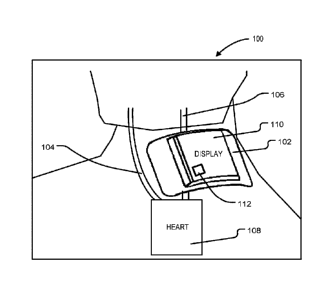
a probability of
proper acoustic coupling between the ultrasound unit and the body part of the
individual by
enabling a plurality of redundant effective placement options of the housing
on the body part
of the individual, the plurality of redundant effective placement options
reducing a required
precision of placement of the device.
[0007] In accordance with another aspect, the signal processing routine
includes
processing the reflected ultrasonic waves according to a continuous wave
Doppler
ultrasound process.
[0008] In accordance with another aspect, the at least one transducer pair
comprises a
chain of transducer pairs.
[0009] In accordance with another aspect, the at least one transducer
pair is at least one
flexible polymer based transducer pair.
[0010] In accordance with another aspect, the at least one transducer
pair is oriented in a
saw tooth pattern, the saw tooth pattern causing the ultrasonic waves to be
produced at the
angle of incidence between about 25 degrees to about 60 degrees in respect of
a plane of
fluid flow through the at least one vessel of interest.
[0011] In accordance with another aspect, the housing includes a tension
bandage that is
utilized to provide the removable coupling between the housing and the body
part of the
individual, the tension bandage being tensioned such that a sufficient
downward force is
applied to the ultrasound unit.
- 2 -
CA 03005790 2018-05-18
WO 2017/096487
PCT/CA2016/051451
[0012] In accordance with another aspect, the tension bandage is
configured to maintain
a substantially constant angle of incidence of the adduced ultrasonic waves
relative to the at
least one vessel of interest in order to enhance consistency of repeat
measurements over a
duration of time.
[0013] In accordance with another aspect, the sensory output device is
configured to
generate a sensory output indicating an effectiveness of placement of the
ultrasound unit.
[0014] In accordance with another aspect, processor is further
configured to detect an
estimated return of spontaneous circulation (ROSC) event by measuring a
difference
between a first relative blood flow from a chest compression and a second
relative blood
flow from a heartbeat, and the sensory output device is configured to generate
a sensory
output indicating the occurrence of the detected estimated return of
spontaneous circulation
(ROSC) event and indicating that any chest compression activities should
cease.
[0015] In accordance with another aspect, the housing includes at least
one data
communication device operable to transmit the extracted hemodynamic parameters
from
one or more characteristics of the detected reflected ultrasonic waves over a
data network.
[0016] In accordance with another aspect, the data communication device
transmits the
extracted hemodynamic parameters from one or more characteristics of the
detected
reflected ultrasonic waves over the data network to an external computer
system.
[0017] In accordance with another aspect, the housing includes at least
one data transfer
bus operable to transmit the extracted hemodynamic parameters from one or more
characteristics of the detected reflected ultrasonic waves over a data
connection.
[0018] In accordance with another aspect, the data transfer bus is
operable to transmit the
extracted hemodynamic parameters from one or more characteristics of the
detected
reflected ultrasonic waves over the data connection to one or more external
connected
devices.
[0019] In accordance with another aspect, the hemodynamic parameters include
at least
one of: a peak velocity of a Doppler shift detected in the at least one vessel
of interest; a
- 3 -
CA 03005790 2018-05-18
WO 2017/096487
PCT/CA2016/051451
velocity-time integral of signal changes between heartbeats; and a ratio
measured between
a post-intervention velocity-time integral and a pre-intervention velocity-
time integral.
[0020] In accordance with another aspect, the frequency of the ultrasonic
waves is a
frequency between about 3 MHz to about 12 MHz.
[0021] In accordance with another aspect, the frequency of the ultrasonic
waves is a
frequency is about 5 MHz.
[0022] In accordance with another aspect, the processor is configured to
determine
whether the individual is undergoing compensated shock by: continuously
monitoring a ratio
between a heart rate and a velocity-time integral of fluid flow through the at
least one vessel
of interest; entering a compensated shock alarm state when the ratio exceeds a
pre-defined
threshold; and producing an alarm signal when the compensated shock alarm
state is
entered.
[0023] In accordance with another aspect, the sensory output device is
configured to
transmit a signal when the processor determines that the individual is
undergoing
compensated shock.
[0024] In accordance with another aspect, the processor is further
configured to: extract at
least one first feature of interest from one or more characteristics of the
detected reflected
ultrasonic waves prior to an intervention event; extract at least one second
feature of interest
from one or more characteristics of the detected reflected ultrasonic waves
subsequent to
the intervention event; determine at least one post-intervention change value
equivalent to
the difference between the at least one first feature of interest and the at
least one second
feature of interest.
[0025] In accordance with another aspect, the intervention event is the
administering of at
least one medicament.
[0026] In accordance with another aspect, there is provided a device adapted
for
automatically assessing functional hemodynamics of a patient, the device
comprising: a
housing; an ultrasound unit coupled to the housing and adapted for adducing
ultrasonic
- 4 -
CA 03005790 2018-05-18
WO 2017/096487
PCT/CA2016/051451
waves into the patient at a blood vessel; a detector adapted to sense signals
obtained as a
result of adducing ultrasonic waves into the patient at the blood vessel and
to record the
signals in the form of raw data; and a processor adapted for receiving the raw
data and
transforming the data for output at an interface.
[0027] In accordance with another aspect, the processor is further adapted to
monitor
functional hemodynamics (e.g., fluid dynamics) when the patient undertakes a
fluid
challenge activity.
[0028] In accordance with another aspect, the processor is further adapted to
monitor
functional hemodynamics both before and after the patient undertakes a fluid
challenge
activity.
[0029] In accordance with another aspect, the processor is further adapted to
compare
the data before the patient undertakes a fluid challenge activity and after
the patient
undertakes a fluid challenge activity to determine a change in velocity time
integral of blood
flow in the blood vessel.
[0030] In accordance with another aspect, the change in velocity time
integral of blood
flow in the blood vessel is tracked as a ratio.
[0031] In accordance with another aspect, the processor is further
adapted to provide the
ratio and a notification for a clinician if the ratio is 10% or greater.
[0032] In accordance with another aspect, the ultrasound unit is
provided as an ultrasonic
.. probe separate from the housing and coupled operatively to the housing.
[0033] In accordance with another aspect, the device is provided in the
form of a portable
ultrasound unit.
[0034] In accordance with another aspect, the device is provided in the form
of a cart
mounted ultrasound unit.
[0035] In accordance with another aspect, the ultrasound unit is integrated
into the
housing.
-5..
CA 03005790 2018-05-18
WO 2017/096487
PCT/CA2016/051451
[0036] In accordance with another aspect, the processor is adapted to
perform the
automated detection of blood flow in the blood vessel, the processor receiving
the raw data
from adducing the ultrasonic waves (e.g., in a continuous beam or a pulsed
beam) into the
patient at an angle opposing the blood flow in the blood vessel, obtaining a
velocity time
trace in relation to the blood flow, determining a velocity time integral,
determining a cross-
sectional surface area of the blood vessel, and utilizing the velocity time
integral and the
cross-sectional surface area of the blood vessel to establish the blood flow
through the
vessel across a period of time.
[0037] In accordance with another aspect, the processor is adapted to perform
a
validation protocol for identifying an optimal set of parameters for operation
of the device.
[0038] In accordance with another aspect, the optimal set of parameters
includes at least
one of placement position, fixation type, patch placement, and angle of
incidence. In various
further aspects, the disclosure provides corresponding systems and devices,
and logic
structures such as machine-executable coded instruction sets for implementing
such
systems, devices, and methods.
[0039] In this respect, before explaining at least one embodiment in
detail, it is to be
understood that the embodiments are not limited in application to the details
of construction
and to the arrangements of the components set forth in the following
description or illustrated
in the drawings. Also, it is to be understood that the phraseology and
terminology employed
herein are for the purpose of description and should not be regarded as
limiting.
[0040] In various further aspects, the disclosure provides corresponding
systems and
devices, and logic structures such as machine-executable coded instruction
sets for
implementing such systems, devices, and methods.
[0041] In this respect, before explaining at least one embodiment in
detail, it is to be
understood that the embodiments are not limited in application to the details
of construction
and to the arrangements of the components set forth in the following
description or illustrated
in the drawings. Also, it is to be understood that the phraseology and
terminology employed
herein are for the purpose of description and should not be regarded as
limiting.
- 6 -
CA 03005790 2018-05-18
WO 2017/096487
PCT/CA2016/051451
[0042] Many further features and combinations thereof concerning embodiments
described herein will appear to those skilled in the art following a reading
of the instant
disclosure.
DESCRIPTION OF THE FIGURES
[0043] In the figures, embodiments are illustrated by way of example. It is
to be expressly
understood that the description and figures are only for the purpose of
illustration and as an
aid to understanding.
[0044] Embodiments will now be described, by way of example only, with
reference to the
attached figures, wherein in the figures:
[0045] Fig. 1 is a perspective view of a device placed on the neck of a
patient, according
to some embodiments.
[0046] Fig. 2 is illustrative of a conventional ultrasound unit, the GE
VScanTM having a
display, and a probe.
[0047] Fig. 3 is an illustration of an example neck profile, according to some
embodiments.
[0048] Fig. 4 is a depiction of a common carotid artery (CCA) Flow Measurement
Angle,
according to some embodiments.
[0049] Fig. 5 is an illustration of an example tensioning mechanism for
maintaining
acoustic coupling between the device and a body part according to some
embodiments.
[0050] Fig. 6 is an example block schematic diagram of a device, according
to some
embodiments.
[0051] Fig. 7A-7B is illustrative of some example components that may be
utilized for
interfacing with a patient's body, according to some embodiments.
[0052] Fig. 8 depicts an example adhesive by 3MTm.
- 7 -
CA 03005790 2018-05-18
WO 2017/096487
PCT/CA2016/051451
[0053] Fig. 9A-9C illustrate a pocket-size embodiment; Fig. 9A provides
a top elevation
view, Fig. 9B provides a perspective view, and Fig. 9C provides a side cross-
sectional view,
according to some embodiments.
[0054] Fig. 10 is an illustration of a pocket sized embodiment.
[0055] Figs. 11A-11C are illustrative of a small embodiment with a coupled
probe,
according to some embodiments. Fig. 11A is a front perspective view of the
embodiment;
Fig. 116 is a rear perspective view of the embodiment; and Fig. 11C is a
partial view of the
embodiment.
[0056] Figs. 12A-12B and Figs. 13A-13C are illustrative of a small embodiment
with
integrated probe, according to some embodiments. Fig 12A is a side view of
this
embodiment, and Fig. 12B is a perspective view of this embodiment. Fig 13A is
a
perspective view of a second version of the embodiment with integrated probe
being held at
a handle. Fig. 13B is a perspective view of the second version; and Fig 13C is
a side view.
[0057] A cart embodiment is provided at Figs. 14A and 14B; Fig. 14A is a front
perspective view of a cart embodiment, and Fig. 14B is a side elevational view
of the cart
embodiment.
[0058] Fig. 15 is a flow diagram displaying the typical stages of
medical care a patient
may undergo in the event of critical illness.
[0059] Fig. 16 is a cross-sectional diagram of a device according to some
embodiments.
[0060] Fig. 17 is a top view diagram of a device according to some
embodiments.
[0061] Fig. 18 is a cross-sectional diagram of a device displaying the
"saw tooth"
configuring of the transducer-receiver pair and its orientation relative to
blood vessels,
according to some embodiments.
[0062] Fig. 19 is a top view diagram of an ultrasound sensor and its
orientation relative to
.. blood vessels, according to some embodiments.
- 8 -
CA 03005790 2018-05-18
WO 2017/096487
PCT/CA2016/051451
DETAILED DESCRIPTION
[0063] Embodiments of methods, systems, and apparatus are described through
reference to the drawings.
[0064] The following discussion provides many example embodiments of the
inventive
subject matter. Although each embodiment represents a single combination of
inventive
elements, the inventive subject matter is considered to include all possible
combinations of
the disclosed elements. Thus if one embodiment comprises elements A, B, and C,
and a
second embodiment comprises elements B and D, then the inventive subject
matter is also
considered to include other remaining combinations of A, B, C, or D, even if
not explicitly
disclosed.
[0065] Innovative, affordable, and/or portable non-invasive hemodynamic
monitoring
devices are desirable. Such devices, for example, aid in the provisioning of
care of various
individuals, (e.g., the critically-ill) by providing functional hemodynamic
assessments (which,
in some embodiments, may be instantaneous or near instantaneous).
[0066] There may be, however, various technical challenges in providing such a
device,
such as ensuring that readings are accurate, specific, and reliable within a
tolerable
performance range (e.g., accounting for the presence of noise, accounting for
transient
signals and/or aberrations); accounting for variations in physical dimensions
and/or device
placement, contact, and environment (e.g., differing neck sizes, contours,
proximity of
device, signal transfer characteristics); accounting for variations in
procedures performed in
conjunction with the device (e.g., differing fluid challenges).
[0067] The device may also encounter challenges as it relates to practical
implementation, for example, the device may benefit from a level of
intuitiveness and/or ease
of use (e.g., portability, disposability, cost, understandable process, form
factor), heat
management, power (e.g., battery) management, adaptability to a variety of
settings
(including inside and outside of a hospital), etc.
[0068] Further, the device may benefit from a level of user-independent
measurement
repeatability, such that a patient, for whom many care-providers will be
responsible, can
- 9 -
CA 03005790 2018-05-18
WO 2017/096487
PCT/CA2016/051451
monitor functional hemodynamics accurately over a protracted period of time.
It may be
desirable for such a device to contain access to memory and log sensor data
over said
protracted period of time.
[0069] A robust measurement device may be desirable, such that the device can
provide
real-time feedback during, for example, chest compressions associated with
cardiopulmonary resuscitation (CPR), among other operations where comparing
pre-/post-
intervention measurements may also be desirable. Further still, it may be
desirable to
provide a device that adheres to the patient such that the care-provider's
hands may be
freed to perform other critical functions.
[0070] FIG. 1 illustrates the device 102 placed on the neck of a patient,
according to some
embodiments. The device 102 is illustrated having various components and
structural
aspects, and it should be noted that the device 102 is provided merely as an
example and
embodiments may have different, alternate, the same, more, and/or less
components and
structural aspects.
[0071] The patients that may use this device 102, may, for example, be older
in age and
suffering from heart complications. The patients may be weak, may not be in a
state of full
awareness, and may be in danger of acute and critical illness. The device 102
may also be
suitable for various other patient types.
[0072] Those patients who are alert are often in a stressful state.
Though efficacy may be
a significant factor, keeping the patient calm and comfortable is also an
important factor.
[0073] A device 102 that seems to constrict or feel unnatural on the
patient, such as a
bulky or heavy neck mounted device 102, might serve to increase patient
stress. A smaller
device 102, or one with a detached probe, may be advantageous in this regard.
[0074] The device 102 shown is configured for providing automated fluid
response
ultrasound (AFRU), and may, for example, be a body mounted device 102 that may
be
configured to incorporate a portable ultrasound unit to provide one or more
assessments of a
patient with consistency and/or accuracy. The device 102 may provide
functional
hemodynamic assessments, for example the device 102 may determine a patient's
fluid
- 10-
CA 03005790 2018-05-18
WO 2017/096487
PCT/CA2016/051451
responsiveness (FR), in an automated fashion. In some embodiments, the local
site on the
patient is generally the neck area, such that the carotid artery is the vessel
of interest and
carotid flow is the target measurement. In some embodiments, the vessel of
interest may be
another vessel (e.g., brachial artery, femoral artery, etc.) and, as a result,
the target
.. measurement may change accordingly.
[0075] The device 102 may, for example, be used in the context of
various uses,
including an automated ultrasound in combination with a leg raise, the use of
an automated
ultrasound to give live readings during a fluid challenge (e.g., passive leg
raise), etc. Further,
the solution of the present disclosure may be non-intrusive, may be used by
untrained users,
.. may include methods by which certain target blood vessels are automatically
differentiated
from the other blood vessels, etc., and the device 102 may, in some
embodiments, be used
for multiple measurements where the device 102 may be fixed in place between
measurements. For example, in some embodiments in order to differentiate
target blood
vessels from other blood vessels, forward and reverse flow signals may be
classified as
venous or arterial by application of a flow profile (e.g., pulsatile positive
direction against
non-pulsatile + opposite of positive direction). The transducer beam may be
wide enough to
capture the entirety of both arterial and venous signals at a particular
monitored cross-
section.
[0076] A user interface 110 may be integrated or operatively paired with
the device and
thus the device 102 may not require external supporting hardware. However, the
device
may, in some embodiments, be integrated with a data communication device 112,
for
example using the Bluetooth or Wi-Fi protocol. The data communication device
112 may
allow the device to transmit outputs to an external system (e.g., an external
computer
system) for processing, data storage, display, etc. The user interface 110 may
be a visual
display, a speaker, or another interface capable of communicating messages to
a user of the
device. In some embodiments, the device 102 may contain one or more data
transfer buses
operable to provide non-networked data connection means that may allow the
device 102 to
transfer and receive data to and/or from external connected devices (e.g.,
universal serial
bus (USB) hard drives, monitors, etc.).
-11 -
CA 03005790 2018-05-18
WO 2017/096487
PCT/CA2016/051451
[0077] In some embodiments, various disposables may be used with the device
102, such
as a disposable which integrates a patient interface with an acoustic carrier
(e.g., the gel and
adhesive). According to some embodiments, the device may communicate data to a
secondary processing system via a communications network ¨ the secondary
processing
system may process received data according to data-analytics models and/or may
integrate
received data with previously stored data.
[0078] In FIG. 1, the device 102 is depicted along with a patient's
blood vessels (noted as
reference numerals 104 and 106, in this example, the carotid). In such an
embodiment, the
output of the device 102 may be indicative of reflected hypersonic waves
transmitted by
transponders forming part of the device 102, and reflecting off of a vessel of
interest (in this
example, the carotid artery). The received reflected signals, when processed,
may produce
an output indicative of hemodynamic properties of blood flow from the
patient's heart 108,
through the vessel of interest. The device 102 may output through the user
interface 110, for
example, as various readings that can be interpreted by a machine and/or a
healthcare
practitioner. The blood flow and/or vessel walls may be tracked using an
ultrasound sensor,
and denoted as reflected signals undergoing a Doppler shift. The measured
Doppler shift
may be indicative of the movement of red blood vessels in blood through an
artery or vein
relative to the device 102 over time. The reflected signals may, when
measured, produce
values distinct from all other vasculature, which may facilitate isolation of
reflected signals
from a vessel of interest. The measured Doppler shift over a span of time may
form the
velocity time integral, and may be indicative of the amount of blood passing
through a cross
section over the span of time.
[0079] The device 102 may be configured to perform automated functional
hemodynamic
assessments in a vessel (e.g., a carotid artery, brachial artery, femoral
artery, etc.). For
example, the device 102 may be utilized to perform auto- focusing of an
ultrasonic source
(e.g., an ultrasound probe) at a number of different depths and angles, and
then collect data
that best fits the structure of a targeted blood vessel. In some embodiments,
the device 102
may include a chain of transducer pairs oriented in a saw tooth pattern such
that, in concert,
the transducer pairs produce ultrasonic waves at an angle of incidence between
about 25
- 12 -
CA 03005790 2018-05-18
WO 2017/096487
PCT/CA2016/051451
degrees to about 60 degrees in respect of a plane of fluid flow (e.g., the
direction of blood
flow through a blood vessel) through the at least one targeted blood vessel.
[0080] According to some embodiments, the saw tooth pattern arrangement may
function
to aim the ultrasonic beam so as to reliably generate an angle of incidence of
about 25-60
degrees (or thereabout) with general anatomical angle (for normal body types
of 45
degrees). Use of this angle may enable reliable detection of reflected
ultrasonic signals from
the body part of the individual containing the vessel of interest toward which
the ultrasonic
beam (e.g., a continuous beam or pulsed beam) is directed without the
intervention of a
specifically trained technician or other individual. Acceptable angles, in
accordance with
some embodiments, include +1- 1 degrees, +1- 2 degrees, +1- 3 degrees, +1- 4
degrees,
among others.
[0081]
Current methods require careful placement of ultrasonic monitors, often
requiring
the skill of an expert or trained individual in order to ensure effective
readings. According to
some embodiments, the saw tooth pattern arrangement may function to make
available a
plurality of redundant, but effective, placement options on the body part of
the individual,
thus making it less difficult to obtain an effective reading from the vessel
of interest. The
redundant positioning may allow for the device to be used by a less skilled
or, in some
embodiments, even an unskilled user.
Further, redundant positioning is helpful in
emergency situations where non-ideal conditions in conjunction with a need for
speed (e.g.,
individual is otherwise in great pain or dying), even for the skilled
practitioner.
[0082] According to some embodiments, multiple transducer element pair
designs, such
as the saw tooth pattern, may also enable multi- or single element activation
depending, for
example, on the quality of the reflected signal received from the vessel of
interest. For
example, where a multi transducer element array containing 10 elements
receives a
reflected signal from a vessel of interest that is sufficient to allow
effective functional
hemodynamic monitoring, the remaining eight elements may be de-activated or
may enter a
low power mode. This may provide benefits to power consumption and efficiency
of
operation (e.g., computational efficiency) and vessel identification.
- 13-
CA 03005790 2018-05-18
WO 2017/096487
PCT/CA2016/051451
[0083] The device 102 may be functional to perform automated functional
hemodynamic
assessments of a number of types of blood vessels. Depending on a particular
vessel
operable with the device 102 at a certain time, different depths and angles
may be selected.
The selection, for example, may be automated, based on the application of
various pre-
programmed instruction sets. The selection of such parameters is a non-trivial
technical
problem in view of variations of human physiology, blood vessel types, and
practitioner skill
levels. Further, the device 102 may operate, in some embodiments, such that it
may be
operable by unskilled practitioners and/or practitioners having less training
(who may need to
rely on the device 102 to select parameters based on sensed data and/or input
data). The
data retrieved from the ultrasound unit may be utilized, for example, to
calculate relative
blood flow (e.g., amount of blood / heart beat or unit time), and a potential
advantage may
enable variance in how the probe is oriented to the particular vessel being
examined.
[0084] The device 102 may be configured to detect relative blood flow through
a particular
vessel (e.g., the carotid artery, brachial artery, femoral artery, etc.) . The
device 102 may
further be configured to indicate the level of cerebral perfusion that has
occurred. The device
102 may further be configured to indicate whether the return of spontaneous
circulation
("ROSC") has occurred. Where functional hemodynamics are measured during CPR,
the
device may be adapted to measure functional hemodynamic parameters (e.g.,
fluid
dynamics) in a "binary" mode (i.e., fluid is either flowing through a vessel,
or it is not). In
other embodiments, the device may be adapted to provide a relative measure of
a
hemodynamic parameter such as the amount of fluid flowing through a cross-
section of the
vessel over a particular period of time (e.g., carotid, femoral, brachial,
etc. blood flow rate).
Measurement of relative carotid flow rate may be the most effective way to
automatically
detect ROSC.
[0085] As described in further embodiments, there may be various methods
and/or
techniques to aid in affixing and immobilizing the ultrasound unit (e.g., an
ultrasound probe)
to the local site on the patient in order to improve accuracy / fidelity of
repeat measurements
and, in some embodiments, provide real-time monitoring. For example,
adhesives,
tensioning bands, collars, pillows, etc. may be utilized. In some embodiments,
housing is
provided to which vascular probes could be attached and fixed to the neck at
varying angles.
- 14 -
CA 03005790 2018-05-18
WO 2017/096487
PCT/CA2016/051451
[0086] In some embodiments, the device 102 may be configured to
communicate through
one or more communication links (e.g., wired, wireless, cellular, local area
networks, wide
area networks, infrared, Bluetooth) with one or more receiver computing
devices (e.g., for
further analysis) and/or downstream computing devices (e.g., a data centre
associated with
a healthcare facility). Accordingly, the device 102 may or may not have a
display 110.
[0087] For example, the device 102 may be configured to provide outputs
that may
inform the function of other devices. The output of the measure can inform
various
individuals and/or machines of various hemodynamic parameters (e.g., features
of the flow
of blood through a vessel). For example, machines delivering cardio pulmonary
respiration
(CPR) can provide feedback on the efficacy and timing of chest compressions.
The reader
will understand that many other applications may be contemplated.
[0088] The device 102 may have various components to detect (e.g.,
monitor, track,
probe, sense, determine, identify, investigate) various physical
characteristics of the patient.
[0089] The device 102 of FIG. 1 may be used in conjunction with specific
workflows that
may be adapted such that the device 102 and the workflows intemperate to
provide accurate
and repeatable localization (e.g., using the ultrasound readings).
[0090] The portable ultrasound unit may, for example, be a continuous
wave Doppler
ultrasound module that is capable of emitting ultrasonic waves in a continuous
beam, and
that is accurate and fast enough to provide a real or near-real-time analysis
of parameters of
the fluid flow in the blood vessel, in some embodiments, free of a bulky cart
or cord. The
device 102 may, for example, be portable enough to be carried around by a
physician (e.g.,
for extended periods of time) or stored for sharing by multiple practitioners
(e.g., in a 'grab-
and-go' charging station for physicians).
[0091] In other embodiments, a pulsed wave Dopper ultrasound may be provided
instead.
[0092] Continuous wave Doppler ultrasound modules may function to measure
fluid
velocities along the entirety of a scanned channel. For example, where the
scanned area is
a blood vessel, a continuous wave Doppler method may measure the velocities of
fluids
traveling through the entire scanned portion of the blood vessel over a period
of time. In
- 15-
CA 03005790 2018-05-18
WO 2017/096487
PCT/CA2016/051451
contrast, pulsed wave Doppler ultrasound modules may only allow measurement of
fluid
velocities at a single point, or a very finite sequence of points, along a
scanned channel.
[0093] Pulsed wave Doppler ultrasound modules may function by emitting a
pulsed signal
toward an area of focus for a finite period of time, then ceasing the emission
of said signal
and monitoring received signals in order to record a reflected frequency shift
related to the
original emitted signal for a finite period of time. This process is then
repeated. Once the
reflected signal is received, a processor calculates the velocity and flow of
liquid through a
channel at the area of focus (e.g., a blood vessel). Since pulsed wave Doppler
ultrasound
techniques require a finite signal emission period and a second finite signal
monitoring
period, there is a limit to how fast said techniques can accurately measure
the flow of liquid
through a channel ¨ where the velocity of the fluid surpasses a certain point,
temporal
aliasing (a phenomenon whereby a recorded signal appears distorted due to a
recording
system with an insufficient sampling rate). This mode of operation can be
described as "half-
duplex".
[0094] Continuous wave Doppler ultrasound modules function by emitting
ultrasound
signals in a continuous beam along a channel and continuously monitoring the
multitude of
reflected frequency shifts via a detector. This mode of operation can be
described as "full-
duplex" as the continuous wave Doppler ultrasound is continuously emitting and
receiving
signals. A potential advantage realized by this mode is that it enables the
measurement of
high-velocity flows of liquids through channels (e.g., blood through blood
vessels) that could
not be accurately measured using pulse wave Doppler ultrasound techniques due
to the
above-described temporal aliasing problem.
[0095] The device 102 may further include and/or be associated with a
locating
disposable that may be affixed once to the patient for various measurements,
the
measurements of which can be compared with one another. The device 102 and/or
the
locating disposable may require a level of ease of use and sufficient accuracy
such that
practitioners and care centres may readily adopt its usage.
[0096] The device 102 may be battery powered and may use a transducer
array which
may function to measure the Doppler shift produced by fluid passing through a
vessel (e.g.,
- 16 -
CA 03005790 2018-05-18
WO 2017/096487
PCT/CA2016/051451
a Doppler shift produced by red blood cells in blood travelling through an
artery relative to
the position of the device 102. A technical challenge arises in relation to
ensuring that the
device 102 is configurable to identify (e.g., delineate, distinguish) flow
through particular
vessels (e.g., distinguish carotid flow from the jugular vein or other
confounding objects).
[0097] In operation, a patch-like (or collar-style) probe may be adhered to
local area of
skin on a patient under which the patient's carotid artery (or other
vasculature) passes. The
probe may utilize ultrasound signal processing methods (e.g., Doppler signal
processing
functions) to identify pulsatile flow. When the ultrasound (e.g., continuous
wave Doppler)
function of the ultrasound signal is directed at an opposing angle to the
blood flow, a
velocity-time trace may be obtained. By defining one cardiac cycle
(pulse/heart beat), a unit
time may be defined.
[0098] Calculating the area under the velocity-time curve (i.e., the
calculus integral), the
device 102 and/or a downstream device may utilize the data to determine the
velocity-time
integral ("VTI"), and the VTI may be multiplied by the cross-sectional surface
area of the
vessel over the time of one cardiac cycle (heart beat). Accordingly, an
automated physical
measurement of a blood flow through the vessel per heartbeat may be obtained
using an
ultrasonic approach.
[0099] In some embodiments, an auto focusing mechanism is provided,
where the device
102 may conduct a validation protocol to identify which settings are optimal
(e.g., frequency,
angle) for the patient's body, patch placement, and/or other parameters. A
challenge with
conventional technologies is that the selection of these signals is non-
trivial and may often
lead to a high level of training required. For example, this aspect of the
technology aids in
allowing un-trained or less trained personnel to use the device 102 reliably.
[00100] A computing device may apply an algorithm in conjunction with detected
readings
to determine the patient's velocity time integral (VTI, pre-challenge); prompt
the physician for
a fluid challenge (e.g., passive leg raise); detect and/or calculate a post-
challenge VTI; and
deliver an assessment of the patient's fluid responsiveness (increase of >10%
VTI or output
following fluid challenge). A ratio may be found between pre and post-
challenge VTIs, and
other thresholds may be used for assessments (e.g., 10%, 5%, 3%, etc. and may
be
- 17-
CA 03005790 2018-05-18
WO 2017/096487
PCT/CA2016/051451
indicative of an increase or decrease). Where a condition is broken (e.g., as
provided
through a business rule), or a trigger triggered, a notification may be
generated and/or
provided (e.g., an alert, a sound, a display, a pop-up).
[00101] A display may, for example, aid the physician by providing various
types of views,
some views having various transformations (e.g., a simplified view),
annotations (e.g.,
display markers, dynamic markers), analytics (e.g., determined aspects,
averages, means,
medians, identified aberrations), and/or a raw data view. For example, a post-
/pre-VTI ratio
may be determined, and a 10% or greater ratio may be indicative of a fluid
responsiveness
condition. Accordingly, some embodiments may be utilized to detect and/or
determine
various characteristics in relation to a carotid anomaly, or detect a carotid
anomaly or an
anomaly regarding another vessel of interest (e.g., brachial artery, femoral
artery, etc.).
[00102] There may be other types of ultrasound devices that can perform flow
monitoring,
however, drawbacks with conventional devices may be those typical of a
multipurpose
device: they are large, difficult to use, and may often require lengthy
training or experience.
[00103] The device 102 of some embodiments may be configured such that there
may
only be a minimum level of required hardware to effectively monitor blood
flow, and may
reflect a trade of multi-functionality for size, providing additional benefits
as in relation to
operation for use with carotid flow procedures. At FIG. 2, a conventional
ultrasound unit, the
GE VScan TM 202 is pictured, having a display 204, and a probe 206.
[00104] Other catheter-based technologies may also be used for hemodynamic
monitoring, but the conventional products may be cumbersome and add risk in
the context of
various procedures. Pulmonary artery catheterization is another technique that
may be
available for hemodynamic monitoring, wherein a catheter is inserted into the
pulmonary
artery via the vena cava to directly measure cardiac output. Pulmonary artery
catheterization
can measure right atrium, right ventricle, and pulmonary artery pressure, as
well as left
atrium input pressure, but a major drawback is that the catheterization is
invasive and limited
to surgical use. Pulse Pressure Waveform Analysis (PPWA) is another technique
that
utilizes the arterial waveform, obtained either from an arterial catheter or a
finger probe, in
- 18-
CA 03005790 2018-05-18
WO 2017/096487
PCT/CA2016/051451
order to calculate the stroke volume (SV) and the systemic vascular resistance
(SVR), but
complications may arise in view of non-linear and varying arterial wall
compliance.
[00105] Phase shift technology / bio reactance approaches may be considered
for use,
wherein when an AC current is applied to the thorax, the pulsatile blood flow
taking place in
the large thoracic arteries causes the amplitude of the applied thoracic
voltage to change.
Research, however, has indicated poor performance in relation to critically-
ill/post-operative
patients; further, this approach may be hindered by environmental factors,
such as
overweight or patients which perspire heavily. Gas rebreathing techniques may
also be used
in relation to estimating CO non-invasively, but while easy to use, they have
been shown to
be adverse affected by spontaneously breathing patients. Septic Shock
Algorithms may use
aggregated historical data to predict the onset of septic shock, which can be
diagnosed
through blood pressure readings.
[00106] In some embodiments, the device may produce outputs functioning to
allow
detection of various types of compensated shock. Compensated shock may be
defined, in
an adult example, as systolic blood pressure above 90 mm Hg while exhibiting
signs of
inadequate perfusion (e.g., tachycardia). In such situations, the device may
transmit an alert
signal via a sensory output device.
[00107] FIG. 3 is an illustration of an example neck profile, according to
some
embodiments.
[00108] Patients may have differing anthropometric parameters, including, for
example,
carotid anthropornetries, neck anthropometries, etc. These parameters may be
taken into
consideration, for example, as the device may need to be fitted on to and/or
used in close
proximity to bodily features of the patients, and thus may need to be
calibrated and/or
accurately positioned.
[00109] For example, the minimum size (i.e. length) of the neck may determine
the
maximum size of a body mounted device. The neck length for the smallest 5% of
the
population is roughly 8 cm, and accordingly, the maximum comfortable height of
the device
may approximately be 8 cm.
- 19-
CA 03005790 2018-05-18
WO 2017/096487
PCT/CA2016/051451
[00110] Neck circumference also varies from person to person. The smallest
neck
circumferences may be about 312 mm, and the largest about 463 mm. For a round
or
square device (e.g. 8 cm in diameter or 8x8 cm square), the patient's neck
would have to
conform roughly 14 mm at the edges, too much for patient comfort. If the
device were
curved, the neck would have to only conform roughly 5 mm, but a curved feature
may add
complexity (and likely size) to the device and/or components thereof.
[00111] As indicated in FIG. 3, the neck anatomy may be fairly consistent from
patient to
patient. For example, the internal diameter of the Common Carotid Artery (CCA)
may be
approximately 6.2 mm for women and 6.5 mm for men, ranging between 4.3 and 7.7
mm in
maximum and minimum sizes (as noted in a study having a study size of 123).
The standard
depth for a patient's CCA may be 20-40 mm below the skin. The wall thickness
may be
roughly 0.75 mm. Additionally, the diameter of the CCP, may expand roughly 0.5
mm with
every heartbeat.
[00112] Another study suggests that the ratio of the internal carotid and
external carotid
artery diameters can be predicted as approximately .65 and .58 respectively
(e.g., each is
roughly 1/2 to 2/3 of the diameter of the CCA). The vertebral artery may be
hidden in bone,
very far away and very small. The jugular vein has blood flowing in the
opposite direction
(therefore "up" may have to be established).
[00113] Directional information can aid in the assessment of position.
Dimensional
measurements can also be used to aid in position by assuming any measurement
of a 5
diameter less than 4.3 mm is likely not the CCA.
[00114] Furthermore, a relationship can be established between distance and
diameter of
the CCA. A CCA further from the skin implies a larger bodied patient, who
would be
expected to have a larger CCA.
[00115] One study placed the ideal measurement location at 15-20 cm below the
bifurcation. The subclavian artery sits very close to the clavicle, and the
CCA bifurcation is
near the larynx (Adam's apple) meaning a reasonable measurement location is
anywhere
from 5-20 cm above the clavicle, or midway between the larynx and the
clavicle.
- 20 -
CA 03005790 2018-05-18
WO 2017/096487
PCT/CA2016/051451
[00116] Patients with larger neck diameters are likely to have a thicker layer
of cutaneous
tissue between the probe and the CCA. No papers studied described a
correlation between
bariatric patients and difficulty in reading CCA flow, meaning this may not be
an issue. It may
also mean that the full sized ultrasound machines currently in use are
variable enough to
account for these differences. This patient profile may not be suitable for
the AFRU. In some
embodiments, multiple transducer pairs may be arranged end to end to form the
transducer
array. This may enable the device to contour to different patient morphologies
on the neck,
arm, torso and/or thigh, etc.
[00117] FIG. 4 is a depiction of a CCA flow measurement angle, according to
some
embodiments. A probe 402 is shown for measuring flow in relation to vessels
404, being
incident flow at plane 406. Accordingly, sample images 408 (greyscale) and 410
(color) are
shown.
[00118] Applicants considered various approaches to the ultrasound unit and
made
various decisions related to the design. In some embodiments, the ultrasound
unit may
include a transverse and oblique array.
[00119] Other possible approaches included a single transducer or
multidimensional
transducers (i.e., a 2D array or a scanning 1D array). Though these other
approaches still
have a possibility of success, the transverse oblique array may be preferable
in some
embodiments. The array may be oriented obliquely (as shown by the line
indicative of plane
406) to pass through the CCA such that the array can read anatomical
information in the
transverse plane, and Doppler signal processing information in the
longitudinal plane.
[00120] In this architecture, there is a risk that suitably effective Doppler
signal processing
measurements may be difficult to obtain with a transversely oriented array.
There is also risk
that many elements will be required, resulting in a higher cost and size of
the device.
[00121] As the ultrasound architecture may be an important aspect of the
device, an
ultrasound investigation was conducted to test the effectiveness of the
transverse array
configuration.
- 21 -
CA 03005790 2018-05-18
WO 2017/096487
PCT/CA2016/051451
[00122] FIG. 5 is an illustration of an example tensioning mechanism for
maintaining
acoustic coupling between a device 504 and a body part 502 according to some
embodiments. The device 504 housed in a housing 512 which may adhere to the
surface of
a body part 502 containing a vessel of interest. The housing may further
contain a tensioning
cover 510 which may be coupled on one side to the device 504. The housing may
further
contain two or more latching mechanisms 506 which may be situated
perpendicularly to the
body part 502 of the individual and within the housing 512. The latching
mechanisms may
each contain a plurality of latching channels 506a-f which may function to
receive the edges
of the tensioning cover 510 when downward force is applied thereto and hold
the tensioning
cover 510 in place, thereby causing the tensioning cover 510 to maintain
position and, by
extension, apply downward force to the device 504 such that it remains secure
against the
body part of the individual 502. This may cause the device 504 to be situated
such that it
maintains a position functional to produce a correct signal and read a correct
reflected signal
to and from the vessel of interest (e.g., within a correct range of distances
from the vessel of
interest).
[00123] FIG. 6 is an example block schematic diagram of a device, according to
some
embodiments. FIG. 6 illustrates an electrical architecture and may include
various elements
of electronic circuitry, etc. The device may be implemented in various forms,
including, for
example, by software, hardware, embedded firmware, and/or a combination
thereof.
[00124] A user interface 602 may be provided for various input I sensory-
output
functionality, including the ability to receive parameters, etc. from users
(e.g., patients,
clinicians). Output functionality may be used to, for example, provide a
graphical interface for
clinicians and/or to communicate information to downstream computing systems
(e.g., a
clinical data center).
[00125] There may be various data storage units included for storing data
(e.g., raw data,
pre-processed data, processed data, post-processed data), and there may be one
or more
processors 604 utilized for conducting various determinations and/or
calculations. The
device may also, in some embodiments, have on-board memory that may be used to
support various functionality, such as processing data for display to a
clinician, etc. Various
peripherals 606 may be utilized to provide various input signals and/or to
receive various
- 22 -
CA 03005790 2018-05-18
WO 2017/096487
PCT/CA2016/051451
outputs (e.g., through USB, Bluetooth, etc.). The processor 604, for example,
may be
configured to control the peripherals 606, and the user interface 602.
Ultrasound
components may be provided, for example, through an ultrasound front end 612,
a probe,
transducers 616a ... 616n, which may be placed on and/or in proximity to a
patient 618. A
power supply 610, (e.g., a battery), may be utilized to supply power to the
ultrasound
components.
[00126] In some embodiments, the user interface 602 may be provided on a
separate
computing device, communicating with the Central Processing Unit ("CPU") 604
via one or
more Peripherals 605, hence the user interface 602 is depicted as connecting
to the CPU
604 via a dashed line.
[00127] In operation, the front end may be provided and, in some embodiments,
may
include an eight-channel integrated circuit that may include the ultrasound
front-end 612, the
probe 614, and the transducers 616a ... 616n. Signals passing through the
front-end first
may be amplified and/or filtered, and then passed through an anti-aliasing
filter which may
remove frequencies that may be too high to be sampled. These signals may then
pass
through an analog-to-digital converter and may be provided to a configurable
integrated
circuit (e.g., a field programmable gate array (FPGA), or a custom integrated
circuit) 608 as,
in some embodiments, low-voltage differential signals (LVDS).
[00128] An ultrasound emitter may be utilized to produce the high-voltage
signal needed to
drive the ultrasound transducers. The emitter may be provided a +60V and a -
60V power
supply, and controlled by low-voltage logic signals from the configurable
integrated circuit.
Other voltages and/or power supplies may be utilized and the above is provided
as an
example.
[00129] The configurable integrated circuit 608 may be configured to control
the emitter
and receive LVDS signals from the front-end. The configurable integrated
circuit 608 may be
configured to perform digital signal processing that may be utilized to both
send and receive
signals, including beam-forming and Doppler shift computations.
- 23 -
CA 03005790 2018-05-18
WO 2017/096487
PCT/CA2016/051451
[00130] The CPU 604 may host the operating system of the device, and may
liaise
between the configurable integrated circuit 608, user interface (UI) 602 and
any peripherals
606 that may be added to the system and/or perform post-processing on signals
received
from the configurable integrated circuit 608.
[00131] The Ul 602 may include an LCD touchscreen and/or an LCD screen with
buttons.
Other types of displays may be contemplated. Indicator lights, comprising for
example LEDs,
and indicators sounds generated from a speaker may also be part of the user
interface 602
to provide feedback to the operator. In some embodiments, feedback corresponds
to
operating conditions of device 102 in order to direct the operator to orient
device 102 to a
desirable and/or acceptable local site on the patient. The purpose of this
direction may be to
permit full operability of the device with a minimum of training or
experience. If the device is
required to be connected to the cloud, an onboard Wi-Fi module can be included
along with
additional peripherals 606 such as Bluetooth or USB.
[00132] A printed circuit board (PCB) may be provided to host some or all of
the electronic
components within a structure (e.g., a housing, a base). In some embodiments,
the device
may be powered by a rechargeable or replaceable battery, which may be used to
drive both
the ultrasound and/or the other electronics (e.g., LCD screens, etc.).
[00133] As size is a consideration, lithium-ion technology may, in some
embodiments, be
selected as an option for compact power density. Operating under the
assumption that these
batteries typically can store 77,000 Ah/cm3 (amp-hours per cubic centimetre),
the battery in
the device may have to be, for example, 125 cm3 for 1 hour of continuous
active use. A Li-
ion battery of this size may typically weighs about 250 g. Additional lifetime
can be achieved
by adding a larger (and heavier) battery, which may be suitable for a larger
embodiment.
[00134] FIG. 7 is illustrative of some example components that may be utilized
for
interfacing with a patient's body, according to some embodiments. The method
with which
the device interfaces with the body may be an important factor for
consideration. Such a
component, for example, may be a "disposable" to connect (acoustically) the
probe to the
skin, and connect (mechanically) the device to the patient. Sample disposables
are indicated
at 702 and 704.
- 24 -
CA 03005790 2018-05-18
WO 2017/096487
PCT/CA2016/051451
[00135] In some embodiments, the disposable may integrate these aspects for
quick
application and disposition. This may avoid the disadvantage of applying
ultrasound gel
separately, which creates variability and mess.
[00136] In some embodiments, an approach includes combining the requirements
into one
solution: applying an acoustically transmissive adhesive (i.e., the adhesive
also serves as
the gel).
[00137] In some embodiments, the requirements may be separated: a material is
provided
for acoustic coupling and a material is provided for physical connection.
[00138] An example design may include utilizing an adhesive ring with a gel
pad center.
The adhesive connects the device and the gel (solid or liquid) provides an
acoustic
connection. The user would simply peel back the inside of the disposable stick
it to the
device, and then remove the cover of the patient side immediately before
application.
[00139] The disposables may not need to be sterile (unless applications to
open wounds
are included in the indications), but should be held to a level of cleanliness
typical of the
industry.
[00140] Disposable ultrasound pads such as Rich-Mar AutoGelTM, BlueMTechTm and
AquaflexTM ultrasound gel pads, may be provided to replace ultrasound gel (for
the purposes
of limiting cleanup) and may be utilized with the device. These pads may need
to be wetted
with water, and a standoff pad may be used to position the probe away to get a
clearer
picture of superficial areas of the skin (for example, ATSTm phantoms).
[00141] In some embodiments, a custom sized block can be centered under the
probe
head, adhered on one side to the device and covered on the other side by a
dust cover that
also keeps the disposable wetted.
[00142] As an alternative, ultrasound gel may be utilized. Gel could be pre-
applied in a
cavity in the disposable. A similar peel-back cover could expose the gel and
make it ready
for application. After use, the gel may have to be wiped off the patient.
- 25 -
CA 03005790 2018-05-18
WO 2017/096487
PCT/CA2016/051451
[00143] An alternative method of acoustic coupling is a liquid filled pad.
Similar to an
ultrasound pad in its function and composition, these bags are filled entirely
with liquid. A
thinner liquid eliminates the likelihood of bubbles in the medium, but adds
issues at the wall
interface and with filling. For these reasons, liquid pads may be a less
desirable alternative
to those listed above.
[00144] In some embodiments, a tensioning material (e.g., a tension bandage)
may be
positioned around the body in order to provide a force normal to device 102
and ensure
sufficient acoustic coupling.
[00145] In some embodiments, adhesives may also be utilized. FIG. 8 depicts an
example
adhesive by 3NATm. Adhesives may by hypo-allergenic and may stay in place for
a number of
days. The adhesive 802 may help keep a device and/or a portion thereof
positioned in the
target local site on the patient.
[00146] Silicone may be used as a protector and an acoustic coupling material
for the
probe/human interface, often used in conjunction with gel. For limited
movement across the
patient's skin, (e.g., with some embodiments of the device), the need for gel
may be reduced
and silicone alone might be effective. A drawback to silicone is a reduced
speed of sound,
which may require an algorithm to correct.
[00147] The device may require a mechanical housing that may vary in detail
between
embodiments. A rigid two part housing, for example, may be sufficient to
provide a structure,
with standoff points to hold the electronics. The housing may include spacers
between
components to avoid rattle or internal movement. The housing may need to be
cleanable,
and so may include gaskets to prevent water ingress at the seam, display, and
buttons
(keypad membrane).
[00148] Heat management may be an important consideration. The device may
consume
significant amounts of energy during use, and accordingly, the hot features of
the device
may need to be kept away from the patient to reduce the risk of burning (e.g.,
must be less
than 43 C per ISO 60601).
- 26 -
CA 03005790 2018-05-18
WO 2017/096487
PCT/CA2016/051451
[00149] If the heat generation is excessive, numerous strategies can be
utilized, such as
insulation or heat fins, designed inconspicuously into the exterior of the
housing. A plastic
housing may be a natural insulator but may cause the electronics to overheat.
[00150] A metal housing may have advantageous attributes: protecting the
electronics at
the expense of patient safety. If an embodiment is applied that does not
connect to the
patient directly then heat protection may not be a requirement.
[00151] If the housing is plastic, injection moulding can be used for
manufacturing. If a
metal housing is used, numerous options are available, though some may be more
costly
than injection moulding.
[00152] In some embodiments, the device may be provided as a single part. In
some
embodiments, the device is provided in having two or more parts; these parts
may comprise
a body, a probe, a separate computing device, a stand-alone cart, among
others. For these
embodiments, a probe wire may be provided to connect the body of the device to
the probe.
[00153] Wires for this application may be available and may need to be strong
enough to
avoid pullout if the patient moves or the device falls. The probe itself (or
the connecting
surface on a one-part device) may benefit from a silicone interface piece, to
protect the
device and allow some conformity.
[00154] FIGS. 9A-9C, 10, 11A-11C, 12A, 128, 13A-13C, 14A-148, 16, 17 may be
illustrative of some sample embodiments of the device.
[00155] FIGS. 9A-9C may illustrate a pocket-sized embodiment; FIG. 9A provides
a top
elevational view, FIG. 9B provides a perspective view, and FIG. 9C provides a
side cross-
sectional view, according to some embodiments.
[00156] As depicted in FIGS. 9A-9C, an embodiment may include a pocket sized,
body
mounted ultrasound device that can be carried around by a clinician (e.g., the
attending
physician). The device may have an adhesive ring 902, a body section 904,
and/or a display
906. The adhesive ring 902 may provide an adhesive force between the periphery
of the
- 27 -
CA 03005790 2018-05-18
WO 2017/096487
PCT/CA2016/051451
body section 904 and the local site on the patient's skin. A concave space 908
may be
provided for a gel pad or liquid gel.
[00157] The size of this embodiment may be reduced by using a number of
strategies,
including, for example: offloading processing and display to another device
such as a tablet
(or smartphone), changing its geometry to rest partially elsewhere, measuring
flow in a
different artery (to reduce comparative size), or reducing the battery life or
removing it
entirely (plug-in power only).
[00158] FIG. 10 is an illustration of a pocket-sized embodiment. The
embodiment may
have display 1004, a power button 1002, and other buttons 1006 and 1008 that
may be
used, for example, to perform various input and output functions.
[00159] FIGS. 11A-11C may be illustrative of a small embodiment with a coupled
probe,
according to some embodiments.
[00160] FIG. 11A is a front perspective view of the embodiment; FIG. 11B is a
rear
perspective view of the embodiment; and FIG. 11C is a partial view of the
embodiment. The
device, may, for example, have a display 1102, control button 1104, power
button 1106, an
integrated handle 1108, a cable management apparatus 1110, a probe 1112, a
charging port
interface 1114, a printed circuit board stack 1116, and a battery 1118.
[00161] The footprint and height of the device illustrated in FIG. 11A-11C in
some
embodiments may be approximately 6x6 inches and 4 inches respectively.
[00162] A coupled probe may provide a lower risk alternative to the pocket
sized unit,
while still being small and portable. The adhesive probe-patch may be coupled
to the base
unit via a cable.
[00163] The clinician (e.g., a physician) may place the unit on the
examination table
anywhere within range of the patient's neck, and extend and connect the probe.
The probe
may stay in place during the examination and possibly longer (for repeated
exams). The
device may be powered by a larger, more powerful battery than possible in a
pocket-sized
unit, but is also can support a wall plug for heavy use.
- 28 -
CA 03005790 2018-05-18
WO 2017/096487
PCT/CA2016/051451
[00164] As the device may (in some embodiments) be too large to be carried
around
constantly, the device may be left at the charging station between patients,
further extending
battery life. The device may also be large enough to carry a gel holder, or an
area to keep
extra disposables. The device may include WI-Fi and/or Bluetooth connectivity,
or can
transfer data via a base station. In other embodiments, the device may be
miniaturized for
portable use.
[00165] FIGS. 12A-12B and FIGS. 13A-13C may be illustrative of a small
embodiment
with integrated probe, according to some embodiments. FIG. 12A is a side view
of this
embodiment, and FIG. 12B is a perspective view of this embodiment. FIG. 13A is
a
perspective view of a second version of the embodiment with integrated probe
being held at
a handle, FIG. 13B is a perspective view of the second version; and FIG. 13C
is a side view.
[00166] For example, the embodiment may include a probe 1204 for use with a
patient
1202/1310, a base 1206, a display 1208/1306, and an adjustable neck 1210/1305.
In some
embodiments, a handle 1312 is provided. In these example embodiments, the
device does
not have a separated probe. Instead, and area of the main chassis contains the
ultrasound
head which may be placed against the patient.
[00167] Similar to the coupled probe model described above, these embodiments
may
contain a larger battery, a plug, wireless connectivity, and may contain
storage for
disposables. The embodiments may be simpler in design and use than the coupled
probe,
and more durable. A disposable may not be necessary, or if necessary may be a
simpler
disposable.
[00168] A cart embodiment is provided at FIGS. 14A and 12B; FIG. 14A is a
front
perspective view of a cart embodiment, and FIG. 14B is a side elevational view
of the cart
embodiment. The application on a medical cart may provide some advantages. For
example,
the central architecture does not differ significantly to other carts: the
device 1402 may be
provided on a cart apparatus 1406, having a handle 1404, and a base 1408. The
embodiment may include a computer, monitor and screen 1410, with an ultrasound
probe
1412, which may aid in simplifying the development process though the use of
commercial
- 29 -
CA 03005790 2018-05-18
WO 2017/096487
PCT/CA2016/051451
components. Less effort may need to be focused on miniaturization and
integration than a
more aggressive size.
[00169] The cart embodiment may also avoid logistical issues that may be
present with
portable units: for example, the embodiment may be unlikely to be lost or
dropped, it may not
require an area on the patient's bed to be positioned upon, and can be easily
plugged in (or
battery powered) with room for a long cord. The device may also not require an
included
charging station, and may thus be marketed as an individual unit.
[00170] The device may include a chassis 1206 and a user interface 1208 (i.e.,
a large
touchscreen), and a probe 1204 that may differ significantly from other
ultrasound units.
[00171] The probe 1204 may include a small adhesive patch at the end of a
connection
cable 1210 which can be installed on the patient and remains stuck during the
procedure (or
longer). The software may be configured to automatically find the CCA and to
obtain
readings, displaying only the results to the clinician (e.g., a physician) and
eliminating the
need for ultrasound expertise.
[00172] Some embodiments are adapted to respond to a protracted period of
patient care.
An example patient-care profile 1500 is provided in FIG. 15. Assessment of
functional
hemodynannic parameters (e.g., fluid dynamics) may be desirable at each phase
of the
patient-care profile 1500. Different care-providers may be responsible at
designated phases.
A functional hennodynannics measurement device that is unique to a patient may
be
.. desirable in order to provide continuous monitoring between phases and care-
providers.
Similarly, a functional hennodynamics measurement device that is unique to a
care-provider
may be desirable to provide monitoring of functional hemodynamics measurement
to a
plurality of patients.
[00173] A cross-sectional view and overhead view of some embodiments of the
device are
provided in FIG. 16 and FIG. 17 respectively. In some embodiments, an adhesive
1606/1704
may be employed to fix the device to the local site on the patient's skin; a
tensioning material
1608/1708 may further be employed to apply a force normal to sensor housing
1612/1712,
and towards the local site in order to fix the device relative to the local
site. The tensioning
- 30 -
CA 03005790 2018-05-18
WO 2017/096487
PCT/CA2016/051451
material 1608/1708 may comprise a band, to be slug around the patient in order
that tension
may be adjusted by altering the length of upstretched band. The tensioning
material may
further be elastic, in order to eliminate discomfort to the patient, and also
continue providing
sufficient normal force while the patient moves and the body change shape.
[00174] In some embodiments, sensor housing 1602 may contain a transducer
array 1602,
electronics, including but not limited to a visual display, speakers, and/or
battery 1610, and
other electronics. In some embodiments, sensor housing 1612 is shaped like a
half of an
ellipsoid or American football. These shapes were found to be particularly
useful in aiding
proper positioning and tension characteristics to be applied.
[00175] In some embodiments, the sensor housing 1612 may contain a user
interface unit
1620, which may be a sensory output device operable with the visual display,
speakers,
and/or battery 1610. The user interface unit 1620 may function to communicate
inputs
generated by a user interaction to the device. For example, the user interface
unit 1620 may
comprise a sensory output device such as a capacitive touch input device
coupled with a
visual display 1610. The user interface unit 1620 may function to allow the
user to input
selections that, when received by the device, cause the device to modify its
operation mode
(e.g., user input may cause the device to being a process operable to
determine a pre-
intervention/post-intervention VTI ratio as described below).
[00176] In some embodiments, the transducer array 1602 comprises transducer-
receiver
pairs, where the transducer component generates acoustic waves, transferred
into the
patient acoustically through a hydrogel or acoustic coupler 1604. The acoustic
waves travel
through the patient, and are modulated and reflected by media interfaces, for
example fluid
within a blood vessel. Reflected and modulated waves are sensed by a receiver
component
in the transducer-receiver pair and within the larger transducer array 1602.
In some
embodiments, the Doppler shift in a frequency modulated signal generated by
the transducer
1602 may provide an accurate representation of the velocity of an element. In
some
embodiments, the element to be measured is the fluid flow within a blood
vessel; non-limiting
examples including the carotid artery, the brachial artery, or the femoral
artery.
- 31 -
CA 03005790 2018-05-18
WO 2017/096487
PCT/CA2016/051451
[00177] According to some embodiments, a sensor consisting of a ultrasound
transducer
1804 and receiver 1806 may be configured as depicted in FIG. 18 and FIG. 19,
representing
a cross-sectional view and an overhead view respectively. Transducer 1804 and
Receiver
1806 may be configured such that they parallel to each other, and each
directed at a 45
degree angle to the transverse plane (i.e., the plane defined by the surface
of the skin 1808
at the local site on the patient). The transducer 1804 and receiver 1806 may
resemble two
match-sticks, tangent along one edge, as depicted in FIG. 18.
[00178] In some embodiments, the orientation of the transducer 1804 and
receiver 1806 is
configured such that the angle between the vectors defined by the direction of
the ultrasound
wave and the direction of blood flow is approximately 45 degrees. The
transducer pair 1802
can detect blood flow direction by the sign (i.e., positive or negative) of
the Doppler shift. An
increase in frequency (positive shift) indicates blood flowing towards the
ultrasound wave
generated by the transducer 1804, and vice versa. In some embodiments, the
acceptable
functional range is about 45-degrees 15-degrees, with 45-degrees
representing the
preferred embodiment. The reader will understand that in some embodiments, the
acceptable functional range may be about 44-degrees 14-degrees, about 46-
degrees
16-degrees, etc.
[00179] In some embodiments the ultrasound wave (i.e., Doppler beam) is
"unfocused";
that is, the beams sweep out an angle of approximately 20-degrees, such that
by directing
the beam approximately 45-degrees from the transverse plane (as defined by the
plane of
the skin at the local site), the transducer 1804 will generate a wave that
intersects with the
blood vessel, and the angle of acceptance of the receiver 1806 (also 20-
degrees) is large
enough to receive the reflected and modulated signal generated by the blood
flow. In this
example, the width of the transducer face may be as small as 0.34 mm. This
size is suitable
for high-frequency (i.e., approximately 7 MHz) applications where high focal
loss may be
acceptable. Further, the width of the transducer may be as large as 2.15 mm.
This size is
suitable for low-frequency (i.e., approximately 4 MHz) applications where only
low focal loss
is acceptable.
[00180] In another example, a beam that sweeps out approximately 60-degrees
can be
constructed. The width of the transducer face may be as small as 0.924 mm.
This size is
- 32 -
CA 03005790 2018-05-18
WO 2017/096487
PCT/CA2016/051451
suitable for low-frequency (i.e., approximately 4 MHz) applications where high
focal loss may
be acceptable. Further, the width of the transducer may be as large as 1.52
mm. This size is
suitable for high-frequency (i.e., approximately 7 MHz) applications where
only low focal loss
is acceptable.
[00181] In some embodiments, the functional frequencies of ultrasound waves
generated
by the transducer 1804 and detectable by the receiver 1806 are approximately 3-
8 MHz. In
some embodiments, approximately 5 MHz may be the frequency generated by the
transducer 1804. Typically, a lower frequency may desirable for larger
patients, as vessels
such as the carotid artery, femoral artery, brachial artery, etc. are
typically further below the
skin surface 1808.
[00182] The orientation of the "match-stick" configured transducer pair 1802
may, in some
embodiments, be fixed relative to the skin surface 1808 by a tensioned bandage
1810 which
provides a force normal to the transducer pair 1812 and directed towards the
skin surface
1808.
[00183] In some embodiments, an audio or visual cue based on the Doppler shift
measured by the ultrasound transducer pair 1802 may guide the placement of the
device
relative to the local site on the patient. A stronger signal relative to the
noise sensed by the
transducer pair 1802 may be processed by the CPU 604 and outputted to a
peripheral
speaker or display 606. The output may comprise, for example, a particular
sound (e.g.
"whoosh"), a change in volume, or frequency of beeps in the case of an audio
cue, or it may
comprise, for example, brightness, number of LEDs activated, the flashing of a
pre-selected
image, among others in the case of a visual cue.
[00184] In some embodiments, the sensor consists of a "chain" of transducer
pairs 1802,
as depicted in FIG. 19. Transducer pairs 1802 are mutually connected via co-
axial cable.
The increased length to the overall sensor increases the acceptable area that
the device
may be placed by increasing the likelihood that the target blood vessel passes
under at least
one transducer pair 1802.
- 33 -
CA 03005790 2018-05-18
WO 2017/096487
PCT/CA2016/051451
[00185] In some embodiments, a "chain" may be preferable to a singular, but
longer
transducer pair 1802, because it may allow the housing to be constructed out
of a flexible
material that in turn will better conform to the various shapes and sizes of
patients.
Alternatively, in some embodiments, the transducer may be a flexible polymer
transducer
(e.g., a highly non-reactive thermoplastic fluoropolymer such as
Polyvinylidene difluoride
(PVDF)), this may also allow the housing to be constructed out of a flexible
material and
enable the housing to better conform to patients of various shapes and sizes.
Producing the
external housing in a flexible fashion may facilitate better acoustic coupling
between the
device 102 and the body and a higher signal to noise ratio.
[00186] In some embodiments, the transducer pair 1804 is a piezo ceramic
material. In
some embodiments, each piezo ceramic element, representing an individual
transducer
1804 or receiver 1806 is 10x1x1 mm in shape. The piezo ceramics in some
embodiments
may be a PZT-5A or PZT-5H or a combination thereof. In some embodiments, the
current
draw is approximately 25 mAh. In some embodiments, the device may have a mode
designed to conserve power and extend battery life. For example, the device
may turn on
only a pre-set times or it may only turn on at the request of a user and
automatically turn off
(or "sleep") after a defined period of time.
[00187] In some embodiments, the transducer pairs 1804 may be adapted to sense
peak
velocity of the Doppler Shift, VTI, or the pre-intervention/post-intervention
VTI ratio. The
device 108 may, according to a pre-programmed instruction set, implement one
or more
"sampling windows" which may perform a calibration routine functional to
eliminate the
inherent variability of Doppler signal. The calibration routine may: a) record
signals recorded
by the transducer pair during a pre-defined span of time prior to the
intervention (e.g., 10
seconds) as a "pre-intervention window"; b) cease recording signals during a
corresponding
pre-defined span of time during the intervention as an "intervention window";
and c) record
signals during a corresponding pre-defined span of time subsequent to the
intervention as a
"post-intervention window". One advantage of the device is the ability to
measure both heart
rate ("HR") and VTI. This permits the calculation of the HR/VTI ratio ¨ an
index that (unlike
the Shock Index) is not subject to vascular constriction as a compensatory
mechanism
during the onset of shock.
- 34 -
CA 03005790 2018-05-18
WO 2017/096487
PCT/CA2016/051451
[00188] In some embodiments, the design, orientation, and frequency of the
ultrasonic
transducers are specifically designed to facilitate the rapid and repeatable
measurement of
the signal of interest (e.g., the ultrasonic signal reflected from the vessel
of interest).
Subsequently, the measured signal of interest is automatically processed by
the signal
processing routine in order to generate an output.
[00189] Currently available point of care ultrasound machines require several
manual steps
to be completed in order to return a valid output. Further, successfully
completing several of
these steps requires users to have specific training and skill. For example,
currently
available point of care ultrasound machines may require: manual identification
of the vessel
of interest using an ultrasound imaging screen; manual orientation of the
angle of an
ultrasound probe in relation to the vessel of interest to produce useful
readings; manual
identification of the vessel of interest to activate a Doppler function (e.g.,
a Doppler signal
processing function); maintaining a substantially motionless positioning of
both the
ultrasound probe and the body part containing the vessel of interest; and
manually executing
a command in order to cause a reading to be taken. Further, often these steps
must be
executed repeatedly in order to compare changes in outputs received pre and
post
intervention (e.g., pre and post introduction of a medicament).
[00190] In contrast, some embodiments described herein may: automate some or
all of the
previously described steps; remove or reduce the requirement for manual
identification of a
vessel of interest; remove or reduce the requirement to manually orientate the
ultrasonic
transducer(s) relative to the body part of the person containing the vessel of
interest; may
automate the process of identifying a vessel of interest and activating a
Doppler function;
may serve to automatically or substantially automatically maintain a
substantially motionless
positioning of the ultrasonic transducer(s) relative to the vessel of
interest; and may
automatically generate readings, outputs, and compare pre and post
intervention values to
measure changes.
[00191] The embodiments of the devices, systems and methods described herein
may be
implemented in a combination of both hardware and software. These embodiments
may be
implemented on programmable computers, each computer including at least one
processor,
- 35 -
CA 03005790 2018-05-18
WO 2017/096487
PCT/CA2016/051451
a data storage system (including volatile memory or non-volatile memory or
other data
storage elements or a combination thereof), and at least one communication
interface.
[00192] Program code is applied to input data to perform the functions
described herein
and to generate output information. The output information is applied to one
or more sensory
output devices. In some embodiments, the communication interface may be a
network
communication interface. In embodiments in which elements may be combined, the
communication interface may be a software communication interface, such as
those for
inter-process communication. In still other embodiments, there may be a
combination of
communication interfaces implemented as hardware, software, and combination
thereof.
[00193] Throughout the foregoing discussion, numerous references is be made
regarding
servers, services, interfaces, platforms, or other systems formed from
computing devices.
[00194] It should be appreciated that the use of such terms is deemed to
represent one or
more computing devices having at least one processor configured to execute
software
instructions stored on a computer readable tangible, non-transitory medium.
For example, a
server can include one or more computers operating as a web server, database
server, or
other type of computer server in a manner to fulfill described roles,
responsibilities, or
functions.
[00195] The embodiments described herein are also implemented by physical
hardware,
including computing devices, servers, receivers, transmitters, processors,
memory, displays,
and networks. The embodiments described herein provide useful physical
machines and
particularly configured computer hardware arrangements.
[00196] The embodiments described herein are also directed to electronic
machines and
methods implemented by electronic machines adapted for processing and
transforming
ultrasonic signals which represent various types of information. The
embodiments described
herein pervasively and integrally relate to machines, and their uses; and the
embodiments
described herein have no meaning or practical applicability outside their use
with computer
hardware, machines, and various hardware components.
- 36 -
CA 03005790 2018-05-18
WO 2017/096487
PCT/CA2016/051451
[00197] Substituting the physical hardware particularly configured to
implement various
acts for non-physical hardware, using mental steps for example, may
substantially affect the
way the embodiments work. Such hardware limitations are clearly essential
elements of the
embodiments described herein, and they cannot be omitted or substituted for
mental means
.. without having a material effect on the operation and structure of the
embodiments
described herein. The hardware is essential to implement the various
embodiments
described herein and is not merely used to perform steps expeditiously and in
an efficient
manner. In some embodiments, the device is a single or special purpose machine
that is
specifically designed to perform limited set of functionality.
[00198] Although the embodiments have been described in detail, it should be
understood
that various changes, substitutions and alterations can be made herein.
[00199] Moreover, the scope of the present application is not intended to be
limited to the
particular embodiments of the process, machine, manufacture, composition of
matter,
means, methods and steps described in the specification. As one of ordinary
skill in the art
will readily appreciate from the disclosure, processes, machines, manufacture,
compositions
of matter, means, methods, or steps, presently existing or later to be
developed, that perform
substantially the same function or achieve substantially the same result as
the
corresponding embodiments described herein may be utilized.
[00200] As can be understood, the examples described above and illustrated are
intended
to be exemplary only.
- 37 -