Note: Descriptions are shown in the official language in which they were submitted.
HEART VALVE REPAIR AND REPLACEMENT
BACKGROUND
[0002] The mitral valve is positioned in the heart left side, between the
left atrium and
the left ventricle. The most typical disease of the mitral valve is
insufficiency or regurgitation
which occurs when the valve leaflets do not coapt properly. Mitral valve
repair by suturing a
ring to reduce the annulus diameter is the procedure of choice to correct
mitral regurgitation.
With the use of current surgical techniques, most regurgitant mitral valves
can be repaired or
replaced with artificial valve prosthesis.
[0003] In the past, mitral valve repair required an extremely invasive
surgical approach
that includes a sternotomy, cardio-pulmonary bypass, cardiac arrest, and an
incision in the
heart itself to expose the mitral valve. Such procedure is associated with
high morbidity and
mortality. A percutaneous device that can effectively treat the disease
without the need for
open heart surgery could greatly improve patient benefit and may include other
patients that
previously could not be treated with surgery being too old or frail for such
invasive procedure.
[0004] Most current surgical practices for mitral valve repair involve
mitral valve
armuloplasty and/or mitral valve valvuloplasty.
[0005] Surgical annuloplasty is a technique aimed to reduce the size of the
fibrous
tissue at the base of the mitral valve, called the annulus. Sometimes the
annulus becomes
1
CA 3005848 2019-07-11
enlarged, enabling blood to back flow up into the left atrium, through the gap
between the
two separated valve leaflets. The repair is done with sutures to make the
opening smaller,
helping the two leaflets meet and co-apt again when the valve closes.
=
[0006] Surgical valvuloplasty is a technique aimed to ensure proper closure
of the
valve leaflets. Leaflet function can be impaired as the result of prolapse of
a leaflet due to
ruptured chordae. The leaflet reconstruction is done by leaflet resection and
reshaped with
sutures. In most cases both annuloplasty and valvuloplasty is needed in order
to regain
optimal mitral valve function.
[0007] Due to the invasive nature of the mitral valve surgery, and the high
risks
involved in the procedure, many heart failure patients are poor surgical
candidates. Thus,
less invasive methods and devices to reduce mitral valve regurgitation would
make this
therapy available to many more patients.
[0008] US2004/102839, 1JS2004/1022840, US6656221, US6718985, US6723038,
and US2004/073302 describe minimal invasive approaches to mitral valve
annuloplasty,
using percutaneous insertion of device into the left ventricle or into the
coronary sinus, in
order to decrease the annulus size.
[0009] US6626930 and US6575971 disclose a device and method of fastening
two
pieces of the valve leaflets together, improving competence of the valve.
[0010] US2004/243227, US2007/244554, US2008/262609, and US2009/0287304
describe percutaneous devices which attach to the valve annulus via anchoring
mechanisms
and contract, thereby reducing annulus diameter in a single step.
[0011] US2007/016286 discloses a transluminal collapsible heart valve
designed to
attach to the native annulus of the native regurgitating mitral valve and
replace all in a single
2
CA 3005848 2018-05-23
step. US2012/010700 provides a method for implanting a prosthetic valve
apparatus that
includes a one way valve and an expandable valve seating. The apparatus is
anchored and
secured in a newly created orifice near or at the center of the anterior valve
leaflet.
[0012] Today it is possible to replace an aortic valve (the valve
positioned between the
left ventricle and aorta) with no surgery through newly developed percutaneous
procedures. In
these procedures an artificial collapsed valve is delivered through the
arteries and positioned
inside the diseased native valve, and then expanded to replace it. Following
the success of
percutaneous replacement of the aortic valve, many attempts have been made to
develop
similar devices intended for percutaneous treatment of the mitral valve but
due to the fact that
this valve annulus is much bigger and amorphously shaped, and there are no
lumen walls or
calcific leaflets that may function as retaining surfaces like in the aortic
valve, make it very
difficult to prevent dislodgment of a valve expanded into place in the mitral
position. Devices
that are attached to the mitral annulus and then collapsed to reduce its
diameter need to be
secured very tightly and accurately to the tissue in order to withhold the
high forces that are
required to reduce the annulus diameter.
[0013] One very promising approach for reinforcing the mitral annulus and
replacing
the mitral valve is disclosed in W02013/088327. The present application
discloses and claims
a number of inventions that build on the disclosure of W02013/088327 and
provides a number
of improvements thereon.
SUMMARY OF THE INVENTION
[0014] The present invention relates to apparatuses and methods for
helping repair or
replace biological valves and is particularly suited for cardiac valves, such
as the mitral and
tricuspid valves.
3
CA 3005848 2019-07-11
[00151 One aspect of the invention is directed to an apparatus for
performing a
procedure on a heart valve that has an annulus and leaflets. This apparatus
includes a tissue
engaging member that has a loop of material configured to contact at least a
portion of the
annulus or the leaflets when the loop of material is deployed, a plurality of
anchors, and a
plurality of linking members. Each of the plurality of anchors has a pointy
front end and a
back end. Each of the plurality of anchors has a slot that runs in a front-to-
back direction,
wherein the front ends of the plurality of anchors are configured for
implantation into the
annulus or the leaflets in a forward direction. The plurality of anchors are
configured so that
subsequent to implantation, the plurality of anchors resist extraction from
the annulus or the
leaflets in a backwards direction. The plurality of anchors are arranged with
respect to the
loop of material so that when the loop of material is deployed the plurality
of anchors are
distributed about the loop of material with the front ends of the plurality of
anchors facing the
annulus or the leaflets. The plurality of linking members are affixed to the
loop of material,
and at least a portion of each of the linking members passes through the slot
in a respective
anchor. Each of the linking members is configured to slide with respect to the
slot in the
respective anchor in the front-to-back direction. The apparatus also includes
means for
implanting the plurality of anchors into the annulus or the leaflets so that
the tissue engaging
member becomes affixed to the annulus or the leaflets.
[0016] In some embodiments, each of the linking members includes a strip of
material that passes through the slot in the respective anchor. Optionally,
the strip of material
is connected to the loop of material through at least one intermediate member.
[0017] In some embodiments, the linking members are disposed inside the
loop, and
in some embodiments, the linking members are disposed outside the loop.
[0018] In some embodiments, the loop of material comprises a closed loop.
4
CA 3005848 2018-05-23
[0019] Another aspect of the invention is directed to a method for
performing a
procedure on a heart valve that has an annulus and leaflets. This method
includes the steps of
delivering a loop of material to the vicinity of the annulus or the leaflets,
delivering a
plurality of anchors to the vicinity of the annulus or the leaflets,
delivering a plurality of
linking members that are affixed to the loop of material to the vicinity of
the annulus or the
leaflets, and implanting the plurality of anchors into the annulus or the
leaflets. Each of the
plurality of anchors has a pointy front end and a back end. Each of the
plurality of anchors
has a slot that runs in a front-to-back direction. The front ends of the
plurality of anchors are
configured for implantation into the annulus or the leaflets in a forward
direction. The
plurality of anchors are configured so that subsequent to implantation, the
plurality of anchors
resist extraction from the annulus or the leaflets in a backwards direction.
The plurality of
anchors are arranged with respect to the loop of material so that when the
loop of material is
deployed the plurality of anchors are distributed about the loop of material
with the front ends
of the plurality of anchors facing the annulus or the leaflets. Each of the
linking members
passes through the slot in a respective anchor, and each of the linking
members is configured
to slide with respect to the slot in the respective anchor in the front-to-
back direction.
[0020] In some embodiments, the linking members are disposed inside the
loop. In
some embodiments, the linking members are disposed outside the loop.
[0021] Another aspect of the invention is directed to an apparatus for
performing a
procedure on a heart valve that has an annulus and leaflets. This apparatus
includes a tissue
engaging member that includes a loop of material configured to contact at
least a portion of
the annulus or the leaflets when the loop of material is deployed, and a
plurality of anchors.
Each of the plurality of anchors has a pointy front end and a back end. Each
of the plurality
of anchors has a slot that runs in a front-to-back direction and at least one
projection
CA 3005848 2018-05-23
configured to automatically spring outward after being implanted. The front
ends of the
plurality of anchors are configured for implantation into the annulus or the
leaflets in a
forward direction. The plurality of anchors are configured so that after the
at least one
projection in each of the plurality of anchors has sprung outward, the
plurality of anchors
resist extraction from the annulus or the leaflets in a backwards direction.
The plurality of
anchors are arranged with respect to the loop of material so that when the
loop of material is
deployed the plurality of anchors are distributed about the loop of material
with the front ends
of the plurality of anchors facing the annulus or the leaflets. The apparatus
also includes
means for implanting the plurality of anchors into the annulus or the leaflets
so that the tissue
engaging member becomes affixed to the annulus or the leaflets.
10022] , In some embodiments, the at least one projection comprises at
least one
spring-loaded tab. In some embodiments, the at least one projection comprises
at least one
arm formed from a shape-memory alloy material.
[0023] In some embodiments, the loop of material comprises a loop of wire
that
passes through the slots in the plurality of anchors, and the slots are
configured so that the
wire can slide with respect to the slots in the front-to-back direction.
[0024] In some embodiments, the apparatus further includes a plurality of
linking
members that are affixed to the loop of material. Each of the linking members
passes through
the slot in a respective anchor, and each of the linking members is configured
to slide with
respect to the slot in the respective anchor in the front-to-back direction.
[0025] In some embodiments, the loop of material comprises a closed loop.
[0026] Another aspect of the invention is directed to a method for
performing a
procedure on a heart valve that has an annulus and leaflets. This method
includes the steps of
6
CA 3005848 2018-05-23
delivering a loop of material to the vicinity of the annulus or the leaflets;
delivering a
plurality of anchors to the vicinity of the annulus or the leaflets; and
implanting the plurality
of anchors into the annulus or the leaflets. Each of the plurality of anchors
has a pointy front
end and a back end. Each of the plurality of anchors has a slot that runs in a
front-to-back
direction and at least one projection configured to automatically spring
outward after being
implanted. The front ends of the plurality of anchors are configured for
implantation into the
annulus or the leaflets in a forward direction. The plurality of anchors are
configured so that
after the at least one projection in each of the plurality of anchors has
sprung outward, the
plurality of anchors resist extraction from the annulus or the leaflets in a
backwards direction.
The plurality of anchors are arranged with respect to the loop of material so
that when the
loop of material is deployed the plurality of anchors are distributed about
the loop of material
with the front ends of the plurality of anchors facing the annulus or the
leaflets.
[0027] Another aspect of the invention is directed to an apparatus for
performing a
procedure on a heart valve that has an annulus and leaflets. This apparatus
includes a tissue
engaging member includes a loop of material configured to contact at least a
portion of the
annulus or the leaflets when the loop of material is deployed, and a plurality
of anchors.
Each of the plurality of anchors has a pointy front end and a back end. Each
of the plurality
of anchors includes a first panel of material that has a cylindrically curved
outer surface and a
second panel of material that has a cylindrically curved outer surface, with a
slot that runs in
a front-to-back direction disposed between the first panel of material and the
second panel of
material. The front ends of the plurality of anchors are configured for
implantation into the
annulus or the leaflets in a forward direction. The plurality of anchors are
configured so that
subsequent to implantation, the plurality of anchors resist extraction from
the annulus or the
leaflets in a backwards direction. The plurality of anchors are arranged with
respect to the
loop of material so that when the loop of material is deployed the plurality
of anchors are
7
CA 3005848 2018-05-23
_
distributed about the loop of material with the front ends of the plurality of
anchors facing the
annulus or the leaflets. The apparatus also includes means for implanting the
plurality of
anchors into the annulus or the leaflets so that the tissue engaging member
becomes affixed
to the annulus or the leaflets.
[0028] In some embodiments, each of the plurality of anchors further
comprises a
ring-shaped portion disposed at a back end of the anchor that connects the
first panel of
material to the second panel of material.
[0029] In some embodiments, a front surface of the ring-shaped portion has
a notch,
and the slot and the notch are disposed on opposite sides of the ring-shaped
portion.
[0030] In some embodiments, the first panel of material includes at least
one barb
with an outer surface that follows the cylindrical curve of the outer surface
of the first panel
of material, and the second panel of material includes at least one barb with
an outer surface
that follows the cylindrical curve of the outer surface of the second panel of
material.
[0031] In some embodiments, the first panel of material includes at least
one tab with
an outer surface that, prior to implantation, follows the cylindrical curve of
the outer surface
of the first panel of material, and the second panel of material includes at
least one tab with
an outer surface that, prior to implantation, follows the cylindrical curve of
the outer surface
of the second panel of material. The tabs automatically spring outward after
implantation.
[0032] In some embodiments, the loop of material comprises a loop of wire
that
passes through the slots in the plurality of anchors, and the slots are
configured so that the
wire can slide with respect to the slots in the front-to-back direction.
[0033] In some embodiments, the apparatus also includes a plurality of
linking
members that are affixed to the loop of material. Each of the linking members
passes through
8
CA 3005848 2018-05-23
the slot in a respective anchor, and each of the linking members is configured
to slide with
respect to the slot in the respective anchor in the front-to-back direction.
[0034] In some embodiments, the loop of material comprises a closed loop.
[0035] Another aspect of the invention is directed to a method for
performing a
procedure on a heart valve that has an annulus and leaflets. This method
includes the steps of
delivering a loop of material to the vicinity of the annulus or the leaflets;
delivering a
plurality of anchors to the vicinity of the annulus or the leaflets, and
implanting the plurality
of anchors into the annulus or the leaflets. Each of the plurality of anchors
has a pointy front
end and a back end. Each of the plurality of anchors includes a first panel of
material that has
a cylindrically curved outer surface and a second panel of material that has a
cylindrically
curved outer surface, with a slot that runs in a front-to-back direction
disposed between the
first panel of material and the second panel of material. The front ends of
the plurality of
anchors are configured for implantation into the annulus or the leaflets in a
forward direction.
The plurality of anchors are configured so that subsequent to implantation,
the plurality of
anchors resist extraction from the annulus or the leaflets in a backwards
direction. The
plurality of anchors are arranged with respect to the loop of material so that
when the loop of
material is deployed the plurality of anchors are distributed about the loop
of material with
the front ends of the plurality of anchors facing the annulus or the leaflets.
[0036] Another aspect of the invention is directed to an apparatus for
affixing a loop
of material to tissue in a heart. This apparatus includes a housing that has
an open front end.
The housing has a cylindrical interior void that includes a first section and
a second section,
and the first section is located in front of the second section. This
apparatus also includes an
anchor disposed in the first section of the void. The anchor has a pointy
front end and a back
end, a first panel of material that has a cylindrically curved outer surface,
a second panel of
9
CA 3005848 2018-05-23
material that has a cylindrically curved outer surface, and a slot disposed
between the first
panel of material and the second panel of material that runs in a front-to-
back direction. The
front end of the anchor is configured for implantation into the tissue in a
forward direction
and the anchor is configured so that subsequent to implantation, the anchor
resists extraction
from the tissue in a backwards direction. This apparatus also includes a
spring disposed in
the second portion of the void in a compressed state, and an actuator
configured to (a) prevent
the spring from expanding from the compressed state prior to being actuated
and (b) permit
the spring to expand from the compressed state upon being actuated. The
housing, the spring,
the anchor, and the actuator are configured so that when the actuator is
actuated, the spring
expands into the first section and pushes the anchor forward such that at
least a portion of the
anchor exits the front end of housing, wherein the spring pushes the anchor
with sufficient
force to implant the anchor into the tissue.
[0037] In some embodiments, the housing has an opening in a sidewall and
the
actuator comprises a member that has a distal portion. The actuator is
configured so that (a)
prior to being actuated the distal portion of the member extends into the
opening and prevents
the spring from expanding from the compressed state and (b) upon being
actuated the distal.
portion of the member is withdrawn from the opening, which permits the spring
to expand
from the compressed state.
[0038] In some embodiments, actuation of the actuator is implemented by
pulling the
member in a backward direction such that the distal portion of the member is
withdrawn from
= the opening. .
[0039] In some embodiments, the anchor has a ring-shaped portion disposed
at a back
end of the anchor that connects the first panel of material to the second
panel of material. A
front surface of the ring-shaped portion has a notch, and the sloe and the
notch in the ring are
CA 3005848 2018-05-23
disposed on radially opposite sides of the ring-shaped portion. The anchor is
oriented with
respect to the housing so that prior to being actuated the distal portion of
the member passes
through the notch in the ring.
[0040] In some embodiments, the housing has an elongated recess at the
front end of
the housing, and the elongated recess in the housing is aligned with the
opening.
[0041] In some embodiments, the spring has a back end and the back end of
the
spring is affixed to the housing.
[0042] In some embodiments, the loop of material comprises a closed loop.
[0043] Another aspect of the invention is directed to a method for affixing
a loop of
material to tissue in a heart. This method includes the step of providing a
housing that has an
open front end. The housing has a cylindrical interior void that includes a
first section and a
second section. The first section is located in front of the second section.
This method also
includes the step of disposing an anchor in the first section of the Void. The
anchor has a
pointy front end and a back end, a first panel of material that has a
cylindrically curved outer
surface, a second panel of material that has a cylindrically curved outer
surface, and a slot
disposed between the first panel of material and the second panel of material
that runs in a
front-to-back direction. The front end of the anchor is configured for
implantation into the
tissue in a forward direction and the anchor is configured so that subsequent
to implantation,
the anchor resists extraction from the tissue in a backwards direction. This
method also
includes the steps of disposing a spring in the second portion of the void in
a compressed
state, and preventing the spring from expanding from the compressed state
prior to actuation
of an actuator. Then, in response to actuation of the actuator, the spring
expands into the first
section so that the spring pushes the anchor forward and at least a portion of
the anchor exits
II
CA 3005848 2018-05-23
the front end of housing, wherein the expansion of the spring pushes the
anchor with
sufficient force to implant the anchor into the tissue.
[0044] In some embodiments, actuation of the actuator is implemented by
pulling at
least a portion of the actuator in a backward direction.
[0045] Another aspect of the invention is directed to an apparatus for
triggering a
plurality of anchor launchers. This apparatus includes a plurality of
actuators housed in a
housing. Each of the actuators has (a) a channel that runs through the housing
in a proximal-
to-distal direction, (b) a shoulder disposed adjacent to the channel, (c) a
compressed spring
disposed in a distal portion of the channel, the spring having a fixed distal
end and a movable
proximal end, wherein the channel is configured to permit expansion of the
spring in a
proximal direction, and (d) a tab that is affixed to the proximal end of the
spring, wherein the
tab is configured to be movable between (i) a first position in which movement
of the tab in a
proximal direction is blocked by the shoulder, .rui (ii) a second position in
which movement
of the tab in a proximal direction is not blocked by the shoulder. The
channel, the shoulder,
the spring, and the tab are configured so that that when the tab is moved from
the first
position to the second position, the spring will expand within the channel,
with the proximal
end of the spring moving in a proximal direction. Each of the actuators also
has a pull wire =
that has a Proximal end that is attached to the spring or the tab and a distal
portion that
extends to the anchor launcher, wherein when the proximal end of the spring
moves in the
proximal direction, the pull wire is pulled in the proximal direction.
[0046] In some embodiments, the housing is cylindrical, the channels are
distributed
within the cylindrical housing, and the tabs extend outside a circumference of
the cylindrical
housing.
12
CA 3005848 2018-05-23
[0047] In some embodiments, the apparatus further includes a rotatable
cap, wherein
an interior surface of the cap defines a cylindrical void configured to
surround the cylindrical
housing, and the interior surface has a single protrusion configured to
sequentially push each
of the tabs from the first position to the second position when the cap is
rotated.
[0048] In some embodiments, the apparatus further includes a rotatable
cap, wherein
an interior surface of the cap defines a cylindrical void configured to
surround the cylindrical
housing, and the interior surface has a plurality of protrusions configured to
simultaneously
push a plurality of the tabs from the first position to the second position
when the cap is
rotated.
= [0049] In some embodiments, the proximal end of the pull wire
is affixed directly to
the spring or the tab.
[0050] Another aspect of the invention is directed to a method for
triggering a
plurality of anchor launchers. This method includes the step of providing a
plurality of
actuators housed in a cylindrical housing. Each of the actuators has (a) a
channel that runs
through the housing in a proximal-to-distal direction, (b) a shoulder disposed
adjacent to the
channel, (c) a compressed spring disposed in a distal portion of the channel,
the spring having
a fixed distal end and a movable proximal end, wherein the channel is
configured to permit
expansion of the spring in a proximal direction, and (d) a tab that is affixed
to the proximal
end of the spring, wherein the tab is configured to be movable between (i) a
first position in
which movement of the tab in a proximal direction is blocked by the shoulder,
and (ii) a
second position in which movement of the tab in a proximal direction is not
blocked by the
shoulder. The channel, the shoulder, the spring, and the tab are configured so
that that when
the tab is moved from the first position to the second position, the spring
will expand within
the channel, with the proximal end of the spring moving in a proximal
direction. Each of the
13
CA 3005848 2018-05-23
actuators also has a pull wire that has a proximal end that is attached to the
spring or the tab and a distal
portion that extends to the anchor launcher, wherein when the proximal end of
the spring moves in the
proximal direction, the pull wire is pulled in the proximal direction. The
channels are distributed within
the cylindrical housing, and the tabs extend outside a circumference of the
cylindrical housing. This
method also includes the step of providing a rotatable cap configured so that
an interior surface of the cap
defines a cylindrical void configured to surround the cylindrical housing. The
interior surface has at least
one protrusion configured to push each of the tabs from the first position to
the second position when the
cap is rotated.
[0051] In some embodiments, the at least one protrusion is configured to
sequentially push each
of the tabs from the first position to the second position when the cap is
rotated.
[0052] In some embodiments, the at least one protrusion comprises a
plurality of protrusions
configured to simultaneously push a plurality of the tabs from the first
position to the second position
when the cap is rotated.
[0052a] In another embodiments, there is provided an apparatus for
triggering a plurality of
anchor launchers, the apparatus comprising: a plurality of actuators housed in
a housing, each of the
actuators having
(a) a channel that runs through the housing in a proximal-to-distal direction,
(b) a shoulder disposed adjacent to the channel,
(c) a compressed spring disposed in a distal portion of the channel, the
spring having a fixed
distal end and a movable proximal end, wherein the channel is configured to
permit expansion of the
spring in a proximal direction,
(d) a tab that is affixed to the proximal end of the spring, wherein the tab
is configured to be
movable between (i) a first position in which movement of the tab in a
proximal direction is blocked by
the shoulder, and (ii) a second position in which movement of the tab in a
proximal direction is not
blocked by the shoulder,
wherein the channel, the shoulder, the spring, and the tab are configured so
that that when the tab
is moved from the first position to the second position, the spring will
expand within the channel, with the
proximal end of the spring moving in a proximal direction, and
(e) a pull wire having a proximal end that is attached to the spring or the
tab and a distal portion
that extends to the anchor launcher, wherein when the proximal end of the
spring moves in the, proximal
direction, the pull wire is pulled in the proximal direction.
14
CA 3005848 2018-05-23
[0052b] In another embodiments, there is provided a method for triggering a
plurality of anchor
launchers, the method comprising the steps of:
providing a plurality of actuators housed in a cylindrical housing, each of
the actuators having
(a) a channel that runs through the housing in a proximal-to-distal direction,
(b) a shoulder disposed adjacent to the channel,
(c) a compressed spring disposed in a distal portion of the channel, the
spring having a fixed
distal end and a movable proximal end, wherein the channel is configured to
permit expansion of the
spring in a proximal direction,
(d) a tab that is affixed to the proximal end of the spring, wherein the tab
is configured to be
movable between (i) a first position in which movement of the tab in a
proximal
direction is blocked by the shoulder, and (ii) a second position in which
movement of the tab in a
proximal direction is not blocked by the shoulder,
wherein the channel, the shoulder, the spring, and the tab are configured so
that that when the tab
is moved from the first position to the second position, the spring will
expand within the channel, with the
proximal end of the spring moving in a proximal direction, and
(e) a pull wire having a proximal end that is attached to the spring or the
tab and a distal portion
that extends to the anchor launcher, wherein when the proximal end of the
spring moves in the proximal
direction, the pull wire is pulled in the proximal direction,
wherein the channels are distributed within the cylindrical housing, and
wherein the tabs extend
outside a circumference of the cylindrical housing; and
providing a rotatable cap configured so that an interior surface of the cap
defines a cylindrical
void configured to surround the cylindrical housing, the interior surface
having at least one protrusion
configured to push each of the tabs from the first position to the second
position when the cap is rotated.
BRIEF DESCRIPTION OF THE DRAWINGS
[0053] FIG. 1 is a front partial cut-away view of an embodiment of a heart
valve repair device of
the present invention.
[0054] FIG. 2 is an enlarged perspective view of the device of FIG. 1.
[0055] FIG. 3 is a perspective view of an implant or tissue engaging member
of the
present device.
[0056] FIGS. 4-6 are perspective views of an anchor launching mechanism of
the device
of FIG. 1.
14a
CA 3005848 2018-05-23
[0057] FIGS. 7 and 7A are perspective views of anchors of the present
device.
[0058] FIGS. 8-10 are perspective views of another embodiment of the
tissue engaging member.
[0059] FIGS. 11 and 12 are perspective views of an exemplary delivery
system for the present
device.
[0060] FIG. 13 is a front partially cut-away view of a heart with the
implant affixed to a mitral
valve from above the valve,
[0061] FIG. 14 is a perspective view of a cinching mechanism of the
device.
[0062] FIGS. 15-17 are perspective views of additional embodiments of
anchors.
[0063] FIGS. 18 and 19 are perspective views of embodiments of anchor
launching mechanisms.
[0064] FIGS. 20-22 are front partially cut-away views of a heart with the
implant affixed to a
mitral valve from below the valve.
[0065] FIGS. 23-27 and 27A are perspective views of further embodiments of
anchor launching
mechanisms.
[0065A] FIG. 28 is a perspective view of an implant deployment mechanism.
[0066] FIGS. 29-34 are perspective views of implant deployment mechanisms.
[0067] FIGS. 35-39 are perspective views illustrating the device in use in
conjunction with an
implantable device.
[0068] FIG. 40 is a perspective partially cut-away view of the heart with
the implant deployed for
use on a tricuspid valve.
CA 3005848 2018-05-23
(0069] FIG. 41 is a perspective partially cut-away view of the heart with
the implant
deployed via the left atrium wall. '
[0070] FIG. 42 is a view illustrating manual cinching of the device after
tissue
healing.
[0071] FIG. 43 is a perspective partially cut-away view of the heart
illustrating
mechanical cinching of the device after tissue healing.
[0072] FIGS. 44-47 depict views of an exemplary embodiment used for
implementing
cinching.
[0073] FIGS. 48A and 48B depict an alternative spindle-based embodiment for
implementing cinching.
=
[00741 FIGS 49A and 49B depict an embodiment of a cylindrically shaped
anchor.
[0075] FIGS 50A and 50B depict another embodiment of a cylindrically shaped
anchor.
[0076] FIGS 51A and 51B depict another embodiment of a cylindrically shaped
anchor.
[0077] FIGS. 52A and 52 B depict an embodiment of an expandable anchor.
[0078] FIG. 53 depicts a tissue engaging member that uses an expandable
anchor.
[0079] FIGS. 54A, 54B, and 54C depict an embodiment of an anchor launching
mechanism.
[0080] FIG. 55 depicts an apparatus for pulling the wire to trigger the
anchor
launching mechanism of FIG. 54.
16 =
CA 3005848 2018-05-23
[0081] FIGS. 56A and 56B depict an alternative approach for implementing
a tissue
engaging member.
[0082] FIGS. 57A, 57B, and 57C depict yet another embodiment of an anchor
in a
launching mechanism.
[0083] FIG. 58 depicts an embodiment for implanting a ring and a valve in
a single
procedure.
[0084] FIG. 59 depicts an embodiment in which anchor positioning is
implemented
using elongated needle-like members.
[0085] FIG. 60 depicts the end result of using the FIG. 59 embodiment.
[0086] The following description of preferred embodiments refers to the
accompanying drawings referred to above. Dimensions of components and features
shown in
the figures are chosen for convenience or clarity of presentation and are not
necessarily
shown to scale. Wherever possible, the same reference numbers will be used
throughout the
drawings and the following description to refer to the same and like parts.
DESCRIPTION OF THE PREFERRED EMBODIMENTS
[0087] A heart valve repair device comprising an implant and delivery
system is
delivered into the heart in four sequential stages: In the first stage the
implant and support
scaffold are advanced in a collapsed configuration inside a capsule through
the vascular
system to the valve annulus (preferably the Mitral annulus but can be also the
Tricuspid
annulus). In the second stage after positioning the capsule close to the
annulus a support
scaffold is pushed outside of the capsule and the implant which is attached to
the scaffold is
spread into a round or D shape circumferential ring onto the valve annulus in
3 optional
ways: 1) On the inflow side of the valve with attachment anchors pointing from
the atrium
=
17
CA 3005848 2018-05-23
side to the ventricle side; 2) On the inflow side of the valve with attachment
anchors pointing
from the ventricle side to the atrium side; and 3) On the outflow side of the
valve with
attachment anchors pointing from the ventricle side to the atrium side.
[0088] In the third stage after the implant is spread out, all the anchors
are launched
into the tissue at once or in a sequential manner and affix the implant to the
tissue. The same
action also separates the implant from the support scaffold and delivery
system. In the fourth
stage the scaffold is retracted and collapsed back into the delivery capsule
and the delivery
system is withdrawn out of the body.
[0089] It is important to note that in some embodiments the spread implant
conforms
at least partially to the valve annulus shape, and in some embodiments the
spread implant
does not conform at all to the valve annulus shape, but is just affixed to the
valve leaflets and
is retained there for a few minutes until a valve prosthesis is deployed into
it as will be
described later on.
[0090] After the implant is attached to the valve tissue it is possible to
treat the valve
insufficiency in 5 optional ways: 1) By direct annuloplasty which impose
cinching of the
implant attached to the valve annulus, hence reducing the annulus diameter and
improving
valve leaflets coaptation; 2) By restricting annulus dilatation over time due
to the constant
perimeter of the implant which is attached to the valve annulus and gets
embedded into the
tissue over time through tissue growth; 3) By facilitating a support ring for
valve prosthesis to
be implanted at a later procedure after the implant which is attached to the
valve annulus gets
embedded into the tissue over time through tissue growth; 4) By performing
annuloplasty at a
later stage in a different procedure weeks or months later after the implant
which is attached
to the valve annulus gets embedded into the tissue over time through tissue
growth; and 5) By
18
CA 3005848 2018-05-23
facilitating a support ring for valve prosthesis that can be implanted into
the ring during the
same procedure right after the ring is attached to the valve leaflets.
[0091] Illustrative embodiments of the invention are described below. In
the interest
of clarity, not all features/components of an actual implementation are
necessarily described.
[0092] FIG. 1 shows an embodiment of a mitral valve adjustment/repair
implant 10 of
the present invention, implanted onto a bio-valve, exemplified by mitral valve
M of the heart.
=
Implant 10 comprises; a tissue engaging member 12, comprising a loop 14 of
wire and a
plurality of tissue anchors 16 associated with the loop and having and an
elongated slot 17
(FIG. 5); a scaffold or implant positioning device 18, in this embodiment
comprising plurality
of support arms 20; and an anchor launching mechanism 22 (FIGS. 2-7). Implant
10 is
typically positioned in proximity of the mitral valve M via a delivery
catheter C. The loop 14
of wire is preferably made of metal wire, but in alternative embodiments the
wire may be a
nonmetallic material. Note that as used herein, "wire" includes metal and/or
non-metallic
materials. In alternative embodiments the loop of wire may be replaced by a
different loop of
material such as a tube, strip, chain, braid, etc. Optionally, a wire may be
disposed within the
different loop of material.
[0093] FIG. 2 shows an enlarged view of the device in FIG. 1 illustrating
anchor
launching mechanism 22 in a ready for deployment (launching) and deployed
state,
respectively; Elongated slot 17 of anchors 16 allow loop 14 to be retained by
(operably
attached to) the anchors - which will be explained further herein below. FIG.
3 shows an
embodiment of implant 10 in its configuration when implanted, as will be
discussed further
below.
[0094] FIGS. 4-6 show details of anchor launching mechanism 22, which
comprises a
housing 24, typically cylindrical; an anchor launching biasing mechanism, such
as coil spring
19
CA 3005848 2018-05-23
26 disposed within the housing; and a spring actuator wire 28, having a bent
distal end 29,
passing through elongated slot 17 and protruding through an opening 30 of
housing 24. Bent
distal end 29 maintains spring 26 is a compressed configuration. Actuator wire
28 passes
longitudinally/coaxially through coil spring 26. Implant support arms 20 are
respectively
attached to housings 24, for example by welding. It should be noted that
actuator wire 28 can
be made of any appropriate material and is not limited to metal.
[0095] Housing 24 has an open end 32 and a spring retention end 34, which
in some
embodiments comprises a crimped portion 36 or other such spring retention
mechanism, to
provide a launching base for spring 26. In some embodiments, to prevent spring
26 from
being ejected from (falling out of) housing 24, spring has a hooked proximal
end 38 adapted
to hook at retention end 34 of the housing. As can be seen, loop 14 is
threaded through each
elongated slot 17 of tissue anchors 16. As best seen in FIG. 4, in some
embodiments,
housing 24 has a pair of elongated recesses 40 at open end 32 whereby loop 14
can pass.
FIGS. 4 and 5 show anchors 16 in a pre-launch state where spring 26 is
compressed, and FIG.
6 shows the anchors in a launched state with the spring in its normally
expanded
configuration.
[0096] As shown, tissue anchors 16 are typically spaced apart all along
loop 14 and
loop 14 is threaded through elongated slot 17, allowing the tissue anchor to
move (be
launched), typically more or less perpendicular (although in some embodiments
at an angle)
with respect to the loop. It should be noted that loop 14 can be made of any
appropriate
material and is not limited to metal. Note that while eight anchors are
depicted in all the
illustrated embodiments, the number of anchors can be varied. Preferably at
least six anchors
are used.
CA 3005848 2018-05-23
[0097] With reference to FIG. 7, in some embodiments, each anchor 16 has a
proximal portion 42 including a spring interfacing portion exemplified by a
pair of flat
shoulders 44. Anchors 16 also have a pointy front end 46, typically with one
or more barbs
48. After an anchor is implanted in the forward direction, the barbs 48 resist
extraction of the
anchor 16 in a backwards direction. In some embodiments, elongated slot 17 has
a relatively
large or bulbous open portion or eyelet 50 adjacent proximal portion 42, which
can be useful
to provide additional space for bent distal end 29 to pass through the
elongated slot along
with loop 14.
[0098] FIGS. 8-10 shows a modification of the implant wherein loop 14 has a
plurality of tissue growth-promotion tubes 52 coaxially surrounding the loop
14 between
anchor positions. In some embodiments, tissue growth-promotion tubes 52 have
respective
tissue growth inhibiting liners or surfaces 54 (FIG. 10). Tissue growth-
promotion tubes 52
are made of a material and/or substance adapted to promote and facilitate the
growth of tissue
thereon, for example an appropriate fabric or coating. If indeed in the form
of liners, tissue
growth inhibiting liners 54 are disposed tissue growth-promotion tubes 52,
e.g. coaxially, and
include tissue growth inhibiting material/substance.
[0099] FIGS. 9 and 10 additionally show another embodiment wherein there
are two
loops, the aforementioned loop 14 and a relatively sturdy auxiliary loop 56 to
provide
additional robustness to the implant if so desired. FIG. 10 shows a
modification wherein
auxiliary loop further includes a proximal portion 58 that can be used to
position the implant
10, in addition to or in place of the above mentioned implant positioning
device IS.
[00100] Operation: implant 10 is deployed to a position adjacent the bio-
valve (e.g.
Mita( valve M) via/through delivery catheter C (see FIGS. 11 and 12; and also
FIGS. 1 and
2). When implant 10 is appropriately located, using support arms 20 and\or
auxiliary loop 56
21
CA 3005848 2018-05-23
with its proximal portion 58, actuator wire 28 of each anchor launching
mechanism 22 is
retracted thereby withdrawing their bent distal ends 29 from respective
openings 30 of
housings 24. As a result, springs 26 are released from their compressed state
to their
expanded state thereby launching tissue anchors 16 into the bio-valve tissue.
Typically,
pointy end 46 of each anchor 16 enters the tissue, and barbs 48 help to
prevent inadvertent
detachment of the anchors.
[001611 FIG. 13 illustrates implant 10 connected to the tissue of mitral
valve M of the
heart after the launching of tissue anchors 16 into the tissue. Implant 10 is
positioned on the
top of the mitral valve M, as a result of being inserted into the heart in a
manner such as
shown in FIG. 1, and anchors 16 face generally downward. After the
implantation natural
tissue growth start to occur all around the parts of implant 10 that are
within the tissue
notably the anchors, and later on tissue growth will cover also parts of the
implant at close
proximity to the tissue surface. When tissue growth fills the anchors slot 17
they become
mechanically locked within the tissue, and over time the entire implant 10
will get embedded
in the valve annulus tissue. Since the implant is largely comprised of loop 14
which is made
of non-elastic substance, further annulus dilatation over time due to
progression of the valve
regurgitation disease is prevented.
[00102] With reference to FIG. 14, in some embodiments, the implant further
comprises a cinching mechanism 60, for example wherein loop. 14 is not in a
closed loop.
configuration rather has generally adjacent free ends 62 and 64. The ring-like
portion of loop
14 passes through elongated slots 17 of anchors 16 (and in suitable
embodiments, through
tissue growth-promotion tubes 52), as before. After sufficient tissue grows on
implant 10,
which typically takes one week to several months, depending on the tissue
growth rate, the
implant may be cinched via pulling on one or both of the free ends 62 and/or
64 to reduce the
22
CA 3005848 2018-05-23
diameter of tissue engaging member 12, (however, in some implementations of
the operation,
cinching action is not required, and could be excluded from procedure). Free
ends 62 and 64
may extend outside the patient's body or remain under the skin at the upper
portion of the
chest, much like pace maker leads. The tissue growth causes implant 10 to be
embedded and
integrated to the valve annulus. In addition, tissue growth within elongated
slot 17 helps
secure anchors 16 and prevents the implant from being dislodged from the valve
annulus.
=
[00103] FIG. 14 further illustrates a D-shaped loop 14, in contrast to the
circular or
oval shaped loops illustrated in the aforementioned figures. 0-shaped loop 14
is particularly
suited for use with a human mitral heart valve. In this regard, it should be
understood that
loop 14 can be configured by choice or design to appropriately correspond to
the particular
bio-valve for which repair is required.
[00104] FIG. 15 shows another embodiment wherein instead of anchors 16
engaging
loop 14 via elongated slot 17, the anchors pass thru a coaxial tube 66
coaxially surrounding
the loop ¨ the tube could be, for example a tissue growth promotion tube such
as tissue
growth-promotion tubes 52. Retention of anchors 16 with coaxial tube 66 is
aided by a
retention hook 68 at the proximal end of the anchors.
[00105] FIGS. 16 and 17 depict an embodiment where anchor 16 has a
cylindrical
shape, similar to housing 24 and no such housing is required. In this case
spring 26 is held in
compression between end 34 of cylindrical anchor 16 and a spring launching
base,
exemplified by a launching base ring 70, attached to implant support arms 20.
End 34 now
provides the function of the aforementioned flat shoulders 44; and launching
base ring
provides the function of the aforementioned crimped portion 36. When actuator
wire 28 is
retracted, its bent distal end 29 (here, illustrated in the form of a half-
loop) is retracted from
23
CA 3005848 2018-05-23
opening 30 thereby releasing cylindrical anchor 16 so that spring 26 expands
to launch the
anchor.
[00106] FIGS. 18 and 19' shows implant positioning device 18 configured,
mutatis
mutandis, wherein anchor launching mechanism 22 is adapted to launch anchors
16 into the
tissue in a generally upward direction (i.e. from the ventricle side to the
atrium side). This
embodiment is particularly useful in the case where the tissue engaging member
12 serves as
a support to prevent dislodgement of a valve prosthesis that can be expanded
into it right after
the tissue engaging member 12 has been deployed.
[00107] FIGS. 20-24 illustrate embodiments adapted for situations where
launching
anchors 16 upwardly may also be used in cases where access to the insufficient
valve is from
below, for example via the Apex (see FIG. 20), is preferable rather than from
above. FIGS.
20 and 22 show loop 14 disposed under the Mitral valve leaflets and FIG. 21
shows loop 14
disposed onto the Mitral valve leaflets M as the anchors 16 penetrates through
the leaflets
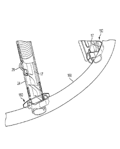
pointing from the ventricle side to the atrium side.
[00108] FIGS. 23 and 24 show the pre-launch and launch situations for
upward
launching of anchors 16. FIG. 23 further illustrates that catheter C can be
used to help orient
the angle of housings 24, and thus the launch angle of anchors 16. If the
distance between
catheter C and loop 14 is relatively small, anchors 16 tends to be positioned
and launched at a
greater angle (relative to being launched perpendicular to loop 14, as was
shown in FIGS. 2
and 3, for example). Adjustment of the launch angle, i.e. pivoting of anchors
angle, is made
possible by the shape of the support arms 20 to which the housing 24 is
attached. FIG. 24
also illustrates another modification wherein anchors 16 comprise multiple
barbs 48 and
wherein elongated slot 17 extends about half-way within the length of the
anchors, as seen in
FIG. 7A.
24
CA 3005848 2018-05-23
[00109] FIGS. 25-27 and 27A illustrate particular embodiments wherein
anchor
launching mechanism 22 is adapted to be used with tissue anchors 16 that are
launched in a
generally upward direction; and can be actuated by a direct pull, or by a
mechanism removed
from the valve area. Anchor launching mechanism 22 comprises actuation wire 28
and
housing 24, however the mechanism does not include spring 26 disposed in the
housing.
Regardless, for rapid actuation purposes (anchor launch), anchor launch
mechanism 22 may
further include an external launch actuator device, typically including a
spring (not shown),
for example, at the proximal end of catheter C, to pull on actuation wire 28.
When the
catheter approaches from the inflow side of the valve, and routes the anchors
so that they are
below the valve with the tip directed from the ventricle side to the atrium
side, this
configuration and approach to the valve permits pull wires to be used.
[00110] For the purposes of these embodiments, anchor 16 may be modified to
further
comprise an actuation wire eyelet 72 where-through actuation wire 28. Distal
end 29 of
actuation wire 28 is threaded through eyelet 72 and typically has a hook-like
configuration
while disposed within housing 24 (FIGS. 25 and 27). Pulling on actuator wire
28 proximal
end to pull (launch) anchor 16 as a result of pulling at eyelet 72 (FIG. 26).
In such
embodiments, housing 24 need not include an opening such as opening 30, nor
does not need
a crimped portion 36 or other such spring retention mechanism, as there is no
spring in the
housing. FIGS. 27 and 27A illustrates a modification wherein instead of eyelet
72; each
anchor 16 has an actuator-wire distal-end receiving portion such as recess 74,
which operates
to launch anchors 16 in the same fashion as noted above.
[00111] FIGS. 28-30 show embodiments, wherein implant 10 further comprises
a loop-
arrangement/anchor-orientation mechanism 76 useful for arranging the position
and/or shape
of loop 14 and/or for orienting the angle of housings 24, and thereby
orienting the launch
CA 3005848 2018-05-23
angle of tissue anchors 16. Anchor orientation mechanism 76 includes a
plurality of curved
arrangement leads 78 respectively attached to at least some of housings 24,
for example by
welding. Leads 78 may be an extension of implant support arms 20 and may be
arranged to
cross at a singular intersection point 80. Leads 78 are attached (e.g. by
welding) to housing
24. Thus, leads 78 of orientate mechanism 76 are movable to arrange loop 14 in
a desired
location and depending on the shape of the leads, the angle of housings 24,
and thus anchors
16, can be determined.
[00112] Regarding the launch angle of anchors 16, in some embodiments,
leads 78 can
be attached "ad hoc" prior to insertion into a patient, whereby, depending on
the attachment
location, arrangement leads 78 also be used to orient anchors 16 i.e. control
the angle at
which the anchors enter the tissue (i.e. changing the length or shape of one
or more leads 78
will thus change the angle of the anchors, e.g. shortening the that length
will cause the
anchors to point outward, whereas increasing that length will bring
intersection point 80
farther from loop 14 and thus angle the anchors more parallel to each other
(less outward). In
= such case, leads 78 will not be welded to housings 24, rather there will
be included an "ad
hoc" connection or fastening arrangement (not shown), whereby the leads and
housings are
connected at more than one location along the leads. Arrangement/orientation
mechanism 76
can be useful for arranging the shape of loop 14 as well as'positioning the
loop and orienting
the anchor angle. In alternative embodiments, loop-arrangement/anchor-
orientation
mechanism 76 either has a predetermined shape, such as a nipple shape (FIGS.
29 and 30) or
is adapted to allow its shape to be changed; i.e. leads 78 can be bent.
[00113] FIGS. 31-34 show embodiments wherein loop arrangement and/or
implant
positioning device 18 comprises an inflatable balloon 82. The figures show
exemplary
balloons 82 useful for a) making sure support arms 20 are fully expanded
before deploying
26
CA 3005848 2018-05-23
implant 10, b) make sure that loop 14 is concentric with the valve annulus
prior to
implantation, and c) facilitating an interference step or backing against
which to press to be
used for pressing implant positioning device 18 and implant 10 onto the valve
annulus before
implantation as illustrated in figure 34. FIG. 31 illustrates an oval balloon
82; FIGS. 32-34
illustrate a droplet-shaped or bulbous balloon 82.
[00114] As seen in FIG. 34, as well as being useful to orient loop 14
relative to the
valve annulus, the balloon can be used to secure the implant positioning
device 18 and
implant 10 in place during launching of anchors 16. FIGS. 32 and 33 also
illustrate that
balloon 82 can be positioned proximally or distally with respect to loop 14
and implant
positioning device 18. Since the balloon can be positioned inside the
ventricle and be inflated
to a diameter bigger than the diameter, of biological valve annulus, it can
serve as a backing
against which to press positioning device 18 and implant 10 onto the valve
annulus before
implantation. This will ensure good contact between each of the anchor
launching
mechanisms 22 and the valve annulus and will create optimal penetration
conditions of
anchor 16 into the tissue upon launching. Furthermore, the launch angle of
anchors 16 (i.e.
insertion into the tissue) can be controlled by inflating/deflating balloon
82, with
consideration to the size of the biological valve.
[00115] FIGS. 35-37 illustrate how a device 100 (e.g., a replacement valve)
can be
fixed to a native valve annulus or leaflets like the mitral valve M or
tricuspid valve. In this
embodiment, implant 10 is first implanted and secured with anchors 16 that
penetrate the
valve leaflets pointing from the ventricle V side toward the atrium A side
(hereinafter
upwards) as in FIG. 21 and/or FIG. 22. Then, when device 100 is expanded into
implant 10,
the friction between anchors 16 and the device 100 secures device 100 in
place. Since
27
CA 3005848 2018-05-23
anchors 16 are directed generally upward, the high pressure in ventricle V
helps to further
enhance the anchoring of implant 10 to the valve leaflets.
[00116] Device 100 in the illustrating figures represents any suitable
commercial
expandable heart valve prosthesis that can be tracked in a collapsed
configuration through the
vascular system and delivered to the heart. It can be a self-expanding
prosthesis or a balloon
expanding prosthesis or any other type of expanding heart valve prosthesis.
FIG. 35 further
illustrates an exemplary delivery system 101 that can deliver device 100 to
the heart.
[00117] FIGS. 36 and 37 illustrate how implant 10 can be associated with
device 100
for fixing the device to a mitral valve M (or tricuspid valve) leaflets. In
this embodiment,
implant 10 and device 100 are implanted via the heart's apex P, preferably, in
a minimally
invasive surgery as illustrated in FIG.20. As in FIG. 22, implant 10 is first
located at the
proper location with respect to the bio-valve (mitral in this case) and then
secured with
anchors 16 facing upward, in accordance with any appropriate embodiment as
described -
herein. After implant 10 is attached to the valve leaflets, device 100 is
advanced, as shown in
FIG. 36. Through a delivery catheter (not shown), and expanded into implant 10
as seen in
FIG. 37. Since anchors 16 are directed generally upward, the high pressure in
the ventricle V
helps to further enhance the anchoring of the implant 10 and device 100 to the
valve leaflets.
However, for the purpose of this embodiment, wherein implant 10 is configured
to be
particularly suited to securing a device in place such as device 100, each
anchor 16 has a
relatively shorter slot 17, typically extending only about half-way along the
longitudinal
dimension of each anchor, from about half-way along the anchor to relatively
close to the
anchors' pointy front end 46, as seen in FIG. 7A.
[00118] With reference to FIGS. 38 and 39, when device 100 is disposed in
the
appropriate heart (or other biological) valve and expanded, the contact and
sliding motion
28
=
CA 3005848 2018-05-23
between the device and anchor 16 changes the angle of the anchors from
typically
approximately 45 degrees (FIG. 38), although, depending on the angle of
support arms 20, to
an angle wherein the anchors are more parallel to each other, typically
substantially parallel.
The movement of anchors 16 is illustrated by arc A-B in FIG. 38. In other
words, anchors 16
pivots at the end of slot 17, as in FIG. 7A which is generally at mid-point 84
of the anchors.
This angle change provides increased friction between anchors 16 and device
100 thereby
securing the device in place.
[00119] To further explain, device 100 is expanded in the bio-valve until
the device
presses on a non-slotted portion 86 of anchors 16. As a result of pressing on
non-slotted
portion 86, that portion is forced outward, and thus the tip of the anchors 46
is moved inward,
as the anchors pivot around loop 14. Since anchor tips 46 are locked within
the tissue of the
valve leaflet, the inward motion of the tips pulls the leaflets closer to
device 100 and presses
the leaflets against the device, thereby enhancing the sealing and prevent
blood flow between
the native valve leaflet and the device. It should be understood that device
100 is
appropriately sized for the above-described positioning.
[00120] FIG. 40 illustrates deployment of implant 10 in the tricuspid heart
valve T and
it should be understood that all the features and functions of the implant and
delivery system
as illustrated in FIGS. 1 to 39 are applicable to the tricuspid valve.
[00121] FIG. 41 illustrates deployment of implant 10 through the left
atrium wall
rather than tracking in through the vascular system, or deploying the implant
through the
apex of the heart. Again, it should be understood that all the features and
functions of the
implant and delivery system illustrated in FIGS Ito 39 are applicable to
deployment through
the atrium wall.
= 29
CA 3005848 2018-05-23
[00122] FIG. 42 illustrates manual cinching of the device in a later
procedure after
tissue healing has occurred as described above with reference to FIG. 14.
[00123] FIG. 43 illustrates cinching of the device in a later procedure
after tissue
healing has occurred as described above with reference to FIG. 14. Using a
mechanical
actuator 110 that is implanted during procedure. The mechanical actuator can
be actuated
and operated magnetically, electrically or by any other appropriate mechanism
from outside
of the body.
[00124] FIGS. 44-47 depict one exemplary embodiment for implementing
cinching. In
this embodiment, the implant has a tissue engaging member 12 that includes a
loop of wire
14 and a plurality of tissue growth-promotion tubes 52 coaxially arranged
about the loop of
wire. The tissue growth-promotion tubes 52 are made of a material that
promotes ingrowth
of tissue, such as a fabric segments, optionally coated with a tissue growth
promoting
substance. Taken together, the loop of wire 14 and the plurality of tissue
growth-promotion
tubes 52 collectively form a loop of material.
[00125] The tissue engaging member 12 also includes a plurality of tissue
anchors 16
that are arranged with respect to the loop of wire. In the illustrated
embodiment, the anchors
16 are spaced apart all along the loop of wire 14 and the loop of wire is
threaded through
slots in the anchors 16. Preferably at least six anchors are used. Note that
although the
anchors depicted in FIGS. 44-47 most closely resemble the configuration of
anchors shown in
FIG. 52B, any alternative anchor style many be used in place of that
configuration for the
anchor. In alternative embodiments, the anchors may be attached to the wire
using linking
members like those shown in FIGS. 56A and 56B. The anchors 16 may be launched
using
any of the approaches described herein.
CA 3005848 2018-05-23
[00126] This embodiment also includes a cinching cable 200, which is
preferably
covered with a slippery coating such as PTFE or the like. Cinching cable 200
has two ends
that are threaded through a cinching collar 202 and are attached to a cinching
member 204
that has a cinching aperture or eyelet. A cinching lead 206 is threaded
through cinching
aperture and the lead's free ends may extend outside the patient's body or
remain under the
skin at the upper portion of the chest, much like pace maker leads. After
sufficient tissue
grows on the implant, which typically takes one week to several months,
depending on the
tissue growth rate, the implant may be cinched by pulling on one or both of
the free ends of
cinching lead 206 to thereby pull on cinching cable 200 and reduce the
diameter of tissue
engaging member 12.
[00127] To effect cinching, a cinching sleeve 208 is pushed along over the
cinching
lead 206 until the distal end of the cinching sleeve 208 bottoms out at the
cinching collar 202.
Then, a cinching tube 210 is pushed through cinching sleeve 208 by a pushing
member 214
until the cinching tube 210 reaches cinching collar 202, as seen in FIG. 45.
After this, by
pulling on both ends of the cinching lead 206, cinching eyelet member 204 is
retracted into
cinching tube 210, as seen in FIGS. 46. In the illustrated embodiment,
cinching tube 210 has
a plurality of one way flaps or steps 216 spaced apart along the length of the
tube for holding
cinching member 204 in place as the cinching member 204 retracts in the
cinching tube 210,
thereby controlling the ultimate length/diameter of cinching cable 200 so as
to constrict the
annulus of the bio-valve. Alternative approaches for implementing one-way
motion of the
cinching member 204 will be apparent to persons skilled in the relevant arts.
[00128] After the cinching cable 200 has been cinched to the appropriate
length/diameter, one end of cinching lead 206 may be pulled to remove the
cinching lead, the
pushing member 214 may be removed, and the cinching sleeve 208 may also be
removed.
31
CA 3005848 2018-05-23
The resultant implant would then appear as is seen in FIG. 47. In alternative
embodiments,
some or. all of these components 206, 208, 214 may remain behind as part of
the implant e.g.,
for implementing additional cinching at a later point in time.
[00129] FIGS. 48A and 488 depict an alternative cinching mechanism in which
the
ends of the cinching cable 200 are pulled by rotating a spindle 232 in a
mechanism 230 that is
preferably implanted in the patient's body. In some embodiments, the rotation
may be
implemented by a motor that is powered by a battery (not show) and controlled
remotely
from outside the patient's body. In the illustrated embodiment, the loop 201
is biased against
a spring element 235. When the spring element 235 is initially implanted, it
will be flexible.
But after implantation, tissue ingrowth will cause the spring to become rigid
and capable of
sustaining a compression load. Rotation of the spindle is preferably delayed
until after such
tissue ingrowth has occurred. The rotating mechanism preferably includes a
ratchet that
permits rotation in only one direction. Rotation of the spindle 232 will wind
up the ends of
the cinching cable 200 from the state depicted in FIG. 48A to the state
depicted in FIG. 48B,
which pulls the main loop 201 of the cinching cable 200 against the bottom of
the spring
element 235, thereby tightening the main loop 201.
[00130j FIGS. 49-52 illustrate a variety of alternative aµnchors that may
be used in
place of the anchors 16 shown in FIG. 7.
[00131] FIGS 49A and 49B depict one such anchor I6a that is partially
tubular or
cylindrical in shape. This anchor has a first panel of material 120 that has a
cylindrically
curved outer surface and a second panel of material 122 that also has a
cylindrically curved
outer surface. A slot 17 runs in a front-to-back direction disposed between
the first panel of
material and the second panel of material. The pointy front end 46 of the
anchor is
configured for implantation into the annulus or the leaflets in a forward
direction. There are
32
=
CA 3005848 2018-05-23
also a plurality of barbs 48a that are configured so that subsequent to
implantation, the barbs
resist extraction of the anchor from the annulus or the leaflets in a
backwards direction.
Preferably, this anchor 16a also has a ring-shaped portion 125 disposed at a
back end of the
anchor that connects the first panel of material 120 to the second panel of
material 122.
[00132] Preferably, a front surface of the ring-shaped portion has a notch
128, and the
slot 17 and the notch 128 are disposed on opposite sides of the ring-shaped
portion 125. In
some embodiments, the outer surface of the barbs 48a is curved so as to follow
the cylindrical
curve of the outer surface of the panel of material to which it is attached
(i.e., panels 120 and
122). This type of anchor I 6a can be advantageously produced by cutting it
out from a tube
of material. Preferred materials for this anchor I6a include metals (e.g.,
steel alloys, stainless
steel, nitinol), biocompatible plastics, and ceramics. The overall length of
the anchor 16a is
preferably between 3 and 30 mm, and more preferably between 5-10 mm. The
diameter of
the ring 125 is preferably between 0.5 and 5 mm, and more preferably between 1
and 2 mm.
[00133] FIGS 50A and 50B depict another anchor 16b that is partially
tubular or
cylindrical in shape. This anchor 16b also has a first panel of material 120
that has a
cylindrically curved outer surface and a second panel of material 122 that
also has a
cylindrically curved outer surface. A slot 17 runs in a front-to-back
direction disposed
between the first panel of material and the second panel of material. The
pointy front end 46
of the anchor is configured for implantation into the annulus or the leaflets
in a forward
direction. This anchor 16b has at least one tab 130 that is configured to
automatically spring
outward after being implanted, so that after the tab has sprung outward (as
seen in FIG. 50B),
the tab causes the anchor to resist extraction from the annulus or the
leaflets in a backwards
direction. Note that prior to implantation, the tabs 130 remain in the
collapsed state depicted
=
33
CA 3005848 2018-05-23
in FIG. 50A and do not spring outward because they are restrained from doing
so by a
housing (such as the housing 24 shown in FIGS 4, 5, and 54A).
[00134] As in the FIG. 49 embodiment, this anchor 16b also preferably has a
ring-
shaped portion 125 disposed at a back end of the anchor that connects the
first panel of
material 120 to the second panel of material 122. Preferably, a front surface
of the ring-
shaped portion has a notch 128, and wherein the slot 17 and the notch 128 are
disposed on
opposite sides of the ring-shaped portion 125. This type of anchor 16b can
also be
advantageously produced by cutting it out from a tube of material. The spring-
out tabs 130
may be implemented using spring material or using a shape memory alloy. The
preferred
materials and dimensions for this embodiment are similar to those for the
embodiment
described above in connection with FIGS. 49A and 4913.
. [00135] FIGS 51A and 51B depict another anchor 16c that is partially
tubular or
cylindrical in shape. This anchor 16c is similar to the anchor 16b depicted in
FIGS. 50A and
50B, but instead of the tabs that are configured to automatically spring
outward after being
implanted, this anchor 16c uses one or more arms 145 formed from a shape-
memory alloy
(SMA) material. These arms are configured to automatically spring outward
after being
implanted by operation of the SMA material, so that after the arm has sprung
outward (as
seen in FIG. 51B), the arm causes the anchor to resist extraction from the
annulus or the
leaflets in a backwards direction. Note that prior to implantation, the arms
145 remain in the
collapsed state depicted in FIG. 51A and do not spring outward because they
are restrained
from doing so by a housing (such as the housing 24 shown in FIGS 4, 5, and
54A). This type
of anchor 16c can also be advantageously produced by cutting it out from a
tube of material.
The preferred materials and dimensions for this embodiment are also similar to
those for the
embodiment described above in connection with FIGS. 49A and 4913.
34
CA 3005848 2018-05-23
[00136] FIGS. 52A and 52 B depict yet another anchor 16d that may be used
in place
of the anchors 16 shown in FIG. 7. This anchor 16d is similar to the anchor 16
depicted in
FIG. 7, but instead of the barb depicted in FIG. 7, this anchor 16d uses one
or more arms 140
formed from a shape-memory alloy (SMA) material. These arms are configured to
automatically spring outward after being implanted by operation of the SMA
material, so that
after the arms 140 have sprung outward (as seen in FIG. 52B), the arms cause
the anchor to
resist extraction from the annulus or the leaflets in a backwards direction.
Note that prior to
implantation, the arms 140 remain in the collapsed state depicted in FIG. 52A
and do not
spring outward because they are restrained from doing so by a housing (such as
the housing
24 shown in FIGS 4, 5, and 54A). The preferred materials for this embodiment
are similar to
those for the embodiment described above in connection with FIGS. 49A and 49B.
The
length of the anchor 16d is preferably between 3 and 30 mm, more preferably
between 5 and
mm. The thickness of the material is preferably between 0.1 and 1.5 mm, more
preferably
between 0.2 and 0.6 mm.
[00137] FIG. 53 depicts a tissue engaging member that includes a loop of
wire 14, a set
of anchors 16 of the type depicted in FIG. 52B that have been implanted into a
mitral valve
annulus, with a plurality of tissue growth-promotion tubes 52 that are
coaxially arranged
about the loop of wire. Taken together, the loop of wire 14 and the plurality
of tissue growth-
promotion tubes 52 collectively form a loop of material. The usage and
operation of this
tissue engaging member is similar to the tissue engaging member discussed
above in
connection with FIG. 8, and differs mainly because a different type of anchor
is used. Of
course, any of alternative anchors described herein may be used in place of
the anchor
depicted in FIG. 53.
CA 3005848 2018-05-23
[00138] FIGS. 54A and 54B illustrate an embodiment of anchor launching
mechanism
for launching anchors 16 into the biovalve tissue, e.g. the Mitral valve
annulus or leaflets.
The anchor launching mechanism in includes a housing 24 that has an open front
end. The
housing has a cylindrical interior void that includes a first front section
and a second rear
section. An anchor 16 (e.g., any of the anchors described above) is disposed
in the front
section of the void in the housing, and an anchor launching spring 26 is
disposed in the rear
portion of the void in the housing 24 in a compressed state. The spring 26 is
preferably a coil
spring. In the illustrated embodiment, the back end (i.e., the proximal end)
of the spring 26 is
retained in housing 24 by a spring retention loop or hook 38. Of course,
alternative
configurations may be used for preventing the spring 26 from exiting the
housing 24.
[00139] The anchor launching mechanism includes an actuator configured to
(a)
prevent the spring from expanding from the compressed state prior to being
actuated and (b)
permit the spring to expand from the compressed state upon being actuated. The
actuator is
preferably implemented using an actuator 350 that initially passes coaxially
through the
anchor launching spring 26. Preferred materials for the actuator 350 include
metals (e.g.,
steel alloys, stainless steel, nitinol), biocompatible plastics, and ceramics.
The thickness of
the actuator 350 is preferably between 0.05 and 1.0 mm, more preferably
between 0.1 and 0.3
MM.
[00140] In the initial state (i.e., prior to actuation) depicted in FIG.
54A, the distal
portion 355 of actuator 350 passes through and/or interfaces with an opening
30 in the
housing. Optionally, the distal portion 355 may be forked (as best seen in
FIG. 54C) to
engage the opening 30 more securely. The front end (i.e., the distal end) of
the spring 26
presses against the back end of the anchor 16. In the illustrated embodiment,
the back end of
the anchor is the ring 125 located at the back of the anchor 16. Prior to
actuation, the actuator
36
CA 3005848 2018-05-23
350 passes through the notch 128 in the ring 125 at the back of the anchor 16
and also passes
through the opening 30 of the housing 24. The presence of the distal portion
355 of the
actuator 350 in this position, engaged with the opening 30, prevents the
spring 26 from
expanding, thereby keeping the anchor launching spring 26 in a compressed
state, In some
embodiments, distal portion 355 includes a fork-like tip for engaging the
opening 30 of the
housing 24 more securely.
[00141] The passage of the distal portion 355 through the notch 128 and the
opening
30 also operates to align the notch 128 with the opening 30. Preferably, there
is an elongated
recess 40 at the open end of the housing 24, located directly in front of the
opening 30 in the
axial direction. Because the notch 128 is aligned with the opening 30, and the
elongated
recess 40 is directly in front of the opening 30, and the slot 17 is opposite
from the notch 128,
the anchor 16 will be oriented so that the slot 17 in the anchor 16 is
opposite from elongated
recess 40. This is advantageous because when the elongated recess 40 is
opposite from the
slot 17, those features 40, 17 will align so that a loop of wire 14 can pass
easily through all
the elongated recesses 40 and all the slots 17 in each of the anchor launchers
and anchors,
which makes it easier to launch the anchors into the target tissue.
[00142] An actuation wire 28 (i.e., the "pull wire") is attached to the
proximal portion
of the actuator 350 using any suitable attachment approach (e.g., welding,
crimping, etc.).
The actuator 350 then can be pulled in a proximal direction by pulling on the
pull wire 28.
When this occurs, the distal portion 355 of actuator 350 is pulled inwardly
through the
opening 30 and is withdrawn from the opening 30. At this point, the spring 26
will expand
into the front section of the housing 24 and push the anchor 16 forward such
that at least a
portion of the anchor 16 exits the front end of housing. The spring 26 pushes
the anchor 16
with sufficient force to implant the anchor into the tissue.
37
CA 3005848 2018-05-23
[00143] Note that in alternative embodiments (not shown), instead of using
a discrete
actuator 350 that is connected to the end of the actuation wire 28, the
discrete actuator can be
eliminated, and the distal end of the actuation wire 28 itself can serve as
the actuator. In
either case, it is preferable to pull the wire 28 in the proximal direction
with a jerk (i.e., with
rapid acceleration), because it makes the launching more reliable and prevents
the tissue
engaging member from lifting away from the surface of the tissue prior to
implantation.
[00144] FIG. 55 depicts a suitable apparatus for pulling the wires 28 in
the proximal
direction in this manner, to trigger the anchor launchers shown in FIGS 54A
and 54B. The
pulling apparatus has a plurality of actuators housed in a housing 400. Each
of these
actuators is housed in a channel 402 that runs through the housing 400 in a
proximal-to-distal
direction. Preferably, the housing 400 is cylindrical, and the channels 402
are distributed
within the cylindrical housing close to the circumference of the cylinder.
=
[00145] Each actuator has a shoulder 404 disposed adjacent to the channel
402. A
compressed spring 406 is disposed in a distal portion of the channel. The
distal end of the
spring 406 is preferably fixed, and the proximal end is preferably movable.
The channel 402
is configured to permit expansion of the spring 406 in a proximal direction.
[00146] Each actuator also has a tab 408 that is affixed to the proximal
end of the
spring 406 using any suitable attachment system (e.g., screws, crimping,
etc.). In the
embodiments where the housing is cylindrical, it is preferable to have the
tabs 408 extend
radially outward from the channels 402 beyond the circumference of the
cylindrical housing
400. The tab may be affixed directly to the spring 406, or the tab may be
connected through
intermediate members. The tab 408 is configured to be movable between (I) a
first position
in which movement of the tab 408 in a proximal direction is blocked by the
shoulder 404, and
(2) a second position in which movement of the tab 408 in a proximal direction
is not blocked
38
CA 3005848 2018-05-23
by the shoulder 404. As soon as the tab 408 is moved from the first position
to the second
position, the spring 406 will expand within the channel and move from its
compressed state
to its released state, with the proximal end of the spring moving in a
proximal direction.
[00147] The proximal end of the pull wire 28 is attached (either directly
or indirectly)
to the spring 406 or the tab 408 and a distal portion of the pull wire 28
extends to the anchor
launcher. When the proximal end of the spring 406 moves in the proximal
direction, the pull
wire 28 is pulled in the proximal direction with the preferred jerking motion.
Optionally, the
pull wires may be threaded though individual corresponding apertures to avoid
tangling. For
such purpose, a pull wire distribution collar 420 with respective distribution
holes (not
shown) disposed therein may be provided at the distal end of the housing 400.
[00148] A rotatable cap (not shown) may be is used to push the tabs from
the first
position to the second position. In some embodiments, the interior surface of
the cap has a
cylindrical void configured to surround the cylindrical housing 400, and the
interior surface
has a single protrusion configured to sequentially push each of the tabs from
the first position
to the second position when the cap is rotated. In this case, the anchors will
launch
sequentially. In alternative embodiments, the interior surface has a plurality
of protrusions
configured to simultaneously push a plurality of the tabs from the first
position to the second
position when the cap is rotated. In this case, a plurality of anchors will
launch
simultaneously.
[00149] FIGS. 56A and 568 depict an alternative approach for implementing
the tissue
engaging member. In this embodiment, the tissue engaging member includes three
sets of
parts. The first set is a loop of material 160 configured to contact at least
a portion of the
annulus or the leaflets when the loop of material is deployed. This loop of
material can be a
39
CA 3005848 2018-05-23
wire, or in alternative embodiments the loop may be a different loop of
material such as a
tube, strip, chain, braid, etc., or a combination of multiple materials.
[00150] The second set is a plurality of anchors 16, each of which has a
pointy front
end and a back end. Each of the anchors also has a slot 17 that runs in a
front-to-back
direction. The front ends of the anchors 16 are configured for implantation
into the annulus
or the leaflets in a forward direction. The anchors are configured so that
subsequent to
implantation, the anchors resist extraction from the annulus or the leaflets
in a backwards
direction. The anchor embodiments described above can be used for this
purpose. The
anchors are arranged with respect to the loop of material so that when the
loop of material is
deployed the anchors are distributed about the loop of material with the front
ends of the
anchors facing the annulus or the leaflets.
[00151] The third set is a plurality of linking members 150 that are
affixed to the loop
of material 160. At least a portion of each of the linking members 150 passes
through the slot
in a respective anchor, and each of the linking members is configured to slide
with respect to
the slot in the respective anchor in the front-to-back direction. In some
embodiments, the
linking members include a strip of material 155 that passes through the slot
in the respective
anchor. This strip of material 155 may be connected to the loop of material
160 through at
least one intermediate member 152. For example, if the loop of material 160 is
a hollow
tube, the intermediate member 152 could be a C-shaped bracket that connects
the loop of
material 160 to the strip of material 155. In alternative embodiments (not
shown), the strip of
material 155 may be directly connected to the loop of material 160. Preferred
materials for
the linking members 150 include metals (e.g., steel alloys, stainless steel,
nitinol),
biocompatible plastics, and ceramics. The width is preferably between 0.2 and
3 rpm, more
CA 3005848 2018-05-23
preferably between 0.5 and 1.5 mm. The thickness of the material is preferably
between 0.05
and 1.0 mm, more preferably between 0.1 and 0.3 mm.
[001521 In the embodiment illustrated in FIG. 56B, the anchors and the
linking
members are disposed inside the loop at the inner circumference of the loop.
In alternative
embodiments (not shown), the anchors and the linking members may be disposed
outside the
loop at the outer circumference of the loop.
[001531 Prior to launching, the loop of material 160 is delivered to its
desired location
in contact with the annulus or leaflets, in a manner similar to the
embodiments described
above. The anchors 16 are then launched (e.g., using any of the launching
mechanisms
described above). When the anchors are launched, the anchors will move forward
while the
strips 155 of the linking members 150 remain stationary. This will implant the
tissue
engaging member in the desired location. As a result of the movement of the
anchors, the
linking members 150 will have shifted (with respect to the slot 17) from the
front of the slot
17 towards the back of the slot 17.
[00154] Note that the embodiment depicted in FIG. 56B differs from the FIG.
3
embodiment because in the FIG. 56B embodiment the loop of material 160 is
connected to
the slot 17 in the anchor via the linking members 150. In contrast, the loop
of wire 14 passes
directly through the slot in the anchor 16 in the FIG. 3 embodiment.
[00155] FIGS. 57A, 57B, and 57C show yet another embodiment of an anchor,
which
relies on a component 316 that normally has the curled configuration shown in
FIG. 57B.
However, when the anchor 316 is still disposed in the launch mechanism housing
324, the
anchor 316 is deformed and takes on a generally elongated shape shown in FIG.
57A.
Housing 324 is preferably generally cylindrical in shape. A spring 326 is
dispose in the
proximal end of the housing 324, and the spring is configured to push the
anchor 316 out of
41
CA 3005848 2018-05-23
the housing 324 when it is triggered. The trigger mechanisms described above
in connection
with other embodiments may be used for this embodiment.
[00156] Upon being launched, the anchor 316 is pushed out of the distal end
of the
housing 324 and will immediately spring back to its original curled
configuration shown in
FIG. 57B. In some embodiments, the return to the original shape may be
accomplished by
forming the anchor 316 from a shape memory alloy. The distal tip 316b of the
anchor 316 is
preferably sharp, and is configured to pierce the tissue upon exiting the
housing 324. The
distal tip 316b will then curl around and engage the target tissue.
[00157] In this embodiment, anchor 316 preferably has atop portion 3I6a
that forms a
, loop engaging eyelet, through which the loop of wire 314 is threaded, as
seen in FIG. 57C. In
such an embodiment, eyelet can also surround any tissue growth-promotion tubes
(not
shown) that may coaxially surround the loop 314. When the distal tip 316b is
engaged with
the tissue, the eyelet of the top portion 316a will hold the loop next to the
tissue. Note that
unlike the embodiments described above, the loop 314 in this embodiment is not
positioned
in contact with the tissue prior to launching. Instead, the loop 314 travels
down to its final
destination together with the eyelet of the top portion 316a during the
implantation procedure.
Note also that the delivery of the launch mechanism housings 324 to their
intended locations
prior to launching for this embodiment may be implemented in similar ways to
the other
embodiments described herein.
[00158] FIG. 58 illustrates an embodiment for implanting a ring and a valve
in a single
procedure. In this embodiment, a tissue engaging member that includes anchors
16 and a
loop 14 is mounted on anchor positioning leads 260 that extend from a delivery
catheter C.
In the case of a Mitral valve repair., the tissue engaging member is
preferably inserted via the
heart's apex, via an insertion catheter or encapsulating cylinder, together
with an expandable
42 =
CA 3005848 2018-05-23
valve 100. Initially, the valve 100 is located between the anchor positioning
leads 260 in a
collapsed state. Optionally, a nose cone 250 may be used to help guide the
tissue engaging
member to the correct location. After the anchors 16 are inserted/launched,
the valve 100 is
advanced to a location within the loop 14 and expanded from its original
collapsed state
(depicted in FIG. 38) to its final expanded configuration (depicted in FIGS.
39 and 37). The
insertion catheter and encapsulating cylinder 102 are then removed, leaving
the tissue
engaging member and the valve 100 behind.
[00159] FIG. 59 shows an embodiment wherein the anchor positioning leads
260 are
implemented in the form of elongated needle-like members. In some embodiments,
the
elongated needle-like anchor positioning leads are stiff enough so that they
can press the
anchors 16 directly into the bio-tissue, without relying on a spring-based
anchor launching
mechanism. FIG. 60 shows the situation after the anchor positioning leads 260
have been
removed from the heart.
[00160] While the present invention has been disclosed with reference to
certain
embodiments, numerous modifications, alterations, and changes to the described
embodiments are possible without departing from the sphere and scope of the
present
invention, as defined in the appended claims. Accordingly, it is intended that
the present
invention not be limited to the described embodiments, but that it has the
full scope defined
by the language of the following claims, and equivalents thereof.
43
CA 3005848 2018-05-23