Note: Descriptions are shown in the official language in which they were submitted.
CA 03005908 2018-05-18
WO 2017/096157
PCT/US2016/064610
FRAME FEATURES FOR PROSTHETIC MITRAL VALVES
Cross-Reference to Related Applications
110011 This application claims priority to and the benefit of U.S.
Provisional Patent
Application No. 62/262,511, filed December 3, 2015, entitled "Frame Features
for Prosthetic
Mitral Valves," the disclosure of which is incorporated herein by reference in
its entirety.
Background
110021 Prosthetic heart valves, including those for insertion into
atrioventricular valves
(tricuspid and mitral valves) are susceptible to various problems, including
problems with
ventricular outflow tract obstruction. Some known prosthetic mitral valves,
for example,
apply undesirable forces to the anterior segment of the native valve thereby
contributing to
undesirable interruption of blood flow into the aorta, which anatomically sits
immediately
behind the anterior segment of the mitral annulus. As another example, some
known
prosthetic mitral valves include subvalvular components that obstruct the left
ventricular
outflow tract (LVOT) and/or direct blood flow from the atrium to the ventricle
in a manner
that creates undesirable flow gradients and LVOT interruption. Accordingly,
there is still a
need for a prosthetic heart valve that can address some or all of these
problems.
Summary
110031 Prosthetic heart valves are described herein that can provide
clearance to the
LVOT, reduce the possibility of undesirable outflow gradients, and/or limit or
prevent LVOT
obstructions when implanted in the heart. In some embodiments, a prosthetic
heart valve can
include an outer frame assembly including an outer frame having a cuff portion
configured to
be disposed at least partially within an atrium of a heart and a body portion
configured to be
disposed in a ventricle of the heart. The body portion has a posterior side
and an opposite
anterior side and the anterior side can have a maximum height larger than a
maximum height
of the posterior side such that when the prosthetic heart valve is disposed
within a native
annulus of the heart, an anterior end of the outer frame is disposed at an
acute angle relative
to a centerline of the outer frame. An inner valve assembly is disposed within
and coupled to
the outer frame assembly and includes an inner frame having an atrium end and
a ventricle
1
CA 03005908 2018-05-18
WO 2017/096157
PCT/US2016/064610
end and having a centerline substantially parallel to the centerline of the
outer frame. The
inner valve assembly including a valve leaflet assembly supported on the inner
frame.
Brief Description of the Drawings
110041 FIGS. IA and 1B are schematic perspective and side cross sectional
views of a
prosthetic heart valve according to an embodiment.
[1005] FIGS. 2A-C are schematic views of an inner valve assembly of the
prosthetic
heart valve of FIGS. IA and 1B.
[1006] FIG. 3 is a top view of a prosthetic heart valve according to
another embodiment.
[1007] FIG. 4 is a top view of a prosthetic heart valve according to
another embodiment.
[1008] FIG. 5 is a perspective side view of a portion of a prosthetic heart
valve according
to another embodiment.
[1009] FIG. 6 is an exploded view of a prosthetic heart valve system
according to another
embodiment.
[1010] FIGS. 7-9 are front, bottom, and top views of a prosthetic heart
valve according to
another embodiment.
[1011] FIG. 10 is an opened and flattened view of the inner frame of the
valve of FIGS.
7-9, in an unexpanded configuration.
[1012] FIGS. 11 and 12 are side and bottom views, respectively, of the
inner frame of
FIG. 10 in an expanded configuration.
[1013] FIG. 13 is an opened and flattened view of the outer frame of the
valve of FIGS.
7-9, in an unexpanded configuration.
[1014] FIGS. 14 and 15 are side and top views, respectively, of the outer
frame of FIG.
13 in an expanded configuration.
[1015] FIGS. 16-18 are side, front, and top views of an assembly of the
inner frame of
FIGS. 10-12 and the outer frame of FIGS. 13-15.
[1016] FIG. 19 is a plan view of a fabric pattern for the inner and outer
coverings of the
outer frame assembly of the valve of FIGS. 7-9.
2
CA 03005908 2018-05-18
WO 2017/096157
PCT/US2016/064610
[1017] FIG. 20 is a plan view of a fabric pattern for the leaflets and
outer covering of the
inner valve assembly of the valve of FIGS. 7-9.
[1018] FIGS. 21 and 22 are schematic perspective and side cross sectional
views of a
prosthetic heart valve according to another embodiment.
[1019] FIGS. 23-25 are top and perspective views of a prosthetic heart
valve according to
another embodiment.
[1020] FIG. 26 is an exploded view of a prosthetic heart valve system
according to
another embodiment.
[1021] FIGS. 27 and 28 are schematic perspective and side cross sectional
views of a
prosthetic heart valve according to another embodiment.
[1022] FIGS. 29A-D are schematic illustrations of stiffiless profiles of a
prosthetic heart
valve according to another embodiment.
[1023] FIG. 30A is a side view of an outer frame of a prosthetic heart
valve having an
angled cuff arrangement, in a deployed or biased configuration, according to
an embodiment.
[1024] FIG. 30B is a schematic cross-sectional side view of the prosthetic
heart valve
shown in FIG. 30A, including an inner valve assembly.
[1025] FIG. 30C is a side view of the prosthetic heart valve shown in FIG.
30A, having
an angled cuff arrangement and in a deployed or biased configuration, and a
side view of a
prosthetic heart valve without an angled cuff arrangement, in a deployed or
biased
configuration.
[1026] FIG. 31A is a side view of an outer frame of a prosthetic heart
valve having a
short body portion, in a deployed or biased configuration, according to an
embodiment.
[1027] FIGS. 31B and 31C illustrate schematic cross-sectional perspective
and side
views, respectively, of the prosthetic heart valve shown in FIG. 31A,
including an inner valve
assembly.
[1028] FIGS. 32A-32C are top views of a prosthetic heart valve having an
inner valve
assembly radially off-set (FIGS. 32A and 32C) and radially centered (FIG.
32B), in a
deployed or biased configuration, according to an embodiment.
3
CA 03005908 2018-05-18
WO 2017/096157
PCT/US2016/064610
[1029] FIG. 33A is a top view a prosthetic heart valve having an inner
valve assembly
rotated relative to an A2 segment of an outer frame, in a deployed or based
configuration,
according to an embodiment.
[1030] FIG. 33B is a bottom view of the inner frame of the prosthetic valve
of FIG. 33A.
[1031] FIGS. 34A and 34B illustrate in side view an inner frame of a
prosthetic heart
valve in a compressed and an uncompressed arrangement, respectfully, in a
deployed or
biased configuration, according to an embodiment.
[1032] FIGS. 35A and 35B illustrate in a partial cross-sectional side view
and top view,
respectfully, an exemplary prosthetic heart mitral valve in a deployed or
biased configuration
and seated in a native mitral valve annulus of a heart.
Detailed Description
[1033] Prosthetic heart valves are described herein that can provide
clearance to the
LVOT, reduce the possibility of undesirable outflow gradients, and/or limit or
prevent LVOT
obstructions when implanted in the heart. In some embodiments, a prosthetic
heart valve can
include an outer frame having a cuff portion that is disposed at an angle
(e.g., 80 degrees)
relative to the vertical axis of a body portion of the outer frame, so that
the prosthetic valve
can seat securely in the annulus while not obstructing the ventricular outflow
tract of the
heart. A prosthetic heart valve can alternatively, or additionally, include
subvalvular
components having a short profile, so that the prosthetic valve can seat
securely in the
annulus while not obstructing the ventricular outflow tract of the heart.
[1034] A schematic representation of a prosthetic heart valve 100 is shown
in FIGS. IA
and 1B. Prosthetic heart valve 100 is designed to replace a damaged or
diseased native heart
valve such as a mitral valve. Valve 100 includes an outer frame assembly 110
and an inner
valve assembly 140 coupled to the outer frame assembly.
[10351 Although not separately shown in the schematic illustration of outer
frame
assembly 110 in FIGS. IA and 1B, outer fame assembly 110 may be formed of an
outer
frame 120, covered on all or a portion of its outer face with an outer
covering 130, and
covered on all or a portion of its inner face by an inner covering 132.
[1036] Outer frame 120 can provide several functions for prosthetic heart
valve 100,
including serving as the primary structure, as anchoring mechanism and/or an
attachment
4
CA 03005908 2018-05-18
WO 2017/096157
PCT/US2016/064610
point for a separate anchoring mechanism to anchor the valve to the native
heart valve
apparatus, a support to carry inner valve assembly 140, and/or a seal to
inhibit paravalvular
leakage between prosthetic heart valve 100 and the native heart valve
apparatus.
[1037] Outer frame 120 is preferably formed so that it can be deformed
(compressed
and/or expanded) and, when released, return to its original (undeformed)
shape. To achieve
this, outer frame 120 is preferably formed of materials, such as metals or
plastics, that have
shape memory properties. With regards to metals, NitinoKR) has been found to
be especially
useful since it can be processed to be austenitic, martensitic or super
elastic. Other shape
memory alloys, such as Cu-Zn-Al-Ni alloys, and Cu-Al-Ni alloys, may be used.
[1038] Outer frame 120 is preferably formed from a laser cut, thin-walled
tube of
Nitinolt. The laser cuts form regular cutouts in the thin Nitinol tube. The
tube can be
expanded radially, placed on a mold or mandrel of the desired shape, heated to
the
martensitic temperature, and quenched. The treatment of the frame in this
manner will form
an open lattice frame structure, and may have a flared end or cuff at the
atrium end portion
126 of outer frame 120. Outer frame 120 thus has shape memory properties and
will readily
revert to the memory shape at the calibrated temperature. Alternatively, outer
frame 120 may
be constructed from braided wire or other suitable material.
[1039] Inner valve assembly 140 is shown schematically in more detail in
FIGS. 2A-2C.
Inner valve assembly 140 can include an inner frame 150, an outer covering
160, and leaflets
170. In the simplified form shown schematically in FIG. 2A, inner frame 150
includes six
axial posts or frame members that support outer covering 160 and leaflets 170.
Leaflets 170
are attached along three of the posts, shown as commissure posts 152 in FIG.
2A, and outer
covering 160 is attached to the other three posts, 154 in FIG. 2A, and
optionally to
conunissure posts 152. In the simplified form illustrated schematically in
FIG. 2A, each of
outer covering 160 and leaflets 170 are formed of approximately rectangular
sheets of
material, which are joined together at their upper, or atrium end. The lower,
ventricle end of
outer covering 160 may be joined to inner covering 132 of outer frame assembly
110 (not
shown in FIG. 2A), and the lower, ventricle end of leaflets 170 may form free
edges, though
coupled to the lower ends of conunissure posts 152.
[1040] As shown in FIGS. 2B and 2C, leaflets 170 are movable between an
open
configuration (FIG. 2B) and a closed configuration (FIG. 2C) in which the
leaflets coapt, or
meet in sealing abutment.
CA 03005908 2018-05-18
WO 2017/096157
PCT/US2016/064610
[1041] At the lower, or ventricle end, leaflets 170 may have a smaller
perimeter than
outer covering 160. Thus, the free lower edges of the leaflets, between
commissure posts 152
(each portion of leaflets 170 between adjacent commissure posts being referred
to as a
"belly" of leaflets 170) are spaced radially from the lower edge of outer
covering 160. This
radial spacing facilitates movement of the leaflets from the open position in
FIG. 2B to the
closed position in FIG. 2C, as the counter flow of blood from the ventricle to
the atrium
during systole can catch the free edges of the bellies and push the leaflets
closed.
[1042] Outer covering 130 and inner covering 132 of outer frame 120, outer
covering 160
and leaflets 170 may be formed of any suitable material, or combination of
materials. In
some embodiments, the tissue is optionally a biological tissue, such as a
chemically stabilized
tissue from a heart valve of an animal, such as a pig, or pericardial tissue
of an animal, such
as cow (bovine pericardium), sheep (ovine pericardium), pig (porcine
pericardium), or horse
(equine pericardium). Preferably, the tissue is bovine pericardial tissue.
Examples of
suitable tissue include that used in the products Duraguard , Peri-Guard , and
Vascu-
Guard , all products currently used in surgical procedures, and which are
marketed as being
harvested generally from cattle less than 30 months old. Alternatively, valve
leaflets 170
may optionally be made from pericardial tissue or small intestine submucosal
(SIS) tissue.
[1043] Synthetic materials, such as polyurethane or
polytetrafluoroethylene, may also be
used for valve leaflets 170. Where a thin, durable synthetic material is
contemplated, e.g. for
outer covering 130 or inner cover 132, synthetic polymer materials such
expanded
polytetrafluoroethylene or polyester may optionally be used. Other suitable
materials may
optionally include thermoplastic polycarbonate urethane, polyether urethane,
segmented
polyether urethane, silicone polyether urethane, silicone-polycarbonate
urethane, and ultra-
high molecular weight polyethylene. Additional biocompatible polymers may
optionally
include polyolefins, elastomers, polyethylene-glycols, polyethersulphones,
polysulphones,
polyvinylpyrrolidones, polyvinylchlorides, other fluoropolymers, silicone
polyesters, siloxane
polymers and/or oligomers, and/or polylactones, and block co-polymers using
the same.
[1044] In another embodiment, valve leaflets 170 may optionally have a
surface that has
been treated with (or reacted with) an anti-coagulant, such as, without
limitation,
immobilized heparin. Such currently available heparinized polymers are known
and
available to a person of ordinary skill in the art.
[1045] As shown in FIGS. 1A, 1B, and 2A, inner valve assembly 140 may be
substantially cylindrical, and outer frame assembly 110 may be tapered,
extending from a
6
CA 03005908 2018-05-18
WO 2017/096157
PCT/US2016/064610
smaller diameter (slightly larger than the outer diameter of inner valve
assembly 140) at a
lower, ventricle portion 112 (where it is coupled to inner valve assembly 140)
to a larger
diameter, atrium portion 116, with an intermediate diameter, annulus portion
114 between the
atrium and ventricle portions.
(1046i A tapered annular space or pocket 185 is thus formed between the
outer surface of
inner valve assembly 140 and the inner surface of outer frame assembly 110,
open to the
atrium end of valve assembly 100. When valve assembly 100 is disposed in the
annulus of a
native heart valve, blood from the atrium can move in and out of pocket 185.
The blood can
clot, forming thrombus, and the thrombus can be washed out by the flow of
blood during the
cyclic pumping of the heart, which is undesirable. To inhibit such washout of
thrombus, and
to enhance clotting, ingrowth of tissue into the surfaces of valve 100. and
produce other
benefits, the pocket can be covered, or enclosed, by a pocket closure 180.
11.0471 Pocket closure 180 can be formed at least in part of any suitable
material that is
sufficiently porous to allow blood, including particularly red blood cells, to
enter pocket 185,
but is not so porous as to allow undesirably large thrombi to leave the pocket
185, or to allow
washout of thrombus formed in the pocket 185. For example, pocket closure 180
may be
formed at least in part from a woven or knit polyester fabric with apertures
less than 160 p,
and preferably between 90 and 120 p. It is not necessary for the entirety of
pocket closure
180 to be formed of the same material, with the same porosity. For example,
some portions
of pocket closure 180 may be formed of a less porous, or blood impermeable,
material and
other portions fonned of material of the porosity range noted above. It is
also contemplated
that a portion of the outer frame assembly 110 or the inner valve assembly 140
may be
formed with an aperture that communicates with pocket 180, covered by a
closure formed of
material having the desired porosity, thus providing another path by which
blood may enter,
but thrombi are prevented from leaving, atrial pocket 185.
110481 The outer surface of inner valve assembly 110, and/or the inner
surface of outer
frame assembly 140, need not by circular in cross-section as shown
schematically in FIGS.
IA and 1B, but may be of non-constant radius at a given location along the
central axis of
valve 100. Thus, pocket 185 may not be of constant cross-section, and may not
be
continuous, but rather may be formed in two or more fluidically isolated,
partially annular
volumes. Similarly, pocket closure 180 need not be shaped as a ring with
constant width as
shown schematically in FIGS. IA and 1B, but rather can be a continuous ring of
varying
with, a more complicated continuous shape, or may be formed in multiple,
discrete sections.
7
CA 03005908 2018-05-18
WO 2017/096157
PCT/US2016/064610
[1049] Pocket closure 180 serves to trap and/or slow the flow of blood
within pocket 185,
reducing hemodynamic washout and increasing formation of thrombus in pocket
185. It also
promotes active in-growth of native tissue into the several coverings of
prosthetic heart valve
100, further stabilizing valve 100 in the native heart valve. The material
forming the outer
covering of inner valve assembly 140 can also be hardened or stiffened,
providing better
support for leaflets 170. Also, a mass of thrombus filling pocket 185 can
serve as potting for
inner valve assembly 140, further stabilizing the valve assembly. Greater
stability for inner
valve assembly 140 can provide more reliable coaption of valve leaflets 170,
and thus more
effective performance. The mass of thrombus can also stabilize the outer frame
assembly
110 after it has been installed in, and flexibly conformed to, the native
valve apparatus. This
can provide a more effective seal between prosthetic heart valve 100 and the
native valve
apparatus, and reduce perivalvular leakage.
[1050] One possible implementation of the prosthetic heart valve shown
schematically in
FIGS. 1A-2C is prosthetic heart valve 200, shown in top view in FIG. 3.
Prosthetic heart
valve 200 includes an outer frame assembly 210 and an inner valve assembly 240
coupled to
the outer frame assembly.
[1051] The outer frame assembly 210 includes an outer frame 220, covered on
all or a
portion of its outer face with an outer covering 230 (not visible), and
covered on all or a
portion of its inner face by an inner covering 232.
[1052] The inner valve assembly 240 includes an inner frame 250, an outer
covering 260
(not visible), and leaflets 270. Inner frame 250 includes six axial posts or
frame members
that support outer covering 260 and leaflets 270. The inner valve assembly 240
may be
substantially cylindrical, and outer frame assembly 210 may be tapered,
extending from a
smaller diameter (slightly larger than the outer diameter of inner valve
assembly 240) at a
lower, ventricle portion (where it is coupled to inner valve assembly 240) to
a larger
diameter, atrium portion, with an intermediate diameter, annulus portion
between the atrium
and ventricle portions.
[1053] A tapered annular space or pocket 285 (e.g., atrial thrombogenic
sealing pocket) is
thus formed between the outer surface of inner valve assembly 240 and the
inner surface of
outer frame assembly 210, open to the atrium end of valve assembly 200. The
pocket closure
280 can, for example, be fonned from a circular piece of wire, or halo, with a
permeable
mesh fabric or tissue, that is sewn and thereby connected to the inner frame
250 and/or to the
leaflets 170. The inner frame 250 has an inner wireframe structure (e.g., made
of Nitinol
8
CA 03005908 2018-05-18
WO 2017/096157
PCT/US2016/064610
wire) that supports the leaflets 270 sewn to the inner frame 250 and functions
as a valve. The
inner frame 250 in FIG. 3 includes three U-shaped wire components joined at
their opened
ends to fonn junctions. Leaflets 270 are sewn to these components to form
articulating
leaflets 170 creating and functioning as a prosthetic valve (e.g., prosthetic
tricuspid valve;
prosthetic mitral valve; prosthetic aortic valve, etc.).
[1054] Moreover, the inner frame 250 has (tether) attachment apertures 211
(not shown),
for attaching tether assembly 290 (not shown). Tether assembly 290 is
connected to
epicardial securing pad 254 (not shown).
[1055] In operation, the inner valve assembly 240 is disposed within and
secured within
the outer frame assembly 210. Outer frame assembly 210 may also have in
various
embodiments an outer stent tissue material. Outer frame assembly 210 includes
an
articulating collar 246 which has a collar cover 248. Articulating collar 246
is specifically
shaped to solve leakage issues arising from native structures. In particular,
collar 246 is
composed of an A2 segment 247, a P2 segment 249, and two commissural segments,
the Al-
p! segment 251, and the A3-P3 segment 253. The collar 246 may also have in
preferred
embodiments a shortened or flattened or D-shaped section 262 of the A2 segment
in order to
accommodate and solve left ventricular outflow tract (LVOT) obstruction
issues.
[1056] In operation, the prosthetic heart valve 200 may be deployed (e.g.,
as a prosthetic
mitral valve) using catheter delivery techniques. The prosthetic heart valve
200 is
compressed within a narrow catheter and delivered to the annular region of the
native valve
(e.g., the left atrium) with a pre-attached tether assembly 290. There, the
valve 200 is pushed
out of the catheter where it springs open into its pre-formed functional shape
without the need
for manual expansion (e.g., manual expansion using an inner balloon catheter).
When the
valve 200 is pulled into place, the outer frame assembly 210 is seated in the
native mitral
annulus, leaving the articulating collar 246 to engage the atrial floor and
prevent pull-thru
(where the valve 200 is pulled into the ventricle). In such embodiments, it is
not necessary to
cut-away the native leaflets, as has been taught in prior prosthetic efforts.
Instead, the native
leaflets can be used to provide a tensioning and/or sealing function around
the outer frame
assembly 210. It is advantageous for the valve 200 to be asymmetrically
deployed in order to
address LVOT problems where non-accommodating prosthetic valves push against
the A2
anterior segment of the valve (e.g., mitral valve) and close blood flow
through the aorta,
which anatomically sits immediately behind the A2 segment of the mitral
annulus. Thus, D-
shaped section 262 is deployed substantially immediately adjacent/contacting
the A2 segment
since the flattened D-shaped section 262 is structurally smaller and has a
more vertical profile
9
CA 03005908 2018-05-18
WO 2017/096157
PCT/US2016/064610
(closer to paralleling the longitudinal axis of the outer frame assembly 212)
and thereby
provides less pressure on the A2 segment. Once the valve 200 is properly
seated, tether
assembly 290 may be extended out through the apical region of the left
ventricle and secured
using an epicardial pad 254 or similar suture-locking attachment mechanism
(not shown).
[1057] In an alternate embodiment, the tether assembly 290 is on the outer
frame
assembly 210, which would then have (tether) attachment apertures 213 for
attaching tether
assembly 290 to epicardial securing pad 254.
[1058] FIG. 4 is a top, or atrial, view of another embodiment of a
prosthetic heart valve
300, illustrated without pocket closure 380. FIG. 4 shows the top of the
junction tip 302 of
the three U-shaped wire components of inner frame 350 joined at their opened
ends to form
junctions 302. Leaflets 370 are sewn to these components to form articulating
leaflets 370
creating and functioning as a prosthetic valve (e.g., prosthetic tricuspid
valve, prosthetic
mitral valve, prosthetic aortic valve, etc.). Thrombogenic pocket 385 is shown
below the
plane of the collar. FIG. 4 shows vertical A2 segment 347, the P2 segment 349,
and the
commissural Al-PI segment 351 and A3-P3 segment 353. FIG. 4 shows how upon
deployment blood would fill the void or gap 385 between the inner valve
assembly 340 and
the outer frame assembly 310 of the valve 300. This blood creates a temporary
fluid seal that
pools in that space and provide a pressure buffer against the leakage inducing
forces that
accompany systolic and diastolic related intra-atrial and intra-ventricular
pressure. Moreover,
FIG. 4 provides an illustration of collar 346 that may, in some embodiments,
include a
shortened or flattened or D-shaped section 362 of the A2 segment in order to
accommodate
and solve left ventricular outflow tract (LVOT) obstruction issues.
[1059] FIG. 5 is a perspective side view of the P2 area 447 and A3-P3 area
453 of a self-
expanding pre-configured compressible transcatheter prosthetic cardiovascular
valve 400
contemplated herein, that contains as a sub-component, a self-expanding inner
valve
assembly 440. The valve 400 further includes as a sub-component, an outer
frame assembly
410. The outer frame assembly 410 and the inner valve assembly 440
collectively define
thrombogenic pockets 485. FIG. 5 shows one of the three U-shaped wire
components of
inner frame 450 joined at their opened ends to form junctions 402. Leaflets
470 are sewn to
these components to form articulating leaflets 470 creating and functioning as
a prosthetic
valve. Thrombogenic pocket 485 is shown slightly below the plane of the
majority of collar
446 except for the vertical A2 segment 447, the P2 segment 449, and the
commissural Al-Pi
segment 451 (not shown) and A3-P3 segment 453. FIG. 5 shows how upon
deployment
blood would fill the void or gap (i.e., pocket 485) between the inner valve
assembly 440 and
CA 03005908 2018-05-18
WO 2017/096157
PCT/US2016/064610
the outer frame assembly 410 at the A3-P3 segment 453 area of the valve 400.
This blood
creates a temporary fluid seal that would pool in that space and provide a
pressure buffer
against the leakage inducing forces that accompany systolic and diastolic
related intra-atrial
and in tra-ventricular pressure.
110601 FIG. 6 is an exploded view of an embodiment of the pre-configured
compressible
transcatheter prosthetic cardiovascular valve 400, which contains as a sub-
component, a self-
expanding inner frame 450. The valve 400 further includes as a sub-component,
an outer
frame assembly 410. The outer frame assembly 410 and the inner valve assembly
440
collectively define thrombogenic pockets 485 (not shown). The pocket 485 is
formed
between inner valve assembly 440, as the inside of the V-shaped or U-shaped
pocket, and the
outer frame assembly 410 with outer covering 430, as the outside of the V-
shaped or U-
shaped pocket. In this valve 400, the inner valve assembly 440 has an atrial
thrombogenic
sealing pocket closure 480 (not shown) (e.g., formed from a circular piece of
wire, or halo),
with a permeable mesh fabric or tissue, that is sewn and thereby connected to
the inner frame
450 and/or to the leaflets 470. The inner frame 450 includes an inner
wireframe structure
made of Nitinol wire that supports leaflets 570 sewn to the inner frame 450
and functions as a
valve. The inner frame 450 includes three main U-shaped wire components 407
joined at
their opened ends to form junctions 402. Optionally, in some embodiments, the
inner frame
450 can include additional wire cross-members or struts (e.g., more than
three).
110611 In this valve 400, the inner frame 450 is sewn with tissue and acts
a cover to
prevent valvular leakage. The inner valve assembly 440 includes the leaflets
470. The
leaflets 470 include articulating leaflets that define a valve function. The
leaflets 470 are
sewn to the inner frame 450. The inner frame 450 also has (tether) attachment
apertures 411
for attaching tether assembly 490. Tether assembly 490 is shown in this
example as
connected to epicardial securing pad 454. In operation, the covered inner
valve assembly 440
(with leaflets 470), is disposed within and secured within the outer frame
assembly 410.
Outer frame assembly 410 may also have in various embodiments an outer
covering 460.
Outer frame assembly 410 has an articulating collar 446 which has a collar
cover 448.
Articulating collar 446 may also have in preferred embodiments a flattened or
D-shaped
section 462 at the A2 area to accommodate and solve left ventricular outflow
tract (LVOT)
obstruction issues. Collar 446 may also have specially formed commissural
segments to
prevent commissural leakage at Al-P1 segment 451 and at A3-P3 segment 453
110621 In operation, the valve 400 may be deployed as a prosthetic valve
using catheter
delivery techniques. The valve 400 is compressed within a narrow catheter and
delivered to
11
CA 03005908 2018-05-18
WO 2017/096157
PCT/US2016/064610
the annular region of the native valve (e.g., the left atrium) with a pre-
attached tether
assembly 490. There, the valve 400 is pushed out of the catheter where it
springs open into
its pre-fonned functional shape without the need for manual expansion (e.g.,
manual
expansion using an inner balloon catheter). When the valve 400 is pulled into
place, the
outer frame assembly 410 is seated in the native annulus (e.g., native mitral
annulus), leaving
the articulating collar 446 to engage the atrial floor and prevent pull-thru
(where the valve is
pulled into the ventricle). In such embodiments, it is not necessary to cut-
away the native
leaflets, as has been taught in prior prosthetic efforts. Instead, the native
leaflets can be used
to provide a tensioning and/or sealing function around the valve 400 (e.g.,
around the outer
frame assembly 410). It is advantageous for the valve 400 to be asymmetrically
deployed in
order to address LVOT problems where non-accommodating prosthetic valves push
against
the A2 anterior segment of the valve (e.g., the mitral valve) and close blood
flow through the
aorta, which anatomically sits immediately behind the A2 segment of the
annulus (e.g., mitral
annulus).
110631 Thus, D-
shaped section 462 is deployed substantially immediately
adjacent/contacting the A2 segment since the flattened D-shaped section 462 is
structurally
smaller and has a more vertical profile (closer to paralleling the
longitudinal axis of the outer
frame assembly 410) and thereby provides less pressure on the A2 segment. Once
the valve
400 is properly seated, tether assembly 490 may be extended out through the
apical region of
the left ventricle and secured using an epicardial pad 454 or similar suture-
locking attachment
mechanism.
[1064] FIGS. 7-9
are front, bottom, and top views, respectively, of a prosthetic heart
valve 500 according to an embodiment.
[1065]
Prosthetic heart valve 500 is designed to replace a damaged or diseased native
heart valve such as a mitral valve. Valve 500 includes an outer frame assembly
510 and an
inner valve assembly 540 coupled to the outer frame assembly 510.
[1066] As shown,
outer frame assembly 510 includes an outer frame 520, covered on all
or a portion of its outer face with an outer covering 530, and covered on all
or a portion of its
inner face by an inner covering 532.
[1067] Outer
frame 520 can provide several functions for prosthetic heart valve 500,
including serving as the primary structure, as anchoring mechanism and/or an
attachment
point for a separate anchoring mechanism to anchor the valve to the native
heart valve
12
CA 03005908 2018-05-18
WO 2017/096157
PCT/US2016/064610
apparatus, a support to carry inner valve assembly 540, and/or a seal to
inhibit paravalvular
leakage between prosthetic heart valve 500 and the native heart valve
apparatus.
[1068] Outer frame 520 is configured to be manipulated and/or deformed
(e.g.,
compressed and/or expanded) and, when released, return to its original
(undeformed) shape.
To achieve this, outer frame 520 can be formed of materials, such as metals or
plastics, that
have shape memory properties. With regards to metals, Nitinolt has been found
to be
especially useful since it can be processed to be austenitic, martensitic or
super elastic. Other
shape memory alloys, such as Cu-Zn-Al-Ni alloys, and Cu-Al-Ni alloys, may be
used.
[1069] As best shown in FIG. 7, outer frame assembly 510 has an upper end
(e.g., at the
atrium portion 516), a lower end (e.g., at the ventricle portion 512), and a
medial portion
(e.g., at the annulus portion 514) therebetween. The medial portion of the
outer frame
assembly 510 has a perimeter that is configured (e.g., sized, shaped) to fit
into an annulus of a
native atrioventricular valve. The upper end of the outer frame assembly 510
has a perimeter
that is larger than the perimeter of the medial portion. In some embodiments,
the perimeter
of the upper end of the outer frame assembly 510 has a perimeter that is
substantially larger
than the perimeter of the medial portion. As shown best in FIG. 9, the upper
end and the
medial portion of the outer frame assembly 510 has a D-shaped cross-section.
In this
manner, the outer frame assembly 510 promotes a suitable fit into the annulus
of the native
atriov en tricular valve.
[1070] Inner valve assembly 540 includes an inner frame 550, an outer
covering 560, and
leaflets 570. As shown, the inner valve assembly 540 includes an upper portion
having a
periphery formed with multiple arches. The inner frame 550 includes six axial
posts or frame
members that support outer covering 560 and leaflets 570. Leaflets 570 are
attached along
three of the posts, shown as commissure posts 552 (best illustrated in FIG.
8), and outer
covering 560 is attached to the other three posts, 554 (best illustrated in
FIG. 8), and
optionally to commissure posts 552. Each of outer covering 560 and leaflets
570 are formed
of approximately rectangular sheets of material, which are joined together at
their upper, or
atrium end. The lower, ventricle end of outer covering 560 may be joined to
inner covering
532 of outer frame assembly 510, and the lower, ventricle end of leaflets 570
may form free
edges 575, though coupled to the lower ends of commissure posts 552.
110711 Although inner valve assembly 540 is shown as having three leaflets,
in other
embodiments, an inner valve assembly can include any suitable number of
leaflets. The
13
CA 03005908 2018-05-18
WO 2017/096157
PCT/US2016/064610
leaflets 570 are movable between an open configuration and a close
configuration in which
the leaflets 570 coapt, or meet in a sealing abutment.
11.0721 At the lower, or ventricle end, leaflets 570 may have a smaller
perimeter than
outer covering 560. Thus, the free lower edges of the leaflets, between
conunissure posts 552
(each portion of leaflets 570 between adjacent commissure posts being referred
to as a
"belly" of leaflets 570) are spaced radially from the lower edge of outer
covering 560 of the
inner valve assembly 540. This radial spacing facilitates movement of the
leaflets 570 from
the open position to the closed position as the counterflow of blood from the
ventricle to the
atrium during systole can catch the free edges of the bellies and push the
leaflets 570 closed
(e.g., coapt).
110731 Outer covering 530 of the outer frame assembly 510 and inner
covering 532 of
outer frame assembly 510, outer covering 560 of the inner valve assembly 540
and leaflets
570 of the inner valve assembly 540 may be formed of any suitable material, or
combination
of materials, such as those discussed above. In this embodiment, the inner
covering 532 of
the outer frame assembly 510, the outer covering 560 of the inner valve
assembly 540, and
the leaflets 570 of the inner valve assembly 540 are formed, at least in part,
of porcine
pericardium. Moreover, in this embodiment, the outer covering 530 of the outer
frame
assembly 510 is formed, at least in part, of polyester.
110741 In another embodiment, valve leaflets 570 may optionally have a
surface that has
been treated with (or reacted with) an anti-coagulant, such as, without
limitation,
immobilized heparin. Such currently available heparinized polymers are known
and
available to a person of ordinary skill in the art.
110751 Inner valve assembly 540 is be substantially cylindrical, and outer
frame assembly
510 is be tapered, extending from a smaller diameter (slightly larger than the
outer diameter
of inner valve assembly 540) at a lower, ventricle portion 512 (where it is
coupled to inner
valve assembly 540) to a larger diameter, atrium portion 516, with an
intermediate diameter,
annulus portion 514 between the atrium and ventricle portions.
110761 As shown, a tapered annular space or pocket 585 is thus formed
between the outer
surface of inner valve assembly 540 and the inner surface of outer frame
assembly 510, open
to the atrium end of valve assembly 500. As shown, pocket closure 580 is
coupled along the
periphery of the upper end of the inner valve assembly 540. In some
embodiments, the
14
CA 03005908 2018-05-18
WO 2017/096157
PCT/US2016/064610
pocket closure 580, or a portion thereof, can be coupled along any suitable
portion of the
inner valve assembly 540.
[1077] As discussed above, pocket closure 580 can be formed at least in
part of any
suitable material that is sufficiently porous to allow blood, including
particularly red blood
cells, to enter pocket 585, but is not so porous as to allow undesirably large
thrombi to leave
the pocket 585, or to allow washout of thrombus formed in the pocket 585. In
this
embodiment, pocket closure 580 is formed entirely of knit polyester (i.e., PET
warp knit
fabric) having apertures of about 90-120 microns. In some embodiments, a
pocket closure
can include apertures less than about 160 microns.
[1078] Inner frame 550 is shown in more detail in FIGS. 10-12.
Specifically, FIGS. 10-
12 show inner frame 550 in an undeformed, initial state (FIG. 10), a side view
of the inner
frame 550 in a deployed configuration (FIG. 11), and a bottom view of the
inner frame 550 in
a deployed configuration (FIG. 12), respectively, according to an embodiment.
[1079] In this embodiment, inner frame 550 is formed from a laser-cut tube
of Nitinor.
Inner frame 550 is illustrated in FIG. 10 in an undeformed, initial state,
i.e. as laser-cut, but
cut and unrolled into a flat sheet for ease of illustration. Inner frame 550
can be divided into
four portions, corresponding to functionally different portions of the inner
frame 550 in fmal
form: atrial portion 541, body portion 542, strut portion 543, and tether
clamp portion 544.
Strut portion 543 includes six struts, such as strut 543A, which connect body
portion 542 to
tether clamp portion 544.
[1080] Connecting portion 544 includes longitudinal extensions of the
struts, connected
circumferentially by pairs of opposed, slightly V-shaped connecting members
(or "micro-
Vs"). Connecting portion 544 is configured to be radially collapsed by
application of a
compressive force, which causes the micro-Vs to become more deeply V-shaped,
with the
vertices moving closer together longitudinally and the open ends of the V
shapes moving
closer together circumferentially. Thus, connecting portion 544 can be
configured to
compressively clamp or grip one end of a tether, either connecting directly
onto a tether line
(e.g. braided filament line) or onto an intermediate structure, such as a
polymer or metal piece
that is in term firmly fixed to the tether line.
[1081] In contrast to connecting portion 544, atrial portion 541 and body
portion 542 are
configured to be expanded radially. Strut portion 543 forms a longitudinal
connection, and
CA 03005908 2018-05-18
WO 2017/096157
PCT/US2016/064610
radial transition, between the expanded body portion and the compressed
connecting portion
544.
110821 Body portion 542 includes six longitudinal posts, such as post 542A.
The posts
can be used to attach leaflets 570 to inner frame 540, and/or can be used to
attach inner
assembly 540 to outer assembly 510, such as by connecting inner frame 550 to
outer frame
520. In the illustrated embodiment, the posts include openings through which
connecting
members (such as suture filaments and/or wires) can be passed to couple the
posts to other
structures.
[1083] Inner frame 550 is shown in a fully deformed, i.e. to the final,
deployed
configuration, in side view and bottom view in FIGS. 11 and 12, respectively.
[1084] Outer frame 520 of valve 500 is shown in more detail in FIGS. 13-15.
In this
embodiment, outer frame 520 is also formed from a laser-cut tube of Nitino0).
Outer frame
520 is illustrated in FIG. 13 in an undefornied, initial state, i.e. as laser-
cut, but cut and
unrolled into a flat sheet for ease of illustration. Outer frame 520 can be
divided into a
coupling portion 571, a body portion 572, and a cuff portion 573, as shown in
FIG. 13.
110851 Coupling portion 571 includes multiple openings or apertures, such
as 571A, by
which outer frame 520 can be coupled to inner frame 550, as discussed in more
detail below.
[1086] Outer frame 520 is shown in a fully deformed, i.e. to the final,
deployed
configuration, in side view and top view in FIGS. 14 and 15, respectively. As
best seen in
FIG. 15, the lower end of coupling portion 571 forms a roughly circular
opening (identified
by "0" in FIG. 15). The diameter of this opening preferably corresponds
approximately to
the diameter of body portion 542 of inner frame 550, to facilitate coupling of
the two
components of valve 500.
[1087] Outer frame 520 and inner frame 550 are shown coupled together in
FIGS. 16-18,
in front, side, and top views, respectively. The two frames collectively form
a structural
support for a prosthetic valve such as valve 500. The frames support the valve
leaflet
structure (e.g., leaflets 570) in the desired relationship to the native valve
annulus, support the
coverings (e.g., outer covering 530, inner covering 532, outer covering 560)
for the two
frames to provide a barrier to blood leakage between the atrium and ventricle,
and couple to
the tether (e.g., tether assembly 590) (by the inner frame 550) to aid in
holding the prosthetic
valve in place in the native valve annulus by the tether connection to the
ventricle wall. The
outer frame 520 and the inner frame 550 are connected at six coupling points
(representative
16
CA 03005908 2018-05-18
WO 2017/096157
PCT/US2016/064610
points are identified as "C"). In this embodiment, the coupling points are
implemented with a
mechanical fastener, such as a short length of wire, passed through aperture
(such as aperture
571A) in coupling portion 571 of outer frame 520 and corresponding openings in
longitudinal
posts (such as post 542A) in body portion 542 of inner frame 550. Inner frame
550 is thus
disposed within the outer frame 520 and securely coupled to it.
[1088] A template 534 (or design pattern) for cutting, shaping, and sizing
outer covering
530 of outer frame assembly 510 and/or inner covering 532 of outer frame
assembly is
illustrated in FIG. 19, according to an embodiment. Design pattern 534
includes attachment
location indications 536a, 536b. To arrange outer covering 530 into an
assembled
configuration (i.e., either coupled to or ready to be coupled to outer frame
520), the two ends
of the outer covering 530 are coupled together (e.g., sewn) in accordance with
the attachment
location indications 536a, 536b of the template 534. Similarly, inner covering
532 is
arranged into an assembled configuration by coupling (e.g., sewing) its ends
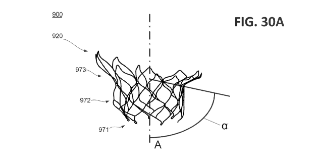
together in
accordance with the attachment location indications 536a, 536b.
[1089] Figure 20 illustrates a design pattern of one leaflet 570 and
associated portion of
outer covering 560 of the inner valve assembly in its initial, pre-assembled
state (i.e., not
attached to inner frame 550), according to an embodiment. As discussed above,
the portion
of leaflet 570 between adjacent commissure posts is referred to as a "belly"
of the leaflet 570.
The belly has a curved edge indicated with reference 'B' in FIG. 20. During
assembly of
inner valve assembly 540, the leaflet 570 is coupled to the inner frame 550 of
the inner valve
assembly 540. Specifically, the belly edge B of the leaflet 570, or a portion
thereof, is
coupled to the inner frame 550 at the arch portion of the inner frame 550. In
addition, outer
covering 560 is folded over a portion of the inner frame 550 (e.g., the arch
portion) along the
axis indicated with 'V, and coupled to a portion of the inner frame 550 (e.g.,
the commissure
post 552) along attachment line A. As shown, a coupling area C (e.g., a
stitching area), is
disposed outside and adjacent to attachment line A. Coupling area C can
facilitate the
assembly process. Subsequently, excess leaflet material and/or excess outer
covering
material can be cut away and disposed of or reused. For example, material
disposed between
the belly edge B and the F-axis, or material in the coupling area C, may, in
some
embodiments, be unnecessary material and thus can be cut away from the leaflet
570 and/or
outer covering 560. The assembly process can be repeated for each leaflet 570,
each outer
covering 560, and each commissure post 552.
17
CA 03005908 2018-05-18
WO 2017/096157
PCT/US2016/064610
[1090] The leaflets 570 and the outer covering 560 can have any suitable
size, shape,
material, and/or configuration. For example, in this embodiment, leaflets 570
and/or outer
covering 560 is formed of fixed porcine pericardium, with a thickness of about
0.01 inches.
[1091] A schematic representation of another embodiment of a prosthetic
heart valve is
shown in FIGS. 21 and 22. Prosthetic heart valve 600 is designed to replace a
damaged or
diseased native heart valve such as a mitral valve. Valve 600 includes an
outer frame
assembly 610 and an inner valve assembly 640 coupled to the outer frame
assembly 610.
[1092] Although not separately shown in the schematic illustration of outer
frame
assembly 610 in FIGS. 21 and 22, outer fame assembly 610 may be formed of an
outer frame
620, covered on all or a portion of its outer face with an outer covering 630,
and covered on
all or a portion of its inner face by an inner covering 632. The materials and
construction of
the components of prosthetic heart valve 600 can be similar to those of the
other
embodiments described above. The following discussion focuses on the aspects
of this
embodiment that differ from the previous embodiments.
[1093] Inner valve assembly 640 includes an inner frame 650 (not shown), an
outer
covering 660 (not shown), leaflets 670 (not shown), and atrial structure 655
(e.g., halo). As
shown, the halo 655 is disposed at the atrium portion 616 of inner valve
assembly 640. In
such a configuration, when valve 600 is implanted into a heart of a patient,
halo 655 will be
disposed above the atrial floor and/or native valve annulus of the patient's
heart. In this
manner, the halo 655 provides extended functionality (e.g., above the native
mitral valve
annulus) of the inner frame 650. In some instances, for example, if prosthetic
leaflets are
seated too low relative to the native valve annulus, the leaflets may
improperly coapt (e.g.,
incomplete coaptation) and/or hemodynamic leakage can occur. Thus, disposing
halo 655
above the native valve annulus can provide for and/or promote complete
coaptation.
[1094] Halo 655 can be formed from any suitable method and material. For
example, in
some embodiments, halo 655 can be formed from a substantially circular piece
of wire. In
such embodiments. halo 655 can be coupled to (e.g., sewn) to inner frame 650.
[1095] Outer covering 630 and inner covering 632 of outer frame 620, outer
covering 660
and leaflets 670 may be formed of any suitable material, or combination of
materials, such as
those discussed above in connection with other embodiments.
[1096] As shown in FIGS. 21 and 22, inner valve assembly 640 may be
substantially
cylindrical, and outer frame assembly 610 may be tapered, extending from a
smaller diameter
18
CA 03005908 2018-05-18
WO 2017/096157
PCT/US2016/064610
(slightly larger than the outer diameter of inner valve assembly 640) at a
lower, ventricle
portion 612 (where it is coupled to inner valve assembly 640) to a larger
diameter, atrium
portion 616, with an intermediate diameter, annulus portion 614 between the
atrium and
ventricle portions.
[1097] In some
embodiments, the outer surface of inner valve assembly 610, and/or the
inner surface of outer frame assembly 640, need not by circular in cross-
section as shown
schematically in FIGS. 21 and 22, but may be of non-constant radius at a given
location along
the central axis of valve 600.
[1098] The
atrial halo 655 functions by extending the inner frame of an inner valve
assembly above the plane of atrial floor in an improved prosthetic heart valve
that includes an
inner frame that holds the leaflets and which is disposed within an outer
frame for reducing or
preventing leaking when the prosthetic heart valve is disposed within a heart
valve (e.g.,
mitral valve, tricuspid valve).
[1099] A benefit
to having leaflets within a raised leaflet silo or cylinder (e.g., halo 650)
is improved blood flow and leaflet closure. It has been observed that where
the leaflet
cylinder is at the atrial floor, leaflet coaptation is incomplete and can
result in hemodynamic
leakage.
[1100]
Accordingly, by providing an atrial halo or ring structure that is raised
above the
plane of the native annulus or atrial floor, complete leaflet coaptation is
encouraged. During
ventricular contraction or systole, the blood is ejected towards aortic valve
to exit the heart
but is also ejected towards the prosthetic mitral valve, which needs to remain
closed during
systole. Retrograde blood hitting the prosthetic valve leaflets cause the
leaflets to close,
preventing regurgitation into the left atrium. During diastole or ventricular
filling, the blood
needs to flow from the atrium into the ventricle without obstruction. However,
when
prosthetic leaflets are not properly placed or properly aligned, the leaflets
can obstruct
efficient filling of the ventricle or cause uneven ventricular output.
[1101] FIG. 23
is a top-view of a prosthetic heart valve 700 according to an embodiment
that is one possible implementation of the prosthetic heart valve shown
schematically in
FIGS. 21 and 22. Prosthetic heart valve 700 includes an outer frame assembly
710, an inner
valve assembly 740, and a tether assembly 790. The inner valve assembly 740
includes an
inner frame 750, and outer covering 760 (not shown), leaflets 770, and atrial
structure 755
(e.g., halo). Halo 755 can be formed from a circular piece of wire that can be
connected to
19
CA 03005908 2018-05-18
WO 2017/096157
PCT/US2016/064610
the inner frame 750 and sewn to the leaflets 770. The inner frame 750 can be
made of
Nitinolt wire that supports leaflets 770 sewn to the inner frame 750 and
functions as a valve.
The inner frame 750 shown in FIG. 23 includes three U-shaped wire components
joined at
their opened ends to form junctions 702. Leaflets 770 are sewn to these
components to form
articulating leaflets, creating and functioning as a prosthetic valve (e.g.,
prosthetic mitral
valve, prosthetic tricuspid valve).
[1102] In some embodiments, the inner frame 750 has tether attachment
apertures 711
(not shown) for attaching tether assembly 790. Tether assembly 790 is
connected to
epicardial securing pad 754 (not shown).
[1103] In operation, the inner frame 750 (with leaflets 770), is disposed
within and
secured within the outer frame 720 of the outer frame assembly 710. Outer
frame 720
includes an outer covering 730 (not shown) (e.g., tissue material) and an
inner covering 732
(e.g., tissue material). Outer frame 720 has an articulating collar 746 which
has a collar cover
748. Articulating collar 746 is configured (e.g., shaped and sized) to solve
leakage issues
arising from native structures. In particular, collar 746 is composed of an A2
segment 747, a
P2 segment 749, and two commissural segments, the A 1-P1 segment 751, and the
A3-P3
segment 753. The collar 746 may also have, in some embodiments a shortened or
flattened
or D-shaped section 762 of the A2 segment in order to accommodate and solve
left
ventricular outflow tract (LVOT) obstruction issues.
[1104] In operation, the valve 700 may be deployed as a prosthetic mitral
valve using
catheter delivery techniques. The entire valve 700 is compressed within a
narrow catheter
and delivered to the annular region of the native valve, preferably the left
atrium, with a pre-
attached tether apparatus. Upon delivery, the valve 700 is pushed out of the
catheter where it
springs open into its pre-formed functional shape without the need for manual
expansion
(e.g., manual expansion using an inner balloon catheter). When the valve 700
is pushed
and/or pulled into place, the outer frame assembly 710 is seated in the native
valve annulus
(e.g., native mitral annulus), leaving the articulating collar 746 to engage
the atrial floor and
prevent pull-through (where the valve is pulled into the ventricle). In such
embodiments, it is
not necessary to cut-away the native leaflets, as has been taught in prior
prosthetic efforts.
Instead, the native leaflets can be used to provide a tensioning and/or
sealing function around
the outer frame assembly 710. It is advantageous for the valve 700 to be
asymmetrically
deployed in order to address LVOT problems where non-accommodating prosthetic
valves
push against the A2 anterior segment of the valve (e.g., mitral valve) and
close blood flow
through the aorta, which anatomically sits immediately behind the A2 segment
of the mitral
CA 03005908 2018-05-18
WO 2017/096157
PCT/US2016/064610
annulus. Thus, D-shaped section 762 is deployed substantially immediately
adjacent/contacting the A2 segment since the flattened D-shaped section 762 is
structurally
smaller and has a more vertical profile (closer to paralleling the
longitudinal axis of the outer
stent) and thereby provides less pressure on the A2 segment. Once the valve
700 is properly
seated, tether assembly 790 may be extended out through the apical region of
the left
ventricle and secured using an epicardial pad 754 or similar suture-locking
attachment
mechanism (not shown).
[1105] In an alternate embodiment, the tether assembly 790 is on the outer
frame 720,
which would then have tether attachment apertures 713 for attaching tether
assembly 790 to
epicardial securing pad 754.
[1106] FIG. 24 is a perspective view of the Al-P1 side of the prosthetic
heart valve 700
according to an embodiment. FIG. 24 shows one of the three U-shaped wire
components of
inner frame 750 joined at their opened ends to form junctions 702. Although
three U-shaped
wire components are shown, in other embodiments, any suitable number of U-
shaped wire
components can be joined at their opened ends to form junctions. Similarly, in
some
embodiments, the wire components of inner frame 750 can by any suitable shape
or size.
Leaflets 770 are sewn to these components to form articulating leaflets 770
creating and
functioning as a prosthetic heart valve (e.g., mitral valve, tricuspid valve).
Atrial halo 755 is
shown with the plane of the circular wire above the plane of the majority of
collar except for
the vertical A2 segment 747, the P2 segment 749, and the conunissural Al-PI
segment 751
an A3-P3 segment 753. FIG. 26 shows how upon deployment blood would fill the
void or
gap 707 between the inner frame 750 and the outer frame 720 at the Al-P1
segment 751 of
the valve 700. This blood creates a temporary fluid seal that would pool in
that space and
provide a pressure buffer against the leakage inducing forces that accompany
systolic and
diastolic related intra-atrial and intra-ventricular pressure.
[1107] FIG. 25 is a perspective view of the A3-P3 side 753 of prosthetic
heart valve 700
according to an embodiment. FIG. 25 shows one of the three U-shaped wire
components of
inner frame 750 joined at their opened ends to form junctions 702. Leaflets
770 are sewn to
these components to form articulating leaflets 770 creating and functioning as
a prosthetic
tricuspid valve. Atrial halo 755 is shown with the plane of the circular wire
above the plane
of the majority of collar except for the vertical A2 segment 747, the P2
segment 749, and the
commissural A 1-P1 segment 751 and A3-P3 segment 753. FIG. 25 shows how upon
deployment blood would fill the void or gap 708 between the inner frame 750
and outer
frame 720 at the A3-P3 segment 753 area of the valve 700. This blood creates a
temporary
21
CA 03005908 2018-05-18
WO 2017/096157
PCT/US2016/064610
fluid seal that would pool in that space and provide a pressure buffer against
the leakage
inducing forces that accompany systolic and diastolic related intra-atrial and
intra-ventricular
pressure.
111081 FIG. 26 is an exploded view of prosthetic heart valve 700 according
to an
embodiment. In this valve 700, the inner frame 750 is sewn with tissue 706 and
acts a cover
to prevent valvular leakage. The inner frame 750 contains the leaflets 770
comprised of
articulating leaflets that define a valve function. The leaflets 770 are sewn
to the inner frame
750. The inner frame 750 also has tether attachment apertures 711 for
attaching tether
assembly 790. Tether assembly 790 is shown in this example as connected to
epicardial
securing pad 754. In operation, the covered inner frame 750 (e.g., covered
with outer
covering 760) (with leaflets 770), is disposed within and secured within the
outer frame 720
of the outer frame assembly 710. Outer frame 720 may also have in various
embodiments a
covering (e.g., outer covering 730). Outer frame 720 has an articulating
collar 746 which has
a collar cover 748. Articulating collar 746 may also have in some embodiments
a D-shaped
section 762 to accommodate and solve left ventricular outflow tract (LVOT)
obstruction
issues.
[1109] In operation, the valve 700 may be deployed as a prosthetic valve
(e.g., mitral
valve) using catheter delivery techniques. The entire valve 700 is compressed
within a
narrow catheter and delivered to the annular region of the native valve, such
as, for example,
with a pre-attached tether assembly 790. There, the valve 700 is pushed out of
the catheter
where it springs open into its pre-formed functional shape without the need
for manual
expansion (e.g., manual expansion using an inner balloon catheter). When the
valve 700 is
pushed and/or pulled into place, the outer frame assembly 710 is seated in the
native mitral
annulus, leaving the articulating collar 746 to engage the atrial floor and
prevent pull-through
(where the valve is pulled into the ventricle). In such embodiments, it is not
necessary to cut-
away the native leaflets, as has been taught in prior prosthetic efforts.
Instead, the native
leaflets can be used to provide a tensioning and/or sealing function around
the outer frame
assembly 710. It is advantageous for the valve 700 to be asymmetrically
deployed in order to
address LVOT problems where non-accommodating prosthetic valves push against
the A2
anterior segment of the valve (e.g., the mitral valve) and close blood flow
through the aorta,
which anatomically sits immediately behind the A2 segment of the mitral
annulus. Thus, D-
shaped section 762 is deployed immediately adjacent/contacting the A2 segment
since the
flattened D-shaped section 762 is structurally smaller and has a more vertical
profile (closer
to paralleling the longitudinal axis of the outer stent) and thereby provides
less pressure on
22
CA 03005908 2018-05-18
WO 2017/096157
PCT/US2016/064610
the A2 segment. Once the valve 700 is properly seated, tether assembly 790 may
be extended
out through the apical region of the left ventricle and secured using an
epicardial pad 754 or
similar suture-locking attachment mechanism.
[1110] Any of the prosthetic heart valve embodiments described above can
incorporate
additional structural features to enhance their performance. The structural
features are
discussed below with reference to prosthetic heart valve 800, illustrated
schematically in
perspective and side views in FIGS. 27 and 28, respectively.
[1111] As shown, the outer frame 820 has an atrium portion 826, a ventricle
portion 822,
and an annulus portion 824 disposed between the atrium portion 826 and the
ventricle portion
822. The inner frame 850 of the inner valve assembly 840 has a first end and a
second end.
The inner valve assembly 840 can be coupled to the outer frame 820 by a
connection between
the first end of the inner frame 850 and the ventricle portion 812 of the
outer frame assembly
810. The inner frame assembly 840 can extend from the connection towards the
atrium
portion 816 of the outer frame assembly 810. The inner frame assembly 840 and
the outer
frame assembly 810 can diverge from the connection towards the atrium portion
816 of the
outer frame assembly 810. The annulus portion 814 of the outer frame assembly
810 can be
spaced radially from the inner valve assembly 840 and radially inwardly
deflectable towards
the inner valve assembly 840 to accommodate a natural heart valve annulus in
the annulus
portion 814.
[1112] The outer frame assembly 810 can be shaped and sized in any suitable
manner to
facilitate a proper fit into a native heart valve. For example, as shown, the
outer frame 820
can be shaped and sized to resemble, at least in part, an hourglass shape.
Specifically. the
annulus portion 814 of outer frame assembly 810 varies from an intermediate
diameter (or
perimeter) near ventricle portion 812 to a smaller diameter (or perimeter)
near the middle of
annulus portion 814, to a larger diameter (or perimeter) near atrium portion
816. Thus,
annulus portion 814 has an hourglass shape. Ventricle portion 812 has a
maximum diameter
larger than a maximum diameter of annulus portion 816. The ventricle portion
has a
minimum diameter smaller than a minimum diameter of the annulus portion 814.
[1113] The diameters and/or perimeters for each portion of the outer frame
820 can be
selected based on the size and/or shape of a native heart valve into which
prosthetic heart
valve 800 is to be implanted. For example, the minimum diameter of the annulus
portion 824
of the outer frame 820 can be smaller than that of the native valve annulus.
Thus, in such a
configuration, the diameters of the ventricle portion 822, annulus portion
824, and atrium
23
CA 03005908 2018-05-18
WO 2017/096157
PCT/US2016/064610
portion 826 can collectively promote a suitable fit (e.g., a snug, secure fit)
of the prosthetic
heart valve 800 in a native heart valve. In this manner, the outer frame 820
can be configured
to optimize securement and sealing between the prosthetic heart valve 800
(particularly outer
frame assembly 810) and a native valve annulus of a native heart valve. Thus,
such a
configuration minimizes the likelihood of paravalvular leaks.
[1114] Although the outer frame 820 is shown to have a circular cross-
section, in some
embodiments, the outer frame 820 can by any suitable shape or size. For
example, in some
embodiments, the outer frame 820 can have a D-shape cross-section. In this
manner, the
outer frame 820 can have a shape configured to correspond to (e.g., mate with)
a native heart
valve annulus.
[1115] In addition to, or instead of, outer frame 820 and/or outer frame
assembly 810
with the hourglass shape described above, valve 800, or in some instances,
outer frame 820
and/or outer frame assembly 810, in particular, can be formed to provide
stiffness, such as
resistance to hoop compression, that is varied spatially, i.e., axially and/or
circumferentially.
[1116] In this manner, a suitable stiffness profile can be arranged such
that the valve 800
promotes a desirable shape and sealing region when disposed in a native heart
valve, thus
minimizing the likelihood of paravalvular leaks and undesired movement of the
valve.
Similarly stated, valve 800 can be configured to have a stiffness profile
suitable to cause
desirable deformation of the native heart valve annulus (i.e., the sealing
region), and thus,
proper implantation of valve 800.
[1117] A desired stiffness profile of prosthetic valve 800 can be achieved
by varying
properties, characteristics, and/or the arrangement of the outer frame
assembly 810 and the
inner valve assembly 840. For example, the outer frame 820 and/or the inner
frame 850 can
contain portions of varying material states. For example, a first portion of
outer frame 820
can be in an elastic state, while a second portion of outer frame 820 is in a
super-elastic state.
Similarly, for example, portions of the outer frame 820 and/or the inner frame
850 can be in
an austenitic state and/or a martensitic state (e.g., a stress induced
martensitic state). In this
manner, portions of valve 800 can be configured to suitably mate with a native
valve annulus,
thus improving sealing and limiting paravalvular leaks.
[1118] In addition, the outer frame assembly 810 and/or inner valve
assembly 840 can
have varying widths, thicknesses, shapes (e.g., longitudinal shape), angles
(e.g., angle of
attachment between inner valve assembly 840 and outer frame assembly 810), and
the like.
24
CA 03005908 2018-05-18
WO 2017/096157
PCT/US2016/064610
In some embodiments, the outer covering 830, inner covering 832, outer
covering 860, and/or
pocket closure 880 can be configured to determine, at least in part, the
stiffness profile and/or
shape of valve 800 (e.g., based on sewing pattern).
[1119] FIGS. 29B, and 29C and 29D illustrate axial and circumferential
stiffness profiles,
respectively, of prosthetic heart valve 800 (shown in FIG. 29A) according to
an embodiment.
The stiffness of heart valve 800 can vary axially and/or circumferentially in
any suitable
manner. For example, FIG. 29B represents an axial stiffness profile of valve
800.
Specifically, as shown, the Z-axis represents an axial location on valve 800
(e.g., a location of
the stifftiess value). The S-axis represents a range of stiffness (or range of
stiffness values),
increasing from left (starting at origin 0) to right.
111201 Further to this example, as illustrated in FIG. 29B, in some
embodiments,
locations near the ventricle portion 822 (e.g., indicated as B in FIG. 29A) of
the outer frame
822 can have a larger stiffness value, locations near the annulus portion 824
of the outer
frame 820 can have a smaller stiffiless value relative to the ventricle
portion 822 (e.g., to
facilitate cooperation with the native valve annulus), and locations near the
atrium portion
826 (e.g., indicated as A in FIG. 29A) of the outer frame 820 can have a
smaller, the same, or
larger stiffiless value (illustrated by the dotted line) than the stiffness
value near the annulus
portion 824. In this manner, the outer frame assembly 810 can be relatively
more compliant
in hoop compression in a central, annulus portion 814, than at the ventricle
portion 812.
Thus, in use, the prosthetic valve 800 can seat securely in the annulus of the
native heart
valve while imposing minimal loads on the inner valve assembly 840 that could
degrade the
performance of the valve leaflets 870. Although, for ease of illustration, the
stiffness profile
shown in FIG. 29B includes linear portions, in some embodiments, the stiffness
profile can
include non-linear portions instead of or in addition to the linear portions
as shown.
[1121] Similarly, the stiffness of heart valve 800, or portions of heart
valve 800, can have
varying degrees of stiffiless circumferentially, as illustrated by the
stiffness profiles shown in
FIGS. 29C and 29 D. By way of example, FIG. 29C illustrates a circumferential
stiffness
profile at axial location A (as shown by reference 'A' in FIG. 29A).
Similarly, FIG. 29D
illustrates a circumferential stiffness profile at axial location B (as shown
by reference '13' in
FIG. 29A). As the profile extends radially from the origin (indicated as '0'),
the stiffness
value increases.
[1122] Thus, as shown in FIG. 29C, the stiffness at S 1(90 degrees) is
greater than the
stiffness at S2 (270 degrees). Further to this example, in some embodiments,
the
CA 03005908 2018-05-18
WO 2017/096157
PCT/US2016/064610
circumferential portion from zero to 180 degrees can represent a relatively
flat portion of an
outer frame 820 of the outer frame assembly 810 having a D-shape
configuration, and 180 to
360 degrees can represent a relatively curved portion of the outer frame 820
having the D-
shape configuration.
[1123] In a similar fashion, FIG. 29D illustrates a circumferential
stiffiiess profile at axial
location B (as shown by reference '13' in FIG. 29A). As shown, axial location
B has a
different stiffness profile than axial location A. Such variability in design,
as discussed
above, can provide for advantageous customization of heart valve 800, and
cooperation of
heart valve 800 with a native heart valve. Similar to FIG. 29C, FIG. 29D
illustrates the
stiffness at one side of valve 800 being be greater than a stiffness at
another side of the valve
800. In this manner, in some instances, a portion of valve 800 that will
experience greater
forces from the native heart valve annulus can have a smaller stiffness value
(e.g., more
compliant) than a portion of the valve 800 that will experience smaller or
fewer forces, thus
optimizing the cooperation of the prosthetic heart valve 800 with the native
heart (particularly
the native heart valve annular region).
[1124] As discussed above, in some instances, a patient having an implanted
prosthetic
heart valve may experience postoperative LVOT obstructions resulting from, for
example,
subvalvular positioning or configuration of the prosthetic heart valve.
Described below and
illustrated in FIGS. 30-35B are various embodiments of prosthetic heart
valves, in expanded
or deployed configurations, that are configured to avoid, reduce or otherwise
limit
undesirable LVOT obstruction. To assist in the understanding of the
relationship between the
various prosthetic valve embodiments and the anatomy of a heart, FIGS. 35A and
35B
illustrate in a partial cross-sectional side view and top view, respectfully,
an exemplary
prosthetic heart mitral valve 1400 (also referred to herein as "valve")
implanted in a native
mitral annulus of a heart. The prosthetic heart mitral valve 1400 can be
constructed and
function similar to any of the prosthetic heart valves described herein. For
example the valve
1400 can include an outer frame 1420 and an inner frame 1450. Thus, some
details regarding
the valve 1400 are not described below. It should be understood that for
features and
functions not specifically discussed, those features and functions can be the
same as or
similar to any of the valves described herein.
[1125] As shown in FIGS. 35A and 35B, the prosthetic heart mitral valve
1400 (also
referred to herein as "valve") in its expanded or deployed configuration is
seated in the native
mitral valve annulus NA between the left ventricle LV and the left atrium LA
of the heart H.
In operation, the left ventricle LV contracts and blood flows outwardly from
the left ventricle
26
CA 03005908 2018-05-18
WO 2017/096157
PCT/US2016/064610
LV through the aortic valve AV via the left ventricular outflow tract (LVOT).
The path of
such blood flow is shown in FIG. 35A by arrow LVOT. When the prosthetic heart
valve
1400 is deployed and seated in the native mitral valve annulus NA, as shown,
the A2 segment
1447 of the outer frame 1420 of the valve 1400 (i.e., the anterior side of the
valve 1400) is
aligned with or seated at the A2 anterior segment (labeled A2 in FIGS. 35A and
35B) of the
native mitral valve annulus. Further, as shown, when the valve 1400 is
deployed and seated
in the native mitral valve annulus NA, the P2 segment 1449 of the outer frame
1420 of the
valve 1400 (i.e., the posterior side of the valve 1400) is aligned with or
seated at the P2
posterior segment (labeled P2 in FIGS. 35A and 35B) of the native mitral valve
annulus.
[1126] As shown in FIG. 35A, during operation of the heart, the native
leaflet NL1 on the
anterior side (native leaflet NL2 is on the posterior side) can intrude into
the LVOT,
identified by the change in position of the native anterior leaflet NL1 from a
first position
shown in solid-line to a second position shown in dashed-line in which the
leaflet NL1
intrudes into or obstructs the LVOT.
[1127] In one embodiment, an outer frame of a prosthetic heart valve can be
configured
similar to the outer frames described above (e.g., the outer frame 520) except
that the cuff
portion is disposed at an angle (e.g., less than 90 degrees) relative to the
vertical axis of a
body portion of the outer frame (also referred to herein as the "angled cuff
arrangement"
discussed in more detail below with reference to FIGS. 30A-30C). In this
manner, in use, a
prosthetic heart valve can be delivered and deployed to a native heart (i.e.,
seated in the
native annulus of the heart) such that at least a portion of the prosthetic
heart valve disposed
in the ventricle of the heart is oriented or positioned away from the LVOT of
the heart. As
such, the angled cuff arrangement can prevent, reduce or otherwise limit LVOT
obstruction
(or intrusion by the native leaflet). Similarly stated, the angled cuff
arrangement can provide
additional LVOT clearance. As discussed above, an implanted prosthetic valve,
or a portion
thereof, can contribute to the postoperative complication of LVOT obstruction.
An angled
cuff arrangement, however, can limit or prevent LVOT obstruction by limiting
the placement
of the prosthetic valve in the LVOT of the heart.
111281 FIG. 30A illustrates an outer frame 920 of a prosthetic heart valve
900 having an
angled cuff arrangement, and FIG. 30B illustrates a schematic side cross-
sectional view of
the prosthetic heart valve 900 shown in FIG. 30A, including an inner valve
assembly 940.
The prosthetic heart valve 900 (also referred to herein as "valve") can be
constructed and
function similar to any of the prosthetic heart valves described herein, e.g.,
the prosthetic
heart valve 500. Thus, some details regarding the valve 900 are not described
below. It
CA 03005908 2018-05-18
WO 2017/096157
PCT/US2016/064610
should be understood that for features and functions not specifically
discussed, those features
and functions can be the same as or similar to any of the valves described
herein.
11129j As shown in FIG. 30A, the outer frame 920 includes a coupling
portion 971, a
body portion 972, and a cuff portion 973. When the valve 900 is disposed
within a native
mitral annulus of a heart, the cuff portion 973 is configured to be seated
within the native
mitral annulus and extend within the atrium of the heart and the body portion
972 is
configured to be disposed within a ventricle of the heart. The coupling
portion 971 is
configured to be coupled to the inner valve assembly. In this embodiment, the
cuff portion
973 is disposed at an angle a relative to the vertical axis A. Said another
way, as shown in
FIG. 30A, the cuff portion 973 slopes downward from the anterior side of the
valve 900 to
the posterior side of the valve 900. In this manner, when the prosthetic heart
valve 900 is
implanted into a heart (e.g., when the cuff portion 973 is seated in the
native annulus of the
heart), the angle of the cuff portion 973 will cause the body portion 972 of
the valve 900 to be
oriented or positioned away from the LVOT of the heart, thereby preventing,
reducing or
otherwise limiting undesirable LVOT obstruction.
WM An angle a defmed by the cuff portion 973 and the vertical axis A of
the
prosthetic valve 900 can be any suitable value configured to create, increase
or otherwise
promote LVOT clearance and limit or prevent LVOT obstruction (or undesirable
outflow
gradients). The angle a can be, for example, an acute angle. As shown in FIG.
30A, the
angle a is about 80 degrees. In other embodiments, however, the angle a can
be, for
example, from about 70 degrees to about less than 90 degrees. As the LVOT
varies across
patients, e.g., depending on a patient's particular anatomy, the angle can be
adjusted
accordingly.
111311 As shown in FIG. 30B, the body portion 972 of the outer frame 920
has varying
lengths due to the angled cuff arrangement. More particularly, an anterior
portion of the
body portion 972 has an anterior length La!, and a posterior portion has a
relatively smaller
posterior length Lp 1 . The anterior length Lal and the posterior length Lp I
can be any
suitable value configured to create, increase or otherwise promote LVOT
clearance and limit
LVOT obstruction or undesirable outflow gradients. In some instances, for
example, the
anterior length Lal can be about 18 mm and the posterior length Lp I can be
about 14mm. As
the LVOT varies across patients, e.g., depending on a patient's particular
anatomy, the
anterior length Lal and the posterior Lpl can be adjusted accordingly.
28
CA 03005908 2018-05-18
WO 2017/096157
PCT/US2016/064610
[1132] Further, as shown in FIG. 30B, an atrial end portion 955 of the
inner valve
assembly 940 extends a length of Li 1 from the location at which the inner
valve assembly
940 is coupled to the outer frame 920 when the inner valve assembly 940 is
seated in the
outer frame 920. Also shown in FIG. 30B, in some embodiments, the inner valve
assembly
940 (e.g., the inner frame 950 shown in FIG. 30C) can have a centerline
substantially parallel
to the centerline of the outer frame 920.
111331 FIG. 30C shows a side view of the prosthetic heart valve 900 (with
the angled cuff
arrangement), in a deployed or biased configuration, and for comparison, a
side view of a
prosthetic heart valve 900' without the angled cuff arrangement. Both the
valve 900 and the
valve 900' are shown similar to how each valve would be arranged when seated
within a
native annulus of a heart (not shown). The valve 900', for example, can be
constructed and
function similar to or the same as the prosthetic heart valve 500.
111341 As discussed with respect to FIGS. 35A and 35B, in operation, the
left ventricle of
the heart (not shown in FIG. 30C) contracts and blood flows outwardly from the
left ventricle
through the aortic valve via the LVOT. The path of such blood flow is shown in
FIG. 30C by
arrow LVOT. As shown, the outer frame 920 of the valve 900 provides additional
LVOT
clearance when compared to the outer frame 920' of the valve 900'. For
example, as shown
in FIG. 30C, the strut 954 of the inner frame 950 of the valve 900 is disposed
a greater
distance from and provides greater clearance to the LVOT. As shown by
comparison to the
valve 900-, the strut 954 of the inner frame 950 of valve 900 is displaced
from the strut 954'
of the inner frame 950' of valve 900% and a tether clamp or coupling portion
944 of the inner
frame 950 of valve 900 is displaced from a tether clamp or coupling portion
944' of the inner
frame 950' of the valve 900'. In this manner, when the valve 900 is implanted
in a native
annulus of a heart, the valve 900 can provide clearance to the A2 anterior
segment of the
native valve (e.g., the mitral valve) such that interruption of blood flow
through the aorta,
which anatomically sits immediately behind the A2 segment of the mitral
annulus, can be
prevented or limited.
[1135] In an alternative embodiment, an outer frame of a prosthetic heart
valve can be
configured similar to the outer frame 920 of valve 900 having the angled cuff
arrangement,
except that a body portion and coupling portion of the outer frame has a
shorter profile (also
referred to herein as the "short profile arrangement"). In this manner, when
the prosthetic
heart valve having a shorter profile is implanted into a heart (e.g., when the
cuff portion is
seated in the native annulus of the heart), at least a portion of the outer
frame disposed in the
ventricle of the heart (e.g., the body portion and the coupling portion of the
outer frame) is
29
CA 03005908 2018-05-18
WO 2017/096157
PCT/US2016/064610
positioned outside of or substantially away from the LVOT of the heart,
thereby reducing the
likelihood of undesirable postoperative LVOT obstruction.
111361 FIG. 31A illustrates an outer frame 1020 of a prosthetic heart valve
1000 having
an angled cuff arrangement and a short profile arrangement. The prosthetic
heart valve 1000
(also referred to herein as "valve") can be constructed and function similar
to any of the
prosthetic heart valves described herein, e.g. the valve 900. Thus, some
details regarding the
valve 1000 are not described below. It should be understood that for features
and functions
not specifically discussed, those features and functions can be the same as or
similar to any of
the valves described herein.
111371 As shown in FIG. 31A, the outer frame 1000 includes a coupling
portion 1071, a
body portion 1072, and a cuff portion 1073. The cuff portion 1073 is disposed
at an angle a
relative to the vertical axis A. In this manner, when the prosthetic heart
valve 1000 is
implanted into a heart (e.g., when the cuff portion 1073 is seated in the
native annulus of the
heart), the short profile of the body portion 1072 of the valve 1000 can be
oriented or
positioned away from the LVOT of the heart, thereby providing additional
clearance to the
LVOT preventing, reducing or otherwise limiting undesirable LVOT obstruction.
111381 As shown in FIG. 31A, the body portion 1072 of the outer frame 1020
has varying
lengths due to the angled cuff arrangement. More particularly, an anterior
portion of the
body portion 1072 has an anterior length La2, and a posterior portion of the
body portion
1072 has a relatively smaller posterior length Lp2. The anterior length La2
and the posterior
length Lp2 can be any suitable value configured to create, increase or
otherwise promote
LVOT clearance and limit LVOT obstruction or undesirable outflow gradients. In
some
instances, for example, the anterior length La2 can be about lOmm and the
posterior length
Lp2 can be about 4mm. As the LVOT varies across patients, e.g., depending on a
patient's
particular anatomy, the anterior length La2 and the posterior Lp2 can be
adjusted
accordingly.
111391 FIGS. 31B and 31C illustrate schematic perspective and side cross-
sectional views
of the prosthetic heart valve 1000 shown in FIG. 31A, including an inner valve
assembly
1040. The inner valve assembly 1040 extends a length Li2 from the location at
which the
inner valve assembly 1040 is coupled to the outer frame 1020 when the inner
valve assembly
1040 is seated in the outer frame 1020. In comparison to the embodiment
described with
respect to the valve 900 and FIGS. 30A and 30B, the anterior length La2 of the
valve 1000
(see e.g., FIGS. 31A-C) is less than the anterior length Lal of the valve 900
(see e.g., FIG.
CA 03005908 2018-05-18
WO 2017/096157
PCT/US2016/064610
30B) and the posterior length Lp2 of the valve 1000 (see e.g., FIGS. 31A-C) is
less than the
posterior length Lpl of the valve 900 (see e.g., FIGS. 30B). The length Li2 of
the inner valve
assembly 1040 (see e.g., FIGS. 30B and 30C), however, is equal to the length
Lil of the inner
valve assembly 940 (see e.g., FIG. 30B). For example, as shown in FIG. 30B,
the atrial end
portion 955 of the inner valve assembly 940 is disposed below an atrial end
956 of the outer
frame 920 when the inner valve assembly 940 is seated in and coupled to the
outer frame 920,
and as shown in FIGS. 31B and 31C, the atrial end portion 1055 of the inner
valve assembly
1040 extends above at least a portion of an atrial end 1056 of the outer frame
1020 when the
inner valve assembly 1040 is seated in and coupled to the outer fame 1020.
Thus, when the
cuff portion 1073 is seated in a native annulus, the inner valve assembly 1040
of valve 1000
sits higher into the atrium of the heart than would the inner valve assembly
940 of the valve
900.
[1140] In an alternative embodiment, a prosthetic heart valve can be
configured similar to
any of the prosthetic heart valves described herein, except that the inner
valve assembly of
the prosthetic heart valve can be displaced radially (e.g., off-center)
relative to the outer
frame assembly of the prosthetic heart valve. In this manner, when the
prosthetic heart valve
is implanted into a heart (e.g., when the cuff portion is seated in the native
annulus of the
heart), the inner valve assembly, and thus the fluid flow path therethrough,
are positioned
further away from the LVOT of the heart, thereby reducing the likelihood of
undesirable
postoperative LVOT obstruction or undesirable outflow gradients.
[1141] FIGS. 32A-32C illustrate a prosthetic heart valve 1100 having an
outer frame
1120 (of an outer frame assembly, not shown) and an inner valve assembly 1140
coupled to
and displaced radially (off-center) from the outer frame 1120. The outer frame
1120 includes
a cuff portion 1173, a body portion 1150 and a coupling portion 1171.
[1142] The prosthetic heart valve 1100 (also referred to herein as "valve")
can be
constructed and function similar to any of the prosthetic heart valves
described herein, e.g.,
the valve 500. Thus, some details regarding the valve 1100 are not described
below. It
should be understood that for features and functions not specifically
discussed, those features
and functions can be the same as or similar to any of the valves described
herein.
[1143] For ease of explanation, the inner frame assembly 1140 is
represented by a solid-
lined circle. Further, as shown, for example, in FIG. 32A, the lower end of
the coupling
portion 1171 of the outer frame 1120 forms a roughly circular opening (shown
as a dashed-
line circle and identified by "0" in FIG. 32A). The radial displacement is
represented by
31
CA 03005908 2018-05-18
WO 2017/096157
PCT/US2016/064610
radial off-set R.0 and is illustrated by the gap defined between the inner
frame assembly 1140
and the circular opening 0 of the outer frame 1120 on an A2 segment or side
1147 of the
outer frame 1120. Said another way, a centerline of the inner frame 1140 is
radially offset
from a centerline of the outer frame 1120. In previous embodiments of a
prosthetic valve,
such as, for example, valve 500, the outer frame is configured to hold an
inner valve
assembly centered within circular opening 0. In some embodiments, to
accommodate the
offset of the inner valve assembly as shown in FIG. 32A, the outer frame can
be modified so
that the circular opening 0 is aligned with the inner frame assembly (e.g.,
the centerline of
the inner frame and the center line of the outer frame are aligned or
coaxial). In other
embodiments, the inner frame can be coupled to the outer frame in a manner to
accommodate
the offset.
[1144] The radial displacement RO can be any suitable value (i.e.,
distance) configured to
create, increase or otherwise promote LVOT clearance and limit LVOT
obstruction or
undesirable outflow gradients. As shown in FIG. 32A, the inner valve assembly
1140 is
radially off-set towards a P2 segment or side 1149 (i.e., posterior side) of
the outer frame
1120 such that when the outer frame 1120 is seated in a native mitral annulus,
the inner valve
assembly 1140 is positioned further away from the LVOT. The inner valve
assembly 1140
can be displaced, for example, as far as the posterior sealing surface Ps of
outer frame 1120
(e.g., as shown in FIG. 32C). For example, as shown by comparing FIGS. 32B and
32C, the
inner frame 1140 can be shifted from a substantially centered position
relative to the outer
frame 1120 (as shown in FIG. 32B (i.e., substantially no radial offset)) to
the posterior
sealing surface Ps of the outer frame 1120 (as shown in FIG. 32C (i.e.,
radially offset relative
to the outer frame 1120)) such that a gap G defined between the posterior side
Pi of the inner
valve 1140 and the posterior sealing surface Ps of the outer frame 1120
decreases (e.g., g = 0
when the inner valve assembly 1140 is displaced as far as the posterior
sealing surface Ps of
the outer frame 1120 such that a portion of the inner valve assembly 1140 is
in contact with
the posterior side of the outer frame 1120, as shown in FIG. 32C).
[1145] The displacement away from the LVOT can be further increased by
reducing the
diameter of the inner valve assembly. This is illustrated in FIG. 32A, in
which the inner
frame assembly is slightly smaller in diameter than the opening 0 of the outer
frame. The
effect on the displacement away from the LVOT could be increased by further
reducing the
diameter of the inner valve assembly.
111461 In use, when the prosthetic heart valve 1100 is implanted into a
heart (e.g., when
the cuff portion 1173 is seated in the native mitral annulus of the heart),
for example, the
32
CA 03005908 2018-05-18
WO 2017/096157
PCT/US2016/064610
blood flow from the left atrium to the left ventricle through the leaflet
assembly (not shown)
of the inner valve assembly 1140 is directed to avoid LVOT obstruction or
undesirable
outflow gradients. Similarly stated, the inner valve assembly 1140 and the
outer frame 1120
can be collectively configured to manage desirable blood flow from the atrium
to the
ventricle of the heart without interrupting or otherwise promoting undesirable
affects to the
LVOT.
[1147] In another alternative embodiment, a prosthetic heart valve can be
configured
similar to any of the prosthetic heart valves described herein except that the
inner valve
assembly of the prosthetic heart valve can be rotated about its vertical axis
and relative to the
valve's outer frame. More specifically, the inner frame can be rotated such
that the A2
segment of the outer frame is aligned with a commissure post of the inner
frame of the inner
valve assembly rather than a belly of a leaflet (each portion of the leaflets
between adjacent
commissure posts being referred to as a "belly" of a leaflet) and
correspondingly with a belly
post of the inner frame of the inner valve assembly. As discussed above in
connection with
the embodiment of FIGS. 2A-2C, and apparent from FIG. 12 in connection with
the
embodiment of FIGS. 7-18, the commissure posts 152 lie on a slightly smaller
diameter circle
than the belly posts 154. Thus, orienting the inner valve so that a belly post
(and leaflet
belly) is closest to the LVOT, positions the portion of the inner valve body
closest to the
LVOT slightly further away from the LVOT.
[1148] FIG. 33A is a top view of a prosthetic heart valve 1200 having an
outer frame
1220 (of an outer frame assembly, not shown) and an inner frame 1250 (of an
inner valve
assembly, not shown). As shown, the inner frame 1250 is coupled to the outer
frame 1220
and rotated about its vertical axis (i.e., the axis extending through the
centerline of the inner
frame 1250 (see e.g., a top view of the vertical axis identified as "VA" in
FIG. 33A) relative
to the outer frame 1220. As such, an A2 segment 1247 of the outer frame 1220
is aligned
with a commissure post 1252 of the inner frame 1250 and not aligned with a
leaflet belly (the
location of which is identified by "LB" in FIG. 33A) or a belly post 1254 of
the inner frame
1250 as described in more detail below.
[1149] The prosthetic heart valve 1200 (also referred to herein as "valve")
can be
constructed and function similar to any of the prosthetic heart valves
described herein, e.g.,
the valve 500. Thus, some details regarding the valve 1200 are not described
below. It
should be understood that for features and functions not specifically
discussed, those features
and functions can be the same as or similar to any of the valves described
herein.
33
CA 03005908 2018-05-18
WO 2017/096157
PCT/US2016/064610
[1150] As shown in FIG. 33A, the outer frame 1220 of the valve 1200
includes the A2
segment 1247 and a P2 segment 1249. The inner frame 1250 includes an inner
wireframe
structure made of Nitinol wire that supports leaflets (not shown) sewn to the
inner frame
1250 and functions as a valve. The inner frame 1250 includes three main U-
shaped wire
components 1207 joined at their opened ends to form junctions 1202 (see FIG.
33A).
Prosthetic leaflets (not shown) are sewn to these components to form
articulating leaflets,
creating and functioning as a prosthetic valve (e.g., a prosthetic mitral
valve). The inner
frame 1250 also includes three commissure posts 1252-1, 1252-2, 1252-3 and
three belly
posts 1254-1, 1254-2, 1254-3 (as shown in FIGS. 33A and 33B).
[1151] As shown, for example, in FIG. 33A, the inner frame 1250 and the
outer frame
1220 are positioned/oriented relative to each other such that a center portion
CP of the A2
segment 1247 of the outer frame 1220 is aligned with the commissure post 1252-
1 of the
inner frame 1250. Further, the belly posts 1254-1 and 1254-2 on each side of
the commissure
post 1252-1 are misaligned with the center portion CP of the A2 segment 1247
of the outer
frame 1220. Said another way, the belly posts 1254-1 and 1254-2 on each side
of the
commissure post 1252-1 of the outer frame 1220 are disposed relative to a
horizontal axis B
(which passes through the center portion CP of the A2 segment 1247) by an
angle 13, as
shown in FIG. 33A. In this embodiment, the angle 13 is about 60 degrees. In
alternative
embodiments, an angle 13 can be any suitable value configured to position the
inner frame to
the outer frame such that the belly posts 1254-1 and 1254-2 are misaligned
with the center
portion CP of the A2 segment of the outer frame.
[1152] FIG. 33B illustrates a bottom view of the inner frame 1250 to
further illustrate the
position of the inner frame 1250 relative to the A2 segment 1247 of the outer
frame 1220. As
shown in FIG. 33B, the belly posts 1254-1, 1254-2, 1254-3 define an outer
perimeter or circle
CPC and the commissure posts 1252-1, 1252-2, 1252-3 are each disposed within
the circle
CPC such that a space SP is defined between the circle CPC and each of the
commissure
posts 1252-1, 1252-2, 1252-3. The space SP between the commissure post 1252-1
and the
center portion CP of the A2 segment of the outer frame 1250 provides
additional clearance to
the LVOT when the prosthetic valve 1200 is seated in a native annulus of a
heart.
[1153] In another alternative embodiment, a prosthetic heart valve can be
configured
similar to any of the prosthetic heart valves described herein, except that
the post of the inner
frame that is aligned with the A2 segment of the outer frame (a commissure
post or a belly
post) is radially compressed (or pushed-in) (e.g., about 2-3 mm) when the
inner valve
34
CA 03005908 2018-05-18
WO 2017/096157
PCT/US2016/064610
assembly and the outer valve assembly are coupled to each other and in their
deployed or
biased configuration. In this manner, when the prosthetic heart valve is
implanted into a
heart (e.g., when the cuff portion is seated in the native annulus of the
heart), the portion of
the inner valve assembly that is closest to the LVOT, i.e., the compressed or
pushed-in post
of the inner frame, is further away from the LVOT, and thus provides more
clearance to the
LVOT of the heart. An example of such an embodiment is described below.
111541 FIGS. 34A and 34B illustrate an embodiment of a prosthetic valve in
which a
portion of a prosthetic valve to be positioned on the anterior side of the
mitral annulus can be
formed or shaped to achieve additional clearance to the LVOT. For example,
FIG. 34A
illustrates a prosthetic heart valve 1300, in its deployed or biased
configuration, with a strut
1354 (e.g., belly post) of its inner frame 1350, which is aligned with and
coupled to the A2
segment 1347 of the outer frame 1320, is displaced (e.g., compressed or
pressed-in)
perpendicular to the corner of the outer frame 1320 (when viewed in the side
view of FIG.
34A). FIG. 34B shows a prosthetic heart valve for comparison with a strut
1354'
(corresponding to strut 1354 in FIG. 34A) of its inner frame not displaced.
111551 The prosthetic heart valve 1300 (also referred to herein as "valve")
can be
constructed and function similar to any of the prosthetic heart valves
described herein, e.g.,
the valve 500. Thus, some details regarding the valve 1300 are not described
below. It
should be understood that for features and functions not specifically
discussed, those features
and functions can be the same as or similar to any of the valves described
herein.
[1156] As shown in FIG. 34A, the prosthetic heart valve 1300 has an inner
frame 1350
and an outer frame 1320 coupled to the inner frame 1350. The inner frame 1350
includes
three main-U-shaped wire components 1307 joined at their open ends to form
junctions 1302.
Prosthetic leaflets (not shown) are sewn to these components to form
articulating leaflets,
creating and functioning as a prosthetic valve (e.g., a prosthetic mitral
valve). In this
example embodiment, a strut 1354 of the inner frame 1350 that is aligned with
the A2
segment 1347 of the outer frame 1320 is disposed in a displaced position
perpendicular to a
corner portion of the outer frame 1320. For ease of explanation, FIG. 34A
illustrates (and
partially exaggerates) the displacement of the strut 1354 in a direction P
from a first position
(shown by the dashed curved line) to a second position, as shown by the actual
position of the
strut 1354 within the valve 1300. In this manner, when the prosthetic heart
valve 1300 is
implanted into a heart (e.g., when the cuff portion is seated in the native
annulus of the heart),
the inner frame 1350 is prevented or limited from interrupting the LVOT. In
other words, as
discussed above, it is advantageous for the valve 1300 to be disposed at a
distance away from
CA 03005908 2018-05-18
WO 2017/096157
PCT/US2016/064610
the A2 anterior segment of the native valve (e.g., the mitral valve) to
prevent interruption of
blood flow through the aorta, which anatomically sits immediately behind the
A2 segment of
the mitral annulus. To achieve the displacement of the strut 1354, the valve
can be
formed/constructed and then the strut 1354 can be displaced by compressing or
pushing the
strut in the perpendicular direction as discussed above. Alternatively, the
prosthetic valve
1300 can be formed/constructed with the strut configured to provide the
desired clearance on
the A2 side of the valve. The strut 1354 can be pressed-in (or heat set) any
suitable distance
configured to clear the strut from contributing to LVOT obstruction while
providing
appropriate structural support to the inner valve assembly and without
interrupting
functioning of the inner valve assembly. in some embodiments, for example, the
strut can be
pressed-in or offset about 2-3mm.
[1157] While various embodiments have been described above, it should be
understood
that they have been presented by way of example only, and not limitation, and
as such,
various changes in form and/or detail may be made. Any portion of the
apparatus and/or
methods described herein may be combined in any suitable combination, unless
explicitly
expressed otherwise. Where methods and/or schematics described above indicate
certain
events occurring in certain order, the ordering of certain events and/or flow
patterns may be
modified. Additionally, certain events may be performed concurrently in
parallel processes
when possible, as well as performed sequentially.
36