Note: Descriptions are shown in the official language in which they were submitted.
CA 03005986 2018-05-23
WO 2017/089334
PCT/EP2016/078395
1
CD39 VASCULAR ISOFORM TARGETING AGENTS
CROSS-REFERENCE TO RELATED APPLICATIONS
This application claims the benefit of U.S. Provisional Application Nos. US
62/258,701 filed 23 November 2015, US 62/263,760 filed 7 December 2015, US
62/267,343
filed 15 December 2015, US 62/320,738 filed 11 April 2016, and US 62/404,779
filed 6
October 2016, the disclosures of which are incorporated herein by reference in
their
entireties, including any drawings.
REFERENCE TO SEQUENCE LISTING
The present application is being filed along with a Sequence Listing in
electronic
format. The Sequence Listing is provided as a file entitled "CD39-1_5T25",
created 21
November 2016, which is 55 KB in size. The information in the electronic
format of the
Sequence Listing is incorporated herein by reference in its entirety.
FIELD OF THE INVENTION
The present invention relates to antigen-binding compounds (e.g. antibodies)
that
inhibit CD39. The invention also relates to cells producing such compounds;
methods of
making such compounds, and antibodies, fragments, variants, and derivatives
thereof;
pharmaceutical compositions comprising the same; methods of using the
compounds to
diagnose, treat or prevent diseases, e.g. cancer.
BACKGROUND
Eight different ENTPD genes encode members of the NTPDase protein family. The
individual NTPDase subtypes differ in cellular location and functional
properties. Plasma
membrane-bound nucleoside triphosphate diphosphohydrolases control nucleotide
levels at
the cell surface by hydrolyzing the c and b phosphates of nucleotides.
NTPDase 1 (ectonucleoside triphosphate diphosphohydrolase1), also known as
CD39/ENTPD1 or vascular CD39, functions together with another enzyme, CD73
(ecto-5`-
nucleotidase), to hydrolyze extracellular adenosine triphosphate (ATP) and
adenosine
diphosphate (ADP) to generate adenosine, which binds to adenosine receptors
and inhibits
T-cell and natural killer (NK)-cell responses, thereby suppressing the immune
system. The
generation of adenosine via the CD73/CD39 pathway is recognized as a major
mechanism
of regulatory T cell (Treg) immunosuppressive function. The number of CD39 +
Tregs is
increased in some human cancers, and the importance of CD39 + Tregs in
promoting tumor
CA 03005986 2018-05-23
WO 2017/089334
PCT/EP2016/078395
2
growth and metastasis has been demonstrated using several in vivo models.
However, CD39
is also expressed by tumor cells and CD39 + tumor cells can mediate
immunosuppression via
the adenosine pathway. CD39 in cancer cells displays ATPase activity and,
together with
CD73, generates adenosine. CD73+CD39+ cancer cells inhibited the proliferation
of CD4 and
CD8 T cells and the generation of cytotoxic effector CD8 T cells (CTL) in a
CD39- and
adenosine-dependent manner. Antibodies that bind and inhibit CD39 antibodies
are
disclosed in W02009/095478. Hayes et al. (2015) Am. J. Trans!. Res. 7(6):1181-
1188 makes
use of an anti-CD39 that binds Fc7R and has effector function but it stated to
also be
blocking.
Despite the long-standing interest in CD39 as a therapeutic target, the
characteristics
of the most effective anti-CD39 antibodies remains to be determined. The
NTPDase family
includes at least 8 members which differ in their expression and substrate
preference. There
exist, notably, within the NTPDase family, several CD39 isoforms, including
vascular CD39,
CD39L1 (NTPDa5e2), CD39L2 (NTPDa5e6), CD39L3 (NTPDa5e3) and CD39L4
(NTPDa5e5). The CD39, CD39-L1, and CD39-L3 genes encode hydrophobic portions
in their
carboxy and amino termini, serving as transmembrane domains that anchor the
CD39
enzyme to the surface of cells and position the enzymatic activity outside the
cell. The CD39-
L2 and CD39-L4 genes encode hydrophobic portions in their amino termini,
consistent with
presence in secreted, soluble form. See, e.g., Yeung et al., (2000) Biochem.
39:12916-
12923. CD39 is generally referred to as "vascular" CD39, a membrane bound
protein
expressed, inter alia, on endothelial cells and was initially described as
having a role in
modulating circulating levels of nucleotides in the blood. CD39L2 and CD39L4
represent a
type of CD39 which can be present as both membrane-bound and soluble form.
Specificity
for hydrolysis of ADP over ATP has been reported to implicate these L2 and L4
forms as
possible regulators that have a role in preventing excessive platelet
aggregation that could
lead to thrombosis. The membrane bound isoform CD39-L3 has been reported to be
the
major ectonucleotidase in pancreatic [3 -cells that can regulate insulin
secretion (Syed et al.
(2013) Endocrin. Metabol. 305(10):E1319-1326).
Consequently, CD39 expression on different cell types, including leukocytes
and
tumor cells, combined with use of antibodies that either do not actually block
CD39 or are not
pure blockers, create a complex setting for evaluation of the underlying
activity of antibodies.
SUMMARY OF THE INVENTION
The inventors have discovered antibodies that bind an epitope present on human
CD39 expressed at the surface of cells, including tumor cells, and that
potently inhibit the
enzymatic (ATPase activity) activity of the CD39 enzyme, including cell
surface (membrane
CA 03005986 2018-05-23
WO 2017/089334
PCT/EP2016/078395
3
bound) enzyme, moreover without dependence on ability to substantially induce
or increase
CD39 internalization.
CD39 is widely expressed within human tissues, implying that maintaining
continuous
antibody-mediated receptor saturation may be challenging. By avoiding
induction of receptor
internalization, the antibodies reduce the re-cycling of free CD39 to the cell
surface, in turn
reducing the concentration of antibody required to maintain saturation of cell
surface CD39.
The antibodies of the disclosure thereby enable methods of treatment (e.g. of
individuals
having cancer, infectious disease), wherein an anti-CD39 antibody is
administered (e.g. by
intravenous administration) at lower frequencies, e.g. less than daily. For
example the
antibody can be administered (e.g. by intravenous administration) about once
every week,
once every two weeks, 1-4 times per month, 1-2 times per month, 1-2 times
every two
months, or less frequently.
Provided in another aspect are assay methods for identifying antibodies that
potently
inhibit the enzymatic (ATPase activity) activity of the CD39 enzyme, including
cell surface
(membrane bound) enzyme, moreover without dependence on ability to inducing or
increasing receptor internalization.
In another embodiment, the inventors have determined the co-crystal structure
of the
antibodies (as Fab fragment) with CD39, thereby identifying key structural
features
underlying the mechanism of action of the antibodies by which the antibody is
capable of
binding to the N-terminal domain of CD39 and the C-terminal domain of CD39,
e.g., to inhibit
the domain movement of the cell surface CD39 polypeptide. The disclosure of
such structural
features enables the modification of antibodies while maintaining
functionality in CD39
inhibition.
In yet a further embodiment, provided are anti-CD39 antibodies whose key
structural
features include a VH CDR3 comprising a plurality of aromatic amino acid
residues.
In yet a further embodiment the antibody comprises a VH and VL, wherein the VH
comprises a Kabat CDR3 comprising at least one first aromatic amino acid
residue capable
of interacting with a residue of the VI_ and at least one second aromatic
amino acid residue
capable of interacting with a residue of CD39. Optionally, the first and
second aromatic
residues are each independently a tyrosine or a phenylalanine. Advantageously,
the
antibodies can comprise an Fc domain comprising one of more amino acid
modifications
(e.g. substitutions) that enhance the in vitro and/or in vivo stability of the
antibody.
In yet a further embodiment, the disclosure provides modified human IgG1 Fc
domains that confer increased physical stability (e.g. in a pharmaceutical
formulation) to an
antibody characterized by high hydrophobicity (e.g. predicted hydrophobicity)
and/or by the
presence of a plurality of surface exposed aromatic amino acid residues in
their CDRs. While
CA 03005986 2018-05-23
WO 2017/089334
PCT/EP2016/078395
4
these modified Fc domain can be used to improve the stability the anti-CD39
antibodies
described herein that comprise aromatic amino acid residue in their Kabat
CDR3, it will be
appreciated that the modified Fc domains can also be used to increase the
stability of
antibodies that bind antigens other than CD39. For example, the modified Fc
domains can be
used to increase the stability of non-depleting antibodies, for example
antibodies that bind a
soluble or cell-expressed protein without a need to mediate effector function
(e.g. ADCC), for
example function-blocking antibodies or antibody-drug conjugates. In one
embodiment,
provided is a monoclonal antibody comprising a plurality of aromatic amino
acid residues in
one or more CDRs (e.g. VH CDR2 and/or VH CDR3), wherein the antibody comprises
a
modified human IgG1 Fc domain that confers increased physical stability and/or
solubility
(e.g. in a pharmaceutical formulation) to the antibody. Such modified Fc
domain can be
particularly useful in antibodies having extended VH CDR3s, e.g. comprising a
Kabat CDR3
of 9, 10, 11, 12, 13 or 14 amino acids in length, or comprising an amino acid
residue present
at one or more (e.g. 2, 3, 4, 5 or 6 or 7) of Kabat positions 100a to 100f. In
one embodiment,
a CDR (e.g. the VH CDR3) of the antibody comprises a sequence of amino
residues having
the formula X1 X2 X3 X4 X5 (SEQ ID NO: 5), wherein any two, three or more of
X1, X2, X3, X4
and X5 represent an aromatic amino acid, optionally a tyrosine or a
phenylalanine. In one
embodiment, provided is a monoclonal antibody comprising a heavy chain
comprising a
Kabat VH CDR2 comprising three, four or more aromatic acid residues. In one
embodiment,
provided is a monoclonal antibody comprising a heavy chain comprising a Kabat
VH CDR3
comprising three, four, five, six or seven (or more) aromatic acid residues.
In one
embodiment, provided is a monoclonal antibody comprising a heavy chain
comprising a
Kabat VH CDR3 comprising three, four, five, six or seven (or more) aromatic
acid residues,
and an Fc domain of human IgG1 isotype comprising an amino acid substitution
in a heavy
chain constant region (e.g. compared to a reference Fc domain, e.g. a wild
type human IgG1
Fc domain) at Kabat positions 234, 235 and 331, optionally at Kabat positions
234, 235, 237
and 331, or optionally at Kabat positions 234, 235, 237, 330 and 331. In one
embodiment,
the modified Fc domain comprises an amino acid sequence of any one of SEQ ID
NOS : 21,
22, 23 or 24. In one embodiment, the aromatic acid residues are selected from
tyrosine and
phenylalanine. In one embodiment, the VH comprises human framework amino acid
sequences. In one embodiment, the antibody comprises a light chain comprising
a VL,
optionally a VL comprising human framework amino acid sequences. In one
embodiment,
the antibody is a full-length IgG antibody comprising two light chain and two
heavy chains. In
one embodiment, provided is a pharmaceutical formulation comprising such
antibody.
Provided in one aspect are anti-CD39 antibodies capable of binding to and
inhibiting
the activity of a human CD39 polypeptide, the antigen-binding protein
comprising a VH and a
CA 03005986 2018-05-23
WO 2017/089334
PCT/EP2016/078395
VI_ that each comprise a framework (e.g. a framework having an amino acid
sequence of
human origin) and a CDR1, CDR2 and CDR3, wherein the antigen-binding protein
is capable
of binding to the N-terminal domain of CD39 and the C-terminal domain of CD39.
In one
embodiment, the antigen-binding protein restricts the domain movement of CD39
when
5 bound to CD39. Optionally, the VH and/or VI_ framework (e.g. FR1, FR2,
FR3 and/or FR4) is
of human origin. In one embodiment, the VH comprises a first CDR (or antigen
binding
domain) that is capable of binding to the N-terminal domain of CD39 and a
second CDR (or
antigen binding domain) that is capable of binding to amino acid residues of
the C-terminal
domain of CD39.
In one aspect of the invention (e.g. in any aspect herein), an anti-CD39
antibody
comprises a VH and a VI_ domain each comprising a CDR1, CDR2 and CDR3, wherein
the
Kabat CDR2 (optionally together with the FR3) of the VH binds to amino acid
residues and/or
to the N292-linked glycan in the C-terminal domain of CD39. Optionally, the
Kabat CDR1 of
the VH binds to amino acid residues in the N-terminal domain of CD39.
Optionally, the Kabat
CDR3 of the VH binds to amino acid residues the N-terminal domain of CD39.
Optionally the
CDR2 of the VH comprises a first amino acid segment that binds to the N-
terminal domain of
CD39 together and an amino acid segment that binds, together with FR3
residues, to the C-
terminal domain of CD39. Optionally, the CDR3 comprises an aromatic residue
(e.g. a
tyrosine) that is capable of binding an amino acid residue in the N-terminal
domain of CD39
and a second aromatic amino acid residue (e.g., a tyrosine, a phenylalanine)
that is capable
of contacting an amino acid residue in the VL. Optionally, the binding
molecule or antigen-
binding fragment comprises a VI_ that binds, via a residue in a Kabat CDR, to
the Kabat
CDR3 of the VH.
Provided in one aspect are compositions (e.g., binding molecules) and methods
for
substantially completely inhibiting, in vivo or in vitro, the ATPase activity
of cellular CD39 with
a pure antagonist, or e.g., an agent that lacks effector function, pro-
apoptotic activity, or toxin
linkage. In one embodiment, the antagonist (e.g. anti-CD39 antibody) is
administered at less
than daily frequencies, optionally less than weekly frequency, e.g. less than
daily, once about
every week, once every two weeks, 1-4 times per month, 2-4 times per month, 1-
2 times per
month, 1-2 times every two months, or less frequently.
Provided in one aspect are methods for modulating the ability of anti-CD39
antibodies
to undergo intracellular internalization and/or induce or increase receptor
internalization in
CD39-expressing cells. Also provided are antigen binding molecules (including
antigen-
binding fragments thereof) having modified ability to cause CD39
internalization on cells,
notably in immune cells (e.g. B cells, T cells) and tumor cells.
CA 03005986 2018-05-23
WO 2017/089334
PCT/EP2016/078395
6
While CD39 is expressed on tumor cells (in addition to immune cells), CD39 can
also
be advantageously targeted for immunomodulation (on tumor cell and immune
cells). The
CD39-binding molecules provided are particularly advantageous as a medicament
destined
to act as a pure inhibitor of CD39, e.g., by decrease CD39 ATPase activity in
a cell without
conjugation to a cytotoxic agent, inducing apoptosis or induction of ADCC
toward a CD39-
expressing cell.
The antibody does not induce or increase CD39 down-modulation on cells,
despite
retaining the ability to bind CD39 polypeptides in bivalent manner (the
antibody employed in
the Examples has two antigen binding domains that are each capable of binding
a CD39
polypeptide). Advantageously, in one embodiment the antibody comprises a human
Fc
domain that is modified to have decreased or substantially lack binding to a
human Fcy
receptor, e.g. one or more (or all of) human CD16, CD32a, CD32b and CD64,
thereby
eliminating potential induction of CD39 down-modulation (e.g., in vivo; in the
presence of Fcy
receptor-expressing cells). The property of non-internalization and non-down-
modulation can
confer an improved pharmacology in vivo, in turn leading to a more complete
neutralization
of CD39 activity in vivo. In one embodiment, the binding molecule (e.g.
antibody) comprises
the variable heavy chain domain (VH) comprising a CDR1, 2 and 3 as described
herein, and
a variable light chain domain (VL) comprising a CDR1, 2 and 3 as described
herein. In one
embodiment, the binding molecule (e.g. antibody) comprises the variable heavy
chain
domain (VH) of formula I and a variable light chain domain (VL) of formula II.
In alternative embodiment, the binding molecule can be produced such that it
retains
and/or mediates effector function via its Fc domain. In one embodiment the
antibody
comprises a human Fc domain that binds to a human Fcy receptor, e.g. one or
more (or all
of) human CD16, CD32a, CD32b and CD64.
In another embodiment, the Fc domain can be modified to reduce Fcy receptor
binding, optionally by retaining binding to one or more human Fcy receptor(s)
but having
decreased binding to one or more other human Fcy receptor(s).
In one aspect of any embodiment herein, the binding molecule (e.g., antibody)
comprises a variable light chain domain (VH) CDR1, CDR2 and/or CDR3 described
herein. In
one aspect of any embodiment herein, the binding molecule (e.g., antibody)
comprises a
variable light chain domain (VL) CDR1, CDR2 and/or CDR3 described herein.
In one aspect of any embodiment herein, the binding molecule (e.g., antibody)
comprises the variable light chain domain (VH) described herein a variable
heavy chain
domain (VL) described herein. In one aspect of any embodiment herein, the
binding molecule
(e.g., antibody) comprises the variable light chain domain (VH) of formula I
and a variable
heavy chain domain (VL) of formula 11 as described herein.
CA 03005986 2018-05-23
WO 2017/089334
PCT/EP2016/078395
7
In one aspect, provided is an antibody or antibody fragment comprising a VH
that binds
CD39 and a VI_ that binds to the Kabat CDR3 of the VH, optionally wherein the
Kabat CDR3
of the VH is an extended VH CDR3, e.g. comprising a Kabat CDR3 of 9, 10, 11,
12, 13 or 14
amino acids in length, or comprising an amino acid residue present at one or
more (e.g. 2, 3,
4, 5 or 6 or 7) of Kabat positions 100a to 100f, wherein the Kabat CDR3 of the
VH comprises
at least 2, 3, 4, 5, 6 or more aromatic amino acid residues. In one
embodiment, the aromatic
amino acid residues are independently selected from tyrosine and
phenylalanine. In one
embodiment, the Kabat CDR3 of the VH comprises at least 2, 3, 4, 5, 6 or more
tyrosine
residues. In one embodiment, the Kabat CDR3 of the VH comprises a first
aromatic amino
acid residue that is capable of contacting an amino acid residue in CD39 and a
second
aromatic amino acid residue that is capable of contacting an amino acid
residue in the VL.
In one aspect, an antibody or antibody fragment comprises a VH that binds CD39
and a
VI_ that binds the CDR3 of the VH, wherein the VH comprises:
(a) a CDR1 capable of contacting the N-terminal domain of CD39, optionally
comprising a residue at Kabat position 33, optionally at both positions 31 and
33,
that is capable of contacting amino acid residues in CD39;
(b) a CDR2-FR3 domain comprising:
a. optionally, a segment comprising amino acid residues capable of contacting
the N-terminal domain of CD39, optionally wherein the segment comprises
residues within Kabat positions 50-56, optionally wherein the segment
comprises one, two, three, four or more of (or all of) the residues at Kabat
positions 50, 52, 52a, 53 and/or 56, optionally wherein the residue at
position 53 aromatic is an amino acid residue;
b. a segment comprising amino acid residues capable of contacting the C-
terminal domain of CD39, optionally wherein the segment comprises
residues within Kabat positions 59-71, optionally wherein the segment
comprises one, two, three, four or more of (or all of) the residues at Kabat
positions 59, 65, 67, 68, 69, 70 and/or 71, optionally wherein the segment
further the residue at Kabat position 54, optionally further the residues at
Kabat positions 72, 72a and/or 72b; and
(c) a CDR3 (e.g. according to Kabat) capable of contacting the N-terminal of
CD39,
optionally capable of contacting the N-terminal domain of CD39 and the VL,
optionally
comprising a first aromatic amino acid residue that is capable of contacting
an amino acid
residue in CD39 and a second aromatic amino acid residue that is capable of
contacting an
amino acid residue in the VL, optionally further wherein the first and second
aromatic
residues are at any of Kabat positions 100, 100b, 100c, 100d, 100e and/or 100f
(to the extent
CA 03005986 2018-05-23
WO 2017/089334
PCT/EP2016/078395
8
a residue is present at the particular Kabat position). Optionally, the
antibody or antibody
fragment comprises an Fc domain as disclosed herein, e.g., a modified Fc
domain that
improves antibody stability such as an Fc domain of human IgG1 isotype
comprising an
amino acid substitution in a heavy chain constant region (e.g. compared to a
reference Fc
domain, e.g. a wild type human IgG1 Fc domain) at Kabat positions 234, 235 and
331,
optionally at Kabat positions 234, 235, 237 and 331, or optionally at Kabat
positions 234,
235, 237, 330 and 331.
In one aspect of any embodiment herein, an antibody or antibody fragment
comprises a VH comprising:
(a) a CDR1 capable of contacting the N-terminal domain of CD39, optionally
wherein
the residues at Kabat position 31, 32 and 33 have the formula X1 X2 X3,
wherein X1
represents any amino acid, optionally a histidine or asparagine, X2 represents
any amino
acid, optionally an aromatic residue, optionally a tyrosine, or optionally an
amino acid residue
other than a proline or glycine, and X3 represents glycine, or another amino
acid that avoids
steric hindrance;
(b) a CDR2-FR3 segment capable of contacting the C-terminal domain of CD39,
optionally wherein the residues at Kabat position 59-71 have the formula X1 X2
X3 X4 X5 X6 X7
X8 X9 Xi9 Xi 1 Xi2 Xi3 (SEQ ID NO: 12), wherein X1 represents a tyrosine, each
of X2, X3, X4, X5
and X6 each represent any amino acid, X7 represents glycine or another residue
which does
not introduce steric hindrance that reduces antigen binding, X8 represents any
amino acid, X9
represents phenylalanine or another hydrophobic residue capable of maintaining
the beta-
strand position and VH domain structure integrity, X10 represents alanine or
valine, or
optionally leucine, optionally threonine, optionally a hydrophobic residue,
X11 represents
phenylalanine or another hydrophobic residue (e.g. isoleucine) capable of
maintaining the
beta-strand position and VH domain structure integrity and X12 represents
serine, optionally
further wherein and X13 represents any amino acid, optionally leucine,
optionally alanine,
valine, threonine or arginine, optionally wherein the CDR2-FR3 segment further
comprises
residues at Kabat positions 72, 72a and 72b having the formula X24 X25 X26 ,
wherein X24
represents aspartic acid, glutamic acid or alanine, X25 represents any amino
acid, optionally
alanine or threonine, and X26 represents serine, optionally alanine; and
(c) a CDR3 capable of contacting the N-terminal of CD39, optionally wherein
the
residues at Kabat position 100 to 100f, to the extent residues are present at
these positions,
comprise a sequence of amino residues having the formula X1 X2 X3 X4 X5 (SEQ
ID NO: 15),
wherein any two, three or more of X1, X2, X3, X4 and X5 represent an aromatic
amino acid.
In one aspect of any embodiment herein, an antibody or antibody fragment
comprises a VI_ comprising:
CA 03005986 2018-05-23
WO 2017/089334
PCT/EP2016/078395
9
a CDR1 wherein the residues at Kabat position 31, 32, 33 and 34 have the
formula X1
X2 X3 X4, wherein X1 represents a threonine, serine or a conservative
substitution thereof, X2
represents alanine or asparagine, or a conservative substitution thereof, X3
represents valine
or a conservative substitution thereof, and X4 represents alanine or a
conservative
substitution thereof;
a FR2 comprising an aromatic residue, optionally a tyrosine, at Kabat position
49;
a CDR2 wherein the residue at Kabat position 50 is a serine, lysine or
threonine or a
conservative substitution thereof; and
a CDR3 wherein the residues at Kabat position 89 is a glutamine or histidine,
or a
conservative substitution thereof, the residue at position 91 is a tyrosine,
threonine or
histidine, or a conservative substitution thereof, optionally wherein the
residue at position 95
is a proline, or a conservative substitution thereof, optionally wherein the
residue at position
96 is an aromatic residue, optionally a tyrosine or phenylalanine, or a
conservative
substitution thereof.
The exemplary antibodies can advantageously bind specifically to the cell
membrane-
bound isoform of CD39 known as "vascular" CD39, but not substantially to other
NTPDases,
notably the CD39 forms known as CD39-L1, -L2, L3 and/or -L4. The lack of
binding to
secreted, soluble L2 and L4 isoforms may provide, inter alia, advantageous
pharmacological
profiles. Avoiding binding to the secreted isoforms as well as to membrane
bound L1 and L3
may furthermore help in avoiding undesired side effects of CD39 blockade.
The antibodies of the disclosure can inhibit the enzymatic activity of
membrane-
bound CD39 protein expressed at the surface of cells, and, in certain
embodiments, without
substantially inducing or increasing intracellular internalization of, or more
generally down-
modulation of, cell surface-expressed CD39.
In one aspect, the antibodies do not substantially induce or increase
intracellular
internalization and therefore do not depend on CD39 down-modulation or ADCC-,
CDC- or
toxin-mediated depletion of CD39-expressing cells for their CD39 inhibitory
activity. These
antibodies can be used as "pure" CD39 blockers, targeted to vascular CD39,
permitting
immunomodulatory activity.
The antibodies of the disclosure can be capable of inhibiting the enzymatic
activity of
membrane-bound CD39 protein expressed at the surface of cells, with or without
induction of
CD39 internalization, and with or without binding of CD16 (FcylIl receptor)
and/or with or
without substantially directing ADCC and/or CDC toward a CD39-expressing cell.
Optionally,
the antibodies retain an Fc domain and retain binding to human FcRn.
Also provided are methods and assays that have low sensitivity to down-
modulation
of CD39 expression on cells. Such assays can be used advantageously to screen
or test
CA 03005986 2018-05-23
WO 2017/089334
PCT/EP2016/078395
antibodies or other antigen binding agents for their ability to neutralize
CD39, and can
optionally be useful to separate antibodies that either have or lack the
ability undergo
internalization, or to increase or induce intracellular internalization of
CD39. In one
embodiment of such an assay, the method comprises: (i) bringing CD39-
expressing cells,
5 optionally Ramos lymphoma cells (e.g. as used in the Examples herein,
available for
example from the ATCC, reference CRL-1596) into contact with a test antibody
(e.g. a
plurality of test antibodies), and (ii) assessing production of AMP by mass
spectrometry,
wherein a decrease in AMP generated (e.g. compared to a negative control, for
example an
isotype control antibody) indicates neutralization of ATPase activity.
Optionally an antibody
10 causes a decrease of AMP generated by at least 70%, 80% or 90% in this
assay. Optionally
the method further comprises selecting an antibody (e.g. for use in therapy,
for production of
a batch of antibody, for further processing or evaluation) that results in a
decrease of AMP
generated by at least 70%, 80% or 90%.
Advantageously, the antibodies exemplified herein target the membrane-bound
vascular isoform of CD39 (the polypeptide shown in SEQ ID NO: 1) without
binding to a
soluble CD39 isoform, e.g. isoforms L2 and/or L4. Additionally, the antibodies
exemplified
herein furthermore do not bind the L1 and/or L3 isoforms of CD39.
While antibodies that function by inducing ADCC and/or CDC may be efficient
even
without complete neutralization/inhibition of the ATPase activity of CD39, as
long as enough
antibody is bound to a CD39-expressing cell to induce ADCC, neutralizing non-
depleting
antibodies are believed to require strong inhibition of the enzymatic activity
of ATPase. In
one embodiment, a non-depleting antibody will provide an at least 70%, 80%,
90% reduction
in the ATPase activity of a CD39-expressing cell (e.g. as assessed by decrease
in AMP
generation by a CD39+ cell such as a B cell, a Ramos cell, as measured by mass
spectrometry), at a concentration compatible with administration of an
antibody to a human.
The antibodies identified by these methods were then tested in cellular
enzymatic activity
assays using purified antibody, and found to strongly neutralize the enzymatic
activity of
vascular human CD39 (>90% inhibition of AMP generation by B cells (Ramos)).
The epitope
on CD39 bound by these antibodies is present on CD39 polypeptides as expressed
by a
range of cells, e.g. cancer cells, CD4 T cells, CD8 T cells, B cells,
transfected cells, and
binds with high affinity as determined by flow cytometry. For example, an
antibody can be
characterized by an EC50, as determined by flow cytometry, of no more than 2
pg/ml, no
more than 1 pg/ml, no more than 0.5 pg/ml, no more than 0.1 pg/ml or no more
than 0.05
pg/ml, for binding to cells that express at their surface a CD39 polypeptide.
In one
embodiment the cells are cells that are made to express CD39 at their surface.
In one
embodiment the cells are cells that endogenously express CD39 at their
surface, e.g.
CA 03005986 2018-05-23
WO 2017/089334
PCT/EP2016/078395
11
regulatory T (TReg) cells, B cells, cancer cells, lymphoma cells (e.g. Ramos
cells), leukemia
cells, bladder cancer cells, glioma cells, glioblastoma cells, ovarian cancer
cells, melanoma
cells, prostate cancer cells, thyroid cancer cells, esophageal cancer cells or
breast cancer
cells.
In one aspect, provided is a CD39-binding agent that binds an antigenic
determinant
present on human "vascular" CD39 (e.g. a polypeptide of SEQ ID NO: 1) but not
present on
a soluble CD39 isoform, e.g., CD39-L2 and/or -L4. In one aspect, provided is a
CD39-binding
agent that binds an antigenic determinant present on the "vascular" CD39 (e.g.
a polypeptide
of SEQ ID NO: 1) but lacking on any one or more (or all of) the L2, L3 and/or
L4 isoforms of
CD39. Optionally, the CD39-binding agent further binds to cells expressing at
their surface
human non-human primate CD39 polypeptide (e.g. a cynomolgus monkey CD39
polypeptide).
In one aspect of any embodiment herein, an antibody that binds human CD39
comprises an Fc domain that is modified (compared to a wild-type Fc domain of
the same
isotype) to reduce binding between the Fc domain and human CD16A, CD16B,
CD32A,
CD32B and/or CD64 polypeptides, wherein the Fc domain comprises an amino acid
substitution (e.g. compared to a reference Fc domain, e.g. a human IgG1 Fc
domain) in a
heavy chain constant region at Kabat positions 234, 235 and 331, optionally at
Kabat
positions 234, 235, 237 and 331, or optionally at Kabat positions 234, 235,
237, 330 and 331.
In one embodiment, the antibody has an amino acid substitution in a heavy
chain constant
region at any three, four, five or more of residues selected from the group
consisting of: 234,
235, 237, 322, 330 and 331 (Kabat numbering). Optionally, a phenylalanine or
an alanine is
present at Kabat position 234. Optionally, a glutamic acid is present at
position 235.
Optionally, an alanine is present at position 237. Optionally, a serine is
present at position
330. Optionally, a serine is present at position 331. In one embodiment, the
VH CDR3 of the
antibody comprises a plurality of surface-exposed aromatic residues,
optionally, a Kabat VH
CDR3 may comprise a sequence of amino residues having the formula X1 X2 X3 X4
X5 (SEQ
ID NO: 5), wherein any three or more of X1, X2, X3, X4 and X5 represent an
aromatic amino
acid. Optionally, at least three of the aromatic residues are tyrosines.
Optionally at least two
aromatic residues are tyrosines and at least one aromatic residue is a
phenylalanine.
Optionally, at least one of the aromatic residues in VH CDR3 is capable of
interacting with
CD39, optionally further wherein at least one of the aromatic amino acids
within VH CDR3 is
capable of interacting with the residues of the VL. In one embodiment, the
substitutions in
the Fc domain improve the pharmaceutical properties, optionally in vitro
and/or in vivo
stability of the antibody, optionally wherein the substitutions decrease the
aggregation
propensity of the antibody.
CA 03005986 2018-05-23
WO 2017/089334
PCT/EP2016/078395
12
In one aspect, provided is an antibody comprising an Fc domain that is
modified
(compared to a wild-type Fc domain of the same isotype) to reduce binding
between the Fc
domain and human CD16A, CD16B, CD32A, CD32B and/or CD64 polypeptides, wherein
the
antibody comprises: (i) a heavy chain comprising CDR 1, 2 and 3 of the heavy
chain variable
region of SEQ ID NO: 6 and (ii) a light chain comprising CDR 1, 2 and 3 of the
light chain
variable region of SEQ ID NO: 7. In one aspect, the Fc domain is modified
(compared to a
wild-type Fc domain of the same isotype) to reduce binding between the Fc
domain and
human C1q polypeptide. In one embodiment, the antibody comprises an amino acid
substitution in a heavy chain constant region at any one, two, three, four,
five or more of
residues selected from the group consisting of: 220, 226, 229, 233, 234, 235,
236, 237, 238,
243, 264, 268, 297, 298, 299, 309, 310, 318, 320, 322, 327, 330 and 331 (Kabat
EU
numbering). In one embodiment, the antibody has an amino acid substitution in
a heavy
chain constant region at any three, four, five or more of residues (Kabat
numbering) selected
from the group consisting of: 234, 235, 237, 322, 330 and 331.
In one aspect, provided is an antibody comprising an Fc domain that is
modified
(compared to a wild-type Fc domain of the same isotype) to reduce binding
between the Fc
domain and human CD16A, CD16B, CD32A, CD32B and/or CD64 polypeptides, wherein
the
antibody comprises: (i) a heavy chain comprising CDR 1, 2 and 3 of the heavy
chain variable
region of SEQ ID NO: 8 and (ii) a light chain comprising CDR 1, 2 and 3 of the
light chain
variable region of SEQ ID NO: 9. In one aspect, the Fc domain is modified
(compared to a
wild-type Fc domain of the same isotype) to reduce binding between the Fc
domain and
human C1q polypeptide. In one embodiment, the antibody comprises an amino acid
substitution in a heavy chain constant region at any one, two, three, four,
five or more of
residues selected from the group consisting of: 220, 226, 229, 233, 234, 235,
236, 237, 238,
243, 264, 268, 297, 298, 299, 309, 310, 318, 320, 322, 327, 330 and 331 (Kabat
EU
numbering). In one embodiment, the antibody has an amino acid substitution in
a heavy
chain constant region at any three, four, five or more of residues selected
from the group
consisting of: 234, 235, 237, 322, 330 and 331.
In one aspect, provided is an anti-CD39 antibody capable of specifically
inhibiting the
enzymatic activity of membrane-bound CD39 protein (vascular CD39; the
polypeptide of
SEQ ID NO: 1) expressed at the surface of cells without substantially binding
to human
CD16 (and/or other Fcy receptors) and/or C1q, and/or without substantially
directing ADCC
and/or CDC toward a CD39-expressing cell.
In one aspect, provided is an anti-CD39 antibody capable of inhibiting the
enzymatic
activity of membrane-bound vascular CD39 protein (comprising an amino acid
sequence of
SEQ ID NO: 1) expressed at the surface of cells without substantially causing
the down-
CA 03005986 2018-05-23
WO 2017/089334
PCT/EP2016/078395
13
modulation (e.g. internalization) of cell surface-expressed CD39. In one
embodiment, the
antibodies do not substantially bind (e.g. via their Fc domain) to human Fcy
receptors (e.g.
CD16, CD32a, CD32b, CD64) and/or C1q, and/or do not substantially directing
ADCC and/or
CDC toward a CD39-expressing cell. Optionally, the antibodies retain an Fc
domain and
retain binding to human FcRn.
In one aspect, the CD39-binding agent has decreased binding or substantially
lacks
binding to one or more soluble isoforms of human CD39 (e.g., isoforms L2
and/or L4). In one
aspect, the CD39-binding agent has decreased binding or substantially lacks
binding to one
or more (or all of) isoforms L1, L2, L3 and L4 of human CD39.
In one embodiment, the antibodies are administered in an amount effective to
neutralize the enzymatic activity of CD39 for a desired period of time, e.g. 1
week, 2 weeks,
a month, until the next successive administration of anti-CD39 antibody.
In one embodiment, the antibodies are administered at a dosage and/or
frequency
that provides a blood concentration of antibody equal to at least the EC50,
EC70 or ECioo for
inhibition of ATPase activity, optionally wherein the concentration is
maintained for at least 1
week, 2 weeks, a month, or until the next successive administration of the
anti-CD39
antibody. In one embodiment, the blood concentration is greater than the
respective EC50,
ECnor ECioo for ADCC activity towards CD39-expressing cells by an equivalent
antibody that
has an Fc domain that mediates CD16 binding, e.g. IgG1 (e.g. tumor cells, TReg
cells and/or
B cells).
In one aspect, provided are neutralizing anti-CD39 antibodies that do not
cause
substantial intracellular internalization of, or more generally down-
modulation of, cell surface-
expressed CD39 and/or do not depend thereupon for their CD39 inhibitory
activity.
The disclosure in one aspect provides antibodies that bind an epitope present
on
human CD39 polypeptide expressed at the surface of cells, including but
limited to tumor
cells, and that inhibit the enzymatic (ATPase) activity of the CD39 enzyme
without
substantially causing the intracellular internalization of, or more generally
down-modulation
of, cell surface-expressed CD39 and/or do not depend thereupon for their CD39
inhibitory
activity.
In one aspect, provided is an anti-CD39 antibody that binds an epitope on CD39
comprising an amino acid residue (e.g. one, two, three or four of the
residues) selected from
the group consisting of Q96, N99, E143 and R147 (with reference to SEQ ID NO:
1).
In one aspect, provided is an anti-CD39 antibody that has reduced binding to a
CD39
polypeptide having a mutation at one, two, three or four of the residues
selected from the
group consisting of: Q96, N99, E143 and R147 (with reference to SEQ ID NO: 1);
optionally,
the mutant CD39 polypeptide has the mutations: Q96A, N99A, E143A and R147E.
CA 03005986 2018-05-23
WO 2017/089334
PCT/EP2016/078395
14
In one embodiment, the CD39 neutralizing antibodies can be characterized by
being
capable of causing a decrease in cells' ATPase activity of CD39, optionally
causing a
decrease of AMP generation by a CD39-expressing cell, by at least 70%, 80% or
90%. In
one embodiment, the CD39-neutralizing antibodies can be characterized by an
EC50 for
inhibition of ATPase activity (e.g., EC50 for inhibition of AMP generation by
a CD39-
expressing cell) of CD39 expressed by a cell of no more than 1 pg/ml,
optionally no more
than 0.5 pg/ml, optionally no more than 0.2 pg/ml.
Optionally, inhibition of ATPase activity of CD39 expressed by a cell is
determined by
assessing neutralization of ATPase activity in Ramos cells by quantifying AMP
generated by
hydrolysis of ATP (see, e.g., Example 6).
In one aspect, neutralization of the ATPase activity is determined by bringing
CD39-
expressing cells (e.g. Ramos lymphoma cells as used herein, available for
example from the
ATCC, reference CRL-1596) into contact with an antibody, and assessing
production of
AMP, e.g. by mass spectrometry, wherein a decrease in AMP generated indicates
neutralization of ATPase activity. Optionally an antibody causes a decrease of
AMP
generated by at least 70%, 80% or 90% in this assay. Optionally an antibody
causes a
decrease of extracellular ATPase activity by a B cell of at least 70%, 80% or
90%.
In one aspect, provided is a neutralizing anti-CD39 antibody that binds an
antigenic
determinant present on CD39 expressed at the cell surface but lacking on
membrane bound
CD39 isoforms L1 and L3.
Provided in one aspect provided is a neutralizing anti-CD39 antibody that
competes
for binding to an epitope on CD39 bound by 1-391, (e.g., that competes for
binding to an
epitope on a CD39 polypeptide with an antibody having the heavy and light
chain CDRs or
variable regions of any of 1-391).
In one aspect of any of the embodiments herein, provided is an antigen-binding
compound that binds the same epitope and/or competes for binding to a CD39
polypeptide
with monoclonal antibodies 1-391 (e.g., that competes for binding to a CD39
polypeptide with
an antibody having the heavy and light chain CDRs or variable regions of 1-
391. In one
embodiment, provided is antigen-binding compound binds the same epitope and/or
competes for binding to a CD39 polypeptide with an antibody having
respectively a VH and
VL region of SEQ ID NOS: 6 and 7.
In one embodiment, an anti-CD39 antibody binds an epitope comprising one, two
or
three amino acid residues selected from the group consisting of the amino acid
residues on
CD39 bound by 1-391.
In one aspect of any of the embodiments herein, the antibody may comprise a
heavy
chain comprising the three CDRs of the heavy chain variable region (VH) of
antibody 1-391
CA 03005986 2018-05-23
WO 2017/089334
PCT/EP2016/078395
and a light chain comprising the three CDRs of the light chain variable region
(VL) of
antibody 1-391.
In one aspect of any of the embodiments herein, the antibody may comprise a
heavy
chain comprising the three CDRs of the heavy chain variable region (VH) of
antibody 1-392
5 and a light chain comprising the three CDRs of the light chain variable
region (VL) of
antibody 1-392.
In any of the embodiments herein, the anti-CD39 antibodies can be
characterized by
binding to human CD39 polypeptides expressed on the surface of a cell (e.g. a
tumor cell, a
cell made to express CD39, e.g. an Ramos tumor cell line, or a recombinant
host cell made
10 to express CD39, as shown in the Examples), and optionally further
wherein the antibody
binds with high affinity as determined by flow cytometry. For example, an
antibody can be
characterized by an EC50, as determined by flow cytometry, of no more than 1
pg/ml, no
more than 0.5 pg/ml, no more than 0.1 pg/ml or no more than 0.05 pg/ml, for
binding to cells
that express at their surface a CD39 polypeptide, e.g. tumor cells expressing
CD39, cells
15 expressing at their surface a CD39 polypeptide, lymphocytes expressing
CD39, etc.
Optionally, an antigen-binding compound has an EC50 of no more than 1 pg/ml,
optionally no
more than 0.5 pg/ml, no more than 0.1 pg/ml, or no more than 0.05 pg/ml for
binding to (i)
cells expressing at their surface human CD39 (e.g. a polypeptide having the
amino acid
sequence of SEQ ID NO: 1) and/or (ii) cells expressing at their surface human
non-human
primate CD39 (e.g. a cynomolgus monkey CD39).
In one aspect of any of the embodiments herein, the anti-CD39 antibody is a
tetrameric antibody comprising two heavy and two light chains, the heavy
chains comprising
Fc regions of human isotype and which substantially lack binding to human Fcy
receptors
(e.g. CD16A, CD16B, CD32A, CD32B and/or CD64), and optionally further which
substantially lack binding to human C1q polypeptides.
In one embodiment, the antibodies are administered to an individual having a
cancer
in an amount and frequency sufficient to neutralize the activity of CD39 in
the tumor
microenvironment. In one embodiment, the antibodies are administered in an
amount and
frequency sufficient to decrease the generation and/or concentration of
adenosine in the
tumor microenvironment. In one embodiment, the antibodies are administered in
an amount
and frequency sufficient to decrease the generation and/or concentration of
AMP and/or
adenosine in the tumor microenvironment.
In one embodiment, the antibodies are
administered in an amount and frequency sufficient to neutralize the activity
of CD39
expressed by tumor cells. In one embodiment, the antibodies are administered
in an amount
and frequency sufficient to neutralize the activity of CD39 expressed by
leukocytes or
lymphocytes, e.g. CD4 T cells, CD8 T cells, TReg cells and/or B cells.
CA 03005986 2018-05-23
WO 2017/089334
PCT/EP2016/078395
16
The antibodies will be useful in inhibiting CD39-mediated ATP hydrolysis, e.g.
thereby
leading to a decrease in the concentration of adenosine in the tumor
microenvironment.
These antibodies will therefore be useful in reversing the immunosuppressive
effect of CD39
and/or adenosine on T cells, B cells and other cells that express adenosine
receptors (A2A
receptors), for example in the treatment of cancer. In one embodiment, the
anti-CD39
antibody neutralizes adenosine-mediated inhibition of proliferation, cytokine
production,
cytotoxicity and/or NFKB activity in T cells.
The antibodies will be useful in inhibiting the production, amounts and/or
concentrations of adenosine into the tumor microenvironment.
In another aspect provided is a method for treating an individual, the method
comprising administering to an individual (e.g. an individual having a
disease, a tumor, etc.) a
therapeutically active amount of any of the anti-CD39 antigen binding
compounds described
herein. In one aspect provided is a method for treating an individual, the
method comprising,
consisting essentially of or consisting of: administering to an individual
(e.g. an individual
having a disease, a tumor, etc.) a therapeutically active amount of an antigen
binding
compound of the disclosure that inhibits a CD39 polypeptide. In one
embodiment, the anti-
CD39 antigen binding compound (e.g. antibody) is administered to an individual
in
combination with a second therapeutic agent, optionally a therapeutic agent
(e.g. antibody)
that neutralizes the inhibitory activity of human PD-1, optionally an anti-PD-
1 antibody,
optionally an anti-PD-L1 antibody. In one embodiment, the anti-CD39 antigen
binding
compound (e.g. antibody) is administered to an individual having a cancer and
who has a
poor response, or prognostic for response, to treatment with an agent that
neutralizes the
inhibitory activity of human PD-1. In one embodiment, the antibody inhibits a
CD39
polypeptide in a cellular assay. The compound is in one embodiment a non-
depleting
antibody (an antibody that does not deplete cells to which it binds, e.g., an
Fc silent
antibody). Optionally, the compound binds to CD39 polypeptides in bivalent
manner.
Optionally, the antibody is a chimeric, humanized or human antibody.
Optionally, the
antibody comprises a heavy chain constant region of IgG (e.g. IgG1) isotype
modified to
eliminate binding to human Fcy receptors (e.g. CD16A, CD16B, CD32A, CD32B
and/or
CD64).
In another aspect, antibodies having increased stability and/or solubility in
conventional pharmaceutical formulations can advantageously be combined in
pharmaceutical formulations with other antibodies. Provided in one embodiment
is a
pharmaceutical formulation comprising (i) an antibody that inhibits a CD39
polypeptide and
displays increased stability, e.g. an antibody that inhibits a CD39
polypeptide comprising a
plurality of aromatic resides in a CDR and a modified human IgG1 Fc domain
comprising an
CA 03005986 2018-05-23
WO 2017/089334
PCT/EP2016/078395
17
amino acid substitution at any three, four, five or more of residues at Kabat
positions 234,
235, 237, 322, 330 and 331, and (ii) a second antibody of human IgG isotype,
optionally
wherein the second antibody has anti-cancer activity. In one embodiment, the
second
antibody is capable of inducing ADCC toward a cell to which it is bound,
optionally the
second antibody binds to an antigen present on a tumor cell (a tumor antigen).
In one
embodiment, the second antibody is capable of a neutralizing the activity of a
protein to
which its hypervariable region binds. Provided in one embodiment is a
pharmaceutical
formulation comprising (i) an antibody that inhibits a CD39 polypeptide and
displays
increased stability, e.g. an antibody that inhibits a CD39 polypeptide
comprising a plurality of
aromatic resides in a CDR and a modified human IgG1 Fc domain comprising an
amino acid
substitution at any three, four, five or more of residues at Kabat positions
234, 235, 237, 322,
330 and 331, and (ii) a second antibody of human IgG isotype, wherein the
second antibody
neutralizes the inhibitory activity of human PD-1, optionally an anti-PD-1
antibody, optionally
an anti-PD-L1 antibody. In one embodiment, both the anti-CD39 antibody and the
second
antibody comprise a modified human IgG1 Fc domain comprising an amino acid
substitution
at any three, four, five or more of residues at Kabat positions 234, 235, 237,
322, 330 and
331.
In one aspect provided is a method for decreasing ATP hydrolysis by a CD39-
expressing cell (e.g. a leukocyte and/or a tumor cell in an individual), or a
method for
neutralizing of the enzymatic activity of cellular CD39, the method
comprising, consisting
essentially of or consisting of: bringing the CD39-expressing cell into
contact with an
antibody of the disclosure that inhibits CD39. In one embodiment, the step of
bringing the
CD39-expressing cell into contact with an antigen binding compound of the
disclosure
comprises administering to an individual a therapeutically active amount of an
antibody that
inhibits CD39. In one embodiment the individual has a cancer.
In one aspect provided is a method for decreasing adenosine present in the
tumor
environment (e.g. in an individual), the method comprising, consisting
essentially of or
consisting of: administering to an individual a therapeutically active amount
of an antibody of
the disclosure that inhibits a CD39 polypeptide. In one embodiment the
individual has a
cancer.
In one embodiment, the active amount of an antibody that inhibits a CD39
polypeptide is an amount effective to achieve and/or maintain (e.g. until the
subsequent
administration of antigen binding compound) a blood concentration of at least
the EC50,
optionally the EC70, optionally substantially the ECioo, for inhibition of
CD39-mediated
catabolism of ATP to AMP in an individual. In one embodiment, the active
amount of an
antigen binding compound that inhibits a CD39 polypeptide is an amount
effective to achieve
CA 03005986 2018-05-23
WO 2017/089334
PCT/EP2016/078395
18
the EC50, optionally the EC70, optionally substantially the ECioo, for
inhibition of CD39-
mediated catabolism of ATP to AMP in an extravascular tissue of an individual.
In one
embodiment, the active amount an antigen binding compound that inhibits a CD39
polypeptide is an amount effective to achieve the EC50, optionally the EC70,
optionally
substantially the ECioo, for inhibition of CD39-mediated catabolism of ATP to
AMP in an
individual. In one embodiment, the active amount of an antigen binding
compound that
inhibits a CD39 polypeptide is between 1 and 20 mg/kg body weight. In one
embodiment, the
active amount is administered to an individual weekly, every two weeks,
monthly or every
two months.
Optionally the individual is human having or who is susceptible to having a
cancer.
Optionally the individual is human having or who is susceptible to having a
cancer
characterized by malignant cells that express vascular CD39. Optionally the
individual is
human having or who is susceptible to having a cancer and who has detectable
levels of
circulating or tumor-infiltrating leukocytes that express vascular CD39.
Optionally, the
individual treated with a vascular CD39-specific antibody of the disclosure
has detectable
levels of a soluble CD39 isoform, e.g. isoforms L2 and/or L4 (e.g. the isoform
is at detectable
levels in circulation or in an extravascular tissue).
The antibodies are optionally characterized by binding affinity (KD) for a
human CD39
polypeptide of less than (better than) 10-9 M, preferably less than 10-10 M,
or preferably less
than 10-11M, and/or by binding human CD39 with an EC50 lower than (better
binding than) 1
pg/ml, preferably wherein the antibody has an EC50 of no more than 0.5 pg/ml,
optionally no
more than 0.2 pg/ml, optionally no more than 0.1 pg/ml, for binding to cells
(e.g. tumor cells)
expressing human CD39 at the cell surface.
The antibodies are optionally chimeric, human or humanized antibodies.
The antibodies are optionally characterized by an EC50 for neutralization of
the
enzymatic activity of CD39 in CD39-expressing cells of less than (better than)
1 pg/ml,
optionally less than 0.5 pg/ml.
In one embodiment, the antibody is a monoclonal antibody or a fragment thereof
that retains binding specificity and ability to neutralize the enzymatic
activity of CD39. In one
embodiment, the antibody is an IgG1 antibody. For example, the antibody may be
an
antibody comprising an Fc domain of human IgG1 isotype modified to reduce
binding
between the Fc domain and an Fcy receptor (e.g. CD16). In one embodimentõ the
antigen-
binding compound does not comprise a Fc domain capable of inducing antibody
mediated
cellular cytotoxicity (ADCC) and/or CDC; optionally the antigen-binding
compound does not
comprise an Fc domain capable of substantially binding to a FcyRIIIA (CD16)
polypeptide
(e.g., comprises an Fc domain not capable of substantially binding to a
FcyRIIIA (CD16)
CA 03005986 2018-05-23
WO 2017/089334
PCT/EP2016/078395
19
polypeptide; lacks an Fc domain (e.g. lacks a CH2 and/or CH3 domain; comprises
an Fc
domain of IgG4 isotype). In one embodiment, the Fc domain (e.g. of human IgG1,
IgG2,
IgG3 or IgG4 isotype) comprises an amino acid modification (e.g. substitution)
compared to a
wild-type Fc domain, wherein the substitution reduces the ability of the Fc
domain (or
antibodies containing it) to bind to an Fcy receptor (e.g. CD16) and/or to
bind complement.
Optionally, the substitution increases or ameliorates the in vivo and/or in
vitro stability (e.g.
decreases aggregation propensity) of an antibody comprising a CDR (e.g. VH
CDR3)
comprising a plurality (e.g., 3, 4, 5, 6 or more) of aromatic amino acid
residues, optionally
tyrosines and/or phenylalanines. In one embodiment, the antigen-binding
compound is not
linked to a toxic moiety.
Also provided are nucleic acids encoding the human or humanized antibody or
antibody fragment having any of the foregoing properties, a vector comprising
such a nucleic
acid, a cell comprising such a vector, and a method of producing a human anti-
CD39
antibody, comprising culturing such a cell under conditions suitable for
expression of the anti-
CD39 antibody. The disclosure also relates to compositions, such as
pharmaceutically
acceptable compositions and kits, comprising such proteins, nucleic acids,
vectors, and/or
cells and typically one or more additional ingredients that can be active
ingredients or
inactive ingredients that promote formulation, delivery, stability, or other
characteristics of the
composition (e.g., various carriers). The disclosure further relates various
new and useful
methods making and using such antibodies, nucleic acids, vectors, cells,
organisms, and/or
compositions, such as in the modulation of CD39-mediated biological
activities, for example
in the treatment of diseases related thereto, notably cancers.
The disclosure also provides a method of potentiating the activity of
lymphocytes
(e.g., T cells) in a subject in need thereof, or for restoring the activity of
lymphocytes (e.g., T
cells), or a method of relieving the adenosine-mediated inhibition of
lymphocytes (e.g., T
cells), which method comprises administering to the subject an effective
amount of any of the
foregoing compositions. In one embodiment, the subject is a patient suffering
from cancer.
For example, the patient may be suffering from a solid tumor, e.g. colorectal
cancer, renal
cancer, ovarian cancer, lung cancer, breast cancer or malignant melanoma.
Alternatively,
the patient may be suffering from a hematopoietic cancer, e.g., acute myeloid
leukaemia,
chronic myeloid leukaemia, multiple myeloma, or non-Hodgkin's lymphoma.
The disclosure also provides a method for treatment of disease in an
individual, the
treatment comprising administering to the individual an anti-CD39 antibody
that neutralizes
the enzymatic activity of CD39 for at least one administration cycle in which
the anti-CD39
antibody is administered at least once, optionally at least twice, in an
amount effective to
achieve, and/or to maintain between two successive administrations of the anti-
CD39
CA 03005986 2018-05-23
WO 2017/089334
PCT/EP2016/078395
antibody, a concentration in blood (serum) or an extravascular tissue (e.g.
tumor
environment) that corresponds to at least the EC50 (e.g. an EC50 between 0.01
and 0.5
pg/ml), optionally the EC70 or optionally the ECioo, for neutralization of the
enzymatic activity
of CD39 (e.g. an ECioo between 0.05 and 1 pg/ml, between 0.1 and 1 pg/ml) .
The antibody
5
can for example be administered in an amount to achieve and/or maintained a
concentration
in circulation or in an extravascular tissue (e.g. tumor environment) of at
least about 0.1
pg/ml, 0.5 pg/ml, 1 pg/ml or 2 pg/ml). For example, to achieve a concentration
in an
extravascular tissue of between 0.05 and 1 pg/ml, or between 0.1 and 1 pg/ml,
the anti-CD39
antibody is administered in amounts effective to achieve a concentration in
circulation of the
10
anti-CD39 antibody of between 0.5 and 10 pg/ml, or between 1 and 10 pg/ml.
Optionally, the
anti-CD39 antibody is administered at least twice and in amounts effective to
maintain the
concentration of the anti-CD39 antibody at least the aforementioned
concentration for at
least 1 week, 2 weeks, 3 weeks, 4 weeks, between two successive
administrations of the
anti-CD39 antibody and/or throughout the administration cycle.
15
The disclosure also provides a method for treatment of disease in an
individual, the
treatment comprising administering to the individual an anti-CD39 antibody
that neutralizes
the enzymatic activity of CD39 for at least one administration cycle in which
the anti-CD39
antibody is administered at least once, optionally at least twice, in an
amount effective to
achieve, and/or to maintain between two successive administrations of the anti-
CD39
20
antibody, a blood or tissue concentration of anti-CD39 antibody of at least 1
pg/ml, optionally
at least 10 pg/ml, optionally between 1 and 100 pg/ml. Optionally, the anti-
CD39 antibody is
administered at least twice and in amounts effective to maintain a continuous
blood or tissue
concentration of the anti-CD39 antibody of at least 1 pg/ml, optionally at
least 10 pg/ml,
optionally between 1 and 100 pg/ml, for at least 1 week, 2 weeks, 3 weeks, 4
weeks,
between two successive administrations of the anti-CD39 antibody and/or
throughout the
administration cycle.
These aspects are more fully described in, and additional aspects, features,
and
advantages will be apparent from, the description provided herein.
BRIEF DESCRIPTION OF THE DRAWINGS
Figure 1 shows titration by ELISA for binding to recombinant human and
cynomolgus CD39.
Figure 2 shows titration by ELISA for binding by 1-391 antibody to recombinant
human CD39 isoforms: vascular CD39, CD39-L1, CD39-L2, CD39-L3 and CD39-L4.
Antibody 1-391 bound only vascular CD39, without any binding to -L1, CD39-L2,
CD39-L3 or
CD39-L4. lsotype control (IC) of HUS2 or mouse IgG2a format antibodies do not
bind any
CA 03005986 2018-05-23
WO 2017/089334
PCT/EP2016/078395
21
CD39 or CD39-L molecules. The top panel shows antibody 1-391 or isotype
control having a
human IgG1 Fc domain mutated to lose binding to human Fcy receptors (HUS2);
the bottom
panel shows antibodies with Fc domain of mouse IgGa isotype (MOGA).
Figure 3 shows that following incubation with 1-391, CD39 expression remained
stable and comparable to incubation in the absence of Ab, and no decrease in
bound 1-391
could be detected, indicated that 1-391 did not induce CD39 down modulation
nor CD39
internalization. CD39 expression is assessed using the Al antibody which does
not compete
for binding to CD39 with 1-391.
Figures 4 and 5 show results from a study of anti-CD39/CD39 complexes by X-ray
diffraction. The 3-dimensional structure is illustrated, showing that binding
of the neutralizing
anti-CD39 to the target antigen CD39 entirely relies on the heavy chain
variable domain; the
anti-CD39 antibody light chain does not contact the antigen directly.
Figure 6 shows results from a study of anti-CD39/CD39 complexes by X-ray
diffraction. The anti-CD39 heavy chain binds to both the CD39 N-terminal
domain 1 and C-
terminal domain 2 of CD39). The anti-CD39 binding site is located at the apex
of the two
CD39 domains and at the entry of the catalytic cleft.
Figure 7 shows results from a study of anti-CD39/CD39 complexes by X-ray
diffraction. The human CD39/anti-CD39 frozen conformation perfectly
superimposes with rat
CD39 form A of the pdb crystal 3ZX3. Binding of the antibody to both domains
at the same
time thus likely inhibits domain motion and block the enzyme in a given frozen
status.
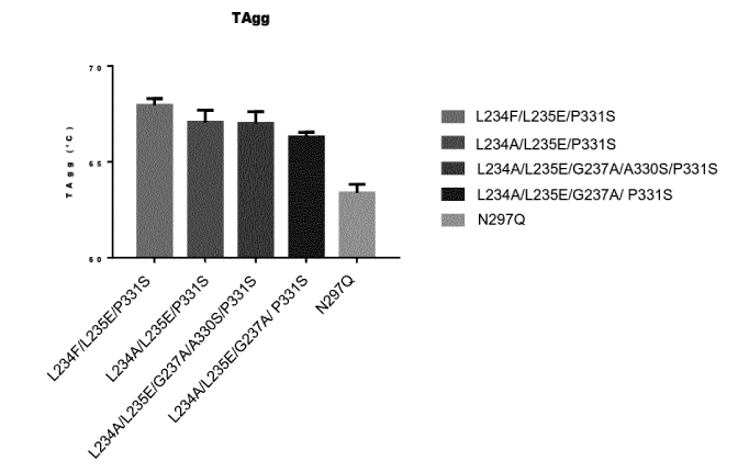
Figure 8 shows several human IgG1 Fc domain mutants showed a higher
aggregation temperature (TAgg) and improved stability of the antibody compared
to wild-type
human Fc domains.
DETAILED DESCRIPTION OF THE INVENTION
Definitions
As used in the specification, "a" or "an" may mean one or more. As used in the
claim(s), when used in conjunction with the word "comprising", the words "a"
or "an" may
mean one or more than one. As used herein "another" may mean at least a second
or more.
Where "comprising" is used, this can optionally be replaced by "consisting
essentially of" or by "consisting of".
Human CD39, also known as "vascular" CD39, NTPdasel , ENTPD1, ATPDase and
vascular ATP diphosphohydrolase, exhibits ATPase activity. CD39 hydrolyzes
extracellular
ATP and ADP to AMP, which is further converted to adenosine by another enzyme,
5-prime
nucleotidase. The amino acid sequence of the "vascular" human CD39 mature
polypeptide
CA 03005986 2018-05-23
WO 2017/089334 PCT/EP2016/078395
22
chain is shown in Genbank under accession number P49961, the entire disclosure
of which
is incorporated herein by reference, and as follows:
1 MEDTKESNVK TFCSKNILAI LGFSSIIAVI ALLAVGLTQN KALPENVKYG IVLDAGSSHT
61 SLYIYKWPAE KENDTGVVHQ VEECRVKGPG ISKFVQKVNE IGIYLTDCME RAREVIPRSQ
121 HQETPVYLGA TAGMRLLRME SEELADRVLD VVERSLSNYP FDFQGARIIT GQEEGAYGWI
181 TINYLLGKFS QKTRWFSIVP YETNNQETFG ALDLGGASTQ VTFVPQNQTI ESPDNALQFR
241 LYGKDYNVYT HSFLCYGKDQ ALWQKLAKDI QVASNEILRD PCFHPGYKKV VNVSDLYKTP
301 CTKRFEMTLP FQQFEIQGIG NYQQCHQSIL ELFNTSYCPY SQCAFNGIFL PPLQGDFGAF
361 SAFYFVMKFL NLTSEKVSQE KVTEMMKKFC AQPWEEIKTS YAGVKEKYLS EYCFSGTYIL
421 SLLLQGYHFT ADSWEHIHFI GKIQGSDAGW TLGYMLNLTN MIPAEQPLST PLSHSTYVFL
481 MVLFSLVLFT VAIIGLLIFH KPSYFWKDMV (SEQ ID NO: 1)
Human CD39-L1, also known as NTPDase2 or ENTPD2, is shown in Genbank
under accession number NP_001237, the entire disclosure of which is
incorporated herein by
reference, and as follows:
1 MAGKVRSLLP PLLLAAAGLA GLLLLCVPTR DVREPPALKY GIVLDAGSSH TSMFIYKWPA
61 DKENDTGIVG QHSSCDVPGG GISSYADNPS GASQSLVGCL EQALQDVPKE RHAGTPLYLG
121 ATAGMRLLNL TNPEASTSVL MAVTHTLTQY PFDFRGARIL SGQEEGVFGW VTANYLLENF
181 IKYGWVGRWF RPRKGTLGAM DLGGASTQIT FETTSPAEDR ASEVQLHLYG QHYRVYTHSF
241 LCYGRDQVLQ RLLASALQTH GFHPCWPRGF STQVLLGDVY QSPCTMAQRP QNFNSSARVS
301 LSGSSDPHLC RDLVSGLFSF SSCPFSRCSF NGVFQPPVAG NFVAFSAFFY TVDFLRTSMG
361 LPVATLQQLE AAAVNVCNQT WAQQLLSRGY GFDERAFGGV IFQKKAADTA VGWALGYMLN
421 LTNLIPADPP GLRKGTDFSS WVVLLLLFAS ALLAALVLLL RQVHSAKLPS TI
(SEQ ID NO: 2)
Human CD39-L2, also known as NTPDase6 or ENTPD6; ENTPD6 isoform 1 is
shown in Genbank under accession number NP_001238, the entire disclosure of
which is
incorporated herein by reference, and as follows:
1 MKKGIRYETS RKTSYIFQQP QHGPWQTRMR KISNHGSLRV AKVAYPLGLC VGVFIYVAYI
61 KWHRATATQA FFSITRAAPG ARWGQQAHSP LGTAADGHEV FYGIMFDAGS TGTRVHVFQF
121 TRPPRETPTL THETFKALKP GLSAYADDVE KSAQGIRELL DVAKQDIPFD FWKATPLVLK
181 ATAGLRLLPG EKAQKLLQKV KEVFKASPFL VGDDCVSIMN GTDEGVSAWI TINFLTGSLK
241 TPGGSSVGML DLGGGSTQIA FLPRVEGTLQ ASPPGYLTAL RMFNRTYKLY SYSYLGLGLM
301 SARLAILGGV EGQPAKDGKE LVSPCLSPSF KGEWEHAEVT YRVSGQKAAA SLHELCAARV
361 SEVLQNRVHR TEEVKHVDFY AFSYYYDLAA GVGLIDAEKG GSLVVGDFEI AAKYVCRTLE
421 TQPQSSPFSC MDLTYVSLLL QEFGFPRSKV LKLTRKIDNV ETSWALGAIF HYIDSLNRQK
481 SPAS
(SEQ ID NO: 3)
Human CD39-L3, also known as NTPDase3 or ENTPD3; ENTPD3 isoform is shown in
Genbank under accession number NP_001239, the entire disclosure of which is
incorporated
herein by reference, and as follows:
1 MFTVLTRQPC EQAGLKALYR TPTIIALVVL LVSIVVLVSI TVIQIHKQEV LPPGLKYGIV
61 LDAGSSRTTV YVYQWPAEKE NNTGVVSQTF KCSVKGSGIS SYGNNPQDVP RAFEECMQKV
121 KGQVPSHLHG STPIHLGATA GMRLLRLQNE TAANEVLESI QSYFKSQPFD FRGAQIISGQ
181 EEGVYGWITA NYLMGNFLEK NLWHMWVHPH GVETTGALDL GGASTQISFV AGEKMDLNTS
241 DIMQVSLYGY VYTLYTHSFQ CYGRNEAEKK FLAMLLQNSP TKNHLTNPCY PRDYSISFTM
301 GHVFDSLCTV DQRPESYNPN DVITFEGTGD PSLCKEKVAS IFDFKACHDQ ETCSFDGVYQ
361 PKIKGPFVAF AGFYYTASAL NLSGSFSLDT FNSSTWNFCS QNWSQLPLLL PKFDEVYARS
421 YCFSANYIYH LFVNGYKFTE ETWPQIHFEK EVGNSSIAWS LGYMLSLTNQ IPAESPLIRL
481 PIEPPVFVGT LAFFTAAALL CLAFLAYLCS ATRRKRHSEH AFDHAVDSD
(SEQ ID NO: 4)
CA 03005986 2018-05-23
WO 2017/089334
PCT/EP2016/078395
23
Human CD39-L4, also known as NTPDase5 or ENTPD5, is shown in Genbank under
accession number NP_001240 (precursor), the entire disclosure of which is
incorporated
herein by reference, and as follows:
1 MATSWGTVFF MLVVSCVCSA VSHRNQQTWF EGIFLSSMCP INVSASTLYG IMFDAGSTGT
61 RIHVYTFVQK MPGQLPILEG EVFDSVKPGL SAFVDQPKQG AETVQGLLEV AKDSIPRSHW
121 KKTPVVLKAT AGLRLLPEHK AKALLFEVKE IFRKSPFLVP KGSVSIMDGS DEGILAWVTV
181 NFLTGQLHGH RQETVGTLDL GGASTQITFL PQFEKTLEQT PRGYLTSFEM FNSTYKLYTH
241 SYLGFGLKAA RLATLGALET EGTDGHTFRS ACLPRWLEAE WIFGGVKYQY GGNQEGEVGF
301 EPCYAEVLRV VRGKLHQPEE VQRGSFYAFS YYYDRAVDTD MIDYEKGGIL KVEDFERKAR
361 EVCDNLENFT SGSPFLCMDL SYITALLKDG FGFADSTVLQ LTKKVNNIET GWALGATFHL
421 LQSLGISH
(SEQ ID NO: 5)
In the context herein, "neutralize" or neutralizing" when referring to the
CD39
polypeptide (e.g. "neutralize CD39", "neutralize the activity of CD39" or
"neutralize the
enzymatic activity of CD39"), refers to a process in which the ATP hydrolysis
(ATPase)
activity of CD39 is inhibited. This comprises, notably the inhibition of CD39-
mediated
generation of AMP and/or ADP, i.e. the inhibition of CD39-mediated catabolism
of ATP to
AMP and/or ADP. This can be measured for example in a cellular assay that
measures the
capacity of a test compound to inhibit the conversion of ATP to AMP and/or
ADP, either
directly or indirectly. For example, disappearance of ATP and/or generation of
AMP can be
assessed, as described herein. In one embodiment, an antibody preparation
causes at least
a 60% decrease in the conversion of ATP to AMP, at least a 70% decrease in the
conversion
of ATP to AMP, or at least an 80% or 90% decrease in the conversion of ATP to
AMP,
referring, for example, to the assays described herein (e.g. disappearance of
ATP and/or
generation of AMP).
Whenever "treatment of cancer" or the like is mentioned with reference to anti-
CD39
binding agent (e.g. antibody), this can include: (a) method of treatment of
cancer, said
method comprising the step of administering (for at least one treatment) an
anti-CD39
binding agent, (preferably in a pharmaceutically acceptable carrier material)
to an individual,
a mammal, especially a human, in need of such treatment, in a dose that allows
for the
treatment of cancer, (a therapeutically effective amount), preferably in a
dose (amount) as
specified herein; (b) the use of an anti-CD39 binding agent for the treatment
of cancer, or an
anti-CD39 binding agent, for use in said treatment (especially in a human);
(c) the use of an
anti-CD39 binding agent for the manufacture of a pharmaceutical preparation
for the
treatment of cancer, a method of using an anti-CD39 binding agent for the
manufacture of a
pharmaceutical preparation for the treatment of cancer, comprising admixing an
anti-CD39
binding agent with a pharmaceutically acceptable carrier, or a pharmaceutical
preparation
comprising an effective dose of an anti-CD39 binding agent that is appropriate
for the
CA 03005986 2018-05-23
WO 2017/089334
PCT/EP2016/078395
24
treatment of cancer; or (d) any combination of a), b), and c), in accordance
with the subject
matter allowable for patenting in a country where this application is filed.
The term "antibody," as used herein, refers to polyclonal and monoclonal
antibodies.
Depending on the type of constant domain in the heavy chains, antibodies are
assigned to
one of five major classes: IgA, IgD, IgE, IgG, and IgM. Several of these are
further divided
into subclasses or isotypes, such as IgG1, IgG2, IgG3, IgG4, and the like. An
exemplary
immunoglobulin (antibody) structural unit comprises a tetramer. Each tetramer
is composed
of two identical pairs of polypeptide chains, each pair having one "light"
(about 25 kDa) and
one "heavy" chain (about 50-70 kDa). The N-terminus of each chain defines a
variable region
of about 100 to 110 or more amino acids that is primarily responsible for
antigen recognition.
The terms variable light chain (VL) and variable heavy chain (VH) refer to
these light and
heavy chains respectively. The heavy-chain constant domains that correspond to
the
different classes of immunoglobulins are termed "alpha," "delta," "epsilon,"
"gamma" and
"mu," respectively. The subunit structures and three-dimensional
configurations of different
classes of immunoglobulins are well known. IgG are the exemplary classes of
antibodies
employed herein because they are the most common antibodies in the
physiological situation
and because they are most easily made in a laboratory setting. Optionally the
antibody is a
monoclonal antibody. Particular examples of antibodies are humanized,
chimeric, human, or
otherwise-human-suitable antibodies. "Antibodies" also includes any fragment
or derivative of
any of the herein described antibodies.
The term "specifically binds to" means that an antibody can bind preferably in
a
competitive binding assay to the binding partner, e.g. CD39, as assessed using
either
recombinant forms of the proteins, epitopes therein, or native proteins
present on the surface
of isolated target cells. Competitive binding assays and other methods for
determining
specific binding are further described below and are well known in the art.
When an antibody is said to "compete with" a particular monoclonal antibody,
it
means that the antibody competes with the monoclonal antibody in a binding
assay using
either recombinant CD39 molecules or surface expressed CD39 molecules. For
example, if a
test antibody reduces the binding of a reference antibody to a CD39
polypeptide or CD39-
expressing cell in a binding assay, the antibody is said to "compete"
respectively with the
reference antibody.
The term "affinity", as used herein, means the strength of the binding of an
antibody
to an epitope. The affinity of an antibody is given by the dissociation
constant Kd, defined as
[AID] x [Ag] / [Ab-Ag], where [Ab-Ag] is the molar concentration of the
antibody-antigen
complex, [AID] is the molar concentration of the unbound antibody and [Ag] is
the molar
concentration of the unbound antigen. The affinity constant Ka is defined by
1/Kd. Methods
CA 03005986 2018-05-23
WO 2017/089334
PCT/EP2016/078395
for determining the affinity of mAbs can be found in Harlow, et al.,
Antibodies: A Laboratory
Manual, Cold Spring Harbor Laboratory Press, Cold Spring Harbor, N.Y., 1988),
Coligan et
al., eds., Current Protocols in Immunology, Greene Publishing Assoc. and Wiley
lnterscience, N.Y., (1992, 1993), and Muller, Meth. Enzymol. 92:589-601
(1983), which
5
references are entirely incorporated herein by reference. One standard method
well known in
the art for determining the affinity of mAbs is the use of surface plasmon
resonance (SPR)
screening (such as by analysis with a BIAcOreTM SPR analytical device).
Within the context herein a "determinant" designates a site of interaction or
binding
on a polypeptide.
10
The term "epitope" refers to an antigenic determinant, and is the area or
region on
an antigen to which an antibody binds. A protein epitope may comprise amino
acid residues
directly involved in the binding as well as amino acid residues which are
effectively blocked
by the specific antigen binding antibody or peptide, i.e., amino acid residues
within the
"footprint" of the antibody. It is the simplest form or smallest structural
area on a complex
15
antigen molecule that can combine with e.g., an antibody or a receptor.
Epitopes can be
linear or conformational/structural. The term "linear epitope" is defined as
an epitope
composed of amino acid residues that are contiguous on the linear sequence of
amino acids
(primary structure). The term "conformational or structural epitope" is
defined as an epitope
composed of amino acid residues that are not all contiguous and thus represent
separated
20
parts of the linear sequence of amino acids that are brought into proximity to
one another by
folding of the molecule (secondary, tertiary and/or quaternary structures). A
conformational
epitope is dependent on the 3-dimensional structure. The term 'conformational'
is therefore
often used interchangeably with 'structural'.
The term "internalization", used interchangeably with "intracellular
internalization",
25
refers to the molecular, biochemical and cellular events associated with the
process of
translocating a molecule from the extracellular surface of a cell to the
intracellular surface of
a cell. The processes responsible for intracellular internalization of
molecules are well-known
and can involve, inter alia, the internalization of extracellular molecules
(such as hormones,
antibodies, and small organic molecules); membrane-associated molecules (such
as cell-
surface receptors); and complexes of membrane-associated molecules bound to
extracellular molecules (for example, a ligand bound to a transmembrane
receptor or an
antibody bound to a membrane-associated molecule). Thus, "inducing and/or
increasing
internalization" comprises events wherein intracellular internalization is
initiated and/or the
rate and/or extent of intracellular internalization is increased.
CA 03005986 2018-05-23
WO 2017/089334
PCT/EP2016/078395
26
The term "agent" is used herein to denote a chemical compound, a mixture of
chemical compounds, a biological macromolecule, or an extract made from
biological
materials. The term "therapeutic agent" refers to an agent that has biological
activity.
For the purposes herein, a "humanized" or "human" antibody refers to an
antibody in
which the constant and variable framework region of one or more human
immunoglobulins is
fused with the binding region, e.g. the CDR, of an animal immunoglobulin. Such
antibodies
are designed to maintain the binding specificity of the non-human antibody
from which the
binding regions are derived, but to avoid an immune reaction against the non-
human
antibody. Such antibodies can be obtained from transgenic mice or other
animals that have
been "engineered" to produce specific human antibodies in response to
antigenic challenge
(see, e.g., Green et al. (1994) Nature Genet 7:13; Lonberg et al. (1994)
Nature 368:856;
Taylor et al. (1994) Int lmmun 6:579, the entire teachings of which are herein
incorporated by
reference). A fully human antibody also can be constructed by genetic or
chromosomal
transfection methods, as well as phage display technology, all of which are
known in the art
(see, e.g., McCafferty et al. (1990) Nature 348:552-553). Human antibodies may
also be
generated by in vitro activated B cells (see, e.g., U.S. Pat. Nos. 5,567,610
and 5,229,275,
which are incorporated in their entirety by reference).
A "chimeric antibody" is an antibody molecule in which (a) the constant
region, or a
portion thereof, is altered, replaced or exchanged so that the antigen binding
site (variable
region) is linked to a constant region of a different or altered class,
effector function and/or
species, or an entirely different molecule which confers new properties to the
chimeric
antibody, e.g., an enzyme, toxin, hormone, growth factor, drug, etc.; or (b)
the variable
region, or a portion thereof, is altered, replaced or exchanged with a
variable region having a
different or altered antigen specificity.
The term "hypervariable region" when used herein refers to the amino acid
residues
of an antibody that are responsible for antigen binding. The hypervariable
region generally
comprises amino acid residues from a "complementarity-determining region" or
"CDR" (e.g.
residues 24-34 (L1), 50-56 (L2) and 89-97 (L3) in the light-chain variable
domain and 31-35
(H1), 50-65 (H2) and 95-102 (H3) in the heavy-chain variable domain; Kabat et
al. 1991)
and/or those residues from a "hypervariable loop" (e.g. residues 26-32 (L1),
50-52 (L2) and
91-96 (L3) in the light-chain variable domain and 26-32 (H1), 53-55 (H2) and
96-101 (H3) in
the heavy-chain variable domain; Chothia and Lesk, J. Mol. Biol 1987;196:901-
917), or a
similar system for determining essential amino acids responsible for antigen
binding.
Typically, the numbering of amino acid residues in this region is performed by
the method
described in Kabat et al., supra. Phrases such as "Kabat position", "variable
domain residue
numbering as in Kabat" and "according to Kabat" herein refer to this numbering
system for
CA 03005986 2018-05-23
WO 2017/089334
PCT/EP2016/078395
27
heavy chain variable domains or light chain variable domains. Using the Kabat
numbering
system, the actual linear amino acid sequence of a peptide may contain fewer
or additional
amino acids corresponding to a shortening of, or insertion into, a FR or CDR
of the variable
domain. For example, a heavy chain variable domain may include a single amino
acid insert
(residue 52a according to Kabat) after residue 52 of CDR H2 and inserted
residues (e.g.
residues 82a, 82b, and 82c, etc. according to Kabat) after heavy chain FR
residue 82. The
Kabat numbering of residues may be determined for a given antibody by
alignment at
regions of homology of the sequence of the antibody with a "standard" Kabat
numbered
sequence.
By "framework" or "FR" residues as used herein is meant the region of an
antibody
variable domain exclusive of those regions defined as CDRs. Each antibody
variable domain
framework can be further subdivided into the contiguous regions separated by
the CDRs
(FR1, FR2, FR3 and FR4).
The terms "Fc domain," "Fc portion," and "Fc region" refer to a C-terminal
fragment
of an antibody heavy chain, e.g., from about amino acid (aa) 230 to about aa
450 of human y
(gamma) heavy chain or its counterpart sequence in other types of antibody
heavy chains
(e.g., a, 6, E and p for human antibodies), or a naturally occurring allotype
thereof. Unless
otherwise specified, the commonly accepted Kabat amino acid numbering for
immunoglobulins is used throughout this disclosure (see Kabat et al. (1991 )
Sequences of
Protein of Immunological Interest, 5th ed., United States Public Health
Service, National
Institute of Health, Bethesda, MD).
The terms "isolated", "purified" or "biologically pure" refer to material that
is
substantially or essentially free from components which normally accompany it
as found in its
native state. Purity and homogeneity are typically determined using analytical
chemistry
techniques such as polyacrylamide gel electrophoresis or high performance
liquid
chromatography. A protein that is the predominant species present in a
preparation is
substantially purified.
The terms "polypeptide," "peptide" and "protein" are used interchangeably
herein to
refer to a polymer of amino acid residues. The terms apply to amino acid
polymers in which
one or more amino acid residue is an artificial chemical mimetic of a
corresponding naturally
occurring amino acid, as well as to naturally occurring amino acid polymers
and non-naturally
occurring amino acid polymer.
The term "recombinant" when used with reference, e.g., to a cell, or nucleic
acid,
protein, or vector, indicates that the cell, nucleic acid, protein or vector,
has been modified by
the introduction of a heterologous nucleic acid or protein or the alteration
of a native nucleic
acid or protein, or that the cell is derived from a cell so modified. Thus,
for example,
CA 03005986 2018-05-23
WO 2017/089334
PCT/EP2016/078395
28
recombinant cells express genes that are not found within the native
(nonrecombinant) form
of the cell or express native genes that are otherwise abnormally expressed,
under
expressed or not expressed at all.
Within the context herein, the term antibody that "binds" a polypeptide or
epitope
designates an antibody that binds said determinant with specificity and/or
affinity.
The term "identity" or "identical", when used in a relationship between the
sequences of two or more polypeptides, refers to the degree of sequence
relatedness
between polypeptides, as determined by the number of matches between strings
of two or
more amino acid residues. "Identity" measures the percent of identical matches
between the
smaller of two or more sequences with gap alignments (if any) addressed by a
particular
mathematical model or computer program (i.e., "algorithms"). Identity of
related polypeptides
can be readily calculated by known methods. Such methods include, but are not
limited to,
those described in Computational Molecular Biology, Lesk, A. M., ed., Oxford
University
Press, New York, 1988; Biocomputing: Informatics and Genome Projects, Smith,
D. W., ed.,
Academic Press, New York, 1993; Computer Analysis of Sequence Data, Part 1,
Griffin, A.
M., and Griffin, H. G., eds., Humana Press, New Jersey, 1994; Sequence
Analysis in
Molecular Biology, von Heinje, G., Academic Press, 1987; Sequence Analysis
Primer,
Gribskov, M. and Devereux, J., eds., M. Stockton Press, New York, 1991; and
Carillo et al.,
SIAM J. Applied Math. 48, 1073 (1988).
Methods for determining identity are designed to give the largest match
between the
sequences tested. Methods of determining identity are described in publicly
available
computer programs. Computer program methods for determining identity between
two
sequences include the GCG program package, including GAP (Devereux et al.,
Nucl. Acid.
Res. 12, 387 (1984); Genetics Computer Group, University of Wisconsin,
Madison, Wis.),
BLASTP, BLASTN, and FASTA (Altschul et al., J. Mol. Biol. 215, 403-410
(1990)). The
BLASTX program is publicly available from the National Center for
Biotechnology Information
(NCB!) and other sources (BLAST Manual, Altschul et al. NCB/NLM/NIH Bethesda,
Md.
20894; Altschul et al., supra). The well-known Smith Waterman algorithm may
also be used
to determine identity.
Production of antibodies
The anti-CD39 antibody that can be used for the treatment of cancers and/or
other
diseases (e.g., infectious disease) binds an extra-cellular portion of human
CD39 polypeptide
and neutralizes the enzymatic activity of CD39 expressed on the surface of a
cell, e.g. a
tumor cell. In one embodiment the agent inhibits the ATPase activity of CD39.
In one
embodiment the antibody inhibits CD39-mediated generation of adenosine.
In one
CA 03005986 2018-05-23
WO 2017/089334
PCT/EP2016/078395
29
embodiment the antibody inhibits CD39-mediated catabolism of ATP to AMP. In
one
embodiment the antibody inhibits adenosine-mediated inhibition of lymphocyte
activity (e.g. T
cells). In one aspect, the antibody is selected from a full-length antibody,
an antibody
fragment, and a synthetic or semi-synthetic antibody-derived molecule.
Antibodies that potently inhibit the enzymatic (ATPase activity) activity of
the CD39
enzyme appear to do so by immobilizing the enzyme in one of its conformations
thereby
preventing it from hydrolyzing its substrate. The antibodies achieve this by
binding to both C-
and N-terminal domains of CD39 at the same time.
Thus in any embodiment an anti-CD39 antibody can bind to both C- and N-
terminal
domains of CD39 via their VH CDRs (there is no need for the VI_ CDRs to bind
CD39 at all).
As shown in the Examples, binding to the N-terminal domain (domain 1) of CD39
can be
achieved by two of the CDRs (CDR1 and CDR3, as well as a first segment of
Kabat CDR2,
while binding to the C-terminal domain (domain 2) can be achieved through a
single CDR
(CDR2) (a CDR2 that extends into the Kabat FR3). Not only is the contribution
of CDR2
surprising, but additionally the CDR2 cooperates with FR3 in binding to CD39.
Furthermore,
the binding by CDR2-FR to CD39 involves the glycan at N292 of CD39 (the N292
glycan
covers a significant amount of the apical surface of the C-terminal domain).
The resulting
binding by the VH to both domains 1 and 2 is believed to be important in
immobilizing the
CD39 enzyme so as to prevent substrate hydrolysis. The antibodies thus have a
VH CDR2
that binds to the N-terminal domain of CD39 via a first portion of the CDR2
and to the C-
terminal domain of CD39 (including the glycan at N292) via a second portion of
the CDR2, in
combination with FR3 residues.
The inventors further observed that the positioning of the VH CDR3 for binding
to
CD39 occurs via a surprising mechanism involving a VI_ CDR / VH CDR3 / CD39
binding
matrix in which VH CDR3 is trapped between the VI_ CDRs and CD39. The VI_ CDRs
form a
paratope that binds to the CDR3 of the VH. The VH CDR3 comprises numerous
aromatic
residues, and while one of the aromatic residues in VH CDR3 interacts with
CD39, other
aromatic amino acids within VH CDR3 interact with the residues of the VL. The
VH CDR3
comprises multiple aromatic residues which form pi-interactions with the VI_
on one face, and
CD39 on another face, resulting in a matrix of pi-interactions structure that
traps the VH
CDR3 between the VI_ CDRs and CD39 and permits a large binding area across the
surface
of CD39. Further surprisingly, the structure appears to have arisen in the
context of certain
framework residues, as both the VH CD39 interaction and the VH CDR3-VL
interaction involve
framework residues (according to Kabat numbering), such that the overall
contact residues
involved a limited set of Kabat CDR residues combined with Kabat FR residues.
CA 03005986 2018-05-23
WO 2017/089334
PCT/EP2016/078395
The overall structure permits the VH to bind N-terminal domain of CD39 (e.g.,
at
residues V95, Q96, K97, L137, E140, L144 of CD39, with reference to the CD39
amino acid
sequence of SEQ ID NO: 1) on one side of the enzymatic active site, and the C-
terminal
domain of CD39 (e.g., at residues S294, K298, as well as to the glycan linked
to N292 of
5 CD39, of CD39) on the opposite side of the enzymatic active site.
These findings permit a wide range of CD39-binding polypeptides to be
generated
that retain the mechanism of action and permits desired amino acid sequences
and features
to be incorporated. Antibodies and other VHNL-containing proteins can be
designed which
include, inter alia, the VH CDR2 segment that binds the N-terminal domain, the
VH CDR2-FR3
10 segment capable of binding the C-terminal domain (and, notably, the N292-
linked
glycosylation) and the VH CDR3 that binds the C-terminal domain and gives rise
to the
VLCDR - VHCDR3 - CD39 matrix. Amino acid sequences of VL domains can be
selected in
the context of the desired VH sequence, e.g., by introducing substitutions
that maintain the
overall VH or VL domain structure without affecting V domain interactions, and
where V
15 domain interactions would be modified, making amino acid substitutions
pair-wise in both VH
and VL at the respective contact positions. The resulting protein binds to
CD39 essentially or
exclusively via the VH domain(s), and the VL domain(s) binds to VH CDR3 but is
not involved
in binding to CD39. A wide range of both VL CDR amino acid sequences can be
employed in
such protein; optionally VL CDR residues and the respective VH residues can be
chosen and
20 substituted as pairs such that the VH/VL CDR contact is maintained.
Similarly, a wide range of
human (or non-human) VH and VL framework sequences can be selected as acceptor
frameworks that bear resides at the specified positions that maintain the VH
CDR-VLCDR
contact and the VHCDR-CD39 contact.
Consequently, in one embodiment, an anti-CD39 antigen binding domain, or an
25 antigen-binding protein that comprises the antigen binding domain (e.g.,
an antibody or
antibody fragment, a multispecific binding protein, a bispecific antibody,
etc.), comprises
complementary determining regions (CDR) and framework regions (FR). The
antigen binding
domains can be designed or modified so as to provide desired and/or improved
properties.
In one embodiment, an anti-CD39 antigen-binding protein is capable of binding
to and
30 inhibiting the activity of a human CD39 polypeptide, the antigen-binding
protein comprising a
VH and a VL that each comprise a framework (e.g. a framework having an amino
acid
sequence of human origin) and a CDR1, CDR2 and CDR3, wherein the antigen-
binding
protein is capable of binding to the N-terminal domain of CD39 and the C-
terminal domain of
CD39. In one embodiment, the antigen-binding protein restricts the domain
movement of
CD39 when bound to CD39. Optionally, the VH and/or VL framework (e.g. FR1,
FR2, FR3
and/or FR4) is of human origin. In one embodiment, the VH comprises a first
CDR (or antigen
CA 03005986 2018-05-23
WO 2017/089334
PCT/EP2016/078395
31
binding domain) that is capable of binding to the N-terminal domain of CD39
and a second
CDR (or antigen binding domain) that that is capable of binding to amino acid
residues of the
C-terminal domain of CD39.
In one aspect (e.g. in any aspect herein), a binding molecule or antigen-
binding
fragment thereof is capable of binding to and inhibiting the activity of CD39,
comprising a VH
and a VL, wherein the Kabat CDR1 of the VH binds to amino acid residues in the
N-terminal
domain of CD39, the Kabat CDR2 (optionally together with the FR3) of the VH
binds to amino
acid residues and/or to the N292-linked glycan in the C-terminal domain of
CD39, and the
Kabat CDR3 of the VH binds to amino acid residues the N-terminal domain of
CD39.
Optionally the CDR2 of the VH comprises a first amino acid segment that binds
to the N-
terminal domain of CD39 together and an amino acid segment that binds,
together with FR3
residues, to the C-terminal domain of CD39. Optionally, the CDR3 comprises an
aromatic
residue (e.g. a tyrosine) that is capable of binding an amino acid residue in
the N-terminal
domain of CD39 and a second aromatic amino acid residue (e.g., a tyrosine, a
phenylalanine) that is capable of contacting an amino acid residue in the VL.
Optionally, the
binding molecule or antigen-binding fragment comprises a VI_ that binds, via a
residue in a
Kabat CDR, to the Kabat CDR3 of the VH.
In one embodiment, the CDR1 comprises a residue that is capable of contacting
amino acid residues in the N-terminal domain of CD39. Optionally a CD39
contact residue is
at Kabat position 33, optionally at both positions 31 and 33. In one
embodiment, the Kabat
FR1 comprise a CD39 contact residue at Kabat position 30, optionally wherein
the residue is
a threonine. Optionally the residue at position 31 is a histidine or
asparagine. Optionally the
residue at position 33 is a glycine. Optionally, the residue at position 32 is
a residue with an
aromatic side chain (an aromatic residue).
In one embodiment, the CDR2 and FR3 comprise a segment of residues within
Kabat positions 59-71, optionally within 59-72b, that is capable of contacting
amino acid
residues in the C-terminal domain of CD39, optionally further wherein residues
within Kabat
positions 59-71 contact the glycan at residue N292 of CD39. For example, the
Kabat CDR2
and FR3 can comprise residues at Kabat positions 59, 65, 67, 68, 69, 70 and/or
71, and
optionally further at residue 72, 72a and/or 72b that are capable of
contacting the C-terminal
domain of CD39, e.g. including amino acid resides in CD39 and the glycan at
N292 of the
CD39 polypeptide.
In one embodiment, the CDR2 (e.g., Kabat CDR2-FR3 segment) comprises
residues at Kabat position 59-71 having the formula X1 X2 X3 X4 X5 X6 X7 X8 X9
X10 X11 X12 X13
(SEQ ID NO: 12), wherein X1 represents a tyrosine, each of X2, X3, X4, X5 and
X6 each
represent any amino acid, X7 represents glycine or another residue which does
not introduce
CA 03005986 2018-05-23
WO 2017/089334
PCT/EP2016/078395
32
steric hindrance that reduces antigen binding, X8 represents any amino acid,
X9 represents
phenylalanine or another hydrophobic residue capable of maintaining the beta-
strand
position and VH domain structure integrity, Xio represents alanine or valine,
or optionally
leucine, optionally threonine, optionally a hydrophobic residue, X11
represents phenylalanine
or another hydrophobic residue (e.g. isoleucine) capable of maintaining the
beta-strand
position and VH domain structure integrity and X12 represents serine, and X13
represents any
amino acid, optionally leucine, optionally alanine, valine, threonine or
arginine. In one
embodiment, the VH FR3 residues at Kabat positions 72, 72a and 72b have the
formula X1 X2
X3, wherein X1 represents aspartic acid, glutamic acid or alanine, X2
represents any amino
acid, optionally alanine, threonine or asparagine, or a conservative
substitution thereof, and
X3 represents serine, optionally alanine, or a conservative substitution
thereof.
In one embodiment, the VH comprises a leucine residue at Kabat position 71
(FR3).
Human or humanized antibodies can advantageously have the FR3 signature
sequence
FVFSL at Kabat positions 67-71, present in certain human VH FR domains. Thus
in one
aspect of any embodiment herein, the segment of residues at Kabat positions 67-
71
comprises the amino acid sequence: FVFSL.
In one embodiment, the CDR2 optionally comprises a segment of residues in
Kabat
positions 50-56, optionally residues 50, 52, 52a, 53 and/or 56, that is
capable of contacting
amino acid residues in CD39 (e.g. in the N-terminal domain of CD39),
optionally wherein the
residue at position 53 aromatic is an amino acid residue. In one embodiment,
the CDR2
comprises residues 50, 52, 52a, 53 and/or 56, that are capable of contacting
amino acid
residues in CD39. In one embodiment, the CDR2 comprises residues at Kabat
position 50-56
having the formula X1 X2 X3 X4 X5 X6 X7 X8 (SEQ ID NO: 13), wherein X1
represents
tryptophan, X2 represents any amino acid, optionally an isoleucine, X3
represents asparagine
or optionally glutamine, X4 represents threonine, X5 represents any amino acid
residue,
optionally a tyrosine or optionally phenylalanine, X6 represents any amino
acid, optionally
threonine, optionally serine, optionally asparagine, alanine or glycine,
optionally a residue
other than a large or hydrophobic residue, X7 represents any amino acid,
optionally glycine,
optionally alanine, serine, threonine, asparagine or glutamine, optionally a
residue other than
aspartic acid or glutamic acid, optionally a residue other than lysine or
arginine, X8
represents glutamic acid, optionally aspartic acid.
In another embodiment, a VH comprises a Kabat CDR2-FR3 segment that binds to
both the N- and C- terminal domain of CD39 (e.g., across the N- and C- domain
surface,
across the substrate cleft, groove entry or active site) the CDR2-FR3 segment
comprising
residues (e.g. at Kabat position 50-71) having the formula X1 X2 X3 X4 X5 X6
X7 X8 X9 X10 X11 X12
X13 X14 X15 X16 X17 X18 X19 X20 X21 X22 X23 (SEQ ID NO: 14), wherein X1
represents tryptophan,
CA 03005986 2018-05-23
WO 2017/089334
PCT/EP2016/078395
33
X2 represents any amino acid, optionally an isoleucine, X3 represents
asparagine or
optionally glutamine, X4 represents threonine, X5 represents any amino acid
residue,
optionally tyrosine or optionally phenylalanine, X6 represents any amino acid,
optionally
threonine, optionally serine, optionally asparagine, alanine or glycine,
optionally residues
other than large or hydrophobic resides, X7 represents any amino acid,
optionally glycine,
optionally alanine, serine, threonine, asparagine or glutamine, optionally a
residue other than
aspartic acid or glutamic acid, optionally a residue other than lysine or
arginine, X8
represents glutamic acid, optionally aspartic acid, X9 represents any amino
acid, optionally
proline, X10 represents any amino acid, optionally threonine, optionally
serine, asparagine,
glutamine, histidine, glutamic acid, aspartic acid, arginine, lysine, alanine
or tyrosine,
optionally any residue other than a hydrophobic residue or proline, X11
represents a tyrosine,
each of X12, X13, X14, X15 and X16 each represent any amino acid, X17
represents glycine or
another residue which does not introduce steric hindrance that reduces antigen
binding, X18
represents any amino acid, optionally arginine, X19 represents phenylalanine
or another
hydrophobic residue capable of maintaining the beta-strand position and VH
domain structure
integrity, X20 represents alanine or valine, or optionally leucine, optionally
a hydrophobic
residue, X21 represents phenylalanine or another hydrophobic residue capable
of maintaining
the beta-strand position and VH domain structure integrity, X22 represents
serine, and
wherein and X23 represents any amino acid, optionally leucine, optionally
alanine, valine or
threonine. In one embodiment, the CDR2-FR3 segment further comprises residues
at Kabat
positions 72, 72a and 72b having the formula X24 X25 X26, wherein X24
represents aspartic
acid, glutamic acid or alanine, X25 represents any amino acid, optionally
alanine or threonine,
or a conservative substitution thereof, and X26 represents serine, optionally
alanine, or a
conservative substitution thereof.
In one embodiment, the CDR3 comprises a first aromatic amino acid residue that
is
capable of contacting an amino acid residue in CD39 and a second aromatic
amino acid
residue that is capable of contacting an amino acid residue in the VL, wherein
the first and
second aromatic residues are at any of Kabat positions 100, 100b, 100c, 100d,
100e and/or
100f. In one embodiment the Kabat CDR3 (e.g. Kabat positions 100 to 100f, to
the extent
residues are present at these positions) comprises a sequence of amino
residues having the
formula X1 X2 X3 X4 X5 (SEQ ID NO: 15), wherein any two, three or more of X1,
X2, X3, X4 and
X5 represent an aromatic amino acid.
In one embodiment, provided is an antigen-binding protein capable of binding
to and
inhibiting the activity of a human CD39 polypeptide, the protein comprising a
VH that binds
CD39 and a VI_ (e.g., a VI_ that binds the CDR3 of the VH), wherein the VH
comprises:
CA 03005986 2018-05-23
WO 2017/089334
PCT/EP2016/078395
34
(a) a CDR1 capable of contacting the N-terminal domain of CD39, optionally
comprising a residue at Kabat position 33, optionally at both positions 31 and
33, that is
capable of contacting amino acid residues in CD39;
(b)a CDR2-FR3 domain comprising:
a. a segment comprising amino acid residues capable of contacting the N-
terminal domain of CD39, optionally wherein the segment comprises
residues within Kabat positions 50-56, optionally wherein the segment
comprises one, two, three, four or more of (or all of) the residues at Kabat
positions 50, 52, 52a, 53 and/or 56, optionally wherein the residue at
position 53 aromatic is an amino acid residue;
b. a segment comprising amino acid residues capable of contacting the C-
terminal domain of CD39, optionally wherein the segment comprises
residues within Kabat positions 59-71, optionally wherein the segment
comprises one, two, three, four or more of (or all of) the residues at Kabat
positions 59, 65, 67, 68, 69, 70 and/or 71, optionally further the residue at
Kabat position 54, optionally further the residues at Kabat positions 72, 72a
and/or 72b; and
(c)a CDR3 (e.g. according to Kabat) capable of contacting the N-terminal
domain of
CD39, optionally capable of contacting the N-terminal domain of CD39 and the
VL, optionally
comprising a first aromatic amino acid residue that is capable of contacting
an amino acid
residue in CD39 and a second aromatic amino acid residue that is capable of
contacting an
amino acid residue in the VL, wherein the first and second aromatic residues
are at any of
Kabat positions 100, 100b, 100c, 100d, 100e and/or 100f (to the extent a
residue is present
at the particular Kabat position).
Optionally, the CDR3 further comprises a further (third) aromatic amino acid
residue,
and optionally further a fourth aromatic amino acid residue, wherein the third
aromatic
residue (and fourth aromatic residue, where present) is capable of contacting
an amino acid
residue in the VL, wherein the third aromatic residue (and fourth residue,
where present) is at
any of Kabat positions 100, 100b, 100c, 100d, 100e and/or 100f.
In one embodiment, provided is an antigen-binding protein capable of binding
to and
inhibiting the activity of a human CD39 polypeptide, the protein comprising a
VH that binds
CD39 and a VI_ (e.g., a VI_ that binds the CDR3 of the VH), wherein the VH
comprises:
(a) a CDR1 (e.g. according to Kabat) capable of contacting the N-terminal
domain of
CD39, optionally wherein the residues at Kabat position 31, 32 and 33 have the
formula X1 X2 X3, wherein X1 represents any amino acid, optionally a histidine
or
asparagine, or optionally a conservative substitution thereof, X2 represents
any
CA 03005986 2018-05-23
WO 2017/089334
PCT/EP2016/078395
amino acid, optionally an aromatic residue, optionally a tyrosine or a
conservative
substitution thereof, or optionally an amino acid residue other than a proline
or
glycine, and X3 represents glycine, or another amino acid that avoids steric
hindrance;
5 (b) a CDR2-FR3 segment (e.g. according to Kabat) capable of contacting
the C-
terminal domain of CD39, optionally wherein the residues at Kabat position 50-
71
having the formula X1 X2 X3 X4 X5 X6 X7 X8 X9 X10 X11 X12 X13 X14 X15 X16 X17
X18 X19
X20 X21 X22 X23 (SEQ ID NO: 14), wherein X1 represents tryptophan, X2
represents
any amino acid, optionally an isoleucine, X3 represents asparagine or
optionally
10 glutamine, X4 represents threonine, X5 represents any amino acid
residue,
optionally tyrosine or optionally phenylalanine, X6 represents any amino acid,
optionally threonine, optionally serine, optionally asparagine, alanine or
glycine,
optionally a residue other than a large or hydrophobic residue, X7 represents
any
amino acid, optionally glycine, optionally alanine, serine, threonine,
asparagine or
15 glutamine, optionally residues other than aspartic acid or glutamic
acid, optionally a
residue other than lysine or arginine, X8 represents glutamic acid, optionally
aspartic acid, X9 represents any amino acid, optionally proline, X10
represents any
amino acid, optionally threonine, optionally serine, asparagine, glutamine,
histidine,
glutamic acid, aspartic acid, arginine, lysine, alanine or tyrosine,
optionally any
20 residue other than a hydrophobic residue or proline, X11 represents
a tyrosine, each
of X12, X13, X14, X15 and X16 each represent any amino acid, X17 represents
glycine
or another residue which does not introduce steric hindrance that reduces
antigen
binding, X18 represents any amino acid, optionally arginine, X19 represents
phenylalanine or another hydrophobic residue capable of maintaining the beta-
25 strand position and VH domain structure integrity, X20 represents
alanine or valine,
or optionally leucine, optionally a hydrophobic residue, X21 represents
phenylalanine or another hydrophobic residue capable of maintaining the beta-
strand position and VH domain structure integrity and X22 represents serine,
optionally further wherein and X23 represents any amino acid, optionally
leucine,
30 optionally alanine, valine or threonine, optionally wherein the CDR2-
FR3 segment
further comprises residues at Kabat positions 72, 72a and 72b having the
formula
X24 X25 X26 , wherein X24 represents aspartic acid, glutamic acid or alanine,
X25
represents any amino acid, optionally alanine or threonine, or a conservative
substitution thereof, and X26 represents serine, optionally alanine, or a
conservative
35 substitution thereof; and
CA 03005986 2018-05-23
WO 2017/089334
PCT/EP2016/078395
36
(c) a CDR3 (e.g. according to Kabat) capable of contacting the N-terminal of
CD39,
optionally capable of contacting the N-terminal domain of CD39 and the VI,
optionally wherein the residues at Kabat position 95-102 have the formula X1
X2 X3
X4 X5 X6 X7 X8 X9 X10 X9 X10 X11 X12 X13 Xi4 (SEQ ID NO: 16), wherein
- X1 represents arginine or lysine, or optionally a conservative
substitution
thereof,
- X2 represents any amino acid, optionally arginine, optionally lysine or
alanine, or optionally a conservative substitution thereof,
- X3 represents any amino acid residue, optionally a residue comprising an
aromatic ring, optionally a tyrosine,
- X4 represents any amino acid, optionally glutamic acid or tyrosine, or
optionally a conservative substitution thereof, or an amino acid residue other
than
proline or glycine,
- X5 represents glycine, optionally arginine, or optionally a conservative
substitution thereof,
- X6 represents any amino acid, optionally asparagine, serine or tyrosine,
or
optionally a conservative substitution thereof,
- X7 represents any amino acid, optionally tyrosine, asparagine or aspartic
acid, or optionally a conservative substitution thereof, optionally an amino
acid
residue other than proline or glycine,
- X8 represents valine or optionally alanine, isoleucine or leucine,
optionally
an aromatic amino acid, optionally tyrosine,
- X9 represents any amino acid, optionally an aromatic amino acid,
optionally
phenylalanine, optionally tyrosine, optionally valine or a conservative
substitution
thereof,
- X10 represents tyrosine, optionally phenylalanine, optionally methionine,
or
optionally a conservative substitution thereof,
- X11 is absent or represents any amino acid, optionally tyrosine,
optionally
phenylalanine, optionally tryptophan, or optionally a conservative
substitution
thereof, optionally an amino acid residue other than P, G, E or D, or other
than a
small hydrophobic residue (e.g. T, S),
- X12 is absent or represents any amino acid, optionally phenylalanine, or
optionally a conservative substitution thereof,
- X13 represents any amino acid, optionally aspartic acid, or optionally a
conservative substitution thereof, optionally a serine, optionally a
threonine,
CA 03005986 2018-05-23
WO 2017/089334
PCT/EP2016/078395
37
optionally a glutamic acid, optionally an asparagine, optionally a residue
other than a
large and hydrophobic residue, and
- X14 represents any amino acid, optionally tyrosine or optionally a
conservative substitution thereof, optionally an aromatic amino acid,
optionally a
non-aromatic amino acid.
In one embodiment, provided is an antigen-binding protein capable of binding
to and
inhibiting the activity of a human CD39 polypeptide, the protein comprising a
VH that binds
CD39 and a VL (e.g., a VL that binds the CDR3 of the VH), wherein the VH
comprises:
(a) a CDR1 (e.g. according to Kabat) capable of contacting the N-terminal
domain of
CD39, optionally wherein the residues at Kabat position 31, 32 and 33 have the
formula X1 X2
X3, wherein X1 represents any amino acid, optionally a histidine or
asparagine, or optionally a
conservative substitution thereof, X2 represents any amino acid, optionally an
aromatic
residue, optionally a tyrosine or a conservative substitution thereof, or
optionally an amino
acid residue other than a proline or glycine, and X3 represents glycine, or
another amino acid
that avoids steric hindrance;
(b) a CDR2-FR3 segment (e.g. according to Kabat) capable of
contacting the C-
terminal domain of CD39, optionally wherein the residues at Kabat position 59-
71 have the
formula X1 X2 X3 X4 X5 X6 X7 X9 X9 X10 X11 X12 X13 (SEQ ID NO: 12), wherein X1
represents a
tyrosine, each of X2, X3, X4, X5 and X6 each represent any amino acid, X7
represents glycine
or another residue which does not introduce steric hindrance that reduces
antigen binding, X8
represents any amino acid, X9 represents phenylalanine or another hydrophobic
residue
capable of maintaining the beta-strand position and VH domain structure
integrity, X10
represents alanine or valine, or optionally leucine, optionally threonine,
optionally a
hydrophobic residue, X11 represents phenylalanine or another hydrophobic
residue (e.g.
isoleucine) capable of maintaining the beta-strand position and VH domain
structure integrity
and X12 represents serine, optionally further wherein and X13 represents any
amino acid,
optionally leucine, optionally alanine, valine, threonine or arginine; and
(c) a CDR3 (e.g. according to Kabat) capable of contacting the N-terminal of
CD39,
optionally capable of contacting the N-terminal domain of CD39 and the VL,
optionally
wherein the residues at Kabat position 95-102 have the formula X1 X2 X3 X4 X5
X6 X7 X9 X9 X10
X9 X10 X11 X12 X13 Xi4(SEQ ID NO: 16), wherein
- X1 represents arginine, lysine or alanine, or optionally a conservative
substitution thereof,
- X2 represents any amino acid, optionally arginine, optionally lysine or
alanine, optionally tyrosine, or optionally a conservative substitution
thereof,
CA 03005986 2018-05-23
WO 2017/089334
PCT/EP2016/078395
38
- X3 represents any amino acid residue, optionally a residue comprising an
aromatic ring, optionally a tyrosine,
- X4 represents any amino acid, optionally glutamic acid, tyrosine or
asparagine, or optionally a conservative substitution thereof, or an amino
acid
residue other than proline or glycine,
- X5 represents glycine, optionally arginine, optionally asparagine, or
optionally a conservative substitution thereof,
- X6 represents any amino acid, optionally asparagine, serine or tyrosine,
or
optionally a conservative substitution thereof,
- X7
represents any amino acid, optionally tyrosine, asparagine or aspartic
acid, or optionally a conservative substitution thereof, optionally an amino
acid
residue other than proline or glycine,
- X8 represents valine or optionally alanine, isoleucine, glycine or
leucine,
optionally an aromatic amino acid, optionally tyrosine,
- X9
represents any amino acid, optionally an aromatic amino acid, optionally
phenylalanine, optionally tyrosine, optionally valine, optionally leucine or a
conservative substitution thereof,
- X10 represents tyrosine, optionally phenylalanine, optionally methionine,
or
optionally a conservative substitution thereof,
- X11 is
absent or represents any amino acid, optionally tyrosine, optionally
phenylalanine, optionally tryptophan, optionally alanine, or optionally a
conservative
substitution thereof, optionally an amino acid residue other than P, G, E or
D, or
other than a small hydrophobic residue (e.g. T, S),
- X12 is absent or represents any amino acid, optionally phenylalanine,
optionally methionine, or optionally a conservative substitution thereof,
- X13 represents any amino acid, optionally aspartic acid, or optionally a
conservative substitution thereof, optionally a serine, optionally a
threonine,
optionally a glutamic acid, optionally an asparagine, optionally a residue
other than a
large and hydrophobic residue, and
- X14
represents any amino acid, optionally tyrosine or optionally a
conservative substitution thereof, optionally an aromatic amino acid,
optionally a
non-aromatic amino acid, optionally alanine.
In one aspect of any embodiment herein, any amino acid residue in a VH or VI_
can
be specified to be a residue that maintains V domain (e.g. VH or VI_
respectively) domain
structure integrity. In one aspect of any embodiment herein, an amino acid
residue in a VH or
VI_ can that contacts an antigen or binding partner (e.g. CD39, a VI_ or a VL)
can be specified
CA 03005986 2018-05-23
WO 2017/089334
PCT/EP2016/078395
39
to be a residue which does not introduce steric hindrance that reduces antigen
or binding
partner binding.
In one aspect, the binding molecule or antigen-binding fragment thereof
comprises
human framework regions, e.g. the molecule comprises a VH comprising human VH
FRi, FR2,
FR3 and FR4 amino acid sequences (optionally comprising one or more amino acid
substitutions), and/or a VI_ comprising human VI_ FRi, FR2, FR3 and FR4 amino
acid
sequences (optionally comprising one or more amino acid substitutions).
In one embodiment, the CDR2 comprises an aromatic amino acid residue that
contacts an amino acid residues V95, Q96 and/or L137 of CD39. In one
embodiment, the
aromatic amino acid residue is an aromatic residue, e.g. a tyrosine,
optionally a
phenylalanine.
In one embodiment, the VH comprises a FR3 comprising an amino acid residue
that
contacts CD39, optionally wherein the residue in the VH is a leucine at Kabat
position 71.
In one embodiment, the VH comprises an amino acid residue in FR1 at Kabat
position
19 that contacts CD39, optionally wherein the residue is a lysine.
In one embodiment, the VI_ comprises a FR2 comprising an aromatic amino acid
residue that contacts the CDR3 of the VH, optionally wherein the residue in
the VI_ is at Kabat
position 49. Optionally the residue at VI_ Kabat position is an aromatic
residue, optionally
further wherein a VH comprises an aromatic residue (e.g. at Kabat 100e)
capable of forming
a pi stacking interaction therewith.
In one embodiment, the VH CDR3 comprises an amino acid residue(s) comprising
an
aromatic ring (optionally tyrosine, histidine, tryptophan or phenylalanine),
capable of forming
a pi interaction (e.g., attractive, noncovalent interactions between an
aromatic ring and an
binding partner, for example in an amide pi-stacked interaction, a pi-pi
stacked interaction or
a pi-donor interaction) with an amino acid residue (e.g. an aromatic residue)
in the VI_
polypeptide. In one embodiment, the residue(s) in the CDR3 comprising an
aromatic ring is a
tyrosine, e.g. at Kabat position 100e. In one embodiment, the residue(s) in
the VI_ comprising
an aromatic ring is a tyrosine, e.g. a tyrosine at Kabat position 49 in the
VL.
In one embodiment, the VH CDR3 comprises an amino acid residue(s) comprising
an
aromatic ring (optionally tyrosine, histidine, tryptophan or phenylalanine),
capable of forming
a pi interaction (e.g., attractive, noncovalent interactions between an
aromatic ring and an
binding partner, for example in an amide pi-stacked interaction) with an amino
acid residue in
the CD39 polypeptide (e.g. at residue Q96 in CD39 of SEQ ID NO: 1). In one
embodiment,
the residue comprising an aromatic ring is a phenylalanine.
In one embodiment, the VH CDR3 comprises (e.g. at Kabat position 100e) an
amino
acid residue comprising an aromatic ring (e.g. a tyrosine, optionally a
phenylalanine), which
CA 03005986 2018-05-23
WO 2017/089334
PCT/EP2016/078395
forms or is capable of forming a pi-pi stacking interaction with an amino acid
residue
comprising an aromatic ring in the VL, e.g. the tyrosine at Kabat framework
(FR2) position 49
in the VL. In one embodiment, the VH CDR3 comprises (e.g. at Kabat position
100f) an amino
acid residue comprising an aromatic ring (e.g. a phenylalanine, optionally a
tyrosine), which
5
is capable of a pi-donor interaction with an amino acid residue comprising an
aromatic ring in
the VL, e.g. a glutamine at Kabat CDR3 position 89 in the VL.
In one embodiment, the VH CDR3 comprises (i) a first amino acid residue
comprising
an aromatic ring, which is capable of forming a pi-stacking interaction with
an amino acid
residue in the VL, (ii) a second an amino acid residue comprising an aromatic
ring, which is
10
capable of forming a pi interaction (e.g. an amide pi-stacked interaction)
with an amino acid
residue comprising an aromatic ring in the CD39 polypeptide. In one
embodiment, the first
and/or second residue in the CDR3 comprising an aromatic ring is a tyrosine.
In one embodiment, the VH CDR3 comprises (i) a first and second amino acid
residue
comprising an aromatic ring (optionally a tyrosine or phenylalanine), which
has formed or is
15
capable of a pi (e.g. pi-stacked) interaction with an amino acid residue in
the VL; (ii) a third
amino acid residue comprising an aromatic ring (optionally a tyrosine), which
has formed or
is capable of a pi interaction with an amino acid residue in the CD39
polypeptide. In one
embodiment, one of the first and second residue comprising an aromatic ring in
the VH CDR3
is capable of or has formed a pi-stacking interaction with an amino acid
residue comprising
20
an aromatic ring in the VL. In one embodiment, the first residue in the CDR3
comprising an
aromatic ring is a tyrosine and the second residue in the CDR3 comprising an
aromatic ring
is a phenylalanine. Optionally, the third amino acid residue comprising an
aromatic ring is a
tyrosine.
An exemplary VI_ can comprise:
25 -
a CDR1 comprising a residue, e.g. at one, two, three of four of Kabat
positions 31,
32, 33 and/or 34, capable of contacting the CDR3 of the VH;
- a FR2 comprising an aromatic residue, optionally a tyrosine, at Kabat
position 49
capable of contacting the CDR3 of the VH, and/or
- a CDR3 comprising a residue, e.g. at Kabat positions 89 and/or 91,
capable of
30 contacting the CDR3 of the VH.
In one embodiment, the invention provides a binding molecule or antigen-
binding
fragment thereof capable of binding to and inhibiting the activity of CD39,
comprising a VH of
any of the embodiment herein, and a VL, wherein the VI_ comprises:
- a CDR1 comprising a residue at Kabat positions 31, 32, 33 and/or 34
capable of
35 contacting the CDR3 of the VH;
CA 03005986 2018-05-23
WO 2017/089334
PCT/EP2016/078395
41
- a FR2 comprising an aromatic residue, optionally a tyrosine, at Kabat
position 49;
and/or
- a CDR3 comprising a residue at Kabat positions 89 and/or 91 capable of
contacting
the CDR3 of the VH.
In one embodiment, the invention provides a binding molecule or antigen-
binding
fragment thereof capable of binding to and inhibiting the activity of CD39,
comprising an
antibody VH and an antibody VL,
wherein the VH comprises the amino acid sequence of Formula I:
[FIR1]CDR1[FR2]CDR2[FR3]CDR3[FIR4] (Formula l)
wherein [FRi], [FR2], [FR3] and [FR4] represent VH framework regions and CDR1,
CDR2 and CDR3 represent VH CDRs, wherein:
= CDR1 comprises a residue, optionally at Kabat positions 31 and/or 33,
that is
capable of contacting the N-terminal domain of CD39,
= CDR2 comprises residues capable of contacting CD39, optionally the N-
terminal domain of CD39, optionally wherein two, three, four or five of Kabat
positions 50, 52, 52a, 53 and 56 are capable of contacting CD39, optionally in
the N-terminal domain, optionally wherein the residue at position 53
comprises an aromatic ring, optionally tyrosine, optionally further wherein a
residue in the Kabat CDR2 (e.g. at one, two or three of Kabat positions 57, 59
and/or 65), in combination with residues in the Kabat FR3 (e.g. at one, two or
three of Kabat positions 67, 68, 69, 70 and/or 71) are capable of contacting
the C-terminal domain of CD39,
= CDR3 comprises an aromatic residue capable of contacting CD39, optionally
in the N-terminal domain of CD39, optionally wherein the CDR3 further
comprises an aromatic residue capable of contacting the VL, optionally
wherein the aromatic residue(s) is/are at any of Kabat positions 100, 100b,
100c, 100d, 100e and/or 100f (to the extent residues are present at the
particular position), optionally wherein the aromatic residue capable of
contacting the VI_ is a tyrosine or a phenylalanine and optionally wherein the
aromatic residue capable of contacting CD39 is a tyrosine or a phenylalanine;
and
wherein the VI_ comprises the amino acid sequence of Formula II:
[FIR1]CDR1[FR2]CDR2[FR3]CDR3[FIR4] (Formula II)
CA 03005986 2018-05-23
WO 2017/089334
PCT/EP2016/078395
42
wherein [FRi], [FR2], [FR3] and [FR4] represent VI_ framework regions and
CDR1,
CDR2 and CDR3 represent VI_ CDRs, wherein:
= CDR1 comprises a residue, optionally at Kabat positions 31, 32, 33 and/or
34,
capable of contacting the CDR3 of the VH;
= FR2 comprises a residue, optionally an aromatic residue at Kabat position
49,
capable of contacting the CDR3 of the VH, and
= CDR3 comprises a residue, optionally at Kabat positions 89 and/or 91,
capable of contacting the CDR3 of the VH.
In one embodiment, the VH CDR1 contacts the N-terminal domain of CD39. In one
embodiment, the VH CDR2 contacts the C-terminal domain of CD39. In one
embodiment, the
VH CDR3 contacts the N-terminal domain of CD39.
Optionally, in formula I and/or II, any CDR can be defined as further
described
herein.
In one embodiment, the VH of formula I comprises a segment of residues within
Kabat positions 59-71 (CDR2-FR3), optionally within 59-72b, that are capable
of contacting
amino acid residues in the C-terminal domain of CD39, optionally further
wherein residues
within Kabat positions 59-71 contact the glycan at residue N292 of CD39. For
example, the
Kabat CDR2 and FR3 can comprise residues at Kabat positions 59, 65, 67, 68,
69, 70 and/or
71, and optionally further at residue 72, 72a and/or 72b that are capable of
contacting the C-
terminal domain of CD39, e.g. including amino acid resides in CD39 and the
glycan at N292
of the CD39 polypeptide.
In one embodiment, the VI_ comprises a CDR1 wherein the residues at Kabat
positions 31- 34 have the formula X1 X2 X3 X4, wherein X1 represents a serine,
optionally a
threonine, alanine or asparagine, or optionally a conservative substitution
thereof, or
optionally a residue other than lysine, arginine, isoleucine or leucine,
optionally a residue
other than phenylalanine, tyrosine or tryptophan, X2 represents a tyrosine, or
optionally
alanine or asparagine, or a conservative substitution thereof, X3 represents
phenylalanine or
optionally leucine, isoleucine, methionine, valine, tryptophan, optionally a
residue other than
aspartic acid, glutamic acid, asparagine or lysine, and X4 represents serine,
optionally
alanine, optionally a small residue (e.g. alanine, threonine, glycine,
asparagine or histidine),
optionally other than a large hydrophobic residue.
In one embodiment, the VI_ comprises a CDR1 wherein the residues at Kabat
positions 31-34 have the formula X1 X2 X3 X4, wherein X1 represents a
threonine or a
conservative substitution thereof, X2 represents alanine or asparagine, or a
conservative
CA 03005986 2018-05-23
WO 2017/089334
PCT/EP2016/078395
43
substitution thereof, X3 represents valine or a conservative substitution
thereof, and X4
represents alanine or a conservative substitution thereof.
In one embodiment, the VI_ comprises a CDR1 wherein the residues at Kabat
position 24 to 34 have the formula X1 X2 X3 X4 X5 X6 X7 X8 X9 X10 X9 X10 X11
(SEQ ID NO: 17),
wherein
X1 represents any amino acid, optionally arginine or lysine, or a conservative
substitution thereof,
X2 represents any amino acid, optionally alanine, or a conservative
substitution
thereof,
X3 represents any amino acid, optionally serine, or a conservative
substitution
thereof,
X4 represents any amino acid, optionally glutamic acid or histidine, or a
conservative
substitution thereof,
X5 represents any amino acid, optionally asparagine or aspartic acid, or a
conservative substitution thereof,
X6 represents any amino acid, optionally isoleucine or valine, or a
conservative
substitution thereof,
X7 represents any amino acid, optionally tyrosine or glycine, or a
conservative
substitution thereof,
X8 represents serine or threonine, or a conservative substitution thereof,
X9 represents tyrosine, alanine or asparagine, or a conservative substitution
thereof,
X10 represents a hydrophobic residue, optionally a phenylalanine, isoleucine
or
valine, or a conservative substitution thereof, and
X11 represents serine, histidine or alanine, or a conservative substitution
thereof.
In one embodiment, the VI_ comprises a CDR2 wherein the residues at Kabat
position 50-56 have the formula X1 X2 X3 X4 X5 X6 X7 (SEQ ID NO: 18), wherein
X1 represents any amino acid, optionally threonine, serine or lysine, or a
conservative substitution thereof,
X2 represents any amino acid, optionally alanine, or a conservative
substitution
thereof,
X3 represents any amino acid, optionally lysine or serine, or a conservative
substitution thereof,
X4 represents any amino acid, optionally threonine, tyrosine or asparagine, or
a
conservative substitution thereof,
X5 represents any amino acid, optionally leucine or arginine, or a
conservative
substitution thereof,
CA 03005986 2018-05-23
WO 2017/089334
PCT/EP2016/078395
44
X6 represents any amino acid, optionally alanine or tyrosine, or a
conservative
substitution thereof, and
X7 represents any amino acid, optionally glutamic acid, threonine or serine,
or a
conservative substitution thereof.
In one embodiment, the VI_ comprises a CDR2 wherein the residues at Kabat
position
50-56 have the formula Xi X2 X3 X4 X5 X6 X7 (SEQ ID NO: 19), wherein:
X1 represents serine, or a conservative substitution thereof,
X2 represents alanine, or a conservative substitution thereof,
X3 represents serine, or a conservative substitution thereof,
X4 represents tyrosine, or a conservative substitution thereof,
X5 represents arginine, or a conservative substitution thereof,
X6 represents tyrosine, or a conservative substitution thereof, and
X7 represents threonine, optionally serine, or a conservative substitution
thereof.
In one embodiment, the VI_ comprises a FR2 comprising a tyrosine, or
optionally a
phenylalanine, at Kabat position 49.
In one embodiment, the VI_ comprises a CDR3 wherein the residues at Kabat
position
89-91 have the formula X1 X2 X3, wherein X1 represents any amino acid,
optionally a
glutamine, optionally a histidine, or a conservative substitution thereof, X2
represents any
amino acid, optionally a glutamine or histidine, or a conservative
substitution thereof, and X3
represents histidine, or optionally tyrosine, or a conservative substitution
thereof, or
optionally asparagine, or optionally a residue other than a large or
hydrophobic residue.
Optionally, in one embodiment, the VI_ comprises residues at any one, two,
three or
four of Kabat positions 94, 95, 96 and 97 (Kabat CDR3) that contact the VH
domain
framework.
In one embodiment, the VI_ comprises a CDR3 wherein the residues at Kabat
position
89 is a glutamine or histidine, or a conservative substitution thereof, the
residue at position
91 is a tyrosine or histidine, or a conservative substitution thereof, the
residue at position 95
is a proline, or a conservative substitution thereof, and the residue at
position 96 is a
tyrosine, or a conservative substitution thereof.
In one embodiment, the VI_ comprises a CDR3 wherein the residues at Kabat
position
89-97 have the formula X1 X2 X3 X4 X5 X6 X7 X8 X9Xio (SEQ ID NO: 20), wherein
X1 represents glutamine or histidine, or a conservative substitution thereof,
X2 represents any amino acid, optionally glutamine or histidine, or a
conservative
substitution thereof,
X3 represents tyrosine, histidine or threonine or a conservative substitution
thereof,
CA 03005986 2018-05-23
WO 2017/089334
PCT/EP2016/078395
X4 represents any amino acid, optionally tyrosine, asparagine or tryptophan,
or a
conservative substitution thereof,
X5 represents any amino acid, optionally valine or asparagine, or a
conservative
substitution thereof,
5 X6 represents any amino acid, optionally threonine, tyrosine or
aspartic acid, or a
conservative substitution thereof,
X7 represents any amino acid, optionally proline, or a conservative
substitution
thereof,
X8 is absent or represents any one or more amino acids, optionally a proline,
or a
10 conservative substitution thereof,
X9 represents any amino acid, optionally an aromatic residue, optionally
tyrosine,
phenylalanine, or a conservative substitution thereof, and
X10 represents any amino acid, optionally threonine, or a conservative
substitution
thereof.
15 In one embodiment, the VI_ comprises:
- a CDR1 wherein the residues at Kabat position 31, 32, 33 and 34 have the
formula X1
X2 X3 X4, wherein X1 represents a threonine or a conservative substitution
thereof, X2
represents alanine or asparagine, or a conservative substitution thereof, X3
represents valine or a conservative substitution thereof, and X4 represents
alanine or
20 a conservative substitution thereof;
- a FR2 comprising an aromatic residue, optionally a tyrosine, at Kabat
position 49; and
- a CDR3 wherein the residues at Kabat position 89 is a glutamine or
histidine, or a
conservative substitution thereof, the residue at position 91 is a tyrosine or
histidine
(optionally the residue at position 91 is an aromatic residue), or a
conservative
25 substitution thereof, optionally wherein the residue at position 95 is
a proline, or a
conservative substitution thereof, optionally wherein the residue at position
96 is a
tyrosine, or a conservative substitution thereof.
In another embodiment, the VI_ comprises:
- a CDR1 wherein the residues at Kabat position 31, 32, 33 and 34 have the
formula X1
30 X2 X3 X4, wherein X1 represents a serine or a conservative
substitution thereof, x2
represents tyrosine, or a conservative substitution thereof, X3 represents a
hydrophobic residue, optionally a phenylalanine or an isoleucine, and X4
represents
any amino acid, optionally a serine or histidine or a conservative
substitution thereof;
- a FR2 comprising an aromatic residue, optionally a tyrosine, at Kabat
position 49; and
35 - a CDR3 wherein the residues at Kabat position 89 is a glutamine or
histidine, or a
conservative substitution thereof, optionally the residue at position 91 is a
tyrosine,
CA 03005986 2018-05-23
WO 2017/089334
PCT/EP2016/078395
46
histidine, threonine, or a conservative substitution thereof, optionally
wherein the
residue at position 95 is a proline, or a conservative substitution thereof,
optionally
wherein the residue at position 96 is an aromatic residue, optionally tyrosine
or
phenylalanine.
It will be appreciated that the crystal structures disclosed can be used to
guide design
of FR and CDR amino acid sequences while retaining the desired functional
properties.
In certain embodiment, the binding molecules and domains can be derived from
immunoglobulin variable domains, for example in the form of associated VI_ and
VH domains
found on two polypeptide chains, or a single chain antigen binding domain such
as a scFv, a
VH domain, a VI_ domain, a dAb, a V-NAR domain or a VHH domain.
In one aspect, the CD39 binding agent or molecule is an antibody selected from
a
fully human antibody, a humanized antibody, and a chimeric antibody.
In one aspect, the agent is a fragment of an antibody comprising a constant or
Fc
domain derived from a human IgG1 constant or Fc domain, e.g., modified, as
further
disclosed herein.
In one aspect, the agent comprises an antibody fragment selected from a Fab
fragment, a Fab' fragment, a Fab'-SH fragment, a F(ab)2 fragment, a F(ab')2
fragment, an Fv
fragment, a Heavy chain Ig (a llama or camel Ig), a VHH fragment, a single
domain FV, and a
single-chain antibody fragment. In one aspect, the agent comprises a synthetic
or
semisynthetic antibody-derived molecule selected from a scFV, a dsFV, a
minibody, a
diabody, a triabody, a kappa body, an IgNAR; and a multispecific (e.g.
bispecific) antibody.
The agent can optionally further comprise an Fc domain.
In one aspect, the antibody is in at least partially purified form.
In one aspect, the antibody is in essentially isolated form.
Antibodies may be produced by a variety of techniques known in the art.
Typically,
they are produced by selection from an antibody library (e.g. as generated
from phage
display library), or by immunization of a non-human animal, preferably a
mouse, with an
immunogen comprising a CD39 polypeptide, preferably a human CD39 polypeptide.
The
CD39 polypeptide may comprise the full length sequence of a human CD39
polypeptide, or a
fragment or derivative thereof, typically an immunogenic fragment, i.e., a
portion of the
polypeptide comprising an epitope exposed on the surface of cells expressing a
CD39
polypeptide. Such fragments typically contain at least about 7 consecutive
amino acids of the
mature polypeptide sequence, even more preferably at least about 10
consecutive amino
acids thereof. Fragments typically are essentially derived from the extra-
cellular domain of
the receptor. In one embodiment, the immunogen comprises a wild-type human
CD39
polypeptide in a lipid membrane, typically at the surface of a cell. In a
specific embodiment,
CA 03005986 2018-05-23
WO 2017/089334
PCT/EP2016/078395
47
the immunogen comprises intact cells, particularly intact human cells,
optionally treated or
lysed. In another embodiment, the polypeptide is a recombinant CD39
polypeptide.
The step of immunizing a non-human mammal with an antigen may be carried out
in
any manner well known in the art for stimulating the production of antibodies
in a mouse
(see, for example, E. Harlow and D. Lane, Antibodies: A Laboratory Manual.,
Cold Spring
Harbor Laboratory Press, Cold Spring Harbor, NY (1988), the entire disclosure
of which is
herein incorporated by reference). The immunogen is suspended or dissolved in
a buffer,
optionally with an adjuvant, such as complete or incomplete Freund's adjuvant.
Methods for
determining the amount of immunogen, types of buffers and amounts of adjuvant
are well
known to those of skill in the art and are not limiting in any way. These
parameters may be
different for different immunogens, but are easily elucidated.
Similarly, the location and frequency of immunization sufficient to stimulate
the
production of antibodies is also well known in the art. In a typical
immunization protocol, the
non-human animals are injected intraperitoneally with antigen on day 1 and
again about a
week later. This is followed by recall injections of the antigen around day
20, optionally with
an adjuvant such as incomplete Freund's adjuvant. The recall injections are
performed
intravenously and may be repeated for several consecutive days. This is
followed by a
booster injection at day 40, either intravenously or intraperitoneally,
typically without
adjuvant. This protocol results in the production of antigen-specific antibody-
producing B
cells after about 40 days. Other protocols may also be used as long as they
result in the
production of B cells expressing an antibody directed to the antigen used in
immunization.
In an alternate embodiment, lymphocytes from a non-immunized non-human
mammal are isolated, grown in vitro, and then exposed to the immunogen in cell
culture. The
lymphocytes are then harvested and the fusion step described below is carried
out.
For monoclonal antibodies, the next step is the isolation of splenocytes from
the
immunized non-human mammal and the subsequent fusion of those splenocytes with
an
immortalized cell in order to form an antibody-producing hybridoma. The
isolation of
splenocytes from a non-human mammal is well-known in the art and typically
involves
removing the spleen from an anesthetized non-human mammal, cutting it into
small pieces
and squeezing the splenocytes from the splenic capsule through a nylon mesh of
a cell
strainer into an appropriate buffer so as to produce a single cell suspension.
The cells are
washed, centrifuged and resuspended in a buffer that lyses any red blood
cells. The solution
is again centrifuged and remaining lymphocytes in the pellet are finally
resuspended in fresh
buffer.
Once isolated and present in single cell suspension, the lymphocytes can be
fused
to an immortal cell line. This is typically a mouse myeloma cell line,
although many other
CA 03005986 2018-05-23
WO 2017/089334
PCT/EP2016/078395
48
immortal cell lines useful for creating hybridomas are known in the art.
Murine myeloma lines
include, but are not limited to, those derived from MOPC-21 and MPC-11 mouse
tumors
available from the Salk Institute Cell Distribution Center, San Diego, U. S.
A., X63 Ag8653
and SP-2 cells available from the American Type Culture Collection, Rockville,
Maryland U.
S. A. The fusion is effected using polyethylene glycol or the like. The
resulting hybridomas
are then grown in selective media that contains one or more substances that
inhibit the
growth or survival of the unfused, parental myeloma cells. For example, if the
parental
myeloma cells lack the enzyme hypoxanthine guanine phosphoribosyl transferase
(HGPRT
or HPRT), the culture medium for the hybridomas typically will include
hypoxanthine,
aminopterin, and thymidine (HAT medium), which substances prevent the growth
of HGPRT-
deficient cells.
Hybridomas are typically grown on a feeder layer of macrophages. The
macrophages are preferably from littermates of the non-human mammal used to
isolate
splenocytes and are typically primed with incomplete Freund's adjuvant or the
like several
days before plating the hybridomas. Fusion methods are described in Goding,
"Monoclonal
Antibodies: Principles and Practice," pp. 59-103 (Academic Press, 1986), the
disclosure of
which is herein incorporated by reference.
The cells are allowed to grow in the selection media for sufficient time for
colony
formation and antibody production. This is usually between about 7 and about
14 days.
The hybridoma colonies are then assayed for the production of antibodies that
specifically bind to CD39 polypeptide gene products. The assay is typically a
colorimetric
ELISA-type assay, although any assay may be employed that can be adapted to
the wells
that the hybridomas are grown in. Other assays include radioimmunoassays or
fluorescence
activated cell sorting. The wells positive for the desired antibody production
are examined to
determine if one or more distinct colonies are present. If more than one
colony is present, the
cells may be re-cloned and grown to ensure that only a single cell has given
rise to the
colony producing the desired antibody. Typically, the antibodies will also be
tested for the
ability to bind to CD39 polypeptides, e.g., on CD39-expressing cells.
Hybridomas that are confirmed to produce a monoclonal antibody can be grown up
in larger amounts in an appropriate medium, such as DMEM or RPMI-1640.
Alternatively, the
hybridoma cells can be grown in vivo as ascites tumors in an animal.
After sufficient growth to produce the desired monoclonal antibody, the growth
media containing monoclonal antibody (or the ascites fluid) is separated away
from the cells
and the monoclonal antibody present therein is purified. Purification is
typically achieved by
gel electrophoresis, dialysis, chromatography using protein A or protein G-
Sepharose, or an
anti-mouse Ig linked to a solid support such as agarose or Sepharose beads
(all described,
CA 03005986 2018-05-23
WO 2017/089334
PCT/EP2016/078395
49
for example, in the Antibody Purification Handbook, Biosciences, publication
No. 18-1037-46,
Edition AC, the disclosure of which is hereby incorporated by reference). The
bound antibody
is typically eluted from protein A/protein G columns by using low pH buffers
(glycine or
acetate buffers of pH 3.0 or less) with immediate neutralization of antibody-
containing
fractions. These fractions are pooled, dialyzed, and concentrated as needed.
Positive wells with a single apparent colony are typically re-cloned and re-
assayed
to insure only one monoclonal antibody is being detected and produced.
Antibodies may also be produced by selection of combinatorial libraries of
immunoglobulins, as disclosed for instance in (Ward et al. Nature, 341 (1989)
p. 544, the
entire disclosure of which is herein incorporated by reference).
The identification of one or more antibodies that bind(s) to CD39,
particularly
substantially or essentially the same region on CD39 as monoclonal antibody 1-
391 or 1-392,
can be readily determined using any one of a variety of immunological
screening assays in
which antibody competition can be assessed. Many such assays are routinely
practiced and
are well known in the art (see, e. g., U. S. Pat. No. 5,660,827, issued Aug.
26, 1997, which is
specifically incorporated herein by reference).
For example, where the test antibodies to be examined are obtained from
different
source animals, or are even of a different Ig isotype, a simple competition
assay may be
employed in which the control (1-391, for example) and test antibodies are
admixed (or pre-
adsorbed) and applied to a sample containing CD39 polypeptides. Protocols
based upon
western blotting and the use of BIACORE analysis are suitable for use in such
competition
studies.
In certain embodiments, one pre-mixes the control antibodies (1-391 or 1-392,
for
example) with varying amounts of the test antibodies (e.g., about 1:10 or
about 1:100) for a
period of time prior to applying to the CD39 antigen sample. In other
embodiments, the
control and varying amounts of test antibodies can simply be admixed during
exposure to the
CD39 antigen sample. As long as one can distinguish bound from free antibodies
(e. g., by
using separation or washing techniques to eliminate unbound antibodies) and 1-
391 from the
test antibodies (e. g., by using species-specific or isotype-specific
secondary antibodies or by
specifically labelling 1-391 or 1-392 with a detectable label) one can
determine if the test
antibodies reduce the binding of respective 1-391 or 1-392 to the antigens.
The binding of the
(labelled) control antibodies in the absence of a completely irrelevant
antibody can serve as
the control high value. The control low value can be obtained by incubating
the labelled (I-
391 or 1-392) antibodies with unlabelled antibodies of exactly the same type
(1-391 or 1-392),
where competition would occur and reduce binding of the labelled antibodies.
In a test assay,
a significant reduction in labelled antibody reactivity in the presence of a
test antibody is
CA 03005986 2018-05-23
WO 2017/089334
PCT/EP2016/078395
indicative of a test antibody that recognizes substantially the same epitope,
i.e., one that
"cross-reacts" or competes with the labelled (1-391 or 1-392) antibody. A test
antibody can be
selected that reduces the binding of 1-391 or 1-392 to CD39 antigens by at
least about 50%,
such as at least about 60%, or more preferably at least about 80% or 90% (e.
g., about 65-
5
100%), at any ratio of I-391:test antibody between about 1:10 and about 1:100.
Preferably,
such test antibody will reduce the binding of the respective 1-391 or 1-392 to
the CD39
antigen by at least about 90% (e.g., about 95%).
Competition can also be assessed by, for example, a flow cytometry test. In
such a
test, cells bearing a given CD39 polypeptide can be incubated first with 1-391
or 1-392, for
10
example, and then with the test antibody labelled with a fluorochrome or
biotin. The antibody
is said to compete with 1-391 or 1-392 if the binding obtained upon
preincubation with a
saturating amount of the respective 1-391 or 1-392 is about 80%, preferably
about 50%, about
40% or less (e.g., about 30%, 20% or 10%) of the binding (as measured by mean
of
fluorescence) obtained by the antibody without preincubation with the
respective 1-391 or I-
15
392. Alternatively, an antibody is said to compete with 1-391 or I-392if the
binding obtained
with a labelled 1-391 or I-392antibody (by a fluorochrome or biotin) on cells
preincubated with
a saturating amount of test antibody is about 80%, preferably about 50%, about
40%, or less
(e.g., about 30%, 20% or 10%) of the binding obtained without preincubation
with the test
antibody.
20
A simple competition assay in which a test antibody is pre-adsorbed and
applied at
saturating concentration to a surface onto which a CD39 antigen is immobilized
may also be
employed. The surface in the simple competition assay is preferably a BIACORE
chip (or
other media suitable for surface plasmon resonance analysis). The control
antibody (e.g., I-
391) is then brought into contact with the surface at a CD39-saturating
concentration and the
25
CD39 and surface binding of the control antibody is measured. This binding of
the control
antibody is compared with the binding of the control antibody to the CD39-
containing surface
in the absence of test antibody. In a test assay, a significant reduction in
binding of the
CD39-containing surface by the control antibody in the presence of a test
antibody indicates
that the test antibody recognizes substantially the same region of CD39 as the
control
30
antibody such that the test antibody "cross-reacts" with the control antibody.
Any test
antibody that reduces the binding of control (such as 1-391) antibody to a
CD39 antigen by at
least about 30% or more, preferably about 40%, can be considered to be an
antibody that
competes with a control (e.g., 1-391). Preferably, such a test antibody will
reduce the binding
of the control antibody (e.g., 1-391) to the CD39 antigen by at least about
50% (e. g., at least
35
about 60%, at least about 70%, or more). It will be appreciated that the order
of control and
test antibodies can be reversed: that is, the control antibody can be first
bound to the surface
CA 03005986 2018-05-23
WO 2017/089334
PCT/EP2016/078395
51
and the test antibody is brought into contact with the surface thereafter in a
competition
assay. Preferably, the antibody having higher affinity for the CD39 antigen is
bound to the
surface first, as it will be expected that the decrease in binding seen for
the second antibody
(assuming the antibodies are cross-reacting) will be of greater magnitude.
Further examples
of such assays are provided in, e.g., Sauna! (1995) J. lmmunol. Methods 183:
33-41, the
disclosure of which is incorporated herein by reference.
The antibodies will bind to CD39-expressing cells from an individual or
individuals
with a disease characterized by expression of CD39-positive cells, i.e. an
individual that is a
candidate for treatment with one of the herein-described methods using an anti-
CD39
antibody. Accordingly, once an antibody that specifically recognizes CD39 on
cells is
obtained, it can optionally be tested for its ability to bind to CD39-positive
cells (e.g. cancer
cells). In particular, prior to treating a patient with one of the present
antibodies, one may
optionally test the ability of the antibody to bind malignant cells taken from
the patient, e.g. in
a blood sample or tumor biopsy, to maximize the likelihood that the therapy
will be beneficial
in the patient.
In one embodiment, the antibodies are validated in an immunoassay to test
their
ability to bind to CD39-expressing cells, e.g. malignant cells. For example, a
blood sample or
tumor biopsy is performed and tumor cells are collected. The ability of a
given antibody to
bind to the cells is then assessed using standard methods well known to those
in the art.
Antibodies may bind for example to a substantial proportion (e.g., 20%, 30%,
40%, 50%,
60%, 70%, 80% or more) of cells known to express CD39, e.g. tumor cells, from
a significant
percentage of individuals or patients (e.g., 10%, 20%, 30%, 40%, 50% or more).
Antibodies
can be used for diagnostic purposes to determine the presence or level of
malignant cells in
a patient, for example as a biomarker to assess whether a patient is suitable
for treatment
with an anti-CD39 agent, or for use in the herein-described therapeutic
methods. To assess
the binding of the antibodies to the cells, the antibodies can either be
directly or indirectly
labelled. When indirectly labelled, a secondary, labelled antibody is
typically added.
Determination of whether an antibody binds within an epitope region can be
carried
out in ways known to the person skilled in the art. As one example of such
mapping/characterization methods, an epitope region for an anti-CD39 antibody
may be
determined by epitope "foot-printing" using chemical modification of the
exposed
amines/carboxyls in the CD39 protein. One specific example of such a foot-
printing
technique is the use of HXMS (hydrogen-deuterium exchange detected by mass
spectrometry) wherein a hydrogen/deuterium exchange of receptor and ligand
protein amide
protons, binding, and back exchange occurs, wherein the backbone amide groups
participating in protein binding are protected from back exchange and
therefore will remain
CA 03005986 2018-05-23
WO 2017/089334
PCT/EP2016/078395
52
deuterated. Relevant regions can be identified at this point by peptic
proteolysis, fast
microbore high-performance liquid chromatography separation, and/or
electrospray
ionization mass spectrometry. See, e. g., Ehring H, Analytical Biochemistry,
Vol. 267 (2) pp.
252-259 (1999) Engen, J. R. and Smith, D. L. (2001) Anal. Chem. 73, 256A-265A.
Another
example of a suitable epitope identification technique is nuclear magnetic
resonance epitope
mapping (NMR), where typically the position of the signals in two-dimensional
NMR spectra
of the free antigen and the antigen complexed with the antigen binding
peptide, such as an
antibody, are compared. The antigen typically is selectively isotopically
labeled with 15N so
that only signals corresponding to the antigen and no signals from the antigen
binding
peptide are seen in the NMR-spectrum. Antigen signals originating from amino
acids
involved in the interaction with the antigen binding peptide typically will
shift position in the
spectrum of the complex compared to the spectrum of the free antigen, and the
amino acids
involved in the binding can be identified that way. See, e. g., Ernst Schering
Res Found
Workshop. 2004; (44): 149-67; Huang et al., Journal of Molecular Biology, Vol.
281 (1) pp.
61-67 (1998); and Saito and Patterson, Methods. 1996 Jun; 9 (3): 516-24.
Epitope mapping/characterization also can be performed using mass spectrometry
methods. See, e.g., Downard, J Mass Spectrom. 2000 Apr; 35 (4): 493-503 and
Kiselar and
Downard, Anal Chem. 1999 May 1; 71 (9): 1792-1801. Protease digestion
techniques also
can be useful in the context of epitope mapping and identification. Antigenic
determinant-
relevant regions/sequences can be determined by protease digestion, e.g. by
using trypsin in
a ratio of about 1:50 to CD39 or o/n digestion at and pH 7-8, followed by mass
spectrometry
(MS) analysis for peptide identification. The peptides protected from trypsin
cleavage by the
anti-CD39 binder can subsequently be identified by comparison of samples
subjected to
trypsin digestion and samples incubated with antibody and then subjected to
digestion by
e.g. trypsin (thereby revealing a footprint for the binder). Other enzymes
like chymotrypsin,
pepsin, etc., also or alternatively can be used in similar epitope
characterization methods.
Moreover, enzymatic digestion can provide a quick method for analyzing whether
a potential
antigenic determinant sequence is within a region of the CD39 polypeptide that
is not surface
exposed and, accordingly, most likely not relevant in terms of
immunogenicity/antigenicity.
Site-directed mutagenesis is another technique useful for elucidation of a
binding
epitope. For example, in "alanine-scanning", each residue within a protein
segment is re-
placed with an alanine residue, and the consequences for binding affinity
measured. If the
mutation leads to a significant reduction in binding affinity, it is most
likely involved in binding.
Monoclonal antibodies specific for structural epitopes (i.e., antibodies which
do not bind the
unfolded protein) can be used to verify that the alanine-replacement does not
influence over-
CA 03005986 2018-05-23
WO 2017/089334
PCT/EP2016/078395
53
all fold of the protein. See, e.g., Clackson and Wells, Science 1995; 267:383-
386; and
Wells, Proc Natl Acad Sci USA 1996; 93:1-6.
Electron microscopy can also be used for epitope "foot-printing". For example,
Wang et al., Nature 1992; 355:275-278 used coordinated application of
cryoelectron micros-
copy, three-dimensional image reconstruction, and X-ray crystallography to
determine the
physical footprint of a Fab-fragment on the capsid surface of native cowpea
mosaic virus.
Other forms of "label-free" assay for epitope evaluation include surface
plasmon
resonance (SPR, BIACORE) and reflectometric interference spectroscopy (RifS).
See, e.g.,
Fagerstam et al., Journal Of Molecular Recognition 1990;3:208-14; Nice et al.,
J. Chroma-
togr. 1993; 646:159-168; Leipert et al., Angew. Chem. Int. Ed. 1998; 37:3308-
3311; Kroger
et al., Biosensors and Bioelectronics 2002; 17:937-944.
It should also be noted that an antibody that binds the same or substantially
the
same epitope as an antibody can be identified in one or more of the exemplary
competition
assays described herein.
Upon immunization and production of antibodies in a vertebrate or cell,
particular
selection steps may be performed to isolate antibodies as claimed. In this
regard, in a
specific embodiment, the disclosure also relates to methods of producing such
antibodies,
comprising: (a) immunizing a non-human mammal with an immunogen comprising a
CD39
polypeptide; and (b) preparing antibodies from said immunized animal; and (c)
selecting
antibodies from step (b) that are capable of binding CD39.
Typically, an anti-CD39 antibody provided herein has an affinity for a CD39
polypeptide (e.g., a monomeric CD39 polypeptide as produced in the Examples
herein) in
the range of about 104 to about 1011 M1 (e.g., about 108 to about 1010 M-1).
For example, in a
particular aspect the disclosure provides Anti-CD39 antibody that have an
average
disassociation constant (KD) of less than 1 x 10⁻⁹ M with respect to CD39, as
determined by,
e.g., surface plasmon resonance (SPR) screening (such as by analysis with a
BIAcOreTM
SPR analytical device). In a more particular exemplary aspect, the disclosure
provides anti-
CD39 antibodies that have a KD of about 1 x 10⁻⁸ M to about 1 x 10⁻¹⁰ M, or
about 1 x 10⁻⁹ M
to about 1 x 10-11 M, for CD39.
Antibodies can be characterized for example by a mean KD of no more than about
(i.e. better affinity than) 100, 60, 10, 5, or 1 nanomolar, preferably sub-
nanomolar or
optionally no more than about 500, 200, 100 or 10 picomolar. KD can be
determined for
example for example by immobilizing recombinantly produced human CD39 proteins
on a
chip surface, followed by application of the antibody to be tested in
solution. In one
embodiment, the method further comprises a step (d), selecting antibodies from
(b) that are
capable of competing for binding to CD39 with antibody 1-391.
CA 03005986 2018-05-23
WO 2017/089334
PCT/EP2016/078395
54
In one aspect of any of the embodiments, the antibodies prepared according to
the
present methods are monoclonal antibodies. In another aspect, the non-human
animal used
to produce antibodies according to the methods herein is a mammal, such as a
rodent,
bovine, porcine, fowl, horse, rabbit, goat, or sheep.
DNA encoding an antibody that binds an epitope present on CD39 polypeptides is
isolated from a hybridoma and placed in an appropriate expression vector for
transfection
into an appropriate host. The host is then used for the recombinant production
of the
antibody, or variants thereof, such as a humanized version of that monoclonal
antibody,
active fragments of the antibody, chimeric antibodies comprising the antigen
recognition
portion of the antibody, or versions comprising a detectable moiety.
DNA encoding the monoclonal antibodies of the disclosure, e.g., antibody 1-
391, can
be readily isolated and sequenced using conventional procedures (e. g., by
using
oligonucleotide probes that are capable of binding specifically to genes
encoding the heavy
and light chains of murine antibodies). In one aspect, provided is a nucleic
acid encoding a
heavy chain or a light chain of an anti-CD39 antibody of any embodiment
herein. Once
isolated, the DNA can be placed into expression vectors, which are then
transfected into host
cells such as E. coli cells, simian COS cells, Chinese hamster ovary (CHO)
cells, or
myeloma cells that do not otherwise produce immunoglobulin protein, to obtain
the synthesis
of monoclonal antibodies in the recombinant host cells. As described elsewhere
in the
present specification, such DNA sequences can be modified for any of a large
number of
purposes, e.g., for humanizing antibodies, producing fragments or derivatives,
or for
modifying the sequence of the antibody, e.g., in the antigen binding site in
order to optimize
the binding specificity of the antibody. In one embodiment, provided is an
isolated nucleic
acid sequence encoding a light chain and/or a heavy chain of an antibody (e.g.
1-391), as
well as a recombinant host cell comprising (e.g. in its genome) such nucleic
acid.
Recombinant expression in bacteria of DNA encoding the antibody is well known
in the art
(see, for example, Skerra et al., Curr. Opinion in Immunol., 5, pp. 256
(1993); and Pluckthun,
lmmunol. 130, p. 151 (1992).
Once antibodies are identified that are capable of binding CD39 and/or having
other
desired properties, they will also typically be assessed, using methods such
as those
described herein, for their ability to bind to other polypeptides, including
unrelated
polypeptides. Ideally, the antibodies bind with substantial affinity only to
CD39, and do not
bind at a significant level to unrelated polypeptides, or other polypeptides
of the NTPDase
family, notably CD39-L1, L2, L3 and L4 or NTPDase8. However, it will be
appreciated that,
as long as the affinity for CD39 is substantially greater (e.g., 10x, 100x,
500x, 1000x,
CA 03005986 2018-05-23
WO 2017/089334
PCT/EP2016/078395
10,000x, or more) than it is for other, unrelated polypeptides), then the
antibodies are
suitable for use in the present methods.
In one embodiment, the anti-CD39 antibodies can be prepared such that they do
not
have substantial specific binding to human Foy receptors, e.g., any one or
more of CD16A,
5 CD16B, CD32A, CD32B and/or CD64). Such antibodies may comprise constant
regions of
various heavy chains that are known to lack or have low binding to Foy
receptors.
Alternatively, antibody fragments that do not comprise (or comprise portions
of) constant
regions, such as F(ab')2 fragments, can be used to avoid Fc receptor binding.
Fc receptor
binding can be assessed according to methods known in the art, including for
example
10 testing binding of an antibody to Fc receptor protein in a BIACORE
assay. Also, generally
any antibody IgG isotype can be used in which the Fc portion is modified
(e.g., by introducing
1, 2, 3, 4, 5 or more amino acid substitutions) to minimize or eliminate
binding to Fc
receptors (see, e.g., WO 03/101485, the disclosure of which is herein
incorporated by
reference). Assays such as cell based assays, to assess Fc receptor binding
are well known
15 in the art, and are described in, e.g., WO 03/101485.
In one embodiment, the antibody can comprise one or more specific mutations in
the Fc region that result in "Fc silent" antibodies that have minimal
interaction with effector
cells. Silenced effector functions can be obtained by mutation in the Fc
region of the
antibodies and have been described in the art: N297A mutation, the LALA
mutations, (Stroh!,
20 W., 2009, Curr. Opin. Biotechnol. vol. 20(6):685-691); and D265A
(Baudino et al., 2008, J.
lmmunol. 181: 6664-69) see also Heusser et al., W02012/065950, the disclosures
of which
are incorporated herein by reference. In one embodiment, an antibody comprises
one, two,
three or more amino acid substitutions in the hinge region. In one embodiment,
the antibody
is an IgG1 or IgG2 and comprises one, two or three substitutions at residues
233-236,
25 optionally 233-238 (EU numbering). In one embodiment, the antibody is an
IgG4 and
comprises one, two or three substitutions at residues 327, 330 and/or 331 (EU
numbering).
Examples of silent Fc IgG1 antibodies are the LALA mutant comprising L234A and
L235A
mutation in the IgG1 Fc amino acid sequence. Another example of an Fc silent
mutation is a
mutation at residue D265, or at D265 and P329 for example as used in an IgG1
antibody as
30 the DAPA (D265A, P329A) mutation (US 6,737,056). Another silent IgG1
antibody comprises
a mutation at residue N297 (e.g. N297A, N2975 mutation), which results in
aglycosylated/non-glycosylated antibodies. Other silent mutations include:
substitutions at
residues L234 and G237 (L234A/G237A); substitutions at residues S228, L235 and
R409
(5228P/L235E/R409K,T,M,L); substitutions at residues H268, V309, A330 and A331
35 (H268QN309L/A3305/A3315); substitutions at residues C220, C226, C229 and
P238
(C2205/C2265/C2295/P2385); substitutions at residues C226, C229, E233, L234
and L235
CA 03005986 2018-05-23
WO 2017/089334
PCT/EP2016/078395
56
(C226S/C229S/E233P/L234V/L235A; substitutions at residues K322, L235 and L235
(K322A/L234A/L235A); substitutions at residues L234, L235 and P331
(L234F/L235E/P3315); substitutions at residues 234, 235 and 297; substitutions
at residues
E318, K320 and K322 (L235E/E318A/K320A/K322A); substitutions at residues
(V234A,
G237A, P2385); substitutions at residues 243 and 264; substitutions at
residues 297 and
299; substitutions such that residues 233, 234, 235, 237, and 238 defined by
the EU
numbering system, comprise a sequence selected from PAAAP, PAAAS and SAAAS
(see
W02011/066501).
In one embodiment, the antibody can comprise one or more specific mutations in
the Fc region that result in improved stability of an antibody of the
disclosure, e.g. comprising
multiple aromatic amino acid residues and/or having high hydrophobicity. For
example, such
an antibody can comprise an Fc domain of human IgG1 origin, comprises a
mutation at
Kabat residue(s) 234, 235, 237, 330 and/or 331. One example of such an Fc
domain
comprises substitutions at Kabat residues L234, L235 and P331 (e.g.,
L234A/L235E/P331S
or (L234F/L235E/P331S). Another example of such an Fc domain comprises
substitutions at
Kabat residues L234, L235, G237 and P331 (e.g., L234A/L235E/G237A/P3315).
Another
example of such an Fc domain comprises substitutions at Kabat residues L234,
L235, G237,
A330 and P331 (e.g., L234A/L235E/G237A/A3305/P3315). In one embodiment, the
antibody
comprises an Fc domain, optionally of human IgG1 isotype, comprising: a L234X1
substitution, a L235X2 substitution, and a P331X3 substitution, wherein X1 is
any amino acid
residue other than leucine, X2 is any amino acid residue other than leucine,
and X3 is any
amino acid residue other than proline; optionally wherein X1 is an alanine or
phenylalanine or
a conservative substitution thereof; optionally wherein X2 is glutamic acid or
a conservative
substitution thereof; optionally wherein X3 is a serine or a conservative
substitution thereof. In
another embodiment, the antibody comprises an Fc domain, optionally of human
IgG1
isotype, comprising: a L234X1 substitution, a L235X2 substitution, a G237X4
substitution and
a P331X4 substitution, wherein X1 is any amino acid residue other than
leucine, X2 is any
amino acid residue other than leucine, X3 is any amino acid residue other than
glycine, and
X4 is any amino acid residue other than proline; optionally wherein X1 is an
alanine or
phenylalanine or a conservative substitution thereof; optionally wherein X2 is
glutamic acid or
a conservative substitution thereof; optionally, X3 is alanine or a
conservative substitution
thereof; optionally X4 is a serine or a conservative substitution thereof. In
another
embodiment, the antibody comprises an Fc domain, optionally of human IgG1
isotype,
comprising: a L234X1 substitution, a L235X2 substitution, a G237X4
substitution, G330X4
substitution, and a P331X5 substitution, wherein X1 is any amino acid residue
other than
leucine, X2 is any amino acid residue other than leucine, X3 is any amino acid
residue other
CA 03005986 2018-05-23
WO 2017/089334
PCT/EP2016/078395
57
than glycine, X4 is any amino acid residue other than alanine, and X5 is any
amino acid
residue other than proline; optionally wherein X1 is an alanine or
phenylalanine or a
conservative substitution thereof; optionally wherein X2 is glutamic acid or a
conservative
substitution thereof; optionally, X3 is alanine or a conservative substitution
thereof; optionally,
X4 is serine or a conservative substitution thereof; optionally X5 is a serine
or a conservative
substitution thereof. In the shorthand notation used here, the format is: Wild
type residue:
Position in polypeptide: Mutant residue, wherein residue positions are
indicated according to
EU numbering according to Kabat.
In one embodiment, an antibody comprises a heavy chain constant region
comprising the amino acid sequence below, or an amino acid sequence at least
90%, 95% or
99% identical thereto but retaining the amino acid residues at Kabat positions
234, 235 and
331 (underlined):
ASTKGPSVFPLAPSSKSTSGGTAALGCLVKDYFP
EPVTVSWNSGALTSGVHTFPAVLQSSGLYSLSSV
VTVPSSSLGTQTYICNVNHKPSNTKVDKRVEPKS
CDKTHTCPPCPAPEAEGGPSVFLFPPKPKDTLMI
SRTPEVTCVVVDVSHEDPEVKFNWYVDGVEVHNA
KTKPREEQYNSTYRVVSVLTVLHQDWLNGKEYKC
KVSNKALPASIEKTISKAKGQPREPQVYTLPPSR
_
EEMTKNQVSLTCLVKGFYPSDIAVEWESNGQPEN
NYKTTPPVLDSDGSFFLYSKLTVDKSRWQQGNVF
SCSVMHEALHNHYTQKSLSLSPGK (SEQ ID NO: 21)
In one embodiment, an antibody comprises a heavy chain constant region
comprising the amino acid sequence below, or an amino acid sequence at least
90%, 95% or
99% identical thereto but retaining the amino acid residues at Kabat positions
234, 235 and
331 (underlined):
ASTKGPSVFPLAPSSKSTSGGTAALGCLVKDYFP
EPVTVSWNSGALTSGVHTFPAVLQSSGLYSLSSV
/TVPSSSLGTQTYICNVNHKPSNTKVDKRVEPKS
CDKTHTCPPCPAPEFEGGPSVFLFPPKPKDTLMI
SRTPEVTCVVVDVSHEDPEVKFNWYVDGVEVHNA
KTKPREEQYNSTYRVVSVLTVLHQDWLNGKEYKC
KVSNKALPASIEKTISKAKGQPREPQVYTLPPSR
_
EEMTKNQVSLTCLVKGFYPSDIAVEWESNGQPEN
NYKTTPPVLDSDGSFFLYSKLTVDKSRWQQGNVF
SCSVMHEALHNHYTQKSLSLSPGK (SEQ ID NO: 22)
CA 03005986 2018-05-23
WO 2017/089334
PCT/EP2016/078395
58
In one embodiment, an antibody comprises a heavy chain constant region
comprising the amino acid sequence below, or an amino acid sequence at least
90%, 95% or
99% identical thereto but retaining the amino acid residues at Kabat positions
234, 235, 237,
330 and 331 (underlined):
AS TKGPSVFPLAPSSKS T SGGTAALGCLVKDYFP
EPVTVSWNSGALTSGVHTFPAVLQSSGLYSLSSV
/TVPSSSLGTQTYICNVNHKPSNTKVDKRVEPKS
CDKTHTCPPCPAPEAEGAPSVFLFPPKPKDTLMI
_
SRT PEVTCVVVDVSHEDPEVKFNWYVDGVEVHNA
KTKPREEQYNSTYRVVSVLTVLHQDWLNGKEYKC
KVSNKALPSSIEKT I SKAKGQPREPQVY TLPPSR
EEMTKNQVSL TCLVKGFYPSDIAVEWESNGQPEN
NYKT TPPVLDSDGSFFLYSKLTVDKSRWQQGNVF
SCSVMHEALHNHYTQKSLSLSPGK (SEQ ID NO: 23)
In one embodiment, an antibody comprises a heavy chain constant region
comprising the amino acid sequence below, or a sequence at least 90%, 95% or
99%
identical thereto but retaining the amino acid residues at Kabat positions
234, 235, 237 and
331 (underlined):
AS TKGPSVFPLAPSSKS T SGGTAALGCLVKDYFP
EPVTVSWNSGALTSGVHTFPAVLQSSGLYSLSSV
/TVPSSSLGTQTYICNVNHKPSNTKVDKRVEPKS
CDKTHTCPPCPAPEAEGAPSVFLFPPKPKDTLMI
_
SRT PEVTCVVVDVSHEDPEVKFNWYVDGVEVHNA
KTKPREEQYNSTYRVVSVLTVLHQDWLNGKEYKC
KVSNKALPASIEKT I SKAKGQPREPQVY TLPPSR
_
EEMTKNQVSLTCLVKGFYPSDIAVEWESNGQPEN
NYKT TPPVLDSDGSFFLYSKLTVDKSRWQQGNVF
SCSVMHEALHNHYTQKSLSLSPGK (SEQ ID NO: 24)
Fc silent antibodies result in no or low ADCC activity, meaning that an Fc
silent
antibody exhibits an ADCC activity that is below 50% specific cell lysis.
Preferably an
antibody substantially lacks ADCC activity, e.g., the Fc silent antibody
exhibits an ADCC
activity (specific cell lysis) that is below 5% or below 1 %. Fc silent
antibodies can also result
in lack of Fc7R-mediated cross-linking of CD39 at the surface of a CD39-
expression.
In one embodiment, the antibody has a substitution in a heavy chain constant
region
at any one, two, three, four, five or more of residues selected from the group
consisting of:
CA 03005986 2018-05-23
WO 2017/089334
PCT/EP2016/078395
59
220, 226, 229, 233, 234, 235, 236, 237, 238, 243, 264, 268, 297, 298, 299,
309, 310, 318,
320, 322, 327, 330, 331 and 409 (numbering of residues in the heavy chain
constant region
is according to EU numbering according to Kabat). In one embodiment, the
antibody
comprises a substitution at residues 234, 235 and 322. In one embodiment, the
antibody has
a substitution at residues 234, 235 and 331. In one embodiment, the antibody
has a
substitution at residues 234, 235, 237 and 331. In one embodiment, the
antibody has a
substitution at residues 234, 235, 237, 330 and 331. In one embodiment, the Fc
domain is of
human IgG1 subtype. Amino acid residues are indicated according to EU
numbering
according to Kabat.
In one embodiment, the antibody comprises an Fc domain comprising an amino
acid
substitution that increases binding to human FcRn polypeptides in order to
increase the in
vivo half-life of the antibody. Exemplary mutations are described in Stroh!,
W., 2009, Curr.
Opin. Biotechnol. vol. 20(6):685-691, the disclosure of which is incorporated
herein by
reference. Examples of substitutions used in antibodies of human IgG1 isotype
are
substitutions at residues M252, S254 and T256; substitutions at residues T250
and M428;
substitutions at residue N434; substitutions at residues H433 and N434;
substitutions at
residues T307, E380 and N434; substitutions at residues T307, E380, and N434;
substitutions at residues M252, S254, T256, H433, N434 and 436; substitutions
at residue
1253; substitutions at residues P257, N434, D376 and N434.
In one embodiment, the antibody comprises an Fc domain comprising an amino
acid
substitution that confers decreased sensitivity to cleavage by proteases.
Matrix
metalloproteinases (MMPs) represent the most prominent family of proteinases
associated
with tumorigenesis. While cancer cells can express MMPs, the bulk of the
extracellular MMP
is provided by different types of stromal cells that infiltrate the tumor and
each produce a
specific set of proteinases and proteinase inhibitors, which are released into
the extracellular
space and specifically alter the milieu around the tumor. The MMPs present in
the tumor
microenvironment can cleave antibodies within the hinge region and may thus
lead to the
inactivation of therapeutic antibodies that are designed to function within
the tumor site. In
one embodiment, the Fc domain comprising an amino acid substitution has
decreased
sensitivity to cleavage by any one, two, three or more (or all of) of the
proteases selected
from the group consisting of: GluV8, IdeS, gelatinase A (MMP2), gelatinase B
(MMP-9),
matrix metalloproteinase-7 (MMP-7), stromelysin (MMP-3), and macrophage
elastase (MMP-
12). In one embodiment, the antibody decreased sensitivity to cleavage
comprises an Fc
domain comprising an amino acid substitution at residues E233-L234 and/or
L235. In one
embodiment, the antibody comprises an Fc domain comprising an amino acid
substitution at
residues E233, L234, L235 and G236. In one embodiment, the antibody comprises
an Fc
CA 03005986 2018-05-23
WO 2017/089334
PCT/EP2016/078395
domain comprising an amino acid substitution at one or more residues 233-238,
e.g., such
that E233-L234-L235-G236 sequence is replaced by P233-V234-A235 (G236 is
deleted).
See, e.g., W099/58572 and W02012087746, the disclosures of which are
incorporated
herein by reference.
5
An antigen-binding compound can at any desired stage be assessed for its
ability to
inhibit the enzymatic activity of CD39, notably to block the ATPase activity
of CD39 and to
reduce the production of ADP and AMP (and, together with CD73, adenosine) by a
CD39-
expressing cell, and in turn restore the activity of and/or relieve the
adenosine-mediated
inhibition of lymphocytes.
10
The inhibitory activity (e.g., immune enhancing potential) of an antibody can
be
assessed for example, in an assay to detect the disappearance (hydrolysis) of
ATP and/or
the generation of AMP. In one aspect, an assay is used that is insensitive to
CD39 down-
modulation is used. An example of such an assay is assessing generation of AMP
by
detection AMP after incubating CD39-expressing cells (e.g. Ramos cells) with a
test
15
antibody, and measuring in supernatants the generation of AMP by mass
spectrometry (e.g.
MALDI TOF). See, e.g., Example 6.
A decrease in hydrolysis of ATP into AMP, and/or a decrease in generation of
AMP,
in the presence of antibody indicate the antibody inhibits CD39. In one
embodiment, an
antibody preparation is capable of causing at least a 60% decrease in the
enzymatic activity
20
of a CD39 polypeptide, preferably at least a 70%, 80% or 90% decrease in the
enzymatic
activity of a CD39 polypeptide, as assessed by detecting generation of AMP by
detection
AMP after incubating CD39-expressing cells (e.g. Ramos cells) with a test
antibody, and
measuring in supernatants the generation of AMP by mass spectrometry (e.g.
MALDI TOF),
e.g., as in Example 6.
25
The activity of an antibody can also be measured in an indirect assay for its
ability to
modulate the activity of immune cells (e.g. adenosine receptor-expressing
immune cells;
A2A-receptor expressing cells), for example to relieve the adenosine-mediated
inhibition of
lymphocyte activity, or to cause the activation of lymphocyte activity. This
can be addressed,
for example, using a cytokine-release assay. In another example, an antibody
can be
30 evaluated in an indirect assay for its ability to modulate the
proliferation of lymphocytes.
The antibody can be tested for its ability to internalize or to induce down-
modulation
of CD39, e.g. whether by internalization or induction of CD39 shedding from
the cell surface.
Whether an anti-CD39 antibody internalizes upon binding CD39 on a mammalian
cell, or
whether a CD39 polypeptide undergoes intracellular internalization (e.g. upon
being bound
35
by an antibody) can be determined by various assays including those described
in the
experimental examples herein (e.g., Example 5). In other examples, to test
internalization in
CA 03005986 2018-05-23
WO 2017/089334
PCT/EP2016/078395
61
vivo, the test antibody is labeled and introduced into an animal known to have
CD39
expressed on the surface of certain cells. The antibody can be radiolabeled or
labeled with
fluorescent or gold particles, for instance. Animals suitable for this assay
include a mammal
such as a nude mouse that contains a human CD39-expressing B cells, T cells,
TReg cells,
tumor transplant or xenograft, or a mouse into which cells transfected with
human CD39
have been introduced, or a transgenic mouse expressing the human CD39
transgene.
Appropriate controls include animals that did not receive the test antibody or
that received an
unrelated antibody, and animals that received an antibody to another antigen
on the cells of
interest, which antibody is known to be internalized upon binding to the
antigen. The antibody
can be administered to the animal, e.g., by intravenous injection. At suitable
time intervals,
tissue sections of the animal can be prepared using known methods or as
described in the
experimental examples below, and analyzed by light microscopy or electron
microscopy, for
internalization as well as the location of the internalized antibody in the
cell. For
internalization in vitro, the cells can be incubated in tissue culture dishes
in the presence or
absence of the relevant antibodies added to the culture media and processed
for microscopic
analysis at desired time points. The presence of an internalized, labeled
antibody in the cells
can be directly visualized by microscopy or by autoradiography if radiolabeled
antibody is
used. Optionally, in microscopy, co-localization with a known polypeptide or
other cellular
component can be assessed; for example co-localization with
endosomal/lysosomal marker
LAMP-1 (CD107a) can provide information about the subcellular localization of
the
internalized antibody. Alternatively, in a quantitative biochemical assay, a
population of cells
comprising CD39-expressing cells are contacted in vitro or in vivo with a
radiolabeled test
antibody and the cells (if contacted in vivo, cells are then isolated after a
suitable amount of
time) are treated with a protease or subjected to an acid wash to remove un-
internalized
antibody on the cell surface. The cells are ground up and the amount of
protease resistant,
radioactive counts per minute (cpm) associated with each batch of cells is
measured by
passing the homogenate through a scintillation counter. Based on the known
specific activity
of the radiolabeled antibody, the number of antibody molecules internalized
per cell can be
deduced from the scintillation counts of the ground- up cells. Cells are
"contacted" with
antibody in vitro preferably in solution form such as by adding the cells to
the cell culture
media in the culture dish or flask and mixing the antibody well with the media
to ensure
uniform exposure of the cells to the antibody.
In one example, antibodies screening can comprise use of MALDI-TOF-based
assays described herein to produce or test an antibody which binds and
neutralizes the
enzymatic activity of CD39 without dependent on induction of or increasing
down-modulation
of CD39 cell surface expression, can comprise the steps of:
CA 03005986 2018-05-23
WO 2017/089334
PCT/EP2016/078395
62
(a) providing a plurality of binding molecules (e.g. antibodies) that bind
a CD39
polypeptide,
(b) bringing each of the binding molecules into contact with CD39-
expressing
cells, optionally human B cells, optionally Ramos human lymphoma cells;
(c)
assessing production of AMP by mass spectrometry, wherein a decrease in
AMP generated indicates neutralization of ATPase activity;
(d)
selecting a binding molecule (e.g. for further evaluation, for further
processing, production of a quantity of, for use in treatment) that results in
a decrease of
AMP generated by at least 70%, optionally 80% or optionally 90%;
(e)
optionally, where the molecule comprises an Fc domain, modifying the Fc
domain (e.g. by introduction of one or more amino acid modifications) to
reduce binding to
human CD16, CD32a, CD32b and/or CD64 polypeptides;
(f)
assessing the ability of the molecule to induce or increase intracellular
internalization of CD39; and
(g)
optionally, selecting a binding molecule (e.g. for further evaluation, for
further processing, production of a quantity of, for use in treatment) that
does not
substantially induce or increase intracellular internalization of CD39.
In one example, antibodies screening can comprise use of mutant CD39
polypeptides to orient the selection of antibodies to the epitopes of antibody
1-391. For
example, a method of producing or testing an antibody which binds and
neutralizes the
enzymatic activity of CD39 without inducing or increasing down-modulation of
CD39 cell
surface expression, can comprise the steps of:
(a) providing a plurality of antibodies that bind a CD39 polypeptide,
(b) bringing each of said antibodies into contact with a mutant CD39
polypeptide
comprising a mutation at 1, 2, 3 or 4 residues selected from the group
consisting of Q96,
N99, E143 and R147 (with reference to SEQ ID NO: 1), and assessing binding
between the
antibody and the mutant CD39 polypeptide, relative to binding between the
antibody and a
wild-type CD39 polypeptide comprising the amino acid sequence of SEQ ID NO: 1,
and
(c) selecting an antibody (e.g. for further evaluation, for further
processing, production
of a quantity of, for use in treatment) that has reduced binding to the mutant
CD39
polypeptide, relative to binding between the antibody and a wild-type CD39
polypeptide
comprising the amino acid sequence of SEQ ID NO: 1; and optionally further:
(d) bringing each of the antibodies selected in step (c) into contact with
CD39-
expressing cells, optionally human B cells, optionally Ramos human lymphoma
cells;
(e) assessing production of AMP by mass spectrometry, wherein a decrease in
AMP
generated indicates neutralization of ATPase activity; and
CA 03005986 2018-05-23
WO 2017/089334
PCT/EP2016/078395
63
(f) selecting an antibody that results in a decrease of AMP generated by at
least
70%, optionally 80% or optionally 90%.
Epitopes on CD39
In one aspect, the antibodies bind an antigenic determinant present on CD39
expressed at the cell surface.
In one aspect, the antibodies bind substantially the same epitope as antibody
1-391
and/or 1-392. In one embodiment, the antibodies bind to an epitope of CD39
that at least
partially overlaps with, or includes at least one residue in, the epitope
bound by antibody 1-
391 and/or 1-392. The residues bound by the antibody can be specified as being
present on
the surface of the of the CD39 polypeptide, e.g. in a CD39 polypeptide
expressed on the
surface of a cell.
Binding of anti-CD39 antibody to cells transfected with CD39 mutants can be
measured and compared to the ability of anti-CD39 antibody to bind wild-type
CD39
polypeptide (e.g., SEQ ID NO: 1). A reduction in binding between an anti-CD39
antibody and
a mutant CD39 polypeptide (e.g. a mutant of Table 1) means that there is a
reduction in
binding affinity (e.g., as measured by known methods such FACS testing of
cells expressing
a particular mutant, or by Biacore testing of binding to mutant polypeptides)
and/or a
reduction in the total binding capacity of the anti- CD39 antibody (e.g., as
evidenced by a
decrease in Bmax in a plot of anti-CD39 antibody concentration versus
polypeptide
concentration). A significant reduction in binding indicates that the mutated
residue is directly
involved in binding to the anti-CD39 antibody or is in close proximity to the
binding protein
when the anti-CD39 antibody is bound to CD39.
In some embodiments, a significant reduction in binding means that the binding
affinity and/or capacity between an anti-CD39 antibody and a mutant CD39
polypeptide is
reduced by greater than 40 %, greater than 50 %, greater than 55 %, greater
than 60 %,
greater than 65 %, greater than 70 %, greater than 75 %, greater than 80 %,
greater than 85
%, greater than 90% or greater than 95% relative to binding between the
antibody and a wild
type CD39 polypeptide. In certain embodiments, binding is reduced below
detectable limits.
In some embodiments, a significant reduction in binding is evidenced when
binding of an
anti-CD39 antibody to a mutant CD39 polypeptide is less than 50% (e.g., less
than 45%,
40%, 35%, 30%, 25%, 20%, 15% or 10%) of the binding observed between the anti-
CD39
antibody and a wild-type CD39 polypeptide.
In some embodiments, anti-CD39 antibodies are provided that exhibit
significantly
lower binding for a mutant CD39 polypeptide in which a residue in a segment
comprising an
CA 03005986 2018-05-23
WO 2017/089334
PCT/EP2016/078395
64
amino acid residue bound by antibody 1-391 or 1-392 is substituted with a
different amino
acid.
In some embodiments, anti-CD39 antibodies (e.g. other than antibodies 1-391 or
I-
392) are provided that bind the epitope on CD39 bound by antibodies 1-391 or 1-
392.
In one aspect, the anti-CD39 antibodies have reduced binding to a CD39
polypeptide having a mutation at a residue selected from the group consisting
of: Q96, N99,
E143 and R147 (with reference to SEQ ID NO: 1); optionally, the mutant CD39
polypeptide
has the mutations: Q96A, N99A, E143A and R147E.
In some embodiments, the antibodies do not exhibit significantly lower binding
for a
mutant CD39 polypeptide (e.g. mutants 7, 16 and 17 of Table 1) in which a
residue in a
segment comprising an amino acid residue bound by antibody Al is substituted
with a
different amino acid, for example a mutant CD39 polypeptide comprising a
substitution at
one or more (or all of) residues A272, N274, 1276, R278, Q332, Q323, Q326,
E330, N333,
S335, Y336 and N345 (with reference to SEQ ID NO: 1). For example, in one
embodiment,
the epitope of the anti-CD39 antibodies does not comprise residues A272,
N274,1276, R278
(or A272, N274,1276, R278, Q332, Q323, Q326, E330, N333, S335, Y336 and N345),
and/or
the anti-CD39 antibodies do not have reduced binding to a CD39 polypeptide
having a
mutation at a residue selected from the group consisting of: A272, N274, 1276,
R278 (with
reference to SEQ ID NO: 1); optionally, the mutant CD39 polypeptide has the
mutations
A2725, N274A, 1276S, R278A.
In one aspect, the anti-CD39 antibodies bind an epitope on CD39 comprising an
amino acid residue (e.g. one, two, three or four of the residues) selected
from the group
consisting of Q96, N99, E143 and R147 (with reference to SEQ ID NO: 1).
In one aspect, the anti-CD39 antibodies bind an epitope on CD39 comprising an
amino acid residue (e.g. at least one, two, three, four, five or six of the
residues) selected
from the group consisting of S92, K93, V95, Q96, K97, V98, N99, E100, L136,
L137, E140,
S141, L144, R147, D150, V151, R154, S294, D295, Y296, K298, P300, E306, T308
and
Q312 (with reference to SEQ ID NO: 1).
In one aspect, the anti-CD39 antibodies bind an epitope on CD39 comprising (a)
an
amino acid residue (e.g. at least one, two, three of the residues) in a first
segment of
residues of CD39 comprising residues S92, K93, V95, Q96, K97, V98, N99 and
E100; (b) an
amino acid residue (e.g. at least one, two, three of the residues) in a second
segment of
residues of CD39comprising residues L136, L137, E140, S141, L144, R147, D150,
V151,
R154; and (c) an amino acid residue (e.g. at least one, two, three of the
residues) in a third
segment of residues of CD39 comprising residues S294, D295, Y296, K298, P300,
E306,
T308 and Q312 (with reference to SEQ ID NO: 1).
CA 03005986 2018-05-23
WO 2017/089334
PCT/EP2016/078395
In one aspect, the anti-CD39 antibodies bind an epitope on CD39 comprising an
amino acid residue (e.g. at least one, two or three of the residues) selected
from the group
consisting of Q96, L137, and E140 (with reference to SEQ ID NO: 1).
In one aspect of any embodiment, the antibodies additionally bind to the
glycan at
5 position N292 of CD39.
Exemplary antibody variable region sequences
An exemplary anti-CD39 VH and VL pair that can adapted according ot the
disclosure
is that of antibody 1-391, the amino acid sequence of the heavy chain variable
region of
10 which is listed below (SEQ ID NO: 6), and the amino acid sequence of the
light chain
variable region of which is listed below (SEQ ID NO: 7). Optionally, the VH
and VL comprise
(e.g. are modified to incorporate) human acceptor frameworks. In one
embodiment, an anti-
CD39 antibody of the disclosure comprises the VH CDR1, CDR2 and/or CDR3 (e.g.,
according to Kabat numbering) of the heavy chain variable region having the
amino acid
15 sequence of SEQ ID NO: 6. In one embodiment, an anti-CD39 antibody of
the disclosure
comprise the VL CDR1, CDR2 and/or CDR3 (e.g., according to Kabat numbering) of
the light
chain variable region having the amino acid sequence of SEQ ID NO: 7. In one
embodiment,
an anti-CD39 antibody of the disclosure comprises a VH comprising the Kabat
CDR1, CDR2
and/or CDR3 of the heavy chain variable region having the amino acid sequence
of SEQ ID
20 NO: 6 and a VL comprising a Kabat CDR1, CDR2 and/or CDR3 of the light
chain variable
region having the amino acid sequence of SEQ ID NO: 7.
1-391 VH
QIQLVQSGPELKKPGETVKISCKASGYTFRNYGMNWVKQAPGKGLKWMGWINTYTGEPTY
ADDFKGRFAFSLATSASTAYLQISNLKNEDTATYFCARKAYYGSNYYFDYWGQGTTLTVSS
25 (SEQ ID NO: 6)
1-391 VL
DIVMTQSHKFMSTSVGDRVSITCKASQDVSTAVAWYQQKPGQSPKLLIYSASYRYTGVPDR
FTGSGSGTDFTFTISTVQAEDLAVYYCQQHYTTPPYTFGGGTKLEIK
30 (SEQ ID NO: 7)
An anti-CD39 antibody may for example comprise: a HCDR1 comprising an amino
acid sequence : NYGMN (SEQ ID NO: 25), or a sequence of at least 4, 5, 6, 7,
8, 9 or 10
contiguous amino acids thereof, optionally wherein one or more of these amino
acids may be
substituted by a different amino acid; a HCDR2 comprising an amino acid
sequence :
35 WINTYTGEPTYADDFKG (SEQ ID NO: 26), or a sequence of at least 4, 5, 6, 7,
8, 9 or 10
contiguous amino acids thereof, optionally wherein one or more of these amino
acids may be
CA 03005986 2018-05-23
WO 2017/089334
PCT/EP2016/078395
66
substituted by a different amino acid; a FR3 comprising an amino acid sequence
RFAFSL
(SEQ ID NO: 27) or RFVFSL (SEQ ID NO: 28) at Kabat residues 66-70 optionally,
a FR3
comprising an amino acid sequence LATS or LEAS (or, optionally LDTS or LETS)
at Kabat
residues 71 to 72b; a HCDR3 comprising an amino acid sequence : KAYYGSNYYFDY
(SEQ ID NO: 29) , or a sequence of at least 4, 5, 6, 7, 8, 9 or 10 contiguous
amino acids
thereof, optionally wherein one or more of these amino acids may be
substituted by a
different amino acid; a LCDR1 comprising an amino acid sequence : KASQDVSTAVA
(SEQ
ID NO: 30), or a sequence of at least 4, 5, 6, 7, 8, 9 or 10 contiguous amino
acids thereof,
optionally wherein one or more of these amino acids may be substituted by a
different amino
acid; a FR2 region comprising a tyrosine at Kabat residue 49; a LCDR2 region
comprising an
amino acid sequence: SASYRYT (SEQ ID NO: 31) or a sequence of at least 4, 5,
6, 7, 8, 9
or 10 contiguous amino acids thereof, optionally wherein one or more of these
amino acids
may be substituted by a different amino acid; and/or a LCDR3 region of 1-391
comprising an
amino acid sequence: QQHYTTPPYT (SEQ ID NO: 32), or a sequence of at least 4,
5, 6, 7,
8, 9 or 10 contiguous amino acids thereof, optionally wherein one or more of
these amino
acids may be deleted or substituted by a different amino acid. CDR positions
may be
according to Kabat numbering.
Another exemplary anti-CD39 VH and VL pair that can adapted according to the
disclosure is that of antibody 1-392, the amino acid sequence of the heavy
chain variable
region of which is listed below (SEQ ID NO: 8), and the amino acid sequence of
the light
chain variable region of which is listed below (SEQ ID NO: 9). Optionally, the
VH and VL
comprise (e.g. are modified to incorporate) human acceptor frameworks. In one
embodiment,
an anti-CD39 antibody of the disclosure comprises the VH CDR1, CDR2 and/or
CDR3 (e.g.,
according to Kabat numbering) of the heavy chain variable region having the
amino acid
sequence of SEQ ID NO: 8. In one embodiment, an anti-CD39 antibody of the
disclosure
comprise the VL CDR1, CDR2 and/or CDR3 (e.g., according to Kabat numbering) of
the light
chain variable region having the amino acid sequence of SEQ ID NO: 9. In one
embodiment,
an anti-CD39 antibody of the disclosure comprises a VH comprising the Kabat
CDR1, CDR2
and/or CDR3 of the heavy chain variable region having the amino acid sequence
of SEQ ID
NO: 8 and a VL comprising a Kabat CDR1, CDR2 and/or CDR3 of the light chain
variable
region having the amino acid sequence of SEQ ID NO: 9.
1-392 VH
QIQLVQSGPEVKKPRETVKISCKASGYTFTHYGMNWVKQAPGKGLKWMGWINTYTGEPTY
ADDFKGRFAFSLEASASTAYLQI N N LKN EDTATYFCARRRYEGNYVFYYFDYWGQGTTLTV
SS
CA 03005986 2018-05-23
WO 2017/089334
PCT/EP2016/078395
67
(SEQ ID NO: 8)
1-392 VL
DIQMTQSPASLSASVGETVTITCRASENIYSYFSWYQQKQGKSPQLLVYTAKTLAEGVPSRF
SGSGSGTQFSLKINSLQPEDFGSYYCQHHYVTPYTFGGGTKLEIK
(SEQ ID NO: 9)
An anti-CD39 antibody may for example comprise: a HCDR1 comprising an amino
acid sequence: HYGMN (SEQ ID NO: 33); a HCDR2 comprising an amino acid
sequence:
WINTYTGEPTYADDFKG (SEQ ID NO: 26); a FR3 comprising an amino acid sequence
RFAFSL (SEQ ID NO: 27) or RFVFSL (SEQ ID NO: 28) at Kabat residues 66-70
optionally, a
FR3 comprising an amino acid sequence LATS or LEAS (or, optionally LDTS or
LETS) at
Kabat residues 71 to 72b; a HCDR3 comprising an amino acid sequence :
RRYEGNYVFYYFDY (SEQ ID NO: 34); a LCDR1 comprising an amino acid sequence :
RASENIYSYFS (SEQ ID NO: 35); a FR2 region comprising a tyrosine at Kabat
residue 49; a
LCDR2 region comprising an amino acid sequence : TAKTLAE (SEQ ID NO: 36);
and/or a
LCDR3 region comprising an an amino acid sequence: QHHYVTPYT (SEQ ID NO: 37).
CDR positions may be according to Kabat numbering.
Another exemplary anti-CD39 VH and VL pair that can adapted according to the
disclosure is that of the antibody having the amino acid sequence of the heavy
chain
variable region of which is listed below (SEQ ID NO: 10), and the amino acid
sequence of the
light chain variable region of which is listed below (SEQ ID NO: 11).
Optionally, the VH and
VL comprise (e.g. are modified to incorporate) human acceptor frameworks. In
one
embodiment, an anti-CD39 antibody of the disclosure comprises the VH CDR1,
CDR2 and/or
CDR3 (e.g., according to Kabat numbering) of the heavy chain variable region
having the
amino acid sequence of SEQ ID NO: 10. In one embodiment, an anti-CD39 antibody
of the
disclosure comprise the VL CDR1, CDR2 and/or CDR3 (e.g., according to Kabat
numbering)
of the light chain variable region having the amino acid sequence of SEQ ID
NO: 11. In one
embodiment, an anti-CD39 antibody of the disclosure comprises a VH comprising
the Kabat
CDR1, CDR2 and/or CDR3 of the heavy chain variable region having the amino
acid
sequence of SEQ ID NO: 10 and a VL comprising a Kabat CDR1, CDR2 and/or CDR3
of the
light chain variable region having the amino acid sequence of SEQ ID NO: 11.
VH
QVQLVQSGSELKKPGASVKISCKASGYTFTHYGMNWVRQAPGQGLEWMGWINTYTGELT
YAD D F KG RFVFS L DTSVSTAYLQI SS L KAE DTAVYYCARRAYYRYDYVM DYWGQGT LVTVS
S (SEQ ID NO: 10)
CA 03005986 2018-05-23
WO 2017/089334
PCT/EP2016/078395
68
VL
DIQMTQSPSSLSASVGDRVTITCKASH NVGTNVAWFQQKPGKAP KSLIYSASYRYSGVPSR
FSGSGSGTDFTLTISSLQPEDFATYYCHQYNNYPYTFGQGTKLEIK (SEQ ID NO: 11)
In one embodiment, an antibody comprises: a HCDR1 comprising an amino acid
sequence: HYGMN (SEQ ID NO: 33); a HCDR2 comprising an amino acid sequence :
WINTYTGELTYADDFKG (SEQ ID NO: 38); optionally, a FR3 comprising an amino acid
sequence RFAFSL (SEQ ID NO: 27) or RFVFSL (SEQ ID NO: 28) at Kabat residues 66-
70
optionally, a FR3 comprising an amino acid sequence LATS or LEAS (or,
optionally LDTS or
LETS) at Kabat residues 71 to 72b; a HCDR3 comprising an amino acid sequence :
RAYYRYDYVMDY (SEQ ID NO: 39); a LCDR1 comprising an amino acid sequence:
KASHNVGTNVA (SEQ ID NO: 40); a FR2 region comprising a tyrosine at Kabat
residue 49;
a LCDR2 region comprising an amino acid sequence : SASYRYS (SEQ ID NO: 41);
and/or
a LCDR3 region comprising an LCDR3 comprising an amino acid sequence:
HQYNNYPYT
(SEQ ID NO: 42). CDR positions may be according to Kabat numbering.
In any of the antibodies, the specified variable region, FR and/or CDR
sequences
may comprise one or more sequence modifications, e.g. a substitution (1, 2, 3,
4, 5, 6, 7, 8 or
more sequence modifications). In one embodiment the substitution is a
conservative
modification.
In another aspect, the anti-CD39 compound comprises a VH domain having at
least
about 60%, 70% or 80% sequence identity, optionally at least about 85%, 90%,
95%, 97%,
98% or 99% identity, to the VH domain of SEQ ID NO: 6. In another aspect, the
anti-CD39
antibody comprises a VI_ domain having at least about 60%, 70% or 80% sequence
identity,
optionally at least about 85%, 90%, 95%, 97%, 98% or 99% identity, to the VL
domain of
SEQ ID NO: 7.
In another aspect, the anti-CD39 compound comprises a VH domain having at
least
about 60%, 70% or 80% sequence identity, optionally at least about 85%, 90%,
95%, 97%,
98% or 99% identity, to the VH domain of SEQ ID NO: 8. In another aspect, the
anti-CD39
antibody comprises a VI_ domain having at least about 60%, 70% or 80% sequence
identity,
optionally at least about 85%, 90%, 95%, 97%, 98% or 99% identity, to the VL
domain of
SEQ ID NO: 9.
In another aspect, the anti-CD39 compound comprises a VH domain having at
least
about 60%, 70% or 80% sequence identity, optionally at least about 85%, 90%,
95%, 97%,
98% or 99% identity,to the VH domain of SEQ ID NO: 10. In another aspect, the
anti-CD39
antibody comprises a VI_ domain having at least about 60%, 70% or 80% sequence
identity,
optionally at least about 85%, 90%, 95%, 97%, 98% or 99% identity, to the VL
domain of
SEQ ID NO: 11.
CA 03005986 2018-05-23
WO 2017/089334
PCT/EP2016/078395
69
A range of other VH and VL domains, and antibodies comprising such, can be
prepared based on the structural information provided herein. An exemplary
binding
molecule or antigen-binding fragment thereof capable of binding to and
inhibiting the activity
of CD39 may comprise a VH and a VL, wherein the VH comprises:
-
optionally, a FR1 comprising a residue that is capable of contacting CD39,
optionally
wherein the residue is at Kabat position 30, optionally wherein the residue is
a threonine,
- a CDR1 comprising a residue at Kabat position 33, optionally at both
positions 31
and 33, that is capable of contacting CD39;
- a CDR2 comprising a residue at any 1, 2, 3, 4, 5 of 6 of Kabat positions
50, 52, 52a,
53, 54 and 56 that are capable of contacting CD39, optionally wherein the
residue at position
53 comprises an aromatic ring, optionally tyrosine;
- a FR3 comprising a residue at any 1, 2, 3, 4 of 5 of Kabat positions 67,
68, 69, 70
and 71 capable of contacting with CD39, optionally wherein FR3 further
comprises a residue
at any 1, 2 of 3 of Kabat positions 72, 72a and 72b capable of contacting with
CD39, and
- a CDR3
comprising a residue at any 1, 2 or more of Kabat positions 100, 100b,
100c, 100d, 100e and/or 100f (to the extent a residue is present at the
particular position)
that is capable of contacting CD39, wherein the residue(s) comprise an
aromatic ring,
optionally tyrosine. Optionally, the CDR3 comprises a residue at any 1, 2 or
more of Kabat
positions 100, 100b, 100c, 100d, 100e and/or 100f (to the extent a residue is
present at the
particular position) that is capable of contacting the VL, wherein the residue
comprises an
aromatic ring.
Heavy chain CDR1 (and FR1)
An exemplary CDR1 (optionally together with one or more residues in the FR1)
binds
the N-terminal domain of CD39. A VH can for example have CD39 contact residues
at Kabat
positions 30 (within the Kabat FR1, adjacent to the CDR1); residue 30 may
contact residue
Q96 of CD39. In one embodiment, a VH can for example have CD39 contact
residues at
Kabat position 31 (within the Kabat CDR1). In one embodiment, a VH can for
example have
CD39 contact residues at Kabat position 32 (within the Kabat CDR1). In one
embodiment, a
VH can for example have CD39 contact residues at Kabat position 33 (within the
Kabat
CDR1).
In one embodiment, a VH comprises a CDR1 wherein the residues at Kabat
position
31, 32 and 33 have the formula X1 X2 X3, wherein X1 represents any amino acid,
optionally a
histidine or asparagine, or optionally a conservative substitution thereof, X2
represents any
amino acid, optionally an aromatic residue, optionally a tyrosine or a
conservative
substitution thereof, or optionally an amino acid other than proline or
glycine, and X3
CA 03005986 2018-05-23
WO 2017/089334
PCT/EP2016/078395
represents glycine. Optionally, the residues at Kabat positions 32, 34 and/or
35 are identical
to the corresponding residue in the human acceptor sequence of the VH.
In one embodiment, the VH comprises a CDR1 wherein the residues at Kabat
position
31 to 35 have the formula X1 X2 X3 X4 X5 (SEQ ID NO: 43), wherein X1
represents histidine or
5
asparagine, or a conservative substitution thereof, X2 represents any amino
acid, optionally
an aromatic residue, optionally tyrosine, or a conservative substitution
thereof, X3 represents
glycine, or a conservative substitution thereof, X4 represents any amino acid,
optionally a
methionine, or a conservative substitution thereof, and X5 represents any
amino acid,
optionally an asparagine, or a conservative substitution thereof. In one
embodiment, the
10
CDR1 comprises an amino acid sequence HYGMN (SEQ ID NO: 33), optionally
comprising
one or two amino acid substitutions. In one embodiment, the Kabat positions 31-
35 have an
amino acid sequence that differs (e.g. by one or more amino acid residues)
from the amino
acid sequence HYGMN (SEQ ID NO: 33).
Heavy chain CDR2-FR3 segment
15
A CDR2 (e.g. according to Kabat) can be capable of contacting the N-terminal
domain of CD39 (e.g. via residues within the segment of Kabat positions 50-
56), and can
further comprise residues capable of contacting the C-terminal domain of CD39
(together
with residues of the Kabat FR3 domain, e.g. within the segment of Kabat
positions 59-71 or
optionally 59-72b).
20
For example, a CDR2 can have a residue at Kabat position 50 capable of
contacting
CD39. In one embodiment, a CDR2 can have a residue at Kabat position 52
capable of
contacting CD39. In one embodiment, a CDR2 can have a residue at Kabat
position 52a
capable of contacting CD39. In one embodiment, a CDR2 can have a residue at
Kabat
position 53 capable of contacting CD39, optionally an aromatic residue. In one
embodiment,
25
CDR2 can have a residue at Kabat position 54 capable of contacting CD39. In
one
embodiment, a CDR2 can have a residue at Kabat position 56 capable of
contacting CD39.
In one embodiment, the VH comprises a CDR2 wherein the residues at Kabat
position
50-56 have the formula X1 X2 X3 X4 X5 X6 X7 X8 (SEQ ID NO: 13), wherein X1
represents
tryptophan, X2 represents any amino acid, optionally an isoleucine, X3
represents asparagine
30
or optionally glutamine, X4 represents threonine, X5 represents any amino
acid, optionally
tyrosine or optionally phenylalanine, X6 represents any amino acid, optionally
threonine,
optionally serine, optionally asparagine, alanine or glycine, optionally
residues other than
large or hydrophobic resides, X7 represents any amino acid, optionally
glycine, optionally
alanine, serine, threonine, asparagine or glutamine, optionally residues other
than aspartic
35
acid or glutamic acid, optionally residues other than lysine or arginine, and
X8 represents
glutamic acid, optionally aspartic acid. In one embodiment, the Kabat
positions 50-56 have
CA 03005986 2018-05-23
WO 2017/089334
PCT/EP2016/078395
71
an amino acid sequence WINTYTGE (SEQ ID NO: 44), optionally comprising one or
two
amino acid substitutions. In one embodiment, the Kabat positions 50-65 have an
amino acid
sequence WINTYTGEPTYADDFKG (SEQ ID NO: 26) or WINTYTGELTYADDFKG (SEQ ID
NO: 38). In another embodiment, the Kabat positions 50-65 have an amino acid
sequence
that differs (e.g. by one or more amino acid residues) from the amino acid
sequence
WINTYTGEPTYADDFKG (SEQ ID NO: 26) and/or WINTYTGELTYADDFKG (SEQ ID NO:
38).
In one embodiment, the VH FR3 (according to Kabat) comprises residues that are
capable of contacting amino acid residues in the C-terminal domain of CD39,
optionally
further wherein residues within Kabat positions 59-71 contact the glycan at
residue N292 of
CD39. In one embodiment, the VH Kabat FR3 comprises residues at Kabat
positions 67, 68,
69, 70 and/or 71, and optionally further at residue 72, 72a and/or 72b that
are capable of
contacting the C-terminal domain of CD39, e.g. including amino acid resides in
CD39 and/or
the glycan at N292 of the CD39 polypeptide.
In one embodiment, the Kabat positions 59-71 have an amino acid sequence
YADDFKGRFAFSL (SEQ ID NO: 45) or YADDFKGRFVFSL (SEQ ID NO: 46), optionally
comprising one or two amino acid substitutions, or an amino acid sequence that
differs (e.g.
by one or more amino acid residues) from the amino acid sequence YADDFKGRFAFSL
(SEQ ID NO: 45) or YADDFKGRFVFSL (SEQ ID NO: 46).
For example, a CDR2 can have a residue at Kabat position 67 capable of
contacting
the C-terminal domain of CD39. In one embodiment, a CDR2 can have a residue at
Kabat
position 68 capable of contacting CD39. In one embodiment, a CDR2 can have a
residue at
Kabat position 69 capable of contacting CD39. In one embodiment, a CDR2 can
have a
residue at Kabat position 70 capable of contacting CD39. In one embodiment, a
CDR2 can
have a residue at Kabat position 71 capable of contacting CD39.
In one embodiment, the VH FR3 (e.g. the N-terminal segment of the Kabat FR3)
comprises residues at Kabat position 66-71 having the formula X1 X2 X3 X4 X5
X6 (SEQ ID NO:
47), wherein X1 represents any amino acid, optionally arginine, X2 represents
phenylalanine
or another hydrophobic residue capable of maintaining the beta-strand position
and VH
domain structure integrity, X3 represents alanine or valine, or optionally
leucine, optionally a
hydrophobic residue, X4 represents phenylalanine or another hydrophobic
residue capable of
maintaining the beta-strand position and VH domain structure integrity and X5
represents
serine, and X6 represents any amino acid, optionally leucine, optionally
alanine, valine or
threonine.
In one embodiment, the VH FR3 (according to Kabat) comprises residues at Kabat
positions 72, 72a and 72b having the formula X1 X2 X3, wherein X1 represents
aspartic acid,
CA 03005986 2018-05-23
WO 2017/089334
PCT/EP2016/078395
72
glutamic acid or alanine, X2 represents any amino acid, optionally alanine or
threonine, or a
conservative substitution thereof, and X3 represents serine, optionally
alanine, or a
conservative substitution thereof.
In one embodiment, the VH comprises the FR3 signature sequence FVFSL (SEQ ID
NO: 59) at Kabat positions 67-71. In one embodiment, the VH comprises a human
acceptor
framework or portion thereof (e.g. an FR3 domain) that naturally comprises the
amino acid
sequence FVFSL (SEQ ID NO: 59) at Kabat positions 67-71. In another
embodiment, the VH
comprises a human acceptor framework or portion thereof (e.g. an FR3 domain)
that is
comprises one or more amino acid modifications (e.g., one or more
substitution(s) at Kabat
positions 67-71) and comprises the amino acid sequence FVFSL (SEQ ID NO: 59)
at Kabat
positions 67-71.
Heavy chain CDR3
In one embodiment, the VH comprises a CDR3 (e.g. according to Kabat) capable
of
contacting the N-terminal of CD39, optionally capable of contacting the N-
terminal domain of
CD39 and the VL, optionally wherein the CDR3 comprises an aromatic residue
(e.g. a
tyrosine, a phenylalanine) that is capable of binding an amino acid residue in
the N-terminal
domain of CD39 and a second aromatic amino acid residue (e.g. a tyrosine, a
phenylalanine)
that is capable of contacting an amino acid residue in the VL.
An exemplary Kabat CDR3 may comprise a sequence of amino residues having the
formula X1 X2 X3 X4 X5 (SEQ ID NO: 15), wherein any three or more of X1, X2,
X3, X4 and X5
represent an aromatic amino acid. Optionally, at least three of the aromatic
residues are
tyrosines. Optionally at least two aromatic residues are tyrosines and at
least one aromatic
residue is a phenylalanine.
For example, a CDR3 can have a residue at Kabat position 95 capable of
contacting
CD39. In one embodiment, a CDR3 can have a residue at Kabat position 99
capable of
contacting CD39. In one embodiment, a CDR3 can have a residue at Kabat
position 100b
capable of contacting CD39. In one embodiment, a CDR3 can have a residue at
Kabat
position 100d capable of contacting CD39, optionally an aromatic residue. In
one
embodiment, CDR3 can have a residue at Kabat position 100f capable of
contacting CD39.
Optionally the residues at Kabat position 95-102 have the formula X1 X2 X3 X4
X5 X6 X7
X8 X9 X10 X9 X10 X11 X12 X13 X14 (SEQ ID NO: 16), wherein:
- X1 represents arginine or lysine, or optionally a conservative
substitution
thereof,
- X2 represents any amino acid, optionally arginine, optionally lysine or
alanine, or optionally a conservative substitution thereof,
CA 03005986 2018-05-23
WO 2017/089334
PCT/EP2016/078395
73
- X3 represents any amino acid residue, optionally a residue comprising an
aromatic ring, optionally a tyrosine,
- X4 represents any amino acid, optionally glutamic acid or tyrosine, or
optionally a conservative substitution thereof, or amino acid residues other
than
proline or glycine,
- X5 represents glycine, optionally arginine, or optionally a conservative
substitution thereof,
- X6 represents any amino acid, optionally asparagine, serine or tyrosine,
or
optionally a conservative substitution thereof,
- X7
represents any amino acid, optionally tyrosine, asparagine or aspartic
acid, or optionally a conservative substitution thereof, optionally an amino
acid
residue other than proline or glycine,
- X8 represents valine or optionally alanine, isoleucine or leucine,
optionally
an aromatic amino acid, optionally tyrosine,
- X9
represents any amino acid, optionally an aromatic amino acid, optionally
phenylalanine, optionally tyrosine, optionally valine or a conservative
substitution
thereof,
- X10 represents tyrosine, optionally phenylalanine, optionally methionine,
or
optionally a conservative substitution thereof,
- X11 is
absent or represents any amino acid, optionally tyrosine, optionally
phenylalanine, optionally phenylalanine, or optionally a conservative
substitution
thereof, optionally an amino acid residue other than P, G, E or D, or other
than a
small hydrophobic residue (e.g. T, S),
- X12 is absent or represents any amino acid, optionally phenylalanine, or
optionally a conservative substitution thereof,
- X13 represents any amino acid, optionally aspartic acid, or optionally a
conservative substitution thereof, optionally a serine, optionally a
threonine,
optionally a glutamic acid, optionally an asparagine, optionally a residue
other than a
large and hydrophobic residue, and
- X14
represents any amino acid, optionally tyrosine or optionally a
conservative substitution thereof, optionally an aromatic amino acid,
optionally a
non-aromatic amino acid.
In one embodiment, the Kabat positions 95-100f are present and have an amino
acid
sequence RRYEGNYVFYYF (SEQ ID NO: 48). In one embodiment, the Kabat positions
95-
100d are present and have an amino acid sequence KAYYGSNYYF (SEQ ID NO: 49) or
RAYYRYDYVM (SEQ ID NO: 50), optionally comprising one, two, three, four or
five amino
CA 03005986 2018-05-23
WO 2017/089334
PCT/EP2016/078395
74
acid substitutions. In one embodiment, the residue at Kabat position 101 is an
aspartic acid
(D). In another embodiment, the Kabat positions 95-102 have an amino acid
sequence that
differs (e.g. by one or more amino acid residues) from the amino acid sequence
RRYEGNYVFYYFDY (SEQ ID NO: 48), KAYYGSNYYFDY (SEQ ID NO: 49) and/or
RAYYRYDYVMDY (SEQ ID NO: 50).
An exemplary VH can comprise:
(a)a CDR1 (e.g. according to Kabat) capable of contacting the N-terminal
domain of
CD39, optionally wherein the residues at Kabat position 31, 32 and 33 have the
formula X1 X2 X3, wherein X1 represents any amino acid, optionally a histidine
or
asparagine, or optionally a conservative substitution thereof, X2 represents
any
amino acid, optionally an aromatic residue, optionally a tyrosine or a
conservative
substitution thereof, or optionally an amino acid other than proline or
glycine, and
X3 represents glycine;
(b) a CDR2 (e.g. according to Kabat) capable of contacting the C-terminal
domain of
CD39, optionally wherein the residues at Kabat position 50-56 have the formula
X1
X2 X3 X4 X5 X6 X7 X8 (SEQ ID NO: 13), wherein X1 represents tryptophan, X2
represents any amino acid, optionally an isoleucine, X3 represents asparagine
or
optionally glutamine, X4 represents threonine, X5 represents any amino acid,
optionally tyrosine or optionally phenylalanine, X6 represents any amino acid,
optionally threonine, optionally serine, optionally asparagine, alanine or
glycine,
optionally residues other than large or hydrophobic resides, X7 represents any
amino acid, optionally glycine, optionally alanine, serine, threonine,
asparagine or
glutamine, optionally residues other than aspartic acid or glutamic acid,
optionally
residues other than lysine or arginine, and X8 represents glutamic acid,
optionally
aspartic acid;
(c) optionally, an FR3 comprising residues at Kabat position 66-71 having the
formula
Xi X2 X3 X4 X5 X6 (SEQ ID NO: 47), wherein X1 represents any amino acid,
optionally
arginine, X2 represents phenylalanine or another hydrophobic residue capable
of
maintaining the beta-strand position and VH domain structure integrity, X3
represents alanine or valine, or optionally leucine, optionally a hydrophobic
residue,
X4 represents phenylalanine or another hydrophobic residue capable of
maintaining
the beta-strand position and VH domain structure integrity and X5 represents
serine,
optionally further wherein and X6 represents any amino acid, optionally
leucine,
optionally alanine, valine or threonine; and
(d) a CDR3 (e.g. according to Kabat) capable of contacting the N-terminal of
CD39,
optionally capable of contacting the N-terminal domain of CD39 and the VI,
CA 03005986 2018-05-23
WO 2017/089334
PCT/EP2016/078395
optionally wherein the CDR3 comprises an aromatic residue (e.g. a tyrosine, a
phenylalanine) that is capable of binding an amino acid residue in the N-
terminal
domain of CD39 and a second aromatic amino acid residue (e.g. a tyrosine, a
phenylalanine) that is capable of contacting an amino acid residue in the VL,
5
optionally wherein the residues at Kabat position 95-102 have the formula X1
X2 X3
X4 X5 X6 X7 X8 X9 X10 X9 X10 X11 X12 X13 X14 (SEQ ID NO: 16), wherein
- X1 represents arginine or lysine, or optionally a conservative
substitution
thereof,
- X2 represents any amino acid, optionally arginine, optionally lysine or
10 alanine, or optionally a conservative substitution thereof,
- X3 represents any amino acid residue, optionally a residue comprising an
aromatic ring, optionally a tyrosine,
- X4 represents any amino acid, optionally glutamic acid or tyrosine, or
optionally a conservative substitution thereof, or amino acid residues other
than
15 proline or glycine,
- X5 represents glycine, optionally arginine, or optionally a conservative
substitution thereof,
- X6 represents any amino acid, optionally asparagine, serine or tyrosine,
or
optionally a conservative substitution thereof,
20 -
X7 represents any amino acid, optionally tyrosine, asparagine or aspartic
acid, or optionally a conservative substitution thereof, optionally amino acid
residues
other than proline or glycine,
- X8 represents valine or optionally alanine, isoleucine or leucine,
optionally
an aromatic amino acid, optionally tyrosine,
25 -
X9 represents any amino acid, optionally an aromatic amino acid, optionally
phenylalanine, optionally tyrosine, optionally valine or a conservative
substitution
thereof,
- X10 represents tyrosine, optionally phenylalanine, optionally methionine,
or
optionally a conservative substitution thereof,
30 -
X11 is absent or represents any amino acid, optionally tyrosine, optionally
phenylalanine, optionally phenylalanine, or optionally a conservative
substitution
thereof, optionally an amino acid residue other than P, G, E or D, or other
thana
small hydrophobic residues (e.g. T, S),
- X12 is absent or represents any amino acid, optionally phenylalanine, or
35 optionally a conservative substitution thereof,
CA 03005986 2018-05-23
WO 2017/089334
PCT/EP2016/078395
76
-
X13 represents any amino acid, optionally aspartic acid, or optionally a
conservative substitution thereof, optionally a serine, optionally a
threonine,
optionally a glutamic acid, optionally an asparagine, optionally a residue
other than a
large and hydrophobic residue, and
- X14
represents any amino acid, optionally tyrosine or optionally a
conservative substitution thereof, optionally an aromatic amino acid,
optionally a
non-aromatic amino acid. Optionally, the residue at Kabat position 30 (FR1) is
a
threonine.
Another exemplary VH can comprise:
(a) a CDR1 (e.g. according to Kabat) capable of contacting the N-terminal
domain of
CD39, optionally wherein the residues at Kabat position 31, 32 and 33 have the
formula X1 X2 X3, wherein X1 represents any amino acid, optionally a
histidine,
optionally a conservative substitution thereof, X2 represents any amino acid,
optionally an aromatic residue, optionally a tyrosine, or a conservative
substitution
thereof, or optionally an amino acid residue other that proline or glycine,
and X3
represents glycine;
(b) a CDR2 (e.g. according to Kabat) capable of contacting the C-terminal
domain of
CD39, optionally wherein the residues at Kabat position 50-56 having the
formula
X1 X2 X3 X4 X5 X6 X7 X8 (SEQ ID NO: 13), wherein X1 represents tryptophan, X2
represents any amino acid, optionally an isoleucine, X3 represents asparagine
or
optionally glutamine, X4 represents threonine, X5 represents any amino acid,
optionally tyrosine or optionally phenylalanine, X6 represents any amino acid,
optionally threonine, optionally serine, optionally asparagine, alanine or
glycine,
optionally a residue other than large or hydrophobic resides, X7 represents
any
amino acid, optionally glycine, optionally alanine, serine, threonine,
asparagine or
glutamine, optionally a residue other than aspartic acid or glutamic acid,
optionally
a residue other than lysine or arginine, and X8 represents glutamic acid,
optionally
aspartic acid;
(c) optionally, an FR3 comprising residues at Kabat position 66-71 having the
formula
X1 X2 X3 X4 X5 X6 (SEQ ID NO: 47), wherein X1 represents any amino acid, X2
represents phenylalanine or another hydrophobic residue capable of maintaining
the beta-strand position and VH domain structure integrity, X3 represents
alanine or
valine, or optionally leucine, optionally a hydrophobic residue, X4 represents
phenylalanine or another hydrophobic residue capable of maintaining the beta-
strand position and VH domain structure integrity and X5 represents serine,
CA 03005986 2018-05-23
WO 2017/089334
PCT/EP2016/078395
77
optionally further wherein and X6 represents any amino acid, optionally
leucine,
optionally alanine, valine or threonine; and
(d) a CDR3 (e.g. according to Kabat) capable of contacting the N-terminal of
CD39,
optionally capable of contacting the N-terminal domain of CD39 and the VI,
optionally wherein the CDR3 comprises an aromatic residue (e.g. a tyrosine, a
phenylalanine) that is capable of binding an amino acid residue in the N-
terminal
domain of CD39 and a second aromatic amino acid residue (e.g. a tyrosine, a
phenylalanine) that is capable of contacting an amino acid residue in the VL,
optionally wherein the residues at Kabat position 95-102 have the formula X1
X2 X3
X4 X5 X6 X7 X8 X9 X10 X9 X10 X11 X12 X13 X14 (SEQ ID NO: 16), wherein
- X1 represents arginine or lysine, or optionally a conservative
substitution
thereof,
- X2 represents any amino acid, optionally arginine, optionally lysine, or
optionally a conservative substitution thereof,
15- X3 represents any amino acid residue, optionally a
residue comprising an
aromatic ring, optionally a tyrosine,
- X4 represents any amino acid, optionally glutamic acid, or optionally a
conservative substitution thereof, or an amino acid residue other than proline
or
glycine,
20- X5 represents glycine, or optionally a conservative
substitution thereof,
- X6 represents any amino acid, optionally asparagine, or optionally a
conservative substitution thereof,
- X7 represents any amino acid, optionally tyrosine, asparagine or aspartic
acid, or optionally a conservative substitution thereof, optionally an amino
acid
25 residue other than proline or glycine,
- X8 represents valine or optionally alanine, isoleucine or leucine,
- X9 represents any amino acid, optionally an aromatic amino acid,
optionally
phenylalanine, optionally tyrosine,
- X10 represents tyrosine, optionally phenylalanine,
30 - X11 or represents any amino acid, optionally tyrosine,
optionally
phenylalanine, optionally phenylalanine, or optionally a conservative
substitution
thereof, optionally an amino acid residue other than P, G, E or D, or other
than a
small hydrophobic residue (e.g. T, S),
- X12 or represents any amino acid, optionally phenylalanine, or optionally
a
35 conservative substitution thereof,
CA 03005986 2018-05-23
WO 2017/089334
PCT/EP2016/078395
78
-
X13 represents any amino acid, optionally aspartic acid, or optionally a
conservative substitution thereof, optionally a serine, optionally a
threonine,
optionally a glutamic acid, optionally an asparagine, optionally a residue
other than a
large and hydrophobic residue, and
X14 represents any amino acid, optionally tyrosine or optionally a
conservative
substitution thereof, optionally an aromatic amino acid, optionally a non-
aromatic
amino acid.
An exemplary VI_ can comprise:
- a CDR1 comprising a residue at Kabat positions 31, 32, 33 and/or 34
capable of
contacting the CDR3 of the VH;
- a FR2 comprising an aromatic residue, optionally a tyrosine, at Kabat
position 49; and
- a CDR3 comprising a residue at Kabat positions 89 and/or 91 capable of
contacting
the CDR3 of the VH.
Light chain CDR1
In one embodiment, the VI_ comprises a CDR1 wherein the residues at Kabat
positions 31- 34 have the formula X1 X2 X3 X4, wherein X1 represents a serine
or a threonine,
or a conservative substitution thereof, X2 represents a tyrosine, alanine or
asparagine, or a
conservative substitution thereof, X3 represents phenylalanine or valine, and
X4 represents
serine or alanine, or a conservative substitution thereof.
In one embodiment, the VI_ comprises a CDR1 wherein the residues at Kabat
positions 31-34 have the formula X1 X2 X3 X4, wherein X1 represents a
threonine or a
conservative substitution thereof, X2 represents alanine or asparagine, or a
conservative
substitution thereof, X3 represents valine or a conservative substitution
thereof, and X4
represents alanine or a conservative substitution thereof.
In one embodiment, the VI_ comprises a CDR1 wherein the residues at Kabat
positions 31- 34 have the formula SYX1X2, wherein S is a serine, Y is a
tyrosine, X1
represents a hydrophobic residue (e.g. a phenylalanine, isoleucine, valine),
and X2
represents any amino acid, optionally histidine, serine or alanine, or a
conservative
substitution thereof.
a CDR1 wherein the residues at Kabat position 31, 32, 33 and 34 have (a) the
formula TXNA, wherein T is a threonine, V is a valine, A is an alanine, and X1
represents
alanine or asparagine, or a conservative substitution thereof.
In one embodiment, the VI_ comprises a CDR1 wherein the residues at Kabat
position 24 to 34 have the formula X1 X2 X3 X4 X5 X6 X7 X8 X9 X10 X9 X10 X11
(SEQ ID NO: 17),
wherein
CA 03005986 2018-05-23
WO 2017/089334
PCT/EP2016/078395
79
X1 represents any amino acid, optionally arginine or lysine, or a conservative
substitution thereof,
X2 represents any amino acid, optionally alanine, or a conservative
substitution
thereof,
X3 represents any amino acid, optionally serine, or a conservative
substitution
thereof,
X4 represents any amino acid, optionally glutamic acid or histidine, or a
conservative
substitution thereof,
X5 represents any amino acid, optionally asparagine or aspartic acid, or a
conservative substitution thereof,
X6 represents any amino acid, optionally isoleucine or valine, or a
conservative
substitution thereof,
X7 represents any amino acid, optionally tyrosine or glycine, or a
conservative
substitution thereof,
X8 represents serine or threonine, or a conservative substitution thereof,
X9 represents tyrosine, alanine or asparagine, or a conservative substitution
thereof,
X10 represents phenylalanine or valine, or a conservative substitution
thereof, and
X11 represents serine or alanine, or a conservative substitution thereof.
Optionally, the residues at Kabat positions 24 to 30 are at least 60%, 70%,
80%, 90%
identical, or 100% identical, to the corresponding residue in the human
acceptor sequence of
the VL. In one embodiment, the Kabat positions 24 to 30 have an amino acid
sequence
RASENIY (SEQ ID NO: 51), KASQDVS (SEQ ID NO: 52) or KASHNVG (SEQ ID NO: 53).
In
one embodiment, the Kabat positions 31-34 have an amino acid sequence SYFS,
TAVA or
TNVA. In one embodiment, the Kabat positions 24-34 have an amino acid sequence
RASENIYSYFS (SEQ ID NO: 35), KASQDVSTAVA (SEQ ID NO: 30) or KASHNVGTNVA
(SEQ ID NO: 40), optionally comprising one, two or three amino acid
substitutions. In
another embodiment, the Kabat positions 24-34 have an amino acid sequence that
differs
(e.g. by one or more amino acid residues) from the amino acid sequence
RASENIYSYFS
(SEQ ID NO: 35), KASQDVSTAVA(SEQ ID NO: 30) and/or KASHNVGTNVA (SEQ ID NO:
40).
Light chain CDR2
In one embodiment, the VI_ comprises a CDR2 that comprises a Kabat FR residue.
In
one embodiment, the residue at Kabat position 49 is an aromatic amino acid,
optionally a
tyrosine. In one embodiment, the residue at Kabat position 50 is a serine or
threonine or a
conservative substitution thereof.
CA 03005986 2018-05-23
WO 2017/089334
PCT/EP2016/078395
In one embodiment, the VI_ comprises a CDR2 wherein the residues at Kabat
position 49-56 have the formula X1 X2 X3 X4 X5 X6 X7 X8 (SEQ ID NO: 54),
wherein
X1 represents an aromatic amino acid residue, optionally a tyrosine,
X2 represents threonine or serine, or a conservative substitution thereof,
5
X3 represents any amino acid, optionally alanine, or a conservative
substitution
thereof,
X4 represents any amino acid, optionally lysine or serine, or a conservative
substitution thereof,
X5 represents any amino acid, optionally threonine or tyrosine, or a
conservative
10 substitution thereof,
X6 represents any amino acid, optionally leucine or arginine, or a
conservative
substitution thereof,
X, represents any amino acid, optionally alanine or tyrosine, or a
conservative
substitution thereof, and
15
X8 represents any amino acid, optionally glutamic acid, threonine or a
conservative
substitution thereof.
In one embodiment, the VI_ comprises a CDR2 wherein the residues at Kabat
position
50-56 have the formula Xi X2 X3 X4 X5 X6 X7 (SEQ ID NO: 19), wherein:
X1 represents serine, or a conservative substitution thereof,
20 X2 represents alanine, or a conservative substitution thereof,
X3 represents serine, or a conservative substitution thereof,
X4 represents tyrosine, or a conservative substitution thereof,
X5 represents arginine, or a conservative substitution thereof,
X6 represents tyrosine, or a conservative substitution thereof, and
25
X, represents threonine or a conservative substitution thereof. Optionally,
the VI_
further comprises an aromatic residue, e.g. a tyrosine, at Kabat residue 49.
Optionally, the residues at Kabat positions 51 to 56 are at least 60%, 70%,
80%, 90%
identical, or 100% identical, to the corresponding residue in the human
acceptor sequence of
the VL. In one embodiment, the Kabat positions 49 to 51 have an amino acid
sequence YTA
30
or YSA. In one embodiment, the Kabat positions 49 to 56 have an amino acid
sequence
YTAKTLAE (SEQ ID NO: 55), YSASYRYT (SEQ ID NO: 56) or YSASYRYS (SEQ ID NO:
57). In another embodiment, the Kabat positions 50-56 have an amino acid
sequence that
differs (e.g. by one or more amino acid residues) from the amino acid sequence
TAKTLAE
(SEQ ID NO: 36), YSASYRYT (SEQ ID NO: 31) and/or YSASYRYS (SEQ ID NO: 41).
35 Light chain CDR3
CA 03005986 2018-05-23
WO 2017/089334
PCT/EP2016/078395
81
In one embodiment, the VI_ comprises a CDR3 wherein the residues at Kabat
position
89-91 have the formula X1 X2 X3, wherein X1 represents any amino acid,
optionally a
glutamine or histidine, or a conservative substitution thereof, X2 represents
any amino acid,
optionally a glutamine or histidine, or a conservative substitution thereof,
and X3 represents
tyrosine or histidine, or a conservative substitution thereof.
In one embodiment, the VI_ comprises a CDR3 wherein the residues at Kabat
position
89 is a glutamine or histidine, or a conservative substitution thereof, the
residue at position
91 is a tyrosine or histidine, or a conservative substitution thereof, the
residue at position 95
is a proline, or a conservative substitution thereof, and the residue at
position 96 is a
tyrosine, or a conservative substitution thereof.
In one embodiment, the VI_ comprises a CDR3 wherein the residues at Kabat
position
89-97 have the formula X1 X2 X3 X4 X5 X6 X7 X8 X9X19 (SEQ ID NO: 20), wherein
X1 represents glutamine or histidine, or a conservative substitution thereof,
X2 represents any amino acid, optionally glutamine or histidine, or a
conservative
substitution thereof,
X3 represents tyrosine or histidine, or a conservative substitution thereof,
X4 represents any amino acid, optionally tyrosine or asparagine, or a
conservative
substitution thereof,
X5 represents any amino acid, optionally valine or asparagine, or a
conservative
substitution thereof,
X6 represents any amino acid, optionally threonine or tyrosine, or a
conservative
substitution thereof,
X7 represents any amino acid, optionally proline, or a conservative
substitution
thereof,
X8 is absent or represents any one or more amino acids, optionally a proline,
or a
conservative substitution thereof,
X9 represents any amino acid, optionally tyrosine, or a conservative
substitution
thereof, and
X10 represents any amino acid, optionally threonine, or a conservative
substitution
thereof.
In one embodiment, the VI_ comprises a CDR3 wherein the residues at Kabat
position
89-97 have the formula QX1HX2X3TPYT (SEQ ID NO: 58), wherein
X1 represents any amino acid, optionally glutamine or histidine, or a
conservative
substitution thereof,
X2 represents any amino acid, and
X3 represents any amino acid.
CA 03005986 2018-05-23
WO 2017/089334
PCT/EP2016/078395
82
In one embodiment, the Kabat positions 89-97 have an amino acid sequence
QHHYVTPYT (SEQ ID NO: 37), QQHYTTPPYT (SEQ ID NO: 32) or HQYNNYPYT (SEQ ID
NO: 42). In another embodiment, the Kabat positions 89-97 have an amino acid
sequence
that differs (e.g. by one or more amino acid residues) from the amino acid
sequence
QHHYVTPYT (SEQ ID NO: 37), QQHYTTPPYT (SEQ ID NO: 32) and/or HQYNNYPYT
(SEQ ID NO: 42).
In one embodiment, the VI_ comprises:
- a CDR1 wherein the residues at Kabat position 31, 32, 33 and 34 have the
formula X1
X2 X3 X4, wherein X1 represents a threonine or a conservative substitution
thereof, X2
represents alanine or asparagine, or a conservative substitution thereof, X3
represents valine or a conservative substitution thereof, and X4 represents
alanine or
a conservative substitution thereof;
- a FR2 comprising an aromatic residue, optionally a tyrosine, at Kabat
position 49;
- a CDR2 wherein the residue at Kabat position 50 is a serine or threonine
or a
conservative substitution thereof; and
- a CDR3 wherein the residues at Kabat position 89 is a glutamine or
histidine, or a
conservative substitution thereof, the residue at position 91 is a tyrosine or
histidine,
or a conservative substitution thereof, optionally wherein the residue at
position 95 is
a proline, or a conservative substitution thereof, optionally wherein the
residue at
position 96 is a tyrosine, or a conservative substitution thereof.
Fragments and derivatives of antibodies (which are encompassed by the term
"antibody" or "antibodies" as used in this application, unless otherwise
stated or clearly
contradicted by context) can be produced by techniques that are known in the
art.
"Fragments" comprise a portion of the intact antibody, generally the antigen
binding site or
variable region. Examples of antibody fragments include Fab, Fab', Fab'-SH, F
(ab') 2, and
Fv fragments; diabodies; any antibody fragment that is a polypeptide having a
primary
structure consisting of one uninterrupted sequence of contiguous amino acid
residues
(referred to herein as a "single-chain antibody fragment" or "single chain
polypeptide"),
including without limitation (1) single-chain Fv molecules (2) single chain
polypeptides
containing only one light chain variable domain, or a fragment thereof that
contains the three
CDRs of the light chain variable domain, without an associated heavy chain
moiety and (3)
single chain polypeptides containing only one heavy chain variable region, or
a fragment
thereof containing the three CDRs of the heavy chain variable region, without
an associated
light chain moiety; and multispecific (e.g. bispecific) antibodies formed from
antibody
fragments. Included, inter alia, are a nanobody, domain antibody, single
domain antibody or
a "dAb".
CA 03005986 2018-05-23
WO 2017/089334
PCT/EP2016/078395
83
In certain embodiments, the DNA of a hybridoma producing an antibody, can be
modified prior to insertion into an expression vector, for example, by
substituting the coding
sequence for human heavy- and light-chain constant domains in place of the
homologous
non-human sequences (e.g., Morrison et al., PNAS pp. 6851 (1984)), or by
covalently joining
to the immunoglobulin coding sequence all or part of the coding sequence for a
non-
immunoglobulin polypeptide. In that manner, "chimeric" or "hybrid" antibodies
are prepared
that have the binding specificity of the original antibody. Typically, such
non-immunoglobulin
polypeptides are substituted for the constant domains of an antibody.
Optionally an antibody is humanized. "Humanized" forms of antibodies are
specific
chimeric immunoglobulins, immunoglobulin chains or fragments thereof (such as
Fv, Fab,
Fab', F (ab') 2, or other antigen-binding subsequences of antibodies) which
contain minimal
sequence derived from the murine immunoglobulin. For the most part, humanized
antibodies
are human immunoglobulins (recipient antibody) in which residues from a
complementary-
determining region (CDR) of the recipient are replaced by residues from a CDR
of the
original antibody (donor antibody) while maintaining the desired specificity,
affinity, and
capacity of the original antibody.
In some instances, Fv framework residues of the human immunoglobulin may be
replaced by corresponding non-human residues. Furthermore, humanized
antibodies can
comprise residues that are not found in either the recipient antibody or in
the imported CDR
or framework sequences. These modifications are made to further refine and
optimize
antibody performance. In general, the humanized antibody will comprise
substantially all of at
least one, and typically two, variable domains, in which all or substantially
all of the CDR
regions correspond to those of the original antibody and all or substantially
all of the FR
regions are those of a human immunoglobulin consensus sequence. The humanized
antibody optimally also will comprise at least a portion of an immunoglobulin
constant region
(Fc), typically that of a human immunoglobulin. For further details see Jones
et al., Nature,
321, pp. 522 (1986); Reichmann et al, Nature, 332, pp. 323 (1988); Presta,
Curr. Op. Struct.
Biol., 2, pp. 593 (1992); Verhoeyen et Science, 239, pp. 1534; and U.S. Patent
No.
4,816,567, the entire disclosures of which are herein incorporated by
reference.) Methods for
humanizing the antibodies are well known in the art.
The choice of human variable domains, both light and heavy, to be used in
making
the humanized antibodies is very important to reduce antigenicity. According
to the so-called
"best-fit" method, the sequence of the variable domain of an antibody is
screened against the
entire library of known human variable-domain sequences. The human sequence
which is
closest to that of the mouse is then accepted as the human framework (FR) for
the
humanized antibody (Sims et al., J. lmmunol. 151, pp. 2296 (1993); Chothia and
Lesk, J.
CA 03005986 2018-05-23
WO 2017/089334
PCT/EP2016/078395
84
Mol. 196, 1987, pp. 901). Another method uses a particular framework from the
consensus
sequence of all human antibodies of a particular subgroup of light or heavy
chains. The same
framework can be used for several different humanized antibodies (Carter et
al., PNAS 89,
pp. 4285 (1992); Presta et al., J. Immunol., 151, p. 2623 (1993)).
It is further important that antibodies be humanized with retention of high
affinity for
CD39 and other favorable biological properties. To achieve this goal,
according to one
method, humanized antibodies are prepared by a process of analysis of the
parental
sequences and various conceptual humanized products using three-dimensional
models of
the parental and humanized sequences. Three-dimensional immunoglobulin models
are
commonly available and are familiar to those skilled in the art. Computer
programs are
available which illustrate and display probable three-dimensional structures
of selected
candidate immunoglobulin sequences. Inspection of these displays permits
analysis of the
likely role of the residues in the functioning of the candidate immunoglobulin
sequence. In
this way, FR residues can be selected and combined from the consensus and
import
sequences so that the desired antibody characteristic is achieved.
Another method of making "humanized" monoclonal antibodies is to use a
XenoMouse (Abgenix, Fremont, CA) as the mouse used for immunization. A
XenoMouse is a
murine host according that has had its immunoglobulin genes replaced by
functional human
immunoglobulin genes. Thus, antibodies produced by this mouse or in hybridomas
made
from the B cells of this mouse, are already humanized. The XenoMouse is
described in
United States Patent No. 6,162,963, which is herein incorporated in its
entirety by reference.
Human antibodies may also be produced according to various other techniques,
such as by using, for immunization, other transgenic animals that have been
engineered to
express a human antibody repertoire (Jakobovitz et al., Nature 362 (1993)
255), or by
selection of antibody repertoires using phage display methods. Such techniques
are known
to the skilled person and can be implemented starting from monoclonal
antibodies as
disclosed in the present application.
Antibody Formulations
An anti-CD39 antibody can be incorporated in a pharmaceutical formulation
comprising in a concentration from 1 mg/ml to 500 mg/ml, wherein said
formulation has a pH
from 2.0 to 10Ø The formulation may further comprise a buffer system,
preservative(s),
tonicity agent(s), chelating agent(s), stabilizers and surfactants. In one
embodiment, the
pharmaceutical formulation is an aqueous formulation, i.e., formulation
comprising water.
Such formulation is typically a solution or a suspension. In a further
embodiment, the
pharmaceutical formulation is an aqueous solution. The term "aqueous
formulation" is
CA 03005986 2018-05-23
WO 2017/089334
PCT/EP2016/078395
defined as a formulation comprising at least 50 (Yow/w water. Likewise, the
term "aqueous
solution" is defined as a solution comprising at least 50 (Yow/w water, and
the term "aqueous
suspension" is defined as a suspension comprising at least 50 %w/w water.
In another embodiment, the pharmaceutical formulation is a freeze-dried
5 formulation, whereto the physician or the patient adds solvents and/or
diluents prior to use.
In another embodiment, the pharmaceutical formulation is a dried formulation
(e.g.
freeze-dried or spray-dried) ready for use without any prior dissolution.
In a further aspect, the pharmaceutical formulation comprises an aqueous
solution
of such an antibody, and a buffer, wherein the antibody is present in a
concentration from 1
10 mg/ml or above, and wherein said formulation has a pH from about 2.0 to
about 10Ø
In a another embodiment, the pH of the formulation is in the range selected
from the
list consisting of from about 2.0 to about 10.0, about 3.0 to about 9.0, about
4.0 to about 8.5,
about 5.0 to about 8.0, and about 5.5 to about 7.5.
In a further embodiment, the buffer is selected from the group consisting of
sodium
15 acetate, sodium carbonate, citrate, glycylglycine, histidine, glycine,
lysine, arginine, sodium
dihydrogen phosphate, disodium hydrogen phosphate, sodium phosphate, and
tris(hydroxymethyl)-aminomethan, bicine, tricine, malic acid, succinate,
maleic acid, fumaric
acid, tartaric acid, aspartic acid or mixtures thereof. Each one of these
specific buffers
constitutes an alternative embodiment.
20 In a further embodiment, the formulation further comprises a
pharmaceutically
acceptable preservative. In a further embodiment, the formulation further
comprises an
isotonic agent. In a further embodiment, the formulation also comprises a
chelating agent. In
a further embodiment the formulation further comprises a stabilizer. In a
further embodiment,
the formulation further comprises a surfactant. For convenience reference is
made to
25 Remington: The Science and Practice of Pharmacy, 19th edition, 1995.
It is possible that other ingredients may be present in the peptide
pharmaceutical
formulation. Such additional ingredients may include wetting agents,
emulsifiers,
antioxidants, bulking agents, tonicity modifiers, chelating agents, metal
ions, oleaginous
vehicles, proteins (e.g., human serum albumin, gelatine or proteins) and a
zwitterion (e.g., an
30 amino acid such as betaine, taurine, arginine, glycine, lysine and
histidine). Such additional
ingredients, of course, should not adversely affect the overall stability of
the pharmaceutical
formulation.
Pharmaceutical compositions containing an antibody may be administered to a
patient in need of such treatment at several sites, for example, at topical
sites, for example,
35 skin and mucosal sites, at sites which bypass absorption, for example,
administration in an
artery, in a vein, in the heart, and at sites which involve absorption, for
example,
CA 03005986 2018-05-23
WO 2017/089334
PCT/EP2016/078395
86
administration in the skin, under the skin, in a muscle or in the abdomen.
Administration of
pharmaceutical compositions may be through several routes of administration,
for example,
subcutaneous, intramuscular, intraperitoneal, intravenous, lingual,
sublingual, buccal, in the
mouth, oral, in the stomach and intestine, nasal, pulmonary, for example,
through the
bronchioles and alveoli or a combination thereof, epidermal, dermal,
transdermal, vaginal,
rectal, ocular, for examples through the conjunctiva, uretal, and parenteral
to patients in need
of such a treatment.
Suitable antibody formulations can also be determined by examining experiences
with other already developed therapeutic monoclonal antibodies. Several
monoclonal
antibodies have been shown to be efficient in clinical situations, such as
Rituxan (Rituximab),
Herceptin (Trastuzumab) Xolair (Omalizumab), Bexxar (Tositumomab), Campath
(Alemtuzumab), Zevalin, Oncolym and similar formulations may be used with the
antibodies.
For example, a monoclonal antibody can be supplied at a concentration of 10
mg/mL in
either 100 mg (10 mL) or 500 mg (50 mL) single-use vials, formulated for IV
administration in
9.0 mg/mL sodium chloride, 7.35 mg/mL sodium citrate dihydrate, 0.7 mg/mL
polysorbate 80,
and Sterile Water for Injection. The pH is adjusted to 6.5. In another
embodiment, the
antibody is supplied in a formulation comprising about 20 mM Na-Citrate, about
150 mM
NaCI, at pH of about 6Ø
Diagnosis and treatment of disease
Methods of treating an individual, notably a human patient, using an anti-CD39
antibody as described herein are also provided for. In one embodiment, the
disclosure
provides for the use of an antibody as described herein in the preparation of
a
pharmaceutical composition for administration to a human patient. Typically,
the patient
suffers from, or is at risk for, cancer or an infectious disease (e.g. a viral
infection, bacterial
infection).
For example, in one aspect, provided is a method of restoring or potentiating
the
activity of lymphocytes in a patient in need thereof, comprising the step of
administering a
neutralizing anti-CD39 antibody to said patient. The antibody can be for
example a human or
humanized anti-CD39 antibody that specifically binds "vascular" CD39 without
binding CD39-
L1, L2, L3 and/or L4, which antibody reduces or abrogates the ATPase activity
of human
CD39 and which is not substantially bound by human CD16 (and optionally
further is not
bound by other human Foy receptors such as CD32a, CD32b or CD64). Such
antibodies will
have reduced unwanted side effects or toxicity due to lack of binding at
isoforms other than
vascular CD39, and will have reduced unwanted side effects or toxicity
resulting from
CA 03005986 2018-05-23
WO 2017/089334
PCT/EP2016/078395
87
depletion or other Fc-mediated effects on CD39-expressing endothelial cells in
the
vascu latu re.
In one embodiment, the method is directed at increasing the activity of
lymphocytes
(e.g. T cells) in patients having a disease in which increased lymphocyte
activity is beneficial
or which is caused or characterized by immunosuppression, immunosuppressive
cells, or,
e.g., adenosine generated by CD4 T cells, CD8 T cells, B cells). The methods
will be
particularly useful for example patients having a solid tumor in which it is
suspected the
tumor microenvironment (and CD39-mediated adenosine production therein) may
contribute
to lack of recognition by the immune system (immune escape). The tumor may,
for example,
be characterized by CD39-expressing immune cells, e.g., CD4 T cells, CD8 T
cells, B cells.
More specifically, the methods and compositions are utilized for the treatment
of a
variety of cancers and other proliferative diseases, and infectious diseases.
Because these
methods operate by reducing adenosine that inhibits the anti-target cell (e.g.
anti-tumor)
activity of lymphocytes and possibly additionally by increasing ATP that can
increase the
anti-tumor activity of lymphocytes, they are applicable to a very broad range
of cancers and
infectious disease. In one embodiment, the anti-CD39 compositions are useful
to treat
cancer in individuals who are poor responders to (or not sensitive to)
treatment with agent
that neutralizes the inhibitory activity of human PD-1, e.g. that inhibits the
interaction
between PD-1 and PD-L1. Representative examples of cancers that can be treated
include
in particular solid tumors in which adenosine in the tumor microenvironment
may play a
strong role in suppressing the anti-tumor immune response. In one embodiment,
a human
patient treated with an anti-CD39 antibody has liver cancer, bone cancer,
pancreatic cancer,
skin cancer, cancer of the head or neck, breast cancer, lung cancer, non-
small cell lung
cancer (NSCLC), castrate resistant prostate cancer (CRPC), melanoma, uterine
cancer,
colon cancer, rectal cancer, cancer of the anal region, stomach cancer,
testicular cancer,
uterine cancer, carcinoma of the fallopian tubes, carcinoma of the
endometrium, carcinoma
of the cervix, carcinoma of the vagina, carcinoma of the vulva, non-Hodgkin's
lymphoma,
cancer of the esophagus, cancer of the small intestine, cancer of the
endocrine system,
cancer of the thyroid gland, cancer of the parathyroid gland, cancer of the
adrenal gland,
sarcoma of soft tissue, cancer of the urethra, cancer of the penis, solid
tumors of childhood,
lymphocytic lymphoma, cancer of the bladder, cancer of the kidney or ureter,
carcinoma of
the renal pelvis, neoplasm of the central nervous system (CNS), primary CNS
lymphoma,
tumor angiogenesis, spinal axis tumor, brain stem glioma, pituitary adenoma,
Kaposi's
sarcoma, epidermoid cancer, squamous cell cancer, environmentally induced
cancers
including those induced by asbestos, hematologic malignancies including, for
example,
multiple myeloma, B-cell lymphoma, Hodgkin lymphoma/primary mediastinal B-cell
CA 03005986 2018-05-23
WO 2017/089334
PCT/EP2016/078395
88
lymphoma, non-Hodgkin's lymphomas, acute myeloid lymphoma, chronic myelogenous
leukemia, chronic lymphoid leukemia, follicular lymphoma, diffuse large B-cell
lymphoma,
Burkitt's lymphoma, immunoblastic large cell lymphoma, precursor B -
Iymphoblastic
lymphoma, mantle cell lymphoma, acute lymphoblastic leukemia, mycosis
fungoides,
anaplastic large cell lymphoma, T-cell lymphoma, and precursor T-Iymphoblastic
lymphoma,
and any combinations of said cancers. The present disclosure is also
applicable to treatment
of metastatic cancers. Patients can be tested or selected for one or more of
the above
described clinical attributes prior to, during or after treatment.
In one embodiment the anti-CD39 antibody is used in the treatment of a cancer
characterized by malignant cells expressing vascular CD39. In one embodiment,
the anti-
CD39 antibody is used in the treatment of a cancer characterized by malignant
cells
expressing vascular CD39 wherein the CD39-positive malignant cells do not
substantially
express CD39L1-, -L2, -L3 and/or ¨L4. In one embodiment, the anti-CD39
antibody is used
in the treatment of a cancer in a patient who comprises detectable a soluble
CD39 isoform,
optionally CD39-L2 and/or ¨L4.
In one embodiment, the anti-CD39 antibody is administered in an amount
effective to
achieve and/or maintain in an individual (e.g. for 1, 2, 3, 4 weeks, and/or
until the subsequent
administration of antigen binding compound) a blood concentration of at least
the EC50,
optionally the EC70, optionally substantially the ECioo, for neutralization of
the enzymatic
activity of CD39. In one embodiment, the active amount of anti-CD39 antibody
is an amount
effective to achieve the EC50, optionally the EC70, optionally substantially
the ECioo, for
neutralization of the enzymatic activity of CD39 in an extravascular tissue of
an individual. In
one embodiment, the active amount of anti-CD39 antibody is an amount effective
to achieve
(or maintain) in an individual the EC50, optionally the EC70, optionally
substantially the ECioo,
for inhibition of neutralize the enzymatic activity of CD39.
Optionally, in one embodiment, in contrast to some antibodies that are
directed to the
depletion of CD39-expressing tumor cells by ADCC (which, e.g., can provide
full efficacy at
concentrations equal or substantially lower than that which provides receptor
saturation), the
anti-CD39 antibody does not exhibit substantial Fcy receptor-mediated activity
and is
administered in an amount effective to neutralize the enzymatic activity of,
optionally further
CD39, without substantially causing down-modulation of CD39 expression, for a
desired
period of time, e.g. 1 week, 2 weeks, a month, until the next successive
administration of
anti-CD39 antibody.
In one embodiment, the anti-CD39 antibody is administered in an amount
effective to
achieve and/or maintain (e.g. for 1, 2, 3, 4 weeks, and/or until the
subsequent administration
of anti-CD39 antibody) in an individual a blood concentration of at least the
EC50, optionally
CA 03005986 2018-05-23
WO 2017/089334
PCT/EP2016/078395
89
the EC70, optionally substantially the ECioo, for inhibition of CD39-mediated
catabolism of
ATP to AMP (e.g., by assessing neutralization of ATPase activity in B cells,
optionally Ramos
lymphoma cells, by quantifying hydrolysis of ATP to AMP, see Example 6). In
one
embodiment, the amount of anti-CD39 antibody is an amount effective to achieve
(or
maintain), in an extravascular tissue of an individual, the EC50, optionally
the EC70, optionally
substantially the ECioo, for inhibition of CD39-mediated catabolism of ATP to
AMP.
In one embodiment, provided is a method for treating or preventing cancer in
an
individual, the method comprising administering to an individual having
disease an anti-CD39
antibody in an amount that achieves or maintains for a specified period of
time a
concentration in circulation, optionally in an extravascular tissue of
interest (e.g. the tumor or
tumor environment), that is higher than the concentration required for 50%,
70%, or full (e.g.
90%) receptor saturation CD39-expressing cells in circulation (for example as
assessed in
PBMC). Optionally the concentration achieved is at least 20%, 50% or 100%
higher than the
concentration required for the specified receptor saturation.
In one embodiment, provided is a method for treating or preventing cancer in
an
individual, the method comprising administering to the individual an anti-CD39
antibody in an
amount that achieves or maintains for a specified period of time a
concentration in
circulation, optionally in an extravascular tissue of interest (e.g. the tumor
or tumor
environment), that is higher than the EC50, optionally EC70 or optionally
ECioo, for binding to
CD39-expressing cells (e.g., as assessed by titrating anti-CD39 antibody on
CD39-
expressing cells, for example Ramos cells as in Example 3). Optionally the
concentration
achieved is at least 20%, 50% or 100% higher than the EC50, optionally EC70 or
optionally
ECioo, for binding to CD39-expressing cells.
The EC50, EC70 or the ECioo can be assessed for example in a cellular assay
for
neutralization of the enzymatic activity of CD39 as shown in the Examples
herein, e.g.,
neutralization of ATPase activity in B cells by quantifying hydrolysis of ATP
to AMP (or ATP
to downstream adenosine), see Example 6. "EC50" with respect to neutralization
of the
enzymatic activity of CD39, refers to the efficient concentration of anti-CD39
antibody which
produces 50% of its maximum response or effect with respect to neutralization
of the
enzymatic activity. "EC70" with respect to neutralization of the enzymatic
activity of CD39,
refers to the efficient concentration of anti-CD39 antibody which produces 70%
of its
maximum response or effect. "ECioo" with respect to neutralization of the
enzymatic activity
of CD39, refers to the efficient concentration of anti-CD39 antibody which
produces its
substantially maximum response or effect with respect to such neutralization
of the
enzymatic activity.
CA 03005986 2018-05-23
WO 2017/089334
PCT/EP2016/078395
In some embodiments, particularly for the treatment of solid tumors, the
concentration
achieved is designed to lead to a concentration in tissues (outside of the
vasculature, e.g. in
the tumor or tumor environment) that corresponds to at least the EC50 or EC70
for
neutralization of the enzymatic activity, optionally at about, or at least
about, the ECioo=
5
In one embodiment, the amount of anti-CD39 antibody is between 1 and 20 mg/kg
body weight. In one embodiment, the amount is administered to an individual
weekly, every
two weeks, monthly or every two months.
In one embodiment provided is a method of treating a human individual having a
cancer, comprising administering to the individual an effective amount of an
anti-CD39
10
antibody of the disclosure for at least one administration cycle (optionally
at least 2, 3, 4 or
more administration cycles), wherein the cycle is a period of eight weeks or
less, wherein for
each of the at least one cycles, one, two, three or four doses of the anti-
CD39 antibody are
administered at a dose of 1-20 mg/kg body weight. In one embodiment, the anti-
CD39
antibody is administered by intravenous infusion.
15
Suitable treatment protocols for treating a human include, for example,
administering
to the patient an amount as disclosed herein of an anti-CD39 antibody, wherein
the method
comprises at least one administration cycle in which at least one dose of the
anti-CD39
antibody is administered. Optionally, at least 2, 3, 4, 5, 6, 7 or 8 doses of
the anti- CD39
antibody are administered. In one embodiment, the administration cycle is
between 2 weeks
20 and 8 weeks.
In one embodiment, provided is a method for treating or preventing a disease
(e.g. a
cancer, a solid tumor, a hematological tumor) in an individual, the method
comprising
administering to an individual having disease (e.g. a cancer, a solid tumor, a
hematological
tumor) an anti-CD39 antibody that neutralizes the enzymatic activity of CD39
for at least one
25
administration cycle, the administration cycle comprising at least a first and
second (and
optionally a 3rd, 4th, 5th, 6th, 7th
and/or 8th or further) administration of the anti-CD39 antibody,
wherein the anti-CD39 antibody is administered in an amount effective to
achieve, or to
maintain between two successive administrations, a blood (serum) concentration
of anti-
CD39 antibody of at least 0.1 pg/ml, optionally at least 0.2 pg/ml, optionally
at least 1 pg/ml,
30
or optionally at least 2 pg/ml (e.g. for treatment of a hematological tumor),
or optionally at
least about 1 pg/ml, 2 pg/ml, 10 pg/ml, or 20 pg/ml, e.g. between 1-100 pg/ml,
1-50 pg/ml, 1-
20 pg/ml, or 1-10 pg/ml (e.g. for treatment of a solid tumor, for treatment of
a hematological
tumor). In one embodiment, a specified continuous blood concentration is
maintained,
wherein the blood concentration does not drop substantially below the
specified blood
35
concentration for the duration of the specified time period (e.g. between two
administrations
of antibody, number of weeks, 1 week, 2 weeks, 3 weeks, 4 weeks), i.e.
although the blood
CA 03005986 2018-05-23
WO 2017/089334
PCT/EP2016/078395
91
concentration can vary during the specified time period, the specified blood
concentration
maintained represents a minimum or "trough" concentration. In one embodiment,
a
therapeutically active amount of an anti-CD39 antibody is an amount of such
antibody
capable of providing (at least) the EC50 concentration, optionally the EC70
concentration
optionally the ECioo concentration, in blood and/or in a tissue for
neutralization of the
enzymatic activity of CD39 for a period of at least about 1 week, about 2
weeks, or about one
month, following administration of the antibody.
Prior to or during a course of treatment with an anti-CD39 antibody of the
disclosure,
presence or levels or CD39-expressing cells, adenosine, ATP, ADP and/or AMP
levels can
be assessed within and/or adjacent to a patient's tumor to assess whether the
patient is
suitable for treatment (e.g. to predict whether the patient is likely to
respond to treatment).
Increased presence or levels or CD39-expressing cells, levels of adenosine,
ATP, ADP
and/or AMP may indicate an individual is suitable for treatment with (e.g.
likely to benefit
from) an anti-CD39 antibody of the disclosure (including but not limited to an
antibody that
inhibits substrate-bound CD39).
Prior to or during a course of treatment with an anti-CD39 antibody of the
disclosure,
adenosine, ADP and/or AMP levels can also be assessed within and/or adjacent
to a
patient's tumor to assess whether the patient is benefitting from treatment
with an anti-CD39
antibody. Decreased levels of adenosine, ATP, ADP and/or AMP compared
following an
administration (or dosing of antibody) compared to levels prior to treatment
(or dosing of
antibody) may indicate an individual is benefitting from treatment with an
anti-CD39 antibody
of the disclosure (including but not limited to an antibody that inhibits
substrate-bound CD39).
Optionally, if a patient is benefitting from treatment with the anti-CD39
antibody, methods can
further comprise administering a further dose of the anti-CD39 antibody to the
patient (e.g.,
continuing treatment).
In one embodiment, assessing adenosine, ADP and/or AMP levels within and/or
adjacent to a patient's tumor the tissue sample comprises obtaining from the
patient a
biological sample of a human tissue selected from the group consisting of
tissue from a
cancer patient, e.g., cancer tissue, tissue proximal to or at the periphery of
a cancer, cancer
adjacent tissue, adjacent non-tumorous tissue or normal adjacent tissue, and
detecting
adenosine, ATP, ADP and/or AMP levels within the tissue. The levels from the
patient can be
comparing the level to a reference level, e.g. corresponding to a healthy
individual.
In one embodiment, the disclosure provides a method for the treatment or
prevention
of a cancer in an individual in need thereof, the method comprising:
CA 03005986 2018-05-23
WO 2017/089334
PCT/EP2016/078395
92
a) detecting CD39-expressing cells (or adenosine, ATP, ADP and/or AMP) in
circulation or in the tumor environment, optionally within the tumor and/or
within adjacent
tissue, and
b) upon a determination that CD39-expressing cells (or adenosine, ATP, ADP
and/or
AMP) are comprised in circulation or the tumor environment, optionally at a
level that is
increased compared to a reference level (e.g. corresponding to a healthy
individual or an
individual not deriving substantial benefit from an anti-CD39 antibody),
administering to the
individual an anti-CD39 antibody. The CD39-expressing cells may comprise tumor
cells or
leukocytes, for example circulating or tumor infiltrating cells, for example
CD4 T cells, CD8 T
cells, TReg cells, B cells.
In one embodiment, the disclosure provides a method for the treatment or
prevention
of a cancer in an individual in need thereof, the method comprising:
a) detecting cells in circulation that express vascular CD39 (e.g. from a
blood
sample), and
b) upon a detection of cells in circulation that express vascular CD39,
optionally at a
level that is increased compared to a reference level (e.g. corresponding to a
healthy
individual or an individual not deriving substantial benefit from an anti-CD39
antibody),
administering to the individual an anti-CD39 antibody. The CD39-expressing
cells may
comprise tumor cells or leukocytes, for example circulating CD4 T cells, CD8 T
cells, TReg
cells, B cells.
Optionally, the anti-CD39 antibody specifically binds vascular CD39, e.g. the
antibody binds a polypeptide having the sequence of SEQ ID NO: 1 but not does
bind a
secreted CD39 isoform polypeptide, e.g., a CD39-L2 and/or ¨L4 polypeptide.
Optionally, the
anti-CD39 antibody specifically binds vascular CD39, e.g. the antibody binds a
polypeptide
having the sequence of SEQ ID NO: 1 but not does bind a membrane bound CD39
isoform ,
e.g. CD39-L1 and/or -L3 polypeptide.
In one embodiment, the disclosure provides a method for the treatment or
prevention
of a cancer in an individual in need thereof, the method comprising:
a) detecting cells that express vascular CD39 in the tumor environment,
optionally
within the tumor and/or within adjacent tissue, and
b) upon a detection of cells in the tumor environment that express vascular
CD39,
optionally at a level that is increased compared to a reference level (e.g.
corresponding to a
healthy individual or an individual not deriving substantial benefit from an
anti-CD39
antibody), administering to the individual an anti-CD39 antibody. The CD39-
expressing cells
may comprise tumor cells or leukocytes, for example tumor infiltrating cells,
for example CD4
T cells, CD8 T cells, TReg cells, B cells. Optionally, the anti-CD39 antibody
specifically binds
CA 03005986 2018-05-23
WO 2017/089334
PCT/EP2016/078395
93
vascular CD39, e.g. the antibody binds a polypeptide having the sequence of
SEQ ID NO: 1
but not does bind a secreted CD39 isoform polypeptide, e.g., a CD39-L2 and/or
¨L4
polypeptide. Optionally, the anti-CD39 antibody specifically binds vascular
CD39, e.g. the
antibody binds a polypeptide having the sequence of SEQ ID NO: 1 but not does
bind a
membrane bound CD39 isoform , e.g. CD39-L1 and/or -L3 polypeptide.
Optionally, in any of the methods, detecting CD39-expressing cells (or
adenosine,
ATP, ADP and/or AMP) within the tumor environment comprises obtaining from the
individual
a biological sample that comprises cancer tissue and/or tissue proximal to or
at the periphery
of a cancer (e.g., cancer adjacent tissue, adjacent non-tumorous tissue or
normal adjacent
tissue), and detecting levels of CD39-expressing cells (or adenosine, ATP, ADP
and/or
AMP). CD39-expressing cells may comprise, for example, tumor cells, CD4 T
cells, CD8 T
cells, TReg cells, B cells.
A patient having a cancer can be treated with the anti-CD39 antibody with our
without
a prior detection step to assess expression of CD39 on circulating cells or on
cells in the
tumor microenvironment (e.g. on tumor cells, CD4 T cells, CD8 T cells, TReg
cells, B cells).
Optionally, the treatment method can comprise a step of detecting a CD39
nucleic acid or
polypeptide in a biological sample from blood or of a tumor from an individual
(e.g., in cancer
tissue, tissue proximal to or at the periphery of a cancer, cancer adjacent
tissue, adjacent
non-tumorous tissue or normal adjacent tissue). A determination that a
biological sample
comprises cells expressing CD39 (e.g. prominently expressing; expressing CD39
at a high
level, high intensity of staining with an anti-CD39 antibody, compared to a
reference)
indicates that the patient has a cancer that may have a strong benefit from
treatment with an
agent that inhibits CD39. In one embodiment, the method comprises determining
the level of
expression of a CD39 nucleic acid or polypeptide in a biological sample and
comparing the
level to a reference level corresponding to a healthy individual. A
determination that a
biological sample comprises cells expressing CD39 nucleic acid or polypeptide
at a level that
is increased compared to the reference level indicates that the patient has a
cancer that can
be advantageously treated with an anti-CD39 antibody of the disclosure.
Optionally,
detecting a CD39 polypeptide in a biological sample comprises detecting CD39
polypeptide
expressed on the surface of a malignant cell, a CD4 T cell, CD8 T cell, TReg
cell, B cell. In
one embodiment, a determination that a biological sample comprises cells that
prominently
expresses CD39 nucleic acid or polypeptide indicates that the patients has a
cancer that can
be advantageously treated with an anti-CD39 antibody of the disclosure.
"Prominently
expressed", when referring to a CD39 polypeptide, means that the CD39
polypeptide is
expressed in a substantial number of cells taken from a given patient. While
the definition of
the term "prominently expressed" is not bound by a precise percentage value,
in some
CA 03005986 2018-05-23
WO 2017/089334
PCT/EP2016/078395
94
examples a receptor said to be "prominently expressed" will be present on at
least 10%, 20%
30%, 40%, 50"/o, 60%, 70%, 80%, or more of the tumor cells taken from a
patient.
Determining whether an individual has a cancer characterized by cells that
express a
CD39 polypeptide can for example comprise obtaining a biological sample (e.g.
by
performing a biopsy) from the individual that comprises cells from the cancer
environment
(e.g. tumor or tumor adjacent tissue), bringing said cells into contact with
an antibody that
binds an CD39 polypeptide, and detecting whether the cells express CD39 on
their surface.
Optionally, determining whether an individual has cells that express CD39
comprises
conducting an immunohistochemistry assay.
The antibody compositions may be used in as monotherapy or combined treatments
with one or more other therapeutic agents, including agents normally utilized
for the
particular therapeutic purpose for which the antibody is being administered.
The additional
therapeutic agent will normally be administered in amounts and treatment
regimens typically
used for that agent in a monotherapy for the particular disease or condition
being treated.
Such therapeutic agents include, but are not limited to anti-cancer agents and
chemotherapeutic agents.
In one embodiment, the anti-CD39 neutralizing antibodies lack binding to human
CD16 yet potentiate the activity of CD16-expressing effector cells (e.g. NK or
effector T
cells). Accordingly, in one embodiment, the second or additional second
therapeutic agent is
an antibody or other Fc domain-containing protein capable of inducing ADCC
toward a cell to
which it is bound, e.g. via CD16 expressed by an NK cell. Typically, such
antibody or other
protein will comprise a domain that binds to an antigen of interest, e.g. an
antigen present on
a tumor cell (tumor antigen), and an Fc domain or portion thereof, and will
exhibit binding to
the antigen via the antigen binding domain and to Fcy receptors (e.g. CD16)
via the Fc
domain. In one embodiment, its ADCC activity will be mediated at least in part
by CD16. In
one embodiment, the additional therapeutic agent is an antibody having a
native or modified
human Fc domain, for example a Fc domain from a human IgG1 or IgG3 antibody.
The term
"antibody-dependent cell-mediated cytotoxicity" or "ADCC" is a term well
understood in the
art, and refers to a cell-mediated reaction in which non-specific cytotoxic
cells that express
Fc receptors (FcRs) recognize bound antibody on a target cell and subsequently
cause lysis
of the target cell. Non-specific cytotoxic cells that mediate ADCC include
natural killer (NK)
cells, macrophages, monocytes, neutrophils, and eosinophils. The term "ADCC-
inducing
antibody" refers to an antibody that demonstrates ADCC as measured by assay(s)
known to
those of skill in the art. Such activity is typically characterized by the
binding of the Fc region
with various FcRs. Without being limited by any particular mechanism, those of
skill in the art
will recognize that the ability of an antibody to demonstrate ADCC can be, for
example, by
CA 03005986 2018-05-23
WO 2017/089334
PCT/EP2016/078395
virtue of it subclass (such as IgG1 or IgG3), by mutations introduced into the
Fc region, or by
virtue of modifications to the carbohydrate patterns in the Fc region of the
antibody.
Examples of antibodies that induce ADCC include rituximab (for the treatment
of lymphomas,
CLL, trastuzumab (for the treatment of breast cancer), alemtuzumab (for the
treatment of
5 chronic lymphocytic leukemia) and cetuximab (for the treatment of
colorectal cancer, head
and neck squamous cell carcinoma). Examples of ADCC-enhanced antibodies
include but
are not limited to: GA-101 (hypofucosylated anti-CD20), margetuximab (Fc
enhanced anti-
HER2), mepolizumab, MEDI-551 (Fc engineered anti-CD19), obinutuzumab (glyco-
engineered/hypofucosuylated anti-CD20), ocaratuzumab (Fc engineered anti-
CD20),
10 XmAb 5574/M0R208 (Fc engineered anti-CD19).
In one embodiment, the anti-CD39 neutralizing antibodies augments the efficacy
of
agents that neutralizes the inhibitory activity of human PD-1, e.g. that
inhibits the interaction
between PD-1 and PD-L1, notably in individuals who are poor responders to (or
not sensitive
to) treatment with agent that neutralizes the inhibitory activity of human PD-
1. Accordingly, in
15 one embodiment, the second or additional second therapeutic agent is an
antibody or other
agent that neutralizes the inhibitory activity of human PD-1.
Programmed Death 1 (PD-1) (also referred to as "Programmed Cell Death 1") is
an
inhibitory member of the CD28 family of receptors. The complete human PD-1
sequence can
be found under GenBank Accession No. U64863. Inhibition or neutralization the
inhibitory
20 activity of PD-1 can involve use of a polypeptide agent (e.g., an
antibody, a polypeptide
fused to an Fc domain, an immunoadhesin, etc.) that prevents PD-L1-induced PD-
1
signalling. There are currently at least six agents blocking the PD-1/PD-L1
pathway that are
marketed or in clinical evaluation. One agent is BMS-936558 (Nivolumab/ONO-
4538, Bristol-
Myers Squibb; formerly MDX-1106). Nivolumab, (Trade name OpdivoC) is an FDA-
approved
25 fully human IgG4 anti-PD-L1 mAb that inhibits the binding of the PD-L1
ligand to both PD-1
and CD80 and is described as antibody 5C4 in WO 2006/121168, the disclosure of
which is
incorporated herein by reference. For melanoma patients, the most significant
OR was
observed at a dose of 3 mg/kg, while for other cancer types it was at 10
mg/kg. Nivolumab is
generally dosed at 10 mg/kg every 3 weeks until cancer progression. The terms
"reduces the
30 inhibitory activity of human PD-1", "neutralizes PD-1" or "neutralizes
the inhibitory activity of
human PD-1" refers to a process in which PD-1 is inhibited in its signal
transduction capacity
resulting from the interaction of PD-1 with one or more of its binding
partners, such as PD-L1
or PD-L2. An agent that neutralizes the inhibitory activity of PD-1 decreases,
blocks, inhibits,
abrogates or interferes with signal transduction resulting from the
interaction of PD-1 with
35 one or more of its binding partners, such as PD-L1, PD-L2. Such an agent
can thereby
reduce the negative co-stimulatory signal mediated by or through cell surface
proteins
CA 03005986 2018-05-23
WO 2017/089334
PCT/EP2016/078395
96
expressed on T lymphocytes, so as to enhance T-cell effector functions such as
proliferation,
cytokine production and/or cytotoxicity.
MK-3475 (human IgG4 anti-PD1 mAb from Merck), also referred to as
lambrolizumab
or pembrolizumab (Trade name Keytruda0) has been approved by the FDA for the
treatment
of melanoma and is being tested in other cancers. Pembrolizumab was tested at
2 mg/kg or
mg/kg every 2 or 3 weeks until disease progression. MK-3475, also known as
Merck 3745
or SCH-900475, is also described in W02009/114335.
MPDL3280A/RG7446 (anti-PD-L1 from Roche/Genentech) is a human anti-PD-L1
mAb that contains an engineered Fc domain designed to optimize efficacy and
safety by
10 minimizing Fc7R binding and consequential antibody-dependent cellular
cytotoxicity (ADCC).
Doses of 1, 10, 15, and 25 mg/kg MPDL3280A were administered every 3 weeks for
up to 1
year. In phase 3 trial, MPDL3280A is administered at 1200 mg by intravenous
infusion every
three weeks in NSCLC.
AMP-224 (Amp!immune and GSK) is an immunoadhesin comprising a PD-L2
extracellular domain fused to an Fc domain. Other examples of agents that
neutralize PD-1
may include an antibody that binds PD-L2 (an anti-PD-L2 antibody) and blocks
the
interaction between PD-1 and PD-L2.
Pidlizumab (CT-011; CureTech) (humanized IgG1 anti-PD1 mAb from
CureTech/Teva), Pidlizumab (CT-011; CureTech) (see e.g., W02009/101611) is
another
example; the agent was tested in thirty patients with rituximab-sensitive
relapsed FL were
treated with 3 mg/kg intravenous CT-011 every 4 weeks for 4 infusions in
combination with
rituximab dosed at 375 mg/m2 weekly for 4 weeks, starting 2 weeks after the
first infusion of
CT-011.
Further known PD-1 antibodies and other PD-1 inhibitors include AMP-224 (a B7-
DC/IgG1 fusion protein licensed to GSK), AMP- 514 described in WO 2012/145493,
antibody
MEDI-4736 (an anti-PD-L1 developed by AstraZeneca/Medimmune) described in
W02011/066389 and US2013/034559, antibody YW243.55.570 (an anti-PD-L1)
described in
W02010/077634, MDX-1105, also known as BMS-936559, is an anti-PD-L1 antibody
developed by Bristol-Myers Squibb described in W02007/005874, and antibodies
and
inhibitors described in W02006/121168, W02009/014708, W02009/114335 and
W02013/019906, the disclosures of which are hereby incorporated by reference.
Further
examples of anti-PD1 antibodies are disclosed in W02015/085847 (Shanghai
Hengrui
Pharmaceutical Co. Ltd.), for example antibodies having light chain variable
domain CDR1, 2
and 3 of SEQ ID NO: 6, SEQ ID NO: 7 and/or SEQ ID NO: 8, respectively, and
antibody
heavy chain variable domain CDR1, 2 and 3 of SEQ ID NO: 3, SEQ ID NO: 4 or SEQ
ID NO:
5, respectively, wherein the SEQ ID NO references are the numbering according
to
CA 03005986 2018-05-23
WO 2017/089334
PCT/EP2016/078395
97
W02015/085847, the disclosure of which is incorporated herein by reference.
Antibodies
that compete with any of these antibodies for binding to PD-1 or PD-L1 also
can be used. An
exemplary anti-PD-1 antibody is pembrolizumab (commercialized by Merck & Co.
as
KeytrudaTM, see, also WO 2009/114335 the disclosure of which is incorporated
herein by
reference).
In some embodiments, the PD-1 neutralizing agent is an anti-PD-L1 mAb that
inhibits
the binding of PD-L1 to PD-1. In some embodiments, the PD-1 neutralizing agent
is an anti-
PD1 mAb that inhibits the binding of PD-1 to PD-L1. In some embodiments, the
PD-1
neutralizing agent is an immunoadhesin (e.g., an immunoadhesin comprising an
extracellular
or PD-1 binding portion of PD-L1 or PD-L2 fused to a constant region (e.g., an
Fc region of
an immunoglobulin sequence).
In the treatment methods, the CD39-binding compound and the second therapeutic
agent can be administered separately, together or sequentially, or in a
cocktail. In some
embodiments, the antigen-binding compound is administered prior to the
administration of
the second therapeutic agent. For example, the CD39-binding compound can be
administered approximately 0 to 30 days prior to the administration of the
second therapeutic
agent. In some embodiments, an CD39-binding compound is administered from
about 30
minutes to about 2 weeks, from about 30 minutes to about 1 week, from about 1
hour to
about 2 hours, from about 2 hours to about 4 hours, from about 4 hours to
about 6 hours,
from about 6 hours to about 8 hours, from about 8 hours to 1 day, or from
about 1 to 5 days
prior to the administration of the second therapeutic agent. In some
embodiments, a CD39-
binding compound is administered concurrently with the administration of the
therapeutic
agents. In some embodiments, a CD39-binding compound is administered after the
administration of the second therapeutic agent. For example, a CD39-binding
compound can
be administered approximately 0 to 30 days after the administration of the
second
therapeutic agent. In some embodiments, a CD39-binding compound is
administered from
about 30 minutes to about 2 weeks, from about 30 minutes to about 1 week, from
about 1
hour to about 2 hours, from about 2 hours to about 4 hours, from about 4 hours
to about 6
hours, from about 6 hours to about 8 hours, from about 8 hours to 1 day, or
from about 1 to 5
days after the administration of the second therapeutic agent.
Examples
Example 1: Generation of new anti-huCD39 antibodies
Cloning, production and purification of huCD39
Molecular Biology
CA 03005986 2018-05-23
WO 2017/089334
PCT/EP2016/078395
98
The huCD39 protein was cloned from human PBMC cDNA using the following
primers TACGACTCACAAGCTTGCCGCCACCATGGAAGATACAAAGGAGTC (SEQ ID NO:
60) (Forward)
and
CCGCCCCGACTCTAGATCACTTGTCATCGTCATCTTTGTAATCGACATAGGTGGAGTGG
GAGAG (SEQ ID NO: 61) (Reverse). The purified PCR product was then cloned into
an
expression vector using the InFusion cloning system. A M2 tag was added in the
C-terminal
part of the protein for the purification step.
Expression and purification of the huCD39 proteins
After validation of the sequence cloned, CHO cells were nucleofected and the
producing pool was then sub-cloned to obtain a cell clone producing the huCD39
protein.
Supernatant from the huCD39 clone grown in roller was harvested and purified
using M2
chromatography column and eluted using the M2 peptide. The purified proteins
were then
loaded onto a S200 size exclusion chromatography column. The purified protein
corresponding to a monomer was formulated in a TBS PH7.5 buffer.
Immunization and screen
To obtain anti-human CD39 antibodies, Balb/c mice were immunized with a
recombinant human CD39-M2 extracellular domain recombinant protein. Mice
received one
primo-immunization with an emulsion of 50 pg CD39 protein and Complete Freund
Adjuvant,
intraperitoneally, a 2nd immunization with an emulsion of 50 pg CD39 protein
and Incomplete
Freund Adjuvant, intraperitoneally, and finally a boost with 10 pg CD39
protein,
intravenously. Immune spleen cells were fused 3 days after the boost with
X63.Ag8.653
immortalized B cells, and cultured in the presence of irradiated spleen cells.
Hydridomas
were plated in semi-solid methylcellulose-containing medium and growing clones
were
picked using a clonepix 2 apparatus (Molecular Devices).
Primary screen: Supernatant (SN) of growing clones were tested in a primary
screen
by flow cytometry using CHO cells expressing huCD39. Cells were stained with
0.1pM and
0.005pM Cell Trace Red, respectively. For the flow cytometry screening, all
cells were
equally mixed and the presence of reacting antibodies in supernanants was
revealed by
Goat anti-mouse polyclonal antibody (pAb) labeled with PE. Antibodies that
bind huCD39
were cloned and produced as recombinant chimeric human IgG1 antibodies with a
heavy
chain N297Q (Kabat EU numbering) mutation which results in lack of N-linked
glycosylation
and lack of binding to human Fcy receptors CD16A, CD16B, CD32A, CD32B and
CD64.
Example 2: Production of antibodies 1-391 and 1-392 as mutated human 1gG1
Antibody 1-391 having the VH and Vk variable regions shown in SEQ ID NOS 6 and
7, respectively was produced as an Fc silent recombinant chimeric human IgG1
antibodies
CA 03005986 2018-05-23
WO 2017/089334
PCT/EP2016/078395
99
with a heavy chain N297Q (Kabat EU numbering) mutation which results in lack
of N-linked
glycosylation and reduces binding to human Fpy receptors CD16A, CD16B, CD32A,
CD32B
and low residual binding to CD64.
Briefly, the VH and Vk sequences of the 1-391 antibody were cloned into
expression
vectors containing the hulgG1 constant domains (harbouring the N297Q mutation)
and the
huCk constant domain respectively. The two obtained vectors were co-
transfected into the
CHO cell line. The established pool of cell was used to produce the antibody
in the CHO
medium. The antibody was then purified using protein A. The amino acid
sequences of the
respective heavy and light chains of 1-391 are shown below. Antibody 1-392 was
produced in
the same way using the same hulgG1 constant domains harbouring the N297Q
mutation.
1-391 heavy chain sequence:
Q I QLVQSGPELKKPGETVKI SCKASGYTFRNYGMNWVKQAPGKGLKWMGWINT YTGE PTYADDFKGRFAFS
LAT
SAS TAYLQ I SNLKNEDTATYFCARKAYYGSNYYFDYWGQGTTLTVS SAS TKGPSVFPLAPS SKS T
SGGTAALG
CLVKDYFPEPVTVSWNSGALTSGVHTFPAVLQS SGLYSLS SVVTVPS S S LGTQTY I
CNVNHKPSNTKVDKRV
EPKSCDKTHTCPPCPAPELLGGPSVFLEPPKPKDTLMI SRTPEVTCVVVDVSHEDPEVKFNWYVDGVEVHNAKTK
PREEQYQS TYRVVSVLTVLHQDWLNGKEYKCKVSNKALPAP I EKT I SKAKGQPREPQVYTLPPSREEMTKN
QVSLTCLVKGFYPSDIAVEWESNGQPENNYKTTPPVLDSDGSFELYSKLTVDKSRWQQGNVESCSVMHEAL
HNHYTQKSLSLS PGK (SEQ ID NO: 62)
1-391 light chain sequence:
DIVMTQSHKFMSTSVGDRVS I TCKASQDVSTAVAWYQQKPGQS PKLL I YSASYRYTGVPDRFTGSGSGTDFTF
TI STVQAEDLAVYYCQQHYTTPPYTFGGGTKLE I KRTVAAPSVF I EPPS DEQLKS
GTASVVCLLNNFYPREAKVQ
WKVDNALQSGNSQE SVTEQDSKDS TYS LS STLTLSKADYEKHKVYACEVTHQGLS S PVTKSFNRGEC (SEQ
ID
NO: 63)
Example 3: Ab titration on rec CD39 protein by flow cytometry
Antibody 1-391 was tested for binding to soluble recombinant human and
cynomolgus
CD39. Briefly, 1x10⁵ HEK-huCD39or cynoCD39 cells were incubated with various
concentration of unlabeled anti-CD39 antibody or isotype control (IC) from
99nM to 0.045nM,
for 30 minutes at 4 C. After washes, cells were incubated with Goat anti-mouse
H+L labeled
secondary antibody for 30min at 4 C.
Results are shown in Figure 1. Antibody 1-391 bound both human and cynomolgus
vascular CD39. EC50 values for binding to human CD39 was 14.9 nM while binding
to
cynomolgus CD39 was 10.6 nM.
Example 4: ELISA titration on CD39-L1, L2, L3, L4 isoforms
CA 03005986 2018-05-23
WO 2017/089334
PCT/EP2016/078395
100
Antibody 1-391 and 1-392 were tested for binding to recombinant human CD39
isoforms (Rec-huCD39 isoforms) having amino acid sequences shown below were
coated in
96-well plate in PBS 1X at 500ng/m1 or 1pg/m1 at 4 C overnight. Wells were
washed in TBS
Tween 20, and further saturated 2H at RT in TBS Blocking buffer. Dose range
concentration
of primary antibody was incubated in TBS blocking buffer for 2h at RT. Wells
were washed in
TBS Tween 20. Secondary Antibody (GAM-HRP or GAH-HRP in TBS blocking buffer)
was
incubated for 1H at RT, and was revealed with TMB. Optical density was
measured on
Enspire at OD=450.
Amino acid sequence of the cloned huCD39 (vascular isoform):
Human CD39-L1, also known as NTPDase2 or ENTPD2:
1 magkvrsllp plllaaagla g1111cvptr dvreppalky givldagssh tsmfiykwpa
61 dkendtgivg qhsscdvpgg gissyadnps gasgslvgcl egalqdvpke rhagtplylg
121 atagmrllnl tnpeastsvl mavthtltqy pfdfrgaril sgqeegvfgw vtanyllenf
181 ikygwvgrwf rprkgtlgam dlggastqit fettspaedr asevqlhlyg qhyrvythsf
241 lcygrdqvlq rllasalgth gfhpcwprgf stqvllgdvy gspctmagrp qnfnssarvs
301 lsgssdphlc rdlvsglfsf sscpfsrcsf ngvfqppvag nfvafsaffy tvdflrtsmg
361 lpvatlqqle aaavnvongt waqqllsrgy gfderafggv ifqkkaadta vgwalgymln
421 ltnlipadpp glrkgtdfss wvv1111fas allaalv111 rqvhsaklps ti
(SEQ ID NO: 2)
Human CD39-L2, also known as NTPDase6 or ENTPD6:
1 mkkgiryets rktsyifqqp qhgpwqtrmr kisnhgslrv akvayplglc vgvfiyvayi
61 kwhratatqa ffsitraapg arwgqqahsp lgtaadghev fygimfdags tgtrvhvfqf
121 trppretptl thetfkalkp glsayaddve ksaggirell dvakqdipfd fwkatplvlk
181 ataglrllpg ekaqkllqkv kevfkaspfl vgddcvsimn gtdegvsawi tinfltgslk
241 tpggssvgml dlgggstqia flprvegtlq asppgyltal rmfnrtykly sysylglglm
301 sarlailggv egqpakdgke lvspclspsf kgewehaevt yrvsgqkaaa slhelcaarv
361 sevlqnrvhr teevkhvdfy afsyyydlaa gvglidaekg gslvvgdfei aakyvcrtle
421 tqpqsspfsc mdltyvs111 qefgfprskv lkltrkidnv etswalgaif hyidslnrqk
481 spas
(SEQ ID NO: 3)
Human CD39-L3, also known as NTPDase3 or ENTPD3:
1 mftvltrqpc eqaglkalyr tptiialvvl lvsivvlvsi tvigihkqev lppglkygiv
61 ldagssrttv yvyqwpaeke nntgvvsqtf kcsvkgsgis sygnnpqdvp rafeecmqkv
121 kgqvpshlhg stpihlgata gmrllrlqne taanevlesi qsyfksqpfd frgaqiisgq
181 eegvygwita nylmgnflek nlwhmwvhph gvettgaldl ggastgisfv agekmdlnts
241 dimqvslygy vytlythsfq cygrneaekk flamllqnsp tknhltnpcy prdysisftm
301 ghvfdslctv dqrpesynpn dvitfegtgd pslckekvas ifdfkachdq etcsfdgvyq
361 pkikgpfvaf agfyytasal nlsgsfsldt fnsstwnfcs qnwsqlp111 pkfdevyars
421 ycfsanyiyh lfvngykfte etwpqihfek evgnssiaws lgymlsltnq ipaesplirl
481 pieppvfvgt lafftaaall claflaylcs atrrkrhseh afdhavdsd
(SEQ ID NO: 4)
Human CD39-L4, also known as NTPDase5 or ENTPD5:
1 matswgtvff mlvvscvcsa vshrnqqtwf egiflssmcp invsastlyg imfdagstgt
61 rihvytfvqk mpgqlpileg evfdsvkpgl safvdqpkqg aetvggllev akdsiprshw
121 kktpvvlkat aglrllpehk akallfevke ifrkspflvp kgsvsimdgs degilawvtv
181 nfltgqlhgh rgetvgt1d1 ggastgitfl pgfektlegt prgyltsfem fnstyklyth
241 sylgfglkaa rlatlgalet egtdghtfrs aclprwleae wifggvkyqy ggngegevgf
CA 03005986 2018-05-23
WO 2017/089334
PCT/EP2016/078395
101
301 epcyaevlrv vrgklhqpee vqrgsfyafs yyydravdtd midyekggil kvedferkar
361 evcdnlenft sgspflcmdl syitallkdg fgfadstvlq ltkkvnniet gwalgatfhl
421 lgslgish
(SEQ ID NO: 5)
Neither antibody 1-391 nor 1-392 bound to the vascular CD39 but not to any of
the
CD39 isoforms CD39-L1, -L2, -L3 or ¨L4. lsotype control antibodies (IC) did
not bind to any
CD39 or CD39-L molecule. Results are shown in Figure 2 for 1-391. The top
panel shows
antibody 1-391 or isotype control having a human IgG1 Fc domain with a N297Q
mutation to
lose binding to human Fcy receptors; the bottom panel shows antibodies with Fc
domain of
mouse IgGa isotype (MOGA).
Example 5: Neither 1-391 nor 1-392 induce or increase CD39 down-modulation
1-391 and 1-392 were each incubated on Ramos human lymphoma cells at 10pg/ml,
during the indicated time period, at 4 C or 37 C. Cells were then stained with
either GAM-PE
to reveal the presence of bound 1-391 or 1-392 Ab at cell surface, or with A1-
PE, an anti-
huCD39 Ab that does not compete with 1-391 or 1-392, to reveal the total
amount of human
CD39 at cell surface. As shown in Figure 3 for 1-391 (top panel: 1-391; bottom
panel: A1),
following incubation with 1-391 or 1-392, CD39 expression remained stable and
comparable
to incubation in the absence of Ab, and no decrease in bound 1-391 or 1-392
could be
detected, indicated that 1-391 and 1-392 do not induce CD39 down modulation
nor CD39
internalization.
Example 6: 1-391 and 1-392 are capable of substantially full neutralization of
ATPase
activity
The inhibition of CD39 enzymatic activity by antibodies was evaluated by Maldi
TOF
mass spectrometry (production of AMP). This assay represents an assay that is
relatively
insensitive to down-modulation of CD39 expression. Briefly, 105 Ramos human
lymphoma
cells were incubated overnight at 37 C with 1-391 or 1-392, or a chemical CD39
inhibitor (ARL
100 pM). After washes, cells were incubated with anti-CD39 antibodies, isotype
control, or
ARL and 50 pM ATP for 30 minutes at 4 C in PBS. AMP generated is quantified in
supernatants by Maldi TOP.
1-391 are 1-392 were both very potent at blocking CD39 enzymatic activity. The
calculated EC50 (inhibition of 50% of the enzymatic activity of CD39 expressed
by 100,000
Ramos cells) is 1.2 nM (n=3). The maximum inhibition achieved is 93.4%.
lsotype control
had no effect.
CA 03005986 2018-05-23
WO 2017/089334 PCT/EP2016/078395
1 02
Example 7: Comparative ATPase blockade by internalizing and non-internalizing
mAb
CD39 blockade by 1-391 was compared to other anti-CD39 antibodies and to the
chemical inhibitor ARL. Comparator anti-CD39 bodies tested included antibody
"A1" an
antibody that induces at least partial down-modulation of cell surface CD39
and/or
dissociates from CD39 rapidly, available from ABD Serotec, product code
MCA1268GA;
reported for example in Hausler et al. Am J Trans! Res. 2014 Jan 15;6(2):129-
39).
The inhibition of CD39 enzymatic activity by antibodies was evaluated by Maldi
TOF
mass spectrometry (production of AMP). Briefly, 105 Ramos human lymphoma cells
were
incubated overnight at 37 C with anti-CD39 antibodies or a chemical CD39
inhibitor (ARL
100 pM). After washes, cells were incubated with anti-CD39 antibodies, isotype
control, or
ARL and 50 pM ATP for 30 minutes at 4 C in PBS. AMP generated is quantified in
supernatants by Maldi TOP. Antibodies were used at 33 nM and ARL at 100 nM.
Antibody 1-391 led to a strong inhibition of AMP generated and again was very
potent
at blocking CD39 enzymatic activity. The chemical CD39 inhibitor ARL led to a
strong
inhibition of AMP generated, but resulted in significantly less CD39 blockade
than 1-391.
Antibody Al results in minimal detectable reduction in AMP generated. In
summary 1-391
had vastly superior CD39 blocking ability compared to the antibodies Al or
compared to the
chemical inhibitor (although non-specific).
Example 8: Epitope mapping
In order to define the epitopes of anti-CD39 antibodies, we designed CD39
mutants
defined by substitutions of amino acids exposed at the molecular surface over
the surface of
CD39. Mutants were transfected in Hek-293T cells, as shown in the table below.
The
targeted amino acid mutations in the table 1 below are shown using numbering
of SEQ ID
NO: 1.
Table 1
Mutant Substitutions
_
1 V77G H790 Q445K G446D
2A V81S E82A R111A V115A
2B E110A R113T E114A
3 R118A S119A Q120K Q122H E123A
4 D150A E153S R154A S157K N158A L278F
5 Q96A N99A E143A R147E
CA 03005986 2018-05-23
WO 2017/089334 PCT/EP2016/078395
103
6 K188R Replacement of the residues 190 to 206 by KTPGGS
7 A272S N274A 1276S R278A
8 S293A K297G K302A E305A T307 K 0311A
9 K287E K288A V289A E314R
10A Q353A D355S E436A H437Q
10B H429A T431A A432D D433A
11 N370K L371 K E374A K375G -377V V378S
12 K389 N Q393K P394S E397A
13 A403P G404A K406A E407A
15 K87A E100A D107A
16 Q322A Q323A Q326A E330K
17 N333A S335A Y336G N345A
18 Q227A 1229S D233A Q237A
19 R138A M 139A E142K
Generation of mutants
CD39 mutants were generated by PCR. The sequences amplified were run on
agarose gel and purified using the Macherey Nagel PCR Clean-Up Gel Extraction
kit
(reference 740609). The purified PCR products generated for each mutant were
then ligated
into an expression vector, with the ClonTech InFusion system. The vectors
containing the
mutated sequences were prepared as Miniprep and sequenced. After sequencing,
the
vectors containing the mutated sequences were prepared as Midiprep using the
Promega
PureYieldTM Plasmid Midiprep System. HEK293T cells were grown in DMEM medium
(Invitrogen), transfected with vectors using lnvitrogen's Lipofectamine 2000
and incubated at
37 C in a CO2 incubator for 48 hours prior to testing for transgene
expression.
Flow cytometty analysis of anti-CD39 binding to the HEK293T transfected cells
Dose-ranges of 1-391 and Al antibodies (10 ¨ 2.5 ¨ 0.625 ¨ 0.1563 ¨ 0.0391 ¨
0.0098 ¨ 0.0024 ¨ 0.0006 pg/ml) were tested on the 20 generated mutants by
flow
cytometry. 1-391 antibody lost binding to mutant 5 of CD39, but not to any
other mutant.
Mutant 5 contains amino acid substitutions at residues Q96, N99, E143 and R147
indicating
that one or more, or all of, the residues of the mutant are important to the
core epitope of this
antibody. Antibody Al lost binding to mutants 7, 16 and 17. Mutant 7 contains
amino acid
substitutions at residues A272, N274, 1276 and R278, indicating that these
residues are
CA 03005986 2018-05-23
WO 2017/089334
PCT/EP2016/078395
104
important to the core epitope of A1; Mutant 16 contains amino acid
substitutions at residues,
Q332, Q323, Q326 and E330, indicating that these residues are also important
to the core
epitope of A1. Mutant 17 contains amino acid substitutions at residues N333,
S335, Y336
and N345, indicating that these residues are also important to the core
epitope of A1.
Example 8: Study of anti-CD39/CD39 complexes by X-ray diffraction
Purification and cystallogenesis
Protein production : Anti-CD39 antibody having the VH and VL CDRs of 1-392
(the
parental VH and VL of SEQ ID NOS: 8 and 9) was modified by introduction of
human VH and
VL acceptor frameworks. This antibody, produced as a human IgG1 lacking
binding to
human Fc receptors and was found to retain CD39 binding and neutralization of
ATPase
activity with a potency comparable to parental 1-391, without induction of
intracellular
internalization of CD39, as also observed for parental 1-391 antibody. The
antibody
furthermore lost binding to CD39 mutants 5 (shown in Example 7) but not to any
other
mutant, and competes for binding to CD39 with antibody 1-391. The VH and Vk
sequences of
each antibody were cloned into vectors containing the hulgG1 CH1 constant
domain and the
huCk constant domain respectively. The two obtained vectors were co-
transfected into the
CHO cell line. The established pool of cell was used to produce the Fab
antibody in the CHO
medium. CD39 protein was produced in CHO cells using standard methods.
Protein purification:
Antibody Fab fragments were purified in two steps, by affinity chromatography
on
Nickel-beads (Ni-NTA) followed by Size Exclusion Chromatography (SEC).
CD39/Fab complexes were purified in five steps. First, purified Fab were added
to
CD39 recombinant protein culture supernatant in order to form the complexes
directly in
culture medium. Complexes were purified from culture supernatant by affinity
chromatography on Nickel-beads thanks to the Fab his tag. Ni-NTA purified
complexes were
then separated from free Fabs by ion exchange chromatography (IEC). CD39/Fab
complexes were treated with PNGaseF in order to reduce the complexity of the
antigen
glycosylations. Finally, deglycosylated complexes were separated from PNGaseF
by a
second SEC and concentrated to about 15 mg/mL for crystallogenesis.
Crystallogenesis was
performed separately on ab Fab alone and CD39/fab complexes by an automated
process
using standard crystallogneneis kit, Wizard, MDL and Morpheus. Anti-CD39 Fab
were
crystallized in 0.1M Mes pH 6.5, 1.8M ammonium sulfate buffer; Crystals were
frozen in 30%
glycerol cryoprotectant and analysed at the Soleil synchrotron in Saclay using
the Proxima 1
beamline. Fab/CD39 complexes were crystallized in 0.1M citrate pH5.5, 2M
ammonium
CA 03005986 2018-05-23
WO 2017/089334
PCT/EP2016/078395
105
sulfate buffer. Crystals were frozen in 20 % glycerol cryoprotectant and
analysed at the IBS
synchrotron in Grenoble.
Crystals of Fab and of Ag/Ab complexes diffracted at 2.14 and 2.26 A
respectively.
Structures were solved by molecular isomorphous replacement (MIR) using pdb
templates of
Fabs and pdb templates of the rat CD39 molecule.
Results
The 3-dimensional structure showed that binding of the neutralizing anti-CD39
to the
target antigen CD39 entirely relies on the heavy chain variable domain
(Summary table 2;
Table 3; Figure 4). The anti-CD39 antibody light chain does not contact the
antigen directly
(Figure 4 and Figure 5).
A total of 37 heavy chain residues are interfacing with CD39. Of these, 11 (-
30%) are
Kabat framework (FR) residues and 26 (¨ 70%) are Kabat complementarity
determining
region (CDR) residues. 7 of the interfacing FR residues contact the glycan at
the asparagine
residue at position 292 of CD39 and 4 are interfacing with the CD39 protein
but all have a
minor contribution to the interface. Among the 11 FR residues facing the
antigen, three were
not conserved from the parental murine antibody (see Table 4). The parental
residues were
substituted by human residues showing very conservative physicochemical
properties and
structures: A68 replaced by V, E72 replaced by D and A72a replaced by T.
Substitution of
FR parental interfacing residues with human ones had no impact on antigen
binding or
antibody blocking activity.
Although the anti-CD39 light chain does not contact CD39 directly it appears
to play
an important role in the spatial organization of the heavy chain paratope.
Indeed, the anti-
CD39 heavy chain has a long CDR_H3 which strongly interacts with the light
chain CDRs
(Figure 5; Table 5). The CDR_H3 is positioned just above the light chain CDRs,
between the
antigen and the VL domain. Light chain CDRs form numerous interactions with
CDR_H3.
The light chain CDRs drive the orientation and the positioning of the CDR_H3
loop, they
restrain the flexibility and motion of the CDR_H3 loop and they contribute
indirectly to the
binding to CD39. Interestingly, the CDR_H3 has a particularly high number of
aromatic
residues (principally tyrosines), and these aromatic residues permit a light
chain/CDR_H3/CD39 matrix where the CDR_H3 is trapped between the VL CDRs that,
together with some FR residues, form a paratope directed to the CDR_H3, and
CD39. The
matrix is stabilized by several pi-interactions between aromatic residues in
CDR_H3 and
respective contact residues in the VL CDRs and CD39.
The anti-CD39 heavy chain binds to both the CD39 N-terminal domain 1 and C-
terminal domain 2 of CD39). The anti-CD39 binding site is located at the apex
of the two
CA 03005986 2018-05-23
WO 2017/089334
PCT/EP2016/078395
106
CD39 domains and at the entry of the catalytic cleft (Figure 6; Table 2; Table
6). The N-
terminal domain 1 of CD39 has a major contribution to the epitope (Table 6),
and half (13) of
the 26 heavy chain paratope residues form a direct bond with CD39-N-terminal
domain 1. In
contrast, only two heavy chain paratope residues form a direct bond with the C-
terminal
domain 2 of CD39. Instead, the C-terminal domain 2 of CD39 interacts with anti-
CD39
antibody by the N292-sugar moiety and 8 AA residues located at the domain apex
and cleft
entry (Table 7). The 8 amino acid residues include both CDR (in Kabat CDR2)
and
framework residues (in both Kabat FR1 and FR3).
The fact that anti-CD39 antibody binds simultaneously to both CD39 domains 1
and
2, the latter optimized by binding via the N292-sugar moiety, likely explains
how the antibody
blocks the enzymatic activity. Indeed, based on structural data available in
the literature
(PDB database, reference 3ZX3) domain motion may be a key parameter required
for the
ability of CD39 to hydrolyse substrate. Moreover, the human CD39 structure
obtained from
the anti-CD39/CD39 co-crystal complex shows only one unique enzyme
conformation
corresponding to one fixed relative positioning of the two domains. On the
contrary, rat CD39
when crystalized alone (pdb entry 3ZX3) exists under different conformations
(i.e. with
slightly different positions of the domains 1 and 2). The human CD39/anti-CD39
frozen
conformation perfectly superimposes with rat CD39 form A of the pdb crystal
3ZX3 (Figure
7). Binding of the antibody to both domains at the same time thus likely
inhibits domain
motion and block the enzyme in a given frozen status.
1 07
Table 2 : VH residues interfacing with CD39
co
w
o
1¨
Kabat Kabat Location in Bond Contact Location of
contact PISA --4
o
position in position in VH residue in
residue in CD39 (N- confirmation oe
o
VH; facing VH; facing CD39 terminal
domain is of h-bond and w
.6.
CD39 CD39 N292- Domain 1; C-
terminal salt bridge
protein linked glycan domain is
Domain 2)
K19 FR NA NAG31 Domain 2
T30 FR h-bond CD39-Q96 Domain 1
YES
H31 CDR_H1 Pi-alkyl CD39-L144 Domain 1
h-bond / salt bridge CD39-E140
YES
Y32 CDR_H1 NA
G33 CDR H1 h-bond CD39-Q96 Domain 1
P
W50 CDR__H2 Hydrophobic CD39-K97 Domain 1
u,
N52 CDR_H2 h-bond CD39-V95 Domain 1
YES '
T52a CDR_H2 h-bond CD39-Q96 Domain 1
YES "
,
Y53 CDR_H2 h-bond CD39-L137 Domain 1
.
,
u,
'
Pi-alkyl CD39-L137
N,
Pi-alkyl CD39-V95
T54 CDR_H2 h-bond CD39-K298 Domain 2
G55 CDR_H2 NA NA
E56 CDR_H2 Salt bridge/h-bond CD39-K97 Domain 1
YES
P57 CDR_H2 NA NAG31 Domain 2
T58 CDR_H2 NA NA
1-d
n
Y59 CDR_H2 h-bond NAG32 Domain 2
YES 1-3
t=1
G65 CDR_H2 NA NAG32 Domain 2
1-d
i.)
F67 FR3 NA NAG32 Domain 2
o
1¨
o
V68 FR3 NA NAG31 Domain 2
'a
--4
NAG32
oe
vD
FUC33
vi
108
F69 FR3 NA NAG31 Domain 2
0
NAG32
w
o
1-
570 FR3 NA NAG31 Domain 2
--4
o
h-bond FUC33
oe
vD
L71 L71 FR3 NA FUC33 Domain 2
c,.)
.6.
h-bond CD39-S294
D72 FR3 NA NA
T72a FR3 NA NA
S72b FR3 NA NA
Q78 FR3 NA FUC33 Domain 2
R95 CDR_H3 H bond CD39-Q96 Domain 1
YES
R96 CDR_H3 NA NA
P
E98 CDR_H3 NA NA
.
G99 CDR_H3 H-bond CD39-R154 Domain 1
YES .
u,
Y100a CDR_H3 NA NA
V100b CDR_H3 Hydrophobic CD39-V151 Domain 1
,
,
F100c CDR_H3 NA NA
.
u,
,
Y100d CDR_H3 Amide Pi-stacked CD39-Q96 Domain 1
"
h-bond CD39-K97
YES
h-bond CD39-V98
YES
Y100e CDR_H3 Pi-Pi stacked VL-Y49 Domain 1
F100f CDR_H3 Pi-donor h-bond VL-Q89 Domain 1
D101 CDR_H3 NA NA
Y102 CDR_H3 NA NA
1-d
n
,-i
m
,-o
t..)
=
c7,
'a
-4
oe
u,
109
Table 3 : Buried surface at the antigen/antibody interface.
0
Ab Ag Buried surface (A)
H1 CD39 protein 810.2
H1 N292-sugar
46.9
93.3
95.1
H1 CD39 1045.5
Table 4: VH FR residues of humanized and parental antibody interfacing with
CD39
Humanized HC : H1 Parental mouse VH
K19 K19
T30 T30
F67 F67
V68 A68
F69 F69
S70 S70
L71 L71
D72 E72
T72a A72a
S72b S72b
078 078
1-d
c7,
00
110
Table 5 : VL residues interfacing with Heavy chain CDR_H3
co
w
o
1¨
Kabat Light chain LO Location Nature of bond
Target residue --4
o
S31 S31 CDR_L1 NA NA
oe
o
Y32 Y32 CDR_L1 Hydrogen bond CDR_H3-Y105
c,.)
F33 F33 CDR_L1 NA NA
S34 S34 CDR_L1 NA NA
Y49 Y49 FR2 Pi-Stacking CDR_H3-Y109
T50 T50 CDR_L2 NA NA
089 089 CDR L3 Pi-donor H bond CDR H3-F110
_ _
H91 H91 CDR_L3 Hydrogen bond CDR_H3-Y108
P
Table 6: CD39 interfacing residues
o
u,
.3
Position on CD39 Location Bond Target residue Comment
,
.3
,
S92 NTD1 / groove NA NA
S92 and K93 are partially buried at the
CDR_H2 / u,
,
r.,
K93 NTD1 / groove NA NA CD39
interface. Close to CDR _H2-T55
V95 NTD1/ groove Pi-Alkyl CDR H2-Y54
_ V95 is 100%
buried at the interface and has a direct
entry h-bond CDR H2-N52 contact with CDR H2-Y54 and N52
_ _
096 NTD1/ groove h-bonds VH: T30, G33, 096 is a key
residue of the Ag/Ab interface.
entry N52, R99 It contacts many CDR residues
including the three
amide-pi stacked Y108 VH CDRs
K97 NTD1/ apex h-bond E57 K97 is almost
completely buried at the interface and 1-d
n
salt bridge it forms strong
salt bridge and h-bond with 1-3
CDR_H2-E57
t=1
1-d
V98 NTD1/ apex Potential Pi-alkyl Y101
V98 is 100% buried at the interface and it
may form =
1¨
o
a hydrophobic interaction with CDR_H3-Y101
'a
--4
N99 NTD1/ apex NA NA N99 is partially
buried at the interface. Close to the oe
CDR_H3.
o
vi
1 1 1
E100 NTD1/ apex NA NA Minor
contribution to the interface. 0
o
1¨
L136 NTD1/ groove NA NA Exposed at the
molecular surface. --4
o
oe
Minor contribution to the interface.
o
L137 NTD1/ apex Pi-alkyl CDR H2-Y54
_ L137 is located
in the groove at the NTD1 apex and c,.)
oriented inside the domain hydrophobic cavity.
It likely forms a Pi-alkyl interaction with CDR_H2-
Y54. It also forms a hydrophobic interaction with
CD39-V95 which is contacting Y54 as well.
E140 NTD1/ apex h-bond / salt CDR H1-H31
_ E140 is a key
residue of the interface.
bridge
S141 NTD1/ apex NA NA Partially buried
at the interface.
L144 NTD1/ apex Pi-alkyl CDR H1-H31
_ L144 is totally
buried at the interface and it likely P
forms a Pi-alkyl interaction with CDR_H1-H31.
u,
R147 NTD1/ apex Pi-alkyl CDR_H3-Y101
R147 is exposed at the molecular surface and is
.
.3
oriented toward the Ab/Ag interface. It may forms a
,
.3
,
Pi-alkyl interaction with CDR_H3-Y101
.
u,
'
D150 NTD1/ lateral side NA NA Minor
contribution to the interface.
May form a salt bridge with R154 and play a role in
CD39-R154 positioning. R154 forms a h-bond with
CDR_H3-G103
V151 NTD1/ lateral side Alkyl CDR_H3-V106 V151 is almost
completely buried at the interface in
front of the CDR_H3. It forms hydrophobic
interactions with CDR_H3-V106.
R154 NTD1/ lateral side h-bond CDR_H3-G103 R154 is oriented
toward the interface and interacts 1-d
n
directly with CDR_H3 (at the top of the loop).
t=1
S294 CTD2/ apex Potential h-bond FR3-L72
S294 is almost completely buried at the
interface 1-d
and may forms a h-bond with the Calpha part of
o
1¨
o
FR3-L72
-4
D295 CTD2/ apex NA NA Partially buried
at the interface. No specific oe
o
comment.
vi
112
Y296 CTD2/ apex NA NA Minor
contribution to the interface. o
K298 CTD2/ groove Potential h-bond CDR H2-T55
_ K298 is
partially buried at the interface and may o
entry (front) (front) forms a h-bond
with CDR H2-T55.
_
--4
o
P300 CTD2/ groove NA NA Minor
contribution to the interface. oe
o
entry (back)
c,.)
E306 CTD2/ apex NA NA All these
residues show a minor contribution to the
T308 interface. They
are located in front of the VH
0312 domain lateral
side (FR3-L72-D73-T74-575)
P
.
.
.
,r,
.3
r.,
.
,
.3
,
.
,r,
,
r.,
1-d
n
,-i
m
.0
t..)
=
c,
-a,
-4
oe
u,
113
Table 7: Residues in VH that bind the CD39 N292 glycan
0
w
.- i
o
Kabat Location in VH Type of bond Target moiety on
--4
o
position CD39-N292 glycan
oe
vD
.6.
in VH
.1
K19 FR1 NA NAG31
P57 CDR_H2 NA NAG31
Y59 CDR_H2 h-bond NAG32
G65 CDR_H2 NA NAG32
P
F67 FR3 NA NAG32
.
V68 FR3 NA NAG31
u,
.3
NAG32
"
,
.3
,
FUC33
u,
,
r.,
F69 FR3 NA NAG31
NAG32
S70 FR3 NA NAG31
h-bond FUC33
171 FR3 NA FUC33
Iv
n
,-i
Q78 FR3 NA FUC33
m
Iv
n.)
o
1¨,
c:
'a
--4
oe
vD
vi
CA 03005986 2018-05-23
WO 2017/089334 PCT/EP2016/078395
114
Example 9: Fc mutations that increase the stability of antibodies with high
hydrophobicity
Antibody 1-392 of Example 8 (having the VH and VL CDRs of 1-392 (the parental
VH
and VL of SEQ ID NOS: 8 and 9 modified by introduction of human VH and VL
acceptor
frameworks) was produced as a human IgG1 in a variety of different variants
having different
mutations in the heavy chain constant regions that each caused a reduction
and/or loss of
binding to human Fc receptors while retaining CD39 binding. The VH and Vk
sequences of
each antibody were cloned into vectors containing the hulgG1 CH1 constant
domain and the
huCk constant domain respectively. The two obtained vectors were co-
transfected into the
CHO cell line.
All antibodies were tested for stability in the following reference
formulation at a
concentration of approximatively 7mg/mL: pH 6.0; histidine buffer (10mM);
sucrose (200mM);
NaCI (50mM); Polysorbate 80 (PS80) (0.2g/L). A high concentration of PS80 was
tested
separately at 0.5g/L, however this did not permit a reduction in the
macroscopic aggregation
of the 1-392 antibody. So for this study the concentration is set at 0.2g/L.
The stability of the formulations was monitored in two storage conditions (at
+5 C
3 C and at +40 3 C. For each study, 3 times point were performed: TO, T15D
(15 days)
and T1M (1 month). A freeze thaw (F/T) and a thermal shift stability assay
(TSSA) were
conducted for the format comparison. To perform FIT cycles, the samples were
frozen at
least 2 hours at -20 C and thawed at least 1 hour at room temperature, the FIT
cycle is
repeated three times and samples are tested 24h after the last Freeze/Thaw
cycle. At each
time point, the following tests were performed:
= Particulate Matter (MFI)
= Visual Inspection (Appearance)
= Impurities (SE-HPLC)
= Turbidity (400nm)
= Protein Concentration (280nm) (performed with Nanodrop, Thermo Fisher
Scientific
Inc.)
As shown in Figure 8, several mutants showed a higher aggregation temperature
(TAgg) compared to the N297Q mutant of human IgG1. Aggregation temperature is
correlated with the intrinsic stability. The higher the TAgg, the higher the
stability of the
protein. Surprisingly, Fc domain variants with mutations in the hinge
displayed high stability,
and moreover improved the stability of the antibody compared to the N297Q
variant, and
furthermore stability was improved compared to the parental mouse antibody and
to the
antibody as a human IgG4, the latter displaying particularly low stability
(aggregation). The
CA 03005986 2018-05-23
WO 2017/089334 PCT/EP2016/078395
115
stability of each of the Fc domain of human IgG1 isotype comprises an amino
acid
substitution at Kabat residue(s) 234, 235, 237, 330 and/or 331
(L234A/L235E/P331S
substitutions, L234F/L235E/P331S substitutions, L234A/L235E/G237A/P331S
substitutions,
and L234A/L235E/G237A/A330S/P331S substitutions) improved the antibody, as
shown in
the table below. Such mutations can therefore enhance the pharmacological
properties
and/or activity of the antibody.
Format (human IgG1 Fc mutations) Tagg run1 Tagg run2 Tagg run3 SD Tagg Mean
L234 F/L235E/P331S 67.65 67.87 68.34 0.35
68.0
L234A/L235E/P3315
66,50 66.91 67.77 0.65 67.1
L234A/L235E/G237A/A3305/P3315 66.35 67.58 67.07 0.62 67.0
L234A/L235E/G237A/ P331S 66.08 66.55 66.29 0.24
66.3
N297Q 63.41 62.91 63.81 0.45
63.4
SD = Standard Deviation
TAgg = Temperature of Aggregation
The amino acid sequence of the mutated Fc domains that increased antibody
stability are shown below:
1. L234F/L235E/P331S mutation
AS TKGPSVFPLAPS SK S T SGGTAALGCLVKDYFP
EPVTVSWNSGAL TSGVHTFPAVLQSSGLYSLSSV
VTVPSSSLGTQT Y I CNVNHK P SNTKVDKRVE PK S
CDK THTCPPCPAPEFEGGPSVFL FP PK PK DT LMI
SRT PEVTCVVVDVSHEDPEVKFNWYVDGVEVHNA
KTKPREEQYNST YRVVSVL TVLHQDWLNGKEYKC
KVSNK AL PASIEK T I SKAK GQPREPQVY TL PP SR
_
EEMTKNQVSL T CLVKGFYP SDI AVEWESNGQPEN
NYK T T PPVLDSDGSFFLYSKL TVDKSRWQQGNVF
SCSVMHEALHNHYTQKSLSLSPGK(SEQ ID NO: 22)
2. L234A/L235E/P331S mutation
AS TKGPSVFPLAPSSKS T SGGTAALGCLVKDYFP
EPVTVSWNSGALTSGVHTFPAVLQSSGLYSLSSV
/TVPSSSLGTQTYICNVNHKPSNTKVDKRVEPKS
CDKTHTCPPCPAPEAEGGPSVFLFPPKPKDTLMI
SR T PEVTCVVVDVSHEDPEVKFNWYVDGVEVHNA
KTKPREEQYNSTYRVVSVLTVLHQDWLNGKEYKC
KVSNKALPASIEKTISKAKGQPREPQVYTLPPSR
_
CA 03005986 2018-05-23
WO 2017/089334 PCT/EP2016/078395
116
EEMTKNQVSLTCLVKGFYPSDIAVEWESNGQPEN
NYKT TPPVLDSDGSFFLYSKLTVDKSRWQQGNVF
SCSVMHEALHNHYTQKSLSLSPGK(SEQ ID NO: 21)
3. L234A/L235E/G237A/A330S/P331S mutation:
AS TKGPSVFPLAPSSKS T SGGTAALGCLVKDYFP
EPVTVSWNSGALTSGVHTFPAVLQSSGLYSLSSV
/TVPSSSLGTQTYICNVNHKPSNTKVDKRVEPKS
CDKTHTCPPCPAPEAEGAPSVFLFPPKPKDTLMI
_
SRT PEVTCVVVDVSHEDPEVKFNWYVDGVEVHNA
KTKPREEQYNSTYRVVSVLTVLHQDWLNGKEYKC
KVSNKALPSSIEKT I SKAKGQPREPQVY TLPPSR
EEMTKNQVSL TCLVKGFYPSDIAVEWESNGQPEN
NYKT TPPVLDSDGSFFLYSKLTVDKSRWQQGNVF
SCSVMHEALHNHYTQKSLSLSPGK(SEQ ID NO: 23)
4. L234A/L235E/G237A/ P331S mutation
AS TKGPSVFPLAPSSKS T SGGTAALGCLVKDYFP
EPVTVSWNSGALTSGVHTFPAVLQSSGLYSLSSV
/TVPSSSLGTQTYICNVNHKPSNTKVDKRVEPKS
CDKTHTCPPCPAPEAEGAPSVFLFPPKPKDTLMI
_
SRTPEVTCVVVDVSHEDPEVKFNWYVDGVEVHNA
KTKPREEQYNSTYRVVSVLTVLHQDWLNGKEYKC
KVSNKALPASIEKT I SKAKGQPREPQVY TLPPSR
_
EEMTKNQVSLTCLVKGFYPSDIAVEWESNGQPEN
NYKT TPPVLDSDGSFFLYSKLTVDKSRWQQGNVF
SCSVMHEALHNHYTQKSLSLSPGK(SEQ ID NO: 24)
1-392 displays a relatively low inherent stability which is believed to be due
to the
numerous aromatic amino acid residues at the surface of the mAb, located in
the CDRs. As
illustrated in Example 9, antibodies such as 1-391 and 1-392 that bind to both
the N- and C-
domains and act as allosteric inhibitors have an unusually high number of
aromatic acid
residues in their CDRs. These aromatic residues confer a relatively high
predicted
hydrophobicity to the antibody. However, the aromatic residues in these
antibodies appear to
be important for antibody function, as several of the residues are involved in
important
interactions with CD39 and/or VH-VL interactions and can probably not be
replaced by a
non-aromatic residue without a reduction or loss of activity.
CA 03005986 2018-05-23
WO 2017/089334 PCT/EP2016/078395
117
The ability to improve the stability of antibodies having high hydrophobicity
with Fc
domain substitutions may therefore be applicable to other antibodies having
high
hydrophobicity and/or aromatic residues in their CDRs (e.g. anti-CD39
antibodies with
multiple tyrosines in their heavy chain CDR3).
All references, including publications, patent applications, and patents,
cited herein
are hereby incorporated by reference in their entirety and to the same extent
as if each
reference were individually and specifically indicated to be incorporated by
reference and
were set forth in its entirety herein (to the maximum extent permitted by
law), regardless of
any separately provided incorporation of particular documents made elsewhere
herein.
The use of the terms "a" and "an" and "the" and similar references are to be
construed to cover both the singular and the plural, unless otherwise
indicated herein or
clearly contradicted by context.
Unless otherwise stated, all exact values provided herein are representative
of
corresponding approximate values (e.g., all exact exemplary values provided
with respect to
a particular factor or measurement can be considered to also provide a
corresponding
approximate measurement, modified by "about," where appropriate).
The description herein of any aspect or embodiment herein using terms such as
"comprising", "having," "including," or "containing" with reference to an
element or elements is
intended to provide support for a similar aspect or embodiment herein that
"consists of",
"consists essentially of', or "substantially comprises" that particular
element or elements,
unless otherwise stated or clearly contradicted by context (e.g., a
composition described
herein as comprising a particular element should be understood as also
describing a
composition consisting of that element, unless otherwise stated or clearly
contradicted by
context).
The use of any and all examples, or exemplary language (e.g., "such as")
provided
herein, is intended merely to better illuminate the invention and does not
pose a limitation on
the scope of the invention unless otherwise claimed. No language in the
specification should
be construed as indicating any non-claimed element as essential to the
practice of the
invention.