Note: Descriptions are shown in the official language in which they were submitted.
CA 03006045 2018-05-22
WO 2017/091445
PCT/US2016/062549
SURGICAL INSTRUMENTFOR PENETRATION OF TISSUE LAYERS
CROSS-REFERENCE TO RELATED APPLICATIONS
This Application claims the benefit of U.S. Provisional Patent Application No.
62/260,112, entitled "SURGICAL INSTRUMENT FOR PENETRATION OF
TISSUE LAYERS," which was filed on November 25, 2015 and which is hereby
incorporated by reference in its entirety.
FIELD OF THE INVENTION
The present invention generally relates to a surgical instrument, apparatus or
device for surgical insertion and/or penetration through patient tissue, such
as an
abdominal wall.
BACKGROUND OF THE INVENTION
Subcutaneous surgical spaces generally have been accessed during surgery using
devices that either cut or bluntly penetrate the patient's tissue, with each
type of
device providing respective benefits and drawbacks. The current state of
abdominal surgery consists of two general surgical methods - open surgery and
minimally invasive surgery (MIS). With open surgery, the surgeon creates a cut
in
the abdomen that can be many inches long in order to create an opening in the
abdomen through which the operation is performed. Drawbacks of this method are
that a large scar is left on the patient's abdomen and there is a lengthy
healing
period. MIS techniques, on the other hand, limit the scarring and shorten the
recovery period. While there are a number of different types of MIS based upon
the type of surgery and the location on the body, certain features, aspects
and
advantages of the present invention relate to abdominal MIS surgery, also
known
as laparoscopy.
In laparoscopy, the abdomen is inflated, thereby moving the abdomen away from
other internal organs to create a surgical space. The abdomen is inflated to
create
the expanded abdominal state called pneumoperitonetm). The next step is to
introduce surgical instruments to perform an operation within the abdominal
cavity, The current method of introducing surgical instruments is to use a
trocar to
-1-
CA 03006045 2018-05-22
WO 2017/091445
PCT/US2016/062549
allow surgical instruments to pass in and out of the abdominal cavity. The
trocar is
a combination of a cannula and an obturator. The obturator is the piercing
portion
of the device that introduces the cannula portion through the abdominal wall.
The
obturator then is removed after introduction, leaving the cannula in place.
The
cannula has a seal that reduces the likelihood of the insufflation gas
escaping
while allowing medical instruments to pass through it.
While the use of the trocar is a standard practice for MIS surgery, it has
some
shortcomings. First, the obturator used to introduce the cannula has a very
sharp
point that, if driven too far into the cavity can nick organs or arteries,
which in
turn can cause complications. Second, a scar is created at the site of the
trocar
because of the relatively larger diameter of the trocar compared to the
instruments
that pass through the trocar.
Certain features, aspects and advantages of the proposed surgical instruments
reduce or eliminate the need for a trocar without incurring any significant
costs to
implement the proposed surgical instruments. On the other hand, overall
procedure cost is reduced through the use of the proposed surgical
instruments.
SUMMARY OF THE INVENTION
Certain features, aspects and advantages of the present invention allow
standard
surgical instruments to enter the abdominal cavity without using a
trocar. Although the proposed surgical instruments facilitate access to the
abdominal cavity, the proposed surgical instruments are intended to be
atraumatic
to the internal organs once a distal portion of the proposed surgical
instruments
enter the abdominal cavity.
While the prior art demonstrates percutaneously introducible instruments
having
extendable blades that are retracted after introduction of the instruments
into the
surgical space, these designs add complexity and cost and have not been widely
adopted by the surgical community. These instruments also present safety
concerns with the prospect of having a sharp blade enter the abdominal cavity.
- 2 -
CA 03006045 2018-05-22
WO 2017/091445
PCT/US2016/062549
Certain existing devices have an extending blade for percutaneous penetration
that
retracts once the device is in the body.
Certain features, aspects and advantages of the present invention pertain to
one or
more cutting features configured to be positioned on distal ends of surgical
instruments. In some cases, the instruments can comprise graspers or forceps.
The feature or features on the distal end of the instrument allow the
instrument to
cut through the abdominal wall when the instrument is moved with a rotational
or
torsional motion about a longitudinal axis extending from the distal end of
the
instrument, either with a unidirectional rotation or torsion (e.g., robotic
rotation or
torsion) or bi-directional reciprocating rotation (e.g., hand-held rotation or
torsion)
while the instrument is concurrently urged axially along the same longitudinal
axis against the abdominal wall tissues intended to be cut. With the
illustrated
surgical instruments, the cutting features are not retractable or otherwise
movable
relative to the distal end of the instrument. The contemplated instruments
have
cutting features positioned on external distal-facing surfaces. In addition,
the
cutting features remain in the same position relative to the instruments and
are not
retractable or otherwise movable in relation to the distal end of the
instruments
throughout the surgery. Thus, the simplicity and added safety offered by this
design are an improvement over existing devices.
The at least one cutting feature that allows the instrument to cut through the
tissues ofthe abdominal wall can include an edge created by adjacent surfaces
that
converge at an included angle that is greater than the included angle of a
standard
blade. The standard blade is believed to consist of two walls that converge at
an
included angle of 20 degrees or less. The convergence of the adjacent surfaces
at
an angle of more than 20 degrees causes the juncture of these adjacent
surfaces to
be sufficiently blunt to be atraumatic during axial movements or rotational
movements without axial force such as may be encountered during normal
surgical maneuvers. The larger the included angle, the more blunt the edge.
Axial force is used to urge the distal end of the instrument, which
incorporates the
at least one cutting feature, into the abdominal tissue. In some
configurations, the
- 3 -
CA 03006045 2018-05-22
WO 2017/091445
PCT/US2016/062549
at least one cutting feature must be fully or sufficiently surrounded by the
tissue
intended to be cut, With the distal end of the instrument urged against the
abdominal tissue, the instrument can be rotated about the axis of the
instrument
shaft while the abdominal tissue surrounds the cutting features. Once inside
the
body, the instrument can be used in the same way it is typically used (e.g.,
grasping in the case of graspers) without concern from the surgeon of
inadvertent
damage being caused by the cutting features, which are atraumatic during axial
movement and during rotational movement without significant simultaneous axial
forces.
An aspect of an embodiment of the present invention contemplates an instrument
for surgical insertion. The instrument may include an instrument shaft and at
least
one jaw moveably connected to the shaft. An example of such an instrument can
be found in U.S. Patent No. 8,968,358, issued on March 3, 2015, which is
hereby
incorporated by reference in its entirety. In that instrument, a pair of
moveable
jaws is disclosed. In accordance with certain features, aspects and advantages
of
the present invention, one or more of a pair of jaws of the instrument can
include
one or more cutting feature. Any tissue grasping features on the inside or
opposed
grasping surfaces of the one or more of the pair jaws may remain unaltered.
In some aspects of embodiments of the present invention, the surfaces that
converge or approach each other to create the one or more cutting feature(s)
are
not overly acute in relation to each other. In some embodiments, the surfaces
approach each other and define an included angle of between 45 and 120
degrees.
In some embodiments, the one or more cutting feature(s) do not intersect the
rotational axis of the instrument. In some embodiments, a tissue relief area
is
positioned adjacent to the one or more cutting feature(s). The cutting
feature(s)
may be bounded on the top and bottom by a planar or gently arched surface. The
combination of the bounding surface and the larger included angle allows
impacted tissue to conform to the one or more cutting feature(s) and, when the
instrument is rotated (or at least the distal end of the instrument is
rotated), the one
or more cutting feature(s) may be swept across the impacted tissue, thus
effecting
the cutting action.
- 4 -
CA 03006045 2018-05-22
WO 2017/091445
PCT/US2016/062549
A certain amount of axial pushing force is required to make the tissue move
into
the tissue relief and wrap around or otherwise engage the one or more cutting
feature(s). The surgical instrument is atraumatic during normal surgical
maneuvers because the requisite level of axial force is only used when
initially
introducing instruments through the abdominal tissues. The additional
necessity of
a rotational motion in order to cut tissue yields a surgical instrument that
requires
two distinct motions or actions to generate the desired cutting performance.
The one or more cutting feature(s) may be a flat edge that lies within a plane
that
is perpendicular to the axis of rotation of the distal end of the surgical
instrument.
The one or more cutting feature(s) may also have the ends of the one or more
cutting feature(s) that are disposed within a plane that is perpendicular to
the axis
of rotation while the center portion of the edge bows in a distal direction
out of
that plane. In addition to either of these configurations, the surfaces
defining the
one or more cutting feature(s) may be tangential to a circle that is
concentric to, or
coaxial with, the axis of rotation of the distal end of the surgical
instrument, or the
edge itself is concentric to, or axially aligned with, the axis of rotation of
the distal
end of the surgical instrument. In some embodiments, the surgical instrument
has
a single axis of rotation that extends through the axis of rotation of the
distal end
of the surgical instrument. This, in some non-limiting aspects of embodiments
of
the present invention, gives four general edge shapes: tangential-flat,
tangential-
bowed, concentric-flat, and concentric-bowed.
The atraumatic cutting feature(s) described herein can be put on any
instrument
that can accommodate the feature combinations that create the atraumatic
cutting
feature(s). In one aspect of an embodiment of the present invention, the
contemplated surgical instrument, which may be used and designed for
laparoscopic surgery, may include a distal end of a generally cylindrical
shape
having a combination of opposing chamfered surfaces with one or more tissue
relief areas within the bounds of the chamfered surfaces. The surgical
instrument
may be divided between two parts that together form the end shape, such as
with a
grasper that has two opposable members that in the closed position create a
- 5 -
CA 03006045 2018-05-22
WO 2017/091445
PCT/US2016/062549
cylindrical shape. In some configurations, the at least one cutting feature
can be
positioned on an otherwise standard obturator.
In some configurations, the at least one cutting feature of the surgical
instrument
may be on either or both jaws. In at least some of the illustrated
embodiments, the
features on both jaws may combine to create the full cutting feature(s). In
some
configurations, the at least one cutting feature on one jaw does not align
with any
cutting feature on the opposing jaw. In some embodiments, the at least one
cutting feature of the first jaw may be misaligned with all cutting features
of the
second jaw. In some configurations, the at least one cutting feature of the
first jaw
may be aligned with a corresponding at least one cutting feature of the second
jaw.
In some embodiments, the at least one cutting feature may be positioned away
from the center or axis of rotation of the distal end of the surgical
instrument or
the complete surgical instrument. Tissue relief features of the distal-facing
end
surface of the surgical instrument may be positioned away from, or cross, the
axis
of rotation of the tissue-penetrating portion of the surgical instrument. The
at least
one cutting feature may be positioned so as to not intersect the rotational
axis
(and/or not have a longitudinally-extending surface defined through the at
least
one edge of the at least one cutting feature extend through the rotational
axis),
with the at least one cutting feature being bound on either end by a generally
planar surface. In some embodiments, the generally planar surface may be
generally perpendicular with respect to the edge of the at least one cutting
feature.
In some embodiments, the planar surface extend at an angle of between 5 and 20
degrees with respect to grasping plane with the distal portion of the planar
surface
being closer to the axis of rotation than the proximal end of the planar
surface.
In accordance with certain features, aspects and advantage of an embodiment of
the present invention, a surgical instrument comprises a shaft having a
proximal
end and a distal end. A distally-facing surface is disposed at the distal end
of the
surgical instrument. A rotational axis extends through the distally-facing
surface
at the distal end of the surgical instrument. At least one cutting feature is
- 6 -
CA 03006045 2018-05-22
WO 2017/091445
PCT/US2016/062549
positioned on the distally-facing surface at the distal end of the surgical
instrument. The at least one cutting feature is formed by an edge created by
at
least two adjacent surfaces. At least one tissue relief feature is positioned
adjacent
to the cutting feature and is partially defined by at least one of the at
least two
adjacent surfaces that create the edge of the at least one cutting feature.
The at
least one cutting feature is arranged to cut tissue when the shaft is rotated
about
the rotational axis while the distally-facing surface at the distal end of the
surgical
instrument is urged into the tissue.
In some configurations, the surgical instrument comprises a first jaw and a
second
jaw with at least one of the first jaw and the second jaw being moveably
connected to the shaft. The distally-facing surface at the distal end of the
surgical
instrument comprises a distal end of at least one of the first jaw and the
second
jaw.
In some configurations, an included angle is formed between the at least two
adjacent surfaces forming the edge of the at least one cutting feature and the
included angle is within a range of 45 to 120 degrees.
In some configurations, an included angle is formed between the at least two
adjacent surfaces forming the edge of the at least one cutting feature and the
included angle being greater than 45 degrees.
In some configurations, the included angle is greater than 55 degrees.
In some configurations, an included angle is formed between the at least two
adjacent surfaces forming the edge of the at least one cutting feature and the
included angle is large enough that the resulting edge can cut through the
abdominal wall only when subjected to at least 1 pound of axial force while
being
rotated.
In some configurations, a shape of the edge of the at least one cutting
feature is a
linear edge that extends tangential to a circle that is centered on the
rotational axis
while the linear edge also is within a plane that is perpendicular to the
rotational
axis.
- 7 -
CA 03006045 2018-05-22
WO 2017/091445
PCT/US2016/062549
In some configurations, the edge extends in a first direction and at least one
jaw
pivots about a pivot axis with the pivot axis extending perpendicular to the
first
direction.
In some configurations, the at least one cutting feature is defined by three
distinct
surfaces with the three distinct surfaces comprising an inner surface, an
outer
surface that converges with the inner surface, and a fillet surface that forms
an
outer boundary of the outer surface.
In some configurations, a shape of the edge of the at least one cutting
feature is
concentric to the rotational axis while also being positioned within a plane
that is
perpendicular to the rotational axis.
In some configurations, a shape of the edge of the at least one cutting
feature is
concentric to the rotational axis while the edge bows outwardly in a distal
direction out of a plane that is perpendicular to the rotational axis.
In some configurations, a shape of the edge of the at least one cutting
feature is a
straight edge that extends tangential to a circle that is centered on the
rotational
axis while the edge bows outwardly in a distal direction out of a plane that
is
perpendicular to the rotational axis.
In some configurations, the edge of the at least one cutting feature does not
cross
the rotational axis.
In some configurations, the edge of the at least one cutting feature is
bounded at
each end by a planar surface that is angled such that a distal portion of the
planar
surface is closer to the rotational axis than a proximal portion of the planar
surface.
In some configurations, the planar surface has an incline angle of between 5
and
20 degrees.
In some configurations, the angled surface is oriented perpendicular to the
edge of
the at least one cutting feature.
- 8 -
CA 03006045 2018-05-22
WO 2017/091445
PCT/US2016/062549
In some configurations, the at least one cutting feature is enhanced with a
spur-
like feature.
In some configurations, the at least one cutting feature is enhanced with a
serration-like feature.
In some configurations, the at least one tissue relief feature comprises a
rounded
tissue relief edge that is positioned away from the edge of the at least one
cutting
edge.
BRIEF DESCRIPTION OF THE DRAWINGS
Further aspects of embodiments of the present invention may be featured,
displayed, and/or represented in the accompanying drawings.
Further aspects of embodiments of the present invention may be featured,
displayed, and/or represented in the accompanying drawings, which are intended
to illustrate but to not limit the present invention.
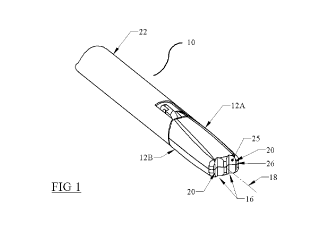
FIG. 1 is an isometric view of a surgical instrument that is arranged and
configured in accordance with certain features, aspects and advantages of an
embodiment of the present invention. The illustrated surgical instrument
presents
a tangential-flat edge type of cutting feature.
FIG. 1B is another isometric view of the surgical instrument of FIG. 1.
FIG. 2 is a top view of the surgical instrument of FIG. 1.
FIG. 2A is an enlarged isometric view of the surgical instrument of FIG. 1.
FIG. 2B is an enlarged isometric view of another surgical instrument that is
arranged and configured in accordance with certain features, aspects and
advantages of an embodiment of the present invention.
FIG. 3 is an isometricview of a surgical instrument that is arranged and
configured
in accordance with certain features, aspects and advantages of an embodiment
of
the present invention. The illustrated surgical instrument presentsa
tangential-flat
- 9 -
CA 03006045 2018-05-22
WO 2017/091445
PCT/US2016/062549
edge type bounded by an angled surface that does not extend a full length of a
jaw.
FIG. 4 is an isometric view of a surgical instrument that is arranged and
configured in accordance with certain features, aspects and advantages of an
embodiment of the present invention. The illustrated surgical instrument
presents
a concentric-flat edge type bounded by an angled surface that does not extend
a
full length of a jaw.
FIG. 5 is an isometric view of a surgical instrument that is arranged and
configured in accordance with certain features, aspects and advantages of an
embodiment of the present invention. The illustrated surgical instrument
presents
a tangential-bowed edge type.
FIG.5B is an isometric view of a surgical instrument that is similar in many
respects to the surgical instrument of FIG.5 but that includes a spur-type
edge
enhancement.
FIG.5C is an isometric view of a surgical instrument that is similar in many
respects to the surgical instrument of FIG.5 but that includes a serration-
type edge
enhancement.
FIG. 6 is an isometric view of a surgical instrument that is arranged and
configured in accordance with certain features, aspects and advantages of an
embodiment of the present invention. The illustrated surgical instrument
presents
a concentric-bowed edge type.
FIG. 7 is an isometric view of a surgical instrument that is arranged and
configured in accordance with certain features, aspects and advantages of an
embodiment of the present invention. The illustrated surgical instrument
presents
a tangential-bowed edge type bounded by an angled surface that does not extend
a
full length of a jaw.
FIG. 8 is a side view of the surgical instrument of FIG. 7, which further
demonstrates the bowed edge.
- 10 -
CA 03006045 2018-05-22
WO 2017/091445
PCT/US2016/062549
FIG. 9 is an isometric view of a surgical instrument that is arranged and
configured in accordance with certain features, aspects and advantage of an
embodiment of the present invention. This embodiment demonstrates the
tangential-bowed edge type with the cutting feature parallel to the grasping
plane
of the surgical instrument.
FIG. lOillustrates an isometric view of a surgical instrument arranged and
configured in accordance with certain features, aspects and advantages of an
embodiment of the present invention. The illustrated surgical instrument
presents
a tangential-bowed edge type with a cutting feature that is angled between a
perpendicular style, such as that of FIG. 5, and a parallel style, such as
that of
FIG. 9.
FIG. 11 is a side view of the surgical instrument of FIG. 10, which further
demonstrates the bowed cutting edge.
FIG. 12is an isometric view of a Maryland-type grasper with jaws presenting a
tangential-bowed edge that is bounded by an angled surface that does not
extend a
full length of a jaw.
FIG.12B is an enlarged view of the Maryland-type grasper of FIG.12, which view
further illustrates the edge construction.
DETAILED DESCRIPTION OF THE INVENTION
FIG. 1 demonstrates a surgical instrument 10. The surgical instrument 10 has a
shaft 22. The shaft 22 has a proximal end (not shown) and a distal end. The
distal
end is configured to be inserted into a body cavity during a surgical
operation.
In some configurations, the distal end of the shaft 22 includes at least one
jaw. In
the illustrated configuration, a pair of jaws 12A,12B are moveably connected
to
the distal end of the shaft 22. In some configurations, one of the pair of
jaws 12A,
12B is fixed relative to the shaft 22 while the other of the pair of jaws 12A,
12B is
moveable relative to the shaft 22. The pair of jaws 12A, 12B are structured to
- 11 -
CA 03006045 2018-05-22
WO 2017/091445
PCT/US2016/062549
grasp and release body tissue, for example, by opening and closing about a
grasping plane 8 (see FIG. 1B).
With reference again to FIG. 1, the jaws 12A, 12B of the surgical instrument
10
preferably include one or more cutting edges 20. In the illustrated
configuration,
contiguous to the cutting edges 20 are formed tissue relief features 16. The
cutting
edges 20 and the tissue relief features 16 are positioned on the distal end of
the
surgical instrument 10. More particularly, in some configurations, the cutting
edges 20 and the tissue relief features 16 are positioned on a distally-facing
end
surface of the surgical instrument 10. Such a placement allows the cutting
edges
20 to engage with tissue through axial movement and/or force. While the
presence of the tissue relief areas 16 may have a small effect on the grasping
surfaces of the jaws 12A,12B, the tissue relief 16 is not believed to
interfere or
significantly impact grasping use or operation of the jaws 12A, 12B.
During surgical use of the surgical instrument 10, the tissue relief
features16 allow
tissue to conform around the cutting edge 20 when the distal end of the
surgical
instrument 10 is pushed axially in a distal direction against body tissue.
With the
tissue conformed around the cutting edge 20, when the surgical instrument 10
is
the rotated around a rotational axis 18, the edge 20 will penetrate, carve or
scrape
tissue that passes against the cutting edge 20, which facilitates penetration
of the
body tissue by the surgical instrument 10.
As shown in the configuration of Figure 1, in some configurations, the largest
profile of the most distal end of the surgical instrument is smaller than or
consistent with the diameter or profile of the portion of the shaft that is
intended to
pass through the tissue of the patient during a surgical operation. In other
words,
the entirety of the pair of jaws 12A, 12B of FIG. 1 have a smaller perimeter
(e.g.,
a smaller vertical cross sectional area) than the most distal portion of the
shaft to
which the pair of jaws 12A, 12B are connected. As such, the portion of the
surgical instrument that first penetrates the tissue does not require a larger
bore
through the tissue than the portion of the shaft or body of the surgical
instrument
that is expected to pass through the bore.
- 12 -
CA 03006045 2018-05-22
WO 2017/091445
PCT/US2016/062549
The portion of the edge that initiates the cutting action is dependent on the
edge
type. Currently, at least four different primary types of cutting edges have
been
developed and will be described in the context of the various embodiments
illustrated herein: (1) tangential-flat; (2) tangential-bowed; (3) concentric-
flat; and
(4) concentric-bowed. In addition, each of the edge types can be provided with
one or more edge enhancements that can be added to each edge. The edge
enhancements are believed to improve cutting performance. For example, with
reference to FIG. 2B, the cutting edge 20 may be enhanced by adding a spur-
like
protrusion 27. This protrusion causes a more torturous path for the tissue
follow,
which enhances the ability of the edge to cut tissue. The edge enhancements
and
edge rounding features described in the context of the various embodiments are
not limited to those embodiments, but may be used where applicable on any of
the
other embodiments and in any desired combination.
The cutting edge 20 can be formed at the junction of two or more surfaces.
With
reference to FIG. 1, the illustrated cutting edge 20 is defined where the
outer
surface 26 and the inner surface 25 intersect. The illustrated cutting edge 20
generally extends vertically while the illustrated jaws 12A, 12B open about a
horizontally extending axis. Accordingly, the cutting edge 20 can extend in a
direction that is perpendicular to the pivot axis of the jaws 12A, 12B. In
some
configurations, the cutting edge can extend in a direction that is parallel to
the
pivot axis of the jaws 12A, 12B. In some configurations, the cutting edge can
extend in a direction that is neither parallel nor perpendicular to the pivot
axis of
the jaws 12A, 12B. In this and other embodiments, the full cutting edge on
each
jaw aligns with the cutting edge on the other jaw to create the composite
cutting
edge.
As described above, the cutting edge 20 can be formed along an included angle
A
(see FIG. 2) that is defined by the converging surfaces 25, 26. The included
angle
A desirably is greater than 45 degrees. In some configurations, the included
angle
A is greater than 55 degrees. In some configurations, the included angle A is
less
than 120 degrees. In some configurations, the included angle is less than 110
degrees. In some configurations, the included angle A is between 45 degrees
and
- 13 -
CA 03006045 2018-05-22
WO 2017/091445
PCT/US2016/062549
less than 120 degrees. In some configurations, the included angle A is within
a
range that has a lower boundary between 45 and 55 degrees and an upper
boundary between 110 and 120 degrees. As described above, a typical surgical
blade has surfaces that converge at an included angle of less than 20 degrees.
With the included angle A being greater than 45 degrees, the resulting edge is
less
prone to cut tissue due to axial impaction or force. More particularly, it is
currently believed that an edge formed by converging surfaces that define an
included angle of greater than 45 degrees will not cut tissue when pressed
against
the tissue under axial forces commonly encountered in surgery or through
inadvertently contacting tissue. Moreover, it is currently believed that
limiting the
included angle to angles less than 120 degrees enables the edge to gain
sufficient
purchase into the tissue to effect the desired tissue removal. Thus, the
included
angle A preferably is large enough that the resulting edge can cut through the
abdominal wall only when subjected to at least 1 pound of axial force while
being
rotated.
In some embodiments, when pressed in an axial direction with a standard
introductory force (i.e., 1 pound) and while being rotated or torqued about
the
rotational or torque axis 18, the surgical instrument 10 is capable of cutting
tissue
due to a combination of: (1)the cutting edge 20 formed by the surfaces 25, 26
that
defined the included angle A; and (2) the tissue relief areas 16 that are
disposed
adjacent to or contiguous with the cutting edge 20. Yet, because of the
structural
configuration and the angling of cutting edge 20 and the placement of the
tissue
relief areas 16, each of the embodiments disclosed herein is believed to be
atraumatic when not presented with sufficient axial forces or when rotated
without
sufficient axial forces being applied.
Referring now specifically to FIG. 2A, the cutting edge 20 is formed an
intersection of three distinct surfaces 23, 25, 26. As described above the
inner
surfaces 25 and the outer surfaces 26 can be joined together to defined the
edge
20. In FIG. 2A, however, the outer portions of the outer surfaces 26 can be
modified using the fillet surfaces 23 such that the fillet surfaces 23 bound
the
outer surfaces 26. In other words, the fillet surfaces 23 define an outer
boundary
- 14 -
CA 03006045 2018-05-22
WO 2017/091445
PCT/US2016/062549
of the outer surface 26. This combination of inner surfaces 25 meeting the
surfaces 26 and the fillet surfaces 23 creating the edge 20 and the at least
one
cutting feature helps to decrease the likelihood of inadvertent cutting of
tissue. In
other words, while this combination of surfaces 23, 25, 26 creates the full
cutting
edge of the jaw, this combination of surfaces 23, 25, 26 also results in the
outer
profile of the distal most end of the surgical instrument having a rounded
contour.
In the embodiment shown in Figure 2A, which is a tangent- flat type, for
example,
the cutting is initiated by the edge 21, which is defined by the intersection
of the
fillet surface 23 and the inner surface. This edge 21 is primarily tangent to
an
imaginary circle that is concentric to the axis of rotation 18 (with the
imaginary
circle demonstrated as 9 in Figure 1B) and lays within plane 4 with is
perpendicular to the axis 18. Because the edge 21 primarily falls within the
plane
4, the edge 21 is considered flat. The fillet surface 23 preferably is round
enough
to not tear the body tissue while the edge 21 is able to cut the body tissue.
Generally, the fillet surface 23 is large enough to cut, but not tear, tissue.
In
summary, the cutting edge 21 in Figure 2A results from the intersection of the
outer surfaces 26 and the fillet surfaces 23 on one side and from the
intersection of
the fillet surfaces 23 and the inner surfaces 25 on the other side. The edge
21
preferably is sharp while all other edges around the tissue relief may be
rounded.
With continued reference to FIG. 2A, the depth ofthe tissue relief 16, which
is
indicated by dimension Z, can be proportional to the length of the full
cutting
edge, which is indicated by dimension X. In some
configurations, the
proportionality of the dimensions Z and X defines a ratio of roughly 0.5:1.
Other
ratios are possible. The width of the tissue relief 16, which is indicated as
dimension Y, can be proportional to the depth of tissue relief 16, which is
indicated by dimension X. In some configurations, the proportionality of the
dimensions Y and X defines a ratio that is generally 1:0.5 or greater. In
other
words, the width can increase while the depth remains constant. These are
general
geometrical guides and will vary somewhat between the various embodiments.
- 15 -
CA 03006045 2018-05-22
WO 2017/091445
PCT/US2016/062549
With reference to FIG 3, a surgical instrument 30 may include an angled
surface
31 located on either or both of the jaws 32A, 32B. The angled surfaces 31 may
bound the cutting edges 34. In the illustrated configuration, the angled
surfaces
31 extend to the distal end of the surgical instrument 30. In some
configurations,
the angled surfaces 31 extend to the distal ends of the jaws 32A, 32B. The
angled
surfaces 31 assist in directing body tissue into the tissue relief features 16
that are
contiguous with the cutting edges 34. Because the angled surfaces 31 are
configured to direct body tissue into the tissue relief features 16, the
angled
surfaces 31 need not extend the full length of the jaws. In the illustrated
configuration, the angled surfaces 31 extend to a middle region along a full
length
of the jaws. In some configurations, the angled surfaces 31 extend between
half
way and the full length of the jaws. In some configurations, the angled
surfaces
31 extend the full length of the jaws. In some configurations, the angled
surfaces
31 extend less than half of the full length of the jaws.
Each angled surface 31 (i.e., the angled surface 31 of the top jaw 32A and the
angled surface of the bottom jaw 32B) may be angled away from the grasping
plane 8 of the jaws (compare with FIG. 1B). In other words, the proximal end
(e.g., the end toward the shaft 22) of the angled surface 31 is spaced further
from
the grasping plane 8 than the distal end (e.g., the end away from shaft 22) of
the
angled surface 31.
The surgical instrument 30 may also include tissue relief areas 36. The tissue
relief areas 36 can be positioned away from the rotational axis 38 of the
surgical
instrument 30 or may cross the rotational axis 38. If the tissue relief area
36
crosses the axis of rotation 38, the tissue relief area 36 will combine to
create one
tissue relief between the edges 34. Of course, in the illustrated
configuration,
neither the tissue relief areas 36 nor the cutting edges 34 cross or intersect
with the
rotational axis 38.
Referring to FIG 4, an embodiment 40 of the surgical instrument demonstrates
the
concentric-flat type of cutting edge 44. In this embodiment, the cutting edge
44 is
a concentric-flat type of cutting edge because the cutting edge 44 is
concentric to
- 16 -
CA 03006045 2018-05-22
WO 2017/091445
PCT/US2016/062549
the imaginary circle or cylinder defined by the circle 9 and falls within the
end
plane 4 (see FIG. 1B, for reference). In the illustrated embodiment, the
cutting
edge 44 is bounded on either end by the edge 43. The edge 43 initiates the
cutting
action and preferably is round enough to not tear tissue. The concentric shape
of
the edge 44 creates a circular cut as the surgical instrument 30 is rotated
about the
rotational axis 48. This embodiment also has an angled surface 41 to better
allow
tissue to conform to the relief feature 46, which is positioned between the
edge 43
and the rotational axis 48. In some configurations, the angle of the angled
surface
41 is between 5 and 20 degrees relative to the clamping plane. Other
configurations also are possible.
Referring to FIG 5, an embodiment 50 of the surgical instrument demonstrates
the
surgical instrument with a tangential-bowed type edge as the cutting edge 54.
The
cutting edge 54 is tangent to an imaginary circle 9 or an imaginary cylinder
defined by the circle 9. The end points of the cutting edges 54 are positioned
within the end plane 4 while the middle region of the cutting edges 54 bow
distally away from the end plane 4. With this type of cutting edge 54, the
cutting
is done primarily at the most distal part 55 of the cutting edge 54, which is
where
the jaws 52A, 52B meet. To enhance the cutting ability of the edge 54, a spur
can
be incorporated. In some configurations, the spur can be placed at location 55
to
create the spur 57 (see FIG. 5B). Alternatively or in addition, a serration
can be
placed at location 55 (see FIG 5C). This serration creates two most distal
areas
59a, 59b on the composite cutting edge. As used herein a full cutting edge is
any
cutting edge that defines the entire cutting feature while a composite cutting
edge
is a cutting feature defined by a combination of aligned cutting edges. The
spurs
57 and the serrations 59a, 59b can be positioned in other locations as
desired.
The embodiment of FIG. 6 demonstrates a surgical instrument 60 that has a
concentric-bowed cutting edge 64. The concentric-bowed cutting edge 64 is
concentric to the imaginary circle 9 or the imaginary cylinder defined by the
imaginary circle. The cutting edge 64 has endpoints that are positioned within
the
end plane 4 while a middle portion of the cutting edge 64 bows distally out of
the
plane with the most distal point 66a being positioned where the jaws 62A, 62B
- 17 -
CA 03006045 2018-05-22
WO 2017/091445
PCT/US2016/062549
meet. The edge enhancements discussed above also can be used in the
embodiment of FIG. 6.
The embodiment of FIG. 7 demonstrates a surgical instrument 70 that has a
tangential-bowed type edge. This embodiment has jaws 72A, 72B that feature a
cutting edge 74 that is contiguous to a tissue relief region 76 and that is
bounded
by an angled surface 71. This embodiment demonstrates rounded tissue relief
edges 73. In other words, the edges 73 of the tissue relief define a curved or
arcuate transition to the distal-facing surface.
The embodiment of FIG 9 demonstrates a surgical instrument 80 where the
cutting
edges of each jaw do not align with each other (e.g., do not intersect or
connect
with each other). In this embodiment, the tissue relief and cutting edge
remain
within their respective jaws. This embodiment is used for jaws that can't have
their inner grasping surface altered or impeded upon in any manner.
The embodiment of FIG 10 shows a device 90 that has a tangent-bowed edge 94
on jaws 92A, 92B that are aligned at point 94a but that do not form a
composite
cutting edge because it is not one aligned continuous cutting edge. This
surgical
instrument is for cases where the inner jaw surface features near the axis of
rotation 98 must be avoided when forming, creating of providing the cutting
edges
94. The configuration also creates different tissue conformity around the
cutting
edge 94 as the surgical instrument is rotated about the axis 98 when compared
with at least some of the devices 50, 80 described above.
The embodiment of FIG. 12 shows a surgical instrument 100 that incorporates
Maryland-style grasper jaws 102A, 102B. The jaws 102A, 102B include one or
more angled surface 101. The angled surfaces 101 bound the cutting edge 104.
The full cutting edge includes the cutting edge 104 and the fillet edge 105.
Also
demonstrated in FIG. 12 and FIG. 12B is a rounded tissue relief edge 106 that
enables that area to be fully atraumatic. As with the other embodiments, this
surgical instrument 100 is pushed axially along and rotated about a rotational
axis
108 to cut tissue.
- 18 -
CA 03006045 2018-05-22
WO 2017/091445
PCT/US2016/062549
The invention has been described in detail with particular reference to
certain
preferred embodiments thereof, but it will be understood that variations and
modifications can be effected within the spirit and scope of the invention.
- 19 -