Note: Descriptions are shown in the official language in which they were submitted.
CA 03006644 2018-05-28
WO 2017/095288 PCT/SE2016/000072
1
HAEMORRHAGE AVOIDING MICROELECTRODE
FIELD OF THE INVENTION
The present invention relates to a proto-
microelectrode which avoids haemorrhage during implantation into
soft tissue, in particular nervous tissue, to a microelectrode
formed from the proto-microelectrode upon implantation, and to
their manufacture and use. The present invention furthermore
relates to sets of such proto-microelectrodes and
microelectrodes, and to their manufacture, implantation, and
use.
BACKGROUND OF THE INVENTION
During implantation of microelectrodes into nervous
tissue by inserting them with their distal end foremost there is
a substantial risk of damaging penetration of blood vessel.
There is also a risk of implanted microelectrodes damaging blood
vessels or cells.
While not being life threatening micro-haemorrhage
resulting from such damage does substantially impair the
analytical or therapeutic performance of the microelectrode or,
if haemorrhage is caused by a microelectrode pertaining to a
set, also the analytical or therapeutic performance of
neighbouring electrodes.
In the event that several microelectrodes are used to
treat a neurological condition or in research, identification of
their disposition in tissue is called for, in particular in
respect of anatomical landmarks such as nuclei or sub-nuclei.
Due to, i.a., their small size it is difficult to determine the
position of single microelectrodes by means of current non-
invasive techniques such as Magnetic Resonance Imaging (MRI),
Computer Tomography (CT), and Doppler techniques. In regard of
an implanted set of microelectrodes it is therefore difficult or
CA 03006644 2018-05-28
WO 2017/095288 PCT/SE2016/000072
2
impossible to determine their positions in respect of each other
and of anatomical landmarks.
Due to their flexibility thin oblong microelectrodes
cannot be implanted by inserting them into soft tissue in the
absence of a support making them sufficiently rigid for this
purpose, they are even more difficult to implant in a desired
disposition in respect of a selected target in the brain like a
nerve cell. The targeted disposition of a set of such
microelectrodes therefore requires measures not readily
available in the art, such as avoiding them damaging a blood
vessel and to spread them out in the tissue in a desired manner.
OBJECTS OF THE INVENTION
A primary object of the present invention is to
provide a microelectrode that does not damage blood vessels
during implantation by insertion into soft tissue and/or during
its disposition in non-static soft tissue or, at least, to
provide a microelectrode of which the risk of damage is
substantially reduced. The soft tissue concerned is primarily
nervous tissue, in particular brain or spinal cord tissue, but
also endocrine tissue such tissue of the pituitary, pineal,
thyroid, parathyroid, and adrenal glands, testes and ovaries
tissue, and pancreatic islets of Langerhans tissue.
Another primary object of the present invention is to
provide a microelectrode that minimizes cell damage.
Another object of the present invention is to provide
a set of such microelectrodes.
Still another object of the present invention is to
provide a means for determining the position of a microelectrode
pertaining to a set of implanted microelectrodes in respect of
any other microelectrode and/or an anatomical landmark like a
particular nerve cell or a nucleus.
A further object of the present invention is to
provide a set of such microelectrodes of which each electrode
can be identified in situ upon insertion.
CA 03006644 2018-05-28
WO 2017/095288 PCT/SE2016/000072
3
A still further object of the invention is to provide
a method of manufacture of the microelectrode of the invention
and of a set comprising two or more microelectrodes of the
invention.
Additional objects of the present invention will
become apparent from the study of the following summary of the
invention, the description of preferred embodiments thereof, and
the appended claims.
SUMMARY OF THE INVENTION
The microelectrode of the invention is of such kind
that it only can be disposed in soft tissue by means of a rigid
support. In this application the combination of microelectrode
and rigid support is termed proto-microelectrode. Upon
implantation the rigid support is removed by dissolution in body
fluid and the microelectrode of the invention is formed in situ.
Soft tissue according to the invention is, in
particular, nervous tissue such as brain tissue, spinal cord
tissue, dorsal root ganglia and peripheral nerves but includes
also body fluid and membranes enclosing such tissue.
According to the present invention is provided a
microelectrode for implantation into soft tissue, in particular
nervous tissue, comprising an oblong electrode body of an
electrically conducting material having a proximal end and a
distal end, wherein the electrode body is covered by a layer of
first insulating material except for at an annular contact
section thereof disposed between a distal section extending from
the distal end in a proximal direction and a proximal section
extending from the annular section to the proximal end, the
layer of insulating material on the distal terminal section
extending distally of the distal end of the electrode body so as
to fully enclose the distal end to form a blunt distal bulge or
wherein the layer of first insulating material on the distal
terminal section is covered or substituted by a layer of second
insulating material forming the bulge. The bulge has a radial
CA 03006644 2018-05-28
WO 2017/095288 PCT/SE2016/000072
4
extension that is substantially greater than the radial
extension of the layer of non-conducting material disposed on
the proximal section.
According to the invention is furthermore provided a
microelectrode for implantation into soft tissue, in particular
nervous or endocrine tissue, comprising an oblong electrode body
of electrically conducting material having a proximal end and a
distal end, the body comprising insulated distal and proximal
sections extending from the distal and proximal ends, the distal
section optionally comprising a bulb at its distal end; wherein
the distal an,0 proximal sections are separated by an annular
section, wherein the proximal section is covered by a layer of
first insulating material and wherein the electrode comprises a
blunt distal terminal bulge formed by one of: layer of first
insulating material on the distal section; layer of first
insulating material on the distal section covered or fully or
partly substituted by a layer of a second insulating material;
layer of first insulating material and/or second insulating
material on the bulb; wherein the bulge has a radial extension
substantially greater than that of the proximal section covered
by layer of first insulating material.
According to a preferred aspect of the invention, a
microelectrode comprises two or more annular sections separated
by insulated section(s). It is preferred for the two or more
annular sections to be arranged in close vicinity of each other,
such as within a section of the electrode body extending axially
by 20 % or 10 % or 5 % or 2 % of the axial extension (length) of
the electrode body.
It is preferred for the microelectrode body to be
rotationally symmetric. It is also preferred for the
microelectrode body to be flexible, in particular resiliently
flexible. According an advantageous aspect of the invention the
electrode body is flexible and curved and of a shape so as to be
of rotationally symmetric form in a straight conformation.
A distal face of the distal bulge can be about
hemispherical or hemi-elliptic or of paraboloid or hyperboloid
CA 03006644 2018-05-28
WO 2017/095288 PCT/SE2016/000072
form, the distal bulge comprising a circular or elliptic base
facing in a proximal direction and being centered on the axis of
rotational symmetry.
According to a preferred aspect of the invention the
5 radial extension of the bulge is preferably greater by 50 % or
more, in particular by 100 % or more, or 1000 % or more than the
radial extension of the layer of first insulating material on
the proximal section.
According to another preferred aspect of the invention
the axial length ratio of the distal section to the proximal
section is preferably 1:2 or 1:5 or more, in particular 1:10 or
more, and may even be 1:20 or more and 1:50 or more. It is
preferred for the axial length of the annular section to be 10 %
or less, in particular 5 % or 2 % or 1 % or less of the length
of the electrode body.
According to a further preferred aspect of the
invention an insulating material on the distal section is of a
material different from that on the proximal section. It is
preferred for an insulating material on the distal section to be
covered by a layer of material capable of swelling in contact
with aqueous body fluid, in particular swelling at body
temperature to reach an equilibrium swollen state of a radial
extension greater by a factor of 2 or 5 and even of 10 or more
than that of the non-swollen material. The material capable of
swelling has a preferred Bloom strength of from 200 to 300 or
more, in particular from 200 to 350, most preferred of about
300.
According to another preferred aspect of the invention
an insulating material on the distal section is resilient, in
particular resilient by comprising closed gas-filled cells.
According to a preferred aspect of the invention a
bulge material or insulating material comprises an agent to
improve the visibility of a microelectrode of the invention in
MRI, for instance an agent comprising or consisting of
ferromagnetic particles.
CA 03006644 2018-05-28
WO 2017/095288 PCT/SE2016/000072
6
The microelectrode of the invention can be attached or
be attachable to flexible insulated electrical conductor in a
conducting manner, the attachment is to the electrode body being
preferably at or near the proximal end thereof.
The electrode body of the microelectrode of the invention
consists of a metal or comprises a metal or consists of or
comprises an electrically conducting polymer.
According to the present invention is also disclosed a
proto-microelectrode comprising or consisting of the
microelectrode of the invention and a biocompatible solid
support material. The support material is attached to the
microelectrode in a manner to stabilize it sufficiently so as to
allow implantation of the microelectrode into soft tissue by
insertion with its distal end foremost. According to an
important aspect of the invention the support material is rigid;
it is furthermore dissolvable in body fluid. The support
material may enclose the microelectrode partially or fully.
The support material can consist of or comprise
carbohydrate and/or protein and optionally comprises a
pharmacologically active agent selected from the group
consisting of coagulant, anticoagulant, antibiotic, osmotic
pressure adjusting agent, anti-inflammatory agent, nutrient,
factor stimulating growth, factor stimulating cell
differentiation, hormone.
According to the present invention is furthermore
disclosed a set of axially aligned proto-microelectrodes of the
invention sharing the support material, that is, a proto-set of
microelectrodes. The terms proto-set of microelectrodes and set
of proto-microelectrodes thus are used indiscriminatingly in
this application. The microelectrodes can be disposed in the
proto-set in parallel or in a mode fanning out in a distal
direction, such as by an angle of 10 degrees or 15 degrees in
respect of a centrally disposed electrode used as reference. A
microelectrode of the proto-set can comprise one or more
additional subsections of second insulating material of a radial
CA 03006644 2018-05-28
WO 2017/095288 PCT/SE2016/000072
7
extension substantially greater than that of the insulating
layer on the proximal section. The subsections can be disposed
on the electrode body so as to be comprised by the distal and/or
the proximal section. The subsections can be fully separated or
be separated by intermediate sections of smaller radial
extension, which radial extension is, however, greater than the
radial extension of the first insulating layer.
According to a preferred aspect of the invention a
proto-set of microelectrodes can comprise an expandable material
capable of swelling in body fluid. The expandable material is
disposed between two or more microelectrodes in the proximity of
their distal ends. According to an important aspect of the
invention the dissolution rate of the expandable material in
body fluid is lower than the dissolution rate of the support
material. The expandable material has a preferred Bloom strength
of from 80 to 200, in particular of about 100 to 150, that is, a
Bloom strength inferior to that of the material capable of
swelling in contact with aqueous body fluid disposed on a layer
of first and/or second insulating material of a distal bulge.
According to a further preferred aspect of the
invention is disclosed a proto-set wherein a distal bulge of
insulating material of one microelectrode is of non-spherical
form. The distal bulge is disposed on a distal terminal section
of the electrode and in a manner so as to slant radially
outwardly in respect of the electrode body axis. This makes the
distal bulge display a front face, which is the face exhibited
by the bulge in a proximal view, that slants radially outwardly.
To allow identification by radiative means of single
electrodes pertaining of a set upon implantation of the
corresponding proto-set and dissolution of the support material,
the electrodes are provided with different numbers of
subsections of substantially greater diameter than that of the
insulating layer on the proximal section differing in their
radial extension. Alternatively the microelectrodes of a set can
be distinguished by making the composition of the insulating
CA 03006644 2018-05-28
WO 2017/095288 PCT/SE2016/000072
8
material forming their distal bulges and/or forming one or more
of said subsections differ.
Furthermore disclosed herein is the use of a
microelectrode, a proto-microelectrode and a set of proto-
microelectrodes of the invention in a method comprising
electrical stimulation of cells, in particular nerve cells, and
in a method comprising monitoring the electrical activity of
such cells.
Additionally disclosed herein is the use of a
microelectrode, of a proto-microelectrode, and of a of set of
proto-microelectrodes of the invention in a method of treating a
condition comprising an aberrant function of cells in brain
tissue, spinal cord tissue, dorsal root ganglia and peripheral
nerves by electrical stimulation and in monitoring the
electrical activity of cells in such tissue.
A microelectrode pertaining to a set or proto-set of
microelectrodes of the invention need not be of equal length.
A proto-set and a corresponding set of the invention thus can
comprise two or more microelectrodes of different length.
To facilitate implantation into soft tissue by
insertion a proto-microelectrode or a proto-set of
microelectrodes of the invention can comprise a friction
reducing coat on the support material.
The invention will now be explained in more detail by
reference to a number of preferred embodiments thereof
illustrated in a rough drawing, of which the figures are not to
scale for reasons of clarity. In general, the radial extension
of an illustrated microelectrode is exaggerated in relation to
its axial extension. The illustrated microelectrodes are
rotationally symmetric or at least substantially rotationally
symmetric.
DESCRIPTION OF THE FIGURES
Fig. la is an axial section through a first rotationally
symmetric embodiment of the microelectrode of the invention;
CA 03006644 2018-05-28
WO 2017/095288 PCT/SE2016/000072
9
Fig. lb is an axial section through a second rotationally
symmetric embodiment of the microelectrode of the invention;
Fig. lc is an axial section through a third rotationally
symmetric embodiment of the microelectrode of the invention;
Fig. id is an axial section through a fourthrotationally
symmetric embodiment of the microelectrode of the invention;
Fig. le is an axial section through a fifth rotationally
symmetric embodiment of the microelectrode of the invention;
Fig. if is an axial section through a sixth rotationally
symmetric embodiment of the microelectrode of the invention;
Fig. 2a is an axial section through a first embodiment of a set
of proto-microelectrodes of the invention comprising three
different rotationally symmetric microelectrodes;
Fig. 2b is an axial section through a second embodiment of a set
of proto-microelectrodes of the invention comprising three
different rotationally symmetric microelectrodes;
Fig. 3 is an axial section through a set of three
microelectrodes of the invention of which one is rotationally
symmetric and the other two are rotationally symmetric except
for at their distal end portion;
Fig. 3a is a set of proto-microelectrodes corresponding to the
set of microelectrodes of Fig. 3, enclosed in a rigid support
material dissolvable in body fluid, in the same axial (with
regard to the set and to each individual electrode) section;
Fig. 3b illustrates the disposition of the set of
microelectrodes of Fig. 3 formed upon the insertion of the set
of proto-microelectrodes of Fig. 3a into nervous tissue and
dissolution of the support material by body fluid, in the same
axial section;
CA 03006644 2018-05-28
WO 2017/095288 PCT/SE2016/000072
Fig. 3c illustrates the disposition of the electrodes of the set
of Fig. 3 upon inserting them further into nervous tissue by an
axial force acting on their proximal ends, in the same axial
(with regard to the bundle and to each individual electrode)
5 section;
Fig. 4a illustrates another embodiment of the set of proto-
microelectrodes of the invention enclosed in a rigid support
material, which is dissolvable in body fluid, further comprising
sections of a material forming a gel on contact with body fluid,
10 disposed between the distal terminal portions of the electrodes,
in an axial (R-R, Fig. 4b; with regard to the set and to each
individual electrode) section;
Fig. 4b is a radial section (S-S, Fig. 4a) through the set of
proto-microelectrodes of Fig. 4a;
Fig. 4c is a radial section through another set of proto-
microelectrodes (not shown in axial section), the section
corresponding to that of Fig. 4b;
Fig. 5 illustrates a further embodiment of the set of proto-
microelectrodes of the invention enclosed by a rigid support
material dissolvable in body fluid, in an axial section (Q-Q,
Fig. 5a);
Fig. 5a illustrates the embodiment of Fig. 5 in a radial section
(P-P, Fig. 5);
Fig. 5b illustrates a still further embodiment of the set of
proto-microelectrodes of the invention in a radial section
corresponding to that of Fig. 5a upon dissolution of the rigid
support material and expansion of a gel forming material
covering distal terminal bulge portions of the electrodes,
resulting in forcing them radially apart;
Fig. 6 illustrates a set of five proto-microelectrodes of the
invention attached at their underside to one face of a gelatin
sheet, in a top view;
CA 03006644 2018-05-28
WO 2017/095288 PCT/SE2016/000072
11
Fig. 6a is a transverse section L-L through the set of proto-
microelectrodes of Fig. 6;
Fig. 7 is an axial section through a seventh rotationally
symmetric embodiment of the microelectrode of the invention;
Fig. 7a is an axial section through a eight rotationally
symmetric embodiment of the microelectrode of the invention;
Fig. 7b is an axial section through a ninth rotationally
symmetric embodiment of the microelectrode of the invention;
Fig. 7c is an axial section through a tenth embodiment of the
microelectrode of the invention, which is rotationally symmetric
except for its distal head or bulge;
Figs. 8, 8a are axial sections through the microelectrode of
Fig. 7b comprising a layer of gel-forming material on its distal
terminal bulge prior and upon contact with aqueous body fluid,
respectively;
Figs. 9 - 9c illustrated the process of insertion of the proto-
set of microelectrodes of the invention into soft tissue;
wherein:
Fig. 9 is an axial section of a proto-set of two
microelectrodes;
Fig. 9a shows the set of microelectrodes formed upon insertion
of the proto-set into soft tissue and dissolution of the glue
connecting the microelectrodes, in the same section;
Fig. 9b shows the set of microelectrodes upon swelling of a
layer of a material expandable in contact with aqueous body
fluid disposed on their heads forcing their distal portions
apart, in the same section;
Fig. 9c shows the set of microelectrodes of Fig. 9b upon
dissolution/degradation of the expandable material further
inserted into the tissue, in the same section.
CA 03006644 2018-05-28
WO 2017/095288 PCT/SE2016/000072
12
DESCRIPTION OF PREFERRED EMBODIMENTS
EXAMPLE 1. Microelectrodes
Figs. la - if and 7 - 7b illustrate nine embodiments
of the microelectrode of the invention.
The microelectrode 1 of Fig. la comprises an oblong
cylindrical electrode body 2 of metal or electrically conducting
polymer. The electrode body 2 is electrically insulated except
for an annular zone 5 disposed near its distal end from which it
is separated by a sphere 4 of polymer material surrounding and
enclosing the distal end of the electrode body 2. Starting at
the proximal border of the annular zone 5 the electrode body 2
is electrically insulated by a thin layer 3 of polymer material,
which extends to and encloses the proximal end of the electrode
body 2. The proximal end of the electrode body 2 is in
electrical contact with an insulated 6 flexible wire 7 attached
to the electrode body 2 by solder 8 or welding.
The microelectrode 11 of Fig. lb differs from that 1
of Fig. la by the sphere 4 being substituted by a pear-like
distal terminal bulge 14 of polymer material. Reference numbers
12, 13, 15, 16, 17, and 18 designate elements/features
corresponding to those of reference numbers 2, 3, and 5 through
8 of Fig. la. In general, the polymer material used for forming
a bulge is a material with insulating properties although these
properties are only effective if the bulge is disposed on the
naked electrode body 2 and not the thin insulating layer 3
covering sections of the electrode body 2.
The microelectrode 21 of Fig. lc differs from that 1
of Fig. la by the thin layer of insulating material 23, 23' on
the electrode body 22 being interrupted by near the distal end
to form a proximal portion 23 and a distal portion 23'
delimitating an annular electrode zone 25 disposed between them,
the distal portion 23', a sphere 24 of insulating polymer
material surrounding and enclosing the distal end of the
electrode body 22 and a terminal section of the distal portion
23' of insulating material.
CA 03006644 2018-05-28
WO 2017/095288 PCT/SE2016/000072
13
The microelectrode 31 of Fig. id differs from the
microelectrode 1 of Fig. la by a second sphere of polymer
material disposed around the proximal end of the thin layer of
insulating polymer material 33 extending from the proximal end
of the electrode body 32 to the proximal border of the annular
electrode zone 35.
The microelectrode 41 of Fig. le differs from that 31
of Fig. id by a third sphere 44" arranged in addition to the
first 44 and second 44' sphere, the third sphere 44" being
disposed proximally of the second sphere 44' on a portion of the
electrode body 42 covered by a thin layer 43 of insulating
material. Reference numbers 45-48 designate elements/features
corresponding to elements/features 35-38 of the embodiment
The microelectrode 51 of Fig. lf differs from that (1d) of Fig.
la by four partially merged spheres 54', 54", 54"', 54"" of
same diameter disposed on the electrode body 52, the first
(distal) sphere 54' proximally delimiting the annular electrode
zone 55 whereas the fourth sphere 54'"' surrounds and seals the
distal end of the thin insulating layer 53 on the proximal
portion of the electrode body 52. Features 56-58 correspond to
features 36-38 of the embodiment of Fig. ld.
The microelectrode 230 of Fig. 7 comprises an oblong
cylindrical electrode body 222 of metal or electrically
conducting polymer. The electrode body 222 is insulated by a
polymer layer 223, 223' except for an annular zone 225 disposed
near its distal end. The distal end of the electrode body 222 is
radially widened so as to form a bulb 229. The bulb 229 and a
short cylindrical section of the electrode body 222 extending
between the bulb 229 and the annular zone 225 are covered by a
distal section 223' of the polymer layer so as to form a bulge
223', 229. A proximal section 223 of the polymer layer extends
from the annular zone 225 to the proximal end of the electrode
body 222, and surrounds and encloses it. The proximal end of the
electrode body 222 is in electrical contact with an insulated
226 flexible wire 227 attached to the electrode body 222 by
solder 228 or by welding. The bulb 229 improves the adherence of
CA 03006644 2018-05-28
WO 2017/095288
PCT/SE2016/000072
14
the polymer layer 223' to the distal portion of the electrode
body 222, which is particularly important in the event the
electrode 230 is withdrawn from the tissue; this minimizes the
risk of the polymer layer 231' coming loose and being left in
the tissue.
The microelectrode 231 of Fig. 7a differs from the
microelectrode 230 of Fig. 7 by the bulge 231', 224, 229 being
formed by the bulb 229 covered by an insulating material 224
different from the insulating material 231 on the proximal
section and insulating material 231' of same kind as that of the
proximal section disposed on a short distal section extending
between the distal end of the annular zone 225 and the bulb 229.
The microelectrode 232 of Fig. 7b differs from the
microelectrode 230 of Fig. 7 by the bulge 224, 229 being formed
by the bulb 229 covered by an insulating material 224 different
from the insulating material 231 on the proximal section and
extending to the distal end of the annular zone 225.
The microelectrode 233 of Fig. 8 differs from that of Fig. 7b by
the insulating material 224 of the bulge 224, 229 being covered
by a layer 221 of dry gelatin. Upon contact with aqueous body
fluid the gelatin layer 221 absorbs water and is transformed to
a gel forming an expanded gellous layer 221' on the insulating
material 224. The gellous layer 221' is not permanent but is
dissolved or degraded over time, the rate of dissolution/
degradation being dependent on its physical and chemical
properties, such as its degree of crosslinking.
EXAMPLE 2. Sets of proto-microelectrodes
The set of proto-microelectrodes 60 of the invention
shown in Fig. 2a comprises three microelectrodes A, B, C, which
are identical with the microelectrodes 1, 31, 41, respectively,
of Figs. la, id, and le. Except for their insulated flexible
wires 6, 36, 46 the microelectrodes A, B, C are fully embedded
in a rigid support material 61 that is soluble in aqueous body
fluid. The layer of rigid support material embedding the set of
CA 03006644 2018-05-28
WO 2017/095288 PCT/SE2016/000072
microelectrodes A, B, C is rotationally symmetric about an axis
(not shown) corresponding to that of the central microelectrode
C. The material 61 consists or comprises biocompatible
carbohydrate or protein that is soluble in body fluid. The
5 microelectrodes A, B, C are aligned in parallel and of about
same length. It is however within the ambit of the invention to
use microelectrodes of different length.
Another embodiment of the set of proto-microelectrodes
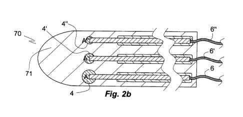
of the invention is shown in Fig. 2b. The proto-set 70 comprises
10 three microelectrodes A, A', A" of about equal length disposed
in parallel. The microelectrodes A, A', A" are identical with
the microelectrode 1 of Fig. la, except for the spheres 4', 4"
of microelectrodes A', A" being consecutively smaller than the
sphere 4 of microelectrode A. Except for their insulated
15 flexible wires 6, 6', 6" attached at their proximal ends the
microelectrodes A, A', A" are embeddedin a rotationally
symmetric rigid support material in a manner that the layer of
support material 71 is centred about the central microelectrode
A', that is, has an axis of rotation superimposed on that of the
axis of rotationally symmetric microelectrode A'. Useful layer
71 materials comprise low or medium molecular weight
carbohydrate or protein.
A further set 110 of microelectrodes is shown in Fig.
3a. Strictly speaking the set 110 is a proto-set of
microelectrodes. The proto-set 110 comprises three
microelectrodes 80, 90, 100 in the disposition of Fig. 3. The
centrally disposed microelectrode 80 is of same design as
microelectrode 41 of Fig. le except for its distal bulge 84
being ellipsoid in longitudinal section instead of the circular
section of distal bulge 44 of Fig. le. The long axis of the
distal bulge 84 K-K is aligned with the axis of electrode body
82, whereas the long axes J-J, L-L of the bulges 94 and 104 are
not aligned with the axes I-I, M-M of electrode bodies 92, 102
but form an angle a, a' of about -300, 30 with them.
CA 03006644 2018-05-28
WO 2017/095288 PCT/SE2016/000072
16
The microelectrodes E, F, G of Fig. 3 are embedded in
a layer 111 of same design and composition as that of the proto-
set of microelectrodes of Figs. 2a, 2b so as to form the proto-
set of microelectrodes 110 illustrated in Fig. 3a. Except for
its rear terminal face 113 the layer 111 is covered by a thin
outer layer 112 of a friction reducing material, which dissolves
or disintegrates upon insertion of the layer 111 of material
soluble in body fluid comprising the proto-set of
microelectrodes 110 into nervous tissue. The outer layer 112 can
also protect the layer 111 material from premature dissolution
prior to completion of insertion.
Upon insertion into nervous tissue 113 and dissolution
of the layer 111 the state shown in Fig. 3b is reached. The
microelectrodes E, G, F of the set formed in situ are disposed
in the tissue 113 in about the same disposition as that of Fig.
3a.
During their further insertion by a force X acting
about axially on each microelectrode E, F, G in a distal
direction as indicated by arrow X, the centrally disposed
microelectrode E is displaced in an axial, distal direction,
whereas the flanking microelectrodes F and G are deflected away
from the central electrode F as indicated by arrows Y, Y' to be
disposed in the tissue in a skew axial direction. The deflection
is caused by bulges 94, 104, which are disposed asymmetrically
in regard of axis K-K of the central microelectrode axes E. This
design allows to fan out peripherally disposed, axially aligned
or about aligned members of a proto-set of microelectrodes once
the support material immobilizing the set E, F, G has been
dissolved upon insertion into nervous tissue. This arrangement
provides the additional advantage of a small wound caused by the
insertion allowing disposition of the distal microelectrode end
portions over an area substantially wider than the wound area,
that is, the area of the wound transverse to the direction of
insertion of the bundle into nervous tissue.
The third embodiment of the proto-set of
microelectrodes of the invention shown in Figs. 4a and 4b
CA 03006644 2018-05-28
WO 2017/095288 PCT/SE2016/000072
17
comprises three microelectrodes A, A', A" aligned in parallel
and in a spaced configuration, of which microelectrode A is
identical with that of Fig. la whereas the other microelectrodes
A' and A" have distal terminal bulges 124', 124" of differing
and, in respect of bulge 124 of microelectrode A, reduced
diameter. In the space between distal terminal portions the
pairs A, A' and A', A" are disposed inserts 127, 127' of a
material, which swells on contact with body fluid. The axial
extension of the inserts 127, 127' comprises part of that of the
bulges 124, 124', 124" and of the annular, insulation free
electrode body sections 125, 125', 125". Upon insertion of the
proto-set 120 into nervous tissue and dissolution of the matrix
121 the inserts 127, 127' are contacted by body fluid, which
they take up while swelling; their swelling pushes the distal
terminal portions including the bulges 124, 124', 124" of the
microelectrodes A, A', A" apart, so that their further
insertion into the tissue results in pushing the outer
microelectrodes A, A" away from the central microelectrode A'
so as to make their distal portions fan out. The dispositional
effect thus is similar to that of the embodiment of Fig. 3a. The
position of each of the microelectrodes A, A', A" in neural
tissue can be identified by tissue penetrating imaging
techniques due to them differing in respect of the size of their
distal terminal bulges 124, 124' 124". Insertion of the
microelectrode proto-set 120 into neural tissue is accomplished
by, for instance, use of a tongue-like instrument holding the
bundle 120 at opposite lateral rear indentations by means of
tongues 128, 129.
Fig. 4c illustrates a proto-set 130 of
microelectrodes, which is a variation of the embodiment of Figs.
4a, 4b. The proto-set 130 of microelectrodes comprises five
microelectrodes covered by a rotationally symmetric support
material 131 that is dissolvable in body fluid and of same shape
as that of Figs. 4a, 4b. The about centrally disposed
microelectrode 134 is surrounded by four microelectrodes 134',
134", 134"', 134"", all of which are fully embedded in the
CA 03006644 2018-05-28
WO 2017/095288 PCT/SE2016/000072
18
support material 131. A distal terminal portion of the central
microelectrode 134 is surrounded by a gel forming layer 137,
which extends to the peripherally disposed microelectrodes 134,
134", 134"', 134"" and into the spaces between pairs of
them. Similar to the microelectrode proto-set 120 of Figs. 4a,
4b insertion of the proto-set 130 of microelectrodes immobilized
in the layer of rigid support material dissolvable in body fluid
into neural tissue results in the dissolution of the support
material 131 followed by uptake of water by the gel forming
layer 137, which results in its radial expansion combined with
radial displacement of the terminal distal portions of the
peripheral microelectrodes 134', 134", 134"', 134"" so as to
make them fan out radially.
The fourth embodiment 140 of the proto-set 140 of
microelectrodes of the invention shown in Figs. 5 and 5a
comprises five microelectrodes H, H', H", H"', H"" of
identical design embedded in a parallel aligned disposition in a
rotationally symmetric layer of rigid support material 141 that
is dissolvable in body fluid. At its proximal end each of the
microelectrodes H, H', H", H"', H"" is provided with a
flexible insulated wire 146 for establishing electrical
communication with an implanted apparatus for electrode control
(not shown). Only wire 146 is exemplarily identified in Fig. 5.
The microelectrodes H, H', H", H"', H"" are of same design
as the microelectrode of Fig. la except for their distal
terminal bulge 144 (only identified for the central
microelectrode H) being covered by a layer 149 (only identified
for the central microelectrode H) of a material capable of
forming a gel on contact with body fluid.
Upon insertion of the proto-set 140 of microelectrodes
embedded in the layer 141 of rigid support material dissolvable
in body fluid into neural tissue 147 the support material 141 is
dissolved and the gel forming layers 149 on the terminal bulges
144 of microelectrodes H, H', H", H"', H"" are contacted by
body fluid, which makes them expand and merge, as shown in Fig.
5b. The expansion of the gel 149* results in the distal terminal
CA 03006644 2018-05-28
WO 2017/095288 PCT/SE2016/000072
19
portions of the thus transformed peripherally disposed
microelectrodes h', h", h"', h"" to be deflected radially
outwardly from the central microelectrode h (identifies
microelectrode H minus gel forming layer 149), so that their
further insertion into neural tissue (not shown) results in
making them fan out radially. Reference numbers 142, 143, 145
identify exemplarily for all electrodes H through H"" an
electrode body, an insulating layer on the electrode body, and
an annular non-insulated portion of the electrode body in
contact with neural tissue.
The proto-set of five microelectrodes 150, 150',
150", 150"', 150"" of the invention shown in Fig. 6 is
immobilized on the upper face a gelatin sheet 151 in a
disposition with the front ends of the microelectrodes fanning
out in a distal direction. Their immobilization is accomplished
by moistening the area selected for disposing the
microelectrodes on the gelatin sheet 151 to form a gel-like
surface, disposing the microelectrodes 150, 150', 150", 150'",
150'"' on the gel-like surface in the desired disposition and
pushing them against the surface, then drying to produce a
permanent adhesive connection indicated exemplarily in Fig. 6a
for microelectrode 150" by reference number 152. The
microelectrodes 150, 150', 150", 150"', 150"" are of same
kind as the microelectrode of Fig. la. The thus immobilised set
can be disposed, for instance, on either side of the dura mater
where it is contacted by body fluid. Upon transformation of the
dry gelatin sheet 151 into a gel and the dissolution of the gel
the set of electrodes 150, 150', 150", 150'", 150"" is
disposed in or on the tissue in the desired configuration with
each electrode being free to move or be displaced independent of
the other electrodes. Since the dry gelatin sheet 151 is not
flexible, it need to be made humid to allow bending it so as to
make it abut a curved tissue surface. Alternatively the set of
electrodes 150, 150', 150", 150"', 150"" can be disposed on
a sheet of dry gelatin already bent in a manner to make it fit
with a particular tissue surface.
CA 03006644 2018-05-28
WO 2017/095288
PCT/SE2016/000072
EXAMPLE 3. Implantation of a preferred embodiment of a proto-set
of microelectrodes
Fig. 9 shows a rotationally symmetric (axis Y-Y)
5 proto-set 300 of two microelectrodes of Fig 8., comprising
oblong, rotationally symmetric (axes V-V, W-W) gold electrode
bodies 322, 422 insulated by layers 323, 423 of Parylene C,
distal gold bulbs 329, 429 integral with the bodies 322, 422,
polyurethane caps 321, 421 covering the bulbs 329, 429, and
10 sections 325, 425 free of insulation disposed between the
insulated proximal and distal sections. The microelectrodes of
the set and a centrally disposed cannula 428 comprising a
through passage 427 are enclosed by and held by a glue matrix
426 that is easily dissolved by aqueous body fluid, for instance
15 gelatin or glucose. To prevent premature dissolution and improve
gliding properties the matrix 426 can be provided with a thin
wax coat melting slightly above body temperature (not shown).
Electrode axes V-V. W-W are disposed in parallel with the prot-
set axis Y-Y.
20 Within a short time upon insertion, such as within a
minute or a couple of minutes, the glue matrix 326 is dissolved.
It can be removed by sucking it up through the cannula passage
427. Fig. 9a shows the set 301 of electrodes formed upon
dissolution of the matrix 326 and removal of the aqueous
solution formed. Reference numbers refer to same elements as in
Fig. 9.
Holding the set 301 in the disposition of Fig. 9a for
an extended period of time, such a for 30 min or more, allows
the layer of expandable material 324, 424 on the caps 321, 421
to take up water from aqueous body fluid and to thereby expand
so as to form a gel 424'. Expansion of portions of the layer
324, 424 disposed between the caps 321, 421 pushes the caps 321,
421 radially away from the central axis Y-Y, thereby changing
the parallel disposition of the electrode bodies 322, 422 to an
angular one (angle 13) opening up in a distal direction. After
dissolution/degradation of the gel 424 or after substantial
CA 03006644 2018-05-28
WO 2017/095288 PCT/SE2016/000072
21
softening of the gel 424 the electrodes 321, 322, 323, 329; 421,
422, 423, 429 can be further inserted (from depth D1 to depth
D2, distance d) into the tissue along their axes V-V, W-W, so as
to increase the distance dd between their insulation-free
sections 325, 425 (Fig. 9c).
Materials
The electrode body is preferably of a noble metal or
an alloy of noble metals or comprising noble metals such as
gold, silver, platinum, iridium, but other biologically
acceptable metals such as stainless steel and tantalum can also
be used as well as gold plated copper. The metallic surface of
the electrode body can be modified by applying a layer of
another metal or metal alloy or a layer comprising or consisting
of an electrically conducting non-metallic material such as
titanium nitride, iridum oxide, platinum grey.
Alternatively the electrode body may consist of or
comprise an electrically conducting polymer. Alternatively the
electrode body can be made of a core of nonconductive polymer
material coated with a metal, in particular a noble metal. The
annular portions of the electrode body lacking insulation may be
advantageously provided with surface enlarging elements or
structures such as a roughened surface, forests of conducting
nanowires, for instance carbon nanowires, or be porous. Surface
enlarging structures of this kind will reduce the impedance of
the electrode body. The electrode body can be connected with a
control unit by an insulated separate electrical conductor
coupled between the rear end of the electrode and the control
unit or by the electrode body itself, a rear section thereof
functioning as a coupling conductor. In such case the rear
section is electrically insulated.
As a material for insulation of the electrode body
suitable biocompatible polymer materials of all kinds can be
used. The layer of insulating material can be applied by
deposition of a monomer from the gas or liquid phase followed by
CA 03006644 2018-05-28
WO 2017/095288 PCT/SE2016/000072
22
polymerization on the electrode body or, such as for providing a
silicone or Parylene C coat, dipping of the electrode body into
a polymer or prepolymer solution, withdrawing it from the
solution, and evaporating the solvent, optionally allowing a
prepolymer to settle, is also useful. Suitable polymers comprise
biocompatible types of polyurethane, polyurethane urea,
polyimide, and Teflon . An electrically insulating material of
this kind can also be used for forming the bulge of the
invention by locally applying a larger amount of it on the
electrode body. Alternatively a polymer material different from
that used for electrical insulation of the electrode body may be
used such as, for instance, polyester or polyimide. The material
of the bulge can comprise a visibility enhancing agent to
improve its visibility in imaging techniques such as MRI or
ultrasound.
The biocompatible electrode support material of the
invention, which is soluble in body fluid, consists of or
comprises water soluble carbohydrate or protein as well as their
mixtures. A suitable rigid biocompatible material of this kind
of which the dissolution rate can be controlled is obtained by
repeatedly boiling and cooling an aqueous solution of a sugar or
a mixture of sugars selected from sucrose, lactose, mannose,
maltose and an organic acid selected from citric acid, malic
acid, phosphoric acid, tartaric acid. By selecting particular
combinations of sugar(s) and organic acid(s) it is possible to
obtain materials with different dissolution times. Gelatin and
various kinds of natural gums that are soluble in body fluid may
also be used as a rigid biocompatible material. The
biocompatible electrode support material of the invention can be
applied on an electrode or on a set of electrodes by dipping
it/them into an aqueous solution of the material followed by
drying, which procedure can be repeated until a layer or shell
of desired thickness has been formed on the electrode or set of
electrodes. Alternatively spray coating can be used to apply
CA 03006644 2018-05-28
WO 2017/095288 PCT/SE2016/000072
23
layers of support material. Several layers can be applied in
sequence, a drying step following upon each application.
Further useful electrode support materials include:
arabinogalactan; arabinoxylan; galactan; galactomannan;
lichenan; xylan; cellulose derivatives such as
hydroxymethylpropyl cellulose and carboxymethyl cellulose;
chitosan; gum Arabic; pullulan; polyvinylpyrrolidone; karaya
gum; pectin; xanthane gum; tragacanth; alginic acid; heparan
sulfate; RGD peptide; polyethylene oxide; chrondroitin sulfate;
keratan sulfate; VEGF biomimetic peptide; perlecan (heparan
sulfate proteoglycan 2); modified heparin; fibrin fragment; with
the proviso that they are of sufficiently low molecular weight
to make them soluble in body fluid.
The expandable material capable of swelling in body fluid for
disposition between two or more microelectrodes in the vicinity
of their distal ends in a proto-set microelectrodes of the
invention is a material of a lower dissolution rate than the
electrode support material. It is, for instance cross-linked
gelatin or cross-linked hyaluronic acie. Other materials capable
of forming protein gels can also be used, such as whey protein,
soy protein, casein, but also one of the following agents:
arabinogalactan; arabinoxylan; galactan; galactomannan;
lichenan; xylan; cellulose derivatives such as
hydroxymethylpropyl cellulose; chitosan; gum Arabic;
carboxyvinyl polymer; sodium polyacrylate; carboxymethyl
cellulose; sodium carboxymethyl cellulose; pullulan;
polyvinylpyrrolidone; karaya gum; pectin; xanthane gum;
tragacanth; alginic acid; polyoxymethylene; polyimide;
polyether; chitin; poly-glycolic acid; poly-lactic acid; co-
polymer of poly-glycolic and poly-lactic acid; co-polymer of
poly-lactic acid and polyethylene oxide; polyamide;
polyanhydride; polycaprolactone; maleic anhydride copolymer;
poly-hydroxybutyrate co-polymer; poly(1,3-bis(p-
carbophenoxy)propane anhydride); polymer formed by co-
polymerization with sebacic acid or with poly-terephthalic acid;
poly(glycolide-co-trimethylene carbonate); polyethylene glycol;
CA 03006644 2018-05-28
WO 2017/095288 PCT/SE2016/000072
24
polydioxanone; polypropylene fumarate; poly(ethyl glutamate-co-
glutamic acid); poly(tert-butyloxy carbonylmethyl glutamate);
poly-caprolactone; poly(caprolactone-co-butylacrylate); poly-
hydroxybutyrate and copolymers thereof; poly(phosphazene);
poly(D,L-lactide-co-caprolactone); poly(glycolide-co-
caprolactone); poly(phosphate ester); poly(amino acid);
poly(hydroxybutyrate); polydepsidpeptide; maleic anhydride
copolymer; polyphosphazene; polyiminocarbonate; poly[(7.5%
dimethyl-trimethylene carbonate)-co-(2.5% trimethlyene
carbonate)]; polyethylene oxide; hydroxypropylmethylcellulose,
poly(ethylene-co-vinyl acetate); isobutylene-based copolymer of
isobutylene and at least one other repeating unit such as butyl
acrylate: butyl methacrylate; substituted styrene such as amino
styrene, hydroxy styrene, carboxy styrene, sulfonated styrene;
homopolymer of polyvinyl alcohol; co-polymer of polyvinyl
alcohol and at least one other repeating unit such as a vinyl
cyclohexyl ether; hydroxymethyl methacrylate; hydroxyl- or
amino-terminated polyethylene glycol; acrylate-based copolymer
such as methacrylic acid, methacrylamide, hydroxymethyl
methacrylate; ethylene vinyl alcohol copolymer; silicone based
copolymer of aryl or alkyl siloxane and at least one repeating
unit; polyurethane; heparan sulfate; RGD peptide; polyethylene
oxide; chrondroitin sulfate; YIGSR peptides; keratan sulfate;
VEGF biomimetic peptide; perlecan (heparan sulfate proteoglycan
2); Ile-Lys-Val-Ala-Val (IKVAV) containing laminin alpha-1 chain
peptide; modified heparin; fragment of fibrin.
The friction reducing coat of the invention can comprise or
consist of, for instance, Kollicoat or shellack. It can be
applied to the layer or shell of rigid biocompatible material by
dipping the shell into an aqueous solution of the friction
reducing agent followed by drying.
Implantation
The microelectrode or the set of microelectrodes of
the invention can be implanted into soft tissue by insertion in
CA 03006644 2018-05-28
WO 2017/095288 PCT/SE2016/000072
their stabilized form by embedment in a layer or shell of rigid
biocompatible material soluble in body fluid. Alternatively the
microelectrode or set of microelectrodes can be implanted by
disposing them in a cannula, inserting the cannula into soft
5 tissue, and displacing the microelectrode or set of
microelectrodes in a distal direction so as to make it or them
protrude from the distal end of the cannula into the tissue,
followed by withdrawal of the cannula.
An alternative method of implantation is by means of a
10 cannula. The microelectrode or set of microelectrodes are
disposed or disposed in an about parallel configuration in the
cannula with its or their distal ends foremost; the cannula is
inserted into the tissue to a desired depth; the microelectrode
or set of microelectrodes is/are displaced in a distal direction
15 by a force applied to their terminal proximal portion(s) so as
to emerge from the distal opening of the cannula and be inserted
into the tissue do a desired depth; the cannula is withdrawn,
leaving the microelectrode(s) implanted in the tissue. The
microelectrode or the set of microelectrodes can be also
20 implanted in this manner in form of a proto-microelectrode or a
proto-set of microelectrodes.