Note: Descriptions are shown in the official language in which they were submitted.
CA 03006743 2018-05-29
0 2017/096165
PCT/US2016/064619
MAT2A INHIBITORS FOR TREATING MTAP NULL CANCER
FIELD OF TWI IETIOI
[0001] The present invention is directed to methods lor treating and
diagnosing
cancer patients. In particular, the present invention is directed to methods
for
determining which patients will benefit from treatment with inhibitor of
methionine
adenosyltransferase (MAT2A).
BACKGROUND OF THE INVENTION
[00021 The identification and characterization of oncogenic gain-of-function
mutations and their corresponding molecular pathways has spurred the
development
of a number of targeted therapies that provide substantial benefit to cancer
patients
with the corresponding mutation. This includes drugs selective for cancers
driven by
gain-of-function point mutations (such as erlotinib and gefitinib in mutant
EGFR non-
small cell lung cancer (Lynch & Haber, NEJM 2004 and Pao & Varmus PNAS
2004)), genomic amplifications (such as trastuaunab in HER2¨amplified breast
cancer (Slamon and Norton NEJM 2001)), or oncogenic gene fusions (such as
imatinib in BCR-ABL-positive chronic myelogenous leukemia (Druker & Sawyers
NEJM 2001)). In each case, the therapy directly inhibits the oncogenic mutant
protein, abrogating its function. Loss-of function mutations in tumor
suppressor genes
are highly prevalent, and equally important in the molecular pathogenesis of
cancer,
yet there are vei),, few examples of therapies that selectively target cancers
on the
basis of loss-of-function mutations in tumor suppressors (Morris & Chan Cancer
2015). This discord can be explained by the simple observation that the mutant
protein cannot be directly inhibited for therapeutic benefit. Tumor
suppressors that are
inactivated by homozygous deletion are most problematic for targeted therapy,
since
the lack of residual protein obviates therapeutic strategies that would
directly activate,
stabilize, or repair the defective tumor suppressor.
[0003] Methionine adenosyltransferase (MAT) also known as S-adenosylmethionine
synthetase is a cellular enzyme that catalyzes the synthesis of S-adenosyl
methionine
1
CA 03006743 2018-05-29
WO 2017/096165
PCT/US2016/064619
(SAM or AdoMet) from methionine and ATP and is considered the rate-limiting
step
of the methionine cycle. SAM is the propylatnino donor in polyamine
biosynthesis
and the principal methyl donor for DNA methylation and is involved in gene
transcription and cellular proliferation as well as the production of
secondary
metabolites.
[0004] Two genes, MAT1A and MAT2A, encode two distinct catalytic MAT
isoforms. A third gene, MAT2B, encodes a MAT2A regulatory subunit. MAT1A is
specifically expressed in the adult liver, whereas MAT2A is widely
distributed.
Because MAT isoforins differ in catalytic kinetics and regulatory properties,
MAT1A-
expressing cells have considerably higher SAM levels than do MAT2A-expressing
cells. It has been found that hypomethylation of the MAT2A promoter and
histone
acetylation causes upregulation of MAT2A expression.
[0005] In hepatocellular carcinoma (HCC), the downregulation of MAT and the up-
regulation of MAT2A occur, which is known as the MAT1A:MAT2A switch. The
switch accompanied with up-regulation of MAT2B results in lower SAM contents,
which provide a growth advantage to hepatoma cells. Because MAT2A plays
crucial
role in facilitating the growth of hepatoma cells, it is a target for
antineoplastic
therapy. Recent studies have shown that silencing by using small interfering
RNA
substantially suppress growth and induce apoptosis in hepatoma cells.
[0006] Methylthioadenosine phosphotylase (MTAP) is an enzyme found in all
normal
tissues that catalyzes the conversion of methylthioadenosine (MTA) into
adenine and
5-methylthioribose-l-phosphate. The adenine is salvaged to generate adenosine
monophosphate, and the 5-methylthioribose-1-phosphate is converted to
methionine
and formate. Because of this salvage pathway, MTA can serve as an alternative
purine
source when de novo purine synthesis is blocked, e.g., with antimetabolites,
such as
L-alanosine.
[0007] Many human and murine malignant cells lack MTAP activity. MTAP
deficiency is not only found in tissue culture cells but the deficiency is
also present in
primary leukemias, gliomas, melanomas, pancreatic cancers, non-small cell lung
cancers (NSLC), bladder cancers, astrocytomas, osteosarcomas, head and neck
cancers, myxoid chondrosarcomas, ovarian cancers, endometrial cancers, breast
cancers, soft tissue sarcomas, non-Hodgkin lymphomas, and mesotheliomas. The
2
CA 03006743 2018-05-29
WO 2017/096165
PCT/US2016/064619
gene encoding for human MTAP maps to region 9p21 on human chromosome 9p.
This region also contains the tumor suppressor genes p 6rNK4A (also known as
CDKN2A), and p15INK4B. These genes code for p16 and p15, which are inhibitors
of
the cyclin D-dependent kinases cdk4 and cdk6, respectively.
[0008] The pl6INK4A transcript can alternatively be ARF spliced into a
transcript
encoding p14ARF. p14ARF binds to MDM2 and prevents degradation of p53
(Pomerantz et al. (1998) Cell 92:713-723). The 9p21 chromosomal region is of
interest because it is frequently homozygously deleted in a variety of
cancers,
including leukemias, NSLC, pancreatic cancers, gliomas, melanomas, and
mesothelioma. The deletions often inactivate more than one gene. For example,
Cairns et al. ((1995) Nat. Gen. 11:210-212) reported that after studying more
than
500 primary tumors, almost all the deletions identified in such tumors
involved a 170
kb region containing MTAP, pl4ARF and P16INK4A Carson et al (WO 99/67634)
reported that a correlation exists between the stage of tumor development and
loss of
homozygosity of the gene encoding MTAP and the gene encoding p16. For example,
deletion of the MTAP gene, but not p161NK4A was reported to be indicative of a
cancer
at an early stage of development, whereas deletion of the genes encoding for
p16 and
MTAP was reported to be indicative of a cancer at a more advanced stage of
tumor
development. Garcia-Castellano et al reported that in some osteosarcoma
patients, the
MTAP gene was present at diagnosis but was deleted at a later time point
(Garcia-
Castellano et al., supra).
SUMMARY OF THE INVENTION
[0009] The present invention provides a method for treating a cancer in a
subject
wherein said cancer is characterized by reduction or absence MTAP expression
or
absence of the MTAP gene or reduced function of MTAP protein said method
comprising administering to the subject a therapeutically effective amount of
a
MAT2A inhibitor.
[0010J The present invention provides a method for determining whether
survival or
proliferation of a tumor cell can be inhibited by contacting said tumor cell
with a
MAT2A inhibitor, said method comprising determining the status of MTAP in said
3
CA 03006743 2018-05-29
WO 2017/096165
PCT/US2016/064619
tumor cell, wherein the reduction or absence MTAP expression or absence of the
MTAP gene or reduced level or function of MTAP protein indicates survival or
proliferation of said tumor cell can be inhibited by a MAT2A inhibitor.
[0011] In another aspect, the present invention provides a method for
characterizing a
tumor cell comprising measuring in said tumor cell the level of MTAP gene
expression, the presence or absence of an MTAP gene or the level of MTAP
protein
present, wherein the reduction or absence MTAP expression or absence of the
MTAP
gene or reduced level or function of MTAP protein relative to a reference cell
indicates that survival or proliferation of said tumor cell can be inhibited
by a
MAT2A inhibitor.
[0012] In another aspect, the present invention provides a method of
determining the
responsiveness of a tumor to MAT2A inhibition comprising determining in a
sample
of said tumor a reduced expression level of an MTAP gene, the absence of an
MTAP
gene or reduction of the level or function of MTAP protein, wherein a reduced
expression level of an MTAP gene, the absence of an MTAP gene or reduction of
the
level or function of MTAP protein indicates said tumor is responsive to a
MAT2A
inhibitor.
[0013] In another aspect, the present invention provides a kit comprising a
reagent for
measuring in a tumor sample the expression level of an MTAP gene, the absence
of
an MTAP gene or reduction of the level or function of MTAP protein, said kit
further
comprising instructions for administering a therapeutically effective amount
of a
MAT2A inhibitor.
BRIEF DESCRIPTION OF THE FIGURES
100141 Figures 1A-F. Functional Genomics Screening Identifies Genes that are
Synthetic Lethal with MTAP loss. Schematic depicting chromosome 9 and 9p21.3
region containing MTAP gene in close proximity to CDKV2A genomic region
encompassing p16/INK4A.1914/ARE genes. (B) Schematic depicting shRNA depletion
screen in colon carcinoma HCT116 MTAP wt and MTAP-/- isogenic cell line pair.
(C)
Immunoblot analysis demonstrating a lack of MTAP protein expression in HCT116
MTAP-/- cells. (D) Gene scores in HCT116 MTAP 4- vs. MTAP wt cells. The gene
score was calculated as SUM log2 fold change in the abundance of each of the 8
4
CA 03006743 2018-05-29
WO 2017/096165
PCT/US2016/064619
shRNAs targeting that gene in HCT116 MTAP"' cells vs. HCT116 MTAP wt cells at
the end of cell culture period vs. prior to introduction to cells. (E) Top 10
genes that
scored as differentially depleted in the MTAP-deficient HCT116 cells. Genes
pursued
in subsequent studies are highlighted in green (MAT2A), red (PRMT5), and
magenta
(RIOK1). (F) Changes in the abundance of the individual MAT2A, PRMT5, and
RIOK1 shRNAs in HCT116 MTAP-1- vs. HCT116 wt cells in the screen. Individual
shRNAs are highlighted in green (MAT2A), red (PRMT5), or magenta (RIOK1). The
rest of the shRNAs in the library are shown as grey diamonds.
[0015] Figures 2A-F. PRMT5 is selectively essential in MTAP-null cells upon
genetic ablation but not pharmacologic targeting. Immunoblot analysis of the
indicated proteins in HCT116 MTAP 4- and HCT116 MTAP wt cells stably
expressing PRMT5 shRNA and p-LVX empty vector control (EV). (B) PRMT5 is
selectively essential in MTAP-null cells in viiro. Percent growth of HCT116 wt
and
HCT116 MTAP 4- cells upon PRMT5 knockdown (+dox), with or without PRMT5 wt
or R368A mutant rescue, versus no knockdown (no dox) control in a 10-day soft
agar
colony growth assay. Colonies were stained with crystal violet and then
quantified
using Li-Cor (mean SD, n=3). (C) Immunoblot analysis of the indicated proteins
in
HCT116 MTAP' - and HCT116 MTAP wt cells stably expressing PRMT5 shRNA
and shRNA-resistant PRMT5 wt cDNA or PRMT5 R368A catalytically-dead mutant
cDNA. (D) Immunoblot analysis of symmetric di-methylarginine marks in HCT116
MTAP 4- and HCT116 MTAP wt cells stably expressing PRMT5 shRNA and p-LVX
empty vector control (EV), or PRMT5 shRNA and shRNA-resistant PRMT5 wt
cDNA or PRMT5 R368A catalytically-dead mutant cDNA. (E) Dose response
analysis with EPZ015666 titrated from 201.tM top dose in HCT116 MTAP wt vs.
HCT116 MTAP 4- cells. Cells were treated with EPZ015666 for 5 days and their
response to the compound is measured as fold growth of treated cells vs.
untreated
control (mean SD, n=3). (F) Immunoblot analysis of PRMT5-dependent di-
methylarginine marks in HCT116 isogenic pair treated with indicated doses of
EPZ015666 for 5 days. HCT116 wt and HCT116 MTAP' - cells expressing PRMT5
shRNA were used as a control and PRMT5 knockdown was induced with
doxycycline for 6 days. Dox indicates where doxycycline (200 ng/m1) was added
for
6 days to induce PRMT5 shRNA expression prior to cell collection and
immunoblot
analysis.
CA 03006743 2018-05-29
WO 2017/096165
PCT/US2016/064619
[00161 Figures 3A-D. MTA Accumulates in MTAP-deficient cancers. Schematic of
methionine recycling and salvage pathways. MTAP is the enzyme in methionine
salvage pathway that converts methylthioadenosine (MTA), a byproduct of
polyamines biosynthesis, from decarbox-ylated S-adenosylmethionine (dcSAM) and
Putrescine, back to methionine and adenine. MTAP deletion results in
accumulation
of its substrate MTA that is inhibitory to the activity of methyltransferases,
enzymes
mediating one-carbon methyl group (CH3) transfer from SAM. SAM is generated by
MAT2A in cells. 5-adenosylhommysteine (SAM) is generated as a byproduct of
methyl transfer reactions and it is recycled back to methionine via re-
methylation of
homocysteine. Alternatively, homocysteine is converted to cysteine and is
directed
into transsulfuration pathway generating glutathione. (B) intracellular
metabolite
levels analysis using un-targeted LC-MS in HCT116 isogenic pair. Waterfall
plot
demonstrates the log2 of mean fold change (FC) in HCT116 MTAP 4- cells
compared
to HCT116 wt control vs. metabolite ID. Volcano plot of the log2 of mean fold
change
(FC) in HCT116 MTAP' - cells compared to HCT116 wt control vs. logio p value
for
each metabolite is also shown. MTA and dcSAM are highlighted in red. (C)
Quantitative measurement of intracellular MTA levels in HCT116 isogenic cell
lines
(mean SD, n=3). (D) Media MTA levels in a panel of 249 cancer cell lines of
various
tumor origin.
[0017] Figures 4A-E. MTA inhibits PRMT5 activity in vitro and in vivo. (A) MTA
sensitivity of a panel of N-methyltransferases. A panel of small molecule,
DNA, as
well as lysine and arginine N-methyltransferases was tested using an in vitro
assay in
presence of 10 and 10011M concentrations of MTA. (B) Dose response curve for
MTA inhibition of PRMT5 complex activity in an in vitro assay. (C) PRMT5 is
the
most sensitive to inhibition by MTA among all methyltransferases tested.
Waterfall
plot of the MTA Ki values is shown and PRMT5 data point is highlighted in red.
(D)
MTAP deletion reduces basal activity of PRMT5 in cells. Immunoblot analysis of
the
indicated proteins in a panel of MTAP wt and MTAP-deleted cancer cell lines of
various tumor origin. HCT116 wt and HCT116 MTAP' - cell lines were included as
a
reference. Levels of H4R3me2s marks were quantified using Li-Cor software,
normalized to the total levels of histone H4, and average value SEM was
reported on
the bar graph. p value was calculated using 2-tailed unpaired t-test. (E) MTAP
pharmacologic inhibition with 5-methylthioadenosine transition state analogue
6
CA 03006743 2018-05-29
WO 2017/096165
PCT/US2016/064619
inhibitor (MTAPi) leads to the reduction in symmetric di-methylarginine marks
in
HCT116 wt cells. Immunoblot analysis of the indicated proteins in HCT116 MTAP"
cells and HCT116 MTAP wt cells treated with MTAP inhibitor at 250 or 500 nM
for
3 days,
[0018] Figures 5A-J. MAT2A is selectively essential in MTAP-null HCT116 cells.
Immunoblot analysis of the indicated proteins in HCT116 MTAP" and HCT116
MTAP wt cells stably expressing non-targeting shRNA (shNT), MAT2A shRNA,
MAT2A shRNA and shRNA-resistant MAT2A wt cDNA (+Resc), or MAT2A
shRNA and MTAP cDNA (+MTAP). Dox indicates where doxycycline (200 neml)
was added for 7 days to induce MAT2A shRNA expression prior to cell collection
and analysis. (B) MAT2A knockdown in vitro results in equal SAM depletion in
HCT116 wt and HCT116 MTAP" cells. SAM levels were measured using targeted
LC-MS analysis in the HCT116 isogenic pair expressing inducible shMAT2A with
(+dox) and without (-dox) MAT2A knockdown. (C) MAT2A is selectively essential
in MTAP-deficient HCT116 cells in vitro. Percent growth of HCT116 wt and
HCT116 MTAP" cells upon MAT2A knockdown (+dox), with or without MAT2A
wt (+Resc) or MTAP (+MTAP) rescue, versus no knockdown (-dox) control
measured in a 4- and 6-day in vitro growth assay (mean SD, n=5). Cells were
pre-
treated with 200 neml dox for 4 days prior to plating for a growth assay. (D)
Immunoblot analysis of the indicated proteins in HCT116 MTAP wt and HCT116
MTAP" xenografts stably expressing MAT2A shRNA. Dox indicates where
doxycycline (2,000 mg/kg) was added to the mouse chow to induce MAT2A shRNA
expression. (E) MAT2A knockdown in vivo results in equal SAM depletion in
HCT116 wt and HCT116 MTAP"xenografts. SAM levels were measured using
targeted LC-MS analysis in xenografts formed from the HCT116 isogenic pair
expressing inducible shMAT2A with (dox) or without (no dox) MAT2A knockdown.
(F) MAT2A is selectively essential in MTAP-deficient HCT116 cells in vivo.
Kinetics
of tumor growth upon in vivo ablation of MAT2A in subcutaneous xenografts of
shMAT2A HCT116 isogenic pair cell lines. Doxycycline treatment was initiated
once
tumors reached 200-300 mm3 in diameter (mean SEM, n-5-6). (G) Growth of
MTAP-deficient HCT I 16 cells in vivo upon MAT2A knockdown is rescued by
MAT2A wt cDNA. Kinetics of tumor growth upon in vivo ablation of MAT2A in
subcutaneous xenografts of HCT116 MTAP cell lines stably expressing shNT,
7
CA 03006743 2018-05-29
WO 2017/096165
PCT/US2016/064619
shMAT2A, or shMAT2A and hairpin-resistant MAT2A cDNA. Dox-ycycline
treatment was initiated once tumors reached 200-300 mm3 in diameter (mean SEM,
n=5-6). (H) Immunoblot analysis of the indicated proteins in HCT116 MTAP4-
xenografts stably expressing shNT, shMAT2A, or shMAT2A and hairpin-resistant
MAT2A cDNA. Dox indicates where doxycycline (2,000 mg/kg) was added to the
mouse chow to induce MAT2A shRNA expression. (T) MAT2A is essential in
MTAP-deleted MCF7 cells in vitro. Percent growth of MCF7 cells upon MAT2A
knockdown (+dox), with or without MAT2A wt (+Resc) rescue, versus no
knockdown (-dox) control measured in a 7-day in vitro growth assay (mean SD,
n=5). (J) Immunoblot analysis of the indicated proteins in MCF7 cells stably
expressing non-targeting shRNA (shNT), MAT2A shRNA, MAT2A shRNA and
shRNA-resistant MAT2A wt cDNA (+Resc). Dox indicates where doxycycline (200
ng/m1) was added for 7 days to induce MAT2A shRNA expression prior to cell
collection and analysis.
[00191 Figures 6A-C. MAT2A ablation selectively inhibits PRMT5 activity in
MTAP-null cells. PRMT activity is reduced upon genetic ablation of MAT2A.
Immunoblot analysis of the indicated proteins was performed in the HCT116
isogenic
cell lines stably expressing non-targeting shRNA (shNT), MAT2A shRNA, MAT2A
shRNA and shRNA-resistant MAT2A wt cDNA (+Resc), or MAT2A shRNA and
MTAP cDNA (+MTAP). Dox indicates where doxycycline (200 ng/m1) was added
for 7 days to induce MAT2A shRNA expression prior to cell collection and
analysis.
(B) PRMT5 exhibits the lowest affinity for SAM. SAM Km values (CM) were
plotted
for all methyltransferases analyzed for their sensitivity to inhibition by
MTA. (C)
Schematic depicting convergence of MTAP deficiency-induced metabolic
vulnerability due to MTA accumulation and reduced levels of SAM upon MAT2A
ablation upon PRMT5, resulting in reduced PRMT5 function in MTAP-deleted,
SAM-deprived environment.
[0020] Figures 7A-D. Multiple PRMT5 co-complexes are vulnerable in MTAP-null
cells. Immunoblot analysis of the indicated proteins in HCT116 MTAP 4- and
HCT116 MTAP wt cells stably expressing RIOK1 shRNA, RIOK1 shRNA and
empty vector control (EV), RIOK1 shRNA and shRNA-resistant RIOK1 wt cDNA
(wt RIOK1) or RIOK1 K208R/D324N catalytically-dead mutant cDNA. Dox
indicates where doxycycline (200 ng/ml) was added for 6 days to induce PRMT5
shRNA expression
8
CA 03006743 2018-05-29
WO 2017/096165
PCT/US2016/064619
prior to cell collection and analysis. (B) RIOK1 is selectively essential in
MTAP-null
cells in vitro. Percent growth of HCT116 wt and HCT116 MTAP' cells upon RIOK1
knockdown (dox), with or without RIOK1 wt or RIOK1 K208R/D324N mutant
(RIOKlmut) rescue, versus no knockdown (no dox) control in a 10-day soft agar
colony growth assay. Colonies were stained with crystal violet and then
quantified
using Li-Cor (meanISD, n=3). (C) Additional PRMT5-binding partners are
selectively essential in MTAP-null cells. Percent growth of HCT116 wt and
HCT116
MTAP" cells upon transfection with non-targeting siRNA (NT), or siRNA
targeting
PRMT5, RIOK1, pIC1n, MEP50, COPR5, or SMRACA4 normalized to NT control as
measured in a 4-day growth assay following two rounds of transfection with
siRNA
pools (mean SD, n=5). (D) qPCR confirmation of PRMT5 and PRMT5 binding
partners knockdown using siRNA pools. Knockdown efficiencies were calculated
relative to the levels of mRNA detected in non-targeting (NT) siRNA pool-
transfected
cells.
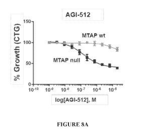
[0021] Figures 8A-B. (A) Percent growth inhibition of MTAP null and MTAP wild
type HCT116 cells treated with MAT2A inhibitor AGI-512. (B) Percent growth
inhibition of MTAP null an dMTAP wildtype HCT116 cells treated with MAT2A
inhibitor AGI-673.
[00221 Figure 9. Iminunoblot analysis of PRMT5, MTAP and beta-actin proteins
and
SDMA marks in HCT116 MTAP-i- and MTAPvvt cells.
[00231 Figure 10. Effect of Mat2a knockdown in in vivo orthotopic MCF7 model.
100241 Figures 11A-D. PRMT5 is a selective vulnerability in MTAP-null cancers
[0025] Figure 12. MAT2A depletion reduces PRMT5 methyl marks in MTAP
null cells.
DETAILED DESCRIPTION
[0026] Chromosome 9p21 (Chr9p21) is homozygous deleted in approximately 15%
of all human cancer (Berhoukim Meyerson nature 2010), including a number of
different tumor types and ranging in frequency up to the >50% deletion
frequency
observed in Glioblastoma Multiforme (Parsons and Kinsler, Science 2008). The
9p21
locus includes the CDK.N2a gene, which encodes both p14-ARF and p16-INK4a
9
CA 03006743 2018-05-29
WO 2017/096165
PCT/US2016/064619
(Figure 1A). Both proteins have tumor suppressive roles, with p14-ARF known to
stabilize p53 (Kamijo & Sherr Cell 1997 and Zhang & Yarbough Cell 1998) and
p16-
INK4a demonstrated to be a critical cell cycle regulator and potent tumor
suppressor
via negative regulation of the CDK4/6 cell cycle kinases (Serrano & Beach
Nature
1993). Although Chr9p21 deletion was first discovered over 30 years ago
(Chilcote
NEJM 1985), molecularly targeted therapies for CDKN2A loss have proven
elusive,
and it may be necessary to identify alternative approaches to target tumors
with
deletion of Chr9p21.
[0027] Notably, Chr9p21 deletions frequently involve co-deletion of genes
proximal
to CDKN2A (Figure 1A). Foremost among these co-deleted genes is MTAP, which
resides on Chr9p21 adjacent to CDKN2a (Figure 1A). The MTAP gene is within 100
kb of CDKN2A, and homozygous deletion of MTAP is found in 80-90% of tumors
with CDKN2A deletion 011ie & Ladanyi Clin Canc Res 1993 and Zhang & Savarese
Canc Genet Cytogenet 1996). MTAP encodes Methylthioadenosine Phosphorylase, a
critical enzyme in the methionine salvage pathway. MTAP metabolizes the
byproduct
of polyamine synthesis, methylthioadenosine, leading to the eventual
regeneration of
methionine and adenine from MTA (Zappia & Cartena-Farrina Adv Exp Med Biol
1988). Thus MTAP resides at the intersection of methionine metabolism,
polyarnine
biosynthesis, and nucleotide metabolism - metabolic pathways that are each
important
in the proliferative metabolism of cancer cells. In fact, MTAP deletion has
been
reported to create sensitivity to inhibitors of purine biosynthesis (Li and
Bertino
Oncol Res 2004), although this metabolic vulnerability is lost in vivo, as
tumors
uptake circulating adenine and escape the purine biosynthesis sensitivity
(Rueffli-
Brasse and Wickramasinghe JCI 2011). We sought to ask whether MTAP deletion
creates other targetable collateral vulnerabilities in cancers with Chr9p21
deletion.
100281 To screen for vulnerabilities that arise upon MTAP loss in cancer,
shRNA
depletion screening was used in an isogenic cancer cell line pair that vary
only in
MTAP status. Although MTAP encodes a metabolic enzyme, we hypothesized that
MTAP loss may create collateral vulnerabilities in biologic pathways that
extend
beyond metabolism. Precedent for such cross-talk between metabolic and non-
metabolic pathways includes the observation that the metabolite 2-
hydroxyglutarate,
produced by gain-of-function mutant IDH1/2 proteins, can inhibit members of
the
alpha-ketoglutarate dependent dioxygenase enzyme family (Xu & Xiong Cancer
Cell
CA 03006743 2018-05-29
WO 2017/096165
PCT/US2016/064619
2011, Roble & Mellinghoff Science 2013). A similar mechanism has also been
implicated in tumors with mutations in Succinate Dehydrogenase (SDH) or
Fumarate
Hydratase (FH), where the substrates of those enzymes accumulate to high
levels
(Selak and Gottlieb, Cancer Cell 2005 and lssacs and Neckers, Cancer Cell
2005).
Thus, aberrations in the cancer metabolome can impinge on non-metabolic
pathways.
To test the hypothesis that MTAP deletion would create collateral
vulnerabilities in
metabolic and non-metabolic pathways, an shRNA library was used consisting of
shRNA hairpins targeting the 3000+ genes of the metabolome as well as an
additional
3000+ additional non-metabolic genes.
[0029] Through this screen and subsequent investigation, a signaling axis was
identified that becomes vulnerable upon MTAP loss in cancer. Central in this
signaling axis is the arginine methyltransferase, PRMT5. Using metaboloinic
and
biochemical approaches, it was discovered that MTA, the substrate of the MTAP
enzyme reaction, accumulates in MTAP-null cancers. MTA inhibits PRMT5 enzyme
activity and leads to reduced basal PRMT5 methylation in MTAP-null cancers.
This
vulnerability extends both upstream and downstream of PRMT5. We show that the
metabolic enzyme Methionine-adenosyltransferase-2A (MAT2A), which produces
PRMT5 substrate 5-adenosyl methionine (SAM), is also selectively essential in
MTAP-null cancers, as are multiple different PRMT5 binding partners, including
the
kinase WOK 1 .
shRNA depletion screen in HCT116 MTAP wt/MTAP 4- isogenic pair.
[0030] In order to identify genes whose loss would lead to selective killing
of MTAP-
deficient cells, an shRNA-based depletion screen were performed in HCT116
colon
carcinoma cell line and an isogenic clone of HCT116 cells that had been
genetically
modified to delete exon 6 of the MTAP gene (Figure 1B). This deletion led to
complete loss of MTAP protein expression (Figure 1C). To provide broad
coverage
for potential synthetic lethal interactions, we constructed a library that
encompassed
the complete metabolome (3,067genes), the mitochondrial proteome (Pagliarini
and
Mootha Cell 2008), the epigenome (Arrowsmith and Shapira Nature Reviews Drug
Discovery 2012), the kinome (http://www.uniprot.org/), and >1500 additional
genes
representing diverse biologic pathways. HCT116 MTAP and HCT116 wt cells were
transduced with the shRNA library containing 8 shRNAs per gene, and the pool
of
11
CA 03006743 2018-05-29
WO 2017/096165
PCT/US2016/064619
knockdown cells was passaged for 12 cell divisions. At the end of the culture
we
measured the relative abundance of each shRNA barcode via deep sequencing, and
calculated the fold depletion of each shRNA compared to the untransduced
library
DNA. We then calculated an MTAP selectivity score for each gene based on the
difference in the log2 fold change in the abundance of each of the 8 shRNAs
targeting
the gene in HCT116 MTAP-1- vs. HCT116 wt cells (Figure ID).
[0031] This analysis demonstrated that while the majority of the genes, as
well as
shRNA controls, had a similar score in both HCT116 MTAP-I- and HCT116 MTAP wt
cells (Figure 1D ), a subset of genes was selectively depleted in MTAP-
deficient cells
(Figure 1D-E). The top hit in the screen was MAT2A, which encodes the
metabolic
enzyme Methionine Adenosyltransferase II, alpha (Figure 1D-F). MAT2A catalyzes
the synthesis of the universal biological donor of methyl groups, 5-
adenosylmethionine (SAM) via adenosylation of methionine. The second best
scoring
gene in the screen was Protein Arginine Methyltransferase 5 (PRMT5) (Figure 1D-
F),
which is the catalytic subunit of a multiprotein methyltransferase complex
that
includes PRMT5 in complex with obligate binding partner WD45/MEP50 (WD repeat
domain 45/methylosome protein 50), and other scaffolding proteins (Meister et
al.,
2001; Pesiridis et al., 2009). PRMT5 belongs to the type II PRMT subfamily of
arginine methyl transferases and catalyzes the formation of symmetric di-
methylarginines in target proteins. Interestingly, the sixth highest scoring
gene,
RIOK1, encodes a Rio domain containing protein, which is a binding partner of
PRMT5 that directs PRMT5 towards selective methylation of a subset of PRMT5
substrates (Guderian et al 2011). These data suggest that MAT2A and PRMT5-
catalyzed reactions are critical for maintaining viability of MTAP-deficient
cells.
Although all three highlighted hits represent therapeutically and biologically
interesting targets, we initially focused our attention on PRMT5 as there are
currently
ongoing efforts targeting this enzyme for the treatment of human cancer (Chan
Penebre Nature Chem Bio 2015).
12
CA 03006743 2018-05-29
WO 2017/096165
PCT/US2016/064619
PRMT5 is selectively essential in MTAP-null cells upon genetic ablation but
not
pharmacologic targeting.
[0032] To further investigate the connection between MTAP deficiency and PRMT5
function in cells, we generated HCT116 MTAP" - and HCT116 wt cell lines stably
expressing inducible shRNAs targeting PRMT5. We confirmed that PRMT5 was
efficiently knocked down by measuring levels of PRMT5 protein. Consistent with
our genomic screening results, PRMT5 knockdown with doxycycline-inducible
shRNA led to more complete growth reduction in cells with MTAP deletion than
in
MTAP WT cells (Figure 2B). Expression of an shPRMT5-resistant PRMT5 cDNA in
the MTAP-null cells rescued growth inhibition upon endogenous PRMT5
knockdown, while expression of catalytically-dead R368A PRMT5 mutant (Pollack
et
al., 1999) cDNA did not (Figure 2B-C). Thus, the anti-proliferative effects of
our
shRNA are due to PRMT5 depletion and not due to off-target shRNA effects. Lack
of
rescue by R386A-mutant PRMT5 indicates that PRMT5 enzyme activity is essential
in MTAP-/- cells. Interestingly, equivalent reduction in PRMT5 protein level
in
MTAP-/- and WT cells resulted in greater reduction in the levels of symmetric
di-
methylarginine marks in the MTAP-/- cell line and was rescued by PRMT5 but not
R368A-mutant cDNA (Figure 2D). These findings provide validation of our
screening
results and further suggest that PRMT5 catalytic function is critical for
maintaining
growth of MTAP-deficient cells.
[0033] Next we wanted to interrogate PRMT5 function in MTAP-deficient cells
using
a pharmacologic tool. A potent and selective inhibitor of PRMT5, EPZ015666,
was
recently developed (Chan-Penebre et al., 2015). We utilized the EPZ015666
compound and performed dose response analysis in the HCT116 isogenic pair
(Figure
2E). However, unlike genetic targeting of PRMT5, growth inhibition upon
pharmacologic targeting of PRMT5 was not selective for the MTAP-deficient
genetic
background (Figure 2E). This finding was unexpected considering that the
catalytically-dead mutant of PRMT5 did not rescue the growth phenotype in
HCT116
MTAP-/- cells, suggesting it is the loss of catalytic function of PRMT5 that
was
necessary to selectively inhibit the growth of these cells (Figure 2A).
Interestingly,
unlike with genetic ablation of PRMT5 function, equal degree of PRMT5 activity
inhibition was achieved in both MTAP' - and wt HCT116 cells with EPZ015666, as
evidenced by reduced levels of PRMT5-dependent di-methylarginine marks in
total
13
CA 03006743 2018-05-29
WO 2017/096165
PCT/US2016/064619
cell lysates (Figure 2F). This surprising discrepancy between impact of
genetic and
pharmacologic PRMT5 ablation on the growth of MTAP-deficient cells led us to
further interrogate the basic biology and metabolism behind PRMT5 and MTAP.
MTAP deficiency creates an altered metabolic state.
[0034] In order to explain the difference in the impact of genetic vs.
pharmacologic
targeting of PRMT5 on growth of HCT116 isogenic pair, we wanted to further
build
our mechanistic understanding of MTAP and PRMT5 synthetic lethality. MTAP is
an
enzyme in the methionine salvage pathway that converts a byproduct of
polyamine
biosynthesis, methylthioadenosine (MTA), back to methionine and adenine
(Figure
3A). Since MTAP is the only enzyme in mammalian cells known to catalyze the
degradation of MTA, we hypothesized that MTAP deficiency would result in
accumulation of MTA. We first tested this hypothesis in the context of a
broader,
untargeted LC-MS based metabolomics assessment of intracellular metabolite
levels
in the HCT116 MTAP isogenic pair (Figure 3B). This analysis revealed that,
among
237 annotated metabolites that were detected, MTA displayed the largest
increase in
HCT116 MTAP-/- cells compared to HCT116 wt control. Interestingly,
decarboxylated S-adenosylmethionine (dcSAM), the metabolite upstream from MTA
in the polyamine biosynthetic pathway, displayed the second-largest increase.
The
enrichment of these two metabolites in HCT116 MTAP' - cells was highly
statistically-significant (Figure 3B). Elevation of MTA was further confirmed
using
quantitative measurement of MTA levels in HCT116 isogenic pair (Figure 3C).
Furthermore, a screen of a large cancer cell line panel comprising 249 cell
lines of
different tumor origin demonstrated very consistent accumulation of MTA in the
media of cells with endogenous MTAP deletion (Figure 3D).
MTA inhibits PRMT5 activity in vitro and in vivo.
[0035] MTA has been reported to inhibit activity of protein methyltransferases
(Enouf
et al., 1979). To test this notion directly, we performed an in vitro
biochemical screen
assessing enzyme activity of 33 different N-methyltransferases following
treatment
with 10 M and 100 1.1M. MTA (Figure 4A). Inhibition by MTA was only observed
in
a small subset of the panel, and strongest inhibition was observed for PRMT5
and
PRMT4, members of the arginine methyltransferase family (Figure 4A). Further,
14
CA 03006743 2018-05-29
WO 2017/096165
PCT/US2016/064619
PRMT5 demonstrated potent sensitivity to MTA in subsequent experiments testing
a
wide range of MTA concentrations (Figure 4B). Next, we analyzed the MTA Ki for
PRMT5, PRMT4, and a diverse subset of methyltransferases (Figure 4C).
[0036] Strikingly, the MTA Ki for PRMT5 (0.46 p,M) was >20-fold lower than
that
for any other methyltransferase, indicating that PRMT5 is far more sensitive
to
inhibition by MTA than any other methyltransferase tested. This biochemical
observation is consistent with our shRNA screening data demonstrating that
PRMT5
was the strongest hit among all the methyltransferases that were represented
in the
library and were selectively depleted in HCT116 MTAP' - cells (Fig 1D).
[0037] We next addressed the impact of MTA accumulation on PRMT5 activity in
cells. According to our LC-MS analysis of intracellular MTA levels in MTAP-
deficient cells (-100 M) and PRMT5 IC50 for MTA measured in our biochemical
assay (3 04), we hypothesized that MTA accumulation in MTAP-deficient cells
would be sufficient to result in inhibition of PRMT5 activity. During our
analysis of
PRMT5-dependent methyl marks in total cell lysates of the HCT116 isogenic
pair, we
noted that HCT116 MTAP-I- cells appeared to have lower basal levels of
methylation
(Figure 2D). To further substantiate this finding, we performed western blot
analysis
of PRMT5-dependent methyl marks in total cell lysates of a subset of MTAP wt
and
MTAP-deleted cell lines (Figure 4D). We observed that MTAP-deleted cell lines
consistently demonstrated lower levels of symmetric di-methylarginine marks
(Figure
4D). Finally, we took advantage of the availability of a potent, cell
permeable
transition state analogue inhibitor of MTAP (Basu et al., 2011; Longshaw et
al.,
2010). We treated HCT116 wt cells with the MTAP inhibitor for three days and
measured the impact of pharmacologic inhibition of MTAP on the levels of di-
methylarginine marks (Figure 4E). Treatment with MTAP inhibitor at the dose
sufficient to increase MTA levels to those observed in HCT116 MTAP 4- cells
(Figure
S4) resulted in reduction in the levels of di-methylarginine methyl marks
similarly to
what is observed upon genetic ablation of MTAP (Figure 4E). These data
strongly
indicate that PRMT5 activity is impaired by MTA in MTAP-null cells, resulting
in
reduced methylation of its protein substrates and creating a vulnerability to
additional
reduction of PRMT5 activity by shRNA. Furthermore, our finding that MTA
inhibits
of PRMT5 provides an explanation for the lack of MTAP-selective growth
inhibition
with the PRMT5 inhibitor EPZ015666. This inhibitor binds selectively to the
SAM-
CA 03006743 2018-05-29
WO 2017/096165
PCT/US2016/064619
PRMT5 complex (Chan-Penebre et al., 2015) via a cation-pi molecular
interaction
that is not possible with the MTA-PRMT5 complex. Since MTA prevents binding of
SAM to PRMT5, and EPZ015666 only interacts with SAM-bound PRMT5, MTA
binding is mutually exclusive with EPZ015666 binding. Two inhibitors of a
single
enzyme can only be synergistic if they bind to separate binding sites and
their
interaction with target is not mutually exclusive (Breitinger).
MAT2A is selectively essential in MTAP-deficient cells.
[0038] We next wanted to test whether MAT2A, the top hit in our shRNA screen,
also represents a bona fide synthetic lethal target in MTAP-deficient cells.
We thus
utilized the HCT116 isogenic pair and created cell lines stably expressing non-
targeting shRNA, MAT2A-targeting shRNA, as well as cell lines that were
additionally reconstituted with shRNA-resistant MAT2A cDNA, or that expressed
MTAP cDNA. We confirmed efficient MAT2A knockdown, and MAT2A and MTAP
re-expression in HCT116 cells by western blot (Figure 5A). We also confirmed
that
MAT2A knockdown resulted in reduced cellular levels of SAM in both HCT116
genotypes using LC-MS analysis (Figure 5B). We further confirmed that MTAP re-
expression depleted high MTA levels present in the media of HCT116 MTAP' -
cells.
We then tested the impact of MAT2A knockdown in HCT116 wt vs. HCT116 MTAP
-
/- cells in a 4- and 6-day in vitro growth assay (Figure 5C). The results were
in
agreement with our genomic screen. MAT2A knockdown selectively attenuated
growth of HCT116 MTAP', but not HCT116 wt cells (Figure 5C). Importantly, this
growth defect was rescued by introduction of shRNA-resistant MAT2A cDNA
construct, indicating on-target effect of the shRNA, and was also partially
rescued by
MTAP re-expression (Figure 5C).
[0039] To investigate the in vitro to in vivo translation of our findings, we
conducted
xenograft efficacy studies with HCT116 isogenic cell lines expressing
inducible
MAT2A shRNA. In these studies, tumors were allowed to form prior to treatment
of
animals with doxycycline, to assess the role of MAT2A in proliferation of
established
tumors. Efficiency of MAT2A knockdown in vivo was confirmed by western blot
(Figure 5D). We further confirmed that MAT2A genetic ablation in vivo resulted
in a
similar reduction in SAM levels in HCT116 xenografts of both genotypes (Figure
16
CA 03006743 2018-05-29
WO 2017/096165
PCT/US2016/064619
5E). In accordance with our findings in vitro. MTAP-selective growth
inhibition was
observed in vivo upon MAT2A depletion by shRNA (Figure 5F). To demonstrate
that
this selective growth inhibition in vivo was an on-target effect, we performed
expanded in vivo study with a wild type MAT2A rescue arm of shMAT2A (Figure 5G
and 5H). This experiment confirmed the efficacy observed in our first in vivo
study
(Figure 5G and 5H) and, as with the in vitro studies, growth inhibition was
rescued in
the xenograft expressing a MAT2A cDNA that was resistant to the MAT2A shRNA
(Figure 5G and 5H).
[0040] Finally, we wanted to confirm our findings in a model that possesses
endogenous deletion in MTAP locus. Thus, we generated breast carcinoma MCF7
cell
lines stably expressing non-targeting shRNA, MAT2A-targeting shRNA, as well as
cell lines that were additionally reconstituted with shRNA-resistant MAT2A
cDNA.
We demonstrated efficiency of MAT2A knockdown and re-expression by western
blot (Figure 5J). Consistent with observations made in the HCT116 model
system,
MAT2A knockdown attenuated growth of MTAP-deleted MCF7 cells in a 7-day
growth assays (Figure 51), while MAT2A cDNA reconstitution resulted in
complete
rescue of the growth phenotype. Thus. MAT2A demonstrates consistent synthetic
lethality with MTAP deficiency in our models.
MAT2A loss selectively inhibits PRMT5 activity in MTAP null cells.
[0041] Having confirmed that both PRMT5 and MAT2A are true synthetic lethal
partners of MTAP, we wanted to assess whether there is a mechanistic link
between
these two top hits in our screen. Indeed, MAT2A generates SAM, which is
necessary
for the activity of all cellular methyltransferases, and reduction in SAM
levels upon
MAT2A genetic ablation would be expected to broadly impact their function,
including that of PRMT5. Thus, we measured levels of PRMT5-dependent symmetric
di-methylarginine marks on histone H4 in our MAT2A shRNA HCT116 isogenic
pair, as well as in MAT2A reconstituted and MTAP re-expression cell lines upon
knockdown of MAT2A (Figure 6A). Interestingly, we observed that despite
equivalent degree of reduction in SAM levels in HCT116 cells of both genotype
(Figure 5B), H4R3me2s marks were selectively reduced in MTAP-deficient cells,
but
not in MTAP wt cells, and were rescued in presence of MAT2A and MTAP cDNA
17
CA 03006743 2018-05-29
WO 2017/096165
PCT/US2016/064619
(Figure 6A). In combination with our observation regarding the strong
inhibitory
impact of MTA on PRMT5 activity, these data suggest that PRMT5 function in the
MTAP-null background is highly dependent on adequate availability of SAM.
PRMT5 was reported in the literature to exhibit low affinity for SAM
(Antonysamy et
al., 2012; Sun et al., 2011), We thus compared SAM Km values for the N-
methyltransferases from our in vitro biochemical panel analysis and observed
that
indeed PRMT5 exhibited the lowest affinity for SAM (Figure 6B). This finding
may
explain PRMT5 dependence on proper MAT2A function, especially in the
metabolically-altered, high-MTA environment of MTAP-deficient cells (Figure
6C).
Thus, metabolic vulnerability due to MTAP deficiency extends upstream of PRMT5
creating dependence on the availability of PRMT5 substrate SAM and therefore
the
activity of SAM-producing enzyme MAT2A.
Multiple PRMT5 co-complexes are vulnerable in MTAP-null cells.
100421 The Rio domain containing protein RIOK1 was another strong hit in our
shRNA depletion screening campaign. Since it is a PRMT5 binding partner, we
sought to confirm the synthetic lethal phenotype upon genetic ablation of RIOK
I in
the HCT116 MTAP isogenic cells. Similar to the characterization that was
performed
for PRMT5 and MAT2A, inducible RIOK1 shRNA cell lines, as well as RIOK1 wt
rescue and RIOK1 active site (D324N) and ATP-binding domain
(K208R)catalytically inactive mutant (Angermayr et al., 2002; Widmann et al.,
2012)
cell lines were created. RIOK1 knockdown and re-expression efficiencies were
evaluated by western blot (Figure 7A). Confirming our finding in the genomic
screening, RIOK1 knockdown resulted in a selective inhibition of growth of
HCT116
MTAP 4- cells with minimal impact on growth of HCT116 wt cells (Figure 7B).
The
growth phenotype was rescued by the expression of shRNA-resistant wt RIOK1 and
not catalytically inactive K208R, D324N mutant RIOK1 (Figure 7B). These data
suggest that the metabolic vulnerability created via accumulation of MTA in
MTAP-
deficient background further extends downstream of PRMT5 via impact on PRMT5
binding partner RIOK1.
100431 PRMT5 participates in several multimeric protein co-complexes,
including
obligatory binding partner WD45/MEP50(Wilczek et al., 2011), the mutually
18
CA 03006743 2018-05-29
WO 2017/096165
PCT/US2016/064619
exclusive partners pICIn and RIOK1 (Guderian et al., 2011), the nuclear
regulator of
specificity COPR5 (cooperator of PRMT5)(Lacroix et al., 2008), and others.
Neither
IVIEP50, nor pICIn or other binding partners of PRMT5 were represented in our
shRNA library. Thus, to evaluate the possibility that the vulnerability of
MTAP-
deficient cells further extends to PRMT5 co-complexes beyond the RIOK1 co-
complex, we performed siRNA pool-mediated knockdown of multiple PRMT5 co-
complex members, including PRMT5 itself, RIOK1, MEP50, pICIn, and COPR5 in
the HCT116 isogenic pair (Figure 7C and 7D). We observed the selective
inhibition
of the growth of MTAP-deficient cells upon knockdown of each member of the
PRMT5 co-complex (Figure 7C). Importantly, knockdown of a separate PRMT5-
binding protein, the ATP-dependent helicase Brgl encoded by the SMARCA4 gene,
(Pal et al., 2004) inhibited growth of HCT116 cells regardless of their MTAP
status
(Figure 7C). These data suggest that vulnerability of MTAP-deficient cells
downstream from PRMT5 is not restricted to RIOK1 co-complex but is rather
broad
impacting several co-complexes involving PRMT5 as a binding partner. MTA
accumulation in MTAP-null cells reduces PRMT5 activity and creates a
collateral
vulnerability to targeting of PRMT5. This vulnerability extends to the
metabolic,
epigenetic, and signaling pathway members that reside upstream, and
downstream, of
PRMT5.
[0044] The mammalian metabolome is characterized by a high degree of
flexibility
and redundancy (Thiene & Pallson Nat Biotech 2013 and Folger and Shlomi Molec
Sys Bio 2011.). MTA is thus unusual in that it is consumed by a solitary, non-
redundant enzyme, MTAP. We observed that upon MTAP deletion, MTA
accumulates to an intracellular concentration of approximately 100 uM, and
cells
begin to excrete excess MTA. This accumulation of MTA led to an unexpected
collateral vulnerability in the arginine methyltransferase PRMT5. While the
shRNA
library contained 39 methyltransferases, PRMT5 was unique in its high degree
of
MTAP-selectivity. Biochemical profiling of methyltransferases revealed a
molecular
basis for this phenomenon. Amongst the 32 methyltransferases that we tested in
vitro,
PRMT5 was the enzyme most sensitive to inhibition by MTA. In vitro inhibition
of
PRMT5 by MTA occurs at the concentrations very similar to those observed in
MTAP-null cells, suggesting that this is a biologically-relevant phenomenon.
19
CA 03006743 2018-05-29
WO 2017/096165
PCT/US2016/064619
Consistent with this, we observed substantially reduced basal levels of PRMT5
methyl marks in cells with MTAP deletion.
[0045] Reduced basal PRMT5 activity creates a vulnerability to further
ablation of
PRMT5 by shRNA. Interestingly, treatment with PRMT5 inhibitor EPZ-015666 did
not lead to selective growth inhibition in MTAP-null cells. EPZ-015666 has a
very
distinctive mode of inhibition of PRMT5. This inhibitor is SAM-uncompetitive
and
forms key binding interactions with enzyme-bound SAM via an unusual cation-pi
interaction with the partial positively charged methyl group on SAM (Chan-
penebre
Nat Chem Bio 2015). MTA is unable to form this synergistic binding interaction
with
EPZ-015666 (CITE Chan-penebre). Thus this existing PRMT5 inhibitor does not
display preferential activity in MTAP-null cancers. Exploiting the PRMT5
vulnerability in MTAP-null cancers may require the development of MTA-
selective
PRMT5 inhibitors that bind to the MTA-bound form of PRMT5 and trap the enzyme
in that state. MTA-selective inhibitors might afford a greater therapeutic
window than
non-selective inhibitors, as MTAP expression in normal tissues should provide
a
protective effect by maintaining low MTA levels. Mouse genetics studies have
revealed that PRMT5 has important roles in normal physiology; PRMT5 knockout
leads to embryonic lethality (Tee 2010), and substantial toxicities arise upon
tissue
specific PRMT5 knockout in the CNS (Bezzi 2013) skeletal muscle (Zhang 2015)
and
hematopoietic lineages(Liu 2015). These toxicities may become dose-limiting in
the
clinical setting, narrowing the therapeutic potential of agents that target
PRMT5 in a
non-selective manner.
[00461 Cellular methyltransferase activity is subject to regulatory control by
small
molecule metabolites. It has previously been established that
methyltransferases are
regulated by the relative balance of substrate SAM and product SAH (Vance Cui
Biochim Biophys Acta 1997). The SAM/SAH ratio is used to calculate cellular
'methylation potential' as a measure of cellular poise to conduct
methyltransferase
reactions (Williams & Schalinske J Nutrition 2006). Our observation that PRMT5
can
be inhibited by MTA implicates PRMT5 as the exemplar member of a biochemically-
distinct family of methyltransferases that can be regulated by SAM/MTA ratio.
This
novel regulatory mode is revealed very clearly in MTAP-null cancer cells,
where
MTA levels accumulate dramatically. There exists only limited information
regarding
MTA levels across normal tissues (Stevens & Oefiier, J chromatography 2010),
and
CA 03006743 2018-05-29
WO 2017/096165
PCT/US2016/064619
wider MTA screening might reveal other settings in which MTA accumulation
leads
to inhibition of PRMT5. We note also that PRMT5 has a fairly weak binding
affinity
for SAM. This is unusual amongst the methyltransferase family, as most
mammalian
methyltransferases have SAM Km values 10- to 100-fold below the physiologic
concentration of SAM (Richon & Copeland Chem Biol Drug Design 2011). This
biochemical finding implies that PRMT5 is poised as a SAM-sensitive
methyltransferase, and this sensitivity is exemplified by the reduction in
PRMT5
methyl marks that is observed upon MAT2A depletion in MTAP-null cells.
[0047] PRMT5 regulates a number of proliferative and biosynthetic processes,
such
as histone methylation that controls expression of cell cycle genes (Chung &
SifJBC
2013), methylation of growth factor signaling components like EGFR and Raf
(Hsu &
Hung Nat Cell Bio 2011, Andreu-Perez & Recio, Sci Signaling 2011), and
methylation of key protein components required for maturation of ribosome and
spliceosome complexes (Ren & Xu, JBC 2010, and Friesen & Dreyfuss Mol Cell Bio
2001). Thus PRMT5 activity leads to coordinated upregulation of a range of pro-
proliferative and biosynthetic pathways. The vulnerability of PRMT5 in MTAP-
deficient cancers extends both upstream of PRMT5 (to MAT2A) and downstream of
PRMT5 (to RIOK1 and other PRMT5 cocomplex members). Collectively, these
proteins comprise a metabolic-epigenetic-signaling axis which senses and
transmits
information about nutrient availability (MAT2A substrate Methionine) to the
multiple
biosynthetic pathways that reside downstream of PRMT5. This axis presents
intriguing opportunities for targeted therapy of MTAP-deficient cancers. In
addition
to the potential to devise MTA-selective PRMT5 inhibitors, our work
demonstrates
that therapeutic targeting of MAT2A, RTOK I , or other PRMT5 co-complex
members,
could selectively impact MTAP-null cancers while sparing MTAP-expressing
normal
tissues. Thus this vulnerable axis includes a number of proteins that merit
further
consideration as therapeutic targets to address the ¨15% of human cancers with
deletion of the MTAP/p16/CDKN2A locus.
Cell line screen with AGI-512 and AG 1-673
100481 AG-512 and AG-673 are small molecule inhibitors of MAT2A enzymatic
activity demonstrating an IC50 of 83 nM and 143 nM respectively in a
biochemical
assay and inhibited the production of SAM in cells with ICsos of 80 and 490 nM
21
CA 03006743 2018-05-29
WO 2017/096165 PCT/US2016/064619
respectively. These compounds were screened for growth inhibition against
several
cancer cell lines having varied tissue origin for which MTAP status (null or
wild type)
was determined. The results are presented in table I.
Table 1
MTAP AGI-673 ' AGI-512
CELL LINE TISSUE
STATUS IC50 (m.M) IC50 (JiM)
LN-18 null brain 0.397 0.485
HMCB WT skin 0.465 i 0.473
K-562 null herne 0.596 0.901
MDA-MB-231 null breast 0.560 0.364
SW 1088 null brain 0.639 0.508
GB-1 null brain 1.302 3.844
NC1-H1437 null lung 2.020 0.695
A172 null brain 2.174 0.283
A549 null lung 3.052 3.647
fiCC70 WT breast 3.156 2.162
U-87 MG null brain 4.001 2.115
Jurkat null heme 3.667 1.168
MDA-MB-468 WT breast 3.981 3.034
NCI-H661 WT lung 5.137 6.249
HCT 116 WT colon 10.165 >20
Hs 695T WT skin 10.866 >20
NCI-H460 wr lung 13.307 >20
COLO 741 null skin 12.652 ' 7.643
CCF-STTG1 wr brain >20 >20
NMC-G1 WT brain 8.504 >20
LN-229 WT brain 0.159 0.111
RI-112 null bladder 0.496 0.326
H4 null brain 0.774 0.886
SW 780 null bladder 0.375 0.628
Hs 294T WT skin 0.329 0.529
RT4 null bladder 6.373 ' 0.566
DLD-1 WT colon 13.996 0.941
U-251 MG WT brain 2.822 13.412
HT-1197 WT bladder 0.889 0.761
KNS-42 WT brain >20 :00
Y1-1-13 null brain 0.823 0.485
Daoy null brain 1.030 0.852 .
M059K . WT brain . 0.401 1.116
U-118 MG null brain 0.759 :00 .
SNU-1105 . null brain . 0.267 0.103
SW 1783 WT brain 3.605 :00 .
U251 . WT brain . 1.681 0.511
MV-4-11 WT fieme 2.307 5.741 .
NCI-H1568 . WT lung . 1.384 1.780
MeWo WT skin 1.101 1.281 .
Hs 839.T . WT skin . 0.269 4.068
IGR-1 null skin 0.044 0.023
11
CA 03006743 2018-05-29
WO 2017/096165 PCT/US2016/064619
MTAP AGI-673 AGI-512
C ELL LINE TISSUE
STATUS 1050 ( M) 1050 (g111)
KS-I null brain 0.487 ' 0.505
COLO 829 WI' skin 1.762 2.229
ON S-76 null brain 2.794 0.794
HS 683 null brain 1.804 1.118
DBTRG-05MG WT brain 4.237 0.323
SFI26 WT brain 6.357 8.273
HT-I44 WT skin 7.693 2.255
YKG-I WT brain 10.256 10.093
Becker WT brain 9.652 :00 .
U-138 MG . null brain . 12.292 >20
GI-1 WT brain 0.958 0.967 .
KNS-60 . WT brain . 15.640 >20
KNS-81 WT brain 11.775 11.279 .
382 . WT bladder . 1.634 1.900
SCaBER WT bladder 1.350 1.142 .
T98G . WT brain . 2.101 3.788
KALS- I WT brain 1.589 1.822 .
Hs 940.T . WT skin . 0.737 0.514
Hs 688(A).T WT skin 1.361 0.001 .
RT I 12/84 . null bladder . 0.395 0.578
AM-38 null brain 0.499 6.774 .
UM-UC-3 . null bladder . 1.945 2.770
ClL-I WT skin 1.539 2.663 .
A-375 . WT skin . 2.817 6.554
5637 WT bladder 4.220 1 11.338
TCCSUP WT bladder 1.375 0.791
T24 WT bladder 5.506 >20 .
D283 MED WT brain 8.196 19.702
SK-MEL-3 WT skin 9.811 10.232 ,
SK-IvIEL-5 null skin 3.376 6.266
G-361 WT skin 14.766 I 0.102 ,
D341 Med WT brain 11.843 0.731
Hs 852.T WT skin 17.235 I 19.244 ,
Malme-3M null skin >20 >20
SK-MEL-24 null skin >20 I 14.226 ,
Hs 934.T WT skin 0.230 9.282
AIOID null skin 0.109 I 0.088 ,
Reh null heme 3.782 2.217
WM-266-4 WI' skin 4.030 I 1.266 ,
A2058 WT skin 2.030 i 6.732
LN-229 WT brain 1.353 1.128
HT1376 WT bladder 5.649 ___ >20
NCI-H1568 WT lung 4.306 16.885
NCI-H929 WT heme _____ 5.965 _ 23.811
RPM1-8226 WT heme 8.132 21.801
SKM-1 WT heme 1.746 >20
KALS-1 WT brain 9.902 i >20
C0L0679 WT skin 8.179 4.496
RPM1-7951 WI skin 1.169 11.916
SK-MEL-I WT skin >20 j >20
23
CA 03006743 2018-05-29
WO 2017/096165 PCT/US2016/064619
MTAP AGI-673 AGI-512
C ELL LINE TISSUE
STATUS IC50 ( M) IC50 (AM)
C32 WI skin 1.906 ' 2.259
WM-115 WI' skin 1.783 6.376
SK-MEL-28 WI skin 1.257 >20
HEL9217 null heme 0.229 0.202
MEG-01 WT heme 0.608 0.416
REC1 WT heme 0.799 0.985
LIM 1 null heme 0.990 0.763
SUP-B15 null heme 0.892 0.618
HEL null heme 1.006 1.844 .
EB2 . WT heme . 1.332 5.502
KU812 WT heme 2.655 5.283 .
RI . WT heme . 1.757 0.423
NOMO-1 null heme 5.071 0.269 .
SH4 . null skin . 1.917 1.195
DB WT heme 4.618 3.885 .
RS4;11 . null heme . 4.478 1.488
111,60 WT heme 5.813 3.212 .
BCP-1 . WT heme . 6.508 0.107
KASUMI1 WT heme 8.943 5.404 .
CA46 . WT heme . 8.362 0.719
RAH WT heme 8.362 0.788 .
SK-MEL-31 . WT skin . 10.649 2.041
EBI WT heme 11.520 1.035 .
NAMALWA . WT heme . 19.282 0.000
SK-MEL-24 null skin 31.337 1 0.450
HUT78 null heme 0.503 0.299
TOLEDO WT heme 1.076 2.047 .
Pane 03.27 null pane 1.383 0.696
HUTIO2 WT heme 1.211 2.453 ,
SUDHL6 WT heme 2.407 3.702
LN-229 WT brain 2.752 I 0.108 ,
Pane 10.05 WT pane 2.844 1.328
Daudi WT heme 5.069 I 6.917 ,
SUPT! WT heme 4.657 7.947
MOLT4 WT heme 5.855 I 13.816 ,
U266B I WI heme 5.314 5.525
Pam 02.03 WT pane 6.640 I 2.656 ,
LOUCY WT heme 9.192 6.823
TALL! WT heme 8.538 I >20 ,
S1486 WT heme 0.613 i 0.257
HUP-T4 null pane 0.700 0.220
MIA PaCa-2_ null Tam 0.517 1.246
KP-4 null pane 0.359_ 1.423
HPAC_ WT Tam 1.294 4.959
HU P-T3 null pane 0.644 0.413
IMP-1 null heme 1.398 i 0.513
CFPAC-1 WI pane 0.901 I 0.542
PSN I null pane 2.946 4.313
!
Pane 05.04 WI pane 1.048 I 7.773
BxPC-3 null pane 1.695 2.601
24
CA 03006743 2018-05-29
WO 2017/096165 PCT/US2016/064619
MTAP AGI-673 AGI-512
C ELL LINE TISSUE
STATUS IC50 ( M) IC50 (AM)
Pane 04.03 WT pane 7.407 ' 1.283
PANC-1 null pane 10.316 11.727
Hii WT heme 13.138 0.018
HT WT heme 11.680 9.479
MC116 WT heme 16.238 1.662
Mina WT heme 14.508 8.845
KASUM16 WT heme >20 0.142
KG! WT heme 24.366 27.277
U937 WT heme 1.988 39.785 .
JVM2 . WT heme . 1.496 1.723
P3HR-1 WT heme 0.103 0.042 .
Hs 766T . WT pane . >20 0.576
PL45 WT pane >20 :00 .
SW 1990 . WT pane . 54.601 0.743
HPAF-II WT pane 1.063 3.541 .
KP-2 . WT pane . 0.000 8.705
AsPC-1 WT pane 17.503 5.492 .
F-36P . null heme . 0.398 0.247
Capan-I null pane 0.703 0.514 .
Pfeiffer . WT heme . 1.996 0.859
GDM I WT heme 7.738 3.414 .
D341 IsilED . WT brain . 19.049 1.140
QGP-I WT pane 43.654 1.567 .
AML-193 . WT heme . 1.447 8.164
NALM-1 null heme 3.424 I 2.871
..
NCI-H69 WT lung 1.046 0.379
MM I S WI' heme 3.859 8.932 .
NCI-H524 WT lung 0.740 24.999
NCI-H2228 null lung 0.969 0.703 ,
HUP-T3 null pane 0.501 i 0. 599
I
NCI-H647 null lung 0.753 I 0.694 ,
MIA PaCa-2 null pane 0.852 0.217
NCI-H1755 null lung 2.420 I 1.372 ,
NCI-H226 WT lung 1.515 0.831
NCI-H1975 WI lung 1.515 I 0.831 ,
NCI-H1944 WT lung 5.125 0.478
NCI-H1915 WI lung 7.065 I 5.852 ,
NCI-H1299 WT lung 5.026 >20
Capan 2 WI pane 7.248 I 6.810 ,
RERF-LC-Sq I null- 5.454 1 0.493
_
NCI-H292 null_ lung 5.750 0.842
Malme-3M_ null skin 7.819 14.205
LU-DLU-1 null ____ lung 2.145 0.658
MDA-MB-361_ WT breast >20 5.643
HCC202 WI breast >20 >20
NCI-H196 WI lung >20 i 0.626
KP4 null pane 0.251 1 1.831
HCC15 null lung 0.357 0.204
!
SU.86.86 null pane 0.935 ! 0.845
NCI-H1703 WT lung 13.537 8.721
CA 03006743 2018-05-29
WO 2017/096165 PCT/US2016/064619
MTAP AGI-673 AGI-512
C ELL LINE TISSUE
STATUS 1050 ( M) 1050 (gM)
IvIDA-MB-134-VI WT breast 4.785 1 >20
HCC1937 wr breast 1.060 >20
Mi WT heme >20 0.026
BDCM wr heme 89.161 16.145
NCI-H2030 WT lung 2.199 1.268
NCI-H1838 wr lung 1.061 0.930
NCI-H1563 null lung 1.683 0.264
MCF-7 null breast 0.989 0.717
HCC1395 null breast 2.069 0.657 .
FICC38 . WT breast . 3.263 1.795
MDA-MB-453 WT breast 3.672 4.680 .
HCC1954 . WT breast . 4.128 8.622
LK-2 WT lung 6.260 1.775 .
BI474 . WT breast . 2.502 0.027
Hs 578T WT breast 6.956 9.053 .
HCC1428 . WT breast . 9.441 10.594
NCI-H2172 WT lung 8.970 0.989 .
FICC 70 . WT breast . 7.105 2.298
NCI-H146 WT lung 1.566 :00 .
MDA-MB-175-VII . WT breast . 11.262 >20
NCI-I-1522 WT lung 13.416 18.612 .
ALI565 . WT breast . 19.738 2.393
D1J4475 WT breast >20 :00 .
HCC1. 143 . WT breast . >20 8.529
HCC1806 WT breast >20 1 0.457
T-47I) WT breast ___ >20 4.795
CAMA.- I WT breast 25.406 1.133 .
NCI-H520 WT lung 27.989 2.142
NCI-H2291 WT lung 4.790 0.420 ,
HCC44 WT lung 0.692 1.385
MDA-MB-436 WT breast 2.248 I 5.193 ,
NCI-H441 WT lung 0.694 0.746
HCC1419 WT breast 2.222 I 0.146 ,
NCI-H2347 WT lung 0.442 0.187
NCI-H1930 WT lung 8.053 I 6.523 ,
BT-549 WT breast 7.090 0.445
NCI-H1693 WT lung 9.512 I >20 ,
BT-20 WT breast 18.422 >20
MCC:2157 WT breast >20 I 3.620 ,
MDA-MB-415 WT breast 19.726 i >20
UACC-812 WT breast 35.105 0.683
Hs 739.T WT breast 1.222____ 191.391
NCI-H889 WT lung 23.187 _ 11.735
NCI-H 1 703 WT lunR. 23.738 8.911
NCI-H2444 WT lung 33.212 6.953
NCI-H2170 null lung 0.824 i 0.700
1
HCI116 MTAP -I- null colon 0.659 I 0.277
HUI 16 MTAP wt WT colon 5.207 15.323
MCC I 187 WI breast 0.582 1.700
Hs 606.T WT breast 1.340 j 1.162
26
CA 03006743 2018-05-29
WO 2017/096165 PCT/US2016/064619
MTAP AGI-673 AGI-512
C ELL LINE TISSUE
STATUS IC50 ( M) IC50 (g111)
NCI-H446 WT lung 2.015 : 2.405
NCI-H2122 WI' lung 5.229 0.001
HCC1599 WT breast 6.037 3.249
NC1-H526 WI' lung 11.991 0.263
HCC2218 WT breast 13.118 >20
NCI-H358 WT lung 1.428 0.403
NCI-H23 WT lung 3.565 21.483
NCI-1-182 WT lung 4.760 33.337
FICC366 WT lung 8.414 70.746 .
SK-BR-3 . WT breast . 9.993 16.009
UACC-893 WT breast 15.015 33.788 .
MDA-MB-157 . WT breast . 19.566 >20
0E33 WT esophageal 0.921 0.798 .
TE-10 . null esophageal . 0.843 0.882
TE- I 5 null esophageal 1.657 1.698 .
GRANTA-519 . null heme . 2.036 0.421
TE- I 4 null esophageal 1.994 3.952 .
0E19 . WT esophageal . 3.880 16.026
OCI-LYI9 null heme 5.902 i 7.667 .
SU-DHL-4 . WT heme . 7.434 13.328
KYSE-5 10 WT esophageal 9.418 ; >20 .
KYSE-180 . WT esophageal . 9.858 10.349
HCC1569 WT breast >20 >20 .
MM. IS WT heme . 10.955 i 16.561
HCT1.1 6 MTAP-/- null colon _
T.T WT esophageal 3.115 _________________ 8.420
EC-GI-10 WT esophageal 39.047 5.632 ,
TE-4 WT esophageal 10.018 15.434
TE-11 WT esophageal 7.007 1.584 ,
IF-1 WT heme 2.559 1.371
TE-9 WT esophageal 5.632 I 25.186 ,
OPM-2 WT heme 8.483 0.029
OCI-AML-2 WT heme 2.259 I 18.758 ,
KY-SE-140 WT esophageal 2.953 1.875
HCI116 MTAP Nvt WI' colon .
:
. .
Z-138 WT heme 24.954 20.204
DOHH-2 null heme 1.120 I 0.495 ,
ZR-75-30 WT breast 50.312 33.299
ZR-75-1 WT breast 19.106 I 22.777 ,
TE-8 _________________ WT esophapal 7.429 I 38.030
Kopn-8 ________________ WT heme _____ 6.946 ________ 13.265
OCI-M1 WT heme 5.633 35.024
TE-5 WT esophageal 15.301 69.747
_
KY SE-150 WT esophageal _ 22.057 25.111
TE-6 null esophageal 19.731 1.026
LP-1 WT heme 2.396 i 0.687
WSU-DLCL2 WT heme 5.645 I 13.844
KMS-12-BM WT heme 7.360 , 30.068
U-937 WT heme 1.122 I 1.773
KG-1 WT heme 2.214 13.265
27
CA 03006743 2018-05-29
WO 2017/096165 PCT/US2016/064619
ICELL LINE MTAP TI SSUE AGI-673 AGI-512
STATUS IC50 (j01) IC50 (01)
CCRF-CEM null heme 3.489 12.392
AML-193 WT heme 15.814 20.501
0E21 WT esophageal 1.230 1.615
KYSE-270 wr esophageal 1.611 1.411
TE-1 WT esophageal 2.307 4.424
KYSE-70 WT esophageal 4.864 2.456
KYSE-30 WT esophageal 6.490 4.350
KYSE-410 WT esophageal 5.424 3.269
OCI-AML-5 WT heme 4.838 29.590
OCI-AML-3 WT heme 11.460 >20
[00491 The data shown in table 1 demonstrate that tumor cells that are MTAP
null,
grown either in cell culture or in vivo, show unexpected sensitivity to
inhibition by
MAT2A inhibitors. The data indicates that the MTAP status determines the level
of
sensitivity of tumors to MAT2A inhibitors. It is demonstrated that the level
of
sensitivity of tumors to MAT2A inhibitors can be assessed by determining the
status
of MTAP expressed by a tumor cell. For example, tumor cells in which the MTAP
gene is not present (i.e. MTAP null) or expression is downregulated or MTAP
protein
function is impaired, correlates with higher sensitivity to MAT2A inhibitors
than
tumor cells having normal MTAP gene expression and MTAP protein function.
Thus, these observations can form the basis of valuable new diagnostic methods
for
predicting the effects of MAT2A inhibitors on tumor growth, and give
oncologists an
additional tool to assist them in choosing the most appropriate treatment for
their
patients.
100501 Accordingly, the present invention provides a method for treating a
cancer in a
subject wherein said tumor is characterized by reduction or absence of MTAP
expression or absence of the MTAP gene or reduced function or nonfunction of
MTAP protein said method comprising administering to the subject a
therapeutically
effective amount of a MAT2A inhibitor. In an embodiment, the cancer is
characterized by the absence of MTAP i.e. it is MTAP null. In another
embodiment,
the cancer is characterized by reduced expression of the MTAP gene, for
example, to
the extent that the level of MTA in the cancer is sufficient to inhibit PRMT5
methylation activity. In another embodiment, the cancer is characterized by
reduced
function or nonftmction of MTAP protein, for example, to the extent that the
level of
MTA in the cancer is elevated to an extent that inhibits normal PRMT5
methylation
28
CA 03006743 2018-05-29
WO 2017/096165
PCT/US2016/064619
activity. PRMT5 inhibitor include, without limitation, those described in
WO/2014/145214, WO/2014/100716, WO/2014/100730, WO/2014/100695,
WO/2014/100734 and WO/2011/079236.
[0051] In a particular embodiment, the invention provides a method of treating
an
MTAP null cancer in a subject comprising administering to the subject a
therapeutically effective amount of a MAT2A inhibitor. In an embodiment, the
foregoing method further comprises detecting the absence of the MTAP gene in
the
cancer, e.g. from a sample of the cancer taken from the patient.
[0052] "Cancer" in a mammal refers to the presence of cells possessing
characteristics
typical of cancers, such as uncontrolled proliferation, immortality,
metastatic
potential, rapid growth and proliferation rate, and certain characteristic
morphological
features. The term cancer and tumor is used herein interchangeably. Often,
cancer
cells will be in the form of a solid tumor, but such cells may exist alone
within an
animal, or may circulate in the blood stream as independent cells, such as
leukemic
cells.
[0053] The term "treating" as used herein, unless otherwise indicated, means
reversing, alleviating, inhibiting the progress of, or preventing, either
partially or
completely, the growth of tumors, tumor metastases, or other cancer-causing or
neoplastic cells in a patient. The term "treatment" as used herein, unless
otherwise
indicated, refers to the act of treating. A "method of treating cancer" refers
to a
procedure or course of action that is designed to reduce or eliminate the
number of
cancer cells in an animal, or to alleviate the symptoms of a cancer.
[0054] The term "effective amount" or "effective amount" means the amount of
the
MAT2A inhibitor compound or combination with another drug that will elicit the
biological or medical response of a tissue, system or animal e.g. human that
is being
sought. In an embodiment, the response is inhibition of tumor volume or the
rate of
increase in tumor volume over time, for example, static volume or decreased
volume.
In another embodiment, an effective amount is the amount of MAT2A inhibitor
that
reduces the number of cancer cells or the reduces the rate of increase in
number of
cancer cells. In another embodiment, an effective amount is the amount of
MAT2A
inhibitor sufficient to cause differentiation of at least a portion of the
cancer cells, for
example, in hematological tumors the conversion of undifferentiated blast
cells to
29
CA 03006743 2018-05-29
WO 2017/096165
PCT/US2016/064619
functional neutrophils. A therapeutically effective amount does not
necessarily mean
that the cancer cells will be entirely eliminated or that the number of cells
will be
reduced to zero or undetectable, or that the symptoms of the cancer will
completely
alleviated.
[0055] Expression level and the presence or absence of the MTAP gene and the
function of MTAP protein in a tumor or tumor cell may be determined using
standard
techniques. For example, methods for determining MTAP status in tumor cells is
described in U.S. Pat. No. 5,942,393 using oligonucleotide probes. Norbori et
al.
((1991) Cancer Res. 51:3193-3197); and (1993) Cancer Res. 53:1098-1101)
describe
the use of a polyclonal antisera to bovine MTAP to detect MTAP protein
isolated
from tumor cell lines or primary tumor specimens in an immunoblot analysis.
Garcia-
Castellano et al. (2002, supra) describe the use of antihuman MTAP chicken
antibody
to screen osteosarcoma tumor samples that were embedded in OCT frozen blocks.
MTAP protein function can be determined by sequencing the MTAP protein to
identify any loss-of-function mutations or else isolating the protein from a
sample and
measuring its ability to convert MTA into methionine and/or adenine either
directly or
indirectly.
[0056] In another aspect of the invention, there is provided a method for
inhibiting
proliferation or survival of a cancer cell wherein said cancer cell is
characterized by
reduction or absence MTAP expression or absence of the MTAP gene or reduced
function of MTAP protein said method comprising contacting said cancer cell
with an
effective amount of a MAT2A inhibitor.
[00571 In another aspect, the present invention provides a method of
diagnosing a
tumor in a patient comprising determining in a sample of said tumor reduced
level of
an MTAP gene expression, the absence of an MTAP gene or reduction of the level
or
function of MTAP protein and administering to said patient a therapeutically
acceptable amount of a MAT2A inhibitor.
[0058] In another aspect, the present invention provides a method for
characterizing a
tumor cell comprising measuring in said tumor cell the level of MTAP gene
expression, the presence or absence of an MTAP gene or the level of MTAP
protein
present, wherein the reduction or absence MTAP expression or absence of the
MTAP
gene or reduced level or function of MTAP protein relative to a reference cell
CA 03006743 2018-05-29
WO 2017/096165 PCT/US2016/064619
indicates that survival or proliferation of said tumor cell can be inhibited
by a
MAT2A inhibitor.
[0059] In another aspect of the present invention, there is provided a method
for
determining whether survival or proliferation of a tumor cell can be inhibited
by
contacting said tumor cell with a MAT2A inhibitor, said method comprising
determining the status of MTAP in said tumor cell, wherein the reduction or
absence
MTAP expression or absence of the MTAP gene or reduced level or function of
MTAP protein indicates survival or proliferation of said tumor cell can be
inhibited
by a MAT2A inhibitor.
[0060] Further genomic analysis of cell lines in table 1 revealed that in 16
MTAP null
cell lines that also incorporate a KRAS mutation 14 (88%) were sensitive to
MAT2A
inhibition with AGI-512 and AGI-673 compared to 24 of 49 (49%) of MTAP wild
type cell lines sensitive when a KRAS mutation was present (p.008).
Furthermore, it
was discovered that the presence of a co-mutation p53 mutation with MTAP null
status correlated with improved sensitivity to MAT2A inhibitors compared to
the
absence of a p53 mutation. See table 2.
Table 2
MTAP KRAS P53 AG1-673 AG I-
512
CELL LINE TISSUE STATUS STATUS STATUS IC50
tu NI) IC50 (u t)
JM I heme null WT WT 1.028 0.756
HUT78 heme null WT p.RI96* 0.602 0.395
LN-I8 brain null WT p.C238S 0.429 0.616
SW 1088 brain null WT p.R273C 0.876 0.905
HCC 1 5 lung null WT p.D259V 0.545 0.334
Granta-519 heme null WT WT 2.609 1.093
DOHH-2 heme null WT WT 1.501 0.627
_
F-36P heme null WT p.Y 126 splice 0.513 0.338
HU P-T4 pancreas null p.G12V p.12551 0.960 1.185
HUP-T3 _pancreas null _p.G12R ___p.R282W 1.034 5.849
MIA PaCa-2 _pancreas null _2.G12C p. R248W
0.984 2.707
Panc 03.27 pancreas . null p.G12V WI' 1.503 4.937
PSN I pancreas null p.GI2R p.K132Q 2.917 8.675
Capan-1 pancreas null p.G12V p.A159V 1.788 3.131
MDA-MB-23 I breast null p.GI3D p.R280K 0.842 1.210
NCI-H647 lung null p.GI3D p. S261 splice 1.116 1.947
SU.86.86 pancreas . null p.GI2D p.G245S 1.230 2.398
_
UM-UC-3 bladder null p.GI2C WT 1.915 4.249
31
CA 03006743 2018-05-29
WO 2017/096165 PCT/US2016/064619
MTAP KRAS P53 AGI-
673 AGI-512
CELL LINE TISSUE STATUS STATUS STATUS IC50
(uM) IC50 (uM)
A101D skin null WT %VT 0.157 0.159
AM-38 brain null WT WT 1.169 9.518
H4 brain null WT WT 0.788 1.175
HEL heme null WT p.M133K 1.133 2.817
IGR-1 skin null WT WT 0.050 0.025
GB-1 brain null WT WT 2.004 3.697
KS-1 brain null WT WT 2.134 20.000
HCC1395 breast null WT p.RI 75H 4.180 4.011
K-562 heme null WT p.Q136fs 0.764 1.510
MCF-7 breast null WT WT 1.418 4.318
NCI-H1437 lung _ null WT p.R267P 2.020 1.944
.
RT-112 bladder null WT p.R248Q 0.765 1.095
RT I 12/84 bladder null WT p.R175H 0.453 0.694
SW 780 bladder null WT_ NA 1.098
1.850
TE-10 _ esophageal null ________________ _ WT
...p.C242Y 1.781 1.858
_ -
THP-1 heme null WT p.R174fs 1.882 3.825
* N-termin al fragment 1-195
[0061] Accordingly, the methods of the invention further provide determining
the
presence of a mutant KRAS or p53 in the cancer or a cancer cell whereby the
presence of a KRAS or p53 mutation indicates the cancer or cancer cell is
susceptible
to treatment with a MAT2A inhibitor. By mutant KRAS, or KRAS mutation, is
meant
KRAS protein incorporating an activating mutation that alters its normal
function and
the gene encoding such a protein. For example, a mutant KRAS protein may
incorporate a single amino acid substitution at position 12 or 13. In a
particular
embodiment, the KRAS mutant incorporates a 612X or 613X substitution. In a
particular embodiment, the substitution is 612V, 612R, 612C or 613D. In
another
embodiment, the substitution is 613D. By "mutant p53" or "p53 mutation" is
meant
p53 protein (or gene encoding said protein) incorporating a mutation that
inhibits or
eliminates its tumor suppressor function. Examples of p53 mutations applicable
to
the invention are shown in table 2.
[0062] Accordingly, the present invention provides a method for treating a
cancer in a
subject wherein said cancer is characterized by reduction or absence MTAP
expression or absence of the MTAP gene or reduced function of MTAP protein
said
method comprising administering to the subject a therapeutically effective
amount of
32
CA 03006743 2018-05-29
WO 2017/096165
PCT/US2016/064619
a MAT2A inhibitor wherein said cancer is further characterized by the presence
of
mutant KRAS or mutant p53.
[0063] The present invention provides a method for determining whether
survival or
proliferation of a tumor cell can be inhibited by contacting said tumor cell
with a
MAT2A inhibitor, said method comprising determining the status of MTAP and the
presence of a KRAS or p53 mutation in said tumor cell, wherein the reduction
or
absence MTAP expression or absence of the MTAP gene or reduced level or
function
of MTAP protein in addition to a KRAS or p53 mutation indicates survival or
proliferation of said tumor cell can be inhibited by a MAT2A inhibitor.
[0064] in another aspect, the present invention provides a method for
characterizing a
tumor cell comprising measuring in said tumor cell the level of MTAP gene
expression, the presence or absence of an MTAP gene or the level of MTAP
protein
present and determining the presence of a KRAS or p53 mutation, wherein the
reduction or absence MTAP expression or absence of the MTAP gene or reduced
level or function of MTAP protein relative to a reference cell and the
presence of a
KRAS or p53 mutation indicates that survival or proliferation of said tumor
cell can
be inhibited by a MAT2A inhibitor.
[0065] In another aspect, the present invention provides a method of
determining the
responsiveness of a tumor to MAT2A inhibition comprising determining in a
sample
of said tumor a reduced expression level of an MTAP gene, the absence of an
MTAP
gene or reduction of the level or function of MTAP protein in combination with
a
KRAS or p53 mutation, wherein a reduced expression level of an MTAP gene, the
absence of an MTAP gene or reduction of the level or function of MTAP protein
and
the presence of a KRAS or p53 mutation indicates said tumor is responsive to a
MAT2A inhibitor.
[0066] In another aspect, the present invention provides a kit comprising a
reagent for
measuring in a tumor sample the expression level of an MTAP gene, the absence
of
an MTAP gene or reduction of the level or function of MTAP protein and the
presence of a KRAS or p53 mutation, said kit further comprising instructions
for
administering a therapeutically effective amount of a MAT2A inhibitor.
[00671 In the methods described herein the tumor cell will typically be from a
patient
diagnosed with cancer, a precancerous condition, or another form of abnormal
cell
33
CA 03006743 2018-05-29
WO 2017/096165
PCT/US2016/064619
growth, and in need of treatment. The cancer may be lung cancer (e.g. non-
small cell
lung cancer (NSCLC)), pancreatic cancer, head and neck cancer, gastric cancer,
breast
cancer, colon cancer, ovarian cancer, or any of a variety of other cancers
described
herein below.
[0068] In the methods of this invention, MTAP expression level and MTAP
protein
function can be assessed relative to that in a reference cell, e.g. a non-
cancerous cell.
In the methods of this invention, the level of MTAP expressed by a tumor cell
can be
assessed by using any of the standard bioassay procedures known in the art for
determination of the level of expression of a gene, including for example
ELISA,
RIA, immunoprecipitation, immunoblotting, iminunofluorescence microscopy, RT-
PCR, in situ hybridization, cDNA microarray, or the like, as described in more
detail
below. In the methods of this invention, the expression level of MTAP is
preferably
assessed by assaying a biopsy.
[0069] In the methods of this invention, the cancer cell can be any tissue
type, for
example, pancreatic, lung, bladder, breast, esophageal, colon, ovarian. In a
particular
embodiment, the cancer cell is pancreatic. In another embodiment, the cancer
cell is
lung. In another embodiment, the cancer cell is esophageal. The tumor cell is
preferably of a type known to or expected to be MTAP null.
[0070] MAT2A inhibitors are any agent that modulates MAT2A function, for
example, an agent that interacts with MAT2A to inhibit or enhance MAT2A
activity
or otherwise affect normal MAT2A function. MAT2A function can be affected at
any
level, including transcription, protein expression, protein localization, and
cellular or
extra-cellular activity. hi the methods of this invention, the MAT2A inhibitor
can be
any MAT2A inhibitor. In a particular embodiment, the MAT2A inhibitor is an
oligonucleotide that represses MAT2A gene expression or product activity by,
for
example, binding to and inhibiting MAT2A nucleic acid (i.e. DNA or mRNA). In a
particular embodiment, the MAT2A inhibitor is an oligonucleotide e.g. an
antisense
oligonucleotide, shRNA, siRNA, microRNA or an aptamer. In a particular
embodiment, the MAT2A inhibitor is a oligonucleotide, for example, as
described in
W02004065542. In a particular embodiment, the MAT2A inhibitor is an siRNA, for
example, as described in patent application CN 2015-10476981 or in Wang et al,
Zhonghua Shiyan Waike Zazhi, 2009, 26(2):184-186 or Wang et al, Journal of
Experimental & Clinical Cancer Research (2008) volume 27. In a particular
34
CA 03006743 2018-05-29
WO 2017/096165
PCT/US2016/064619
embodiment, the MAT2A inhibitor is a microRNA oligonucleotide, for example, as
described in US patent application publication no. 20150225719 or in Lo et al,
PLoS
One (2013), 8(9), e75628. In an embodiment, the MAT2A inhibitor is an antibody
that binds to MAT2A.
[0071] In a particular embodiment, the MAT2A inhibitor is a small molecule
compound, e.g. AGT-512 or AGT-673. In an embodiment, the MAT2A inhibitor is a
fluorinated N,N-dialk-ylaminostilbene described in Zhang et a1, ACS Chem Biol,
2013, 8(4):796-803. In an embodiment, the MAT2A inhibitor is a 2',6'-
dihalostyiylaniline, pyridine or pyrimidine described in Sviripa et al, J Med
Chem,
2014, 57:6083-6091. In a particular embodiment the compound is selected from
the
group consisting of compound la-12b:
e.'"'kv.'"'N':k a ,...õ,...k,,,, ....,;.14Rf4 .. ...,õ,--
µ,=..,...*,,...., NA lki.
F I; I iii
1. 4, 1 1. ......,?:,:, 2, ......,, .....,
........... :<..., itõ...s...
1 h
",..p. N.µ,<......õ.,= ...ti,i kk\: = ,s-1
Ii'' Ri. =""' f;Ã. R1: ..> a K: ..sit, lit '''' ii, It.:,z ¨Oh
:R.
0
ZE
,..)-sk ,."µ, õ...,:k %,0'= i ..,,,,,, I .....J i
..õ,...,.... = ...... --*......-,.. p 3.\--'.A.NO
44 it,, zz ite," aiz. SA i..1 zz k -,',:: i'.*::.% &.= R.* Z'''; k "..z:
Oh
....-=?=\, A:z ,õ,,,= NR Oti,
r e \-,1 cg
I: I 1 I. 4 1' : 4
43\ ..... ..,,,,,%.....e.....A.........$ vv, ...., \
..,.......k\sz.....,,,,,....õ....i; N ....szk, ,...ANs,sz.õ ....1%. 4
'N
e, -..y. =,-^ NN::--
4
IA k µµ Rz, ''. 0.4 Ra k: ==== k CHI, % 1,1 === R:z =":01%
sz% k ". il, k 'N 01* Ri? iti :", tt ki, "t 074. %,Jfl
Oh
7t1k
CA 03006743 2018-05-29
WO 2017/096165
PCT/US2016/064619
N.
F
4 N
\r3
Itk3 R C.% kiE /IS gks
H t ss%
[0072] In another embodiment, the MAT2A inhibitor is a compound disclosed in
W02012103457. In an embodiment, the MAT2A inhibitor is a compound of the
formula:
X-Ar1-CRa=C.Rb-Ar2
where Ra and Rb are independently H, alkyl, halo, alkoxy, cyano; X represents
at least
one halogen, e.g., a fluorine, chlorine, bromine, or iodine substituent, on
An; each of
An and Ar2are aryl, e.g., phenyl, naphthyl, and heteroaryl, e.g., priidyl,
pyrolidyl,
piperidyl, pyrimidyl, indolyl, thienyl, which can be further substituted with
halo,
amino, alk-ylamino, dialkylamino, arylallcylamino, N-oxides of dialkylamino,
triallcylammonium, mercapto, alkylthio, alkanoyl, nitro, nitrosyl, cyano,
alkenyloxy, aryl, heteroa0, sulfonyl, sulfonamide, CONRI1R12, NRIICO(R13),
NRiiC00(R1.3), NRIICONRI2Ro where Rii, R12, RI3 are independently, H, alkyl,
aryl,
heteroaryl or a fluorine; provided that An contains at least one nitrogen atom
in the
aryl ring or at least one nitrogen substituent on the aryl ring; e.g., an
NRcRdZ
substituent on Ar2 where Rc is H, alkyl, alkoxy, aryl, heteroaryl, Rd is an
alkyl group,
Z is a an unshared pair of electrons, H, alkyl, oxygen.
[00731 In another embodiment, the MAT2A inhibitor is a compound of formula:
R ===='======c' -----
\¨==/
\
Stzt rc`
where Ra and Rb are as defined above, Ri to Rio are independently H, halo,
amino,
alk-ylamino, dialkylamino, N-oxides of dialkylamino, mylallcylamino,
dialkyloxyamino, hialkylammonium, mercapto, alkylthio, alkanoyl, nitro,
nitrosyl,
36
CA 03006743 2018-05-29
WO 2017/096165
PCT/US2016/064619
cyano, alkoxy, alkenyloxy, aryl, heterowyl, sulfonyl, sulfonamide, CONR11R12,
NRIICO(R13), NRIIC,00(R13), NRIICONRI2R13 where Rii, R12, R13, are
independently, H, alkyl, aryl, heteroaryl or a fluorine; provided at least one
of Ri to Rs
is a halogen, e.g. a fluorine and/or chlorine; and at least one of R6 to Rio
is a nitrogen
containing substituent, e.g., an NRcRdZ substituent where RC is H, alkyl,
e.g., a lower
alkyl, alkoxy, my!, heteroaryl, Rd is an alkyl group, Z is a an unshared pair
of electrons,
H, alkyl, oxygen, or a pharmaceutically acceptable salt thereof, or a
biotinylated
derivative thereof.
[0074] In another embodiment, the MAT2A inhibitor is a compound of formula:
1
.--J`N,,H-C= .----. ,...-- ..., R7
I k
: 1 t
õAI
Ro
where RI, R2, R3, R5, R6, R7, R9, Rio, Ra, Rb and NRcRdZ are the same as
defined above,
or pharmaceutically acceptable salts thereof, or a biotinylated derivative
thereof. In one
aspect of the present disclosure, Ra, Rb are both H, one or more of RI, R2,
R3, or Rs, are
fluorine or chlorine and RC is H or lower alkyl, such as a methyl, ethyl,
propyl group,
and Rd is a lower alkyl, such as a methyl, ethyl, propyl group. In an
embodiment, the
MAT2A inhibitor is selected from the group consisting of: (E)-4-(2-
Fluorostyiy1)-N
,N-dimethylaniline; (E)-4-(3-Fluorostyry1)-N ,N-dimethylaniline; 0-44 4-
Fluorostyiy1)-N ,N-dimethylaniline; (E)-4-(2-Fluorostyry1)-N,N-diethylaniline;
(E)-4-
(2-Fluorostyiy1)-N ,N-diphenylaniline; (E)-1-( 4-(2-Fluorostyryl)phenyl )-4-
methylpiperazine; (E)-4-(2-Fluorostyry1)-N ,N-dimethylnaphthalen-1-amine; (E)-
2-(
4-(2-Fluorostyryl )phenyl )-1-methy 1-1 H-imidazole; (E)-4-(2,3-Di fl
uorostyry1)-N,N-
di methylaniline; (E)-4-(2, 4-Di fluorosty ry1)-N,N-dimethylaniline; (E)-
4-(2,5-
Difluorostyry1)-N ,N-dimethylaniline; (E)-2-(2,6-Difluorosty ry1)-N ,N-dimethy
'aniline;
(E)-3-(2,6-Difluorostyry1)-N,N-dimethylaniline; (E)-4-(2,6-
Difluorosty ry1)-N,N-
di methy laniline; (E)-4-(2,6-Difluorostyiy1 )-N,N-
diethylaniline; (E)-4-(3,4-
Difluorostyry1)-N,N-dimethylaniline; (E)-4-(3,5-Difluorostyryl)N, N-
dimethylaniline;
(E)-N,N-Dimethy1-4-(2,3,6-trifluorostyrypaniline; (E)-N,N-
Dimethy1-4-(2,4,6-
37
CA 03006743 2018-05-29
WO 2017/096165
PCT/US2016/064619
trifluorostyrypaniline; (E)-4-(2-chloro-6-fluorostpy1)-N,N-dimethylaniline;
(E)-4-
(2,6-dichlorostyry1)-N,N-dimethylaniline; (E)-4-(2,6-Difluorophenethyl)-N,N-
dimethylaniline; and (E)-2-benzamide-4-(2,6-difluorostyry1)-N,N-
dimethylaniline.
[0075] In another aspect of the invention, provided is a method for treating a
cancer
in a subject wherein said tumor is characterized by reduction or absence of
MTAP
expression or absence of the MTAP gene or reduced function or nonfunction of
MTAP protein said method comprising administering to the subject a
therapeutically
effective amount of a RIOK1 inhibitor. In an embodiment, a MAT2A inhibitor is
co-
administered with the RIOK1 inhibitor. In an embodiment, the cancer is
characterized by the absence of MTAP i.e. it is MTAP null. In another
embodiment,
the cancer is characterized by reduced expression of the MTAP gene. In another
embodiment, the cancer is further characterized by the presence of a KRAS or
p53
mutation. In another aspect, there is provided a method for treating an MTAP
null
cancer comprising administering an effective amount of a RIOK1 inhibitor. In
an
embodiment, the cancer incorporates mutant KRAS or mutant p53.
[0076] In another aspect of the invention, provided is a method for treating a
cancer
in a subject wherein said tumor is characterized by reduction or absence of
MTAP
expression or absence of the MTAP gene or reduced function or nonfunction of
MTAP protein said method comprising administering to the subject a
therapeutically
effective amount of a PRMT5 inhibitor. In an embodiment, a MAT2A inhibitor is
co-
administered with the PRMT5 inhibitor. In an embodiment, the cancer is
characterized by the absence of MTAP i.e. it is MTAP null. In another
embodiment,
the cancer is characterized by reduced expression of the MTAP gene. In another
embodiment, the cancer is further characterized by the presence of a KRAS or
p53
mutation. In another aspect, there is provided a method for treating an MTAP
null
cancer comprising administering an effective amount of a PRMT5 inhibitor. In
an
embodiment, the cancer incorporates mutant KRAS or mutant p53.
[0077] In any of the above methods referring to a patient sample, an example
of such
a sample can be a tumor biopsy. For assessment of tumor cell MTAP expression,
patient samples containing tumor cells, or proteins or nucleic acids produced
by these
tumor cells, may be used in the methods of the present invention. In these
embodiments, the level of expression of MTAP can be assessed by assessing the
amount (e.g. absolute amount or concentration) of MTAP in a tumor cell sample,
e.g.,
38
CA 03006743 2018-05-29
WO 2017/096165
PCT/US2016/064619
a tumor biopsy obtained from a patient, or other patient sample containing
material
derived from the tumor (e.g. blood, serum, urine, or other bodily fluids or
excretions
as described herein above). The cell sample can, of course, be subjected to a
variety of
well-known post-collection preparative and storage techniques (e.g., nucleic
acid
and/or protein extraction, fixation, storage, freezing, ultrafiltration,
concentration,
evaporation, centrifugation, etc.) prior to assessing the amount of the marker
in the
sample. Likewise, tumor biopsies may also be subjected to post-collection
preparative
and storage techniques, e.g., fixation.
[0078] In another embodiment, expression of MTAP is assessed by preparing
m.RNA/cDNA (i.e. a transcribed polynucleotide) from cells or a in a patient
sample,
and by hybridizing the mRNA/cDNA with a reference polynucleotide which is a
complement of MTAP nucleic acid, or a fragment thereof. cDNA can, optionally,
be
amplified using any of a variety of polymerase chain reaction methods prior to
hybridization with the reference polynucleotide. Expression of one or more
biomarkers can likewise be detected using quantitative PCR to assess the level
of
expression of the MTAP.
[0079] The level of expression of MTAP in normal (i.e. non-cancerous) human
tissue
can be assessed in a variety of ways. In one embodiment, this normal level of
expression is assessed by assessing the level of expression of the biomarker
in a
portion of cells which appears to be non-cancerous, and then comparing this
normal
level of expression with the level of expression in a portion of the tumor
cells.
Alternately, and particularly as further information becomes available as a
result of
routine performance of the methods described herein, population-average values
for
normal expression of the biotnarkers of the invention may be used. In other
embodiments, the 'normal' level of expression MTAP may be determined by
assessing expression in a patient sample obtained from a non-cancer-afflicted
patient,
from a patient sample obtained from a patient before the suspected onset of
cancer in
the patient, from archived patient samples, and the like.
[0080] An exemplary method for detecting the presence or absence of MTAP
protein
or nucleic acid in a biological sample involves obtaining a biological sample
(e.g. a
tumor-associated body fluid) from a test subject and contacting the biological
sample
with a compound or an agent capable of detecting the polypeptide or nucleic
acid
(e.g., mRNA, genomic DNA, or cDNA). The detection methods of the invention can
39
CA 03006743 2018-05-29
WO 2017/096165
PCT/US2016/064619
thus be used to detect mRNA, protein, cDNA, or genomic DNA, for example, in a
biological sample in vitro as well as in vivo. For example, in vitro
techniques for
detection of inRNA include Northern hybridizations and in situ hybridizations.
In
vitro techniques for detection of a biomarker protein include enzyme linked
immunosorbent assays (ELISAs), Western blots, immunoprecipitations and
immunofluorescence. In vitro techniques for detection of genomic DNA include
Southern hybridizations. In vivo techniques for detection of mRNA include
polymerase chain reaction (PCR), Northern hybridizations and in situ
hybridizations.
Furthermore, in vivo techniques for detection of a biomarker protein include
introducing into a subject a labeled antibody directed against the protein or
fragment
thereof. For example, the antibody can be labeled with a radioactive marker
whose
presence and location in a subject can be detected by standard imaging
techniques.
[0081] A general principle of such diagnostic and prognostic assays involves
preparing a sample or reaction mixture that may contain MTAP gene, and a
probe,
under appropriate conditions and for a time sufficient to allow the MTAP gene
and
probe to interact and bind, thus forming a complex that can be removed and/or
detected in the reaction mixture. These assays can be conducted in a variety
of ways.
For example, one method to conduct such an assay would involve anchoring the
MTAP gene or fragment thereof or probe onto a solid phase support, also
referred to
as a substrate, and detecting target MTAP gene/probe complexes anchored on the
solid phase at the end of the reaction. In one embodiment of such a method, a
sample
from a subject, which is to be assayed for presence and/or concentration of
MTAP
gene, can be anchored onto a carrier or solid phase support. In another
embodiment,
the reverse situation is possible, in which the probe can be anchored to a
solid phase
and a sample from a subject can be allowed to react as an unanchored component
of
the assay.
[0082] There are many established methods for anchoring assay components to a
solid phase. These include, without limitation, MTAP gene or fragment thereof
or
probe molecules which are immobilized through conjugation of biotin and
streptavidin. Such biotinylated assay components can be prepared from biotin-
NHS
(N-hydroxy-succinimide) using techniques known in the art (e.g., biotinylation
kit,
Pierce Chemicals, Rockford, Ill.), and immobilized in the wells of
streptavidin-coated
96 well plates (Pierce Chemical). In certain embodiments, the surfaces with
CA 03006743 2018-05-29
WO 2017/096165
PCT/US2016/064619
immobilized assay components can be prepared in advance and stored. Well-known
supports or carriers include, but are not limited to, glass, polystyrene,
nylon,
polypropylene, nylon, polyethylene, dextran, amylases, natural and modified
celluloses, polyacrylamides, gabbros, and magnetite.
[0083] In order to conduct assays with the above mentioned approaches, the non-
immobilized component is added to the solid phase upon which the second
component is anchored. After the reaction is complete, uncomplexed components
may
be removed (e.g., by washing) under conditions such that any complexes formed
will
remain immobilized upon the solid phase. The detection of MTAP gene/probe
complexes anchored to the solid phase can be accomplished in a number of
methods
outlined herein. In one embodiment, the probe, when it is the unanchored assay
component, can be labeled for the purpose of detection and readout of the
assay,
either directly or indirectly, with detectable labels discussed herein and
which are
well-known to one skilled in the art. It is also possible to directly detect
MTAP
gene/probe complex formation without further manipulation or labeling of
either
component (gene or probe), for example by utilizing the technique of
fluorescence
resonance energy transfer (i.e. FRET, see for example, Lakowicz et al., U.S.
Pat. No.
5,631,169; Stavrianopoulos, et al., U.S. Pat. No. 4;868,103). A fluorophore
label on
the first, 'donor' molecule is selected such that, upon excitation with
incident light of
appropriate wavelength, its emitted fluorescent energy will be absorbed by a
fluorescent label on a second 'acceptor' molecule, which in turn is able to
fluoresce
due to the absorbed energy. Alternately, the 'donor' protein molecule may
simply
utilize the natural fluorescent energy of tryptophan residues. Labels are
chosen that
emit different wavelengths of light, such that the 'acceptor' molecule label
may be
differentiated from that of the donor'. Since the efficiency of energy
transfer between
the labels is related to the distance separating the molecules, spatial
relationships
between the molecules can be assessed. In a situation in which binding occurs
between the molecules, the fluorescent emission of the 'acceptor' molecule
label in
the assay should be maximal. A FRET binding event can be conveniently measured
through standard fluorometric detection means well known in the art (e.g.,
using a
fluorimeter).
[0084] In another embodiment, determination of the ability of a probe to
recognize a
biomarker can be accomplished without labeling either assay component (probe
or
41
CA 03006743 2018-05-29
WO 2017/096165
PCT/US2016/064619
MTAP gene) by utilizing a technology such as real-time Biomolecular
Interaction
Analysis (BR) (see, e.g., Sjolander, S. and Urbaniczky, C., 1991, Anal. Chem.
63:2338-2345 and Szabo et al., 1995, Curr. Opin. Struct. Biol. 5:699-705). As
used
herein, "BIA" or "surface plasmon resonance" is a technology for studying
biospecific
interactions in real time, without labeling any of the interactants (e.g.,
BIAcore).
Changes in the mass at the binding surface (indicative of a binding event)
result in
alterations of the refractive index of light near the surface (the optical
phenomenon of
surface plasmon resonance (SPR)), resulting in a detectable signal which can
be used
as an indication of real-time reactions between biological molecules.
[0085] Alternatively, in another embodiment, analogous diagnostic and
prognostic
assays can be conducted with MTAP gene and probe as solutes in a liquid phase.
In
such an assay, the complexed biomarker and probe are separated from
uncomplexed
components by any of a number of standard techniques, including but not
limited to:
differential centrifugation, chromatography, electrophoresis and
immunoprecipitation.
In differential centrifugation, MTAP gene/probe complexes may be separated
from
uncomplexed assay components through a series of centrifugal steps, due to the
different sedimentation equilibria of complexes based on their different sizes
and
densities (see, for example, Rivas, G., and Minton, A. P., 1993, Trends
Biochem Sci.
18(8):284-7). Standard chromatographic techniques may also be utilized to
separate
complexed molecules from uncomplexed ones. For example, gel filtration
chromatography separates molecules based on size, and through the utilization
of an
appropriate gel filtration resin in a column format, for example, the
relatively larger
complex may be separated from the relatively smaller uncomplexed components.
Similarly, the relatively different charge properties of the MTAP gene/probe
complex
as compared to the uncomplexed components may be exploited to differentiate
the
complex from uncomplexed components, for example through the utilization of
ion-
exchange chromatography resins. Such resins and chromatographic techniques are
well known to one skilled in the art (see, e.g., Heegaard, N. H., 1998, J.
Mol.
Recognit. Winter 11(1-6):141-8; Hage, D. S., and Tweed, S. A. J. Chromatogr B
Biomed Sci Appl 1997 Oct 10;699(1-2):499-525). Gel electrophoresis may also be
employed to separate complexed assay components from unbound components (see,
e.g., Ausubel et al., ed., Current Protocols in Molecular Biology, John Wiley
& Sons,
New York, 1987-1999). In this technique, protein or nucleic acid complexes are
42
CA 03006743 2018-05-29
WO 2017/096165
PCT/US2016/064619
separated based on size or charge, for example. In order to maintain the
binding
interaction during the electrophoretic process, non-denaturing gel matrix
materials
and conditions in the absence of reducing agent are typically preferred.
Appropriate
conditions to the particular assay and components thereof will be well known
to one
skilled in the art.
[0086] In a particular embodiment, the level of MTAP mRNA can be determined
both by in situ and by in vitro formats in a biological sample using methods
known in
the art. The term "biological sample" is intended to include tissues, cells,
biological
fluids and isolates thereof, isolated from a subject, as well as tissues,
cells and fluids
present within a subject. Many expression detection methods use isolated RNA.
For in
vitro methods, any RNA isolation technique that does not select against the
isolation
of mRNA can be utilized for the purification of RNA from tumor cells (see,
e.g.,
Ausubel et al., ed., Current Protocols in Molecular Biology. John Wiley &
Sons, New
York 1987-1999). Additionally, large numbers of tissue samples can readily be
processed using techniques well known to those of skill in the art, such as,
for
example, the single-step RNA isolation process of Chomczynski (1989, U.S. Pat.
No.
4,843,155). The isolated mRNA can be used in hybridization or amplification
assays
that include, but are not limited to, Southern or Northern analyses,
polymerase chain
reaction analyses and probe arrays. One preferred diagnostic method for the
detection
of mRNA levels involves contacting the isolated mRNA with a nucleic acid
molecule
(probe) that can hybridize to the mRNA encoded by the gene being detected. The
nucleic acid probe can be, for example, a full-length cDNA, or a portion
thereof, such
as an oligonucleotide of at least 7, 15, 30, 50, 100, 250 or 500 nucleotides
in length
and sufficient to specifically hybridize under stringent conditions to a mRNA
or
genomic DNA encoding MTAP. Other suitable probes for use in the diagnostic
assays
of the invention are described herein. Hybridization of an mRNA with the probe
indicates that MTAP gene is being expressed. In one format, the mRNA is
immobilized on a solid surface and contacted with a probe, for example by
running
the isolated mRNA on an agarose gel and transferring the mRNA from the gel to
a
membrane, such as nitrocellulose. In an alternative format, the probe(s) are
immobilized on a solid surface and the mRNA is contacted with the probe(s),
for
example, in an Affymetrix gene chip array. A skilled artisan can readily adapt
known
43
CA 03006743 2018-05-29
WO 2017/096165
PCT/US2016/064619
mRNA detection methods for use in detecting the level of mRNA encoded by MTAP
gene.
100871 An alternative method for determining the level of MTAP mRNA in a
sample
involves the process of nucleic acid amplification, e.g., by RT-PCR (the
experimental
embodiment set forth in Mullis, 1987, U.S. Pat. No. 4,683,202), ligase chain
reaction
(Barany, 1991, Proc. Natl. Acad. Sci. USA, 88:189-193), self-sustained
sequence
replication (Guatelli et al., 1990, Proc. Natl. Acad. Sci. USA 87:1874-1878),
transcriptional amplification system (Kwoh et al., 1989, Proc. Natl. Acad.
Sci. USA
86:1173-1177), Q-Beta Replicase (Lizardi et al., 1988, Bio/Technology 6:1197),
rolling circle replication (Lizardi et al., U.S. Pat. No. 5,854,033) or any
other nucleic
acid amplification method, followed by the detection of the amplified
molecules using
techniques well known to those of skill in the art. These detection schemes
are
especially useful for the detection of nucleic acid molecules if such
molecules are
present in vei),, low numbers. As used herein, amplification primers are
defined as
being a pair of nucleic acid molecules that can anneal to 5' or 3' regions of
a gene
(plus and minus strands, respectively, or vice-versa) and contain a short
region in
between. In general, amplification primers are from about 10 to 30 nucleotides
in
length and flank a region from about 50 to 200 nucleotides in length. Under
appropriate conditions and with appropriate reagents, such primers permit the
amplification of a nucleic acid molecule comprising the nucleotide sequence
flanked
by the primers.
[0088] For in situ methods, mRNA does not need to be isolated from the tumor
cells
prior to detection. In such methods, a cell or tissue sample is
prepared/processed using
known histological methods. The sample is then immobilized on a support,
typically a
glass slide, and then contacted with a probe that can hybridize to mRNA that
encodes
the biomarker.
[0089] In another embodiment of the present invention, MTAP protein is
detected. A
preferred agent for detecting MTAP protein is an antibody capable of binding
to
MTAP protein or a fragment thereof, preferably an antibody with a detectable
label.
Antibodies can be polyclonal, or more preferably, monoclonal. An intact
antibody, or
a fragment or derivative thereof (e.g., Fab or F(ab1)2.) can be used. The term
"labeled",
with regard to the probe or antibody, is intended to encompass direct labeling
of the
probe or antibody by coupling (i.e., physically linking) a detectable
substance to the
44
CA 03006743 2018-05-29
WO 2017/096165
PCT/US2016/064619
probe or antibody, as well as indirect labeling of the probe or antibody by
reactivity
with another reagent that is directly labeled. Examples of indirect labeling
include
detection of a primary antibody using a fluorescently labeled secondary
antibody and
end-labeling of a DNA probe with biotin such that it can be detected with
fluorescent; labeled streptavidin.
[0090] MTAP protein can be isolated from tumor cells using techniques that are
well
known to those of skill in the art. The protein isolation methods employed
can, for
example, be such as those described in Harlow and Lane (Harlow and Lane, 1988,
Antibodies: A Laboratoly Manual, Cold Spring Harbor Laboratory Press, Cold
Spring
Harbor, N.Y.). A variety of formats can be employed to determine whether a
sample
contains a protein that binds to a given antibody. Examples of such formats
include,
but are not limited to, enzyme immunoassay (EIA), radioimmunoassay (RIA),
Western blot analysis and enzyme linked immunosorbant assay (ELISA). A skilled
artisan can readily adapt known protein/antibody detection methods for use in
determining whether tumor cells express a biomarker of the present invention.
In one
format, antibodies, or antibody fragments or derivatives, can be used in
methods such
as Western blots or immunofluorescence techniques to detect the expressed MTAP
protein. In such uses, it is generally preferable to immobilize either the
antibody or
MTAP protein on a solid support. Suitable solid phase supports or carriers
include any
support capable of binding an antigen or an antibody. Well-known supports or
carriers
include glass, polystyrene, polypropylene, polyethylene, dextran, nylon,
amylases,
natural and modified celluloses, polyamylamides, gabbros, and magnetite. One
skilled in the art will appreciate that there are many other suitable carriers
for binding
antibody or antigen, and will be able to adapt such support for use with the
present
invention. For example, MTAP protein isolated from tumor cells can be run on a
polyactylamide gel electrophoresis and immobilized onto a solid phase support
such
as nitrocellulose. The support can then be washed with suitable buffers
followed by
treatment with the detectably labeled antibody. The solid phase support can
then be
washed with the buffer a second time to remove unbound antibody. The amount of
bound label on the solid support can then be detected by conventional means.
[0091] For ELISA assays, specific binding pairs can be of the immune or non-
immune type. Immune specific binding pairs are exemplified by antigen-antibody
systems or hapten/anti-hapten systems. There can be mentioned fluorescein/anti-
CA 03006743 2018-05-29
WO 2017/096165
PCT/US2016/064619
fluorescein, dinitrophenylianti-dinitrophenyl, biotin/anti-biotin,
peptide/anti-peptide
and the like. The antibody member of the specific binding pair can be produced
by
customary methods familiar to those skilled in the art. Such methods involve
immunizing an animal with the antigen member of the specific binding pair. If
the
antigen member of the specific binding pair is not immunogenic, e.g., a
hapten, it can
be covalently coupled to a carrier protein to render it immunogenic. Non-
immune
binding pairs include systems wherein the two components share a natural
affinity for
each other but are not antibodies. Exemplary non-immune pairs are biotin-
streptavidin, intrinsic factor-vitamin B12, folic acid-folate binding protein
and the like.
[0092] A variety of methods are available to covalently label antibodies with
members of specific binding pairs. Methods are selected based upon the nature
of the
member of the specific binding pair, the type of linkage desired, and the
tolerance of
the antibody to various conjugation chemistries. Biotin can be covalently
coupled to
antibodies by utilizing commercially available active derivatives. Some of
these are
biotin-N-hydroxy-succinimide which binds to amine groups on proteins; biotin
hydrazide which binds to carbohydrate moieties, aldehydes and carboxyl groups
via a
carbodiimide coupling; and biotin maleimide and iodoacetyl biotin which bind
to
sulfliydryl groups. Fluorescein can be coupled to protein amine groups using
fluorescein isothiocyanate. Dinitrophenyl groups can be coupled to protein
amine
groups using 2,4-dinitrobenzene sulfate or 2,4-dinitrofluorobenzene. Other
standard
methods of conjugation can be employed to couple monoclonal antibodies to a
member of a specific binding pair including dialdehyde, carbodiimide coupling,
homofunctional crosslinking, and heterobifunctional crosslinking. Carbodiimide
coupling is an effective method of coupling carboxyl groups on one substance
to
amine groups on another. Carbodiimide coupling is facilitated by using the
commercially available reagent 1-ethy1-3-(dimethyl-aminopropy1)-carbodiimide
(EDAC).
[0093] Homobifunctional crosslinkers, including the bifunctional imidoesters
and
bifunctional N-hydroxysuccinimide esters, are commercially available and are
employed for coupling amine groups on one substance to amine groups on
another.
Heterobifunctional crosslinkers are reagents which possess different
functional
groups. The most common commercially available heterobifunctional crosslinkers
have an amine reactive N-hydroxysuccinimide ester as one functional group, and
a
46
CA 03006743 2018-05-29
WO 2017/096165
PCT/US2016/064619
sulfhydryl reactive group as the second functional group. The most common
sulfhydryl reactive groups are maleimides, pyridyl disulfides and active
halogens.
One of the functional groups can be a photoactive aryl nitrene, which upon
irradiation
reacts with a variety of groups.
[0094] The detectably-labeled antibody or detectably-labeled member of the
specific
binding pair is prepared by coupling to a reporter, which can be a radioactive
isotope,
enzyme, fluorogenic, chemiltuninescent or electrochemical materials. Two
commonly
used radioactive isotopes are 125I and 3H. Standard radioactive isotopic
labeling
procedures include the chloramine T, lactoperoxidase and Bolton-Hunter methods
for
1251 and reductive methylation for 3H. The term "detectably-labeled" refers to
a
molecule labeled in such a way that it can be readily detected by the
intrinsic enzymic
activity of the label or by the binding to the label of another component,
which can
itself be readily detected.
[0095] Enzymes suitable for use in this invention include, but are not limited
to,
horseradish peroxidase, alkaline phosphatase, 0-galactosidase, glucose
oxidase,
luciferases, including firefly and renilla, p-lactamase, urease, green
fluorescent protein
(GFP) and lysozyme. Enzyme labeling is facilitated by using dialdehyde,
carbodiiinide coupling, homobifunctional crosslinkers and heterobifunctional
crosslinkers as described above for coupling an antibody with a member of a
specific
binding pair.
[0096] The labeling method chosen depends on the functional groups available
on the
enzyme and the material to be labeled, and the tolerance of both to the
conjugation
conditions. The labeling method used in the present invention can be one of,
but not
limited to, any conventional methods currently employed including those
described
by Engvall and Pearlinann, Immunochemistry 8, 871 (1971), Avrameas and
Temynck. Immunochemistry 8, 1175 (1975), Ishikawa et al., J. Immunoassay
4(3):209-327 (1983) and Jablonski, Anal. Biochem. 148:199 (1985). Labeling can
be
accomplished by indirect methods such as using spacers or other members of
specific
binding pairs. An example of this is the detection of a biotinylated antibody
with
unlabeled streptavidin and biotinylated enzyme, with streptavidin and
biotinylated
enzyme being added either sequentially or simultaneously. Thus, according to
the
present invention, the antibody used to detect can be detectably-labeled
directly with a
reporter or indirectly with a first member of a specific binding pair. When
the
47
CA 03006743 2018-05-29
WO 2017/096165
PCT/US2016/064619
antibody is coupled to a first member of a specific binding pair, then
detection is
effected by reacting the antibody-first member of a specific binding complex
with the
second member of the binding pair that is labeled or unlabeled as mentioned
above.
Moreover, the unlabeled detector antibody can be detected by reacting the
unlabeled
antibody with a labeled antibody specific for the unlabeled antibody. In this
instance
"detectably-labeled" as used above is taken to mean containing an epitope by
which
an antibody specific for the unlabeled antibody can bind. Such an anti-
antibody can be
labeled directly or indirectly using any of the approaches discussed above.
For
example, the anti-antibody can be coupled to biotin which is detected by
reacting with
the streptaviclin-horseradish peroxidase system discussed above. In one
embodiment
of this invention biotin is utilized. The biotinylatecl antibody is in turn
reacted with
streptavidin-horseradish peroxidase complex. Orthophenylenediamine, 4-chloro-
naphthol, tetramethylbenzidine (TMB), ABTS, BTS or ASA can be used to effect
chromogenic detection.
[00971 in one immunoassay format for practicing this invention, a forward
sandwich
assay is used in which the capture reagent has been immobilized, using
conventional
techniques, on the surface of a support. Suitable supports used in assays
include
synthetic polymer supports, such as polypropylene, polystyrene, substituted
polystyrene, e.g. aminated or carboxylated polystyrene, polyacrylamides,
polyamides,
polyvinylchloride, glass beads, agarose, or nitrocellulose.
[0098] In an aspect of the invention there is provided a kit comprising a
reagent for
measuring in a tumor sample the expression level of an MTAP gene, the absence
of
an MTAP gene or reduction of the level or function of MTAP protein, said kit
further
comprising instructions for administering a therapeutically effective amount
of a
MAT2A inhibitor. Such kits can be used to determine if a subject is suffering
from or
is at increased risk of developing a tumor that is less susceptible to
inhibition by a
MAT2A inhibitors. For example, the kit can comprise a labeled compound or
agent
capable of detecting MTAP protein or nucleic acid in a biological sample and
means
for determining the amount of the protein or inRNA in the sample (e.g., an
antibody
which binds the protein or a fragment thereof, or an oligonucleotide probe
which
binds to DNA or mRNA encoding the protein). Kits can also include instructions
for
interpreting the results obtained using the kit. For antibody-based kits, the
kit can
comprise, for example: (1) a first antibody (e.g., attached to a solid
support) which
48
CA 03006743 2018-05-29
WO 2017/096165
PCT/US2016/064619
binds to MTAP protein; and, optionally, (2) a second, different antibody which
binds
to either the protein or the first antibody and is conjugated to a detectable
label.
[0099] For oligonucleotide-based kits, the kit can comprise, for example: (1)
an
oligonucleotide, e.g., a detectably labeled oligonucleotide, which hybridizes
to a
nucleic acid sequence encoding MTAP protein or (2) a pair of primers useful
for
amplifying MTAP nucleic acid. The kit can also comprise, e.g., a buffering
agent, a
preservative, or a protein stabilizing agent. The kit can further comprise
components
necessary for detecting the detectable label (e.g., an enzyme or a substrate).
The kit
can also contain a control sample or a series of control samples which can be
assayed
and compared to the test sample. Each component of the kit can be enclosed
within an
individual container and all of the various containers can be within a single
package,
along with instructions for interpreting the results of the assays performed
using the
kit.
[00100] The present invention further provides a method for treating
tumors in
a patient, comprising the steps of diagnosing a patient's likely
responsiveness to a
MAT2A inhibitor by assessing the MTAP status i.e. whether the expression of
the
MTAP gene has been reduced, the MTAP gene is absent, or the MTAP protein is
absent or of reduced function, by for example any of the methods described
herein for
determining the expression level of MTAP gene, and administering to said
patient a
therapeutically effective amount of a MAT2A inhibitor. In this method one or
more
additional anti-cancer agents or treatments can be co-administered
simultaneously or
sequentially with the MAT2A inhibitor, as judged to be appropriate by the
administering physician given the prediction of the likely responsiveness of
the
patient to a MTAP inhibitor, in combination with any additional circumstances
pertaining to the individual patient.
[00101] It will be appreciated by one of skill in the medical arts that
the exact
manner of administering to said patient of a therapeutically effective amount
of a
MAT2A inhibitor following a diagnosis of a patient's likely responsiveness to
a
MAT2A inhibitor will be at the discretion of the attending physician. The mode
of
administration, including dosage, combination with other anti-cancer agents,
timing
and frequency of administration, and the like, may be affected by the
diagnosis of a
patient's likely responsiveness to a MAT2A inhibitor, as well as the patient's
condition and history.
49
CA 03006743 2018-05-29
WO 2017/096165
PCT/US2016/064619
[00102] In the context of the invention, the MAT2A inhibitor may be
administered in combination with cytotoxic, chemotherapeutic or anti-cancer
agents,
including for example: alkylating agents or agents with an alk-ylating action.
such as
cyclophosphamide (CTX; e.g. CYTOXANO), chlorambucil (CHL; e.g.
LEUKERANO), cisplatin (CisP; e.g. PLATINOLO) busulfan (e.g. MYLERANt),
melphalan, cannustine (BCNU), streptozotocin, triethylenemelamine (TEM),
mitomycin C, and the like; anti-metabolites, such as methotrexate (MIX),
etoposide
(VP16; e.g. VEPESIDO), 6-mercaptopurine (6MP), 6-thiocguanine (6TG),
cytarabine
(Ara-C), 5-fluorouracil (5-FU), capecitabine (e.g.XELODAg), dacarbazine
(DTIC),
and the like; antibiotics, such as actinomycin D, doxorubicin (DXR; e.g.
ADRIAMYCINO), daunorubicin (daunomycin), bleomycin, mithramycin and the
like; alkaloids, such as vinca alkaloids such as vincristine (VCR),
vinblastine, and the
like; and other antitumor agents, such as paclitaxel (e.g. TAXOLg) and
pactitaxel
derivatives, the cytostatic agents, glucocorticoids such as dexamethasone
(DEX; e.g.
DECADRONO) and corticosteroids such as prednisone, nucleoside enzyme
inhibitors
such as hydroxyurea, amino acid depleting enzymes such as asparaginase,
leucovorin
and other folic acid derivatives, and similar, diverse antitumor agents. The
following
agents may also be used as additional agents: arnifostine (e.g. ETHYOLg),
dactinomycin, mechlorethamine (nitrogen mustard), streptozocin,
cyclophosphamide,
lomustine (CCNU), doxorubicin lipo (e.g. DOXILO), gemcitabine (e.g. GEMZARt),
daunorubicin lipo (e.g. DAUNOXOME0), procarbazine, mitomycin, docetaxel (e.g.
TAXOTERE0), aldesleukin, carboplatin, oxaliplatin, cladribine, camptothecin,
CPT
11 (irinotecan), 10-hydroxy 7-ethyl-camptothecin (SN38), floxuridine,
fludarabine,
ifosfamide, idarubicin, mesna, interferon beta, interferon alpha,
mitoxantrone,
topotecan, leuprolide, megestrol, melphalan, mercaptopurine, plicamycin,
mitotane,
pegaspargase, pentostatin, pipobroman, plicatnycin, tamoxifen, teniposide,
testolactone, thioguanine, thiotepa, uracil mustard, vinorelbine,
chlorambucil.
[00103] The present invention further provides the preceding methods for
treating tumors in a patient, comprising administering to the patient a
therapeutically
effective amount of a MAT2A inhibitor and in addition, simultaneously or
sequentially, one or more anti-hormonal agents. As used herein, the term "anti-
hormonal agent" includes natural or synthetic organic or peptidic compounds
that act
to regulate or inhibit hormone action on tumors. Antihormonal agents include,
for
CA 03006743 2018-05-29
WO 2017/096165
PCT/US2016/064619
example: steroid receptor antagonists, anti-estrogens such as tamoxifen,
raloxifene,
aromatase inhibiting 4(5)-imidazoles, other aromatase inhibitors, 42-
hydroxytamoxifen, trioxifene, keoxifene, LY 117018, onapristone, and
toremifene
(e.g. FARESTONO); anti-androgens such as flutamide, nilutamide, bicalutamide,
leuprolide, and goserelin; and pharmaceutically acceptable salts, acids or
derivatives
of any of the above; agonists and/or antagonists of glycoprotein hormones such
as
follicle stimulating hormone (FSH), thyroid stimulating hormone (TSH), and
luteinizing hormone (LH) and LHRH (leuteinizing hormone-releasing hormone);
the
LHRH agonist goserelin acetate, commercially available as ZOLADEX
(AstraZeneca): the LHRH antagonist D-alaninamide N-acety1-3-(2-naphthaleny1)-D-
alany1-4-chloro-D-phenylalany1-3-(3-pyridiny1)-D-alanyl-L-seryl-N6-( 3-
pyridinylcarbony1)-L-lysyl-N6-(3-pyridinylcarbony1)-D-lysyl-L-leucyl-N6- (1-
methylethyl)-L-lysyl -L-proline (e.g ANTIDE , Ares-Serono); the LHRH
antagonist
ganirelix acetate; the steroidal anti-androgens cyproterone acetate (CPA) and
megestrol acetate, commercially available as MEGACE (Bristol-Myers Oncology);
the nonsteroidal anti-androgen flutamide (2-methyl-N-[4, 20-nitro-3-
(trifluoromethyl)
phenylpropanamide), commercially available as EULEXIN (Schering Corp.); the
non-steroidal anti-androgen nilutamide, (5,5-dimethy1-344-nitro-3-
(trifluoromethy1-
4'-nitropheny1)-4,4-dimethyl-imidazolidine-dione); and antagonists for other
non-
permissive receptors, such as antagonists for RAR, RXR, TR, VDR, and the like.
[00104] The use of the cytotoxic and other anticancer agents described
above in
chemotherapeutic regimens is generally well characterized in the cancer
therapy arts,
and their use herein falls under the same considerations for monitoring
tolerance and
effectiveness and for controlling administration routes and dosages, with some
adjustments. For example, the actual dosages of the cytotoxic agents may vary
depending upon the patient's cultured cell response determined by using
histoculture
methods. Generally, the dosage will be reduced compared to the amount used in
the
absence of additional other agents. Typical dosages of an effective cytotoxic
agent
can be in the ranges recommended by the manufacturer, and where indicated by
in
vitro responses or responses in animal models, can be reduced by up to about
one
order of magnitude concentration or amount. Thus, the actual dosage will
depend
upon the judgment of the physician, the condition of the patient, and the
effectiveness
of the therapeutic method based on the in vitro responsiveness of the primary
cultured
51
CA 03006743 2018-05-29
WO 2017/096165
PCT/US2016/064619
malignant cells or histocultured tissue sample, or the responses observed in
the
appropriate animal models.
1001051 The present invention further provides the preceding methods for
treating tumors or tumor metastases in a patient, comprising administering to
the
patient a therapeutically effective amount of a MAT2A inhibitor and in
addition,
simultaneously or sequentially, one or more angiogenesis inhibitors. Anti-
angiogenic
agents include, for example: VEGFR inhibitors, such as SU-5416 and SU-6668
(Sugen Inc. of South San Francisco, Calif , USA), or as described in, for
example
International Application Nos. WO 99/24440, WO 99/62890, WO 95/21613, WO
99/61422, WO 98/50356, WO 99/10349, WO 97/32856, WO 97/22596, WO
98/54093, WO 98/02438, WO 99/16755, and WO 98/02437, and U.S. Patent Nos.
5,883,113, 5,886,020, 5,792,783, 5,834,504 and 6,235,764; VEGF inhibitors such
as
IM862 (Cytran Inc. of Kirkland, Wash., USA); angiozyme, a synthetic ribozyme
from
Ribozyme (Boulder, Colo.) and Chiron (Emeryville, Calif.); and antibodies to
VEGF,
such as bevaciztunab (e.g. AVAST1NTm, Genentech, South San Francisco, CA), a
recombinant humanized antibody to VEGF; integrin receptor antagonists and
integrin
antagonists, such as to avI33, av135 and avf36 integrins, and subtypes
thereof, e.g.
cilengitide (EMD 121974), or the anti-integrin antibodies, such as for example
avI33
specific humanized antibodies (e.g. VITAXINC); factors such as IFN-alpha (U.S.
Patent Nos. 41530,901, 4,503,035, and 5,231,176); angiostatin and plasminogen
fragments (e.g. kringle 1-4, kringle 5, kringle 1-3 (O'Reilly, M. S. et al.
(1994) Cell
79:315-328; Cao et al. (1996) J. Biol. Chem. 271: 29461-29467; Cao et al.
(1997) J.
Biol. Chem. 272:22924-22928); endostatin (O'Reilly, M. S. et al. (1997) Cell
88:277;
and International Patent Publication No. WO 97/15666); thrombospondin (TSP-1:
Frazier, (1991) Curr. Opin. Cell Biol. 3:792); platelet factor 4 (PF4);
plasminogen
activatorlurokinase inhibitors; urokinase receptor antagonists; heparinases;
fumagillin
analogs such as TNP-4701; suramin and suramin analogs; angiostatic steroids:
bFGF
antagonists; flk-1 and fit-1 antagonists; anti-angiogenesis agents such as MMP-
2
(matrix-metalloproteinase 2) inhibitors and MMP-9 (matrix-metalloproteinase 9)
inhibitors. Examples of useful matrix metalloproteinase inhibitors are
described in
International Patent Publication Nos. WO 96/33172, WO 96/27583, WO 98/07697,
WO 98/03516, WO 98/34918, WO 98/34915, WO 98/33768, WO 98/30566, WO
90/05719, WO 99/52910, WO 99/52889, WO 99/29667, and WO 99/07675, European
52
CA 03006743 2018-05-29
WO 2017/096165
PCT/US2016/064619
Patent Publication Nos. 818,442, 780,386, 1,004,578, 606,046, and 931,788;
Great
Britain Patent Publication No. 9912961, and U.S. patent Nos. 5,863,949 and
5,861,510. Preferred MMP-2 and MMP-9 inhibitors are those that have little or
no
activity inhibiting MMP-1. More preferred, are those that selectively inhibit
MMP-2
and/or MMP-9 relative to the other matrix-metalloproteinases (i.e. MMP-1, MMP-
3,
MMP-4, MMP-5, MMP-6, MMP-7, MMP-8, MMP-10, MMP-11, MMP-12, and
MMP-13).
[001061 The present invention further provides the preceding methods for
treating tumors in a patient, comprising administering to the patient a
therapeutically
effective amount of a MAT2A inhibitor and in addition, simultaneously or
sequentially, one or more tumor cell pro-apoptotic or apoptosis-stimulating
agents.
The present invention further provides the preceding methods for treating
tumors in a
patient, comprising administering to the patient a therapeutically effective
amount of a
MAT2A inhibitor and in addition, simultaneously or sequentially, one or more
signal
transduction inhibitors. Signal transduction inhibitors include, for example:
erbB2
receptor inhibitors, such as organic molecules, or antibodies that bind to the
erbB2
receptor, for example, trastuzumab (e.g. HERCEPTINg); inhibitors of other
protein
tyrosine-kinases, e.g. imitinib (e.g. GLEEVECal)); ras inhibitors; raf
inhibitors (e.g.
BAY 43-9006, Onyx Pharmaceuticals/Bayer Pharmaceuticals); MEK inhibitors;
mTOR inhibitors; cyclin dependent kinase inhibitors; protein kinase C
inhibitors; and
PDK-1 inhibitors (see Dancey, J. and Sausville, E.A. (2003) Nature Rev. Drug
Discovery 2:92-313, for a description of several examples of such inhibitors,
and their
use in clinical trials for the treatment of cancer). ErbB2 receptor inhibitors
include,
for example: ErbB2 receptor inhibitors, such as GW-282974 (Glaxo Wellcome
plc),
monoclonal antibodies such as AR-209 (Aronex Pharmaceuticals Inc. of The
Woodlands, Tex., USA) and 2B-1 (Chiron), and erbB2 inhibitors such as those
described in International Publication Nos. WO 98/02434, WO 99/35146, WO
99/35132, WO 98/02437, WO 97/13760, and WO 95/19970, and U.S. Patent Nos.
5,587,458, 5,877,305, 6,465,449 and 6,541,481.
[001071 The present invention further provides the preceding methods for
treating tumors in a patient, comprising administering to the patient a
therapeutically
effective amount of a MAT2A inhibitor and in addition, simultaneously or
sequentially, one or more additional anti-proliferative agents. Additional
53
CA 03006743 2018-05-29
WO 2017/096165
PCT/US2016/064619
antiproliferative agents include, for example: Inhibitors of the enzyme
farnesyl
protein transferase and inhibitors of the receptor tyrosine kinase PDGFR,
including
the compounds disclosed and claimed in U.S. patent Nos. 6,080,769, 6,194,438,
6,258,824, 6,586,447,6,071,935, 6,495,564, 6,150,377, 6,596,735 and 6,479,513,
and
International Patent Publication WO 01/40217.
[00108] The present invention further provides the preceding methods for
treating tumors in a patient, comprising administering to the patient a
therapeutically
effective amount of MAT2A inhibitor and in addition, simultaneously or
sequentially,
treatment with radiation or a radiopharmaceutical. The source of radiation can
be
either external or internal to the patient being treated. When the source is
external to
the patient, the therapy is known as external beam radiation therapy (EBRT).
When
the source of radiation is internal to the patient, the treatment is called
brachytherapy
(BT). Radioactive atoms for use in the context of this invention can be
selected from
the group including, but not limited to, radium, cesium-137, iridium-192,
americium-
241, gold-198, cobalt-57, copper-67, technetium-99, iodine-123, iodine-131,
and
indium-111. Where the MAT2A inhibitor according to this invention is an
antibody, it
is also possible to label the antibody with such radioactive isotopes.
Radiation
therapy is a standard treatment for controlling unresectable or inoperable
tumors
and/or tumor metastases. Improved results have been seen when radiation
therapy has
been combined with chemotherapy. Radiation therapy is based on the principle
that
high-dose radiation delivered to a target area will result in the death of
reproductive
cells in both tumor and normal tissues. The radiation dosage regimen is
generally
defined in terms of radiation absorbed dose (Gy), time and fractionation, and
must be
carefully defined by the oncologist. The amount of radiation a patient
receives will
depend on various considerations, but the two most important are the location
of the
tumor in relation to other critical structures or organs of the body, and the
extent to
which the tumor has spread. A typical course of treatment for a patient
undergoing
radiation therapy will be a treatment schedule over a 1 to 6 week period, with
a total
dose of between 10 and 80 Gy administered to the patient in a single daily
fraction of
about 1.8 to 2.0 Gy, 5 days a week. In a preferred embodiment of this
invention there
is synergy when tumors in human patients are treated with the combination
treatment
of the invention and radiation. In other words, the inhibition of tumor growth
by
means of the agents comprising the combination of the invention is enhanced
when
54
CA 03006743 2018-05-29
WO 2017/096165
PCT/US2016/064619
combined with radiation, optionally with additional chemotherapeutic or
anticancer
agents. Parameters of adjuvant radiation therapies are, for example, contained
in
International Patent Publication WO 99/60023.
1001091 The present invention further provides the preceding methods for
treating tumors or tumor metastases in a patient, comprising administering to
the
patient a therapeutically effective amount of MAT2A inhibitor and in addition,
simultaneously or sequentially, treatment with one or more agents capable of
enhancing antitumor immune responses. Agents capable of enhancing antitumor
immune responses include, for example: CTLA4 (cytotoxic lymphocyte antigen 4)
antibodies (e.g. MDX-CTLA4), and other agents capable of blocking CTLA4.
Specific CTLA4 antibodies that can be used in the present invention include
those
described in U.S. Patent No. 6,682,736.
[00110] As used herein, the term "patient" preferably refers to a human
in need
of treatment with a MAT2A inhibitor for any purpose, and more preferably a
human
in need of such a treatment to treat cancer, or a precancerous condition or
lesion.
However, the term "patient" can also refer to non-human animals, preferably
mammals such as dogs, cats, horses, cows, pigs, sheep and non-human primates,
among others, that are in need of treatment with a MAT2A inhibitor.
[00111] The cancer is preferably any cancer treatable, either partially
or
completely, by administration of MAT2A inhibitor. The cancer may be, for
example,
lung cancer, non small cell lung (NSCL) cancer, bronchioloalviolar cell lung
cancer,
bone cancer, pancreatic cancer, skin cancer, cancer of the head or neck,
cutaneous or
intraocular melanoma, uterine cancer, ovarian cancer, rectal cancer, cancer of
the anal
region, stomach cancer, gastric cancer, colon cancer, breast cancer, uterine
cancer,
carcinoma of the fallopian tubes, carcinoma of the endometrium, carcinoma of
the
cervix, carcinoma of the vagina, carcinoma of the vulva, Hodgkin's Disease,
cancer of
the esophagus, cancer of the small intestine, cancer of the endocrine system,
cancer of
the thyroid gland, cancer of the parathyroid gland, cancer of the adrenal
gland,
sarcoma of soft tissue, cancer of the urethra, cancer of the penis, prostate
cancer,
cancer of the bladder, cancer of the kidney or ureter, renal cell carcinoma,
carcinoma
of the renal pelvis, mesothelioma, hepatocellular cancer, biliary cancer,
chronic or
acute leukemia, lymphocytic lymphomas, neoplasms of the central nervous system
(CNS), spinal axis tumors, brain stem glioma, glioblastoma multiforme,
astrocytomas,
CA 03006743 2018-05-29
WO 2017/096165
PCT/US2016/064619
schwannomas, ependymomas, medulloblastomas, meningiomas, squamous cell
carcinomas, pituitary adenomas, including refractory, versions of any of the
above
cancers, or a combination of one or more of the above cancers. The
precancerous
condition or lesion includes, for example, the group consisting of oral
leukoplakia,
actinic keratosis (solar keratosis), precancerous polyps of the colon or
rectum, gastric
epithelial dysplasia, adenomatous dysplasia, hereditary nonpolyposis colon
cancer
syndrome (HNPCC), Barrett's esophagus, bladder dysplasia, and precancerous
cervical conditions.
[00112] The MAT2A inhibitor will typically be administered to the patient
in a
dose regimen that provides for the most effective treatment of the cancer
(from both
efficacy and safety perspectives) for which the patient is being treated, as
known in
the art. In conducting the treatment method of the present invention, the
MAT2A
inhibitor can be administered in any effective manner known in the art, such
as by
oral, topical, intravenous, intra-peritoneal, intramuscular, intra-articular,
subcutaneous, intranasal, intra-ocular, vaginal, rectal, or intradermal
routes,
depending upon the type of cancer being treated, the type of MAT2A inhibitor
being
used (for example, small molecule, antibody, RNAi, ribozyme or antisense
construct),
and the medical judgement of the prescribing physician as based, e.g., on the
results
of published clinical studies.
The amount of MAT2A kinase inhibitor administered and the timing of
administration will depend on the type (species, gender, age, weight, etc.)
and
condition of the patient being treated, the severity of the disease or
condition being
treated, and on the route of administration. For example, small molecule MAT2A
inhibitors can be administered to a patient in doses ranging from 0.001 to 100
mg/kg
of body weight per day or per week in single or divided doses, or by
continuous
infusion. Antibody-based MAT2A inhibitors, or antisense, RNAi or ribozyme
constructs, can be administered to a patient in doses ranging from 0.1 to 100
mg/kg of
body weight per day or per week in single or divided doses, or by continuous
infusion. In some instances, dosage levels below the lower limit of the
aforesaid range
may be more than adequate, while in other cases still larger doses may be
employed
without causing any harmful side effect, provided that such larger doses are
first
divided into several small doses for administration throughout the day.
56
CA 03006743 2018-05-29
WO 2017/096165
PCT/US2016/064619
[001131 The MAT2A inhibitor can be administered with various
pharmaceutically acceptable inert carriers in the form of tablets, capsules,
lozenges,
troches, hard candies, powders, sprays, creams, salves, suppositories,
jellies, gels,
pastes, lotions, ointments, elixirs, syrups, and the like. Administration of
such dosage
forms can be carried out in single or multiple doses. Carriers include solid
diluents or
fillers, sterile aqueous media and various non-toxic organic solvents, etc.
Oral
pharmaceutical compositions can be suitably sweetened and/or flavored. The
inhibitor can be combined together with various pharmaceutically acceptable
inert
carriers in the form of sprays, creams, salves, suppositories, jellies, gels,
pastes,
lotions, ointments, and the like. Administration of such dosage forms can be
carried
out in single or multiple doses. Carriers include solid diluents or fillers,
sterile
aqueous media, and various non-toxic organic solvents, etc. All formulations
comprising proteinaceous inhibitors should be selected so as to avoid
denaturation
and/or degradation and loss of biological activity of the inhibitor.
[001141 Methods of preparing pharmaceutical compositions comprising a
MAT2A inhibitor are known in the art, and for example are described, in
Remington's
Pharmaceutical Sciences, Mack Publishing Company, Easton. Pa., 18'h edition
(1990).
For oral administration of inhibitors, tablets containing one or both of the
active
agents are combined with any of various excipients such as, for example, micro-
crystalline cellulose, sodium citrate, calcium carbonate, dicalcium phosphate
and
glycine, along with various disintegrants such as starch (and preferably corn,
potato or
tapioca starch), alginic acid and certain complex silicates, together with
granulation
binders like polyvinyl pyrrolidone, sucrose, gelatin and acacia. Additionally,
lubricating agents such as magnesium stearate, sodium latuyl sulfate and talc
are often
very useful for tableting purposes. Solid compositions of a similar type may
also be
employed as fillers in gelatin capsules; preferred materials in this
connection also
include lactose or milk sugar as well as high molecular weight polyethylene
glycols.
When aqueous suspensions and/or elixirs are desired for oral administration,
the
inhibitor may be combined with various sweetening or flavoring agents,
coloring
matter or dyes, and, if so desired, emulsifying and/or suspending agents as
well,
together with such diluents as water, ethanol, propylene glycol, glycerin and
various
like combinations thereof. For parenteral administration of either or both of
the
active agents, solutions in either sesame or peanut oil or in aqueous
propylene glycol
57
CA 03006743 2018-05-29
WO 2017/096165
PCT/US2016/064619
may be employed, as well as sterile aqueous solutions comprising the active
agent or
a corresponding water-soluble salt thereof. Such sterile aqueous solutions are
preferably suitably buffered, and are also preferably rendered isotonic, e.g.,
with
sufficient saline or glucose. These particular aqueous solutions are
especially suitable
for intravenous, intramuscular, subcutaneous and intraperitoneal injection
purposes.
The oily solutions are suitable for intra-articular, intramuscular and
subcutaneous
injection purposes. The preparation of all these solutions under sterile
conditions is
readily accomplished by standard pharmaceutical techniques well known to those
skilled in the art. Any parenteral formulation selected for administration of
proteinaceous inhibitors should be selected so as to avoid denaturation and
loss of
biological activity of the inhibitor.
[00115] Additionally, it is possible to topically administer either or
both of the
active agents, by way of, for example, creams, lotions, jellies, gels, pastes,
ointments,
salves and the like, in accordance with standard pharmaceutical practice. For
example,
a topical formulation comprising a MAT2A inhibitor in about 0.1% (w/v) to
about 5%
(w/v) concentration can be prepared.
[00116] For veterinary purposes, the active agents can be administered
separately or together to animals using any of the forms and by any of the
routes
described above. In a preferred embodiment, the inhibitor is administered in
the form
of a capsule, bolus, tablet, liquid drench, by injection or as an implant. As
an
alternative, the inhibitor can be administered with the animal feedstuff, and
for this
purpose a concentrated feed additive or premix may be prepared for a normal
animal
feed. Such formulations are prepared in a conventional manner in accordance
with
standard veterinary practice.
[00117] Techniques for the production and isolation of monoclonal
antibodies
and antibody fragments are well-known in the art, and are described in Harlow
and
Lane, 1988, Antibodies: A Laboratory Manual, Cold Spring Harbor Laboratory,
and
in J. W. Goding, 1986, Monoclonal Antibodies: Principles and Practice,
Academic
Press, London. Humanized anti-MAT2A antibodies and antibody fragments can also
be prepared according to known techniques such as those described in Vaughn,
T. J.
et al., 1998, Nature Biotech. 16:535-539 and references cited therein, and
such
antibodies or fragments thereof are also useful in practicing the present
invention.
58
CA 03006743 2018-05-29
WO 2017/096165
PCT/US2016/064619
[001181 MAT2A inhibitors for use in the present invention can
alternatively be
based on antisense oligonucleotide constructs. Anti-sense oligonucleofides,
including
anti-sense RNA molecules and anti-sense DNA molecules, would act to directly
block
the translation of MAT2A mRNA by binding thereto and thus preventing protein
translation or increasing mRNA degradation, thus decreasing the level MAT2A
protein, and thus activity, in a cell. For example, antisense oligonucleotides
of at least
about 15 bases and complementary to unique regions of the mRNA transcript
sequence encoding MAT2A can be synthesized, e.g., by conventional
phosphodiester
techniques and administered by e.g., intravenous injection or infusion.
Methods for
using antisense techniques for specifically inhibiting gene expression of
genes whose
sequence is known are well known in the art (e.g. see U.S. Patent Nos.
6,566,135;
6,566,131; 6,365,354; 6,410,323; 6,107,091; 6,046,321; and 5,981,732).
[00119] Small inhibitory RNAs (siRNAs) can also function as inhibitors
for use
in the present invention. MAT2A gene expression can be reduced by contacting
the
tumor, subject or cell with a small double stranded RNA (dsRNA), or a vector
or
construct causing the production of a small double stranded RNA, such that
expression of MAT2A is specifically inhibited (i.e. RNA interference or RNAi).
Methods for selecting an appropriate dsRNA or dsRNA-encoding vector are well
known in the art for genes whose sequence is known (e.g. see Tuschi, T., et
al. (1999)
Genes Dev. 13(24):3191-3197; Elbashir, S.M. et al. (2001) Nature 411:494-498;
Hannon, G.J. (2002) Nature 418:244-251; McManus, M.T. and Sharp, P. A. (2002)
Nature Reviews Genetics 3:737-747; Bremmelkamp, T.R. et al. (2002) Science
296:550-553; U.S. Patent Nos. 6,573,099 and 6,506,559; and International
Patent
Publication Nos. WO 01/36646, WO 99/32619, and WO 01/68836).
Ribozymes can also function as inhibitors for use in the present invention.
Ribozymes
are enzymatic RNA molecules capable of catalyzing the specific cleavage of
RNA.
The mechanism of ribozyme action involves sequence specific hybridization of
the
ribozyme molecule to complementary target RNA, followed by endonucleolytic
cleavage. Engineered hairpin or hammerhead motif ribozyme molecules that
specifically and efficiently catalyze endonucleolytic cleavage of mRNA
sequences are
thereby useful within the scope of the present invention. Specific ribozyme
cleavage
sites within any potential RNA target are initially identified by scanning the
target
molecule for ribozyme cleavage sites, which typically include the following
59
CA 03006743 2018-05-29
WO 2017/096165
PCT/US2016/064619
sequences, GUA, GUU, and GUC. Once identified, short RNA sequences of between
about 15 and 20 ribonucleofides corresponding to the region of the target gene
containing the cleavage site can be evaluated for predicted structural
features, such as
secondary structure, that can render the oligonucleotide sequence unsuitable.
The
suitability of candidate targets can also be evaluated by testing their
accessibility to
hybridization with complementary oligonucleotides, using, e.g., ribonuclease
protection assays.
[00120] Both antisense oligonucleotides and ribozymes useful as
inhibitors can
be prepared by known methods. These include techniques for chemical synthesis
such
as, e.g., by solid phase phosphoramadite chemical synthesis. Alternatively,
anti-sense
RNA molecules can be generated by in vitro or in vivo transcription of DNA
sequences encoding the RNA molecule. Such DNA sequences can be incorporated
into a wide variety of vectors that incorporate suitable RNA polymerase
promoters
such as the T7 or SP6 polymerase promoters. Various modifications to the
oligonucleotides of the invention can be introduced as a means of increasing
intracellular stability and half-life. Possible modifications include but are
not limited
to the addition of flanking sequences of ribonucleotides or
deoxyribonucleotides to
the 5' and/or 3' ends of the molecule, or the use of phosphorothioate or 2'-0-
methyl
rather than phosphodiesterase linkages within the oligonucleotide backbone.
[00121] The term "pharmaceutically acceptable salts" refers to salts
prepared
from pharmaceutically acceptable non-toxic bases or acids. When a compound of
the
present invention is acidic, its corresponding salt can be conveniently
prepared from
pharmaceutically acceptable non-toxic bases, including inorganic bases and
organic
bases. Salts derived from such inorganic bases include aluminum, ammonium,
calcium, copper (cupric and cuprous), ferric, ferrous, lithium, magnesium,
manganese
(manganic and manganous), potassium, sodium, zinc and the like salts.
Particularly
preferred are the ammonium, calcium, magnesium, potassium and sodium salts.
Salts
derived from pharmaceutically acceptable organic non-toxic bases include salts
of
primary, secondary, and tertiary amines, as well as cyclic amines and
substituted
amines such as naturally occurring and synthesized substituted amines. Other
pharmaceutically acceptable organic non-toxic bases from which salts can be
formed
include ion exchange resins such as, for example, arginine, betaine, caffeine,
choline,
N',N--dibenzylethylenediamine, diethylamine, 2-diethylaminoethanol, 2-
CA 03006743 2018-05-29
WO 2017/096165
PCT/US2016/064619
dimethylaminoethanol, ethanolamine, ethylenediamine, N-ethylmorpholine, N-
ethylpiperidine, glucamine, glucosamine, histidine, hydrabamine,
isopropylamine,
lysine, methylglucamine, morpholine, piperazine, piperidine, polyamine resins,
procaine, purines, theobromine, triethylameine, trimethylainine,
tripropylamine,
tromethamine and the like.
[00122] When a compound used in the present invention is basic, its
corresponding salt can be conveniently prepared from pharmaceutically
acceptable
non-toxic acids, including inorganic and organic acids. Such acids include,
for
example, acetic, benzenesulfonic, benzoic, camphorsulfonic, citric,
ethanesulfonic,
fumaric, gluconic, glutamic, hydrobromic, hydrochloric, isethionic, lactic,
maleic,
malic, mandelic, methanesulfonic, mucic, nitric, pamoic, pantothenic,
phosphoric,
succinic, sulfuric, tartaric, p-toluenesulfonic acid and the like.
Particularly preferred
are citric, hydrobromic, hydrochloric, maleic, phosphoric, sulfuric and
tartaric acids.
[00123] Pharmaceutical compositions used in the present invention
comprising
a MAT2A inhibitor compound (including pharmaceutically acceptable salts
thereof)
as active ingredient, can include a pharmaceutically acceptable carrier and
optionally
other therapeutic ingredients or adj uvants. Other therapeutic agents may
include
those cytotoxic, chemotherapeutic or anti-cancer agents, or agents which
enhance the
effects of such agents, as listed above. The compositions include compositions
suitable for oral, rectal, topical, and parenteral (including subcutaneous,
intramuscular, and intravenous) administration, although the most suitable
route in
any given case will depend on the particular host, and nature and severity of
the
conditions for which the active ingredient is being administered. The
pharmaceutical
compositions may be conveniently presented in unit dosage form and prepared by
any
of the methods well known in the art of pharmacy.
[00124] In practice, the inhibitor compounds (including pharmaceutically
acceptable salts thereof) of this invention can be combined as the active
ingredient in
intimate admixture with a pharmaceutical carrier according to conventional
pharmaceutical compounding techniques. The carrier may take a wide variety of
forms depending on the form of preparation desired for administration, e.g.
oral or
parenteral (including intravenous). Thus, the pharmaceutical compositions of
the
present invention can be presented as discrete units suitable for oral
administration
such as capsules, cachets or tablets each containing a predetermined amount of
the
61
CA 03006743 2018-05-29
WO 2017/096165
PCT/US2016/064619
active ingredient. Further, the compositions can be presented as a powder, as
granules, as a solution, as a suspension in an aqueous liquid, as a non-
aqueous liquid,
as an oil-in-water emulsion, or as a water-in-oil liquid emulsion. In addition
to the
common dosage forms set out above, a MAT2A inhibitor compound (including
pharmaceutically acceptable salts of each component thereof) may also be
administered by controlled release means and/or delivery devices. The
combination
compositions may be prepared by any of the methods of pharmacy. In general,
such
methods include a step of bringing into association the active ingredients
with the
carrier that constitutes one or more necessary ingredients. In general, the
compositions are prepared by uniformly and intimately admixing the active
ingredient
with liquid carriers or finely divided solid carriers or both. The product can
then be
conveniently shaped into the desired presentation.
[00125] An inhibitor compound (including pharmaceutically acceptable
salts
thereof) used in this invention, can also be included in pharmaceutical
compositions
in combination with one or more other therapeutically active compounds. Other
therapeutically active compounds may include those cytotoxic, chemotherapeutic
or
anti-cancer agents, or agents which enhance the effects of such agents, as
listed above.
Thus in one embodiment of this invention, the pharmaceutical composition can
comprise a MA'T2A inhibitor compound in combination with an anticancer agent,
wherein said anti-cancer agent is a member selected from the group consisting
of
aklating drugs, antimetabolites, microtubule inhibitors, podophyllotoxins,
antibiotics, nitrosoureas, hormone therapies, kinase inhibitors, activators of
tumor cell
apoptosis, and antiangiogenic agents. The pharmaceutical carrier employed can
be,
for example, a solid, liquid, or gas. Examples of solid carriers include
lactose, terra
alba, sucrose, talc, gelatin, agar, pectin, acacia, magnesium stearate, and
stearic acid.
Examples of liquid carriers are sugar syrup, peanut oil, olive oil, and water.
Examples
of gaseous carriers include carbon dioxide and nitrogen. In preparing the
compositions for oral dosage form, any convenient pharmaceutical media may be
employed. For example, water, glycols, oils, alcohols, flavoring agents,
preservatives,
coloring agents, and the like may be used to form oral liquid preparations
such as
suspensions, elixirs and solutions; while carriers such as starches, sugars,
microcrystalline cellulose, diluents, granulating agents, lubricants, binders,
disintegrating agents, and the like may be used to form oral solid
preparations such as
62
CA 03006743 2018-05-29
WO 2017/096165
PCT/US2016/064619
powders, capsules and tablets. Because of their ease of administration,
tablets and
capsules are the preferred oral dosage units whereby solid pharmaceutical
carriers are
employed. Optionally, tablets may be coated by standard aqueous or nonaqueous
techniques. A tablet containing the composition used fot this invention may be
prepared by compression or molding, optionally with one or more accessory
ingredients or adjuvants. Compressed tablets may be prepared by compressing,
in a
suitable machine, the active ingredient in a free-flowing form such as powder
or
granules, optionally mixed with a binder, lubricant, inert diluent, surface
active or
dispersing agent. Molded tablets may be made by molding in a suitable machine,
a
mixture of the powdered compound moistened with an inert liquid diluent. Each
tablet preferably contains from about 0.05mg to about 5g of the active
ingredient and
each cachet or capsule preferably contains from about 0.05mg to about 5g of
the
active ingredient. For example, a formulation intended for the oral
administration to
humans may contain from about 0.5mg to about 5g of active agent, compounded
with
an appropriate and convenient amount of carrier material that may vary from
about 5
to about 95 percent of the total composition. Unit dosage forms will generally
contain
between from about I mg to about 2g of the active ingredient, typically 25mg,
50mg,
100mg, 200mg, 300mg, 400mg, 500mg, 600mg, 800mg, or 1000mg.
[00126] Pharmaceutical compositions used in the present invention
suitable for
parenteral administration may be prepared as solutions or suspensions of the
active
compounds in water. A suitable surfactant can be included such as, for
example,
hydroxypropylcellulose. Dispersions can also be prepared in glycerol, liquid
polyethylene glycols, and mixtures thereof in oils. Further, a preservative
can be
included to prevent the detrimental growth of microorganisms. Pharmaceutical
compositions used in the present invention suitable for injectable use include
sterile
aqueous solutions or dispersions. Furthermore, the compositions can be in the
form
of sterile powders for the extemporaneous preparation of such sterile
injectable
solutions or dispersions. In all cases, the final injectable form must be
sterile and
must be effectively fluid for easy syringability. The pharmaceutical
compositions
must be stable under the conditions of manufacture and storage; thus,
preferably
should be preserved against the contaminating action of microorganisms such as
bacteria and fungi. The carrier can be a solvent or dispersion medium
containing, for
example, water, ethanol, polyol (e.g., glycerol, propylene glycol and liquid
63
CA 03006743 2018-05-29
WO 2017/096165
PCT/US2016/064619
polyethylene glycol), vegetable oils, and suitable mixtures thereof.
Pharmaceutical
compositions for the present invention can be in a form suitable for topical
sue such
as, for example, an aerosol, cream, ointment, lotion, dusting powder, or the
like.
Further, the compositions can be in a form suitable for use in transdermal
devices.
These formulations may be prepared, utilizing a MAT2A inhibitor compound
(including pharmaceutically acceptable salts thereof), via conventional
processing
methods. As an example, a cream or ointment is prepared by admixing
hydrophilic
material and water, together with about 5wt% to about lOwt% of the compound,
to
produce a cream or ointment having a desired consistency.
[00127] Pharmaceutical compositions for this invention can be in a form
suitable for rectal administration wherein the carrier is a solid. It is
preferable that the
mixture forms unit dose suppositories. Suitable carriers include cocoa butter
and
other materials commonly used in the art. The suppositories may be
conveniently
formed by first admixing the composition with the softened or melted
carrier(s)
followed by chilling and shaping in molds. In addition to the aforementioned
carrier
ingredients, the pharmaceutical formulations described above may include, as
appropriate, one or more additional carrier ingredients such as diluents,
buffers,
flavoring agents, binders, surface-active agents, thickeners, lubricants,
preservatives
(including anti-oxidants) and the like. Furthermore, other adjuvants can be
included
to render the formulation isotonic with the blood of the intended recipient.
Compositions containing a MAT2A inhibitor compound (including pharmaceutically
acceptable salts thereof) may also be prepared in powder or liquid concentrate
form.
[00128] Dosage levels for the compounds used for practicing this
invention will
be approximately as described herein, or as described in the art for these
compounds.
It is understood, however, that the specific dose level for any particular
patient will
depend upon a variety of factors including the age, body weight, general
health, sex,
diet, time of administration, route of administration, rate of excretion, drug
combination and the severity of the particular disease undergoing therapy.
[00129] Many alternative experimental methods known in the art may be
successfully substituted for those specifically described herein in the
practice of this
invention, as for example described in many of the excellent manuals and
textbooks
available in the areas of technology relevant to this invention (e.g. Using
Antibodies,
A Laboratory Manual, edited by Harlow, E. and Lane, D., 1999, Cold Spring
Harbor
64
CA 03006743 2018-05-29
WO 2017/096165
PCT/US2016/064619
Laboratory Press, (e.g. ISBN 0-87969-544-7); Roe B.A. et. al. 1996, DNA
Isolation
and Sequencing (Essential Techniques Series), John Wiley & Sons.(e.g. ISBN 0-
471-
97324-0): Methods in Enzymology: Chimeric Genes and Proteins", 2000, ed.
J.Abelson, M.Simon, S.Emr, J.Thorner. Academic Press; Molecular Cloning: a
Laboratory Manual, 2001, 3rd Edition, by Joseph Sambrook and Peter MacCallum,
(the former Maniatis Cloning manual) (e.g. ISBN 0-87969-577-3); Current
Protocols
in Molecular Biology, Ed. Fred M. Ausubel, et. al. John Wiley & Sons (e.g.
ISBN 0-
471-50338-X); Current Protocols in Protein Science, Ed. John E. Coligan, John
Wiley
& Sons (e.g. ISBN 0-471-11184-8); and Methods in Enzymology: Guide to protein
Purification, 1990, Vol. 182, Ed. Deutscher, M.P., Acedemic Press, Inc. (e.g.
ISBN 0-
12-213585-7)), or as described in the many university and commercial websites
devoted to describing experimental methods in molecular biology.
[00130] REFERENCES
Angermayr, M., Roidl, A., and Bandlow, W. (2002). Yeast Riolp is the founding
member of a novel subfamily of protein serine kinases involved in the control
of
cell cycle progression. Molecular microbiology 44, 309-324.
Antonysamy, S., Bonday, Z., Campbell, R.M., Doyle, B., Druzina, Z., Gheyi, T.,
Han,
B., Jtmgheim, L.N., Qian, Y., Rauch, C., et al. (2012). Crystal structure of
the
human PRMT5:MEP50 complex. Proceedings of the National Academy of
Sciences of the United States of America 109, 17960-17965.
Basu, I., Locker, J., Cassera, M.B., Belbin, T.J., Merino, E.F., Dong, X.,
Hemeon, I.,
Evans, G.B., Guha, C., and Schramm, V.L. (2011). Growth and metastases of
human lung cancer are inhibited in mouse xenografts by a transition state
analogue of 5'-methylthioadenosine phosphotylase. The Journal of biological
chemistry 286, 4902-4911.
Chan-Penebre, E., Kuplast, K.G., Majer, C.R., Boriack-Sjodin, P.A., Wigle,
T.J.,
Johnston, L.D., Rioux, N., Munchhof, M.J., Jin, L., Jacques, S.L., et al.
(2015).
A selective inhibitor of PRMT5 with in vivo and in vitro potency in MCL
models. Nature chemical biology 11, 432-437.
Guderian, G., Peter, C., Wiesner, J., Sickmann, A., Schulze-Osthoff, K.,
Fischer, U.,
and Grimmler, M. (2011). RioKl, a new interactor of protein arginine
methyltransferase 5 (PRMT5), competes with pICIn for binding and modulates
CA 03006743 2018-05-29
WO 2017/096165
PCT/US2016/064619
PRMT5 complex composition and substrate specificity. The Journal of
biological chemistry 286, 1976-1986.
Lacroix, M., El Messaoudi, S., Rodier, G., Le Cam, A., Sardet, C., and
Fabbrizio, E.
(2008). The histone-binding protein COPR5 is required for nuclear functions of
the protein arginine methyltransferase PRMT5. EMBO Rep 9, 452-458.
Longshaw, A.I., Adanitsch, F., Gutierrez, J.A., Evans, G.B., Tyler, P.C., and
Schramm,
V.L. (2010). Design and synthesis of potent "sulfur-free" transition state
analogue inhibitors of 5'-methylthioadenosine nucleosidase and 5'-
methylthioadenosine phosphorylase. J Med Chem 53, 6730-6746.
Meister, G., Eggert, C., Bulller, D., Brahms, H., Kambach, C., and Fischer, U.
(2001).
Methylation of Sm proteins by a complex containing PRMT5 and the putative U
snRNP assembly factor pIC1n. Current biology : CB 11, 1990-1994.
Pal, S., Vishwanath, S.N., Erdjtunent-Bromage, H., Tempst, P., and Sif, S.
(2004).
Human SWI/SNF-associated PRMT5 methylates histone H3 arginine 8 and
negatively regulates expression of ST7 and NM23 tumor suppressor genes.
Molecular and cellular biology 24, 9630-9645.
Pesiridis, G.S., Diamond, E., and Van Duyne, G.D. (2009). Role of pICLn in
methylation of Sm proteins by PRMT5. The Journal of biological chemistry 284,
21347-21359.
Pollack, B.P., Kotenko, S.V., He, W., Izotova, L.S., Bamoski, B.L., and
Pestka, S.
(1999). The human homologue of the yeast proteins Skb 1 and Hs17p interacts
with Jak kinases and contains protein methyltransferase activity. The Journal
of
biological chemistry 274, 31531-31542.
Sun, L., Wang, M., Lv, Z., Yang, N., Liu, Y., Bao, S., Gong, W., and Xu, R.M.
(2011).
Structural insights into protein arginine symmetric dimethylation by PRMT5.
Proceedings of the National Academy of Sciences of the United States of
America 108, 20538-20543.
Widmann, B., Wandrey, F., Badertscher, L., Wyler, E., Pfannstiel, J., Zemp,
I., and
Kutay, U. (2012). The kinase activity of human Riol is required for final
steps
of cytoplasmic maturation of 40S subunits. Molecular biology of the cell 23,
22-
35.
Wiederschain, D., Wee, S., Chen, L., Loo, A., Yang, G., Huang, A., Chen, Y.,
Caponigro, G., Yao, Y.M., Lengauer, C., et al. (2009). Single-vector inducible
lentiviral RNAl system for oncology target validation. Cell cycle 8, 498-504.
66
CA 03006743 2018-05-29
WO 2017/096165
PCT/US2016/064619
Wilczek, C., Chitta, R., Woo, E., Shabanowitz, J., Chait, B.T., Hunt, D.F.,
and Shechter,
D. (2011). Protein arginine methyltransferase Prmt5-Mep50 methylates histones
H2A and H4 and the histone chaperone nucleoplasinin in Xenopus laevis eggs.
The Journal of biological chemistry 286, 42221-42231.
[00131] EXAMPLES
This invention will be better understood from the Examples that follow.
However, one
skilled in the art will readily appreciate that the specific methods and
results discussed
are merely illustrative of the invention as described more fully in the claims
which
follow thereafter, and are not to be considered in any way limited thereto
Cell lines
[00132] HCT116 colon carcinoma MTAP wt and MTAP4- isogenic cell lines
were licensed from Horizon Discovery. All other cell lines were obtained from
American Type Culture Collection (ATCC), RIKEN Bioresource Center cell bank,
or
DSMZ.
shRNA-Based Genomic Screen
[00133] An shRNA library comprising 50,468 shRNA targeting 6317 genes
was prepared by Cellecta, Inc, by on-chip DNA synthesis, and subsequently
cloned
into the pRS116-U6-sh-13kCB22-HTS6-UbiC-TagRFP-2A-Puro vector (hGW
Module 1 library available from Cellecta, Inc). Lentiviral vector preparation,
titering
and transduction of HCT116-MTAP-/- and HCT116 MTAP WT cells was conducted
as per vendor shRNA Library Screening Reference Manual, v2a (www.cellecta.com)
and (Kampinann and Weissman Nature Protocols 2014). shRNA library barcode
inserts were amplified by 2-round PCR and sequenced using Illumina Hiseq 2000.
All
reads with exact match to a library barcode were included in data analysis.
67
CA 03006743 2018-05-29
WO 2017/096165
PCT/US2016/064619
Generation of stable inducible shRNA and cDNA rescue cell lines.
[00134] All shRNAs constructs were cloned into the pLKO-Tet-on lentiviral
backbone vector (Wiederschain et al., 2009). MTAP, PRMT5, Mat2a, and RIOK 1 wt
and catalytically dead mutant cDNAs were cloned into in pLVX-IRES-
neo/puro/blast
lentiviral vector. Specific sequences targeted were:
shNT: 5'-CAACAAGATGAAGAGCACCAA-3'
shPRMT5: 5 '-GGATAAAGCTGTATGCTGT-3'
shMat2a: 5'-CAG1TTAATGAAGATCTAAAT-3'
shMat2al: 5'-CTTGTGAAACTGTTGCTAA-3'
shRIOK1: 5'-GTCATGAG1TTCATTGGTAAA-3'
All constructs were confirmed by sequencing. Lentivirus-based (shRNAs or cDNA
overexpression) constructs were made using the standard TRC protocol from the
Broad
Institute (http://www.broadinstitute.orgimai/public/resources/protocols).
Following
viral transduction, shRNA or cDNA-expressing cell pools were selected with
appropriate drug (puromycin, neomycin, blasticidin).
siRNA transfections
[00135] Cells were transfected with ON-Target plus SMARTpool siRNAs
(Dharmacon) using Lipofectamine RNAiMAX (13778-150, Life Technologies) per
vendor protocol. To ensure robust and durable knock-down of target, two
sequential
transfections were performed, separated by 24 hours of recovery in full growth
media
(RPM! + 10% FBS). 24 hours after the second transfection, cells were
trypsinized,
counted. and plated for 96we11 format growth assays.
Growth Assays
100136] Following siRNA transfection or 4-day pre-treatment with 200
ng/ml
doxycycline as relevant, cells were plated in 96-well tissue culture plates at
2000 or
3000 cells per well. Cell titer glo AT'P assay (Promega) was performed on
parallel
assay plates at to and at the end of cell culture period as indicated in
Figure Legends.
Percent growth was calculated as percent change in tend/to. For colony
formation
assays, cells were plated at 1,000 per well of a 6-well plate and doxycycline
treatment
68
CA 03006743 2018-05-29
WO 2017/096165
PCT/US2016/064619
(200 ng/m1) was initiated at the time of plating using equivalent volume of
sterile
water as vehicle control. Colonies were fixed after 10 days and stained with
0.05%
ci),,stal violet in 4.5% paraformaldehyde solution for 24h. Colonies were
quantified
using Li-Cor image processing software (Li-Cor Bisciences, Lincoln NE).
Immunobloting
[00137] Antibodies used were PRMT5 (2252S, Cell Signaling Technology),
Mat2a (sc-166452, Sanat Cruz Biotechnology), MTAP (sc-100782, Santa Cruz
Biotechnology), H4R3me2s (A-3718, Epigentek), histone H4 (ab10158, abcam),
eIF4E (9742, Cell Signaling) RIOK1 (A302-456A, Bethyl Laboratories, Inc.), fit-
actin
(3700S, Cell Signaling Technology). Secondary antibodies used were IRDye 680RD
Donkey anti-Rabbit (926-68073, LI-COR) and 1RDye 800CW Donkey anti-Mouse
(926-32212, LI-COR).
N-Methyltransferase in vitro activity assays
[00138] In vitro screening of methyltransferase inhibition by MTA, as
well as
SAM Km measurements, was conducted using a panel of methyltransferase assays
at
Eurofins CEREP Panlabs.
Metabolite extraction and targeted LC-MS analysis
[00139] For media analysis, conditioned media was collected from cells
that
were cultured for at least 24 hr and diluted 20-fold prior to LC-MS analysis.
For
intracellular metabolites, organic extraction was performed with cold 80/20
(v/v)
methanol/water with d8-putrescine added as an internal standard following
normalization to cell number (100,000 cells per sample were analyzed). Samples
were then dried under reduced pressure and stored at -80 C until LC-MS
analysis.
[00140] Snap frozen tumors were extracted with 80/20 Me0H/water (v/v)
containing d8-putrescine internal standard following normalization to weight
(mg).
Tumor samples were homogenized using the tissue lyser at the maximum frequency
for 1 min. Homogenized samples were centrifuged at 14,000 RPMs for 15 minutes
at
4 C. A volume of supernatant equivalent to 2 mg of tissue per well was
evaporated
69
CA 03006743 2018-05-29
WO 2017/096165
PCT/US2016/064619
under reduced pressure, and and stored at -80 C until LC-MS analysis. Prior to
injection, dried extracts were reconstituted in LC-MS grade water with 0.1%
formic
acid.
[00141] The extracted samples were analyzed using quantitative liquid
chromatography/mass spectrometty on a QExactive orbitrap mass spectrometer
(Thermo Fisher Scientific, San Jose, CA) as previously described (Jha et al.,
2015). Briefly, a Thermo Accela 1250 pump delivered a gradient of 0.025%
heptafluorobutyric acid, 0.1% formic acid in water and acetonitrile at
4004/min. Stationary phase was an Atlantis T3, 3i.tm, 2.1x150mm column. A
QExactive Mass Spectrometer was used at 70,000 resolving power to acquire data
in
full-scan mode. Data analysis was conducted in MAVEN (Melamud et al., 2010)
and
Spotfire. Quantitation was performed using an external calibration curve.
MAT2A inhibifion in colon cancer xenograft
[00142] To investigate the effect of MAT2A inhibition in vivo, xenografts
with
HCT116 isogenic cell lines expressing inducible MAT2A shRNA were prepared.
Tumors were allowed to form prior to treatment of animals with doxycycline, to
assess the role of MAT2A in proliferation of established tumors. Efficiency of
MAT2A knockdown in vivo was confirmed by western blot. MAT2A genetic ablation
in vivo was confirmed to reduce SAM levels in HCT116 xenografts of both MTAP4-
and wt MTAP genotypes. To demonstrate selective growth inhibition in vivo was
an
on-target effect, an expanded in vivo study was performed with a wild type
MAT2A
rescue arm of shMAT2A. This experiment confirmed the efficacy observed in our
first in vivo study and, as with the in vitro studies, growth inhibition was
rescued in
the xenograft expressing a MAT2A cDNA that was resistant to the MAT2A shRNA.
Tumor cell growth inhibition with MAT2A inhibitors AG-512 and AG-673
[00143] AG-512 and AG-673 are small molecule inhibitors of MAT2A
enzymatic activity with IC5o of 83 nM and 143 nM respectively in a biochemical
assay and inhibited the production of SAM in cells with 1C5Os of 80 and 490 nM
respectively.
CA 03006743 2018-05-29
WO 2017/096165
PCT/US2016/064619
An isogenic clone of HCT116 cells was genetically modified to delete exon 6 of
the
MTAP gene leading to complete loss of MTAP expression was compared to parental
HCT116 cells. Cells were grown in 96-well plates and treated for 4 days with
MAT2A
inhibitors AG-512 and AG-673. % growth was determined by measuring ATP levels
in
wells at day 4 vs a control plate that was assayed at day 0 (ie time of
initial drug
treatment). As shown in Figure 8A, AG-512 inhibited tumor cell growth of wt
MTAP
cells with an ICso of 8.98 M but with an 1C5o of 143 nM in MTAP null cells.
Similarly,
AG-673 inhibited wt MTAP cells with an ICso of 2.76 1.1M and MTAP null cells
with
an ICso of 552 nM. More than 50 small molecule inhibitors having diverse
chemical
structure were observed to inhibit growth of MTAP-null tumor cells which
correlated
with potency of the compounds to reduce SAM levels.
MAT2A inhibition in cell line panel
[00144] 332 cell lines (68 MTAP null, 224 MTAP wt) were grown in 96-well
plates and treated for 6 days with a dose range of MAT2A inhibitor or AGI-673
Percent (%) growth for each dose point was calculated, and curve fit used to
determine GIs() (concentration of drug that leads to 50% reduction in growth).
Using
a Glso < 2 M as a threshold for sensitivity, 36 of 68 (53%) of MTAP null cell
lines
were sensitive to AGI-673 inhibition while only 34 of 224 (15%) of MTAP wt
cell
lines were sensitive. (p=2e-9). Further genomic analysis revealed that in 16
MTAP
null cell lines that also incorporate a KRAS mutation (G12X or Gl3X) 14 (88%)
were
sensitive to MAT2A inhibition with AGI-673 while only 24 of 49 (49%) of MTAP
wild type cell lines were sensitive when a KRAS mutation was present (p.008).
Incorporation by Reference
[00145] All patents, published patent applications and other references
disclosed herein are hereby expressly incorporated herein by reference.
Equivalents
[00146] Those skilled in the art will recognize, or be able to ascertain,
using no
more than routine experimentation, many equivalents to specific embodiments of
the
71
CA 03006743 2018-05-29
WO 2017/096165
PCT/US2016/064619
invention described specifically herein. Such equivalents are intended to be
encompassed in the scope of the following claims.
72