Note: Descriptions are shown in the official language in which they were submitted.
CA 03006763 2018-05-29
WO 2017/100650
PCT/US2016/065938
BIOSYNTHESIS OF EVERNINOMICIN ANALOGS IN MICROMONOSPORA
CARBONACEA VAR AURANTIACA
CROSS REFERENCE TO RELATED APPLICATIONS
This application claims the benefit of priority to U.S. Provisional
Application No.
62/265,126, filed December 9, 2015, which is incorporated by reference herein
in its
entirety.
BACKGROUND
The increasing prevalence of drug-resistant bacteria in the clinical setting
has
necessitated the need for new antibacterial agents. According to the 2013
report by the
Centers for Disease Control and Prevention, antibiotic resistance infections
resulted in more
than 2,049,442 illnesses and 23,000 deaths. Methicillin-resistant
Staphylococcus aureus
(MRSA) and vancomycin-resistant enterococci (VRE) alone are responsible for
approximately 100,000 infections and about half of the deaths each year. With
these
dangerous infections raging in the clinic, there is a desperate need for new
antibiotics.
While modification of tried and true scaffolds is the simplest method for
generating new
antimicrobials, new scaffolds with novel targets are needed. Most current
classes of
antibiotics were discovered during the "golden era" of antibiotic research
from the 1930s to
the 1970s. However, from the early 1970s to 1999, only one new class of
antibiotic was
launched. Although the situation has improved somewhat with the approval of
five new
classes of antibiotics since 2000, the statistics presented above show that
there is still a
desperate need for new classes of antibiotics with novel modes of actions that
will not
exhibit cross-resistance with those currently on the market.
Orthosomycins, polysaccharides defined by an orthoester linkage, are an
underexplored class of antibiotics. Everninomicins are broad spectrum
orthosomycin
antibiotics produced by the soil bacterium Micromonospora carbonacea and that
display
activity against a variety of Gram-positive organisms including MRSA and VRE.
To date,
fourteen everninomicins have been reported. FIG. 1 shows the variety of
everninomicins
isolated from Micromonospora carbonacea. All everninomicins, with the
exception of
Ever-2, which lacks the A ring nitrosugar, are octasaccharides containing
dichloroisoeverninic acid. The majority of everninomicins also contain
orsellinic acid at the
opposite end of the saccharide chain. Everninomicins possess three unique
oxidative
features. The first is a methylenedioxy bridge attached to ring F. The second
is its namesake
orthoester linkages located between rings C and D and rings G and H. Finally,
L-evernitrose
1
CA 03006763 2018-05-29
WO 2017/100650 PCT/US2016/065938
(ring A) is a nitrosugar unique to everninomicins. In contrast with the other
polysaccharides, the everninomicins contain a large proportion of deoxy
sugars. Rings A, B
(D-olivose), and C (D-olivose), and sometimes ring D (D-evalose) are all 2,6-
dideoxy
sugars while ring E (4-0-methyl-D-fucose) is 6-deoxygenated. Ring F is 2,6-di-
O-methyl-
D-mannose, ring G is L-lyxose, and ring H is eurekanate.
Avilamycins, produced by Streptomyces viridochromogenes Ti.157, are
heptasaccharides similar to everninomicin but lacking the nitrosugar. At least
sixteen
avilamycins have been characterized to date (FIG. 1). Avilamycins have the
same seven-
sugar core as the everninomicins. All avilamycins contain dichloroisoeverninic
acid but lack
orsellinic acid at the eastern side of the molecule. The main points of
differentiation among
the avilamycins are the decorations of rings G and H. As in the
everninomicins, the
avilamycins also contain a methylenedioxy bridge and two orthoester linkages
located
between rings C and D and rings G and H.
Interest in the everninomicins peaked in the early 2000s when Schering-Plough
Corporation (now Merck & Co.) was developing everninomicin A (Ziracin) as an
antimicrobial agent. Everninomicin A (1) advanced to phase III clinical trials
before being
discontinued due to a poor balance between efficacy and safety. However,
investigation of
the orthosomycins is still of interest as members of this class possess potent
activity against
clinically important strains such as methicillin-resistant staphylococci,
glycopeptide-
resistant enterococci, vancomycin-resistant enterococci, and penicillin-
resistant
streptococci, and may be effective for treating infective endocarditis.
The orthosomycins act as bacterial translation inhibitors; although, they
target a
different site on the large ribosomal subunit than other antibiotics currently
on the market.
Everninomicin has been shown to bind to a unique site on the 505 ribosomal
subunit and
prevent formation of the 70S initiation complex in an IF2 dependent manner
thereby
inhibiting bacterial translation. Specifically, everninomicin appears to
interact with
ribosomal protein L16 and r235 RNA helices 89 and 91 (FIG. 2). Everninomicin
is also a
potent inhibitor of back-translocation by inhibiting the GTPase activity of EF-
4.
Due to their activity against a variety of drug-resistant Gram-positive
bacteria as
well as their novel bacterial targets, the orthosomycins can be clinically
useful drugs.
Nature has already provided a variety of everninomicins to begin understanding
their
structure-activity relationship. This is encouraging as the natural pathway
appears to contain
some flexibility and promiscuity as to substrates. Unfortunately, making
analogs by
chemical synthesis is impractical as the total synthesis involves over 130
steps. Therefore,
2
CA 03006763 2018-05-29
WO 2017/100650 PCT/US2016/065938
there is a need to access new everninomicin congeners with pharmacological and
biological
properties. The methods and compositions disclosed herein address these and
other needs.
SUMMARY
In accordance with the purposes of the disclosed materials and methods, as
embodied and broadly described herein, the disclosed subject matter, in one
aspect, relates
to compounds, compositions and methods of making and using compounds and
compositions. In specific aspects, the disclosed subject matter relates to
methods of
preparing everninomicin analogs by genetic alteration ofMicromonospora
carbonacea.
Everninomicin analogs prepared by these methods and methods of using these
analogs to
treat infections are also disclosed.
Additional advantages will be set forth in part in the description that
follows, and in
part will be obvious from the description, or may be learned by practice of
the aspects
described below. The advantages described below will be realized and attained
by means of
the elements and combinations particularly pointed out in the appended claims.
It is to be
understood that both the foregoing general description and the following
detailed
description are exemplary and explanatory only and are not restrictive.
BRIEF DESCRIPTION OF THE FIGURES
The accompanying figures, which are incorporated in and constitute a part of
this
specification, illustrate several aspects described below.
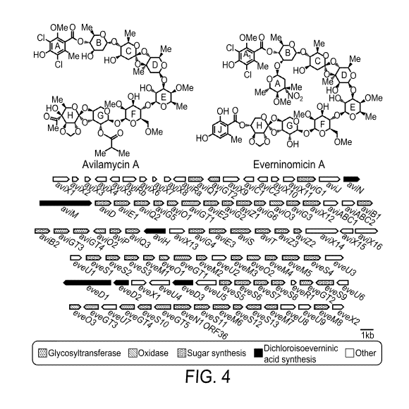
FIG. 1 contains structures of everninomicins and avilamycins.
FIG. 2 shows the ribosomal binding site of orthosomycin antibiotics. Small
ribosomal subunit (PDB 2J00) is shown in dark grey and large subunit (PDB
2J01) is shown
in lighter grey. The A and P sites are shown in salmon. Ribosomal protein L16
is shown in
green (chain Q), helix 89 (chain A, residues 2454-2498) in blue, and helix 91
(chain A,
residues 2520-2545) in magenta. Amino acid residues and nucleotides known to
interact
with everninomicin and avilamycin are highlighted in yellow.
FIG. 3 contains structures of new everninomicins Ever-2, and Ever-H (11), Ever
J
(12), and Ever-K (13). Also shown are the mass spectra fragmentation patterns.
Dashed
lines indicate position of cleavage during fragmentation experiments.
FIG. 4 shows the structures of avilamycin A and everninomicin A. Avi gene
cluster
from S. viridichromogenes Ti.157 and eve gene cluster from M carbonacea var
africana.
Genes are shaded according to putative functions.
FIG. 5 is a graph showing relative levels of everninomicin F produced by each
culture condition are shown.
3
CA 03006763 2018-05-29
WO 2017/100650 PCT/US2016/065938
FIGs. 6A-6C are graphs showing the minimal inhibitory concentration of each
everninomicin analog was tested against S. aureus subsp. aureus Rosenbach.
FIG. 6A
shows activity of everninomicin A against S. aureus subsp. aureus Rosenbach at
various
concentrations. FIG. 6B shows activity of full-length everninomicin-
rosaramicin conjugate
against S. aureus subsp. aureus Rosenbach at various concentrations. FIG. 6C
shows
activity of truncated everninomicin-rosaramicin conjugate against S. aureus
subsp. aureus
Rosenbach at various concentrations.
FIG. 7A is a photograph of a washer/membrane assembly with conjugation mixture
plate in the center. FIG. 7B is a photograph showing that after 9 days the
washer/membrane
assembly was removed, pure colonies of apramycin-resistant exconjugants
remained.
FIG. 8 contains maps of pSET152 and pSET152ermE. aac(3)IV is the apramycin
resistance marker; hyg is the hygromycin resistance marker hph; oriT is the
origin of
transfer; int is the phage (pC31 integrase; attP is the phage (pC31 attachment
site; ermE*
encodes a constitutively active promoter directly upstream of the multiple
cloning site.
FIG. 9 is a map of pSET152ermE, the genetic complementation plasmid. Plasmid
map was generated using Savvy (Scalable Vector Graphics & Plasmid Map
Copyright
2001, Malay K Basu) at http://www.bioinformatics.org/savvy/. Hyg is the
hygromycin
resistance marker hph; oriT is the origin of transfer; int is the phage (pC31
integrase; attP is
the phage (pC31 attachment site; ermE* is the constitutively active promoter
directly
upstream of the multiple cloning site.
FIG. 10 is a depiction and deduced functional assignment of ORFs from the evd
gene cluster of M carbonacea var aurantiaca.
FIG. 11 is a depiction and deduced functional assignment of ORFs from the eve
gene cluster of M carbonacea var africana.
FIG. 12 is a depiction and deduced functional assignment of ORFs from the ava
gene cluster of S. mobaraensis.
FIG. 13 is a phylogenetic analysis of methyltransferases from four class I
orthosomycin gene clusters, evd, eve, ava, and avi.
FIG. 14 is a scheme for two step targeted gene disruptions.
FIG. 15 is a depiction of a single crossover versus a double crossover
replacement.
FIG. 16 shows the results from Southern hybridization of targeted replacement
mutants verifying a double crossover event. All blots show predicted shifts
were observed
experimentally, thus confirming the double crossovers. Panel A shows the
Southern blot
analysis of evc/M2 ::aac(3)IV. Diagrams depict the relative shifts expected
for replacement
4
CA 03006763 2018-05-29
WO 2017/100650 PCT/US2016/065938
of evc1M2 with the apramycin cassette. Panel B shows the Southern blot
analysis of
Jevdi113::aac(3)1V. Diagrams depict the relative shifts expected for
replacement of evdM3
with the apramycin cassette. Panel C shows the Southern blot analysis of
JevdN1::aac(3)IV. Diagrams depict the relative shifts for replacement of evdN1
with the
apramycin cassette. Ladder is DNA molecular weight marker VII, DIG-labeled
(product no.
11669940910; Roche Life Sciences). WT is wild-type M carbonacea var
aurantiaca. ApaI,
KpnI, BamHI, XhoI, and EcoRV are restriction.
FIG. 17 shows a phylogenetic analysis of orthosomycin-associated oxygenases.
Analysis was conducted using MEGA 5 as described in the methods section. Class
I
orthosomycin-associated oxygenases formed three distinct group with each group
containing one oxygenase from each pathway. The Class II-associated oxygenase,
HygX,
did not cluster with the others oxygenases.
FIGs. 18A-18C show results from Southern hybridization of targeted deletion
mutants verifying a double crossover event. FIG. 18A is a Southern blot
analysis of
Jevd01::aac(3)1V. Diagrams depict the relative shifts expected for replacement
of evd01
with the apramycin cassette. Blots show predicted shifts were observed
experimentally, thus
confirming the double crossover. FIG. 18B is a Southern blot analysis of
JevdM01::aac(3)IV. Diagrams depict the relative shifts expected for
replacement of
evdi1101 with the apramycin cassette. Blots show predicted shifts were
observed
experimentally, thus confirming the double crossover. FIG. 18C is a Southern
blot analysis
of Jevd02::aac(3)IV. Diagrams depict the relative shifts for replacement of
evd02 with the
apramycin cassette. Blots do not have predicted shifts showing that the gene
replacement
was not successful. Ladder is DNA molecular weight marker VII, DIG-labeled
(product no.
11669940910; Roche Life Sciences). WT is wild-type M carbonacea var
aurantiaca. ApaI,
KpnI, NheI, XhoI, SphI, and BamHI are restriction endonucleases used to cleave
the
genomic DNA into predictably sized fragments. Blots show predicted shifts were
observed
experimentally, thus confirming the double crossover.
FIG. 19 shows the truncated everninomicin-rosaramicin conjugate (8) NMR data.
FIG. 20 shows the everninomicin-rosaramicin conjugate (9) NMR data
FIG. 21 shows the everninomicin H (11) NMR data.
FIGs. 22A-22C show LC/MS chromatograms of wild type M carbonacea var.
aurantiaca and gene replacements of evdM5 (xlevdill5::aac(3)1V). FIG. 22A is a
chromatogram showing summed ion intensities in negative mode for
everninomicins D¨G
5
CA 03006763 2018-05-29
WO 2017/100650 PCT/US2016/065938
and novel metabolites. FIG. 22B shows the structure for zlevc1M5::aac(3)1V
metabolites.
FIG. 22C shows the fragmentation pattern for des-methyl Ever F.
FIGs. 23A and 23B show LC/MS chromatograms of wild type M carbonacea var.
aurantiaca and gene replacements of evdD2 (zlevdD2::aac(3)IV). FIG. 23A is a
chromatogram showing summed ion intensities in negative mode for
everninomicins D¨G
and novel metabolites. FIG. 23B shows structure for JevdD2::aac(3)1V
metabolites.
FIGs. 24A and 24B show LC/MS chromatograms of wild type M carbonacea var.
aurantiaca and gene replacements of evdD1 (zlevdDl::aac(3)IV) and evdD3
(zlevdD3::aac(3)IV). FIG. 24A is a chromatogram showing summed ion intensities
in
negative mode for everninomicins D¨G and novel metabolites. FIG. 24B shows
fragmentation pattern for everninomicin Q.
DETAILED DESCRIPTION
The materials, compounds, compositions, and methods described herein may be
understood more readily by reference to the following detailed description of
specific
aspects of the disclosed subject matter, the Figures, and the Examples
included therein.
Before the present materials, compounds, compositions, and methods are
disclosed
and described, it is to be understood that the aspects described below are not
limited to
specific synthetic methods or specific reagents, as such may, of course, vary.
It is also to be
understood that the terminology used herein is for the purpose of describing
particular
aspects only and is not intended to be limiting.
Also, throughout this specification, various publications are referenced. The
disclosures of these publications in their entireties are hereby incorporated
by reference into
this application in order to more fully describe the state of the art to which
the disclosed
matter pertains. The references disclosed are also individually and
specifically incorporated
by reference herein for the material contained in them that is discussed in
the sentence in
which the reference is relied upon.
General Definitions
In this specification and in the claims that follow, reference will be made to
a
number of terms, which shall be defined to have the following meanings:
Throughout the specification and claims the word "comprise" and other forms of
the
word, such as "comprising" and "comprises," means including but not limited
to, and is not
intended to exclude, for example, other additives, components, integers, or
steps.
As used in the description and the appended claims, the singular forms "a,"
"an,"
and "the" include plural referents unless the context clearly dictates
otherwise. Thus, for
6
CA 03006763 2018-05-29
WO 2017/100650 PCT/US2016/065938
example, reference to "a composition" includes mixtures of two or more such
compositions,
reference to "an antibiotic" includes mixtures of two or more such
antibiotics, reference to
"the compound" includes mixtures of two or more such compounds, and the like.
"Optional" or "optionally" means that the subsequently described event or
circumstance can or cannot occur, and that the description includes instances
where the
event or circumstance occurs and instances where it does not.
Notwithstanding that the numerical ranges and parameters setting forth the
broad
scope of the disclosure are approximations, the numerical values set forth in
the specific
examples are reported as precisely as possible. Any numerical value, however,
inherently
contain certain errors necessarily resulting from the standard deviation found
in their
respective testing measurements. Furthermore, when numerical ranges of varying
scope are
set forth herein, it is contemplated that any combination of these values
inclusive of the
recited values may be used. Further, ranges can be expressed herein as from
"about" one
particular value, and/or to "about" another particular value. When such a
range is
expressed, another aspect includes from the one particular value and/or to the
other
particular value. Similarly, when values are expressed as approximations, by
use of the
antecedent "about," it will be understood that the particular value forms
another aspect. It
will be further understood that the endpoints of each of the ranges are
significant both in
relation to the other endpoint, and independently of the other endpoint.
Unless stated
otherwise, the term "about" means within 5% (e.g., within 2% or 1%) of the
particular value
modified by the term "about."
By "reduce" or other forms of the word, such as "reducing" or "reduction," is
meant
lowering of an event or characteristic (e.g., bacterial growth or infection).
It is understood
that this is typically in relation to some standard or expected value, in
other words it is
relative, but that it is not always necessary for the standard or relative
value to be referred
to. For example, "reduces bacterial growth" means decreasing the amount of
bacteria
relative to a standard or a control.
By "prevent" or other forms of the word, such as "preventing" or "prevention,"
is
meant to stop a particular event or characteristic, to stabilize or delay the
development or
progression of a particular event or characteristic, or to minimize the
chances that a
particular event or characteristic will occur. Prevent does not require
comparison to a
control as it is typically more absolute than, for example, reduce. As used
herein,
something could be reduced but not prevented, but something that is reduced
could also be
prevented. Likewise, something could be prevented but not reduced, but
something that is
7
CA 03006763 2018-05-29
WO 2017/100650 PCT/US2016/065938
prevented could also be reduced. It is understood that where reduce or prevent
are used,
unless specifically indicated otherwise, the use of the other word is also
expressly disclosed.
As used herein, "treatment" refers to obtaining beneficial or desired clinical
results.
Beneficial or desired clinical results include, but are not limited to, any
one or more of:
alleviation of one or more symptoms (such as bacterial growth or infection),
diminishment
of extent of infection, stabilized (i.e., not worsening) state of infection,
preventing or
delaying spread of the infection, preventing or delaying occurrence or
recurrence of
infection, and delay or slowing of infection progression.
The term "patient" preferably refers to a human in need of treatment with an
antibiotic or treatment for any purpose, and more preferably a human in need
of such a
treatment to treat bacterial infection. However, the term "patient" can also
refer to non-
human animals, preferably mammals such as dogs, cats, rabbits, horses, cows,
pigs, sheep,
goats, and non-human primates, among others, that are in need of treatment
with an
antibiotics. In other examples, the term "patient" can refer to poultry.
It is understood that throughout this specification the identifiers "first"
and "second"
are used solely to aid in distinguishing the various components and steps of
the disclosed
subject matter. The identifiers "first" and "second" are not intended to imply
any particular
order, amount, preference, or importance to the components or steps modified
by these
terms.
Chemical Definitions
As used herein, the term "composition" is intended to encompass a product
comprising the specified ingredients in the specified amounts, as well as any
product which
results, directly or indirectly, from combination of the specified ingredients
in the specified
amounts.
References in the specification and concluding claims to parts by weight of a
particular element or component in a composition denotes the weight
relationship between
the element or component and any other elements or components in the
composition or
article for which a part by weight is expressed. Thus, in a mixture containing
2 parts by
weight of component X and 5 parts by weight component Y, X and Y are present
at a
weight ratio of 2:5, and are present in such ratio regardless of whether
additional
components are contained in the mixture.
A weight percent (wt.%) of a component, unless specifically stated to the
contrary,
is based on the total weight of the formulation or composition in which the
component is
included.
8
CA 03006763 2018-05-29
WO 2017/100650 PCT/US2016/065938
As used herein, the term "substituted" is contemplated to include all
permissible
substituents of organic compounds. In a broad aspect, the permissible sub
stituents include
acyclic and cyclic, branched and unbranched, carbocyclic and heterocyclic, and
aromatic
and nonaromatic substituents of organic compounds. Illustrative substituents
include, for
example, those described below. The permissible substituents can be one or
more and the
same or different for appropriate organic compounds. For purposes of this
disclosure, the
heteroatoms, such as nitrogen, can have hydrogen substituents and/or any
permissible
substituents of organic compounds described herein which satisfy the valencies
of the
heteroatoms. This disclosure is not intended to be limited in any manner by
the permissible
substituents of organic compounds. Also, the terms "substitution" or
"substituted with"
include the implicit proviso that such substitution is in accordance with
permitted valence of
the substituted atom and the substituent, and that the substitution results in
a stable
compound, e.g., a compound that does not spontaneously undergo transformation
such as by
rearrangement, cyclization, elimination, etc.
The term "aliphatic" as used herein refers to a non-aromatic hydrocarbon group
and
includes branched and unbranched, alkyl, alkenyl, or alkynyl groups.
The term "alkyl" as used herein is a branched or unbranched saturated
hydrocarbon
group of 1 to 24 carbon atoms, such as methyl, ethyl, n-propyl, isopropyl, n-
butyl, isobutyl,
t-butyl, pentyl, hexyl, heptyl, octyl, nonyl, decyl, dodecyl, tetradecyl,
hexadecyl, eicosyl,
tetracosyl, and the like. The alkyl group can also be substituted or
unsubstituted. The alkyl
group can be substituted with one or more groups including, but not limited
to, alkyl,
alkoxy, alkenyl, alkynyl, aryl, heteroaryl, aldehyde, amino, azido, carboxylic
acid, cyano,
ester, ether, halide, hydroxy, ketone, nitro, silyl, sulfo-oxo, sulfonyl,
sulfone, sulfoxide, or
thiol as described herein.
The symbols A' is used herein as merely a generic substituent in the
definitions
below.
The term "alkoxy" as used herein is an alkyl group bound through a single,
terminal
ether linkage; that is, an "alkoxy" group can be defined as ¨OA' where Al is
alkyl as
defined above.
The term "alkenyl" as used herein is a hydrocarbon group of from 2 to 24
carbon
atoms with a structural formula containing at least one carbon-carbon double
bond.
Asymmetric structures such as (A1A2)C=C(A3A4) are intended to include both the
E and Z
isomers. This may be presumed in structural formulae herein wherein an
asymmetric
alkene is present, or it may be explicitly indicated by the bond symbol C=C.
The alkenyl
9
CA 03006763 2018-05-29
WO 2017/100650 PCT/US2016/065938
group can be substituted with one or more groups including, but not limited
to, alkyl,
alkoxy, alkenyl, alkynyl, aryl, heteroaryl, aldehyde, amino, azido, carboxylic
acid, cyano,
ester, ether, halide, hydroxy, ketone, nitro, silyl, sulfo-oxo, sulfonyl,
sulfone, sulfoxide, or
thiol as described herein.
The term "alkynyl" as used herein is a hydrocarbon group of 2 to 24 carbon
atoms
with a structural formula containing at least one carbon-carbon triple bond.
The alkynyl
group can be substituted with one or more groups including, but not limited
to, alkyl,
alkoxy, alkenyl, alkynyl, aryl, heteroaryl, aldehyde, amino, azido, carboxylic
acid, cyano,
ester, ether, halide, hydroxy, ketone, nitro, silyl, sulfo-oxo, sulfonyl,
sulfone, sulfoxide, or
thiol as described herein.
The term "aryl" as used herein is a group that contains any carbon-based
aromatic
group including, but not limited to, benzene, naphthalene, phenyl, biphenyl,
phenoxybenzene, and the like. The term "heteroaryl" is defined as a group that
contains an
aromatic group that has at least one heteroatom incorporated within the ring
of the aromatic
group. Examples of heteroatoms include, but are not limited to, nitrogen,
oxygen, sulfur,
and phosphorus. The term "non-heteroaryl," which is included in the term
"aryl," defines a
group that contains an aromatic group that does not contain a heteroatom. The
aryl and
heteroaryl group can be substituted or unsubstituted. The aryl and heteroaryl
group can be
substituted with one or more groups including, but not limited to, alkyl,
alkoxy, alkenyl,
alkynyl, aryl, heteroaryl, aldehyde, amino, azido, carboxylic acid, cyano,
ester, ether, halide,
hydroxy, ketone, nitro, silyl, sulfo-oxo, sulfonyl, sulfone, sulfoxide, or
thiol as described
herein. The term "biaryl" is a specific type of aryl group and is included in
the definition of
aryl. Biaryl refers to two aryl groups that are bound together via a fused
ring structure, as in
naphthalene, or are attached via one or more carbon-carbon bonds, as in
biphenyl.
The term "cycloalkyl" as used herein is a non-aromatic carbon-based ring
composed
of at least three carbon atoms. Examples of cycloalkyl groups include, but are
not limited
to, cyclopropyl, cyclobutyl, cyclopentyl, cyclohexyl, etc. The term
"heterocycloalkyl" is a
cycloalkyl group as defined above where at least one of the carbon atoms of
the ring is
substituted with a heteroatom such as, but not limited to, nitrogen, oxygen,
sulfur, or
phosphorus. The cycloalkyl group and heterocycloalkyl group can be substituted
or
unsubstituted. The cycloalkyl group and heterocycloalkyl group can be
substituted with one
or more groups including, but not limited to, alkyl, alkoxy, alkenyl, alkynyl,
aryl,
heteroaryl, aldehyde, amino, azido, carboxylic acid, cyano, ester, ether,
halide, hydroxy,
ketone, nitro, silyl, sulfo-oxo, sulfonyl, sulfone, sulfoxide, or thiol as
described herein.
CA 03006763 2018-05-29
WO 2017/100650 PCT/US2016/065938
The term "cycloalkenyl" as used herein is a non-aromatic carbon-based ring
composed of at least three carbon atoms and containing at least one double
bound, i.e.,
C=C. Examples of cycloalkenyl groups include, but are not limited to,
cyclopropenyl,
cyclobutenyl, cyclopentenyl, cyclopentadienyl, cyclohexenyl, cyclohexadienyl,
and the like.
The term "heterocycloalkenyl" is a type of cycloalkenyl group as defined above
where at
least one of the carbon atoms of the ring is substituted with a heteroatom
such as, but not
limited to, nitrogen, oxygen, sulfur, or phosphorus. The cycloalkenyl group
and
heterocycloalkenyl group can be substituted or unsubstituted. The cycloalkenyl
group and
heterocycloalkenyl group can be substituted with one or more groups including,
but not
limited to, alkyl, alkoxy, alkenyl, alkynyl, aryl, heteroaryl, aldehyde,
amino, azido,
carboxylic acid, cyano, ester, ether, halide, hydroxy, ketone, nitro, silyl,
sulfo-oxo, sulfonyl,
sulfone, sulfoxide, or thiol as described herein.
The term "cyclic group" is used herein to refer to either aryl groups, non-
aryl groups
(i.e., cycloalkyl, heterocycloalkyl, cycloalkenyl, and heterocycloalkenyl
groups), or both.
Cyclic groups have one or more ring systems that can be substituted or
unsubstituted. A
cyclic group can contain one or more aryl groups, one or more non-aryl groups,
or one or
more aryl groups and one or more non-aryl groups.
The term "aldehyde" as used herein is represented by the formula ¨C(0)H.
Throughout this specification "C(0)" is a short hand notation for CO.
The terms "amine" or "amino" as used herein are represented by the formula
NA1A2A3, where A', A2, and A3 can be, independently, hydrogen, an alkyl,
halogenated
alkyl, alkenyl, alkynyl, aryl, heteroaryl, cycloalkyl, cycloalkenyl,
heterocycloalkyl, or
heterocycloalkenyl group described above.
The term "carboxylic acid" as used herein is represented by the formula
¨C(0)0H.
A "carboxylate" as used herein is represented by the formula ¨C(0)0-.
The term "ester" as used herein is represented by the formula ¨0C(0)A1 or ¨
C(0)0A1, where A' can be an alkyl, halogenated alkyl, alkenyl, alkynyl, aryl,
heteroaryl,
cycloalkyl, cycloalkenyl, heterocycloalkyl, or heterocycloalkenyl group
described above.
The term "ether" as used herein is represented by the formula Al0A2, where A'
and
A2 can be, independently, an alkyl, halogenated alkyl, alkenyl, alkynyl, aryl,
heteroaryl,
cycloalkyl, cycloalkenyl, heterocycloalkyl, or heterocycloalkenyl group
described above.
The term "ketone" as used herein is represented by the formula A1C(0)A2, where
A'
and A2 can be, independently, an alkyl, halogenated alkyl, alkenyl, alkynyl,
aryl, heteroaryl,
cycloalkyl, cycloalkenyl, heterocycloalkyl, or heterocycloalkenyl group
described above.
11
CA 03006763 2018-05-29
WO 2017/100650 PCT/US2016/065938
The term "halide" as used herein refers to the halogens fluorine, chlorine,
bromine,
and iodine.
The term "hydroxyl" as used herein is represented by the formula ¨OH.
The term "nitro" as used herein is represented by the formula ¨NO2.
The term "cyano" as used herein is represented by the formula ¨CN
The term "azido" as used herein is represted by the formula ¨N3.
The term "sulfonyl" is used herein to refer to the sulfo-oxo group represented
by the
formula --S(0)2A1, where Al can be hydrogen, an alkyl, halogenated alkyl,
alkenyl, alkynyl,
aryl, heteroaryl, cycloalkyl, cycloalkenyl, heterocycloalkyl, or
heterocycloalkenyl group
described above.
The term "sulfonylamino" or "sulfonamide" as used herein is represented by the
formula --S(0)2NH2.
The term "thiol" as used herein is represented by the formula --SH.
It is to be understood that the compounds provided herein may contain chiral
centers. Such chiral centers may be of either the (R-) or (S-) configuration.
The compounds
provided herein may either be enantiomerically pure, or be diastereomeric or
enantiomeric
mixtures. It is to be understood that the chiral centers of the compounds
provided herein
may undergo epimerization in vivo. As such, one of skill in the art will
recognize that
administration of a compound in its (R-) form is equivalent, for compounds
that undergo
epimerization in vivo, to administration of the compound in its (S-) form.
As used herein, substantially pure means sufficiently homogeneous to appear
free of
readily detectable impurities as determined by standard methods of analysis,
such as thin
layer chromatography (TLC), nuclear magnetic resonance (NMR), gel
electrophoresis, high
performance liquid chromatography (HPLC) and mass spectrometry (MS), gas-
chromatography mass spectrometry (GC-MS), and similar, used by those of skill
in the art
to assess such purity, or sufficiently pure such that further purification
would not detectably
alter the physical and chemical properties, such as enzymatic and biological
activities, of
the substance. Both traditional and modern methods for purification of the
compounds to
produce substantially chemically pure compounds are known to those of skill in
the art. A
substantially chemically pure compound may, however, be a mixture of
stereoisomers.
Unless stated to the contrary, a formula with chemical bonds shown only as
solid
lines and not as wedges or dashed lines contemplates each possible isomer,
e.g., each
enantiomer, diastereomer, and meso compound, and a mixture of isomers, such as
a racemic
or scalemic mixture.
12
CA 03006763 2018-05-29
WO 2017/100650 PCT/US2016/065938
A "pharmaceutically acceptable" component is one that is suitable for use with
humans and/or animals without undue adverse side effects (such as toxicity,
irritation, and
allergic response) commensurate with a reasonable benefit/risk ratio.
"Pharmaceutically acceptable salt" refers to a salt that is pharmaceutically
acceptable and has the desired pharmacological properties. Such salts include
those that
may be formed where acidic protons present in the compounds are capable of
reacting with
inorganic or organic bases. Suitable inorganic salts include those formed with
the alkali
metals, e.g., sodium, potassium, magnesium, calcium, and aluminum. Suitable
organic salts
include those formed with organic bases such as the amine bases, e.g.,
ethanolamine,
diethanolamine, triethanolamine, tromethamine, N-methylglucamine, and the
like. Such
salts also include acid addition salts formed with inorganic acids (e.g.,
hydrochloric and
hydrobromic acids) and organic acids (e.g., acetic acid, citric acid, maleic
acid, and the
alkane- and arene-sulfonic acids such as methanesulfonic acid and
benzenesulfonic acid).
When two acidic groups are present, a pharmaceutically acceptable salt may be
a mono-
acid-mono-salt or a di-salt; similarly, where there are more than two acidic
groups present,
some or all of such groups can be converted into salts.
"Pharmaceutically acceptable excipient" refers to an excipient that is
conventionally
useful in preparing a pharmaceutical composition that is generally safe, non-
toxic, and
desirable, and includes excipients that are acceptable for veterinary use as
well as for human
pharmaceutical use. Such excipients can be solid, liquid, semisolid, or, in
the case of an
aerosol composition, gaseous.
A "pharmaceutically acceptable carrier" is a carrier, such as a solvent,
suspending
agent or vehicle, for delivering the disclosed compounds to the patient. The
carrier can be
liquid or solid and is selected with the planned manner of administration in
mind.
Liposomes are also a pharmaceutical carrier. As used herein, "carrier"
includes any and all
solvents, dispersion media, vehicles, coatings, diluents, antibacterial and
antifungal agents,
isotonic and absorption delaying agents, buffers, carrier solutions,
suspensions, colloids,
and the like. The use of such media and agents for pharmaceutical active
substances is well
known in the art. Except insofar as any conventional media or agent is
incompatible with
the active ingredient, its use in the therapeutic compositions is
contemplated.
The term "therapeutically effective amount" as used herein means that amount
of
active compound or pharmaceutical agent that elicits the biological or
medicinal response in
a tissue, system, animal or human that is being sought by a researcher,
veterinarian, medical
doctor or other clinician. In reference to infection, an effective amount
comprises an
13
CA 03006763 2018-05-29
WO 2017/100650 PCT/US2016/065938
amount sufficient to cause a bacterial cell to shrink and/or to decrease the
growth rate of the
cells (such as to suppress bacterial growth) or to prevent or delay other
unwanted cell
proliferation. In some embodiments, an effective amount is an amount
sufficient to delay
development. In some embodiments, an effective amount is an amount sufficient
to prevent
or delay occurrence and/or recurrence. An effective amount can be administered
in one or
more doses. In the case of infection, the effective amount of the drug or
composition may:
(i) reduce the number of bacterial cells; (ii) inhibit, retard, slow to some
extent and
preferably stop bacterial cell infiltration into peripheral organs; (iii)
inhibit bacterial growth;
(iv) prevent or delay occurrence and/or recurrence of infection; and/or (v)
relieve to some
extent one or more of the symptoms associated with the infection.
Effective amounts of a compound or composition described herein for treating a
mammalian subject can include about 0.1 to about 1000 mg/Kg of body weight of
the
subject/day, such as from about 1 to about 100 mg/Kg/day, especially from
about 10 to
about 100 mg/Kg/day. The doses can be acute or chronic. A broad range of
disclosed
composition dosages are believed to be both safe and effective.
Biology Definition
The use of italics indicates a nucleic acid molecule (e.g., end cDNA, gene,
etc.);
normal text indicates the polypeptide or protein.
"Sequence-conservative variants" of a polynucleotide sequence are those in
which a
change of one or more nucleotides in a given codon position results in no
alteration in the
amino acid encoded at that position.
"Function-conservative variants" are those in which a given amino acid residue
in a
protein or enzyme has been changed without altering the overall conformation
and function
of the polypeptide, including, but not limited to, replacement of an amino
acid with one
having similar properties (such as, for example, polarity, hydrogen bonding
potential,
acidic, basic, hydrophobic, aromatic, and the like). Amino acids with similar
properties are
well known in the art. For example, arginine, histidine and lysine are
hydrophilic-basic
amino acids and may be interchangeable. Similarly, isoleucine, a hydrophobic
amino acid,
may be replaced with leucine, methionine or valine. Such changes are expected
to have little
or no effect on the apparent molecular weight or isoelectric point of the
protein or
polypeptide. Amino acids other than those indicated as conserved may differ in
a protein or
enzyme so that the percent protein or amino acid sequence similarity between
any two
proteins of similar unction may vary and may be, for example, from 70% to 99%
as
determined according to an alignment scheme such as by the Cluster Method,
wherein
14
CA 03006763 2018-05-29
WO 2017/100650
PCT/US2016/065938
similarity is based on the MEGALIGN algorithm. A "function-conservative
variant" also
includes a polypeptide or enzyme which has at least 60% amino acid identity as
determined
by BLAST or FASTA algorithms, preferably at least 75%, most preferably at
least 85%, ad
even more preferably at least 90%, and which has the same or substantially
similar
properties or functions as the native or parent protein or enzyme to which it
is compared.
The terms "mutant" and "mutation" mean any detectable change in genetic
material,
e.g. DNA, or any process, mechanism, or result of such a change. This includes
gene
mutations, in which the structure (e.g. DNA sequence) of a gene is altered,
any gene or
DNA arising from any mutation process, and any expression product (e.g.
protein or
enzyme) expressed by a modified gene or DNA sequence. The term "variant" may
also be
used to indicate a modified or altered gene, DNA sequence, enzyme, cell, etc.,
i.e., any kind
of mutant.
As used herein, the term "homologous" in all its grammatical forms and
spelling
variations refers to the relationship between proteins that possess a "common
evolutionary
origin," including proteins from superfamilies (e.g., the immunoglobulin
superfamily) and
homologous proteins from different species (e.g., myosin light chain, etc.)
(Reeck et al.,
Cell 50:667, 1987). Such proteins (and their encoding genes) have sequence
homology, as
reflected by their sequence similarity, whether in terms of percent similarity
or the presence
of specific residues or motifs at conserved positions.
Accordingly, the term "sequence similarity" in all its grammatical forms
refers to
the degree of identity or correspondence between nucleic acid or amino acid
sequences of
proteins that may or may not share a common evolutionary origin (see Reeck et
al., supra).
However, in common usage and in the instant application, the term
"homologous," when
modified with an adverb such as "highly," may refer to sequence similarity and
may or may
not relate to a common evolutionary origin.
In a specific embodiment, two DNA sequences are "substantially homologous" or
"substantially similar" when the encoded polypeptides are at least 35-40%
similar as
determined by one of the algorithms disclosed herein, preferably at least
about 60%, and
most preferably at least about 90 or 95% in a highly conserved domain, or, for
alleles,
across the entire amino acid sequence. Sequence comparison algorithms include
BLAST
(BLAST P, BLAST N, BLAST X), FASTA, DNA Strider, the GCG (Genetics Computer
Group, Program Manual for the GCG Package, Version 7, Madison, Wis.) pileup
program,
etc. using the default parameters provided with these algorithms. An example
of such a
sequence is an allelic or species variant of the specific everninomicin
biosynthetic genes of
CA 03006763 2018-05-29
WO 2017/100650 PCT/US2016/065938
the invention. Sequences that are substantially homologous can be identified
by comparing
the sequences using standard software available in sequence data banks, or in
a Southern
hybridization experiment under, for example, stringent conditions as defined
for that
particular system.
"Amplification" of DNA, as used herein, denotes the use of polymerase chain
reaction (PCR) to increase the concentration of a particular DNA sequence
within a mixture
of DNA sequences. For a description of PCR see Saiki et an, Science, 239:487,
1988.
A "nucleic acid molecule" refers to the phosphate ester polymeric form of
ribonucleosides (adenosine, guanosine, uridine or cytidine; "RNA molecules");
or
deoxyribonucleosides (deoxyadenosine, deoxyguanosine, deoxythymidine, or
deoxycytidine; "DNA molecules"); or any phosphoester analogs thereof, such as
phosphorothioates and thioesters, in either single stranded form, or a double-
stranded helix;
or "protein nucleic acids" (PNA) formed by conjugating bases to an amino acid
backbone;
or nucleic acids containing modified bases, for example thiouracil, thio-
guanine and fluoro-
uracil. Double stranded DNA-DNA, DNA-RNA and RNA-RNA helices are possible. The
term nucleic acid molecule, and in particular DNA or RNA molecule, refers only
to the
primary and secondary structure of the molecule, and does not limit it to any
particular
tertiary forms. Thus, this term includes double-stranded DNA found, inter
alia, in linear
(e.g., restriction fragments) or circular DNA molecules, plasmids, and
chromosomes. In
discussing the structure of particular double-stranded DNA molecules,
sequences may be
described herein according to the normal convention of giving only the
sequence in the 5' to
3' direction along the nontranscribed strand of DNA (i.e., the strand having a
sequence
homologous to the mRNA). A "recombinant DNA molecule" is a DNA molecule that
has
undergone a molecular biological manipulation.
A "polynucleotide" or "nucleotide sequence" is a series of nucleotide bases
(also
called "nucleotides") in DNA and RNA, and means any chain of two or more
nucleotides.
A nucleotide sequence typically carries genetic information, including the
information used
by cellular machinery to make proteins and enzymes. These terms include double
or single
stranded genomic and cDNA, RNA, any synthetic and genetically manipulated
polynucleotide, and both sense and anti-sense polynucleotide (although only
sense stands
are being represented herein). This includes single- and double-stranded
molecules, i.e.,
DNA-DNA, DNA-RNA and RNA-RNA hybrids.
The polynucleotides herein may be flanked by natural regulatory (expression
control) sequences, or may be associated with heterologous sequences,
including promoters,
16
CA 03006763 2018-05-29
WO 2017/100650 PCT/US2016/065938
internal ribosome entry sites (IRES) and other ribosome binding site
sequences, enhancers,
response elements, suppressors, signal sequences, polyadenylation sequences,
introns, 5'-
and 3'-non-coding regions, and the like. The nucleic acids may also be
modified by many
means known in the art. Furthermore, the polynucleotides herein may also be
oligonucleotides modified with a label capable of providing a detectable
signal, either
directly or indirectly. Exemplary labels include radioisotopes, fluorescent
molecules, biotin,
and the like.
A "coding sequence" or a sequence "encoding" an expression product, such as a
RNA, polypeptide, protein, or enzyme, is a minimum nucleotide sequence that,
when
expressed, results in the production of that RNA, polypeptide, protein, or
enzyme, i.e., the
nucleotide sequence encodes an amino acid sequence for that polypeptide,
protein or
enzyme. A coding sequence for a protein may include a start codon (usually
ATG, though
as shown herein, alternative start codons can be used) and a stop codon.
The term "gene", also called a "structural gene" means a DNA sequence that
codes
for a particular sequence of amino acids, which comprise all or part of one or
more proteins
or enzymes, and may include regulatory (non-transcribed) DNA sequences, such
as
promoter sequences, which determine for example the conditions under which the
gene is
expressed. The transcribed region of the gene may include untranslated
regions, including a
5'-untranslated region (UTR) and 3'-UTR, as well as the coding sequence.
A "promoter sequence" is a DNA regulatory region capable of binding RNA
polymerase in a cell and initiating transcription of a downstream (3'
direction) coding
sequence. For purposes of defining the present invention, the promoter
sequence is bounded
at its 3' terminus by the transcription initiation site and extends upstream
(5' direction) to
include the minimum number of bases or elements necessary to initiate
transcription at
levels detectable above background. Within the promoter sequence will be found
a
transcription initiation site (conveniently defined for example, by mapping
with nuclease
Si), as well as protein binding domains (consensus sequences) responsible for
the binding
of RNA polymerase.
A coding sequence is "under the control of' or "operably (or operatively)
associated
with" transcriptional and translational control sequences in a cell when RNA
polymerase
transcribes the coding sequence into mRNA, which is then trans-RNA spliced (if
it contains
introns) and translated into the protein encoded by the coding sequence.
The terms "express" and "expression" mean allowing or causing the information
in a
gene or DNA sequence to become manifest, for example producing a protein by
activating
17
CA 03006763 2018-05-29
WO 2017/100650 PCT/US2016/065938
the cellular functions involved in transcription and translation of a
corresponding gene or
DNA sequence. A DNA sequence is expressed in or by a cell to form an
"expression
product" such as mRNA or a protein. The expression product itself, e.g. the
resulting
mRNA or protein, may also be said to be "expressed" by the cell. An expression
product
can be characterized as intracellular, extracellular or secreted. The term
"intracellular"
means something that is inside a cell. The term "extracellular" means
something that is
outside a cell. A substance is "secreted" by a cell if it appears in
significant measure outside
the cell, from somewhere on or inside the cell.
The term "transfection" means the introduction of a heterologous nucleic acid
into a
host cell. The term "transformation" means the introduction of a heterologous
gene, DNA
or RNA sequence to a host cell, so that the host cell will express the
introduced gene or
sequence to produce a desired product. The introduced gene or sequence may
also be called
a "cloned" or "heterologous" gene or sequence, and may include regulatory or
control
sequences, such as start, stop, promoter, signal, secretion, or other
sequences used by a cell's
genetic machinery. The gene or sequence may include nonfunctional sequences or
sequences with no known function. A host cell that receives and expresses
introduced DNA
or RNA has been "transformed" and is a "transformant" or a "clone." The DNA or
RNA
introduced to a host cell can come from any source, including cells of the
same genus or
species as the host cell, or cells of a different genus or species.
The terms "vector", "cloning vector" and "expression vector" mean the vehicle
by
which a DNA or RNA sequence (e.g. a foreign gene) can be introduced into a
host cell, so
as to transform the host and promote expression (e.g. transcription and
translation) of the
introduced sequence. Vectors include plasmids, phages, viruses, etc.; they are
discussed in
greater detail below.
Vectors typically comprise the DNA of a transmissible agent, into which
heterologous DNA is inserted. A common way to insert one segment of DNA into
another
segment of DNA involves the use of enzymes called restriction enzymes that
cleave DNA at
specific sites (specific groups of nucleotides) called restriction sites. A
"cassette" refers to a
DNA coding sequence or segment of DNA that codes for an expression product
that can be
inserted into a vector at defined restriction sites. The cassette restriction
sites are designed
to ensure insertion of the cassette in the proper reading frame. Generally,
foreign DNA is
inserted at one or more restriction sites of the vector DNA, and then is
carried by the vector
into a host cell along with the transmissible vector DNA. A segment or
sequence of DNA
having inserted or added DNA, such as an expression vector, can also be called
a "DNA
18
CA 03006763 2018-05-29
WO 2017/100650 PCT/US2016/065938
construct." A common type of vector is a "plasmid", which generally is a self-
contained
molecule of double-stranded DNA, usually of bacterial origin, that can readily
accept
additional (foreign) DNA and which can readily introduced into a suitable host
cell. A
plasmid vector often contains coding DNA and promoter DNA and has one or more
restriction sites suitable for inserting foreign DNA. Promoter DNA is a DNA
sequence
which initiates, regulates, or otherwise mediates or controls the expression
of the coding
DNA. Promoter DNA and coding DNA may be from the same gene or from different
genes,
and may be from the same or different organisms. A large number of vectors,
including
plasmid and fungal vectors, have been described for replication and/or
expression in a
variety of eukaryotic and prokaryotic hosts. Non-limiting examples include pKK
plasmids
(Clonetech), pUC plasmids, pET plasmids (Novagen, Inc., Madison, Wis.), pRSET
or pREP
plasmids (Invitrogen, San Diego, Calif), or pMAL plasmids (New England
Biolabs,
Beverly, Mass.), and many appropriate host cells, using methods disclosed or
cited herein or
otherwise known to those skilled in the relevant art. Recombinant cloning
vectors will often
include one or more replication systems for cloning or expression, one or more
markers for
selection in the host, e.g. antibiotic resistance, and one or more expression
cassettes.
The term "host cell" means any cell of any organism that is selected,
modified,
transformed, grown, or used or manipulated in any way, for the production of a
substance
by the cell, for example the expression by the cell of a gene, a DNA or RNA
sequence, a
protein or an enzyme. Host cells can further be used for screening or other
assays, as
described infra. In a preferred aspect, a host cell of the invention is an
actinomycete,
preferably of the genus Streptomyces (e.g., a host cell as described in
Ziermann and Betlach,
BioTechniques, 1999, 26:106) or alternatively Micromonospera. Additional
examples
include, but are not limited to, the strains S. pristinaespiralis (ATCC
25486), S. antibioticus
(DSM 40868), S. bikiniensis (ATCC 11062), S. parvulus (ATCC 12434), S.
glauescens
(ETH 22794), S. actuosus (ATCC 25421), S. coelicolor (A3(2)), S. ambofaciens,
S.
lividans, S. griseofuscus, S. limosus, and the like (see also Smokvina et al.,
Proceedings,
1:403-407).
The term "expression system" means a host cell and compatible vector under
suitable conditions, e.g., for the expression of a protein coded for by
foreign DNA carried
by the vector and introduced to the host cell. Common expression systems
include E. col/
host cells and plasmid vectors, although the actinomycte host cell expression
systems are
preferred for biosynthesis of everninomicin and related products.
19
CA 03006763 2018-05-29
WO 2017/100650 PCT/US2016/065938
The term "heterologous" refers to a combination of elements not naturally
occurring.
For example, heterologous DNA refers to DNA not naturally located in the cell,
or in a
chromosomal site of the cell. A heterologous gene is a gene in which the
regulatory control
sequences are not found naturally in association with the coding sequence. In
the context of
the present invention, an EV biosynthetic enzyme gene is heterologous to the
vector DNA
in which it is inserted for cloning or expression, and it is heterologous to a
host cell
containing such a vector, in which it is expressed, e.g., a K562 cell.
A nucleic acid molecule is "hybridizable" to another nucleic acid molecule,
such as
a cDNA, genomic DNA, or RNA, when a single stranded form of the nucleic acid
molecule
can anneal to the other nucleic acid molecule under the appropriate conditions
of
temperature and solution ionic strength (see Sambrook et al., supra). The
conditions of
temperature and ionic strength determine the "stringency" of the
hybridization. For
preliminary screening for homologous nucleic acids, low stringency
hybridization
conditions, corresponding to a T. (melting temperature) of 55 C., can be
used, e.g., 5x SSC,
0.1% SDS, 0.25% milk, and no formamide; or 30% formamide, 5x SSC, 0.5% SDS).
Moderate stringency hybridization conditions correspond to a higher T., e.g.,
40%
formamide, with 5x or 6x SCC. High stringency hybridization conditions
correspond to the
highest T., e.g., 50% formamide, 5x or 6x SCC. SCC is a 0.15M NaC1, 0.015M Na-
citrate.
Hybridization requires that the two nucleic acids contain complementary
sequences,
although depending on the stringency of the hybridization, mismatches between
bases are
possible. The appropriate stringency for hybridizing nucleic acids depends on
the length of
the nucleic acids and the degree of complementation, variables well known in
the art. The
greater the degree of similarity or homology between two nucleotide sequences,
the greater
the value of T. for hybrids of nucleic acids having those sequences. The
relative stability
(corresponding to higher T.) of nucleic acid hybridizations decreases in the
following
order: RNA:RNA, DNA:RNA, DNA:DNA. For hybrids of greater than 100 nucleotides
in
length, equations for calculating T. have been derived (see Sambrook et al.,
supra, 9.50-
9.51). For hybridization with shorter nucleic acids, i.e., oligonucleotides,
the position of
mismatches becomes more important, and the length of the oligonucleotide
determines its
specificity (see Sambrook et al., supra, 11.7-11.8). A minimum length for a
hybridizable
nucleic acid is at least about 10 nucleotides; preferably at least about 15
nucleotides; and
more preferably the length is at least about 20 nucleotides.
In a specific embodiment, the term "standard hybridization conditions" refers
to a
T. of 55 C., and utilizes conditions as set forth above. In a preferred
embodiment, the Tiflis
CA 03006763 2018-05-29
WO 2017/100650
PCT/US2016/065938
60 C.; in a more preferred embodiment, the Tiflis 65 C. In a specific
embodiment, "high
stringency" refers to hybridization and/or washing conditions at 68 C. in
0.2x SSC, at 42
C. in 50% formamide, 4x SSC, or under conditions that afford levels of
hybridization
equivalent to those observed under either of these two conditions.
As used herein, the term "oligonucleotide" refers to a nucleic acid, generally
of at
least 10, preferably at least 15, and more preferably at least 20 nucleotides,
preferably no
more than 100 nucleotides, that is hybridizable to a genomic DNA molecule, a
cDNA
molecule, or an mRNA molecule encoding a gene, mRNA, cDNA, or other nucleic
acid of
interest. Oligonucleotides can be labeled, e.g., with 32P-nucleotides or
nucleotides to which
a label, such as biotin, has been covalently conjugated. In one embodiment, a
labeled
oligonucleotide can be used as a probe to detect the presence of a nucleic
acid. In another
embodiment, oligonucleotides (one or both of which may be labeled) can be used
as PCR
primers, either for cloning full length or a fragment of EV biosynthetic
enzyme, or to detect
the presence of nucleic acids encoding EV biosynthetic enzyme. In a further
embodiment,
an oligonucleotide of the invention can form a triple helix with a EV
biosynthetic enzyme
DNA molecule. Generally, oligonucleotides are prepared synthetically,
preferably on a
nucleic acid synthesizer. Accordingly, oligonucleotides can be prepared with
non-naturally
occurring phosphoester analog bonds, such as thioester bonds, etc.
Reference will now be made in detail to specific aspects of the disclosed
materials,
compounds, compositions, articles, and methods, examples of which are
illustrated in the
accompanying Examples and Figures.
Compounds
To date four everninomicin congeners, Ever D-G 2-5, have been reported from M
carbonacea var aurantiaca all of which vary in the oxidation state of the
nitrogen on the A
ring. Disclosed herein, in certain examples, are everninomicin-rosaramicin
conjugates 8 and
9. Rosaramicin (7) is a glycosylated macrolactone also produced by M
carbonacea.
21
CA 03006763 2018-05-29
WO 2017/100650 PCT/US2016/065938
OMe 0 Me Me
0
CI
\
OH
Oh 13 . Oh. C
IA1
HO Me H 0
0 CI
Hcf
=`MeCHO A Me
Me, I Mew
NMe2 C) Me
0 HoOpme 00' \ Me 0
0 ' Me2N
Me
Me Me =
Rosaramicin (7) HO .Me
=
CH2Me
Truncated Everninomicin-Rosaramicin (8)
Me Me OMe OMe 0/\0
OMe 0 Me Me
Me0 0 0 7 0 S
A Me
CI 0 0õ OH
I Ai
0
M e
0 . Cr. OMe
HO Me 6H OH Med
CI 0
Everninomicin-Rosaramicin (9)
Mew
Me 000' \ Me
Me2N
Me..t\-001-1 Me
Me ==
0 "(
HO ..iMe
=
CH2Me
The hydroxyl amino functionality of everninomicin F (4) reacts with the
aldehyde
moiety of rosaramicin to generate a nitrone linkage/to create a nitrone which
links the two
natural products. The full length everninomicin-rosaramicin conjugate 9 is the
intact
precursor to the degraded saccharide complex 8. The chemical precedent for
formation of
the nitrone is well established and the data herein have shown that 9 degrades
to 8 when
exposed to normal culture conditions. The structures are shown as having
either or both cis
and trans geometries at the nitrone, thus contemplated herein are the cis,
trans, and mixtures
thereof. Excitingly, trapping of everninomicins by rosaramicins via nitrone
formation
results in increased ionization which aids in mass spec identification of new
everninomicins. Although Nature has provided natural everninomicin congeners
to begin to
study the relationship between structure and activity, there is still a need
to make non-
22
CA 03006763 2018-05-29
WO 2017/100650
PCT/US2016/065938
natural analogs for further study. As chemical synthesis of new analogs is not
practical, new
analogs are prepared herein by modification of the everninomicin gene cluster.
Disclosed in certain examples are compounds having the structure:
Me R4 R5 OR7
OMe 0 Me Me
Oup..01..p
HO Me z Rii
OH
9 6
k 1
wherein
R4-R6 are each, individually, H, OH, OCH3, CH2OH, CHO, CO2H, CO2R12, C(0)1t12,
or
substituted Ci-C6 alkyl;
R7 is H, CH3, CH2OH, C(0)1t12, substituted Ci-C6 alkyl; or orsellinyl;
Rg is H, OH, OCH3, CH2OH, CHO, CO2H, CO2R12, C(0)R12, or substituted Ci-C6
alkyl;
R9is H, OH, OCH3, CH2OH, CHO, CO2H, CO2R12, C(0)R12, or substituted Ci-C6
alkyl;;
RH is H, NH2, NO2, NOH, OMe, Ci-C6 alkyl, optionally substituted with alkyl,
alkoxy,
alkenyl, alkynyl, aryl, heteroaryl, aldehyde, amino, azido, carboxylic acid,
cyano, ester,
ether, halide, hydroxy, ketone, nitro, silyl, sulfo-oxo, sulfonyl, sulfone,
sulfoxide, or
thiol, or a 1-20 atom linker bound to rosaramicin; and
Ri2 is Cu-C6 alkyl, optionally substituted with alkyl, alkoxy, alkenyl,
alkynyl, aryl,
heteroaryl, aldehyde, amino, azido, carboxylic acid, cyano, ester, ether,
halide, hydroxy,
ketone, nitro, silyl, sulfo-oxo, sulfonyl, sulfone, sulfoxide, or thiol,
or a pharmaceutically acceptable salt thereof.
For example, discloses is a compound having the structure:
Me Me OMe OMe 10/.0
HO
OMe 0 Me Me
0 0õ. oMe0 0 0 0 - .00 = OH
CI
$11
0 Cr. OMe
Me H
OH H
Ever-2 (10)
or a pharmaceutically acceptable salt thereof.
Also disclosed are compounds having the structure:
23
CA 03006763 2018-05-29
WO 2017/100650 PCT/US2016/065938
Me Me OMe 0R6 10/0
OMe 0 Me Me
oMe0 0 MOR 5 r
CI
HO Me 61-1 H
oelµAe
R11
wherein
R3 and R5 are each, individually, H, OH, OCH3, CH2OH, CHO, CO2H, CO2R12,
C(0)1t12, or
substituted Ci-C6 alkyl;
R6 is H, OH, OCH3, CH2OH, CHO, CO2H, CO2R12, C(0)R12, substituted Ci-C6 alkyl;
or
orsellinyl;
R7 is H, OH, OCH3, CH2OH, CHO, CO2H, CO2R12, C(0)R12, or substituted Ci-C6
alkyl;
R8 is OH, OCH3, CH2OH, CHO, CO2H, CO2R12, C(0)R12, or substituted C1-C6 alkyl;
RH is H, OMe, NH2, NO2, NOH, C1-C6 alkyl, optionally substituted with alkyl,
alkoxy,
alkenyl, alkynyl, aryl, heteroaryl, aldehyde, amino, azido, carboxylic acid,
cyano, ester,
ether, halide, hydroxy, ketone, nitro, silyl, sulfo-oxo, sulfonyl, sulfone,
sulfoxide, or
thiol, or a 1-20 atom linker bound to rosaramicin; and
R12 is Ci-C6 alkyl, optionally substituted with alkyl, alkoxy, alkenyl,
alkynyl, aryl,
heteroaryl, aldehyde, amino, azido, carboxylic acid, cyano, ester, ether,
halide, hydroxy,
ketone, nitro, silyl, sulfo-oxo, sulfonyl, sulfone, sulfoxide, or thiol,
or a pharmaceutically acceptable salt thereof.
For example, disclosed are compounds having the structure:
24
CA 03006763 2018-05-29
WO 2017/100650
PCT/US2016/065938
OMe 0 Me Me Me Me OMe OH (D/c)
oh, oMe0 0 0 0 : .00 =
OH
CI
illo Chop...0hp, 44õ,õ Li,lx 40, , ..,.. me
t) 0 . Cr. OMe
HO Me H MeCr
H 6H H
I Me
CNO2
mi OMe
Ever H (11) '
Me Me OMe OH (D/c)
OMe 0 Me Me
oh, oMe0 0 0 0 - .00,. =
OH
CI
(1101 Ohp-o0hp,, .....), Ltlx 40. ...._ me
.0 0 . Cr. OMe
HO Me H Meg'
H 6H H
I Me
CN H2
a OMe
, or
Ever J (12)
OMe 0 Me Me Me Me OMe OH (D/c)
O
......1,4, L(01x041....D....: .00, = .,....OHme
oh, oMe0 0
CI
40 0...p..Ø.
t) 0 . Cr. OMe
HO Me H e-
H 6H Me Me
I Me
CN H2
mi OMe
Ever K (13)
or a pharmaceutically acceptable salt thereof.
Also disclosed are compound having the structure:
OH 0 Me MeMe OMe R6 "'o
O
c( (:)
p ,,
oise,.......MeMe0)aN
CI `-'. 0
'0 0 . Cr. OMe R8
HO Me
01..p..
H OH H
I Me 5
C'R11
Me': 0
'R3
wherein
R3 and R5 are each, individually, H, OH, OCH3, CH2OH, CHO, CO2H, CO2R12,
C(0)R12, or substituted Ci-C6 alkyl;
CA 03006763 2018-05-29
WO 2017/100650 PCT/US2016/065938
R6 is H, OH, OCH3, CH2OH, CHO, CO2H, CO2R12, C(0)R12, substituted Ci-C6
alkyl; or orsellinyl;
R7 is H, OH, OCH3, CH2OH, CHO, CO2H, CO2R12, C(0)R12, or substituted Ci-C6
alkyl;
Rgis OH, OCH3, CH2OH, CHO, CO2H, CO2R12, C(0)R12, or substituted Ci-C6
alkyl;
RH is H, OMe, NH2, NO2, NOH, Ci-C6 alkyl, optionally substituted with alkyl,
alkoxy, alkenyl, alkynyl, aryl, heteroaryl, aldehyde, amino, azido, carboxylic
acid, cyano, ester, ether, halide, hydroxy, ketone, nitro, silyl, sulfo-oxo,
sulfonyl,
sulfone, sulfoxide, or thiol, or a 1-20 atom linker bound to rosaramicin; and
R12 is Ci-C6 alkyl, optionally substituted with alkyl, alkoxy, alkenyl,
alkynyl, aryl,
heteroaryl, aldehyde, amino, azido, carboxylic acid, cyano, ester, ether,
halide,
hydroxy, ketone, nitro, silyl, sulfo-oxo, sulfonyl, sulfone, sulfoxide, or
thiol,
or a pharmaceutically acceptable salt thereof.
-- Specific examples include the following structures:
26
CA 03006763 2018-05-29
WO 2017/100650 PCT/US2016/065938
OH 0 Me Me Me Me OMe OMe (:)/0
CI 10 0 0õ, oMe0 01.p..01.. . 440. ''
=,.
t) 0 Ots
Me OMe -rs.. OMe
HO H /
6H H
NO2
Me 0
'Me des-methyl Ever-D
Me Me OMe OMe (:)/0
0
OH 0 Me Me =
ioo
Me H
CI i 0.. chip,
HO 6H H
I Me 5
GiNh12
Me 0
'Me
des-methyl Ever E
OH 0 Me MeMe OMe OMe (:)/0
0 0 00c6OH
CI ,...õ Me0
, 0
HO
01..p.01.p.µ.....,,Me
OMe OMe
/ Me H 6H H
I Me 5
qNHOH
M6,' 'Me des-methyl Ever-F
OH 0 Me MeMe OMe OMe (:)/0
Me0i)az,
0 0 - 0:60H
HO /
01.p...01.pF...4,1\Ae
'ID 0 CPs' OMe %1õ...0Me Me H OH
H V
I Me 5
(:)'NO
M e 0
'Me
des-methyl Ever G
and pharmaceutically salts thereof.
Also disclosed are compounds haying the following structure:
27
CA 03006763 2018-05-29
WO 2017/100650 PCT/US2016/065938
OH 0 Me Me
MeMe0 R4 R5 0 R7 Cl/C)
OH
=
01..p...0,..peoõ.
HO Me
OH
9 6
1411
wherein
R4-R6 are each, individually, H, OH, OCH3, CH2OH, CHO, CO2H, CO2R12,
C(0)R12, or substituted Ci-C6 alkyl;
R7 is H, CH3, CH2OH, C(0)R12, substituted Ci-C6 alkyl; or orsellinyl;
Rg is H, OH, OCH3, CH2OH, CHO, CO2H, CO2R12, C(0)R12, or substituted Ci-C6
alkyl;
R9 is H, OH, OCH3, CH2OH, CHO, CO2H, CO2R12, C(0)R12, or substituted Ci-C6
alkyl;
Rii is H, OMe, Ci-C6 alkyl, optionally substituted with alkyl, alkoxy,
alkenyl,
alkynyl, aryl, heteroaryl, aldehyde, amino, azido, carboxylic acid, cyano,
ester,
ether, halide, hydroxy, ketone, nitro, silyl, sulfo-oxo, sulfonyl, sulfone,
sulfoxide, or thiol, or a 1-20 atom linker bound to rosaramicin; and
R12 is Ci-C6 alkyl, optionally substituted with alkyl, alkoxy, alkenyl,
alkynyl, aryl,
heteroaryl, aldehyde, amino, azido, carboxylic acid, cyano, ester, ether,
halide,
hydroxy, ketone, nitro, silyl, sulfo-oxo, sulfonyl, sulfone, sulfoxide, or
thiol,
or a pharmaceutically acceptable salt thereof.
Specific examples of these compounds are:
Me Me OH OMe (D/0
OH 0 Me Me
OH
oMe0 0 0...p..Ø..p. 4.,
OMe
HO Me H
OH H
Ever R
OH 0 Me Me OMe 10/0
Me Me OMe
0 Me0 OH
Ou.
0 r
OMe
HOMe 01..
H
OH H
Ever S
20 .
28
CA 03006763 2018-05-29
WO 2017/100650 PCT/US2016/065938
Also disclosed are compounds having the structure:
Me Me Me Me OMe OR6
Ri 0
oMe0 0
CI
OMe
0 0`s. 1R8
HO Me z
O 0H H
0
Me
Me"'
Me
eo- me -
Me2N
Me ...t\-6:0)1-1 Me
Me
0
HO
=
CH2Me
wherein
R1, R3 and R5 are each, individually, H, OH, OCH3, CH2OH, CHO, CO2H, CO2R12,
5 C(0)R12, or substituted C1-C6 alkyl;
R2 is H or Cl;
R6 is H, OH, OCH3, CH2OH, CHO, CO2H, CO2R12, C(0)R12, substituted C1-C6 alkyl;
or
orsellinyl;
R7 is H, OH, OCH3, CH2OH, CHO, CO2H, CO2R12, C(0)R12, or substituted C1-C6
alkyl;
R8 1S OH, OCH3, CH2OH, CHO, CO2H, CO2R12, C(0)R12, or substituted C1-C6 alkyl;
and
R12 is C1-C6 alkyl, optionally substituted with alkyl, alkoxy, alkenyl,
alkynyl, aryl,
heteroaryl, aldehyde, amino, azido, carboxylic acid, cyano, ester, ether,
halide, hydroxy,
ketone, nitro, silyl, sulfo-oxo, sulfonyl, sulfone, sulfoxide, or thiol,
or a pharmaceutically acceptable salt thereof.
For example, disclosed are compounds having the following structure:
29
CA 03006763 2018-05-29
WO 2017/100650 PCT/US2016/065938
OMe 0 Me Me
HOCI OH
Me H 0
0
Me
Me". OMe 0
00- \ Me -
Me2N
Me
Me
0 '
HO
=
, Or
CH2Me
Me Me OMe OMe
OMe 0 Me Me
Me0 0 0
io
CI 0 0 - õO ' OH 0 it,L-cxx
40:, = me
0 Cr'
HO Me H z OMe
OH H
0
Me
Mew 0 me
C)O' \ Me
Me2N
Me.1 Me
Me
0 "'()
HO
=
CH2Me
or a pharmaceutically acceptable salt thereof Also disclosed are,
individually, the
cis nitrone and the trans nitrone structures.
In specific examples of the disclosed compounds Ri is OCH3. In other examples
Ri
is Cl. In other specific examples of the disclosed compounds R2 is Cl. In
still further
examples, R3 is H, CH3, or Cl. In still other examples, R4 is NO2, NH3, CH2OH,
CH3. In
other examples, R5 H, OH, or OCH3. In other examples, R6 is H, CH3, or OCH3.
In
further examples, R7 is h, Cl, COCH3, COC2H9, or a ketone. In other examples,
R8 is H or
COCH3.
Methods of Making
The chemical synthesis of orthosomycins is complex and requires over 100
steps.
Thus, disclosed herein is an alternative to chemical synthesis of analogs
whereby the
CA 03006763 2018-05-29
WO 2017/100650 PCT/US2016/065938
biosynthetic pathway responsible for production of everninomicins is altered.
By deleting,
adding, or modifying enzymes in the pathway, new analogs can be created. Here
translated
sequence similarities were used to deduce the function of each enzyme in the
everninomicin
biosynthetic pathway from M carbonacea var aurantiaca. Additionally, two
additional
orthosomycin gene clusters, eve and ava, were annotated to provide a fuller
picture of
orthosomycin biosynthesis. Targeted gene replacement of 3 genes from the
everninomicin
pathway in M carbonacea var aurantiaca provided the first functional
assessment of this
gene cluster and resulted in the accumulation of 5 new everninomicin
congeners. By
providing information about the mutability of the gene cluster as well as
tolerance of the
biosynthetic enzymes for non-natural substrates, the work presented here
provides a method
for constructing everninomicin analogs with improved efficacy and
pharmacological
properties through manipulation of the biosynthetic pathway.
The avilamycin A biosynthetic gene cluster from Streptomyces viridochromogenes
Ti.157 was first reported in 1997. Inactivation of two genes confirmed the
role of this cluster
in avilamycin biosynthesis. This large cluster appears to contain 4
glycosyltransferases, 22
sugar synthesis and tailoring genes, 2 genes for orsellinic acid biosynthesis,
1 halogenase, 3
oxygenases, 5 genes involved in regulation and transport, and 2 genes
responsible for
avilamycin resistance (FIG. 4).
Four genes from the avilamycin cluster have been implicated in
dichloroisoeverninic
acid biosynthesis. AviM is responsible for orsellinic acid synthesis while
AviN may control
the starter unit. Inactivation of aviG4 resulted in loss of a methyl group
from
dichloroisoeverninic acid confirming it as an 0-methyltransferase.
Additionally,
inactivation of the halogenase aviH resulted in an avilamycin analog lacking
the two
chlorine atoms of ring A.
The exact function of the remaining three 0-methyltransferases was determined
by
gene inactivation. AviG2 methylates the C6 oxygen of ring F, AviG5 is
responsible for 0-
methylation of ring E, and AviG6 methylates the C2 hydroxyl of ring F (Scheme
1, B).
Bechthold and coworkers generated double and triple mutant combinations of
these
methyltransferases to produce an array of avilamycin analogs termed
gavibamycins.
Disruption of the gene encoding the putative C-methyltransferase, AviG1,
resulted in
abolished avilamycin production. However, complementation of a C-
methyltransferase
eryBIII mutant with aviG1 from the erythromycin pathway in Saccharopolyspora
erythraea
resulted in restored erythromycin production. This experiment confirmed the
role of AviG1
as a C-methyltransferase likely involved in the synthesis of ring D.
31
CA 03006763 2018-05-29
WO 2017/100650 PCT/US2016/065938
Scheme 1: Orthosomycin biosynthesis
A) B) C)
OH OMe
0
Me
H01.= )=.10-UDP
0
-01.= )-410-
-= 0
HO )=.10-TDP
H2Nr
HO -OH HO -OH Me
UDP-D-glucose 6-0-methyl-D-glucose TDP-L-epi-vancosamine
1
1
1 1 AviX12 I0RF36
+
OH Me
0 OMe --
= 0
0 HO )..10-TDP
I-101.= )..10-UDP 0 HO
-01.= ti0- H Me
HO bH
HO OH 1 0RF36
UDP-D-glucuronic acid
6-0-methyl-D-mannose
lAviE2 Me
1
.....n
AviG6 HO ..10-TDP
0
H01c
.= )..10-UDP
: V
OMe
0 Nµµµ
HO OH '
UDP-D-xylose 0 Me +
-01.= F to- o
1
1
HO A )..10-TDP
i HO OMe
0 ON Me
2' 6-di-O-methyl-D-mannose
HO )=.10-UDP
L-evernitrose
HO '0H
UDP-L-arabinose
1
1
1
+
0
HOG =.10-UDP
...<¨
HC -OH
UDP-L-Iyxose
In Scheme 1, column A is a proposed scheme for formation of ring G, L-lyxose.
AviE2 has been
shown to catalyze the decarboxylation of UDP-D-glucuronic acid to UDP-D-
xylose. In column B, is
a scheme formation of 2,6-di-O-methyl-D-mannose from 6-0-methyl-D-glucose.
AviX12 catalyzes
a unique radical epimerization. In column C is a scheme showing the formation
of L-evernitrose
from L-epi-vancosamine. ORF36 catalyzes the oxidation of the nitrogen from the
amino to the
32
CA 03006763 2018-05-29
WO 2017/100650 PCT/US2016/065938
nitroso oxidation state. It is likely spontaneous oxidation of the nitroso
congener which leads to the
nitro form.
In vitro characterization of AviE2 revealed that it is a UDP-D-glucuronic acid
decarboxylase involved in conversion of UDP-D-glucuronic acid to UDP-D-xylose.
This
results indicate that the pentose L-lyxose is originally derived from UDP-D-
glucose. Two
additional epimerization steps are necessary to convert D-xylose to L-lyxose
(Scheme 1,
A). The authors hypothesize that aviQl , aviQ2, or aviQ3 may encode the
necessary
chemistries for these epimerizations. This is the first description of a UDP-
glucuronic acid
decarboxylase involved in secondary metabolism.
Inactivation of aviX12 resulted in formation of an avilamycin analog
containing D-
glucose rather than D-mannose (ring F) which possess different
stereochemistries at C2.
Additionally the C2 hydroxyl was not methylated suggesting that epimerization
precedes
methylation of this position. As mentioned above, epimerization of the
hydroxyl at C2
results in complete loss of antibiotic activity. Therefore, AviX12 is
necessary for formation
of an active avilamycin. However, this epimerization is notable as it takes
place at an
unactivated carbon (Scheme 1, B). Upon characterization of its [Fe-S] cluster,
AviX12 was
determined to be a member of the radical AdoMet family, and AviX12 appears to
be the
first reported member of the radical AdoMet family involved in epimerization
of a sugar.
Gene inactivation experiments suggest that Avi02 and AviBl are involved in
eurekanate biosynthesis. Loss of ay/02 and aviB I resulted in an avilamycin
derivative
proposed to have lost the acetyl residue at position C4 of ring H. It was
hypothesized that
AviBl and AviB2 are part of an incomplete pyruvate decarboxylase complex that
catalyzes
the conversion of pyruvate to an acetyl carbanion which is subsequently
attached to the
saccharide chain through the action of Avi02.
However, it has been previously proposed that Avi01, Avi02, and AviO3 were
oxidases involved in orthoester and methylenedioxy bridge formation. Their
original
analysis of the avilamycin gene cluster found that these three genes had
homology to non-
heme iron, a-ketoglutarate dependent oxidases which are not likely involved in
deoxysugar
biosynthesis. Inactivation of avi01 and aviO3 resulted in abolished production
although, as
detailed above, inactivation of ay/02 resulted in a putative de-acetylated
avilamycin analog.
These results are curious in light of inspection of the everninomicin gene
clusters from M
carbonacea var africana (GenBank accession number AX195929) (FIG. 4) and M.
carbonacea var aurantiaca (GenBank accession numbers AX574200-2). Although
33
CA 03006763 2018-05-29
WO 2017/100650 PCT/US2016/065938
everninomicin contains orsellinic acid attached to eurekanate rather than an
acetyl group, its
gene cluster still contains a close homolog of ay/O2. Based on translated
sequence
similarities, putative functions for the genes have been proposed (see FIG.
4). Additionally
all known class I orthosomycins gene clusters contain three oxidases with
striking
homology to the three from the avi cluster. The class II orthosomycin
hygromycin B gene
cluster also contains a putative non-heme iron, a-ketoglutarate dependent
oxidase, HygX.
Based on this evidence, the family of a-ketoglutarate dependent oxidases is
believed to be
responsible for orthoester and methylenedioxy bridge formation.
Gene inactivation of aviGT4 resulted in an avilamycin derivative which lacked
the
terminal eurekanate moiety. Interestingly, eurekanate is attached to the
saccharide chain via
an orthoester linkage in all orthosomycins. The lack of this linkage suggests
that either
AviGT4 alone is responsible for orthoester formation or, more likely,
glycosylation
precedes orthoester formation.
The everninomicin gene cluster from M carbonacea var africana ATCC39149 was
reported in 2001. Insertional inactivation of everJ, everF, and everW resulted
in abolished
everninomicin production confirming the role of this gene cluster in
everninomicin
biosynthesis. Although few biosynthetic studies of the everninomicin gene
cluster have
been reported, the nitrososynthase 0RF36 from M carbonacea var africana has
been well
characterized. Analysis of two everninomicin gene clusters and two avilamycin
gene
clusters accompanied by subtractive analysis identified a cassette of genes
involved in
L-evernitrose formation (FIG. 4, genes N1-M7). Of particular interest is 0RF36
(Ni) a
flavin-dependent monooxygenase which has been shown to oxidize the amino sugar
L-
TDP-epi-vancosamine to the nitroso form (Scheme 1, C). Fermentation under
aphotic
conditions also results in accumulation of the nitroso compound indicating
that full
oxidation to the nitro may not be enzymatically catalyzed. A five-enzyme in
vitro pathway
was constructed to test the catalytic competence of 0RF36. 0RF36 was able to
convert
TDP-L-epi-vancosamine progenitors to the hydroxylamine oxidation state. 1802
labelling
experiments revealed that molecular oxygen is incorporated into the
hydroxylamine and
nitroso products. Additionally, an X-ray crystal structure of 0RF36 was solved
revealing a
tetrameric enzyme with a fold similar to that of class D flavin-containing
monooxygenases.
The structure also revealed an unusually open active site which may explain
their
promiscuity. Inactivation of aviP , a putative phosphatase, did not influence
avilamycin
production. However, inactivation of aviD, avi01, aviO3, aviE2, aviG1, everJ,
everF, and
everW resulted in abolished orthosomycin production.
34
CA 03006763 2018-05-29
WO 2017/100650 PCT/US2016/065938
Each cluster contains a putative glucose-l-phophate thymidyltransferase
(EvdS4,
EveS1, and AvaS2) responsible for formation of dTDP-glucose, a proposed
precursor for all
sugar residues. After formation of dTDP-glucose, a variety of enzymes are
necessary to
produce the deoxy- and dideoxysugars of the orthosomycins. In each of the
everninomicin
clusters, there are two putative 4,6 dehydratases (EvdS5, Evd10, EveS2, and
EveS6) and
one 2,3-dehydratase (EvdS9 and EveS7). In the avilamycin cluster, there are
three putative
4,6-dehydratases (AvaS3, AvaS5, and AvaS8) and one 2,3-dehydratase (AvaS9).
These
dehydratases in the ava pathway correspond to the number and type of
dehydratases
proposed for the avi cluster. Each everninomicin cluster contains 5 putative
epimerases
(EvdS2, EvdS3, EvdS6, EvdS11, EvdS13, EveS3, EveS5, EveS10, EveS11, and
EveS13).
The ava cluster only contains 4 putative epimerases (AvaS1, AvaS4, AvaS6, and
AvaS7) as
it lacks the epimerase needed for formation of evernitrose. Based on homology
to AviX12
in the avilamycin pathway, functions of EvdS2 (71% identity), EveS5 (79%
identity), and
AvaS6 (96% identity) were assigned as epimerases, which act on the C-2
position of ring F.
Additionally, in each of the everninomicin clusters, there are four genes
which encode
putative ketoreductases (evdS1, evdS7, evdS8, evdS14, eveS4, eveS8, eveS9, and
eveS14).
However, only two genes encoding putative ketoreductases were found in the ava
cluster
(avaS10 and avaS11). This is in contrast to the avi cluster which putatively
encodes 4
ketoreductases. Due the large number of deoxysugars present in everninomicin
and
avilamycin, it is difficult to propose exact functions for each enzyme.
For formation of the dichloroisoeverninic acid moiety, a polyketide synthase,
an
acyltransferase, a halogenase, and an 0-methyl transferase are necessary.
Based on
translated sequence similarities, evdD3, eveD1, and avaD2 encode polyketide
transferases.
EvdDl, EveD2, and AvaD1 are putative acyltransferases. Notably, in the eve,
ava, and avi
gene clusters, the acyltransferase directly precedes the polyketide synthase,
while in the evd
gene cluster they are separated by 16 genes. EvdD2, EveD3, and AvaD3 are
putative
halogenases with homology to AviH (78, 72, and 92% identities respectively)
which has
been shown to chlorinate isoeverninic acid. Finally, evc1M5, eveM8, and avaM6
encode
putative aromatic 0-methyltransferases. These genes have high homology to
aviG4 (60, 61,
and 87% identities respectively), which has been shown to methylate the ortho
position of
dichloroisoeverninic acid.
Unlike the other sugar residues, the genes responsible for evernitrose
formation are
clustered together at the end of the everninomicin gene clusters. Notably, no
homologs of
these genes are found in the pathways for avilamycin production which does not
contain
CA 03006763 2018-05-29
WO 2017/100650 PCT/US2016/065938
evernitrose. Previous work has shown that EveN1 (also known as 0RF36) is
responsible for
oxidation of the amine to the nitroso. Consequently, the homolog in the evd
cluster evdN1 is
also proposed to be a nitrososynthase. EvdM8 and eveM6 appear to encode C-3-
methyltransferases (both have 71 % identity to kijD1). Based on sequence
similarity to the
0-methyltransferase from the rubradirin pathway, RubN7, Evc1M9 (61% identity)
and
eveM7 (61% identity) encode 0-methyltransferases responsible for methylating
the C-3-0H
of evernitrose. Other enzymes proposed to be involved in evernitrose
biosynthesis include
the 3-aminotransferase (EvdS12 and EveS12), a 3,5-epimerase (EvdS13 and Eve
S13), and
a 4-ketoreductase (EvdS14 and EveS14).
Interestingly, the number of glycosyltransferases in each cluster does not
correspond
directly to the number of glycosidic linkages. Each everninomicin and
avilamycin contain
two more glycosidic linkages than the number of glycosyltransferases. In each
of the
everninomicin pathways, 5 putative glycosyltransferases (EvdGT1, EvdGT2,
EvdGT3,
EvdGT4, EvdGT5, EveGT1, EveGT2, EveGT3, EveGT4, and EveGT5) were identified.
There were four putative glycosyltransferases in the ava cluster (AvaGT1,
AvaGT2,
AvaGT3, and AvaGT4) corresponding to four proposed glycosyltransferases in the
avi
cluster. Based on homology to AviGT4, a glycosyltransferase characterized from
the
avilamycin pathway, EvdGT1, EveGT3, and AvaGT4 are responsible for glycosidic
attachment of ring H. The fact the number of glycosyltransferase does not
correlated
directly with the number of sugar linkages suggests that some
glycosyltransferases act
iteratively or that another type of enzyme is responsible for both glycosidic
linkage and
orthoester linkage formation. A conserved family of oxygenases has been
identified in each
pathway (Evd01, Evd02, Evc1M01, Eve01, Eve02, Eve03, Ava01, Ava02, and Ava03).
Their role in the formation of the orthoester linkages and methylenedioxy
bridges of the
orthosomycins is be discussed herein.
Genes involved in tailoring
The orthosomycins are highly decorated oligosaccharides which require a large
number of tailoring enzymes. The evd cluster putatively encodes 8 0-
methyltransferases
(evdM1, evdM2, evc1M4, evc1M5, evc1M6, evc1M7, evc1M9, and evc1M01) and 2 C-
methyltransferases (evc1M3 and evdM8). Notably, evc111101 appears to be a
fusion of an 0-
methyltransferase and an oxygenase. The eve cluster putatively encodes 6 0-
methyltransferases (eveMl, eveM2, eveM3, eveM4, eveM7, and eveM8) and 2 C-
methyltransferases (eveM5 and eveM6). Generation of fully decorated
everninomicin
requires 9 methylation events. The evd cluster contains one additional
methyltransferase
36
CA 03006763 2018-05-29
WO 2017/100650 PCT/US2016/065938
which could be responsible for alternative everninomicin analogs. Notably, the
evd cluster
and eve cluster have been shown to produce different everninomicin analogs,
and this
explains the variation in the number of methyltransferases found in each
cluster. The ava
cluster putatively encodes 5 0-methyltransferases (avaM2, avaM3, avaM4, avaM5,
and
availl6) and 1 C-methyltransferase (availl1). This corresponds to number and
types of
methyltransferases predicted in the avi gene cluster. Each avilamycin gene
cluster appears
to contain one extra methyltransferase than the number of required methylation
events for
formation of avilamycin A.
Based on phylogenetic analysis of methyltransferases from the four class I
orthosomycin gene clusters, evd, eve, ava, and avi, the studies of the avi
cluster were
extrapolated to putatively assign the function of seven classes of
methyltransferases (FIG.
13). For the sake of simplicity, this section will focus on the putative
function of the
methyltransferases from the evd cluster. Evc1M3 is homologous to aviG1 (45%
identity)
from the avilamycin pathway which encodes a C-methyltransferase responsible
for
methylating the C-3 of the D ring. As discussed previously, evc/M5 is
homologous to aviG4
and is proposed to methylate the hydroxyl of dichloroisoeverninic acid. EvdM6
is
homologous (57% identity) to aviG2 which has been shown to methylate the C-6
hydroxyl
of the F ring. EvdM7 shares 66% sequence identity with AviG6 which has been
shown to
methylate the C-2 hydroxyl of the F ring. As discussed herein, evdM8 and
evclM9 are
responsible for methylation of evernitrose. Finally, evclM01 encodes a C-
terminal 0-
methyltransferase with homology to aviG5, the product of which has been shown
to
methylate the C-4 hydroxyl of the E ring of avilamycin.
Genes involved in regulation and resistance.
Resistance genes are commonly clustered with biosynthetic genes for bacterial
secondary metabolites. Indeed, in the orthosomycin pathways, there are several
genes which
appear to be involved in resistance. In the evd pathway, evdX1, evdX2, and
evdX2 appear to
encode RNA methyltransferases. In the eve pathway, evdX1 and eveX2 appear to
encode
RNA methyltransferases. In the ava pathway, avaX1 and avaX2 have homology to
aviRb
and aviRa respectively. AviRa and AviRb have been shown to methylate 23S rRNA
and
confer resistance to avilamycin. Additional, the ava pathway encodes two
putative ABC
transporters, AVAX3 and AvaX4.
The evd cluster putatively encodes 3 regulators, EvdR1, EvdR2, and EvdR3.
Interestingly, the eve cluster only appears to encode one regulator, EveRl.
The ava cluster
putatively encodes two regulators, AvaR1 and AvaR2, which have homology to
AviC1 and
37
CA 03006763 2018-05-29
WO 2017/100650 PCT/US2016/065938
AviC2 in the avilamycin pathway. AviC1 and AviC2 have been shown to be
transcriptional
activators of the avilamycin pathway in S. viridochromogenes TO 7.
Construction of gene replacement mutants
In the disclosed methods, everninomicins are produced from M carbonacea var
aurantiaca. Alteration of production parameters results in a drastic increase
in production
levels and allowed for the identification of new everninomicin congeners each
varying in
the N-oxidation state of the nitro sugar. Additionally, unusual everninomicin-
rosaramicin
conjugates were identified which retained potency against Staphylococcus
aureus. To
investigate the biosynthesis of everninomicins, methods were developed for the
genetic
manipulation of M carbonacea and for facile analysis of everninomicin analogs.
A classical conjugation method for Streptomyces developed by Bierman and
Mazodier did not produce M carbonacea transformants, thus a new method was
developed
and disclosed herein. For the conjugal transfer of DNA into actinomycetes,
Escherichia
coli is commonly used as the donor bacterium. As many actinomycetes are methyl-
restricting, DNA is passaged through a non-methylating strain, E. coli 12567,
prior to
transfer. Vectors containing oriT can then be mobilized into M carbonacea by
E. coli
12567 containing the non-transmissible plasmid pUZ8002. E. coli 12567/pUZ8002
was
employed as the donor strain but modified other parameters of the Bierman
protocol
including temperature, mode of selection, and preparation of recipient M
carbonacea
strains. Additionally, as no suitable vector was available for genetic
complementation of
gene replacements, so a new vector system was designed and implemented for the
successful transformation of M carbonacea.
Further, a modified protocol for genetic manipulation of M carbonacea was used
to
produce transformants. Modifications to the preparation of recipient
bacterium, method of
exconjugant isolation, and incubation temperature resulted in much higher
conjugation
efficiencies. Development of a membrane-washer assembly now allows for quick
isolation
of exconjugants. Previously, low concentrations of nalidixic acid were used to
stunt the
growth of the donor E. coli. Isolation of colonies using these conditions was
tedious and
required multiple steps to obtain pure M carbonacea colonies with E. coli
frequently
overtaking the slower-growing M. carbonacea colonies. A membrane allows the
mycelia of
M carbonacea to penetrate beneath to the agar while the larger E. coli are
trapped on top of
membrane. Removal of the membrane after the appropriate incubation time
reveals colonies
which do not have to be further separated from E. coli. The washer-membrane
assembly
38
CA 03006763 2018-05-29
WO 2017/100650 PCT/US2016/065938
greatly simplifies the isolation procedure and reduces the time it takes to
obtain pure
exconjugants.
In specific examples, disclosed herein are methods for producing an
everninomicin
congener that comprise culturing in a fermentor a Micromonospora carbonacea
var.
aurantiaca bacterium in a production medium to thereby produce a fermentation
culture;
obtaining from the fermentation culture an extract containing the
everninomicin congener;
and isolating and purifying the everninomicin congener from the fermentation
culture
extract. These methods can be used to produce everninomicin congers comprising
a
everninomicin conjugated to a rosaramicin, such as compounds 8 and 9 disclosed
herein.
In specific examples, disclosed herein are methods for producing an
everninomicin
congener that comprise culturing in a fermentor a Micromonospora carbonacea
bacterium
in a production medium to thereby produce a fermentation culture, wherein the
bacterium
has altered or deleted 0-methyltransferase, C-methyltransferase, and/or
nitrososynthase
activity; obtaining from the fermentation culture an extract containing the
everninomicin
congener; and isolating and purifying the everninomicin congener from the
fermentation
culture extract. In certain examples, the bacterium can comprise one or more
mutations in a
gene of the evd gene cluster. In other examples, the bacterium can comprise
one or more
mutations in a gene selected from the group consisting of evc1M2, evc1M3,
evdN1, and
evd01. In still other examples, bacterium can comprise Micromonospora
carbonacea var.
aurantiaca.
Also disclosed are methods of transforming Micromonospora, comprising
contacting one side of a membrane with a conjugation composition comprising a
donor
bacterium and a recipient Micromonospora bacterium, wherein them membrane is
able to
be penetrated by Micromonospora mycelia but not the donor bacterium;
incubating the
composition for a time and temperature sufficient to grow colonies of
Micromonospora
which penetrate the membrane; and removing the membrane and donor bacterium,
thereby
leaving the transformed Micromonospora. In certain examples, the donor
bacterium is E.
coil. In other examples wherein the recipient Micromonospora is M carbonacea.
In still
other examples wherein the M. carbonacera is M carbonacera var aurantiaca. In
further
examples wherein the membrane forms the bottom of a container, which holds the
conjugation composition. In certain examples, the transformed Micromonospora
can
comprise one or more mutations in a gene of the evd gene cluster. In certain
examples the
transformed Micromonospora can comprise one or more mutations in a gene
selected from
the group consisting of evc1M2, evc1M3, evdN1, and evd01.
39
CA 03006763 2018-05-29
WO 2017/100650 PCT/US2016/065938
Further, disclosed are methods of culturing Micromonospora, comprising:
incubating Micromonospora at from 28 to 34 C for from 8 to 12 days in a media
comprising less than 2 % lactose and at least 2 % glucose. For example,
incubating can be
at 30 C. In other examples, incubating can be for 10 days. In certain
examples, the media
can comprise 3 % or more glucose and substantially no lactose.
For the first time, the evd cluster was experimentally verified to be
responsible for
biosynthesis of everninomicin by construction of gene replacements in M
carbonacea var
aurantiaca. Targeted gene replacements of evdN1, evdM3, and evdill2 were
accomplished
using a two-step PCR targeting strategy (FIG. 14). The gene replacements were
first
prepared on a cosmid in E. coil using k-Red recombination. The cassette,
encoding
apramycin resistance and an origin of transfer, was designed with 39 base pair
extensions
that have homology to regions flanking the target gene. Induction of the three
genes of the
k-Red recombination system (gam, bet, and exo) stimulated homologous
recombination
between the PCR-generated linear cassette and the cosmid containing the gene
of interest in
E. coil to generate the desired gene replacement. Gene replacements were
confirmed by
PCR amplification of the cassette and sequencing.
Due to the methylation sensitivity of actinomycetes, the cosmid was then
transformed via electroporation into ET12567, a non-methylating strain of E.
coil
containing plasmid pUZ8002 which is responsible for transmission of the cosmid
during
conjugation. The de-methylated cosmid was subsequently transformed into M
carbonacea
var aurantiaca by conjugation with a donor E. coil strain harboring a cosmid
with the
desired gene replacement. As discussed herein, a new method for isolating
exconjugants
was developed. This method used a 0.41.tm membrane which the mycelia ofM
carbonacea
could penetrate while the donor E. coil remained trapped beneath.
Upon transformation of the cosmid into M carbonacea, two rounds of homologous
recombination must take place to generate a double crossover mutant (FIG. 15).
The first
recombination event yielded a single crossover where the entire cosmid was
incorporated
into the gene cluster. The insertion of such a large amount of DNA in a gene
cluster can
lead to polar effects and disruption of the entire gene cluster. Therefore, a
double crossover
generated by a second round of recombination was desirable. To select for
double
crossover mutants, exconjugants which were apramycin resistant and kanamycin
sensitive
were chosen for further analysis. These mutant strains were then analyzed via
PCR
amplification of the apramycin and kanamycin resistance genes to verify the
double-
crossover. Using this method of verification, evdN1, evd1111, and evdill2
appeared to have
CA 03006763 2018-05-29
WO 2017/100650 PCT/US2016/065938
been successfully disrupted in M carbonacea var aurantiaca. However, as PCR
cannot
verify the genomic position of the crossover, a Southern blot analysis was
used to confirm
the replacement mutants (FIG. 16). Digoxigenin (DIG) probes were designed
upstream of
each putative gene replacement. Genomic DNA from wildtype M carbonacea and
each
mutant stain was isolated and digested with appropriate endonucleases to give
predictably
sized fragments. Blots were analyzed for specific shift of probe-labeled
fragments for
wildtype M carbonacea and each mutant strain. Gene replacements were confirmed
for
evdN1, evdM3 and evc1M2 as predictable band shifts were observed (FIG. 16).
To assess the effect of the three gene replacements on everninomicin
production,
tandem liquid chromatography mass spectral (LC/MS) analysis of the crude
extracts of
mutant strains was employed. Analysis of LC/MS data revealed abolished
production of
everninomicins D-G in all three gene replacement strains. These results
provide the first
experimental confirmation of the everninomicin gene cluster in M carbonacea
var
aurantiaca.
Role of evdN1 in everninomicin biosynthesis
The nitrososynthase, 0RF36, of the M. carbonacea var africana everninomicin
gene
cluster was previously characterized in vitro. Biochemical characterization
revealed that
0RF36 catalyzes the double oxidation of the amino sugar of everninomicin E to
the
corresponding nitroso sugar of everninomicin G. In order to characterize the
nitrososynthase
in vivo, evdN1 was replaced with the apramycin cassette to generate
AevdN1::aac(3)IV.
Analysis of extracts of this mutant revealed loss of production of full length
everninomicins
D-G (1-4), but accumulation of everninomicin-2 (5) which lacked the
nitrosugar. Of note,
the everninomicin-rosaramicin conjugates (6 and 7) were no longer formed due
to loss of
the hydroxylamino functionality. The structure of Ever-2 was confirmed by mass
spectrometric fragmentation (FIG. 3). Genetic complementation did not result
in restored
production of wild type everninomicins indicating that, although the
replacement was
precise, polar effects caused loss of activity of downstream genes.
Examination of the
everninomicin gene cluster revealed that the genes for biosynthesis of the
nitrosugar cluster
in one operon with evdN1 at the beginning of the operon. Because of polar
effects from the
gene replacement of the nitrososynthase, complete functional loss of the nitro
sugar operon
and therefore the A-ring was observed.
Role of evdM3 in everninomicin biosynthesis
As stated previously, evc11113 was proposed to encode a C-3methyltransferase
with
homology to AviG1 from the avilamycin pathway in S. viridochromogenes Tij.57.
Previous
41
CA 03006763 2018-05-29
WO 2017/100650 PCT/US2016/065938
in vitro work with AviG1 has shown that it is a C-methyltransferase which can
complement
the activity of EryBIII, a C-3-methyltransferase involved in L-mycarose
biosynthesis in the
erythromycin pathway. When AviG1 was deleted in the avilamycin producer, S.
viridochromogenes the Bechthold group reported abolished production of all
avilamycins.15
However, when evdM3 was inactivated in M carbonacea, three new metabolites
accumulated which are termed everninomicins H, J, and K (FIG. 3).
Everninomicin H is the major metabolite in this mutant strain and its
structure was
determined by NMR and confirmed by mass spectrometric fragmentation. Structure
determination of minor metabolites Ever J and Ever K was accomplished using
high-
resolution mass spectrometric fragmentation (FIG. 3). Each of these
metabolites lacked the
C-3 methyl of the D-ring as well as the 0-methyl on C-2 of the G-ring.
Additionally, a
hydroxyl was added to the C-2 position of the D-ring. A hydroxyl in this
position has been
identified in other everninomicins but was not identified in previous
everninomicins
produced by M carbonacea var aurantiaca. Downstream of evdM3 is evclM4 which
has
homology to 0-methyltransferases. Likely, polar effects from gene replacement
of evdM3
caused loss of function of evclM4 in turn resulting in loss of the 0-methyl on
the G-ring.
Intriguingly, Ever K gained a methyl on the F-ring which has not been observed
before in
the everninomicins.
Genetic complementation with evdM3 resulted in production of a metabolite
which
is termed Ever L (10) that had a mass corresponding to addition of a methyl
group.
Unfortunately, low production levels of Ever L precluded precise structural
assignment.
However as this metabolite only appeared after complementation with the C-
methyltransferase evdM3, it is likely that complementation restored the C-
methyl of the D
ring. These results are consistent with the predicted function of evdM3 as a C-
3
methyltransferase and with polar effects causing loss of function of evclM4
that are not
restored with genetic complementation of evdM3.
Excitingly, everninomicin H maintained activity against S. aureus subsp.
aureus
Rosenbach with an MIC of 16 i.tg/mL. Although everninomcin H is less potent
than
everninomicin A (ZiracinTM, MIC = 1 pg/mL), it is still moderately active
against S. aureus,
and can provide important information about the structure-activity
relationship of the
everninomicins.
Role of evclM2 in everninomicin biosynthesis
Based on translated sequence similarities, evc/M2 encodes a sugar 0-
methyltransferase. To determine the function of this putative
methyltransferase, the gene
42
CA 03006763 2018-05-29
WO 2017/100650
PCT/US2016/065938
replacement Jevc/M2::aac(3)IV was constructed. Upon analysis of the mutant's
extracts, no
desmethyl analogs were identified. However, the truncated everninomicin-
rosaramicin
conjugate (6) was detected. Unfortunately, genetic complementation with evclM2
did not
restore the production of any additional metabolites. Although the exact
function for
EvdM2 is uncertain, sequence similarities and the gene replacement data
presented here
indicate that EvdM2 installs an 0-methyl on the eastern side of the molecule,
likely the
methylene of the methylenedioxy bridge.
Mutability of the everninomicin gene cluster
Targeted gene replacement of evdN1, evclM3, and evclM2 confirmed the role of
the
evd gene cluster in everninomicin biosynthesis as everninomicins D-G were not
produced
by these mutants. Furthermore, 5 new everninomicin analogs were generated and
the role of
evclM3 as a C-3 methyltransferase responsible for methylating the C-3 position
of the D ring
of everninomicin was assigned. Notably, polar effects drastically effected
downstream
genes and resulted in accumulation of unexpected metabolites.
Analysis of the gene replacement mutants revealed that when the first gene in
an
operon is replaced with the cassette, disruptive polar effects cause loss of
function of the
entire operon. In the case of JevdN1::aac(3)IV, replacement of evdN1, which
encodes a
nitrososynthase, resulted in loss of the evernitrose entirely. As evidenced by
the fact that
everninomicins of various N-oxidation states are produced by the wild type
strain, full
oxidation of the sugar is not required for glycosylation. Thus, replacement of
the
nitrososynthase should have yielded the amino sugar. However, as evdN1 is at
the first gene
in an operon which encodes the enzymes necessary for evernitrose formation, it
is likely
that polar effects from the gene replacement disrupted many downstream genes
and resulted
in abolished production of evernitrose.
Additionally, analysis of the Jew/A/2: :aac(3)IV mutant revealed loss of the
entire
eastern portion of the molecule. This result is curious as evclM2 is proposed
to encode an 0-
methyltransferase, and loss of evclM2 would be expected to result in a
desmethyl compound.
However, evclM2 is also the first gene in an operon which encodes two
additional
methyltransferases, a glycosyltransferase, and a gene of unknown function.
Replacement of
evclM2 with the cassette caused polar effects which resulted in loss of
function of
downstream genes leading to altered everninomicin production.
Replacement of evclM3 resulted in the production of three new metabolites,
everninomicins H, J, and K. Notably, all of these metabolites are lacking the
C-3 methyl of
the D ring which is consistent with the proposed function of EvdM3. However,
additional
43
CA 03006763 2018-05-29
WO 2017/100650 PCT/US2016/065938
modifications were observed, such as hydroxylation of C-2 of the D-ring and
loss of the 0-
methyl from C-2 of the G ring. Intriguingly, the C-3 hydroxyl of the F ring
was methylated
which has never before been observed in everninomicins. Inspection of the
genomic
surroundings of evdM3 provided some insight into one of these unexpected
changes. As
evclM3 was not the first gene in the operon, its replacement did not result in
entire loss of
the operon as replacement of evclM2 or evdN1 did. However, directly downstream
of evclM3
is another methyltransferase, evdM4. EvdM4 is proposed to be an 0-
methyltransferase but
has no homology to genes in the avilamycin pathways. As the new metabolites
are lacking
an 0-methyl on the G ring and this same position has a different decoration in
the
avilamycins, it is likely that polar effects resulted in the loss of function
of evclM4, and that
EvdM4 is responsible for methylation of the C-2 hydroxyl of the G ring.
The metabolites produced by the gene replacement mutants provided helpful
information about the tolerance of everninomicin biosynthetic enzymes toward
unnatural
substrates. Most intriguingly, despite the loss of the methyl group at C-3 of
the D ring, the
orthoester linkage between the C and D rings was still formed. This result is
evidence that
the orthoestersynthase can tolerate large changes to its substrate as the loss
of a methyl
group directly at the site of modification did not affect its enzymatic
capabilities.
Additionally, other changes to the structure, such as the loss of the 0-methyl
of the G ring,
addition of the 0-methyl on the F ring, and loss of evernitrose, were also
well tolerated by
the glycosyltransferases and other biosynthetic machinery as the structure was
still fully
assembled and elaborated.
Analysis of the structures can also provide information about timing of
orthosomycin biosynthesis. Specifically, as Ever-2 is a fully elaborated
heptasaccharide
lacking only evernitrose, this nitrosugar must be the last sugar residue to be
attached to the
oligosaccharide chain. Additionally, the 1,1-linkage between rings F and G
must be
assembled first to provide the appropriate glycosyl acceptor for addition of
subsequent
sugar residues. After coupling of the F and G rings, the chain would then be
assembled from
this bidirectional glycosyl acceptor terminating with addition of evernitrose.
Role of Oxygenases from Orthosomycin Clusters
Using translated sequence similarities, 13 open reading frames were identified
from
five orthosomycin gene clusters which putatively encode non-heme iron, a-
ketoglutarate
dependent oxygenases among the five orthosomycin gene clusters. The number of
putative
oxygenases directly correlates with how many anticipated oxidative
cyclizations are
required for orthoester linkage and methylenedioxy bridge formation in each
orthosomycin.
44
CA 03006763 2018-05-29
WO 2017/100650 PCT/US2016/065938
The everninomicin and avilamycin gene clusters each contain three of these
oxygenases
which correspond to the two orthoester linkages and the methylenedioxy bridge
found in
each molecule. Conversely, only one of these putative oxygenases was found in
the
biosynthetic cluster for hygromycin B which only contains one orthoester
linkage.
Furthermore, these are the only enzymes within each gene cluster that appear
to have
sufficient catalytic capacity for these oxidations.
Phylogenetic analysis of the thirteen orthosomycin-associated oxygenases form
a
distinct subfamily of non-heme iron, a-ketoglutarate dependent oxygenases most
closely
related to the phytanoyl-CoA 2-hydroxylase (PhyH) subfamily (FIG. 17). The
PhyH
subfamily encodes enzymes with varying enzymatic capabilities including
halogenations,
dioxygenations, and hydroxylations. The orthosomycin-associated oxygenase
subfamily can
be further separated into subgroups. Three subgroups contain one oxygenase
from each of
the avilamycin and everninomicin gene clusters. The fourth subgroup contains
only HygX
from the hygromycin gene cluster. Sequence identity between enzymes of
different
subgroups is 22-43% which is consistent with a related mechanism but different
substrates.
Enzymes belonging to the same subgroup have much higher sequence identities of
65-93%.
This high sequence identity suggests that oxygenases within subgroups catalyze
the same
reaction on closely related substrates. Sequence identities can be found in
Table 3.
Table 3: Comparison of sequence identities (% identity) among the 13
orthosomycin-associated oxygenases.
Evd0 Eve Ava0 Ali() Evd0 Eve Ava0 Ali() EvdM0 Eve Ali() Ava0 Hyg
1 2 2 3 2 3 3 2 1 1 1 1 X
Evd01 n
26 24 24 23 26 30 28 28 24
Eve02
23 26 23 22 24 27 30 30 24
Ava02 89 26 23 25 25 24 25 29 29 25
AviO3 a a ::SW
24 35 43 27 23 25 29 30 23
Evd02 26 23 26 24 ¨ 71 66 66 29 30 29 30 36
Eve03 24 26 23 35 71 ¨ 69 69 31 29 31 30 29
Ava03 24 23 25 43 66 69 ¨ 88 30 30 31 30 28
Avi02 23 22 25 27 66 69 88 ¨ 32 36 32 32 28
EvdM0
26 24 24 23 29 31 30 32 12 29
1
Eve03 30 27 25 25 30 29 30 36
$:Jt 28
CA 03006763 2018-05-29
WO 2017/100650 PCT/US2016/065938
Avi01 28 30 29 29 29 31 31 32 VT 32
Ava01 28 30 29 30 30 30 30 32 It µ&it =
35
HygX 24 24 25 23 36 29 28 28 29 28 32 35 ---
Oxygenase Requirement for Everninomicin Biosynthesis
In order to determine the role of the putative oxygenases in orthosomycin
biosynthesis, gene replacements of evd01, evd02, and evd11101 were created
from the
everninomicin pathway in M carbonacea var aurantiaca. Targeted gene
replacements of
evd01, evd02, and evM01 were accomplished using a two-step PCR targeting
strategy
described herein. To select for double crossover mutants, exconjugants which
were
apramycin resistant and kanamycin sensitive were chosen for further analysis.
These mutant
strains were then analyzed via PCR amplification of the apramycin and
kanamycin
resistance genes to verify the double-crossover. Using this method of
verification, evd01,
evd02, and evd11101 appeared to have been successfully disrupted in M
carbonacea var
aurantiaca.
However, as PCR cannot verify the position of the crossover, Southern blot
analysis
was used to confirm the replacement mutants (FIG. 18). Digoxigenin (DIG)
probes were
designed upstream of each putative gene replacement. Genomic DNA from wildtype
and
each mutant strain was isolated and digested with appropriate endonucleases to
give
predictably sized fragments. Blots were analyzed for specific shifts of probe-
labeled
fragments for wildtype M carbonacea and each mutant strain. Gene replacements
were
confirmed for evd01 and evd11101 predictable band shifts were observed.
However,
although PCR analysis suggested that evd02 had been successfully replaced,
Southern blot
analysis revealed that replacement of evd02 in fact was not successful as the
predicted band
shifts were not observed. Likely the apramycin cassette was integrated into a
different
region of the genome. Further efforts to generate an evd02 replacement were
unsuccessful.
This results underscores the importance of thoroughly analyzing each mutant
strain by not
only PCR but also Southern blot analysis.
To assess the effect of the oxygenase gene replacements on everninomicin
production, tandem liquid chromatography mass spectral (LC/MS) analysis of the
crude
extracts of mutant strains was employed. Analysis of LC/MS data revealed
abolished
production of everninomicins D-G in both evd01 and evd11101 gene replacement
strains
(FIG. 4). Consistent with the Southern blot analysis of the evd02 mutant
strains,
everninomicin production was not affected in any of these mutants. These
results confirm
46
CA 03006763 2018-05-29
WO 2017/100650 PCT/US2016/065938
that evd01 and evclM01 are indeed involved in everninomicin biosynthesis and
constitutes
the first confirmation of the everninomicin gene cluster in M carbonacea var
aurantiaca.
Genetic Complementation of Jevd01::aac(3)IV and JewlM01::aac(3)1V strains
To determine if polar effects were influencing everninomicin production in the
gene
replacements, Jevd01::aac(3)1V and Jevc/M01::aac(3)1V were genetically
complemented
to generate strains: Jevd01::aac(3)117 GC and JevdM01::aac(3)117 GC. In the
case of
JewlM01::aac(3)117 GC, everninomicin production was not restored by genetic
complementation. This result was consistent with polar effects causing
disruption of other
critical genes in the gene cluster leading to abolished production. However in
the case of
Jevd01::aac(3)1V GC, while genetic complementation did not restore production
of
everninomicins D-G, intriguingly, production of the truncated everninomicin-
rosaramicin
conjugate was observed. Although this conjugate is a degradation product of a
larger
metabolite, the C-1 position of ring C is consistent with the oxidation state
of an orthoester
linkage. Given that this conjugate was not observed in Jevd01::aac(3)1V, this
data is highly
suggestive that evd01 is responsible for forming the orthoester linkage
between the C and D
rings.
Structural Characterization of Orthosomycin-Related Oxygenases
To further understand the role of these oxygenases in orthosomycin
biosynthesis,
crystal structures were determined for a representative oxygenase from each of
the
phylogenetic subgroups (Avi01, Evd01, Evd02, and HygX). Each enzyme adopted a
double stranded 13-helix motif with the active site housing a metallocenter
between 13-sheets
containing antiparallel 13-strands. Although the fold was conserved among
these enzymes,
the oligomerization state varied, with Avi01 and Evd02 as monomers, Evd01 as a
dimer,
and HygX as a tetramer. Consistent with our sequence analysis, structural
similarity
searches revealed that the orthosomycin-associated oxygenases are related to
the PhyH
subfamily of non-heme iron, a-ketoglutarate dependent oxygenases.
Previous research has suggested that loop insertions between the 13-strands of
the
double stranded 13-helix of non-heme iron, a-ketoglutarate dependent
oxygenases control
substrate specificity. Indeed, all of the oxygenases characterized here
contain loop inserts to
form large binding clefts. Notably, all loop insertions have high
crystallographic
temperature factors which are commonly interpreted as a metric of flexibility.
This
flexibility is suggestive of substrate binding loops that change conformation
upon substrate
binding. Consistent with this proposal, upon a-ketoglutarate binding to HygX,
comparison
47
CA 03006763 2018-05-29
WO 2017/100650
PCT/US2016/065938
of the loops of the four protomers showed that the loops moved nearly 20 A to
promote
active site closure.
In the majority of non-heme iron, a-ketoglutarate dependent oxygenases, iron
coordination in the active site involves two histidines and one acidic residue
to form a
conserved H-X-D/E...H motif known as the facial triad. Although the crystal
structures
described here contained catalytically inactive Ni' rather than Fe' in the
active site, it was
verified that Ni' retained the octahedral coordination geometry typical of Fe'
coordination
in the orthosomycin-associated oxygenases. Whereas, Avi01, Evd01, and Evd02
retained
the canonical facial triad, HygX contained a variation where the acidic
residue was
substituted with a glycine and a glutamic acid located four residues before
the distal
histidine completes the metal coordination sphere to form a novel H-X-G...E-X3-
H motif
As expected, costructures of the oxygenases with a-ketoglutarate or succinate
revealed that
a-ketoglutarate binds directly to the metal with the 2-keto group trans to the
acidic ligand.
Unfortunately, the substrates for the orthosomycin-associated oxygenases are
not
known and synthesis of a library of possible substrates is impractical.
However, as enzymes
have affinity for their products, binding of hygromycin B to HygX was measured
using
tryptophan fluorescence quenching (Kd = 3.4 0.5
This low-micromolar affinity is
consistent with affinities observed between enzymes and their products and
suggests that
HygX catalyzes the last step in hygromycin B biosynthesis. The costructure of
HygX was
determined with a-ketoglutarate and hygromycin B to 1.6-A resolution.
Unambiguous
electron density for hygromycin B showed one of the bridging oxygens of the
orthoester
approaching the metal center. The binding was highly specific with the
position stabilized
by 10 direct and 5 water-mediated interactions. Hygromycin B was oriented with
the
anomeric carbon of destomic acid 5.2 A from the metal, close enough for
oxygenation of
the anomeric carbon. Interestingly, structural comparison of the HygX-
hygromycin B
costructure with Evd01, Evd02, and Avi01 structures shows that the hygromycin
B ligand
geometry would result in a steric clash if HygX retained the canonical facial
triad. Because
Evd01, Evd02, and Avi01 likely catalyze the same chemical reaction as HygX,
the facial
triad was most likely modified for substrate accommodation. The fact that HygX
was able
to bind hygromycin B in a chemically productive orientation for oxygenation of
the
anomeric carbon is highly suggestive that this family of enzymes forms the
orthoester
linkages of the orthosomycins.
48
CA 03006763 2018-05-29
WO 2017/100650 PCT/US2016/065938
Method of Use
The compunds disclosed herein can be used to treat infections and inhibit the
growth
of bacteria. In certain examples, disclosed are methods of treating an
infection in a patient,
comprising administering to the patient a thereapeutically effective amount of
any of the
compounds disclosed herein. Specific examples of infections that can be
treated include,
but are not limited to, leprosy bacteria, Mycobacteria, Neisseria,
tuberculosis bacteria,
actinomycetes, Corynebacteria, Listeria, clostridia, bacilli, enterococci,
Bortedellen,
pseudomonads, Helicobacter, Haemophilus, vibrios, Shigella, Yersinia, and
Salmonella.
Examples include the following diseases include: tuberculosis; Pneumonia;
Typhoid; Paratyphoid; Syphilis, Gastritis; Gastroenteritis; Ruhr; Pestilence;
Enteritis;
extraintestinal infections, peritonitis and appendicitis with E. coli and
intestinal infections
with EHEC, EPEC, ETEC and EIEC; Cholera, Legionnaires' disease, whooping
cough,
brucellosis, Lyme disease, leptospirosis, typhus, trachoma, gonorrhea,
meningitis,
septicemia, leprosy etc.
A further subject of the disclosed methods is the treatment of infectious
diseases
involving, in particular of Staphylococcus aureus, in a human or animal by
administering a
compound disclosed herein to the human or animal.
In other examples, disclosed herein are methods of treating an infection in a
patient,
comprising administering to the patient a thereapeutically effective amount of
any of the
modified organisms disclosed herein. These organisms can be administered neat,
or in
lyopholized form, or in a suspension. The organisms can act as a probiotic and
be
administered with other probiodiotics and/or nutritional supplements.
In these disclosed methods, one can treat humans with infections, but also can
treat
livestock (horses, cows, pigs, sheep, goats etc.), poultry, and companion
animals (dogs,
cats, rabbits, etc.). The compositions or organisms can be administered alone
or in
combination with other therapeutics or nutritional supplements, for example
the
composition can be combined into a feed.
Administration
The disclosed compounds can be administered either sequentially or
simultaneously
in separate or combined pharmaceutical formulations. When one or more of the
disclosed
compounds is used in combination with a second therapeutic agent the dose of
each
compound can be either the same as or differ from that when the compound is
used alone.
Appropriate doses will be readily appreciated by those skilled in the art.
49
CA 03006763 2018-05-29
WO 2017/100650 PCT/US2016/065938
The term "administration" and variants thereof (e.g., "administering" a
compound)
in reference to a compound of the invention means introducing the compound or
a prodrug
of the compound into the system of the animal in need of treatment. When a
compound of
the invention or prodrug thereof is provided in combination with one or more
other active
agents (e.g., a cytotoxic agent, etc.), "administration" and its variants are
each understood to
include concurrent and sequential introduction of the compound or prodrug
thereof and
other agents.
In vivo application of the disclosed compounds, and compositions containing
them,
can be accomplished by any suitable method and technique presently or
prospectively
known to those skilled in the art. For example, the disclosed compounds can be
formulated
in a physiologically- or pharmaceutically-acceptable form and administered by
any suitable
route known in the art including, for example, oral, nasal, rectal, topical,
and parenteral
routes of administration. As used herein, the term parenteral includes
subcutaneous,
intradermal, intravenous, intramuscular, intraperitoneal, and intrasternal
administration,
such as by injection. Administration of the disclosed compounds or
compositions can be a
single administration, or at continuous or distinct intervals as can be
readily determined by a
person skilled in the art.
The compounds disclosed herein, and compositions comprising them, can also be
administered utilizing liposome technology, slow release capsules, implantable
pumps, and
biodegradable containers. These delivery methods can, advantageously, provide
a uniform
dosage over an extended period of time. The compounds can also be administered
in their
salt derivative forms or crystalline forms.
The compounds disclosed herein can be formulated according to known methods
for
preparing pharmaceutically acceptable compositions. Formulations are described
in detail
in a number of sources which are well known and readily available to those
skilled in the
art. For example, Remington's Pharmaceutical Science by E.W. Martin (1995)
describes
formulations that can be used in connection with the disclosed methods. In
general, the
compounds disclosed herein can be formulated such that an effective amount of
the
compound is combined with a suitable carrier in order to facilitate effective
administration
of the compound. The compositions used can also be in a variety of forms.
These include,
for example, solid, semi-solid, and liquid dosage forms, such as tablets,
pills, powders,
liquid solutions or suspension, suppositories, injectable and infusible
solutions, and sprays.
The preferred form depends on the intended mode of administration and
therapeutic
application. The compositions also preferably include conventional
pharmaceutically-
CA 03006763 2018-05-29
WO 2017/100650 PCT/US2016/065938
acceptable carriers and diluents which are known to those skilled in the art.
Examples of
carriers or diluents for use with the compounds include ethanol, dimethyl
sulfoxide,
glycerol, alumina, starch, saline, and equivalent carriers and diluents. To
provide for the
administration of such dosages for the desired therapeutic treatment,
compositions disclosed
herein can advantageously comprise between about 0.1% and 99%, and especially,
1 and
15% by weight of the total of one or more of the subject compounds based on
the weight of
the total composition including carrier or diluent.
Formulations suitable for administration include, for example, aqueous sterile
injection solutions, which can contain antioxidants, buffers, bacteriostats,
and solutes that
render the formulation isotonic with the blood of the intended recipient; and
aqueous and
nonaqueous sterile suspensions, which can include suspending agents and
thickening
agents. The formulations can be presented in unit-dose or multi-dose
containers, for
example sealed ampoules and vials, and can be stored in a freeze dried
(lyophilized)
condition requiring only the condition of the sterile liquid carrier, for
example, water for
injections, prior to use. Extemporaneous injection solutions and suspensions
can be
prepared from sterile powder, granules, tablets, etc. It should be understood
that in addition
to the ingredients particularly mentioned above, the compositions disclosed
herein can
include other agents conventional in the art having regard to the type of
formulation in
question.
Compounds disclosed herein, and compositions comprising them, can be delivered
to a cell either through direct contact with the cell or via a carrier means.
Carrier means for
delivering compounds and compositions to cells are known in the art and
include, for
example, encapsulating the composition in a liposome moiety. Another means for
delivery
of compounds and compositions disclosed herein to a cell comprises attaching
the
compounds to a protein or nucleic acid that is targeted for delivery to the
target cell. U.S.
Patent No. 6,960,648 and U.S. Application Publication Nos. 20030032594 and
20020120100 disclose amino acid sequences that can be coupled to another
composition
and that allows the composition to be translocated across biological
membranes. U.S.
Application Publiation No. 20020035243 also describes compositions for
transporting
biological moieties across cell membranes for intracellular delivery.
Compounds can also
be incorporated into polymers, examples of which include poly (D-L lactide-co-
glycolide)
polymer for intracranial tumors; poly[bis(p-carboxyphenoxy) propane:sebacic
acid] in a
20:80 molar ratio (as used in GLIADEL); chondroitin; chitin; and chitosan.
51
CA 03006763 2018-05-29
WO 2017/100650 PCT/US2016/065938
To provide for the administration of such dosages for the desired therapeutic
treatment, in some embodiments, pharmaceutical compositions disclosed herein
can
comprise between about 0.1% and 45%, and especially, 1 and 15%, by weight of
the total of
one or more of the compounds based on the weight of the total composition
including
carrier or diluents. Illustratively, dosage levels of the administered active
ingredients can
be: intravenous, 0.01 to about 20 mg/kg; intraperitoneal, 0.01 to about 100
mg/kg;
subcutaneous, 0.01 to about 100 mg/kg; intramuscular, 0.01 to about 100 mg/kg;
orally 0.01
to about 200 mg/kg, and preferably about 1 to 100 mg/kg; intranasal
instillation, 0.01 to
about 20 mg/kg; and aerosol, 0.01 to about 20 mg/kg of animal (body) weight.
EXAMPLES
The following examples are set forth below to illustrate the methods,
compositions,
and results according to the disclosed subject matter. These examples are not
intended to be
inclusive of all aspects of the subject matter disclosed herein, but rather to
illustrate
representative methods, compositions, and results. These examples are not
intended to
exclude equivalents and variations of the present invention, which are
apparent to one
skilled in the art.
Efforts have been made to ensure accuracy with respect to numbers (e.g.,
amounts,
temperature, etc.) but some errors and deviations should be accounted for.
Unless indicated
otherwise, parts are parts by weight, temperature is in C or is at ambient
temperature, and
pressure is at or near atmospheric. There are numerous variations and
combinations of
reaction conditions, e.g., component concentrations, temperatures, pressures,
and other
reaction ranges and conditions that can be used to optimize the product purity
and yield
obtained from the described process. Only reasonable and routine
experimentation will be
required to optimize such process conditions.
E. colt strains were grown in LB broth. M carbonacea var aurantiaca NRRL 2997
and replacement mutants were grown on TSB (Tryptone Soy Broth) agar and in TSB
liquid.
Intergeneric conjugations were performed on solid AS1 media (0.1% yeast
extract, 0.5%
soluble starch, 0.02% L-alanine, 0.02% L-arginine, 0.05% L-asparagine, 0.25%
NaC1, 1%
Na2504, 2% agarose at pH 7.5, supplemented with 10 mM MgC12). Apramycin (50
[tg/mL), nalidixic acid (25 [tg/mL), chloramphenicol (30 [tg/mL), and
kanamycin (50
[tg/mL) were used when required for selection as described below.
52
CA 03006763 2018-05-29
WO 2017/100650 PCT/US2016/065938
Production of everninomicins from M. carbonacea var aurantiaca
Seed cultures were generated by inoculating a loop of mycelia from TSB agar
into
100 mL of 2997 Germination Medium (0.3% beef extract, 0.5% tryptose, 0.1%
dextrose,
2.4% soluble starch, 0.5% yeast extract, and 0.1% calcium carbonate) for 5
days at 30 C in
a 500 mL Erlenmeyer flask with shaking. For everninomicin production, 25 mL of
the seed
culture was added to 500 mL Production Medium (0.5% yeast extract, 0.1% corn
steep
solids, 0.1% calcium carbonate, 3% glucose) in a 2 L baffled Fernbach flask
and grown
with shaking at 30 C for 10 days. Diaion HP-20 resin (100 mL, previously pre-
equilibrated
with methanol and washed with water) was added to the fermentation cultures
and
incubated for 60 minutes with shaking. The combined resin and mycelia were
collected by
centrifugation at 3000 x g, extracted successively with 250 mL methanol and
250 mL
acetone, and evaporated to dryness by rotary evaporation. The resulting crude
extract was
resuspended in 300 mL solvent grade methanol and filtered through a fritted
glass funnel
containing silica gel (9 x 2 cm) via vacuum filtration and concentrated to
dryness. Extracts
were resuspended at a final concentration of 200 mg/mL in HPLC grade methanol
prior to
analysis by LC/MS.
Isolation of everninomicin-rosaramicin conjugates
The first dimension of separation for crude extracts was size-exclusion
chromatography using a Sephadex LH20 column in methanol. Fractions were
analyzed by
LC/MS, and the fractions containing the everninomicins were combined and
separated on a
RP-HPLC using a linear gradient. Mobile phases were: (A) 99% water/1%
acetonitrile with
10mM ammonium acetate, pH = 8 and (B) 5% water/95% acetonitrile with 10mM
ammonium acetate, pH = 8.
Degradation of everninomicin-rosaramicin conjugate
Purified, full-length everninomicin-rosaramicin conjugate (9) (concentration
of 0.2
mg/mL in 90% water/10% DMSO) was incubated at 30 C with shaking for 48 hours.
Aliquots were taken at designated time points and subjected to LC/MS analysis.
Mass spectral analysis of everninomicins
Extracts were analyzed in both negative and positive ion modes using a TSQ
Quantum Access Max triple stage quadrupole mass spectrometer (Thermo
Scientific,
Waltham, MA) equipped with a HESI electrospray ionization source. Injections
of 20111
were separated on an Accucore C18 column (particle size: 2.6 p.m, 150 x 4.6
mm, Thermo
Scientific, Waltham, MA) or a Luna C18(2) column (particle size: 5 p.m, 250 x
4.6 mm,
Phenomenex, Torrance, CA) using a Finnigan Surveyor LC Pump Plus (Thermo
Scientific,
53
CA 03006763 2018-05-29
WO 2017/100650 PCT/US2016/065938
Waltham, MA). Mobile phases were: (A) 95% water/5% acetonitrile with 10 mM
ammonium acetate and (B) 5% water/95% acetonitrile with 10mM ammonium acetate.
Gradient conditions for the Accucore C18 column were: 0-1 min, 100% A; 1-20
min, linear
gradient to 100% B; 20-26 min, 100% B; 26-7 min, linear gradient to 100% A; 27-
30 min,
100% A. Gradient conditions for the Luna C18 column were: 0-1 min, 100% A; 1-
30 min,
linear gradient to 100% B; 30-45 min, 100% B; 45-47, linear gradient to 100%
A; 47-50
min, 100% A. The flow rate was maintained at 1 mL/min with 15 pL sent to an
Accela PDA
detector (Thermo Scientific) and 5 pL subjected to mass spectral analysis.
Nitrogen was
used for both the auxiliary and sheath gas set to 10 psi and 54 psi
respectively. For analysis
in positive ion mode: capillary temperature 275 C; spray voltage 4.5 kV;
capillary offset
35V; tube lens voltage 133V; skimmer offset 5V. For analysis in negative ion
mode:
capillary temperature 275 C; spray voltage 3.0 kV; capillary offset -35V;
tube lens voltage
-132V; skimmer offset 5V. For fragmentation studies, a collision energy or 20,
30 35, or 40
V were used with a collision energy of 35 V producing the best results.
Bioactivity testing against S. aureus subsp. aureus Rosenbach
The antibacterial activity of purified everninomicins and conjugates was
determined
by the broth microdilution assay according to NCCLS guidelines using
Staphyloccocus
aureus subsp. aureus Rosenbach (ATCC 6538P) as the test organism.
M. carbonacea var aurantiaca conjugation without membrane
M carbonacea was grown on TSB agar (OxoidTM Tryptone Soy Broth, 2% agarose)
for 7 days at 30 C. Conjugal acceptor mycelia were prepared by inoculating a
loop of
mycelia into 10 mL of TSB medium in a 50 mL Falcon tube and incubating with
shaking at
C for 5 days. The culture was then centrifuged at 3000 x g for 10 minutes and
the pellet
resuspended in 2 mL fresh TSB. 150 aliquots were transferred into sterile
1.5 mL
25 Eppendorf tubes and homogenized using a sterile plastic cell
homogenizer. Donor E. colt
ET12567/pUZ8002 cells containing the gene replacement were prepared by
inoculating 1%
of a freshly prepared overnight LB culture into 10 mL LB medium in a 50 mL
Falcon tube
containing apramycin and kanamycin and grown to an 0D600 of 0.4 at 37 C with
shaking.
The culture was centrifuged at 3000 x g for 10 minutes, and the pellet was
washed three
30
times with 10 mL fresh LB. After the final wash, the pellet was resuspended in
150 LB.
50 tL of donor E. colt was added to 150 tL of recipient M carbonacea. The
bacterial
mixture was plated on AS1 agar (0.1% yeast extract, 0.5% soluble starch, 0.02%
L-alanine,
0.02% L-arginine, 0.05% L-asparagine, 0.25% NaC1, 1% Na2504, 2% agarose at pH
7.5,
supplemented with 10 mM MgC12). The plates were then incubated at 37 C for 1-
2 hours
54
CA 03006763 2018-05-29
WO 2017/100650 PCT/US2016/065938
until thoroughly dried. After 16-20 hours of incubation at 30 C, apramycin
(50 pg/mL) and
nalidixic acid (12.5 i.tg/mL) were spread on the plates. The plates were then
incubated for an
additional at 30 C for an additional 6-9 days until colonies were clearly
visible.
Conjugation colonies were then picked using a sterile pipette tip onto a fresh
TSB plate
containing apramycin (50 pg/mL) and nalidixic acid (12.5 pg/mL). This process
was
repeated until pure M carbonacea colonies were isolated.
M. carbonacea var aurantiaca conjugation with membrane
Donor and recipient cultures were prepared as above. Prior to plating, a
sterile 0.4
p.m membrane (EMD Millipore, Item No. HTTP04700) was attached to a sterile
plastic
washer using Dow Corning Tm 732 multipurpose sealant (100% silicon rubber).
After
drying, each membrane-washer apparatus was placed on an AS1 agar plate. Then
the
mixture of bacteria was plated on top of the membrane. Each plate was
incubated at 37 C
for 1-2 hours until completely dried. After 16 hours of incubation at 30 C,
apramycin (50
i.tg/mL) was added to the bacteria mixture on top of the washer to select for
apramycin-
resistant exconjugants. After 7-9 days of incubation at 30 C, membranes were
removed and
pure colonies were streaked onto TSB plates containing apramycin.
Improvements in everninomicin production parameters
Initially, production of everninomicins was extremely low rendering analysis
of
wild-type everninomicins difficult and analysis of metabolites from mutant
strains, where
production was even lower, impossible. To improve everninomicin titers,
production
parameters including media components, temperature, and time were modified.
Original
production parameters, which were extracted from a patent, were media
components of 3%
lactose, 0.5% yeast extract, 0.1% corn steep solids, and 0.1% calcium
carbonate with an
incubation temperature of 26 C and a production time of 4 days. As glucose is
the
precursor to most of the sugars of everninomicin, glucose was added to
increase production
levels. Indeed the addition of 2% glucose to the media increased everninomicin
production
slightly, and increasing the temperature to 30 C produced even greater
everninomicin
levels (35% and 133% improvements respectively, FIG. 5). However, increasing
the
temperature to 37 C had a negative impact on production. Adding additional
glucose and
removing the disaccharide lactose from the media resulted in another
substantial (384%)
improvement in everninomicin production. The final parameter that was modified
was time.
The length of time the culture spent in the production phase was directly
linked with
everninomicin production levels with a length of 10 days producing the highest
titers of
everninomicin, an increase of over 3,000%.
CA 03006763 2018-05-29
WO 2017/100650 PCT/US2016/065938
Identification of everninomicins produced by M. carbonacea var aurantiaca
To characterize the everninomicins produced by M carbonacea var aurantiaca,
mass spectrometric fragmentation was employed. Using this method, the major
everninomicin analogs that were produced by M carbonacea var aurantiaca,
termed
everninomicins D-G (1-4), were identified (extracted ion chromatogram from
LC/MS
analysis of everninomicins in negative mode: ever D, m/z = 1534.5 [M-H]; ever
D, m/z =
1504.5, [M-H]; ever F, m/z = 1522.5 [M-H]; ever G, m/z = 1520.5 EM-Hr). Each
of these
congeners differ in the oxidation state of the nitrogen providing a ladder of
biosynthetic
intermediates moving from the amino through the hydroxyl amino and nitroso
stages to the
fully oxidized nitro. Based solely on relative ion intensity, the hydroxyl
amino oxidation
state is the major everninomicin congener produced by M carbonacea var
aurantiaca
(Scheme 2, "R" is the oxidation state of the nitrogen).
Scheme 2
OMe 0 Me Me
CI
0
HO * Me 0
HO 0 Me
0
Me' e
CH3 HOAOMe
OH
Me0
Me00.=
.%0 0 Me
HOe0ycoL
1µ 0 0
Me
OCH3 Me
Everninomicins D-G (14)
The lability of the glycosidic linkages was exploited to generate a
predictable
fragmentation pattern where each transition represents the loss of a sugar
residue. Of note,
loss of the A ring to give a positively charged ion is used to diagnose the N-
oxidation state.
Fragmentation of everninomicin F (3), the most abundant everninomicin, reveals
that the
highly labile orthoester linkage between rings C and D fragments first to give
a
pentasaccharide fragment. The sequential loss of sugar residues E-H then
occurs in a
predictable fashion. On the western portion of the molecule, fragments are
observed for Ai-
(A)-B-C-D rings with loss of the Ai and B residues as a unit and sequential
loss of other
residues. A similar fragmentation pattern is observed for everninomicin E (2)
although not
56
CA 03006763 2018-05-29
WO 2017/100650 PCT/US2016/065938
as many fragments were identified due to lower production levels. This
predictable
fragmentation pattern allowed for the facile identification of wild type
everninomicins and
was essential for the characterization of metabolites produced by mutant
strains.
345.03 060.21
WI¨ I I,
, s
,
,
OMe 0 Me Me Me Me OMe OMe c)/0
,
0 0 cõ. oNlePtc) 0 0 7 iloc7¨yH
CI
"--.. on. B Ni01.. C = D E F ; G . ..-= I-1 .,,,
me
I Aib : 0
HO Me
OH H
i Q,Me
s
A .
:. . : ..'H.. : ,=
= i..=
a OMe .`fg.'n a .:.s3 ia,14
Everninomicin E (2)
345 662 808
HE ,
,
,
OMe 0 eMe Me Me Me OMe OMe 0/.0
0 0 0õ. 01\ne)accl ! 0 0t:::(ScOH
CI
HO 0 I
====-. 01.. 13 ..01.= c , D E G
i '= H Ai
Me
M
OH H
0 AMe :
:
174 i s '''M-01-1 '.:,.., k....... .
s....., z.......
:.....
MeP Me 042 715 5'55 :.?.05 218
Everninomicin F (3)
Identification of a bifunctional antibiotic
A surprising discovery while evaluating the everninomicins produced by this
variant
is that everninomicin F reacts with another natural product, rosaramicin (5),
also produced
by M carbonacea. Specifically, the hydroxyl amino functionality of
everninomicin F reacts
with the aldehyde of rosaramicin to produce a nitrone that tethers the two
metabolites
together. Rosaramicin (also known as rosamicin) is a 16-membered macrolide
antibiotic
which has previously been characterized from M rosaria and has activity
against a variety
of organisms including S. aureus, Neisseria gonorrhoeae, and Chlamydia
trachomatis. In
addition to the full-length everninomicin-rosaramicin conjugate (6), a
truncated version is
also present in the crude extracts of wild type M carbonacea. Structures of
both the
truncated and full-length conjugate were solved by NIVIR (FIG. 19 and FIG.
20). The full-
length everninomicin-rosaramicin conjugate degrades under normal culture
conditions to an
everninomicin-trisaccharide which is still tethered through the nitrone
linkage to
rosaramicin (7). When these two conjugates were tested against S. aureus
subsp. aureus
57
CA 03006763 2018-05-29
WO 2017/100650 PCT/US2016/065938
Rosenbach in a broth microdilution assay, both everninomicin-rosaramicin
conjugates were
found to have an MIC equal to that of everninomicin A (FIG. 6A, 1 pg/mL). The
unexpected discovery of this everninomicin-rosaramicin conjugate provides an
interesting
study of a bifunctional antibiotic composed of members of two distinct classes
of
molecules.
Transformation of M. carbonacea var aurantiaca via conjugation
In order to interrogate the biosynthesis of the everninomicins and to create
new
analogs by alteration of the biosynthetic machinery, a robust procedure for
genetic
manipulation of M carbonacea was required. Previous reports of transformation
of M
carbonacea and other Micromonospora species relied on intergeneric conjugation
although
few details were reported. Unfortunately, classical methods for intergeneric
conjugation did
not produce efficient transformation results. Therefore, an alternative method
for the
transformation of M carbonacea by intergeneric conjugation was developed.
Typically,
nalidixic acid is used to remove the E. coil from the conjugation mixture.
However, at
concentrations that effectively kill E. coli,M carbonacea cannot survive.
Different
concentrations of nalidixic acid were tested to find the best balance between
killing of E.
coil and survival of M carbonacea. At 50 i.tg/mL, M carbonacea was not viable.
A
nalidixic acid concentration of 25 i.tg/mL, stunted the growth of E. coil but
also stunted the
growth of M carbonacea. Lowering the concentration of nalidixic acid to 12.5
i.tg/mL still
stunted E. coil growth but allowed for substantially more M carbonacea growth.
Subsequent transformations were conducted with 12.5 i.tg/mL nalidixic acid.
To further improve conjugation efficiencies, excess E. coil were gently washed
from
the conjugation plates after 16 hours of incubation immediately before
application of
antibiotics. As conjugation requires physical interaction between the donor
and recipient
organisms, M carbonacea cultures were mechanically homogenized to create a
greater
surface area for conjugation between the donor E. coil and the recipient M
carbonacea.
Additionally, since conjugation between bacteria happens most efficiently on
solid surfaces
rather than in liquid, thoroughly drying the plates at 37 C after initial
plating of the bacteria
resulted in higher conjugation efficiencies. Finally, conjugation efficiencies
were evaluated
at both 30 C and 37 C with incubation at 30 C yielding the greatest number
of colonies.
Although the above modifications to the conjugation procedure yielded
sufficient
conjugation efficiencies, a nalidixic acid-free method for the removal of E.
coil has been
developed allowing easier and faster isolation of pure M carbonacea
exconjugants. The
conjugation mixture of the donor and recipient bacteria is plated on a 0.4 p.m
membrane
58
CA 03006763 2018-05-29
WO 2017/100650 PCT/US2016/065938
surrounded by a sterile plastic washer on AS1 agar (FIG. 7A). Prior to
plating, the
membrane is attached to the washer by silicon glue. The washer contains the
bacterial
mixture while the membrane allows selective penetration of M carbonacea but
not the E.
coil to the agar beneath. After 9 days the washer/membrane assembly is removed
revealing
pure colonies of apramycin-resistant exconjugants on the agar (FIG. 7B).
Development of a genetic complementation system
As no suitable genetic complementation plasmid was available, a new vector,
pSET152ermE, was created by modifying pSET152, a commonly used integrative
vector
for use in actinomycetes (FIG. 8). The modified vector was designed in our lab
and then
ordered from Mutagenex. Requirements for a genetic complementation plasmid
included an
appropriate resistance marker, a constitutively active promoter for expression
of the gene of
interest, an origin of transfer site (oriT) for conjugation into an
actinomycete, and an
integrase for stable incorporation into the host chromosome. pSET152 already
contained an
integrase and oriT but lacked the appropriate resistance marker and promoter.
pSET152 was
first modified by replacing the apramycin resistance element (aac(3)I17) with
a hygromycin
B phosphotransfersase, hph, conferring resistance to hygromycin B.
Additionally, ermE*,
which encodes a constitutively active promoter, in combination with a
downstream multiple
cloning site was cloned into the XbaI and EcoRI sites of pSET152. The newly
created
pSET152ermE was readily transformed into wild type M carbonacea via
conjugation.
Successful transformation was confirmed by PCR amplification of the hygromycin
resistance gene. pSET152ermE is not restricted to use in only M. carbonacea.
pSET152ermE can be successfully transformed into Nocardiopsis FU40, an
unrelated soil
actinomycete.
Annotation of the Evd, Eve, and Ava Gene Clusters
Five orthosomycin gene clusters are available in GenBank: ava (avilamycin
biosynthesis from Streptomyces mobaraensis), avi (avilamycin biosynthesis from
S.
viridochromogenes TO 7), evd (everninomicin biosynthesis from Micromonospora
carbonacea var aurantiaca), eve (everninomicin biosynthesis from M carbonacea
var
africana), and hyg (hygromycin B biosynthesis from S. hygroscopicus). However,
only two
of these clusters, avi and hyg, include functional annotation. Therefore,
translated sequence
similarities and comparative genomics were used to propose functions for the
ava, evd, and
eve gene clusters. FIG. 10, FIG. 11, and FIG. 12 depict the arrangement and
deduced
functions of the evd, eve, and ava gene clusters respectively.
59
CA 03006763 2018-05-29
WO 2017/100650 PCT/US2016/065938
Using antiSMASH, open reading frames (ORFs) were identified from GenBank
nucleotide sequences. Each ORF was analyzed using Translated BLAST (BlastX).
Based on
the function of homologous proteins, gene names were assigned and functions
were
proposed.
Generation of gene replacements in E. coli
The genes evcitlI2, evcitlI3, and evdN1 were individually deleted on cosmids
CA or
CG using a PCR-targeted gene replacement strategy. Lambda Red competent cells
were
prepared by inoculating 1% of a fresh overnight culture of E. colt
BW25113/pU790
containing either cosmid CA or cosmid CG into 10 mL LB medium containing 20 mM
Mg504, 50 g/mL kanamycin, 30 g/mL chloramphenicol, and 10 mM L-arabinose.
The
culture was grown with shaking at 30 C to an 0D600 of 0.6. The cells were
recovered by
centrifugation at 3000 x g for 10 minutes at 4 C. The pellet was washed three
times with 10
mL ice-cold 10% glycerol. The pellet was then resuspended in 100 tL ice-cold
10%
glycerol and kept on ice until transformation.
The gene replacement cassette containing the apramcycin resistance marker
(aac(3)IV), oriT, and FRT regions was amplified by PCR using the primers
listed in Table
1. The 1.4 kb PCR products were then directly transformed via electroporation
into the
arabinose-induced strain BW25113/pIJ790 containing the cosmid where lambda Red
mediated homologous recombination enabled replacement of the gene of interest.
Transformed E. colt were plated on LB agar containing apramycin and incubated
overnight
at 37 C to promote loss of the temperature sensitive plasmid 0.1790. Colonies
from these
plates were inoculated into liquid LB containing apramycin and grown with
shaking
overnight at 37 C. The gene replacements were confirmed by PCR using primers
DelUp
and DelDn and sequencing. The resultant cosmids were transformed via
electroporation into
the non-methyl ating E. colt strain ET12567 containing plasmid pUZ8002, which
contains
the genes necessary for conjugal transfer of the cosmid. The gene replacements
in E. colt
were maintained at 37 C in liquid LB medium containing kanamycin, apramycin,
and
chloramphenicol.
The second step of the PCR-targeted Streptomyces gene-replacement strategy was
replacement of the gene(s) of interest in the everninomicin-producing
organism.
Transformation ofM carbonacea var aurantiaca was accomplished using the
methods
described herein. Two rounds of homologous recombination were necessary to
generate in-
frame double crossovers. After 7-9 d of incubation at 30 C, exconjugants were
streaked
onto solid TSB medium containing either apramycin or kanamycin to identify
double-
CA 03006763 2018-05-29
WO 2017/100650 PCT/US2016/065938
crossover mutants. Double crossovers were confirmed by PCR amplification of
the
kanamycin and apramycin resistance genes using the primers AprUp and AprDn for
amplifying the apramycin resistance gene and NeoUp and NeoDn for amplifying
the
kanamycin resistance gene (sequences can be found in Table 1).
Double-crossover mutants in M carbonacea were confirmed by Southern
hybridization. Gene specific probes were designed upstream of the genes of
interest (primer
sequences can be found in Table 1). The evdM2 probe (782 bp) was amplified
using
primers EvdM2-Southern-For and EvdM2-Southern-Rev. The evdM3 probe (574 bp)
was
amplified using primers EvdM3 Southern-For and EvdM3-Southern-Rev. The evdN1
probe
(700 bp) was amplified using primers EvdN1-Southern-For and EvdN1-Southern-
Rev. An
884 bp probe specific to the apramycin resistance gene was also designed and
amplified
using primers Apr-Southern-For and Apr Southern-Rev. All probes were labeled
with
digoxigenin using the DIG High Prime DNA Labeling and Detection Starter Kit II
(Roche,
Cat No: 11585614910). Hybridization and detection were performed using the
aforementioned DIG Starter Kit.
Table 1: Primer Sequences
Primer Purpose Sequence (5'-3') SEQ
Name ID.
-RED-N1-
AevdN1::aac(3)IV ATGGTCGACCTGCTGACCGGCG 1
For TACTCCCGCAGATCCGG
ATTCCGGGGATCCGTCGACC
RED-N1-
AevdN1::aac(3)IV ATTCCGGCAGGTAGTCCCACAC 2
Rev TCGGATGGTCATGTTCA
TGTAGGCTGGAGCTGCTTC
RED-M2- AevdM2::aac(3)IV GACACCGCCGGTCCACCGTGG 3
For GCAGGAGCCCCGGCGGT
GATTCCGGGGATCCGTCGACC
RED-M2- AevdM2::aac(3)IV CCACGCTCTCGTCATACGCTGA 4
Rev TGCGGTCCGACTCACGT
TGTAGGCTGGAGCTGCTTC
RED-M3- AevdM3::aac(3)IV CGCCCGGAAACCCCACACGAA 5
For GGAGACCGCTACGTGAG
TATTCCGGGGATCCGTCGACC
RED-M3- AevdM3::aac(3)IV CCGCCGCGGCGAGCAGCCGCT 6
Rev GGACGAGCGAGCCGGT
CATGTAGGCTGGAGCTGCTTC
EvdM2- EvdM2 Southern CGTTCGGGTAGTCGTAGACC 7
Southern- Probe
For
EvdM2- EvdM2 Southern ACTAGGGTTTCCCCCACAAC 8
Southern- Probe
Rev
61
CA 03006763 2018-05-29
WO 2017/100650 PCT/US2016/065938
EvdM3- EvdM3 Southern TACGCGCACTTCATCGATCT 9
Southern- Probe
For
EvdM3- EvdM3 Southern GATACGTGTCCAGGGAGCTG 10
Southern- Probe
Rev
EvdN1- EvdN1 Southern ACGACGAGCACTTCTTCCTG 11
Southern- Probe
For
EvdN1- EvdN1 Southern GAAGACCGAGTCCAGGTACG 12
Southern- Probe
Rev
Apr- Apramycin Southern ACCGACTGGACCTTCCTTCT 13
Southern- Probe
For
Apr- Apramycin Southern TCGCTATAATGACCCCGAAG 14
Southern- Probe
Rev
EvdM2-GC- p SET152ermE* - CATATGGTGATCGGCTTGCTGG 15
For evdM2 GC
EvdM2-GC- p SET152ermE* - AGTACTGTAGCGGTCTCCTTCG 16
Rev evdM2 TGTG
EvdN1 -GC- p SET152ermE* - CATATGAGCGAATTCATGGTCG 17
For evdN1 ACCTG
EvdN1 -GC- p SET152ermE* - GATATCCACTCGGATGGTCATG 18
Rev evdN1 TTCA
EvdM3 -GC- p SET152ermE* - CATATGGTGAGTCGGACCGCAT 19
For evdM3 CA
EvdM3 -GC- p SET152ermE* - GATATCTCACGACCCCACCCGC 20
Rev evdM3 GA
HygB Check Confirm GC vectors GATTCGGATGATTCCTACGC 21
-For
HygBCheck Confirm GC vectors GAAGGCGTTGAGATGCAGTT 22
-Rev
Apr-For Confirm gene ATTCCGGGGATCCGTCGACC 23
replacements
Apr-Rev Confirm gene TGTAGGCTGGAGCTGCTTC 24
replacements
Neo-For Confirm gene TGAATGAACTGCAGGACGAG 25
replacements
Neo-Rev Confirm gene AATATCACGGGTAGCCAA 26
replacements
Complementation of gene replacement mutants
To generate a suitable complementation plasmid for use in M carbonacea var
aurantiaca, a pSET152 derivative was designed and ordered from Mutagenex.
Starting with
pSET152, the constitutive promoter ermE* was inserted upstream of the multiple
cloning
site. Next, the apramycin resistance gene (aac(3)IV) was replaced with the
hygromycin B
resistance marker hyg to generate the new complementation plasmid, pSET152ermE
(map
in FIG. 9). For complementation of JevdM2::aac(3)IV, ZlevdM3::aac(3)IV, and
62
CA 03006763 2018-05-29
WO 2017/100650 PCT/US2016/065938
zlevdN1::aac(3)IV, evdi112, evdi113, and evdN1 were amplified by PCR using the
primers
listed in Table 1. The PCR products were subsequently cloned into the NdeI and
EcoRV
sites of pSET152ermE to generate complementation plasmids for each mutant
strain.
Each of the complementation plasmids above were transformed into the conjugal
E.
coil strain ET12567/pUZ8002. Conjugation between the donor E. coil and
recipient M.
carbonacea was performed in the same manner as described previously except
that
apramycin and hygromycin were added after 16 hours of incubation to select for
mutants
that contained the gene replacement as well as the genetic complementation
plasmid. Crude
extracts of the complemented strains were prepared and analyzed by HPLC/MS as
described herein.
Analysis of Metabolites from M. carbonacea var aurantiaca Mutants
Seed cultures were generated by inoculating a loop of mycelia from TSB agar
into
100 mL of 2997 Germination Medium (0.3% beef extract, 0.5% tryptose, 0.1%
dextrose,
2.4% soluble starch, 0.5% yeast extract, 0.1% calcium carbonate, 50 [tg/m1
apramycin) for 5
days at 30 C in a 500 mL Erlenmeyer flask with shaking. For production, 25 mL
of the
seed culture was added to 500 mL apramycin-free Production Medium (0.5% yeast
extract,
0.1% corn steep solids, 0.1% calcium carbonate, 3% glucose) in a 2 L baffled
Fernbach
flask and grown with shaking at 30 C for 10 days. Diaion HP-20 resin (100 mL,
previously pre-equilibrated with methanol and washed with water) was added to
the
fermentation cultures and incubated for 60 minutes with shaking. The combined
resin and
mycelia were collected by centrifugation at 3000 x g, extracted successively
with 250 mL
methanol and 250 mL acetone, and evaporated to dryness by rotary evaporation.
The
resulting crude extract was resuspended in 300 mL solvent grade methanol and
filtered
through a fritted glass funnel containing silica gel (9 x 2 cm) via vacuum
filtration and
concentrated to dryness. Extracts were resuspended at a final concentration of
200 mg/mL
in HPLC grade methanol prior to analysis by LC/MS. Mass spectral analysis of
crude
extracts was accomplished using the methods described herein.
Isolation of Everninomicin H
The first dimension of separation for crude extracts was size-exclusion
chromatography using a Sephadex LH20 column in methanol. Fractions were
analyzed by
LC/MS, and the fractions containing everninomicin H were combined and
separated using
reverse phase HPLC using a linear gradient. Mobile phases were: (A) 99%
water/1%
acetonitrile with 10mM ammonium acetate, pH = 8 and (B) 5% water/95%
acetonitrile with
10 mM ammonium acetate, pH = 8.
63
CA 03006763 2018-05-29
WO 2017/100650 PCT/US2016/065938
Structural Analysis of Everninomicin Analogs
The structure of Ever-2 was confirmed using the TSQ Quantum Access Max triple
stage quadrupole mass spectrometer and parameters described hererin. Collision
energies of
20 V ¨ 40 V with a skimmer offset of 5 V were employed in positive mode to
fragment
Ever-2. The structure of everninomicin H was determined by NMR analysis.
Structures of
everninomicins H, J, and K were determined using a 15T Bruker FTICR (FIG. 21).
LC/MS analysis of wild type M carbonacea var aurantiaca, gene replacement of
evdN1 (zlevdN1::aac(3)IV), and genetic complementation of evdN1 gene
replacement
(zlevdN1::aac(3)IV GC). The chromatogram ion intensities for everninomicins
D¨G and
the truncated everninomicin-rosaramicin conjugate: Negative mode ever D (1),
m/z =
1,534.5 [M¨H]; ever E (2), m/z = 1,504.5 [M¨H]; ever F (3), m/z = 1,520.5
[M¨H]; and
ever G (4), m/z = 1,518.5 [M¨H]; conjugate (6) 1261.5 [M-H]; ever-2, m/z =
1347.5 [M-
1-1]-. Positive mode (solid lines): ever D, m/z = 1,536.5 [M+H]+; ever E, m/z
= 1,506.5
[M+H]+; ever F, m/z = 1,522.5 [M+H]+ ; and ever G, m/z = 1,520.5 [M+H]+;
conjugate
1261.5 [M+H]+; ever-2 (5), m/z = 1349.5 [M+H]t
LC/MS analysis of wild type M carbonacea var aurantiaca, gene replacement of
evc1M3 (zlevc/M3::aac(3)1V), and genetic complementation of evc1M3 gene
replacement
(zlevd3::aac(3)IV GC). The chromatogram ion intensities for everninomicins D¨G
and the
truncated everninomicin-rosaramicin conjugate: Negative mode (dotted lines):
ever D (1),
m/z = 1,534.5 [M¨H]; ever E (2), m/z = 1,504.5 [M¨H]; ever F (3), m/z =
1,520.5
[M¨H]; and ever G (4), m/z = 1,518.5 [M¨Hf; conjugate (6) 1261.5 [M-H]; ever
H, m/z
= 1521.5 EM-Hr. Positive mode (solid lines): ever D, m/z = 1,536.5 [M+H]+;
ever E, m/z =
1,506.5 [M+H]+; ever F, m/z = 1,522.5 [M+H] ; and ever G, m/z = 1,520.5
[M+H]+;
conjugate 1261.5 [M+H]+; ever J, m/z = 1494.5 [M+H]+; ever K, m/z= 1508.5
[M+H]+;
ever L, m/z = 1555.5 [M+H20]+.
LC/MS analysis of wild type M carbonacea var aurantiaca, gene replacement of
evc1M2 (Jew/A/2::aac(3)IV), and genetic complementation of evc1M3 gene
replacement
(zlevdill2::aac(3)1V GC) and structure of the truncated everninomicin-
rosaramicin
conjugate. The chromatogram ion intensities for everninomicins D¨G and the
truncated
everninomicin-rosaramicin conjugate: Negative mode (dotted lines): ever D (1),
m/z =
1,534.5 EM¨HF; ever E (2), m/z = 1,504.5 EM¨HF; ever F (3), m/z = 1,520.5
EM¨HF ; and
ever G (4), m/z = 1,518.5 EM¨HF; conjugate (6), m/z= 1261.5 EM-Hr. Positive
mode (solid
lines): ever D, m/z = 1,536.5 [M+H]+; ever E, m/z = 1,506.5 [M+H]+; ever F,
m/z = 1,522.5
[M+H]+ ; and ever G, m/z = 1,520.5 [M+H]+; conjugate 1261.5 [M+H]+.
64
CA 03006763 2018-05-29
WO 2017/100650 PCT/US2016/065938
Phylogenetic analysis
Sequences of oxygenases from each of the known orthosomycin pathways and
related oxidases were analyzed with MEGA 5 using the neighbor-joining
statistical method.
Test of phylogeny was the bootstrap method with 1000 replicates.
Construction and analysis of gene replacement mutants
The genes evd01, evd02, and evdM01 were individually deleted in M carbonacea
var aurantiaca using a modification of the PCR-targeted Streptomyces gene-
replacement
strategy described in detail herein. The gene replacement cassette containing
the aac(3)IV
resistance marker, oriT, and flippase recombinase target (FRT) regions was
amplified by
PCR using primers Evd01-Red-F and Evd01-Red-R for the evd01 gene replacement,
Evd02-Red-F and Evd02-Red-R for the evd02 gene replacement, and EvdM01-Red-F
and
EvdM01-Red-R for the evc1M01 gene replacement (primer sequences are found in
Table
2). PCR products were then directly transformed via electroporation into the
arabinose-
induced strain E. colt BW25113/pIJ790 containing cosmid CA in which gene
replacement
of evd01, evd02, or evc1M01 was enabled via X. Red-mediated homologous
recombination.
The resultant cosmids were transformed via electroporation into the non-
methylating E. colt
strain ET12567 containing plasmid pUZ8002, which contains the genes necessary
for
conjugal transfer of the cosmid. The gene replacements in E. coli were
maintained at 37 C
in liquid LB medium containing kanamycin, apramycin, and chloramphenicol.
Table 2. Primer Sequences
Primer Name Purpose Sequence (5'-3') SEQ ID
Evd01-Red-For Aevd01::aac(3)IV CGGGCCCGCGACCGCTGATCAGA 27
AGGGTGTGGACTGATGATTCCGG
GGATCCGTCGACC
Evd01-Red-Rev Aevd01::aac(3)IV CTGTCGCCCGGAACGCTCATCGG 28
ATGCCCCCCGAGCTCATGTAGGC
TGGAGCTGCTTC
Evd02-Red-For Aevd02::aac(3)IV TCGTGACTGTCGAGGTCATCCCTT 29
GAAGGAGACGGCATGATTCCGGG
GATCCGTCGACC
Evd02-Red-Rev Aevd02::aac(3)IV TGGCCTTCTTCGGGTAGGGGGGC 30
GTGGTCGGGCCGGCTATGTAGGC
TGGAGCTGCTTC
EvdM01-Red- AevdM01::aac(3) TTTCCCGCGCGCACCCGAACACT 31
AGGCTTGGAATCCATGATTCCGG
For /V
GGATCCGTCGACC
EvdM01-Red- AevdM01::aac(3) GTGGGGTCGCCGCAGGCGGCATC 32
R /V
CGCGTCCGGCCGGTCATGTAGGC
ev
TGGAGCTGCTTC
AprUp Confirm Gene ATTCCGGGGATCCGTCGACC 33
Replacements
CA 03006763 2018-05-29
WO 2017/100650 PCT/US2016/065938
AprDn Confirm Gene TGTAGGCTGGAGCTGCTTC 34
Replacements
NeoUp Confirm Gene TGAATGAACTGCAGGACGAG 35
Replacements
NeoDn Confirm Gene AATATCACGGGTAGCCAA 36
Replacements
Evd01- Evd01 Southern TCAGTCCACACCCTTCTGAT 37
Southern-For Probe
Evd01- Evd01
Southern GGCCTGTACCTGATGACGAG 38
Southern-Rev Probe
Evd02- Evd02
Southern TGCTGCACTGTCGTTCCTAC 39
Southern-For Probe
Evd02- Evd02 Southern ATACCAGCGCTTTCACGAGT 40
Southern-Rev Probe
EvdM01- EvdM01 Southern GTATGGCTCACTGCCTGGTC 41
Southern-For Probe
EvdM01- Evd0M1 Southern GGTGCACGATCGGATGAT 42
Southern-Rev Probe
Apr-Southern- Apramycin ACCGACTGGACCTTCCTTCT 43
For Southern Probe
Apr-Southern- Apramycin TCGCTATAATGACCCCGAAG 44
Rev Southern Probe
EvdM01-GC- EvdM01 Genetic CATATGATGGACCGTAGGGAGAT 45
For Complementation TCA
EvM01-GC- EvdM01 Genetic GATATCTCAGGACGGGAGGCTCG 46
Rev Complementation
Construction of gene replacements in M carbonacea was performed as described
herein using the genetic manipulation methods described above. After 7-9 days
of
incubation at 30 C, membranes were removed and colonies were streaked onto
TSB plates
containing apramycin. Double-crossover mutants were identified by PCR
amplification of
kanamycin and apramycin resistance genes using primers Apr-For and Apr-Rev for
amplifying the apramycin resistance gene and Neo-For and Neo-Rev for
amplifying the
kanamycin resistance gene (sequences of primers found in Table 2). Double-
crossover
mutants in M carbonacea were confirmed by Southern hybridization. Gene
specific probes
were designed upstream of the genes of interest (primer sequences can be found
in Table
2). The evd01 probe (785 bp) was amplified using primers Evd01- Southern-For
and
Evd01-Southern-Rev. The evd02 probe (719 bp) was amplified using primers Evd02-
Southern-For and Evd02-Southern-Rev. The evdM01 probe (798 bp) was amplified
using
primers EvdM01-Southern-For and EvdM01-Southern-Rev. An apramycin cassette
probe
was designed which hybridized to the apramycin resistance gene. The apramycin
probe
(884 bp) was amplified using primers: Apr-Southern-For and Apr-Southern-Rev.
All probes
were labeled with digoxigenin (DIG) using DIG High Prime DNA Labeling and
Detection
Starter Kit II (catalog no. 11585614910; Roche). Hybridization and detection
were
66
CA 03006763 2018-05-29
WO 2017/100650 PCT/US2016/065938
performed using the aforementioned DIG Starter Kit. Everninomicins produced by
the
mutant strains were produced and analyzed via HPLC/MS as described above.
Genetic Complementation of Oxygenase Replacements
Genetic complementation was performed as described in above. For
complementation of zlevd01::aac(3)IV, an additional plasmid was ordered from
Mutagenex
that included evd01 cloned into the EcoRV and EcoRI sites of pSET152ermE to
generate
pSET152ermE+evd01. For complementation of zlevdOMI: :aac(3)IV, evdM01 was
amplified by PCR using primers EvdM01-GC-For and EvdM01-GC-Rev (sequences can
be found in Table 2). The PCR product was subsequently cloned into the NdeI
and EcoRV
sites of pSET152ermE.
Protein Expression and Purification
All genes were synthesized (Mr. Gene for Evd01 and Evd02, Genscript for Avi01,
GeneArt for HygX) and subcloned into either pET28a(+) (Evd01, Evd02, HygX) or
pET23
(Avi01). The resulting vector transformed into E. coil BL21(DE3). Cultures
were grown at
37 C in LB (40 [tg/mL kanamycin) with shaking to an 0D600 of 0.4, when the
temperature
was lowered to 18 C. Protein expression was induced 45 min later at an 0D600
of 0.6-0.9
by the addition of 0.5 mM IPTG. The cultures continued to shake at low
temperature for 16
h and then were harvested by centrifugation at 5,000 x g for 15 min and stored
at ¨20 C.
Cell pellets were thawed and resuspended in 15 mL of lysis buffer (50 mM
NaH2PO4, 300
mM NaC1, 10 mM imidazole; pH 8.0) per liter of culture and supplemented with
one
Complete EDTA-free protease inhibitor mixture tablet (Roche Applied Science).
The
sample was lysed by sonication. After cell lysis, all purification steps were
performed at 4
C. Crude lysate was clarified by centrifugation at 40,000 x g for 1 h. The
supernatant was
passed over a Ni-NTA column (Qiagen) equilibrated with lysis buffer. The
column was then
washed with lysis buffer containing 20 mM imidazole. Protein was eluted using
lysis buffer
with 250 mM imidazole and immediately diluted 1:1 with lysis buffer. The
sample was
dialyzed into storage buffer (25 mM Tris, 75 mM NaC1 at pH 7.4). To
incorporate Fe2+,
HygX was first incubated with 0.5 mM EDTA for 1 h to remove any Ni2+ and then
dialyzed
extensively against PBS. The sample was then buffer-exchanged to 50 mM Mops
(pH 7.0)
using a PD-10 column (GE Healthcare Life Sciences). (NH4)2Fe(504)2 was added
to a final
concentration of 1 mM and allowed to incubate for 30 min. The sample was then
run over
the PD-10 column to remove excess iron. HygX-Fe was concentrated to 12 mg/mL,
flash
frozen, and stored at ¨80 C. The histidine tag was removed from Evd02 and
Avi01 before
crystallization. Thrombin (20 U) was added and incubated overnight at 4 C to
remove the
67
CA 03006763 2018-05-29
WO 2017/100650 PCT/US2016/065938
N-terminal hexahistidine tag. Cleaved proteins were passed over a Ni-NTA
column to
separate unprocessed sample, and the flow-through was collected. Samples were
further
purified through size-exclusion chromatography on a Superdex 200 10/300 GL
column
equilibrated in storage buffer. Fractions were analyzed using SDS/PAGE,
pooled, and
concentrated to 18 mg/mL (Evd01), 6 mg/mL (Evd02), 16 mg/mL (Avi01), or 16
mg/mL
(HygX). Proteins were flash-frozen and stored at ¨80 C in aliquots.
Tryptophan Fluorescence Quenching Assay
The Kd of hygromycin B binding to HygX was determined by monitoring the
quenching of intrinsic fluorescence from the single tryptophan residue of HygX
upon
hygromycin B binding. Using a Cary Eclipse Varian fluorescence spectrometer,
sample
fluorescence was measured at 20 C with both emission and excitation slits set
at 10 nm and
detector voltage set to 800 V. The emission wavelength was set to 280 nm, and
spectra
collected were from 300 to 400 nm, with 350 nm used for calculating binding
affinity. Each
sample contained 990 pL of 0.5 tM HygX in 25 mM Tris (pH 7.4), 75 mM NaC1,
0.05 mM
AKG, and 0.05 mM NiC12, which was then mixed with 10 pL of hygromycin B
(diluted in
the above buffer) of varying concentrations. Spectra were measured in
triplicate, and the
experiment repeated three times. Because hygromycin B at higher concentrations
has
background fluorescence between 300 and 400 nm, the experiment was repeated
using only
buffer and subtracted from the measurement taken with HygX present. Change in
fluorescence resulting from changing hygromycin B concentration was plotted
against
hygromycin B concentration and fit to a single binding-site model using
Kaleidagraph
Version 4Ø
Crystallization
Crystals were grown using the hanging-drop vapor diffusion method at room
temperature in 3-pL drops containing an equal ratio of protein to reservoir
solution. Crystals
of Evd01 (18 mg/mL in storage buffer plus 0.4 mM NiC12) appeared after 3 d
with a
reservoir solution of 100 mM sodium citrate tribasic (pH 5.1) and 13% (wt/wt)
PEG8000.
Evd02 (6 mg/mL in storage buffer) crystallized in 100 mM imidazole (pH 8.0),
38%
(wt/wt) PEG8000, and 250 mM NaCl. The Evd02-AKG cocrystals used fully formed
Evd02 crystals soaked with freshly prepared 200 mM AKG in 100 mM imidazole (pH
8.0).
Avi01 (16 mg/mL in storage buffer) crystallized in 100 mM CAPS (pH 10.5), 1.2
M
NaH2PO4, 0.8 M K2HPO4, and 200 mM Li2504. HygX (16 mg/mL in storage buffer)
crystallized from 100 mM Bis-Tris (pH 6.8), 100 mM MgC12, and 12% (wt/wt)
PEG8000.
HygX-AKG crystals were grown by incubating HygX (16 mg/mL in storage buffer)
with 3
68
CA 03006763 2018-05-29
WO 2017/100650 PCT/US2016/065938
mM AKG for 30 min before setting up drops; crystallization conditions
consisted of 50 mM
CsCl, 100 mM Mes (pH 6.5), and 30% (wt/wt) Jeffamine M-600.
HygX¨AKG¨hygromycin
B crystals were grown from HygX (16 mg/mL in storage buffer plus 1 mM NiC12, 3
mM
AKG, and 5 mM hygromycin B) using a reservoir containing 100 mM Mes (pH 6.3)
and
18% (wt/wt) PEG20000. HygX-Fe crystallized in 0.6 M succinic acid (pH ¨7). All
crystals
except those grown from Jeffamine M-600 were cryoprotected by creating an
artificial
mother liquor of the reservoir solution containing a cryoprotectant and
soaking the crystals
for one minute before cryocooling by plunging into liquid nitrogen. For Evd01
and Avi01,
the crystallization conditions were supplemented by 20% of a 50/50 (vol/vol)
glycerol/ethylene glycol mix. For Evd02 and HygX, the crystallization
conditions were
supplemented 17% (vol/vol) ethylene glycol.
Crystallographic Data Collection, Processing, Structure Determination, and
Refinement
Diffraction data were collected on the LS-CAT beamlines of the Advanced Photon
Source (Argonne, IL) on Mar300 CCD detectors. All data were processed and
scaled using
the HKL2000 suite of programs. Structures of Evd01, Avi01, and HygX were
determined
through single wavelength anomalous diffraction (SAD)-phasing from anomalous
signal
from bound nickel ions using data collected in wedges at 1.484 A. This
wavelength was
experimentally determined using X-ray fluorescence scans around the Fe and Ni
K-edges
using an )(Flash 1001 SD detector (Bruker-AXS). The HygX-Fe2+ dataset was
collected at
1.739 A, a wavelength identified through X-ray fluorescence scans as
maximizing the
anomalous signal from Fe2+. Nickel-binding sites were determined using the
program
HKL2MAP and SHELXC/D/E and input into the AutoSol routine of PHENIX for
phasing
and density modification. Evd02 was determined using molecular replacement
with
PHASER. To develop the search model for Evd02, human phytanoyl-CoA dioxygenase
phyhdl (PDB ID code 30BZ) was structurally aligned with human phytanoyl-CoA 2
of 17
2-hydroxylase (PDB ID code 2A1X), and all nonconserved secondary structure,
ligands and
water molecules were removed. Costructures of Evd02 with AKG and HygX with
hygromycin B were determined by isomorphous replacement from the unliganded
structure.
All structures were improved using AutoBuild of PHENIX. Model building was
performed
in COOT with composite omit maps calculated in CNS. Refinement was performed
using
phenix.refine. The costructure of HygX¨AKG¨hygromycin B contains significant
disorder
at the N termini in two of the four protomers. Omit maps and additional
refinement with
strict restraints were used to minimize model bias during refinement of this
structure.
69
CA 03006763 2018-05-29
WO 2017/100650 PCT/US2016/065938
Importantly, clear electron density of a quality expected for a 1.6-A
resolution structure are
observed for two chains and these were the chains used for computational
docking controls
and all figures.
Analogs based on modifications to DCE
The importance of the dichloroisoeverninic (DCE) acid moiety in the mechanism
of
action of the everninomicins was recently clarified via work from the Wilson
group (Proc.
Natl. Acad. Sci. USA 2016, 113:7527). The aromatic ring Al forms a vital
interaction with
the ribosomal protein L16 via stacking with arginine residues. Mutations of
the ribosome at
this position result in complete everninomicin resistance (Aarestrup, et al.,
Antimicrobial
Agents and Chemotherapy 2000, 44:3425; Zarazaga, et al. Antimicrobial Agents
and
Chemotherapy 2002, 46:3657; Adrian, et al., Antimicrobial Agents and
Chemotherapy
2000, 44:732). In addition, mutations observed at this position are not
associated with the
resistance encoded by the rRNA methyltranferases present in the everninomicin
biosynthetic gene cluster (Mosbacher, et al. I Mol. Biol. 2003, 329:147;
Weitnauer, et al.
Antimicrobial Agents and Chemotherapy 2001, 45:690). Therefore, the vital
interaction
between the DCE ring and the L16 protein can provide an opportunity for
directed
everninomicin derivatization targeted to prevent the emergence of resistance.
Due to the
difficulty in synthetically obtaining everninomicin analogs, the bacterial
machinery can be
used to make novel everninomicin metabolites. This first requires a more
detailed
understanding of the biosynthesis of the DCE moiety. The four genes putatively
associated
with DCE biosynthesis include an acyltransferase (evdD 1), an iterative type I
polyketide
synthase (evdD3), a flavin-dependent halogenase (evdD2), and an o-
methyltransferase
(evc1M-5). A functional analysis of the four DCE genes was accomplished by
targeted gene
replacement using the lambda-RED method combined with a microporous bacterial
conjugation method. The genes of interest were replaced with an apramycin
resistance
cassette in the producer organism Micromonaspora carbonacea var. aurantiaca.
Analysis
of the mutant strain extracts provided us with a number of novel everninomicin
metabolites,
as well as a more complete understanding of everninomicin biosynthesis.
The extracts of the o-methyltransferase mutant strain JewlM5: :aac(3)IV were
evaluated by liquid chromatography-mass spectrometry (LC/MS). The mutant
strain did not
produce the wildtype everninomicins D-G. However, the JevdM5::aac(3)1V
extracts did
produce four novel halogenated metabolites with exact masses 1521.5, 1491.5,
1507.5, and
1505.5. These masses differ by exactly 14.0 from the wildtype everninomicins
(Ever D -
1535.5; Ever E - 1505.5; Ever F - 1521.5; Ever G - 1519.5), indicating the
loss of a methyl
CA 03006763 2018-05-29
WO 2017/100650 PCT/US2016/065938
group (FIG. 22A and FIG. 22B). In order to confirm that the genetic deletion
resulted in
the loss of the o-methyl group on the DCE, the metabolites were further
evaluated by
tandem LC/MS.9 Fragmentation (MS2) allowed for more detailed analysis of these
novel
metabolites to confirm the loss of the o-methyl group from the DCE ring (FIG.
22C). The
remainder of the everninomicin structures were unchanged. Overall, four des-
methyl
everninomicin structures that differ only in the oxidation state of the amine
group of the
evernitrose sugar were identified. The loss of the o-methyl group is expected
to increase the
water solubility of the everninomicins, which may alter their in vivo
pharmacokinetics
(Weitnauer, et al. Chem. Biol. 2004, 11:1403).
Next targeted genetic deletion of the putative flavin-dependent halogenase
(evdD2)
was analyzed. Given the results from the deletion of the o-methyltransferase,
it was
hypothesized that deletion of the halogenase would produce four everninomicin
metabolites
lacking the two chlorines from the DCE ring but otherwise retaining the
octasaccharide
backbone. As expected, LC/MS evaluation of the JevdD2::aac(3)1V extracts
showed the
complete loss of production of the wildtype everninomicins. However, the
predicted
metabolites were also not present. Instead two apparently non-halogenated
metabolites with
exact masses 1252.3 and 1266.3 were observed (FIGs. 23A and 23B). These
metabolites
were further evaluated via tandem LC/MS as previously described. This revealed
that the
metabolites were everninomicin-related molecules lacking the two chlorines and
the methyl
group on the DCE moiety. Additionally, the metabolites also appeared to lack
the entire
evernitrose sugar (ring A). The metabolites were designated as everninomicin R
(mass
1252.3) and everninomicin S (mass 1266.3). The mass difference of 14.0 between
Ever R
and Ever S indicated the difference of a methyl group between the two
metabolites.
Fragmentation data showed that the additional methyl group is mostly likely
located on the
2,6-di-O-methyl-d-mannose (ring F). Previous functional analysis and genetic
homology
comparison performed in our lab allowed for the assignment of all nine
methyltransferases
in the everninomicin biosynthetic pathway. A review of this work shows the
gene encoding
for the o-methyltransferase (evdM7) responsible for methylation at the C-6
hydroxyl of ring
F is directly downstream of the deleted halogenase gene. It is therefore
likely that the
deletion of the halogenase resulted in the malfunction of o-methyltransferase
evclM7 due to
polar effects.
The putative acyltransferase (evdD1) and the iterative type I polyketide
synthase
(evdD3) were also deleted to confirm their role in the biosynthesis of DCE. It
was expected
that the deletion of either of these genes would provide everninomicin analogs
completely
71
CA 03006763 2018-05-29
WO 2017/100650 PCT/US2016/065938
lacking the DCE ring. However, based on the results from the deletion of the
flavin-
dependent halogenase (evdD2), it was also hypothesized that any new
metabolites may also
lack the evernitrose sugar due to its apparent reliance on the presence of the
DCE ring.
Extracts from both mutant strains zlevdD1::aac(3)IV and zlevdD3::aac(3)IV
showed a
complete loss of production of the wildtype everninomicins. A single novel
metabolite with
an exact mass of 1116.4 was observed in the extracts of both mutant strains
with the same
elution time. The fragmentation pattern from tandem MS confirmed the
metabolite to be an
everninomicin shunt product lacking the dichloroisoeverninic acid (ring Al)
and the
evernitrose sugar (ring A). This metabolite was termed everninomicin Q (FIG.
24A and
24B). The loss of evernitrose indicates that its attachment to d-olivose (ring
B) by a
glycosyltransferase is dependent on the presence of the fully elaborated DCE
component.
The present invention is not to be limited in scope by the specific
embodiments
described herein. Indeed, various modifications of the invention in addition
to those
described herein will become apparent to those skilled in the art from the
foregoing
description and the accompanying figures. Such modifications are intended to
fall within
the scope of the appended claims.
72