Note: Descriptions are shown in the official language in which they were submitted.
CA 03006874 2018-05-28
WO 2017/117280
PCT/US2016/068994
SYSTEMS, DEVICES, AND METHODS FOR PERFORMING TRANS-
ABDOMINAL FETAL OXIMETRY AND/OR TRANS-ABDOMINAL FETAL
PULSE OXIMETRY
Related Application
[0001] This application is a non-provisional of, and claims priority to, U.S.
Provisional Patent Application No. 62/273,196 entitled "SYSTEMS, DEVICES,
AND METHODS FOR DETECTING/DETERMINING FETAL HEMOGLOBIN
OXYGEN SATURATION LEVELS" filed December 30, 2015, which is
incorporated by reference, in its entirety, herein.
Field of Invention
[0002] The present invention is in the field of medical devices and, more
particularly, in the field of trans-abdominal fetal oximetry and trans-
abdominal
fetal pulse oximetry.
Background
[0003] When a pregnant mammal is engaged in the labor and delivery
process for her fetus, a common practice is to monitor both the heart rate of
the fetus and the uterine tone of the pregnant mammal. The uterine tone of
the pregnant mammal provides information regarding the uterine contractions
of the pregnant mammal by measuring the pressure exerted by the uterine
muscle in units of pressure, for example, millimeters of mercury (mmHg)
and/or kilo Pascals (kPg). One way to provide information regarding the fetal
heartbeat and uterine tone to a doctor or other healthcare provider is to
provide a graph, either in paper or electronic form, that displays a fetal
heart
rate over time and uterine tone over time. In most cases, this information is
synchronized so that the fetal heartbeat and uterine tone for a particular
moment in time may be simultaneously observed. By comparing the fetal
heart rate at a particular moment in time with the uterine tone at that same
moment in time, a doctor may be able to determine whether the fetal heart
rate decreases when the pregnant mammal experiences a contraction.
1
SUBSTITUTE SHEET (RULE 26)
CA 03006874 2018-05-28
WO 2017/117280
PCT/US2016/068994
[0004] Figures. 1A and 1B provide two examples of simultaneously displayed
fetal heartbeat and uterine tone for corresponding moments in time. In
Figures 1A and 1B, graphs 10A and 10B, respectively, display fetal heartbeat
in beats per minute as a function of time where each vertical line provided on
the grid represents one minute. In Figures 1A and 1B, graphs 12A and 12B,
respectively, display uterine tone in mmHg and kPa as a function of time. In
Figure 1A, graph 10A shows fetal heart rate within a normal range of 120 ¨
180 beats per minute and there are no obvious fluctuations in the fetal heart
rate that correspond with changes in uterine tone. With the information
provided by Figure 1A, a doctor may draw the conclusion that the fetus is not
being negatively impacted by the uterine contractions and is not in distress.
In contrast, graph 10B shows a fetal heart rate that experiences significant
dips (e.g., from approximately 150 beats per minute prior to a uterine
contraction to below 90 beats per minute during an immediately following a
uterine contraction) that correspond with uterine contractions (i.e.,
increases
in pressure within the uterus). With the information provided by Figure 1B, a
doctor may draw the conclusion that the fetus is being negatively impacted by
the uterine contractions and may be in distress (e.g., experiencing a lack of
oxygen that may cause neurologic damage). Upon drawing this conclusion,
the doctor may decide that the fetus' health is in danger and, therefore, it
should be surgically removed from the uterus via a Caesarian section (C-
section). However, a change in fetal heart rate of the type shown in Figure 1B
does not always indicate that the fetus is in distress as there are many other
possible causes for a drop in fetal heart rate. Thus, the doctor may prescribe
a C-section when one is not needed causing undue harm to the pregnant
mammal.
[0005] Oximetry is a method for determining the oxygen saturation of
hemoglobin in a mammal's blood. Typically, 90% (or higher) of an adult
human's hemoglobin is saturated with (i.e., bonded to) oxygen while only 30-
60% of a fetus's blood is saturated with oxygen.
[0006] Pulse oximetry is a type of oximetry that uses changes in arterial
blood
volume through a heart beat cycle to internally calibrate oxygen saturation
measurements of the oxygen level of the blood.
[0007] Current methods of performing fetal oximetry are flawed for many
2
CA 03006874 2018-05-28
WO 2017/117280
PCT/US2016/068994
reasons. For example, while U.S. Patent Publication No. 2004/0116789
describes a fetal oximeter using pulse oximetry, this oximeter is flawed for
at
least three reasons. First, the wavelengths of the electro-magnetic radiation
used by the 789 Publication to determine fetal oximetry are short and
consequently cannot travel a distance through the abdomen of the pregnant
mammal so as to reach the fetus with sufficient strength. Thus, the signal
reflected signal is too weak to decipher. Second, the 789 Publication is
flawed because of the assumptions included therein are based on research
with adult hemoglobin, which is fundamentally different from fetal hemoglobin
because fetal hemoglobin has a different structure than adult hemoglobin and
therefore absorbs/reflects light differently. Finally, the 789 Application
does
not process the received signal to reduce noise.
[0008] Like the 789 Publication, Patent WO 2009032168 describes a fetal
oximeter using near-infrared spectroscopy but fails to provide a signal
processing algorithm. In addition, the WO 2009032168 uses assumptions
regarding adult hemoglobin to determine fetal oximetry, which yields
inaccurate results because, as noted above, fetal hemoglobin and adult
hemoglobin have different structures and, therefore reflect light differently.
[0009] U.S. Patent Publication No. 2011/0218413 describes an algorithm for
signal processing that uses maternal electrocardiography (ECG), Doppler,
and pulse oximetry. However, for at least the reasons pointed out above,
trying to obtain a fetal oximetry signal using maternal (i.e., adult) pulse
oximetry won't work. Furthermore, the '413 Publication fails to make any
compensation for structural differences in fetal and adult hemoglobin.
[00010] U.S. Patent Publication No. 2011/0218413 provides another
example wherein a pregnant mammal wears a belt that shines light towards
the belly and fetus that is detected on the other side of the abdomen. The
distance traveled by the light would be 15-30 inches, or 35 to 75 cm, and this
is not technically feasible because the signal received by the detector would
be too weak to decipher. The light looses intensity quickly and there are FDA
limitations on how intense the light directed into a pregnant mammal's
abdomen can be because light that is too intense could cause, for example,
burns to the pregnant mammal and retinal damage to the fetus.
3
CA 03006874 2018-05-28
WO 2017/117280
PCT/US2016/068994
Summary
[00011] Disclosed herein are systems, devices, and methods for
performing trans-abdominal fetal oximetry and/or trans-abdominal fetal pulse
oximetry. The systems, devices, and methods may be performed using one
or more fetal hemoglobin probes that are in contact with an abdomen of
pregnant mammal (i.e., attached to the pregnant mammal via an adhesive,
strap, harness, etc.). In some embodiments, all, or a portion of, a fetal
hemoglobin probe may not be in contact with the pregnant mammal's
abdomen as may be the case when performing a contactless pulse oximetry
measurement and calculation. When a contactless pulse oximetry
measurement and calculation is used, fetal hemoglobin probe and/or parts
thereof may be positioned above the pregnant mammal's abdomen on, for
example, a scaffold or cart.
[00012] Exemplary fetal hemoglobin probes disclosed herein may
include a housing, a plurality of light sources, one or more detectors, a
transceiver, and a power source. Exemplary systems disclosed herein
include one or more fetal hemoglobin probes and a processor or computer
that may be coupled with a display device (e.g., monitor or touch screen).
More particularly, the housing of a fetal hemoglobin probe may be configured
to house a first light source, a second light source, a detector, a
transceiver,
and a power source. In some cases the housing, first light source, second
light source, detector, transceiver, and/or power source are configured to be
disposable following a single use.
[00013] The first light source adapted to project light of a first
wavelength
into the abdomen of a pregnant mammal toward a fetus contained therein and
the second light source adapted to project light of a second wavelength into
the abdomen of the pregnant mammal toward the fetus. In some instances,
the first and second light sources may reside in a single light housing
configured with multiple light sources (e.g., LEDs) and, in other instances,
the
first and second light sources may be separately housed. Exemplary
wavelengths for light emitted from the first light source may be between
700nm and 740nm and exemplary wavelengths for light emitted from the
second light source may be between 800 and 900nm.
[00014] The detector may be adapted to detect light reflected from the
4
CA 03006874 2018-05-28
WO 2017/117280
PCT/US2016/068994
pregnant mammal's abdomen and the fetus. Exemplary detectors include but
are not limited to photo detectors, light sensors, photodiodes and cameras.
When the detector is a photo detector (or the like) the detector may also
convert the detected light into an electronic reflected signal and communicate
the electronic reflected signal to the transceiver.
[00015] The transceiver may be adapted to receive the electronic
reflected signal from the detector and communicate the received electronic
reflected signal to a processor or computer. The transceiver may be any
device capable of receiving information from the detector and communicating
information from the fetal hemoglobin probe.
[00016] The power source may be electrically coupled to the first light
source, the second light source, and the detector and adapted to provide
electrical power to first light source, the second light source, the detector,
and
the transceiver. Exemplary power sources include, but are not limited to,
batteries and equipment to couple the fetal hemoglobin probe to a
conventional power source (e.g., wall socket).
[00017] The processor may be configured to receive the electronic
reflected signal from the detector and isolate a portion of the reflected
electronic signal that is reflected from the fetus. The processor may then
analyze the isolated portion of the reflected electronic signal to determine a
fetal hemoglobin oxygen saturation level and provide an indication of the
oxygen level of fetal blood to a display device, such as a monitor.
[00018] In some embodiments, the system may include an adjustment
mechanism coupled to at least one of the first and second light sources. The
adjustment mechanism may be adapted to adjust, for example, a frequency of
light emitted by the respective first and/or second light sources, an incident
angle of the light emitted by the respective first and/or second light sources
when projected into the pregnant mammal's abdomen, and focus a beam of
light as it is projected into the pregnant mammal's abdomen as it emitted from
the respective first and/or second light sources.
[00019] In one exemplary embodiment, the system further includes an
adjustment device coupled to the housing, or a portion thereof. The
adjustment device may be adapted to adjust, for example, a frequency of light
emitted by the respective first and second light sources, an incident angle of
CA 03006874 2018-05-28
WO 2017/117280
PCT/US2016/068994
the light emitted by the respective first and/or second light sources when
projected into the pregnant mammal's abdomen, and focus a beam of light as
it is projected into the pregnant mammal's abdomen as it emitted from the
respective first and/or second light sources.
[00020] In some embodiments, the system may include an additional
detector, the additional detector may be positioned within the housing and
coupled to the transceiver and power source. The additional detector may be
adapted to detect light reflected from the pregnant mammal's abdomen and
the fetus, convert the detected light into an additional electronic reflected
signal, and communicate the additional electronic reflected signal to the
transceiver and/or processor or a computer.
[00021] In some embodiments, the system and/or fetal hemoglobin
probe may include four or more additional light sources housed within the
housing, or housed in a separate housing. Each of the additional light
sources being coupled to a power source. These embodiments may also
include an additional detector. The additional detector may be positioned
within the housing and coupled to the transceiver and power sources and may
be adapted to detect light reflected from the pregnant mammal's abdomen
and the fetus, convert the detected light into an additional electronic
reflected
signal, and communicate the additional electronic reflected signal to the
transceiver and/or processor or a computer. In these embodiments, the
housing may be adapted to have a length of at least 10cm so as to extend
around a portion of the pregnant mammal's abdomen and direct light at
multiple positions (e.g., two or more sides) of the fetus. In these
embodiments, the detector may be positioned on a first side of the housing
and the additional detector may be positioned on a second side of the housing
and the light sources are positioned between the first and second sides of the
housing.
[00022] In some cases, the system may include a temperature probe
housed within the housing and coupled to the power supply and transceiver.
The temperature probe may be adapted to measure a temperature of the
pregnant mammal's abdomen and/or skin and communicate the temperature
measurements to, for example, the transceiver and/or controller. At times, a
temperature measurement in excess of a threshold may indicate that the
6
CA 03006874 2018-05-28
WO 2017/117280
PCT/US2016/068994
system is too hot and may cause injury to the pregnant mammal and/or fetus.
When this happens, controller may shut off one or more components of the
system and/or notify an operator of the pregnant mammal's elevated
temperature.
[00023] In another embodiment, the system may include an ultrasonic
detector being housed within the housing and coupled to the power supply
and transceiver. The ultrasonic detector may be adapted to detect ultrasonic
emissions of the pregnant mammal's abdomen and fetus caused by transient
thermoelastic expansion resultant from an interaction of the pregnant
mammal's abdomen and the fetus' tissue to light emitted from at least one of
the first light source and the second light source due to the so-called
photoacoustic effect.
[00024] In another embodiment, the system may further include a
uterine contraction measurement that is housed within the housing and
coupled to the power supply and transceiver, processor, and/or a computer.
The uterine contraction measurement may be adapted to measure changes in
a muscular state of the pregnant mammal's uterus and communicate these
measurements to the transceiver, the processor, and/or a computer.
[00025] Exemplary methods described herein may include directing, by
a light source, a light beam emitted from the light source into an abdomen of
a
pregnant mammal toward a fetus contained therein. Light reflected by the
pregnant mammal and the fetus may be received at a detector over a first
time domain. The detector may then convert the received light into an
electronic reflected signal and communicate the electronic reflected signal to
a computer/processor.
[00026] The computer may then process the electronic reflected signal
to isolate a portion of the electronic reflected signal reflected from the
fetus
and analyze the portion of the electronic reflected signal reflected from the
fetus to determine a fetal hemoglobin oxygen saturation level of the fetus.
The computer may then facilitate provision of an indication of the fetal
hemoglobin oxygen saturation level to an operator, such as a doctor or
medical technician.
[00027] In some embodiments, processing the electronic reflected signal
to isolate a portion of the electronic reflected signal reflected from the
fetus
7
CA 03006874 2018-05-28
WO 2017/117280
PCT/US2016/068994
includes receiving a heartbeat signal for the pregnant mammal over a second
time domain. The heartbeat signal indicates when, in the second time
domain, a pregnant mammal's heartbeat occurs. The electronic reflected
signal and the pregnant mammal's heartbeat signal may then be
synchronized over the first time domain and the second time domain and a
portion of the electronic received signal that corresponds in the synchronized
first and second time domains with the heartbeat signal for the pregnant
mammal may be determined. The portion of the electronic received signal
that corresponds with the heartbeat signal for the pregnant mammal from the
electronic received signal may then be subtracted electronic received signal.
[00028] In another embodiment, the processing of the electronic
reflected signal to isolate a portion of the electronic reflected signal
reflected
from the fetus may include receiving a fetal heartbeat signal for the fetus
over
a second time domain. The fetal heartbeat signal may indicate when, in the
second time domain, a fetal heartbeat occurs. The electronic reflected signal
and the fetal heartbeat signal may then be synchronized over the first time
domain and the second time domain and portions of the electronic reflected
signal that correspond in the synchronized first and second time domains with
individual heartbeats of the fetus as indicated by the received heartbeat
signal
for the fetus may be examined to determine the fetal hemoglobin saturation
level of the fetus.
[00029] In a further embodiment, processing the electronic reflected
signal to isolate a portion of the electronic reflected signal reflected from
the
fetus comprises receiving a fetal heartbeat signal for the fetus over a second
time domain, the heartbeat signal indicating when, in the second time domain,
a fetal heartbeat occurs. The electronic reflected signal and the fetal
heartbeat signal might then be synchronized over the first time domain and
the second time domain. Then, the synchronized electronic reflected signal
may be multiplied by the synchronized fetal heartbeat signal.
8
CA 03006874 2018-05-28
WO 2017/117280
PCT/US2016/068994
Brief Description of the Figures
[0001] The present invention is illustrated by way of example, and not
limitation, in the figures of the accompanying drawings in which:
[0002] Figures 1A and 1B provide examples of simultaneously displayed fetal
heartbeat and uterine tone for corresponding moments in time.
[0003] Figure 2A provides an exemplary system 100 for determining a fetal
oxygen level, consistent with an embodiment of the invention;
[0004] Figures 2B-2E provide block diagrams of exemplary fetal hemoglobin
probes, consistent with embodiments of the invention;
[0005] Figures 3A, 3B, 3C, and 3D provide illustrations of how light from a
fetal hemoglobin probe may be directed into a pregnant mammal's abdomen,
consistent with embodiments of the invention;
[0006] Figure 4A is a flowchart illustrating a process for determining fetal
hemoglobin saturation level, consistent with embodiments of the invention;
[0007] Figures 4B and 4C are flowcharts illustrating processes for processing
the reflected electronic signal to isolate the portion of the reflected
electronic
signal reflected from the fetus, consistent with embodiments of the invention;
[0008] Figure 5A provides a graph of total reflected electronic signal
intensity
vs. time, consistent with an embodiment of the invention;
[0009] Figure 5B provides a graph of a fetal Doppler signal vs. time,
consistent with an embodiment of the invention;
[00010] Figure 5C provides a graph that shows the product of multiplying
the total reflected electronic signal intensity and the Doppler signal
together
while synchronizing over time, consistent with an embodiment of the
invention;
[00011] Figure 5D, provides a graph of the total reflected electronic
signal intensity, the fetal heartbeat/Doppler signal and the result of
multiplying
total reflected electronic signal intensity and Doppler signal synchronized
over
time, consistent with an embodiment of the invention;
[00012] Figure 6A provides a graph of a fetal Doppler signal vs. time,
consistent with an embodiment of the invention;
[00013] Figure 6B provides a graph of reflected electronic signal
intensity for A1 vs. time, consistent with an embodiment of the invention;
9
CA 03006874 2018-05-28
WO 2017/117280
PCT/US2016/068994
[00014] Figure 6C provides a graph that shows the product of multiplying
the total reflected electronic signal intensity for Al and the fetal Doppler
signal
together while synchronizing over time, consistent with an embodiment of the
invention;
[00015] Figure 6D, provides a graph that shows the product of
multiplying the total reflected electronic signal intensity for Al and the
fetal
Doppler signal together while synchronizing over time averaged over several
periods, consistent with an embodiment of the invention;
[00016] Figure 6E provides a graph of reflected electronic signal
intensity for A2 vs. time, consistent with an embodiment of the invention;
[00017] Figure 6F provides a graph that shows the product of multiplying
the total reflected electronic signal intensity for A2 and the fetal Doppler
signal
together while synchronizing over time, consistent with an embodiment of the
invention;
[00018] Figure 6G, provides a graph that shows the product of
multiplying the total reflected electronic signal intensity for A2 and the
fetal
Doppler signal together while synchronizing over time averaged over several
periods, consistent with an embodiment of the invention;
[00019] Figure 6H provides a graph that shows a relationship between a
red/IR wavelength modulation ration and arterial oxygen saturation (VoSa02);
[00020] Figure 7A provides a table of various hemoglobin
measurements as a function of light wavelength shone into the blood of an
adult donor and fetal blood obtained by puncture of the umbilical cord
immediately after delivery, consistent with an embodiment of the invention;
[00021] Figure 7B depicts a graph that shows difference in absorptivities
between oxygenated and deoxygenated state of fetal and the pregnant
woman's hemoglobin in visible wavelengths of light, consistent with an
embodiment of the invention;
[00022] Figure 7C depicts a graph that shows difference in absorptivities
between oxy- and deoxy-state of fetal and the pregnant woman's hemoglobin
in the near infrared (NIR) wavelengths of light, consistent with an embodiment
of the invention;
[00023] Figure 8A provides an exemplary display that provides a level of
fetal hemoglobin oxygen saturation along with other information regarding
CA 03006874 2018-05-28
WO 2017/117280
PCT/US2016/068994
measurements of the pregnant mammal and fetus, consistent with an
embodiment of the invention; and
[00024] Figure 8B provides an exemplary display of synchronized fetal
heartbeat, fetal hemoglobin oxygen saturation rate, and uterine tone for
corresponding moments in time, consistent with an embodiment of the
invention.
[00025] Throughout the drawings, the same reference numerals and
characters, unless otherwise stated, are used to denote like features,
elements, components, or portions of the illustrated embodiments. Moreover,
while the subject invention will now be described in detail with reference to
the
drawings, the description is done in connection with the illustrative
embodiments. It is intended that changes and modifications can be made to
the described embodiments without departing from the true scope and spirit of
the subject invention as defined by the appended claims.
Description
[00026] Described herein are systems, devices, and methods for fetal
oximetry and/or fetal pulse oximetry both trans-abdominally and in-utero. A
key output of fetal oximetry and/or fetal pulse oximetry is the level of
oxygen
saturation of the fetus's blood (also referred to herein as "fetal hemoglobin
oxygen saturation level" and "oxygen saturation level", which may also be
understood as the percentage of hemoglobin present in the fetus' blood that is
bound to oxygen. The oxygen saturation level of a fetus' blood may be used
by trained medical professionals to assess the health of a fetus as well as a
level of stress it may be under during, for example, a labor and delivery
process. Typically values of oxygen saturation for fetal blood fall within the
range of 30-60% with anything lower than 30% indicating that the fetus may
be in distress.
[00027] For the purposes of the following discussion, the terms
"pregnant mammal" or "maternal" "mother" is used to refer to female human
being or animal (e.g., horse or cow) pregnant with a fetus. In most
embodiments, the pregnant individual will be a human being but this need not
be the case as the invention may be used for nearly any pregnant mammal.
11
CA 03006874 2018-05-28
WO 2017/117280
PCT/US2016/068994
Whether, or not, the pregnant mammal is the biological mother of the fetus
(i.e., source of the egg from which the fetus grows) is not relevant to this
invention. What is relevant is that the woman is pregnant with the fetus.
[00028] Typically, fetal well being is assessed during labor and delivery
by looking at the absolute fetal heart rate as measured in beats per minute
and observing how fetal heart rate changes, or reacts to, uterine
contractions.
It is generally accepted that a fetal heart rate within the range of 120 - 160
beats per minute is normal and does not indicate fetal distress. However,
sudden changes in fetal heart rate as well as fetal heart rates that are too
high
(e.g., 180 beats per minute) or too low (e.g., 100 or 80 beats per minute) are
cause for concern, especially if these changes occur during a prolonged,
difficult, or otherwise complicated labor and delivery process.
[00029] For example, as the uterus contracts to expel the baby out of the
birth canal, the contracting uterus constricts the blood vessels and hence
blood flow to and from the placenta, which supplies blood to and from the
fetus. It is expected that restricted blood flow to the fetus may result in a
slowing of the fetal heart rate. However, a drop in fetal heart rate from 150
to
120 after every uterine contraction may be an indication of fetal distress and
may prompt intervention (e.g., a C-section, drug administration, etc.) by a
physician or other clinician during the birthing process.
[00030] However, in some instances, this intervention may not be
necessary because not all drops in fetal heart rate are caused by fetal
distress. In fact, the fetus is frequently just fine when its heart rate
changes -
but the physician has no further information to assist in determining whether
the change in fetal heart rate is normal or pathological. Thus, an indication
of
the oxygen saturation level of the fetus' hemoglobin would be a useful
additional indication of fetal well being when, for example, determining
whether to intervene in the labor and delivery process with surgery or other
treatment administration. For example, an indication that the fetal hemoglobin
oxygen saturation level is constant provides an indication to the physician
that
the fetus is in good health even when the heart rate of the fetus drops or
changes. Conversely, a drop in the fetal hemoglobin oxygen saturation level
following uterine contractions coupled with a decreasing heart rate would be a
12
CA 03006874 2018-05-28
WO 2017/117280
PCT/US2016/068994
cause for concern and may indicate to the physician that an intervention, like
a C-section, is necessary.
[00031] Currently, many C-sections are performed solely because of
variations in, or drops of, fetal heart rate, which are seen by physicians as
a
sign of fetal distress. 2 million C-sections are performed annually in the
United States and, in some regions of the United States, C-sections are
performed in nearly half (50%) of all births. In some instances, these C-
sections may not be necessary because the fetus may not truly be in distress.
However, without further information (as may be provided via fetal pulse
oximetry), physicians may over-prescribe C-sections and other interventions
out of an abundance of caution
[00032] The present invention provides a more complete picture of fetal
health during the labor and delivery process and may thereby reduce the
number of unnecessarily performed C-sections when the decision to perform
a C-section is based on fetal heart rate readings alone. It is expected that
reducing the number of unnecessarily performed C-sections will reduce the
overall cost of health care for pregnant women and newborns and reduce the
number of complications that result from C-sections, which can be very
significant. For example, 1 in 1000 C-sections will result in a major
complication such as a blood clot, requirement of a blood transfusion, or
surgical wound infection and 1 in 10,000 C-sections will result in death of
the
mother.
[00033] Fetal hemoglobin has a structure that is slightly different from
the structure hemoglobin of adult hemoglobin. More specifically, adult
hemoglobin has 2 alpha and 2 beta polypeptide chains and fetal hemoglobin
has 2 alpha and 2 gamma polypeptide chains. Additionally, fetal hemoglobin
has a stronger affinity for oxygen than adult hemoglobin. Because of these
factors, fetal hemoglobin absorbs light differently than maternal hemoglobin.
[00034] Additionally, fetal hemoglobin has a conformation when bound
to oxygen that is different from the conformation of the fetal hemoglobin when
unbound to oxygen. These different conformations of the hemoglobin absorb
light at different amounts and hence reflect light at different amounts.
Therefore, observation of fetal venous hemoglobin oxygen saturation levels
may be clinically more useful than fetal arterial hemoglobin oxygen saturation
13
CA 03006874 2018-05-28
WO 2017/117280
PCT/US2016/068994
levels.
[00035] Disclosed herein are systems, devices, and methods for
performing non-invasive in-utero fetal oximetry using near infrared
spectroscopy (NIRS) to determine the oxygen saturation level of arterial
and/or venous fetal hemoglobin. The determined oxygen saturation level of
arterial and/or venous fetal hemoglobin may then be used by, for example, a
physical or other caregiver to ascertain information regarding fetal health
and/or distress. In some embodiments, the systems, devices, and methods
may employ a non-invasive monitor that can be placed on a pregnant
mammal's abdomen to monitor fetal oxygen saturation levels.
[00036] Because fetal hemoglobin is microscopic, it cannot be observed
directly. However, reflections of near infrared light from the fetal
hemoglobin
may be observed. Furthermore, different intensities for different wavelengths
of light that are reflected by the fetal hemoglobin may also be observed.
Additionally, different intensities for light that is reflected by fetal
oxyhemoglobin when compared to fetal de-oxyhemoglobin may also be
observed. Processing of this observed reflected light might yield a
determination of a fetal oxygen saturation level.
[00037] Figure 2A provides an exemplary system 100 for determining a
fetal oxygen level and, in some instances, detecting and/or determining fetal
hemoglobin oxygen saturation levels. The components of system 100 may be
coupled together via wired or wireless communication links. In some
instances wireless communication of one or more components of system 100
may be enabled using short-range wireless communication protocols
designed to communicate over relatively short distances (e.g., BLUETOOTH
near field communication (NFC), radio-frequency identification (RFID), and
Wi-Fi) with, for example, a computer or personal electronic device as
described below. In some embodiments, one or more components of system
100 may include one or more devices configured to communicate via one or
more short-range communication protocols (e.g., near field communication
(NFC), Bluetooth, Radio-frequency identification (RFID), and Wi-Fi).
[00038] System 100 includes a number of independent sensors/probes
designed to monitor various aspects of maternal and/or fetal health and be in
contact with a pregnant mammal. These probes/sensors are a fetal
14
CA 03006874 2018-05-28
WO 2017/117280
PCT/US2016/068994
hemoglobin probe 115, a NIRS adult hemoglobin probe 125 a pulse oximetry
probe 130, and a Doppler and/or ultrasound probe 135. In some
embodiments, system 100 may also include an electrocardiography (EKG, or
ECG) machine (not shown) that may be used to determine the pregnant
mammal's and/or fetus' heart rate and/or an intrauterine pulse oximetry probe
that may be used to determine the fetus' heart rate. The Doppler and/or
ultrasound probe 135 may be configured to be placed on the abdomen of the
pregnant mammal and may be of a size and shape that approximates a silver
U.S. dollar coin. Pulse oximetry probe 130 may be a conventional pulse
oximetry probe placed on pregnant mammal's hand and/or finger to measure
the pregnant mammal's oxygen saturation. NIRS adult hemoglobin probe 125
may be placed on, for example, the pregnant mammal's 2nd finger and may
be configured to, for example, use near infrared spectroscopy to calculate the
ratio of adult oxyhemoglobin to adult de-oxyhemoglobin. NIRS Adult
hemoglobin probe 125 may also be used to determine the pregnant
mammal's heart rate.
[00039] Optionally, system 100 may include a uterine contraction
measurement device 140 configured to measure the strength and/or timing of
the pregnant mammal's uterine contractions. In some embodiments, uterine
contractions will be measured by uterine contraction measurement device 140
as a function of pressure (measured in e.g., mmHg) over time. In some
instances, the uterine contraction measurement device 140 is and/or includes
a tocotransducer, which is an instrument that includes a pressure-sensing
area that detects changes in the abdominal contour to measure uterine
activity and, in this way, monitors frequency and duration of contractions.
[00040] In another embodiment, uterine contraction measurement
device 140 may be configured to pass an electrical current through the
pregnant mammal and measure changes in the electrical current as the
uterus contracts. Additionally, or alternatively, uterine contractions may
also
be measured via near infrared spectroscopy because uterine contractions,
which are muscle contractions, are oscillations of the uterine muscle between
a contracted state and a relaxed state. Oxygen consumption of the uterine
muscle during both of these stages is different and these differences may be
detectable using NIRS.
CA 03006874 2018-05-28
WO 2017/117280
PCT/US2016/068994
[00041] Measurements from NIRS adult hemoglobin probe 125, pulse
oximetry probe 130, Doppler and/or ultrasound probe 135, and/or uterine
contraction measurement device 140 may be communicated to receiver 145
for communication to computer 150 and display on display device 155. In
some instances, one or more of NIRS adult hemoglobin probe 125, pulse
oximetry probe 130, a Doppler and/or ultrasound probe 135, uterine
contraction measurement device 140 may include a dedicated display that
provides the measurements to, for example, an operator or medical treatment
provider.
[00042] As will be discussed below, measurements provided by NIRS
adult hemoglobin probe 125, pulse oximetry probe 130, a Doppler and/or
ultrasound probe 135, uterine contraction measurement device 140 may be
used in conjunction with fetal hemoglobin probe 115 to isolate a fetal pulse
signal and/or fetal heart rate from a maternal pulse signal and/or maternal
heart rate.
[00043] It is important to note that not all of these probes may be used
in
every instance. For example, when the pregnant mammal is using fetal
hemoglobin probe 115 in a setting outside of a hospital or treatment facility
(e.g., at home or work) then, some of the probes (e.g., NIRS adult hemoglobin
probe 125, pulse oximetry probe 130, a Doppler and/or ultrasound probe 135,
uterine contraction measurement device 140) of system 100 may not be used.
[00044] Receiver 145 may be configured to receive signals and/or data
from one or more components of system 100 including, but not limited to, fetal
hemoglobin probe 115, NIRS adult hemoglobin probe 125, pulse oximetry
probe 130, Doppler and/or ultrasound probe 135, and/or uterine contraction
measurement device 140. Communication of receiver 145 with other
components of system may be made using wired or wireless communication.
[00045] In some instances, receiver 145 may be configured to process
or pre-process received signals so as to, for example, make the signals
compatible with computer 150 (e.g., convert an optical signal to an electrical
signal), improve SNR, amplify a received signal, etc. In some instances,
receiver 145 may be resident within and/or a component of computer 150.
Also, while receiver 145 is depicted in Figure 2A as a single receiver, that
is
not necessarily the case as any number of appropriate receivers (e.g., 2, 3,
4,
16
CA 03006874 2018-05-28
WO 2017/117280
PCT/US2016/068994
5) may be used to receive signals from system 100 components and
communicate them to computer 150. In some embodiments, computer 150
may amplify or otherwise condition the received reflected signal so as to, for
example, improve the signal-to-noise ratio.
[00046] Receiver 145 may communicate received, pre-processed,
and/or processed signals to computer 150. Computer 150 may act to process
the received signals, as discussed in greater detail below, and facilitate
provision of the results to a display device 155. Exemplary computers 150
include desktop and laptop computers, servers, tablet computers, personal
electronic devices, mobile devices (e.g., smart phones), and so on.
Exemplary display devices 155 are computer monitors, tablet computer
devices, and displays provided by one or more of the components of system
100. In some instances, display device 155 may be resident in receiver 145
and/or computer 150.
[00047] Fetal hemoglobin probe 115 may be used to direct N IR light into
the abdomen of the pregnant mammal so as to reach the fetus and to detect
light reflected from the fetus. The NIR light may be emitted by fetal
hemoglobin probe 115 in, for example, a continuous and/or pulsed manner.
This reflected light might then be processed in order to determine how much
light, at various wavelengths, is reflected and/or absorbed by the fetal
oxyhemoglobin and/or de-oxyhemoglobin so that a fetal hemoglobin oxygen
saturation level may be determined. This processing will be discussed in
greater detail below. In some embodiments, fetal hemoglobin probe 115 may
be configured, partially or wholly, as a single-use, or disposable, probe that
is
affixed to the pregnant mammal's skin on, for example, the pregnant
mammal's abdomen and, in some embodiments, in the supra-pubic (bikini)
region.
[00048] Exemplary dimensions for fetal hemoglobin probe 115 include,
but are not limited to, 2-16 inches in length and 0.5-8 inches in width. In
some
instances, fetal hemoglobin probe 115 may come in a variety of sizes so as
to, for example, accommodate varying clinical needs, the size of the fetus,
fetal position, the size of the pregnant mammal, and/or the size of the
pregnant mammal's abdomen.
[00049] Fetal hemoglobin probe 115 may include one or more
17
CA 03006874 2018-05-28
WO 2017/117280
PCT/US2016/068994
components as will be described in greater detail below with regard to Figures
2B-2E, of which the fetal hemoglobin probes of Figure 2B-2D (i.e., 115A,
115B, 115C, and 115D) are trans-abdominal fetal hemoglobin probes. The
fetal hemoglobin probes 115 disclosed herein may include a housing 102
configured to house one or more components of fetal hemoglobin probe 115.
Although the embodiments disclosed herein have all of the components of
fetal hemoglobin probes 115 contained within a single housing 102, this is not
necessarily the case as, for example, two or more components of a fetal
hemoglobin probe 115 may be housed in separate housings 102. Housings
102 may be, for example, square, circular, or rectangular in shape and may
be designed to be, in some instances, adjustable depending on, for example,
a topology of the pregnant mammal's abdomen, a level of skin pigmentation
for the pregnant mammal and/or her fetus, and so on.
[00050] In some embodiments, fetal hemoglobin probe 115 and/or
housing 102 may be disposable and in other embodiments, fetal hemoglobin
probe 115 (including and/or housing 102) may be configured for multiple uses
(i.e., reusable). In some embodiments, (e.g., when fetal hemoglobin probe is
configured to be disposable), may include an adhesive designed to be applied
to the skin of the pregnant mammal's abdomen (e.g., glue, tape, etc.)
configured to apply housing 102/fetal hemoglobin probe 115 directly to the
skin of the pregnant mammal's abdomen and hold it in place there in a
manner similar to a sticker. In some instances, the fetal hemoglobin probe
115 may be applied to the pregnant mammal's skin via tape or a strap that
cooperates with a mechanism (e.g., snap, loop, etc.) (not shown) provided by
the housing 102. In some circumstances, housing 102 may be
attached/adjacent to the pregnant mammal's skin so that it does not move
and, in other instances, it may be allowed to move in order to, for example,
attain better measurements/readings. In some cases, housing 102 and/or a
portion thereof may not be adapted to be in contact with the pregnant
mammal's abdomen.
[00051] In some embodiments, housing 102 and/or a portion thereof
may cooperate with a reusable and/or disposable sleeve (not shown) that fits
over fetal hemoglobin probe 115 so that fetal hemoglobin probe 115 may be
placed within a housing 102 reusable and/or disposable sleeve so that it may
18
CA 03006874 2018-05-28
WO 2017/117280
PCT/US2016/068994
be applied to the pregnant mammal's skin.
[00052] Fetal hemoglobin probe 115 may be adapted to direct, or shine,
light of one or more wavelengths into the abdomen of a pregnant mammal
and receive a signal corresponding to a reflection of a portion of that light
from
the pregnant mammal's tissue and fluid as well as the tissue and fluids of the
fetus.
[00053] Optionally, fetal hemoglobin probe 115 may include one or more
mechanisms that enable the emitted light to be directed in a particular
direction. Such mechanisms include, but are not limited to, wedges or
adhesive material, that may be transparent or substantially transparent. For
example, a fetal hemoglobin probe 115 may include a wedge positioned on
one side that operates to direct the light in a particular direction relative
to the
surface of the pregnant mammal's skin and/or position a detector or
transceiver to receive an optimized amount of reflected light.
[00054] In some embodiments, a fetal hemoglobin probe 115 may be
adapted to be worn by a pregnant mammal for an extended period of time
(e.g., days, weeks, etc.) that is not necessarily coincident with the labor
and
delivery process in order to, for example, monitor the health of a fetus. In
some embodiments, one or more components of fetal hemoglobin probe 115
may be positioned outside the fetal hemoglobin probe 115 and may be
optically connected thereto via, for example, one or more fiber optic or
Ethernet cable(s).
[00055] A fetal hemoglobin probe 115 may be of any appropriate size
and, in some circumstances, may be sized so as to accommodate the size of
the pregnant mammal using any appropriate sizing system (e.g., waist size
and/or small, medium, large, etc.). Exemplary lengths for a fetal hemoglobin
probe 115 include a length of 4cm-40cm and a width of 2cm-10cm. In some
circumstances, the size and/or configuration of a fetal hemoglobin probe 115,
or components thereof, may be responsive to skin pigmentation of the
pregnant mammal and/or fetus.
[00056] It will be understood that although the components of fetal
hemoglobin probe 115 are described herein as being included in a single
probe, that is not necessarily so as the components of fetal hemoglobin probe
115 may be present in two or more different objects/devices applied to a
19
CA 03006874 2018-05-28
WO 2017/117280
PCT/US2016/068994
pregnant mammal. In some instances, more than one fetal hemoglobin probe
115 may be used so as to, for example, improve accuracy of the fetal oxygen
saturation measurement. For example, a first fetal hemoglobin probe 115 (or
a component thereof) may be placed on a left side of a pregnant mammal's
abdomen and a second fetal hemoglobin probe 115 (or a component thereof)
may be placed on a right side of the pregnant mammal's abdomen.
[00057] In some embodiments, fetal hemoglobin probe 115 and/or a
pregnant mammal wearing a fetal hemoglobin probe 115 may be electrically
insulated from one or more components of system 100 by, for example, an
electricity isolator 120. Exemplary electricity insulators 120 include circuit
breakers, ground fault switches, and fuses.
[00058] Turning now to Figures 2B-2E, which show different
embodiments of exemplary fetal hemoglobin probes 115 labeled as 115A,
115B, 115C, and 115D, respectively, intended to be used trans-abdominally.
It will be understood that reference to fetal hemoglobin probe 115 made
herein may also refer to, and include, other embodiments of fetal hemoglobin
probe including fetal hemoglobin probe 115A, fetal hemoglobin probe 115B,
fetal hemoglobin probe 115C, and fetal hemoglobin probe 115D. Figure 2B
illustrates exemplary fetal hemoglobin probe 115A, which includes a power
supply 160, light source(s) 105, a transceiver 107, and a detector 114.
[00059] Exemplary power supplies 160 include an on-board battery
and/or an electrical connection to an external power source. Detector 114
may be adapted to receive a light signal reflected from the pregnant mammal
and/or the fetus and convert this light signal into an electronic signal,
which
may be communicated to transceiver 107. Some embodiments of fetal
hemoglobin probe 115 may not include a transceiver 107 as may be the case
when, for example, detector 114 is in direct communication with, for example,
computer 150. Exemplary detectors 114 include, but are not limited to,
cameras, traditional photomultiplier tubes (PMTs), silicon PMTs, avalanche
photodiodes, and silicon photodiodes. In some embodiments, the detectors
will have a relatively low cost (e.g., $50 or below), a low voltage
requirement
(e.g., less than 100 volts), and non-glass (e.g., plastic) form factor.
However,
these alternatives do not have the same sensitivity to PMTs. In other
embodiments, (e.g., contactless pulse oximetry) an extremely sensitive
CA 03006874 2018-05-28
WO 2017/117280
PCT/US2016/068994
camera may be deployed to receive light reflected by the pregnant mammal's
abdomen.
[00060] Light source(s) 105 may transmit light at various wavelengths,
including NIR, into the pregnant mammal's abdomen. Typically, the light
emitted by light source(s) 105 will be focused or emitted as a narrow beam so
as to reduce spreading of the light upon entry into the pregnant mammal's
abdomen. Light source(s) 105 may be, for example, a LED and/or a LASER.
In some embodiments, light source(s) 105 may be an array of two or more
light source(s) 105 as will be discussed below with regard to Figures 2C-2E.
An exemplary light source 105 is one with a relatively small form factor and
high efficiency so as to limit heat emitted by the light source 105. In one
embodiment, light source 105 is configured to emit light at 850nm an example
of which is the LED in Dragon Dome Package that Emits Light of 850 nm
manufactured by OSRAM Opto Semiconductors (model number SFH 4783),
which has a length of 7.080mm and a width of 6.080mm. Another exemplary
light source 105 is a LED configured to emit light of 730nm, such as the GF
CSHPM1.24-3545-1 manufactured by OSRAM Opto Semiconductors, which
has a height of 1.58mm and a length of 3.1mm. Exemplary flux ratios for light
source(s) include, but are not limited to a luminous flux/radiant flux of 175-
260mW, a total radiant flux of 300-550mW and a power rating of 0.6W-3.5W.
[00061] In some embodiments, one or more light sources 105 may be a
fiber optic cable transmitting light produced by another source (e.g., a LASER
or tunable light bulb or LED) not resident within fetal hemoglobin probe 115.
In some instances, the light source(s) 105 may be tunable or otherwise user
configurable while, in other instances, one or more of the light sources may
be
configured to emit light within a pre-defined range of wavelengths.
Additionally, or alternatively, one or more filters (not shown) and/or
polarizers
may filter/polarize the light emitted by light source(s) 105 to be of one or
more
preferred wavelengths. These filters/polarizers may also be tunable or user
configurable.
[00062] In some embodiments, the fetal hemoglobin probe 115 may
direct NIR light of a plurality of wavelengths (e.g., 7, 6, 5, 4, 3, 2) via
light
sources 105. In a preferred embodiment, five different wavelengths are used
wherein a first wavelength is used to measure an oxygen saturation level of
21
CA 03006874 2018-05-28
WO 2017/117280
PCT/US2016/068994
adult oxyhemoglobin, a second wavelength is used to measure an oxygen
saturation level of adult de-oxyhemoglobin, a third wavelength is used to
measure an oxygen saturation level of fetal oxyhemoglobin, and a fourth
wavelength is used to measure an oxygen saturation level of fetal de-
oxyhemoglobin. The fifth wavelength may be used to clean up/improve the
signal by assisting in the detection of portions of the reflected signal that
may
be caused and/or distorted by substances other than the pregnant mammal's
and/or the fetal hemoglobin. For example, melanin and bilirubin are known to
absorb infrared light. Thus, in instances where the fetus and/or pregnant
mammal has a darker pigment or when either or both are jaundiced, the
associated melanin and/or bilirubin may distort the readings of the fetal
hemoglobin probe 115 which may result in incorrectly calculating the oxygen
saturation of the fetal and/or pregnant mammal's hemoglobin. The fifth
wavelength may acts to test for these distortions so that they may be removed
from the received signal and accurate oxygen saturation levels may be
determined.
[00063] In some embodiments, detector 114 may be a sensitive camera
adapted to capture small changes in fetal skin tone caused by changes in
cardiovascular pressure as the fetus' heart beats. In these embodiments,
fetal hemoglobin probe 115 may be in contact with the pregnant mammal's
abdomen, or not, as this embodiment may be used to perform so-called
contactless pulse oximetry. In these embodiments, light source(s) 105 of fetal
hemoglobin probe 115 may be adapted to provide light (e.g., in the visible
spectrum, near-infrared, etc.) directed toward the pregnant mammal's
abdomen so that the detector 114 is able to receive light reflected by the
pregnant mammal's abdomen and fetus. The reflected light captured by
detector 114 in this embodiment may be communicated, via transceiver 107,
to computer 150 for processing so as to convert the images to a
measurement of fetal hemoglobin oxygen saturation according to, for
example, one or more of the processes described herein.
[00064] In this embodiment, adjustment mechanism 122 may be
adapted to, for example, focus light source(s) 105, change a frequency of
light
emitted by light source(s) 105, change a distance light source(s) 105 and/or
detector 114 is positioned away from the surface of the pregnant mammal's
22
CA 03006874 2018-05-28
WO 2017/117280
PCT/US2016/068994
abdomen, and/or change a incident location of the emitted light.
[00065] Optionally, fetal hemoglobin probe 115 may also include one or
more polarizers (not shown). A polarizer may act to polarize one more of the
wavelengths of light prior to emission by fetal hemoglobin probe 115.
Polarizing the light and giving it a specific orientation may serve to, for
example, assist in the identification of a signal and/or distinguish a desired
signal from noise and thereby improve a signal to noise ratio (SNR) of the
received signal.
[00066] Transceiver 107 may be configured to the electronic signal
(corresponding to the reflected light signal detected by detector 114) from
detector 114 and communicate the electronic signal to equipment (e.g.,
receiver 145 and/or computer 150) external to fetal hemoglobin probe 115 via,
for example, a fiber optic cable (in the case of a light signal) and/or a
wireless
or a wired signal (e.g., via an Ethernet port or hard-wired connection in the
case of an electrical signal). In some instances, transceiver 107 may be a
solid-state transceiver. In some embodiments, transceiver 107 may be
resident in and/or a part of detector 114 and may be configured to detect
light
and/or photons reflected from the pregnant mammal and fetus and convert
the detected light/photons into an electrical signal.
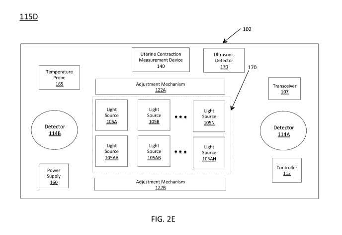
[00067] Figure 2C shows another exemplary fetal hemoglobin probe
115B that includes power supply 160, light source(s) 105, transceiver 107,
detector 114, an adjustment mechanism 122, a temperature probe 165, and a
controller 112.
[00068] Temperature probe 165 may be any appropriate mechanism for
obtaining a temperature measurement for the pregnant mammal. Adjustment
mechanism 122 may be one or more mechanisms adapted to adjust one or
more properties of the light emitted by light source(s) 105 and/or a
direction/incident angle of the light directed into the abdomen of the
pregnant
mammal. Exemplary adjustment mechanisms include, but are not limited to,
filters and polarizers that may be used to adjust a frequency/wavelength of
the
light emitted by light source(s) 105 and/or an orientation for the light.
Other
exemplary adjustment mechanisms 122 include lenses adapted to, for
example, focus or spread light directed into the pregnant mammal's abdomen.
In some instances, the lenses may also change the angle of incidence for the
23
CA 03006874 2018-05-28
WO 2017/117280
PCT/US2016/068994
light directed to the pregnant mammal's abdomen. In some embodiments,
adjustment mechanisms 122 may also include mechanisms enabled to move
a light source 105 and/or operate a lens, filter, or polarizer. In some
embodiments, adjustment mechanism 122 may include a material that is
sensitive to electricity and may be enabled to become transparent and/or
partially opaque upon application of electricity. Often times, adjustment
mechanism(s) 122 may receive instructions from controller 112 that may
control (wholly or partially) the operation of the adjustment mechanism 122.
[00069] Optionally, fetal hemoglobin probe 115 may also include one or
more one or more ultrasonic detectors 170. An ultrasonic detector 170 may
be employed in embodiments of fetal hemoglobin probe 115 configured to
perform optoacoustic/photoacoustic and/or thermoacoustic imaging by way of
directing a light or radio frequency pulse from light source(s) 105 into the
pregnant mammal's 305 abdomen. A portion of the incident light may be
absorbed by the fetus and pregnant mammal and converted into heat, which
leads to transient thermoelastic expansion, which causes an ultrasonic
emission from the fetus and pregnant mammal. This ultrasonic emission may
be detected by ultrasonic detector 170 and analyzed to determine a level of
oxygen saturation for the fetus' and/or pregnant mammal's blood. In some
instances, deploying fetal hemoglobin probe 115 to perform
optoacoustic/photoacoustic and/or thermoacoustic imaging may require use of
a laser and/or radio frequency pulse emitter (not shown).
[00070] Controller 112 may be adapted to control one or more
components (e.g., adjustment mechanism 122, light source(s) 105, power
supply 160, temperature probe 165, detector 114, and/or transceiver 107) of
fetal hemoglobin probe 115. In some circumstances, controller 112 may
include a processor adapted to receive measurements/information from one
more components (e.g., adjustment mechanism 122, light source(s) 105,
power supply 160, temperature probe 165, detector 114, and/or transceiver
107) of fetal hemoglobin probe 115. The processor may be further adapted to
process the received measurements, make decisions therewith, and
communicate instructions based on those decisions and/or measurements to
one or more components of fetal hemoglobin probe 115. For example,
temperature probe 165 may act to measure the body temperature of the
24
CA 03006874 2018-05-28
WO 2017/117280
PCT/US2016/068994
pregnant mammal and may provide these measurements to controller 112
and/or transceiver. In some embodiments, these measurements may be used
to determine whether the temperature of the pregnant mammal exceeds a
threshold measurement, which in some instances, may indicate that light
source(s) 105 and/or fetal hemoglobin probe 115 are delivering too much
heat/energy to the pregnant mammal. Upon reaching such a determination,
controller 112 may provide instructions to light source(s) 105 and/or
adjustment mechanism 122 to correct for this. Exemplary instructions include,
but are not limited to, directions to redirect incident light, turn off,
adjust a
frequency, and adjust an intensity of one or more of the light source(s) 105.
[00071] In some instances, instructions provided by controller 112 may
be based on, for example, feedback from, for example detector 114 and/or
transceiver 107 regarding, for example, the strength/intensity of the
reflected
signal, the frequency/wavelength of light received in the reflected signal.
For
example, if controller 112, transceiver 107, and/or detector 114 determines
that a received signal reflected from the pregnant mammal's abdomen is of
insufficient strength/intensity, then controller 112 may provide instructions
to
adjustment mechanism 112 and/or light source(s) 105 to increase the
intensity and/or wavelength/frequency of the light incident on the abdomen of
the pregnant mammal.
[00072] In another example, temperature probe 165 may act to measure
the body temperature of the pregnant mammal and may provide these
measurements to controller 112 and/or transceiver. In some embodiments,
these measurements may be used to determine whether the temperature of
the pregnant mammal exceeds a threshold measurement, which in some
instances, may indicate that light source(s) 105 and/or fetal hemoglobin probe
115 are delivering too much heat/energy to the pregnant mammal. Upon
reaching such a determination, controller 112 may provide instructions to
light
source(s) 105 and/or adjustment mechanism 122 to correct for this.
Exemplary instructions include, but are not limited to, directions to redirect
incident light, turn off, adjust a frequency, and/or adjust an intensity of
one or
more of the light source(s) 105.
[00073] In some instances, light source(s) 105 may be tunable, or
otherwise user configurable, by, for example, a physician or clinician
assisting
CA 03006874 2018-05-28
WO 2017/117280
PCT/US2016/068994
the pregnant mammal during the delivery process. For example, a light
source 105 may be configured to emit light in multiple
frequencies/wavelengths and/or intensities and the light source 105 may be
tuned via, for example, direct physical manipulation of the light source 105
(e.g., via a button on knob), or the entering of an instruction regarding the
desired frequency/wavelength and/or intensity into, for example, computer
150 and/or controller 112.
[00074] Tuning the frequency/wavelength and/or intensity of light emitted
by one or more light source(s) 105 may be helpful in achieving a return signal
of sufficient strength or clarity in a variety of circumstances (e.g., fetus
position, fetus size, the amount of melanin in the skin of the pregnant mammal
and/or fetus, the size and/or shape of the pregnant mammal, etc.). For
example, light of a relatively higher intensity may be desired when the
pregnant mammal has a relatively high body mass index (BMI) or body fat
positioned in such a way as to inhibit the strength of a signal reflected from
the fetus (i.e., return signal). In another example, a fetus may be positioned
against the internal organs of the pregnant mammal (i.e., away from the skin
of the belly), and light of relatively higher intensity and/or different
wavelength
may be desired so that the light reaches the fetus with a sufficiently strong
signal so that a return signal may be detected by, for example, detector 114.
[00075] When fetal hemoglobin probe 115 includes more than one light
source 105, the light sources 105 may be arranged in an array adapted to
maximize the strength of the returned signal such as array 170 as discussed
below with regard to Figures 2D and 2E. Array 170 may include any
appropriate number of light sources 105. In some instances, array 170 may
include a first row of a first type of light source 105A, 105B, through 105N
and
a second row of a second type of light source 105 AA, 105AB, through
105AN. The different types of light sources may be configured to, for
example, emit light of a particular frequency/wavelength and/or intensity. For
example, light sources 105 A, 105B, through 105N may be configured to emit
light with wavelengths in the red spectrum and light sources 105 AA, 105AB,
through 105AN may be configured to emit light with wavelengths in the
infrared or near-infrared spectrum. Although array 170 to have two rows, it
will be appreciated that any number of rows (e.g., 3, 4, 5, 6, 7, 8, and so
on)
26
CA 03006874 2018-05-28
WO 2017/117280
PCT/US2016/068994
may be included in array 170.
[00076] Embodiments of fetal hemoglobin probe 115 with a relatively
large length (e.g., 10cm-40cm) may have arrays 170 with rows of multiple
light sources long fetal hemoglobin probe 115 that include, for example, 10,
15, 20, 25, 30, 35, 40, 45, or 50 light sources 105 each. A fetal hemoglobin
probe 115 may also include more than one detector 114, as shown in Figure
2E, which includes a first detector 114A and a second detector 114B. In
some embodiments, first detector 114A may be the same as second detector
114B and, in other embodiments, they may be different. For example, first
detector 114A may be sensitive to a first range of frequencies for reflected
light and second detector 114B may be sensitive to a second range of
frequencies for reflected light. Additionally, or alternatively, first
detector 114A
may be of a different size than second detector 114B. Any of the fetal
hemoglobin probes 115 disclosed herein may include multiple detectors
adapted to, for example, detect light reflected for one or more the light
source(s) 105 included in array 170.
[00077] Although shown as a separate component in Figures 2C-2E, it
will be appreciated by those of skill in the art that adjustment mechanism 122
may be partially and/or wholly positioned within and/or adjacent to one or
more light sources 105.
[00078] Components of system 100 may be applied to a pregnant
mammal in any acceptable manner. For example, NIRS adult hemoglobin
probe 125 may be placed on the second finger of the pregnant mammal 305,
pulse oximetry probe 130 may be placed on the thumb of the pregnant
mammal 305, and Doppler and/or ultrasound probe 135 may be placed on the
abdomen of the on the pregnant mammal.
[00079] In some implementations, uterine contraction measurement
device 140 may also be on placed on the abdomen of the pregnant mammal.
In other implementations, uterine contraction measurement device 140 may
be embodied in the fetal hemoglobin device 115. In some cases, uterine
contraction measurement device 140 may be a pressure sensor configured to
detect the changes in pressure of the uterine muscle in units of pressure
(mmHg and/or kPa).
[00080] In some embodiments, one or more light source(s) 105 and
27
CA 03006874 2018-05-28
WO 2017/117280
PCT/US2016/068994
detector(s) 114 may act as an optoelectronic muscle contraction sensor
without the need for a separate uterine contraction measurement device 140.
In these embodiments, the light reflected from the pregnant mammal's uterus
might change in nature when the uterus is in a relaxed state (more scattering)
as opposed to a contracted state (less scattering). These changes in the rate
of scattering of the light may be detected by one or more detector(s) 114 and
processed by, for example, computer 150 to determine changes in the state of
the uterine muscle. In some embodiments, one or more light source(s) 105
may direct light of a particular frequency/wavelength so that measurements of
uterine contractions have a dedicated beam/frequency of light.
[00081] Preferably, the fetal hemoglobin probe 115 is placed at, or near,
the bikini/supra-pubic region of the pregnant mammal 305. This area is
typically right above the pubic hairline. This position is advantageous in the
later stages of pregnancy, for example, after 9 months or 36 weeks of
gestational development because the fetus's head will engage into the
cervical birth canal and will, therefore, be in a fairly predictable location
within
the abdomen of the pregnant mammal. Additionally, when the head of the
fetus is positioned within the cervical birth canal, the distance between
pregnant mammal and fetus is minimal and therefore NIR light passing
through the abdomen of the pregnant mammal is more likely to come into
contact with the fetus and be reflected back to the fetal hemoglobin probe
115.
[00082] Figures 3A, 3B, and 3C provide illustrations of how light from
fetal hemoglobin probe 115 may be directed into a pregnant mammal's 305
abdomen and reflected light may be detected by one or more detectors 114 of
fetal hemoglobin probe 115. More specifically, Figure 3A provides a cross
sectional view of fetal hemoglobin probe 115 and of the pregnant mammal
305 as divided along a midline extending through the center of pregnant
mammal 305 when she is viewed from the front (i.e., through the center of the
face, between the breasts, etc.). Figure 3A depicts an approximation of a
fetus 310 that is surrounded by amniotic fluid and other tissue 315 present in
a uterus 320 of the pregnant mammal 305. fetal hemoglobin probe 115 is
show in Figure 2C to be positioned on the lower abdomen of the pregnant
mammal 305 at, or near, the bikini/supra-pubic region of the pregnant
28
CA 03006874 2018-05-28
WO 2017/117280
PCT/US2016/068994
mammal 305.
[00083] As shown in Figure 3A, a beam of light 325 (also referred to
herein as an "incident beam") emitted from one or more light source(s) 105 is
incident on pregnant mammal's 305 abdomen and is directed toward fetus
310. Beam of light 325 may be of any wavelength/frequency or combination of
wavelengths/frequencies. In one embodiment, incident beam 325 may
include light that is in the red spectrum and the near infrared spectrum.
[00084] In some embodiments, incident beam 325 may include two or
more beams of light that may be emitted from, for example a single light
source 105 (that emits two beams of light of the same frequency and/or a
beam of light of two different frequencies) or two different light sources 105
(e.g., one frequency per light source). When two or more beams are included
in incident beam 325, they may, on occasion be directed in slightly different
directions so as to, for example, accommodate differences in the frequency of
the light of the beam, a condition of the pregnant mammal 305 (e.g., skin
pigmentation, body mass index, etc.) and/or a condition of the fetus (e.g.,
size, position, location within the uterus, skin pigmentation, etc.).
[00085] A portion of incident beam 325 may reflect from the fetus 310,
amniotic fluid and other tissue 315, and uterus 320 as a reflected beam 330
and may be received by one or more detectors 114 provided by fetal
hemoglobin probe 115. Although reflected beam 330 is shown as one beam,
it may be any number of beams or individual photons. It is expected that not
all of the light of incident beam 325 will be included in reflected beam 330
as
some of the light of incident beam 325 may be lost/undetected due to, for
example, scattering and/or absorption.
[00086] Figure 3B provides an image of fetal hemoglobin probe 115 with
an adjustment device 335 positioned between the skin of the pregnant
mammal's 305 abdomen and a portion of fetal hemoglobin probe 115. In the
embodiment of Figure 3B, adjustment device 335 is triangular in shape and
acts as a wedge to change an orientation/position of fetal hemoglobin probe
115 (and the corresponding orientation/position of light source(s) 105 and/or
detector(s) 114) relative to the pregnant mammal's abdomen. In some cases,
adjustment device 335 may change the angle of incidence for incident beam
325 and/or an orientation of one or more detectors 114. In some
29
CA 03006874 2018-05-28
WO 2017/117280
PCT/US2016/068994
embodiments, adjustment device 335 may be transparent so as to allow for
the passage of light into, and out of, the pregnant mammal's 305 abdomen.
In other embodiments, adjustment device 335 may be semi-transparent or
opaque so as to, for example, change a frequency of the incident beam 325
and/or reflected beam 330.
[00087] Adjustment device 335 may be configured to adjust for
physiological conditions of the pregnant mammal's 305 abdomen that make it
difficult to receive a reflected beam of sufficient strength. For example, for
a
pregnant mammal 305 with a high fat content around her abdomen, applying
the fetal hemoglobin probe 115 directly to the pregnant mammal's 305 skin
may not direct the incident beam 325 in the proper direction and/or enable
detection of the reflected beam 330. Additionally, or alternatively,
adjustment
device 335 may be configured to adjust for physiological conditions of the
fetus 310 including the size and/or placement of the fetus 310 within the
uterus 320. For example, adjustment device 335 may be deployed so as to
direct incident beam 325 toward the head of fetus 310.
[00088] In some embodiments, two or more adjustment mechanisms
335 may be used. An adjustment device 335 may be of any appropriate
shape and/or configuration including, but not limited to, a triangle, circle,
or
rectangle and may be configured to adjust the positioning or operation of
some, or all, of the components of fetal hemoglobin probe 115. In some
instances, adjustment device 335 may be designed to improve the comfort of
the pregnant mammal 305 while wearing fetal hemoglobin probe 115 and, to
that end, may be configured to include soft and/or flexible material (e.g.,
foam)
designed to adapt to a contour of the pregnant mammal's abdomen. In these
instances, adjustment device 335 would be designed to engage with fetal
hemoglobin probe 115 in a manner that does not obscure one or more
components thereof.
[00089] In another embodiment, adjustment device 335 may include
optics, filters, or other mechanical and/or electrical components configured
to
adjust one or more features of incident beam 325 and/or reflected beam 330.
In some instances, one or more operations of adjustment device 335 may be
performed upon receipt of instructions from, for example, a component of fetal
hemoglobin probe 115 and/or computer 150.
CA 03006874 2018-05-28
WO 2017/117280
PCT/US2016/068994
[00090] Figure 3C provides a front view of pregnant mammal's 305
abdomen with fetal hemoglobin probe 115 affixed thereto. The perspective is
somewhat adjusted for Figure 3C so that incident beam 325 and reflected
beam 330 may be seen. In reality, both incident beam 325 and reflected
beam 330 are directed into/reflected from the pregnant mammal's 305
abdomen along the ¨Z-axis.
[00091] Figure 3D provides a front cross section view of pregnant
mammal's 305 abdomen with fetal hemoglobin probe 115 and
Doppler/ultrasound probe 135 coincident therewith. As shown in Figure 3D,
Doppler/ultrasound probe 135 transmits a beam into pregnant mammal's 305
abdomen towards fetus 310 and receives a reflected signal.
Doppler/ultrasound probe 135 is then uses this reflected signal to determine a
fetal heart beat signal and/or determine a number of fetal heart beats per
minute. and
[00092] The fetal hemoglobin probe 115 of Figure 3D has two light
sources, a first of which, 105A, emits a light beam 325A of a first wavelength
(Ai) (noted on the figure as 105A, Al and 325A, Al, respectively) and a second
of which, 105B, emits a light beam 325B of a second wavelength (A2) (noted
on the figure as 105B, A2 and 325B, A2, respectively). A portion of incident
beams 325A and 325B is reflected by the pregnant mammal 305 and fetus
310 and received by detector 114 as reflected beam 330A and 330B,
respectively ((noted on the figure as 330A, Al and 330B, A2, respectively).
[00093] Figure 4A illustrates an exemplary process 400 for performing
fetal oximetry and/or fetal pulse oximetry trans-abdominally and/or in-utero
to
determine fetal hemoglobin oxygen saturation level. Process 400 may be
performed by, for example, system 100 and/or a component thereof.
[00094] Initially, a light beam, such as incident beam 325, is directed
into
the abdomen of a pregnant mammal, such as pregnant mammal 305 (step
405) by, for example, one or more light sources such as light source(s) 105
provided by one or more of the fetal hemoglobin mammal' pregnant
mammal's abdomen may be directed toward the pregnant mammal's fetus,
such as fetus 310 as shown in Figures 3A and 3B discussed above.
[00095] The light beam directed into the pregnant mammal's abdomen
may include any number of light beams and/or frequencies/wavelengths of
31
CA 03006874 2018-05-28
WO 2017/117280
PCT/US2016/068994
light as described above with regard to incident beam 325. In some instances,
the light beam of step 405 may be a plurality of light beams emitted from a
plurality of light sources positioned at a plurality of different locations
along the
abdomen of the pregnant mammal as shown in, for example, Figures 2D and
2E. Additionally, or alternatively, the light beam of step 405 may include a
plurality of wavelengths/frequencies emitted by a single of light source that
may, for example, include multiple LEDs.
[00096] In some embodiments, the light beam of step 405 may include
light of first and second wavelengths with a first of the wavelength being in
the
red portion of the electromagnetic spectrum (i.e., 620-750nm) and a second of
wavelengths in the near-infrared (NIR) portion of the electromagnetic
spectrum (e.g., 750nm-2,500 nm). Use of these wavelengths is preferred, but
not required, because light of wavelengths in the red and near-infrared
spectrum are known to travel through, and/or be reflected by, skin and body
tissue. In some embodiments, light of, for example, a third, fourth, fifth, or
more different wavelengths may be directed toward the abdomen of the
pregnant mammal. In some circumstances, use of more than two
wavelengths of light may be useful to enhance reflected signal strength and/or
clarity in various circumstances including, but not limited to, distance of
the
fetus from the external skin, or uterine wall, of the pregnant mammal (i.e.,
depth of the fetus), level of melanin/pigment in the skin of the pregnant
mammal and/or fetus, strength of fetal pulse signal, how much the fetus
moves within the placenta and/or uterus of the pregnant mammal, and so on.
[00097] In some embodiments, an intensity of the light directed into the
pregnant mammal at step 405 may be varied and/or different for different
wavelengths of light. For example, the intensity of red light directed into
the
pregnant mammal's abdomen may be greater than the intensity of the near-
infrared light due to the transmission/reflection properties of red light
verses
near infrared light (i.e., near-infrared light is know to reflect more light
when
shown into body tissue than red light). However, it is expected that an
intensity of the light beam of step 405 will be safe for both the pregnant
mammal and her fetus (e.g., not cause burns to the pregnant mammal's skin
and/or damage to fetal tissue (e.g., eyes)).
32
CA 03006874 2018-05-28
WO 2017/117280
PCT/US2016/068994
[00098] In step 410, light (e.g., waves and/or photons) reflected by the
abdomen of the pregnant mammal (and the fetus) may be received by one or
more detectors (e.g., photo-sensor, photo detectors or photodiodes), such as
detector 115 and/or transceiver 107 and converted (step 415) into an
electronic signal that represents the reflected light (this signal may be
referred
to herein as a "reflected electronic signal" by the photo-
sensor/photodiode/photo detector. In some instances, the light directed into
the abdomen of the pregnant mammal, may travel a distance of, for example,
3-5 cm to contact the fetus and another 3-5 cm once reflected from the fetus
to be detected by the detector. Thus, the total travel distance for the
incident
and reflected beam may be as high as 8 or 10 cm. When traveling this
distance, a substantial amount of scattering and other interference in the
detection of a reflected signal may occur and it is possible that only a small
fraction (e.g., 0.5-5%) of the light incident on the abdomen of the pregnant
mammal will be reflected by the fetus and received by detector.
[00099] Optionally, in step 420, it may be determined whether the
electronic reflected signal is of sufficient strength to detect, for example,
the
pulse and/or fetal oxygen saturation of the fetus. Exemplary signal strengths
that are sufficient are in the range of 30-500 dB with a signal-to-noise (SNR)
ratio of 1-8, with a preferred SNR of approximately 3-4.5.
[000100] When the signal isn't of sufficient strength, the light source(s)
and/or detector(s) may be adjusted automatically (i.e., without operator
intervention) and/or provision of an indication that an adjustment of the
light
source(s) and/or detector(s) may be desired or needed to an operator (e.g.,
doctor or nurse) may be facilitated (step 425). Exemplary indications provided
in step 425 include, but are not limited to, an alarm, a message (e.g.,
written
or audio), and a recommendation. Exemplary automatic adjustments include,
but are not limited to, adjusting a lens positioned between the pregnant
mammal's abdomen and the light source(s) and/or detector(s) so as to focus
the light emitted by the light source(s) and/or received by the detector(s),
adjusting an amount of power delivered to the light source(s) and/or
detector(s), adjusting an intensity and/or frequency of the light emitted by
one
or more of the light source(s) and so on. In some embodiments, activation of
additional light sources to direct light into the pregnant mammal's abdomen
33
CA 03006874 2018-05-28
WO 2017/117280
PCT/US2016/068994
may be responsive to a determination that the electronic reflected signal is
not
of sufficient strength.
[000101] In some instances, the adjustment(s) of step 425 may be
performed and/or facilitated by one or more adjustment mechanisms, such as
adjustment mechanisms 122 and/or controllers, such as controller 112. Once
adjusted, the light beam may again be directed into the pregnant mammal's
abdomen (i.e., step 405 may be repeated) and steps 410-420 may be
repeated. When the electronic reflected signal is of sufficient strength, or
when steps 420 and 425 are not performed, process 400 may advance to
step 430.
[000102] In step 430, the electronic reflected signal may be processed to
isolate a portion of the reflected electronic signal reflected from the fetus
(as
opposed to the pregnant mammal or noise). For ease of discussion, the
portion of the reflected electronic signal reflected from the fetus may be
referred to herein as the fetal reflected electronic signal. Examples of how
step 430 may be executed are discussed below with regard Figures 5A-5D.
Following step 430, the fetal reflected electronic signal may be analyzed to
determine the oxygen saturation level of hemoglobin contained in the fetus'
blood via, for example, oximetry and/or pulse oximetry techniques (step 440).
Typical values for the oxygen saturation of fetal blood fall with in the range
of
30-70%. An exemplary method of determining fetal hemoglobin saturation
level uses a version of the Beer-Lambert law modified to account for the
scattering effect of the reflected light as it is scattered by tissues in the
body
as described by Zourabian, Anna, et al., Trans-abdominal Monitoring of Fetal
Arterial Blood Oxygenation Using Pulse Oximetry, Journal of Biomedical
Optics, 5(4), pp. 391-405 (October 2000), which is incorporated by reference
herein. Further details regarding execution of step 435 is provided below with
regard to Figures 6A-6H.
[000103] Then, in step 440, provision of an indication of fetal oxygen
level
to an operator may be facilitated. Exemplary operators include, but are not
limited to, doctors, nurses, and other caregivers. Exemplary indicators
include a waveform shown on a display device (e.g., computer monitor), a
numerical value provided via a display device and/or message (e.g., SMS text
message), such as a fetal hemoglobin oxygen saturation level. Facilitating
34
CA 03006874 2018-05-28
WO 2017/117280
PCT/US2016/068994
provision of the indication of step 465 may include providing the indication
to a
computer, such as computer 150 and/or a display device such as display
device 155. An example of such a display of fetal hemoglobin oxygen
saturation level is provided by Figures 8A and 8B and is discussed below.
[000104] One method of processing the signal to isolate the portion of the
reflected electronic signal reflected from the fetus from the total reflected
electronic signal is to multiply the total reflected electronic signal by
signal that
provides the fetal heart (i.e., perform step 430) beat as may be provided by,
for example, a Doppler and/or ultrasound probe such as Doppler/ultrasound
probe 135. The resultant signal (i.e., the signal that is the product of
multiplying the total reflected electronic signal and the fetal heartbeat
signal)
may approximate the portion of the total reflected electronic signal reflected
by the fetus. To improve this approximation, the signal reading may be
averaged over a number of cycles to provide a more accurate approximation
of the portion of the total reflected electronic signal reflected by the
fetus. An
example of this process is provided by Figures 5A-5D, of which Figure 5A
provides a graph 500 of total reflected electronic signal intensity vs. time
and
represents light reflected by the abdomen of the pregnant mammal detected
in step 410. Figure 5B provides a graph 501 of a Doppler signal vs. time.
This signal and represents light reflected by the abdomen of the pregnant
mammal detected in step 410. The Doppler signal represents the fetus'
heartbeat. This signal may be received from, for example, Doppler/ultrasound
probe 135. Figure 5C provides a graph 502 that shows the product of
multiplying the total reflected electronic signal intensity (from Figure 2A)
and
the Doppler signal (from Figure 2B) together while synchronizing over time so
that a signal intensity of the total reflected electronic signal at a
particular
moment in time is multiplied by the Doppler signal intensity at that same
particular moment in time. The resultant signal shown in Figure 5C
approximates the portion of the total reflected electronic signal reflected
from
the fetus. This signal may then be analyzed to determine fetal oxygen
saturation levels using, for example, oximetry or pulse oximetry techniques.
[000105] In some embodiments, the accuracy of the approximated portion
of the total reflected electronic signal reflected from the fetus may be
improved by averaging a number of signal intensities over a period of time
CA 03006874 2018-05-28
WO 2017/117280
PCT/US2016/068994
(e.g., a number of periods) as shown in Figure 5D, which provides a graph
503 of the total reflected electronic signal intensity, the fetal
heartbeat/Doppler
signal and the result of multiplying total reflected electronic signal
intensity
and Doppler signal synchronized over time (referred to on graph 503 as "fetal
reflected signal."
[000106] Another method of processing the electronic reflected signal to
isolate the portion of the reflected electronic signal reflected from the
fetus
from the total reflected electronic signal is to multiply the total reflected
electronic signal by signal that provides the fetal heart (i.e., perform step
430)
is provided by Figure 4B, which shows sub-process 401.
[000107] In step 445 of sub-process 401, a heartbeat signal for the
pregnant mammal is received from, for example, a pulse oximetry probe like
pulse oximetry probe 130 and/or an adult hemoglobin probe like NIRS adult
hemoglobin probe 125. Next, the received pregnant mammal's heartbeat
signal may be synchronized in the time domain with the electronic reflected
signal (step 450). Then a correlation between the pregnant mammal's
heartbeat and changes in the electronic reflected signal may be established
so as to determine a portion of the electronic reflected signal that is
reflected
by the pregnant mammal (step 455). In step 460, the portion of the portion of
the electronic reflected signal that is reflected by the pregnant mammal is
then
subtracted from the electronic reflected signal with the portion of the
electronic
reflected signal reflected by the fetus being thereby isolated.
[000108] Another method of processing the signal to isolate the portion of
the reflected electronic signal reflected from the fetus from the total
reflected
electronic signal is to multiply the total reflected electronic signal by
signal that
provides the fetal heart (i.e., perform step 430) is provided by Figure 4C,
which shows sub-process 402.
[000109] In step 465 of sub-process 465, a heartbeat signal for the fetus
may be received from, for example, an ultrasound device and/or a Doppler
device, such as Doppler/ultrasound probe 135. Next, the received fetus'
heartbeat signal may be synchronized in the time domain with the electronic
reflected signal (step 470). Then, portions of the electronic reflected signal
that correspond in the time domain with the individual heartbeats may be
examined. In this 'way, the entire electronic reflected signal does not have
to
36
CA 03006874 2018-05-28
WO 2017/117280
PCT/US2016/068994
be processed/analyzed; only the portions of the electronic reflected signal
where a fetal heartbeat, or pulse, occur are examined. This saves processing
time and resources because the entire signal does not have to be processed.
[000110] In some embodiments, the processing of step 430 and/or
analysis of step 435 may include processing the reflected electronic signal in
order to ascertain a signal that corresponds to the absorption/reflection of
NIR
light by oxygenated hemoglobin and deoxygenated hemoglobin of the fetus.
Using this information, a level (or percentage) of fetal hemoglobin oxygen
saturation (step 435) may be determined.
[000111] Because fetal hemoglobin is structurally different from adult
hemoglobin it absorbs light differently and the signal reflected from the
fetal
hemoglobin at various wavelengths will be of a different magnitude when
compared to the magnitude of the signal at those same wavelengths reflected
by the pregnant woman. In this way, measuring a quantity of light reflected
from the hemoglobin of the pregnant woman and fetus at various wavelengths
will provide an indication of the amount of light of a particular wavelength
that
is absorbed by the fetal hemoglobin as well as the pregnant woman's
hemoglobin. Looking at the ratios of light reflected at various wavelengths
will
provide a benchmark that correlates to a specific fetal blood oxygen level. In
some instances, the variations in wavelength absorption of the fetal
hemoglobin when compared to the pregnant woman's hemoglobin may not be
sufficient to provide an adequately strong or clear signal indicating fetal
hemoglobin oxygen saturation levels for clinical and/or diagnostic purposes.
Therefore, one or more signal processing techniques may be applied to the
signal received by the fetal hemoglobin probe 115 to determine fetal
hemoglobin oxygen saturation as will be discussed in detail below.
[000112] In an exemplary signal processing technique, a signal received
from the pregnant woman's pulse oximetry probe (e.g., pulse oximetry probe
130) may be used to determine the oxygen saturation level of the pregnant
woman's arterial blood, which corresponds to an oxygenated state of
pregnant woman's hemoglobin. The pulse oximetry probe is used to make this
determination because the depth of a human finger is 1-2 cm, a measurable
amount of light can pass through the finger tip and there is no interference
from fetal blood flow or circulating fetal hemoglobin at the pregnant woman's
37
CA 03006874 2018-05-28
WO 2017/117280
PCT/US2016/068994
fingertip position. Hence, a reading from pulse oximetry probe 130 will
directly
correspond to how much light is absorbed and/or reflected at various
wavelengths by the pregnant woman's adult hemoglobin. This information
may be used to understand how the light is interacting with pregnant woman's
hemoglobin near the fetus and this information may be subtracted from the
signal received by the fetal hemoglobin probe 115 to determine how much
light is absorbed and/or reflected at various wavelengths by the fetus'
hemoglobin.
[000113] Additionally, or alternatively, the signal received by the fetal
hemoglobin probe 115 may be processed using a heart rate of the fetus
and/or pregnant woman. The timing of the pregnant woman's heartbeat
correlates to the timing for various levels of blood oxygen saturation for the
pregnant woman. This correlation may be used to detect a signal
corresponding the level of blood oxygen saturation for the pregnant woman
within the signal received by the fetal hemoglobin probe 115. The fetal
oxygen saturation level may then determined by subtracting, or otherwise
filtering, the detected signal corresponding the level of blood oxygen
saturation for the pregnant woman from signal received by the fetal
hemoglobin probe 115.
[000114] Additionally, or alternatively, the fetal heartbeat correlates to
the
timing for various levels of blood oxygen saturation for the fetus. This
correlation may then be used to detect a signal corresponding the level of
blood oxygen saturation for the fetus within the signal received by the fetal
hemoglobin probe 115. For example, Doppler/ultrasound probe 135 and/or
an ultrasound device may indicate that the fetus' heart rate is in the range
of
120-160 beats per minute and this fetal heart rate may be used to gate and/or
correlate a NIR signal from the fetus.
[000115] In the rare circumstance when the fetal heart rate and maternal
heart rate are similar (fetal bradycardia and maternal tachycardia) the two
heartbeats may be distinguished from one another using the known fact that
there is a slight pause in the heart rate during respiration. So, by
monitoring
the heart rate signal (via, e.g., pulse oximetry probe 130), one may observe
that the pregnant woman's the heart rate slows down for a moment when she
takes in a deep breath. This slowing will only be present in the signal
38
CA 03006874 2018-05-28
WO 2017/117280
PCT/US2016/068994
providing the pregnant woman's heart rate because fetuses do not breathe
while in utero. In this way, the two heart rates may be distinguished from one
another.
[000116] In some embodiments, a signal from NIRS adult hemoglobin
probe 125 may be processed to determine a ratio of adult oxyhemoglobin to
adult de-oxyhemoglobin. This ratio may then be used to subtract readings
from the pregnant woman's blood flow so that a signal from the fetus's blood
flow may be isolated and analyzed to, for example, determine a level of fetal
hemoglobin oxygen saturation.
[000117] In other embodiments, processing the signals received by the
fetal hemoglobin probe 115 may include oscillating between time domain and
frequency domain analysis. This oscillation may allow identification signals
that have a cyclical (periodic) component as opposed to signals that are
random or non-periodic (acyclic/aperiodic). Random or non-periodic signals
are more likely to be noise and examining the received signal for random or
non-periodic signals will assist in determining a noise level of the signal as
well as portions of the signal that may be filtered or otherwise removed
therefrom.
[000118] In some embodiments, process 400 may include the
establishment of a set of correlations between the intensity of light
reflected/absorbed at certain wavelengths by fetal oxyhemoglobin and de-
oxyhemoglobin and the oxygen saturation levels of the fetal oxyhemoglobin
and de-oxyhemoglobin. This set of correlations may be performed prior to
executing process 400 for a particular pregnant mammal during the fetal labor
and delivery process and may be stored in, for example, computer 150. An
exemplary correlation may be a reflection of light of wavelength A with an
intensity X and a reflection of light of wavelength B with an intensity 0.8X
to an
fetal oxygen saturation level of 50% of fetal hemoglobin being bound to
oxygen. Another exemplary correlation may be a reflection of light of
wavelength A with an intensity X and a reflection of light of wavelength B
with
an intensity 0.5X to an fetal oxygen saturation level of 25% of fetal
hemoglobin being bound to oxygen.
[000119] (noted on the figure as 105A, A1 and 325A, Al, respectively) and
a second of which, 105B, emits a light beam 325B of a second wavelength
39
CA 03006874 2018-05-28
WO 2017/117280
PCT/US2016/068994
(A2) (noted on the figure as 105B, A2 and 325B, A2, respectively). A portion
of
incident beams 325A and 325B is reflected by the pregnant mammal 305 and
fetus 310 and received by detector 114 as reflected beam 330A and 330B,
respectively ((noted on the figure as 330A, Al and 330B, A2,
[000120] Figures 6A-6H provide information in the form of graphs
regarding an example of how reflected electronic signal is analyzed to
determine fetal hemoglobin oxygen saturation level. At times, fetal
hemoglobin oxygen saturation level may also be referred to herein as fetal
arterial oxygen saturation level, which may be abbreviated to (VoSa02). More
specifically, Figure 6A provides a graph 601 of a Doppler signal vs. time. The
Doppler signal corresponds to a fetal heart beat signal. The Doppler signal of
Figure 6A is similar to the Doppler signal of Figure 5B.
[000121] Figure 6B provides a graph 602 of reflected electronic signal
intensity for Al vs. time. This graph may correspond to reflected signal 330A,
Al. Any of the processes discussed above may be used to isolate the portion
of the signal reflected by the fetus from the reflected electronic signal
intensity
for Al. In the example provided, the total reflected electronic signal
intensity
for Al and the fetal Doppler signal are multiplied together while
synchronizing
over time to provide the product of multiplying the total reflected electronic
signal intensity for Al and the Doppler signal together while synchronizing
over
time as shown in graph 603 of Figure 6C.
[000122] Figure 6D provides a graph 604 that shows the product of
multiplying the total reflected electronic signal intensity for Al and the
fetal
Doppler signal together while synchronizing over time averaged over several
periods. This graph (or the data used to generate the graph) is analyzed to
determine an intensity of a systolic value for the first wavelength Al 610,
which
corresponds to the peak of the curve (i.e., highest value) and an intensity of
a
diastolic value for the first wavelength Al 615, which corresponds to the
trough
of the curve (i.e., lowest//smallest value).
[000123] Figure 6E provides a graph 605 of reflected electronic signal
intensity for A2 vs. time. Any of the processes discussed above may be used
to isolate the portion of the signal reflected by the fetus from the reflected
electronic signal intensity for A2. In the example provided, the total
reflected
electronic signal intensity for A2 and the fetal Doppler signal are multiplied
CA 03006874 2018-05-28
WO 2017/117280
PCT/US2016/068994
together while synchronizing over time to provide the product of multiplying
the total reflected electronic signal intensity for A2 and the Doppler signal
together while synchronizing over time as shown in graph 606 of Figure 6F.
[000124] Figure 6G provides a graph 607 that shows the product of
multiplying the total reflected electronic signal intensity for A2 and the
fetal
Doppler signal together while synchronizing over time averaged over several
periods. This graph (or the data used to generate the graph) is analyzed to
determine an intensity of a systolic value for the second wavelength A2620,
which corresponds to the peak of the curve (i.e., highest value) and an
intensity of a diastolic value for the second wavelength A2625, which
corresponds to the trough of the curve (i.e., lowest/smallest value).
[000125] A modulation ratio (R) between the reflected intensity of two
wavelengths of light may be calculated as follows:
Tsysxi Tsysx2
R = log ( )/log ( ) Equation 1
Tdiasxi Tdiasx2
where:
Tsysid is the intensity of the systolic value for the first wavelength (Ai);
TdiasAl is the intensity of the diastolic value for the first wavelength (Ai);
TsysA2 is the intensity of the systolic value for the second wavelength (A2);
and
TdiasA2 is the intensity of the diastolic value for the second wavelength
(A2).
[000126] The modulation ratio, R, may then be used to determine a level
of arterial oxygen saturation value (VoSa02) in one of at least two fashions.
When a relationship between a modulation ratio, R, for a pair of wavelengths
(i.e., A1 and A2) and arterial oxygen saturation is known (from, for example,
experimentally determined values), then the value of R may be used to look
up a corresponding arterial oxygen saturation level. Figure 6H1 provides an
exemplary graph that plots a known relationship between values for R (when
Al is in the red spectrum and A2 is in the infrared spectrum) with arterial
oxygen saturation values.
[000127] Following through with the above example (with the appropriate
reference numbers for intensity values inserted from graphs 604 and 607),
would yield the following calculation for equation 1:
1 Source of Figure 6H: Paul D. et al., Wavelength Selection for Low-Saturation
Pulse Oximetry, IEEE Transactions on Biomedical Engineering, Vol. 44, No.3,
March 1997, p. 149.
41
CA 03006874 2018-05-28
WO 2017/117280
PCT/US2016/068994
610 620
R = log (6_5Vlog (-625)
[000128] The ratio, R, calculated from this equation may then be used to
find a corresponding arterial oxygen saturation level for the fetus (i.e.,
fetal
hemoglobin oxygen saturation level).
[000129] Fetal oxygen saturation level may also be calculated using the
following equation (Equation 2):
lekg0.1VV.B.14.0 ¨ 3e'l
'M = 1%
S= k
. lib0 lite MO EtV Equation 2
where:
S is the hemoglobin oxygen saturation,
R is the modulation ratio calculated using equation 1;
EHb is the molar extinction coefficient for deoxygenated hemoglobin;
EHb0 is the molar extinction coefficient for oxygenated hemoglobin; and
B is a factor the can be estimated by solving the photon diffusion equation
for
the appropriate measurement geometry via the following expression
(Equation 3):
)
,
1
0 = ¨ --.....:.=:.:¨ _____________________ 1. ¨
2 .k "4 I +1(3,44416411?"4 )121 '
' Equation 3
where:
L is the length;
Ps is the scattering coefficient;
Pa is the absorption coefficient;
ps, is the transport scattering coefficient, which is provided by the
following
expression (Equation 4):
Equation 4
where:
42
CA 03006874 2018-05-28
WO 2017/117280
PCT/US2016/068994
g is the anisotrophy factor of scattering equal to the average cosine of the
sing phase scattering function.
[000130] Further details regarding the calculations using equations 1, 2,
3, and 4 as well as how to determine fetal hemoglobin oxygen saturation
levels are provided by Mannheimer, Paul D. et al., Wavelength Selection for
Low-Saturation Pulse Oximetry, IEEE Transactions on Biomedical
Engineering, Vol. 44, No.3, March 1997, pp. 148-158 and Zourabian, Anna, et
al., Trans-abdominal Monitoring of Fetal Arterial Blood Oxygenation Using
Pulse Oximetry, Journal of Biomedical Optics, 5(4), pp. 391-405 (October
2000), both of which are incorporated by reference herein.
[000131] Figure 7A provides a table 700 of various hemoglobin
measurements as a function of light wavelength shone into the blood of an
adult donor and fetal blood obtained by puncture of the umbilical cord
immediately after delivery2. The values in columns 2-8 of the table are
measured in millimolar absorptivities (L * mmol-1* cm-1). More specifically,
the
first column of table 700 provides a list of wavelengths measured in
nanometers (nm) ranging from 450 nm to 1000 nm, the second column of
table 700 provides a fetal hemoglobin (HbF) measurement in a
deoxyhemoglobin state (Hb), the third column of table 700 provides an adult
hemoglobin (HbA) measurement in a deoxyhemoglobin state (Hb), the fourth
column of table 700 provides a fetal hemoglobin measurement in an
oxyhemoglobin state (Hb02), the fifth column of table 700 provides an adult
hemoglobin measurement in an oxyhemoglobin state (Hb02), the sixth
column of table 700 provides a value representing a difference between the
fetal hemoglobin measurement deoxyhemoglobin state and the fetal
hemoglobin measurement in an oxyhemoglobin state (Hb-Hb02), the seventh
column of table 700 provides a value representing a difference between the
adult hemoglobin measurement in a deoxyhemoglobin state and the adult
hemoglobin measurement in an oxyhemoglobin state (Hb-Hb02), and the
2 Experimental results are provided by Zijistra, W.G., et al. Absorption
Spectra
of Human Fetal and Adult Oxyhemoglobin, De-Oxyhemoglobin,
Carboxyhemoglobin, and Methemoglobin, Clin. Chem. Vol. 39/9, pp. 1633-
1638 (1991).
43
CA 03006874 2018-05-28
WO 2017/117280
PCT/US2016/068994
eighth column of table 700 provides a ratio of the fetal (Hb-Hb02)/Hb02. The
data from table 700 is used to make the graphs depicted in Figures 7B and
7C.
[000132] Figure 7B depicts a graph 701 that shows difference in
absorptivities between oxygenated (oxy-) and deoxygenated (deoxy-) state of
fetal and the pregnant woman's hemoglobin in visible wavelengths of light
from 450 nm to 700 nm wherein the green dashed line represents the
difference in absorptivities between oxy- and deoxy-state of fetal hemoglobin
as a function of wavelength and the red dashed line represents the difference
in absorptivities between oxy- and deoxy-state of fetal hemoglobin of the
pregnant woman as a function of wavelength.
[000133] Figure 7C depicts a graph 702 that shows difference in
absorptivities between oxy- and deoxy-state of fetal and the pregnant
woman's hemoglobin in the near infrared (NIR) wavelengths of light from 700
nm to 1000 nm wherein the green dashed line represents the difference in
absorptivities between oxy- and deoxy-state of fetal hemoglobin as a function
of wavelength and the red dashed line represents the difference in
absorptivities between oxy- and deoxy-state of fetal hemoglobin of the
pregnant woman as a function of wavelength.
[000134] As can be seen in Figures 7A-7C, the greatest difference in
absorbativities between the fetus and the pregnant woman occur within the
wavelength ranges of approximately 700-750 nm and 950-1000 nm. Thus,
emission of infrared light in these wavelength ranges by fetal hemoglobin
probe 115 is preferred so as to achieve optimal differentiation between the
signal from the pregnant woman's hemoglobin and the fetus' hemoglobin.
[000135] All of the signal processing and analysis techniques described
herein may employ one or more noise reduction techniques including, but not
limited to, cancelling out of ambient noise as may occur from lights in the
room where the pregnant mammal is located and the operation of electrical
equipment near the pregnant mammal. Noise cancelling techniques may also
include looking for non-periodic modulations of the electronic reflected
signal
and cancelling such modulations from the signal because it is unlikely that a
non-periodic contribution to the signal is indicative of blood flow for either
the
pregnant mammal or the fetus.
44
CA 03006874 2018-05-28
WO 2017/117280
PCT/US2016/068994
[000136] Additionally, or alternatively, one or more of signal processing
and analysis techniques described herein may be combined with one another.
For example, an electronic reflected signal may be processed using process
401 and 402 so as to isolate the portion of the electronic reflected signal
reflected by the fetus.
[000137] Figure 8A provides an exemplary display 800 that provides a
level of fetal hemoglobin oxygen saturation along with other information
regarding measurements of the pregnant mammal and fetus. Display 800
provides a fetal hemoglobin oxygen saturation level 805 that is, for example,
expressed as a percentage of 100, a continuous waveform (i.e., a
plethysmogram) that represents the fetal heart rate over time 810, and a
numerical value representing fetal heart rate represented in beats per minute
815. Display 800 also provides, the pregnant mammal's hemoglobin oxygen
saturation level 820 that is, for example, expressed as a percentage of 100, a
continuous waveform that represents the pregnant mammal's heart rate over
time 825, a numerical value representing the pregnant woman's heart rate
represented in beats per minute 830. Display 800 further provides a graph
showing fetal heart rate over time as measured in hours 835, and an
indication of uterine tone or pressure generated by uterine contractions as
measured over time as measured in mmHG vs. time in minutes is provided as
numerical value 845. The fetal heart rate over time graph 835 enables a
physician to visually assess how the fetal heart rate changes during uterine
contractions and may determine how well the fetus is tolerating the labor and
delivery process. Uterine contraction numerical value 845 is a number from
0-50 calculated by a pressure sensor and it allows the physician to assess
how long contractions are lasting, the intensity of the contractions, and the
frequency of the contractions.
[000138] Figure 8B provides an exemplary display 801 of synchronized
fetal heartbeat, fetal hemoglobin oxygen saturation rate, and uterine tone for
corresponding moments in time. Display 801 is provided on a paper tape that
has a Cartesian grid printed thereon with the vertical lines representing the
passage of time (e.g., each vertical line represents a minute) and horizontal
lines indicating a measurement scale. Paper tape of this type is not printed
with a specific time scale as these tapes are typically used continuously
CA 03006874 2018-05-28
WO 2017/117280
PCT/US2016/068994
through a monitoring period that may last many hours so, starting a time scale
at 1, and progressing to 2, 3, 4, etc. is not relevant to the information
being
provided to the physician attending the pregnant mammal.
[000139] The upper graph of display 801 provides a graph of fetal heart
rate as measured in beats per minute over time 860. The second graph of
display 801 provides a graph of fetal hemoglobin oxygen concentration
(termed "fetal oxygen" for brevity's sake on the graph) over time 865. The
third graph of display 801 provides a graph of uterine tone (termed
"contractions" for brevity's sake on the graph) 870. All three of graphs 860,
865 and 870 are synchronized in the time domain so that a measurement of
fetal heartbeat for a particular moment in time corresponds with the fetal
hemoglobin oxygen concentration level and the uterine tone at that particular
moment in time. In this way, the attending doctor (or other medical
professional) can simultaneously monitor pregnant mammal's uterine tone,
the fetus' heartbeat and the fetus' hemoglobin oxygen concentration level
during, for example, the labor and delivery process, to assess the health of
the fetus.
[000140] Hence, systems, devices, and methods for determining fetal
oxygen level have been herein disclosed. In some embodiments, use of the
systems, devices, and methods described herein may be particularly useful
during the labor and delivery of the fetus (e.g., during the first and/or
second
stage of labor) because it is difficult to assess fetal health during the
labor and
delivery process.
46