Note: Descriptions are shown in the official language in which they were submitted.
Title: GENERATION OF OLIGODENDROGENIC NEURAL PROGENITOR CELLS
Field
[0001] The disclosure relates to methods and compositions for the
generation of
oligodendrogenic neural progenitor cells (o-NPCs) from human induced
pluripotent stem
cells (hiPSCs).
Background
[0002] Transplantation of human induced pluripotent stem cell-derived
neural
precursor cells (hiPS-NPCs) represents an exciting approach to regenerate the
central
nervous system (CNS) after insult such as trauma, e.g., traumatic brain
injury; traumatic
spinal cord injury (SCI); autoimmune disease, e.g., multiple sclerosis (MS);
amyotrophic
lateral sclerosis; degeneration, e.g., Alzheimer's disease or Parkinson's
disease; and a
plethora of other illnesses (Ahuja & Fehlings, 2016; Plaisted et al., 2016;
Skop, Calderon,
Cho, Gandhi, & Levison, 2016; Zweckberger, Ahuja, Liu, Wang, & Fehlings,
2016). However,
the proportion of neurons, astrocytes, and oligodendrocytes required to repair
and/or replace
damaged cells is not known. In several conditions, such as SCI and MS, it is
clear that
chronic demyelination of long-tract axons plays an important role in producing
neurological
deficits (Fehlings & Tator, 1995). In these instances, tripotent hiPS-NPCs,
which have the
ability to differentiate into oligodendrocytes, neurons, and astrocytes remain
a viable
strategy, however, it may be desirable to bias differentiation towards an
oligodendrocyte
lineage to enhance regeneration of myelin and promote sensorimotor recovery
(Ahuja,
Martin, & Fehlings, 2016; Hawryluk et al., 2014; Papastefanaki & Matsas,
2015).
[0003] Goldman published a method for generating oligodendrocyte precursor
cells (OPCs)
from human iPSCs that takes about 160 days (Wang et al., 2013).
Summary
[0004] An aspect of the disclosure includes a method of producing
oligodendrogenic
neural progenitor cells (o-NPCs), the method comprising:
a) obtaining ventralized neural progenitor cells (NPCs), the ventralized NPCs
expressing Sox2, Nkx6-1, decreased level of Pax6 compared to
unpatterned NPCs, and elevated expression of HoxA4 compared to
unpatterned NPCs;
b) culturing the ventralized NPCs for about 12 to about 16 days (days 26-40
of Fig. 7; days 12 to 27 of Fig. 10) in neural expansion media (NEM)
supplemented with i) PDGF for the about 12 to about 16 days and ii)
1
2306351
CA 3006897 2018-06-01
thyroxine or a thyroxine analogue for the latter about 7 to about 9 days, to
produce o-NPC expressing Sox2 and Nkx2.2, decresed level of Pax6 and
Nkx6.1 compared to ventralized NPCs and elevated level of HoxA4 and
01ig2 compared to ventralized NPCs.
[0005]
In an embodiment, the NEM of steps b) i) and ii) is also supplemented with
FGF2.
[0006]
In an embodiment, the o-NPCs produced are biased to differentiation
towards oligodendrocytes, and optionally produce at least 30% oligodendrocytes
when
differentiated.
[0007]
In an embodiment,the ventralized NPCs are obtained from unpatterned
NPCs, optionally by culturing unpatterned NPCs expressing Sox2+, Pax6+ and
0tx2+ for
about 12 days in NEM supplemented with i) retinoic acid and/or a retinoic acid
analogue,
optionally synthetic retinoid EC23 for the preliminary about 7 to 11 days,
optionally about 9
days, and ii) a sonic hedgehog (Shh) agonist for the latter about 6 to about
12 days or until
0tx2 expression is lost or decreased by at least 3 folds (10g2 scale) and/or
HoxA4
expression is gained or increased by at least 3 folds (10g2 scale) compared to
the
unpatterned NPCs.
[0008]
In an embodiment, the Ssh agonist is selected from purmorphamine,
smoothened agonist (SAG) and recombinant Shh polypeptide.
[0009]
In an embodiment,the unpatterned NPCs are cultured in NEM supplemented
with EGF for the preliminary about 7 to 11days of the about 12 day culture and
cultured in
NEM supplemented with FGF2 and lacking RA for a latter about 3 days of the
about 12 day
culture.
[0010]
In an embodiment, the unpatterned NPCs are obtained by culturing columnar
cells that are in the form of rosettes and which express Pax6, in NIM
supplemented with
EGF.
[0011]
In an embodiment, the columnar cells that are in the form of rosettes are
obtained by culturing iPSCs in neural induction media (NIM) for about 8 to
about 10 days.
[0012]
In an embodiment, wherein one or more of the culturing steps are cultured
using a monolayer system.
[0013] In an
embodiment, the columnar cells are cultured in a vessel coated with a
gelatinous matrix.
2
2306351
CA 3006897 2018-06-01
ri
[0014] Also provided in another aspect is a method of producing o-NPCs, the
method comprising:
a) obtaining iPSCs cultured for at least about 2 days in vessels
comprising a gelatinous matrix with an induced pluripotent cell
media/embryonic cell media supplemented with a ROCK inhibitor culturing
the iPSCs:
b) in NIM supplemented with leukemia inhibitory factor (LIF), FGF, B27
lacking vitamin A, N2 supplement, TGFb inhibitor, BMP inhibitor, optionally
Noggin, AMP-activated protein kinase (AMPK) inhibitor for about 7 days; and
c) in NIM supplemented with EGF, FGF, B27 lacking vitamin A and N2
supplement, wherein the iPSCs are cultured in vessels coated with a
gelatinous matrix comprising ploy-L-lysine/laminin for about 1 to 2 days to
produce columnar cells in the form of rosettes expressing Pax 6;
d) culturing the columnar cells in the form of rosettes from step b. in NEM
comprising EGF, FGF, B27 lacking vitamin A and N2 supplement for about 4
days, wherein the iPSCs are cultured in vessels coated with a gelatinous
matrix comprising ploy-L-lysine/larninin, to produce upatterned NPCs;
e) culturing the unpatterned NPCs from step c) for about 6 days in NEM
comprising retinoic acid, N2 supplement, B27, EGF and a Shh agonist to
produce caudalized NPCs;
culturing the caudalized NPCs from step d):
9) in NEM comprising EGF, N2 supplement, B27, retinoic acid
and Shh
agonist for about 3 days (days 20 to 23 of Fig. 6); and
h) in NEM comprising FGF2, N2 supplement, B27 and a Shh
agonist for
about 3 days (days 23 to 26 of Fig. 6) to obtain ventralized NPCs;
i) culturing the ventralized NPCs for about 12 to about 16 days in NEM
comprising i) PDGF for the about 12 to about 16 days; ii) B27 and Ni
supplement for the preliminary about 12 days; and iii) a thyroxine analogue
for the latter about 7 to about 9 days, to produce o-NPCs.
[0015] In an embodiment, the iPSCs are hiPSCs.
[0016] In an embodiment,the hiPSCs are a cell line.
3
2306351
CA 3006897 2018-06-01
[0017] In an embodiment, wherein the thyroxine analogue is selected from
thyroxine,
levothyroxine sodium hydrate and triiodothyronine/thyroid hormone 3 (T3).
[0018] A further aspect includes a tripotent cell population produced
according to the
method described herein comprising at least or ab0ut50%, at least or about
60%, at least or
about 70%, at least or about 80%, at least or 90%, optionally about 50% to
about 95% or
about 90% to about 95% o-NPCs based on immunocytochennical 01ig2 staining and
a
carrier, optionally a pharmaceutically acceptable carrier.
[0019] In an embodiment, the o-NPCs have been passaged 2, 3, 4 5 or 6
passages.
[0020] In an embodiment, the method further comprises differentiating
the oNPCs to
obtain a differentiated population enriched for oligodendrocyte lineage cells,
optionally
01ig2+ immature and GST-pi+ mature oligodendrocytes.
[0021] In an embodiment, the step of differentiating the oNPCs
comprises culturing
oNPCs in NEM lacking FGF2/EGF to produce a radial glial cell 3CB2 enriched
population of
cells.
[0022] In an embodiment,the oNPCs are on vessels coated with spinal
cord
homogenate, optionally injured or naïve spinal cord homogenate.
[0023] A cell population comprising oligodendrocytes produced according
to the
method described herein and a carrier, optionally a pharmaceutically
acceptable carrier.
[0024] In an embodiment, the pharmaceutically acceptable carrier is a
culture media,
optionally GMP grade or sterile.
[0025] In an embodiment, the culture media is NEM.
[0026] A further aspect is use of a cell population of described herein
to treat a
subject with a spinal cord injury or demyelination disease.
[0027] In an embodiment, the spinal injury is a cervical or thoracic
spinal cord injury,
optionally acute or chronic.
[0028] In an embodiment, the demyelination disease is MS or CP.
[0029] Other features and advantages of the present disclosure will
become
apparent from the following detailed description. It should be understood,
however, that the
detailed description and the specific examples while indicating embodiments of
the
disclosure are given by way of illustration only, since various changes and
modifications
4
2306351
CA 3006897 2018-06-01
within the spirit and scope of the disclosure will become apparent to those
skilled in the art
from this detailed description.
Brief description of the drawings
[0030] Embodiments are described below in relation to the drawings in
which:
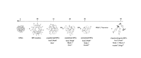
[0031] Fig. 1 Overview of the generation of o-NPCs from hiPSCs using this
40 day
protocol. o-NPCs, oligodendrogenic neural progenitor cells; hiPSCs, human
induced
pluripotent stem cells.
[0032] Fig. 2 Morphology of hiPSC-NPCs and hiPSC-o-NPCs. hiPSC-NPCs,
human
induced pluripotent stem cell-derived neural precursor cells; hiPSC-o-NPCs,
human induced
pluripotent stem cell-oligodendrogenic neural progenitor cells.
[0033] Fig. 3 Example of daily culture conditions for differentiation
of NPCs from
hiPSCs. Monolayer cells can be treated with dual SMAD inhibitors for 7-8 days.
At the end of
this step, neuro ectodermal rosettes emerge. Cells can be passaged every 3-4
days and
replatedat the density of 250,000 cells/cm2. For the first 24 hr after each
passage, cells can
be supplemented with ROCK inhibitor. NPCs, neural progenitor cells; hiPSCs,
human
induced pluripotent stem cells.
[0034] Fig. 4 . Two key pathways have been proposed for generation of
oligodendrogenic NPCs: (1) the canonical pathway which is dependent on sonic
hedgehog
(Shh) and is mainly used for generation of spinal oligodendrocytes and (2) the
non-canonical
pathway which is Shh independent and requires FGF2 to generate forebrain
oligodendrocytes.
[0035] Fig. 5 NPCs are mainly differentiated to neurons and astrocytes
after removal
of growth factors FGF2 and EGF, however, o-NPCs are biased towards an
oligodendrocytic
fate and predominantly differentiate to oligodendrocytes. NPCs, neural
progenitor cells; o-
NPCs, oligodendrogenic neural progenitor cells.
[0036] Fig. 6 Caudalization and ventralization of NPCs using RA and a
Shh agonist
(purmorphamine). NPCs, neural progenitor cells; RA, retinoic acid; Shh, sonic
hedgehog.
[0037] Fig. 7 Culture conditions from days 26 to 40; the last step for
the generation
of o-NPCs is supplementation with PDGF-AA and thyroxine. o-NPCs,
oligodendrogenic
neural progenitor cells.
[0038] Fig. 8A Overview of the generation of o-NPCs from hiPSCs-NPCs.
5
2306351
CA 3006897 2018-06-01
[0039] Fig. 8B Changes in the gene expression profile of key transcription
factors
during generation of o-NPCs from un-patterned NPCs.
[0040] Fig. 8C Changes in the morphology of un-patterned NPCs to bi-
polar
morphology of o-NPCs cultured on laminin.
[0041] Fig. 8D o-NPCs have the potential to be differentiated to all
three different cell
types; neurons (6-III Tub), astrocytes (GFAP) and oligodendrocytes (CNPase).
[0042] Fig. 8E q-RT-PCR gene expression analysis of o-NPCs as it
compared to
hiPSCS.
[0043] Fig. 8F Differentiation profile of o-NPCs. Majority of o-NPCs
differentiating
towards oligodendrocytes.
[0044] Fig. 9A Transplanted cells differentiate to express markers of
mature
oligodendrocytes (APC), immature oligodendrocytes (01ig2), astrocytes (GFAP)
and neurons
(TUJ1 and NeuN) in o-NPCs and unpatterned NPCs.
[0045] Fig. 9B Quantitative analysis of tri-lineage in vivo
differentiation profiles (n=5
per each group). *p<0.05 and "p<0.01. Scale bars: 20 pm.
[0046] Fig. 10 A-D Generation of oligodendrogenic NPCs. (A) The gene
expression
pattern of rostral and caudal identity markers compared between human iPSC-
NPCs,
unpatterned NPCs, fetal cortical NPCs and fetal spinal NPCs. Hierarchical
clustering trees
reveal a strong similarity between human iPSC-NPCs, unpatterned NPCs and fetal
cortical
NPCs while fetal spinal NPCs demonstrated caudal identity. (B) Unpatterned
NPCs were
caudalized using retinoic acid (RA) and then ventralized by treatment with
Shh. To generate
oNPCs, these cells were eventually treated with PDGF/Thyroxine. (C) Gradual
changes in
the morphology of NPCs after patterning towards oNPCs with elongated mono- and
bi-polar
morphology. These representative micrographs are from unpatterned NPC derived
cells. (D)
Stepwise changes in the expression profile of NPCs during generation of oNPCs.
The
expression of transcription factor 0tx2, an important marker of brain
identity, is reduced in
caudalized NPCs and they gain the expression of HoxA4, a marker of spinal
identity in
ventralized NPCs (vNPCs). The expression of bHLH transcription factors Nkx2.2,
01ig2 and
Nkx6.1, is upregulated in oNPC stage.
[0047] Fig. 11 A-C In vitro differentiation profile of oNPCs. (A) Both
unpatterned
NPC and oNPCs demonstrated comparable expression of neural progenitor markers,
Pax6,
Sox2 and nestin. (B, C) Comparison of the differentiation profile of
unpatterned NPC and
oNPCs after removal of the growth factors EGF, bFGF and addition of 0.1% FBS.
These
results and representative micrographs belong to drNPC derived cells. Results
are
6
2306351
CA 3006897 2018-06-01
presented as mean SEM from three independent experiments (average of 10
random
fields in each group). *p <0.05, **p <0.01, Student's t test. Scale bar: 20pm.
[0048] Fig. 12 A-E oNPCs predominantly differentiated into oligo-
lineage cells, and
myelinated host axons. (A-D) Representative images of 01ig2+/HuN+ immature (A)
and
GST-pi+/HuN+ mature (B) oligodendrocytes (arrowheads). Cytoplasm of the
transplanted
Stem121+ cells co-localized with MBP (C; arrowheads), and there were
MBP+/Stem121+
mature oligodendrocytes myelinating host NF 200+ neuronal axons (D;
arrowheads). These
cells mainly existed in the white matter area of the spinal cord. (E-l)
Representative images
of immunoelectron microscopy in oNPCs (E-G), NPC (H) and vehicle groups (I).
Grafted
cells were detected by the black dots observed upon anti-Stem121 antibody
staining. At
higher magnifications in the oNPC group, remyelinated axons surrounded by
transplanted
cells were identified (F) and endogenous myelin from oligodendrocytes were
preserved (G).
Arrowheads and arrows indicate myelin derived from transplanted cells and
endogenous
cells, respectively. Scale bar: 10 pm in (A-D), 2 pm in (E, H, I), and 200 nm
in (F, G).
[0049] Fig. 13 A-C in vitro oNPCs differentiation assay with or without
CSPGs.
(Chondroitin Sulfate ProteoGlycan). oNPCs cultured on dishes coated with
spinal cord
homogenates from uninjured (Naïve-h) or SCI-lesioned animals (SCI-h) for a
week. (A) Cells
were fixed and stained for the neural progenitor cell marker (Nestin), radial
glial cell marker
(3CB2; cytoplasmic projection stained), oligodendrocyte marker (01), astrocyte
marker
(GFAP) or neuronal marker (13111 tubulin). (B) The percentage of cells
positive for GFAP, 01,
13111 tubulin or Nestin were quantified (n = 3 biological replicates/group).
(C) qRT-PCR
analysis of the expression profile of neurogenic, astrocytogenic and
oligodendrogenic
transcription factors in oNPCs cultured on SCI-h relative to control-oNPCs
cultured on
Naïve-h with no treatment. Data represent the mean Log2-fold change in gene
expression
relative to control cells (n = 3 biological replicates/group). Values are
expressed as the mean
SEM. *p < 0.05. (Scale bar, 30 pm in A).
[0050] Fig. 14 A-E Functional analysis following cell transplantation.
(A) Time course
of motor functional recovery of hindlimbs in BBB score. Rats with oNPCs
transplantation
showed significant recovery from 7 to 9 weeks after SCI. (B) Representative
images of gait
analysis with CatWalk system 9 weeks after SCI. Light and dark footprints
indicate right and
left hindlimbs, respectively. (C,D) Gait analysis with the CatWalk system.
Note that there
was significantly better recovery in stride length between the oNPC and
vehicle groups, and
swing speed in the oNPC group compared to the other groups. (E) Evaluation of
thermal
allodynia in the tail-flick test. In each test, 10 rats per each group were
examined. *p<0.05;
**p<0.01.
7
2306351
CA 3006897 2018-06-01
[0051] Fig. 15. Levels of BMP4, TGF-13 and Jagged1 detected in the cervical
spinal cord at two weeks post-injury.
Detailed description of the Disclosure
[0052] Unless
otherwise defined, scientific and technical terms used in connection
with the present disclosure shall have the meanings that are commonly
understood by those
of ordinary skill in the art. Further, unless otherwise required by context,
singular terms shall
include pluralities and plural terms shall include the singular. For example,
the term "a cell"
includes a single cell as well as a plurality or population of cells.
Generally, nomenclatures
utilized in connection with, and techniques of, cell and tissue culture,
molecular biology, and
protein and oligonucleotide or polynucleotide chemistry and hybridization
described herein
are those well-known and commonly used in the art (see, e.g. Green and
Sambrook, 2012).
[0053] Terms of
degree such as "about", "substantially", and "approximately" as
used herein mean a reasonable amount of deviation of the modified term such
that the end
result is not significantly changed. These terms of degree should be construed
as including a
deviation of at least 5% of the modified term if this deviation would not
negate the meaning
.. of the word it modifies.
[0054] Further,
the definitions and embodiments described in particular sections are
intended to be applicable to other embodiments herein described for which they
are suitable
as would be understood by a person skilled in the art. For example, in the
following
passages, different aspects of the invention are defined in more detail. Each
aspect so
defined may be combined with any other aspect or aspects unless clearly
indicated to the
contrary. In particular, any feature indicated as being preferred or
advantageous may be
combined with any other feature or features indicated as being preferred or
advantageous.
[0055] Most
current protocols for differentiation of caudalized neural progenitor cells
(also referred to as neural precursor cells) (NPCs) are based on knowledge of
mouse and
chicken spinal cord embryology. Although the embryologic origin of
oligodendrogenic cells
continues to be investigated, a general consensus exists that early stage
oligodendrocyte
precursor cells (OPCs) and motor neurons share a developmental lineage in the
spinal cord.
Goldman and colleagues have described a method for generating OPCs from
hiPSCs,
however, the greatest drawback of their protocol is the lengthy culture time
requiring
proportionally greater quantities of expensive growth factors (Wang et al.,
2013).
[0056]
Described herein are methods for generating a cell type biased to produce
oligodendrocytes, herein referred to as o-NPCs. These cells ae similar to
conventional NPC
in that they are tripotent but are different in that they produce different
ratios of these cells
8
2306351
CA 3006897 2018-06-01
when differentiated. The methods described herein such as the protocol
described in
Example 1 substantially reduces differentiation time making the generation of
o-NPCs for
research and therapy more feasible.
[0057] The differentiation, isolation, and expansion protocols
described herein for
example as shown in Figs. 3, 6 and 7 to generate o-NPCs from hiPSCs requires -
40 days.
Different factors are added to different stages of differentiated hiPSCs
according to an
approximate timeline as described in Fig. 1. References to days generally
correlates to the
days identified in Figs. 1, 3, 6 and 7. Also described are markers to
characterize the cells at
each stage for example as shown in Fig. 1.
[0058] Like conventional NPCs, o-NPCs generated using the present
methods are
tripotent cells and have the ability to differentiate into neurons,
astrocytes, and
oligodendrocytes, however, o-NPCs have a bias to differentiate predominantly
into
oligodendrocytes, both in vitro and in vivo. For example, the methods
described herein have
been found to increase oligodendrocyte production by at least 35%, at least
40%, at least
45%, at least 50%, at least 55%, at least 60% or at least 65% in vitro.
Depending on the type
of spinal cord injury, e.g. cervical, thoracic, chronic and/or acute, the
methods described
herein have been found to increase oligodendrocyte production by at least 35%,
at least
40%, at least 45%, at least 50%, at least 55% or at least 60% compared to
conventionally
prepared NPCs.
[0059] Accordingly an aspect of the present disclosure includes a
method of
producing oligodendrogenic neural progenitor cells (o-NPCs), the method
comprising:
a. obtaining ventralized neural progenitor cells (NPCs), the ventralized NPCs
expressing Sox2 and NKx6.1 and decreased level of Pax6 compared to unpattenred
NPCs
and increased expression of HoxA4 compared ot unpatterned NPCs.
b. culturing the ventralized NPCs for about 12 to about 16 days (days 26-40 of
Fig. 7)
in neural expansion media (NEM) supplemented with i) PDGF for the about 12 to
about 16
days; and ii) thyroxine or a thyroxine analog for the latter about 7 to about
9 days, to produce
o-NPC expressing Sox2, Nkx2.2, decreased excpresison of Pax6 and Nkx6.1
compared to
ventralized NPCs and increased expression of HoxA4 and 01ig2 compared to
ventralzied
NPCs.
[0060] The term "ventralized NPCs" as used herein refers to NPCs which
express
Sox2 and Nestin, have decreased expression of Pax6, FoxG1, 0tx2 and Gbx2, and
have
increased expression of Nkx6.1, HoxA4, HoxB4 HoxC4 and HoxC5, all relative to
un-
patterned-NPCs. For example such cells can have at leat 20% decreased
expression of
9
2306351
CA 3006897 2018-06-01
Pax6, at least 75% decreased level of expression for FoxG1, 0tx2 and Gbx2, at
least 50%
increased expression Nkx6.1, and have at least 50% increased expression of
HoxA4, HoxB4
HoxC4 and HoxC5, all relative to unpatterned-NPCs. Further, the expression
level of 011g2
and Nkx2.2 is less than the expression of these genes compared to o-NPCs, for
example
ventralized NPCs typically express at least 25% less protein and at least
about 2 fold or at
least about 3 fold (Log2 scale) less RNA, determined for example by density of
immune
staining and qRT-PCR respectively, than the expression level of these two
genes compared
to o-NPCs. 011g2 refers to oligodendrocyte transcription factor, Nkx2.2 and
Nkx6.1 refer to
homeobox proteins Nkx2.2 and Nkx6.1 and Sox2 also known as SRY (sex
determining
region Y)-box 2 which is a marker of neural stem progenitor cells (NSPCs).
Sox2 along with
Pax6 and Nestin are three main markers for NSPCs. Pax6 refers to paired box
protein
Pax6. HoxA4, Hox64, HoxC4 and HoxC5 refer to homebox proteins A4, B4, C4 and
C5
respectively. FoxG1 refers to forkhead box protein G1. 0tx2 and Gbx2 refer to
homebox
proteins 01)(2 and Gbx2 respectively.
[0061] The term
"unpatterned NPCs" as used herein means directly reprogrammed
NPCs that have not been
caudalized and express Sox2/Pax6 and 0tx2 (increased relative to
NPC rosettes from which they can be derived). As shown in Fig. 1, they can be
obtained at
about 14 days using a protocol described herein. When the unpatterned NPCs are
derived
from hiPC cells they can be referred to as hiPS-derived unpatterned-NPCs hiPS-
derived
unpatterned-NPCs.
[0062] The term "NPCs" as
used herein refers to neural progenitor cells,
interchangeably referred to as neural precursor cells and neural stem cells
(NPS).
[0063] The term "NEM"
or "neural expansion media" as used herein means a base
media suitable for culturing neural progenitor cells such as DMEM/F12
comprising one or
more of sodium pyruvate, a glutamine product such as glutamine or GlutaMAXTm,
one or
more antibiotics such as penicillin and/or streptomycin, a supplement such as
B27 without
vitamin A, and depending on the stage of cell differentiation, one or more of
FGF2, EGF
and/or heparin. An example of a suitable NEM is provided in Example 1.
[0064] The period of
PDGF incubation including the combined PDGF/thyroxine
PDGF/thyroxine analogue incubation is approximately 12 to 16 days and this
corresponds
generally to days 24 to 40 as shown in Fig. 7. A person skilled in the art
will recognize that
the days of culture will depend on the culture conditions used including for
example the
exact differentiation status of the starting population.
2306351
CA 3006897 2018-06-01
[0065] The o-NPCs (also referred to as oNPCs) produced show for example 10-
20% increased level of expression of HoxA4, and Hox64, 30-40% increased level
of
expression of 01ig2 and a 10-20% decreased level of expression of Pax6 and
Nkx6.1
compared to ventralized-NPCs. These cells have spinal cord identity, meaning
that the
expression level of transcription factors which spatially are specific for
spinal cord, like
HoxA4, Hox64, HoxC4 and HoxC5 which are for example at least 75% more than
those in
un-pattenerd NPCs, and do not express markers associated with brain identity
cells. They
are tripotent meaning that they have the potential to generate neurons,
astrocytes and
oligodendrocytes but are biased to differentiation towards for example at
least 50 % more
oligodendrocytes compared to un-patterned NPCs.
[0066] .. o-NPCs, unlike un-patterned-NPCs, are caudalized, ventralized and
are
oligogenic. The different stages can for example be assessed by expression
levels of one or
more genes. For example, caudalized cells (compared to un-patterned cells)
have elevated
levels of HoxA4, B4, C4 and C5 ( for example about around 50% more) but not as
much as
endpoint stage in o-NPCs which have increased levels that are about or at
least 75% higher.
Ventralized cells have a decrease in Pax6 expression (around 20-25%) and an
increase in
Nkx6.1 expression (around 25% or more) compared to to caudalized cells.
[0067] The PDGF can be PDGF-AA, PDGF-AB, PDGF-BB and/or PDGF-CC.
Preferably mammalian and more preferably, the PDGF when used with human cells
is
human PDGF. The PDGF is in an embodiment, PDGF-AA. In an embodiment, the NEM
comprising PDGF-AA comprises about 20-30 ng/ml PDGF-AA. Recombinant human PDGF-
AA can be obtained from various commercial sources such as ProSpec Hamada St.
8
Rehovot 7670308 Israel (e.g., Catalogue number CRFOO1A CYT-341). Additionally,
PDGF-
AA from other mammalian sources such as mouse, rabbit, sheep or rat as
mammalian
PDGF shares a high degree of conservation (e.g. mamalian PDGF-A is conserved
from 87-
100%, B is 85% to 100 and C is 70% to 100 can be used interchangablly. In the
present
disclosure, PDGF, optionally PDGF-AA, is used as diffrerentiation factor for
ventralized
neural progenitor cells progressing towards an oligodendrogenic fate.
[0068] In an embodiment, the NEM comprising thyroxine comprises about 40-60
ng/ml thyroxine. In another embodiment, a thyroxine analogue is used. The
thyroxine
analogue is, in one embodiment, levothyroxine sodium hydrate, which can be
used in the
place of thyroxine. In an embodiment, the concentration of levothyroxine
sodium hydrate is
about 40 ng/mL. In another embodiment, the thyroxine analogue is
triiodothyronine/thyroid
hormone 3 (T3). In an embodiment, the concentration of
triiodothyronine/thyroid hormone 3
(T3) is about 40 to about 60 ng/m L.
11
2306351
CA 3006897 2018-06-01
[0069] The term "thyroxine" or "T4" as used herein, refers to the
prohormone of the
thyroid hormone triiodothyronine (T3), including all mammalian forms
preferably human. It is
used in this method as a differentiating factor when ventralized neural
progenitor cells are
stimulated towards their oligodendrogenic fate. Thyroxine can be obtained from
various
commercial sources such as Sigma-Aldrich Canada Co. Oakville, Ontario Canada
(e.g.,
Catalogue number T1775).
[0070] Looking at Fig. 7, a particular embodiment of the media,
factors and time
periods that can be used is provided.
[0071] NEM can be replaced daily with the required factors.
[0072] The term "progenitor cell" (interchangeably referred to as
precursor cells)
refers to cells that have a cellular phenotype that is at an earlier step
along a developmental
pathway or progression than is a fully differentiated cell relative to a cell
which it can give
rise to by differentiation. Progenitor cells can give rise to multiple
distinct differentiated cell
types or to a single differentiated cell type, depending on the developmental
pathway and on
the environment in which the cells develop and differentiate.
[0073] In the context of a cell, the term "differentiated", or
"differentiating" is a relative
term and a "differentiated cell" is a cell that has progressed further down
the developmental
pathway than the cell it is being compared with. Thus, stem cells can
differentiate to lineage-
restricted precursor cells (such as a neural progenitor cell), which in turn
can differentiate
into other types of precursor cells further down the pathway and then to an
end-stage
differentiated cell, which plays a characteristic role in a certain tissue
type, and may or may
not retain the capacity to proliferate further.
[0074] In an embodiment, the NEM the NEM of steps b. i) and ii) is
supplemented
with FGF2. As shown for example in Fig. 7, the NEM can comprise PDGF and FGF2
for the
duration of the incubation from ventralized NPCs to produce o-NPCs.
[0075] The FGF2 is added in some embodiments along with heparin. Other
components can also be included as described herein. For example, the NEM for
culturing
ventralized NPCs can comprise FGF2 (e.g. at about 10-20ng/m1), B27 without RA,
heparin
and Ni supplement.
[0076] The term "fibroblast growth factor 2" or "FGF2" (also known as
bFGF or FGF-
beta as well as heparin binding growth factor 2 is a member of the fibroblast
growth factor
family. FGF2, for example human FGF-2 can be obtained from various commercial
sources
such as Cell Sciences®, Canton, Mass., USA, Invitrogen Corporation
products, Grand
12
2306351
CA 3006897 2018-06-01
Island N.Y., USA, ProSpec-Tany TechnoGene Ltd. Rehovot, Israel, and Sigma, St
Louis,
Mo., USA.
[0077] In an embodiment, the ventralized NPCs are obtained by culturing
unpatterned NPCs expressing Sox2, Pax6 + and Otx2+ for about 12 days (days 14
to 26 of
Fig. 6) in NEM with i) retinoic acid or a retinoic acid analogue for the
preliminary about 7 to
11 days and ii) a shh agonist for the latter about 9 days. This step includes
producing
caudalized NPCs from the unpatterned NPCs and differentiating them to
ventralized NPCs
as shown for example in Fig. 6.
[0078] The retinoic acid analogue can be for example synthetic retinoid
EC23.
[0079] The term "caudalized NPCs" as used herein refers to NPCs having
a caudal
spinal cord progenitor fate and which express Sox2, Pax6 and an increased
expression of
Nkx6.1 relative to un-patterned NPCs and a decreased expression of 0tx2 and
FoxG1
relative to un-patterned NPCs. For example, "caudalized NPCs" express Sox2,
Nestin and
Pax6 with equivalent level to un-patterned NPCs, and have for example at least
75%
decreased level of expression for FoxG1, 0tx2 and Gbx2, at least 25% increased
expression
Nkx6.1, and have at least 25-50% increased expression of HoxA4, HoxB4 HoxC4
and
HoxC5, all relative to un-patterned-NPCs. The expression level of Nkx6.1 is
for example at
least 25% less than the expression level this gene compared to ventralized-
NPCs.
[0080] The term "sonic hedgehog agonist" or "Shh agonist" as used
herein includes
recombinant sonic hedgehog, purmorphamine and SAG, which stands for Smoothened
Agonist and is a chlorobenzothiophene-containing compound. Shh can also be
replaced with
recombinant mammalian Desert hedge hog (Dhh) or recombinant mammalian Indian
hedge
hog (lhh).
[0081] In an embodiment, the sonic hedgehog agonist used is selected
from
purmorphamine, SAG and recombinant Shh polypeptide. For example when the Shh
agonist
is Shh the concentration used can be about 10Ong/ml.
[0082] In an embodiment, the concentration of purmorphamine is about
0.5 pM to
about 1pM purmorphamine.
[0083] In an embodiment, the concentration of SAG is about 0,5 pM SAG.
[0084] In an embodiment, the concentration of Shh is about 10Ong/m1Shh.
[0085] In some embodiments, the method comprises obtaining caudalized NPCs
from unpatterned NPCs expressing Sox2/Pax6 + Otx2+ with retinoic acid (RA)(for
example at
13
2306351
CA 3006897 2018-06-01
a concentration of 0.1pM-0.2pM) and/or a retinoic acid analogue and using
caudalized NPCs
to produce the ventralized NPCs.
[0086] The term "unpatterned NPCs" as used herein refers to NPCs that
have yet to
be caudalized and ventralized. Un-partnered NPCs are primitive or definitive
NPCs which
are not yet being treated with any patterning factors like RA or Shh (and its
agonists). Un-
patterned NPCs express Pax6, Nestin and Sox2. The level of expression of Gbx2,
Emx2
and Irx2 is lower in un-patterned NPCs as compared to mid-brain identity NPCs,
and the
level of expression of Hox genes (like A4, B4, C4) are lower in un-patterned
NPCs as
compared to spinal cord identity NPCs.
[0087] Typically unpatterned NPCs are tripotent cells which
differentiate mainly
towards neuronal and astrocytic cell fates after removal of growth factors EGF
and FGF2 as
depicted in Fig. 5. Examination of transcription factor profiles of the NPCs
indicates that the
Pax6 expressing NPCs do not express 01ig2 and Nkx2.2, homeodomain proteins
which are
expressed in ventral neural progenitors (Lu et al., 2002; Zhou, Choi, &
Anderson, 2001).
[0088] As shown in the Examples, the unpatterned NPCs are cultured with
RA for a
period of about 3 days, followed by culturing in NEM comprising RA and a shh
agonist for
about 6 days followed by culturing in media comprising a shh agonist without
RA for about 3
days.
[0089] During treatment with RA (no FGF2) is added to the medium
although in
some embodiments, EGF is added. The culture media used for this stage can
comprise B27
supplement comprising vitamin A.
[0090] The expression of specific markers can be used to determine that
the
unpatterned cells have been caudalized. For example, as shown in the examples
quantitative RT-PCR analyses indicated that RA treatment decreased the
expression of 0tx2
and increased the expression of HoxA4.
[0091] For the step involving culturing with RA or retinoic acid analogue
and the Shh
agonist together, the unpatterned NPCs are also optionally cultured in the
presence of EGF (
for example at a concentration of about 10 to about 20ng/m1) for the first 9
days. For the last
3 days, the NPCs are cultured in the presence of a shh agonist and cultured
with FGF2. The
period of caudalization and ventralization is depicted in Fig 6 and extends
from
approximately day 14 to 26 of the 40 day protocol.
[0092] The removal of RA and the addition of FGF2 for the last 3 days
(e.g. days of
23 to 26 of Fig. 6) prevents for example differentiation of cells into spinal
motoneurons
(MNs). RA treatment of Nkx6.1+ NPC can, for example cause them to
differentiate into
14
2306351
CA 3006897 2018-06-01
spinal MNs. To prevent differentiation to MNs and to promote the generation of
oligodendrogenic NPCs, RA is removed for example after 6 days and FGF2 is
supplemented in place of EGF. As shown herein, the removal of RA and addition
of FGF2
almost completely blocks the caudalized/ventralized cells from differentiating
into MNs and
promotes the generation of 01ig2+/Nkx2.2+ cells.
[0093] In another
embodiment, the method further includes a step of obtaining
unpatterned NPCs from columnar cells in the form of rosettes and expressing
Pax6
[0094] The term
"rosette" as used herein refers to a cellular pattern of columnar
cells. The neural rosette is the developmental signature of neuroprogenitors
in cultures of
differentiating embryonic stem cells; rosettes are radial arrangements of
columnar cells that
express many of the proteins expressed in neuroepithelial cells in the neural
tube. In addition
to similar morphology, neuroprogenitors within neural rosettes can
differentiate into the main
classes of progeny of neuroepithelial cells in vivo: neurons,
oligodendrocytes, and
astrocres. [
[0095] The columnar
cells forming rosettes can be cultured based on Chambers et
al. (2009) dual-SMAD inhibition using chemically defined adherent colony
culture (e.g. neural
induction media (NIM).
[0096] As used herein
"neural induction media" or "NIM" herein means a base media
suitable for culturing neural precursor cells such as DMEM/F12 comprising one
or more of
sodium pyruvate, a glutamine product such as glutamine or GlutaMAXTm, one or
more
antibiotics such as penicillin and/or streptomycin, a supplement such as B27
without vitamin
A, non-essential amino acids, BMP inhibitor such as LDN193189 or Noggin FGF2,
heparin
and EGF An example of a suitable NIM is provided in Example 1.
[0097] As depicted in
Fig. 3, induction of neural cells can be achieved by growth
factors hLIF (e.g. about 1Ong/m1) accompanied by N2, B27(-RA), FGF/heparin and
differentiation factors TGFb inhibitor (SB431542) (1pM), BMP inhibitor (1 pM)
(LDN193189)/
or Noggin (200ng/m1) or any one of factors mentioned in Example 1 for a period
of 7 days.
Following this period, rosettes are re-plated on vessels such as culture
plates pre-coated
with poly-L-lysine/laminin and in NEM comprising EGF (10-20ng/m1) for 4 to 6
days as
described in example 1 and depicted in Fig. 3. At this time cells are positive
for Sox2 and
0tx2, a homeodomain protein expressed by fore- and mid-brain cells, but
negative for
HoxC4, a homeodomain protein produced by cells in the spinal cord.
[0098] In an embodiment,
one or more of the culture steps is performed in a
monolayer system.
2306351
CA 3006897 2018-06-01
[0099] The term "monolayer system" as used herein refers to a cell
culturing system
where cells grow in a single layer on a growth surface, for example in a
plate, flask or other
vessel, in the absence of feeder cells. The growth surface is a feeder-free
system using for
example a gelatinous matrix coated vessel such as a culture plate or dish. The
gelatinous
matrix can for example be gelatin, Matrigel or Geltrex.
[00100] In an embodiment, the monolayer system used to culture the
ventralized
NPCs comprises culturing the ventralized NPCS on gelatinous matrix coated
plates.
[00101] In an embodiment, the gelatinous matrix is selected from gelatin
Matrigel, or
Geltrex, Vitronectin, Fibronectin or Laminin. Matrigel is a gelatinous protein
mixture of
secreted extracellular matrix proteins derived from mouse tumor cells and
Geltrex is as a
reduced growth factor basement membrane extract used for attachment and
maintenance of
human embryonic stem cells (hESCs) and human induced pluripotent stem cells
(hiPSCs).
Any mammalian extracellular or basement matrix used for NPC cell culture can
be used
including for example Vitronectin, Laminin or Fibronectin from any mammalian
sources.
Matrigel and Geltrex coated vessels can be made using Matrigel or Geltrex.
Matrigel is
available for example from Corning, Tewksbury MA 01876, USA and Geltrex is
available for
example from Thermo-Fisher scientific Mississauga, Ontario, Canada.
[00102] Alternatively, a feeder-dependent culturing system can also be
used, wherein
cells grow on mouse embryonic fibroblast cells.
[00103] The term "poly-L-lysineilaminin" as used herein refers to a
polymer of basic
amino acid lysine which enhances the adherence of neural cells to the plate by
changing the
net charge of plates to positive. They are particularly useful for the culture
of central nervous
system (CNS) neurons. The L or D isomers can be used for plating, however, the
D isomer
may be preferred because there is no breakdown released by proteases of the
cells. Laminin
is an extracellular matrix constitutively used for the culture of neural
cells. The plates are first
coated with poly L-lysine (PLL) and then with laminin to increase the
concentration of laminin
applied using this method.
[00104] The term "EGF" as used herein refers to mammalian Epidermal
growth
(EGF), for example human EGF having for example Gene Identification number
(Gene ID:
1950) as well as active conjugates and fragments thereof, including naturally
occurring
active conjugates and fragments. Any mammalian EGF can be used including human
EGF,
mouse EGF, sheep EGF, rabbit EGF and rat EGF, as well as active conjugates and
active
fragments thereof. Human EGF is preferred.
16
2306351
CA 3006897 2018-06-01
[00105] The term "active fragments" as used herein is a polypeptide having
amino
acid sequence which is smaller in size than, but substantially homologous to
the polypeptide
it is a fragment of, and where the active fragment polypeptide is about at
least 50%, or 60%
or 70% or at 80% or 90% or 100% or greater than 100%, for example 1.5-fold, 2-
fold, 3-fold,
4-fold or greater than 4-fold as effective in terms of biological action as
the polypeptide from
which it is a fragment of. Examples include fragments of EGF which bind and
activate EGF
receptor.
[00106] In an embodiment of the present disclosure, the
columnar cells forming
rosettes are cultured a monolayer system.
[00107] In a further embodiment, the columnar cells in the
form of rosettes are
obtained from human pluripotent stem cells (PSCs), optionally human induced
PSC
(hiPSCs) or human embryonic stem cells (hESCs). Any hiPSC or hESC line can be
used in
the methods described herein including for example any fetal or adult derived
human NPCs
including directly reprogrammed NPCs (drNPCs) (e.g. day 14 cells in Fig. 1 or
day 0 cells in
Fig 8. Examples of hiPSC cell lines that can be used include 1.53 and BC1. The
BC1 cell
line one is derived from adult bone marrow CD34+ cells and the 1.53 line which
is derived
from human fibroblasts using piggyBac vectors.
[00108] The term "pluripotent stem cell" as used herein
refers to a cell with the
capacity, under different conditions, to differentiate to more than one
differentiated cell type,
and for example the capacity to differentiate to cell types characteristic of
the three germ cell
layers, and includes embryonic stem cells and induced pluripotent stem cells.
Pluripotent
cells are characterized by their ability to differentiate to more than one
cell type using, for
example, a nude mouse teratoma formation assay. Pluripotency is also evidenced
by the
expression of embryonic stem (ES) cell marker.
[00109] The term "stem cell" as used herein, refers to an
undifferentiated cell which is
capable of proliferation, self-renewal and giving rise to more progenitor
cells having the
ability to generate a large number of mother cells that can in turn give rise
to differentiated or
differentiable daughter cells. The daughter cells can for example be induced
to proliferate
and produce progeny that subsequently differentiate into one or more mature
cell types,
while also retaining one or more cells with parental developmental potential.
[00110] In an embodiment, the pluripotent stem cell is from a mammal, such
as a
human. In an embodiment, the pluripotent stem cell is a human iPSC (hiPSC).
[00111] Further, ROCK inhibitors can be used when the cells
are passaged to
improve cell survival. For example, a ROCK inhibitor (e.g. Y-27632) can be
used for the first
17
2306351
CA 3006897 2018-06-01
it
24 hours after each cell passaging in the entire method of producing o-NPCs
from hiPSC-
NPCs as described in Example 1. In an embodiment, the ROCK inhibitor Y-27632
at a
concentration of 10 pM is used. In other embodiments a JAK inhibitor such as
Jak inhibitor I
is used instead of a ROCk inhibitor. For example JAKi I can be used at a final
concentration
1 pM instead of the ROCK inhibitor.
10 [00112] The term "passaging", "passaged" or "passage" as used herein
refers to
transferring the cultured cells from their current growth medium to a new
growth medium.
Cells can be passaged for example according to as described in Example 1. For
example
hIPSCs should be passaged in order to avoid overgrowth and to maintain them in
an
undifferentiated state. Further it may be preferable to passage iPSCs in
clumps.
[00113] As a person skilled in the art would understand, cells can be
dislodged from
the culture plate with the use of enzymes and enzyme cell detachment solutions
such as the
enzyme cell detachment solution AccutaseTM. Other enzymes like Dispase or
TrypLE can
also be used.
[00114] o-NPCs generated using methods described herein can be expanded for
example for up to three passages without losing their proliferation and
differentiation
capacity. After this stage the proliferation rate of the cells may slow and
they eventually
cease proliferating for example at passage 5 to 6 when they morphologically
appear as flat,
expanded cells.
[00115] Using the methods described herein, one can produce a population of
tripotent o-NPCs differentiated from hiPSC-NPCs, the population comprising for
example
about 90% to about 95% o-NPCs based on immunocytochemical 01ig2 staining.
[00116] The o-NPCs made using the protocols described herein can produce
spinal
oligodendrocytes and can be used in various applications.
[00117] Looking at Fig. 1 which outlines the stages of development from
iPSCs to o-
NPCs, the period corresponding to differentiating ventralized NPCs to o-NPCs
extends
approximately from day 26 to day 40, the period corresponding to
differentiating unpatterned
NPCs to ventralized NPCs is from day 14 to 26, the period corresponding to
differentiating
columnar cells in the form of rosettes to unpatterned NPCs is from day 10 to
14 and the
period of differentiating iPSCs to rossettes is from day 2 to 10.
[00118] Accordingly, in an embodiment, the method of producing o-NPCs
comprises
a) obtaining iPSCs cultured for at least about 2 days (days 0-2 in Fig. 3);
b) culturing the iPSCs:
18
2306351
CA 3006897 2018-06-01
ir
I. in NIM supplemented with leukemia inhibitory factor (LIF), FGF, B27
lacking vitamin A, N2 supplement, TGFb inhibitor, BMP inhibitor,
optionally Noggin, AMP-activated protein kinase (AMPK) inhibitor for
about 7 days (day 2 to day 9 in Fig. 3); and
ii. in NIM supplemented with EGF, FGF, B27 lacking vitamin A and N2
supplement, wherein the iPSCs are cultured in vessels coated with a
gelatinous matrix comprising ploy-L-lysine/larninin for about 1 to 2
days to produce columnar cells in the form of rosettes expressing Pax
6 (day 10 in Fig. 3);
c) culturing the columnar cells in the form of rosettes from step b. in NEM
comprising EGF, FGF, B27 lacking vitamin A and N2 supplement for about 4
days, wherein the iPSCs are cultured in vessels coated with a gelatinous
matrix comprising ploy-L-lysine/laminin, to produce upatterned NPCs (day 14
in Fig. 3 and 6);
d) culturing the unpatterned NPCs from step c. for about 6 days in NEM
comprising retinoic acid and/or a retinoic acid analogue such as synthetic
retinoid EC23, N2 supplement, B27, EGF and a Shh agonist to produce
caudalized NPCs (day 20 in Fig. 6);
e) culturing the caudalized NPCs from step d.:
i. in NEM comprising EGF, N2 supplement, B27, retinoic acid and/or a
retinoic acid analogue and Shh agonist for about 3 days (days 20 to
23 of Fig. 6); and
ii. in NEM comprising FGF2, N2 supplement, B27 and a Shh agonist for
about 3 days (days 23 to 26 of Fig. 6) to obtain ventralized NPCs;
f) culturing the ventralized NPCs for about 12 to about 16 days (days 26-40 of
Fig.7) in NEM comprising i) PDGF for the about 12 to about 16 days; ii) B27
and Ni supplement for the preliminary about 12 days; and iii) thyroxine for
the
latter about 7 to about 9 days, to produce o-NPCs.
[00119] The term
"cell culture medium" (also referred to herein as a "culture medium"
or "medium") as referred to herein is a medium for culturing cells containing
nutrients that
maintain cell viability and support proliferation and optionally
differentiation. The cell culture
medium may contain any of the following in an appropriate combination:
salt(s), buffer(s),
amino acids, glucose or other sugar(s), antibiotics, serum or serum
replacement, and other
19
2306351
CA 3006897 2018-06-01
.. components such as peptide growth factors, vitamins etc. Cell culture media
ordinarily used
for particular cell types are known to those skilled in the art.
[00120] The suitable culture medium can include a suitable base culture
medium
including for example, NIM and NEM including the formulations described herein
and/or any
other or media that supports the growth of cells to provide for example a base
culture
.. medium composition to which components and optionally other agents can be
added.
[00121] As mentioned, the oNPCs are biased to produce oligodendrocytes.
Accordingly, also provided is a method of producing a population of cells
comprising
oligodendrocytes, the method comprising:
i) producing o-NPCs according to a method described herein;
ii) differentiating the cells wherein the step of differentiating optionally
comprises
a) culturing in NEM lacking EGF and bFGF supplementation and comprising low
serum, optionally about 0.1% FBS to about 1% FBS, optionally for about 7 to 15
days,
optionally 10 days to promote formation of oligodendrocytes.
[00122] The o-NPCs can also be used to produce a mixed population of
cells or
promote formation of radial glial cells expressing for example 3CB2, by
culturing the o-NPCs
in NEM lacking FGF2 and EGF supplementation optionally for about 7 to 15 days.
[00123] In certain embodiments, the method further comprises enriching
and/or
isolating the desired cells.
Cells and Compositions and Methods of Use
[00124] Also provided is a population of cells produced according to a
method
described herein. In an embodiment, the population of cells is comprised in a
composition
optionally comprising a carrier, optionally a pharmaceutically acceptable
carrier.
[00125] As used herein, the term "pharmaceutically acceptable carrier" is
intended to
include any and all solvents, dispersion media, coatings, isotonic and
absorption delaying
agents, and the like, compatible with pharmaceutical administration and for
use with cells.
Optional examples of such carriers or diluents include, but are not limited
to, buffered saline,
culture media, ringer's solutions, dextrose solution, and 5% human serum
albumin and
bovine serum albumin (BSA).
[00126]
2306351
CA 3006897 2018-06-01
[00127] In an embodiment,
the cell population is an enriched or isolated cell
population. For example it can be enriched to exclude cells that do not share
the desired
combination of markers.
[00128] The term
"isolated population of cells" as used herein refers to a population of
cells that has been removed and separated from a mixed or heterogeneous
population of
cells. In some embodiments,
an isolated population is a substantially pure population of cells
as compared to the heterogeneous population from which the cells were isolated
or enriched
from, for example at least 90% pure.
[00129] In an embodiment,
the population is a clonal population derived from a single
cone.
[00130] The population of
cells can comprise oNPCs, or cells differentiated
thereofrom.
[00131] The population of
cells can isolated, purified and/or diluted in culture media,
including the nnedias described herein or freezing solution (such as culture
medium with
glycerol and the like). The composition can be frozen. In particular,
unpatterned NPCs can
be frozen for long periods of time (on the order of years).
[00132] The cells can for
example be disociated as single cells, optionally a clonal
single cell suspension in culture media such as NIM or NEM described herein.
[00133] Also provided in
another aspect is a kit comprising PDGF and thyroxine
and/or a thyroxine analog and optionally and other component used in method
herein,
optionally for preparing o-NPCs.
[00134] In some
embodiments, the population of cells are for use in transplantion in a
recipient in need thereof. Such population of cells are resuspended using
sterile and/or GMP
grade pharmaceutically acceptable carriers such as sterile cell culture media.
[00135] As shown in the
Examples, the population of cells produced using a method
describd herein can be used to treat spinal cord injuries. For example it is
demonstrated that
the population of cells described can be used to treat acute cervical and
thoracic SCI as well
as chronic thoracic SCI. The population of cells can also be used for treating
chronic cervical
spinal injuries, the treatment of multiple sclerorsis (MS), and cerebral palsy
(CP) as well as
other demyelination diseases.
[00136] Also included in
other aspect are uses of said cells and compositions
comprising said cells for transplanting and/or treating a subject in need
thereof, for example
21
2306351
CA 3006897 2018-06-01
for transplanting and/or treating a subject with a SPI or a demyelination
disease, optionally
MS or CP.
[00137] The term
"subject" as used herein includes all members of the animal kingdom
including mammals, and suitably refers to humans.
[00138] The term
"treatment" as used herein as applied to a subject, refers to an
approach aimed at obtaining beneficial or desired results, including clinical
results and
includes medical procedures and applications including for example
pharmaceutical
interventions, surgery, radiotherapy and naturopathic interventions as well as
test treatments
and combinations thereof for treating SPI or other neural conditions that
would benefit from
an infusion of oligodendrocytes. Beneficial or desired clinical results can
include, but are not
limited to, alleviation or amelioration of one or more symptoms or conditions,
diminishment of
extent of disease, stabilized (i.e. not worsening) state of disease,
preventing spread of
disease, delay or slowing of disease progression, amelioration or palliation
of the disease
state, and remission (whether partial or total), whether detectable or
undetectable.
[00139] As used
herein, the terms "administering," "introducing" and "transplanting"
are used interchangeably in the context of delivering a population of o-NPCs
or their
differentiated progeny into a subject, by a method or route which results in
at least partial
localization of the introduced cells at a desired site. The cells can be
implanted directly to the
spinal cord, or alternatively be administered by any appropriate route which
results in
delivery to a desired location in the subject where at least a portion of the
implanted cells or
components of the cells remain viable.
[00140]
[00141] For
traumatic injuries the cells can be administered 2 weeks or longer after
the injury.
[00142] Cells
can be induced from a subject to be treated own somatic cells. In
an alternate approach oNPCs produced from an allogeneic donor are used to for
example generate a bank of oNPCs with different HLAs. HLA matched oNPCs or
cells differentiated therein are then administerd to the subject in need
thereof.
[00143] The
above disclosure generally describes the present application. A more
complete understanding can be obtained by reference to the following specific
examples.
These examples are described solely for the purpose of illustration and are
not intended to
limit the scope of the disclosure. Changes in form and substitution of
equivalents are
contemplated as circumstances might suggest or render expedient. Although
specific terms
22
2306351
CA 3006897 2018-06-01
1,
have been employed herein, such terms are intended in a descriptive sense and
not for
purposes of limitation.
[00144] The following non-limiting examples are illustrative
of the present disclosure:
Examples
Example 1
Passaging and maintenance of human induced pluripotent stems cells in culture.
[00145] This protocol is used for the long-term maintenance
of hiPSCs. hiPSCs can
be continuously grown on plates for over 2 years without the acquisition of an
abnormal
karyotype. Media should be changed daily and cells should be passaged once
every 7 days.
hiPSCs are cultured using two different techniques: Feeder dependent culture
on mouse
embryonic fibroblast (MEF) cells and feeder-free culture on Matrigel or
Geltrex. Each one
has distinct advantages and disadvantages (for a full comparison see (Ghasemi-
Dehkordi et
al., 2015). IDescribed is a feeder-free technique on Matrigel. Geltrex can
also be substituted
for Matrigel. hiPSCs cultured on Matrigel or Geltrex have a different
morphology than those
cultured on feeder cells but retain their characteristic pluripotency. Feeder
cells can also be
used. For feed-dependent culture, refer to Takahashi & Yamanaka, 2006.
[00146] A commercially available pre-prepared medium,
mTeSR1Tm available from
Stem Cell Technologies (Vancouver CA) was used. An alternative is StemProTM
(Thermofisher) . Most pre-prepared hiPSC culture media contain IGF1,
heregulin1, FGF2,
and activin A, to maintain pluripotency.
Materials
Matrigel
Human induced pluripotent stem cells (hiPSCs)
mTeSR medium
Accutase
DMEM/F12 medium
ROCK inhibitor Y-27632
Growth medium without ROCK inhibitor
Cell scraper or rubber policeman
15-ml Falcon tubes
P1000 pipet
Sterile needle
23
2306351
CA 3006897 2018-06-01
1. On the day of passaging, exchange medium with freshmTeSR1 medium and
incubate
cells 1 hr. Human induced pluripotent stem cells are passaged in order to
avoid overgrowth
and to maintain them in an undifferentiated state.
2. Replace growth medium with Accutase. Incubate at 37 C, 5 min. If cells are
examined
using a microscope at this stage, the edges of individual colonies should
begin to lift off the
plate while the center remains attached.
3. Replace Accutase with DMEM/F12. Alternate methods of passaging other than
the
enzymatic method described here can also be used. Other enzymes like Dispase
or TrypLE
can also be used, however, Accutase may result in better survival.
4. Using a cell scraper (rubber policeman), gently and mechanically dissociate
colonies into
small pieces and transfer them to a 15-ml Falcon tube.
5. Centrifuge at 500 x g, 2 min at room temperature. Aspirate supernatant and
re-suspend
colonies in growth medium (mTeSR1). Triturate colonies to break them up into
smaller
clumps by pipetting up and down three times with a P1000. The hiPSC lines do
not tolerate
single cell passaging and the viability is significantly enhanced by passaging
them in clumps.
6. Replate clumps in a 1:6 clumps/plate surface area ratio onto Matrigel-
coated plates (see
example 3). Add ROCK inhibitor (10 pM) to the medium. On the following day,
change
medium to growth medium without ROCK inhibitor. ROCK inhibitors (10 pM) can be
used
after each passage for the first 24 hr. JAK inhibitor I (JAKi; final
concentration 1 pM) can also
be used instead of ROCK inhibitor.
7. Replace mTeSR1 medium daily until the colonies have grown and started to
touch each
other. Some moderate differentiation may appear during this phase at the
contact border
between colonies. Remove differentiated cells by scraping off with a sterile
needle under a
microscope prior to changing the medium. The hiPSCs cells should be split in a
ratio of 1:3
to 1:6 every 3 days.The hiPSCs cultured
using this method exhibit a uniform
undifferentiated phenotype.
Differentiation of human induced pluripotent cells to neural precursor cells
[00147] The
protocol presented here is based on Chambers et al. (2009) dual-SMAD
inhibition using chemically defined adherent colony culture. The first day
medium in this
protocol uses ROCK inhibitor (e.g. Y27632). Induction is achieved by LIF,
Noggin (BMP
inhibitor), GSK3 13 inhibitor (CHIR 9902) and TG93-receptor inhibitor
(SB431542) which
drive hiPSCs towards a neuroglial lineage.
Materials
24
2306351
CA 3006897 2018-06-01
Human induced pluripotent stem cells (hiPSCs)
Leukemia inhibitory factor (LIF)
Accutase
DMEM/F12
Non-essential amino acids
B-27 supplement without vitamin A
N2 supplement
Y27632 (ROCK inhibitor)
Noggin (BMP inhibitor)
CHIR 9902 (GSK3 13 inhibitor)
Compound C (AMP Kinase inhibitor)
SB431542 (TGF13-receptor inhibitor)
Neural induction medium
Neural expansion medium
Trypan Blue
Matrigel-coated plates (example 3)
Coverslips (optional)
50-ml Falcon tube
Hemocytometer or automated counting platform
1. Prepare Matrigel coated plates (example 3) and pre-warm neural induction
medium (NIM)
.. and Accutase to 37 C.
NIM is prepared with DMEM/F-12, sodium pyruvate, GlutaMAX,
penicillin/streptomycin, B27 without vitamin A, non-essential amino acids
(NEAA),
Noggin (200 ng/ml), and FGF2 (20 ng/ml), EGF (20 ng/ml).
2. Estimate volume of NIM required for initial seeding and supplement with 10
pM Y-27632
(ROCK inhibitor).
3. Inspect hiPSCs and mechanically remove any areas of differentiated cells.
Starting with a
homogenous and healthy hiPSC culture will achieve a higher yield with purer
NPCs.
4. Add 3 ml Accutase and incubate at 37 C, 5 min.
5. After the incubation period, remove Accutase and add fresh DMEM/F12. Gently
dissociate
cells that are still attached by pipetting medium, then triturate by pipetting
up and down to
make single cells.
6. Add 5 ml plain DMEM/F12 and collect cells in a 50-ml Falcon tube.
2306351
CA 3006897 2018-06-01
7. Count viable cells using Trypan Blue and a hemocytometer or automated
counting
platform.
8. Re-suspend cells in an appropriate volume of NIM supplemented with 10 pM
ROCK
inhibitor to achieve a seeding density of 250,000 cells/cm'. Seed cells onto
Matrigel-coated
plates or coverslips.
9. Replace medium daily with fresh NIM supplemented with morphogens and growth
factors
as indicated in Fig. 3. ROCK inhibitor is not required after seeding. The
first sign of
differentiation to neural lineage is the appearance of columnar cells forming
rosettes in the
center of the colonies 8 to 10 days after culturing in NIM. The columnar cells
in the rosettes,
but not the flat cells in the outgrowth area, are positive for Pax6. After
this step, remove all
dual SMAD inhibitors (Fig. 3). Dual SMAD inhibitors refer to inhibitors of BMP
and TGF-beta.
For BMP inhibitor Noggin (100 ng/ml to 500 ng/ml) or LDN193189 (0.1 to 1pM)
can be
used. For TGF-beta inhibitor SB431542 (1-501pM), can be used.
10. Detach neural tube-like rosettes at day 15 of differentiation mechanically
and culture in
suspension in the same medium. It is also possible to isolate neural rosettes
by using mild
Accutase (1:1 with DMEM/F12) for 15 min. This method removes the neural
rosettes without
the outer non-neural cells. After 15 min neural rosettes will be detached and
surrounding
cells will remain attached. Purity is highly dependent on manual selection of
rosettes and
plating at the optimum density.
11. Re-plate rosettes on culture dishes pre-coated with poly-L-lysine/laminin
(see example
3). After 4 to 6 days in NIM, cells will be positive for Sox2 and Otx2, a
homeodomain protein
expressed by fore- and mid-brain cells, but negative for HoxC4 (Fig. 4), a
homeodomain
protein produced by cells in the spinal cord. At this point, cultures will be
confluent and ready
for passage using Accutase or TrypLE.
12. Maintain NPCs in NIM until passage 3 and in NEM thereafter. By default the
hiPSC-
NPCs generated with this method have a dorsal anterior identity. NEM is
prepared with
DMEM/F12, sodium pyruvate, GlutaMAX, penicillin/streptomycin, B27 without
vitamin A, 40
ng/ml FGF2, 40 ng/ml EGF and 2 pg/ml heparin.
Differentiation of human induced pluripotent stem cell-derived neural
precursor cells to an
oliciodendrogenic fate
[00148] NPCs that have been generated according to the above protocol
are tripotent
cells which differentiate mainly towards neuronal and astrocytic cell fates
after removal of
growth factors EGF and FGF2 (Fig. 5). Examination of transcription factor
profiles of the
NPCs at this stage indicates that the Pax6 expressing NPCs do not express
01ig2 and
26
2306351
CA 3006897 2018-06-01
Nkx2.2, homeodomain proteins which are expressed in ventral neural progenitors
(Lu et al.,
2002; Zhou, Choi, & Anderson, 2001). This intrinsic or default rostral
identity indicates a
need for patterning by caudalization and ventralization to generate spinal
oligodendrogenic
NPCs. In the following procedure, a method for patterning hiPSCderived NPCs
towards a
more oligodendrogenic cell fate using key morphogens is described.
Materials
Accutase or TrypLE
Retinoic acid (RA) (or its agonist synthetic retinoid EC23)
B-27 supplement with vitamin A
B-27 supplement without vitamin A
Sonic hedgehog (Shh)
N2 supplement
Ni supplement
PDGF-AA
FGF
FGF2
EGF
Heparin
Thyroxine or triiodothyronine/thyroid hormone 3 (T3)
Matrigel-coated plates
1. Dissociate NPCs with Accutase or TrypLE and culture single cells at a
density of 100,000
cells/cm2 on Matrigel-coated plates (see example 3). Use culture medium
supplemented with
caudalizing factor retinoic acid (RA; 10 pM) and/or a retinoic acid analogue
such as
synthetic retinoid EC23 for 9 days. During treatment with RA, no FGF2 should
be added to
the medium (EGF is acceptable). At this stage, B27 supplemented with vitamin A
can be
used. Quantitative RT-PCR analyses indicate that RA treatment decreased the
expression of
0tx2 and increased the expression of HoxA4.
2. To pattern cells to ventral spinal progenitors, supplement medium with
ventralizing
morphogen sonic hedgehog (Shh; 100 ng/ml) for 9 days. This step results in the
generation
of Nkx6.1+ cells. About 6 days of Shh treatment overlap with RA
supplementation (Fig. 1).
The resulting Nkx6.1+ cells after RA treatment can, by default, be
differentiated into spinal
motoneurons (MNs). To prevent differentiation to MNs and to promote the
generation of
oligodendrogenic NPCs, RA should be removed after 6 days which overlaps with
Shh (or 9
days in total) and FGF2 should be supplemented in place of EGF. The removal of
RA and
addition of FGF2 almost completely blocks the caudalized/ventralized cells
from
27
2306351
CA 3006897 2018-06-01
differentiating into MNs and they will generate 011g2+/Nkx2.2+ cells in the
steps that follow. It
is also possible to activate Shh signaling through the small molecules
smoothened agonist
(SAG; 0.5 pM) or purmorphamine (1 pM) instead of the human recombinant Shh
protein.
3. Supplement culture medium with PDGF-AA (20 ng/ml) and FGF (20 ng/ml) for 14
days.
4. Seven days after the start of supplementation with PDGF-AA, add 40 ng/ml
thyroxine for
an additional 7 to 9 days (see Fig. 1 and Fig. 7). Oligodendrogenic cells
could also be
stimulated by triiodothyronine/thyroid hormone 3 (T3) as part of the intrinsic
cell division
timer (Barres, Lazar, & Raff, 1994). At the end stage, oligodendrogenic-NPCs
are bipolar or
multipolar and are 01ig2+ and Nkx2.2+. All growth factors and morphogens, such
as RA,
Shh (or SAG), PDGF-AA, throxine, etc are preferably supplemented fresh every
day.
Example 2
[00149] An overview of the method of Example 1 is provided in Fig.
1.Specifically, o-
NPCs are generated from hiPSCs to produce neural tube patterning in vitro
(Fig. 1; Wang et
al., 2013). Retinoic acid (RA), a potent caudalizing factor, and sonic
hedgehog (Shh), a
ventralizing morphogen, are used at key stages to drive hiPSC-NPCs to a
ventral spinal
progenitor fate from days 14 to 26 in vitro. On day 23, removal of RA and
addition of FGF2
are used to inhibit motor neuron differentiation. At this time, cells
demonstrate elongated,
mono- and bi-polar morphology (Fig. 2). These o-NPCs can be expanded for up to
three
passages without losing their proliferation and differentiation capacity.
After this stage the
proliferation rate of the cells slows and they eventually cease proliferating
at passage 5 to 6
when they morphologically appear as flat, expanded cells.
Results
[00150] This protocol results in homogeneous cultures of o-NPCs.
Cultures
comprising 90% to 95% o-NPCs based on immunocytochemical 01ig2 staining were
obtained using this protocol. The methods presented here typically generate up
to 1 x 10⁷ o-
NPCs from 1 x 10⁵ hiPSCs. This can be increased by expansion at the
unpatternedNPC
stage.
Example 3
Preparing coated plates
[00151] This protocol describes the preparation of coated plates for
culture of hiPSCs,
NPCs, and o-NPCs.
28
2306351
CA 3006897 2018-06-01
Materials
Matrigel coating (or other matrix)
Culture medium
Neurobasal medium (e.g. Thermoscientific Catalog number: 21103049)
Poly L-lysine (PLL)
Laminin
0.15 M borate buffer (pH 8.3)
PBS
Incubator, 37 C
0.2-pm filters
1. Thaw one 5-ml vial Matrigel at 4 C overnight to prevent polymerization.
Matrigel matrix
starts to form a gel above 10 C, therefore do not let Matrigel sit at room
temperature. Geltrex
can also be used instead of Matrigel.
2. The next day, dilute Matrigel in cold culture medium to a final
concentration of 3 mg/ml
and mix well.
3. Add 50 pl diluted Matrigel to each cnn2 growth area to cover the whole
surface of the
culture plate.
4. Warm plates with Matrigel in a 37 C incubator 1 hr to allow Matrigel to
adhere. Aspirate
leftover coating solution and wash once with neurobasal medium. Plates can be
used
immediately or stored at 4 C (for up to 1 week).
Poly L-lysine and laminin coating
[00152] Poly L-lysine is the polymer of basic amino acid lysine which
enhances the
adherence of neural cells to the plate by changing the net charge of plates to
positive. They
are particularly useful for the culture of central nervous system (CNS)
neurons. The L or D
isomers can be used for plating, however, the D isomer may be preferred
because there is
no breakdown released by proteases of the cells. Laminin is an extracellular
matrix
constitutively used for the culture of neural cells. The plates are first
coated with poly L-lysine
(PLL) and then with laminin to increase the concentration of laminin applied
using this
method.
Coating with PLL
5. Prepare poly L-lysine (MW 30,000 to 70,000) at a concentration of 0.1 to 1
mg/ml in 0.15
M borate buffer (pH 8.3) and filter sterilize them using 0.2-pm filters.
6. Add enough solution to pool over the surface of the plates.
7. Incubate 2 hr at room temperature.
8. Aspirate solution and wash plates one time with PBS and proceed to coating
with laminin.
29
2306351
CA 3006897 2018-06-01
Coating with laminin
9. Prepare a stock solution of laminin by dissolving 1 mg/ml laminin in PBS.
Filter sterilize
using 0.2-pm filters and aliquot. Freeze aliquots at -80 C.
10. Dilute stock solution to 10 to 100 pg/ml in PBS.
11. Add enough solution to pool over the surface of the PLL-coated plates.
12. Incubate 1 hr at 37 C.
13. Aspirate to remove laminin and rinse one time with PBS.
14. Do not allow coating to dry.
Example 4
Freezing/Thawing human induced pluripotent stem cell-derived neural precursor
cells and
oliqodendrocienic neural progenitor cells
It is optimal to cryopreserve cells when they are at their maximal growth
rate.
Materials
Human induced pluripotent stem cell (hiPSC)-derived neural progenitor cells
(NPCs) and/or oligodendrogenic neural progenitor cells (o-NPCs; see example
1).
DMSO
DMEM/F-12 plus Glutamax
FBS
TrypLE Express enzyme (lx; Thermo Fisher Scientific, cat. no. 12604021)
Neural expansion medium (NEM; see recipe)
Liquid nitrogen
Centrifuge tube, sterile
2-ml cryogenic storage vials
Cell freezing container
Freezing
1. Aspirate medium from the plate.
2. Add enough TrypLE to thinly coat the entire plate.
2306351
CA 3006897 2018-06-01
3. Incubate at room temperature. Every 1 min, tap edges of plate to aid
dissociation and
check cells under a light microscope. Once cells have fully lifted off the
plate, proceed to the
next step.
4. For every one volume of TrypLE, add three volumes of 10% FBS in DMEM/F-12
plus
Glutamax to inhibit ongoing enzymatic digestion.
5. Pipet solution over the surface of the plate twice to ensure all cells are
dissociated and
collect solution in a sterile centrifuge tube.
6. Centrifuge at 1200 x g, 4 min.
7. Aspirate supernatant with a pulled glass pipet.
8. Re-suspend cell pellet in 10% FBS in DMEM/F-12 plus Glutamax and 10% DMSO
and
transfer to 2-ml cryogenic storage vials. Freeze vials at -80 C in a cell
freezing container.
9. Transfer vials to liquid nitrogen storage after 24 to 72 hr.
Thawing
10. Keep cryogenic vial on dry ice for up to 30 min until use. Thaw vial in a
37 C water bath
until half of the contents melt to liquid.
11. Immediately fill vial with warmed 10% FBS in DMEM/F-12 and transfer to a
sterile
centrifuge tube. Add 5 ml additional 10% FBS in DMEM/F-12.
12. Centrifuge at 1200 x g, 4 min
13. Aspirate supernatant and re-suspend cell pellet in NEM.
14. Plate cells onto Matrigel-coated plates.
Example 5
Distinct mechanisms of cortical- vs. spinal oligogenic- neural progenitors
derived from
human induced pluripotent stem cells for the treatment of cervical spinal cord
injury
[00153] HiPSC-OPC cells produced according to the method of example 1
were
characterized in vitro and in vivo in a clinically relevant clip contusion
model of traumatic SCI
where o-NPCs showed a strong preference for differentiation to
oligodendrocytes.
Method for the generation and characterization of Oligodendrogenic Neural
Progenitor Cell
[00154] To generate oligodendrogenic NPCs (o-NPCs), from hiPSCs the Dual
SMAD
inhibition in monolayer culture was applied (Chambers 2009). At the start of
differentiation
(day 0), hiPSCs (line 1.53) are dissociated to single cells and re-plated as a
monolayer with
31
2306351
CA 3006897 2018-06-01
a concentration of 20,000 cells/cm2 in mTeSR1 media, supplemented with FGF2.
After cells
reach 90% confluency, media is changed to induction media supplemented with
Noggin (200
ng/ mL) and SB431542 (10 pM) for 7 days. For the last 5 days, 3 pM GSK3[3
inhibitor
(CHIR99021) is used. The resulting cells are cultured for an additional 7 days
(two
passages) in defined media on Laminin [8 pg/ml] supplemented with DLL4 (500
ng/mL)
.. (Peprotech) to generate definitive NPCs. Defined media was DMEM/F12 with
Glutamax (Life
Technologies #10565-018), supplemented with 50% N2 supplement (Life
Technologies
#175020-01), B27 minus retinoic acid (Life Technologies #12587-010) and FGF
(20ng/m1),
EGF (20ng/m1), and heparin. The definitive NPCs are caudalized by culturing
them on
growth factor reduced matrigel in DMEM/F12, supplemented with 10pM retinoic
acid (RA),
B27 supplement (Life Technologies, Cat # 17504044), N2 supplement, and EGF
(20ng/m1)
for 3 days. Cells undergo ventralization by treatment with 1pM Shh agonist
Purmorphamine
(Millipore, Cat # 540220) for 5 days. EGF is replaced by FGF-2 (lOng/m1) from
the media for
3 days followed by the addition of 20ng/mIPDGF-AA (Peptrotech 100-13A) for 14
days. The
resulting cells are maintained on Laminin coated dishes in DMEM/F12, B27-A, Ni
supplement (Sigma Cat # N6530), PDGF-AA (20ng/m1) and FGF-2 (20ng/m1) for 3
more
passages prior to transplantation. During passaging, 10pM Rock inhibitor (Y-
27632) is
added on day 1.
Results
[00155] Fig. 8A shows an overview of the generation of o-NPCs from
unpatterned
hiPSCs-NPCs (line 1.53 ). Changes in the gene expression profile of key
transcription
factors during generation of o-NPCs from un-patterned NPCs are depicted in B.
As seen in
panels C and D, the morphology of un-patterned NPCs changes to bi-polar
morphology of o-
NPCs cultured on laminin, and further, o-NPCs have the potential to be
differentiated to all
three different cell types; neurons ([3-111 Tub), astrocytes (GFAP) and
oligodendrocytes
(CNPase). Finally, depicted graphically in panels E and F are the q-RT-PCR
gene
expression analysis of o-NPCs compared to hiPSCS, and the differentiation
profile of o-
NPCs. Majority of o-NPCs differentiate towards oligodendrocytes. Fig 9A shows
how
transplanted cells differentiate to express markers of mature oligodendrocytes
(APC),
immature oligodendrocytes (01ig2), astrocytes (GFAP) and neurons (TUJ1 and
NeuN) in o-
NPCs and unpatterened NPCs. Finally, Fig 9B demonstrates quantitative analysis
of tri-
lineage in vivo differentiation profiles (n=5 per each group). *p<0.05 and
'w`p<0.01. Scale
bars: 20 pm.
32
2306351
CA 3006897 2018-06-01
Example 6
Optimization of morphogen exposure
[00156] The optimal duration of caudalization and ventralization may
vary
dependending on the parent cell line used, culture conditions, and quality of
reagents. For cells with ESC origin both caudalization and ventralization are
typically
1 day faster, for hiPSC derived from adult cells, the time can depend on the
origin of
the somatic cells. Several different types of cells have been used to produce
iPSCs,
including fibroblasts, neural progenitor cells, keratinocytes, melanocytes,
CD34+
cells, hepatocytes,cord blood cells and adipose stem cells. In hiPSC derived
from
CD34+ cells caudalization and ventralization may be slower for up to 2 days.
hiPSC
derived from fibroblasts typically follow the time line as explained in the
figure 1.
Example 7
Differentiation of cells at different stages
[00157] Cell types can be differentiated at different stages (e.g.,
Nkx2.2+ or 01ig2+
progenitors) with gRTPCR analysis and/or immunocytochemistry.
[00158] Fluorescence-activated cell sorting (FACS) or magnetic cell
sorting (MCS)
can also be used to select specific cell types at each differentiation stage
(except parent
hiPSCs) for greater purity but with lower yield.
Example 8
[00159] The generation of functional neuroglial subtypes in the vertebrate
CNS is a
complex process with numerous key steps including the induction of
neuroectoderm from
embryonic ectoderm, pattering of the neural plate with regional niches along
rostrocaudal
and dorsoventral axes, and the differentiation of regionalized progenitor
cells into post-
mitotic neurons and glia. In order to generate oNPCs from human NPCs,
exogenous
morphogenic cues were used to replicate neural tube patterning in vitro. To
find a consensus
patterning protocol, an array of factors across concentrations and time points
on four
different human NPC lines was tested: fetal cortical NPCs, fetal spinal NPCs,
iPSC-derived
NPCs and unpattenerd NPCs. Both hiPSC-NPC and unpatented NPC lines
demonstrated a
rostral CNS identity, similar to fetal human cortical NPCs, based on their
expression levels of
0tx2 and FoxG1 (Fig. 10A). Conversely, fetal spinal NPCs demonstrated
expression of
caudal identity markers (HoxA4, B5, C4 and C5) (Fig. 10A). To caudalize the
typically rostral
hNPC lines, they were treated with retinoic acid (RA), a potent caudalizing
factor, for 9 days.
From days 6-12, sonic hedgehog (Shh) or its agonists were used as ventralizing
33
2306351
CA 3006897 2018-06-01
morphogens to drive hNPCs towards a ventral spinal progenitor fate (Fig. 10B).
Fetal human
spinal NPCs were only treated with Shh for 6 Days. After this time, cells
acquired a spinal
identity by losing expression of transcription factor 0tx2, an important
marker of brain
identity, and gaining the expression of HoxA4, a marker of spinal identity
(Fig. 10D). Cells
were treated with PDGF-AA for an additional two weeks after which they
demonstrated
elongated monopolar and bipolar morphologies (Fig 10C). The resulting cells
expressed high
levels of basic helix loop helix (bHLH) transcription factors Nkx2.2 and 01ig2
(Fig. 10D). The
expression of Nkx2.2 and oligodendrogenic transcription factors, such as 01ig2
and Nkx6.1,
were significantly upregulated in cells at this stage of oNPCs as compared to
unpatterned
NPCs (Fig. 10D).
Example 9
oNPCs Generate More Oliciodendrocvtes In Vitro Than Conventional NPCs
[00160] The differentiation of unpatterned NPC and oNPC derivatives in
vitro was
examined. Both unpatterned NPCs and oNPCs demonstrated comparable expression
of
neural progenitor markers Pax6, Sox2 and nestin (Fig. 11A). These oNPCs could
be
expanded for up to three passages without losing their proliferation and
differentiation
capacity. After this stage, the proliferation rate of the cells slowed down
and they eventually
ceased proliferating at passage 5 to 6. At which point they morphologically
appeared as flat,
expanded cells. The cell cycle exit and initiation of differentiation was
triggered by removal of
the growth factors EGF, bFGF and addition of 0.1% FBS. After 10 days in
differentiation
conditions (e.g. , removal of the growth factors EGF, bFGF and addition of
0.1% FBS for 10
days) unpatterned hNPCs were characterized by marked process outgrowth, with
an
increase in the number of processes emanating from the cell body, and
extensive branching
of these processes. The morphological changes in NPCs were accompanied by the
expression of structural markers characteristic of neuroglial differentiation:
astrocytes
(GFAP+; 40.1 7.9%), neurons (f3-111 tubulin+; 17.2 2.05%), and
oligodendrocytes (01+;
7.4.00 4.8%) (Fig. 11B and C). oNPCs cultured in the same differentiation
conditions for 10
days displayed a ramified morphology with an intricate lacework of processes
that
surrounded the cell body. Immunocytochemistry revealed the presence of neurons
(13111-
tubulin+; 19.1 3.23%), but fewer astrocytes (GFAP+; 23.95 4.03%) and a
significant
increase in the numbers of oligodendrocytes (01+; 30.23 6.22%) (Fig. 11C)
demonstrated
the multipotency of oNPC and their predisposition for generating
oligodendrocytes.
34
2306351
CA 3006897 2018-06-01
Example 10
[00161] To analyze the oligodendrocyte-lineage cells differentiated from
oNPCs,
detailed imnnunohistochemistry was conducted with several oligodendrocyte
markers. The
transplanted oNPCs differentiated into 01ig2+ immature and GST-pi+ mature
oligodendrocytes (Fig. 12A and B). Notably, they expressed MBP which are
closely
associated with host NF200+ axons (Fig. 12C-D), indicating the potential of
transplanted
oNPCs to remyelinate host axons in the injured spinal cord.
[00162] To evaluate the distribution of myelin after cell
transplantation, electron
microscopic examination was performed at the lesion epicenter. In the oNPC
group,
immature myelin sheaths derived from engrafted human cells (nanogold-labelled
Stem121+)
were frequently observed (Fig. 12E and F). In addition, endogenous myelin from
host
oligodendrocytes was preserved (Fig. 12E and G). The myelination by the
control NPC
group was not as robust as the oNPC group. The vehicle group showed only a few
myelinated axons at the lesion site (Fig. 121). Therefore, oNPCs generated
myelinating
oligodendrocytes following transplantation in vivo.
Example 11
[00163] oNPCs were cultured in the absence of FGF2/EGF on coverslips
coated with
100 pg/ml homogenate from the injured (SCI-h) or naïve spinal cord (Naïve-h)
for one week.
Ito the method attempts in vitro to mimic the factors which are present in
naïve or injured
spinal cord during the time of transplantation, the naïve homogentate should
contain all (or
most of) the factors which exist in spinal cord normally with out injury, and
injured
homogenate should have most of the factors that are in microenvironment after
injury.
Withdrawal of FGF2/EGF for this period resulted in advancement of the majority
of cells to
radial glial cells expressing 3CB2, while around 15% of cells still remained
in the neural
progenitor stage, as evidenced by Nestin expression, after 1 week of
treatment. Culturing
oNPCs in SCI-h resulted in a significant increase in the number of glial
fibrillary acidic protein
(GFAP)+ cells (52.9 8.4 /0) as compared to cells cultured in Naïve-h (26.8
5.3 %; p <
0.01). A significant decrease in the number of cells expressing the
oligodendrocyte marker
01 was observed when cultured with SCI-h (22.5 7.3 %) as compared to cells
cultured in
Naïve-h (40.8 3.4 %; p < 0.01). However, no significant change in the number
of 3-tubulin
isotype Ill (13111 tubulin)-positive neurons was observed in SCI-h cells (12.5
4.8 %) as
compared to cells treated with Naïve-h (17.5 4.6 %) (Fig. 13A and 4B). As
shown here,the
factors that are present in spinal cord microenvironemet after injury, can
change the fate of
2306351
CA 3006897 2018-06-01
1,
cells from oligodendrocytes to astrocytes but have no effect of the fate of
neruons. When
oNPCs are used, the fate alteration can be reduced resulting in more
oligodendrocytes and
fewer astrocytes in injured spinal cord microenvironment (SCI-h).
[00164] Furthermore, the expression of transcription factors
(TFs) was influenced by
SCI-h. For oNPCs cultured with SCI-h, the expression of pro-astrocytic TFs,
NFla and NFlb,
was significantly upregulated compared to control cells cultured on Naïve-h.
Conversely, the
expression of pro-neuronal TFs, Ascii , Atoh1 and Ngn1, and pro-
oligodendrocytic TFs,
01ig2, Nkx2.2, Nkx6.2, and Sox9, were significantly downregulated as compared
to control
cells cultured on Naïve-h (Figure 13C).
Example 12
Improvement of motor function without allodvnia after oNPC transplantation
[00165] Rats received cell transplantation 2 weeks (subacute
phase of injury) or 8
weeks (Chronic) following SCI. Cells were dissociated into a single-cell
suspension by using
Accutase [or Trypsin, or papaein] at a concentration of 5x10⁴ cells/pi to
20x104 cells/pi in
neural expansion medium, and were transplanted (2 pl) bilaterally at 4
positions caudal and
rostral to the lesion epicenter, bilateral to the midline. Injections sites
were situated
approximately 2 mm from the midline and entered 1 mm deep into the cord.
Intraparenchymal cell transplantation requires slow injections and gradual
needle withdrawal
to ensure cells do not reflux out of the needle tract. When inserting the
needle, the entire
bevel must be below the pia mater to ensure injection into the cord. When
removing the
needle, additional time may be required if reflux is seen. This can be
modified as required.
[00166] Locomotor coordination and trunk stability using the
BBB open-field
locomotion scale was evaluated. BBB scores showed significantly improved
functional
recovery after SCI in the oNPC group compared to the vehicle group (week 7-9;
p<0.05)
(Fig. 14A). Further, a gait analysis using the CatWalk Digital Gait Analysis
system (Noldus
Inc.; Fig 14B) was conducted. Gait analysis revealed that oNPC transplanted
rats had
significantly better recovery in terms of stride length and swing speed
relative to the vehicle
and control unpatterned-NPC group (Fig. 14C and D). To determine whether
sensory
impairments occurred following cell transplantation, the tail-flick test was
used to measure
thermal allodynia. Notably, no significant difference was found between
groups, suggesting
that the transplanted cells did not contribute to post-injury sensory
dysfunction (Fig. 14E).
Example 13
36
2306351
CA 3006897 2018-06-01
[00167] oNPCs were
differentiated as described in Example 12. The
concentration of BMP4, TGF13 and Jagged1 was compared between injured spinal
cord homogenate (SCI-h) and (naIve spinal cord homogenate) Naïve-h (1mg/m1
total
protein) using ELISA.
[00168] An increase in
the expression of BMP4, TGF-13 and Jagged1 was
detected in the cervical spinal cord at two weeks post-injury, the equivalent
timepoint
for cell transplantation
Example 14
Culture Media Formulation used in Examples:
Neural DMEM/F12 medium, supplemented with sodium pyruvate,
glutamax, 1% penicillin, streptomycin solution, N2, B27 without
induction
vitamin A, 1% MEM (containing essential amino acids), FGF2,
medium
EGF (20ng/mL), heparin , TGFP-inhibitor (SB 431542), BMP-
(NIM)
inhibitor (LDN 193189; or Noggin)
NPC
DMEM / F12 medium supplemented with sodium pyruvate,
expansion
Glutamax, 1% penicillin, streptomycin solution, N2, B27
medium
without vitamin A, 1% MEM (containing essential amino
(NEM)
acids), FGF2, EGF and heparin.
37
2306351
CA 3006897 2018-06-01
CITATIONS FOR REFERENCES REFERRED TO IN THE SPECIFICATION
Ahuja, C. S., & Fehlings, M. (2016). Concise review: Bridging the gap: Novel
neuroregenerative and neuroprotective strategies in spinal cord injury. Stem
Cells
Translational Medicine, 5, 914-924. doi: 10.5966/sctm.2015-0381.
Ahuja, C., Martin, A., & Fehlings, M. (2016). Recent advances in managing
patients with
spinal cord injury secondary to trauma. F1000 Faculty Reviews, in press.
Barres, B. A., Lazar,M.A.,&Raff, M. C. (1994). A novel role for thyroid
hormone,
glucocorticoids and retinoic acid in timing oligodendrocyte development.
Development, 120, 1097-1108.
Chambers, S. M., Fasano, C. A., Papapetrou, E. P., Tomishima, M., Sadelain,
M., & Studer,
L. (2009). Highly efficient neural conversion of human ES and iPS cells by
dual
inhibition of SMAD signaling. Nature Biotechnology, 27, 275-280. doi:
10.1038/nbt.1529.
Chang D.J., Oh S.H., Lee N., Choi C., Jeon 1., Kim H.S., Shin D.A., Lee S.E.,
Kim D., Song
J. (2013). Contralaterally transplanted human embryonic stem cell-derived
neural
precursor cells (ENStem-A) migrate and improve brain functions in
strokedamaged
rats. Experimental & Molecular Medicine, 45, e53. doi: 10.1038/emm.2013.93.
Fehlings, M. G., & Tator, C. H. (1995). The relationships among the severity
of spinal cord
injury, residual neurological function, axon counts, and counts of
retrogradely labeled
neurons after experimental spinal cord injury. Experimental Neurology, 132,
220-
228. doi: 10.1016/0014-4886(95)90027-6.
Ghasemi-Dehkordi P, Allahbakhshian-Farsani M, Abdian N, Mirzaeian A, Saffari-
Chaleshtori
J, Heybati F, Mardani G, Karimi-Taghanaki A, Doosti A, Jami MS, Abolhasani M,
Hashemzadeh-Chaleshtori M.(2015). Comparison between the cultures of human
induced pluripotent stem cells (hiPSCs) on feeder-and serumfree system
(Matrigel
matrix), MEF and HDF feeder cell lines. Journal of Cell Communication and
Signaling, 9, 233-246. doi: 10.1007/s12079-015-0289-3.
Hawryluk GW, Spano S, Chew D, Wang S, Erwin M, Chamankhah M, Forgione N,
Fehlings
MG. (2014). An examination of the mechanisms by which neural precursors
augment
recovery following spinal cord injury: A key role for remyelination. Cell
Transplant, 23,
365-380. doi: 10.3727/096368912X662408.
Khazaei, M., Ahuja, C. S., & Fehlings, M. G. (2017). Induced pluripotent stem
cells for
traumatic spinal cord injury. Frontiers in Cell and Developmental Biology, 4,
152 doi:
10.3389/fce11.2016.00152.
Le Dreau, G., & Marti, E. (2012). Dorsal-ventral patterning of the neural
tube: A tale of three
signals. Developmental Neurobiology, 72, 1471¨ 1481. doi: 10.1002/dneu.22015.
Lu, Q. R., Sun, T., Zhu, Z., Ma, N., Garcia, M., Stiles, C. D., & Rowitch, D.
H. (2002).
Common developmental requirement for olig function indicates a motor
neuron/oligodendrocyte connection. Cell, 109, 75-86. doi: 10.1016/S0092-
8674(02)00678-5.
Papastefanaki, F., & Matsas, R. (2015). From demyelination to remyelination:
The road
toward therapies for spinal cord injury. Glia, 63, 1101-1125. doi:
10.1002/glia.22809.
Plaisted WC, Zavala A, Hingco E, Tran H, Coleman R, Lane TE, Loring JF, Walsh
CM.(2016). Remyelination Is correlated with regulatory T cell induction
following
human embryoid body-derived neural precursor cell transplantation in a viral
model
of multiple sclerosis. PLoS One, 11, e0157620. doi:
10.1371/journal.pone.0157620.
Skop, N. B., Calderon, F., Cho, C. H., Gandhi, C.D.,&Levison, S.W.
(2016).Optinnizing a
multifunctionalmicrosphere scaffold to improve neural precursor cell
transplantation
for traumatic brain injury repair. Journal of Tissue Engineering and
RegenerativeMedicine, 10, E419¨E432. doi: 10.1002/term.1832.
Takahashi, K., & Yamanaka, S. (2006). Induction of pluripotent stem cells from
mouse
embryonic and adult fibroblast cultures by defined factors. Cell, 126, 663-
676. doi:
10.1016/j.ce11.2006.07.024.
38
2306351
CA 3006897 2018-06-01
1,
Wang S, Bates J, Li X, Schanz S, Chandler-Militello D, Levine C, Maherali N,
Studer L,
Hochedlinger K, Windrem M, Goldman SA. (2013). Human iPSC-derived
oligodendrocyte progenitor cells can myelinate and rescue a mouse model of
congenital hypomyelination. Cell Stem Cell,
12, 252-264. doi:
10.1016/j.stem.2012.12.002.
Wilson, L., & Maden, M. (2005). The mechanisms of dorsoventral patterning in
the
vertebrate neural tube. Developmental Biology, 282, 1-13. doi:
10.1016/j.ydbio.2005.02.027.
Zhou, Q., Choi, G., & Anderson, D. J. (2001). The bHLH Transcription factor
011g2 promotes
oligodendrocyte differentiation in collaboration with Nkx2.2. Neuron, 31, 791-
807.
doi: 10.1016/S0896-6273(01)00414-7.
Zweckberger, K., Ahuja, C. S., Liu, Y.,Wang, J., & Fehlings, M. G. (2016).
Self-assembling
peptides optimize the post-traumatic milieu and synergistically enhance the
effects of
neural stem cell therapy after cervical spinal cord injury. Acta
Biomaterialia, 42, 77-
89. doi: 10.1016/j.actbio.2016.06.016.
39
2306351
CA 3006897 2018-06-01