Note: Descriptions are shown in the official language in which they were submitted.
,
PCFC-0043
RECOMBINANT ROB02 PROTEINS, COMPOSITIONS, METHODS AND USES
THEREOF
CROSS-REFERENCE TO RELATED APPLICATION
[0001] This application claims the benefit of U.S. Provisional Application No.
62/514,242,
filed June 2, 2017; and 62/663,082, filed April 26, 2018, which are hereby
incorporated by
reference herein their entirety.
PARTIES TO A JOINT RESEARCH STATEMENT
[0002] The presently claimed invention was made by or on behalf of the
below listed
parties to a joint research agreement. The joint research agreement was in
effect on or
before the date the claimed invention was made and the claimed invention was
made as a
result of activities undertaken within the scope of the joint research
agreement. The parties
to the joint research agreement are BOSTON MEDICAL CENTER CORP. and PFIZER
INC.
SEQUENCE LISTING
[0003] The instant application contains a Sequence Listing which has been
submitted
electronically in ASCII format and is hereby incorporated by reference in its
entirety. Said
ASCII copy, created on May 3, 2018, is named PCFC-0043-W01_SL.txt and is
54,754 bytes
in size.
BACKGROUND
[0004] Chronic kidney disease (CKD) is a worldwide public health problem,
which often
leads to end-stage renal failure. CKD affects an estimated 13% of the
population or ¨27
million in the United States and over 500 million people worldwide. The
prevalence of CKD is
predicted to continue to increase because of the ongoing epidemic of diabetes
and obesity
within the general population. About half a million CKD patients in the US (-7
million
worldwide) will progress to end-stage renal disease (ESRD) and need dialysis
or kidney
transplantation for survival. The morbidity and mortality of ESRD are high and
cost the US at
least $40 billion each year. Proteinuria (i.e., the presence of an excess of
serum proteins in
the urine ¨ commonly defined as urine albumin level >30 mg/day) is an early
biomarker, risk
factor and surrogate outcome of CKD in patients with and without diabetes.
Treatment to
reduce the level of protein uria during early stages of CKD can slow
progression to ESRD.
However, there is no kidney podocyte specific anti-proteinuric treatment
currently available
for CKD patients with proteinuria.
[0005] Podocytes are specialized epithelial cells that extend primary and
secondary
processes to cover the outer surface of the glomerular basement membrane. The
actin-rich
1
CA 3006926 2018-06-01
interdigitating secondary processes (i.e., foot processes) from neighboring
podocytes create
filtration slits bridged by a semi-porous slit-diaphragm that forms the final
barrier to protein
permeation. Proteinuria is the clinical signature of podocyte injury in
diabetic and non-
diabetic kidney disease. There is an expanding group of published studies
showing that
hereditary, congenital, or acquired abnormalities in the molecular component
of podocytes
leads to proteinuria. Whereas genetic mutations of podocyte slit-diaphragm
proteins such as
nephrin and podocin are associated with hereditary forms of proteinuric kidney
disease, it
has become increasingly evident that the proteins that make up and associate
with the slit-
diaphragm are more than a simple structural barrier. Thus, substantial
evidence suggests
that these proteins form a balanced signaling network that may influence
podocyte foot
process structure and function through interaction with the actin
cytoskeleton.
Roundabout (ROBO) receptors
[0006] Roundabout Receptor 2 (ROB02, also referred to as Roundabout Guidance
Receptor 2 or Roundabout homolog 2) is a receptor for Slit Guidance Ligand
(SLIT) protein
ligands. ROB02 is expressed at the basal surface of glomerular podocytes in
the kidney and
Slit Guidance Ligand 2 (SLIT2) is present in kidney glomeruli. Upon SLIT2
binding, ROB02
forms a complex with nephrin in the glomerular filtration barrier and acts as
a negative
regulator to inhibit nephrin-induced actin polymerization. The loss of ROB02
increases the
actin polymerization in the podocyte and alleviates the abnormal podocyte
structural
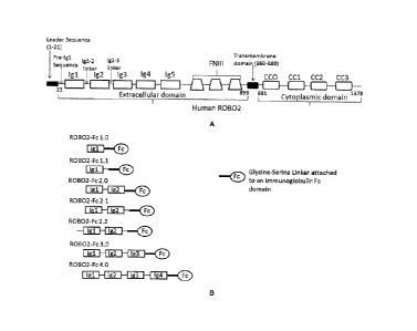
phenotype found in nephrin-null mice. Loss of ROB02 also increases adhesion of
podocytes
to the glomerular basement membrane in mice. These data, along with the
observation that
a patient with ROB02 chromosomal translocation lacks proteinuria, suggests
that blocking of
SLIT2-ROB02 signaling pathway could increase nephrin-induced actin
polymerization to
reduce proteinuria. Blocking of ROB02 signaling may also restore glomerular
filtration
barrier in proteinuric disease by up-regulation of nephrin induced actin
polymerization.
[0007] SLIT1, SLIT2 and SLIT3. SLITs are secreted proteins associated with the
extracellular matrix. The protein sequence of all SLITs shows a high degree of
conservation
and have the same structure: an N-terminus signal peptide; four tandem leucine-
rich repeat
domains (LRR) termed D1-D4; six epidermal growth factor (EGF)-like domains; a
laminin G-
like domain; a further one (invertebrates) or three (vertebrates) EGF-like
domains and a C
terminal cysteine knot domain. SLIT ligands can be cleaved to yield a short C-
terminus
fragment of unknown function (SLIT-C product) and a long N-terminus fragment
(SLIT-N
product) that is active and mediates binding to ROB0s. SLIT ligands, as well
as cleavage
products (e.g., SLIT-N, SLIT2-D2) described herein can be used to assess ROB02
activity.
[0008] Four ROBO receptors have been characterized in vertebrates:
ROB01/Dutt1;
ROB02; ROB03/Rig-1 and ROB04/Magic Roundabout. ROB01, ROB02 and ROB03
2
CA 3006926 2018-06-01
share a common extracellular domain (ECD) structure that is reminiscent of
cell adhesion
molecules. This region contains five immunoglobulin-like (Ig-like) domains
(191 , Ig2, Ig3, Ig4
and Ig5) followed by three fibronectin type 3 (FN3) repeats (FIG. 1A). In
addition, ROB02
has four cytoplasmic conserved (CC) sequences in its intracellular domain as
illustrated in
FIG.1A.
[0009] The sequence of full length human ROB02 precursor is shown as SEQ ID
NO: 24.
A 21 amino acid ROB02 leader sequence (SEQ ID NO: 17; residues 1-21 according
to the
numbering set forth in SEQ ID NO: 24) is cleaved during protein production to
produce
mature ROB02 (FIG.1A). Residues 22-859 according to the numbering set forth in
SEQ ID
NO: 24 form the extracellular domain, residues 860-880 set forth in SEQ ID NO:
24 form the
transmembrane domain, and residues 881-1378 set forth in SEQ ID NO: 24 form
the
cytoplasmic domain (FIG.1A).
[0010] Exemplary sequences of the five Ig-like domains (Ig1, Ig2, Ig3,1g4 and
Ig5) of
ROB02 are shown in Table 23. The ROB02 pre-Ig1sequence (SEQ ID NO: 8), the 191-
1g2
inter-domain linker (SEQ ID NO: 10) and the Ig2-1g3 inter-domain linker (SEQ
ID NO: 12) are
also disclosed in Table 23.
[0011] The D2 LRR domain of the SLITs and Ig1 and 1g2 domains of the ROBOs are
evolutionary conserved and are involved in binding. Ig1 and Ig2 domains of
ROBO together
are also referred to as SLIT-binding domain. Studies have shown that while
both
innmunoglobulin-like (19-like) domains 1 and 2 (191 and Ig2) of ROB02 interact
with SLIT; the
first Ig-like domain (Ig1) is the primary binding site for SLIT. In addition,
previous studies
have indicated that removing the three fibronectin type III (FNIII) repeats
has a greater
negative effect on ROBO binding to SLIT than removal of the third and fourth
immunoglobulin-like domains (Ig3 and 1g4) (see, e.g., Liu et al., 2004,
Molecular Cellular
Neuroanatomy 39:256-261).
[0012] Upon ROBO-SLIT binding, Rho GTPases and their regulators (GAPs and
GEFs)
are involved in the downstream signaling pathway. In the presence of SLIT,
SLIT-ROBO
Rho GTPase activating protein 1 (srGAP1) binds to the CC3 domain of ROBO and
inactivates RhoA and Cdc42. These effector proteins are able to mediate, among
other
.. outcomes, repulsion, control of cytoskeletal dynamics and cell polarity. In
the presence of
SLIT, Vilse/CrossGAP can also bind to the CC2 domain of ROBO and inhibit Rac1
and
Cdc42. Rac1 is also activated by the recruitment of the GEF protein Son of
sevenless (Sos)
via the adaptor protein Dreadlocks (Dock), which binds to the CC2-3 domain of
ROBO. This
activates the downstream target of Rac1 and p21-activated kinase (Pak), which
also binds to
ROBO CC2-3 domains. These downstream signaling partners of ROBO control
repulsion
and cytoskeletal dynamics. The tyrosine kinase Abelson (Abl) can also bind
ROBO CC3
domain and antagonizes ROBO signaling through phosphorylation of the CC1
domain and
3
CA 3006926 2018-06-01
= = =
mediates cell adhesion. Enabled (Ena), a substrate of Abl, also binds ROBO CC1
and CC2
domains. All these downstream ROBO-SLIT molecules may be used to assess ROB02
activity, as well as to assess any neutralizing effect of a novel recombinant
ROB02 protein
disclosed herein.
[0013] In the kidney, ROB02 forms a complex with nephrin through adaptor
protein Nck.
In contrast to the role of nephrin that promotes actin polymerization, SLIT-
ROB02 signaling
inhibits nephrin-induced actin polymerization. Thus, the binding of ROB02
intracellular
domain and Nck may be used to assess ROB02 activity.
[0014] Patients suffering from many glomerular diseases (including Focal
Segmental
Glomerular Sclerosis) currently have no therapies available to preserve renal
function or
otherwise treat the disease. Further, there is no treatment currently
available for CKD
patients with proteinuria. Accordingly, there is a need for developing a
therapeutic that
modulates ROB02-SLIT signaling, thereby preserving or modulating podocyte
functions and
reducing proteinuria or otherwise treating or preventing a renal disease
associated with or
mediated by ROB02-SLIT binding and signaling.
SUMMARY OF THE INVENTION
[0015] The invention provides recombinant ROB02 proteins that bind to SLIT
ligands, as
well as uses, and associated methods thereof. Those skilled in the art will
recognize, or be
able to ascertain using no more than routine experimentation, many equivalents
to the
specific embodiments of the invention described herein. Such equivalents are
intended to be
encompassed by the following embodiments (E).
[0016] El. A recombinant Roundabout Receptor 2 (ROB02) protein comprising
amino
acid residues 1 to 203 according to the numbering of SEQ ID NO: 1, and further
comprising
an immunoglobulin heavy chain constant domain.
[0017] E2. A recombinant Roundabout Receptor 2 (ROB02) protein consisting
essentially of amino acid residues 1 to 203 according to the numbering of SEQ
ID NO: 1,
and an immunoglobulin heavy chain constant domain.
[0018] E3. A recombinant Roundabout Receptor 2 (ROB02) protein comprising (i)
a
SLIT-binding moiety; and (ii) a half-life extending moiety, wherein said SLIT-
binding moiety
comprises a portion of the ROB02 extracellular domain.
[0019] E4. The recombinant ROB02 protein of E3, wherein said portion of said
ROB02
extracellular domain comprises the first two immunoglobulin-like (1g1 and Ig2)
domains of
ROB02 and a C-terminus sequence consisting of the sequence of SEQ ID NO: 12.
[0020] E5. The recombinant ROB02 protein of E3, wherein said portion of said
ROB02
extracellular domain consists essentially of ROB02 pre-immunoglobulin-like 1
(1g1)
sequence (SRLRQEDFP (SEQ ID NO: 8), first immunoglobulin-like domain (Ig1),
inter-
4
CA 3006926 2018-06-01
=
0
domain linker between first and second immunoglobulin-like domains (1g1-1g2
inter-domain
linker; VALLR (SEQ ID NO: 10)), second immunoglobulin-like domain (1g2), and
inter-
domain linker between second and third immunoglobulin-like domains (1g2-1g3
inter-domain
linker; VFER (SEQ ID NO: 12)).
[0021] E6. The recombinant ROB02 protein of any one of E3-E5, wherein said
portion of
said ROB02 extracellular domain consists essentially of amino acid residues 1
to 203
according to the numbering of SEQ ID NO: 1.
[0022] E7. The recombinant ROB02 protein of any one of E3-E6, wherein said
half-life
extending moiety comprises an immunoglobulin domain.
[0023] E8. The recombinant ROB02 protein of any one of El, E2 or E7, wherein
said
immunoglobulin domain is an Fc domain of an lgAi IgA2, IgD, IgE, IgM, IgGi,
IgG2, IgG3, or
lgG4.
[0024] E9. The recombinant ROB02 protein of E8, wherein said Fc domain is the
Fc
domain of human IgGi.
[0025] E10. The recombinant ROB02 protein of E9, wherein said Fc domain is
modified
to eliminate effector function.
[0026] El 1. The recombinant ROB02 protein of El 0, wherein said Fc domain is
the Fc
domain of human IgGi, and wherein said human IgG1 Fc domain comprises at least
one
mutation selected from the group consisting of a substitution from leucine to
alanine at
amino acid residue number 234 (L234A), a substitution from leucine to alanine
at amino acid
residue number 235 (L235A), and a substitution from glycine to alanine at
amino acid
residue number 237 (G237A) all according to the Eu numbering as set forth in
Kabat.
[0027] E12. The recombinant ROB02 protein of E9-Ell, wherein said human IgG1
Fc
domain does not comprise a lysine at residue number 447 according to the Eu
numbering as
set forth in Kabat.
[0028] E13. The recombinant ROB02 protein of any one of Ell-E12, wherein said
Fc
domain comprises amino acid residues 210 to 440 according to the numbering of
SEQ ID
NO: 1.
[0029] E14. The recombinant ROB02 protein of any one of Ell-E12, wherein said
Fc
domain consists of amino acid residues 210 to 440 according to the numbering
of SEQ ID
NO: 1.
[0030] E15. The recombinant ROB02 protein of any one of El, E2 or E7, wherein
said
amino acid residues 1 to 203 according to the numbering of SEQ ID NO: 1 are
contiguous
with said immunoglobulin domain.
[0031] E16. The recombinant ROB02 protein of any one of El, E2 or E7, wherein
said
amino acid residues 1 to 203 according to the numbering of SEQ ID NO: 1 are
connected via
a linker to said immunoglobulin domain.
5
CA 3006926 2018-06-01
[0032] E17. The recombinant ROB02 protein of E16, wherein said linker is a
peptidyl
linker comprising from about 1 to 30 amino acid residues.
[0033] E18. The recombinant ROB02 protein of E17, wherein said peptidyl linker
is
selected from the group consisting of:
a) a glycine rich peptide;
b) a peptide comprising glycine and serine;
c) a peptide having a sequence (Gly-Gly-Ser)n, wherein n is 1, 2, 3, 4, 5, or
6 (SEQ ID NO:
22); and
d) a peptide having a sequence (Gly-Gly-Gly-Gly-Ser)n, wherein n is 1, 2, 3,
4, 5, or 6 (SEQ
ID NO: 23).
[0034] E19. The recombinant ROB02 protein of E18, wherein said peptidyl linker
is (Gly-
Gly-Ser)2 (SEQ ID NO: 15)
[0035] E20. A recombinant ROB02-Fc protein comprising the amino acid sequence
of
SEQ ID NO: 1.
[0036] E21. A recombinant ROB02-Fc protein consisting of the amino acid
sequence of
SEQ ID NO: 1.
[0037] E22. A recombinant ROB02-Fc protein comprising an amino acid sequence
at
least 90% identical to the amino acid sequence of SEQ ID NO: 1.
[0038] E23. The recombinant ROB02 protein of any one of E1-E22, comprising the
amino
acid sequence encoded by the insert of the plasmid deposited at the ATCC and
having
ATCC Accession No. PTA-124008.
[0039] E24. The recombinant ROB02-Fc protein of any one of E4 or E5, wherein
said Ig1
of ROB02 comprises at least one of the following mutations: S17T and R73Y,
each
numbered according to SEQ ID NO: 1.
[0040] E25. A recombinant ROB02-Fc protein comprising the amino acid sequence
of
SEQ ID NO: 19.
[0041] E26. The recombinant ROB02-Fc protein of E25, wherein the protein does
not
comprise a C-terminal lysine located at amino acid residue number 441
according to the
numbering of SEQ ID NO: 19.
[0042] E27. The recombinant ROB02 protein of any one of E1-E26, wherein said
ROB02
is human ROB02.
[0043] E28. The recombinant ROB02 protein of any one of E1-E27, wherein said
protein
binds SLIT2 with a binding affinity (KD) of or less than: about 10 nM, about 5
nM, about 2
nM, about 1 nM, about 900 pM, about 800 pM, about 700 pM, about 600 pM, about
500 pM,
about 400 pM, about 300 pM, about 250 pM, about 200 pM, about 150 pM, about
100 pM,
about 50 pM, about 40 pM, about 30 pM, about 25 pM, about 20 pM, about 15 pM,
about 10
pM, about 5 pM, or about 1 pM.
6
CA 3006926 2018-06-01
[0044] E29. The recombinant ROB02 protein of any one of E1-E27, wherein said
protein
binds SLIT2 with a KD that is at least about 2-fold, about 4-fold, about 6-
fold, about 8-fold,
about 10-fold, about 20-fold, about 40-fold, about 60-fold, about 80-fold,
about 100-fold,
about 120-fold, about 140-fold, about 160-fold, lower than the KD value for
binding of
ROB01 to SLIT2.
[0045] E30. The recombinant ROB02 protein of any one of E1-E29, wherein said
protein
binds SLIT2 with a KD value that is at least about 2-fold, about 4-fold, about
6-fold, about 8-
fold, about 10-fold, about 20-fold, about 40-fold, about 60-fold, about 80-
fold, about 100-fold,
about 120-fold, about 140-fold, about 160-fold, lower than the KD value for
binding of a
ROB01-Fc protein to SLIT2.
[0046] E31. The recombinant ROB02 protein of any one of E28-E30, wherein said
KD is
measured by surface plasmon resonance (SPR).
[0047] E32. The recombinant ROB02 protein of E31, wherein said KD is measured
using
a Biacore T200 instrument.
[0048] E33. The recombinant ROB02 protein of any one of E28-E30, wherein said
KD is
measured by bio-layer interferometry (BLI).
[0049] E34. The recombinant ROB02 protein of E33, wherein said KD is measured
using
a ForteBio Octet instrument.
[0050] E35. The recombinant ROB02 protein of any one of E1-E34, wherein said
protein
inhibits binding of a SLIT ligand and ROB02.
[0051] E36. The recombinant ROB02 protein of any one of El-E34, wherein said
protein
inhibits ROB02-dependent SLITx-N activity.
[0052] E37. The recombinant ROB02 protein of any one of E1-E36, wherein said
protein
inhibits binding of a SLIT ligand and ROB02 and inhibits ROB02-dependent SLIT-
N activity.
[0053] E38. The recombinant ROB02 protein of any one of El-E37, wherein said
ROB02-dependent SLITx-N activity is selected from the group consisting of
actin
polymerization, podocyte adhesion, and inhibition of neuronal cell migration.
[0054] E39. The recombinant ROB02 protein of any one of El-E38, wherein said
protein
has a half maximal inhibitory concentration (IC50) of not more than about
15nM, about 13nM,
.. about 11nM, about 9nM, about 7nM, about 6nM, about 5nM, about 4nM, about
3nM, about
2nM, about 1nM.
[0055] E40. The recombinant ROB02 protein of E39, wherein said IC50 is
measured by a
homogenous time-resolved fluorescence (HTRF) assay for inhibition of binding
of ROB02 to
SLIT2.
[0056] E41. The recombinant ROB02 protein of any one of El-E40, wherein said
protein
has a half maximal IC50 of not more than about 75nM, about 65nM, about 55nM,
about
45nM, about 35nM, about 25nM, about 15nM, about 5nM.
7
CA 3006926 2018-06-01
[0057] E42. The recombinant ROB02 protein of E41, wherein said IC50 is
assessed by
measuring SLIT2-N mediated inhibition of neuronal cell migration.
[0058] E43. The recombinant ROB02 protein of any one of E28-E42, wherein said
SLIT2
is human SLIT2.
[0059] E44. The recombinant ROB02 protein of any one of E1-E43, wherein two of
said
recombinant ROB02 proteins associate to form a homodimer.
[0060] E45. An isolated nucleic acid molecule encoding the recombinant ROB02
protein
of any one of E1-E44.
[0061] E46. The isolated nucleic acid molecule of E45 comprising the nucleic
acid
sequence of SEQ ID NO: 21.
[0062] E47. The isolated nucleic acid molecule of E45 consisting of the
nucleic acid
sequence of SEQ ID NO: 21.
[0063] E48. An isolated nucleic acid comprising the nucleic acid sequence of
the insert of
the plasmid deposited at the ATCC and having ATCC Accession No. PTA-124008.
[0064] E49. A recombinant ROB02 protein comprising an amino acid sequence
encoded
by the sequence of SEQ ID NO: 21.
[0065] E50. A recombinant ROB02 protein comprising an amino acid sequence
encoded
by a sequence that is at least 85%, 90%, 95%, or 99% identical to the sequence
of SEQ ID
NO: 21.
[0066] E51. A recombinant ROB02 protein comprising an amino acid sequence
encoded
by a sequence capable of hybridizing under highly stringent conditions to the
sequence of
SEQ ID NO: 21.
[0067] E52. A vector comprising the nucleic acid molecule of any one of E45-
E48.
[0068] E53. A host cell comprising the nucleic acid molecule of any one of E45-
E48.
[0069] E54. A host cell comprising the vector of E52.
[0070] E55. The host cell of E53 or E54, wherein said cell is a mammalian
cell.
[0071] E56. The host cell of E53 or E54, wherein said host cell is a CHO cell,
a HEK-293
cell, or a Sp2.0 cell.
[0072] E57. A method of making a recombinant ROB02 protein, comprising
culturing the
host cell of any one of E53-E56 under conditions wherein said recombinant
ROB02 protein
is expressed.
[0073] E58. The method of E57, further comprising isolating said recombinant
ROB02
protein.
[0074] E59. A pharmaceutical composition comprising a recombinant ROB02
protein of
any one of E1-E44, and a pharmaceutically acceptable carrier or excipient.
[0075] E60. A method of reducing the biological activity of ROB02, comprising
administering to a subject in need thereof a therapeutically effective amount
of the
8
CA 3006926 2018-06-01
recombinant ROB02 protein of any one of E1-E44, or the pharmaceutical
composition of
E59.
[0076] E61. The method of E60, wherein said biological activity of ROB02 is
selected
from the group consisting of binding to at least one SLIT ligand, actin
polymerization,
podocyte adhesion, inhibiting SLIT2-N-mediated inhibition of neuronal cell
migration, binding
of ROB02 with srGAP1, and binding of ROB02 with Nck.
[0077] E62. A method of treating renal disease, comprising administering to a
subject in
need thereof a therapeutically effective amount of the recombinant ROB02
protein of any
one of E1-E44, or the pharmaceutical composition of E59.
[0078] E63. A method of preserving podocyte function, comprising contacting
said
podocyte with the recombinant ROB02 protein of any one of E1-E44, or the
pharmaceutical
composition of E59.
[0079] E64. A method of modulating podocyte function, comprising contacting
said
podocyte with the recombinant ROB02 protein of any one of E1-E44, or the
pharmaceutical
composition of E59.
[0080] E65. A method of treating glomerular disease, comprising administering
to a
subject in need thereof a therapeutically effective amount of the recombinant
ROB02 protein
of any one of any one of E1-E44, or the pharmaceutical composition of E59.
[0081] E66. A method of treating Focal Segmental Glomerular Sclerosis (FSGS),
comprising administering to a subject in need thereof a therapeutically
effective amount of
the recombinant ROB02 protein, of any one of E1-E44, or the pharmaceutical
composition
of E59.
[0082] E67. A method of treating nephropathy comprising administering to a
subject in
need thereof a therapeutically effective amount of the recombinant ROB02
protein of any
one of E1-E44, or the pharmaceutical composition of E59.
[0083] E68. The method of E67, wherein said nephropathy is IgA nephropathy.
[0084] E69. The method of any one of E60-E68, wherein said subject is a human.
[0085] E70. The method of any one of E60-E69, wherein said recombinant ROB02
protein, or pharmaceutical composition is administered intravenously.
[0086] E71. The method of any one of E60-E69, wherein said recombinant ROB02
protein, or pharmaceutical composition is administered subcutaneously.
[0087] E72. The method of any one of E60-E71, wherein recombinant ROB02
protein, or
pharmaceutical composition, is administered about twice a week, once a week,
once every
two weeks, once every three weeks, once every four weeks, once every five
weeks, once
every six weeks, once every seven weeks, once every eight weeks, once every
nine weeks,
once every ten weeks, twice a month, once a month, once every two months, once
every
three months, or once every four months.
9
CA 3006926 2018-06-01
=
[0088] E73. The recombinant ROB02 protein of any one of El-E44, or the
pharmaceutical
composition of E59, for use as a medicament.
[0089] E74. The recombinant ROB02 protein of any one of El-E44, or the
pharmaceutical
composition of E59, for use in reducing the activity of ROB02 in a cell.
[0090] E75. The recombinant ROB02 protein of any one of El-E44, or the
pharmaceutical
composition of E59, for use in reducing the activity of ROB02 in a subject.
[0091] E76. The recombinant ROB02 protein of any one of El-E44, or the
pharmaceutical
composition of E59, for use in preserving podocyte function in a subject.
[0092] E77. The recombinant ROB02 protein of any one of El-E44, or the
pharmaceutical
composition of E59, for use in modulating podocyte function in a subject.
[0093] E78. The recombinant ROB02 protein of any one of El-E44, or the
pharmaceutical
composition of E59, for use in treating a glomerular disease in a subject.
[0094] E79. The recombinant ROB02 protein of E78, wherein said glomerular
disease is
FSGS.
[0095] E80. The recombinant ROB02 protein of any one of El-E44, or the
pharmaceutical
composition of E59, for use in treating nephropathy in a subject.
E81. Use of the recombinant R01302 protein of any one of El-E44, or the
pharmaceutical
composition of E59, in the manufacture of a medicament for reducing the
activity of ROB02 in a
cell.
E82. Use of the recombinant ROB02 protein of any one of El-E44, or the
pharmaceutical
composition of E59, in the manufacture of a medicament for reducing the
activity of ROB02 in a
subject.
E83. Use of the recombinant ROB02 protein of any one of El-E44, or the
pharmaceutical
composition of E59, in the manufacture of a medicament for preserving podocyte
function in a
subject.
E84. Use of the recombinant ROB02 protein of any one of El-E44, or the
pharmaceutical
composition of E59, in the manufacture of a medicament for modulating podocyte
function in a
subject.
E85. Use of the recombinant ROB02 protein of any one of El-E44, or the
pharmaceutical
composition of E59, in the manufacture of a medicament for treating a
glomerular disease in a
subject.
E86. Use of the recombinant ROB02 protein of any one of El-E44, or the
pharmaceutical
composition of E59, in the manufacture of a medicament for treating
nephropathy in a subject
[0096] E87. The recombinant ROB02 protein of E80, wherein said nephropathy is
an IgA
nephropathy.
[0097] E88. A kit comprising a container, a composition within the container
comprising
the recombinant ROB02 protein of any one of E1-E44, or the pharmaceutical
composition of
CA 3006926 2018-06-01
,
E59 and a package insert containing instructions to administer a
therapeutically effective
amount of the recombinant ROB02 protein or the pharmaceutical composition for
treatment
of a patient in need thereof.
BRIEF DESCRIPTION OF THE DRAWINGS
[0098] FIG. 1A is a graphic presentation showing the domains of human ROB02.
The 21-
amino acid ROB02 leader sequence (SEQ ID NO: 17; residues 1-21 according to
the
numbering set forth in SEQ ID NO: 24) is cleaved during protein production to
produce
mature ROB02. Residues 22-859 according to the numbering set forth in SEQ ID
NO: 24
form the extracellular domain, residues 860-880 according to the numbering of
SEQ ID NO:
24 form the transmembrane domain, and residues 881-1378 according to the
numbering of
SEQ ID NO: 24 form the cytoplasmic domain. The ROB02 pre-Ig1sequence (SEQ ID
NO:
8), the Ig1-1g2 inter-domain linker (SEQ ID NO: 10) and the Ig2-1g3 inter-
domain linker (SEQ
ID NO: 12) are also shown.
[0099] FIG. 1B is a graphic presentation showing exemplary recombinant ROB02-
Fc
fusions proteins described herein: ROB02-Fc. 1.0 (solely contains Ig1 domain,
i.e., amino
acid residues 31 to 127 according to the numbering of SEQ ID NO: 24), ROB02-
Fc. 1.1
(contains Ig1 domain and Ig1-1g2 inter-domain linker, i.e., amino acid
residues 31 to 132
according to the numbering of SEQ ID NO: 24), ROB02-Fc. 2.0 (contains Ig1
domain, Ig1-
Ig2 inter-domain linker, and Ig2 domain, i.e., amino acid residues 31 to 220
according to the
numbering of SEQ ID NO: 24), ROB02-Fc. 2.1 (contains Ig1 domain, Ig1-1g2 inter-
domain
linker, Ig2 domain, and Ig2-1g3 inter-domain linker, i.e., amino acid residues
31 to 224
according to the numbering of SEQ ID NO: 24), ROB02-Fc. 2.2 (contains pre-Ig1
sequence,
Ig1 domain, Ig1-1g2 inter-domain linker, Ig2 domain, and 1g2-1g3 inter-domain
linker, i.e.,
amino acid residues 22 to 224 according to the numbering of SEQ ID NO: 24),
ROB02-Fc.
3.0 (contains Ig1 domain, 1g1-1g2 inter-domain linker, Ig2 domain, Ig2-1g3
inter-domain
linker, Ig3 domain, i.e., amino acid residues 31 to 309 according to the
numbering of SEQ ID
NO: 24) and ROB02-Fc. 4.0 (contains Ig1 domain, Ig1-1g2 inter-domain linker,
Ig2 domain,
Ig2-1g3 inter-domain linker, Ig3 domain, Ig3-1g4 inter-domain linker and Ig4,
i.e., amino acid
.. residues 31 to 409 according to the numbering of SEQ ID NO: 24) described
herein.
[0100] FIG. 2 shows the ROB02-Fc 2.2 amino acid sequence (SEQ ID NO: 1).
Residues
are numbered sequentially starting with the N-terminus. The Ig1 and Ig2
domains are shown
in all caps, while the Fc domain is shown in lower case. The pre-Ig1 sequence
(SEQ ID NO:
8) and the Ig2-1g3 inter-domain linker (SEQ ID NO: 12) are shown in bold and
italics, while
the Ig1-1g2 inter-domain linker (SEQ ID NO: 10) is shown in italics. The
predicted intra- and
inter-chain disulfide bonds are illustrated with connecting lines. A single
polypeptide chain is
shown with disulfide bonds in the Fc hinge region which can dimerize with a
second, Fc
11
CA 3006926 2018-06-01
, .
comprising polypeptide chain. The canonical N-linked glycosylation consensus
sequence
sites are circled (i.e., NXSTT where the glycan is attached to the asparagine
residue and
where X can be any amino acid except proline and the third amino acid is
either serine or
threonine), and the Fc-effector function-null point mutations located at A228,
A229, and
A231 are shown in bold. The 6-amino acid Gly-Ser linker sequence is shown in
the boxed
region.
[0101] FIG. 3 demonstrates that ROB02-Fc 2.1 (SEQ ID NO:2) binds to SLIT2
while
ROB02-Fc 1.1 (SEQ ID NO:4) and ROB02-Fc 2.0 (SEQ ID NO:3) do not. Utilizing
the
Octet Red ROB02-Fc proteins were loaded onto anti human-crystallized fragment
(AHFc)
sensors at 10 pg/ml and incubated with 100 nM SLIT2 for 7 minutes and then the
sensors
were moved to buffer alone for 640 seconds. ROB02-Fc 4.0 (SEQ ID NO: 7) was
included
as a positive control for binding. The addition of the sequence VFER (SEQ ID
NO: 12) after
the Ig2 domain of ROB02 to create ROB02-Fc 2.1 (SEQ ID NO: 2) was essential to
produce a ROB02-Fc fusion protein that binds SLIT2.
[0102] FIGS. 4A-4C demonstrate that ROB02-Fc 2.2 (SEQ ID NO: 1) binds to SLIT2
with
high affinity. KD values were measured using surface plasmon resonance (SPR).
The KD of
ROB02-Fc 2.2 to human/cynomolgus monkey SLIT2-D2 (ROB02 binding domain, 100%
identical) was 0.293 nM (FIG. 4A). The KD of ROB02-Fc 2.2 to human SLIT2-N (N
terminal
fragment) was 0.279 nM (FIG. 4B), and the Ko of ROB02-Fc 2.2 to rat SLIT2-N
was 0.543
nM (FIG. 4C).
[0103] FIG. 5 demonstrates that ROB02-Fc 2.2 (SEQ ID NO: 1) binds with high
affinity
having an EC50 of 9nM to human SLIT2-N that is overexpressed on human
embryonic
kidney (HEK293) cells. A 12-point, 2-fold dilution series of ROB02-Fc 2.2
labeled with Alexa
Fluor 647 (AF647) was incubated with either SLIT2-N expressing HEK293 cells or
control
HEK293 cells. The data are presented as the geometric mean fluorescence
intensity (Geo
MFI) of ROB02-Fc 2.2 AF647 on SLIT2-N HEK293 cells minus the geometric mean
fluorescence intensity of ROB02-Fc 2.2 AF647 on control HEK293 cells.
[0104] FIGS. 6A-6B demonstrate the dose-dependent inhibition of SLIT2-N
binding to cell
surface ROB02 by ROB02-Fc 2.2 (SEQ ID NO: 1) as assessed by Homogenous Time
Resolved Fluorescence (HTRF). An 11-point, 4-fold dose titration of ROBO-Fc
2.2 (black
squares) or an isotype control antibody (black circles) was added to either a
human SLIT2-N
(FIG. 6A) or rat SLIT2-N (FIG. 6B) human ROB02 HTRF assay. ROB02-Fc 2.2 was a
potent neutralizer of both human SLIT2-N:human ROB02 (IC50 of 7 nM) and rat
SLIT2-
N:human ROB02 (IC50 of 4 nM) binding.
[0105] FIG. 7 depicts the dose-dependent inhibition of SLIT2-N mediated
inhibition of
neuronal cell migration by ROB02-Fc 2.2. Subventricular zone (SVZ) neuronal
tissue cell
explants were cultured overnight in the presence of 1 nM SLIT2-N and a dose
range of
12
CA 3006926 2018-06-01
ROB02-Fc 2.2. ROB02-Fc 2.2 was able to restore neuronal cell migration in a
dose-
dependent manner with an IC50 of 51 nM.
[0106] FIG.8 demonstrates inhibition of proteinuria with treatment of ROB02-Fc
2.2 in the
rat Passive Heymann Nephritis model with an exemplary prophylactic dosing
regimen.
Twelve animals in each of the indicated groups were treated subcutaneously
with the
indicated dose of ROB02-Fc 2.2 or an irrelevant isotype control monoclonal
antibody
(control) every three days starting the day before the induction of the model
on day 0. The Y
axis indicates the ratio of urine albumin to creatinine (mg/mg) as a measure
of leakage of
protein into the urine, indicative of podocyte damage. Lewis rats were
injected with sheep
anti-sera raised against rat kidney brush border (anti-Fx1a, basement membrane
and
podocytes). The rats developed an immune response to the sheep sera which
bound the rat
podocytes. As podocytes are damaged and effaced, proteinuria increases.
Treatment with
the highest dose of ROB02-Fc 2.2 at 25 mg/kg reduced proteinuria 45% maximally
with a p
value less than 0.001 by repeated measure ANOVA statistical analyses compared
to the
control antibody treatment. The dose effect was also statistically significant
with a p value
less than 0.001.
[0107] FIG. 9 demonstrates inhibition of proteinuria with treatment of ROB02-
Fc 2.2 in the
rat Passive Heymann Nephritis model with an exemplary therapeutic dosing
regimen.
Twelve animals in each of the indicated groups were treated subcutaneously
with the
indicated dose of ROB02-Fc 2.2 or an irrelevant control monoclonal antibody
(control) every
three days with the following dosing regimen: control antibody was
administered on day 0
(circles) and ROB02-Fc 2.2 was administered on day 0 (squares), day 6
(triangles) or day 9
(inverted triangles). The Y axis indicates the ratio of urine albumin to
creatinine (mg/mg) as
a measure of leakage of protein into the urine, indicative of podocyte damage.
Lewis rats
were injected with sheep anti-sera raised against rat kidney brush border
(anti-Fx1a,
basement membrane and podocytes). The rats developed an immune response to the
sheep sera which bound the rat podocytes. As podocytes are damaged and
effaced,
proteinuria increased. Treatment with ROB02-Fc 2.2 administered on day 0, 6
and 9
reduced proteinuria to a similar extent, 40% maximally and with a p value less
than 0.001 by
repeated measure ANOVA statistical analyses for each ROB02-Fc 2.2 treated
group
compared to the control antibody treatment.
[0108] FIGS. 10A-10B demonstrate that treatment with ROB02-Fc 2.2 reduces
damage to
podocyte substructure in the Passive Heymann Nephritis Model. Twelve animals
in each of
the indicated groups were treated subcutaneously with the indicated dose of
ROB02-Fc 2.2
or an irrelevant control monoclonal antibody every three days at 25 mg/kg to
achieve 100%
target coverage starting the day before the induction of the model on day 0.
Following
animal sacrifice at day 16, selected kidney samples were digitally imaged
using a
13
CA 3006926 2018-06-01
transmission electron microscope. Without repetition, three capillary loops of
the first three
glomeruli found at 200x magnification, were imaged at 5000x and 10,000x
magnification.
ImageJ software (version 1.47v; National Institutes of Health, Bethesda, MD,
USA) was used
to manually trace and measure the width of adjacent foot processes as well as
the density of
slit diaphragms per unit length of the glomerular basement membrane (GBM) on
high
magnification transmission electron microscopy images. Samples were analyzed
over 3
separate experiments. The podocyte foot processes of the ROB02-Fc 2.2 treated
animal
were significantly shorter than the control antibody treated animals (FIG.
10A), and the
density of slit diaphragms was significantly higher in ROB02-Fc 2.2 treated
animals (FIG.
10B; p value less than 0.01 by two tailed T test) indicating that they were
less effaced and
were protected from the glomerular insult.
[0109] FIG. 11 demonstrates that ROB02-Fc S17T/R73Y binds to human 5LIT2-N
overexpressed on human embryonic kidney (HEK293) cells with high affinity
having an EC50
of 2.5 nM. A 12-point, 2-fold dilution series of ROB02-Fc 517T/R73Y labeled
with alexa fluor
.. 647 (AF647) was incubated with either SLIT2-N expressing HEK293 cells or
control HEK293
cells. The data are presented as the geometric mean fluorescence intensity
(Geo MFI) of
ROB02-Fc S17T/R73Y AF647 on SLIT2-N HEK293 cells minus the geometric mean
fluorescence intensity of ROB02-Fc S17T/R73Y AF647 on control HEK293 cells.
[0110] FIG. 12 demonstrates the dose-dependent inhibition of SLIT2-N binding
to cell
surface ROB02 by ROB02-Fc S17T/R73Y as assessed by Homogenous Time Resolved
Fluorescence (HTRF). An 11-point, 4-fold dose titration of ROB02-Fc S17T/R73Y
(black
squares) or an isotype control antibody (black circles) was added to a human
SLIT2-N
human ROB02 HTRF assay. ROB02-Fc S17T/R73Y was a potent neutralizer of human
SLIT2-N:human ROB02 binding with an IC50 of 1.4 nM.
[0111] FIG. 13 depicts the dose-dependent inhibition of SLIT2-N mediated
inhibition of
neuronal cell migration by ROB02-Fc 517T/R73Y. Subventricular zone (SVZ)
neuronal
tissue cell explants were cultured overnight in the presence of 1nM SLIT2-N
and titrated
amounts of ROB02-Fc S17T/R73Y. ROB02-Fc S17T/R73Y was able to restore neuronal
cell migration in a dose-dependent manner with an IC50 of 11.5 nM.
[0112] FIG. 14 shows a drawing depicting the crystal structure of a ROB02-His
construct
that consists of the ROB02 pre-Ig1 sequence (SRLRQEDFP; SEQ ID NO: 8), Ig1
domain,
Ig2 domain and the ROB02 Ig2-3 inter-domain linker (VFER; SEQ ID NO: 12) with
a 6x
histidine tag (His6) (SEQ ID NO: 25) at the C-terminus. The crystal structure
of ROB02-His
reveals that that Asp7, Phe8, and Pro9 are substantially involved in the
interactions vital for
structural integrity of ROB02's Ig1 domain.
[0113] FIG. 15 shows a drawing depicting the crystal structure of the ROB02-
His
construct. The crystal structure of ROB02-His6 ("His6" disclosed as SEQ ID NO:
25)
14
CA 3006926 2018-06-01
reveals that the ROB02 Ig2-3 inter-domain linker, Valine200-Phenylalanine201-
Glutamic
acid202-Arginine203 (SEQ ID NO: 12), effectively stabilizes the structural
fold in the C-
terminal region of ROB02's Ig2 domain.
[0114] FIG. 16 shows the crystal structure of the first immunoglobulin-like
domain (Ig1) of
ROB02 S17T/R73Y in complex with SLIT2.
DETAILED DESCRIPTION OF THE INVENTION
1. OVERVIEW
[0115] The invention encompasses novel recombinant ROB02 proteins capable of
binding
SLIT ligands (for example, the SLIT2 ligand), thereby inhibiting the
interaction of SLIT with
ROB02, and consequently, inhibiting the SLIT2-ROB02 signaling pathway.
Previous
studies have shown that while both immunoglobulin-like (Ig-like) domains 1 and
2 (Ig1 and
Ig2) of ROB02 interact with SLIT ligands; the first Ig-like domain (Ig1) is
the primary binding
site for SLIT. In addition, previous studies have indicated that removing the
three fibronectin
type III (FNIII) repeats has a greater negative effect on ROBO binding to SLIT
ligands than
removal of the third and fourth immunoglobulin-like domains (Ig3 and Ig4).
That is, FNIII
deletion causes a greater reduction in ROBO binding to SLIT ligands than
deletion of Ig3
and Ig4.
[0116] Surprisingly, it is now shown for the first time that a construct,
ROB02-Fc 2.1 (SEQ
ID NO: 2; FIG. 1B), comprising only the first two immunoglobulin-like domains
(Ig1 and Ig2)
along with the Ig2-3 inter-domain linker, VFER (SEQ ID NO: 12), and devoid of
the three
fibronectin type III (FNIII) repeats bound SLIT2 (FIG. 3). In contrast,
recombinant ROB02
proteins lacking the three fibronectin type III (FNIII) repeats but consisting
of:
(i) the Ig1 domain of ROB02 (ROB02-Fc 1.1; SEQ ID NO: 4; FIG. 1B),
(ii) the Ig1 and Ig2 domains of ROB02 (ROB02-Fc 2.0; SEQ ID NO: 3; FIG. 1B),
or
(iii) the Ig1, Ig2 and Ig3 domains of ROB02 (ROB02-Fc 3.0; SEQ ID NO: 6; FIG.
1B)
did not bind SLIT2 (FIG. 3).
[0117] Thus, addition of VFER (SEQ ID NO: 12) to the C-terminus of the Ig1-1g2
domains
was required to create a ROB02-Fc construct (ROB02-Fc 2.1; SEQ ID NO: 2) with
a robust
binding profile to SLIT2 in the absence of the FNIII repeats. This ROB02-Fc
2.1 construct
lacks not only the third, fourth and fifth imnnunoglobulin-like domains (Ig3,
Ig4 and Ig5), but is
also devoid of the three fibronectin type III (FNIII) repeats. Surprisingly,
the difference in
binding or not binding SLIT2 was found to be the presence of the four-amino
acid VFER
sequence (SEQ ID NO: 12).
[0118] None of the recombinant ROB02 protein constructs described above
contain the
ROB02 pre-Ig1 sequence (SEQ ID NO: 8). It was discovered, surprisingly, that
the
production of these recombinant ROB02 proteins, disclosed and exemplified
herein, can be
CA 3006926 2018-06-01
dramatically increased by including the ROB02 pre-Igl sequence (SEQ ID NO: 8).
Addition
of this sequence increased protein production by about 25-fold compared to
constructs
lacking the sequence while preserving high affinity binding to SLIT2 (FIGS. 3-
4A-C). As
shown in FIG. 14, this ROB02 pre-Igl sequence bridges together the two 13-
sheets of
ROB02's Igl domain and is believed to stabilize the structural fold of the N-
terminal region.
Without wishing to be bound by any particular theory, the pre-Igl sequence
appears to
contribute to the enhanced expression of the novel proteins.
2. DEFINITIONS
[0119] In some aspects, provided herein are recombinant ROB02 proteins capable
of
binding SLIT and comprising an immunoglobulin domain.
[0120] The term "recombinant protein" refers to a polypeptide which is
produced by
recombinant DNA techniques, wherein generally, DNA encoding the polypeptide is
inserted
into a suitable expression vector which, in turn, is introduced into a host
cell to produce the
recombinant protein. As used herein, "protein" refers to any composition
comprising amino
acids and recognized as a protein by those of skill in the art. The terms
"protein", "peptide"
and "polypeptide are used interchangeably herein. Amino acids may be referred
to by their
complete names (e.g., alanine) or by the accepted one letter (e.g., A), or
three letter (e.g.,
Ala) abbreviations.
[0121] As used herein, an "immunoglobulin domain" is a polypeptide derived
from an
immunoglobulin. In some embodiments, an immunoglobulin domain comprises an
immunoglobulin heavy chain or a portion thereof. In some embodiments, the
portion of the
heavy chain is the crystallizable fragment (Fc) or a portion thereof. As used
herein, the Fc
fragment comprises the heavy chain hinge region, and the CH2 and CH3 domains
of the
heavy chain of an immunoglobulin. The heavy chain (or portion thereof) may be
derived
from any one of the known heavy chain isotypes: IgG (y), IgM (i.1), IgD (6),
IgE (c), or IgA (a).
In addition, the heavy chain (or portion thereof) may be derived from any one
of the known
heavy chain isotypes or subtypes: IgG1 (y1), IgG2 (y2), IgG3 (y3), IgG4 (y4),
IgAl (al), IgA2
(a2). In some embodiments, the immunoglobulin domain comprises an
uninterrupted native
(i.e., wild-type) sequence of an immunoglobulin. In some embodiments, the
immunoglobulin
Fc domain comprises a variant Fc region.
[0122] For all heavy chain constant region amino acid positions discussed in
the present
invention, numbering is according to the Eu index first described in Edelman
et al., 1969,
Proc. Natl. Acad. Sci. USA 63(1):78-85, describing the amino acid sequence of
myeloma
protein Eu, which is the first human IgG1 sequenced. The Eu index of Edelman
et al. is also
set forth in Kabat et al., 1991, Sequences of Proteins of Immunological
Interest, 5th Ed.,
United States Public Health Service, National Institutes of Health, Bethesda.
Thus, the "EU
16
CA 3006926 2018-06-01
index as set forth in Kabat" or "EU index of Kabat" refers to the residue
numbering system
based on the human IgG1 Eu antibody of Edelman et al. as set forth in Kabat
1991.
[0123] A "native sequence Fc region" comprises an amino acid sequence
identical to the
amino acid sequence of an Fc region found in nature. A "variant Fc region"
comprises an
amino acid sequence which differs from that of a native sequence Fc region by
virtue of at
least one amino acid modification. Preferably, the variant Fc region has at
least one amino
acid substitution compared to a native sequence Fc region, e.g., from about
one to about ten
amino acid substitutions, and preferably, from about one to about five amino
acid
substitutions compared to a native sequence Fc region. The variant Fc region
herein will
preferably possess at least about 80% amino acid sequence identity with a
native sequence
Fc region, and more preferably, at least about 90% amino acid sequence
identity therewith,
more preferably, at least about 95%, at least about 96%, at least about 97%,
at least about
98%, and most preferably at least about 99% amino acid sequence identity
therewith.
[0124] As used herein a "linker" is a molecule or group of molecules that
binds two
separate entities (e.g., the extracellular domain and the immunoglobulin
domain of a
recombinant ROB02-Fc protein) to one another and can provide spacing and
flexibility
between the two entities such that they are able to achieve a conformation in
which they,
e.g., specifically bind their cognate ligand (e.g., SLIT ligand). Protein
linkers are particularly
preferred, and they may be expressed as a component of the recombinant protein
using
standard recombinant DNA techniques well-known in the art.
[0125] The term "IC50" or "the half maximal inhibitory concentration" refers
to the
concentration of the recombinant ROB02 protein that is required for 50%
inhibition of the
ROB02-SLIT signaling pathway, for example the ROB02-SLIT2 signaling pathway.
IC50is a
measure of how much of recombinant ROB02 protein is needed to inhibit a ROB02-
SLIT
biological process by 50%, such as the binding between ROB02 and a SLIT
ligand, binding
of intracellular signaling molecules (such as srGAP1 or Nck) to the
intracellular domain of
ROB02 and/or downstream activities of ROB02-SLIT signaling (such as actin
polymerization, podocyte adhesion, and/or SLITx-N mediated inhibition of
neuronal cell
migration). A lower IC50indicates a more potent effect since a smaller amount
of the
recombinant ROB02 protein mediates a more potent inhibitory effect.
[0126] As used herein, the term "SLITx" refers generally to a SLIT ligand.
Similarly, the
terms "SLITx-N" and SLITx-C" refer generally to N-terminal and C-terminal
fragments,
respectively, of SLIT ligands. The SLIT ligand may be a mammalian SLIT ligand,
preferably
a human SLIT ligand. In some embodiments, the SLIT ligand is selected from the
group
consisting of a SLIT1 ligand, a SLIT2 ligand, and a SLIT3 ligand. The SLIT
ligand may be a
SLIT2 ligand, preferably a human SLIT2 ligand.
17
CA 3006926 2018-06-01
, .
[0127] As used herein, a "subject" is an animal, preferably a mammal, more
preferably a
non-human primate, and most preferably a human. The terms "subject"
"individual" and
"patient" are used interchangeably herein. In all embodiments, human nucleic
acids and
human polypeptides are preferred. It is believed that the results obtained
using the human,
rat and cynomolgus monkey molecules described elsewhere herein are predictive
of the
results that may be obtained using other homologous sequences.
[0128] As used herein, "treatment" is an approach for obtaining beneficial or
desired
clinical results. For purposes of this invention, beneficial or desired
clinical results include,
but are not limited to, one or more of the following: reducing proteinuria
(i.e., reducing the
.. amount of protein in the urine compared with the level of protein in urine
in the absence of
drug administration), reducing edema, and/or restoring blood albumin levels.
The term
"treatment" includes prophylactic and/or therapeutic treatments. If it is
administered prior to
clinical manifestation of a condition, the treatment is considered
prophylactic. Therapeutic
treatment includes, e.g., ameliorating or reducing the severity of a disease,
or shortening the
length of the disease.
[0129] The term "therapeutically effective amount" refers to an amount of a
therapeutic
agent of this invention effective to "treat" a disease or disorder in a
subject. For example, a
therapeutically effective amount may be the amount that alleviates one or more
symptoms of
the disease or the amount necessary to keep a disease in remission. In the
case of a focal
segmental glomerulosclerosis (FSGS), the therapeutically effective amount
refers to that
amount which has at least one of the following effects: reducing proteinuria
(i.e., reducing
the amount of protein in the urine compared with the level of protein in urine
in the absence
of drug administration), reducing edema, and/or restoring blood albumin
levels.
[0130] "About" or "approximately" when used in connection with a measurable
numerical
variable, refers to the indicated value of the variable and to all values of
the variable that are
within the experimental error of the indicated value (e.g., within the 95%
confidence interval
for the mean) or 10% of the indicated value, whichever is greater. Numeric
ranges are
inclusive of the numbers defining the range.
Binding affinity
[0131] "Binding affinity" generally refers to the strength of the sum total of
non-covalent
interactions between a contact residue of one binding partner (e.g., the
recombinant ROB02
protein disclosed herein) and a contact residue of its binding partner (e.g.,
a SLIT ligand).
Unless indicated otherwise, as used herein, "binding affinity" refers to
binding affinity that
.. reflects a 1:1 interaction between members of a binding pair or binding
partners (e.g., the
recombinant ROB02 protein and a 5LI12 ligand).
18
CA 3006926 2018-06-01
. .
[0132] At its most detailed level, the binding affinity for the interaction
between ROB02
and a SLIT ligand can be defined by the spatial coordinates defining the
atomic contacts
present in the R0602/5LIT interaction, as well as information about their
relative
contributions to the binding thermodynamics. At one level, a contact residue
can be
characterized by the spatial coordinates defining the atomic contacts between
ROB02 and
SLIT. In one aspect, the contact residue can be defined by a specific
criterion, e.g., distance
between atoms in the ROB02 protein amino acid residue and the atoms in the
SLIT protein
amino acid residue (e.g., a distance of equal to or less than about 4 A (such
as 3.8 A used in
the Examples here) from a heavy atom of a ROB02 amino acid residue and a heavy
atom of
an amino acid residue of SLIT. In another aspect, a contact residue can be
characterized as
participating in a hydrogen bond interaction with the cognate binding partner,
or with a water
molecule that is also hydrogen bonded to the binding partner (i.e., water-
mediated hydrogen
bonding). In another aspect, a contact residue can be characterized as forming
a salt bridge
with a residue of the binding partner. In yet another aspect, a contact
residue can be
characterized as a residue having a non-zero change in buried surface area
(BSA) due to
interaction with a contact residue of the binding partner. At a less detailed
level, the binding
affinity can be characterized through function, e.g., by competition binding
with other
proteins.
[0133] Low-affinity recombinant proteins generally bind their ligands slowly
and tend to
dissociate readily, whereas high-affinity recombinant proteins generally bind
their ligands
faster and tend to remain bound longer. A variety of methods of measuring
binding affinity
are known in the art, any of which can be used for purposes of the present
invention.
Specific illustrative and exemplary embodiments for measuring binding affinity
are described
in the following.
[0134] The binding affinity can be expressed as KD value, which refers to the
dissociation
rate of a particular recombinant ROB02 protein-SLIT ligand interaction. Kr is
the ratio of the
rate of dissociation, also called the "off-rate (koff)", to the association
rate, or "on- rate (kon)".
Thus, KD equals koff / kõ and is expressed as a molar concentration (M), and
the smaller the
KD, the stronger the affinity of binding. KD values can be determined using
methods well
established in the art. One exemplary method for measuring KD is surface
plasmon
resonance (SPR), a method well-known in the art (e.g., Nguyen et al. Sensors
(Basel). 2015
May 5;15(5):10481-510). KD value may be measured by SPR using a biosensor
system
such as a BIACORE system. BlAcore kinetic analysis comprises analyzing the
binding and
dissociation of an antigen from chips with immobilized molecules (e.g.
molecules comprising
epitope binding domains), on their surface. Another well-known method in the
art for
determining the Kr) of a protein is by using Bio-Layer Interferometry (e.g.,
Shah et al. J Vis
Exp. 2014; (84): 51383). KD value may be measured using OCTET technology
(Octet QKe
19
CA 3006926 2018-06-01
system, ForteBio). Alternatively or in addition, a KinExA (Kinetic Exclusion
Assay) assay,
available from Sapidyne Instruments (Boise, Id.) can also be used. Any method
known in the
art for assessing the binding affinity between two binding partners is
encompassed herein.
3. RECOMBINANT ROB02 PROTEINS
[0135] In some aspects, the instant disclosure provides recombinant ROB02
proteins. In
some embodiments, the recombinant ROB02 proteins disclosed herein bind SLIT
ligands (in
particular, SLIT2 ligand), thereby preventing the binding of SLIT to cellular
ROB02
receptors, and are hence referred to as SLIT neutralizing ligand traps.
Surprisingly, as
shown in the Examples, a construct, ROB02-Fc 2.1 (SEQ ID NO: 2; FIG. 1B),
comprising
the first two immunoglobulin-like domains (Ig1 and Ig2), Ig1-1g2 inter-domain
linker, along
with the Ig2-3 inter-domain linker, VEER (SEQ ID NO: 12), and devoid of the
three
fibronectin type III (FNIII) repeats bound SLIT2 (FIG. 3). Crystal structure
studies also show
that the inclusion of the ROB02 Ig2-1g3 inter-domain linker, VFER (V200-F201-
E202-R203;
.. SEQ ID NO: 12) effectively stabilizes the structural fold in the C-terminal
region of ROB02's
second Ig domain FIG. 15), and notably increases the expression level of the
recombinant
ROB02 protein.
[0136] In some aspects, the instant disclosure provides recombinant
polypeptides
comprising a SLIT-binding moiety and a half-life extending moiety. The "SLIT-
binding
moiety" confers SLIT-binding ability to the recombinant ROB02 protein. In some
embodiments, the SLIT-binding moiety comprises a portion of a ROB02
extracellular
domain. In some embodiments, the portion of the ROB02 extracellular domain
comprises at
least two immunoglobulin-like (Ig-like) domains, and a C-terminus sequence
consisting of
VEER (SEQ ID NO: 12). In some embodiments, the at least two Ig-like domains
are
selected from the group consisting of Ig1, Ig2, Ig3, Ig4 and Ig5. In some
embodiments, the
at least two Ig-like domains are selected from the group consisting of the
sequence of SEQ
ID NO: 9, SEQ ID NO: 11, SEQ ID NO: 13 and SEQ ID NO: 14. In some embodiments,
the
portion of the ROB02 extracellular domain comprises the first two Ig-like
domains (Ig1 and
Ig2) of ROB02. In some embodiments, the portion of the ROB02 extracellular
domain
comprises SEQ ID NO: 9 and/or SEQ ID NO: 11.
[0137] Protein production studies also determined that the production of the
recombinant
ROB02 proteins, disclosed and exemplified herein, can be dramatically
increased by
including the ROB02 pre-Ig1 sequence (SEQ ID NO: 8). Addition of this sequence
increases protein production in transiently and/or stably transfected
mammalian cells by
about 25-fold while preserving high affinity binding to SLIT (FIGS. 3-5).
CA 3006926 2018-06-01
[0138] Accordingly, in some embodiments, the portion of the ROB02
extracellular domain
further comprises the ROB02 pre-Ig1 sequence. In some embodiments, the ROB02
pre-
Ig1 sequence comprises SEQ ID NO: 8.
[0139] In some embodiments, the portion of the ROB02 extracellular domain
comprises
ROB02 pre-Ig1 sequence, Ig1, Ig1-1g2 inter-domain linker, Ig2, and Ig2-1g3
inter-domain
linker. Exemplary sequences of the ROB02 pre-Ig1 sequence (SEQ ID NO: 8), Ig1
(SEQ ID
NO: 9), Ig1-1g2 inter-domain linker (SEQ ID NO: 10), Ig2 (SEQ ID NO: 11), and
Ig2-1g3 inter-
domain linker (SEQ ID NO: 12) are shown in Table 23 and also illustrated in
Figure 2. The
present invention is not limited to the sequences disclosed herein.
Corresponding residues
from other ROB02 homologs, isoforms, variants, or fragments can be identified
according to
sequence alignment or structural alignment that is known in the art. For
example, alignments
can be done by hand or by using well-known sequence alignment programs such as
ClustalW2, or "BLAST 2 Sequences" using default parameters.
[0140] In some embodiments, the portion of the ROB02 extracellular domain
comprises
amino acid residues 1 to 203 according to the numbering of SEQ ID NO: 1. In
some
embodiments, the recombinant ROB02 protein comprises a portion of the ROB02
extracellular domain that shares at least 90%, at least 91%, at least 92%, at
least 93%, at
least 94%, at least 95%, at least 96%, at least 97%, at least 98%, or at least
99% identity to
amino acid residues 1 to 203 according to the numbering of SEQ ID NO: 1. In
some
embodiments, the recombinant ROB02 protein comprises an extracellular domain
consisting
of amino acid residues 1 to 203 according to the numbering of SEQ ID NO: 1.
[0141] In some aspects, the ROB02 is human ROB02. In some aspects, the ROB02
is
rat ROB02. In some aspects, the ROB02 is mouse ROB02. In some aspects, the
ROB02
is primate ROB02. In some aspects, the ROB02 is ape ROB02. In some aspects,
the
ROB02 is monkey ROB02. In some aspects, the ROB02 is cynomologus monkey ROB02.
[0142] In addition to the SLIT-binding moiety, the novel, recombinant ROB02
proteins
comprise a half-life extending moiety. The "half-life extending moiety"
extends the serum
half-life in vivo of the recombinant ROB02 protein compared to the same ROB02
protein
without the half-life extending moiety. Examples of half-life extending
moieties include, but
are not limited to, polyhistidine, Glu-Glu, glutathione S transferase (GST),
thioredoxin,
protein A, protein G, an immunoglobulin domain, maltose binding protein (MBP),
human
serum albumin (HSA), or polyethylene glycol (PEG). In some embodiments, the
half-life
extending moiety comprises an immunoglobulin domain. In some embodiments, the
immunoglobulin domain comprises an Fc domain. In some embodiments, the Fc
domain is
derived from any one of the known heavy chain isotypes: IgG (y), IgM (p),IgD
(6), IgE (E), or
IgA (a). In some embodiments, the Fc domain is derived from any one of the
known heavy
21
CA 3006926 2018-06-01
chain isotypes or subtypes: IgGi (y1), IgG2 (y2), IgG3 (y3), IgG4 (y4), IgAl
(al), IgA2(a2). In
some embodiments, the Fc domain is the Fc domain of human IgGi.
[0143] In some embodiments, the Fc domain comprises an uninterrupted native
sequence
(i.e., wild type sequence) of a Fc domain. In some embodiments, the
immunoglobulin Fc
domain comprises a variant Fc domain resulting in altered biological activity.
For example,
at least one point mutation or deletion may be introduced into the Fc domain
so as to reduce
or eliminate the effector activity (e.g., WO 2005/063815), and/or to increase
the homogeneity
during the production of the recombinant protein. In some embodiments, the Fc
domain is
the Fc domain of human IgGi and comprises one or more of the following
effector-null
substitutions: L234A, L235A, and G237A (Eu numbering) or L228A, L229A and
G231A
relative to the numbering of SEQ ID NO: 1. In some embodiments, the Fc domain
does not
comprise the lysine located at the C-terminal position of human IgGi (i.e.,
K447 by Eu
numbering). The absence of the lysine may increase homogeneity during the
production of
the recombinant protein. In some embodiments, the Fc domain comprises the
lysine located
at the C-terminal position (K447, Eu numbering).
[0144] In some embodiments, the recombinant ROB02 polypeptide comprises one,
two,
three or four intra-chain disulfide bonds which may be located in the ROB02
extracellular
domain or in the Fc domain. In some embodiments, the recombinant ROB02
polypeptide
comprises four intra-chain disulfide bonds, two of which are located in the
ROB02
extracellular domain and two are located in the Fc domain. In some
embodiments, the intra-
chain disulfide bonds located in the ROB02 extracellular domain are between
Cys31 and
Cys89, and between Cys133 and Cys182, all according to the numbering of SEQ ID
NO: 1.
In some embodiments, the intra-chain disulfide bonds located in the Fc domain
are between
Cys255 and Cys315, and between Cys361 and Cys419 all according to the
numbering of
SEQ ID NO: 1.
[0145] In some embodiments, two of the recombinant ROB02 polypeptides
associate,
either covalently, for example, by a disulfide bond, a polypeptide bond or a
crosslinking
agent, or non-covalently, to produce a homodimeric protein. In some
embodiments, two
recombinant ROB02 polypeptides are associated covalently to form a homodimer
by means
of at least one, and more preferably, two inter-chain disulfide bonds via
cysteine residues,
preferably located within the immunoglobulin Fc region of each polypeptide. In
some
embodiments, the two inter-chain disulfide bonds are between Cys220 and
Cys223. In some
embodiments, less than 90%, less than 80%, less than 70%, less than 60%, less
than 50%,
less than 40%, less than 30%, less than 20%, less than 10%, less than 8%, less
than 5%,
less than 4%, less than 2%, less than 1% of the recombinant ROB02 polypeptides
are
associated to form a homodimer.
22
CA 3006926 2018-06-01
=
[0146] In some embodiments, a recombinant ROB02 polypeptide associates with
another
polypeptide, either covalently, for example, by a disulfide bond, a
polypeptide bond or a
crosslinking agent, or non-covalently, to produce a heterodimeric protein. In
some
embodiments, the heterodimeric protein is bispecific or multispecific. In some
embodiments
the other polypeptide comprises an immunoglobulin domain. In some embodiments,
the
polypeptides are associated covalently to form a heterodimer by means of at
least one, and
more preferably, two inter-chain disulfide bonds via cysteine residues,
preferably located
within the immunoglobulin Fc region of each polypeptide. In some embodiments,
the two
inter-chain disulfide bonds are between Cys220 and Cys223 of the recombinant
ROB02
polypeptide. In some embodiments, the heterodimeric protein comprises two
different
recombinant ROB02 polypeptides.
[0147] In some embodiments, the Fc domain of the recombinant ROB02 protein
comprises amino acid residues 210 to 440 according to the numbering of SEQ ID
NO: 1. In
some embodiments, the recombinant ROB02 protein comprises a Fc domain sharing
at
least 90%, at least 91%, at least 92%, at least 93%, at least 94%, at least
95%, at least 96%,
at least 97%, at least 98%, or at least 99% amino acid sequence identity to
amino acid
residues 210 to 440 according to the numbering of SEQ ID NO: 1. In some
embodiments,
the recombinant ROB02 protein comprises a Fe domain consisting of amino acid
residues
210 to 440 of according to the numbering SEQ ID NO: 1.
[0148] In some embodiments, the extracellular domain of the recombinant ROB02
protein
is contiguous with the immunoglobulin domain. That is, the last C-terminal
amino acid
residue of the extracellular domain of the ROB02 protein is covalently linked
by a peptidyl
bond with the first N-terminal amino acid residue of the immunoglobulin
domain. In some
embodiments, the extracellular domain of the recombinant ROB02 protein is
connected via
a linker to the immunoglobulin domain. In some embodiments, the linker is a
peptidyl linker.
In some embodiments, the peptidyl linker comprises about 1 to 30 amino acid
residues. In
some embodiments, the peptidyl linker is selected from the group consisting of
a glycine rich
peptide; a peptide comprising glycine and serine; a peptide having a sequence
[Gly-Gly-
Ser], wherein n is 1,2, 3,4, 5, or 6 (SEQ ID NO: 22); and a peptide having a
sequence
[Gly-Gly-Gly-Gly-Sein, wherein n is 1, 2, 3, 4, 5, 0r6 (SEQ ID NO: 23). In
some
embodiments, the peptidyl linker is Gly-Gly-Ser-Gly-Gly-Ser (SEQ ID NO: 15). A
glycine rich
peptide linker comprises a peptide linker, wherein at least 25% of the
residues are glycine.
Glycine rich peptide linkers are well known in the art (e.g., Chichili et al.
Protein Sci. 2013
Feb; 22(2): 153-167).
[0149] In some embodiments, the recombinant ROB02-Fc protein comprises the
sequence of SEQ ID NO: 1. In some embodiments, the recombinant ROB02-Fc
protein
consists of the sequence of SEQ ID NO: 1. In some embodiments, the recombinant
23
CA 3006926 2018-06-01
ROB02-Fc protein comprises an amino acid sequence having at least 90%, at
least 91%, at
least 92%, at least 93%, at least 94%, at least 95%, at least 96%, at least
97%, at least 98%,
or at least 99% identity to the sequence of SEQ ID NO: 1. In some embodiments,
the
recombinant ROB02-Fc protein comprises an amino acid sequence having at least
95%
identity to the sequence of SEQ ID NO: 1. In some embodiments, the recombinant
ROB02-
Fc protein comprises an amino acid sequence having at least 96% identity to
the sequence
of SEQ ID NO: 1. In some embodiments, the recombinant ROB02-Fc protein
comprises an
amino acid sequence having at least 97% identity to the sequence of SEQ ID NO:
1. In
some embodiments, the recombinant ROB02-Fc protein comprises an amino acid
sequence
having at least 98% identity to the sequence of SEQ ID NO: 1. In some
embodiments, the
recombinant ROB02-Fc protein comprises an amino acid sequence having at least
99%
identity to the sequence of SEQ ID NO: 1.
[0150] In some embodiments, no more than 10, no more than 9, no more than 8,
no more
than 7, no more than 6, no more than 5, no more than 4, no more than 3, no
more than 2, or
no more than 1 substitution is made relative to the sequence of SEQ ID NO: 1.
In some
embodiments, no more than 5 substitutions are made relative to the sequence of
SEQ ID
NO: 1. In some embodiments, no more than 4 substitutions are made relative to
the
sequence of SEQ ID NO: 1. In some embodiments, no more than 3 substitutions
are made
relative to the sequence of SEQ ID NO: 1. In some embodiments, no more than 2
substitutions are made relative to the sequence of SEQ ID NO: 1. In some
embodiments, no
more than 1 substitution is made relative to the sequence of SEQ ID NO: 1. In
some
embodiments, the substitution(s) do not change the KD by more than 1000-fold,
more than
100-fold, or 10-fold compared to the KD of the protein comprising the sequence
of SEQ ID
NO: 1. In certain embodiments, the substitution is a conservative substitution
according to
Table 1.
Table 1: Amino Acid Substitutions
Original Residue Conservative Substitutions Exemplary Substitutions
Ala (A) Val Val; Leu; Ile
Arg (R) Lys Lys; Gin; Asn
Asn (N) Gin Gin; His; Asp, Lys; Arg
Asp (D) Glu Glu; Asn
Cys (C) Ser Ser; Ala
Gin (Q) Asn Asn; Glu
Glu (E) Asp Asp; Gin
Gly (G) Ala Ala
24
CA 3006926 2018-06-01
, ,= =
Original Residue Conservative Substitutions Exemplary
Substitutions
His (H) Arg Asn; Gin; Lys; Arg
Leu; Val; Met Ala; Phe;
Ile (I) Leu
Norleucine
Norleucine; Ile; Val; Met; Ala;
Leu (L) Ile
Phe
Lys (K) Arg Arg; Gin; Asn
Met (M) Leu Leu; Phe; Ile
Phe (F) Tyr Leu; Val; Ile; Ala; Tyr
Pro (P) Ala Ala
Ser (S) Thr Thr
Thr (T) Ser Ser
Trp (W) Tyr Tyr; Phe
Tyr (Y) Phe Trp; Phe; Thr; Ser
Ile; Leu; Met, Phe; Ala;
Val (V) Leu
Norleucine
[0151] In some embodiments, one of more of the ROB02 amino acid residues
listed in
Tables 4-15 are not substituted (for example, E6, D7, F8, P9, V200, F201,
E202, R203 each
numbered relative to SEQ ID NO: 1). In some embodiments, none of the amino
acid resides
listed in Tables 4-15 (for example, E6, D7, F8, P9, V200, F201, E202, R203
numbered
relative to SEQ ID NO: 1) are substituted. ROB02 amino acid residues disclosed
in Tables
4-15 are amino acid residues believed to be important for supporting the
structural integrity
of the SLIT-binding domain, according to the crystal structure study (see
Example 5). Amino
acid substitutions at these positions could potentially affect SLIT binding.
Accordingly, it may
be desirable that the substitution does not occur at these positions. In some
embodiments,
the recombinant ROB02 protein comprises a portion of the ROB02 extracellular
domain that
shares at least 90%, at least 91%, at least 92%, at least 93%, at least 94%,
at least 95%, at
least 96%, at least 97%, at least 98%, or at least 99% identity to the amino
acid residues 1
to 203 of the sequence set forth in SEQ ID NO: 1, and further comprises one or
more
residues E6, D7, F8, P9, V200, F201, E202, and R203 (numbering according to
the
sequence of SEQ ID NO:1).
[0152] In some aspects, one or more point mutations relative to the sequence
of SEQ ID
NO: 1 may be introduced to increase the affinity of the recombinant ROB02
protein to a
SLIT ligand, e.g., SLIT2. As shown in the Examples, the binding affinity to
SLIT2 can be
increased by about 10-fold by introducing point mutations 517T and R73Y
relative to the
sequence of SEQ ID NO: 1 (FIGS. 11-13). Accordingly, in some aspects, the
instant
disclosure provides a recombinant ROB02 protein having one or more of the
following
CA 3006926 2018-06-01
mutations: S17T and R73Y relative to the sequence of SEQ ID NO: 1. In some
embodiments, the recombinant ROB02 protein comprises the sequence set forth in
SEQ ID
NO: 19. In some embodiments, the recombinant ROB02 protein consists of the
sequence
set forth SEQ ID NO: 19. In some embodiments, the recombinant ROB02 protein
does not
comprise the C-terminal lysine located at amino acid residue 441 according to
the
numbering of SEQ ID NO: 19. In certain embodiments, the recombinant ROB02
proteins of
the invention have a KD of not more than about 1x10⁻⁶M, such as not more than
about 1x10⁻⁷
M, not more than about 9x10⁻⁸M, not more than about 8x10⁻⁸M, not more than
about 7x10⁻⁸
M, not more than about 6x10⁻⁸M, not more than about 5x10⁻⁸M, not more than
about 4x10⁻⁸
M, not more than about 3x10⁻⁸ M, not more than about 2x10⁻⁸M, not more than
about 1x10⁻⁸
M, not more than about 9x10⁻⁹M, not more than about 8x1 0-9 M, not more than
about 7x1 0-9
M, not more than about 6x10⁻⁹M, not more than about 5x10⁻⁹M, not more than
about 4x1 0-9
M, not more than about 3x10⁻⁹M, not more than about 2x10⁻⁹M, not more than
about 1x10⁻⁹
M, not more than about 9x101 M, not more than about 8x10⁻¹ M, not more than
about 7x10-
.. 10M, not more than about 6x101 M, not more than about 5x10⁻¹ M, not more
than about
4x10⁻¹ M, not more than about 3x10⁻¹ M, not more than about 2x10⁻¹ M, not
more than
about 1x10⁻¹ M, not more than about 9x10-11M, not more than about 8x10-11M,
not more
than about 7x10¹¹ M, not more than about 6x10-11M, not more than about 5x10¹¹
M, not
more than about 4x10-'1M, not more than about 3x10-11M, not more than about
2x10-11M,
not more than about 1x10-11 M, not more than about 9x10-12M, not more than
about 8x10-12
M, not more than about 7x10'2 M, not more than about 6x10¹² M, not more than
about 5x10-
12 m, not more than about 4x10'2 M, not more than about 3x10-12M, not more
than about
2x10-12M, not more than about 1x10-12M, not more than about 9x10-13M, not more
than
about 8x10-13M, not more than about 7x10-13 M, not more than about 6x10-13M,
not more
than about 5x1 0-13 M, not more than about 4x10'3 M, not more than about 3x10-
13M, not
more than about 2x10-13M, not more than about 1x10-13M.
[0153] In certain embodiments, the recombinant ROB02 proteins of the invention
have a
KD ranging from about 1 x 10⁻⁷ M to about 1 x 10-14M, from about 9 x 10⁻⁸ M to
about 1 x 10-14
M, from about 8 x 10⁻⁸M to about 1 x 10-14M, from about 7 x 10⁻⁸M to about 1 x
10-14M, from
about 6 x 10⁻⁸M to about 1 x10-14M, from about 5 x 10⁻⁸M to about 1 x 10-14M,
from about 4
x 10-8M to about 1 x 10-14M, from about 3 x 10⁻⁸M to about 1 x 10-14M, from
about 2 x 10⁻⁸M
to about 1 x 10-14M, from about 1 x 10⁻⁸M to about 1 x 10-14M, from about 9 x
10-9M to about
1 x 10-14M, from about 8 x 10⁻⁹M to about 1 x 10-14M, from about 7 x 10⁻⁹M to
about 1 x 10-
14 NA, from about 6 x 10⁻⁹M to about 1 x 10-14M, from about 5 x 10⁻⁹M to about
1 x 10-14M,
.. from about 4 x 10⁻⁹M to about 1 x 10-14M, from about 3 x 10⁻⁹M to about 1 x
10-14M, from
about 2 x 10⁻⁹M to about 1 x 10-14M, from about 1 x 10⁻⁹M to about 1 x 10-14
M, from about 1
x 10-7M to about 1 x 10-13M, from about 9 x 10⁻⁸M to about 1 x 10-13M, from
about 8 x 10⁻⁸M
26
CA 3006926 2018-06-01
to about 1 x 10-13M, from about 7 x 10⁻⁸M to about 1 x 10-13M, from about 6 x
10-8M to
about 1 x 10-13M, from about 5 x 10⁻⁸M to about 1 x 10-13M, from about 4 x 10-
8M to about 1
x 10-13M, from about 3 x 10⁻⁸M to about 1 x 10-13M, from about 2 x 10⁻⁸M to
about 1 x 10-13
M, from about 1 x 10⁻⁸M to about 1 x 10-13M, from about 9 x 10⁻⁹M to about 1 x
10-13M, from
about 8 x 10⁻⁹M to about 1 x 10-13M, from about 7 x 10⁻⁹M to about 1 x 10-13M,
from about 6
x 10-9M to about 1 x 10¹³M, from about 5 x 10⁻⁹M to about 1 x 10-13M, from
about 4 x 10⁻⁹M
to about 1 x 10-13M, from about 3 x 10⁻⁹M to about 1 x 10-13M, from about 2 x
10-9M to
about 1 x 10-13M, or from about 1 x 10⁻⁹M to about 1 x 10-13M.
[0154] In some embodiments, the recombinant ROB02 protein binds SLIT2 with a
KD of or
less than: about 10 nM, about 5 nM, about 2 nM, about 1 nM, about 900 pM,
about 800 pM,
about 700 pM, about 600 pM, about 500 pM, about 400 pM, about 300 pM, about
250 pM,
about 200 pM, about 150 pM, about 100 pM, about 50 pM, about 40 pM, about 30
pM, about
25 pM, about 20 pM, about 15 pM, about 10 pM, about 5 pM, or about 1 pM. In
some
embodiments, the recombinant ROB02 protein binds SLIT2 with a KD of about 600
pM. In
some embodiments, the recombinant ROB02 protein binds SLIT2 with a KD of about
500
pM. In some embodiments, the recombinant ROB02 protein binds SLIT2 with a KD
of about
400 pM. In some embodiments, the recombinant ROB02 protein binds SLIT2 with a
KD of
about 300 pM. In some embodiments, the recombinant ROB02 protein binds SLI12
with a
KD of about 250 pM. In some embodiments, the recombinant ROB02 protein binds
SLIT2
with a KD of about 200 pM.
[0155] In general, a recombinant ROB02-Fc protein should bind to a SLIT ligand
(e.g.,
SLIT2) with high affinity, in order to effectively block the activities of
ROB02. It is desirable
that the recombinant ROB02-Fc protein have binding affinities (KD) in low
nanomolar and
picomolar range, such as about 1x10⁻⁸ M or lower.
[0156] In some embodiments, the recombinant ROBO-Fc protein binds SLIT2 with a
KD
that is at least about 2-fold, about 4-fold, about 6-fold, about 8-fold, about
10-fold, about 20-
fold, about 40-fold, about 60-fold, about 80-fold, about 100-fold, about 120-
fold, about 140-
fold, about 160-fold, lower than the Ko value for binding of ROB01 to SLIT2.
In some
embodiments, the recombinant ROB02-Fc protein binds SLIT2 with a KD that is at
least
about 2-fold, about 4-fold, about 6-fold, about 8-fold, about 10-fold, about
20-fold, about 40-
fold, about 60-fold, about 80-fold, about 100-fold, about 120-fold, about 140-
fold, about 160-
fold, lower than the KD value for binding of a ROB01-Fc protein to SLIT2.
Biological Activity Assays
[0157] In certain embodiments, the recombinant ROB02-Fc protein disclosed
herein
reduces at least one biological activity of ROB02-SLIT signaling. Such
activity includes, but
is not limited to, binding between ROB02 and SLIT ligand, binding of
intracellular signaling
27
CA 3006926 2018-06-01
, .
molecules (such as srGAP1 or Nck) to the intracellular domain of ROB02, and/or
downstream activities of ROB02-SLIT signaling, such as, actin polymerization,
podocyte
adhesion, and/or SLIT2-N mediated inhibition of neuronal cell migration, among
other
ROB02-SLIT activities known in the art. Whether a recombinant ROB02-Fc protein
reduces
an activity of ROB02 can be assessed by a number of assays well known in the
art. For
example, assays known in the art can be used to determine whether the
recombinant
ROB02-Fc protein: (a) inhibits the binding of SLIT to ROB02; (b) reduces the
binding of
srGAP1 and ROB02; (c) reduces the binding of Nck and ROB02; (d) inhibits ROB02-
dependent SLIT2-N activity; (e) inhibits actin polymerization; (f) inhibits
podocyte adhesion;
and/or (g) inhibits 5LIT2-N mediated inhibition of neuronal cell migration.
[0158] In certain embodiments, the recombinant ROB02-Fc protein inhibits the
binding of
SLIT ligand to ROB02 (e.g., can be assessed by assessing competitive binding
between the
recombinant ROB02-Fc protein and ROB02 to SLIT2). For example, an assay may
compare (i) the binding of ROB02 and SLIT in the presence of the recombinant
ROB02-Fc
protein with (ii) the binding of ROB02 and SLIT in the absence of the
recombinant ROB02-
Fc protein. The reduction in binding of ROB02 and SLIT can be at least about
10%, at least
about 20%, at least about 30%, at least about 40%, at least about 50%, at
least about 60%,
at least about 70%, at least about 80%, at least about 90%, at least about
95%, at least
about 96%, at least about 97%, at least about 98%, or at least about 99%, in
the presence of
the recombinant ROB02-Fc protein compared with binding of ROB02 and SLIT in
the
absence of the test recombinant ROB02-Fc protein. The expected binding of SLIT
to
ROB02 in the absence of the recombinant ROB02-Fc protein can be set as 100%.
[0159] In certain embodiments, the recombinant ROB02-Fc protein inhibits the
binding of
SLIT to ROB02, with a half maximal inhibitory concentration (IC50) of not more
than about
1x10⁻⁷ M, not more than about 1x10⁸ M, not more than about 1x10⁹ M, not more
than
about 1x10⁻¹ M, not more than about 1x10-11 M, not more than about 1x10¹² M,
not more
than about 1x10-13 M, not more than about 1x10¹⁴ M, not more than about 1x10¹⁵
M, from
about 1x10⁷ M to about 5x10-14 M, from about 1x10⁻⁷ M to about 1x10¹⁴ M, from
about
1x10⁷ M to about 5x10¹³ M, from about 1x10⁷ M to about 1x10¹³ M, from about
1x10⁷ M
to about 5x10¹² M, or from about 1x10⁻⁷ M to about 1x10¹² M. In some
embodiments, the
recombinant ROB02-Fc protein has a half maximal inhibitory concentration
(IC50) of not
more than 15 nM, about 13 nM, about 11 nM, about 9nM, about 7nM, about 6nM,
about 5
nM, about 4 nM, about 3 nM, about 2 nM, about 1 nM as measured by a homogenous
time-
resolved fluorescence (HTRF) assay for inhibition of binding of ROB02 to
SLIT2. The IC50
may be assessed using a fragment of SLIT or ROB02, such as SLIT-N, and Ig
domain 1 of
ROB02, or Ig domains 1 & 2 of ROB02.
28
CA 3006926 2018-06-01
[0160] The inhibitory activity of a recombinant ROB02-Fc protein can also be
assessed by
measuring the level of ROB02-dependent SLIT-N activity, such as actin
polymerization,
podocyte adhesion, and/or SLIT2-N mediated inhibition of neuronal cell
migration. For
example, the assay can compare (i) neuronal cell migration in the presence of
ROB02,
SLIT, and the recombinant ROB02-Fc protein with (ii) neuronal cell migration
in the
presence of ROB02, SLIT, but in the absence of the recombinant ROB02-Fc
protein. The
increase in neuronal cell migration can be at least about 10%, at least about
20%, at least
about 30%, at least about 40%, at least about 50%, at least about 60%, at
least about 70%,
at least about 80%, at least about 90%, at least about 95%, at least about
96%, at least
about 97%, at least about 98%, or at least about 99%, in the presence of the
recombinant
ROB02-Fc protein compared with neuronal cell migration in the absence of the
recombinant
ROB02-Fc protein. The baseline neuronal cell migration in the absence of the
recombinant
ROB02-Fc protein can be set as 0%.
[0161] In certain embodiments, the recombinant ROB02-Fc protein inhibits ROB02-
dependent SLIT-N activity, such as actin polymerization, podocyte adhesion,
and/or 5LIT2-N
mediated inhibition of neuronal cell migration, with a half maximal inhibitory
concentration
(IC50) of not more than about 1x10⁻⁷ M, not more than about lx10-8 M, not more
than about
1x10⁻⁹ M, not more than about 1x10⁻¹ M, not more than about 1x10-11 M, not
more than
about 1x10-12 M, not more than about 1x10-13 M, not more than about 1x10-14 M,
not more
than about 1x10-18 M, from about 1x10⁻⁷ M to about 5x10-14 M, from about 1x10-
7 M to
about 1x10-14 M, from about lx10-7 M to about 5x10-13 M, from about 1x10⁻⁷ M
to about
1x10-13 M, from about 1x10⁻⁷ M to about 5x10-12 M, or from about 1x10⁻⁷ M to
about
1x10-12 M. In certain embodiments, IC50 of from about 1x10⁻¹ M to about 1x10-
13 M is
preferred. In certain embodiments, IC50 of from about 5x10-11 M to about 5x10-
12 M is
.. preferred. In some embodiments, the recombinant ROBO-Fc protein has a half
maximal
inhibitory concentration (IC50) of not more than about 75nM, about 65nM, about
55nM, about
45nM, about 35nM, about 25nM, about 15nM, about 5nM as assessed by measuring
SLIT2-
N mediated inhibition of neuronal cell migration.
[0162] In certain embodiments, the characteristics of the recombinant ROBO-Fc
protein of
the invention is further assessed using other biological activity assays,
e.g., in order to
evaluate its potency, pharmacological activity, and potential efficacy as a
therapeutic agent.
Such assays are known in the art and depend on the intended use for the
recombinant
protein. Examples include e.g., toxicity assays, immunogenicity assays,
stability assays,
anti-drug antibody assays, and/or PK/PD profiling.
Nucleic Acids and Methods of Producing Recombinant ROB02 proteins
29
CA 3006926 2018-06-01
[0163] The invention also provides polynucleotides encoding the recombinant
ROB02
proteins of the invention. The invention also provides a method of making any
of the
polynucleotides described herein. Polynucleotides can be made and expressed by
procedures known in the art.
[0164] In one aspect, the invention provides polynucleotides or compositions
comprising
polynucleotides encoding a recombinant ROB02 protein comprising a portion of a
ROB02
extracellular domain (ECD) and further comprising an immunoglobulin domain,
wherein the
extracellular domain comprises: at least two immunoglobulin-like (Ig-like)
domains; and a C-
terminus sequence consisting of the sequence of SEQ ID NO: 12.
[0165] In one aspect, the invention provides polynucleotides or compositions,
comprising
polynucleotides encoding a recombinant ROB02 protein comprising amino acid
residues 1
to 203 according the numbering set forth in SEQ ID NO: 1 and further
comprising an
immunoglobulin domain.
[0166] In some embodiments, the invention provides polynucleotides or
compositions,
comprising polynucleotides encoding any one of the following recombinant ROB02
proteins:
ROB02-Fc 2.2 (SEQ ID NO: 1), ROB02-Fc 2.1 (SEQ ID NO: 2), ROB02-Fc 2.0 (SEQ ID
NO: 3), ROB02-Fc 1.1 (SEQ ID NO: 4), ROB02-Fc 1.0 (SEQ ID NO: 5), ROB02-Fc 3.0
(SEQ ID NO: 6), ROB02-Fc 4.0 (SEQ ID NO: 7), and ROB02-Fc S17T R73Y (SEQ ID
NO:
19). In some embodiments, the invention provides polynucleotides or
compositions,
comprising polynucleotides encoding ROB02-Fc 2.2 (SEQ ID NO: 1). In some
embodiments, the invention provides polynucleotides or compositions,
comprising
polynucleotides encoding ROB02-Fc 2.1 (SEQ ID NO: 2). In some embodiments, the
invention provides polynucleotides or compositions, comprising polynucleotides
encoding
ROB02-Fc 2.0 (SEQ ID NO: 3).
[0167] The invention also provides polynucleotides or compositions comprising
the same,
wherein the polynucleotide comprises the sequence of the DNA insert of the
plasmid
deposited with the ATCC having ATCC Accession No. PTA-124008.
[0168] In another aspect, the invention provides polynucleotides and variants
thereof
encoding a recombinant ROB02-Fc protein, wherein such variant polynucleotides
share at
least 70%, at least 75%, at least 80%, at least 85%, at least 87%, at least
89%, at least 90%,
at least 91%, at least 92%, at least 93%, at least 94%, at least 95%, at least
96%, at least
97%, at least 98%, or at least 99% sequence identity to any nucleic acid
disclosed herein
such as, but not limited to, a nucleic acid comprising the nucleic acid of SEQ
ID NOs: 1, 2, 3,
4, 5, 6, 7 and 19 and the nucleic acid sequence of the insert of the plasmid
deposited with
the ATCC having ATCC Accession No. PTA-124008. In some embodiments, such
variant
polynucleotides share at least 95%, sequence identity to any nucleic acid
disclosed herein
such as, but not limited to, a nucleic acid comprising the nucleic acid of SEQ
ID NOs: 1, 2, 3,
CA 3006926 2018-06-01
4, 5, 6, 7 and 19. In some embodiments, such variant polynucleotides share at
least 96%,
sequence identity to any nucleic acid disclosed herein such as, but not
limited to, a nucleic
acid comprising the nucleic acid of SEQ ID NOs: 1, 2, 3, 4, 5, 6, 7 and 19. In
some
embodiments, such variant polynucleotides share at least 97%, sequence
identity to any
nucleic acid disclosed herein such as, but not limited to, a nucleic acid
comprising the
nucleic acid of SEQ ID NOs: 1, 2, 3, 4, 5, 6, 7 and 19. In some embodiments,
such variant
polynucleotides share at least 98%, sequence identity to any nucleic acid
disclosed herein
such as, but not limited to, a nucleic acid comprising the nucleic acid of SEQ
ID NOs: 1, 2, 3,
4, 5, 6, 7 and 19. In some embodiments, such variant polynucleotides share at
least 99%,
sequence identity to any nucleic acid disclosed herein such as, but not
limited to, a nucleic
acid comprising the nucleic acid of SEQ ID NOs: 1,2, 3, 4, 5, 6, 7 and 19.
[0169] In another aspect, the invention includes polynucleotides and variants
thereof
comprising the nucleic acid sequence set forth in SEQ ID NO: 21. In some
embodiments,
the invention includes polynucleotides and variants thereof comprising the
nucleic acid
sequence set forth in SEQ ID NO: 21, and further comprising a nucleic acid
sequence
encoding the amino acid sequence set forth in SEQ ID NO:17 or SEQ ID NO: 18.
In some
embodiments, the nucleic acid sequence encoding the amino acid sequence set
forth in
SEQ ID NO: 17 or SEQ ID NO: 18 is N-terminal to the nucleic acid sequence set
forth in
SEQ ID NO: 21.
[0170] In one embodiment, the extracellular domain of ROB02 and the
immunoglobulin Fc
domain are encoded by separate polynucleotides. Alternatively, both the
extracellular
domain of ROB02 and the immunoglobulin Fc domain are encoded by a single
polynucleotide.
[0171] Polynucleotides complementary to any such sequences are also
encompassed by
the present disclosure. Polynucleotides may be single-stranded (coding or
antisense) or
double-stranded, and may be DNA (recombinant, cDNA or synthetic) or RNA
molecules.
RNA molecules include hnRNA molecules, which contain introns and correspond to
a DNA
molecule in a one-to-one manner, and mRNA molecules, which do not contain
introns.
Additional coding or non-coding sequences may, but need not, be present within
a
polynucleotide of the present disclosure, and a polynucleotide may, but need
not, be linked
to other molecules and/or support materials.
[0172] Polynucleotides may comprise a native sequence (i.e., a wild type
sequence) or
may comprise a non-native (i.e., variant) of such a sequence. Polynucleotide
variants
contain one or more substitutions, additions, deletions and/or insertions such
that the SLIT
binding ability of the encoded polypeptide is not diminished, relative to a
native molecule.
The effect on the SLIT binding activity of the encoded polypeptide may
generally be
assessed as described herein. In some embodiments, variants exhibit at least
about 70%
31
CA 3006926 2018-06-01
identity, in some embodiments, at least about 80% identity, in some
embodiments, at least
about 90% identity, and in some embodiments, at least about 95% identity to a
polynucleotide sequence that encodes a recombinant ROB02-Fc protein comprising
the
native (wild type) sequences of ROB02 and a Fc domain.
[0173] In some embodiments, variants encode a recombinant ROB02 protein
comprising
amino acid residues having at least 20, at least 19, at least 18, at least 17,
at least 16, at
least 15, at least 14, at least 13, at least 12, at least 11, at least 10, at
least 9, at least 8, at
least 7, at least 6, at least 5, at least 4, at least 3, at least 2, or at
least 1 amino acid
substitutions of the amino acid residues 1 to 203 according the numbering set
forth in SEQ
ID NO: 1. In some embodiments, variants encode a recombinant ROB02 protein
comprising
amino acid residues having at least 5 amino acid substitutions of the amino
acid residues 1
to 203 according the numbering set forth in SEQ ID NO: 1. In some embodiments,
variants
encode a recombinant ROB02 protein comprising amino acid residues having at
least 4
amino acid substitutions of the amino acid residues 1 to 203 according the
numbering set
forth in SEQ ID NO: 1. In some embodiments, variants encode a recombinant
ROB02
protein comprising amino acid residues having at least 3 amino acid
substitutions of the
amino acid residues 1 to 203 according the numbering set forth in SEQ ID NO:
1. In some
embodiments, variants encode a recombinant ROB02 protein comprising amino acid
residues having at least 2 amino acid substitutions of the amino acid residues
1 to 203
according the numbering set forth in SEQ ID NO: 1. In some embodiments,
variants encode
a recombinant ROB02 protein comprising amino acid residues having at least 1
amino acid
substitution of the amino acid residues 1 to 203 according the numbering set
forth in SEQ ID
NO: 1. These amounts are not meant to be limiting, and increments between the
recited
percentages are specifically envisioned as part of the disclosure.
[0174] Two polynucleotide or polypeptide sequences are said to be "identical"
if the
sequence of nucleotides or amino acids in the two sequences is the same when
aligned for
maximum correspondence as described below. Comparisons between two sequences
are
typically performed by comparing the sequences over a comparison window to
identify and
compare local regions of sequence similarity. A "comparison window" as used
herein, refers
to a segment of at least about 20 contiguous positions, usually 50 to about
450, or 100 to
about 300, in which a sequence may be compared to a reference sequence of the
same
number of contiguous positions after the two sequences are optimally aligned.
[0175] Optimal alignment of sequences for comparison may be conducted using
the
MegAlign program in the Lasergene suite of bioinformatics software (DNASTAR
, Inc.,
Madison, WI), using default parameters. This program embodies several
alignment schemes
described in the following references: Dayhoff, MD., 1978, A model of
evolutionary change
in proteins - Matrices for detecting distant relationships. In Dayhoff, M.O.
(ed.) Atlas of
32
CA 3006926 2018-06-01
. ,
Protein Sequence and Structure, National Biomedical Research Foundation,
Washington DC
Vol. 5, Suppl. 3, pp. 345-358; Hein J., 1990, Unified Approach to Alignment
and Phylogenes
pp. 626-645 Methods in Enzymology vol. 183, Academic Press, Inc., San Diego,
CA;
Higgins, D.G. and Sharp, P.M., 1989, CABIOS 5:151-153; Myers, E.W. and Muller
W., 1988,
CABIOS 4:11-17; Robinson, E.D., 1971, Comb. Theor. 11:105; Santou, N., Nes,
M., 1987,
Mol. Biol. Evol. 4:406-425; Sneath, P.H.A. and Sokal, R.R., 1973, Numerical
Taxonomy the
Principles and Practice of Numerical Taxonomy, Freeman Press, San Francisco,
CA; Wilbur,
W.J. and Lipman, D.J., 1983, Proc. Natl. Acad. Sci. USA 80:726-730.
[0176] In some embodiments, the "percentage of sequence identity" is
determined by
comparing two optimally aligned sequences over a window of comparison of at
least 20
positions, wherein the portion of the polynucleotide or polypeptide sequence
in the
comparison window may comprise additions or deletions (i.e., gaps) of 20
percent or less,
usually 5 to 15 percent, or 10 to 12 percent, as compared to the reference
sequences (which
does not comprise additions or deletions) for optimal alignment of the two
sequences. The
percentage is calculated by determining the number of positions at which the
identical
nucleic acid bases or amino acid residue occurs in both sequences to yield the
number of
matched positions, dividing the number of matched positions by the total
number of positions
in the reference sequence (i.e., the window size) and multiplying the results
by 100 to yield
the percentage of sequence identity.
[0177] Variants may also, or alternatively, be substantially homologous to a
native gene, or
a portion or complement thereof. Such polynucleotide variants are capable of
hybridizing
under moderately stringent conditions to a naturally occurring DNA sequence
encoding a
recombinant ROB02-Fc protein comprising the native (wild type) sequences of
ROB02 and
a Fc domain (or a complementary sequence).
[0178] Suitable "moderately stringent conditions" include prewashing in a
solution of 5X
SSC, 0.5% SDS, 1.0 mM EDTA (pH 8.0); hybridizing at 50 C-65 C, 5X SSC,
overnight;
followed by washing twice at 65 C for 20 minutes with each of 2X, 0.5X and
0.2X SSC
containing 0.1% SDS.
[0179] As used herein, "highly stringent conditions" or "high stringency
conditions" are
those that: (1) employ low ionic strength and high temperature for washing,
for example
0.015 M sodium chloride/0.0015 M sodium citrate/0.1% sodium dodecyl sulfate at
50 C; (2)
employ a denaturing agent during hybridization, such as formamide, for
example, 50% (v/v)
formamide with 0.1% bovine serum albumin/0.1% Fico11/0.1 /0
polyvinylpyrrolidone/50 mM
sodium phosphate buffer at pH 6.5 with 750 mM sodium chloride, 75 mM sodium
citrate at
42 C; or (3) employ 50% formamide, 5X SSC (0.75 M NaCI, 0.075 M sodium
citrate), 50
mM sodium phosphate (pH 6.8), 0.1% sodium pyrophosphate, 5X Denhardt's
solution,
sonicated salmon sperm DNA (50 pg/mL), 0.1% SDS, and 10% dextran sulfate at 42
C,
33
CA 3006926 2018-06-01
=
. .
with washes at 42 C in 0.2X SSC (sodium chloride/sodium citrate) and 50%
formamide at
55 C, followed by a high-stringency wash consisting of 0.1X SSC containing
EDTA at 55
C. The skilled artisan will recognize how to adjust the temperature, ionic
strength, etc. as
necessary to accommodate factors such as probe length and the like.
[0180] It will be appreciated by those of ordinary skill in the art that, as a
result of the
degeneracy of the genetic code, there are many nucleotide sequences that
encode a
ROB02-Fc polypeptide comprising an amino acid sequence as described herein.
Some of
these polynucleotides bear minimal homology to the nucleotide sequence of any
native
gene. Nonetheless, polynucleotides that vary due to differences in codon usage
are
specifically contemplated by the present disclosure. Further, alleles of the
genes comprising
the polynucleotide sequences provided herein are within the scope of the
present disclosure.
Alleles are endogenous genes that are altered as a result of one or more
mutations, such as
deletions, additions and/or substitutions of nucleotides. The resulting mRNA
and protein
may, but need not, have an altered structure or function. Alleles may be
identified using
standard techniques (such as hybridization, amplification and/or database
sequence
comparison).
[0181] The present invention also includes codon-optimized polynucleotides
wherein the
nucleic acid sequence has been optimized to maximize expression in a
particular cell. In
general, codon optimization refers to a process of modifying a nucleic acid
sequence for
enhanced expression in the host cells of interest by replacing at least one
codon (e.g. about
or more than about 1, 2, 3, 4, 5, 10, 15, 20, 25, 50, or more codons) of the
native sequence
with codons that are more frequently or most frequently used in the genes of
that host cell
while maintaining the native amino acid sequence. Various species exhibit
particular bias for
certain codons of a particular amino acid. Codon bias (differences in codon
usage between
organisms) often correlates with the efficiency of translation of messenger
RNA (mRNA),
which is in turn believed to be dependent on, among other things, the
properties of the
codons being translated and the availability of particular transfer RNA (tRNA)
molecules.
The predominance of selected tRNAs in a cell is generally a reflection of the
codons used
most frequently in peptide synthesis. Accordingly, genes can be tailored for
optimal gene
expression in a given organism based on codon optimization. Codon usage tables
are
readily available, and these tables can be adapted in a number of ways (e.g.,
Nakamura, Y.,
et al. "Codon usage tabulated from the international DNA sequence databases:
status for the
year 2000" Nucl. Acids Res. 28:292 (2000)). Computer algorithms for codon
optimizing a
particular sequence for expression in a particular host cell are also
available, such as Gene
Forge (Aptagen; Jacobus, Pa.), are also available. In some embodiments, one or
more
codons (e.g. 1, 2, 3, 4, 5, 10, 15, 20, 25, 50, or more, or all codons) in a
sequence encoding
34
CA 3006926 2018-06-01
=
a recombinant ROB02-Fc protein correspond to the most frequently used codon
for a
particular amino acid.
[0182] Thus, in one aspect, the modified nucleic acid sequence provides a
detectably
greater level of expression of recombinant ROB02 protein in a cell compared
with the
expression of recombinant ROB02 protein from the wild type nucleic acid
sequence of, e.g.,
nucleic acid encoding ROB02-Fc 2.2 (SEQ ID NO:21), in an otherwise identical
cell. This
can be referred to as an "expression optimized" or "enhanced expression"
nucleic acid, or
simply, as a "modified nucleic acid."
[0183] "Optimized" or "codon-optimized" as referred to interchangeably herein,
refers to a
coding sequence that has been optimized relative to a wild type coding
sequence (e.g., a
coding sequence for human ROB02 and/or human Fc domain) to increase expression
of the
coding sequence, e.g., by minimizing usage of rare codons, decreasing the
number of CpG
dinucleotides, removing cryptic splice donor or acceptor sites, removing Kozak
sequences,
removing ribosomal entry sites, and the like.
[0184] Examples of modifications include elimination of one or more cis-acting
motifs and
introduction of one or more Kozak sequences. In one embodiment, one or more
cis-acting
motifs are eliminated and one or more Kozak sequences are introduced.
[0185] Examples of cis acting motifs that may be eliminated include internal
TATA-boxes;
chi-sites; ribosomal entry sites; ARE, INS, and/or CRS sequence elements;
repeat
sequences and/or RNA secondary structures; (cryptic) splice donor and/or
acceptor sites,
branch points; and Sall.
[0186] In one embodiment, the GC content (e.g., the number of G and C
nucleotides
present in a nucleic acid sequence) is enhanced relative to wild-type ROB02
and/or human
IgG1 Fc domain gene sequence of the novel ROB02 proteins of the invention. The
GC
content is preferably at least 5%, more preferably, at least 6%, yet more
preferably, at least
7%, even more preferably, at least 8%, more preferably, at least 9%, even more
preferably,
at least 10%, yet more preferably, at least 12%, even more preferably, at
least 14%, yet
more preferably, at least 15%, more preferably, at least 17%, even more
preferably, at least
20%, even further preferably, at least 30%, yet more preferably, at least 40%,
more
preferably, at least 50%, even more preferably, at least 60%, and most
preferably, at least
70% greater than the wild type gene (e.g., SEQ ID NO:21).
[0187] In another embodiment, the GC content is expressed as a percentage of G
(guanine) and C (cytosine) nucleotides in the sequence. That is, the GC
content of the wild
type nucleic acid encoding ROB02-Fc 2.2 (SEQ ID NO:21) is less than the GC
content of a
codon-optimized nucleic acid sequence encoding ROB02-Fc 2.2.
[0188] In one embodiment, the GC content of a modified nucleic acid of the
invention is
greater than the GC content of the wild type nucleic acid encoding ROB02-Fc
2.2
CA 3006926 2018-06-01
comprising the nucleic acid sequence of SEQ ID NO:21. One skilled in the art
would
appreciate, knowing the degeneracy of the nucleic acid code, that irrespective
of the
sequence of the nucleic acid encoding the protein, the amino acid sequence of
ROB02-Fc
2.2 expressed therefrom is, preferably, the amino acid sequence of SEQ ID
NO:l.
.. [0189] It is known that methylation of CpG dinucleotides plays an important
role in the
regulation of gene expression in eukaryotes. Specifically, methylation of CpG
dinucleotides
in eukaryotes essentially serves to silence gene expression through
interfering with the
transcriptional machinery. As such, because of the gene silencing evoked by
methylation of
CpG motifs, the nucleic acids and vectors of the invention having a reduced
number of CpG
dinucleotides will provide for high and long-lasting transgene expression
level.
[0190] Potential CpG Islands can be identified using publicly available
software found at,
e.g., http://www.bioinformatics.org/sms2/cpg_islands.html. The CpG Islands
software can
report potential CpG island regions using the method described by Gardiner-
Garden and
Frommer, 1987, J. Mol. Biol. 196(2):261-282, among many other methods well-
known in the
.. art for identifying potential CpG islands. The calculation can be performed
using a 200
basepair (bp) window moving across the sequence at 1 bp intervals. CpG islands
are
defined as sequence ranges where the Obs/Exp value is greater than 0.6 and the
GC
content is greater than 50%. The expected number of CpG dimers in a window can
be
calculated as the number of 'C's in the window multiplied by the number of
'G's in the
window, divided by the window length. Thus, the potential CpG islands present
in a nucleic
acid sequence can be readily determined by inputting the sequence at issue
into the window
provided by the software (indicated by the instructions to "Paste the raw
sequence or one or
more FASTA sequences into the text area below. Input limit is 100000
characters."). CpG
islands are often found in the 5' regions of vertebrate genes, therefore this
program can be
used to highlight potential genes in genomic sequences.
[0191] The polynucleotides of this disclosure can be obtained using chemical
synthesis,
recombinant methods, or PCR. Methods of chemical polynucleotide synthesis are
well
known in the art and need not be described in detail herein. One of skill in
the art can use
the sequences provided herein and a commercial DNA synthesizer to produce a
desired
DNA sequence.
[0192] For preparing polynucleotides using recombinant methods, a
polynucleotide
comprising a desired sequence can be inserted into a suitable vector, and the
vector in turn
can be introduced into a suitable host cell for replication and amplification,
as further
discussed herein. Polynucleotides may be inserted into host cells by any means
known in
the art. Cells are transformed by introducing an exogenous polynucleotide by
direct uptake,
endocytosis, transfection, F-mating or electroporation. Once introduced, the
exogenous
polynucleotide can be maintained within the cell as a non-integrated vector
(such as a
36
CA 3006926 2018-06-01
. .
plasmid) or integrated into the host cell genome. The polynucleotide so
amplified can be
isolated from the host cell by methods well known within the art. See, e.g.,
Sambrook et
al.,1989.
[0193] Alternatively, PCR allows reproduction of DNA sequences. PCR technology
is well
known in the art and is described in U.S. Patent Nos. 4,683,195, 4,800,159,
4,754,065 and
4,683,202, as well as PCR: The Polymerase Chain Reaction, Mullis et al. eds.,
Birkauswer
Press, Boston, 1994.
[0194] RNA can be obtained by using the isolated DNA in an appropriate vector
and
inserting it into a suitable host cell. When the cell replicates and the DNA
is transcribed into
RNA, the RNA can then be isolated using methods well known to those of skill
in the art, as
set forth in Sambrook et al., 1989, for example.
[0195] Suitable cloning vectors may be constructed according to standard
techniques, or
may be selected from a large number of cloning vectors available in the art.
While the
cloning vector selected may vary according to the host cell intended to be
used, useful
cloning vectors will generally have the ability to self-replicate, may possess
a single target
for a particular restriction endonuclease, and/or may carry genes for a marker
that can be
used in selecting clones containing the vector. Suitable examples include
plasmids and
bacterial viruses, e.g., pUC18, pUC19, Bluescript (e.g., pBS SK+) and its
derivatives, mp18,
mp19, pBR322, pMB9, ColE1, pCR1, RP4, phage DNAs, and shuttle vectors such as
pSA3
and pAT28. These and many other cloning vectors are available from commercial
vendors
such as BioRad, Strategene, and lnvitrogen.
[0196] Expression vectors are further provided. Expression vectors generally
are
replicable polynucleotide constructs that contain a polynucleotide according
to the
disclosure. It is implied that an expression vector must be replicable in the
host cells either
as episomes or as an integral part of the chromosomal DNA. Suitable expression
vectors
include but are not limited to plasmids, viral vectors, including
adenoviruses, adeno-
associated viruses, retroviruses, cosmids, and expression vector(s) disclosed
in PCT
Publication No. WO 87/04462. Vector components may generally include, but are
not limited
to, one or more of the following: a signal sequence; an origin of replication;
one or more
marker genes; suitable transcriptional controlling elements (such as
promoters, enhancers
and terminator). For expression (i.e., translation), one or more translational
controlling
elements are also usually required, such as ribosome binding sites,
translation initiation
sites, and stop codons.
[0197] In some embodiments, a vector comprises the nucleic acid molecule set
forth in
SEQ ID NO: 21. In some embodiments, a vector comprises the nucleic acid
molecule set
forth in SEQ ID NO: 21 and a nucleic acid sequence encoding the amino acid
sequence set
forth in SEQ ID NO: 17 or SEQ ID NO: 18. In some embodiments, the nucleic acid
37
CA 3006926 2018-06-01
sequence encoding the amino acid sequence set forth in SEQ ID NO: 17 or SEQ ID
NO: 18
is N-terminal to the nucleic acid sequence set forth in SEQ ID NO: 21.
[0198] The vectors containing the polynucleotides of interest and/or the
polynucleotides
themselves, can be introduced into the host cell by any of a number of
appropriate means,
including electroporation, transfection employing calcium chloride, rubidium
chloride, calcium
phosphate, DEAE-dextran, or other substances; nnicroprojectile bombardment;
lipofection;
and infection (e.g., where the vector is an infectious agent such as vaccinia
virus). The
choice of introducing vectors or polynucleotides will often depend on features
of the host
cell.
[0199] Exemplary host cells include an E. coli cell, a yeast cell, an insect
cell, a simian
COS cell, a Chinese hamster ovary (CHO) cell, or a myeloma cell. Preferred
host cells
include a CHO cell, a Human embryonic kidney (HEK) 293 cell, or a Sp2.0 cell,
among many
cells well-known in the art.
[0200] The host cells may be cultured under conditions which allow expression
of the
encoded recombinant ROB02 protein. In some embodiments, the encoded
recombinant
ROB02-Fc protein comprises the ROB02 leader sequence set forth in SEQ ID NO:
17 or
the Ig leader sequence set forth in SEQ ID NO: 18. In some embodiments, the
ROB02
leader sequence (SEQ ID NO: 17) is cleaved during protein production to
produce mature
ROB02-Fc. In some embodiments, the Ig leader sequence (SEQ ID NO: 18) is
cleaved
during protein production to produce mature ROB02-Fc, e.g., ROB02-Fc 2.2.
4. FORMULATIONS AND USES
[0201] The recombinant ROB02 proteins of the invention can be formulated as a
pharmaceutical composition. The pharmaceutical composition may further
comprise a
pharmaceutically acceptable carrier, excipient, and/or stabilizer (Remington:
The Science
and practice of Pharmacy 20th Ed., 2000, Lippincott Williams and Wilkins, Ed.
K. E. Hoover),
in the form of lyophilized formulation or aqueous solution. As used herein,
"pharmaceutically
acceptable carrier" or "pharmaceutical acceptable excipient" includes any
material which,
when combined with an active ingredient, allows the ingredient to retain
biological activity
and is non-reactive with the subject's immune system. Examples include, but
are not limited
to, any of the standard pharmaceutical carriers such as a phosphate buffered
saline solution,
water, emulsions such as oil/water emulsion, and various types of wetting
agents. Preferred
diluents for aerosol or parenteral administration are phosphate buffered
saline (PBS) or
normal (0.9%) saline. Compositions comprising such carriers are formulated by
well-known
conventional methods (see, for example, Remington's Pharmaceutical Sciences,
18th
edition, A. Gennaro, ed., Mack Publishing Co., Easton, PA, 1990; and
Remington, The
Science and Practice of Pharmacy, 20th Ed., Mack Publishing, 2000).
38
CA 3006926 2018-06-01
[0202] Acceptable carriers, excipients, or stabilizers are nontoxic to
recipients at the
dosages and concentrations, and may comprise buffers such as phosphate,
citrate, and
other organic acids; antioxidants including ascorbic acid and methionine;
preservatives (such
as octadecyldimethylbenzyl ammonium chloride; hexamethonium chloride;
benzalkonium
chloride, benzethonium chloride; phenol, butyl or benzyl alcohol; alkyl
parabens such as
methyl or propyl paraben; catechol; resorcinol; cyclohexanol; 3-pentanol; and
m-cresol); low
molecular weight (less than about 10 residues) polypeptides; proteins, such as
serum
albumin, gelatin, or immunoglobulins; hydrophilic polymers such as
polyvinylpyrrolidone;
amino acids such as glycine, glutamine, asparagine, histidine, arginine, or
lysine;
monosaccharides, disaccharides, and other carbohydrates including glucose,
mannose, or
dextrans; chelating agents such as EDTA; sugars such as sucrose, mannitol,
trehalose or
sorbitol; salt-forming counter-ions such as sodium; metal complexes (e.g., Zn-
protein
complexes); and/or non-ionic surfactants such as TWEENTm, PLURONICSTM or
polyethylene glycol (PEG). Pharmaceutically acceptable excipients are further
described
herein.
Diagnostic Uses
[0203] The recombinant ROB02 proteins of the invention can be used for various
therapeutic or diagnostic purposes. For example, the recombinant ROB02
proteins of the
invention may be used as an affinity purification agent (e.g., for in vitro
purification of SLIT
ligands, such as SLIT2), as a diagnostic agent (e.g., for detecting expression
of a SLIT
ligand (e.g., SLIT2 in specific cells, tissues, or serum). Exemplary
diagnostic assays for a
SLIT ligand, such as SLIT2, may comprise, e.g., contacting a sample, obtained
from a
patient, with a recombinant ROB02 protein of the invention, wherein the
recombinant
ROB02 protein is labeled with a detectable label or reporter molecule.
[0204] The invention encompasses use of the recombinant ROB02 proteins
disclosed
herein as diagnostic imaging methods for the visualization of a SLIT ligand,
such as SLIT2,
in a sample, cell, tissue or patient. For instance, the recombinant ROB02
protein can be
conjugated to an imaging agent such that the presence of the recombinant ROB02
protein
can be detected thereby detecting the presence of a SLIT ligand, such as
SLIT2.
Therapeutic Uses
[0205] Exemplary therapeutic uses of the recombinant ROB02 proteins of the
invention
include treating a renal disease, such as a glomerular disease, focal
segmental glomerular
disease (FSGS). The recombinant ROB02 proteins of the invention may also be
used in
prophylactic treatment (e.g., administering to a subject who has not exhibited
a disease
symptom but is susceptible to a renal disease such as a glomerular disease,
FSGS).
39
CA 3006926 2018-06-01
[0206] In another aspect, the invention includes treatment of any disorder,
disease or
condition mediated by or associated with an increased level of protein in the
urine compared
with the level of protein in urine in the absence of the disease, disorder or
condition. Such
disease, disorder or condition includes, but is not limited to, lupus
nephritis, IgA
nephropathy, membranous nephropathy (MN), minimal change disease (MCD),
fibrosis
(such as liver fibrosis), nonalcoholic steatohepatitis (NASH), proteinuria,
albuminuria,
glomerulonephritis, diabetic nephropathy, nephrotic syndrome, focal
glomerulosclerosis,
acute renal failure, acute tubulointerstitial nephritis, pyelonephritis, renal
graft rejection, and
reflux nephropathy.
[0207] For therapeutic applications, the recombinant ROB02 proteins of the
invention can
be administered to a mammal, especially a human, by conventional techniques,
such as
intravenously (as a bolus or by continuous infusion over a period of time),
intramuscularly,
intraperitoneally, intra-cerebrospinally, subcutaneously, intra-articularly,
intrasynovially,
intrathecally, orally, topically, or by inhalation. The recombinant ROB02
proteins of the
invention also can be suitably administered by intra-tumoral, peri-tumoral,
intra-lesional, or
peri-lesional routes.
[0208] Accordingly, in one aspect, the invention provides a method of reducing
the activity
of ROB02, comprising administering to a subject (e.g., a human) in need
thereof a
therapeutically effective amount of a recombinant ROB02 protein of the
invention.
[0209] In another aspect, the invention provides a method of preserving or
modulating
podocyte function, comprising administering to a subject (e.g., a human) in
need thereof a
therapeutically effective amount of a recombinant ROB02 protein of the
invention.
[0210] In certain embodiments, the subject suffers from or is susceptible to a
renal
disease. In certain embodiments, the renal disease is a glomerular disease. In
certain
embodiments, the renal disease is FSGS.
102111 In certain embodiments, the subject suffers from or is susceptible to
nephropathy.
In another aspect, the invention provides a recombinant ROB02 protein of the
invention for use in a method of treatment disclosed herein. For example, the
invention
provides a recombinant ROB02 protein of the invention for use in reducing the
activity of
ROB02 in a cell, reducing the activity of ROB02 in a subject, preserving
podocyte function
in a subject, modulating podocyte function in a subject, treating a glomerular
disease in a
subject and treating nephropathy in a subject.
In a further aspect the invention provide the use of a recombinant ROB02
protein of the invention in the manufacture of a medicament for reducing the
activity of
ROB02 in a cell, reducing the activity of ROB02 in a subject, preserving
podocyte function
CA 3006926 2018-06-01
in a subject, modulating podocyte function in a subject, treating a glomerular
disease in a
subject and treating nephropathy in a subject.
Dosing and Administration
[0212] In certain embodiments, the recombinant ROB02 protein of the invention
is
administered subcutaneously. In certain embodiments, the recombinant ROB02
protein of
the invention is administered intravenously.
[0213] The pharmaceutical compositions may be administered to a subject in
need thereof
at a frequency that may vary with the severity of the renal disease. In the
case of
prophylactic therapy, the frequency may vary depending on the subject's
susceptibility or
predisposition to a renal disease. In some embodiments, the pharmaceutical
composition is
administered as a single dose subcutaneously or intravenously. In some
embodiments, the
pharmaceutical composition is administered as multiple doses subcutaneously or
intravenously.
.. [0214] The compositions may be administered to patients in need as a bolus
or by
continuous infusion. For example, a bolus administration of a recombinant
ROB02 protein
may be in an amount of from 0.0025 to 200 mg/kg body weight, 0.025 to 0.25
mg/kg, 0.010
to 0.10 mg/kg or 0.10-0.50 mg/kg. For continuous infusion, a recombinant ROB02
protein
may be administered at 0.001 to 200 mg/kg body weight/minute, 0.0125 to 1.25
mg/kg/min,
0.010 to 0.75 mg/kg/min, 0.010 to 1.0 mg/kg/min. or 0.10-0.50 mg/kg/min for a
period of 1-24
hours, 1-12 hours, 2-12 hours, 6-12 hours, 2-8 hours, or 1-2 hours.
[0215] For administration of recombinant ROB02 proteins dosage amounts may be
from
about 1 mg/kg to about 10 mg/kg, from about 2 mg/kg to about 10 mg/kg, from
about 3
mg/kg to about 10 mg/kg, from about 4 mg/kg to about 10 mg/kg, from about 5
mg/kg to
about 10 mg/kg, from about 1 mg/kg to about 20 mg/kg, from about 2 mg/kg to
about 20
mg/kg, from about 3 mg/kg to about 20 mg/kg, from about 4 mg/kg to about 20
mg/kg, from
about 5 mg/kg to about 20 mg/kg, about 1 mg/kg or more, about 2 mg/kg or more,
about 3
mg/kg or more, about 4 mg/kg or more, about 5 mg/kg or more, about 6 mg/kg or
more,
about 7 mg/kg or more, about 8 mg/kg or more, about 9 mg/kg or more, about 10
mg/kg or
more, about 11 mg/kg or more, about 12 ring/kg or more, about 13 mg/kg or
more, about 14
mg/kg or more, about 15 mg/kg or more, about 16 mg/kg or more, about 17 mg/kg
or more,
about 19 mg/kg or more, or about 20 mg/kg or more. The frequency of the
administration
would depend upon the severity of the condition. Frequency could range from
three times
per week to once every two or three weeks.
41
CA 3006926 2018-06-01
= =
[0216] Additionally, the compositions may be administered to patients via
subcutaneous
injection. For example, a dose of 1 to 200 mg recombinant ROB02 protein can be
administered to patients via subcutaneous or intravenous injection
administered twice a
week, once a week, once every two weeks, once every three weeks, once every
four weeks,
once every five weeks, once every six weeks, once every seven weeks, once
every eight
weeks, once every nine weeks, once every ten weeks, twice a month, once a
month, once
every two months, or once every three months.
[0217] In certain embodiments, the half-life of the recombinant ROB02 protein
in human is
about 24 hours, about 2 days, about 4 days, about 5 days, about 6 days, about
7 days,
about 8 days, about 9 days, about 10 days, about 11 days, about 12 days, about
13 days,
about 14 days, about 15 days, about 16 days, about 17 days, about 18 days,
about 19 days,
about 20 days, about 21 days, about 22 days, about 23 days, about 24 days,
about 25 days,
about 26 days, about 27 days, about 28 days, about 29 days, about 30 days,
from about 5
days to about 40 days, from about 5 days to about 35 days, from about 5 days
to about 30
days, from about 5 days to about 25 days, from about 10 days to about 40 days,
from about
10 days to about 35 days, from about 10 days to about 30 days, from about 10
days to about
days, from about 15 days to about 40 days, from about 15 days to about 35
days, from
about 15 days to about 30 days, or from about 15 days to about 25 days,
[0218] In certain embodiments, the pharmaceutical composition is administered
20 subcutaneously or intravenously every 2-6 weeks, with a dose from about
0.1 mg/kg to
about 10 mg/kg, from about 0.5 mg/kg to about 10 mg/kg, from about 1 mg/kg to
about 10
mg/kg, from about 1.5 mg/kg to about 10 mg/kg, from about 2 mg/kg to about 10
mg/kg, from
about 0.1 mg/kg to about 8 mg/kg, from about 0.5 mg/kg to about 8 mg/kg, from
about 1
mg/kg to about 8 mg/kg, from about 1.5 mg/kg to about 8 mg/kg, from about 2
mg/kg to
25 about 8 mg/kg, from about 0.1 mg/kg to about 5 mg/kg, from about 0.5
mg/kg to about 5
mg/kg, from about 1 mg/kg to about 5 mg/kg, from about 1.5 mg/kg to about 5
mg/kg, from
about 2 mg/kg to about 5 mg/kg, about 0.5 mg/kg, about 1.0 mg/kg, about 1.5
mg/kg, about
2.0 mg/kg, about 2.5 mg/kg, about 3.0 mg/kg, about 3.5 mg/kg, about 4.0 mg/kg,
about 4.5
mg/kg, about 5.0 mg/kg, about 5.5 mg/kg, about 6.0 mg/kg, about 6.5 mg/kg,
about 7.0
mg/kg, about 7.5 mg/kg, about 8.0 mg/kg, about 8.5 mg/kg, about 9.0 mg/kg,
about 9.5
mg/kg, or about 10.0 mg/kg.
[0219] In certain embodiments, the pharmaceutical composition is administered
subcutaneously or intravenously every 2-6 weeks, with a dose of about 3.0
mg/kg. In certain
embodiments, the pharmaceutical composition is administered subcutaneous or
intravenously every 2-6 weeks, with a dose of from about 2.0 mg/kg to about
10.0 mg/kg.
[0220] In some embodiments, the pharmaceutical composition is administered
subcutaneously or intravenously weekly or every 2 weeks, with a dose of about
25 mg, 50
42
CA 3006926 2018-06-01
mg, 75 mg, 100 mg, 125 mg, 150 mg, 175 mg, 200 mg, 225 mg, 250 mg, 275 mg, or
300
mg.
[0221] In one exemplary embodiment, the pharmaceutical composition is
administered
subcutaneously weekly or every 2 weeks. In certain embodiments, the
pharmaceutical
composition is administered subcutaneously weekly, with a dose of about 2
mg/kg. In certain
embodiments, the pharmaceutical composition is administered subcutaneously
weekly, with
a dose of about 150 mg.
[0222] In certain embodiments, the pharmaceutical composition is administered
intravenously or subcutaneously every 2-6 weeks, with a dose of about 10.0
mg/kg. In
.. certain embodiments, the pharmaceutical composition is administered
subcutaneous or
intravenously every 2-6 weeks, with a dose of from about 1.0 mg/kg to about
10.0 ring/kg.
[0223] In one exemplary embodiment, the pharmaceutical composition is
administered
intravenously every month.
[0224] The recombinant ROB02 protein of the invention can be used as
monotherapy or
.. in combination with other therapies to treat, e.g., a renal disease. Other
therapies for treating
real disease are well-known in the art and are not listed herein.
5. KITS
[0225] The invention also provides kits or an article of manufacture
comprising a
recombinant ROB02 protein of the invention, and instructions for use.
Accordingly, in some
embodiments, the disclosure provides a kit or an article of manufacture,
comprising a
container, a composition within the container comprising a recombinant ROB02
protein, and
a package insert containing instructions to administer a therapeutically
effective amount of
the recombinant ROB02 protein for treatment of a patient in need thereof.
[0226] In certain embodiments, the kit can contain both a first container
having a dried
protein and a second container having an aqueous formulation. In certain
embodiments, kits
containing single and multi-chambered pre-filled syringes (e.g., liquid
syringes and
lyosyringes) are included.
[0227] The instructions relating to the use of recombinant ROB02 proteins of
the invention
generally include information as to dosage, dosing schedule, and route of
administration for
the intended treatment. The containers may be unit doses, bulk packages (e.g.,
multi-dose
packages) or sub-unit doses. Instructions supplied in the kits of the
invention are typically
written instructions on a label or package insert (e.g., a paper sheet
included in the kit), but
machine-readable instructions (e.g., instructions carried on a magnetic or
optical storage
disk) are also provided.
43
CA 3006926 2018-06-01
[0228] The kits of this invention are in suitable packaging. Suitable
packaging includes, but
is not limited to, vials, bottles, jars, flexible packaging (e.g., sealed
Mylar or plastic bags),
and the like. Also contemplated are packages for use in combination with a
specific device,
such as an inhaler, nasal administration device (e.g., an atomizer) or an
infusion device such
as a minipump. A kit may have a sterile access port (for example the container
may be an
intravenous solution bag or a vial having a stopper pierceable by a hypodermic
injection
needle). The container may also have a sterile access port (for example the
container may
be an intravenous solution bag or a vial having a stopper pierceable by a
hypodermic
injection needle). The container may further comprise a second
pharmaceutically active
agent.
[0229] Kits may optionally provide additional components such as buffers and
interpretive
information. Normally, the kit comprises a container and a label or package
insert(s) on or
associated with the container.
6. BIOLOGICAL DEPOSIT
[0230] Representative materials of the present invention were deposited in the
American
Type Culture Collection, 10801 University Boulevard, Manassas, Va. 20110-2209,
USA, on
February 23, 2017. Vector ROB02-Fc 2.2 having ATCC Accession No. PTA-124008
comprises a DNA insert encoding SEQ ID NO: 1. The deposits were made under the
provisions of the Budapest Treaty on the International Recognition of the
Deposit of
Microorganisms for the Purpose of Patent Procedure and Regulations thereunder
(Budapest
Treaty). This assures maintenance of a viable culture of the deposit for 30
years from the
date of deposit. The deposit will be made available by ATCC under the terms of
the
Budapest Treaty, and subject to an agreement between Pfizer Inc. and ATCC,
which
assures permanent and unrestricted availability of the progeny of the culture
of the deposit to
the public upon issuance of the pertinent U.S. patent or upon laying open to
the public of any
U.S. or foreign patent application, whichever comes first, and assures
availability of the
progeny to one determined by the U.S. Commissioner of Patents and Trademarks
to be
entitled thereto according to 35 U.S.C. Section 122 and the Commissioner's
rules pursuant
thereto (including 37 C.F.R. Section 1.14 with particular reference to 886 OG
638).
[0231] Pfizer Inc., an Applicant of the present application has agreed that if
a culture of the
materials on deposit should die or be lost or destroyed when cultivated under
suitable
conditions; the materials will be promptly replaced on notification with
another of the same.
Availability of the deposited material is not to be construed as a license to
practice the
invention in contravention of the rights granted under the authority of any
government in
accordance with its patent laws.
44
CA 3006926 2018-06-01
EXAMPLES
[0232] Exemplary methods and materials are described herein, although methods
and
materials similar or equivalent to those described herein can also be used in
the practice or
testing of the present invention. The materials, methods, and examples are
illustrative only
and not intended to be limiting.
EXAMPLE 1. GENERATION OF ROB02-Fc 2.2
Selection of ROB02-Fc 2.2 through Design of Multiple ROB 02-Fc fusion proteins
[0233] In order to select the optimal ROB02-Fc ligand trap, multiple proteins
were
generated consisting of varying lengths of the ROB02 extracellular domain
fused to an IgG1
Fc domain through a glycine-serine (GS) linker. Studies have shown that for
the ROB01
and ROB02 receptors their Ig1 domain is sufficient to bind to the D2 leucine
rich repeat
(LRR) domain of SLIT ligands. ROB02 contains 5 Ig domains (Ig 1-5). Selection
criteria first
focused on generating a ROB02-Fc fusion protein with the minimal ROB02
sequence that
could bind SLIT2 and then focused on optimizing that molecule for recombinant
protein
expression in HEK 293 cells. Initially, 4 DNA constructs were generated for
expression of
polypeptide sequences consisting of the Ig1 domain (ROB02-Fc 1.0; SEQ ID NO:
5), Ig1
and 1g2 domains (ROB02-Fc 2.0; SEQ ID NO: 3), Ig1, Ig2, and Ig3 domains (ROB02-
Fc
3.0; SEQ ID NO: 6), and Ig1, Ig2, Ig3, and Ig4 domains (ROB02-Fc 4.0; SEQ ID
NO: 7)
fused to the Fc portion of human IgG1 through a GS linker (Table 23).
Expression of the
constructs was driven by the Ig leader (SEQ ID NO: 18). The ROB02-Fc 1.0, 2.0
and 3.0
proteins did not bind SLIT2. ROB02-Fc 4.0 did bind. In order to minimize the
ROB02
portion of the Fc fusion more proteins were generated including a version with
the Ig1
domain comprising at the C-terminal the Ig1-1g2 inter-domain linker (SEQ ID
NO: 10)
(ROB02-Fc 1.1; SEQ ID NO: 4). Another construct involved including the Ig1 and
Ig2
domains and the Ig2-1g3 inter-domain linker (VFER (SEQ ID NO: 12)) (ROB02-Fc
2.1; SEQ
ID NO: 2). Addition of VFER (SEQ ID NO: 12) at the C-terminus of the Ig1-1g2
domains, to
create the ROB02-Fc 2.1 protein, enabled robust binding to SLIT2 (FIG. 3).
ROB02-Fc 1.1
did not bind SLIT2. Utilizing the Octet Red, ROB02-Fc proteins were loaded
onto anti
human-Fc (AHC) sensors at bug/m1 and incubated with 100 nM SLIT2 for 7 minutes
and
then the sensors were moved to buffer alone for 640 seconds. ROB02-Fc 4.0 was
included
as a positive control for binding.
[0234] Recombinant protein expression of ROB02-Fc 2.1 in HEK 293 cells was
very poor
(3 pg/ml). In order to increase expression, the ROB02 pre-Ig1 sequence (SEQ ID
NO: 8)
was added and the ROB02 leader sequence (SEQ ID NO: 17) was also included to
the
CA 3006926 2018-06-01
. ,
ROB02-Fc 2.1 expression construct to create the ROB02-Fc 2.2 expression
construct. The
ROB02 leader sequence (SEQ ID NO: 17) is cleaved during protein production to
produce
mature ROB02-Fc 2.2. Addition of the ROB02 pre-Ig1 sequence (SEQ ID NO: 8)
increased
protein expression over 25-fold compared to expression of an otherwise
identical expression
construct not encoding the pre-Ig1 sequence SRLRQEDFP (SEQ ID NO: 8) in an
otherwise
identical host cell. The increased expression did not affect the biological
activity of ROB02-
Fc 2.2 which still bound SLIT2 with high affinity (FIGS. 3-4A-C).
EXAMPLE 2. CHARACTERIZATION OF ROB02-Fc 2.2 BINDING AND NEUTRALIZING
ACTIVITIES
[0235] ROB02-Fc 2.2 was screened in numerous assays for SLIT2-N binding,
neutralization of SLIT2-N binding, and inhibition of SLITx-N functional
activity. ROB02-Fc
2.2 binding to 5LIT2 was assessed in two ways; first, by surface plasmon
resonance (SPR),
and second, using a cell based flow cytometry assay to detect ROB02-Fc 2.2
binding to cell
expressed human SLIT2-N.
SPR analysis
[0236] Human SLIT2 shares a high level of homology with cynomolgus monkey
SLIT2; the
overall homology is 99% and homology in the leucine-rich repeat domain 2 (D2)
of SLIT2
that contains the ROB02 binding site is 100%. Similar to cynomolgus monkey,
human and
rat SLIT2 share a high degree of homology with overall sequence identity and
within the D2
binding domain being 97%. Binding of ROB02-FC 2.2 to the N terminal fragment
of human
and rat SLIT2 which contains the D2 region (SLIT2-N), and the specific SLIT2
D2 domain of
human and cynomolgus monkey (100% identical) was assessed by SPR. For each
cycle of
the kinetic titration experiment (FIGS. 4A-4C), the ROB02-Fc 2.2 molecule was
non-covalently captured by Anti-Human Fc (AHFc) attached to a Cl chip, which
bound to the
human IgG1 Fc region of ROB02-Fc 2.2. The captured ROB02-Fc 2.2 was then
exposed to
varying concentrations of the SLIT2 ligands to monitor association and
dissociation kinetics.
ROB02-Fc 2.2 was diluted to 2 nM. HBS-EP was used as a diluent.
Human/cynomolgus
monkey SLIT2-D2, human 5LIT2-N and rat SLIT2-N were diluted to 2 nM in HBS-EPB
from
their stock concentrations. An 8 point 2-fold dilution series was performed on
the SLIT2
ligands, ranging from 2 nM to 15.6 pM using HBS-EPB as the diluent. ROB02-Fc
2.2 was
captured until a reading of 10-15 RU equivalents and then exposed to titrated
amounts of a
specific SLIT2 analyte, (i.e. human/cynomolgus monkey 5LIT2-D2, human SLIT2-N,
or rat
5LIT2-N). Association of the indicated SLIT2 ligand was followed for 120s and
dissociation
monitored for 180s. The apparent binding affinity was determined using a
simple 1:1
interaction model of the kinetic rate constants. The KD of ROB02-Fc 2.2
binding to
46
CA 3006926 2018-06-01
human/cynomolgus monkey SLIT2-D2 (ROB02 binding domain, 100% identical) was
determined to be 0.293nM (FIG. 4A). The KD of ROB02-Fc 2.2 binding to human
SLIT2-N
(N terminal fragment) was determined to be 0.279nM (FIG. 4B), and, finally,
the KD of
ROB02-Fc 2.2 binding to rat SLIT2-N was determined to be 0.543nM (FIG. 4C).
Flow Cytometry assay
[0237] ROB02-Fc 2.2 was conjugated to Alexa Fluor (AF) 647 according to the
manufacturer's instructions. Human embryonic kidney (HEK293) overexpressing
human
SLIT2-N were re-suspended in buffer containing 5% fetal bovine serum in
preparation for
staining. To stain cells with ROB02-Fc 2.2-AF647, 2x stocks were prepared and
an 11-12
point, 2-fold dilution series was made in fluorescence-activated cell sorting
(FACS) buffer.
Cells and the relevant dilution of ROB02-Fc 2.2-AF647 were combined in a 96-
well
u-bottomed plate and incubated at 4 C for 45 minutes. After incubation, 150 tL
of FACS
buffer was added per well to wash the cells. After washing, cells were
resuspended in buffer
for data acquisition on a Fortessa Flow Cytometer. Data was analyzed using
FlowJo
software and are represented as geometric mean fluorescence intensity (Geo
MFI) of SLIT2-
N expressing HEK293 cells minus the Geo MFI of control HEK293 cells. ROB02-Fc
2.2
binds to human SLIT2-N overexpressed on HEK293 cells in a dose-dependent
manner with
high affinity having an EC50 of 9nM (FIG. 5).
Homogenous Time Resolved Fluorescence (HTRF) assay
[0238] ROB02-Fc inhibits binding of a SLIT ligand, such as SLIT2, to a
cellular ROB02
receptor likely by acting as a ligand trap. A ROB02-SLITx-N HTRF dye labeling
assay was
used to assess the ability of ROB02-Fc 2.2 to neutralize SLIT ligand binding
to human
ROB02. In this assay, terbium (Tb) labeled (donor), SNAP-tagged (SNAP-tag New
England Biolabs; see also, Keppler et al., 2003, Nat. Biotechnol. 21:86) human
ROB02
expressing HEK293 cells were incubated with 5 nM d2-labeled (acceptor) SLITx-N
from
various species (prepared using a Cisbio d2 dye labeling kit according to
manufacturer's
instructions), in the presence of titrated amounts of ROB02-Fc 2.2 for 1 hour.
After
incubation, fluorescence at 665nm and 620 nm was measured on an Envision
multilabel
plate reader. The HTRF Ratio was calculated as follows: fluorescence at
665nm/fluorescence at 620 nm x 10,000. Maximal signal was defined as the HTRF
ratio of
Tb-labeled ROB02 cells with d2-labeled SLIT2-N in the absence of ROB02-Fc 2.2,
the
minimum signal was defined as the HTRF ratio of Tb-labeled ROB02 expressing
HEK293
cells only. Neutralization of 3 different SLIT ligands (SLIT1-N, SLIT2-N and
SLIT3-N) from
several species, including human, cynomolgus monkey, rabbit, rat and mouse,
were used in
the assay. The IC50 for each SLITx-N was determined using an 11-point, 4-fold
dilution
47
CA 3006926 2018-06-01
series with a top concentration of 4000nM. The geometric mean of the IC50
across 3
independent experiments was calculated and is summarized for all the ligands
evaluated
(Table 2).
Table 2. Summary Table of Results for ROBO-Fc 2.2 Inhibition of the SLITx-
N:human
ROB02 HTRF Assay
SLIT Ligand Species IC5o (Geo Mean, nM) 95% CI (nM)
SLIT1-N Human 5.9 4.4-8.0 3
SLIT1-N Cynomolgus Monkey 6.5 4.8-8.9 3
SLIT1-N Rabbit 9.8 6.4-15.0 3
SLIT1-N Rat 8.3 5.5-12.5 3
SLIT2-N Human 3.9 1.5-10.6 4
SLIT2-N Rabbit 5.7 2.9-10.9 3
SLIT2-N Mouse 5.4 4.6-6.4 3
SLIT3-N Human 5.1 3.5-7.4 3
SLIT3-N Cynomolgus Monkey 3.6 2.0-6.4 3
SLIT3-N Rabbit 10.2 7.5-13.9 3
SLIT3-N Rat 3.0 1.7-5.2 3
SLIT3-N Mouse 4.3 2.2-8.5 3
Cl = confidence interval; Geo Mean = geometric mean; HTRF = Homogenous time
resolved
fluorescence; IC5o = Inhibitory concentration at 50% activity; n = Number of
determinations;
ROB02 = Roundabout guidance receptor 2; SLIT = Slit guidance ligand; x = 1, 2
or 3.
The dose-dependent inhibition of SLIT ligand binding from various species to
human
ROB02 by ROB02-Fc 2.2 as assessed by HTRF. The IC50 was determined using an 11-
point, 4-fold dose titration of ROBO-Fc 2.2 in the HTRF assay against a panel
of human,
cynomolgus monkey, rabbit, rat or mouse SLIT ligands. ROB02-Fc 2.2 was a
potent
neutralizer of human, cynomolgus monkey, rabbit and rat SLIT1-N binding to
human ROB02
with IC50s ranging from a low of 5.9 nM (human) to a high of 9.8 nM (rabbit);
ROB02-Fc 2.2
also demonstrated dose dependent inhibition of SLIT2-N binding to human ROB02
with the
IC50s ranging from 3.9 nM (human) to 5.7 nM (rabbit). ROB02-Fc 2.2 was a
potent
neutralizer of SLIT3-N binding to human ROB02 with IC5os ranging from 3.6 nM
(cynomolgus monkey) to 10.2 nM (rabbit). Finally, ROB02-Fc 2.2 was a potent
neutralizer of
both human SLIT2-N:human ROB02 (FIG. 6A) and rat SLIT2-N:human ROB02 (FIG. 6B)
binding with an IC50 of 7 nM or 4 nM, respectively. Neuronal Cell Migration
assay
[0239] The final selection screen was functional neutralization of ROB02-
dependent
SLIT2-N activity. SLIT2-ROB02 interactions are key regulators of axonal
migration during
development. It is known that 5LIT2 is chemo-repulsive for subventricular zone
neurons and
that this activity is ROB02 dependent. Neuronal tissue explants from the
subventricular zone
(SVZ) of rats were isolated and embedded in a collagen matrix. In the presence
of SLIT2-N,
48
CA 3006926 2018-06-01
neuronal cell migration is inhibited (SVZ assay); tissue explants were
incubated in the
presence of 1 nM SLIT2-N with or without titrated amounts of ROB02-Fc 2.2.
After
incubation, cells were fixed with 4% paraformaldehyde and stained with Hoechst
33342.
Wide-field fluorescence images were acquired on the Operetta High Content
Imager (Perkin
Elmer) with a 10X high NA objective. Nine fields per well with 5% overlap were
taken to
capture the entire center area of the well. A Z-stack for each field was
acquired consisting of
6 planes with 1 pm distance between each plane to capture the full depth of
the tissue
explant. Analysis was performed in Volocity software (Perkin Elmer). All
fields in each well
were stitched together. The area of the tissue explant in the center and each
nucleus
outside of the tissue explant were detected by Hoechst 33342 staining.
Individual nuclei
were counted and the distance of the center of each nucleus to the closest
edge of the
tissue explant was measured in pm. The mean migration distance of nuclei in
the well was
multiplied by the nuclei count to obtain the total migration distance for each
well. ROB02-Fc
2.2 was able to restore neuronal cell migration in a dose-dependent manner
with an IC50 of
51 nM (FIG. 7). These results demonstrate that ROB02-Fc 2.2 can not only
inhibit the
binding of SLIT2 to ROB02, but also has a potent dose-dependent neutralizing
effect on the
ROB02-dependent SLIT2 chemo-repulsive activity for SVZ neurons.
EXAMPLE 3. IN VIVO EFFECTS OF NOVEL ROB02 PROTEINS
[0240] Passive Heymann's Nephritis is a rat model of nephrotoxic injury
affecting
glomerular podocytes, leading to an increase in proteinuria. This model was
used to assess
the efficacy of ROB02-Fc 2.2 to reduce proteinuria. Treatment of rats with
ROB02-Fc 2.2
not only reduced proteinuria but also protected podocyte foot process
architecture. As
shown in FIG. 8 and FIG. 9, treatment of rats with either a prophylactic or
therapeutic dosing
regimen of ROB02-Fc 2.2 reduced the amount of proteinuria. Lewis rats were
injected with
sheep antisera raised against rat kidney brush border (anti-Fx1a (Probetex
Inc, basement
membrane and podocytes). The rats develop an immune response to the sheep
sera.
Complement activation leads to podocyte effacement and an increase in
proteinuria between
day 3 and 12 followed by a plateau. This mechanism closely resembles that
found in
Membranous Nephropathy where autoantibodies against the podocyte protein PLA2R
(in
70% of cases) cause podocyte effacement and nephrotic range proteinuria
following
complement engagement. Rats were pretreated 24 hours before the administration
of sheep
antisera with a dose range to cover approximately 50, 90 and 99% (1, 5, and
25mg/kg) of
glomerular ROB02 and every 72 hours thereafter (prophylactic dosing regimen).
For the
therapeutic dosing regimen ROB02-Fc 2.2 was administered on day 6 or day 9 (5
or 8 days
after administration of sheep antisera), a dose given 24 hours prior to the
administration of
sheep antisera was also included in the study. The maximal reduction in
proteinuria was
49
CA 3006926 2018-06-01
=
45% when treatment began prophylactically (FIG. 8) and a repeated measures
ANOVA
statistical analysis confirmed the dose response with a p value less than
.001. Treatment
with ROB02-Fc 2.2 administered on day 0, 6 and 9 reduced proteinuria to a
similar extent
(FIG. 9), 40% maximally and with a p value less than .001 by repeated measure
ANOVA
statistical analyses for each ROB02-Fc 2.2 treated group compared to the
control antibody
treatment. There was no reduction in immune complex deposition in the kidney
as
determined by complement IHC scoring, indicating the response was due to a
podocyte
protective effect.
[0241] To further provide confidence in the modulation of podocyte function
and structure,
quantitative analysis of electron micrographs of podocyte substructure was
performed as
described below.
Collection, sampling, and sectioning
[0242] Full face sample kidneys (one kidney per animal) fixed by immersion (4%
formaldehyde/1% glutaraldehyde) were received, trimmed to include just the
cortex, and five
samples of each kidney were embedded in epoxy resin. The first embedded sample
of each
kidney was sectioned. If it contained three glomeruli the sample was thin
sectioned and
imaged. If this first sample did not contain glomeruli, the other embedded
samples from that
kidney were sequentially sectioned and similarly evaluated to find a sample
with three
glomeruli.
Viewing and Imaging
[0243] Selected kidney samples were digitally imaged using a transmission
electron
microscope (Hitachi H-7100) and a digital CCD camera system (Advanced
Microscopy
Techniques, Danvers, MA). Without repetition, three capillary loops of the
first three
glomeruli found at 200x magnification, were imaged at 5,000x and 10,000x
magnification.
This resulted in 18 digital images per kidney (i.e., three glomeruli per
kidney sample x three
areas per glomerulus x 2 magnifications). To allow evaluation in a blinded
fashion, each
image was identified only with study number, animal number, sample number, and
magnification.
Podocyte foot process width and slit-diaphragm density measurement
[0244] ImageJ software (version 1.47v; National Institutes of Health,
Bethesda, MD, USA)
was used to manually trace and measure the width of foot processes adjacent to
per unit
length of the glomerular basement membrane (GBM) on high magnification
transmission
electron microscopy images. Briefly, measurements were carried out in six rats
per group
and nine 5000x TEM images from each rat. The podocyte foot process width was
measured
CA 3006926 2018-06-01
,
from one end of the slit diaphragm to the other by drawing a line parallel to
the GBM. This
was repeated for all the foot processes of the image. These results were then
adjusted for
scale bar and the harmonic mean for each image was calculated.
[0245] Slit diaphragm density: The slit diaphragm density was calculated by
counting the
number of slit diaphragms and divided by the total GBM length spanning the
area of these
slit diaphragms. Briefly, measurements were carried out in six rats per group
and nine 5000x
TEM images from each rat. The density was calculated by dividing the number of
slit
diaphragms to the GBM length. If more than one GBM area was visible or the GBM
was
disconnected due to non-visibility of the foot processes, the above procedure
was repeated
.. and average of these measurements were used to calculate the slit diaphragm
density. The
distance between slit diaphragms of interdigitating foot processes was
calculated across
multiple capillary loops, determining the average foot process width. The foot
process width
of an effaced podocyte will be larger than that of a normal uneffaced
podocyte.
[0246] As shown in FIG. 10A, the foot process width of the ROB02-Fc 2.2
treated animals
.. at the 25 ring/kg dose was significantly shorter that the control antibody
treated animals.
These data demonstrate that the reduction of proteinuria was due to an
alteration in
podocyte substructure. Additionally, as shown in FIG. 10B, the treatment with
ROB02-Fc
2.2 increased the density of slit diaphragms (p value less than 0.01 by two
tailed T test)
indicating that they were less effaced and protected from the glomerular
insult.
[0247] These results demonstrate that ROB02-Fc 2.2 is efficacious at
inhibiting two
hallmarks of glomerular disease: proteinuria and diffuse podocyte effacement.
Thus, these
results support the use of ROB02-Fc 2.2 as a potential therapeutic to treat
glomerular
diseases, such as, but not limited to, Focal Segmental Glomerulosclerosis
(FSGS).
[0248] The key pharmacologic properties for ROB02-Fc 2.2 are summarized in
Table 3.
Table 3. Summary of Key Pharmacologic Properties of ROB02-Fc 2.2
Assay Pharmacodynamic Activity
In Vitro Assays
Surface Plasmon Resonance
Human and Cynomolgus KD = 293 70 pM
Monkey SLIT2-D2
Human SLIT2-N KD = 279 37 pM
Rat SLIT2-N KD = 543 72 pM
Cell Based Binding Assays (Flow
Cytometry)
Human SLIT2-N EC50 = 8.6 5.0 nM
Homogenous Time Resolved
Fluorescence
Human 5LIT2-N IC50 = 7.0 3.9 nM
51
CA 3006926 2018-06-01
, .
Rat SLIT2-N IC50 = 4.0 0.7 nM
In Vivo Assay
Rat Passive Heymann's Nephritis Dosing prior to nephrotoxic
injury or on Day 6
or 9 (5 or 8 days after
injury) significantly reduced
proteinuria
[0249] In vitro pharmacology studies demonstrated that ROB02-Fc 2.2 bound with
the
same high affinity to human SLIT2-N and the identical human and cynomolgus
monkey
SLI12-D2 domain. ROB02-Fc 2.2 bound to the rat SLIT2-N with high affinity as
well.
ROB02-Fc 2.2 bound cells expressing human SLIT2-N in a dose-dependent manner.
Additionally, ROB02-Fc 2.2 potently inhibited the binding of rat and human
SLIT2-N to
human ROB02 in a cell-based binding HTRF assay. In vivo mechanistic,
pharmacology, and
efficacy studies were also conducted using a rat model of proteinuric
glomerular disease.
Administration of ROB02-Fc 2.2, in either a prophylactic or therapeutic dosing
regimen, led
to a statistically significant reduction of proteinuria in the PHN model.
These results support
the use of ROB02-Fc 2.2 to treat glomerular diseases such as Focal Segmental
Glomerulosclerosis (FSGS).
EXAMPLE 4. CHARACTERIZATION OF ROB02-Fc S17T/R73Y BINDING AND
NEUTRALIZING ACTIVITIES
Flow Cytometry assay
[0250] ROB02-Fc S17T/R73Y was conjugated to Alexa Fluor (AF) 647 according to
the
manufacturer's instructions. Cells overexpressing human SLIT2-N were
resuspended in
buffer containing 5% fetal bovine serum in preparation for staining. To stain
cells with
ROB02-Fc S17T/R73Y-AF647, 2x stocks were prepared and an 11-12 point, 2-fold
dilution
series was made in FACS buffer. Cells and the relevant dilution of ROB02-Fc
S17T/R73Y-
AF647 were combined in a 96-well u-bottomed plate and placed at 4 C for 45
minutes. After
incubation, 150 piL of FACS buffer was added per well to wash the cells. After
washing,
cells were resuspended in buffer for data acquisition on a Fortessa Flow
Cytometer. Data
was analyzed using FlowJo software and are represented as geometric mean
fluorescence
intensity (Geo MFI) of SLIT2-N expressing HEK293 cells ¨ the Geo MFI of
control HEK293
cells. ROB02-Fc S17T/R73Y bound to human SLIT2-N overexpressed on human
embryonic kidney (HEK293) cells with high affinity having an EC50 of 2.5 nM
(FIG. 11).
Homogenous Time Resolved Fluorescence (HTRF) assay
[0251] A ROB02-SLIT2-N homogenous time resolved fluorescence (HTRF) assay was
52
CA 3006926 2018-06-01
. ,
used to assess the ability of ROB02-Fc S17T/R73Y to block the interaction of
SLIT2-N and
cell expressed ROB02. In this assay, terbium (Tb) labeled (donor), SNAP-tagged
ROB02
expressing HEK293 cells were incubated with 5nM d2-labeled (acceptor) human
SLIT2-N in
the presence of titrated amounts of ROB02-Fc S17T/R73Y for 1 hour per
manufacturer's
instructions (Cisbio HTRF d2 Labeling kit 62D2DPEA and Terbium Cryptate
Labeling kit
62TBSPEA). After incubation, fluorescence at 665nm and 620nm was measured on
an
Envision multilabel plate reader. The HTRF Ratio was calculated as follows:
fluorescence at
665nm/fluorescence at 620 nm x 10,000. Maximal signal was defined as the HTRF
ratio of
Tb-labeled ROB02 cells with d2-labeled 5LIT2-N in the absence of ROB02-Fc S17T
R73Y,
the minimum signal was defined as the HTRF ratio of Tb-labeled ROB02
expressing
HEK293 cells only. ROB02-Fc S17T R73Y was a potent neutralizer of human SLIT2-
N:human ROB02 binding with an IC50 of 1.4 nM (FIG. 12).
Neuronal Cell Migration assay
[0252] As was done for ROB02-Fc 2.2, ROB02-Fc S17T/R73Y was evaluated for the
ability to reverse 5LIT2-N mediated inhibition of neuronal cell migration in a
dose-dependent
manner. Neuronal tissue explants from the subventricular zone (SVZ) of rats
were isolated
and embedded in a collagen matrix. In the presence of 5LIT2-N, neuronal cell
migration is
inhibited (SVZ assay); tissue explants were incubated in the presence of 1nM
SLIT2-N with
or without titrated amounts of ROB02-Fc S17T R73Y. After incubation, cells
were fixed with
4% paraformaldehyde and stained with Hoechst 33342. Wide-field fluorescence
images
were acquired on the Operetta High Content Imager (Perkin Elmer) with a 10X
high NA
objective. Nine fields per well with 5% overlap were taken to capture the
entire center area of
the well. A Z-stack for each field was acquired consisting of 6 planes with 1
pm distance
between each plane to capture the full depth of the tissue explant. Analysis
was performed
using Volocity software (Perkin Elmer). All fields in each well were stitched
together. The
area of the tissue explant in the center and each nucleus outside of the
tissue explant were
detected by Hoechst 33342 staining. Individual nuclei were counted and the
distance of the
center of each nucleus to the closest edge of the tissue explant was measured
in pm. The
mean migration distance of nuclei in the well was multiplied by the nuclei
count to obtain the
total migration distance for each well. ROB02-Fc S171/R73Y was able to restore
neuronal
cell migration in a dose-dependent manner with an IC50 of 11.5 nM (FIG. 13).
[0253] These results demonstrate that like ROB02-Fc 2.2, ROB02-Fc S17T/R73Y
can not
only inhibit the binding of SLIT2 to ROB02, but also has a potent dose-
dependent
neutralizing functional effect on the ROB02-dependent SLIT2 chemo-repulsive
activity for
SVZ neurons. Thus, these data suggest that like ROB02-Fc 2.2, ROB02-Fc
S17T/R73Y
53
CA 3006926 2018-06-01
, .
can be a potential therapeutic that may be useful to treat glomerular diseases
involving
ROB02-SLIT interaction, such as, Focal Segmental Glomerulosclerosis (FSGS).
EXAMPLE 5. STRUCTURAL ANALYSIS OF ROB02-FC 2.2
Material preparation, crystallization, data collection, and structure
determination:
Expression and Purification of a ROB02-His construct
[0254] A ROB02 construct consisting of the ROB02 pre-Ig1 sequence (SEQ ID NO:
8),
Ig1 domain, Ig2 domain and the ROB02 Ig2-3 inter-domain linker (SEQ ID NO: 12)
with a 6x
histidine tag (SEQ ID NO: 25) at the C-terminus (ROB02-His6 ("His6" disclosed
as SEQ ID
NO: 25)) was expressed in HEK293 cells. The construct was purified through Ni
Excel
column with imidazole gradient elution. The construct was further purified to
homogeneity
via size exclusion chromatography using HiLoad 26/200 Superdex 200 (GE
Healthcare).
Crystallization
[0255] Crystals of ROB02-His were obtained under the following condition: 100
mM
Sodium Citrate pH 4.5, 12% PEG8000, which yielded crystals that diffracted to
2.19A.
Data collection
[0256] Crystals were transiently cryo-protected and synchrotron data
collection was
performed remotely at Advanced Photon Source (APS). Image frames were
processed using
software AutoPROC (Global Phasing Ltd). The data belongs to space group P21,
with unit
cells as follows: a = 79.307 A, b = 50.887 A, c = 93.854 A, a = y = 90 , 13 =
114.92, with two
copies of the human ROB02 (1g1+Ig2) per asymmetric unit.
Structure determination and refinement
[0257] Molecular Replacement searches using homology model of ROB01 (1g1+Ig2,
PDB
code: 2V90) yielded convincing solutions of each component. Refinement was
performed
using software autoBUSTER (Global Phasing Ltd), and the final R/Rfree factors
at 2.19 A
are 0.2119 and 0.2425, respectively, with RMSD of bond 0.010 A, RMSD of angles
1.190
.
Structural results and analysis:
Enhancement of expression via the ROB02 pre-Igl sequence
[0258] In Example 1, it has been demonstrated that inclusion of the wild-type
ROB02 pre-
Ig1 sequence SRLRQEDFP (SEQ ID NO: 8) drastically improved the level of
expression for
ROB02-Fc 2.2. It is evident from the crystal structure of ROB02-His6 ("His6"
disclosed as
SEQ ID NO: 25) that Asp7 (D7), Phe8 (F8), and Pro9 (P9) are substantially
involved in the
interactions vital for structural integrity of ROB02's first Ig domain (FIG.
14). Asp7 forms a
54
CA 3006926 2018-06-01
. . , , . .
hydrogen bond with Tyr94, while Phe8 forms a double bond with Arg36 and Asn93
(Tables
4-5; distance between the neighboring residues is within a distance of 3.8 A
or less). In
addition, Asp7 (D7), Phe8 (F8), and Pro9 (P9) also involve extensive van der
Waals'
contacts with neighboring residues, rendering more than 50% of their surface
areas buried
(Tables 6-9). Without wishing to be bound by any particular theory, these data
demonstrate
that the ROB02 pre-Ig1 sequence bridges together the two I3-sheets of ROB02's
first Ig
domain and thereby significantly stabilizes the structural fold of the N-
terminal region.
Surprisingly, these data suggest that the stabilization of the structural fold
mediates the
increased expression of the properly folded ROB02-Fc 2.2 compared with the
expression of
ROB02-Fc constructs lacking the pre-Ig1 sequence.
Table 4: Hydrogen bonds involving N-terminal residues of ROB02 (copyl)
Residue_l Residue_l # Residue_2 Residue_2 #
Distance (A)
ASP 7 TYR 94 2.73
PHE 8 ARG 36 3.3
PHE 8 ASN 93 2.84
PHE 8 ARG 36 3.01
Table 5: Hydrogen bonds involving N-terminal residues of ROB02 (copy2)
Residue_l Residue_l # Residue_2 Residue_2 # Distance (A)
ASP 7 TYR 94 2.58
PHE 8 ARG 36 3.14
PHE 8 ASN 93 ' 2.97
PHE 8 ARG 36 2.79
Table 6: Minimum distance interaction table for the N-terminal residues of
ROB02
(copyl)
Residue_l Residue_l # Residue_2 Residue_2 # Distance (A)
GLU 6 ARG 36 3.44
ASP 7 ARG 36 ' 3.18
ASP 7 TYR 94 2.73
ASP 7 LEU 95 3.54
PHE 8 GLY 35 3.55
PHE 8 ARG 36 3.01
PHE 8 GLU 34 3.89
PHE 8 LEU 95 3.78
PHE 8 ASN 93 2.84
CA 3006926 2018-06-01
' '
. .
. .
PRO 9 ASN 93 3.71
PRO 9 LEU 95 3.59
PRO 9 ARG 11 3.82
Table 7: Minimum distance interaction table for the N-terminal residues of
ROB02
(copy2)
Residue_l Residue_l # Residue_2 Residue_2 # Distance (A)
GLU 6 ARC 36 3.74
ASP 7 ARC 36 3.19
ASP 7 TYR 94 2.58
ASP 7 LEU 95 3.56
PHE 8 GLY 35 3.4
PHE 8 ARC 36 2.79
PHE 8 GLU 34 3.91
PHE 8 LEU 95 3.6
PHE ' 8 ASN 93 2.97
PRO 9 ASN 93 3.85
PRO 9 LEU 95 3.58
PRO 9 ARC 11 3.75
Table 8: N-terminal residue buried surface area (BSA) analysis (copyl)
Residue Residue # Complex ASA Free ASA BSA BSA %
GLU 6 197.12 218.69 21.57 9.86
ASP 7 64.6 148.81 84.21 56.59
PHE 8 82.91 179.66 96.75 53.85
PRO 9 80.69 173.68 92.99 53.54
Table 9: N-terminal residue buried surface area (BSA) analysis (copy2)
Residue Residue # Complex ASA Free ASA BSA BSA %
GLU 6 191.1 216.4 25.3 11.69
ASP 7 59.52 147.69 88.17 59.7
PHE 8 82.87 181.29 98.42 54.29
PRO 9 78.78 172.08 93.3 54.22
Enhancement of expression via the ROB02 Ig2-3 inter-domain linker:
[0259] Inclusion of the ROB02 Ig2-3 inter-domain linker V200-F201-E202-R203
(VFER;
56
CA 3006926 2018-06-01
. . .
. .
SEQ ID NO: 12) notably increased the expression level of ROB02-Fc 2.2. VaI200
is part of
the last I3-strand in the Ig2 domain of ROB02. V200 is deeply encircled by
neighboring
residues through extensive van der Waals' contacts (Tables 10-11);
consequently 85% of its
surface area has been buried (Tables 12-13). In addition to VaI200, Phe201-
G1u202-Arg203
are also substantially involved in hydrogen bonding and van der Waals'
interactions within
the region (Tables 10-11, 14-15). Collectively, these data suggest that these
four amino acid
residues effectively stabilize the structural fold in the C-terminal region of
ROB02's Ig2
domain (FIG. 15). Without wishing to be bound by any particular theory, these
data suggest
that the stabilization of the structural fold increases the expression of the
properly folded
ROB02-Fc 2.2 compared with the decreased expression of ROB02-Fc constructs
lacking
the 1g2-3 interdomain linker.
Table 10: Minimum distance interaction table for the C-terminal residues of
ROB02
(copy1)
Residue_1 Residue_1 # Residue_2 Residue_2 # Distance (A)
VAL 200 ARG 173 3.66
VAL 200 VAL 123 3.17
VAL 200 THR 172 3.33
VAL 200 LYS 174 3.28
VAL 200 ALA 125 3.84
PHE 201 LYS 174 3.87
PHE 201 VAL 122 3.62
PHE 201 ALA 124 3.54
PHE 201 ALA 125 2.87
PHE 201 VAL 123 2.93
GLU 202 LYS 174 3.54
ARG 203 ALA 125 3.62
ARG 203 GLU 127 3.53
Table 11: Minimum distance interaction table for the C-terminal residues of
ROB02
(copy2)
Residue_1 Residue_1 # Residue_2 Residue_2 # Distance (A)
VAL 200 ARG 173 3.55
VAL 200 VAL 123 3.27
VAL 200 THR 172 3.27
VAL 200 LYS 174 3.88
57
CA 3006926 2018-06-01
,
. . . , .
VAL 200 ALA 125 3.75
PHE 201 VAL 122 3.6
PHE 201 ALA 124 3.56
PHE 201 ALA 125 2.84
PHE 201 VAL 123 2.94
GLU 202 LYS 174 3.43
ARG 203 ALA 125 3.03
ARC 203 GLU 127 3.12
Table 12: C-terminal residue buried surface area (BSA) analysis (copy1)
Residue Residue # Complex ASA Free ASA BSA BSA %
VAL 200 21.95 207.67 185.72 89.43
PHE 201 93.47 167.02 73.55 44.04
GLU 202 138.33 166.75 28.42 17.04
ARC 203 96.04 153.35 57.31 37.37
Table 13: C-terminal residue buried surface area (BSA) analysis (copy2)
Residue Residue # Complex ASA Free ASA BSA BSA %
VAL 200 27.85 206.09 178.24 86.49
PHE 201 99.37 171.31 71.94 41.99
GLU 202 113.98 153.45 39.47 25.72
ARC 203 77.8 159.3 81.5 51.16
Table 14: Hydrogen bonds involving C-terminal residues of ROB02 (copy1)
Residue_1 Residue_1 # Residue_2 Residue_2 # Distance (A)
VAL 200 LYS 174 3.28
PHE 201 ALA 125 2.87
PHE 201 VAL 123 2.93
Table 15: Hydrogen bonds involving C-terminal residues of ROB02 (copy2)
Residue_1 Residue_1 # Residue_2 Residue_2 # Distance (A)
PHE 201 ALA 125 2.84
PHE 201 VAL 123 2.94
GLU 202 LYS 174 3.43
ARC 203 ALA 125 3.03
58
CA 3006926 2018-06-01
ARG 203 GLU 127 3.23
ARG 203 GLU 127 3.12
EXAMPLE 6. STRUCTURAL ANALYSIS OF ROB02-Fc S17T/R73Y
Material preparation, crystallization, data collection, and structure
determination:
Expression
[0260] The human ROB02 S17T/R73Y-His6 construct ("His6" disclosed as SEQ ID
NO:
25) consists of human ROB02 Ig1 domain with 2 point mutations (S17T and R73Y)
followed
by a C-terminal His x 6 Tag (SEQ ID NO: 25). The human SLIT2 construct
consists of the D2
domain of SLIT2 (271-479) followed by a C-terminal His6 Tag (SEQ ID NO: 25).
These
constructs were expressed in HEK293 cells separately and the conditioned media
was
harvested 120 hours post transfection.
Purification of human ROB02 Sl7T/R73Y-His6 ("His6" disclosed as SEQ ID NO: 25)
and
SLIT2 D2 domain
[0261] Human ROB02 S17T/R73Y-His6 ("His6" disclosed as SEQ ID NO: 25) was
purified
from conditioned media using Nickel Sepharose HP (GE Healthcare) and a
gradient of
imidazole. The peak fractions were combined and diluted to 20mM NaCI, adjusted
to pH 6.0
and loaded onto a HiTrap, Q Sepharose Fast Flow (Q FE) column placed in tandem
with a
HiTrap Strong sulfopropyl (SP). High Performance (HP) column. After extensive
wash, Q FE
column was removed and a gradient of NaCI was applied to SP HP column.
Fractions were
pooled based on extent of glycosylation and were dialyzed against TBS.
[0262] Human SLIT2 D2 domain was captured from conditioned media using Nickel
Sepharose HP (GE Healthcare) and purified with gradient of imidazole. The peak
fractions
were pooled and further purified through Superdex 200 size exclusion
chromatography
under buffer containing 50 mM Tris HCI pH 7.5, 1000 mM NaCI. Fractions
containing SLIT2
D2 were pooled and adjusted to 500mM NaCI.
Complex formation of human ROB02 S1 7T/R73Y-His and 5L1T2 D2 domain
[0263] Due to unexpected interaction between SLIT2 and the resin of the gel
filtration
column, the complex of ROB02 S17T/R73Y-His6 ("His6" disclosed as SEQ ID NO:
25) and
SLIT2 could not be obtained through size exclusion chromatography. Instead,
purified
ROB02 S17T/R73Y-His6 ("His6" disclosed as SEQ ID NO: 25) was mixed with SLIT2
D2
domain at 1:1 molar ratio and concentrated for crystallization attempts.
59
CA 3006926 2018-06-01
. =
Crystallization
[0264] Crystals of ROB02 S17T/R73Y-His6 ("His6" disclosed as SEQ ID NO: 25) in
complex with SLIT2 D2 domain were obtained in the following condition: 100mM
Sodium
Citrate pH5.5, 15% PEG6000. It yielded crystals that diffracted to 1.78A.
Data collection
[0265] Crystals were transiently cryo-protected and synchrotron data
collection was
performed remotely at Advanced Photon Source (APS). Image frames were
processed using
software AutoPROC (Global Phasing Ltd). The data belongs to space group
P212121, with
unit cells as follows: a = 163.251 A, b = 41.896 A, c = 50.938 A, a = [3 =y =
900, with one
copy of the complex per asymmetric unit.
Structure determination and refinement
[0266] Molecular Replacement searches using homology model of ROB01 Ig1 domain
and SLIT2 D2 domain (PDB code: 2V9T) yielded convincing solutions of each
component.
Refinement was performed using software autoBUSTER (Global Phasing Ltd), and
the final
R/Rfree factors at 1.78 A are 0.1848 and 0.2354, respectively, with RMSD of
bond 0.010 A,
RMSD of angles 1.100
.
Structural results and analysis
[0267] The crystal structure of R0602 S17T/R73Y-His6-SLIT2 ("His6" disclosed
as SEQ
ID NO: 25) aligns extensively with that of ROB01-5LIT2 and the ROBO-SLIT
interface is
highly conserved between ROB02 and ROB01. Therefore, the publicly available
ROB01-
SLIT2 structure can be used as a substitute for ROB02-SLIT2 for structural
comparison to
probe the impacts of 517T and R73Y.
[0268] Ser17 (S17) of ROB02 interacts with Arg287 of SLIT2 via a hydrogen bond
(FIG.
16). The hydrogen bond with Arginine is conserved regardless of the mutation;
however,
Thr17 provides additional van der Waals' contact with Arg287, and thus elicits
slight
energetic advantage over the wild-type Ser17.
[0269] R73Y of ROB02 interacts with Tyr404 of SLIT2 (FIG. 16). The van der
Waals'
interactions caused by pi-pi stacking of two tyrosine residues are clearly
more superior over
those between a tyrosine and a flexible arginine side-chain. This is evident
through the
percentage of buried surface area (42% for Tyr73 vs. 22% for Arg73; Tables 16-
19). The Arg
to Tyr mutation in ROB02 S17T/R73Y is likely to be the main contributor for
the increase in
affinity compared with wild type ROB02 comprising S17 and R73.
[0270] In addition, S17T and R73Y are located at the opposite ends of the
ROB02-SLIT2
interface (FIG. 16). Without wishing to be bound by any particular theory,
stabilizing
CA 3006926 2018-06-01
, . , = . .
interactions at these positions likely help ROB02 to better 'stick' with
SLIT2, and thus
increases global binding affinity of the two proteins.
61
CA 3006926 2018-06-01
0
Table 16: Interaction table for the key mutations in ROB02
w
0 Residue _I Residue _I Chain Residue_2
Residue_2 # Chain Distance (A)
0 _ _
01
to #
n)
01
THR 17 ROB02 ARG 287
SLIT2 3.28
n)
0
1-. TYR 73 ROB02 TYR 404
SLIT2 3.43
co
1
0
01
1
0 Table 17: Interaction table for the corresponding wild-type
residues in ROB01
1-.
Residue_1 Residue_1 Chain Residue_2 Residue_2 Chain
Distance (A)
# #
SER 17 ROB01 ARG 287
SLIT2 3.25
ARG 73 ROB01 TYR 404
SLIT2 3.01
Table 18: Buried surface area analysis for the key mutations in ROB02
Residue Residue # Chain Complex ASA Free ASA
BSA BSA %
THR 17 ROB02 64.76 99.97
35.21 35.22
TYR 73 ROB02 44.17 75.85
31.68 41.77
Table 19: Buried surface area analysis for the
corresponding wild-type residues in ROB01
Residue Residue # Chain Complex ASA
Free ASA BSA BSA %
SER 17 ROB01 53.39
79.32 25.93 32.69
ARG 73 ROB01 98.23
125.94 27.71 22
62
EXAMPLE 7: POST-TRANSLATIONAL MODIFICATIONS OF ROB02-Fc 2.2
LC/MS ¨ Peptide Mapping
[0271] Analysis of post-translational modifications of ROB02-Fc 2.2 was
accomplished by
peptide mapping. Briefly, ROB02-Fc 2.2 was reduced, alkylated and digested
with the lysine
specific protease, Lysyl Endopeptidase (Lys-C). The Lys-C proteolytic peptides
were analyzed
by reversed-phase high performance liquid chromatography (RP-HPLC) with UV
detection at
214 nm.
[0272] All major and minor peaks in the peptide map for ROB02-Fc 2.2 were
identified by on-
line electrospray ionization mass spectrometry (RP-HPLC/ESI MS or LC/MS). The
observed
masses for each peak were consistent with the expected Lys-C proteolytic
peptides from
ROB02-Fc 2.2. These LC/MS ¨peptide mapping results demonstrate that ROB02-Fc
2.2
contains the correct amino acid sequence, as predicted from the cDNA sequence.
LC/MS
analysis indicated the presence of N-linked oligosaccharides in peptide K6
(which was detected
as K4K5K6), which contains a N102 AS consensus sequence for N-linked
glycosylation. The
major oligosaccharide structures identified in the K4K5K6 glycopeptide include
biantennary
complex-type structures containing either two galactose residues with one
terminal N-
acetylneuraminic acid residue (G2F+1NeuAc) or two terminal N-acetylneuraminic
acid residues
(G2F+2NeuAc). A minor level K4K5K6 glycopeptide containing two terminal
galactose residues
(G2F) was observed. Other minor level species representing triantennary and
tetraantennary
complex-type structures with core-substituted fucose containing up to four
terminal N-
acetylneuraminic acid residues were observed.
[0273] LC/MS analysis also indicated the presence of N-linked oligosaccharides
in peptide
K19 (observed as K18K19), which contains the N291S1 consensus sequence for N-
linked
glycosylation. The major oligosaccharide structures identified on K18K19
include asialo-
biantennary complex-type structures containing zero (G0F) or 1 (G1F) terminal
galactose
residues. Minor level K18K19 with the asialo-biantennary complex-type
structure containing two
galactose residues (G2F) was observed. Minor and trace level glycopeptide
K18K19 containing
truncated N-glycans and complex N-glycans containing up to two terminal N-
acetylneuraminic
acid residues were detected.
63
CA 3006926 2018-06-01
Table 20: LC/MS ¨ N-linked glycosylation
Characteristics Results
N-linked glycosylation N-linked glycosylation site occupancy confirmed at
N29151 in the
K18K19 peptide
(Major>40%, Minor 5-40%, Trace<5%):
N-glycans detected (identities and relative abundances):
Major: GOF, G1F
Minor: GO, G2F
Trace: aglycosylated, GO minus GIcNAc (truncated), GOF minus
GIcNAc (truncated), Man5 (high mannose), G1, G1F+1NeuAc,
G2F+1NeuAc, and G2F+2NeuAc
N-linked glycosylation site occupancy confirmed at N102AT in the
K4K5K6 and K5K6 peptides in the peptide map
(Major>40%, Minor 5-40%, Trace<5%):
N-glycans detected (identities and relative abundances):
Major: G2F+1NeuAc, G2F+2NeuAc
Minor: G2F, G3-TriF+2NeuAc, G3-TriF+3NeuAc, G4-
TetraF+3NeuAc, and G2F+2NeuAc (K5K6 peptide)
Trace: GO minus GIcNAc (truncated), G1F+1NeuAc, G4-
TetraF+2NeuAc, G4 TetraF+4NeuAc, G2F+1NeuAc (K5K6
peptide)
EXAMPLE 8: SAFETY PHARMACOLOGY STUDY
[0274] ROB02-Fc 2.2 was administered once weekly by intravenous (IV) injection
for 6 weeks
(total of 6 doses) at a dose of 50 mg/kg/dose to telemetered male monkeys to
evaluate
cardiovascular (CV) endpoints. Statistically significant differences in heart
rate (HR) and activity
were observed. However, these differences were small in magnitude, sporadic in
nature, and
not sustained over time; therefore, they were not considered ROB02-Fc 2.2
related. IV
administration of ROB02-Fc 2.2 at 50 mg/kg/dose once weekly for 6 weeks
produced no
ROB02-Fc 2.2 -related changes in blood pressure (BP), HR, or activity at any
time throughout
the study. The Cma, values were 1060, 1030, and 1070 pg/mL on Days 1, 22, and
36,
respectively.
64
CA 3006926 2018-06-01
. .
. . ,
[0275] In a repeat-dose exploratory toxicity study (ETS) in telemetered male
and female
cynomolgus monkeys, the effects of once weekly IV administration of ROB02-Fc
2.2 at 50
mg/kg/dose for 29 days (total of 5 doses) on electrocardiogram (ECG) and blood
pressure (BP)
parameters and activity were assessed. There were no ROB02-Fc 2.2-related
effects on CV
measurements collected through Day 9. On Day 29, combined sex Cmax was 497
pg/mL.
[0276] ECG and HR measurements were incorporated into the Good Laboratory
Practice
(GLP)-compliant 3-month repeat-dose toxicity study in male and female
cynomolgus monkeys
at doses of 20, 100, or 300 mg/kg/dose IV, or 300 mg/kg/dose subcutaneous
(SC). There were
no ROB02-Fc 2.2-related effects on ECG or HR in this study at a mean combined
sex Cmax up
to 9210 pg/mL. In addition, there were no ROB02-Fc 2.2-related clinical
observations that
would be suggestive of any respiratory or central nervous system effects.
[0277] In addition, neurofunctional endpoints were assessed in the 3-month
toxicity study in
rats. There were no ROB02-Fc 2.2-related effects on locomotor activity or the
functional
observational battery at doses up to 425 mg/kg/dose IV or at 425 mg/kg/dose SC
(mean
combined sex Cmax up to 7610 pg/mL).
EXAMPLE 9: PHARMACOKINETICS AND PRODUCT METABOLISM IN ANIMALS
Methods of Analysis
I. Quantitation of ROB02-Fc 2.2 in Rat and Monkey Serum
[0278] An electrochemiluminescent (ECL) assay was validated for the
quantitation of ROB02-
Fc 2.2 in Wistar Han rat and cynomolgus monkey serum on the Meso Scale
Discovery (man)
assay platform (15-1611, 15-1609). In these assays, samples containing ROB02-
Fc 2.2 were
incubated onto streptavidin-coated MSD plates coated with biotinylated anti-
ROB02-Fc
antibody capture reagent. The bound ROB02-Fc 2.2 was detected with
ruthenylated mouse
anti-human IgG Fc antibody. Final detection was achieved by adding MSD Read
Buffer to
produce an ECL signal that was read using a MSD plate reader. The resulting
ECL signal was
directly proportional to the concentration of ROB02-Fc 2.2. Sample
concentrations were
determined by interpolation from a standard curve that was fit using a 4PL
logistic
(autoestimate) model with a weighting factor of 1/y. The range of quantitation
in 100% serum
was 50 to 1280 ng/mL with a minimum required dilution factor (MRD) of 20x.
II. Detection of Anti ROB02-Fc 2.2 Antibodies in Rat and Monkey Serum
[0279] An ECL assay was validated to detect the presence of anti-drug
antibodies (ADA) in
Wistar Han rat and cynomolgus monkey serum using the MSD assay platform. In
these
CA 3006926 2018-06-01
assays, biotin and ruthenium-labeled ROB02-Fc 2.2 were co-incubated with study
samples,
positive controls (anti- ROB02-Fc 2.2 antibodies in Wistar Han or cynomolgus
monkey serum),
or negative controls (pooled normal Wistar Han or cynomolgus monkey serum).
Antibodies to
ROB02-Fc 2.2 present in the samples must bind to both the biotin- and
ruthenium-labeled
.. versions of ROB02-Fc 2.2 to be detected in these assays. Bound ADA was
captured using
streptavidin-coated MSD plates. Final detection was achieved using
tripropylamine to produce
an ECL signal that was measured using a MSD plate reader and reported in
relative light units
(RLU).
[0280] Study samples were tested for ADA using a tiered strategy. Samples were
initially
tested in a screening assay at a minimum required dilution of 1:50. Samples
that generated an
RLU below the assay cutoff point were reported as negative (<1.70, the log10
of the minimum
required dilution factor, 50). Samples that generated an RLU at or above the
assay cutoff point
were reanalyzed in a full dilution series to confirm the positive result and
determine the antibody
titer. The antibody titer was defined as the reciprocal dilution of the sample
that would have
generated an RLU equivalent to the assay cutoff point RLU and the log (to base
10) of that titer
was reported.
[0281] Conclusions regarding the induction of ADA were made based on the
comparison of
samples collected prior to dosing on Day 1 and post-dose sample results. If
the pre-dose
sample tested negative for ADA and the corresponding post-dose sample tested
positive, the
animal was considered to be positive for the induction of an ADA response to
ROB02-Fc 2.2. If
both the pre-dose and post-dose samples tested positive for ADA, the animal
was considered to
be positive for the induction of an ADA response only if the post-dose sample
titer was
additively at least 0.48 (log 3, the serial dilution factor) or higher than
the titer of the pre-dose
sample.
Results
I. Pharmacokinetics
A. Single-Dose Pharmacokinetics
[0282] The serum PK of ROB02-Fc 2.2 were determined in Lewis rats (n = 3) and
cynomolgus monkeys (n = 2) following a single IV dose at 10 mg/kg. ROB02-Fc
2.2 exhibited a
clearance (CL) of 1.5 mUh/kg in rats, 0.89 mUh/kg in monkey, and a steady
state volume of
distribution (Vss) of 0.19 L/kg in rats, 0.09 Ukg in monkeys, resulting in a
terminal half-life (tY2)
of approximately 5 and 8 days in rat and monkey, respectively. Following 10
mg/kg SC
administration of ROB02-Fc 2.2 to rats, the estimated bioavailability was 56%.
66
CA 3006926 2018-06-01
. .
B. Repeat-Dose Pharmacokinetics (Toxicokinetics; TK)
Rat Toxicokinetics
[0283] TK and ADA evaluations were conducted after IV administration of 25,
125, or 425
mg/kg/dose, and after SC administration of 425 mg/kg/dose, given once every 3
days to male
and female Wistar Han rats (n=6/sex/dose group) as part of a 3-month GLP
pivotal toxicity
study with a 6-week recovery phase.
[0284] There were no quantifiable concentrations of ROB02-Fc 2.2 in samples
collected and
analyzed prior to dosing on Day 1, or in samples collected and analyzed from
the vehicle control
group. Quantifiable concentrations of ROB02-Fc 2.2 were observed until Day 88
(last samples
collected) and until the last day of the recovery phase (Day 135) in the ROB02-
Fc 2.2-dosed
group.
[0285] There were no apparent sex-related differences in systemic exposure (as
assessed by
maximum concentration [Cmax] and area under the concentration curve (AUC) from
time 0 to 72
.. hours [AUC72]) in any dose group. Mean systemic exposure increased with
increasing dose in
an approximately dose-proportional manner from Day 1 to Day 88. ROB02-Fc
2.2systemic
bioavailability was 24.4% and 25.4% after SC dosing on Days 1 and Day 88,
respectively.
Accumulation ratios (AUC, Day 88/Day 1) were < 2.0 for the IV and SC groups
(Table 21).
[0286] The incidence of ADA induction to ROB02-Fc 2.2 in the 25, 125 and 425
mg/kg IV
groups was 0.0% (0/12 animals), 0.0% (0/12 animals), and 0.0% (0/12 animals),
respectively,
and was 41.7% in the 425 mg/kg SC group (5/12 animals). Serum ROB02-Fc 2.2
concentrations were similar in the ADA-positive animals compared with ADA-
negative animals.
However, it should be noted that circulating levels of ROB02-Fc 2.2 present in
samples may
have interfered with the detection of ADA.
Cynomolgus Monkey Toxicokinetics
[0287] TK and ADA evaluations were conducted after IV administration of 20,
100, or 300
mg/kg/dose, and after SC administration of 300 mg/kg/dose, given once every
week to male
and female cynomolgus monkeys (n=3 or 5/sex/dose group) as part of a 3-month
GLP pivotal
toxicity study with a 6-week recovery phase).
[0288] There were no quantifiable concentrations of ROB02-Fc 2.2 in samples
collected and
analyzed prior to dosing on Day 1, or in samples collected and analyzed from
the vehicle control
group. Quantifiable concentrations of ROB02-Fc 2.2 were observed until Day 78
(last samples
67
CA 3006926 2018-06-01
= =
collected) and until the last day of the recovery phase (Day 127 or 128) in
the ROB02-Fc 2.2-
dosed group.
[0289] There were no apparent sex-related differences in systemic exposure (as
assessed by
Cmax and AUC168) in any dose group. Mean systemic exposure increased with
increasing dose in
an approximately dose-proportional manner from Day 1 to Day 78. ROB02-Fc 2.2
systemic
bioavailability (300 mg/kg IV and SC dose) was 53.7% and 77.0% after SC dosing
on Day 1 and
Day 78, respectively. Accumulation ratios (AUC, Day 78/Day 1) were < 2.0 for
the IV groups,
and the ratio was 2.1 for the SC group (Table 21).
[0290] The incidence of ADA induction to ROB02-Fc 2.2 in the 20, 100 and 300
mg/kg IV
groups was 0% (0/6 animals), 0% (0/6 animals), and 20% (2/10 animals),
respectively, and was
33% in the 300 mg/kg SC group (2/6 animals). Serum ROB02-Fc 2.2 concentrations
were
similar in the ADA-positive animals compared to ADA-negative animals. However,
it should be
noted that circulating levels of ROB02-Fc 2.2 present in samples may have
interfered with the
detection of ADA.
II. Distribution
[0291] The Vss of ROB02-Fc 2.2 in rats (0.19 L/kg) and monkeys (0.09 L/kg) was
low
following a single IV dose, consistent with limited distribution into
extracellular fluids for an Fc
containing protein.
III. Pharmacokinetics-Pharmacodynamics and Human PK Predictions
[0292] Based on PK/PD modeling (incorporating predicted human PK, measured
ROB02 and
SLIT2 serum and kidney concentration data) an estimated weekly human SC dose
of 2 mg/kg
(150 mg) is predicted to maintain Cmm target coverage >90% in the kidney. The
projected human
efficacious concentration (Ceff) of ¨11 pg/mIserum provides target coverage
>90% in the
kidney. The projected human steady state Cmax and AUCtau associated with a
weekly human
SC dose of 2 mg/kg (150 mg) are 18.2 pg/mL and 2610 pg=h/mL, respectively.
[0293] A prediction of human PK for ROB02-Fc 2.2 has been made by scaling the
cynomolgus monkey intravenous PK data based on allometric exponents. In human,
ROB02-Fc
2.2 is predicted to exhibit a clearance of 0.5 mL/h/kg and a steady state
volume of distribution of
90 mL/kg providing a terminal half-life of approximately 14 days. The human SC
bioavailability is
predicted to be 60%.
[0294] In initial clinical studies, the exposure limit will be set to a Cmax
of 2930 pg/mL and AUC
of 74500 pg-h/mL determined as associated with exposure margins of 161x and
29x the
68
CA 3006926 2018-06-01
projected human efficacious exposure, respectively at the projected human
efficacious dose
(150 mg SC once weekly). This exposure limit is based on the identified no
observed adverse
effect level (NOAEL) from the 3-month GLP toxicity study in rats.
[0295] While no ROB02-Fc 2.2-related effect on serum cytokine levels was noted
in the 3-
month GLP toxicity studies in rats or cynomolgus monkeys ETS following
administration of
ROB02-Fc 2.2 once weekly at 10 mg/kg/dose SC or 50 or 200 mg/kg/dose IV,
respectively,
increases of TNF-a and/or IL-6 were observed in 1 of 8 human donors and 1 of
12 monkeys in
vitro. Although translatability of the in vitro finding is not well
understood, measures such as
slow administration of ROB02-Fc 2.2 via IV infusion (with a small fraction of
the total dose given
in the first hour prior to administration of the remaining dose over the
second hour) in single
ascending dose phase of the study to allow pause/termination of dosing, close
monitoring of
vital signs, clinical symptoms and assessment of cytokine levels will be
implemented.
69
CA 3006926 2018-06-01
0
.
Table 21: Mean Overall (Male + Female) Toxicokinetic Parameters Standard
Deviationa in Rats and Monkeys
0
0 SpeciesiStud3- Dose Route Day
C(14g/mL) Tõ. ArC7zor AUClos AUCn or AI:Cm/Dose
in Number (Ingilcgidose)b
(Lours) (pg.b./mL)c (Dirli/mLlt[mgika)
to
ki
in Ratd 25 IV 1 431
0.50 10700 428 .
ki 16GR156 _Iv 88 674
050 20900 836
o (3-month) 125 iv 1 1370
050 47100 _ 377
1-. .. ..
. .
co IV 88 2930
0.50 74500 596
O ,
475 IV 1 9270
0.50 180000 424
in
o1 IV 88 , 7610
0.50 199000 468
,
1-. 425 Sc 1 760
48 43900 103
.
,
-
SC 88 842
24 50500 119 ,
,
.
Criomolgus Monkey1 20 IV 1 403
43.7 0,5 0.0 17200 .- 2430 359 - 124
_
. . _ , õ
16GR143 IV 78 470 56
5 ' 0.5 0.0 24300 - 3640 1210 182
(3-month) 100 IV .
I 2470 246 0.5 0.0
95100 15100 951 151-
IV 78 2350
503 0.5 0 0 , 113000 29300 , 1130 293
300 - IV 1 7160
565 . 0 5 0 0 237000 15500 958 51 5
1v 78 9210
1080 0.5 0.0 427000 49800 ' 1420 166
300g SC' 1 1260
250 32 = 20 155000 29000 515 :7.96.7
,
SC 78 2340 .
480 36 29 328000 - 73200 1090 244
ALICIN = Area under the coucentratiou-time curve from time zero to 168 hours;
AUCTi= Area under the concentration-time curve from time zero to 72 hours;
.
C = Maximum observed concentration: IV = Intravenous;
SC = Subcutaneous: T = Time to first occurrence of C .
a. Standard deviation not reported for 16GR156 because non-serial sampling was
used.
b. In study 16GRI56. animals were dosed once every 3 days. In study 16GR143.
animals were dosed once every week
C . AUC72was used in study 16GR156 and ACCIdg was used in study 16GR143.
d N =15/sex/dose group (3/sex/time point).
e. Bioavailability (F%) overall after SC dosing, based on ATiC72 was 24.4 on
Day 1 and 25A on Day 88.
f. N = 3 or 5/sex/dose group.
g. Bioavailability (,F) overall after Sc dosing_ based on AIX j ,Is Wli 23 7 o
n Day 1 and 77.0 on Day 73.
EXAMPLE 10: TOXICOLOGY
[0296] ROB02-Fc 2.2 was administered to rats and cynomolgus monkeys by IV and
SC
injection, in exploratory (non-Good Laboratory Practice [GLP]-compliant) and
pivotal (GLP-
compliant) toxicity studies up to 3 months (13 weeks) in duration. The no
observed adverse
.. effect levels (NOAELs) following 3 months of dosing were:
(i) 125 mg/kg/dose IV (Cma, of 2930 pg/mL and AUC72 of 74,500 pg=h/mL) and 425
mg/kg/dose
SC (Cmax of 842 pg/mL and AUC72 of 50,500 pg=h/mL) in rats, and
(ii) 300 mg/kg/dose IV (Cmax of 9210 pg/mL and AUC168 of 427,000 pg=h/mL) and
SC (Cm ax of
2340 pg/mL and AU0168 of 328,000 pg=h/mL) in monkeys.
I. Repeat-Dose Toxicity
[0297] Exploratory and pivotal repeat-dose toxicity studies were conducted
with ROB02-Fc
2.2 in rats and cynomolgus monkeys.
A. Rat Study
(i) Exploratory Toxicity Study (ETS)
[0298] In an exploratory toxicity study (ETS) in male rats, ROB02-Fc 2.2 was
administered
once every 3 days for 14 days (5 doses total) at 200 mg/kg/dose IV or 10, 50,
01 200
mg/kg/dose SC, and was tolerated at all doses. There were no ROB02-Fc 2.2-
related clinical
.. signs and no changes in body weight or food consumption parameters.
[0299] ROB02-Fc 2.2-related effects included minimal to moderate SC
perivascular
inflammation in SC injection sites at 0 mg/kg/dose SC; higher absolute thymic
weights
(1.21x-1.44x control) and relative thymic weights (1.17x-1.39x control for
organ-to-body weight
and 1.22x-1.46x control for organ-to-brain weight) at 0 mg/kg/dose SC and IV
that were not
associated with a microscopic correlate; higher urine creatinine (1.70x and
1.83x control) and
lower urine volume (0.52x and 0.55x control, respectively) at 200 mg/kg/dose
SC and IV; higher
mean urine specific gravity (1.011x-1.012x control) at 200 SC and 200 IV
mg/kg/dose; and
higher mean serum cholesterol (1.38x control) at 200 mg/kg/dose IV. The
clinical chemistry and
organ weight findings were not associated with any microscopic findings.
(ii) Repeat-Dose Toxicity Study
[0300] In a pivotal, repeat-dose rat toxicity study, ROB02-Fc 2.2 was
administered once
every 3 days for 3 months (31 total doses) at 25, 125, or 425 mg/kg/dose IV or
at 425
mg/kg/dose SC to rats, followed by a 6-week recovery phase (control and 425
mg/kg/dose IV).
71
CA 3006926 2018-06-01
. ,
The neurofunctional effects of ROB02-Fc 2.2 were also evaluated. ROB02-Fc 2.2-
related
clinical signs included skin lesions (dorsal, thorax, cranial, or injection
site) noted in 4/10 male
rats at 425 mg/kg/dose SC during the dosing phase. These changes were not
considered
adverse as the incidence was only slightly higher compared with control group
(2/15 males),
and changes were noted only sporadically in the dosing phase and not present
in animals
administered ROB02-Fc 2.2 at doses up to 425 mg/kg/dose IV. There were no
ROB02-Fc 2.2-
related body weight, food consumption, ophthalmology, neurofunctional, or
macroscopic
findings.
[0301] In males, renal findings consisted of minimal glomerulopathy at .125
mg/kg/dose IV. In
females, minimal glomerulopathy was also observed, only at 425 mg/kg/dose IV,
and
accompanied by minimal to mild tubular basophilia and hyaline tubular casts,
as well as higher
urinary protein (100 mg/dL). The morphologic findings of glomerulopathy,
tubular basophilia,
and hyaline tubular casts in females were adverse because that combination
along with non-
adverse higher urine protein indicated impaired renal function. In males,
glomerulopathy was
non-adverse, because the finding was much less extensive in distribution,
occurred in the
absence of tubular basophilia and hyaline casts, and was not associated with
higher urine
protein. Glomerulopathy was characterized by aggregates of granular
eosinophilic material in
glomeruli in renal cortices observed by light microscopy and podocyte foot
process fusion
surrounding glomerular capillary loops observed by transmission electron
microscopy. At the
end of the recovery phase, glomerulopathy completely recovered in males, and
partially
recovered and was non-adverse in females. There was also complete recovery of
tubular
basophilia, hyaline tubular casts, and urinary protein in females. Higher
serum total protein
(1.09x-1.18x) and serum albumin (1.10x-1.20x) were seen in the male group at
425 mg/kg/dose
IV, and in the female groups at ?_25 mg/kg/dose IV and 425 mg/kg/dose SC. This
was observed
in the female group at 425 mg/kg/dose IV despite higher urinary protein.
[0302] Additional ROB02-Fc 2.2-related microscopic findings occurred in
animals
administered 425 mg/kg/dose SC, and consisted of an increased incidence and/or
severity
(moderate) of hemorrhage and inflammation of the injection site compared with
controls
(minimal to mild) and an increased incidence of minimal lymphoid hyperplasia
in the draining
lymph node. Increased hemorrhage and inflammation at the SC injection site
were non-adverse
because they were only slightly more severe than the concurrent controls and
were not
associated with significant tissue injury beyond what would be expected as a
result of the
injection procedure. The increased incidence of minimal lymphoid hyperplasia
in the draining
lymph node was not adverse as it was morphologically similar to controls and
likely represented
72
CA 3006926 2018-06-01
, .
a minor immune response to the ROB02-Fc 2.2 and/or the dosing procedure. It is
expected that
these findings would recover once injections were no longer administered.
[0303] ROB02-Fc 2.2-related higher liver weights noted at the conclusion of
the dosing phase
were non-adverse and consisted of higher mean absolute and relative to body
and brain
weights (1.11x to 1.17x compared with control) in female groups at ?.125
mg/kg/dose IV and 425
mg/kg/dose SC. These higher liver weights were non-adverse because of the
absence of
correlating macroscopic and microscopic findings and absence of alterations in
hepatic
enzymes indicative of tissue injury. There was complete recovery of higher
liver weights.
[0304] Additional clinical pathology coagulation and clinical chemistry
parameter changes
were non-adverse and consisted of higher fibrinogen (1.21x-1.48x) in male and
female groups
at .125 mg/kg/dose IV, higher cholesterol (1.37x-2.52x) and triglycerides
(1.58x-3.11x) in male
groups at .125 mg/kg/dose IV and female groups at mg/kg/dose IV and 425
mg/kg/dose
SC, and higher globulin (1.16x) and higher calcium (1.09x) in the female group
at 425
mg/kg/dose IV. There was complete recovery of fibrinogen, cholesterol,
triglycerides, total
protein, serum albumin, and globulin in male and female groups, but only
partial recovery in
calcium (1.04x control group at recovery) in the female group at 425
mg/kg/dose IV. These
clinical pathology changes were non-adverse based on the small magnitude of
the differences
between the ROB02-Fc 2.2-administered and control group means and the absence
of
correlating macroscopic and microscopic findings.
[0305] A summary of the toxicokinetics in rats following 3 months of
administration ROB02-Fc
2.2 can be found in Table 22. Following IV administration of ROB02-Fc 2.2 at
25, 125, or 425
mg/kg/dose, or SC administration at 425 mg/kg/dose once every three days for 3
months (31
total doses) to rats, 125 mg/kg/dose IV and 425 mg/kg/dose SC were identified
as the NOAELs.
B. Monkey Study
(i) Exploratory Toxicity Study (ETS)
[0306] In an ETS in male and female cynomolgus monkeys, ROB02-Fc 2.2 was
administered
once weekly for 29 days (5 total doses) at 50 or 200 mg/kg/dose IV or at 10
mg/kg/dose SC.
Cardiovascular effects were evaluated in telemetry-implanted animals in the 50
mg/kg/dose IV
group. Selected animals were necropsied on Day 30, the day following the last
dose. Two
monkeys from the 50 mg/kg/dose IV group were retained to Day 71 for assessment
of
tolerability and toxicokinetics. Administration of ROB02-Fc 2.2 was tolerated
at all doses. There
were no ROB02-Fc 2.2-related effects on survival, clinical signs, body weight,
food
consumption, cardiovascular measurements, in vivo cytokine assessment,
hematology and
73
CA 3006926 2018-06-01
= A
= =
coagulation parameters, and macroscopic and microscopic findings. ROB02-Fc 2.2-
related
effects included minor increases in aspartate anninotransferase in both
females at 50
mg/kg/dose IV (1.26x-1.51x baseline) and in the 200 mg/kg/dose IV male and
female animals
(1.59x and 1.55x baseline, respectively) and minor increases in alanine
aminotransferase in the
same females at 50 mg/kg/dose IV (1.29x-2.05x baseline) and 200 mg/kg/dose IV
(1.29x
baseline). These enzyme increases were not associated with ROB02-Fc 2.2-
related
microscopic changes in the livers of these animals.
(ii) Repeat-Dose Toxicity Study
[0307] In a pivotal repeat-dose cynomolgus monkey toxicity study, ROB02-Fc 2.2
was
administered once weekly for 3 months (13 total doses) at 20, 100, or 300
mg/kg/dose IV or at
300 mg/kg/dose SC, followed by a 6-week recovery phase (control and 300
mg/kg/dose IV).
There were no adverse ROB02-Fc 2.2-related findings in any endpoints evaluated
in this study.
There were no ROB02-Fc 2.2-related clinical signs, body weight, food
consumption,
electrocardiogram/heart rate, hematology, coagulation, urinalysis,
ophthalmology, organ weight,
or macroscopic findings. ROB02-Fc 2.2 -related clinical chemistry alterations
included
increased cholesterol (1.25x-1.61x baseline) at 300 mg/kg/dose IV or SC,
increased
triglycerides (1.58x-4.59x baseline) at mg/kg/dose IV or 300 mg/kg/dose SC,
and
decreased globulin (0.83x-0.87x baseline) at 100 mg/kg/dose IV or 300
mg/kg/dose SC. All
clinical chemistry alterations were non-adverse due to their small magnitude
of change and
absence of associated tissue changes. Clinical chemistry alterations fully
recovered with the
exception of increased triglycerides which persisted in the 300 mg/kg/dose IV
males following
the recovery phase (2.78x-6.79x baseline); quantifiable concentrations of
ROB02-Fc 2.2 were
present until Days 128/127 of the recovery phase.
[0308] Increased cellularity of lymphoid follicles was observed in the
draining (left axillary) and
axillary (right axillary) lymph nodes of animals administered 300 mg/kg/dose
SC. This finding
was characterized by minimally to mildly increased size and number of lymphoid
follicles with
prominent germinal center formation and was consistent with a response against
antigenic
stimuli. This finding was non-adverse due to its minimal to mild severity. It
is expected that this
finding would recover once injections were no longer administered. In
contrast, no ROB02-Fc
2.2-related microscopic findings were present in animals administered ROB02-Fc
2.2 at doses
up to 300 mg/kg/dose IV.
74
CA 3006926 2018-06-01
[0309] A summary of the toxicokinetics in cynomolgus monkeys following 3
months of
administration of ROB02-Fc 2.2 can be found in Table 22. Following
administration of ROB02-
Fc 2.2 at 20, 100, or 300 mg/kg/dose IV, or at 300 mg/kg/dose SC once weekly
for 3 months (13
total doses) to monkeys, 300 mg/kg/dose IV and 300 mg/kg/dose SC were
identified as the
NOAELs.
II. Local Tolerance
[0310] IV and SC injection sites were evaluated microscopically in the
exploratory and pivotal
rat and cynomolgus monkey repeat-dose toxicity studies. There were no ROB02-Fc
2.2 -related
findings in IV injection sites of rats. ROB02-Fc 2.2 -related findings were
only noted in the SC
injections sites of rats. In the rat ETS, minimal to moderate subcutaneous
perivascular
inflammation at 0 mg/kg/dose SC was attributed to the physical trauma of
injection and
considered non-adverse. In the pivotal rat study, increased severity
(moderate) of hemorrhage
and considered non-adverse because they were only slightly more severe than
the concurrent
controls and were not associated with significant tissue injury beyond what
would be expected
as a result of the injection procedure. Also in the pivotal rat study, ROB02-
Fc 2.2 -related skin
lesions (dorsal, thorax, cranial, or injection site) were noted in males at
425 mg/kg/dose SC
during the dosing phase. These changes were not considered adverse as the
incidence was
only slightly higher compared with control group, and were noted sporadically
in the dosing
phase.
[0311] There were no ROB02-Fc 2.2 -related findings in IV injection sites of
cynomolgus
monkeys. Hemorrhage and/or neutrophilic inflammation of various severities
present in the IV
injection sites in most animals, including controls, in the ETS at doses up to
200 mg/kg/dose IV
were not considered to be ROB02-Fc 2.2 related, and there were no ROB02-Fc 2.2
-related IV
injection site findings in the pivotal cynomolgus monkey study at doses up to
300 ring/kg/dose
IV. There were no ROB02-Fc 2.2-related findings in SC injection sites of
monkeys.
Cytokine Release Assays
[0312] In an in vitro cytokine release assay (CRA) using human whole blood
samples,
ROB02-Fc 2.2 elicited tumor necrosis factor-alpha (TNF-a) and interleukin-6
(IL-6) production in
whole blood samples from 1 of 8 human donors tested. No ROB02-Fc 2.2 -related
interferon-
gamma (IFN-y) release was observed in human whole blood samples incubated with
ROB02-
Fc 2.2.
CA 3006926 2018-06-01
=
[0313] In an in vitro CRA using cynomolgus monkey whole blood samples
collected prior to
the initiation of dosing, ROB02-Fc 2.2 elicited IL-6 production in whole blood
samples from 1 of
12 cynomolgus monkeys tested, with no ROB02-Fc 2.2-related INF-a or IFN-y
release
observed.
.. [0314] Additionally, blood samples were collected from cynomolgus monkeys
in the ETS
following administration of ROB02-Fc 2.2 once weekly at 10 mg/kg/dose SC or 50
or 200
mg/kg/dose IV in order to characterize changes in cytokine concentrations.
There were no
ROB02-Fc 2.2-related changes in serum concentrations of INF-a, IL-6, or IFN-y.
IV. Clq and FcyR Binding Assays
[0315] ROB02-Fc 2.2 was evaluated in vitro in a complement protein 1q (C1q)
binding ELISA
to test its potential to elicit complement-dependent cytotoxicity (CDC), and
in a fragment
crystallizable gamma receptor (FcyR) binding assay to test its potential for
antibody-dependent
cell-mediated cytotoxicity (ADCC) activity. ROB02-Fc 2.2 did not bind to C1q
up to the
concentrations tested and therefore is considered to have a low potential for
inducing CDC.
ROB02-Fc 2.2 binding to all FcyRs tested was similar or lower compared with
binding seen with
the assay control antibody, and was lower compared with data from the positive
control
antibody. These data suggest that ROB02-Fc 2.2 has low potential to elicit
ADCC activity.
V. Relationship of Findings to Pharmacokinetics
[0316] ROB02-Fc 2.2 exposure (as assessed by C. and AUC) increased with
increasing
dose in an approximately dose-proportional manner after repeat IV and SC
dosing to rats and
cynomolgus monkeys over the dose ranges tested. There were no apparent sex-
related
differences in exposure observed. The threshold concentrations of ROB02-Fc 2.2
associated
with key responses and exposure margins calculated against these key responses
can be found
in Table 22.
76
CA 3006926 2018-06-01
. .
Table 22: Concentrations of ROB02-Fc 2.2 Associated with Key Responses
Key Response(s) Dose Cm AUCI,õa C., AUCI.
(mg/kg/day) (whimL) Exposure Exposure
Marginb Marginb
Repeat-Dose Toxicity Studies
14-Day IV/SC .ETS in Male Rats (13MA059; 5/group)
Target organs: 10 SC 33.7 2040 1.9 <I
SC injection site: perivascular
inflanunatiou
Lymphoid tissues: t thymic weights
Same as above 50 SC 73.7 3820 4.1 1.5
Same as above. plus 200 SC' 245 12500 13 4.8
Kidney: t urine creatinine. SG, urine
volume
Same as above. plus 200 iv 6550 68600 360 26
Liver: CHOL
3-Month IV/SC Toxicity Study in Rats With a 6-Week Recovery (16GR156; 10 or
15/sex/group)
Target organs: 1.5 IV 674 20900 37 8.0
Liver: T: CHOL (F). TRIG (F)
Other findings: 1' ALB (F)
Kidney: glomendopathy (M) 125 IV 2930 74500 161
29
Liver: T: CHOL (M), TRIG (M), weights (NOAEL)
(F)
Other findings: FIB, T TP (V)
Same as above, plus 425 IV 7610 199400 418
76
Kidney: glomerulopathy (F), tubular
basophilia (F), hyaline casts (F), urinary
protein (F)
Liver: T: CHOL (M), TRIG (M)
Other findings: t: GLOB (F.), CA (F). FIB,
IT (M), ALB (M)
Recovery: partial recovery of
glomenilopatlw and CA (F). complete for
all other findings
SC injection site: inflammation and 425 SC 842. 50500 46 19
hemorrhage (NOAEL)
Liver: T: CHOL (F), TRIG (F), weights (F)
Lymphoid tissues: lymphoid hypeiplasia
in draining lymph nodes
Other findings: ALB (F). skin lesions
(M)
77
CA 3006926 2018-06-01
Table 22: Concentrations of ROB02-Fc 2.2 Associated with Key Responses
(Continued)
Key Response(s) Dose Cmaxa AUCIõta C AUCLisi
(mg/kg/day) (AgimL) ( g=himL) Exposure Exposure
Marginb Margiub
29-Day IV ETS in Telemetered Monkeys With a SC Arm (14111A014; 1 or
2/sex/group)
Target organs: 50W 1050 19100 58
Liver: T: AST (f). ALT (F)
Same as above, plus 200 IV 6420 96200. 353
37
Liver: T AST (M)
3-Month .IV/SC Toxicity Study in Monkeys With a 6-Week Recovery (16GR143; 3 or
5/sex/group)
Target organs: 20 IV 470 24300 26 9.3
Liver: 1µ TRIG
Same as above. plus 100 IV 2350 113000 129
43
Liver: GLOB
Same as above, plus 300W 9210 427000 506
164
Liver: CHOL (NOAEL)
Recovery: I TRIG: complete for GLOB
and CHOL
Lymphoid tissues:T cellularity of lymphoid 300 SC 2340 328000
129 126
follicles (NOAEL)
Liver: T: TRIG. CHOL: 4, GLOB
ALB = Albumin: ALT = Alanine aminotransferase: AST = Aspartate antinotransfera
se: AUC = Area under the
concentration-time curve: CHOL = Cholesterol: C =1\ laximtun (Mean) plasma
concentration: ETS =
Exploratory Toxicity Study:. F = Female: FIB = Fibrinogen: GLOB = Globulin. IV
= Intravenous: M = Male:
NOAEL =No observed adverse effect level: SC = Subcutaneous: SO = Specific
gravity. TP ¨ Total protein:
TRIG = Triglyceride.
a. AUC and Cõ,,, values indicate mean serum concentrations. Repotted values
were obtained near termination.
b. Exposure martins were calculated by dividing C and AL7C4,1 values in animal
toxicity studies by the
projected human C of 18.2 pg/mL and AUC,õ = .2610 prhinI, at the projected
efficacious human dose of
2 angikg [150 mg] SC weekly.
[0317] In sum, these data suggest that ROB02-Fc 2.2 is a potential human
therapeutic for
various diseases, disorders and conditions.
[0318] The invention thus has been disclosed broadly and illustrated in
reference to
representative embodiments described above. Those skilled in the art will
recognize that various
modifications can be made to the present invention without departing from the
spirit and scope
.. thereof. All publications, patent applications, and issued patents, are
herein incorporated by
reference to the same extent as if each individual publication, patent
application or issued
patent were specifically and individually indicated to be incorporated by
reference in its entirety.
78
CA 3006926 2018-06-01
,
Definitions that are contained in text incorporated by reference are excluded
to the extent that
they contradict definitions in this disclosure.
[0319] It is appreciated that certain features of the invention, which are,
for clarity, described
in the context of separate embodiments, may also be provided in combination in
a single
embodiment. Conversely, various features of the invention which are, for
brevity, described in
the context of a single embodiment, may also be provided separately or in any
suitable sub-
combination.
[0320] It is specifically contemplated that any limitation discussed with
respect to one
embodiment of the invention may apply to any other embodiment of the
invention. Furthermore,
any composition of the invention may be used in any method of the invention,
and any method
of the invention may be used to produce or to utilize any composition of the
invention. In
particular, any aspect of the invention described in the claims, alone or in
combination with one
or more additional claims and/or aspects of the description, is to be
understood as being
combinable with other aspects of the invention set out elsewhere in the claims
and/or
description and/or sequence listings and/or drawings.
[0321] In so far as specific examples found herein do not fall within the
scope of an invention,
said specific example may be explicitly disclaimed.
[0322] The use of the term "or" in the claims is used to mean "and/or" unless
explicitly
indicated to refer to alternatives only or the alternatives are mutually
exclusive, although the
disclosure supports a definition that refers to only alternatives and
"and/or." As used herein the
specification, "a" or "an" may mean one or more, unless clearly indicated
otherwise. As used
herein in the claim(s), when used in conjunction with the word "comprising",
the words "a" or
"an" may mean one or more than one. As used herein "another" may mean at least
a second or
more. Unless otherwise defined herein, scientific and technical terms used in
connection with
the present invention shall have the meanings that are commonly understood by
those of
ordinary skill in the art. Further, unless otherwise required by context,
singular terms shall
include pluralities and plural terms shall include the singular. The words
"comprises/comprising"
and the words "having/including" when used herein with reference to the
present invention are
used to specify the presence of stated features, integers, steps or components
but does not
preclude the presence or addition of one or more other features, integers,
steps, components or
groups thereof.
[0323] Although the disclosed teachings have been described with reference to
various
applications, methods, and compositions, it will be appreciated that various
changes and
modifications can be made without departing from the teachings herein and the
claimed
79
CA 3006926 2018-06-01
, =
invention below. The examples are provided to better illustrate the disclosed
teachings and are
not intended to limit the scope of the teachings presented herein. While the
present teachings
have been described in terms of these exemplary embodiments, numerous
variations and
modifications of these exemplary embodiments are possible without undue
experimentation. All
such variations and modifications are within the scope of the current
teachings.
[0324] Where aspects or embodiments of the invention are described in terms of
a Markush
group or other grouping of alternatives, the present invention encompasses not
only the entire
group listed as a whole, but each member of the group individually and all
possible subgroups
of the main group, but also the main group absent one or more of the group
members. The
present invention also envisages the explicit exclusion of one or more of any
of the group
members in the claimed invention.
[0325] All references cited herein, including patents, patent applications,
papers, text books,
and the like, and the references cited therein, to the extent that they are
not already, are hereby
incorporated by reference in their entirety. In the event that one or more of
the incorporated
literature and similar materials differs from or contradicts this application,
including but not
limited to defined terms, term usage, described techniques, or the like, this
application controls.
[0326] The description and examples detail certain specific embodiments of the
invention and
describes the best mode contemplated by the inventors. It will be appreciated,
however, that no
matter how detailed the foregoing may appear in text, the invention may be
practiced in many
ways and the invention should be construed in accordance with the appended
claims and any
equivalents thereof.
TABLE 23: SEQUENCES
SEQ DESCRIPTION SEQUENCE
1 ROB02-Fc 2.2 SRLRQEDFPPRIVEHPSDVIVSKGEPTTLNCKAEGRPTPTIEVVYK
DGERVETDKDDPRSHRMLLPSGSLFFLRIVHGRRSKPDEGSYVC
VARNYLGEAVSRNASLEVALLRDDFRQNPTDVVVAAGEPAILEC
QPPRGHPEPTIYWKKDKVRIDDKEERISIRGGKLMISNTRKSDAG
MYTCVGTNMVGERDSDPAELTVFERGGSGGSEPKSSDKTHTCP
PCPAPEAAGAPSVFLFPPKPKDTLMISRTPEVTCVVVDVSHEDPE
VKFNWYVDGVEVHNAKTKPREEQYNSTYRVVSVLTVLHQDWLN
GKEYKCKVSNKALPAPIEKTISKAKGQPREPQVYTLPPSREEMTK
NQVSLTCLVKGFYPSDIAVEWESNGQPENNYKTTPPVLDSDGSF
FLYSKLTVDKSRWQQGNVFSCSVMHEALHNHYTQKSLSLSPG
2 ROB02-Fc 2.1 PRIVEHPSDVIVSKGEPTTLNCKAEGRPTPTIEVVYKDGERVETDK
DDPRSHRMLLPSGSLFFLRIVHGRRSKPDEGSYVCVARNYLGEA
VSRNASLEVALLRDDFRQNPTDVVVAAGEPAILECQPPRGHPEP
TIYWKKDKVRIDDKEERISIRGGKLMISNTRKSDAGMYTCVGTNM
CA 3006926 2018-06-01
=
VGERDSDPAELTVFERGGSGGSEPKSSDKTHTCPPCPAPEAAG
APSVFLFPPKPKDTLMISRTPEVTCVVVDVSHEDPEVKFNVVYVD
GVEVHNAKTKPREEQYNSTYRVVSVUTVLHQDWLNGKEYKCKV
SNKALPAP I EKTISKAKGQPREPQVYTLPPSREEMTKNQVSLTCL
VKGFYPSDIAVEWESNGQPENNYKTTPPVLDSDGSFFLYSKLTV
DKSRWQQGNVFSCSVMHEALHNHYTQKSLSLSPGK
3 ROB02-Fc 2.0 PRIVEHPSDVIVSKGEPTTLNCKAEGRPTPTIEVVYKDGERVETDK
DDPRSHRMLLPSGSLFFLRIVHGRRSKPDEGSYVCVARNYLGEA
VSRNASLEVALLRDDFRQNPTDVVVAAGEPAILECQPPRGHPEP
TIYWKKDKVRIDDKEERISIRGGKLMISNTRKSDAGMYTCVGTNM
VGERDSDPAELTGGSGGSEPKSSDKTHTCPPCPAPEAAGAPSV
FLFP PKPKDTLM I SRTPEVTCVVVDVSHE DPEVKFNVVYVDGVEV
HNAKTKPREEQYNSTYRVVSVLIVLHQDWLNGKEYKCKVSNKA
LPAPIEKTISKAKGQPREPQVYTLPPSREEMTKNQVSLTCLVKGF
YPSDIAVEWESNGQPENNYKTTPPVLDSDGSFFLYSKLTVDKSR
WQQGNVFSCSVMHEALHNHYTQKSLSLSPGK
4 ROB02-Fc 1.1 PRIVE HPSDVIVS KG EPTTLNC KAEG RPTPTI EVVYKDG ERVETDK
DDPRSHRMLLPSGSLFFLRIVHGRRSKPDEGSYVCVARNYLGEA
VSRNASLEVALLRGGSGGSEPKSSDKTHTCPPCPAPEAAGAPS
VFLFPPKPKDTLMISRTPEVTCVVVDVSHEDPEVKFNVVYVDGVE
VHNAKTKPREEQYNSTYRVVSVLTVLHQDWLNGKEYKCKVSNK
ALPAPIEKTISKAKGQPREPQVYTLPPSREEMTKNQVSLTCLVKG
FYPSDIAVEWESNGQPENNYKTTPPVLDSDGSFFLYSKLTVDKS
RWQQGNVFSCSVMHEALHNHYTQKSLSLSPGK
ROB02-Fc 1.0 PRIVEHPSDVIVSKGEPTTLNCKAEGRPTPTIEWYKDGERVETDK
DDPRSHRMLLPSGSLFFLRIVHGRRSKPDEGSYVCVARNYLGEA
VSRNASLEGGSGGSEPKSSDKTHTCPPCPAPEAAGAPSVFLFP
PKPKDTLM I SRTPEVTCVVVDVSH EDPEVKFNVVYVDGVEVH NAK
TKPREEQYNSTYRVVSVLIVLHQDWLNGKEYKCKVSNKALPAPI
EKTISKAKGQPREPQVYTLPPSREEMTKNQVSLTCLVKGFYPSDI
AVEWESNGQPENNYKTTPPVLDSDGSFFLYSKUTVDKSRWQQG
NVFSCSVMHEALHNHYTQKSLSLSPGK
6 ROB02-Fc 3.0 PRIVEHPSDVIVSKGEPTTLNCKAEGRPTPTIEVVYKDGERVETDK
DDPRSHRMLLPSGSLFFLRIVHGRRSKPDEGSYVCVARNYLGEA
VSRNASLEVALLRDDFRQNPTDVVVAAGEPAILECQPPRGHPEP
TIYWKKDKVRI DDKEERI SI RGGKLM ISNTRKSDAGMYTCVGTNM
VGERDSDPAELTVFERPTFLRRPI NQVVLEEEAVEFRCQVQGDP
QPTVRWKKDDADLPRGRYDIKDDYTLRIKKTMSTDEGTYMCIAE
NRVGKMEASATLTGGSGGSEPKSSDKTHTCPPCPAPEAAGAPS
VFLFPPKPKDTLM I SRTPEVTCVVVDVSH EDPEVKFNVVYVDGVE
VH NAKTKPREEQYNSTYRVVSVLIVLHQDWLNG KEYKCKVSN K
ALPAPIEKTISKAKGQPREPQVYTLPPSREEMTKNQVSLTCLVKG
FYPSDIAVEWESNGQPENNYKTTPPVLDSDGSFFLYSKLTVDKS
RWQQGNVFSCSVMHEALHNHYTQKSLSLSPGK
7 ROB02-Fc 4.0 PRIVEHPSDVIVSKGEPTTLNCKAEGRPTPTIEWYKDGERVETDK
81
CA 3006926 2018-06-01
,
DDPRSHRMLLPSGSLFFLRIVHGRRSKPDEGSYVCVARNYLGEA
VSRNASLEVALLRDDFRQNPTDVVVAAGEPAILECQPPRGHPEP
TIYWKKDKVRIDDKEERISIRGGKLMISNTRKSDAGMYTCVGTNM
VGERDSDPAELTVFERPTFLRRPINQVVLEEEAVEFRCQVQGDP
QPTVRWKKDDADLPRGRYDIKDDYTLRIKKTMSTDEGTYMCIAE
NRVGKMEASATLTVRAPPQFVVRPRDQIVAQGRTVTFPCETKGN
PQPAVFWQKEGSQNLLFPNQPQQPNSRCSVSPTGDLTITNIQRS
DAGYYICQALTVAGSILAMQLEVTGGSGGSEPKSSDKTHTCPP
CPAPEAAGAPSVFLFPPKPKDTLMISRTPEVTCVVVDVSHEDPEV
KFNVVYVDGVEVHNAKTKPREEQYNSTYRVVSVLTVLHQDWLNG
KEYKCKVSNKALPAPIEKTISKAKGQPREPQVYTLPPSREEMTKN
QVSLTCLVKGFYPSDIAVEWESNGQPENNYKTTPPVLDSDGSFF
LYSKLTVDKSRWQQGNVFSCSVMHEALHNHYTQKSLSLSPGK
8 ROB02 pre-Ig1 SRLRQEDFP
sequence
9 ROB02 Ig1 PRIVEHPSDVIVSKGEPTTLNCMEGRPTPTIEVVYKDGERVETDK
DDPRSHRMLLPSGSLFFLRIVHGRRSKPDEGSYVCVARNYLGEA
VSRNASLE
ROB02 Ig1-2 VALLR
linker
11 ROB02 Ig2 DDFRQNPTDVVVAAGEPAILECQPPRGHPEPTIYWKKDKVRIDD
KEERISIRGGKLMISNTRKSDAGMYTCVGTNMVGERDSDPAELT
12 ROB02 Ig2-3 VFER
linker
13 ROB02 Ig3 PTFLRRPINQVVLEEEAVEFRCQVQGDPQPTVRWKKDDADLPR
GRYDIKDDYTLRIKKTMSTDEGTYMCIAENRVGKMEASATLT
14 ROB02 Ig4 VRAPPQFVVRPRDQIVAQGRTVTFPCETKGNPQPAVFWQKEGS
QNLLFPNQPQQPNSRCSVSPTGDLTITNIQRSDAGYYICQALTVA
GSILAKAQLEVT
GS Linker GGSGGS
16 IgG1 Fc 3mut EPKSSDKTHTCPPCPAPEAAGAPSVFLFPPKPKDTLMISRTPEVT
CVVVDVSHEDPEVKFNVVYVDGVEVHNAKTKPREEQYNSTYRVV
SVLIVLHQDWLNGKEYKCKVSNMLPAPIEKTISMKGQPREPQ
VYTLPPSREEMTKNQVSLTCLVKGFYPSDIAVEWESNGQPENNY
KTTPPVLDSDGSFFLYSKLTVDKSRWQQGNVFSCSVMHEALHN
HYTQKSLSLSPGK
17 ROB02 Leader MSLLMFTQLLLCGFLYVRVDG
18 Ig Leader MGWSCIIFLVATAGAHS
19 ROB02-Fc SRLRQEDFPPRIVEHPTDVIVSKGEPTTLNCKAEGRPTPTIEWYK
S17T/R73Y DGERVETDKDDPRSHRMLLPSGSLFFLYIVHGRRSKPDEGSYVC
VARNYLGEAVSRNASLEVALLRDDFRQNPTDVVVAAGEPAILEC
QPPRGHPEPTIYWKKDKVRIDDKEERISIRGGKLMISNTRKSDAG
MYTCVGTNMVGERDSDPAELTVFERGGSGGSEPKSSDKTHTCP
PCPAPEAAGAPSVFLFPPKPKDTLMISRTPEVTCVVVDVSHEDPE
VKFNVVYVDGVEVHNAKTKPREEQYNSTYRVVSVLTVLHQDWLN
GKEYKCKVSNKALPAPIEKTISKAKGQPREPQVYTLPPSREEMTK
NQVSLTCLVKGFYPSDIAVEWESNGQPENNYKTTPPVLDSDGSF
FLYSKLTVDKSRWQQGNVFSCSVMHEALHNHYTQKSLSLSPGK
ROB02 Ig1 S17T PRIVEHPTDVIVSKGEPTTLNCKAEGRPTPTIEVVYKDGERVETDK
82
CA 3006926 2018-06-01
. ,
R73Y DDPRSHRMLLPSGSLFFLYIVHGRRSKPDEGSYVCVARNYLGEA
VSRNASLE
21 ROB02-Fc 2.2 TCGCGTCTTCGCCAGGAGGACTTTCCCCCGCGGATTGTGGAG
CATCCTTCCGATGTCATCGTCTCTAAGGGCGAGCCCACGACT
CTGAACTGCAAGGCGGAGGGCCGGCCAACGCCCACCATTGA
GTGGTACAAAGATGGGGAGCGAGTGGAGACTGACAAGGACG
ATCCCCGGTCCCACAGGATGCTTCTGCCCAGCGGATCCTTAT
TCTTCTTGCGCATCGTGCACGGGCGCAGGAGTAAACCTGATG
AAGGAAGCTACGTTTGTGTTGCGAGGAACTATCTTGGTGAAG
CAGTGAGTCGAAATGCGTCTCTGGAAGTGGCATTGTTACGAG
ATGACTTCCGACAAAACCCCACAGATGTTGTAGTGGCAGCTG
GAGAGCCTGCAATCCTGGAGTGCCAGCCTCCCCGGGGACAC
CCAGAACCCACCATCTACTGGAAAAAAGACAAAGTTCGAATTG
ATGACAAGGAAGAAAGAATAAGTATCCGTGGTGGAAAACTGAT
GATCTCCAATACCAGGAAAAGTGATGCAGGGATGTATACTTGT
GTTGGTACCAATATGGTGGGAGAAAGGGACAGTGACCCAGCA
GAGCTGACTGTCTTTGAACGAGGCGGCAGCGGCGGCAGCGA
GCCCAAATCTTCTGACAAAACTCACACATGCCCACCGTGCCC
AGCACCTGAAGCTGCAGGGGCACCGTCAGTCTTCCTCTTCCC
CCCAAAACCCAAGGACACCCTCATGATCTCCCGGACCCCTGA
GGTCACATGCGTGGTGGTGGACGTGAGCCACGAAGACCCTG
AGGTCAAGTTCAACTGGTACGTGGACGGCGTGGAGGTGCATA
ATGCCAAGACAAAGCCGCGGGAGGAGCAGTACAACAGCACG
TACCGTGTGGTCAGCGTCCTCACCGTCCTGCACCAGGACTGG
CTGAATGGCAAGGAGTACAAGTGCAAGGTCTCCAACAAAGCC
CTCCCAGCCCCCATCGAGAAAACCATCTCCAAAGCCAAAGGG
CAGCCCCGAGAACCACAGGTGTACACCCTGCCCCCATCCCG
GGAGGAGATGACCAAGAACCAGGTCAGCCTGACCTGCCTGG
TCAAAGGCTTCTATCCCAGCGACATCGCCGTGGAGTGGGAGA
GCAATGGGCAGCCGGAGAACAACTACAAGACCACGCCTCCC
GTGCTGGACTCCGACGGCTCCTTCTTCCTCTATAGCAAGCTC
ACCGTGGACAAGAGCAGGTGGCAGCAGGGGAACGTCTTCTC
ATGCTCCGTGATGCATGAGGCTCTGCACAACCACTACACGCA
GAAGAGCCTCTCCCTGTCCCCCGGA
22 Peptidyl linker [Gly-Gly-Serb, n =1, 2, 3, 4, 5, or 6
23 Peptidyl Linker [Gly-Gly-Gly-Gly-Ser]n, n = 1, 2, 3, 4, 5, or 6
24 Human ROB02 MSLLMFTQLLLCGFLYVRVDG
(ROB02 Leader SRLRQEDFPPRIVEHPSDVIVSKGEPTTLNCKAEGRPTPTIEVVYK
sequence DGERVETDKDDPRSHRM LLPSGSLFFLRIVHGRRSKPDEGSYVC
underlined) VARNYLGEAVSRNASLEVALLRDDFRQNPTDVVVAAG EPAI L EC
QPPRGHPEPTIYWKKDKVRIDDKEERISIRGGKLMISNTRKSDAG
MYTCVGTN MVG E RDSDPAELTVFERPTFLRRP I NQVVL EEEAVE
FRCQVQG DPQ PTVRWKKDDADLPRG RYDI KDDYTLRIKKTMST
DEGTYMCIAEN RVG KM EASATLTVRAPPQFVVRPRDQ I VAQG RT
VTFPCETKGNPQPAVFWQKEGSQNLLFPNQPQQPNSRCSVSPT
GDLTITN I Q RSDAGYYI CQALTVAGS I LAKAQLEVTDVLTDRPPPI I
LQGPANQTLAVDGTALLKCKATGDPLPVISWLKEGFTFPGRDPR
ATI QEQGTLQ I KN LR I SDTGTYTCVATSSSG ETSWSAVL DVTESG
ATI SKNYDLSDLPGPPS KPQVTDVTKNSVTLSWQPGTPGTLPAS
83
CA 3006926 2018-06-01
,
=
AYIIEAFSQSVSNSWQTVANHVKTTLYTVRGLRPNTIYLFMVRAIN
PQGLSDPSPMSDPVRTQDISPPAQGVDHRQVQKELGDVLVRLH
NPVVLTPTTVQVTINTVDRQPQFIQGYRVMYRQTSGLQATSSWQ
NLDAKVPTERSAVLVNLKKGVTYEIKVRPYFNEFQGMDSESKTV
RTTEEAPSAPPQSVIVLTVGSYNSTSISVSWDPPPPDHQNGIIQE
YKIWCLGNETRFHINKTVDAAIRSVIIGGLFPGIQYRVEVAASTSA
GVGVKSEPQPIIIGRRNEVVITENNNSITEQITDVVKQPAFIAGIGG
ACVVVILMGFSIWLYWRRKKRKGLSNYAVTFQRGDGGLMSNGSR
PGLLNAGDPSYPWLADSWPATSLPVNNSNSGPNEIGNFGRGDV
LPPVPGQGDKTATMLSDGAIYSSIDFTTKTSYNSSSQITQATPYA
TTQILHSNSIHELAVDLPDPQWKSSIQQKTDLMGFGYSLPDQNK
GNNGGKGGKKKKNKNSSKPQKNNGSTWANVPLPPPPVQPLPG
TELEHYAVEQQENGYDSDSWCPPLPVQTYLHQGLEDELEEDDD
RVPTPPVRGVASSPAISFGQQSTATLTPSPREEMQPMLQAHLDE
LTRAYQFDIAKQTWHIQSNNQPPQPPVPPLGYVSGALISDLETDV
ADDDADDEEEALEIPRPLRALDQTPGSSMDNLDSSVTGKAFTSS
QRPRPTSPFSTDSNTSAALSQSQRPRPTKKHKGGRMDQQPALP
HRREGMTDEEALVPYSKPSFPSPGGHSSSGTASSKGSTGPRKT
EVLRAGHQRNASDLLDIGYMGSNSQGQFTGEL
TABLE 24 SEQUENCE ID ASSIGNMENTS
ROB02-Fc Component
Full SRLRQEDFP
IgG1
ROB02-Fc Ig1-2 Ig2-3 IA GS
Construct Leader (SEQ ID NO: Ig1 la2
Fc
Construct Linker - Linker Igs' Ig-r Linker 8)
3mut
ROB02-Fc 5
18
X 9 X X X XX 15 16
1.0
ROB02-Fc 4
18 X
9 10 X X X X 15 16
1.1
ROB02-Fc 3 X
9 10 11 X X X 15 16
2.0
ROB02-Fc 2
18 X
9 10 11 12 X X 15 16
2.1
ROB02-Fc 1
17 8
9 10 11 12 X X 15 16
2.2
ROB02-Fc 6
18 X
9 10 11 12 13 X 15 16
3.0
ROB02-Fc 7
18 X
9 10 11 12 13 14 15 16
4.0
ROB02-Fc 19
17 8
20 10 11 12 X X 15 16
S17T R73Y
X = COMPONENT NOT PART OF CONSTRUCT
84
CA 3006926 2018-06-01
SEQUENCE LISTING IN ELECTRONIC FORM
In accordance with Section 111(1) of the Patent Rules, this description
contains a
sequence listing in electronic form in ASCII text format (file: 84281418 Seq
01-JUN-18 v1.txt).
A copy of the sequence listing in electronic form is available from the
Canadian
Intellectual Property Office.
The sequences in the sequence listing in electronic form are reproduced in the
following
table.
SEQUENCE TABLE
<110> PFIZER INC.
BOSTON MEDICAL CENTER CORPORATION
<120> RECOMBINANT ROB02 PROTEINS, COMPOSITIONS, METHODS AND USES
THEREOF
<130> 84281418
<140>
<141>
<150> 62/663,082
<151> 2018-04-26
<150> 62/514,242
<151> 2017-06-02
<160> 25
<170> PatentIn version 3.5
<210> 1
<211> 440
<212> PRT
<213> Artificial Sequence
<220>
<223> Description of Artificial Sequence: Synthetic
polypeptide
<400> 1
Ser Arg Leu Arg Gin Glu Asp Phe Pro Pro Arg Ile Val Glu His Pro
1 5 10 15
CA 3006926 2018-06-01
Ser Asp Val Ile Val Ser Lys Gly Glu Pro Thr Thr Leu Asn Cys Lys
20 25 30
Ala Glu Gly Arg Pro Thr Pro Thr Ile Glu Trp Tyr Lys Asp Gly Glu
35 40 45
Arg Val Glu Thr Asp Lys Asp Asp Pro Arg Ser His Arg Met Leu Leu
50 55 60
Pro Ser Gly Ser Leu Phe Phe Leu Arg Ile Val His Gly Arg Arg Ser
65 70 75 80
Lys Pro Asp Glu Gly Ser Tyr Val Cys Val Ala Arg Asn Tyr Leu Gly
85 90 95
Glu Ala Val Ser Arg Asn Ala Ser Leu Glu Val Ala Leu Leu Arg Asp
100 105 110
Asp Phe Arg Gin Asn Pro Thr Asp Val Val Val Ala Ala Gly Glu Pro
115 120 125
Ala Ile Leu Glu Cys Gin Pro Pro Arg Gly His Pro Glu Pro Thr Ile
130 135 140
Tyr Trp Lys Lys Asp Lys Val Arg Ile Asp Asp Lys Glu Glu Arg Ile
145 150 155 160
Ser Ile Arg Gly Gly Lys Leu Met Ile Ser Asn Thr Arg Lys Ser Asp
165 170 175
Ala Gly Met Tyr Thr Cys Val Gly Thr Asn Met Val Gly Glu Arg Asp
180 185 190
Ser Asp Pro Ala Glu Leu Thr Val Phe Glu Arg Gly Gly Ser Gly Gly
195 200 205
Ser Glu Pro Lys Ser Ser Asp Lys Thr His Thr Cys Pro Pro Cys Pro
210 215 220
Ala Pro Glu Ala Ala Gly Ala Pro Ser Val Phe Leu Phe Pro Pro Lys
225 230 235 240
Pro Lys Asp Thr Leu Met Ile Ser Arg Thr Pro Glu Val Thr Cys Val
245 250 255
Val Val Asp Val Ser His Glu Asp Pro Glu Val Lys Phe Asn Trp Tyr
260 265 270
Val Asp Gly Val Glu Val His Asn Ala Lys Thr Lys Pro Arg Glu Glu
275 280 285
Gin Tyr Asn Ser Thr Tyr Arg Val Val Ser Val Leu Thr Val Leu His
290 295 300
Gin Asp Trp Leu Asn Gly Lys Glu Tyr Lys Cys Lys Val Ser Asn Lys
305 310 315 320
Ala Leu Pro Ala Pro Ile Glu Lys Thr Ile Ser Lys Ala Lys Gly Gin
325 330 335
Pro Arg Glu Pro Gin Val Tyr Thr Leu Pro Pro Ser Arg Glu Glu Met
340 345 350
Thr Lys Asn Gin Val Ser Leu Thr Cys Leu Val Lys Gly Phe Tyr Pro
355 360 365
Ser Asp Ile Ala Val Glu Trp Glu Ser Asn Gly Gin Pro Glu Asn Asn
370 375 380
Tyr Lys Thr Thr Pro Pro Val Leu Asp Ser Asp Gly Ser Phe Phe Leu
385 390 395 400
Tyr Ser Lys Leu Thr Val Asp Lys Ser Arg Trp Gin Gin Gly Asn Val
405 410 415
Phe Ser Cys Ser Val Met His Glu Ala Leu His Asn His Tyr Thr Gin
420 425 430
Lys Ser Leu Ser Leu Ser Pro Gly
435 440
86
CA 3006926 2018-06-01
,
<210> 2
<211> 432
<212> PRT
<213> Artificial Sequence
<220>
<223> Description of Artificial Sequence: Synthetic
polypeptide
<400> 2
Pro Arg Ile Val Glu His Pro Ser Asp Val Ile Val Ser Lys Sly Glu
1 5 10 15
Pro Thr Thr Leu Asn Cys Lys Ala Glu Sly Arg Pro Thr Pro Thr Ile
20 25 30
Glu Trp Tyr Lys Asp Sly Glu Arg Val Glu Thr Asp Lys Asp Asp Pro
35 40 45
Arg Ser His Arg Met Leu Leu Pro Ser Sly Ser Leu Phe Phe Leu Arg
50 55 60
Ile Val His Sly Arg Arg Ser Lys Pro Asp Glu Gly Ser Tyr Val Cys
65 70 75 80
Val Ala Arg Asn Tyr Leu Gly Glu Ala Val Ser Arg Asn Ala Ser Leu
85 90 95
Glu Val Ala Leu Leu Arg Asp Asp Phe Arg Gin Asn Pro Thr Asp Val
100 105 110
Val Val Ala Ala Sly Glu Pro Ala Ile Leu Glu Cys Sin Pro Pro Arg
115 120 125
Sly His Pro Glu Pro Thr Ile Tyr Trp Lys Lys Asp Lys Val Arg Ile
130 135 140
Asp Asp Lys Glu Glu Arg Ile Ser Ile Arg Sly Sly Lys Leu Met Ile
145 150 155 160
Ser Asn Thr Arg Lys Ser Asp Ala Sly Met Tyr Thr Cys Val Sly Thr
165 170 175
Asn Met Val Sly Glu Arg Asp Ser Asp Pro Ala Glu Leu Thr Val Phe
180 185 190
Glu Arg Sly Sly Ser Sly Sly Ser Glu Pro Lys Ser Ser Asp Lys Thr
195 200 205
His Thr Cys Pro Pro Cys Pro Ala Pro Glu Ala Ala Sly Ala Pro Ser
210 215 220
Val Phe Leu Phe Pro Pro Lys Pro Lys Asp Thr Leu Met Ile Ser Arg
225 230 235 240
Thr Pro Glu Val Thr Cys Val Val Val Asp Val Ser His Glu Asp Pro
245 250 255
Glu Val Lys Phe Asn Trp Tyr Val Asp Sly Val Glu Val His Asn Ala
260 265 270
Lys Thr Lys Pro Arg Glu Glu Sin Tyr Asn Ser Thr Tyr Arg Val Val
275 280 285
Ser Val Leu Thr Val Leu His Sin Asp Trp Leu Asn Sly Lys Glu Tyr
290 295 300
Lys Cys Lys Val Ser Asn Lys Ala Leu Pro Ala Pro Ile Glu Lys Thr
305 310 315 320
Ile Ser Lys Ala Lys Sly Sin Pro Arg Glu Pro Sin Val Tyr Thr Leu
325 330 335
Pro Pro Ser Arg Glu Glu Met Thr Lys Asn Sin Val Ser Leu Thr Cys
340 345 350
Leu Val Lys Sly Phe Tyr Pro Ser Asp Ile Ala Val Glu Trp Glu Ser
355 360 365
87
CA 3006926 2018-06-01
Asn Gly Gin Pro Glu Asn Asn Tyr Lys Thr Thr Pro Pro Val Leu Asp
370 375 380
Ser Asp Gly Ser Phe Phe Leu Tyr Ser Lys Leu Thr Val Asp Lys Ser
385 390 395 400
Arg Trp Gin Gin Gly Asn Val Phe Ser Cys Ser Val Met His Glu Ala
405 410 415
Leu His Asn His Tyr Thr Gin Lys Ser Leu Ser Leu Ser Pro Gly Lys
420 425 430
<210> 3
<211> 428
<212> PRT
<213> Artificial Sequence
<220>
<223> Description of Artificial Sequence: Synthetic
polypeptide
<400> 3
Pro Arg Ile Val Glu His Pro Ser Asp Val Ile Val Ser Lys Gly Glu
1 5 10 15
Pro Thr Thr Leu Asn Cys Lys Ala Glu Gly Arg Pro Thr Pro Thr Ile
20 25 30
Glu Trp Tyr Lys Asp Gly Glu Arg Val Glu Thr Asp Lys Asp Asp Pro
35 40 45
Arg Ser His Arg Met Leu Leu Pro Ser Gly Ser Leu Phe Phe Leu Arg
50 55 60
Ile Val His Gly Arg Arg Ser Lys Pro Asp Glu Gly Ser Tyr Val Cys
65 70 75 80
Val Ala Arg Asn Tyr Leu Gly Glu Ala Val Ser Arg Asn Ala Ser Leu
85 90 95
Glu Val Ala Leu Leu Arg Asp Asp Phe Arg Gin Asn Pro Thr Asp Val
100 105 110
Val Val Ala Ala Gly Glu Pro Ala Ile Leu Glu Cys Gln Pro Pro Arg
115 120 125
Gly His Pro Glu Pro Thr Ile Tyr Trp Lys Lys Asp Lys Val Arg Ile
130 135 140
Asp Asp Lys Glu Glu Arg Ile Ser Ile Arg Gly Gly Lys Leu Met Ile
145 150 155 160
Ser Asn Thr Arg Lys Ser Asp Ala Gly Met Tyr Thr Cys Val Gly Thr
165 170 175
Asn Met Val Gly Glu Arg Asp Ser Asp Pro Ala Glu Leu Thr Gly Gly
180 185 190
Ser Gly Gly Ser Glu Pro Lys Ser Ser Asp Lys Thr His Thr Cys Pro
195 200 205
Pro Cys Pro Ala Pro Glu Ala Ala Gly Ala Pro Ser Val Phe Leu Phe
210 215 220
Pro Pro Lys Pro Lys Asp Thr Leu Met Ile Ser Arg Thr Pro Glu Val
225 230 235 240
Thr Cys Val Val Val Asp Val Ser His Glu Asp Pro Glu Val Lys Phe
245 250 255
Asn Trp Tyr Val Asp Gly Val Glu Val His Asn Ala Lys Thr Lys Pro
260 265 270
Arg Glu Glu Gin Tyr Asn Ser Thr Tyr Arg Val Val Ser Val Leu Thr
275 280 285
88
CA 3006926 2018-06-01
Val Leu His Gin Asp Trp Leu Asn Gly Lys Glu Tyr Lys Cys Lys Val
290 295 300
Ser Asn Lys Ala Leu Pro Ala Pro Ile Glu Lys Thr Ile Ser Lys Ala
305 310 315 320
Lys Gly Gin Pro Arg Glu Pro Gin Val Tyr Thr Leu Pro Pro Ser Arg
325 330 335
Glu Glu Met Thr Lys Asn Gin Val Ser Leu Thr Cys Leu Val Lys Gly
340 345 350
Phe Tyr Pro Ser Asp Ile Ala Val Glu Trp Glu Ser Asn Gly Gin Pro
355 360 365
Glu Asn Asn Tyr Lys Thr Thr Pro Pro Val Leu Asp Ser Asp Gly Ser
370 375 380
Phe Phe Leu Tyr Ser Lys Leu Thr Val Asp Lys Ser Arg Trp Gin Gin
385 390 395 400
Gly Asn Val Phe Ser Cys Ser Val Met His Glu Ala Leu His Asn His
405 410 415
Tyr Thr Gin Lys Ser Leu Ser Leu Ser Pro Gly Lys
420 425
<210> 4
<211> 340
<212> PRT
<213> Artificial Sequence
<220>
<223> Description of Artificial Sequence: Synthetic
polypeptide
<400> 4
Pro Arg Ile Val Glu His Pro Ser Asp Val Ile Val Ser Lys Gly Glu
1 5 10 15
Pro Thr Thr Leu Asn Cys Lys Ala Glu Gly Arg Pro Thr Pro Thr Ile
20 25 30
Glu Trp Tyr Lys Asp Gly Glu Arg Val Glu Thr Asp Lys Asp Asp Pro
35 40 45
Arg Ser His Arg Met Leu Leu Pro Ser Gly Ser Leu Phe Phe Leu Arg
50 55 60
Ile Val His Gly Arg Arg Ser Lys Pro Asp Glu Gly Ser Tyr Val Cys
65 70 75 80
Val Ala Arg Asn Tyr Leu Gly Glu Ala Val Ser Arg Asn Ala Ser Leu
85 90 95
Glu Val Ala Leu Leu Arg Gly Gly Ser Gly Gly Ser Glu Pro Lys Ser
100 105 110
Ser Asp Lys Thr His Thr Cys Pro Pro Cys Pro Ala Pro Glu Ala Ala
115 120 125
Gly Ala Pro Ser Val Phe Leu Phe Pro Pro Lys Pro Lys Asp Thr Leu
130 135 140
Met Ile Ser Arg Thr Pro Glu Val Thr Cys Val Val Val Asp Val Ser
145 150 155 160
His Glu Asp Pro Glu Val Lys Phe Asn Trp Tyr Val Asp Gly Val Glu
165 170 175
Val His Asn Ala Lys Thr Lys Pro Arg Glu Glu Gin Tyr Asn Ser Thr
180 185 190
Tyr Arg Val Val Ser Val Leu Thr Val Leu His Gin Asp Trp Leu Asn
195 200 205
89
CA 3006926 2018-06-01
Gly Lys Glu Tyr Lys Cys Lys Val Ser Asn Lys Ala Leu Pro Ala Pro
210 215 220
Ile Glu Lys Thr Ile Ser Lys Ala Lys Gly Gln Pro Arg Glu Pro Gln
225 230 235 240
Val Tyr Thr Leu Pro Pro Ser Arg Glu Glu Met Thr Lys Asn Gln Val
245 250 255
Ser Leu Thr Cys Leu Val Lys Gly Phe Tyr Pro Ser Asp Ile Ala Val
260 265 270
Glu Trp Glu Ser Asn Gly Gln Pro Glu Asn Asn Tyr Lys Thr Thr Pro
275 280 285
Pro Val Leu Asp Ser Asp Gly Ser Phe Phe Leu Tyr Ser Lys Leu Thr
290 295 300
Val Asp Lys Ser Arg Trp Gln Gln Gly Asn Val Phe Ser Cys Ser Val
305 310 315 320
Met His Glu Ala Leu His Asn His Tyr Thr Gln Lys Ser Leu Ser Leu
325 330 335
Ser Pro Gly Lys
340
<210> 5
<211> 335
<212> PRT
<213> Artificial Sequence
<220>
<223> Description of Artificial Sequence: Synthetic
polypeptide
<400> 5
Pro Arg Ile Val Glu His Pro Ser Asp Val Ile Val Ser Lys Gly Glu
1 5 10 15
Pro Thr Thr Leu Asn Cys Lys Ala Glu Gly Arg Pro Thr Pro Thr Ile
20 25 30
Glu Trp Tyr Lys Asp Gly Glu Arg Val Glu Thr Asp Lys Asp Asp Pro
35 40 45
Arg Ser His Arg Met Leu Leu Pro Ser Gly Ser Leu Phe Phe Leu Arg
50 55 60
Ile Val His Gly Arg Arg Ser Lys Pro Asp Glu Gly Ser Tyr Val Cys
65 70 75 80
Val Ala Arg Asn Tyr Leu Gly Glu Ala Val Ser Arg Asn Ala Ser Leu
85 90 95
Glu Gly Gly Ser Gly Gly Ser Glu Pro Lys Ser Ser Asp Lys Thr His
100 105 110
Thr Cys Pro Pro Cys Pro Ala Pro Glu Ala Ala Gly Ala Pro Ser Val
115 120 125
Phe Leu Phe Pro Pro Lys Pro Lys Asp Thr Leu Met Ile Ser Arg Thr
130 135 140
Pro Glu Val Thr Cys Val Val Val Asp Val Ser His Glu Asp Pro Glu
145 150 155 160
Val Lys Phe Asn Trp Tyr Val Asp Gly Val Glu Val His Asn Ala Lys
165 170 175
Thr Lys Pro Arg Glu Glu Gln Tyr Asn Ser Thr Tyr Arg Val Val Ser
180 185 190
Val Leu Thr Val Leu His Gln Asp Trp Leu Asn Gly Lys Glu Tyr Lys
195 200 205
CA 3006926 2018-06-01
Cys Lys Val Ser Asn Lys Ala Leu Pro Ala Pro Ile Glu Lys Thr Ile
210 215 220
Ser Lys Ala Lys Gly Gin Pro Arg Glu Pro Gin Val Tyr Thr Leu Pro
225 230 235 240
Pro Ser Arg Glu Glu Met Thr Lys Asn Gin Val Ser Leu Thr Cys Leu
245 250 255
Val Lys Gly Phe Tyr Pro Ser Asp Ile Ala Val Glu Trp Glu Ser Asn
260 265 270
Gly Gin Pro Glu Asn Asn Tyr Lys Thr Thr Pro Pro Val Leu Asp Ser
275 280 285
Asp Gly Ser Phe Phe Leu Tyr Ser Lys Leu Thr Val Asp Lys Ser Arg
290 295 300
Trp Gin Gin Gly Asn Val Phe Ser Cys Ser Val Met His Glu Ala Leu
305 310 315 320
His Asn His Tyr Thr Gin Lys Ser Leu Ser Leu Ser Pro Gly Lys
325 330 335
<210> 6
<211> 517
<212> PRT
<213> Artificial Sequence
<220>
<223> Description of Artificial Sequence: Synthetic
polypeptide
<400> 6
Pro Arg Ile Val Glu His Pro Ser Asp Val Ile Val Ser Lys Gly Glu
1 5 10 15
Pro Thr Thr Leu Asn Cys Lys Ala Glu Gly Arg Pro Thr Pro Thr Ile
20 25 30
Glu Trp Tyr Lys Asp Gly Glu Arg Val Glu Thr Asp Lys Asp Asp Pro
35 40 45
Arg Ser His Arg Met Leu Leu Pro Ser Gly Ser Leu Phe Phe Leu Arg
50 55 60
Ile Val His Gly Arg Arg Ser Lys Pro Asp Glu Gly Ser Tyr Val Cys
65 70 75 80
Val Ala Arg Asn Tyr Leu Gly Glu Ala Val Ser Arg Asn Ala Ser Leu
85 90 95
Glu Val Ala Leu Leu Arg Asp Asp Phe Arg Gin Asn Pro Thr Asp Val
100 105 110
Val Val Ala Ala Gly Glu Pro Ala Ile Leu Glu Cys Gin Pro Pro Arg
115 120 125
Gly His Pro Glu Pro Thr Ile Tyr Trp Lys Lys Asp Lys Val Arg Ile
130 135 140
Asp Asp Lys Glu Glu Arg Ile Ser Ile Arg Gly Gly Lys Leu Met Ile
145 150 155 160
Ser Asn Thr Arg Lys Ser Asp Ala Gly Met Tyr Thr Cys Val Gly Thr
165 170 175
Asn Met Val Gly Glu Arg Asp Ser Asp Pro Ala Glu Leu Thr Val Phe
180 185 190
Glu Arg Pro Thr Phe Leu Arg Arg Pro Ile Asn Gin Val Val Leu Glu
195 200 205
Glu Glu Ala Val Glu Phe Arg Cys Gin Val Gin Gly Asp Pro Gin Pro
210 215 220
91
CA 3006926 2018-06-01
Thr Val Arg Trp Lys Lys Asp Asp Ala Asp Leu Pro Arg Gly Arg Tyr
225 230 235 240
Asp Ile Lys Asp Asp Tyr Thr Leu Arg Ile Lys Lys Thr Met Ser Thr
245 250 255
Asp Glu Gly Thr Tyr Met Cys Ile Ala Glu Asn Arg Val Gly Lys Met
260 265 270
Glu Ala Ser Ala Thr Leu Thr Gly Gly Ser Gly Gly Ser Glu Pro Lys
275 280 285
Ser Ser Asp Lys Thr His Thr Cys Pro Pro Cys Pro Ala Pro Glu Ala
290 295 300
Ala Gly Ala Pro Ser Val Phe Leu Phe Pro Pro Lys Pro Lys Asp Thr
305 310 315 320
Leu Met Ile Ser Arg Thr Pro Glu Val Thr Cys Val Val Val Asp Val
325 330 335
Ser His Glu Asp Pro Glu Val Lys Phe Asn Trp Tyr Val Asp Gly Val
340 345 350
Glu Val His Asn Ala Lys Thr Lys Pro Arg Glu Glu Gln Tyr Asn Ser
355 360 365
Thr Tyr Arg Val Val Ser Val Leu Thr Val Leu His Gln Asp Trp Leu
370 375 380
Asn Gly Lys Glu Tyr Lys Cys Lys Val Ser Asn Lys Ala Leu Pro Ala
385 390 395 400
Pro Ile Glu Lys Thr Ile Ser Lys Ala Lys Gly Gln Pro Arg Glu Pro
405 410 415
Gln Val Tyr Thr Leu Pro Pro Ser Arg Glu Glu Met Thr Lys Asn Gln
420 425 430
Val Ser Leu Thr Cys Leu Val Lys Gly Phe Tyr Pro Ser Asp Ile Ala
435 440 445
Val Glu Trp Glu Ser Asn Gly Gln Pro Glu Asn Asn Tyr Lys Thr Thr
450 455 460
Pro Pro Val Leu Asp Ser Asp Gly Ser Phe Phe Leu Tyr Ser Lys Leu
465 470 475 480
Thr Val Asp Lys Ser Arg Trp Gln Gln Gly Asn Val Phe Ser Cys Ser
485 490 495
Val Met His Glu Ala Leu His Asn His Tyr Thr Gln Lys Ser Leu Ser
500 505 510
Leu Ser Pro Gly Lys
515
<210> 7
<211> 617
<212> PRT
<213> Artificial Sequence
<220>
<223> Description of Artificial Sequence: Synthetic
polypeptide
<400> 7
Pro Arg Ile Val Glu His Pro Ser Asp Val Ile Val Ser Lys Gly Glu
1 5 10 15
Pro Thr Thr Leu Asn Cys Lys Ala Glu Gly Arg Pro Thr Pro Thr Ile
20 25 30
Glu Trp Tyr Lys Asp Gly Glu Arg Val Glu Thr Asp Lys Asp Asp Pro
35 40 45
92
CA 3006926 2018-06-01
. ,
Arg Ser His Arg Met Leu Leu Pro Ser Gly Ser Leu Phe Phe Leu Arg
50 55 60
Ile Val His Gly Arg Arg Ser Lys Pro Asp Glu Gly Ser Tyr Val Cys
65 70 75 80
Val Ala Arg Asn Tyr Leu Gly Glu Ala Val Ser Arg Asn Ala Ser Leu
85 90 95
Glu Val Ala Leu Leu Arg Asp Asp Phe Arg Gln Asn Pro Thr Asp Val
100 105 110
Val Val Ala Ala Gly Glu Pro Ala Ile Leu Glu Cys Gln Pro Pro Arg
115 120 125
Gly His Pro Glu Pro Thr Ile Tyr Trp Lys Lys Asp Lys Val Arg Ile
130 135 140
Asp Asp Lys Glu Glu Arg Ile Ser Ile Arg Gly Gly Lys Leu Met Ile
145 150 155 160
Ser Asn Thr Arg Lys Ser Asp Ala Gly Met Tyr Thr Cys Val Gly Thr
165 170 175
Asn Met Val Gly Glu Arg Asp Ser Asp Pro Ala Glu Leu Thr Val Phe
180 185 190
Glu Arg Pro Thr Phe Leu Arg Arg Pro Ile Asn Gln Val Val Leu Glu
195 200 205
Glu Glu Ala Val Glu Phe Arg Cys Gln Val Gln Gly Asp Pro Gln Pro
210 215 220
Thr Val Arg Trp Lys Lys Asp Asp Ala Asp Leu Pro Arg Gly Arg Tyr
225 230 235 240
Asp Ile Lys Asp Asp Tyr Thr Leu Arg Ile Lys Lys Thr Met Ser Thr
245 250 255
Asp Glu Gly Thr Tyr Met Cys Ile Ala Glu Asn Arg Val Gly Lys Met
260 265 270
Glu Ala Ser Ala Thr Leu Thr Val Arg Ala Pro Pro Gln Phe Val Val
275 280 285
Arg Pro Arg Asp Gln Ile Val Ala Gln Gly Arg Thr Val Thr Phe Pro
290 295 300
Cys Glu Thr Lys Gly Asn Pro Gln Pro Ala Val Phe Trp Gln Lys Glu
305 310 315 320
Gly Ser Gln Asn Leu Leu Phe Pro Asn Gln Pro Gln Gln Pro Asn Ser
325 330 335
Arg Cys Ser Val Ser Pro Thr Gly Asp Leu Thr Ile Thr Asn Ile Gln
340 345 350
Arg Ser Asp Ala Gly Tyr Tyr Ile Cys Gln Ala Leu Thr Val Ala Gly
355 360 365
Ser Ile Leu Ala Lys Ala Gln Leu Glu Val Thr Gly Gly Ser Gly Gly
370 375 380
Ser Glu Pro Lys Ser Ser Asp Lys Thr His Thr Cys Pro Pro Cys Pro
385 390 395 400
Ala Pro Glu Ala Ala Gly Ala Pro Ser Val Phe Leu Phe Pro Pro Lys
405 410 415
Pro Lys Asp Thr Leu Met Ile Ser Arg Thr Pro Glu Val Thr Cys Val
420 425 430
Val Val Asp Val Ser His Glu Asp Pro Glu Val Lys Phe Asn Trp Tyr
435 440 445
Val Asp Gly Val Glu Val His Asn Ala Lys Thr Lys Pro Arg Glu Glu
450 455 460
Gln Tyr Asn Ser Thr Tyr Arg Val Val Ser Val Leu Thr Val Leu His
465 470 475 480
Gln Asp Trp Leu Asn Gly Lys Glu Tyr Lys Cys Lys Val Ser Asn Lys
485 490 495
93
CA 3006926 2018-06-01
Ala Leu Pro Ala Pro Ile Glu Lys Thr Ile Ser Lys Ala Lys Gly Gin
500 505 510
Pro Arg Glu Pro Gin Val Tyr Thr Leu Pro Pro Ser Arg Glu Glu Met
515 520 525
Thr Lys Asn Gin Val Ser Leu Thr Cys Leu Val Lys Gly Phe Tyr Pro
530 535 540
Ser Asp Ile Ala Val Glu Trp Glu Ser Asn Gly Gin Pro Glu Asn Asn
545 550 555 560
Tyr Lys Thr Thr Pro Pro Val Leu Asp Ser Asp Gly Ser Phe Phe Leu
565 570 575
Tyr Ser Lys Leu Thr Val Asp Lys Ser Arg Trp Gin Gin Gly Asn Val
580 585 590
Phe Ser Cys Ser Val Met His Glu Ala Leu His Asn His Tyr Thr Gin
595 600 605
Lys Ser Leu Ser Leu Ser Pro Gly Lys
610 615
<210> 8
<211> 9
<212> PRT
<213> Homo sapiens
<400> 8
Ser Arg Leu Arg Gin Glu Asp Phe Pro
1 5
<210> 9
<211> 97
<212> PRT
<213> Homo sapiens
<400> 9
Pro Arg Ile Val Glu His Pro Ser Asp Val Ile Val Ser Lys Gly Glu
1 5 10 15
Pro Thr Thr Leu Asn Cys Lys Ala Glu Gly Arg Pro Thr Pro Thr Ile
20 25 30
Glu Trp Tyr Lys Asp Gly Glu Arg Val Glu Thr Asp Lys Asp Asp Pro
35 40 45
Arg Ser His Arg Met Leu Leu Pro Ser Gly Ser Leu Phe Phe Leu Arg
50 55 60
Ile Val His Gly Arg Arg Ser Lys Pro Asp Glu Gly Ser Tyr Val Cys
65 70 75 80
Val Ala Arg Asn Tyr Leu Gly Glu Ala Val Ser Arg Asn Ala Ser Leu
85 90 95
Glu
<210> 10
<211> 5
<212> PRT
<213> Homo sapiens
<400> 10
Val Ala Leu Leu Arg
1 5
94
CA 3006926 2018-06-01
. .
<210> 11
<211> 88
<212> PRT
<213> Homo sapiens
<400> 11
Asp Asp Phe Arg Gin Asn Pro Thr Asp Val Val Val Ala Ala Gly Glu
1 5 10 15
Pro Ala Ile Leu Glu Cys Gin Pro Pro Arg Gly His Pro Glu Pro Thr
20 25 30
Ile Tyr Trp Lys Lys Asp Lys Val Arg Ile Asp Asp Lys Glu Glu Arg
35 40 45
Ile Ser Ile Arg Gly Gly Lys Leu Met Ile Ser Asn Thr Arg Lys Ser
50 55 60
Asp Ala Gly Met Tyr Thr Cys Val Gly Thr Asn Met Val Gly Glu Arg
65 70 75 80
Asp Ser Asp Pro Ala Glu Leu Thr
<210> 12
<211> 4
<212> PRT
<213> Homo sapiens
<400> 12
Val Phe Glu Arg
1
<210> 13
<211> 85
<212> PRT
<213> Homo sapiens
<400> 13
Pro Thr Phe Leu Arg Arg Pro Ile Asn Gin Val Val Leu Glu Glu Glu
1 5 10 15
Ala Val Glu Phe Arg Cys Gin Val Gin Gly Asp Pro Gin Pro Thr Val
20 25 30
Arg Trp Lys Lys Asp Asp Ala Asp Leu Pro Arg Gly Arg Tyr Asp Ile
35 40 45
Lys Asp Asp Tyr Thr Leu Arg Ile Lys Lys Thr Met Ser Thr Asp Glu
50 55 60
Gly Thr Tyr Met Cys Ile Ala Glu Asn Arg Val Gly Lys Met Glu Ala
65 70 75 80
Ser Ala Thr Leu Thr
<210> 14
<211> 100
<212> PRT
<213> Homo sapiens
CA 3006926 2018-06-01
, .
<400> 14
Val Arg Ala Pro Pro Gin Phe Val Val Arg Pro Arg Asp Gin Ile Val
1 5 10 15
Ala Gin Gly Arg Thr Val Thr Phe Pro Cys Glu Thr Lys Gly Asn Pro
20 25 30
Gin Pro Ala Val Phe Trp Gin Lys Glu Gly Ser Gin Asn Leu Leu Phe
35 40 45
Pro Asn Gin Pro Gin Gin Pro Asn Ser Arg Cys Ser Val Ser Pro Thr
50 55 60
Gly Asp Leu Thr Ile Thr Asn Ile Gin Arg Ser Asp Ala Gly Tyr Tyr
65 70 75 80
Ile Cys Gin Ala Leu Thr Val Ala Gly Ser Ile Leu Ala Lys Ala Gin
85 90 95
Leu Glu Val Thr
100
<210> 15
<211> 6
<212> PRT
<213> Artificial Sequence
<220>
<223> Description of Artificial Sequence: Synthetic
peptide
<400> 15
Gly Gly Ser Gly Gly Ser
1 5
=
<210> 16
<211> 232
<212> PRT
<213> Artificial Sequence
<220>
<223> Description of Artificial Sequence: Synthetic
polypeptide
<400> 16
Giu Pro Lys Ser Ser Asp Lys Thr His Thr Cys Pro Pro Cys Pro Ala
1 5 10 15
Pro Glu Ala Ala Gly Ala Pro Ser Val Phe Leu Phe Pro Pro Lys Pro
20 25 30
Lys Asp Thr Leu Met Ile Ser Arg Thr Pro Glu Val Thr Cys Val Val
35 40 45
Val Asp Val Ser His Glu Asp Pro Giu Val Lys Phe Asn Trp Tyr Val
50 55 60
Asp Gly Val Glu Val His Asn Ala Lys Thr Lys Pro Arg Glu Glu Gin
65 70 75 80
Tyr Asn Ser Thr Tyr Arg Val Val Ser Val Leu Thr Val Leu His Gin
85 90 95
Asp Trp Leu Asn Gly Lys Glu Tyr Lys Cys Lys Val Ser Asn Lys Ala
100 105 110
Leu Pro Ala Pro Ile Glu Lys Thr Ile Ser Lys Ala Lys Gly Gin Pro
115 120 125
96
CA 3006926 2018-06-01
Arg Glu Pro Gin Val Tyr Thr Leu Pro Pro Ser Arg Glu Glu Met Thr
130 135 140
Lys Asn Gin Val Ser Leu Thr Cys Leu Val Lys Gly Phe Tyr Pro Ser
145 150 155 160
Asp Ile Ala Val Glu Trp Glu Ser Asn Gly Gin Pro Glu Asn Asn Tyr
165 170 175
Lys Thr Thr Pro Pro Val Leu Asp Ser Asp Gly Ser Phe Phe Leu Tyr
180 185 190
Ser Lys Leu Thr Val Asp Lys Ser Arg Trp Gin Gin Gly Asn Val Phe
195 200 205
Ser Cys Ser Val Met His Glu Ala Leu His Asn His Tyr Thr Gin Lys
210 215 220
Ser Leu Ser Leu Ser Pro Gly Lys
225 230
<210> 17
<211> 21
<212> PRT
<213> Homo sapiens
<400> 17
Met Ser Leu Leu Met Phe Thr Gin Leu Leu Leu Cys Gly Phe Leu Tyr
1 5 10 15
Val Arg Val Asp Gly
<210> 18
<211> 17
<212> PRT
<213> Artificial Sequence
<220>
<223> Description of Artificial Sequence: Synthetic
peptide
<400> 18
Met Gly Trp Ser Cys Ile Ile Phe Leu Val Ala Thr Ala Gly Ala His
1 5 10 15
Ser
<210> 19
<211> 441
<212> PRT
<213> Artificial Sequence
<220>
<223> Description of Artificial Sequence: Synthetic
polypeptide
<400> 19
Ser Arg Leu Arg Gin Glu Asp Phe Pro Pro Arg Ile Val Glu His Pro
1 5 10 15
Thr Asp Val Ile Val Ser Lys Gly Glu Pro Thr Thr Leu Asn Cys Lys
20 25 30
97
CA 3006926 2018-06-01
Ala Glu Gly Arg Pro Thr Pro Thr Ile Glu Trp Tyr Lys Asp Gly Glu
35 40 45
Arg Val Glu Thr Asp Lys Asp Asp Pro Arg Ser His Arg Met Leu Leu
50 55 60
Pro Ser Gly Ser Leu Phe Phe Leu Tyr Ile Val His Gly Arg Arg Ser
65 70 75 80
Lys Pro Asp Glu Gly Ser Tyr Val Cys Val Ala Arg Asn Tyr Leu Gly
85 90 95
Glu Ala Val Ser Arg Asn Ala Ser Leu Glu Val Ala Leu Leu Arg Asp
100 105 110
Asp Phe Arg Gin Asn Pro Thr Asp Val Val Val Ala Ala Gly Glu Pro
115 120 125
Ala Ile Leu Glu Cys Gin Pro Pro Arg Gly His Pro Glu Pro Thr Ile
130 135 140
Tyr Trp Lys Lys Asp Lys Val Arg Ile Asp Asp Lys Glu Glu Arg Ile
145 150 155 160
Ser Ile Arg Gly Gly Lys Leu Met Ile Ser Asn Thr Arg Lys Ser Asp
165 170 175
Ala Gly Met Tyr Thr Cys Val Gly Thr Asn Met Val Gly Glu Arg Asp
180 185 190
Ser Asp Pro Ala Glu Leu Thr Val Phe Glu Arg Gly Gly Ser Gly Gly
195 200 205
Ser Glu Pro Lys Ser Ser Asp Lys Thr His Thr Cys Pro Pro Cys Pro
210 215 220
Ala Pro Glu Ala Ala Gly Ala Pro Ser Val Phe Leu Phe Pro Pro Lys
225 230 235 240
Pro Lys Asp Thr Leu Met Ile Ser Arg Thr Pro Glu Val Thr Cys Val
245 250 255
Val Val Asp Val Ser His Glu Asp Pro Glu Val Lys Phe Asn Trp Tyr
260 265 270
Val Asp Gly Val Glu Val His Asn Ala Lys Thr Lys Pro Arg Glu Glu
275 280 285
Gin Tyr Asn Ser Thr Tyr Arg Val Val Ser Val Leu Thr Val Leu His
290 295 300
Gin Asp Trp Leu Asn Gly Lys Glu Tyr Lys Cys Lys Val Ser Asn Lys
305 310 315 320
Ala Leu Pro Ala Pro Ile Glu Lys Thr Ile Ser Lys Ala Lys Gly Gin
325 330 335
Pro Arg Glu Pro Gin Val Tyr Thr Leu Pro Pro Ser Arg Glu Glu Met
340 345 350
Thr Lys Asn Gin Val Ser Leu Thr Cys Leu Val Lys Gly Phe Tyr Pro
355 360 365
Ser Asp Ile Ala Val Glu Trp Glu Ser Asn Gly Gin Pro Glu Asn Asn
370 375 380
Tyr Lys Thr Thr Pro Pro Val Leu Asp Ser Asp Gly Ser Phe Phe Leu
385 390 395 400
Tyr Ser Lys Leu Thr Val Asp Lys Ser Arg Trp Gin Gin Gly Asn Val
405 410 415
Phe Ser Cys Ser Val Met His Glu Ala Leu His Asn His Tyr Thr Gin
420 425 430
Lys Ser Leu Ser Leu Ser Pro Gly Lys
435 440
<210> 20
<211> 97
98
CA 3006926 2018-06-01
<212> PRT
<213> Artificial Sequence
<220>
<223> Description of Artificial Sequence: Synthetic
polypeptide
<400> 20
Pro Arg Ile Val Glu His Pro Thr Asp Val Ile Val Ser Lys Gly Glu
1 5 10 15
Pro Thr Thr Leu Asn Cys Lys Ala Glu Gly Arg Pro Thr Pro Thr Ile
20 25 30
Glu Trp Tyr Lys Asp Gly Glu Arg Val Glu Thr Asp Lys Asp Asp Pro
35 40 45
Arg Ser His Arg Met Leu Leu Pro Ser Gly Ser Leu Phe Phe Lou Tyr
50 55 60
Ile Val His Gly Arg Arg Ser Lys Pro Asp Glu Gly Ser Tyr Val Cys
65 70 75 80
Val Ala Arg Asn Tyr Leu Gly Glu Ala Val Ser Arg Asn Ala Ser Leu
85 90 95
Glu
<210> 21
<211> 1320
<212> DNA
<213> Artificial Sequence
<220>
<223> Description of Artificial Sequence: Synthetic
polynucleotide
<400> 21
tcgcgtcttc gccaggagga ctttcccccg cggattgtgg agcatccttc cgatgtcatc 60
gtctctaagg gcgagcccac gactctgaac tgcaaggcgg agggccggcc aacgcccacc 120
attgagtggt acaaagatgg ggagcgagtg gagactgaca aggacgatcc ccggtcccac 180
aggatgcttc tgcccagcgg atccttattc ttcttgcgca tcgtgcacgg gcgcaggagt 240
aaacctgatg aaggaagcta cgtttgtgtt gcgaggaact atcttggtga agcagtgagt 300
cgaaatgcgt ctctggaagt ggcattgtta cgagatgact tccgacaaaa ccccacagat 360
gttgtagtgg cagctggaga gcctgcaatc ctggagtgcc agcctccccg gggacaccca 420
gaacccacca tctactggaa aaaagacaaa gttcgaattg atgacaagga agaaagaata 480
agtatccgtg gtggaaaact gatgatctcc aataccagga aaagtgatgc agggatgtat 540
acttgtgttg gtaccaatat ggtgggagaa agggacagtg acccagcaga gctgactgtc 600
tttgaacgag gcggcagcgg cggcagcgag cccaaatctt ctgacaaaac tcacacatgc 660
ccaccgtgcc cagcacctga agctgcaggg gcaccgtcag tcttcctctt ccccccaaaa 720
cccaaggaca ccctcatgat ctcccggacc cctgaggtca catgcgtggt ggtggacgtg 780
agccacgaag accctgaggt caagttcaac tggtacgtgg acggcgtgga ggtgcataat 840
gccaagacaa agccgcggga ggagcagtac aacagcacgt accgtgtggt cagcgtcctc 900
accgtcctgc accaggactg gctgaatggc aaggagtaca agtgcaaggt ctccaacaaa 960
gccctcccag cccccatcga gaaaaccatc tccaaagcca aagggcagcc ccgagaacca 1020
caggtgtaca ccctgccccc atcccgggag gagatgacca agaaccaggt cagcctgacc 1080
tgcctggtca aaggcttcta tcccagcgac atcgccgtgg agtgggagag caatgggcag 1140
ccggagaaca actacaagac cacgcctccc gtgctggact ccgacggctc cttcttcctc 1200
tatagcaagc tcaccgtgga caagagcagg tggcagcagg ggaacgtctt ctcatgctcc 1260
gtgatgcatg aggctctgca caaccactac acgcagaaga gcctctccct gtcccccgga 1320
99
CA 3006926 2018-06-01
<210> 22
<211> 18
<212> PRT
<213> Artificial Sequence
<220>
<223> Description of Artificial Sequence: Synthetic
peptide
<220>
<221> MISC FEATURE
<222> (1)..(18)
<223> This sequence may encompass 1-6 "Gly Gly Ser"
repeating units
<400> 22
Gly Gly Ser Gly Gly Ser Gly Gly Ser Gly Gly Ser Gly Gly Ser Gly
1 5 10 15
Gly Ser
<210> 23
<211> 30
<212> PRT
<213> Artificial Sequence
<220>
<223> Description of Artificial Sequence: Synthetic
polypeptide
<220>
<221> MISC_FEATURE
<222> (1)..(30)
<223> This sequence may encompass 1-6 "Gly Gly Gly Gly Ser"
repeating units
<400> 23
Gly Gly Gly Gly Ser Gly Gly Gly Gly Ser Gly Gly Gly Gly Ser Gly
1 5 10 15
Gly Gly Gly Ser Gly Gly Gly Gly Ser Gly Gly Gly Gly Ser
20 25 30
<210> 24
<211> 1378
<212> PRT
<213> Homo sapiens
<400> 24
Met Ser Leu Leu Met Phe Thr Gln Leu Leu Leu Cys Gly Phe Leu Tyr
1 5 10 15
Val Arg Val Asp Gly Ser Arg Leu Arg Gln Glu Asp Phe Pro Pro Arg
20 25 30
Ile Val Glu His Pro Ser Asp Val Ile Val Ser Lys Gly Glu Pro Thr
35 40 45
Thr Leu Asn Cys Lys Ala Glu Gly Arg Pro Thr Pro Thr Ile Glu Trp
50 55 60
100
CA 3006926 2018-06-01
, . . .
, .
Tyr Lys Asp Gly Glu Arg Val Glu Thr Asp Lys Asp Asp Pro Arg Ser
65 70 75 80
His Arg Met Leu Leu Pro Ser Gly Ser Leu Phe Phe Leu Arg Ile Val
85 90 95
His Gly Arg Arg Ser Lys Pro Asp Glu Gly Ser Tyr Val Cys Val Ala
100 105 110
Arg Asn Tyr Leu Gly Glu Ala Val Ser Arg Asn Ala Ser Leu Glu Val
115 120 125
Ala Leu Leu Arg Asp Asp Phe Arg Gin Asn Pro Thr Asp Val Val Val
130 135 140
Ala Ala Gly Glu Pro Ala Ile Leu Glu Cys Gin Pro Pro Arg Gly His
145 150 155 160
Pro Glu Pro Thr Ile Tyr Trp Lys Lys Asp Lys Val Arg Ile Asp Asp
165 170 175
Lys Glu Glu Arg Ile Ser Ile Arg Gly Gly Lys Leu Met Ile Ser Asn
180 185 190
Thr Arg Lys Ser Asp Ala Gly Met Tyr Thr Cys Val Gly Thr Asn Met
195 200 205
Val Gly Glu Arg Asp Ser Asp Pro Ala Glu Leu Thr Val Phe Glu Arg
210 215 220
Pro Thr Phe Leu Arg Arg Pro Ile Asn Gin Val Val Leu Glu Glu Glu
225 230 235 240
Ala Val Glu Phe Arg Cys Gin Val Gin Gly Asp Pro Gin Pro Thr Val
245 250 255
Arg Trp Lys Lys Asp Asp Ala Asp Leu Pro Arg Gly Arg Tyr Asp Ile
260 265 270
Lys Asp Asp Tyr Thr Leu Arg Ile Lys Lys Thr Met Ser Thr Asp Glu
275 280 285
Gly Thr Tyr Met Cys Ile Ala Glu Asn Arg Val Gly Lys Met Glu Ala
290 295 300
Ser Ala Thr Leu Thr Val Arg Ala Pro Pro Gin Phe Val Val Arg Pro
305 310 315 320
Arg Asp Gin Ile Val Ala Gin Gly Arg Thr Val Thr Phe Pro Cys Glu
325 330 335
Thr Lys Gly Asn Pro Gin Pro Ala Val Phe Trp Gin Lys Glu Gly Ser
340 345 350
Gin Asn Leu Leu Phe Pro Asn Gin Pro Gin Gin Pro Asn Ser Arg Cys
355 360 365
Ser Val Ser Pro Thr Gly Asp Leu Thr Ile Thr Asn Ile Gin Arg Ser
370 375 380
Asp Ala Gly Tyr Tyr Ile Cys Gin Ala Leu Thr Val Ala Gly Ser Ile
385 390 395 400
Leu Ala Lys Ala Gin Leu Glu Val Thr Asp Val Leu Thr Asp Arg Pro
405 410 415
Pro Pro Ile Ile Leu Gin Gly Pro Ala Asn Gin Thr Leu Ala Val Asp
420 425 430
Gly Thr Ala Leu Leu Lys Cys Lys Ala Thr Gly Asp Pro Leu Pro Val
435 440 445
Ile Ser Trp Leu Lys Glu Gly Phe Thr Phe Pro Gly Arg Asp Pro Arg
450 455 460
Ala Thr Ile Gin Glu Gin Gly Thr Leu Gin Ile Lys Asn Leu Arg Ile
465 470 475 480
Ser Asp Thr Gly Thr Tyr Thr Cys Val Ala Thr Ser Ser Ser Gly Glu
485 490 495
Thr Ser Trp Ser Ala Val Leu Asp Val Thr Glu Ser Gly Ala Thr Ile
500 505 510
,
101
CA 3006926 2018-06-01
,
Ser Lys Asn Tyr Asp Leu Ser Asp Leu Pro Gly Pro Pro Ser Lys Pro
515 520 525
Gin Val Thr Asp Val Thr Lys Asn Ser Val Thr Leu Ser Trp Gin Pro
530 535 540
Gly Thr Pro Gly Thr Leu Pro Ala Ser Ala Tyr Ile Ile Glu Ala Phe
545 550 555 560
Ser Gin Ser Val Ser Asn Ser Trp Gin Thr Val Ala Asn His Val Lys
565 570 575
Thr Thr Leu Tyr Thr Val Arg Gly Leu Arg Pro Asn Thr Ile Tyr Leu
580 585 590
Phe Met Val Arg Ala Ile Asn Pro Gin Gly Leu Ser Asp Pro Ser Pro
595 600 605
Met Ser Asp Pro Val Arg Thr Gin Asp Ile Ser Pro Pro Ala Gin Gly
610 615 620
Val Asp His Arg Gin Val Gin Lys Glu Leu Gly Asp Val Leu Val Arg
625 630 635 640
Leu His Asn Pro Val Val Leu Thr Pro Thr Thr Val Gin Val Thr Trp
645 650 655
Thr Val Asp Arg Gin Pro Gin Phe Ile Gin Gly Tyr Arg Val Met Tyr
660 665 670
Arg Gin Thr Ser Gly Leu Gin Ala Thr Ser Ser Trp Gin Asn Leu Asp
675 680 685
Ala Lys Val Pro Thr Glu Arg Ser Ala Val Leu Val Asn Leu Lys Lys
690 695 700
Gly Val Thr Tyr Glu Ile Lys Val Arg Pro Tyr Phe Asn Glu Phe Gin
705 710 715 720
Gly Met Asp Ser Glu Ser Lys Thr Val Arg Thr Thr Glu Glu Ala Pro
725 730 735
Ser Ala Pro Pro Gin Ser Val Thr Val Leu Thr Val Gly Ser Tyr Asn
740 745 750
Ser Thr Ser Ile Ser Val Ser Trp Asp Pro Pro Pro Pro Asp His Gin
755 760 765
Asn Gly Ile Ile Gin Glu Tyr Lys Ile Trp Cys Leu Gly Asn Glu Thr
770 775 780
Arg Phe His Ile Asn Lys Thr Val Asp Ala Ala Ile Arg Ser Val Ile
785 790 795 800
Ile Gly Gly Leu Phe Pro Giy Ile Gin Tyr Arg Val Glu Val Ala Ala
805 810 815
Ser Thr Ser Ala Giy Val Gly Val Lys Ser Glu Pro Gin Pro Ile Ile
820 825 830
Ile Gly Arg Arg Asn Glu Val Val Ile Thr Glu Asn Asn Asn Ser Ile
835 840 845
Thr Giu Gin Ile Thr Asp Val Val Lys Gin Pro Ala Phe Ile Ala Gly
850 855 860
Ile Gly Gly Ala Cys Trp Val Ile Leu Met Gly Phe Ser Ile Trp Leu
865 870 875 880
Tyr Trp Arg Arg Lys Lys Arg Lys Gly Leu Ser Asn Tyr Ala Val Thr
885 890 895
Phe Gin Arg Gly Asp Gly Gly Leu Met Ser Asn Gly Ser Arg Pro Gly
900 905 910
Leu Leu Asn Ala Giy Asp Pro Ser Tyr Pro Trp Leu Ala Asp Ser Trp
915 920 925
Pro Ala Thr Ser Leu Pro Val Asn Asn Ser Asn Ser Gly Pro Asn Giu
930 935 940
Ile Gly Asn Phe Gly Arg Gly Asp Val Leu Pro Pro Val Pro Gly Gin
945 950 955 960
102
CA 3006926 2018-06-01
,
Gly Asp Lys Thr Ala Thr Met Leu Ser Asp Gly Ala Ile Tyr Ser Ser
965 970 975
Ile Asp Phe Thr Thr Lys Thr Ser Tyr Asn Ser Ser Ser Gln Ile Thr
980 985 990
Gln Ala Thr Pro Tyr Ala Thr Thr Gln Ile Leu His Ser Asn Ser Ile
995 1000 1005
His Glu Leu Ala Val Asp Leu Pro Asp Pro Gln Trp Lys Ser Ser
1010 1015 1020
Ile Gln Gln Lys Thr Asp Leu Met Gly Phe Gly Tyr Ser Leu Pro
1025 1030 1035
Asp Gln Asn Lys Gly Asn Asn Gly Gly Lys Gly Gly Lys Lys Lys
1040 1045 1050
Lys Asn Lys Asn Ser Ser Lys Pro Gln Lys Asn Asn Gly Ser Thr
1055 1060 1065
Trp Ala Asn Val Pro Leu Pro Pro Pro Pro Val Gln Pro Leu Pro
1070 1075 1080
Gly Thr Glu Leu Glu His Tyr Ala Val Glu Gln Gln Glu Asn Gly
1085 1090 1095
Tyr Asp Ser Asp Ser Trp Cys Pro Pro Leu Pro Val Gln Thr Tyr
1100 1105 1110
Leu His Gln Gly Leu Glu Asp Glu Leu Glu Glu Asp Asp Asp Arg
1115 1120 1125
Val Pro Thr Pro Pro Val Arg Gly Val Ala Ser Ser Pro Ala Ile
1130 1135 1140
Ser Phe Gly Gln Gin Ser Thr Ala Thr Leu Thr Pro Ser Pro Arg
1145 1150 1155
Glu Glu Met Gln Pro Met Leu Gln Ala His Leu Asp Glu Leu Thr
1160 1165 1170
Arg Ala Tyr Gln Phe Asp Ile Ala Lys Gln Thr Trp His Ile Gln
1175 1180 1185
Ser Asn Asn Gln Pro Pro Gln Pro Pro Val Pro Pro Leu Gly Tyr
1190 1195 1200
Val Ser Gly Ala Leu Ile Ser Asp Leu Glu Thr Asp Val Ala Asp
1205 1210 1215
Asp Asp Ala Asp Asp Glu Glu Glu Ala Leu Glu Ile Pro Arg Pro
1220 1225 1230
Leu Arg Ala Leu Asp Gln Thr Pro Gly Ser Ser Met Asp Asn Leu
1235 1240 1245
Asp Ser Ser Val Thr Gly Lys Ala Phe Thr Ser Ser Gln Arg Pro
1250 1255 1260
Arg Pro Thr Ser Pro Phe Ser Thr Asp Ser Asn Thr Ser Ala Ala
1265 1270 1275
Leu Ser Gln Ser Gln Arg Pro Arg Pro Thr Lys Lys His Lys Gly
1280 1285 1290
Gly Arg Met Asp Gln Gln Pro Ala Leu Pro His Arg Arg Glu Gly
1295 1300 1305
Met Thr Asp Glu Glu Ala Leu Val Pro Tyr Ser Lys Pro Ser Phe
1310 1315 1320
Pro Ser Pro Gly Gly His Ser Ser Ser Gly Thr Ala Ser Ser Lys
1325 1330 1335
Gly Ser Thr Gly Pro Arg Lys Thr Glu Val Leu Arg Ala Gly His
1340 1345 1350
Gln Arg Asn Ala Ser Asp Leu Leu Asp Ile Gly Tyr Met Gly Ser
1355 1360 1365
Asn Ser Gln Gly Gln Phe Thr Gly Glu Leu
1370 1375
103
CA 3006926 2018-06-01
<210> 25
<211> 6
<212> PRT
<213> Artificial Sequence
<220>
<223> Description of Artificial Sequence: Synthetic
6xHis tag
<400> 25
His His His His His His
1 5
104
CA 3006926 2018-06-01