Note: Descriptions are shown in the official language in which they were submitted.
CA 03006984 2018-05-30
WO 2017/106372 PCT/US2016/066698
Chimeric and humanized anti-human CTLA4 monoclonal antibodies and uses
thereof
Field of the Invention
This invention relates to chimeric and humanized antibodies that bind to the
human
CTLA4 molecule and to methods of their use.
Background of the Invention
The immune system of humans and other mammals is responsible for providing
protection against infection and disease. Such protection is provided both by
a humoral
immune response and by a cell-mediated immune response. The humoral response
results in the production of antibodies and other biomolecules that are
capable of
recognizing and neutralizing foreign targets (antigens). In contrast, the cell-
mediated
immune response involves the activation of macrophages, neutrophil, natural
killer cells
(NK), and antigen-specific cytotoxic T-Iymphocytes by T cells, and the release
of
.. various cytokines in response to the recognition of an antigen.
The ability of T cells to optimally mediate an immune response against an
antigen
requires two distinct signaling interactions. First, antigen that has been
arrayed on the
surface of antigen-presenting cells (APC) must be presented to an antigen-
specific
naive T cells in the form of MHC: peptide complex (1, 2). Such presentation
delivers a
signal via the T cell receptor (TCR) that directs the T cell to initiate an
immune response
that will be specific to the presented antigen. Second, a series of co-
stimulatory signals,
mediated through interactions between the APC and distinct T cell surface
molecules,
triggers first the activation and proliferation of the T cells and ultimately
their inhibition
(3-5). Thus, the first signal confers specificity to the immune response
whereas the
second signal serves to determine the nature, magnitude and duration of the
response
while limiting immunity to self. Of particular importance among these second
signal
molecules is binding between the B7.1 (CD80) (6) and B7.2 (CD86) (7-9) ligands
of the
1
CA 03006984 2018-05-30
WO 2017/106372 PCT/US2016/066698
Antigen Presenting Cell and the CD28 and CTLA4 receptors (10-12) of the T-
lymphocyte.
Cytotoxic T lymphocyte antigen-4 (CTLA4) is recognized as a key regulators of
adaptive
immune responses, having a central role in the maintenance of peripheral
tolerance and
in shaping the repertoire of emergent T cell responses and, therefore, a
therapeutic
target for the treatment of cancer and inflammation. Treatment with anti-CTLA4
antibodies has been shown to be a powerful tool for enhancing anti-tumor
immunity in
preclinical models (10). Monotherapy with an antibody against CTLA4 promoted
rejection of transplantable tumors of various origins.
Based on promising preclinical tumor model studies, the clinical potential of
antibodies
against CTLA4 has been explored in different human malignancies. Although anti-
CTLA4 (Ipilimumab, marketed as Yervoy) has demonstrated efficacy in treating
melanoma, treatment and targeting of CTLA4 is associated with autoimmune like
toxicities. Characteristic side effects from inhibition of CTLA4 are generally
called
is immune-related adverse events (irAEs) and the most common irAEs are skin
rash,
hepatitis, colitis and endocrinopathies, particularly hypopituitarism.
Therefore, there is a
desire to improve the therapeutic potential of anti-CTLA4 antibodies by
increasing
efficacy while reducing the associated irAEs.
Another focus for the field of immunotherapy and the treatment of tumors, is
the
zo combination of different immune check inhibitors in order to enhance
anti-tumor activity,
particularly against poorly immunogenic tumors. However, this approach is
associated
with the risk of further increasing the autoimmune side effects further
highlighting the
need to selectively modulate cancer immunity without enhancing autoimmunity.
Further investigations into the ligands of the CD28 receptor have led to the
identification
25 and characterization of a set of related B7 molecules (the "B7
Superfamily") (32-33).
There are currently several known members of the family: B7.1 (CD80), B7.2
(CD86),
the inducible co-stimulator ligand (ICOS-L), the programmed death-1 ligand (PD-
L1; B7-
H1), the programmed death-2 ligand (PD-L2; B7-DC), B7-H3, B7-H4 and B7-H6 (35-
36).
2
CA 03006984 2018-05-30
WO 2017/106372 PCT/US2016/066698
B7-H1 is broadly expressed in different human and mouse tissues, such as
heart,
placenta, muscle, fetal liver, spleen, lymph nodes, and thymus for both
species as well
as liver, lung, and kidney in mouse only (37). B7-H1 (PD-L1, 0D274) is a
particularly
significant member of the B7 Superfamily as it is pivotally involved in
shaping the
immune response to tumors (38; U.S. Pat. Nos. 6,803,192; 7,794,710; United
States
Patent Application Publication Nos. 2005/0059051; 2009/0055944; 2009/0274666;
2009/0313687; PCT Publication No. WO 01/39722; WO 02/086083).
Programmed Death-1 ("PD-1") is a receptor of B7-H1 as well as B7-DC. PD-1 is a
type I
membrane protein member of the extended 0D28/CTLA4 family of T cell regulators
(39;
io United States Patent Application Publication No. 2007/0202100;
2008/0311117;
2009/00110667; U.S. Pat. Nos. 6,808,710; 7,101,550; 7,488,802; 7,635,757;
7,722,868;
PCT Publication No. WO 01/14557). Compared to CTLA4, PD-1 more broadly
negatively regulates immune responses. PD-1 is expressed on activated T cells,
B cells,
and monocytes (40-41) and at low levels in natural killer (NK) T cells (42-
43).
is Interaction of B7-H1 and PD-1 has been found to provide a crucial
negative co-
stimulatory signal to T and B cells (43) and functions as a cell death inducer
(39). The
role of B7-H1 and PD-1 in inhibiting T cell activation and proliferation has
suggested
that these biomolecules might serve as therapeutic targets for treatments of
inflammation and cancer. Consequently, the use of anti-PD1 and anti-B7-H1
antibodies
zo to treat infections and tumors and up-modulate an adaptive immune
response has been
proposed and demonstrated to be effective for the treatment of a number of
human
tumors. However, not all subjects respond or have complete responses to anti-
PD-1 or
anti-B7-H1 treatment and so there is a strong interest in combining anti-PD-1
or anti-B7-
H1 antibodies with other immune check inhibitors in order to enhance anti-
tumor activity.
25 4-i BB (also known as CD137 and TNFRSF9) is another immune checkpoint
molecule.
The best characterized activity of CD137 is its costimulatory activity for
activated T cells.
Crosslinking of CD137 enhances T cell proliferation, IL-2 secretion, survival
and
cytolytic activity. Further, like anti-CTLA4, anti-4-1 BB antibodies can
enhance immune
activity to eliminate tumors in mice (27-29). However, unlike the tendency of
anti-CTLA4
30 antibodies to exacerbate autoimmune diseases, cancer therapeutic anti-4-
1 BB mAbs
3
CA 03006984 2018-05-30
WO 2017/106372 PCT/US2016/066698
have been shown to abrogate the development of autoimmune diseases in lupus
prone
mice, in which they inhibited anti-dsDNA antibody production and reduced the
renal
pathology (25, 26). Previously data have demonstrated that it is possible to
reduce the
autoimmune side effects of anti-CTLA4 treatment in a mouse colon cancer tumor
model
by combining treatment of anti-CTLA4 with anti-4-1 BB antibody, while
enhancing the
anti-tumor activity (19). This demonstrates that it is possible to offset the
autoimmune
side effects of anti-CTLA4 tumor therapy.
Preclinical screening of anti-human CTLA4 antibodies is fraught with
difficulty because
in vitro immunological correlates are sometimes of little value, as
demonstrated by
experience with anti-mouse CTLA4 antibodies. The same anti-mouse CTLA4
antibodies
that induce potent anti-tumor immunity in vivo can have variable effects on T
cells in
vitro. Anti-CTLA4 antibodies enhanced T cell proliferation in response to
alloantigen, but
suppressed T cell proliferation in response to costimulation by anti-CD 28
(30, 31). Also,
CTLA4 engagement with antibody could either promote or inhibit proliferation
of
different subsets of T cells in the same culture (32). This complication can
be overcome
if one can study human T cell responses in a rodent model.
Described herein are anti-CTLA4 antibodies with reduced autoimmune side
effects
when used to enhance immune responses and for use in anti-tumor therapy.
Furthermore, these antibodies can be used in combination with other checkpoint
zo inhibitors, such as anti-PD-1 and anti-4-1 BB, to enhance anti-tumor
while abrogating
autoimmune side effects.
Summary of the Invention
This invention relates to antibody compositions and their antigen-binding
fragments that
bind to the human CTLA4 molecule and their use for cancer immunotherapy with
reduced autoimmune side effects. Specifically, the invention relates to
antibodies with
enhanced CTLA4 blocking activity for CTLA4 ligands B7.1 and B7.2, enhanced
effector
function, or reduced binding to soluble CTLA4 relative to membrane bound or
immobilized CTLA4.
4
CA 03006984 2018-05-30
WO 2017/106372 PCT/US2016/066698
The antibody may comprise a light chain variable amino acid sequence having
the
amino acid sequence comprising a light chain variable amino acid sequence
having the
amino acid sequence set forth in SEQ ID NO: 1, and a heavy chain variable
amino acid
sequence having the amino acid sequence set forth in SEQ ID NO: 2. The
antibody
.. may also comprise a heavy chain variable amino acid sequence having the
amino acid
sequence set forth in SEQ ID NO: 27, 28 or 29, and a light chain variable
amino acid
sequence having the amino acid sequence set forth in SEQ ID NO: 30, 31 or 32.
The
antibody may comprise a light chain variable region having CDR sequences set
forth in
SEQ ID NOS: 21, 22 and 23, and a heavy chain variable region having CDR
sequences
set forth in SEQ ID NOS: 24, 25 and 26. More specifically, the antibody may
comprise a
heavy chain variable region having a CDR2 sequence set forth in SEQ ID NO: 33,
34 or
35, and a light chain variable region having CDR sequences set forth in SEQ ID
NO: 36,
37 or 38.
The immunoglobulin heavy chain constant regions of the antibody may comprise
the
amino acid sequence set forth in SEQ ID NO: 3 or 4. The immunoglobulin heavy
chain
constant region of the antibody may also comprise a mutation. Relative to the
sequence
of the hIgG1 backbone in SEQ ID NO: 3, the mutation may be Ml 35Y, Si 37T, Ti
39E,
S181A, E216A, or K217A, or a combination thereof. Preferably, the
immunoglobulin
heavy chain constant region of the antibody may comprise all six mutations.
The
zo .. antibody may comprise a heavy chain amino acid sequence having the amino
acid
sequence set forth in SEQ ID NO: 6, and a light chain amino acid sequence
having the
amino acid sequence set forth in SEQ ID NO: 8. The antibody may also comprise
a
heavy chain amino acid sequence having the amino acid sequence set forth in
SEQ ID
NO: 9, 11 or 13, and a light chain amino acid sequence having the amino acid
sequence set forth in SEQ ID NO: 15, 17 or 19. The antibody may be capable of
binding
human CTLA4. The antibody may also inhibit binding of human CTLA4 to B7-1 or
B7-2.
Further provided herein is an antigen binding fragment of the antibodies
described
herein.
5
CA 03006984 2018-05-30
WO 2017/106372 PCT/US2016/066698
Also provided herein is a pharmaceutical composition comprising a
therapeutically
effective amount of the antibodies described herein. The pharmaceutical
composition
may comprise a physiologically acceptable carrier or excipient.
In another aspect, presented herein are methods for enhancing one or more
immune
functions or responses in a subject, comprising administering to a subject in
need
thereof the anti-CTLA4 antibody compositions and pharmaceutical compositions
described herein. In a specific embodiment, presented herein are methods for
preventing, treating, and/or managing a disease in which it is desirable to
activate or
enhance one or more immune functions or responses. The disease may be a
cancer,
which may be a human malignancy. In particular, the human malignancy may be
melanoma, lung cancer, breast cancer, hepatocellular carcinoma, ovarian
carcinoma,
prostate carcinoma, Hodgkin's or non-Hodgkin's lymphoma, acute myelogenic
leukemia,
chronic myelogenic leukemia, acute lymphocytic leukemia, chronic lymphocytic
leukemia, or renal cell carcinoma. In another embodiment, the disease to be
treated is
an infectious disease. The method described herein may minimize autoimmune
adverse
effects associated with immunotherapy.
In other specific embodiments, the method comprises combination therapy,
wherein the
anti-CTLA4 antibody compositions described herein are administered to a
subject in
combination with another therapy, which may activate or enhance one or more
immune
functions or responses. In another embodiment, the anti-CTLA4 antibody
compositions
described herein are administered as an adjuvant in combination with an
antigenic
composition. In a particular embodiment, the anti-CTLA4 antibody compositions
described herein are administered in combination with a vaccine composition to
induce
or activate or enhance the immune response elicited by the vaccine
composition.
In a specific embodiment, the anti-CTLA4 antibody compositions described
herein are
administered to a subject in combination with one or more other therapies that
target
different immunomodulatory pathways. In a preferred embodiment, the activity
of the
therapy targeting a different immunomodulatory pathway is complementary or
synergistic with the anti-CTLA4 antibody compositions described herein. In one
instance,
the anti-CTLA4 antibody compositions described herein are administered in
6
CA 03006984 2018-05-30
WO 2017/106372 PCT/US2016/066698
combination with other checkpoint inhibitors or small oncoimmunological
modulators
such as indoleamine 2,3-dioxygenase (IDO) inhibitors. In another instance, the
anti-
CTLA4 antibody compositions described herein are administered in combination
with
immune stimulating molecules. Specific embodiments include combining the anti-
s CTLA4 antibody compositions described herein with anti-PD-1
(pembrolizumab
(Keytruda) or Nivolumab (Opdivo)), anti-B7-H1 (atezolizumab (Tecentrio or
durvalumab), anti-B7-H3, anti-B7-H4, anti-LAG3, anti-Tim3, anti-CD40, anti-
0X40, anti-
BTLA, anti-0D27, anti-ICOS or anti-41BB. In another embodiment, the anti-CTLA4
antibody compositions described herein and the second immune stimulating
molecule
are combined in a single bi-specific antibody.
In another embodiment, an anti-human CTLA4 antibody described herein may
preferentially bind to human CTLA-4 expressed on the cell surface relative to
soluble
CTLA4 molecules. The anti-human CTLA4 antibody may bind to human CTLA4 and
preferentially upregulate the expression of B7.1 or B7.2 in vivo. The antibody
may be
contained in a composition for use in modulating immune responses
(immunotherapy)
and the treatment of cancer.
The invention further concerns the method of screening for anti-human CTLA4
mAbs
with preferred activity. Preclinical screening for anti-human CTLA4 mAbs is
fraught with
difficulty because in vitro immunological correlates for cancer immunity and
autoimmune
zo adverse effect are not defined. Significant autoimmune side-effects have
been observed
in clinical trials with human anti-CTLA4 (lpilimumab), especially when
combined with
anti-PD-1. In order to identify anti-CTLA4 antibodies with reduced immune
related
toxicities, antibodies demonstrating anti-tumor activity in humanized mice can
be
screened for their ability to reduce autoimmune adverse effects in vivo using
human
CTLA4 gene knock-in mice.
In another embodiment, the invention concerns a method of screening for anti-
human
CTLA4 mAbs with enhanced anti-tumor effect wherein the antibodies demonstrate
enhanced local depletion of Treg cells in the tumor environment.
7
CA 03006984 2018-05-30
WO 2017/106372 PCT/US2016/066698
In yet another embodiment, the invention concerns methods of monitoring the
blocking
effects of anti-CTLA4 antibodies in vivo by monitoring the expression levels
of 67.1 and
67.2 on immune cells such as antigen presenting cells (APCs). The invention
further
contemplates biomarkers for measuring the biological activity of anti-CTLA4
antibodies
.. in vivo and monitoring patent responses to anti-CTLA4 treatment by
measuring the level
B7.1 and 67.2 expression on immune cells ex vivo.
In order to map the CTLA4 binding epitope of the L3D10 parent antibody and the
humanized variants, PP4631 and PP4637, the fact that the mouse and human CTLA4
proteins are cross-reactive to 67-1, but not to the anti-CTLA-4 antibodies was
exploited.
Accordingly, a number of mutants of the human CTLA-4Fc protein were designed
in
which clusters of amino acids from the human CTLA-4 protein were replaced with
amino
acids from the murine Ctla-4 protein. As the anti-CTLA-4 antibodies used in
this study
do not bind to murine Ctla-4, binding of the anti-human CTLA-4 antibodies can
be
abolished when key residues of the antibody binding epitope are replaced with
murine
amino acids.
Brief Description of the Drawings
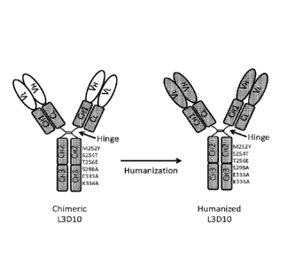
FIG. 1. Schematic diagram of the chimeric (left) and humanized (right) L3D10
antibodies with a novel combination of mutations in the IgG1 Fc region. The
positions of
the mutations in the Fc region are identified by their amino acid position
number and the
amino acids are identified by their single letter code, with the letter before
the number
representing the replace amino acid and the letter after the number
representing the
introduced amino acid. The variable region of the antibodies is depicted with
open ovals
and the human sequence is depicted with gray rectangles. V = variable region;
C =
constant region; L = light chain; H = heavy chain.
FIG. 2. CTLA4 Binding of chimeric L3D10 and 10D1 to plate immobilized CTLA4,
as
determined by ELISA. ELISA plates were coated with 1 g/ml of CTLA4-His
protein
(Sino Biological, China). The given concentration of biotinylated binding
proteins were
added and binding was measured using HRP-conjugated streptavidin. 10D1-1 and -
2
8
CA 03006984 2018-05-30
WO 2017/106372 PCT/US2016/066698
are two independent material lots of the same antibody. B7.1-Fc is a positive
control
and Fc is a negative control.
FIG. 3. L3D10 competition assay. 10D1 is less efficient in blocking chimeric
L3D10
binding to CTLA4 than chimeric L3D10. The experiment was performed as in FIG.
2,
except that biotinylated chimeric L3D10 was mixed with the given concentration
of
unlabeled CTLA4-binding proteins or CTLA4-Fc prior to adding to the ELISA
plates.
Note much better blocking by unlabeled L3D10 than 10D1, which suggest that
these
antibody binding sites are not identical.
FIG. 4. Blocking CTLA4 binding to plate immobilized B7.1. B7.1Fc protein was
coated
.. onto ELISA plates at 0.5 g/ml. After washing and blocking, biotinylated
CTLA4-Fc
protein was added at 0.25 g/mlin the presence of given concentrations of the
competing proteins. Data shown are means of duplicate optical density at 405
nM.
Whereas B7.1-Fc, chimeric L3D10 and CTLA4-Fc all block the CTLA4:137.1
interaction
in a dose-dependent manner, two separate lots of 10D1 antibody failed to block
at all
doses tested. Biotinylation of CTLA4 does not destroy 10D1 epitopes on CTLA4
as both
lots of 10D1 show strong binding to biotinylated CTLA4 (data not shown).
FIG. 5. Blocking CTLA4 binding to plate immobilized B7.2. B7.2Fc protein was
coated
onto ELISA plates at 0.5 lag/ml. After washing and blocking, biotinylated
CTLA4-Fc
protein was added at 0.25 lig/mlin the presence of given concentrations of the
zo competing proteins. Whereas chimeric L3D10 blocks the CTLA4:B7.2
interaction in a
dose-dependent manner, two separate lots of 10D1 antibody failed to completely
block
the CTLA4:137.2 interaction even at the highest concentration.
FIG. 6. Both 10D1 and L3D10 potently block B7-CTLA4 interaction using soluble
B7-1
and B7-2 and immobilized CTLA4-Fc. Varying doses of anti-human CTLA4 mAbs were
added along with 0.25 pg/mlof biotinylated human CTLA4-Fc to plate-coated with
human B7-1Fc. The amounts of CTLA4 bound to plates were measured using HRP-
conjugated streptavidin. Data shown are means of duplicates and are
representative of
two independent experiments.
9
CA 03006984 2018-05-30
WO 2017/106372 PCT/US2016/066698
FIG. 7. Blocking CTLA4 binding to cell surface expressed B7.1. Biotinylated
CTLA4-Fc
protein was added to B7.1 expressing CHO cells at 0.5 lig/m1 in the presence
of the
given concentration of the competing proteins. Binding of biotinylated fusion
protein to
CHO cells transfected with mouse or human B7-1 and B7-2 was detected by flow
cytometry. The amounts of bound receptors were measured using phycoethrythorin-
conjugated streptavidin. Data shown are means fluorescence intensity of
triplicate
samples. Whereas chimeric L3D10 blocks the CTLA4:B7.1 interaction in a dose-
dependent manner, two separate lots of 10D1 antibody failed to block at all
doses
tested.
FIG. 8. Blocking CTLA4 binding to cell surface expressed murine B7.1. Modest
but
detectable blocking of mouse B7-1-human CTLA4 interaction by 10D1 when mB7-1
is
expressed on CHO cells. Varying doses of anti-human CTLA4 mAbs were added
along
with 0.25 g/ml of human CTLA4-Fc to CHO cells expressing mouse B7-1. Data
shown
are means and SEM or triplicate data and are representative of two independent
experiments.
FIG. 9. Blocking CTLA4 binding to cell surface expressed B7.2. Biotinylated
CTLA4-Fc
protein was added to B7.2 expressing CHO cells at 0.5 ilg/ml in the presence
of the
given concentration of the competing proteins. Whereas chimeric L3D10 blocks
the
CTLA4:B7.2 interaction in a dose-dependent manner, two separate lots of 10D1
antibody failed to completely block the CTLA4:B7.2 interaction even at the
highest
concentration. Data shown in this figure has been repeated at least 5 times.
FIG. 10. 10D1 binds to biotinylated human CTLA4-Fc better than L3D10. Varying
doses
of anti-human CTLA4 mAbs or control IgG were coated onto the plate.
Biotinylated
CTLA4-Fc was added at 0.25 g/ml. The amounts of CTLA4 bound to plates were
measured using HRP-conjugated streptavidin. Data shown are means of duplicates
and
are representative of two independent experiments.
FIG. 11. L3D10 but not 10D1 blocks the interaction between polyhistindine
tagged
CTLA4 and CHO cells expressing human B7-1. CHO cells expressing human B7-1
were incubated with polyhistidine-tagged CTLA4 along with given doses of
antibodies,
CA 03006984 2018-05-30
WO 2017/106372 PCT/US2016/066698
the amounts of CTLA4-Fc were detected with PE-streptavidin and measured by
FACSCanto II. Data shown are means fluorescence intensity of triplicate
samples and
are representative of two independent experiments.
FIG. 12. Chimeric L3D10 induces complete remission of established tumors in
the
syngeneic MC38 model. Top panel depicts experimental design and the lower
panels
show growth kinetics of MC38 tumors in mice that received either control IgG
(lower left
panel, n=6) or chimeric L3D10 (lower right panel, n=5).
FIG. 13. Therapeutic effect of chimeric L3D10 and 10D1 in the MC38 tumor
model.
Human CTLA4-knock-in mice with body weight of approximately 20 grams were used
for the study. 1 x 1 06 M038 tumor cells were injected subcutaneously into
Ctla4" mice
and when the tumor reached a size of 0.5 cm in diameter, tumor bearing mice
were
randomized into three groups with 5 or 6 mice each. Mice were then treated
(i.p.) with
100 pg/injection of 10D1, chimeric L3D10 or control hIgGFc on days 7, 10, 13,
and 16
as indicated by the arrows. The results of duplicate expts are shown (left and
right
is panels) and data shown are means and S.D. of tumor size (n = 6 per group
in the left
panel, n=5 per group in the right panel). L3D10 and 10D1 have similar
therapeutic effect
in this model and are both able to induce complete remission of established
tumors. The
diameters (d) of the tumor were calculated using the following formula: D=
(ab),
V=ab2/2, where a is the long diameter, while b is the short diameter.
Statistical analyses
were performed by two-way repeated measures ANOVA (treatment X time). For the
left
panel: P = 10D1 vs. hIgGFc: 5.71e-07; L3D10 vs. hIgGFc: P = 5.53e-07; 10D1 vs.
L3D10: P = 0.869.
FIG. 14. Effective rejection of M038 by anti-CTLA-4 mAbs in CTLA4h/m mice. As
in FIG.
13, except that heterozygous CTLA4h/m mice are used. Data shown are means and
SEM of tumor diameters (6 mice per group); 10D1 vs. hIgGFc: P = 0.0011; L3D10
vs.
hIgGFc: P = 5.55e-05; 10D1 vs. L3D10: P = 0.0346.
FIG. 15. Therapeutic effect of chimeric L3D10 and 10D1 in the B16-F1 melanoma
tumor
model. Human CTLA4-knockin mice with body weight of approximately 20 grams
were
used for the study. Arrows indicate the time of treatment (50
pg/mice/treatment). Data
11
CA 03006984 2018-05-30
WO 2017/106372 PCT/US2016/066698
shown are means and S.D. of the tumor size (n ¨ 4 per group). L3D10 have
similar
therapeutic effect in this model and are both able to delay tumor growth in
this
aggressive and poorly immunogenic tumor model.
FIG. 16. Assay for measuring CTLA4 blocking in vivo. B7.1 or B7.2 binds on
dendritic
cells bind to, and are down-regulated by, CTLA4 on surface of T cells.
However, binding
of blocking anti-CTLA4 antibodies prevents B7.1/B7.2 binding to CTLA4 and thus
prevents the downregulation of B7.1 and B7.2, resulting in a net increase in
B7.1/B7.2
expression. However, with chimeric T cells expressing both human and mouse
CTLA4,
antibodies that bind human CTLA4 do not prevent B7.1/67.2 binding to the
murine
CTLA4, which restores B7.1/137.2 inhibition.
FIGS. 17A-F. 10D1 does not block B7-CTLA4 interaction in vivo. Using the assay
described in FIG. 11, cells from mice treated with anti-CTLA4 antibodies were
used to
assay B7.1 and B7.2 expression. FIG. 17A shows a diagram of experimental
design.
Briefly, age and gender-matched mice received 500 jig of antibodies or their
controls
is intraperitoneally. At 24 hours after injection, mice were sacrificed and
their spleen cells
were stained with anti-CD11c, CD11b, anti-B7-1 and anti-B7-2 mAbs. FIG. 17B
shows
representative data showing the phenotype of CD11chi DC analyzed for B7
expression.
FIG. 170 shows representative histograms depicting the levels of B7-1 on DC
from
mice that received control IgG1-Fc, L3D10 or 1001. Data in the top panel shown
antibody effect in homozygous knockin mice, while that in the bottom panel
show
antibody effect in the heterozygous mice. FIG. 17D shows as in FIG. 170,
except that
expression of B7-2 is shown. Data shown in FIGS. 170 and D are representative
of
those from 3 mice per group and have been repeated once with three mice per
group.
FIG. 17E shows that in human CTLA4 homozygous mice, L3D10 but not 1001 induced
expression of B7-1 (left panel) and B7-2 (right panel). Data shown are
summarized from
two experiments involving a total of 6 mice per group. In each experiment, the
mean
data in the control mice is artificially defined as 100% and those in
experimental groups
are normalized against the control. FIG. 17F as in FIG. 17E, except that
heterozygous
mice are used. Neither L3D10 nor 10D1 block B7-CTLA4 interaction in mice that
co-
dominantly express both mouse and human Ctla4 genes.
12
CA 03006984 2018-05-30
WO 2017/106372 PCT/US2016/066698
FIG. 18. L3D10 binds to human but not mouse CTLA4. Data showing are dot plots
of
intracellular staining of CTLA4 among gated Cd3+Cd4+ cells, using spleen cells
from
Ctla4" (top) or Ctlaelm (bottom) mice. Anti-mouse CTLA4 mAb 4F10 was used as
control.
FIG. 19. Therapeutic effect of chimeric L3D10 and 10D1 in CTLA4" mice. The top
panel depicts the experimental design. The Ctla4" mice were challenged with
colon
cancer cell line MC38 and when the tumor reached a size of approximately 5 mm
in
diameter, the mice were treated 4 times with control human IgG-Fc, L3D10 or
10D1 and
observed tumor size over a 6 weeks period. The lower panels shows the growth
kinetics
io of M038 tumors in mice that received either control IgG, chimeric L3D10
or 10D1 (n=6
per group). Despite apparent differences in CTLA4 blocking activity in vivo as
shown in
FIG. 16, both L3D10 and 10D1 display strong anti-tumor activity against the
M038
model in chimeric CTLA4m/h mice.
FIGS. 20A-B. 10D1 and L3D10 have similar therapeutic effect on B16 melanoma
is growth.1x105 B16 tumor cells were injected (s.c.) into Ctla4" mice (n=4-
5), and treated
(i.p.) with 100 pg (FIG. 20A) or 250pg (FIG. 20B) 10D1, L3D10 or control IgGFc
on day
11,14,17(FIG. 20A) or on day 2, 5, and 8 (FIG. 20B), as indicated by arrows.
For FIG.
20A, 10D1 vs. hIgGFc: P = 0.0265; L3D10 vs. hIgGFc: 10D1 vs. L3D10: P ¨
0.0487;
P= 0.302. For FIG. 20B, 10D1 vs. hIgGFc: P = 0.00616; L3D10 vs. hIgGFc: P =
0.0269:
zo 10D1 vs. L3D10: P=0.370,. Data represent mean SEM of 4-5 mice per
group.
Statistical analyses were performed by two-way repeated measures ANOVA.
FIGS. 21A-B. lmmunotherapeutic effects between L3D10 and 10D1 in Ctla4"
(FIG. 21A) and Ctlaem (FIG. 21B) in mice that were terminated before rejection
in
complete in order to evaluate depletion of Treg within tumor microenvironment.
Data
25 shown are means and SEM of tumor diameters of two independent
experiments,
involving 5 mice per group.
FIGS. 22A-F. Blocking the B7-CTLA4 interaction does not contribute to cancer
immunotherapeutic activity of anti-CTLA4 mAb. FIG. 22A shows comparable
immunotherapeutic effect despite vastly different blocking activity by two
anti-CTLA4
13
CA 03006984 2018-05-30
WO 2017/106372 PCT/US2016/066698
mAbs. 5105 MC38 tumor cells were injected (s.c.) into Ctla4" mice (n=6), and
treated
(i.p.) with 100 pg 10D1, L3D10 or control hIgG-Fc on days 7, 10, 13, and 16,
as
indicated by arrows. Data represent mean SEM of six mice per group.
Statistical
analyses were performed by two-way repeated measures ANOVA (treatment x time).
10D1 vs. hIgG-Fc: P = 5.71e-07; L3D10 vs. hIgG-Fc: P = 5.53e7; 10D1 vs. L3D10:
P =
0.869. Data are representative of three independent experiments. FIG. 22B. In
mice that
neither antibodies block B7-CTLA4 interaction, both induce robust tumor
rejection. As in
FIG. 22A, except that heterozygous mice that express both mouse and human
CTLA4
were used. 1001 vs. hIgG-Fc: P = 0.0011; L3D10 vs. hIgG-Fc: P = 5.55e5; 10D1
vs.
L3D10: P = 0.0346. Data are representative of three independent experiments.
FIGS. 22C-F, Blocking B7-CTLA4 interaction does not contribute to selective
depletion
of Treg in tumor microenvironment. FIGS. 22C and D. Regardless of their
ability to
block B7-CTLA4 interaction, L3D10 and 1001 do not delete Treg in the spleen.
Data
shown are % of Foxp3+ cells among spleen CD4 T cells in Ctla4" (FIG. 22C) and
is Ctla4m/h (FIG. 220) mice. n-6. e and f, both L3D10 and 1001 delete Treg
among tumor
infiltrating CD4 T cells in Ctla4" (FIG. 22E) and Ctla4mm (FIG. 22F) mice.
Data shown
in c-f are % of Treg at 17 (experiment 1) or 19 days (experiment 2) after
tumor cell
challenge and 10 or 12 days after initiation of 4 anti-CTLA4 mAb treatments as
indicated in arrows.
zo FIGS. 23A-F. Evaluation of blocking activities of commonly used anti-
mouse CTLA4
mAbs 9H10 and 909. FIGS. 23A and B show that 9H10 does not block B7-CTLA4
interaction if B7-1 (FIG. 23A) and B7-2 (FIG. 23B) are coated onto plates.
Biotinylated
mouse CTLA4-Fc fusion protein were incubated with B7-coated plates in the
presence
of given concentration of control IgG or anti-mouse CTLA4 mAb 909 and 9H10.
The
25 CTLA4 binding is detected with HRP-conjugated streptavidin. Data shown
are means of
duplicated and are representative of two independent experiments. FIGS. 23C
and D
show that 909 and 9H10 exhibit differential binding to soluble (FIG. 23C) and
plate
bound CTLA4-Fc (FIG. 23D). Data shown are means of duplicated and are
representative of at least two independent experiments. FIGS. 23E and F show
the
30 effects of anti-mouse CTLA4 mAbs 909 and 9H10 on levels of B7-1 (FIG.
23E) and B7-
2 (FIG. 23F) on CD1lchi DC from WT (Ctla4") spleen cells at 24 hours after
treatment
14
CA 03006984 2018-05-30
WO 2017/106372
PCT/US2016/066698
with 500 lig of antibodies i.p. The data are summarized from 6 independent
mice per
group in two independent experiments involving 3 mice per group each.
FIGS. 24A-D. Distinct in vivo and in vivo blocking activities of anti-mouse
CTLA4 mAb
4F10. FIGS. 24A and B show the effect of 4F10 on interaction of CTLA4-Fc to
plate-
coated B7-1 (FIG. 24A) or B7-2 (FIG. 24B). Biotinylated mouse CTLA4-Fc fusion
protein
were incubated with B7-coated plates in the presence of given concentration of
control
IgG or anti-mouse CTLA4 mAb 4F10. The CTLA4 binding is detected with HRP-
conjugated streptavidin. Data shown are means of duplicated and are
representative of
two independent experiments. FIGS. 24C and D show the impact of 4F10 on B7-1
and
B7-2 expression. Summary data on B7-1 (FIG. 24C) and B7-2 (FIG. 240) levels
from 6
mice per group. The B7 levels in the control IgG-treated group are
artificially defined as
100%.
FIG. 25. Adverse effects of chimeric L3D10 and 1001 in combination with anti-
PD-1.
Top panel depicts the experimental design. 10-day old female-only human CTLA4-
is mice
with body weight of greater than 4 grams were used for the study. They
received indicated proteins or their combinations. Arrows indicate time of
treatment (100
14/mice/treatment). Data shown are means and S.D. of % weight gains. Chimeric
L3D10 and 1001 have comparable cancer therapeutic effect in adult mice (FIG.
13) but
distinct adverse effects are seen when 1001 is combined with the anti-PD-1
mAb.
FIG. 26. Adverse effects of chimeric L3D10 and 1001 in combination with anti-
PD-1.
The graph shows the terminal body weight on Day 42 in the mice from the
experiment
outlined in FIG. 25 that received either control IgG, 1001 + anti-PD-1 or
chimeric L3D10
+ anti-PD-1 (n=5 per group). A significant reduction in weight is observed
with the anti-
PD1+1001 combination, which was not seen with the anti-PD-1+Chimeric L3D10
combination.
FIG. 27. Pathological effects of chimeric L3D10 and 10D1 in combination with
anti-PD-1.
To further examine to relative toxicity of L3D10 compared to 1001 when
administered in
combination with anti-PD-1, we looked at the gross anatomy of the mice
described in
FIG. 26 above. The Uterus/Ovary/Bladder and thymus were noticably smaller in
mice
CA 03006984 2018-05-30
WO 2017/106372 PCT/US2016/066698
treated with 10D1 + PD-1, whereas the organs in mice treated with L3D10 + anti-
PD-1
was comparable to hIgG control. In contrast, the hearts dissected from mice
treated with
10D1 appeared larger in size with a noticeably whiter appearance.
FIGS. 28A-D. Treatment with 10D1 in combination with anti-PD-1 results in
abnormal
erythropoiesis. Given the differences in the hearts observed in FIG. 27, we
looked at
erythropoiesis within the mice and observed clear differences in the mice
treated with
10D1 + anti-PD-1 relative to the groups treated with L3D10 + anti-PD-1 or
control
antibody (hIgG), which were fairly similar. The bone marrow from mice treated
with
10D1 + anti-PD-1 had a noticeably whiter color (FIG. 28A) and the isolated
blood was
io almost completely white in color (FIG. 28B). In accordance with this,
when we analyzed
differentiation of the red blood cells using distribution of CD119 and CD71
markers we
observed a statistically significant reduction in the number of cells
undergoing Stage IV
development in the 10D1 + anti-PD-1 treated mice. Representative FACS profiles
are
shown in FIG. 28C, while summary data are presented in FIG. 28D.
is FIG. 29. Flow cytometry analysis of anti-red blood cell antibodies.
Blood samples from
NOD.SCID.I12rg-/- (NSG) mice were stained with plasma samples from the mice
that
received antibody treatment during the perinatal period. Sera from NSG mice
and those
without sera were used as negative control. All sera were used at 1:50
dilution. These
data show that no mice produced anti-red cell antibodies.
zo FIG. 30. Pathology of the heart in mice treated with chimeric L3D10 and
10D1 in
combination with anti-PD-1. To further determine the toxicology of L3D10 vs
10D1 in
combination with anti-PD-1, we performed histological analysis of the heart in
mice
described in FIG. 26. Mice treated with 10D1 + anti-PD-1 displayed a high
level of T cell
infiltration that was not observed in mice treated with L3D10 + anti-PD-1 or
mice treated
25 with human IgG control.
FIG. 31. Pathology of the lung in mice treated with chimeric L3D10 and 10D1 in
combination with anti-PD-1. To further determine the toxicology of L3D10 vs
10D1 in
combination with anti-PD-1, we performed histological analysis of the lung in
mice
described in FIG. 26. Mice treated with 10D1 + anti-PD-1 displayed a high
level of T cell
16
CA 03006984 2018-05-30
WO 2017/106372 PCT/US2016/066698
infiltration that was not observed in mice treated with L3D10 + anti-PD-1 or
mice treated
with human IgG control.
FIG. 32. Pathology of the salivary gland in mice treated with chimeric L3D10
and 10D1
in combination with anti-PD-1. To further determine the toxicology of L3D10 vs
10D1 in
combination with anti-PD-1, we performed histological analysis of the salivary
in mice
described in FIG. 26. Mice treated with 10D1 + anti-PD-1 displayed a much
higher level
of T cell infiltration than observed in mice treated with L3D10 + anti-PD-1 or
mice
treated with human IgG control.
FIGS. 33A-F. Pathology of the kidney and liver in mice treated with chimeric
L3D10 and
10D1 in combination with anti-PD-1. To further determine the toxicology of
L3D10 vs
10D1 in combination with anti-PD-1, we performed histological analysis of the
kidney
and liver in mice described in FIG. 26. FIGS 33A-C are sections from the
kidney and
FIGS. 33D-E are sections taken from the liver. Mice treated with 10D1 + anti-
PD-1
displayed a high level of T cell infiltration than observed in mice treated
with L3D10 +
is anti-PD-1 or mice treated with human IgG control.
FIG. 34. Toxicity scores of mice treated with chimeric L3D10 and 10D1 in
combination
with anti-PD-1. This tissue data shown if FIGS. 30-33 is summarized and shows
the
high toxicity scores of mice treated with 10D1 + anti-PD-1 relative to L3D10 +
anti-PD-1
which has scores only marginally higher than the hIgG control mouse group.
zo FIG. 35. 10D1+anti-PD-1 do not have significant toxicity in the Ctlaem
mice as
evidenced by normal body weight gains in mice that received antibody treatment
during
the perinatal period. The mice received treatments with given antibody or
combinations
on days 10, 13, 16, 19 and 22 intraperitoneally
(10014/mice/injection/antibody). Mice
were weighed at least once every 3 days.
25 FIG. 36. L3D10 and 10D1 display similar binding patterns for plate
immobilized CTLA4.
ELISA plates were coated with 1 [ig/m1 of CTLA4-His protein (Sino Biological,
China).
The given concentration of biotinylated binding proteins were added and
binding
measured using HRP-conjugated streptavidin. 10D1-1 and -2 are two independent
material lots of the same antibody. hIgG-Fc is a human Ig negative control.
17
CA 03006984 2018-05-30
WO 2017/106372 PCT/US2016/066698
FIG. 37. L3D10 displays reduced binding soluble CTLA4. Given concentration of
anti-
human CTLA4 mAbs were coated on the plate overnight, after washing and
blocking
with bovine serum albumin, biotinylated CTLA4-Fc was added at 0.25 lig/ml.
After
incubation ans washing, the amounts of captured CTLA4-Fc were measured using
HRP-labeled streptavidin.
FIG. 38. Alignment of the humanized antibody variable regions with the
parental L3D10
antibody sequence. The heavy chain variable region (top)(SEQ ID NOS: 62-64)
and
light chain variable region (bottom)(SEQ ID NOS: 70-72) of the humanized
antibody
sequences are alignment with the parental L3D10 antibody (heavy chain: SEQ ID
NO: 57; light chain: SEQ ID NO: 65) and the respective human antibody
frameworks
(heavy chain: SEQ ID NOS: 58-61; light chain: SEQ ID NOS: 66-69). Back
mutations
to the mouse parental sequence are highlighed in yellow. Novel amino acids
i.e. amino
acid residues not present in the parental antibody sequence or the respective
human
antibody framework are highlighted in green. Mutations introduced into the
CDR2
sequences are shown in purple. CDR sequences are shown in red based on
www.bioinf.org.uk/abs/.
FIGS. 39A-B. Anti-tumor activity of humanized L3D10 antibodies compared to
10D1.
Using the M038 mouse tumor model in human CTLA4 knockin mice we looked at the
anti-tumor activity of humanized L3D10 antibodies compared to the chimeric
L3D10
antibody and 10D1. The top panel shows the treatment schedule of the in vivo
experiment; mice were given a total of 4 doses of antibody every 3 days
starting on day
7 after inoculation. All humanized antibodies (n = 6 per group) completely
eradicated the
tumors and were comparable to 10D1 (bottom panel).
FIG. 40. Anti-tumor activity of humanized L3D10 antibodies in CTLA4hlm mice.
The top
panel shows the treatment schedule of the in vivo experiment; Ctlaem mice
received
control hlg or one of three different anti-human CTLA4 mabs at doses of 30 (-
30, solid
lines) or 10 (-10, dotted lines) mg per injection at the indicated dates after
MC38 tumor
injection. Tumor sizes were measured once every three days.
18
CA 03006984 2018-05-30
WO 2017/106372 PCT/US2016/066698
FIG. 41. Therapeutic effect of anti-CTLA-4 mAb in minimal disease B16-F1 tumor
model.
Using the B16-F1 mouse tumor model in human CTLA4 knockin mice we looked at
the
anti-tumor activity of humanized L3D10 antibodies. 1x10⁵ B16 tumor cells were
injected
(s.c.) into Ctla4hi" mice (n=5-6). On days 2, 5, and 8, the mice were treated
with control
Ig, 10D1, chimeric L3D10 or PP4637 and PP4638 (250 g/mouse, i.p.). Tumor
incidence and sizes were measured every other day. 10D1 vs. hIgGFc: P =
0.00616;
L3D10 vs. hIgGFc: P =0.0269; 10D1 vs. L3D10: P=0.370; PP4637 vs. hIgGFc:
P=0.0005; PP4637 vs. 10D1: P=0.805; PP4638 vs. hIgGFc: P=0.0016; PP4638 vs.
10D1: P=0.856. Data represent mean SEM of 5-6 mice per group. Sizes of
tumors
were considered as 0 for mice that never developed tumor.
FIG. 42. Comparison among 10D1, PP4631 and PP4637 females for their combined
toxicity with anti-PD-1 mAb. Female CTLA4" mice were treated on days 10 or 11
days
after birth with 4 injections of antibodies (100 g/mice/injection, once every
three days)
or control Fc as specified in the legends. Mice were weighted once every 3
days. Data
shown are means and SEM of % weight gain over a 30 day period. All mice were
sacrificed on day 43 for histological analysis. The number of mice used per
group is
shown in the parentheses of labels.
FIG. 43. Combination therapy with 10D1 and anti-PD-1 cause anemia, whereas
those
with either PP4631+anti-PD-1 or PP4637+anti-PD-1 do not. Data shown are
hematocrit
zo of 43 day old mice that have received four treatments of antibodies on
days 11, 14, 17
and 20 at doses of 100 g/mouse /antibodies.
FIGS. 44A-B. Combination therapy with 10D1+anti-PD-1 cause systemic T cell
activation, whereas those with either PP4631+anti-PD-1 or PP4637+anti-PD-1 do
not.
Data shown are % of CD4 (upper panels) and CD8 T cells (lower panels) with
phenotypes of naïve (CD4410CD62Lhi), central memory (CD44hiCD620) and effector
memory (CD44hiCD62L1o) T cells in either peripheral blood (FIG. 44A) or in the
spleen
(FIG. 44B). The cells were harvested from 43 day old mice that have received
four
treatments of antibodies on days 11, 14, 17 and 20 at doses of
100 g/mouse/antibodies.
19
CA 03006984 2018-05-30
WO 2017/106372 PCT/US2016/066698
FIG. 45. Humanization of L3D10 does not affect binding to immobilized CTLA4.
The
capacity of the humanized L3D10 antibodies to bind immobilized CTLA4 was
determined as described in FIG. 36. X-axis indicates the concentration of anti-
CTLA-4
mAbs added into solution. Humanization does not affect binding to immobilized
CTLA4
and all 3 humanized antibodies demonstrated similar binding to the parental
chimeric
L3D10 antibody and 10D1. Similar patterns were observed when CTLA4-Ig was used
instead of CTLA-4-his.
FIG. 46. Humanization further reduces L3D10 binding to soluble CTLA4. The
capacity
of the humanized L3D10 antibodies to bind soluble CTLA4 was determined as
described in FIG. 37. X-axis indicates the concentration of anti-CTLA-4 mAbs
coated
onto ELISA plates. Humanization further reduces binding to soluble CTLA4
relative to
the parental L3D10 chimeric antibody. Similar patterns were observed when
CTLA4-Ig
was used instead of CTLA-4-his.
FIGS. 47A-B. PP4631, PP4638 and PP4637 do not block B7-CTLA-4 interactions in
is vitro. FIG. 47A shows blocking of the B7-1-CTLA-4 interaction by anti-
human CTLA-4
mAbs 10D1, PP4631, PP4637 and L3D10. B7-1Fc was immobilized at the
concentration of 0.5 pg/ml. Biotinylated CTLA4-Fc was added at 0.25 g/ml
along with
given doses of antibodies. Data shown are means of duplicate optical density
at 405 nM.
FIG. 47B shows blocking of B7-2-CTLA-4 interaction by anti-human CTLA-4 mAbs
10D1 and L3D10. As in FIG. 47A, except that B7-2-Fc was immobilized.
FIG. 48. PP4631 and PP4637 do not block B7-CTLA-4 interactions in vivo as
demonstrated by their lack of effect on B7-1 and B7-2 expression on dendritic
cells.
Summary data on B7-1 (a) and B7-2 (b) levels from 3 mice per group. The B7
levels in
the control IgG-treated group are artificially defined as 100%.
FIG. 49. PP4637, which exhibits the best safety profile in combination with
anti-PD-1
mAb (see FIG. 42), is the most potent in causing tumor rejection based on
tumor
rejection at the lowest therapeutic doses. Ctlaem mice received control IgFc
or one of
three different anti-human CTLA4 mab at doses of 30 (-30, solid lines) or 10 (-
10, dotted
lines) jag per injection at the indicated dates. Tumor sizes were measured
once every
CA 03006984 2018-05-30
WO 2017/106372 PCT/US2016/066698
three days. At 1011g/injection, PP4637 (HL32) is the most efficient in
inducing tumor
rejection.
FIG. 50. Humanized antibody purity assessment. Transiently expressed humanized
L3D10 antibodies were purified by Protein A chromatography and samples from
all 3
.. antibodies was assessed by reducing and non-reducing SOS-PAGE. Purified
proteins
produced gel bands indicative in size of an antibody molecule under both
reducing and
non-reducing conditions. The "Flow out" lanes show the protein A column flow
through,
indicating that the majority of the antibody protein adhered to the protein A
column.
FIG. 51. Size Exclusion Chromatography (SE-HPLC) of transiently expressed
protein.
Protein samples for each of the humanized antibodies were analyzed by SE-HPLC
following single step Protein A chromatography. Top panel: antibody PP4631.
Middle
panel: antibody PP4637. Bottom panel: antibody PP4638.
FIG. 52. CE-SOS analysis of transiently expressed protein. Protein samples for
each of
the humanized antibodies were analyzed by CE-SDS following single step Protein
A
chromatography. Left panels show the results under non-reduced conditions, and
the
right panels show the results under reduced conditions. Top panel: antibody
PP4631.
Middle panel: antibody PP4637. Bottom panel: antibody PP4638.
FIGS. 53A-C. Charge isoform profile and deamidation of the humanized L3D10
antibodies as determined by capillary isoelectric focusing (cIEF). The level
of protein
deamidation under high pH stress was determined by comparing the Humanized
L3D10
antibodies before and after high pH stress treatment over two different time
periods (5
hrs and 12.5 hrs) were analyzed by clEF analysis. FIGS. 53A-C show the
profiles for
antibodies PP4631, PP4637 and PP4638, respectively.
FIGS. 54A-C. Differential Scanning Calorimetry (DSC) Thermal Analysis of the
humanized L3D10 antibodies. In order to determine the thermal stability and
melting
temperatures of the different antibodies, they were subject to Differential
Scanning
Calorimetry (DSC) Thermal Analysis. FIGS. 54A-C show the normalized DSC curves
for
antibodies PP4631, PP4637 and PP4638, respectively.
21
CA 03006984 2018-05-30
WO 2017/106372 PCT/US2016/066698
FIG. 55. Alignment of the human, macaque and mouse CTLA-4 extracellular
domains.
The amino acid sequences of the human (Hm, shown in red)(SEQ ID NO: 73),
macaque
(Mk, shown in black) and mouse (Ms, shown in green) CTLA-4 protein
extracellular
domains are aligned and the conserved amino acids (relative to the human
sequence)
are shown with dashes (-). In order to help the alignment, the mouse sequence
has a
deletion and insertion (relative to the human and monkey sequences) at the
positions
highlighted in yellow. The known B7-1Ig binding site is shown in bold and
underlined.
The sequences demonstrate that the human and monkey sequences are highly
conserved, whereas the mouse sequence has a number of amino acid differences.
Based on this sequence alignment, 11 mutant (M1 - M11)(SEQ ID NOS: 40-50)
human
CTLA-4Fc proteins were designed that incorporate murine specific amino acids -
the
amino acids incorporated into each mutant protein are shown in blue.
FIGS. 56A-B. Amino acid sequence composition of the WT and mutant CTLA-4Fc
proteins. DNA constructs encoding the WT CTLA-4Fc protein (SEQ ID NO: 39) and
11
mutant proteins (SEQ ID NOS: 40-50) incorporating murine Ctla-4 amino acids
were
designed as shown. The amino acid sequences are for mature proteins, including
the
IgG1 Fc portion, but not the signal peptide. The known B7-1Ig binding site is
shown in
large blue letters and double-underlined. The replaced murine amino acid
residues in
the mutant are shown lower case in red. The IgG1 Fc portion of the proteins in
zo underlined.
FIG. 57. Mutation in M11 (AA103-106, YLGI>fcGm) selectively abolish antibody
binding
to human CTLA-4. Data shown are means of duplicates, depicting the binding of
B7-1 Fc
(a), L3D10 (b), PP4631 (c), and PP4637 (d) binding to plate-coated hCTLA4-Fc
(open
circles), mCTLA4-Fc (filled triangles), M11 (filled circles) and IgG1-Fc (open
triangles).
FIG. 58. Mapping L3D10, PP4631 and PP4637 to an epitope adjacent to the B7-1
binding site in a 3-0 structure of the B7-1-CTLA4 complex. The B7-1 binding
motif is
colored in red, while the antibody epitope is colored in purple. B7-1 is
depicted above
CTLA4 with a space-filled ribbon, while that of CTLA-4 is depicted as an
unfilled ribbon.
22
CA 03006984 2018-05-30
WO 2017/106372 PCT/US2016/066698
FIG. 59. Amino acid sequence composition of the WT (SEQ ID NO: 39) and mutant
CTLA-4Fc proteins, M12-M17 (SEQ ID NOS: 51-56). DNA constructs encoding the 6
mutant CTLA-4Fc proteins, M12-M17, incorporating murine Ctla-4 amino acids
were
designed as shown. The amino acid sequences are for mature proteins, including
the
IgG1 Fc portion, but not the signal peptide. The known B7-1Ig binding site is
shown in
large blue letters and double-underlined. The replaced murine amino acid
residues in
the mutant are shown lower case in red. The IgG1 Fc portion of the proteins in
underlined.
FIGS. 60A-C. Mutational analysis reveal distinct binding requirements for 10D1
io (FIG. 60A), PP4631 (FIG. 60B) and PP4637 (FIG. 600) to CTLA-4. CTLA-4Fc
mutants
were coated overnight at 4oC at 1 m/ml. After blocking with BSA, given
concentration
of biotinylated anti-CTLA-4 mAbs were added and incubated for 2 hours. After
washing
away the unbound antibodies, the bound antibodies were detected with HRP-
labeled
streptavidin.
is FIGS. 61A-B. Therapeutic effect of anti-4-1 BB and anti-CTLA-4
antibodies in both
minimal disease (FIG 61A) and established tumor (FIG. 61B) models. FIG. 61A
shows
therapy of minimal disease. C57BL/6 mice were inoculated subcutaneously with
5x10⁵
M038 cells. On days 2, 9 and 16 after tumor cell injection, control hamster
and rat IgG,
anti-CTLA-4, and/or anti-4-1 BB antibodies were injected. Tumor sizes were
measured
zo -- by physical examination. Data shown are growth kinetics of tumors, with
each line
representing tumor growth in one mouse. The sizes presented are products of
long and
short diameters of the tumor. FIG. 61B shows therapy of established tumors. As
in FIG.
61A, except that therapy started on day 14 after tumor challenge; all mice had
established tumors ranging from 9-60 mm2 in size before treatment with mAbs
was
25 started. The combined effect of the two antibodies on established tumors
has been
repeated 3 times.
FIG. 62. CD8 T cells, but not 0D4 or NK cells, are essential for antibody-
induced tumor
rejection. Tumor bearing mice were depleted of CD4, CD8, or NK cells by three
injections of antibodies specific for either CD4, 0D8 or NK1.1 on days 9, 12,
and 16
30 after tumor cell inoculation (*). Therapeutic antibodies (anti-CTLA-4
plus anti-4-1 BB)
23
CA 03006984 2018-05-30
WO 2017/106372 PCT/US2016/066698
were injected on days 9, 16 and 23 (vertical arrows). Data shown are means and
SEM
of tumor sizes (n=3). P < 0.05 for 008-depleted group compared to each of the
other
groups (t).
FIGS. 63A-B. Combination therapy reduced host response to anti-CTLA-4
antibodies.
Hamster-anti-mouse-CTLA-4 (FIG. 63A) or rat-anti-mouse-4-1 BB (FIG. 63B)
antibodies
were coated in ELISA plates. Different dilutions of sera from groups of 5 mice
each
were added to the plates. The relative amounts of antibody bound were
determined
using a secondary step reagent (biotinylated goat anti-mouse antibodies that
were
depleted of reactivity to rat and hamster IgG by absorption). Data shown are
mean and
io SEM of optical density at 490 nm. Similar reduction of host antibody
response to anti-
CTLA-4 and 4-1 BB was observed when tumor-free mice were treated with the same
antibodies (data not shown).
FIGS. 64A-B. Combination therapy with anti-4-1 BB and L3010 (anti-human-CTLA4)
antibody in human CTLA-4 gene knock-in mice. FIG. 64A shows a therapeutic
effect.
is Human CTLA4 knockin mice were inoculated with 5x10⁵ M038 tumor cells
subcutaneously. Two days later, groups of 7 mice were treated with rat and
mouse IgG,
anti-4-1 BB and mouse IgG, L3D10 and rat IgG, or L3D10 and anti-4-1 BB, as
indicated
by the arrows. Data shown are mean tumor volume and SEM (n=7). All treatments
significantly reduced tumor growth (P<0.001), and the double antibody
treatment group
zo show significantly reduce tumor size in comparison to either control
(P<0.0001) or
L3D10 antibody (P=0.0007) or anti-4-1 BB antibody treatment (P=0.03). All
tumor
bearing mice were sacrificed when the control IgG-treated group reached early
removal
criteria. FIG. 64B shows long-lasting immunity in mice that received
combination
therapy. Tumor-free mice in the double antibody-treated group developed long
lasting
25 immunity to MC38 tumors. At 110 days after the first tumor cell
challenge, the double
antibody-treated, tumor-free mice or control naïve mice were challenged with
5x10⁵
tumor cells subcutaneously. Tumor growth was monitored by physical
examination.
Note that all of the mice that rejected the tumors in the first round were
completely
resistant to re-challenge, while all naïve mice had progressive tumor growth.
30 Definitions
24
CA 03006984 2018-05-30
WO 2017/106372 PCT/US2016/066698
As used herein, the term "antibody" is intended to denote an immunoglobulin
molecule
that possesses a "variable region" antigen recognition site. The term
"variable region" is
intended to distinguish such domain of the immunoglobulin from domains that
are
broadly shared by antibodies (such as an antibody Fc domain). The variable
region
comprises a "hypervariable region" whose residues are responsible for antigen
binding.
The hypervariable region comprises amino acid residues from a "Complementarity
Determining Region" or "CDR" (i.e., typically at approximately residues 24-34
(L1), 50-
56 (L2) and 89-97 (L3) in the light chain variable domain and at approximately
residues
27-35 (H1), 50-65 (H2) and 95-102 (H3) in the heavy chain variable domain;
ref. 44)
.. and/or those residues from a "hypervariable loop" (i.e., residues 26-32
(L1), 50-52 (L2)
and 91-96 (L3) in the light chain variable domain and 26-32 (H1), 53-55 (H2)
and 96-
101 (H3) in the heavy chain variable domain; Ref. 45). "Framework Region" or
"FR"
residues are those variable domain residues other than the hypervariable
region
residues as herein defined. The term antibody includes monoclonal antibodies,
multi-
specific antibodies, human antibodies, humanized antibodies, synthetic
antibodies,
chimeric antibodies, camelized antibodies, single chain antibodies, disulfide-
linked Fvs
(sdFv), intrabodies, and anti-idiotypic (anti-Id) antibodies (including, e.g.,
anti-Id and
anti-anti-Id antibodies to antibodies of the invention). In particular, such
antibodies
include immunoglobulin molecules of any type (e.g., IgG, IgE, IgM, IgD, IgA
and IgY),
class (e.g., IgGi, IgG2, IgG3, IgG4, IgAi and IgA2) or subclass.
As used herein, the term "antigen binding fragment" of an antibody refers to
one or
more portions of an antibody that contain the antibody's Complementarity
Determining
Regions ("CDRs") and optionally the framework residues that comprise the
antibody's
"variable region" antigen recognition site, and exhibit an ability to
immunospecifically
bind antigen. Such fragments include Fab', F(ab')2, Fv, single chain
(ScFv),and
mutants thereof, naturally occurring variants, and fusion proteins comprising
the
antibody's "variable region" antigen recognition site and a heterologous
protein (e.g., a
toxin, an antigen recognition site for a different antigen, an enzyme, a
receptor or
receptor ligand, etc.). As used herein, the term "fragment" refers to a
peptide or
.. polypeptide comprising an amino acid sequence of at least 5 contiguous
amino acid
residues, at least 10 contiguous amino acid residues, at least 15 contiguous
amino acid
CA 03006984 2018-05-30
WO 2017/106372 PCT/US2016/066698
residues, at least 20 contiguous amino acid residues, at least 25 contiguous
amino acid
residues, at least 40 contiguous amino acid residues, at least 50 contiguous
amino acid
residues, at least 60 contiguous amino residues, at least 70 contiguous amino
acid
residues, at least 80 contiguous amino acid residues, at least 90 contiguous
amino acid
residues, at least 100 contiguous amino acid residues, at least 125 contiguous
amino
acid residues, at least 150 contiguous amino acid residues, at least 175
contiguous
amino acid residues, at least 200 contiguous amino acid residues, or at least
250
contiguous amino acid residues.
Human, chimeric or humanized antibodies are particularly preferred for in vivo
use in
io humans, however, murine antibodies or antibodies of other species may be
advantageously employed for many uses (for example, in vitro or in situ
detection
assays, acute in vivo use, etc.).
A "chimeric antibody" is a molecule in which different portions of the
antibody are
derived from different immunoglobulin molecules such as antibodies having a
variable
is region derived from a non-human antibody and a human immunoglobulin
constant
region. Chimeric antibodies comprising one or more CDRs from a non-human
species
and framework regions from a human immunoglobulin molecule can be produced
using
a variety of techniques known in the art including, for example, CDR-grafting
(EP
239,400; International Publication No. WO 91/09967; and U.S. Pat. Nos.
5,225,539,
zo 5,530,101, and 5,585,089), veneering or resurfacing (EP 592,106; EP
519,596;46-48),
and chain shuffling (U.S. Pat. No. 5,565,332).
The invention particularly concerns "humanized antibodies". As used herein,
the term
"humanized antibody" refers to an immunoglobulin comprising a human framework
region and one or more CDR's from a non-human (usually a mouse or rat)
25 immunoglobulin. The non-human immunoglobulin providing the CDR's is
called the
"donor" and the human immunoglobulin providing the framework is called the
"acceptor." Constant regions need not be present, but if they are, they must
be
substantially identical to human immunoglobulin constant regions, i.e., at
least about 85-
90%, preferably about 95% or more identical. Hence, all parts of a humanized
30 immunoglobulin, except possibly the CDR's, are substantially identical
to corresponding
26
CA 03006984 2018-05-30
WO 2017/106372 PCT/US2016/066698
parts of natural human immunoglobulin sequences. A humanized antibody is an
antibody comprising a humanized light chain and a humanized heavy chain
immunoglobulin. For example, a humanized antibody would not encompass a
typical
chimeric antibody, because, e.g., the entire variable region of a chimeric
antibody is
non-human. One says that the donor antibody has been "humanized," by the
process of
"humanization," because the resultant humanized antibody is expected to bind
to the
same antigen as the donor antibody that provides the CDR's. For the most part,
humanized antibodies are human immunoglobulins (recipient antibody) in which
hypervariable region residues of the recipient are replaced by hypervariable
region
io residues from a non-human species (donor antibody) such as mouse, rat,
rabbit or a
non-human primate having the desired specificity, affinity, and capacity. In
some
instances, Framework Region (FR) residues of the human immunoglobulin are
replaced
by corresponding non-human residues. Furthermore, humanized antibodies may
comprise residues which are not found in the recipient antibody or in the
donor antibody.
is .. These modifications are made to further refine antibody performance. In
general, the
humanized antibody will comprise substantially all of at least one, and
typically two,
variable domains, in which all or substantially all of the hypervariable
regions
correspond to those of a non-human immunoglobulin and all or substantially all
of the
FRs are those of a human immunoglobulin sequence. The humanized antibody
20 optionally also will comprise at least a portion of an immunoglobulin
constant region (Fc),
typically that of a human immunoglobulin that immunospecifically binds to an
Fc.gamma.RIIB polypeptide, that has been altered by the introduction of amino
acid
residue substitutions, deletions or additions (i.e., mutations).
Detailed Description
25 An antibody against human CTLA4 protein, Ipilimumab, has been shown to
increase
survival of cancer patients, either as the only immunotherapeutic agent or in
combination with other therapeutic agents such as, for example without
limitation, an
anti-PD-1 antibody (13-15). However, the therapeutic effect is associated with
significant adverse effects (13-18). There is a great need to develop novel
anti-CTLA4
30 antibodies to achieve better therapeutic effect and/or less autoimmune
adverse effect.
27
CA 03006984 2018-05-30
WO 2017/106372 PCT/US2016/066698
The inventors have discovered an anti-CTLA4 antibody that, surprisingly, can
be used
to induce cancer rejection while also reducing autoimmune adverse effects
associated
with immunotherapy.
Provided herein are antibody compositions of matter and antigen-binding
fragments
thereof. The invention further concerns the embodiment of such molecules
wherein the
molecule is a monoclonal antibody, a human antibody, a chimeric antibody or a
humanized antibody.
In detail, the invention provides a molecule, comprising an antigen-binding
fragment of
an antibody that immunospecifically binds to CTLA4, and in particular human
CTLA4,
preferably expressed on the surface of a live cell at an endogenous or
transfected
concentration. The invention particularly concerns the embodiment of such a
molecule
wherein the antigen-binding fragment binds to CTLA4, and wherein the live cell
is a T
cell.
The present invention relates to antibodies and their antigen-binding
fragments that are
capable of immunospecifically binding to CTLA4. In some embodiments such
molecules
are additionally capable of blocking the binding of B7.1 and B7.2 to CTLA4.
The invention further concerns the embodiment of such molecules wherein the
molecule
is a monoclonal antibody, a human antibody, a chimeric antibody or a humanized
antibody. The invention includes the embodiments wherein such antibodies are
monospecific, bispecific, trispecific or multispecific.
The invention further concerns the embodiment of such molecules or antibodies
which
binds to CTLA4, and wherein the antigen-binding fragment thereof comprises six
CDRs,
wherein the CDRs comprise the CDRs of anti-CTLA4 antibody L3D10. Specifically,
the
antibody comprises the three light chain and the three heavy chain CDRs of
anti-CTLA4
antibody L3D10.
The invention further concerns the embodiment of the above-described
antibodies,
wherein the antibody is detectably labeled or comprises a conjugated toxin,
drug,
receptor, enzyme, receptor ligand.
28
CA 03006984 2018-05-30
WO 2017/106372 PCT/US2016/066698
The invention further concerns a pharmaceutical composition comprising a
therapeutically effective amount of any of the above-described antibody
compositions,
and a physiologically acceptable carrier or excipient. Preferably,
compositions of the
invention comprise a prophylactically or therapeutically effective amount of
humanized
antibodies of the invention and a pharmaceutically acceptable carrier
In a specific embodiment, the term "pharmaceutically acceptable" means
approved by a
regulatory agency of the Federal or a state government or listed in the U.S.
Pharmacopeia or other generally recognized pharmacopeia for use in animals,
and
more particularly in humans. The term "carrier" refers to a diluent, adjuvant
(e.g.,
Freund's adjuvant (complete and incomplete), excipient, or vehicle with which
the
therapeutic is administered. Such pharmaceutical carriers may be sterile
liquids, such
as water and oils, including those of petroleum, animal, vegetable or
synthetic origin,
such as peanut oil, soybean oil, mineral oil, sesame oil and the like. Water
is a preferred
carrier when the pharmaceutical composition is administered intravenously.
Saline
solutions and aqueous dextrose and glycerol solutions can also be employed as
liquid
carriers, particularly for injectable solutions. Suitable pharmaceutical
excipients include
starch, glucose, lactose, sucrose, gelatin, malt, rice, flour, chalk, silica
gel, sodium
stearate, glycerol monostearate, talc, sodium chloride, dried skim milk,
glycerol,
propylene, glycol, water, ethanol and the like. The composition, if desired,
may also
contain minor amounts of wetting or emulsifying agents, or pH buffering
agents. These
compositions may take the form of solutions, suspensions, emulsion, tablets,
pills,
capsules, powders, sustained-release formulations and the like.
Generally, the ingredients of compositions of the invention may be supplied
either
separately or mixed together in unit dosage form, for example, as a dry
lyophilized
powder or water free concentrate in a hermetically sealed container such as an
ampoule or sachette indicating the quantity of active agent. Where the
composition is to
be administered by infusion, it can be dispensed with an infusion bottle
containing
sterile pharmaceutical grade water or saline. Where the composition is
administered by
injection, an ampoule of sterile water for injection or saline may be provided
so that the
.. ingredients may be mixed prior to administration.
29
CA 03006984 2018-05-30
WO 2017/106372 PCT/US2016/066698
The compositions of the invention may be formulated as neutral or salt forms.
Pharmaceutically acceptable salts include, but are not limited to, those
formed with
anions such as those derived from hydrochloric, phosphoric, acetic, oxalic,
tartaric acids,
etc., and those formed with cations such as those derived from sodium,
potassium,
ammonium, calcium, ferric hydroxides, isopropylamine, triethylamine, 2-
ethylamino
ethanol, histidine, procaine, etc.
The invention further concerns the use of the antibody compositions described
here and
pharmaceutical compositions thereof for the upregulation of immune responses.
Up-
modulation of the immune system is particularly desirable in the treatment of
cancers
io and chronic infections, and thus the present invention has utility in
the treatment of such
disorders. As used herein, the term "cancer" refers to a neoplasm or tumor
resulting
from abnormal uncontrolled growth of cells. As used herein, cancer explicitly
includes
leukemias and lymphomas. The term refers to a disease involving cells that
have the
potential to metastasize to distal sites.
is Accordingly, the methods and compositions of the invention may also be
useful in the
treatment or prevention of a variety of cancers or other abnormal
proliferative diseases,
including (but not limited to) the following: carcinoma, including that of the
bladder,
breast, colon, kidney, liver, lung, ovary, pancreas, stomach, cervix, thyroid
and skin;
including squamous cell carcinoma; hematopoietic tumors of lymphoid lineage,
zo including leukemia, acute lymphocytic leukemia, acute lymphoblastic
leukemia, B-cell
lymphoma, T-cell lymphoma, Berketts lymphoma; hematopoietic tumors of myeloid
lineage, including acute and chronic myelogenous leukemias and promyelocytic
leukemia; tumors of mesenchymal origin, including fibrosarcoma and
rhabdomyoscarcoma; other tumors, including melanoma, seminoma,
tetratocarcinoma,
25 neuroblastoma and glioma; tumors of the central and peripheral nervous
system,
including astrocytoma, neuroblastoma, glioma, and schwannomas; tumors of
mesenchymal origin, including fibrosarcoma, rhabdomyosarcoma, and
osteosarcoma;
and other tumors, including melanoma, xenoderma pegmentosum, keratoactanthoma,
seminoma, thyroid follicular cancer and teratocarcinoma. It is also
contemplated that
30 cancers caused by aberrations in apoptosis would also be treated by the
methods and
CA 03006984 2018-05-30
WO 2017/106372 PCT/US2016/066698
compositions of the invention. Such cancers may include, but are not be
limited to,
follicular lymphomas, carcinomas with p53 mutations, hormone dependent tumors
of the
breast, prostate and ovary, and precancerous lesions such as familial
adenomatous
polyposis, and myelodysplastic syndromes. In specific embodiments, malignancy
or
dysproliferative changes (such as metaplasias and dysplasias), or
hyperproliferative
disorders, are treated or prevented by the methods and compositions of the
invention in
the ovary, bladder, breast, colon, lung, skin, pancreas, or uterus. In other
specific
embodiments, sarcoma, melanoma, or leukemia is treated or prevented by the
methods
and compositions of the invention.
In another embodiment of the invention, the antibody compositions and antigen
binding
fragments thereof can be used with other anti-tumor therapies, including but
not limited
to, current standard and experimental chemotherapies, hormonal therapies,
biological
therapies, immunotherapies, radiation therapies, or surgery. In some
embodiments, the
molecules of the invention may be administered in combination with a
therapeutically or
prophylactically effective amount of one or more agents, therapeutic
antibodies or other
agents known to those skilled in the art for the treatment and/or prevention
of cancer,
autoimmune disease, infectious disease or intoxication. Such agents include
for
example, any of the above-discussed biological response modifiers, cytotoxins,
antimetabolites, alkylating agents, antibiotics, or anti-mitotic agents, as
well as
zo immunotherapeutics.
In preferred embodiment of the invention, the antibody compositions and
antigen
binding fragments thereof can be used with other anti-tumor immunotherapies.
In such
an embodiment the molecules of the invention are administered in combination
with
molecules that disrupt or enhance alternative immunomodulatory pathways (such
as
TIM3, TIM4, 0X40, CD40, GITR, 4-1-BB, B7-H1, PD-1, B7-H3, B7-H4, LIGHT, BTLA,
ICOS, CD27 or LAG3) or modulate the activity of effecter molecules such as
cytokines
(e.g., IL-4, IL-7, IL-10, IL-12, IL-15, IL-17, GF-beta, IFNg, Flt3, BLys) and
chemokines
(e.g., CCL21) in order to enhance the immunomodulatory effects. Specific
embodiments
include a bi-specific antibody comprising the anti-CTLA4 antibody compositions
described herein and anti-PD-1 (pembrolizumab (Keytruda) or Nivolumab
(Opdivo)),
31
CA 03006984 2018-05-30
WO 2017/106372 PCT/US2016/066698
anti-B7-H1 (atezolizumab (Tecentrig) or durvalumab), anti-B7-H3, anti-B7-H4,
anti-
LIGHT, anti-LAG3, anti-TIM3, anti-TIM4 anti-CD40, anti-0X40, anti-GITR, anti-
BTLA,
anti-0027, anti-ICOS or anti-4-1 BB. In yet another embodiment, the molecules
of the
invention are administered in combination with molecules that activate
different stages
or aspects of the immune response in order to achieve a broader immune
response. In
more preferred embodiment, the antibody compositions and antigen binding
fragments
thereof are combined with anti-PD-1 or anti-4-1 BB antibodies, without
exacerbating
autoimmune side effects.
Another embodiment of the invention includes a bi-specific antibody that
comprises an
antibody that binds to CTLA4 bridged to an antibody that binds another immune
stimulating molecules. Specific embodiments include a bi-specific antibody
comprising
the anti-CTLA4 antibody compositions described herein and anti-PD-1, anti-B7-
H1, anti-
B7-H3, anti-B7-H4, anti-LIGHT, anti-LAG3, anti-TIM3, anti-TIM4 anti-CD40, anti-
0X40,
anti-GITR, anti-BTLA, anti-0027, anti-ICOS or anti-4-1 BB. The invention
further
concerns of use of such antibodies for the treatment of cancer.
Methods of administering the antibody compositions of the invention include,
but are not
limited to, parenteral administration (e.g., intradermal, intramuscular,
intraperitoneal,
intravenous and subcutaneous), epidural, and mucosa! (e.g., intranasal and
oral routes).
In a specific embodiment, the antibodies of the invention are administered
zo intramuscularly, intravenously, or subcutaneously. The compositions may
be
administered by any convenient route, for example, by infusion or bolus
injection, by
absorption through epithelial or mucocutaneous linings (e.g., oral mucosa,
rectal and
intestinal mucosa, etc.) and may be administered together with other
biologically active
agents. Administration can be systemic or local.
Yet another embodiment of the invention concerns monitoring the blocking
effects of
anti-CTLA4 antibodies in vivo by monitoring the expression levels of B7.1 and
B7.2 on
immune cells such as antigen presenting cells (APCs). CTLA4 is expressed
predominately among the Treg where it suppresses autoimmune diseases by down-
regulating B7-1 and B7-2 expression on APCs such as dendritic cells.
Therefore,
upregulation of B7 molecules, B7.1 and B7.2, can be as readouts for the in
vivo
32
CA 03006984 2018-05-30
WO 2017/106372 PCT/US2016/066698
blockade of B7-CTLA4 interactions. In a specific embodiment, peripheral or
intra-
tumoral immune cells are removed from the subject before and after anti-CTLA4
treatment and assayed ex vivo for a reduction in the level of B7.1 and/or B7.2
on the
surface of the immune cell, wherein the presence of blocking anti-CTLA4
antibodies
prevents B7.1/B7.2 binding by endogenous CTLA4, which in turn prevents the
downregulation of B7.1 and B7.2, resulting in a net increase in B7.1/137.2
expression. In
a preferred embodiment the level of B7.1 and B7.1 is measured on antigen
presenting
cells. In a most preferred embodiment the level of B7.1 and B7.1 is measured
on
dendritic cells.
In a further embodiment, the change (reduction) in B7.1 and B7.2 on immune
cells
following anti-CTLA4 treatment is used as a biomarker for measuring the
biological
activity of anti-CTLA4 antibodies in vivo and monitoring patent responses to
anti-CTLA4
treatment by measuring the level B7.1 and/or B7.2 expression on immune cells,
and
comparing the level of expression before and after treatment. In a preferred
embodiment the level of B7.1 and/or B7.2 expression is monitored over time
during a
course of anti-CTLA4 therapy.
Examples
Example 1. Generation of chimeric anti-CTLA4 antibody
Using human CTLA4 gene knock-in mice and hu-PBL-Scid mice, it was previously
zo demonstrated that mouse anti-human CTLA4 antibodies reduced tumor
growth, and
identify L3D10 as the most effective among the panel of mAbs tested. However,
none of
the antibodies obtained was able to achieve complete tumor rejection, even
when used
at relatively high doses of (>10mg/kg) and before formation of palpable tumors
(as early
on day 2) after tumor cell challenge (19-21).
Since the mouse antibodies were of IgG1 subclass that does not have strong
antibody-
dependent cellular cytotoxicity (ADCC), and since ADCC maybe involved in tumor
rejection, the Fc of the mAb was modified in several ways, to achieve better
immunotherapeutic effect. First, mouse IgG1, which is weak in ADCC, was
replaced to
produce a chimeric antibody with human IgG1, which has strong ADCC activity.
Second,
33
CA 03006984 2018-05-30
WO 2017/106372 PCT/US2016/066698
based on known art in the literatures (22), three mutations (S298A, E333A and
K334A)
were introduced in the CH to increase ADCC activity. Third, three mutations
(M252Y,
S254T and T256E) were introduced to increase the half-life of the antibody in
vivo (23).
The design of the new chimeric antibody is depicted in FIG. 1, left panel.
To engineer the antibody, the variable regions of L3D10 hybridoma were first
identified
through DNA sequencing using standard methods known in the art. The nucleotide
sequences were translated into amino acids listed in SEQ ID NO: 1 and SEQ ID
NO: 2.
The normal human IgG1 Fc sequence and the mutant Fc sequence are disclosed in
SEQ ID NO: 3 and SEQ ID NO: 4, respectively. The amino acid and codon
optimized
.. nucleotide sequences of heavy and light chain sequences are disclosed in
SEQ ID NOS:
5-8.
DNA corresponding to SEQ ID NO: 5 and SEQ ID NO: 7 were synthesized and
inserted
into expression vectors, and the vectors were transfected with the designed
sequence
into HEK293 cells. Briefly, HEK293 cells were seeded in a shake flask one day
before
.. transfection, and were grown using serum-free chemically defined media. The
DNA
expression constructs were transiently transfected into 0.5 liter of
suspension HEK293
cells using standard operating procedure for transient transfection. After 20
hours, cells
were sampled to obtain the viability and viable cell count, and titer was
measured (Octet
QKe, ForteBio). Additional readings were taken throughout the transient
transfection
zo production runs. The culture was harvested at day 5. The conditioned
media for L3D10
was harvested and clarified from the transient transfection production run by
centrifugation and filtration. The supernatant was run over a Protein A column
and
eluted with a low pH buffer. Filtration using a 0.2 pm membrane filter was
performed
before aliquoting. After purification and filtration, the protein
concentration was
calculated from the 0D280 and the extinction coefficient. A total of 43.2 mg
of Ig
proteins were obtained from one round of transfection.
Example 2. Chimeric L3D10 antibody binding sites only partially overlap with
10D1
34
CA 03006984 2018-05-30
WO 2017/106372 PCT/US2016/066698
In the clinic, the anti-CTLA4 antibody, 1pilimumab, has been shown to improve
the
survival of cancer patients but induce significant autoimmune adverse effect.
In order to
evaluate the comparative binding sites of the chimeric L3D10 antibody and
10D1,
binding to CTLA4 and the ability of the antibodies to compete for binding to
CTLA4 were
compared. While both antibodies bind to immobilized CTLA4 proteins at
comparable
effiency ( FIG. 2), 1001 does not completely block chimeric L3D10 binding to
CTLA4
(FIG. 3). As expected, unlabeled L3D10 completely blocks labeled L3010
binding,
indicating that the antibody binding sites of L3D10 and 10D1 only partially
overlap.
Example 3. More efficient blockade CTLA4:B7.1 and CTLA4:137.2 interactions by
chimeric L3D10 antibody than by 10D1
It has been reported that anti-human CTLA4 mAb, 10D1, can block B7-CTLA4
interaction if soluble B7-1 and B7-2 was used to interact with immobilized
CTLA4 (49).
Since B7-1 and 67-2 function as cell surface co-stimulatory molecules, we
evaluated
the ability of anti-CTLA4 antibodies to block the B7-CTLA4 interaction using
immobilized
is B7-1 and B7-2. Using a competitive ELISA assay format, the abilities of
L3D10 and
10D1 to block binding of the CTLA4 fusion protein, CTLA4-Ig, to both plate-
immobilized-
and cell membrane-expressed B7.1 and B7.2. For these experiments a chimeric
anti-
human CTLA4-mAb with an affinity (2.3 nM) that is similar to 10D1 (4 nM) (49)
was
used. For the plate immobilized assays, B7.1Fc or B7.2Fc were coated onto the
ELISA
zo plate at 1 g/ml over night at 4 C or 2 hours at 37 C. Biotinylated
CTLA4-Fc were mixed
with given concentrations of either B7.1-Fc, 10D1 or chimeric L3D10. The
amounts of
the CTLA4-Fc bound to B7.1 on the plate is determined using horse-radish
peroxidase-
conjugated streptavidin. As shown in FIG. 4, while chimeric L3D10, B7.1Fc and
CTLA4-
Fc all efficiently blocked CTLA4-Fc:B7.1 interaction, two separate material
lots of 1001
25 failed to block the interaction. L3D10 shows significant blocking of
plate-immobilized
B7.1 binding at concentrations as low as 0.2 pg/ml, achieving 50% inhibition
(IC50) at
around 3 lag/ml. Similarly, L3D10 blocked binding of CTLA4-Fc binding to plate
immobilized B7.2 with an IC50 of 0.03 lig/ml, whereas 1001 from two different
material
lots displayed minimal blocking with an IC50 of approximately 200 pg/m1 (FIG.
5).
30 However, consistent with the previous report (49), antibody 10D1
potently inhibited B7-
CA 03006984 2018-05-30
WO 2017/106372 PCT/US2016/066698
1-CTLA4 interaction in the reverse experiment when plate immobilized CTLA4 is
used
to interact with soluble B7-1 (FIG. 6).
For the cell membrane protein binding experiments, when B7.1 is expressed on
the
surface of CHO cells, L3010 blocks binding of CTLA4-Fc but 1001 from two
different
material lots did not, even when used at 5121.1g/m1 (FIG. 7). While much less
potent
than L3D10, high doses of 10D1 achieved approximately 25% blocking between
human
CTLA4 and mouse B7-1 (FIG. 8). For B7.2 expressed on the CHO cell surface,
L3D10
was again blocking whereas 10D1 was only partially blocking, with less than
50%
inhibition observed even when 10D1 was used at 512 fig/m1 (FIG. 9).
A potential caveat is that biotinylation may have affected binding of 10D1 to
CTLA4-Fc.
To address this issue, we compared binding of L3D10 and 10D1 to biotinylated
CTLA4-
Fc used in the blocking studies. As shown in FIG. 10, 1001 is more effective
than
L3D10 in binding the biotinylated CTLA4-Fc. Therefore, the failure in blockade
by 1001
was not due to insufficient binding to biotinylated CTLA4-Fc. A similar
pattern is
observed when polyhistidine-tagged CTLA4 was used to interact with human B7-1
transfected CHO cells (FIG. 11). Taken together, our data suggest that ability
of
antibody 1001 to block B7-CTLA4 interaction is highly dependent on the assay
employed, with minimal to no detectable blocking activity if B7-1 and B7-2 are
immobilized, while antibody L3D10 is a robust blocker for B7-CTLA4 interaction
regardless of whether the B7 protein is immobilized.
Example 4. Chimeric L3D10 antibody is more efficient than unmodified L3D10 in
causing tumor rejection
It was previously reported that mouse L3D10 failed to cause complete remission
of
MC38 tumors, even though significant delays were observed (19, 20). To
determine if
chimeric L3D10 can cause complete remission in syngeneic mice, 1x10⁶ M038
tumor
cells were transplanted into syngeneic C57BL/6 mice. One week later, when the
tumor
reaches around 5 mm in diameter, mice were treated with either control IgG or
chimeric
L3D10 mAb at a dose that is only half of what was used in the previous studies
with the
mouse L3D10. As shown in FIG. 12, despite possible immunogenity of the human
Ig
36
CA 03006984 2018-05-30
WO 2017/106372 PCT/US2016/066698
sequence, it was found that the chimeric L3D10 caused complete remission in
all mice
tested. Since the treatment was initiated when large tumor burdens have been
established, which is much more difficult than when tumors were not palable
(19), these
experiments show that chimeric L3D10 is more efficient than unmodified L3D10.
.. Example 5. Chimeric L3D10 antibody has equivalent activity as 10D1 in
causing
tumor rejection
The availability of human CTLA4 gene knockin mice (20) provided with an
unprecedented opportunity to test biological activity of the chimeric anti-
human CTLA-4
antibody with clinically used anti-CTLA-4 mAb, 10D1. In this humanized mouse
model,
a CTLA4 gene encoding a product with 100% identity to human CTLA-4 protein is
expressed under the control of endogenous mouse Ctla4 locus When the anti-
tumor
activity of the chimeric L3D10 and 10D1 were directly compared in the MC38
tumor
model in human CTLA4-knockin mice, it is clear that both antibodies were
comparable
in causing tumor rejection, whereas the tumors grew progressively in IgG
control group.
is FIG. 13 shows the results of antibody treatment on tumor size from
duplicate
experiments.
An interesting question is whether anti-CTLA-4 mAbs need to interact with all
CTLA-4
(i.e. achieve target saturation) in order to exert immunotherapeutic effect.
Fl mice from
CTLA4" and CTLA4mim mice expresses both mouse and human CTLA-4 protein in a
zo co-dominant manner. Interestingly, as shown in FIG. 14, both chimeric
L3D10 and 10D1
effectively induced tumor rejection, even though approximately 50% of the CTLA-
4
protein (i.e. the murine version of the protein) cannot be bound by anti-human
CTLA-4
mAbs. Importantly, L3D10 is more therapeutically effective than 10D1 in this
setting i.e.
when gene doses are limited (P<0.05).
25 Previous studies have demonstrated that anti-mouse Ctla-4 mAbs cannot
induce
rejection of melanoma cell line B16-F1 without combination with other
therapeutic
modalities. Therefore, the anti-tumor effect of the chimeric L3D10 and 10D1
antibodies
was also tested using this more challenging B16 tumor model in the human CTLA4
knockin mice. As shown in FIG. 15, whereas neither L3D10 nor Ipilimumab were
37
CA 03006984 2018-05-30
WO 2017/106372 PCT/US2016/066698
capable of causing rejection of established tumors, both cause statistically
significant
retardation of tumor growth, while the differences between different
antibodies are not
statistically significant.
Example 6: CTLA4 blocking in vivo
CTLA4 is expressed predominately among the Treg where it suppresses autoimmune
diseases by down-regulating B7-1 and B7-2 expression on dendritic cells (50).
Since
targeted mutation of Ctla4 (50) and treatment with blocking anti-CTLA4 mAb
(51)
upregulated expression of B7-1 and B7-2 on dendritic cells, it has been
suggested that
physiological function of CTLA4 on Treg is to down-regulate B7 on DC.
Therefore,
upregulation of B7 was used as a readout for the in vivo blockade of B7-CTLA4
interactions and developed an assay using T cells from the Ctla4" mice which
had
homozygous knockin of the human CTLA4 gene.
As outlined in FIG. 16, surface expressed B7.1 or B7.2 binds CTLA4 on the
surface of T
cells, which leads to a downregulation in B7.1 and B7.2 expression. However,
binding of
blocking anti-CTLA4 antibodies prevents B7.1/67.2 binding, which prevents the
downregulation of B7.1 and B7.2, resulting in a net increase in B7.1/137.2
expression.
However, with chimeric T cells expressing both human and mouse CTLA4,
antibodies
that bind human CTLA4 do not prevent B7.1/B7.2 binding to the murine CTLA4,
which
restores B7.1/B7.2 inhibition.
CTLA4 humanized mice that express the CTLA4 gene with 100% identify to human
CTLA4 protein under the control of endogenous mouse Ctla4 locus has been
described
(20). The homozygous knock-in mice (CTLA4") were backcrossed to C57BL/6
background for at least 10 generations. Heterozygous mice (CTLA4h/m) were
produced
by crossing the CTLA4 h/h mice with WT BALB/c mice.
To test clinically proven therapeutic anti-CTLA4 mAb, 10D1, we injected very
high
doses of anti-CTLA4 mAb (50014/mouse, which is roughly 25 mg/kg or 8-times the
highest dose used in the clinic) into Ctla4" or Ct/a4m/h mice and harvested
spleen cells
to measure levels of B7-1 and B7-2 on Cd11 Chi DC at 24 hours after injection
(FIGS.
17A- B). As shown FIGS. 17C-E, in comparison to Ctla4" mice that received
human
38
CA 03006984 2018-05-30
WO 2017/106372 PCT/US2016/066698
IgG1-Fc, DC from chimeric L3D10 treated mice had a statistically significant
increase in
B7.1 expression in T cells expressing human CTLA4 but not in T cells
expressing both
human and mouse CTLA4. Similar results were seen for B7.2 as shown in FIGS.
17C-E.
The magnitude of upregulation in B7-2 is comparable to what was achieved using
a
.. blocking anti-CTLA4 mAb in human Treg-DC co-culture (66).
To further confirm the specificity of the in vivo assay, we tested if L3D10
can upregulate
B7 in Ctlaeh mice in which mouse and human CTLA4 are expressed co-dominantly.
Since at least 50% of the CTLA4 does not bind to anti-human CTLA4 antibodies,
it is
expected that they would be less potent in blocking B7-CTLA4 interaction.
Indeed,
neither antibody caused upregulation of B7-1 and B7-2 on DC from Ctlazinil"
mice (FIG.
17C, D, F). The complete lack of blockade by L3D10 in the Ctlael" mice
suggests that
CTLA4 encoded by the mouse allele, which does not bind to L3D10 (FIG. 18), is
sufficient to down-regulate B7 expression. Thus, our data demonstrated that at
doses
that are at least 8-times higher than the highest dose used in clinic, 1001
does not
block B7-CTLA4 interaction when B7 are either immobilized on plate or anchored
on
cell membrane, both in vivo and in vitro.
The complete lack of blockade by L3D10 in the Ctlaev" mice suggests that CTLA4
encoded by the mouse allele, which does not bind to L3D10 (FIG. 18), is
sufficient to
down-regulate B7 expression. In contrast, 1001 did not increase B7.1 or B7.2
zo expression. According to the model, this suggests that L3D10 blocks
CTLA4 activity in
vivo whereas 1001 does not.
However, despite these apparent differences in blocking activity, both L3D10
and 10D1
display strong anti-tumor activity against the MC38 model in chimeric
CTLA4mill mice, as
shown in FIG. 19. While the tumor grew progressively in the control Ig-treated
mice,
complete rejection was achieved by either anti-CTLA4 mAb. In multiple
experiments,
the two antibodies are comparable in causing tumor rejection. In another tumor
model,
B16 melanoma, both antibodies induced similar retardation of tumor growth,
although
complete rejection was not achieved by either antibody (FIG. 20).
Example 7: Anti-tumor effects are associated with intra-tumoral Treg depletion
39
CA 03006984 2018-05-30
WO 2017/106372 PCT/US2016/066698
Immune regulation in vivo results from a balance between immune cell
activation and
immune checkpoints. In particular, regulatory T cells (Tregs) are a
subpopulation of T
cells which regulate the immune system, maintain tolerance to self-antigens,
and
abrogate autoimmune disease. Recent studies have demonstrated that therapeutic
efficacy of anti-mouse CTLA4 mAb is affected by the Fc subclass and host Fc
receptor,
which in turn affect antibody-dependent cytotoxicity of Treg selectively
within tumor
microenvironment (52, 53). As differential CTLA4 blocking activity in vivo
does not
appear to translate to differences in anti-tumor activity, we attempted to
establish the
mechanism of action(s) by which the anti-tumor occurs and looked at Tregs
within the
io tumor microenvironment. To do this, we sacrificed MC38 tumor-bearing
mice before the
rejections were completed (FIG. 21) and analyzed the frequency of Treg in
Ctla4"
knockin mice that received control Ig, 10D1 or L3010. While neither antibody
reduces
Treg in the spleen (FIG. 22C), both reduced Treg in the tumor microenvironment
(FIG. 22E,). Interestingly, 1001 but not L3D10 expanded Treg in the spleen.
Expansion
is .. of Treg in the spleen by 10D1 recapitulates a clinical finding that
Ipilimumab increased
FOXP3 expression by the peripheral blood leukocytes (54). Since the blocking
and non-
blocking antibodies are comparable in depletion of Treg in the tumor
microenvironment,
blockade of B7-CTLA4 interaction does not contribute to Treg depletion. Since
10D1
does not block B7-CTLA4 interaction in vivo and yet confer therapeutic effect
in the
20 Ctla4" mice and in melanoma patients, blockade of this interaction is
not required for
its therapeutic effect. Furthermore, since two mAbs with drastically different
blocking
effect have comparable therapeutic effect and selective Treg depletion in
tumor
microenvironment, blocking CTLA4-B7 interaction does not enhance therapeutic
effect
of an antibody.
25 To substantiate this observation, we tested the therapeutic effect of
the two anti-CTLA4
mAbs in the Ctlaelh mice in which the anti-human CTLA4 mAbs can bind to at
maximal
of 50% of CTLA4 molecules and in which neither antibody can block B7-CTLA4
interaction to achieve upregulation of B7 on dendritic cells (FIG. 16). Again,
both
antibodies cause rapid rejection of the M038 tumors, although L3D10 is
somewhat
30 more effective than 10D1 (FIG. 22B). Correspondingly, both antibodies
selectively
depleted Treg in tumor microenvironment (FIG. 220 and 21F). These genetic data
CA 03006984 2018-05-30
WO 2017/106372 PCT/US2016/066698
further demonstrated the irrelevance of CTLA4 blockade in tumor rejection and
local
Treg depletion and thus refute the prevailing hypothesis that anti-CTLA4 mAb
induce
cancer immunity through blocking B7-CTLA4 interaction (10).
Example 8. Evaluation of blocking activities of commonly used anti-mouse CTLA4
mAbs 9H10 and 9D9
The concept that CTLA4 is a cell-intrinsic negative regulator for T cell
regulation was
proposed based on stimulatory effect of both intact and Fab of two anti-mouse
CTLA4
mAbs (30, 31), 4F10 and 9H10, although no data were presented to demonstrate
that
these antibodies block B7-CTLA4 interaction. More recently, a third anti-mouse
CTLA4
mAb, 9D9, was reported to have therapeutic effect in tumor bearing mice and
cause
local depletion of Treg in tumor microenvironment (52). We therefore set out
to test all
three commercially available anti-mouse CTLA4 mAbs that had been shown to
induce
tumor rejection for their ability to block B7-CTLA4 interaction under
physiologically
relevant configurations. As a first test, we used increasing amounts of anti-
CTLA4 mAbs
is (up to 2,000 fold molar excess over CTLA4-Fc) to block binding of
biotinylated CTLA4-
Fc to plate-immobilized B7-1 and B7-2. As shown in FIG. 23A, anti-mouse CTLA4
mAb
9H10 did not block the B7-1-CTLA4 interaction even at the highest
concentration tested,
although a modest blocking was observed when 909 was used at very high
concentrations. While mAb 9D9 effectively blocked the B7-2-CTLA4 interaction,
9H10
zo failed to do so (FIG. 23B). Interestingly, while 909 shows strong
binding to soluble
CTLA4-Fc, 9H10 showed poor binding (FIG. 23c), even though it is more potent
than
9D9 in binding immobilized mouse CTLA4-Fc (FIG. 23D). Since lack of any
blocking
activity by 9H10 in this assay may simply reflect its poor binding to soluble
CTLA4-Fc,
we again used up-regulation of B7-1 and B7-2 on dendritic cells in WT mice
(CTLA4m/m)
25 to measure in vivo blocking of B7-CTLA4 interaction. As shown in FIGS.
23E and F,
9H10 did not upregulate B7-1 expression on DC, while 9D9 increased B7-1 level
by 15%
(P<0.05). Interestingly, while 909 clearly upregulated B7-2 on DC, 9H10 failed
to do so.
Therefore, 9H10, the first and most extensively studied tumor
immunotherapeutic anti-
CTLA4 mAb does not block B7-CTLA4 interaction. Therefore, blocking B7-CTLA4
30 interaction does not contribute to induction of anti-tumor immunity by
anti-mouse CTLA4
41
CA 03006984 2018-05-30
WO 2017/106372 PCT/US2016/066698
mAbs. Since both mAbs show comparable immunotherapeutic effect and comparable
deletion of Treg in the tumor microenvironment (52), local deletion of Treg,
rather than
blockade of B7-CTLA4 interaction, provides a unifying explanation for
therapeutic effect
of anti-mouse CTLA4 mAbs. Interestingly, while 4F10 blocked B7-CTLA4
interaction in
vitro, it failed to induce upregulation of B7 on DC in vivo (FIG. 24).
Taken together, we have demonstrated that clinically proven therapeutic anti-
human
CTLA4 mAb (10D1) and two anti-mouse CTLA4 mAbs (9H10 and 4F10) confers
immunotherapeutic effect without blocking B7-CTLA4 interaction under
physiologically
relevant conditions. Furthermore, such blockade was not necessary for tumor
rejection
.. even for the mAb (L3D10) that can potently block B7-CTLA4 interaction.
Since the
therapeutic effect is substantially the same for antibodies with 1000-fold
differences in
blocking B7-CTLA4 interaction, such blockade does not contribute to cancer
therapeutic
effect of the anti-CTLA4 mAbs. These data refute the hypothesis that anti-
CTLA4 mAb
confers immunotherapeutic effect through checkpoint blockade (55). By refuting
the
prevailing hypothesis, our data suggest that the therapeutic effect of anti-
CTLA4 mAb
cannot be optimized by improving the blocking activities of the anti-CTLA4
mAbs. In this
context, it is particular interest to note that Tremelimumab, which is
superior in blocking
B7-CTLA4 interaction (56), did not reach clinical endpoint in a phase III
clinical trial (57).
Meanwhile, by demonstrating strong correlation between tumor rejection of
local Treg
zo depletion and by refuting the involvement of blockade of B7-CTLA4
interaction in tumor
immunity, our work favor the hypothesis that local deletion of Treg within the
tumor
environment is the main mechanism for therapeutic anti-CTLA4 mAb, and hence
suggest new approaches to develop next generation of anti-CTLA4 mAb for cancer
immunotherapy.
.. Finally, accumulating genetic data in the mice suggest that the original
concept (30, 31)
that CTLA4 negatively regulates T cell activation and that such regulation was
achieved
through SHP-2 (58, 59) may need to be revisited (60). Thus, while the severe
autoimmune diseases in the Ctla4-i- mice have been used to support the notion
of
CTLA4 as a cell-intrinsic negative regulator for T cell activation (61, 62),
at least three
lines of genetic data have since emerged that are not consistent with this
view. First,
42
CA 03006984 2018-05-30
WO 2017/106372 PCT/US2016/066698
lineage-specific deletion of the Ctla4 gene in Treg but not in the effector T
cells is
sufficient to recapitulate the autoimmune phenotype observed in mice with
germline
deletion of the Ctla4 gene (50). These data suggest that the autoimmunity in
the Ctla4
mice was not due to lack of cell-intrinsic negative regulator CTLA4 in
effector T cells.
Second, in chimera mice consisting of both WT and Ctla4-/- T cells, the
autoimmune
phenotype was prevented by co-existence of WT T cells (63). These data again
strongly
argue that autoimmune diseases were not caused by lack of cell-intrinsic
negative
regulator. The lack of cell-intrinsic negative regulator effect is also
demonstrated by the
fact that in the chimera mice, no preferential expansion of Ctla4 4- T cells
was observed
during viral infection (64). Third, T-cell specific deletion of Shp2, which
was proposed to
be mediating negative regulation of CTLA4 (58, 59), turned out to reduce
rather than
enhance T cell activation (65). In the context of these genetic data reported
since the
proposal of CTLA4 as negative regulator for T cell activation, our data
reported herein
call for a reappraisal of CTLA4 checkpoint blockade in cancer immunotherapy.
Example 9. Chimeric L3D10 demonstrates reduced immune adverse events when
used in combination with other immunotherapeutic antibodies
Recent clinical studies have revealed that combination therapy between anti-PD-
1 and
anti-CTLA4 mAb further increase the suvival of end-stage melanoma patients.
However,
55% of the patients that received the combination therapy developed grades 3
and 4
zo immune related adverse events (irAEs). It is therefore critical to
develop antibodies with
less toxicity. We have developed an in vivo model that recapitulates the irAEs
associated with the combination therapy of anti-CTLA-4 and anti-PD-1 mAbs
observed
in the clinic. In this model we treated human CTLA4 gene knockin mice (CTLA411
during the perinatal period with high doses of anti-PD-1 and anti-CTLA-4 mAbs.
We
found that while the young mice tolerate treatment of individual mAbs,
combination
therapy with anti-PD-1 and 10D1 causes severe irAE with multiple organ
inflammation,
anemia and, as shown in FIG. 25, severely stunted growth. In contrast, when
combined
with anti-PD-1, chimeric L3D10 exhibits only mild irAE as demonstrated by
normal
weight gain.
43
CA 03006984 2018-05-30
WO 2017/106372 PCT/US2016/066698
To further examine to relative toxicity of chimeric L3010 compared to 10D1
when
administered in combination with anti-PD-1, we looked at the pathalogical
effects in the
CTLA4" knockin mice at 42 days after administration. As shown in FIG. 26,
terminal
body weight (day 42) in mice treated with L3D10 + anti-PD-1 was similar to
mice treated
with hIgG negative control antibody. However, by comparison, the weight of
mice
treated with 10D1 + anti-PD-1 was much lower. Accordingly, when we looked at
the
gross anatomy of these mice, the Uterus/Ovary/Bladder and thymus were
noticably
smaller in mice treated with 10D1 + PD-1 (FIG. 27). Again, the organs in mice
treated
with L3D10 + anti-PD-1 was comparable to hIgG control. In contrast, the hearts
io .. dissected from mice treated with 10D1 appeared slightly larger in size
with a noticeably
whiter appearance. As a result we decided to look at erythropoiesis within the
mice and
observed clear differences in the mice treated with 10D1 + anti-PD-1 relative
to the
groups treated with L3D10 + anti-PD-1 or control antibody, which were fairly
similar. As
shown in Fig 27A, the bone marrow from mice treated with 10D1 + anti-PD-1 had
a
is .. noticeably whiter color and the isolated blood was almost completely
white in color (FIG.
28b). In accordance with this, when we took at closer look at the cells
undergoing the
different stages of blood development using CD71 and CD119 markers.
Representative
FACS profiles are shown in FIG. 28C, while summary data are presented in FIG.
28D.
These data revealed a statistically significant reduction in the number of
cells
20 undergoing Stage IV development in the 10D1 + anti-PD-1 treated mice
(FIG. 28D).
To explore the potential mechanism of anemia in the 10D1-treated mice, we
tested if
10D1+PD-1 treatment induces anti-red blood cell antibodies. As shown in FIG.
29, no
anti-red blood cell antibodies are detected. Thus, development of red cell-
specific
autoantibodies are not responsible for anemia in the anti-PD-1+10D1-treated
mice.
25 .. To further determine the toxicology of L3D10 vs 10D1 in combination with
anti-PD-1, we
performed histological analysis of the heart (FIG. 30), lung (FIG. 31),
salivary gland (FIG.
32) and the kidney and liver (FIG. 33) following fixation in 10% formalin for
at least 24
hours. In each of the tissues studied, mice treated with 10D1 + anti-PD-1
displayed a
high level of T cell infiltration. The toxicity score, based on severity of
inflammation, are
30 summarized in FIG. 34, which shows the high toxicity scores of mice
treated with 10D1
44
CA 03006984 2018-05-30
WO 2017/106372 PCT/US2016/066698
+ anti-PD-1 relative to L3D10 + anti-PD-1 which has scores only marginally
higher than
the hIgG control mouse group.
Example 10: L3D10 has reduced binding for soluble CTLA4.
L3D10 and 10D1 display similar binding patterns for plate immobilized CTLA4
(FIG. 36).
As a possible explanation for the reduced toxicity of L3D10 relative to 10D1,
particularly
the increased T cell infiltration/activity associated with 10D1, we decided to
look at the
binding to soluble CTLA4. We chose to look at this because the association
between
CTLA4 polymorphism and multiple autoimmune diseases relates to the defective
production of soluble CTLA4 (nature 2003, 423: 506-511) and genetic silencing
of the
sCTLA4 isoform increased the onset of type I diabetes in mice (Diabetes 2011,
60:1955-1963). Furthermore, soluble CTLA4 (abatacept and belatacept) is a
widely
used drug for immune suppression. In accordance with this idea, when we looked
at the
relative binding to soluble CTLA4, we observed a marked decrease in the
binding of
L3D10 (FIG. 37).
is We have demonstrate that anti-CTLA-4 mAb induce robust tumor injection
in
heterozygous Ctlaem mice in which only 50% of CTLA-4 molecules can bind to
anti-
human CTLA-4 mAbs. To determine if engagement of 50% of CTLA-4 is sufficient
to
induce irAE, we treated the Ctla4" mice with anti-PD-1+10D1. As shown in FIG.
35,
anti-PD-1+10D1 failed to induce weight loss in the Ctla4"" mice. Therefore,
irAE and
.. cancer immunity can be uncoupled genetically.
In vivo activity demonstrates that the L3D10 antibody retains its anti-tumor
activity but
displays reduced autoimmune adverse effect observed with other
immunotherapeutic
antibodies such as 10D1, indicating it is possible to enhance anti-tumor
activity without
exacerbating autoimmune adverse events. Accordingly, autoimmune side effects
are
not a necessary price for cancer immunity and that it is possible to uncouple
these two
activities. Characterization of L3D10 demonstrated that its ability to block
the interaction
of CTLA4 with B7.1 and B7.2 is more effective than by 10D1 and that this
relates to a
difference in the CTLA4 binding site between the antibodies. Furthermore,
L3D10 was
fused to a modified human IgG1 Fe domain that has mutations conferring strong
ADCC
CA 03006984 2018-05-30
WO 2017/106372 PCT/US2016/066698
activity that enhances the therapeutic effect of the antibody. Further
characterization
demonstrates that L3D10 and 10D1 bind to immobilized CTLA4 with a similar
binding
profile. However, L3D10 demonstrates much lower binding affinity to soluble
CTLA4
than 1001. Taken together, our data demonstrate that antibody L3D10 has great
potential for clinical use in treating cancer patients with less severe
adverse events.
Example 11. Humanization of L3D10
The humanization process begins by generating a homology modeled antibody 30
structure and creating a profile of the parental antibody based on structure
modeling.
Acceptor frameworks to utilize were identified based on the overall sequence
identity
io across the framework, matching interface position, similarly classed CDR
canonical
positions, and presence of N-glycosylation sites that would have to be
removed. One
light chain (LC) and one heavy chain (HC) framework were selected for the
humanization design.
Humanized antibodies were designed by creating multiple hybrid sequences that
fuse
is select parts of the parental antibody sequence with the human framework
sequences,
including grafting of the CDR sequences into the acceptor frameworks. The
predicted
CDR sequences of the of parent antibody L3D10 are provided as SEQ ID NOS: 21-
26
as indicated in Table 1A below:
Table 1A: The predicted CDR sequences of the parental antibody L3010
Antibody Chain CDR SEQ ID NO
Variable Light 1 21
2 22
3 23
Variable Heavy 1 24
2 25
3 26
Using the 3D model, these humanized sequences were methodically analyzed by
eye
and computer modeling to isolate the sequences that would most likely retain
antigen
binding. The goal was to maximize the amount of human sequence in the final
humanized antibodies while retaining the original antibody specificity.
46
CA 03006984 2018-05-30
WO 2017/106372 PCT/US2016/066698
Three humanized light chains (LC1, LC2 and LC3) and three humanized heavy
chains
(HC1, HC2 and HC3) were designed based on the selected acceptor frameworks.
Each
of the three HC or three LC sequences were from the same germline, with
different
back mutations to the murine parental sequence as shown in FIG. 38. The
humanized
variable region amino acid sequences and their optimized coding nucleotide
sequence
are listed in Seq ID NOS: 9-20. The CDR2 sequences of both the humanized heavy
and
light chains contain amino acid changes relative to the parental L3D10
antibody
sequence and are listed in SEQ ID NOS 33-38 as indicated in Table 1B below.
Table 1B: CDR2 sequences of the humanized antibody variable regions.
Antibody Sequence CDR2 Sequence SEO ID NO
HC1 YIWYDGNTNFHPSLKSR 33
HC2 YIWYDGNTNFHSSLKSR 34
HC3 YIWYDGNTNFHSPLKSR 35
LC1 AATNLQS 36
LC2 AATNLQD 37
LC3 AATSLQS 38
The light and heavy humanized chains can now be combined to create variant
fully
humanized antibodies. All possible combinations of humanized light and heavy
chains
were tested for their expression level and antigen binding affinity to
identify antibodies
that perform similar to the parental antibody.
A new tool to calculate humanness scores for monoclonal antibodies (24) were
used.
This score represents how human-like an antibody variable region sequence
looks,
which is an important factor when humanizing antibodies. The humanness scores
for
the parental and humanized antibodies are shown in Tables 2 and 3 below. Based
on
our method, for heavy chains a score of 79 or above is indicative of looking
human-like;
for light chains a score of 86 or above is indicative of looking human-like.
Table 2: Humanized light chain information and humanness scores.
Full-length
Chain Name Note (Framework+CDR) Framework Only
Cff 90
Cutoff = 86 uto =
L2872 (Chimeric Parental) Light chain 71.3 78.2
L3106 (LC1) Regular 86.5 96.8
humanized
L3107 (LC2) Regular 83.6 94.0
humanized
L3108 (LC3) Regular 88.8 98.1
humanized
47
CA 03006984 2018-05-30
WO 2017/106372
PCT/US2016/066698
Table 3: Humanized heavy chain information and humanness scores.
Full-length
Framework Only
Chain Name Note (Framework+CDR)
Cutoff = 84
Cutoff = 79
H2872 (Chimeric Parental) Parental 62.0 70.3
H3106 (HC1) Regular humanized 80.4 90.7
H3107 (HC2) 78.9 89.4
Regular humanized
H3108 (HC3) 80.5 93.0
Regular humanized
Full-length antibody genes were constructed by first synthesizing the variable
region
sequences. The sequences were optimized for expression in mammalian cells.
These
variable region sequences were then cloned into expression vectors that
already
contain human Fc domains; for the heavy chain, the hIgG1 (M252Y, S254T, T256E,
S298A, E333A, K334A) backbone was utilized. In addition, for comparison the
variable
region of the chimeric parental heavy and light chains were constructed as
full-length
io chimeric chains using the same backbone Fc sequences.
All 9 humanized antibodies underwent 0.01 liter small scale production. The
chimeric
parental antibody was also scaled-up for direct comparison. Plasmids for the
indicated
heavy and light chains were transfected into suspension HEK293 cells using
chemically
defined media in the absence of serum to make the antibodies. Whole antibodies
in the
is conditioned media were purified using MabSelect SuRe Protein A medium (GE
Healthcare). The 10 antibodies tested are shown in Table 4 below.
Table 4: Ten antibodies produced transiently in HEK293 cells
Antibody name Heavy Chain Light Chain PP #
Yield (mg/L)
Humanized HC1 + LC1 H3106 L3106 4630 54
Humanized HC1 + LC2 H3106 L3107 4631 50
Humanized HC1 + LC3 H3106 L3108 4632 45
Humanized HC2 + LC1 H3107 L3106 4633 37
Humanized HC2 + LC2 H3107 L3107 4634 44
Humanized HC2 + LC3 H3107 L3108 4635 40
48
CA 03006984 2018-05-30
WO 2017/106372 PCT/US2016/066698
Humanized HC3 + LC1 H3108 L3106 4636 46
Humanized HC3 + LC2 H3108 L3107 4637 55
Humanized HC3 + LC3 H3108 L3108 4638 53
Chimeric Parental H2872 L2872 4629 28
The affinity of 9 humanized antibody combinations and the chimeric parental
antibody to
the antigen (huCTLA4) was evaluated by Octet. Multi-concentration kinetic
experiments
were performed on the Octet Red96 system (ForteBio). Anti-hIgG Fc biosensors
(ForteBio, #18-5064) were hydrated in sample diluent (0.1% BSA in PBS and
0.02%
Tween 20) and preconditioned in pH 1.7 Glycine. The antigen was diluted using
a 7-
point, 2-fold serial dilution starting at 600 nM with sample diluent. All
antibodies were
diluted to 10 g/mL with sample diluent and then immobilized onto anti-hIgG Fc
biosensors for 120 seconds. After baselines were established for 60 seconds in
sample
io diluent, the biosensors were moved to wells containing the antigen at a
series of
concentrations to measure the association. Association was observed for 120
seconds
and dissociation was observed for 180 seconds for each protein of interest in
the
sample diluent. The binding affinities were characterized by fitting the
kinetic
sensorgrams to a monovalent binding model (1:1 binding). The full kinetic
is measurements are summarized in Table 5 below.
Table 5: Kinetic measurements of the humanized antibodies and the parental
antibody
Loading
Sample ID 11
Sample ID KD (M) kon(1/Ms) kdis(1/s) Full XA2
Full ^2
PP4629 huCTLA4 2.3E-09 3.5E+05 8.0E-04 0.0033 0.9981
PP4630 huCTLA4 1.3E-08 1.3E+05 1.8E-03 0.0127 0.9848
PP4631 huCTLA4 6.9E-09 2.4E+05 1.6E-03 0.0120 0.9918
PP4632 huCTLA4 1.2E-08 1.6E+05 1.9E-03 0.0109 0.9915
PP4633 huCTLA4 7.1E-09 2.0E+05 1.4E-03 0.0106 0.9933
PP4634 huCTLA4 6.8E-09 2.8E+05 1.9E-03 0.0116 0.9866
PP4635 huCTLA4 8.4E-09 2.4E-F05 2.0E-03 0.0077 0.9934
PP4636 huCTLA4 8.7E-09 2.5E+05 2.2E-03 0.0111 0.9905
49
CA 03006984 2018-05-30
WO 2017/106372 PCT/US2016/066698
PP4637 huCTLA4 6.4E-09 3.2E+05 2.1E-03 0.0173 0.9884
PP4638 huCTLA4 8.1E-09 2.9E+05 2.3E-03 0.0122 0.9920
Example 12. Anti-tumor activity of the humanized anti-CTLA4 antibodies
Based on the relative binding affinity and humanness scores, we chose 3
antibodies for
further evaluation:
PP4631 - high affinity and good expression
PP4637 - high affinity and good expression
PP4638 - slightly lower affinity but highest humanization score
Material for each of these antibodies was produced by transient production in
HEK293
cells at the 0.1 liter scale followed by protein A purification. Binding
affinity of the
io purified antibodies was confirmed by Octet analysis as shown in Table 6
below.
Table 6. Kinetic measurements of the humanized antibodies and the parental
antibody
Replicate st. oa amdpi eg D
Sample ID KD (M) kon(l/Ms) kdis(1/s) Full
XA2 Full FIA2
1 P P4631 huCTLA4 7.2E-09 2.3E+05 1.6E-03
0.0274 0.9894
1 PP4637 huCTLA4 7.1E-09 2.7E+05 1.9E-03 0.0294
0.9899
1 PP4638 huCTLA4 9.4E-09 2.3E+05 2.1E-03 0.0211
0.9919
2 P P4631 huCTLA4 7.4E-09 2.3E+05 1.7E-03
0.0191 0.9919
2 PP4637 huCTLA4 8.4E-09 2.6E+05 2.2E-03 0.0248
0.9899
2 PP4638 huCTLA4 1.1E-08 2.1E+05 2.2E-03 0.0150
0.9934
We evaluated the anti-tumor activity of these three humanized antibodies
compared to
10D1 and the chimeric L3D10 antibody using the syngeneic MC38 mouse tumor
model
is in human CTLA4-knockin mice described in Example 5 above. FIG. 39A shows
the
treatment schedule of the in vivo experiment; mice were given a total of 4
doses of
CA 03006984 2018-05-30
WO 2017/106372 PCT/US2016/066698
antibody every 3 days starting on day 7 after inoculation. As shown in FIG.
39B, all
humanized antibodies completely eradicated the tumor and were comparable to
10D1.
In a another experiment we evaluated the anti-tumor activity of the humanized
antibodies PP4631 and PP4637 compared to 10D1 and the chimeric L3010 antibody
using the syngeneic MC38 mouse tumor model in the heterozygous Ctla4hini mice
described in Example 5 (FIG. 14) at two different doses. As shown in FIG. 40,
whereas
all mAbs are indistinguishable when used at 30 mcg/mouse/injection (1.5
mg/kg),
PP4637 was more effective at 10 mcg/mouse/injection (0.5 mg/kg), whereas
PP4631
and 10D1 showed comparable activity.
The anti-tumor activity of the humanized antibodies compared to 10D1 and the
chimeric
L3D10 antibody was also demonstrated using the syngeneic B16-F1 melanoma mouse
tumor model in human CTLA4-knockin mice as shown in FIG. 41. Mice were given a
total of 3 doses of antibody every 3 days starting on day 2 after inoculation.
As shown in
FIG. 41, L3D10 and the humanized antibodies delayed tumor growth and were
is comparable to 10D1.
Example 13. Humanized clones of L3D10 maintain superior safety profiles over
10D1.
To test if the superior safety profiles of L3D10 can be maintained after
humanization, we
compared PP4631 and PP4637 with 10D1 for their adverse effects when used in
combination with anti-PD-1. As shown in FIG. 42, both PP4631 and PP4637 are
less
toxic than 10D1 when used in combination with anti-PD-1.
Consistent with the defective erythropoiesis described in FIG. 28, mice
treated with
1001 plus anti-PD-1 are anemic based on complete blood cell counts (CBC),
while
those that received anti-PD-1+ PP4631 and anti-PD-1+ PP4637 have largely
normal
CBC profiles as shown in FIG. 43. Moreover, analysis of the T cell profiles in
the PBL
reveal a robust systemic activation of both CD4 and 008 T cells in mice that
received
10D1+anti-PD-1, but not those that received anti-PD-1+ PP4631 or anti-PD-1+
PP4637
(FIG. 44), further supporting the notion that L3D10-based anti-CTLA-4 mAbs do
not
cause systemic T cell activation.
51
CA 03006984 2018-05-30
WO 2017/106372 PCT/US2016/066698
Example 14. Binding characteristics of the humanized anti-CTLA4 antibodies
In order to confirm that the humanized antibodies retained their CTLA4 binding
characteristics, we looked at binding to immobilized and plate bound CTLA4. As
shown
in FIG. 45, humanization does not affect binding to immobilized CTLA4 and all
3
humanized antibodies demonstrated similar binding to the parental chimeric
L3D10
antibody. However, humanization further reduces L3010 binding to soluble CTLA4
as
shown in FIG. 46. Based on reduced binding to soluble CTLA4, it is anticipated
that the
3 humanized antibodies will induce equal tumor rejection with even less
autoimmune
side effects than L3D10.
We have demonstrated that chimeric L3D10 has a 1000-fold higher blocking
activity
than 10D1. This raised an interesting possibility that blocking B7-CTLA-4
interactions
may explain its lack of irAE. As shown in FIGS. 47 and 48, neither PP4631 nor
PP4637
block B7-CTL-A4 interactions in vitro and in vivo. The fact that PP4631 and
PP4637
show diminished irAE further supported the notion that blocking B7-CTLA-4
interaction
is is not responsible for improved safety of L3D10.
Given the proposed role for CTLA-4 in the protection against autoimmune
diseases, we
proposed reduced binding to soluble CTLA-4 as an underlying mechanism for
improved
safety profiles. To test this hypothesis, we used the growth weight gain among
the
female mice that received anti-PD-1 + anti-CTLA-4 mAbs during the perinatal
period as
the basic indicator for irAE. As shown in FIG. 42, severe reduction in weight
gain was
observed in the mice that received both 10D1 and anti-PD-1, whereas those that
received PP4637 + anti-PD-1 had the lowest irAE, followed by PP4631 and then
L3D10.
The strict inverse correlation with reduced binding to sCTLA-4 are consistent
with the
central hypothesis.
Example 15. Processability Evaluation of the humanized anti-CTLA4 antibodies
In order to evaluate the development and manufacturing potential of the three
different
humanized antibodies, a number of analytical methods were performed to
characterize
the different antibodies.
Characteristic Method
52
CA 03006984 2018-05-30
WO 2017/106372 PCT/US2016/066698
Production Transient expression in HEK293 cells, followed by 1-step
Protein A purification
Purity Size exclusion chromatography (SEC)
Purity Capillary Electrophoresis (reduced and non-reduced)
Non-Glyco Capillary Electrophoresis (reduced)
Deamidation Capillary isoelectric focusing (cIEF) and liquid
chromatography¨mass spectrometry
(LC-MS) following DM stress treatment
Thermostability Differential Scanning Calorimetry (DSC)
Oxidation Peptide mapping
Binding specificity CHO, 293 blank cell FACS
As an initial assessment, the predicted molecular weights and isoelectric
point of the
three lead candidate antibodies was calculated based on amino acid sequences.
As
shown in Table 7, all antibodies were fairly similar, although antibody had a
slightly
lower Pl.
Table 7: Theoretical parameters of the three humanized antibodies
Protein Name Theoretical MW (Da)
Theoretical P1
PP4631 (49647.8 + 23483.1) X 2 =
96614.0 7.9
PP4637 (49644.9 + 23483.1) X 2 =
96611.1 7.65
PP4638 (496449+ 23311.9) X 2
96568.7 7.9
Product Yield Assessment
In order to assess the productivity of the different antibodies, HEK293 cells
were
transiently transfected with vectors expressing the heavy and light chains of
the different
antibodies. These cells were then cultured in shake flasks for 6 days using
serum-free
medium. After 6 days, the supernatant was collected and the antibodies were
purified
by one-step Protein A chromatography. As should in Table 8 below, antibodies
PP4631
and PP4637 demonstrated similar protein yields whereas antibody PP4638 was
produced at a much lower relative yield.
Table 8: Humanized antibody production yield assessment.
Antibody Concentration (mg/mL) OD 260/280 Yield (mg/L)
PP4631 1.280 0.53 126
PP4637 4.532 0.53 118
PP4638 0.729 0.57 56
53
CA 03006984 2018-05-30
WO 2017/106372 PCT/US2016/066698
In order to assess the purity of the transiently expressed antibodies, samples
were
analyzed by reducing and non-reducing SDS-PAGE. As shown in FIG. 50, samples
from all 3 antibodies produced gel bands indicative of an antibody molecule
and that the
samples were relatively pure following Protein A purification.
Size Exclusion Chromatography
To further examine the purity and aggregation of the different antibodies
following
transient expression, we performed size exclusion chromatography of the
purified
proteins. Briefly, 50 g of filtered (using 0.22 m filter) sample was used for
SE-HPLC
separation using a TOSOH G3000 SWx15 m column. PBS pH 7.4 was used as the
io mobile phase. As shown in Table 9 below, all the humanized antibodies
show >90%
purity after protein A purification. Antibodies PP4631 and PP4637 demonstrated
similarly low levels of higher molecular weight (MW) aggregates and
degradation
present with the antibody samples with most of the protein within the main
peak. In
contrast, antibody PP4638 had higher levels of aggregation and some
degradation. The
is SE-H PLC chromatograms are shown in FIG. 51.
Table 9: Size Exclusion Chromatography
Antibody Aggregation Main Peak Degradation
PP4631 2.6% 97.4% 0
PP4637 3.0% 97.0% 0
PP4638 6.5% 92.4% 1.1%
Capillary Electrophoresis (CE)
Capillary electrophoresis was used to quantitate the amount of protein within
the peak
zo bands under both reduced and non-reduced conditions, as well as the
amount of
unglycosylated heavy chain protein. Briefly, 100 lig of sample was diluted
into CE-SDS
sample buffer along with lodoacetamide (non-reduced conditions) or p - m e
rcapto eth an o I
(reduced conditions), along with 24 of a 10kDa standard protein. Samples were
then
treated for 10 min at 70 C. For separation, PA-800, 50pm I.D. bare-fused
silica capillary
25 was used; running length 20.2cm; separating voltage 15kV; 00220 for
detection. As
shown in Table 10 below, all three proteins demonstrated high levels of
purity,
54
CA 03006984 2018-05-30
WO 2017/106372 PCT/US2016/066698
consistent with SDS-PAGE, and all were highly glycosylated. The CE-SDS
chromatograms are shown in FIG. 52.
Table 10: Capillary Electrophoresis
Antibody Non-reduced % Reduced % Unglycosylated Heavy Chain
PP4631 97.3 99.5 0.3
PP4637 97.2 99.5 0.4
PP4638 96.9 99.4 0.4
Deamidation: Capillary lsoelectric Focusing (clEF) and Liquid
Chromatography¨Mass
Spectrometry (LC-MS)
The level of protein deamidation under high pH stress was determined by
comparing
the antibodies with and without high pH stress treatment over two different
time periods
(5 hrs and 12.5 hrs), followed by clEF and LC-MS analysis.
The charge isoform profile and isoelectric points of the different antibodies
was
determined by capillary isoelectric focusing (clEF). Briefly, samples
underwent buffer
exchange into 20mM Tris pH 8.0 and then 100 pg of sample protein was mixed
with the
amphoteric electrolyte, methyl cellulose, along with PI 7.05 and PI 9.77
markers. iCE3TM
was used for analysis, with a 100 urn I.D. capillary; 1.5kV plus 3kV; 00280
for detection.
For deamidation stress treatment, samples were treated with 500 mM NaHCO3 for
5hr
or 12.5hr, then examined with clEF and LC-MS. The results of the analysis are
shown in
Table 11 below and the LC-MS graphs are shown in FIG. 53. All three antibodies
show
a predicted increase in the amount of deamidated species with stress
conditions and a
corresponding drop in the main peak. As predicted from the amino acid
sequence, the
zo pl of antibody PP4637 is a little lower than for PP4631 and PP4638
(Table 7) and the
higher observed pl compared to the predicted pl presumably indicates
glycosylation.
Table 11: lsoelectric focusing and deamidation
Antibody Peak P1 DM treatment time DM % Main peak % Basic
%
Untreated 17.3 71.9 3.0
PP4631 8.28 5 h 27.1 65.4 3.0
12.5 h 40.3 53.2 2.5
PP4637 8.11 Untreated 18.1 79.0 2.9
CA 03006984 2018-05-30
WO 2017/106372 PCT/1JS2016/066698
h 31.5 65.7 2.8
12.5 h 44.0 53.8 2.2
Untreated 22.5 69.6 2.3
PP4638 8.36 5 h 34.4 57.6 2.7
12.5 h 45.9 46.5 2.3
Differential Scanning Calorimetry (DSC) Thermal Analysis
In order to determine the thermal stability and melting temperatures of the
different
antibodies, they were subject to Differential Scanning Calorimetry (DSC)
Thermal
5 Analysis. Briefly, 2 mg/mL samples in PBS pH 7.4 were subject to
temperature ramping
from 15 C to 105 C at a rate of 1 C /min. Cp changing with temperature was
monitored
for both samples and buffer (as background). Cp vs temperature curves were
obtained
with background subtraction, and peaks indicated the Tm of the analytes. As
shown in
Table 12 below, all three antibodies demonstrated a similarly high melting
temperature.
io DSC curves for the three antibodies are shown in FIG. 54.
Table 12: Size Exclusion Chromatography
Antibody TM ( C)
PP4631 75.6
PP4637 76.2
PP4638 76.6
Oxidation: Peptide mapping
Oxidative modification of the humanized antibodies was evaluated by peptide
mapping
using LC-MS with or without oxidative stress. The samples were denatured at 65
C in
the presence of 6M GnCI and 5mM 13-ME, then acetylated with iodacetamide. The
processed samples are then digested with Trypsin (Promega, sequencing grade)
at 55
C and the digested mixture was separated on a C18 reversed phase LC column
(ACQUITY UPLC BEH130 C18, 2.1x100mm,1.71Jm) and analyzed by mass
zo spectrometry (Waters XEVO-G2S QTOF) using Masslynx and Biophatmlynx
analysis
tools. For the oxidation stress analysis, samples treated with 0.05% or 0.1%
H202 for
lhr, then examined with LC-MS. The results are shown in Tables 13 ¨ 16 below.
56
Table 13. Oxidation of the humanized antibodies at Methionine sites. Top
panel: antibody PP4631. Middle panel: antibody
PP4637. Bottom panel: antibody PP4638.
o
k..,
PP4631 oxidation (%)
c,
1--,
Fragment Modification
--4
Modifiers start end Sequence
0.05% H202 0.1% H202 iI
Number sites
Oxidation Oh
Oxidation lh
Oxidation lh
1:1001 1 18 4 DIQMTQSPSSLSASVGDR
0.4 0.5 0.4
Oxidation M(1)
2:1037 425 447 436 WQQGNVFSCSVMHEALHNHYTQK 0.6
1.6 2.7
PP4637 oxidation (%)
0
Fragment Modification
Modifiers start end Sequence
2
Number sites
0.05% H202 0.1% 2
Oxidation Oh
Oxidation lh H2 02 Oxidation lh
....1
o'
1:1001 1 18 4 DIQMTQSPSSLSASVGDR
0.4 0.5 0.5 co"
Oxidation M(1)
2
2:T036 425 447 436 WQQGNVFSCSVMHEALHNHYTQK 0.7
1.7 3.0
PP4638 oxidation (%)
Fragment Modification
Modifiers start end Sequence
Number sites
0.05% H202 0.1%
Oxidation Oh 1-0
Oxidation lh H202 Oxidation lh
1:1001 1 18 4 DIQMTQSPSSLSASVGDR
0.7 0.7 0.6 ci)
L,J
c,
Oxidation M(1)
c,
2:T036 425 447 436 WQQGNVFSCSVMHEALHNHYTQK 0.9
1.6 2.8
c,
c,
c,
cc
Table 14. Oxidation of the humanized antibody PP4631 at Tryptophan sites. Red
numbers indicate evidence of
fragmentation found; "¨"indicates none detected; "0" indicates detected at
extremely low levels.
o
PP4631 oxidation (%)
=
Fragment
71'
Modifiers start end position Sequence
0.05% H2o, 0.1% H202 ,
Number
Oxidation Oh =
oxidation lh
oxidation lh c,
(..)
--4
No
1:T003 25 39 35 ASENIYSNLAWYQQK 0.2 0.2 0.2
FSGSGSGTDYTLTISSLQPEDFATYFCQHL
1T007 62 103 92 0 o
WGTPYTFGQGTK
1:T013 146 149 148 VQWK
0.2 0.2 0.1
2T001 1 38 36 QVQLQESGPGLVKPSETLSLTCTVSGFSLT 0
P
SYGLSWIR
.
g
ui 2:T003 44 64 47/52 GLEWIGYIWYDGNTNFHPSLK
0 0.2 0.2
oo
..
Oxidation TEGHYYGSNYGYYALD YVVGQGTSVTV S
0
2:T009 98 129 115
o 0.1 o 0,
,
WK .
o.,
,
.
'
DYFPEPVTVSWNSGALTSGVHTI-PAVLQS
2:T012 156 218 166 SGLYSLSSVVTVPSSSLGTQTYICNVNHKP
0.1 0.1 0.1
SNTK
2:T020 283 296 285 FN W YVDGVEVHNAK
4.5 3.9 3.9
2:T023 310 325 321 VVSVLTVLHQDWLNGK
o o o
-0
n
2 T033:i:i::i:i: 379
40G
i:i:i:i:i:i:i:i:i3890:i:i:i:''''"':i::i:i:i::i:isGFYINDIAVEWESNOQPENNYIC:i:i:i:
i:i::i:isi:i:i:i:i:i:i::i::i:i:i:i:i:i:imi::i::i:i:i:i:i:i:i:i:i:i:i::i:::i::i:
isi:i:i:i:i:i:ini:i:i:i:i:i:i:i:i::i::i::i:i:i:i:i:i:i:i:i::i::i:::i::i:isi:i:i
:Mi::i:i:i:i:i:i:i:i:i:i:i:i::
.,.."""" .= "" .""""""" ." .:"""""" ."""" .= "" .""""" .- " ."""""""" ."""" .=
"" ." - ci)
t.,
=
2:T037 425 447 425 WQQGNVFSCSVMHEALHNHYTQK
0.1 0.1 0.1 .
c"
-o--
c,
c...,
=,
vz
ot
Table 15. Oxidation of the humanized antibody PP4637 at Tryptophan sites. Red
numbers indicate evidence of
fragmentation found; "¨"indicates none detected; "0" indicates detected at
extremely low levels.
o
r.)
PP4637 oxidation (%)
Fragment
Modifiers Number Oxidation Oh start end
position Sequence 0.05% HO 0.1% HO ,
2 2
2 2 =
CA
oxidation lb
oxidation lh f...)
--.1
No
1:T003 25 39 35 ASENIYSNLAWYQQK
0.2 0.2 0.2
FSGSGSGTDYTLTISSLQPEDFATYFCQH
1:T007 62 103 92 0
LWGTPYTFGQGTK
1:T013 146 149 148 VQWK 0.1 0.1 0.1
QVQLQESGPGLVKPSETLSLTCTVSGFSL
2T001 1 38 36 -
- 0.1 -- P
TSYGLSWIR
0
0
g
u. 2:T003 44 64 47/52 GLEWIGYIWYDGNTNFHSPLK
0 0 0.1 0-
.,.,.
Oxidation TEGHYYGSNYGYYALDYWGQGTLVTVS
'g
2T008 98 129 115
0.2 0.2 0.1 0"
,
W(1) SASTK
-
.
0'
0
DYFPEPVTVSWNSGALTSGVHTFPAVLQ
2:T011 156 218 166 SSGLYSLSSVVTVPSSSLGTQTYICNVNH -
- 0 0
KPSNTK
2:T019 283 296 285 FNWYVDGVEVHNAK
4.0 4.0 4.0
2:T022 310 325 321 VVSVLTVLHQDWLNGK
0 0 0
,............:,2,...7......:,2,,,,..........:
......:,2,..........................:,2,...............................:,2,....
............,
........:,.....:,2,.........,!:,..i.i.i.i.i.i.i.?iriyi.i.i.i.i.i.i.i.i.i.?i.,..
i.i.i.i.i.i.i.i.i.i.i
.i.:.i.i.i.i.i.i.i.i.i.i.i.i.?iri.:.i.i.i.i.i.i.i.i.i.i.?i.,..i.i.i.! ICJ
2 T..032E i:37:9a!i!4(41!!!i!aga$9'.gPg0F:YE'SLUVEwigiSNOQPENNY:RE!
:!mmdmEmEn!!!ii!p!a!ii!i:o!!0!2En!RE!!Eu:!!
il;i1;gi;;k::::A k:::::k:::
:::::;;;;;;;:::;;;;;;;;;;=.;;;;;;;;;:;;;=.;;;;;;;::;;;::;;;;::::;=.;;;;;::::::;
;::::::i:i:::::;:::::::::::::::::::::
:::::::::::::::::::::::::w:::::::::::i:i:i::::::i::i:i:i:i:::::
N:::::::::::::::::::::::::::: '-,...
c.)
2:T036 425 447 425 WQQGNVFSCSVMHEALHNHYTQK
0.2 0 0.1 r..)
=
.,
c"
..:"
c"
ot
Table 16. Oxidation of the humanized antibody PP4638 at Tryptophan sites. Red
numbers indicate evidence of
fragmentation found; "¨"indicates none detected; "0" indicates detected at
extremely low levels.
o
r.)
start end position Sequence
PP4638 oxidation (%) '
----11
Fragment
Modifiers
,
0.05% H202
0.1% H202 .
Number
Oxidation Oh
c,
f...)
oxidation lh
oxidation lh --4
No
1:T003 25 42 35 ASENIYSNLAWYQQKPGK
FSGSGSGTDFTLTISSLQPEDFATYYCQH
1T006 62 103 92 0 0 0
LWGTPYTFGGGTK
1:T012 146 149 148 VQWK
0.1
P
QVQLQESGPGLVKPSETLSLTCTVSGFSL
2T001 1 38 36
0 .
TSYGLSWIR
0
.,
c,
0
=
..
2:T003 44 64 47/52 GLEWIGYIVVYDGNTNFHSPLK
0.1 0.2 0.2
0,
,
Oxidation TEGHYYGSNYGYYALDYWGQGTLVTVS
.
o,
wo)
' 2:T008 98 129 115 0.3 0.3 0.4 .
SASTK 0
DYFPEPVTVSWNSGALTSGVHTFPAVLQ
2:T011 156 218 166 SSGLYSLSSVVTVPSSSLGTQTYICNVNH
0 0 0.2
KPSNTK
2:T019 283 296 285 FNWYVDGVEVHNAK
3.7 4.1 4.1
-o
2:T022 310 325 321 VVSVLTVLHQDWENGK
0 0 0 n
;=-,-
c.)
ZT032.ffi:..-
8M;;;.:4,0(0;aa$9;Rii;;R;iii;;EGEYP$DVivoivESTIMPENNyleogiiiiiiiiiii!iiiiii;;ii
iiiiig;.
;i;iii;iiiiiiiiiiiiii!iii2;iiiiii;p2Sismiiiiiiiiiiiiiiiiiiii!iiiiiiiginsisisisi
t'4
=
2:T036 425 425 447 425 WQQGNVFSCSVMHEALHNHYTQK
0 0 0 c,
...z,
=,
vz
ot
CA 03006984 2018-05-30
WO 2017/106372 PCT/US2016/066698
Binding Specificity
The binding specificity of the different antibodies was determined by
assessing the
ability to detect non-specific binding to two different cell lines that do not
express CTLA4
(CHO and HEK293) relative to 10D1 at two different concentrations. Briefly,
100 lig /mL
or 20 pg /mL samples (or reference mAb) in PBS was incubated with 3 x10e6
cells/ml
(CHO or HEK293). FITC labeled rabbit-anti-human-IgG antibody (Boster, Wuhuan,
China) was used for detection and the binding of target mAb to cells was
measured by
FACS. As shown in Table 17 below, antibodies PP4631 and PP4637 demonstrate
very
low binding and good specificity, whereas antibody PP4638 displayed non-
specific
binding activity to the control cell lines.
Table 17: Binding Specificity to CHO and HEK293 cell lines
Samples
MFI
CHO HEK 293
CELL only 3.50583 4.16546
2nd Ab only 4.00062 4.68083
10D1 (10Oug/m1) 3.82459 5.49435
10D1 (20ug/m1) 3.70334 4.95407
PP4631 (10Oug/m1) 10.8065 7.76113
PP4631 (20ug/m1) 5.03402 5.5862
PP4637 (10Oug/m1) 15.0944 10.5987
PP4637 (20ug/m1) 5.89652 5.78233
PP4638 (10Oug/m1) 83.4742 36.8002
PP4638 (20ug/m1) 15.3381 9.86523
61
CA 03006984 2018-05-30
WO 2017/106372 PCT/US2016/066698
Example 16. Epitope Mapping of the L3D10 and humanized antibodies
In order to map the CTLA-4 binding epitope of the L3D10 parent antibody and
the
humanized variants, PP4631 and PP4637, we took advantage of the fact that the
mouse and human CTLA4 proteins are cross-reactive to B7-1, but not to the anti-
CTLA-
.. 4 antibodies. Accordingly, we designed a number of mutants of the human
CTLA-4Fc
protein in which clusters of amino acids from the human CTLA-4 protein were
replaced
with amino acids from the murine Ctla-4 protein. As the anti-CTLA-4 antibodies
used in
this study do not bind to murine Ctla-4, binding of the anti-human CTLA-4
antibodies
should be abolished when key residues of the antibody binding epitope are
replaced
io with murine amino acids.
DNA vectors encoding 11 CTLA-4Fc mutant proteins (Ml-M11)(SEQ ID NOS: 40-50)
were constructed based on the wild type human CTLA-4Fc sequence and proteins
were
produced by transient transfection in HEK293 at the 0.01 mL scale followed by
one-step
Protein A chromatography purification.
is Binding of the anti-CTLA4 antibodies to CTLA4Fc proteins was performed
by ELISA.
Plates were coated with CTLA-4Fc proteins at 1 ig /mL and biotinylated
antibodies or
B7-1Fc fusion protein were then used in soluble phase in the binding assay,
with the
amounts of protein bound measured using horse-radish peroxidase (HRP)-
conjugated
streptavidin.
zo .. The anti-human CTLA-4 antibodies do not cross react with murine Ctla-4,
which
presumably reflects differences in the amino acid sequence between human and
mouse
CTLA-4 in the extracellular domain. FIG. 55 shows the alignment of the human,
macaque and mouse CTLA-4 extracellular domains and highlight the sequence
conservation between human and macaque, while showing the numerous differences
25 between the murine and primate sequences. Due to conservation of the
MYPPPY
binding motif, mouse and human CTLA4 proteins are cross-reactive to B7-1 (72).
In order to map the binding epitope of the anti-human CTLA-4 antibodies we
generated
a number of non-overlapping CTLA-4Fc mutant proteins that incorporate clusters
of
murine-specific amino acids into the human CTLA-4 sequence. The amino acids
62
CA 03006984 2018-05-30
WO 2017/106372 PCT/US2016/066698
incorporated into each of the 11 mutants is shown in FIG. 55, and the amino
acids
sequences of the WT and mutant CTLA-4Fc proteins is shown in FIG. 56. These
proteins were produced by transient transfection in HEK293 cells and the yield
is
provided in Table 18. Many of the mutations appear to affect protein
expression as
indicated by their yields relative to the WT human CTLA-4Fc protein.
Table 18: WT and mutant CTLA-4Fc proteins produced transiently in HEK293
cells.
Protein name Yield (mg)
CTLA-4Fc WT control 0.72
Mutant 1 1.29
Mutant 2 0.03
Mutant 3 0.21
Mutant 4 0.11
Mutant 5 1.89
Mutant 6 0.38
Mutant 7 0.25
Mutant 8 1.61
Mutant 9 0.01
Mutant 10 0.04
Mutant 11 1.70
The capacity of chimeric L3D10 and the humanized antibodies PP4631 and PP4637
to
bind the immobilized CTLA-4Fc mutant constructs was then determined by ELISA
in
63
CA 03006984 2018-05-30
WO 2017/106372 PCT/US2016/066698
which plates were coated with the CTLA-4 mutant constructs and biotinylated
anti-
CTLA-4 antibodies, or B7-1 Ig control protein, were added and binding measured
using
HRP-conjugated streptavidin. The results of binding assays are shown in Tables
19- 22.
As expected, all 4 binding proteins demonstrated nice dose-dependent binding
for the
WT CTLA-4Fc protein. However, mutations that were introduced into the M9 and
M10
proteins appear to alter the overall structure and these mutants failed to
bind B7-1Fc.
Mutations introduced in M2 and M4 also partially altered CTLA-4 conformation
as
indicated by reduced binding relative to the WT protein. Consistent with this
notion, all 4
of these mutants (M2, M4, M9 and M10) were expressed at much lower yield
(Table 18).
In contrast, using binding to the WT CTLA-4Fc protein and binding of the B7-
1Fc
proteins as references, M11 clearly stands out as a protein that is expressed
well, binds
B7-1Fc efficiently but failed to bind two humanized anti-CTLA-4 antibodies.
Its binding
to original L3D10 is also reduced by approximately 100-fold (Table 20). As
expected,
the mutations that affect the overall confirmation also affected the binding
to the anti-
is antibodies.
64
Table 19: Integrity of CTLA4Ig mutants as indicated by their binding to B7-1
Ig fusion protein. Binding to CTLA4Fc
proteins was performed by ELISA, with the amounts of biotinylated protein
bound measured by horse-radish peroxidase
(HRP)-conjugated streptavidin. Values shown are the 0D450 measurements. WT =
wild type CTLA-4Fc. M1 - M11 are
CTLA-4Fc mutant proteins.
Protein Conc. WT Ml M2 M3 M4 M5 M6 M7
M8 M9 M10 M11
o 0.193 0.196 0.202 0.184 0.182
0.182 0.184 0.185 0.18 0.172 0.175 0.174
o 0.19 0.182 0.177 0.175 0.171
0.171 0.173 0.17 0.168 0.164 0.162 0.163
long/m1 0.259 0.328 0.204 0.267 0.199 0.286
0.255 0.218 0.293 0.166 0.167 0.22
1Ong/m1 0.257 0.311 0.187 0.249 0.184 0.271
0.244 0.22 0.276 0.154 0.159 0.217
10Ong/m1 1.137 1.594 0.316 1.087 0.359 1.513
1.093 0.785 1.468 0.164 0.164 0.884
10Ong/m1 1.111 1.553 0.299 1.082 0.34 1.221
1.049 0.695 1.375 0.155 0.15 1.045
lug/m1 2.813 3.147 1.179 3.147 1.375 2.877
3.053 2.703 3.253 0.199 0.171 3.053
lug/m1 2.651 3.053 0.986 2.864 1.413 3.025
2.983 2.716 2.93 0.218 0.172 3.159
JI
0
-0
c.)
C1
C1
C1
1C
Table 20: Epitope mapping of chimeric L3D10 antibody. Binding to CTLA4Fc
proteins was performed by ELISA, with the
amounts of biotinylated protein bound measured by horse-radish peroxidase
(HRP)-conjugated streptavidin. Values
shown are the 0D450 measurements. WT = wild type CTLA-4Fc. M1 - M11 are CTLA-
4Fc mutant proteins
Protein Conc. WT Ml M2 M3 M4 M5 M6 M7 M8
M9 M10 M11
o 0.202 0.196 0.2 0.187 0.184 0.189 0.192
0.198 0.187 0.179 0.179 0.183
0 0.195 0.187 0.185 0.18 0.176 0.176 0.176
0.176 0.17 0.166 0.166 0.167
lOng/m1 1.433 2.47 0.375 0.62 0.507 1.539 1.033
0.714 1.233 0.18 0.18 0.202
1Ong/m1 1.518 2.432 0.317 0.587 0.356 1.366 0.976
0.738 1.237 0.171 0.169 0.203
10Ong/m1 3.053 3.253 1.384 2.318 2.142 2.841 2.699
2.495 2.909 0.295 0.215 0.635
10Ong/m1 3.025 3.239 1.164 2.354 1.409 2.991 2.771
2.483 2.841 0.304 0.216 0.759
lug/m1 3.373 3.268 2.387 3.184 2.651 3.025 3.092
3.147 3.136 0.916 0.804 2.841
lug/m1 3.114 2.967 2.619 3.124 2.659 3.034 3.072
2.991 3.034 0.916 0.868 2.983
-0
c.)
C1
C1
C1
1C
Table 21: Epitope mapping of humanized antibody PP4631. Binding to CTLA4Fc
proteins was performed by ELISA, with
the amounts of biotinylated protein bound measured by horse-radish peroxidase
(HRP)-conjugated streptavidin. Values
shown are the 0D450 measurements. WT = wild type CTLA-4Fc. M1 - M11 are CTLA-
4Fc mutant proteins
Protein Conc. WT Ml M2 M3 M4 M5 M6 M7 M8
M9 M10 M11
1Ong/m1 0.312 2.264 0.207 0.198 0.194 0.407
0.22 0.194 0.247 0.177 0.181 0.172
lOng/m1 0.29 2.297 0.184 0.178 0.174 0.378
0.202 0.185 0.222 0.154 0.16 0.164
10Ong/m1 1.077 2.827 0.203 0.27 0.219 1.371
0.459 0.281 0.725 0.171 0.17 0.172
10Ong/m1 0.841 3.061 0.194 0.264 0.208 1.589
0.42 0.277 0.801 0.154 0.155 0.159
lug/m1 2.51 2.881 0.339 0.882 0.473 2.79
1.992 1.169 2.33 0.175 0.17 0.178
lug/m1 2.471 2.958 0.263 1.121 0.573 2.795
2.016 1.243 2.642 0.167 0.169 0.185
-0
c.)
C1
C1
C1
1C
Table 22: Epitope mapping of humanized antibody PP4637. Binding to CTLA4Fc
proteins was performed by ELISA, with
the amounts of biotinylated protein bound measured by horse-radish peroxidase
(HRP)-conjugated streptavidin. Values
shown are the 0D450 measurements. WT = wild type CTLA-4Fc. M1 - M11 are CTLA-
4Fc mutant proteins
Protein Conc. WT Ml M2 M3 M4 M5 M6 M7 M8
M9 M10 M11
1Ong/m1 0.597 2.307 0.195 0.544 0.189 1.239 0.603
0.19 0.5 0.373 0.169 0.157
lOng/m1 0.535 2.244 0.162 0.195 0.435 1.188 0.516
0.535 0.47 0.148 0.15 0.152
10Ong/m1 1.947 2.632 0.182 0.389 0.248 2.601 1.296
0.521 2.001 0.15 0.15 0.152
10Ong/m1 2.229 2.186 0.175 0.364 0.221 2.425 0.875
0.405 2 0.137 0.139 0.148
lug/m1 2.724 2.05 0.259 1.662 0.725 2.654 2.355
1.418 2.548 0.157 0.151 0.162
lug/m1 2.742 2.297 0.274 1.549 0.724 2.84 2.374
1.369 2.69 0.147 0.143 0.165
-0
c.)
C1
C1
C1
1C
Table 23: Raw data from a repeat study showing specific loss of antigenic
epitope only in M11. As in Table 2-5, except
that additional controls were included to shown specificity of the binding.
o
05/03/2016 Ab Conc hCTLA4-Fc M1 M2 M3 M4 M5
M6 M7 MEI M9 M10 M11 b.)
a
0 0.18 0.187 0.377 0.183 0.22 0.177
0.183 0.186 0.368 0.15 0.215 0.171
-...
0 0.177 0.222 0.538 0.167 0.18 0.229
0.177 0.142 0.217 0.293 0.114 0.155
S
lOng/m1 1.705 2.692 0.469 0.623 0.817 1.853
1.244 0.837 1.27 0.158 0.169 0.19 fr.%
t...)
long/m1 1.799 2.779 0.333 0.593 0.563 1.802
1.331 0.884 1.454 0.213 0.157 0.194
no
Biotin-13D10 10Ong/m1 3.316 3.195 1.313 2.244 2.233
3.251 3.032 2.672 3.015 0.419 0.26 0.752
10Ong/m1 3.458 3.567 1.37 2.535 2.356 3.316
3.032 2.875 3.157 0.346 0.272 0.746
1000ng/m1 3.567 3.509 2.833 3.333 3.08 3.413
3.282 3.299 3.352 1.124 0.945 2.888
1000ng/m1 3.672 3.509 2.755 3.299 3.145 3.537
3.316 3.352 3.435 1.181 0.941 2.914
0 0.195 0.2 0.202 0.193 0.192 0.197
0.195 0.198 0.192 0.186 0.185 0.186
0 0.192 0.185 0.181 0.192 0.178 0.178
0.178 0.187 0.173 0.169 0.168 0.161
1Ong/m1 0.316 0.37 0.216 0.304 0.22 0.345
0.279 0.258 0.326 0.177 0.176 0.239
Biotin-h B7-1 1Ong/m1 0.31 0.356 0.21 0.414 0.26 0.331
0.279 0.253 0.297 0.159 0.167 0.236
10Ong/m1 1.581 1.882 0.333 1.245 0.527 1.813
1.235 0.899 1.557 0.176 0.172 1.092
10Ong/m1 1.525 1.928 0.323 1.345 0.489 1.735
1.385 0.987 1.643 0.162 0.155 1.283 g
1000ng/m1 3.76 3.6 1.167 3.435 1.973 3.316
3.413 3.101 3.635 0.232 0.185 3.568 0
....,
0
1000ng/m1 3.6 3.673 1.316 3.51 2.009 3.459
3.413 3.183 3.635 0.215 0.181 3.673 0
0
0
eN
0
..:.
..
lOng/m1 0.451 2.812 0.207 0.202 0.194 0.626
0.23 0.207 0.327 0.197 0.205 0.181 0
lOng/m1 0.417 2.693 0.181 0.179 0.177 0.642
0.22 0.195 0.32 0.158 0.182 0.162 0
...
0
=
10Ong/m1 1.868 3.568 0.212 0.29 0.256 2.618
0.589 0.345 1.532 0.172 0.174 0.171 c,
===
Biotin-HL12 10Ong/m1 1.938 3.317 0.203 0.274 0.247
2.126 0.571 0.305 1.419 0.155 0.155 0.162 =
....,
1000ng/m1 2.99 3.568 0.268 1.181 0.712 2.922
2.187 1.329 2.817 0.181 0.17 0.177 0
1000ng/m1 3.033 3.51 0.268 1.184 0.759 3.071
2.358 1.475 2.869 0.144 0.171 0.187
lOng/m1 0.983 2.654 0.202 0.218 0.197 1.409
0.429 0.218 0.727 0.176 0.176 0.17
1Ong/m1 0.955 2.604 0.184 0.2 0.168 1.359
0.389 0.21 0.761 0.148 0.154 0.152
10Ong/m1 2.669 3.007 0.232 0.534 0.319 2.908
1.839 0.523 2.669 0.145 0.161 0.16
Biotin-H132 100neml 2.741 3.158 0.203 0.554 0.374
2.895 1.741 0.478 2.604 0.145 0.148 0.157
1000ng/m1 3.183 3.146 0.327 1.837 1.019 2.966
2.817 1.72 3.042 0.173 0.163 0.174
1000ng/m1 3.209 3.316 0.321 1.867 1.015 3.196
2.857 1.766 3.051 0.143 0.163 0.187
MI
n
Biotin-L3D10 Biotin-13D10 Biotin-hB7-1 Biotin-h67-1 Biotin-HL12 Biotin-M.12
Biotin-H132
Ab conc mCTLA4-Fc hIg-Fc mCTLA4-Fc hlg-Fc
,1
cn
0 0.19 0.198 0.202 0.191
t.)
=
0 0.189 0.184 0.18 0.185
mi
.:.%
lOng/m1 0.201 0.201 0.338 0.181 0.179 0.188
0.185 0.179 -6-
lOng/m1 0.18 0.182 0.318 0.164 0.165 0.162
0.17 0.181 eN
c.µ
10Ong/m1 0.303 0.315 1.635 0.176 0.171 0.177
0.185 0.176 cN
%.0
10Ong/m1 0.314 0.326 1.668 0.165 0.162 0.163
0.165 0.171 30
1000neml 0.942 1.385 3.569 0.18 0.177 0.182
0.184 0.183
1000ng/m1 0.94 1.475 3.353 0.179 0.172 0.177
0.176 0.187
mCTLA4 hIgG mCTLA4 hIgG mCTLA4 hIgG
mCTLA4 hIgG
Biotin-13010 Biotin-hB7-1 Biotin-HL12
Biotin-M.32
-
CA 03006984 2018-05-30
WO 2017/106372 PCT/US2016/066698
Since L3D10 retained significant binding to M11, we tested if the binding is
specific. We
coated plate with human CTLA4-Fc (hCTLA4Fc), mouse CTLA4-Fc (mCTLA4-Fc),
Control IgG1-Fc or all mutant hCTLA4-Fc and measured their binding to B7-1Fc
along
with L3D10, PP4631 and PP4637. The bulk of the data are presented in Table 23.
As
shown in FIG. 57, biotinylated B7-1 binds hCTLA-4, mCTLA-4 and M11, equally
well.
The specificity of the assay is demonstrated by lack of binding to IgG1-Fc.
Interesting,
while L3D10-binding to M11 is stronger than those to IgG1-Fc and mCTLA4-Fc,
significant binding to IgG1-Fc suggest that the chimeric antibody binding to
M11 maybe
non-specific. In contrast, none of the humanized antibodies bind to M11, mCTLA-
4, and
IgG1-Fc control. These data demonstrate that mutations introduced in M11
selectively
ablated L3010, PP4631 and PP4637 binding to CTLA-4.
Using known complex structure 133, we mapped the CTLA-4 epitope in a 3-D
structure.
As shown in FIG. 58, the epitope recognized by these mAbs localized within the
area
covered by B7-1. As such, L3D10, PP4631 and PP4637 binding to CTLA-4 would be
mutually exclusive to that of B7-1. The poor blocking PP4631 and PP4637 is due
to
lower avidity rather than distinctive binding domains.
Taking advantage of the fact that the mouse and human CTLA4 proteins are cross-
reactive to B7-1, but that anti-human CTLA-4 antibodies do not cross react
with murine
Ctla-4 protein, we were able to map the binding epitope of the L3D10 derived
antibodies
zo by ELISA. Using a number of mutants of the human CTLA-4Fc protein in
which clusters
of amino acids from the human CTLA-4 protein were replaced with amino acids
from the
murine Ctla-4 protein, we clearly demonstrate that when we replace 4 amino
acids that
immediately follow the known B7-1 binding domain of CTLA-4, dose-dependent
binding
of the antibodies is largely abolished. The fact that the binding epitope maps
directly
adjacent to the B7-1 binding domain correlates well with the demonstrated
ability of the
L3D10 antibodies to block B7-CTLA-4 interactions both in vitro and in vivo.
Since
soluble CTLA4 is produced by fusion of C-terminal amino acids of the
extracellular IgV
domain to intracellular domain, it is tempting to speculate that antibody that
binds to
polymorphic C-terminal domain residues (only 18 amino acid from the C-
terminus) is
CA 03006984 2018-05-30
WO 2017/106372 PCT/US2016/066698
more likely to lose reactivity to soluble CTLA-4, in which a large
intracellular domain is
fused to the C-terminus of the extracellular domain.
To further investigate the binding domain of the anti-CTLA4 antibodies, 6
additional
mutant CTLA4-Fc fusion proteins, designated M12-M17 (SEQ ID NOS: 51-56), were
designed (FIG. 59) and used to compare the binding of anti-CTLA4 antibodies
10D1
(FIG. 60A), PP4631 (FIG. 60B) and PP4637 (FIG. 60C). As shown in FIG. 60,
mutations
in M11, which are at positions Y193L104.106
abrogated binding to 10D1, PP4631 and
PP4637, demonstrating that binding sites for 10D1, PP4631 and PP4637 include
residues Y193L104.106.
Importantly, additional mutation in A29>Y restored binding of
io CTLA-4 with mutations in Y193L1041106 to PP4631 and PP4637. These data
demonstrate
that position A29 in CTLA4 is important for the binding of antibodies PP4631
and
PP4637, but not 10D1.
Example 17. Anti-CTLA-4 mAb synergizes with anti-4-1BB in inducing tumor
rejection
is Studies in animal models have indicated that the anti-tumor response
elicited by anti-
CTLA-4 monoclonal antibody (mAb) is, at least in part, due to an antigen-
specific T cell
response against normal "self" differentiation antigens (73, 74). The tendency
of anti-
CTLA-4 antibodies to exacerbate autoimmune diseases is well documented in the
mouse (75-78). This notion was further corroborated and proved to be a major
limitation
zo in more recent human trials in which the patients developed severe
autoimmune
manifestations that required discontinuation of treatment (79). On the other
hand,
cancer therapeutic anti-4-1 BB mAbs have been shown to abrogate the
development of
autoimmune diseases in lupus prone mice (24, 25).
The fact that anti-4-1BB mAbs can both stimulate anti-tumor responses and
decrease
25 autoimmune manifestations raises the intriguing possibility that the
combination of this
antibody with anti-CTLA-4 mAb may result in cancer rejection without
autoimmunity. In
this study anti-CTLA-4 and anti-4-1 BB were combined to induce rejection of
large
established tumors.
71
CA 03006984 2018-05-30
WO 2017/106372 PCT/US2016/066698
Combined effect of anti-murine-CTLA-4 and anti-murine-4-1BB antibodies in the
induction of CD8 T cell-mediated tumor rejection.
Two models, one of minimal disease and one of large established tumors, were
used to
test the anti-tumor effect of combining anti-murine-4-1BB and anti-murine-CTLA-
4 mAb
treatments. C57BL/6 mice were challenged with a subcutaneous inoculation of
M038
colon cancer cells, and at different times after tumor cell inoculation,
antibodies were
injected into tumor-challenged mice and the tumor size and incidence were
monitored
by physical examination.
In the minimal disease model, the mice were treated with hamster IgG plus rat
IgG, anti-
plus hamster IgG (anti-4-1 BB alone group), anti-CTLA-4 plus rat IgG (anti-
CTLA-
4 alone group), or anti-4-1 BB combined with anti-CTLA-4 starting at 48 hours
after
inoculation of tumor cells. The antibodies were administered intraperitoneally
(i.p.) on
days 2, 9, and 16. Treatment with either anti-4-1 BB or anti-CTLA-4 mAb alone
resulted
in a delay in tumor growth with 1 out of 5 mice in each group rejecting
tumors, while 4
is out of 5 mice treated with both anti-CTLA-4 and anti-4-1 BB mAbs were
tumor-free at the
conclusion of the experiment. FIG. 61A displays the tumor growth measurements
for
each mouse. To compare growth rates between groups, a linear random-effects
model
was applied to the data. The combination therapy significantly reduced the
daily growth
in tumor size by 4.6 mm2/day over anti-CTLA-4 alone (p = 0.0094). Furthermore,
the
zo combination therapy significantly reduced growth by 8.4 mm2/day over
anti-4-1 BB alone
(p = 0.0006). In addition to the growth rate, the actual tumor sizes were
compared
between the treatment groups at six weeks after the initial tumor challenge.
The
average tumor size at six weeks was significantly smaller for mice given the
combination therapy (27.5 mm2) compared to mice given either anti-CTLA-4
(137.8 mm2,
25 p = 0.0251) or anti-4-1 BB separately (287.6, p = 0.0006). Thus, in the
setting of minimal
tumor-burden, the combination of anti-4-1 BB and anti-CTLA-4 mAbs results in
significant delays in tumor growth over anti-4-1 BB or anti-CTLA-4 given
separately.
To determine if the anti-tumor effects of combination mAb treatment against
small tumor
burden could be extended to therapeutic applications against larger tumor
burdens,
30 mice with established tumors were treated with antibodies. Wild type
C57BL/6 mice
72
CA 03006984 2018-05-30
WO 2017/106372 PCT/US2016/066698
were challenged with a subcutaneous inoculation of MC38 colon cancer cells.
Tumors
were allowed to grow for 14 days, at which point, mice with established tumors
(usually >
7mm in diameter) were selected and divided randomly into four treatment
groups:
hamster IgG plus rat IgG, anti-4-1 BB plus hamster IgG, anti-CTLA-4 plus rat
IgG, and
anti-4-1 BB mAb combined with anti-CTLA-4 mAb. The antibodies were
administered i.p.
on days 14, 21, and 28 after tumor challenge. As shown in FIG. 61B, treatment
with
anti-CTLA-4 mAb did not impede tumor growth when compared to control IgG
treatment,
although rejection was seen in one of the eight mice in the group. Treatment
with anti-4-
1 BB mAb slowed tumor growth somewhat, but only one in eight mice rejected the
tumor.
io In contrast, combination therapy with both anti-CTLA-4 and anti-4-1 BB
mAbs led to the
eradication of tumors in 7 out of 8 mice and prevention of further tumor
growth in the
remaining mouse. As above, growth rates between groups were compared by
applying
a linear random-effects model to the data. The combination therapy
significantly
reduced the daily growth in tumor size by 10.6 mm2/day over anti-CTLA-4 alone
(p <
is 0.0001). Furthermore, the combination therapy significantly reduced
growth by 6.2
mm2/day over anti-4-1 BB alone (p ¨ 0.0002). In addition to the growth rate,
the actual
tumor sizes were compared between the treatment groups at eight weeks after
the
initial tumor challenge. The estimated average tumor size at eight weeks was
significantly smaller for mice given the combination therapy (-1.7 mm2, 95%
Cl: -10.8,
20 7.5 mm2) compared to mice given either anti-CTLA-4 (404.9 mm2, 95% Cl:
285.4, 524.4
mm2; p <0.0001) or anti-4-1 BB separately (228.4 mm2, 95% Cl: 200.4, 689.9
mm2; p =
0.0004). Therefore, the combination mAb also appears to significantly delay
tumor
growth over anti-CTLA-4 or anti-4-1 BB separately in larger tumor burdens as
well.
MC38 is known to form liver metastasis.8 To evaluate the effect of
therapeutic
25 antibodies on liver metastasis, all mice enrolled in the experiments
were analyzed for
liver metastasis by histology. As shown in Table 24, approximately 60% of the
control
Ig-treated mice had micro-metastasis in the liver. Treatments with either anti-
CTLA-4 or
anti-4-1 BB antibodies alone reduced the rate of metastasis somewhat, although
the
reduction did not reach statistical significance. Remarkably, only 1/22 mice
in the group
30 treated with both antibodies had liver metastases. Using a logistic
regression model, we
found that the odds of liver metastasis for mice given anti-4-1 BB alone were
73
CA 03006984 2018-05-30
WO 2017/106372 PCT/US2016/066698
approximately 4.7 times higher than the odds for mice given both anti-4-1 BB
and anti-
CTLA-4 (95% Cl: 1.6, 13.7; p = 0.0050). Similarly, the odds of liver
metastasis were 3.6
times higher for mice given anti-CTLA-4 only compared to mice given both
treatments
(95% Cl: 1.3, 10.2; p = 0.0174). Thus, combination therapy significantly
reduces liver
metastasis by MC38 when compared to treatment with either antibody alone.
Table 24: Combination therapy substantially reduces liver metastases*
Number of Group
mice with comparison
Group Treatment n
metastasis
( /0) p-value
G1 Hamster IgG+Rat IgG 19 11(57.8%)
G2 Anti-CTLA-4+Rat IgG 18 6(33.3%) vs.G1: 0.1383
G3 Anti-4-1BB+Hamster IgG 21 8(38.1%) vs.G1: 0.2136
vs.G1: 0.0007
G4 Anti-CTLA-4+Anti-4-1BB 22 1 (4.5%) vs.G2: 0.0174
vs.G3: 0.0050
* Data are summarized from 4 independent experiments. At least two sections
per liver
were examined after H&E staining.
To determine which subset of immune cells was contributing to the anti-tumor
effect
io elicited by combination mAb treatment, the major subsets of lymphocytes
were depleted
with monoclonal antibodies. MC38 tumor cells were injected subcutaneously.
Once
tumors were palpable, tumor-bearing mice were separated into four groups. Each
group
had a series of intraperitoneal antibody injections to deplete differing
subsets of immune
cells, including no depletion with normal rat IgG, CD4 T cell depletion with
anti-CD4
is mAb (GK 1.5), CD8 T cell depletion with anti-CD8 mAb (2.4.3), and NK
cell depletion
with anti-NK1.1 mAb (PK136). In addition, all mice in all groups were treated
with anti-
74
CA 03006984 2018-05-30
WO 2017/106372 PCT/US2016/066698
CTLA-4 plus anti-4-1BB mAbs once weekly for three weeks. Adequate depletion of
immune cell subsets was evaluated by flow cytometry of peripheral blood taken
from
mice immediately prior to completion of the experiment (data not shown). As
expected,
mice with no depletion of immune cells responded to treatment with anti-CTLA-4
.. combined with anti-4-1 BB mAb (FIG. 62). Similarly, depletion of NK cells
and CD4 T
cells did not affect the anti-tumor activity of combination anti-CTLA-4 plus
anti-4-1BB
mAb therapy. The depletion of CD8 T cells, however, abrogated the anti-tumor
activity
of combination antibody therapy. At day 28, the estimated average tumor size
for mice
with depletion of CD8 T cells (92.3 mm2, 95% Cl: 64.5, 120.1 mm2) was
significantly
higher than the average tumor sizes for mice with no depletion of immune cells
(28.7
mm2, 95% Cl: -17.1, 74.4 mm2), mice with depleted CD4 T cells (16.7 mm2, 95%
Cl: 1.0,
32.4 mm2), and mice with depleted NK cells (9.3 mm2, 95% Cl: -8.3, 26.9 mm2).
These
data demonstrate that the tumor-eradicating effect of anti-CTLA-4 and anti-4-1
BB mAb
treatment is CD8 T cell-dependent.
Anti-4-1 BB antibody reduced antibody response to xenogeneic anti-CTLA-4
antibodies.
One of the obstacles to repeated antibody therapy is the enhancement of host
antibody
responses to the therapeutic antibodies.81 Since 4-1BB is known to reduce
antibody
response to proteins, we evaluated the effect of anti-4-1 BB antibodies on
host response
to anti-CTLA-4 antibodies. As shown in FIG. 63, very little, if any anti-
antibody response
was detected in mice treated with either control IgG or anti-4-1 BB.
Consistent with the
ability of anti-CTLA-4 mAb to facilitate 004 T cell responses82, mice treated
with anti-
CTLA-4 plus rat IgG developed strong host antibody responses against the
administered 4F10 antibody and rat IgG (FIGS. 63A-B). This response was
reduced by
more than 30-fold when anti-4-1 BB was co-administered with anti-CTLA-4 mAb.
These
data suggest that anti-4-1 BB antibodies can potentially increase the duration
of other
co-administrated therapeutic proteins by reducing host responses to the
therapeutics.
In human CTLA-4 knock-in mice, a combination of anti-mouse 4-1 BB and anti-
human
CTLA-4 antibodies induced tumor reiection and long-lasting cancer immunity.
CA 03006984 2018-05-30
WO 2017/106372 PCT/US2016/066698
Since anti-4-1 BB reduces the production of antibodies against the anti-CTLA-4
antibodies, an interesting issue is whether the enhancement of tumor rejection
by anti-
4-1 BB is solely due to its effect in suppressing antibody response. This
human CTLA4
gene knock-in mouse allowed us to test if the anti-tumor effect of the anti-
human CTLA4
antibodies can be enhanced by anti-4-1 BB antibody. As shown in FIG. 64A,
while both
anti-human CTLA-4 (L3D10) and anti-4-1 BB antibody (2A) alone caused delayed
tumor
growth, a combination of the two antibodies resulted in the most significant
tumor
rejection. Respectively, in the groups treated with anti-CTLA-4, 4-1 BB or the
two
antibodies, 1/7, 2/7, 5/7 mice never developed tumors, while all mice in the
untreated
group developed tumors. Since the anti-human CTLA-4 antibody is of mouse
origin, the
impact of 4-1 BB antibody cannot be attributed to its suppression of
antibodies to
therapeutic anti-CTLA-4 antibodies. Moreover, our data also demonstrated that
the
superior effect of combination therapy will likely be applicable to anti-human
CTLA-4
antibody-based immunotherapy.
To test whether the double antibody treated mice were immune to further tumor
cell
challenge, we challenged them with tumor cells at 110 days after their first
tumor cell
challenge. As shown in FIG. 64B, all of the five double antibody-treated mice
that had
rejected the tumor cells in the first round remained tumor-free, while control
naïve mice
had progressive tumor growth. Thus, combination therapy also induced long-
lasting
zo immunity to the cancer cells.
One of the obstacles to protein-based immunotherapy is host immunity to the
therapeutic proteins. In the case of antibodies, the host can mount antibodies
to
xenotypic, allotypic and idiotypic epitopes.81 The xenotypic response can be
eliminated
by complete humanization, although other anti-antibody responses require
special
considerations. The obstacle is more obvious for anti-CTLA-4 antibody as it is
an
adjuvant in itself. Previous work by Mittler et al. demonstrated a significant
suppression
of T-cell dependent humoral immune response.83 Our data demonstrate that co-
administration of anti-4-1 BB antibodies reduces host responses to the anti-
CTLA-4
antibody, which suggests another advantage of combination therapy using anti-
CTLA-4
and anti-4-1 BB antibody.
76
Taken together, our data demonstrate that combination therapy with anti-CTLA-4
and
anti-4-1BB antibodies offers three major advantages, namely, an increased
effect in
cancer immunity, mutual suppression of autoimmune side effects, and
amelioration of
anti-antibody responses.
While the invention has been described in connection with specific embodiments
thereof, it will be understood that it is capable of further modifications and
this
application is intended to cover any variations, uses, or adaptations of the
invention
following, in general, the principles of the invention and including such
departures from
the present disclosure as come within known or customary practice within the
art to
which the invention pertains and as may be applied to the essential features
hereinbefore set forth.
References cited.
1. Townsend ARM, Tothbard J, Gotch FM, Bahadur G, Wraith D, McMichael AJ.
The epitope of influenza nucleoprotein recognized by cytotoxic lymphocytes can
be
defined with short synthetic peptides. Cell. 1986;44:959-68.
2. Zinkernagel RM, Doherty PC. Restriction of in vitro T cell-mediated
cytotoxicity in
lymphocytic choriomeningitis within a syngeneic or sem iallogeneic system.
Nature.
1974;248:701-2.
3. Lafferty KJ, Prowse SJ, Simeonovic CJ, Warren HS. lmmunobiology of
tissue
transplantation: a return to the passenger leukocyte concept. Annu Rev
lmmunol.
1983;1:143-73.
4. Liu Y, Linsley PS. Costimulation of T-cell growth. Curr Opin lmmunol.
1992;4(3):265-70.
5. Schwartz RH. Costimulation of T lymphocytes: the role of CD28, CTLA4,
and
B7/BB1 in interleukin-2 production and immunotherapy. Cell. 1992;71(7):1065-8.
77
Date Recue/Date Received 2021-11-10
CA 03006984 2018-05-30
WO 2017/106372 PCT/US2016/066698
6. Freeman GJ, Freedman AS, Segil JM, Lee G, Whitman JF, Nadler LM. B7, a
new member of the Ig superfamily with unique expression on activated and
neoplastic B
cells. J Immunol. 1989;143(8):2714-22.
7. Freeman GJ, Gribben JG, Boussiotis VA, Ng JW, Restivo VA, Jr., Lombard
LA,
et al. Cloning of B7-2: a CTLA4 counter-receptor that costimulates human T
cell
proliferation [see comments]. Science. 1993;262(5135):909-11.
8. Hathcock KS, Laszlo G, Dickler HB, Bradshaw J, Linsley P, Hodes RJ.
Identification of an alternative CTLA4 ligand costimulatory for T cell
activation [see
comments]. Science. 1993;262(5135):905-7.
9. Wu Y, Guo Y, Liu Y. A major costimulatory molecule on antigen-presenting
cells,
CTLA4 ligand A, is distinct from B7. J Exp Med. 1993;178(5):1789-93.
10. Leach DR, Krummel ME, Allison JP. Enhancement of antitumor immunity by
CTLA4 blockade [see comments]. Science. 1996;271(5256):1734-6.
11. Linsley PS, Brady W, Urnes M, Grosmaire LS, Damle NK, Ledbetter JA.
CTLA4
.. is a second receptor for the B cell activation antigen B7. J Exp Med.
1991;174(3):561-9.
12. Linsley PS, Clark EA, Ledbetter JA. T-cell antigen CD28 mediates
adhesion with
B cells by interacting with activation antigen B7/BB-1. Proc Natl Acad Sci U S
A.
1990;87(13):5031-5.
13. Hodi FS, Mihm MC, Soiffer RJ, Haluska FG, Butler M, Seiden MV, et al.
Biologic
zo activity of cytotoxic T lymphocyte-associated antigen 4 antibody
blockade in previously
vaccinated metastatic melanoma and ovarian carcinoma patients. Proc Natl Acad
Sci U
S A. 2003;100(8):4712-7. PubMed PMID: 12682289.
14. Hodi FS, O'Day SJ, McDermott DF, Weber RW, Sosman JA, Haanen JB, et al.
Improved survival with ipilimumab in patients with metastatic melanoma. N Engl
J Med.
2010;363(8):711-23. Epub 2010/06/08. doi: 10.1056/NEJMoa1003466. PubMed PMID:
20525992; PubMed Central PMCID: PMC3549297.
78
CA 03006984 2018-05-30
WO 2017/106372 PCT/US2016/066698
15. Larkin J, Chiarion-Sileni V, Gonzalez R, Grob JJ, Cowey CL, Lao CD,
et at.
Combined Nivolumab and Ipilimumab or Monotherapy in Untreated Melanoma. N Engl
J
Med. 2015;373(1):23-34. Epub 2015/06/02. doi: 10.1056/NEJMoa1504030. PubMed
PMID: 26027431.
16. Ribas A, Hodi FS, Callahan M, Konto C, Wolchok J. Hepatotoxicity with
combination of vemurafenib and ipilimumab. N Engl J Med. 2013;368(14):1365-6.
Epub
2013/04/05. doi: 10.1056/NEJMc1302338. PubMed PMID: 23550685.
17. Delyon J, Mateus C, Lambert T. Hemophilia A induced by ipilimumab. N
Engl J
Med. 2011;365(18)1 747-8. Epub 2011/11/04. doi: 10.1056/NEJMc1110923. PubMed
PMID: 22047582.
18. Fade! F, El Karoui K, Knebelmann B. Anti-CTLA4 antibody-induced lupus
nephritis. N Engl J Med. 2009;361(2):211-2. Epub 2009/07/10. doi:
10.1056/NEJMc0904283. PubMed PMID: 19587352.
19. Kocak E, Lute K, Chang X, May KF, Jr., Exten KR, Zhang H, et al.
Combination
is .. therapy with anti-CTL antigen-4 and anti-4-1 BB antibodies enhances
cancer immunity
and reduces autoimmunity. Cancer Res. 2006;66(14):7276-84. Epub 2006/07/20.
doi:
10.1158/0008-5472.CAN-05-2128. PubMed PMID: 16849577.
20. Lute KD, May KF, Lu P, Zhang H, Kocak E, Mosinger B, et al. Human CTLA4-
knock-in mice unravel the quantitative link between tumor immunity and
autoimmunity
zo induced by anti-CTLA4 antibodies. Blood. 2005. PubMed PMID: 16037385.
21. May KF, Roychowdhury S, Bhatt D, Kocak E, Bai XF, Liu JO, et al. Anti-
human
CTLA4 monoclonal antibody promotes T cell expansion and immunity in a hu-PBL-
SCID
model: a new method for preclinical screening of costimulatory monoclonal
antibodies.
Blood. 2005;105:1114-20. PubMed PMID: 15486062.
25 22. Shields RL, Namenuk AK, Hong K, Meng YG, Rae J, Briggs J, et at.
High
resolution mapping of the binding site on human IgG1 for Fc gamma RI, Fc gamma
RII,
Fc gamma RIII, and FcRn and design of IgG1 variants with improved binding to
the Fc
79
CA 03006984 2018-05-30
WO 2017/106372 PCT/US2016/066698
gamma R. J Biol Chem. 2001;276(9):6591-604. Epub 2000/11/30. doi:
10.1074/jbc.M009483200. PubMed PMID: 11096108.
23. Dall'Acqua WF, Woods RM, Ward ES, Palaszynski SR, Patel NK, Brewah YA,
et
al. Increasing the affinity of a human IgG1 for the neonatal Fc receptor:
biological
consequences. J Immunol. 2002;169(9):5171-80. Epub 2002/10/23. PubMed PMID:
12391234.
24. Gao SH, Huang K, Tu H, Adler AS. Monoclonal antibody humanness score
and
its applications. BMC biotechnology. 2013;13:55. Epub 2013/07/06. doi:
10.1186/1472-
6750-13-55. PubMed PMID: 23826749; PubMed Central PMCID: PMC3729710.
25. Sun Y, Chen HM, Subudhi SK, et al. Costimulatory molecule-targeted
antibody
therapy of a spontaneous autoimmune disease. Nat Med 2002.8: 1405-13
26. FoeII J, Strahotin S, O'Neil SP, et al. CD137 costimulatory T cell
receptor
engagement reverses acute disease in lupus-prone NZB x NZW Fl mice. J Clin
Invest
2003.111: 1505-18
27. Melero I, Shuford WW, Newby SA, et al. Monoclonal antibodies against
the 4-
1 BB T-cell activation molecule eradicate established tumors. Nat Med 1997.3:
682-5
28. May KF, Jr., Chen L, Zheng P and Liu Y Anti-4-1 BB monoclonal
antibody
enhances rejection of large tumor burden by promoting survival but not clonal
expansion of tumor-specific CD8+ T cells. Cancer Res 2002.62: 3459-65
zo 29. Ye Z, Hellstrom I, Hayden-Ledbetter M, et al. Gene therapy for
cancer using
single-chain Fv fragments specific for 4-1 BB. Nat Med 2002.8: 343-8
30. Walunas, T.L., et al., CTLA4 can function as a negative regulator of T
cell
activation. Immunity, 1994. 1(5): p.405-13.
31. Krummel, M.F. and J.P. Allison, CD28 and CTLA4 have opposing effects on
the
response of T cells to stimulation. J Exp Med, 1995. 182(2): p. 459-65.
CA 03006984 2018-05-30
WO 2017/106372
PCT/US2016/066698
32. Anderson, D.E., et al., Paradoxical inhibition of T-cell function in
response to
CTLA4 blockade; heterogeneity within the human T-cell population. Nat Med,
2000. 6(2):
p.211-4.
33. Coyle, A. J. et al. (2001) "The Expanding B7 Superfamily: Increasing
Complexity
In Costimulatory Signals Regulating T Cell Function," Nature Immunol. 2(3):203-
209.
34. Sharpe, A. H. et al. (2002) "The B7-CD28 Superfamily," Nature Rev.
Immunol.
2:116-126.
35. Collins, M. et al. (2005) "The B7 Family Of Immune-Regulatory Ligands,"
Genome Biol. 6:223.1-223.7.
36. Flajnik, M. F. et al. (2012) "Evolution Of The B7 Family: Co-Evolution
Of B7H6
And Nkp30, Identification Of A New B7 Family Member, B7H7, And Of B7's
Historical
Relationship With The MHC," Immunogenetics epub doi.org/10.1007/s00251-012-
0616-
2.
37. Martin-Orozco, N. et al. (2007) "Inhibitory Costimulation And Anti-
Tumor
is Immunity," Semin. Cancer Biol. 17(4):288-298.
38. Flies, D. B. et al. (2007) "The New B7s: Playing a Pivotal Role in
Tumor
Immunity," J. Immunother. 30(3):251-260
39. Ishida, Y. et al. (1992) "Induced Expression Of PD-1, A Novel Member Of
The
lmmunoglobulin Gene Superfamily, Upon Programmed Cell Death," EMBO J. 11:3887-
3895.
40. Agata, Y. et al. (1996) "Expression Of The PD-1 Antigen On The Surface
Of
Stimulated Mouse T And B Lymphocytes," Int. Immunol. 8(5):765-772.
41. Yamazaki, T. et al. (2002) "Expression Of Programmed Death 1 Ligands By
Murine T Cells And APC," J. Immunol. 169:5538-5545.
42. Nishimura, H. et al. (2000) "Facilitation Of Beta Selection And
Modification Of
Positive Selection In The Thymus Of PD-1-Deficient Mice," J. Exp. Med. 191:891-
898.
81
CA 03006984 2018-05-30
WO 2017/106372 PCT/US2016/066698
43. Martin-Orozco, N. et al. (2007) "Inhibitory Costimulation And Anti-
Tumor
Immunity," Semin. Cancer Biol. 17(4):288-298.
44. Kabat et at., Sequences of Proteins of Immunological Interest, 5th Ed.
Public
Health Service, National Institutes of Health, Bethesda, Md. (1991).
45 Chothia and Lesk, 1987, J. Mol. Biol. 196:901-917.
46. PadIan, 1991, Molecular Immunology 28(4/5):489-498.
47. Studnicka et al., 1994, Protein Engineering 7:805.
48. Roguska et al., 1994, Proc. Natl. Acad. Sci. USA 91:969
49. Keler, T. et al. Activity and safety of CTLA4 blockade combined with
vaccines in
.. cynomolgus macaques. J. Immunol. 171, 6251-6259 (2003).
50. Wing, K. et al. CTLA4 control over Foxp3+ regulatory T cell function.
Science 322,
271-275, doi:10.1126/science.1160062 (2008).
51. Schwartz, R. S. The new immunology--the end of immunosuppressive drug
therapy? N. Engl. J. Med. 340, 1754-1756, doi:10.1056/NEJM199906033402209
(1999).
52. Simpson, T. R. et al. Fc-dependent depletion of tumor-infiltrating
regulatory T
cells co-defines the efficacy of anti-CTLA4 therapy against melanoma. J. Exp.
Med. 210,
1695-1710, doi:10.1084/jem.20130579 (2013).
53. Selby, M. J. et al. Anti-CTLA-4 antibodies of IgG2a isotype enhance
antitumor
activity through reduction of intratumoral regulatory T cells. Cancer
immunology
research 1, 32-42, doi:10.1158/2326-6066.CIR-13-0013 (2013).
54. Maker, A. V., Attia, P. & Rosenberg, S. A. Analysis of the cellular
mechanism of
antitumor responses and autoimmunity in patients treated with CTLA4 blockade.
J.
lmmunol. 175, 7746-7754 (2005).
82
CA 03006984 2018-05-30
WO 2017/106372 PCT/US2016/066698
55. Korman, A. J., Peggs, K. S. & Allison, J. P. Checkpoint blockade in
cancer
immunotherapy. Adv. Immunol. 90, 297-339, doi:10.1016/S0065-2776(06)90008-X
(2006).
56. Ribas, A. et al. Tremelimumab (CP-675,206), a cytotoxic T lymphocyte
associated antigen 4 blocking monoclonal antibody in clinical development for
patients
with cancer. Oncologist 12, 873-883, doi:10.1634/theoncologist.12-7-873
(2007).
57. Ribas, A. et al. Phase III randomized clinical trial comparing
tremelimumab with
standard-of-care chemotherapy in patients with advanced melanoma. J. Clin.
Oncol. 31,
616-622, doi:10.1200/JC0.2012.44.6112 (2013).
58. Lee, K. M. et al. Molecular basis of T cell inactivation by CTLA4 [In
Process
Citation]. Science 282, 2263-2266 (1998).
59. Marengere, L. E. et al. Regulation of T cell receptor signaling by
tyrosine
phosphatase SYP association with CTLA4 [published errata appear in Science
1996
Dec 6;274(5293)1597 and 1997 Apr 4;276(5309):21]. Science 272, 1170-1173
(1996).
60. Liu, Y. Is CTLA4 a negative regulator for T-cell activation? Immunol.
Today 18,
569-572 (1997).
61. Tivol, E. A. et al. Loss of CTLA4 leads to massive
lymphoproliferation and fatal
multiorgan tissue destruction, revealing a critical negative regulatory role
of CTLA4.
Immunity 3, 541-547 (1995).
62. Waterhouse, P. et al. Lymphoproliferative disorders with early
lethality in mice
deficient in CTLA4 [see comments]. Science 270, 985-988 (1995).
63. Bachmann, M. F., Kohler, G., Ecabert, B., Mak, T. W. & Kopf, M.
Cutting edge:
lymphoproliferative disease in the absence of CTLA4 is not T cell autonomous.
J.
Immunol. 163, 1128-1131 (1999).
64. Bachmann, M. F. et al. Normal pathogen-specific immune responses
mounted by
CTLA4-deficient T cells: a paradigm reconsidered. Eur. J. Immunol. 31, 450-458
(2001).
83
CA 03006984 2018-05-30
WO 2017/106372 PCT/US2016/066698
65. Nguyen, T. V., Ke, Y., Zhang, E. E. & Feng, G. S. Conditional deletion
of Shp2
tyrosine phosphatase in thymocytes suppresses both pre-TCR and TCR signals. J.
lmmunol. 177, 5990-5996 (2006).
66. Qureshi, 0. S. et al. Trans-endocytosis of CD80 and CD86: a molecular
basis for
the cell-extrinsic function of CTLA-4. Science 332, 600-603,
doi:10.1126/science.1202947 (2011).
67. Ueda, H. et al. Association of the T-cell regulatory gene CTLA4 with
susceptibility
to autoimmune disease. Nature 423, 506-511 (2003).
68. Magistrelli, G. et al. A soluble form of CTLA-4 generated by
alternative splicing is
io expressed by nonstimulated human T cells. Eur. J. Immunol. 29, 3596-
3602,
doi:10.1002/(SICI)1521-4141(199911)29:11<3596::AID-1MMU3596>3Ø00;2-
Y (1999).
69. Kremer, J. M. et al. Treatment of rheumatoid arthritis by selective
inhibition of T-
cell activation with fusion protein CTLA4Ig. N. Engl. J. Med. 349, 1907-1915,
is doi:10.1056/NEJMoa035075 (2003).
70. Abrams, J. R. et al. CTLA4Ig-mediated blockade of T-cell costimulation
in
patients with psoriasis vulgaris. J. Clin. Invest. 103, 1243-1252,
doi:10.1172/JCI5857
(1999).
71. Gerold, K. D. et al. The soluble CTLA-4 splice variant protects from
type 1
zo diabetes and potentiates regulatory T-cell function. Diabetes 60, 1955-
1963,
doi:10.2337/db11-0130 (2011).
72. Peach RJ, Bajorath J, Brady W, Leytze G, Greene J, Naemura J, et al.
Complementarity determining region 1 (CDR1)- and CDR3-analogous regions in
CTLA-
4 and CD28 determine the binding to B7-1. J Exp Med. 1994;180(6):2049-58.
25 73. van Elsas, A., Hurwitz, A. A. & Allison, J. P. Combination
immunotherapy of B16
melanoma using anti-cytotoxic T lymphocyte-associated antigen 4 (CTLA-4) and
granulocyte/macrophage colony-stimulating factor (GM-CSF)-producing vaccines
84
CA 03006984 2018-05-30
WO 2017/106372 PCT/US2016/066698
induces rejection of subcutaneous and metastatic tumors accompanied by
autoimmune
depigmentation. J. Exp. Med. 190, 355-366 (1999).
74. van Elsas, A. et al. Elucidating the autoimmune and antitumor effector
mechanisms of a treatment based on cytotoxic T lymphocyte antigen-4 blockade
in
combination with a B16 melanoma vaccine: comparison of prophylaxis and
therapy. J.
Exp. Med. 194, 481-489. (2001).
75. Karandikar, N. J., Vanderlugt, C. L., Walunas, T. L., Miller, S. D. &
Bluestone, J.
A. CTLA-4: a negative regulator of autoimmune disease. J. Exp. Med. 184, 783-
788
(1996).
io 76. Luhder, F., Chambers, C., Allison, J. P., Benoist, C. & Mathis,
D. Pinpointing
when T cell costimulatory receptor CTLA-4 must be engaged to dampen
diabetogenic T
cells. Proc. Natl. Acad. Sci. U. S. A. 97, 12204-12209 (2000).
77. Hurwitz, A. A., Sullivan, T. J., Sobel, R. A. & Allison, J. P.
Cytotoxic T lymphocyte
antigen-4 (CTLA-4) limits the expansion of encephalitogenic T cells in
experimental
is autoimmune encephalomyelitis (EAE)-resistant BALB/c mice. Proc. Natl.
Acad. Sci. U.
S. A. 99, 3013-3017 (2002).
78. Piganelli, J. D., Poulin, M., Martin, T., Allison, J. P. & Haskins, K.
Cytotoxic T
lymphocyte antigen 4 (CD152) regulates self-reactive T cells in BALB/c but not
in the
autoimmune NOD mouse. J. Autoimmun. 14, 123-131 (2000).
zo 79. Phan, G. Q. et al. Cancer regression and autoimmunity induced by
cytotoxic T
lymphocyte-associated antigen-4 blockade in patients with metastatic melanoma.
Proc
Natl Acad Sci U.S.A. 100, 8372-8377 (2003).
80. Eisenthal, A. et al. Antitumor effects of recombinant interleukin-6
expressed in
eukaryotic cells. Cancer Immunol. Immunother. 36, 101-107 (1993).
25 81. Schroff, R. W., Foon, K. A., Beatty, S. M., Oldham, R. K. &
Morgan, A. C., Jr.
Human anti-murine immunoglobulin responses in patients receiving monoclonal
antibody therapy. Cancer Res. 45, 879-885 (1985).
CA 03006984 2018-05-30
WO 2017/106372 PCT/US2016/066698
82. Kearney, E. R. et al. Antigen-dependent clonal expansion of a trace
population of
antigen- specific CD4+ T cells in vivo is dependent on CD28 costimulation and
inhibited
by CTLA-4. J. Immunol. 155, 1032-1036 (1995).
83. Mittler, R. S., Bailey, T. S., Klussman, K., Trailsmith, M. D. &
Hoffmann, M. K.
Anti-4-1 BB monoclonal antibodies abrogate T cell-dependent humoral immune
responses in vivo through the induction of helper T cell anergy. J. Exp. Med.
190, 1535-
1540 (1999).
86