Note: Descriptions are shown in the official language in which they were submitted.
SCLERAL DEPRESSION MECHANICAL ASSISTANT DEVICE
FIELD OF THE INVENTION
Pars plana virectomy (PPV) is a surgical procedure that involves removal of
vitreous
gel from the eye. The procedure is indicated in different retinal and eye
conditions such as:
rhegmatogenous retinal detachment, tractional retinal detachment, macular
hole, macular
pucker, vitreomacular traction, refractory macular edema, vitreous hemorrhage,
dislocated
intraocular lens, refractory macular edema, uveitis, retained lens fragment,
intraocular
foreign bodies, floaters and aqueous misdirection syndrome.
The surgeon will make three small incisions in the eye to create openings for
the
various instruments that will be inserted to complete the vitrectomy. These
incisions are
placed in the pars plana of the eye, located just behind the iris but in front
of the retina. The
instruments that pass through these incisions include: (1) Light pipe: which
serves as a
microscopic, high-intensity flashlight for use within the eye; (2) Infusion
port: used to
replace fluid in the eye with a saline solution and to maintain proper eye
pressure; (3)
Vitrector, or cutting device, that removes the eye's vitreous gel in a slow,
controlled fashion.
It protects the delicate retina by reducing traction while the vitreous humour
is removed.
Visualization is crucial in retina surgery. PPV surgery poses a number of
unique
challenges: many of the tissues involved are nearly transparent, the globe is
relatively small
in size, and special optical systems are necessary before obtaining a view is
possible. Using
special microscope lenses it is possible to focus on the back of the eye and
see the retina.
During a PPV, to accomplish the goal to completely remove the vitreous-gel it
is key to have
access and visualize the vitreous inside the eye. The view of the surgeon is
through the
dilated pupil and it is limited due to its diameter. For this reason, it is
necessary to have a
- 1 -
CA 3007718 2018-06-11
surgical assistant who uses a manual tool called "scleral depressor" to push
the exterior
sidewalls of the eye inwards, to create an indentation inside the eye. The
scleral depression
will allow the surgeon to visualize the anterior vitreous and will facilitate
the completion of
a 360 degrees vitreous shaving.
SUMMARY OF THE INVENTION
The present invention provides a readily adaptable surgical device, cost-
effective,
safe and suitable for phakic and pseudophakic patients, which will allow the
surgeon to
operate independently with maximum control. The device offers the surgeon the
ability to
perform 360 degrees of sequential scleral depression circumferentially around
the eyeball.
The device it is secured to and by the standard surgical setup, such as the
speculum,
microscope or non-contact visualization system. The device consists of 2
rotating rings, one
is fixed and the other can rotate 360 degrees on the fixed ring holding the
mechanism of the
"scleral depressor" bar (part of the tool that pushes on the external part of
the eye). This
rotating ring installed on the fixed ring, will have spring-loaded notches in
order to keep a
certain resistance to keep its position during surgery. The mounting of the
device is not
limited to any one type, but rather can be any arrangement which permits
proper functioning
of the device.
The mechanism will hold the "depressor" bar and be able to move it in 2
different
ways. First, the mechanism will move transversely the "depressor" bar with a
manual knob
operated by the surgeon or the assistant. This movement can be performed using
a rack and
pinion mechanism (or similar) with a friction effect to keep the adjusted
position. The knob
can be operated on both sides of the tool, in order to cover all angles and
both right and left
eye surgeries. This movement will permit the "depressor" to be located at the
proper depth
- 2 -
CA 3007718 2018-06-11
on the side of the eye. This transversal mechanism will then be mounted on a
pivot point,
above the eye and will permit the "depressor" to be pushed against the side of
the eye. This
pivoting mechanism will have a rotating knob that will change the angle of the
first
mechanism, create pressure on the eye and consequently generate a concave
shape of the
side of the eye, exposing the vitreous and allowing a complete visualization
of the anterior
vitreous.
This complete mechanism will be designed to be re-usable and sterilizable. A
disposable version made of plastic is also an option.
BRIEF DESCRIPTION OF THE DRAWINGS
Having thus generally described the invention, reference will made to the
accompanying drawings illustrating an embodiment thereof, in which:
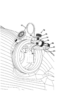
Figure 1 is an illustration of the optical device of the present invention
when attached
to a speculum for a procedure on the eye of a patient;
Figure 2 is a perspective view of the optical device and speculum;
Figure 3 is a perspective side view of the optical device and speculum;
Figure 4 is a side view thereof;
Figure 5 is a further perspective view thereof; and
Figure 6 is a top plan view thereof.
DETAILED DESCRIPTION OF THE INVENTION
Referring to the drawings in greater detail and by reference characters
thereto, there
is illustrated an optical device which is generally designated by reference
numeral 10.
Optical device 10 is connected to a speculum generally designated by reference
numeral 12
as will be set forth in greater detail hereinbelow.
- 3 -
CA 3007718 2018-06-11
Speculum 12 is formed of a wire-like member 14 with a pair of retaining
members 16
designed to engage with the eyelids of the patient to retain the eyelids in a
separated position.
Optical device 10 includes a bottom ring 18 and a top ring 20. In the
arrangement
shown, the bottom ring 18 is fixed while top ring 20 can rotate through 360
degrees with
respect to bottom ring 18. The top ring 20 may have spring loaded notches in
order to
maintain top ring 20 in the desired position during surgery.
Optical device 10 also includes a housing 22 having a rack gear 24 extending
therethrough. Rack gear 24 engages with a pinion gear 25 and is adjustable by
means of first
and second knobs 26 and 28. An upright frame member 30 is mounted on top ring
20. An
adjusting member 32 having a screwthread shaft 34 extends therethrough.
Screwthread shaft
34 is adjusted by means of a knob 36 while an adjusting end 38 rests against
housing 22.
Extending downwardly from rack gear 24 there is a rod 40. At the end of rod 40
there is a
depressing member 42 which is designed to contact the eye to generate a
concave shape on
the side of the eye to expose the vitreous and allow a complete visualization
of the anterior
vitreous. At the same time, a light source such as a fibre optic can be
incorporated with the
device to illuminate the eye.
The optical device has a pair of locking members 44 which are designed to
engage
with wire-like member 14 of speculum 12. A locking screw 46 engages with wire-
like
member 14.
It will be understood that the above described embodiment is for purposes of
illustration only and that changes and modifications may be made thereto
without departing
from the spirit and scope of the invention.
- 4 -
CA 3007718 2018-06-11
