Note: Descriptions are shown in the official language in which they were submitted.
CA 03008213 2018-06-12
1
DESCRIPTION
METHOD FOR MANUFACTURING TISSUE/ORGAN BY USING BLOOD CELLS
TECHNICAL FIELD
The present invention relates to a method for preparing tissues and organs
using
blood cells.
BACKGROUND ART
Recently, methods of generating human functional cells useful for drug
discovery
screening and regenerative medicine by directed differentiation using
pluripotent stem cells,
such as iPS cells capable of differentiating into various functional cells,
have been attracting
attention. To date, the present inventors have established a method of
reconstructing human
tissues/organs which have well-ordered, three-dimensional structures composed
of vascular
endothelial cells and mesenchymal cells seen in adult tissues (Non-Patent
Document No. 1:
Takebe T. et al., Nature (2013); Patent Document No. 1: Method for Preparing
Tissue and
Organ W02013/047639 Al).
On the other hand, as a finding concerning the maturing of hematopoietic cells
and
hepatocytes, reports have been made that a cytokine produced by hematopoietic
cells in the
developmental process of mouse promotes the maturing of hepatocytes (Non-
Patent
Document No. 2: Kamiya A., et al., EMBO J. 1999 18:2127-3; Non-Patent Document
No. 3:
Kinoshita T., et al., Proc Natl Acad Sci U S A. 1999 96:7265-70 (1999)).
Although reports
have been made that human hepatocytes and human hematopoietic cells are
simultaneously
engrafted by transplanting a mixture of hematopoietic cells and immature
hepatocytes in an
immunodeficient animal, no findings have been reported to date regarding the
maturity of
hepatocytes resulting from simultaneous transplantation with hematopoietic
cells (Non-Patent
Document No. 4: Bility MT., et al., Nat Protoc. 7:1608-17 (2012); Non-Patent
Document No.
5: Washburn ML., et al., Gastroenterology. 140:1334-44 (2011)). Further, no
findings have
been reported yet that show the importance of hematopoietic cells in the
maturing of a
functional cell of interest by introducing them into an in vitro reconstructed
three-dimensional tissue.
CA 03008213 2018-06-12
2
PRIOR ART LITERATURE
Non-Patent Documents
Non-Patent Document No. 1: Takebe T. et al., Nature (2013)
Non-Patent Document No. 2: Kamiya A., etal., EMBO J. 1999 18:2127-3
Non-Patent Document No. 3: Kinoshita T., et al., Proc Natl Acad Sci U S A.
1999 96:7265-70
(1999)
Non-Patent Document No. 4: Bility MT., et al., Nat Protoc. 7:1608-17 (2012)
Non-Patent Document No. 5: Washburn ML., et al., Gastroenterology. 140:1334-44
(2011)
Patent Document
Patent Document No. 1: W02013/047639 A 1
DISCLOSURE OF THE INVENTION
PROBLEM FOR SOLUTION BY THE INVENTION
When medical application of human tissues/organs prepared by the conventional
method is attempted, preparation cost is extremely high; approximately 10
million Yen per
adult is needed. This has
been a barrier against the spreading of the technology. In view
of the current situation that there are at least 20 thousands or more absolute
adaptation
patients (international estimate of adaptation-exclusion cases/fatal cases in
the hepatic failure
patients waiting), development of a revised technique which greatly improves
function per
cell and realizes huge cost reduction is eagerly desired in order to widely
adapt such a
therapeutic approach to patients as a general purpose technology.
It is an object of the present invention to provide such a revised technique.
MEANS TO SOLVE THE PROBLEM
The present inventors have succeeded in further improving the function of a
resultant
organ bud by adding hematopoietic cells to a culture system co-culturing
hepatic endoderm
cells, vascular endothelial cells and mesenchymal cells. To date, no technique
has existed
that shows the effect of hematopoietic cells in three-dimensional
reconstruction of human
tissues/organs. Therefore, it is believed that the method of the present
invention is
extremely high in novelty and inventiveness.
The gist of the present invention is as described below.
(1) A method of preparing an organ bud, comprising culturing vascular
endothelial cells,
CA 03008213 2018-06-12
3
mesenchymal cells and a tissue or organ cell in vitro in the presence of blood
cells.
(2) The method of (1) above, wherein the organ bud is a structure capable
of
differentiating into an organ through maturing.
(3) The method of (1) or (2) above, wherein an organ bud with improved
functions is
prepared compared with an organ bud prepared by culturing vascular endothelial
cells,
mesenchymal cells and a tissue or organ cell in vitro in the absence of blood
cells.
(4) The method of any one of (1) to (3) above, wherein cells are cultured
without using
scaffold materials.
(5) The method of any one of (1) to (4) above, wherein the blood cell
comprises an
undifferentiated hematopoietic cell.
(6) The method of (5), wherein the undifferentiated hematopoietic cell is a
hematopoietic
progenitor cell and/or a hematopoietic stem cell.
(7) The method of any one of (1) to (6) above, wherein the blood cell is
derived from cord
blood.
(8) The method of (7) above, wherein the blood cell is a cell of the
monocyte fraction of
cord blood.
(9) The method of any one of (1) to (8) above, wherein the tissue or organ
cell is an
undifferentiated cell induced from a pluripotent stem cell.
(10) The method of (9) above, wherein the pluripotent stem cell is an induced
pluripotent
stem cell.
(11) The method of (9) or (10) above, wherein the pluripotent stem cell is
derived from
human.
(12) The method of any one of (1) to (11) above, wherein the organ bud is a
liver bud.
(13) The method of (12) above, wherein a liver bud with an improved albumin
secretory
capacity is prepared compared with a liver bud prepared by culturing vascular
endothelial
cells, mesenchymal cells and a tissue or organ cell in vitro in the absence of
blood cells.
(14) The method of (12) or (13) above, wherein a liver bud with increased
expression of
hepatocyte differentiation marker genes is prepared compared with a liver bud
prepared by
culturing vascular endothelial cells, mesenchymal cells and a tissue or organ
cell in vitro in
the absence of blood cells.
(15) The method of (14) above, wherein the hepatocyte differentiation marker
gene is at
least one marker selected from the group consisting of a fetoprotein, albumin,
CYP3A7,
CA 03008213 2018-06-12
4
tryptophan metabolic enzyme TD02 and sodium-taurocholate cotransporter.
(16) An organ bud prepared by the method of any one of (1) to (15) above.
(17) A method of preparing a tissue or an organ, comprising transplanting the
organ bud of
(16) above into a non-human animal and differentiating the organ bud into a
tissue or an
organ.
(18) A method of transplanting an organ bud, comprising transplanting the
organ bud of
(16) above into a human or a non-human animal.
(19) A method of regeneration or function recovery of a tissue or an organ,
comprising
transplanting the organ bud of (16) above into a human or a non-human animal
and
differentiating the organ bud into a tissue or an organ.
(20) A method of preparing a non-human chimeric animal, comprising
transplanting the
organ bud of (16) above into a non-human animal and differentiating the organ
bud into a
tissue or an organ.
(21) A method of evaluating a drug, comprising using at least one selected
from the group
consisting of the organ bud of (16) above, the tissue or organ prepared by the
method of (17)
above, and the non-human chimeric animal prepared by the method of (20) above.
As a finding concerning the maturing of hematopoietic cells and hepatocytes,
reports
have been made that a cytokine (OSM) produced by hematopoietic cells in the
developmental
process of mouse promotes the maturing of hepatocytes (Non-Patent Document No.
2:
Kamiya A., et al., EMBO J. 1999 18:2127-3; Non-Patent Document No. 3:
Kinoshita T., et al.,
Proc Natl Acad Sci U S A. 1999 96:7265-70 (1999)). However, in the
conventional
two-dimensional culture, directed differentiation was utterly insufficient
though OSM had
been already added. On the other hand, the patent of the present inventors
(Patent
Document No. 1: Method for Preparing Tissue and Organ W02013/047639 A 1 ),
although it
discloses preparation of organ buds from three types of cells, does not
disclose or even
suggest a methodology that enables organ buds to be formed from four or more
types of cells
including hematopoietic cells and which also promotes the maturing thereof.
Therefore, the
introduction of the fourth cell (hematopoietic cells) into in vitro
reconstructed organ buds and
the finding of its importance in the maturing of a functional cell of interest
have great novelty
and inventiveness.
EFFECT OF THE INVENTION
CA 03008213 2018-06-12
To date, the present inventors have succeeded in constructing a platform
technology
for preparing a human tissue/organ in which a vascular system is appropriately
located; such
a human tissue/organ has never been achieved by conventional techniques
(Method for
Preparing Tissue and Organ). In order to substantially advance the above-
described
technology, the present invention has achieved a technique to greatly promote
the maturing of
a functional cell of interest through direct or indirect cellular action by
adding a new player
(i.e., hematopoietic cells). This is a completely novel technique that
achieves maturing of
metabolic and other in vitro functions that have heretofore been inadequate.
Specifically,
according to the technique of the present invention, hepatocytes were
recognized to improve
in function by a factor of about 3 to 4 (taking albumin secretion as an
indicator).
Accordingly, it is expected that an equivalent function will be exhibited by
cells whose
number is three to four times less than the conventionally needed. As a
result, the
production cost can be reduced to between one third and a fourth. In terms of
the current
treatment cost per adult terminal hepatic failure patient, it is expected that
a reduction of
approx. 6 million Yen will be achieved.
Further, as a result of the functional improvement according to the present
invention,
it is expected that drug metabolism and other functions that have heretofore
been difficult to
evaluate can be detected, which also enables application to in vitro
screenings in drug
discovery.
The present specification encompasses the contents disclosed in the
specification
and/or the drawings of Japanese Patent Application No. 2015-251140 based on
which the
present patent application claims priority.
BRIEF DESCRIPTION OF THE DRAWINGS
[Fig. 11 Isolation of a cell fraction comprising hematopoietic cells from cord
blood
using a cell sorter.
[Fig. 2] Preparation
of liver buds comprising blood cells (left panel); and
measurement by ELISA of albumin secretion into medium from liver buds
comprising blood
cells (right panel).
[Fig. 3] Examination of genetic expression of hepatic differentiation markers
by
quantitative PCR in the preparation of liver buds comprising blood cells.
[Fig. 4] Examination of the amount of blood cells added in the preparation of
liver
CA 03008213 2018-06-12
6
buds comprising blood cells.
[Fig. 51 Albumin secretion dependent on the amount of blood cells added.
BEST MODES FOR CARRYING OUT THE INVENTION
Hereinbelow, the present invention will be described in detail.
The present invention provides a method of preparing an organ bud, comprising
culturing vascular endothelial cells, mesenchymal cells and a tissue or organ
cell in vitro in
the presence of blood cells.
As used herein, the term "organ bud" means a structure capable of
differentiating
into an organ through maturing, the structure comprising four types of cells,
i.e., tissue or
organ cells, vascular endothelial cells, mesenchymal cells (undifferentiated
mesenchymal
cells or cells differentiated therefrom) and blood cells. Whether a structure
is an organ bud
or not can be determined, for example, by transplanting the structure into an
organism and
examining whether or not it is capable of differentiating into an organ of
interest (the
structure can be judged as organ bud if it has differentiated into the organ
of interest); and/or
by examining whether or not the structure comprises all of the above-listed
four types of cells
(the structure can be judged as organ bud if it comprises all of the four
types of cells). The
organ bud may be one which differentiates into an organ such as kidney, heart,
lung, spleen,
esophagus, stomach, thyroid, parathyroid, thymus, gonad, brain, spinal cord or
the like.
Preferably, the organ bud is one which differentiates into an endodermal organ
such as one
which differentiates into liver (liver bud), one which differentiates into
pancreas (pancreas
bud), or one which differentiates into intestinal tract. Whether a certain
structure is an organ
bud which differentiates into an endodermal organ or not can be determined by
examining the
expression of marker proteins (if any one or more of the marker proteins
described later are
expressed, the organ bud can be judged as the organ bud of interest). For
example, HHEX,
SOX2, HNF4A, AFP, ALB and the like are markers for liver buds; PDX1, S0X17,
SOX9 and
the like are markers for pancreas bud; and CDX2, SOX9 and the like are markers
for organ
buds which differentiate into intestinal tract. Among the terms used by those
skilled in the
art, the following are included in the organ bud of the present invention:
liver bud, liver
diverticula, liver organoid, pancreatic (dorsal or ventral) buds, pancreatic
diverticula,
pancreatic organoid, intestinal bud, intestinal diverticula, intestinal
organoid (K. Matsumoto,
et al. Science.19; 294 (5542): 559-63 (2001)) and so on.
CA 03008213 2018-06-12
7
As used herein, the term "blood cell" means a cell isolated from a living
body, a cell
obtained from stem cells such as ES cells or iPS cells by directed
differentiation, or a cell
directly reprogrammed by gene transfer into a differentiated cell, on the
condition that the
following characteristics of blood cells are displayed (e.g., expressing any
one of CD34,
CD2, CD3, CD4, CD7, CD8, CD10, CD14, CD16, CD19, CD20, CD24, CD41, CD45, CD56,
CD66b or CD235a, CD38, CD90, CD49f, VEGFR2, CD43, CD71, GPA (Glycophorin A),
CD42b, c-kit, CD150, Sca-1, Ter119 or the like). Whether a cell is a blood
cell or not may
be determined by checking for the expression of CD34, CD2, CD3, CD4, CD7, CD8,
CD10,
CD14, CD16, CD19, CD20, CD24, CD41, CD45, CD56, CD66b or CD235a, CD38, CD90,
CD49f, VEGFR2, CD43, CD71, GPA (Glycophorin A), CD42b, c-kit, CD150, Sca-1,
Ter119
or the like. The blood cell used in the present invention may be either
differentiated or
undifferentiated. Preferably, the blood cell comprises undifferentiated
hematopoietic cells
such as hematopoietic progenitor cells or hematopoietic stem cells. As
undifferentiated
hematopoietic cells, blood cells derived from pluripotent stem cells such as
induced
pluripotent stem cells (iPS cells), embryonic stem cells (ES cells), etc.;
blood cells collected
from blood (cord blood, bone marrow blood, peripheral blood, etc.); or blood
cells directly
reprogrammed from other differentiated cells may be enumerated, for example.
In the
Example described later, it is believed that hematopoietic progenitor cells
and hematopoietic
stem cells could be enriched by collecting the monocyte fractions of cord
blood. Among the
terms used by those skilled in the art, the following are included in the
blood cell in the
present invention: hematopoietic stem cell, blood stem cell, hematopoietic
progenitor cell,
blood progenitor cell, myeloid progenitor cell, granulocyte/monocyte
progenitor cell,
granulocyte precursor (progenitor?) cell, granulocyte, myeloblast,
promyelocyte, myeloid cell,
metamyelocyte, stab cell, segmented cell (neutrophil), monocyte progenitor,
monocyte,
macrophage, histiocyte, Kupffer cell, alveolar macrophage, microglia,
osteoclast, epithelioid
cell, giant cell (Langhans giant cell, foreign body giant cell, Touton giant
cell), dendritic cell,
Langerhans cell, myelomonocyte, myeloblast, basophilic promyelocyte,
basophilic myeloid
cell, basophilic metamyelocyte, basophil, myeloblast, eosinophilic
promyelocyte,
eosinophilic myeloid cell, eosinophilic metamyelocyte, eosinophilic stab cell,
eosinophilic
segmented cell (eosinophil), megakaryoblast, megakaryocyte, erythroblast,
reticulocyte, mast
cell precursor, and so on. As blood cells, human-derived blood cells are
mainly used.
However, blood cells derived from non-human animals (e.g., animals used, for
example, as
CA 03008213 2018-06-12
8
experimental animals, pet animals, working animals, race horses or fighting
dogs, more
specifically, mouse, rat, rabbit, pig, dog, monkey, cattle, horse, sheep,
chicken, shark,
devilfish, ratfish, salmon, shrimp, crab or the like) may also be used.
As used herein, the term "vascular endothelial cell" means cells constituting
vascular
endothelium or cells capable of differentiating into such cells. Whether a
certain cell is
vascular endothelial cell or not can be determined by checking for the
expression of marker
proteins such as TIE2, VEGFR-1, VEGFR-2, VEGFR-3 and CD41 (if any one or more
of the
above-listed marker proteins are expressed, the cell can be judged as vascular
endothelial
cell). The vascular endothelial cell used in the present invention may be
either differentiated
or undifferentiated. Whether a vascular endothelial cell is a differentiated
cell or not can be
determined by means of CD31 and CD144. Among the terms used by those skilled
in the
art, the following are included in the "vascular endothelial cell" of the
present invention:
endothelial cells, umbilical vein endothelial cells, endothelial progenitor
cells, endothelial
precursor cells, vasculogenic progenitors, hemangioblast (HJ. Joo, et al.
Blood.
25;118(8):2094-104 (2011)) and so on. Preferable vascular endothelial cells
are those
derived from umbilical vein. Vascular endothelial cells may be collected from
blood vessels,
or may be prepared from pluripotent stem cells such as induced pluripotent
stem cells (iPS
cells) or embryonic stem cells (ES cells) according to known methods. As
vascular
endothelial cells, human-derived cells are mainly used. However, vascular
endothelial cells
derived from non-human animals (e.g., animals used, for example, as
experimental animals,
pet animals, working animals, race horses or fighting dogs, more specifically,
mouse, rat,
rabbit, pig, dog, monkey, cattle, horse, sheep, chicken, shark, devilfish,
raffish, salmon,
shrimp, crab or the like) may also be used.
As used herein, the term "mesenchymal cell" means connective tissue cells that
are
mainly located in mesoderm-derived connective tissues and which form support
structures for
cells that function in tissues. The "mesenchymal cell" is a concept that
encompasses those
cells which are destined to, but are yet to, differentiate into mesenchymal
cells.
Mesenchymal cells used in the present invention may be either differentiated
or
undifferentiated. Whether a certain cell is an undifferentiated mesenchymal
cell or not may
be determined by checking for the expression of marker proteins such as Stro-
1, CD29, CD44,
CD73, CD90, CD105, CD133, CD271 or Nestin (if any one or more of the above-
listed
marker proteins are expressed, the cell can be judged as undifferentiated
mesenchymal cell).
CA 03008213 2018-06-12
9
A mesenchymal cell in which none of the above-listed markers are expressed can
be judged
as a differentiated mesenchymal cell. Among the terms used by those skilled in
the art, the
following are included in the "mesenchymal cell" of the present invention:
mesenchymal
stem cells, mesenchymal progenitor cells, mesenchymal cells (R. Peters, et al.
PLoS One. 30;
5(12):e15689 (2010)) and so on. Preferable mesenchymal cells are mesenchymal
cells
derived from bone marrow (especially, mesenchymal stem cells). Mesenchymal
cells may
be collected from tissues such as bone marrow, adipose tissue, placental
tissue, umbilical
tissue, dental pulp or the like, or may be prepared from pluripotent stem
cells such as induced
pluripotent stem cells (iPS cells) or embryonic stem cells (ES cells)
according to known
methods. As mesenchymal cells, human-derived cells are mainly used. However,
mesenchymal cells derived from non-human animals (e.g., animals used, for
example, as
experimental animals, pet animals, working animals, race horses or fighting
dogs, more
specifically, mouse, rat, rabbit, pig, dog, monkey, cattle, horse, sheep,
chicken, shark,
devilfish, raffish, salmon, shrimp, crab or the like) may also be used.
As used herein, the term "tissue or organ cell" means functional cells
constituting
tissues or organs, or undifferentiated cells which differentiate into
functional cells.
Examples of "undifferentiated tissue or organ cell" include, but are not
limited to, cells
capable of differentiating into an organ such as kidney, heart, lung, spleen,
esophagus,
stomach, thyroid, parathyroid, thymus, gonad, brain or spinal cord; cells
capable of
differentiating into an ectodermal organ such as brain, spinal cord, adrenal
medulla,
epidermis, hair/nail/dermal gland, sensory organ, peripheral nerve or lens;
cells capable of
differentiating into a mesodermal organ such as kidney, urinary duct, heart,
blood, gonad,
adrenal cortex, muscle, skeleton, dermis, connective tissue or mesothelium;
and cells
capable of differentiating into an endodermal organ such as liver, pancreas,
intestinal tract,
lung, thyroid, parathyroid or urinary tract. Whether or not a certain cell is
capable of
differentiating into an ectodermal organ, mesodermal organ or endodermal organ
can be
determined by checking for the expression of marker proteins (if any one or
more of marker
proteins are expressed, the cell can be judged as a cell capable of
differentiating into an
endodermal organ). For example, in cells capable of differentiating into
liver, HHEX,
SOX2, HNF4A, AFP, ALB and the like are markers; in cells capable of
differentiating into
pancreas, PDX1, SOX17, SOX9 and the like are markers; in cells capable of
differentiating
into intestinal tract, CDX2, SOX9 and the like are markers; in cells capable
of differentiating
CA 03008213 2018-06-12
into kidney, SIX2 and SALL] are markers; in cells capable of differentiating
into heart,
NKX2-5, MYH6, ACTN2, MYL7 and HPPA are markers; in cells capable of
differentiating
into blood, C-KIT, SCA1, TER119 and HOXB4 are markers; and in cells capable of
differentiating into brain or spinal cord, HNK1, AP2, NESTIN and the like are
markers.
Among the terms used by those skilled in the art, the following are included
in the
"undifferentiated tissue or organ cell" of the present invention: hepatoblast,
hepatic
progenitor cells, hepatic precursor cells, pancreatoblast, pancreatic
progenitors, pancreatic
progenitor cells, pancreatic precursor cells, endocrine precursors, intestinal
progenitor cells,
intestinal precursor cells, intermediate mesoderm, metanephric mesenchymal
precursor cells,
multipotent nephron progenitor, renal progenitor cells, cardiac mesoderm,
cardiovascular
progenitor cells, cardiac progenitor cells (JR. Spence, et al. Nature.;
470(7332):105-9.(2011);
Self, et al. EMBO J.; 25(21): 5214-5228.(2006); J. Zhang, et al. Circulation
Research.; 104:
e30-e41(2009); G. Lee, et al. Nature Biotechnology 25, 1468-1475 (2007)) and
so on.
Undifferentiated tissue or organ cells may be collected from tissues or
organs, or may be
prepared from pluripotent stem cells such as induced pluripotent stem cells
(iPS cells) or
embryonic stem cells (ES cells) according to known methods. Moreover,
undifferentiated
tissue or organ cells may be such cells as primitive gut endoderm cells
(PGECs) (Japanese
Patent No. 5777127) which are at an intermediate stage of differentiation from
pluripotent
stem cells (e.g., iPS cells) into tissues or organs. PGECs are capable of
differentiating into
hepatocytes, pancreatic cells and enterocytes (have high differentiation
function), do not
express markers associated with the malignancy of cancer (are highly safe),
and may be
prepared from iPS cells by directed differentiation without using feeder
cells. Therefore,
PGECs have the advantage of even allowing for clinical application.
Furthermore, mass
preparation of PGECs is possible to. PGECs may be prepared according to the
method
disclosed in Japanese Patent No. 5777127. Alternatively, pluripotent stem
cells such as iPS
cells may be cultured in activin-supplemented serum-free medium so that they
are induced to
endodermal cells that are positive to both CXCR4 and E-cadherin; or the
endodermal cells
thus obtained may be cultured for two days in the presence of added BMP4 and
FGF2 to
obtain CXCR4-negative, HNF4a-positive hepatic endodermal cell populations. To
give
further examples, organ cells capable of differentiating into liver may be
prepared as
previously described (K.Si-Taiyeb, et al. Hepatology, 51(1): 297- 305(2010);
T. Touboul, et
al. Hepatology. 51 (5):1754-65 (2010)); organ cells capable of differentiating
into pancreas
CA 03008213 2018-06-12
11
may be prepared as previously described (D. Zhang, et al. Cell Res.;19(4):429-
38 (2009));
organ cells capable of differentiating into intestinal tract may be prepared
as previously
described (J. Cai, et al. J Mol Cell Biol.; 2(1):50-60 (2010); R. Spence, et
al. Nature.; 470
(7332):105-9 (2011)); cells capable of differentiating into heart may be
prepared as
previously described (J. Zhang, et al. Circulation Research.; 104: e30-
e41(2009); and cells
capable of differentiating into brain or spinal cord may be prepared as
previously described
(G. Lee, et al. Nature Biotechnology 25, 1468 - 1475 (2007)). Examples of
"differentiated
tissue or organ cell" include, but are not limited to, endocrine cells of
pancreas, pancreatic
duct epithelial cells of pancreas, hepatocytes of liver, epithelial cells of
intestinal tract,
tubular epithelial cells of kidney, podocytes of kidney, cardiomyocytes of
heart, lymphocytes
and granulocytes of blood, erythrocytes, neurons and glial cells of brain, and
neurons and
Schwann cells of spinal cord. As tissue or organ cells, human-derived cells
are mainly used.
However, tissue or organ cells derived from non-human animals (e.g., animals
used, for
example, as experimental animals, pet animals, working animals, race horses or
fighting dogs,
more specifically, mouse, rat, rabbit, pig, dog, monkey, cattle, horse, sheep,
chicken, shark,
devilfish, raffish, salmon, shrimp, crab or the like) may also be used.
Culture ratios of four cell types in coculture are not particularly limited as
long as
they are within the range that enables the formation of organ buds. Preferable
cell count
ratio is 10 : 10-5 : 2-0.1 : 2-50 for tissue or organ cell : vascular
endothelial cell :
mesenchymal cell : blood cell. Organ buds of approx. 50 to 250 [tm in size may
be formed by
coculturing approx. 250,000 tissue or organ cells, approx. 170,000 vascular
endothelial cells,
approx. 25,000 mesenchymal cells and approx. 100,000 blood cells.
The medium used for culture may be any medium that enables the formation of
organ
buds and examples that are preferably used include a medium for culturing
vascular
endothelial cells, a medium for culturing tissue or organ cells, and a mixture
of these two
media. As a medium for culturing vascular endothelial cells, any medium may be
used but,
preferably, a medium containing at least one of the following substances may
be used: hEGF
(recombinant human epidermal growth factor), VEGF (vascular endothelial growth
factor),
hydrocortisone, bFGF, ascorbic acid, IGF1, FBS, antibiotics (e.g., gentamycin
or
amphotericin B), heparin, L-glutamine, phenol red and BBE. Specific examples
of media
that may be used for culturing vascular endothelial cells include, but are not
limited to,
EGM-2 BulletKit (Lonza), EGM BulletKit (Lonza), VascuLife EnGS Comp Kit (LCT),
CA 03008213 2018-06-12
12
Human Endothelial-SFM Basal Growth Medium (Invitrogen) and human microvascular
endothelial cell growth medium (TOYOB0). As a medium for culturing tissue or
organ
cells, any medium may be used but in the case where the organ cell is a
hepatocyte, a medium
containing at least one of ascorbic acid, BSA-FAF, insulin, hydrocortisone and
GA-1000 may
preferably be used. As a medium for culturing hepatocytes, HCM BulletKit
(Lonza) from
which hEGF (recombinant human epidermal growth factor) has been removed or
RPMI1640
(Sigma-Aldrich) to which 1% B27 Supplements (GIBCO) and 10 ng/mL hHGF
(Sigma-Aldrich) have been added may typically be used. As regards the
formation of
human liver buds, a 1:1 mixture of GM BulletKit (Lonza) and HCM BulletKit
(Lonza) from
each of which hEGF has been removed and which are each supplemented with
dexamethasone, oncostatin M and HGF has been found effective for maturation of
liver buds.
Although scaffold materials need not be used for culturing cells, a mixture of
four
types of cells may advantageously be cultured on a gel-like support that
allows mesenchymal
cells to contract.
Contraction of mesenchymal cells may be confirmed, for example, by noting the
formation of a 3D tissue morphologically (either under microscope or with the
naked eye) or
by showing that the tissue is strong enough to retain its shape as it is
collected with a spatula
or the like (Takebe et al. Nature 499 (7459), 481-484, 2013).
The support may advantageously be a gel-like substrate having an appropriate
stiffness [e.g., a Young's modulus of 200 kPa of less (in the case of a
Matrigel-coated gel of a
flat shape); however, the appropriate stiffness of the support may vary
depending on the
coating and shape]. Examples of such substrates include, but are not limited
to, hydrogels
(such as acrylamide gel, gelatin and Matrigel). The
stiffness of the support need not be
uniform and may vary with the shape, size and quantity of a cell condensate of
interest so that
it can be provided with a spatial/temporal gradient or can be patterned. In
the case where
the stiffness of the support is uniform, it is preferably 100 kPa or less,
more preferably 1-50
kPa. The gel-like support may be planar, or alternatively, the side on which
culture is to be
performed may have a U- or V-shaped cross section. If the side of the gel-like
support on
which culture is to be performed has a U- or V-shaped cross section, cells
tend to gather on
the culture surface and a cell condensate can advantageously be formed from a
smaller
number of cells and/or tissues. Moreover, the support may be modified
chemically or
physically. Examples of modifying substances include, but are not limited to,
Matrigel,
CA 03008213 2018-06-12
13
laminin, entactin, collagen, fibronectin and vitronectin.
One example of the gel-like culture support that is provided with a spatial
gradient of
stiffness is a gel-like culture support that is stiffer in the central part
than in the peripheral
part. The stiffness of the central part is appropriately 200 kPa or less and
it suffices that the
peripheral part is softer than the central part. Appropriate values for the
stiffness of the
central and peripheral parts of the substrate are variable with the coating
and the shape.
Another example of the gel-like culture support that is provided with a
spatial gradient of
stiffness is a gel-like culture support that is stiffer in the peripheral part
than in the central
part.
One example of the patterned, gel-like culture support is a gel-like culture
support
having one or more patterns in which the central part is stiffer than the
peripheral part. The
stiffness of the central part is appropriately, 200 kPa or less and it
suffices that the peripheral
part is softer than the central part. Appropriate values for the stiffness of
the central and
peripheral parts of the substrate are variable with the coating and the shape.
Another
example of the patterned, gel-like culture support is a gel-like culture
support having one or
more patterns in which the peripheral part is stiffer than the central part.
The stiffness of the
peripheral part is appropriately 200 kPa or less and it suffices that the
central part is softer
than the peripheral part. Appropriate values for the stiffness of the central
and peripheral
parts of the substrate are variable with the coating and the shape.
The temperature during culture is not particularly limited but it is
preferably
30-40 C and more preferably 37 C.
The culture period is not particularly limited but it is preferably 3-10 days
and more
preferably 6 days.
The organ bud prepared by the method of the present invention may be improved
in
function compared with an organ bud prepared by culturing vascular endothelial
cells,
mesenchymal cells and a tissue or organ cell in vitro in the absence of blood
cells.
Consider, for example, the case where the organ bud is a liver bud; the liver
bud
prepared by the method of the present invention may have an improved albumin
secretion
capacity compared with a liver bud prepared by culturing vascular endothelial
cells,
mesenchymal cells and a tissue or organ cell in vitro in the absence of blood
cells.
Moreover, the liver bud prepared by the method of the present invention may
have increased
expression of hepatocyte differentiation marker genes compared with a liver
bud prepared by
CA 03008213 2018-06-12
14
culturing vascular endothelial cells, mesenchymal cells and a tissue or organ
cell in vitro in
the absence of blood cells. Examples of hepatocyte differentiation marker
genes include,
but are not limited to, a fetoprotein, albumin, CYP3A7, tryptophan metabolic
enzyme TD02
and sodium-taurocholate cotransporter.
The present invention also provides an organ bud prepared by the above-
described
method.
The thus prepared organ bud may be transplanted into a non-human animal, in
which
it is allowed to mature, whereby a tissue or organ can be prepared. Briefly,
the present
invention also provides a method of preparing a tissue or an organ, comprising
transplanting
the organ bud prepared by the above-described method into a non-human animal
and
differentiating the organ bud into a tissue or an organ. Examples of the non-
human animal
that may be used include animals that are used, for example, as experimental
animals, pet
animals, working animals, race horses or fighting dogs, more specifically,
mouse, rat, rabbit,
pig, dog, monkey, cattle, horse, sheep, chicken, shark, devilfish, ratfish,
salmon, shrimp, crab
and the like. Moreover, the non-human animal to be used herein is preferably
an
immunodeficient animal in order avoid immunorejection.
Therefore, the present invention also provides a method of transplanting an
organ
bud, comprising transplanting the organ bud prepared by the above-described
method into a
human or a non-human animal. The site of transplantation of the organ bud may
be any site
as long as transplantation is possible. Specific examples of the
transplantation site include,
but are not limited to, the intracranial space, the mesentery, the liver, the
spleen, the kidney,
the kidney subcapsular space, and the supraportal space. For intracranial
transplantation,
about 1 to 3 organ buds of 5 mm in size, prepared in vitro, may be
transplanted. For
intramesenteric transplantation, about 1 to 6 organ buds of 5 mm in size,
prepared in vitro,
may be transplanted. For
transplantation into the supraportal space, about 1 to 20 organ
buds of 5 mm in size, prepared in vitro, may be transplanted. For
transplantation into the
kidney subcapsular space, about 1 to 5 organ buds of 5 mm in size, prepared in
vitro, may be
transplanted. For transplantation into the liver, spleen or kidney, about 100
to 200 organ
buds of 100 1.11ri in size, prepared in vitro, may be transplanted.
The thus prepared tissue or organ may be used in drug discovery screening or
regenerative medicine.
Therefore, the present invention provides a method of regeneration or function
CA 03008213 2018-06-12
recovery of a tissue or an organ, comprising transplanting the organ bud
prepared by the
above-described method into a human or a non-human animal and differentiating
the organ
bud into a tissue or an organ. As non-human animals, animals used for such
purposes as
experimental animal, pet animal, working animal, race horse or fighting dog,
more
specifically, mouse, rat, rabbit, pig, dog, monkey, cattle, horse, sheep,
chicken, shark,
devilfish, ratfish, salmon, shrimp, crab or the like may be used.
The organ bud prepared by the method of the present invention may be
formulated
and used in the form of a composition for regenerative medicine. This
composition of the
present invention may be transplanted into a living body to prepare a tissue
or an organ.
Regeneration or function recovery of a tissue or an organ is also possible by
transplanting the
composition of the present invention into a living body.
Upon transplantation of the composition of the present invention into a living
body,
the organ bud may differentiate into a tissue or an organ with vascular
networks. In such
vascular networks, blood perfusion may occur. It is believed that the
occurrence of blood
perfusion in vascular networks enables generation of a tissue or an organ with
a highly
ordered tissue structure equivalent or close to the tissue structure of adult
tissues.
The composition of the present invention may comprise a tissue vascularization
promoter such as FGF2, HGF or VEGF, a gelatin sponge for hemostasis to cope
with the
bleeding from transplantation (product name: Spongel; Astellas Pharma), and a
tissue
adhesive for fixing transplants such as Bolheal (Teijin Pharma), BeriplastTM
(CSL Behring) or
TachoCombTm (CSL Behring).
The present invention also provides a method of preparing a non-human chimeric
animal, comprising transplanting the organ bud prepared by the above-described
method into
a non-human animal and differentiating the organ bud into a tissue or an
organ. The
non-human animal (e.g., mouse) transplanted with the organ bud may mimic the
physiological function of the organismal species (e.g., human) from which the
tissue or organ
cell used for preparing the organ bud is derived.
Still further, the present invention provides a method of evaluating a drug,
comprising using at least one member selected from the group consisting of the
organ bud,
the tissue or organ and the non-human chimeric animal as prepared by the above-
described
methods, respectively. Specific examples of drug evaluation include, but are
not limited to,
evaluation of drug metabolism (e.g., prediction of drug metabolism profiles),
evaluation of
CA 03008213 2018-06-12
16
drug efficacy (e.g., screening for drugs that are effective as
pharmaceuticals), toxicity
evaluation, and evaluation of drug interactions.
Evaluation of drug metabolism may be performed as follows. Briefly, is at
least
one member
selected from the group consisting of the organ bud, the tissue or organ and
the non-human chimeric animal as prepared by the above-described methods,
respectively, is
administered with a candidate compound for pharmaceuticals and the resulting
biological
sample is then collected and analyzed, whereby a human-type drug metabolism
profile can be
obtained. As a result, prediction of the distribution/metabolism/excretion
processes of
pharmaceuticals in human (which was extremely difficult to achieve by
conventional
methods) becomes possible and it is expected that the development of safe and
efficacious
pharmaceuticals can be greatly accelerated.
Screening for drugs that are effective as pharmaceuticals may be performed as
follows. Briefly, at least one member selected from the group consisting of
the organ bud,
the tissue or organ and the non-human chimeric animal as prepared from a cell
established
from a diseased patient by the above-described methods, respectively, is
administered with a
novel candidate compound for pharmaceuticals. This enables subsequent
analysis. As a
result, a potential is expected for achieving great improvement in the
precision of drug
efficacy prediction for the case of actual administration to human, which has
been
unsatisfactory in conventional in vitro tests.
Evaluation of toxicity may be performed as follows. Briefly, at least one
member
selected from the group consisting of the organ bud, the tissue or organ and
the non-human
chimeric animal as prepared by the above-described methods, respectively, is
administered
with a test substance and, thereafter, histological damage markers or the like
are measured.
This makes it possible to improve the precision of damage prediction.
Evaluation of drug interactions may be performed as follows. Briefly, at least
one
member selected from the group consisting of the organ bud, the tissue or
organ and the
non-human chimeric animal as prepared by the above-described methods,
respectively, is
administered with a plurality of drugs; then, each drug is examined for its
pharmacokinetics
such as distribution/metabolism/excretion processes, evaluated for its
toxicity, and evaluated
for its efficacy.
Further, it is also possible to create tissue stem cells from the tissue or
organ
prepared by the method of the present invention. Thus, the present invention
is applicable
CA 03008213 2018-06-12
17
to cell engineering techniques for large scale creation of human tissue cells
and organ cells.
EXAMPLES
Hereinbelow, the present invention will be described in more detail with
reference to
the following Example.
[Example 1]
[Methods]
1. Preparation of Blood Cells
<Acquisition of Cord Blood>
Blood was collected from the umbilical cord (kindly supplied by a patient who
gave
birth to a child by Caesarean section in the Yokohama City University
Hospital) with a 50 ml
syringe and an 18G needle. Approx. 40 to 50 ml of blood was collected.
<Lysis Treatment>
Lysis buffer (40 ml) was placed in a 50 ml tube, to which 10 ml of the cord
blood
was added.
After stirring, the tube was left stationary for 10 min. at room temperature.
The tube was centrifuged at 200G for 10 min.
The resultant precipitate was re-suspended in 10 ml of lysis buffer. The
contents of
five tubes were collected together to make a 50 ml suspension.
The above suspension was re-centrifuged at 200G for 10 min.
The precipitate was suspended in 10 ml of DMEM+10% FBS and centrifuged at
200G for 5 min.
The precipitate was re-suspended in 10 ml of DMEM+10% FBS, followed by cell
counting.
Materials
1) Preparation of 10 x Lysis Buffer (x10 liquid; 1/10 dilution was used.)
= NH4C182.6 g
= NaHCO3 11.9g
= EDTA2Na 0.378 g
/1L milliQ adjusted to pH 7.3
2) Preparation of 1 x Lysis Buffer
Dilution (xl) with PBS (preferably cooled to 4 C; 2 mM EDTA may be added) was
CA 03008213 2018-06-12
18
used.
<Isolation of PI/CD235- Cells>
Dead cells were stained with propidium iodide (PI); erythrocytes/erythroblasts
were
stained with fluorescence-labeled CD235 antibody; cells not stained with
either PI or the
antibody (viable cells and neither erythrocytes nor erythroblasts) were
isolated with a cell
sorter.
<Isolation of Monocyte Fraction (MNC) Cells>
Using a cell sorter, PI/CD235- cells were developed with FSC (forward scatter)
and
SSC (side scatter) to isolate cell populations classified as monocyte fraction
(MNC) cells.
2. Preparation of Hepatic Endoderm Cells
Method:
Undifferentiated iPS cells (TkDA3 supplied by the University of Tokyo and an
iPS
cell line established from umbilical cord by the present inventors) were
exfoliated to prepare
single cells. The cells were cultured in plastic dishes at a density of 5x10⁴
cells/cm2 in the
presence of RPM1+1% B27+10 uM Rockinhibitor+50 ng/mL Wnt3a+100 ng/mL Activin A
for one day. Subsequently, the resultant cells were cultured in the presence
of RPMI+1%
B27+50 ng/mL Wnt3a+100 ng/mL Activin A for six days to thereby obtain hepatic
endoderm
cells.
[Results]
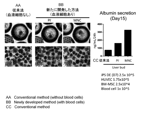
1. (Figs. 1, 2 and 3)
In a 10:7:1 mixture of hepatic endoderm cells (2.5x10⁵ cells), umbilical cord-
derived
vascular endothelial cells (1.7x10⁵ cells) (Lonza, Basel, Switzerland) and
mesenchymal cells
(2.5x10⁴ cells) (Lonza, Basel, Switzerland), a cell sorter isolated PE cells
and monocyte
fraction (MNC) cells (Fig. 1) enriched in hematopoietic stem
cells/hematopoietic progenitor
cells were suspended (1x10⁵ cells for each), followed by preparation of liver
buds (50-250
um in size) on Kuraray microwell plates. After 15 days culture, protein
amounts of human
albumin secreted in 24 hr into the culture supernatants of liver buds cultured
under respective
conditions were analyzed by ELISA. As a result, the groups to which Pl- cells
or
hematopoietic stem cells/hematopoietic progenitor cells-enriched monocyte
fraction (MNC)
cells were added showed a greater amount of albumin secretion than the group
to which no
blood cells were added (conventional method) (Fig. 2). Subsequently, the liver
buds at day
15 of culture under the above-described conditions were collected, followed by
checking for
CA 03008213 2018-06-12
19
the expression of hepatocyte differentiation markers (a fetoprotein (AFP),
albumin (ALB),
CYP3A7, tryptophan metabolic enzyme (TD02) and sodium-taurocholate
cotransporter
(NTCP)) by qPCR. As a result, were found to have increased in the groups to
which PI-
cells or monocyte fraction (MNC) cells were added showed higher gene
expression levels
than the groups to which no blood cells were added (conventional method) (Fig.
3).
2. (Figs. 4 and 5)
In order to examine the dose dependency of blood cells, experimental groups
were
prepared as follows. To a 10:7:1 cell mixture of hepatic endoderm cells
(2.5x10⁵ cells),
umbilical cord-derived vascular endothelial cells (1.7x10⁵ cells) and
mesenchymal cells
(2.5x10⁴ cells), no blood cells were added or blood cells (PI- cells) were
added at varying
densities of 1x10⁵ cells, 2.5x10⁵ cells or 5x10⁵ cells. At day 15 of
liver bud preparation,
the amount of human albumin secretion in 24 hr in culture broth (Fig. 4) was
examined.
The results revealed that the protein amount of human albumin secretion was
enhanced in a
manner dependent on blood cell count, as compared with the conventional method
(Fig. 5).
All publications, patents and patent applications cited herein are
incorporated herein
by reference in their entirety.
INDUSTRIAL APPLICABILITY
The present invention will provide an important platform technology directed
to
industrial production of human functional cells. The invention is applicable
to preparation
of human tissues/organs for transplantation as a technology of regeneration
medicine
targeting refractory diseases. Not only a great cost reduction is expected
compared with
conventional methods, but also improvement in functional maturation in vitro
might be
possible with the technique of the present invention. Therefore, the present
invention is
expected to become applicable to even acute/subacute hepatic failure which has
been difficult
to treat by the conventional technology.
Further, a drug evaluation system established using a human hepatic tissue
artificially prepared according to the present invention would enable large
scale production of
human mature hepatocytes needed in drug discovery/development.