Note: Descriptions are shown in the official language in which they were submitted.
SYSTEMS AND METHODS FOR CONFIRMING ACTIVATION OF BIOLOGICAL
INDICATORS
FIELD
[0001] The subject matter disclosed herein relates to self-contained
biological
sterilization indicators.
BACKGROUND
[0002] Medical devices are typically sterilized before use in order to
minimize the
likelihood that a contaminated device might be used on a subject, which could
cause an
infection in the subject. Various sterilization techniques may be employed,
such as steam,
hydrogen peroxide, and vapor phase sterilization, either with or without a gas
plasma and
ethylene oxide (Et0). Each of these methods depends to a certain extent on the
diffusion
rates of the sterilization fluids, typically gases, upon the medical devices
to be sterilized.
[0003] Before sterilization, medical devices are typically packaged
within containers or
pouches having a semi-permeable barrier that allows transmission of the
sterilizing fluid¨
sometimes referred to as a sterilant¨but prevents admission of contaminating
organisms,
particularly post-sterilization and until the package is opened by medical
personnel. For the
sterilization cycle to be efficacious, the contaminating organisms within the
package must be
killed because any organisms that survive the sterilization cycle could
multiply and re-
contaminate the medical device.
[0004] Although the packaging helps prevent contamination of a sterile
medical device,
the packaging may increase the difficulty of achieving a successful
sterilization cycle
because the packaging impedes the sterilant from reaching the device or
instrument contained
therein. This is particularly problematic for devices and instruments that
have diffusion-
- 1 -
CA 3008926 2018-06-20
restricted spaces therein because these diffusion-restricted spaces reduce the
likelihood that a
sterilization cycle may be effective. For example, endoscopes typically have
long narrow
lumens into which the sterilant must diffuse in sufficient concentration for
sufficient time to
achieve a successful sterilization cycle.
[0005] Confirming that a sterilization cycle has been efficacious helps
medical personnel
avoid using a contaminated medical device on a subject. Typically, the
sterilized medical
device is not itself checked for contaminating organisms because such an
activity would
introduce other contaminating organisms to the medical device, thereby re-
contaminating it.
Thus, an indirect check has been developed in the form of a sterilization
indicator.
[0006] A sterilization indicator is a device that may be placed alongside
or in proximity
to a medical device being subject to a sterilization cycle, such that the
sterilization indicator
is subject to the same sterilization cycle as the medical device. For
instance, a biological
indictor having a predetermined quantity of microorganisms possessing known
resistance to
the sterilant may be placed into a sterilization chamber alongside a medical
device and
subjected to a sterilization cycle. After the cycle is complete, the
microorganisms in the
biological indicator may be cultured to determine whether any of the
microorganisms
survived the cycle.
[0007] Certain biological indicators are referred to as being "self-
contained." These
biological indicators typically include a housing that contains a quantity of
microorganisms
and a source of growth media in a frangible container that is located near the
microorganisms. Like other biological indicators, the "self-contained"
biological indicator
("SCBI") may be subject to a sterilization cycle alongside medical devices,
e.g., in a
STERRAD System, STERRAD NX System or STERRAD 100NX System of
¨ 2 -
CA 3008926 2018-06-20
Advanced Sterilization Products, Division of Ethicon US, LLC, a Johnson &
Johnson
company. Following the cycle, the frangible container may be broken to release
the growth
media and culture any surviving microorganisms in situ. The SCBI may be
incubated at
elevated temperatures, typically around 50 C to 60 C, which encourages
outgrowth of the
surviving microorganisms. Incubation using commercially available products
typically lasts
for about twenty-four hours. During this time, while the effectiveness of the
sterilization
remains unconfirmed, it is desirable that medical personnel do not use the
medical devices.
This may cause inventory management inefficiencies for a health care provider,
such as a
hospital, because, for example, the medical devices should be stored while
they cannot be
used, perhaps requiring the health care provider to keep more medical devices
in its
inventory than it otherwise would to ensure a sufficient supply of medical
devices.
Alternatively, health care providers may use the medical devices before the
incubation is
completed and sterilization efficacy confirmed. However, using the medical
devices before
sterilization efficacy has been confirmed may expose a subject of a medical
procedure to risk
of infection from the medical devices.
100081 After incubation, the SCBI is analyzed to detect the presence of
surviving
microorganisms. Should any microorganisms be detected, some SCBIs are designed
to
incorporate a growth medium that changes color in the presence of
microorganisms. If a
color change is detected, the sterilization cycle may be considered to have
been ineffective.
Should no microorganisms be detected, the sterilization cycle may be
considered to have
been effective. This color change may be due to a shift in pH that occurs due
to acid
production by live microorganisms that metabolize a growth medium, which also
contains a
pH indicating dye. Other SCBIs are designed to incorporate a growth medium
that includes a
¨ 3 -
CA 3008926 2018-06-20
fluorophore whose fluorescence depends on the amount of viable microorganisms
contained
in the medium. For these SCBIs, a color change or change in the amount of
fluorescence
indicates that surviving microorganisms may have multiplied during incubation.
[0009] The frangible container of the SCBI that contains the liquid
growth medium is
often fabricated from glass. The glass must be sufficiently robust to avoid
breakage during
transportation, e.g., from the manufacturer of the SCBI to a health care
provider. Such
robustness, however, corresponds to a greater force required to break the
ampule at the
desired time by medical personnel. Accordingly, some SCBI manufacturers
provide
activation devices to hospital personnel to assist them in breaking the
ampule.
SUMMARY
[0010] The disclosed subject matter is directed to methods of confirming
that a biological
indicator having an ampule containing a growth medium has been properly
activated such
that it may be assayed following a sterilization process to confirm that the
sterilization
process should have been efficacious. The methods may include the steps of
depressing a cap
of the biological indicator; breaking the ampule; positioning the biological
indicator into a
biological indicator analyzer having a heating element and a fluorescence
sensor, activating
the heating element, measuring a first fluorescence intensity of the
biological indicator,
heating the biological indicator; quenching the fluorescence intensity of the
biological
indicator from the first fluorescence intensity to a second fluorescence
intensity, measuring
the second fluorescence intensity; comparing the second fluorescence intensity
and first
fluorescence intensity to obtain a comparison value; and determining that the
comparison
value corresponds to a quenching metric of the liquid growth medium. In some
¨ 4 -
CA 3008926 2018-06-20
embodiments, the step of quenching the fluorescence intensity of the
biological indicator
includes heating the growth medium and the housing of the biological
indicator. In some
100111 embodiments, the step of quenching the fluorescence intensity of
the biological
indicator includes heating the growth medium and the housing of the biological
indicator
from a temperature of between approximately 22 degrees Celsius and 25 degrees
Celsius to a
temperature of approximately 57 degrees Celsius. Further, the method may also
include a
step of cooling the biological indicator prior to the step of positioning the
biological indicator
into the biological indicator analyzer. In those embodiments where the
biological indicator is
cooled, the step of quenching the fluorescence intensity of the biological
indicator may
further include heating the biological indicator. In some embodiments, the
comparison value
is a difference between the second fluorescence intensity and the first
fluorescence intensity.
In other embodiments, the comparison value is a ratio of the second
fluorescence intensity to
the first fluorescence intensity. In some embodiments, the second fluorescence
intensity is
measured after the first fluorescence intensity. For example, in some
embodiments, the
second fluorescence intensity is measured approximately 210 seconds after the
first
fluorescence intensity and the first fluorescence intensity is measured
approximately 70
seconds after the biological indicator is positioned in the biological
indicator analyzer.
Alternatively, the first fluorescence intensity is measured approximately 70
seconds after the
heating element of the biological indicator analyzer is activated. In some
embodiments the
method further includes determining that the first fluorescence intensity
value is between a
minimum threshold value and a maximum threshold value. For example, in some
embodiments, the minimum threshold value is approximately 0.02 11W/cm2 and the
maximum threshold value is approximately 0.10 laW/cm2.
¨ 5 -
CA 3008926 2018-06-20
[0012] The methods may also include the steps of depressing a cap of the
biological
indicator; breaking the ampule; positioning the biological indicator into a
biological
indicator analyzer having a heating element and a fluorescence sensor,
activating the heating
element, measuring a first fluorescence intensity of the biological indicator,
heating the
biological indicator; quenching the fluorescence intensity of the biological
indicator from the
first fluorescence intensity to a second fluorescence intensity, measuring the
second
fluorescence intensity; comparing the second fluorescence intensity and first
fluorescence
intensity to obtain a comparison value; and determining that the comparison
value does not
correspond to a quenching metric of the liquid growth medium. In some
embodiments, the
step of quenching the fluorescence intensity of the biological indicator
includes heating the
growth medium and the housing of the biological indicator. In some
embodiments, the step
of quenching the fluorescence intensity of the biological indicator includes
heating the
growth medium and the housing of the biological indicator from a temperature
of between
approximately 22 degrees Celsius and 25 degrees Celsius to a temperature of
approximately
57 degrees Celsius. Further, the method may also include a stop of cooling the
biological
indicator prior to the step of positioning the biological indicator into the
biological indicator
analyzer. In those embodiments where the biological indicator is cooled, the
step of
quenching the fluorescence intensity of the biological indicator may further
include heating
the biological indicator. In some embodiments, the comparison value is a
difference between
the second fluorescence intensity and the first fluorescence intensity. In
other embodiments,
the comparison value is a ratio of the second fluorescence intensity to the
first fluorescence
intensity. In some embodiments, the second fluorescence intensity is measured
after the first
fluorescence intensity. For example, in some embodiments, the second
fluorescence intensity
¨ 6 -
CA 3008926 2018-06-20
is measured approximately 210 seconds after the first fluorescence intensity
and the first
fluorescence intensity is measured approximately 70 seconds after the
biological indicator is
positioned in the biological indicator analyzer. Alternatively, the first
fluorescence intensity
is measured approximately 70 seconds after the heating element of the
biological indicator
analyzer is activated. In some embodiments the method further includes
determining that the
first fluorescence intensity value is between a minimum threshold value and a
maximum
threshold value. For example, in some embodiments, the minimum threshold value
is
approximately 0.02 W/cm2 and the maximum threshold value is approximately
0.10
W/cm2.
100131
Biological indicators may have properties and features appropriate for
practicing
the methods to which the present subject matter is directed. In some
embodiments, a
biological indicator may include an ampule and a housing, and a liquid growth
medium
contained in the ampule, wherein the liquid growth medium includes a quencher.
In some
embodiments, the quench is not oxygen. The quencher may be chosen from the
group
consisting of aniline, bromobenzene, acrylamide, hydrogen peroxide, imidazole,
indole, and
succinimide. Alternatively, the quencher may be a metal ion, such as one
chosen from the
group consisting of Co2+, Ni2+, Cu2+, Hg2+, Pb2+, Ag+, Cr3+, and Fe3+. In some
embodiments the quencher may be oxygen included in the growth medium at a
concentration
of greater than approximately 37 mg/L. In some embodiments the quencher may be
oxygen
included in the growth medium at a concentration of greater than approximately
40 mg/L. In
some embodiments, the housing of the biological indicator comprises a UV-
transparent
material. In some embodiments the UV-transparent material comprises quartz. In
other
embodiments, the UV-transparent material comprises a cyclo olefin.
¨ 7 -
CA 3008926 2018-06-20
BRIEF DESCRIPTION OF THE DRAWINGS
[0014] While the specification concludes with claims which particularly
point out and
distinctly claim the subject matter described herein, it is believed the
subject matter will be
better understood from the following description of certain examples taken in
conjunction
with the accompanying drawings, in which like reference numerals identify the
same
elements and in which:
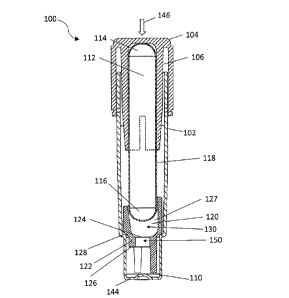
[0015] FIG. 1 depicts a side view of a biological indicator in cross-
section;
[0016] FIG. 2 depicts an exploded view of the biological indicator of
FIG. 1;
[0017] FIG 3 depicts, in block diagram form, a biological indicator
analyzer; and
[0018] FIG. 4 is a flow diagram of an exemplary method for confirming the
activation of
a biological indicator of FIGs. 1 and 2 that may be performed by the
biological indicator
analyzer of FIG. 3.
DETAILED DESCRIPTION
[0019] The following description sets forth certain illustrative examples
of the claimed
subject matter. Other examples, features, aspects, embodiments, and advantages
of the
technology should become apparent to those skilled in the art from the
following description.
Accordingly, the drawings and descriptions should be regarded as illustrative
in nature.
[0020] Referring to Figures 1 and 2, a self-contained biological
indicator ("SCBI") 100 is
shown. SCBI 100 includes a housing 102 and a cap 104 coupled thereto. Cap 104
includes a
projection 106 that has a planar, angled, arcuate, annular, or conical shape,
or some
combination thereof. Cap 104 may further include a chemical indicator 108 that
changes
color when exposed to, e.g., a chemical sterilant such as hydrogen peroxide.
Cap 104 may
also include one or more through-holes 109, to assist in the passage of gasses
(e.g., air or
¨ 8 -
CA 3008926 2018-06-20
sterilant) into or out from the SCBI. Cap 104 is coupled relative to housing
102 in a first
position and is movable from the first position to a second position. In the
first position, cap
104 is coupled to housing 102 in a manner in which gases (e.g., air or
sterilant) may move
from the surrounding environment and into the SCBI, or vice versa. In this
position, any
through-holes in cap 104 are disposed above housing 102 such that the inside
of housing 102
is in fluid communication with the surrounding environment, which permits
introduction and
withdrawal of sterilant into and from SCBI 100. Cap 104 may be depressed to
move it into
the second position relative to housing 102. In this second position, through-
holes 109 are
disposed below a top end of housing 102, which causes a tight fitting
relationship between
housing 102 and cap 104, and blocks the through holes, effectively sealing off
the inside of
the SCBI 100 from the surrounding environment.
100211 SCBI 100 also includes a source of microorganisms or active
enzymes, such as
carrier 110, which is impregnated with bacterial spores, other forms of
bacteria (e.g.,
vegetative), and/or active enzymes. Spores from Bacillus, Geobacillus, and
Clostridia species
are often used to monitor sterilization processes utilizing saturated steam,
hydrogen peroxide,
dry heat, gamma irradiation and ethylene oxide. Accordingly, carrier 110 may
be
impregnated with spores from Bacillus, Geobacillus, and/or Clostridia species.
Carrier 110
may be water-absorbent and may be formed of filter paper. Sheet-like materials
such as cloth,
nonwoven polypropylene, rayon or nylon, and microporous polymeric materials
may also be
used. Non-water absorbent materials are also appropriate for use, such as
metals (e.g.,
aluminum or stainless steel), glass (e.g., glass beads or glass fibers),
porcelain, or plastic.
Additionally, carrier 110 can be constructed of a combination of the
aforementioned
¨ 9 -
CA 3008926 2018-06-20
materials. In some embodiments, carrier 110 may have a thickness of
approximately 0.1 to
0.5 millimeters.
[0022]
The microorganism(s) or other source of biological activity on carrier 110 may
be
chosen based upon the resistance of the source to the particular sterilization
process to be
used in the sterilization cycle. For example, for a steam sterilization
process, Geobacillus
stearothermophilus or spores thereof, can be used. For an ethylene oxide
sterilization
process, Bacillus atrophaeus (formerly Bacillus subtilis), or spores thereof,
can be used. In
some sterilization processes, sterilization process resistant spores can
include, but are not
limited to, at least one of Geobacillus stearothermophilus spores, Bacillus
subtilis spores,
Bacillus atrophaeus spores, Bacillus megaterium spores, Bacillus coagulans
spores,
Clostridium sporogenes spores, Bacillus pumilus spores and combinations
thereof
[0023]
SCBI 100 also includes an ampule 112, having a first end 114, a second end
116,
and a sidewall 118. Sidewall 118 is substantially cylindrical and may have an
elliptical or
circular cross section. Ampule 112 may be fabricated from a frangible or
brittle material such
as glass or plastic. First end 114 and second end 116 are disposed at opposite
ends of
sidewall 118, and may have the form of a hemiellipsoid or hemisphere.
Accordingly, first
end 114 may be referred to as first dome 114 and second end 116 may be
referred to as
second dome 116. Ampule 112 contains a liquid growth medium. The growth medium
should be capable of promoting growth of any viable microorganisms or other
source of
biological activity disposed on carrier 110. Preferably, the microorganisms
are chosen to
generate enzymes that interact with the enzyme substrates to create detectable
product, e.g.,
by having a fluoroscopic intensity or spectrum distinct form the fluoroscopic
intensity or
spectrum of other materials in SCBI 100. Continued growth of the
microorganisms within the
CA 3008926 2018-06-20
growth medium causes an increase in the concentration of the detectable
product within the
growth medium. In certain embodiments, the detectable product is a
fluorophore. Thus, an
increase in concentration of the detectable product causes an increase in
fluorescence. That is
to say, the detectable product is detectable via changes in fluorescence.
[0024] Enzymes and enzyme substrates that may be used to detect efficacy
of a
sterilization cycle are identified in U.S. Pat. No. 5,073,488, entitled "Rapid
Method for
Determining Efficacy of a Sterilization Cycle and Rapid Read-Out Biological
Indicator,"
issued December 17, 1991, the disclosure of which is incorporated by reference
herein; U.S.
Pat. No. 5,418,167, entitled "Rapid Read-Out Biological Indicator," issued May
23, 1995, the
disclosure of which is incorporated by reference herein; U.S. Pat. No.
5,223,401, entitled
"Rapid Read-Out Sterility Indicator," issued June 29, 1993, the disclosure of
which is
incorporated by reference herein; and U.S. Pat. No. 9,322,046, entitled
"Biological
Sterilization Indicator," issued April 26, 2016, the disclosure of which is
incorporated by
reference herein.
[0025] Suitable enzymes may include hydrolytic enzymes and/or enzymes
derived from
spore-forming microorganisms, such as Bacillus subtilis. Enzymes from spore-
forming
microorganisms that can be useful in exemplary biological indicators may
include beta-D-
glucosidase, alpha-D-glucosidase, alkaline phosphatase, acid phosphatase,
butyrate esterase,
caprylate esterase lipase, myristate lipase, leucine aminopeptidase, valine
aminopeptidase,
chymotrypsin, phosphohydrolase, alpha-D-galactosidase, beta-D-galactosidase,
tyrosine
aminopeptidase, phenylalanine aminopeptidase, beta-D-glucuronidase, alpha-L-
arabinofuranosidase, N-acetyl-beta-glucosaminodase, beta-D-cellobiosidase,
alanine
aminopeptidase, proline aminopeptidase, fatty acid esterases and combinations
thereof.
¨ 11 -
CA 3008926 2018-06-20
[0026] In some exemplary methods for determining efficacy of a
sterilization cycle as
disclosed herein, enzyme substrates are converted to detectable product. For
instance, an
enzyme substrate may be characterized by a first emission spectrum (e.g., a
first fluorescent
emission spectrum) and a detectable product may be characterized by a second
emission
spectrum (e.g., a second fluorescent emission spectrum).
[0027] In some exemplary methods for determining efficacy of a
sterilization cycle as
disclosed herein, suitable enzyme substrates of use may include fluorogenic
enzyme
substrates. Useful fluorogenic enzyme substrates may be selected from:
fluorogenic 4-
methylumbelliferyl derivatives (hydrolysable to 4-methylumbelliferone ( "4-Mu"
),
derivatives of 7-amido-4-methyl-coumarin, diacetylfluorescein derivatives,
fluorescamine
and combinations thereof.
[0028] Exemplary 4-methylumbelliferyl derivatives may be selected from: 4-
methylumbellifery1-2-acetamido-4,6-0-benzylidene-2-deoxy-13-D-glucopyranoside,
4-
methylumbelliferyl acetate, 4-methylumbel1ifery1-N-acety1-13-D-
galactosaminide, 4-
methylumbellifery1-N-acety1-a-D-glucosaminide, 4-methylumbellifery1-N-acety1-
13-D-
glucosaminide, 21-(4-methylumbellifery1)-a-D-N-acetyl neuraminic acid, 4-
methylumbelliferyl a-L-arabinofuranoside, 4-methylumbelliferyl a-L-
arabinoside, 4-
methylumbelliferyl butyrate, 4-methylumbelliferyl 13-D-cellobioside,
methylumbelliferyl f3-
D-N,N' diacetyl chitobioside, 4-methylumbelliferyl elaidate, 4-
methylumbelliferyl 13-D-
fucoside, 4-methylumbelliferyl a-L-fucoside, 4-methylumbelliferyl f3-L-
fucoside, 4-
methylumbelliferyl a-D-galactoside, 4-methylumbellifery1P-D-galactoside, 4-
methylumbelliferyl a-D-glucoside, 4-methylumbelliferyl 13-D-g1ucoside, 4-
methylumbelliferyl (3-D-glucuronide, 4-methylumbelliferyl p-guanidinobenzoate,
4-
- 12 -
CA 3008926 2018-06-20
methylumbelliferyl heptanoate, 4-methylumbelliferyl a-D-mannopyranoside, 4-
methylumbelliferyl f3-D-mannopyranoside, 4-methylumbelliferyl oleate, 4-
methylumbelliferyl palmitate, 4-methylumbelliferyl phosphate, 4-
methylumbelliferyl
propionate, 4-methylumbelliferyl stearate, 4-methylumbelliferyl sulfate, 4-
methylumbelliferyl P-D-N,N',N"-triacetylchitotriose, 4-methylumbelliferyl
2,3,5-tri-o-
benzoyl-a-L-arabinofuranoside, 4-methylumbelliferyl-p-trimethylammonium
cinnamate
chloride, 4-methylumbellifery113-D-xy1oside and combinations thereof.
[0029]
In certain embodiments, the fluorescent response in the SCBI may be based on
the
naturally occurring alpha-glucosidase enzyme found in the Geobacillus
stearothermophilus
spore coat, which contains the enzyme and which is believed to be important in
the
germination of G. stearothermophilus. Alpha-glucosidase may be used to
hydrolyze the bond
between the glucose and 4-methylumbelliferyl moieties of 4-methylumbelliferyl
a-D-
glucopyranoside (a-MUG). a-MUG is not fluorescent. However, following
hydrolyzation
and separation of the moieties, the 4-Methylumbelliferone (4-MU) product is
fluorescent. 4-
MU fluoresces when excited by an external energy source, such as a light
source that emits
light having a wavelength of between approximately 360 and 370 nanometers. So
excited, 4-
MU emits light having a wavelength of between approximately 440 and 460
nanometers. In
certain embodiments, the light source emits light having a wavelength of
approximately 365
nanometers and the 4-MU emits light having a wavelength of 450 nm. The
fluorescence of 4-
MU is pH dependent. For example, when excited by light having a wavelength of
365
nanometers, the intensity of the emitted light is highest at a pH of 10.3. The
intensity
decreases with pH until about a pH of 7. Below this pH the intensity becomes
negligible.
¨ 13 -
CA 3008926 2018-06-20
[0030] SCBI 100 may also include an insert 120. Insert 120 may include a
platform 122
having a top surface 124 and a bottom surface 126. Insert 120 also includes a
sidewall 127.
Sidewall 127 of platform 122 may rest upon a support surface 128, which may be
integrally
formed as part of housing 102. Sidewall 127 and top surface 124 of platform
122 together
define a well 130, which is configured to receive second end 116 of ampule
112. Platform
122 defines a bore 150 therethrough, through which the liquid growth medium
may pass
upon breakage of the ampule.
[0031] SCBI 100 may be assembled according to the following steps. First,
housing 102
is provided. Second, carrier 110 is placed into housing 102 such that it rests
upon bottom
wall 144 of housing 102. Third, insert 120 is placed into housing 102 such
that sidewall 127
of platform 122 rests upon support surface 128. Alternatively, not shown, in
some
configurations lacking a support surface 128, insert 120 may rest directly
upon bottom wall
142 and may be in at least partial contact with carrier 110. Fourth, ampule
112 is inserted
into housing 102 such that second end 116 contacts insert 120. Finally, cap
104 is coupled to
housing 102 and ampule 112. Projection 106 has approximately the same diameter
as ampule
112 such that a friction fit is formed between ampule 112 and projection 106.
So assembled,
central longitudinal axes of ampule 112, housing 102, cap 104, and insert 120
are coaxial or
substantially coaxial. Other assembly procedures may be performed to achieve
the same
configuration of SCBI 100.
[0032] Following a sterilization procedure, an SCBI 100 may be activated
and monitored
to determine whether a sterilization cycle was effective. To activate SCBI
100, a compressive
force 146 is applied between housing 102 and cap 104. This compressive force
is resisted by
ampule 112 because ampule 112 is in contact with insert 120 and insert 120 is
in contact
CA 3008926 2018-06-20
with, e.g., support 128 of housing 102. When the compressive force applied to
cap 104 is
greater than a breakage force ampule 112 can withstand, ampule 112 will break.
Once
ampule 112 is broken, cap 104 moves to its second position and growth medium
is released
to immerse carrier 110.
[0033] Various features may be included within the SCBI to facilitate
activating the
SCBI by, e.g., lowering the force that a user must apply to break the ampule.
Exemplary
features directed to this functionality are disclosed in copending U.S. Patent
Application Nos.
15/057,768 and 15/397,018.
[0034] Activation of SCBI 100 should be confirmed. For example, activation
may be
confirmed by, e.g., checking that ampule 112 is broken, that the growth medium
submerses
carrier 110, that a substantial volume of the growth medium is disposed
between bottom
surface 126 of insert 120 and bottom wall 144 of housing 102, and/or that cap
104 is in the
second position. To increase the likelihood that a failed or improper
activation can be
detected, multiple checks may be performed. For example, in addition to
checking that
ampule 112 is broken, submersion of carrier 110 by the growth medium may also
be
performed. A user or an electromechanical device capable of assaying SCBI 100,
such as a
biological indicator analyzer ("BIA") 200, may perform these checks. To
increase the
likelihood that a failed or improper activation can be detected, various
checks should be
performed by both a user and BIA 200.
[0035] Figure 3 depicts an exemplary BIA 200 in block form that is
operable to analyze a
biological indicator, e.g., SCBI 100, which has been subject to a
sterilization cycle. BIA 200
is configured to assay an SCBI, collect information about the SCBI (e.g.,
location of growth
medium, color of the growth medium, light intensity of growth medium), process
the
¨ 15 -
CA 3008926 2018-06-20
information, and determine whether the sterilization cycle was effective. BIA
200 comprises
a plurality of wells 210, each of which is configured to receive a respective
an SCBI 100
specimen therein. While two wells 210 are shown, it should be understood that
any other
suitable number of wells may be provided, including eight wells, less than
eight wells, or
more than eight wells. Each well 210 further includes a heating element 212
that can be used
to incubate SCBI 100 when it is inserted therein. Such incubation promotes the
outgrowth of
any live microorganisms within the SCBI. In various embodiments, the heating
element may
achieve a temperature in the well of between approximately 50 C and
approximately 60 C. In
certain embodiments, the heating element may achieve a temperature of
approximately 57 C
and cause SCBI 100 to reach a substantially similar or same temperature. BIA
200 also
includes a processor 220 that is operable to execute instructions and control
algorithms,
process information, etc.
[0036] Each well 210 has an associated light source 230 and sensor 240.
Each light
source 230 is configured to project light through housing 102 of the SCBI 100
that is inserted
in the corresponding well 210. Each sensor 240 is operable to detect light
fluoresced by the
growth medium. Each sensor 240 is positioned adjacent to each well 210 such
that when an
SCBI 100 is disposed within a well, sensor 240 is adjacent to the portion of
SCBI 100
between bottom surface 126 of insert 120 and bottom wall 144 of housing 102.
[0037] Light source 230 may be in the form of, for example, a laser that
is configured to
emit ultraviolet light. In some embodiments, the light emitted by light source
230 has a
wavelength of 370 nanometers. Various other suitable forms that light source
230 may take
will be apparent to those of ordinary skill in the art in view of the
teachings herein. By way
of further example, sensor 240 may comprise a charge coupled device (CCD).
Further, it may
CA 3008926 2018-06-20
be a sensor optimized to detect light generated by fluorescence, i.e., a
fluorescence sensor. In
some embodiments, sensor 240 is a silicon photodiode, such as silicon
photodiode S2386-5K
manufactured by Hamamatsu. The fluorescence of the growth medium depends
primarily on
the number of living microorganisms contained in the growth medium. Thus,
sensor 240 is
configured to detect the presence of living microorganisms in the growth
medium based on
the degree to which it fluoresces in response to light from light source 230.
However, the
fluorescence of the growth medium also depends on whether any fluorescence
quenching has
occurred. Fluorescence quenching may also be used for confirming proper
activation of an
SCBI 100, as will be explained in detail below.
[0038] BIA 200 optionally further includes a user feedback and/or input
device such as
touch screen display 250. Touch screen display 250 is operable to render
various user
interface display screens associated with operation of biological indicator
analyzer 200.
Touch screen display 250 is further configured to receive user input in the
form of the user
contacting touch screen display 250 in accordance with conventional touch
screen
technology. In addition, or in the alternative, biological indicator analyzer
200 may include
various other kinds of user input features, including but not limited to
buttons, keypads,
keyboards, a mouse, a trackball, etc. Displays provided through touch screen
display 250
may be driven by processor 220. User inputs received through touch screen
display 250
may be processed by processor 220.
[0039] BIA 200 of the present example further includes a memory 280, such
as non-
transitory storage medium (e.g., hard disk drive or a flash memory drive),
which is operable
to store control logic and instructions and that are executed by processor 220
to drive
components such as light source 230 and touch screen display 250 and perform
calculations
CA 3008926 2018-06-20
and analyses on data, particularly data collected by sensor 240. Memory 280
may also be
used to store user inputs, data collected by sensor 240, and calculations
based on this data.
[0040] Fluorescence data collected by BIA 200 may be used to determine
changes in
fluorescence over time. Such data may be used to determine fluorescence
quenching.
Fluorescence quenching is a term that generally describes various processes
that cause the
fluorescence intensity of a substance to decrease. For fluorescent substances,
such processes
include, but are not limited to, 1) heating, 2) lowering pH; and 3) adding
another substance or
material, sometimes referred as a "quencher," that is known to cause a
decrease in
fluorescence intensity. Exemplary quenchers include, but are not limited to,
oxygen, aniline,
bromobenzene, acrylamide, hydrogen peroxide, imidazole, indole, and
succinimide.
Quenchers may also be metal ions, such as: Co2+, Ni2+, Cu2+, Hg2+, pb2+, Ag+,
Cr3+, and Fe3+.
[0041] When an SCBI 100 is inserted into a well 210 of BIA 200, its
temperature may be
substantially equivalent to the ambient temperature, e.g., room temperature.
Possibly,
however, SCBI 100 may have a temperature warmer than the ambient temperature
because
vacuum chambers in sterilization systems often are warmer than the ambient
temperature at
the end of a sterilization cycle. Irrespective of the temperatures of SCBI
100, upon being
inserted or shortly after it is inserted into well 210 of BIA 200 (e.g.,
approximately 1 second,
seconds, or 15 seconds), BIA 200 activates heating element 212 to raise the
temperature in
the well to between approximately 50 C and approximately 60 C such that SCBI
100 reaches
a substantially similar or same temperature. Heating SCBI 100 in this manner
quenches the
fluorescence of the components of SCBI 100. Although the quenching is most
pronounced in
the growth medium, it also may be present, albeit to a lesser degree, in other
features of the
SCBI 100, i.e., the non-liquid components, including housing 102.
¨ 18 -
CA 3008926 2018-06-20
[0042] Various components of SCBI 100 may be assayed to determine whether
an SCBI
100 has been properly activated. In a properly activated SCBI 100, the growth
medium
should immerse carrier 110 and be disposed at the bottom of SCBI 100 between
bottom
surface 126 of insert 120 and bottom wall 144 of housing 102. Accordingly, BIA
200 may
assay this portion of SCBI 100 to determine whether the growth medium is
present there. For
example, BIA 200 may activate light source 230. In certain embodiments, the
light emitted
by light source 230 has a wavelength of approximately 370 nanometers. If the
growth
medium is present, the growth medium will be excited and sensor 240 will
register a
corresponding fluorescence intensity, output a corresponding voltage to
processor 220 to be
stored in memory 280. However, if the growth medium is not present, the growth
medium
will not be excited. Nonetheless, the light source may excite other features
and materials of
SCBI 100 such that sensor 240 will register a corresponding intensity and
output a
corresponding voltage to processor 220 to be stored in memory 280. The
intensity registered
by sensor 240 will be different depending on if the growth medium is present
in the bottom
of an SCBI 100 that has been properly activated or if the growth medium is not
present in the
bottom of an SCBI 100 that has been improperly activated such that the growth
medium
remains, e.g., in an unbroken ampule 112, outside of the assay region that
light source 230
and sensor 240 can interrogate.
[0043] Processor 220 may be programmed to detect the presence of growth
medium at
the bottom of SCBI 100. In some embodiments, threshold values corresponding to
light
and/or fluorescence intensities may be stored in memory 280. Specifically,
light and/or
fluorescence intensities corresponding to a liquid in the bottom of SCBI 100,
between bottom
surface 126 of insert 120 and bottom wall 144 of housing 102, may be stored in
memory 280.
¨ 19 -
CA 3008926 2018-06-20
By comparing measured intensities to the threshold values, a determination may
be made as
to whether SCBI 100 was properly activated. For example, processor 220 may be
programmed to determine whether a measured intensity falls between a minimum
threshold
value and a maximum threshold value. An intensity measurement that falls
between the
threshold values would indicate that growth medium is disposed at the bottom
of SCBI 100
and that the SCBI 100 has been properly activated. An intensity measurement
that falls
below the minimum threshold value would indicate that growth medium is not
disposed at
the bottom of SCBI 100, likely because it remains in an unbroken ampule 112
due to
improper activation. An intensity measurement that falls above the maximum
threshold value
may indicate a malfunction within BIA 200. In those embodiments where light
source 230
provides light having a wavelength of 370 nm, the minimum threshold value may
be
approximately 0.02 W/cm2 and the maximum threshold value may be 0.10 W/cm2.
In
those embodiments where sensor 240 is silicon photodiode S2386-5K by
Hamamatsu, these
minimum and maximum values should be output from sensor 240 as 0.47 volts 2.2
volts,
respectively. In some embodiments, the intensity measurement used to confirm
proper
activation is taken immediately after, or up to approximately 300 seconds
after, SCBI 100 is
inserted into well 210. In certain embodiments, the intensity measurement is
taken
approximately 70 seconds after SCBI 100 is inserted into well 210. SCBI
activation may be
determined in this manner with up to approximately 90% - 95% accuracy.
Variation in
intensity from light source 230 and the temperatures in SCBI 100 maintained by
heating
element 212 may prevent greater accuracy from being achieved.
[0044] Accordingly, it is advisable to supplement this form of
confirmation with other
methods and forms of confirmation, such as visual confirmation performed by a
user, or the
¨ 20 -
CA 3008926 2018-06-20
following method based on quenching effects of the growth medium and other
components
of SCBI 100.
[0045] Quenching may be determined by calculating a difference or ratio
between a first
fluorescence-intensity measurement taken at a first time and a second
fluorescence-intensity
measurement taken at a second time. Quenching effects are typically more
pronounced in
liquids than solids because molecules in liquids collide more frequently than
molecules in
solids. Plastic materials, particularly clear or transparent plastic materials
commonly used in
medical devices, including polycarbonate and cyclo olefin, exhibit a
relatively small drop in
fluorescence due to heating as compared to colored growth media, including
those containing
4-MU, such as those used in SCBI 100. For example, after being heated from
room
temperature to between 50 -60 and maintained at the higher temperature for
about four
minutes, the plastic materials exhibit a quenching effect, i.e., a decrease in
fluorescence
intensity, of between approximately 0 and 5%, whereas the growth medium
exhibits a
quenching effect between approximately 5% and 25%.
[0046] Accordingly, BIA 200, may be used to: 1) take a first fluorescence-
intensity
measurement at a first time, 2) quench the fluorescence of SCBI 100 by heating
SCBI 100, 3)
take a second fluorescence-intensity measurement at a second time subsequent
to the first
time, 4) compare the first fluorescence-intensity measurement with the second
fluorescence-
intensity measurement by computing a difference or a ratio between the two
measurements to
determine a degree of quenching, 5) determine whether the degree of quenching
corresponds
to quenching from only solid or non-liquid components of SCBI 100 or
additionally
corresponds to quenching from the growth medium, and 6) indicate whether SCBI
100 was
improperly activated.
¨ 21 -
CA 3008926 2018-06-20
[0047] The first fluorescence-intensity measurement may be taken at a
first time, i.e.,
between approximately 0 seconds to approximately 100 seconds after SCBI 100 is
inserted
into well 210. The second fluorescence-intensity measurement may be taken at a
second
time, i.e., between approximately 0 seconds to approximately 300 seconds after
the first time.
In certain embodiments the first time is approximately 70 seconds after SCBI
100 is inserted
into well 210 and the second time is approximately 210 seconds after the first
time (or 280
seconds after SCBI 100 is inserted into well 210).
[0048] The comparison and determination steps may be carried out by
processor 220 in
various ways. For example, processor 220 may calculate a difference between
the second
measurement and first measurement and compare the difference to threshold
values stored in
memory 280 corresponding to degrees of quenching from the solid materials and
the growth
medium. Alternatively, processor 220 may calculate a ratio between the second
measurement
and first measurement and compare the ratio to threshold values stored in
memory 280
corresponding to degrees of quenching from the solid materials and the growth
medium.
When BIA 200 performs two checks of activation¨one to determine if a single
intensity
value is between expected minimum and maximum threshold values and another to
determine if two intensity values correspond to an expected degree of
fluorescence
quenching¨proper activation may be confirmed with a high degree of accuracy,
at least as
high as 99%.
[0049] The indication step may take the form of processor 220 causing a
message to be
displayed on display 250 that states whether SCBI 100 was properly or
improperly activated.
Alternatively or additionally, upon a determination that SCBI 100 was
improperly activated,
BIA 200 may sound an alarm.
¨ 22 -
CA 3008926 2018-06-20
[0050] In order to enhance the difference in the degree of quenching the
non-liquid
components and growth medium undergo when subject to heating, the quenching
properties
of the non-liquid components and growth medium may be modified. Specifically,
the effect
of heat on quenching may be increased for the growth medium and decreased for
the non-
liquid components. Doing so may facilitate differentiating between the non-
liquid
components and growth medium based on quenching calculations from light-
intensity
measurements taken by sensor 240. In turn, such modifications may increase the
reliability of
determinations based on the quenching calculations as to whether SCBI 100 has
been
properly activated. For example, a quencher, such as aniline, bromobenzene,
acrylamide,
hydrogen peroxide, imidazole, indole, or succinimide, may be added by, e.g.,
blending, to the
growth medium. Oxygen may also be considered a quencher. The growth medium has
an
oxygen content of approximately 37 mg/L at sea level. Accordingly, the oxygen
content may
be increased to approximately 40 mg/L, approximately 45 mg/L or greater to
increase the
quenching of the growth medium caused by heat. Metal ions may also be
considered
quenchers. Exemplary metal-ion quenchers include: Co2+, Ni2+, Cu2+, Hg2+,
Pb2+, Ag+, Cr3+,
and Fe3+.
[0051] Additionally or alternatively, the growth medium may be modified
to have a
higher pH. In certain embodiments, the growth medium has a pH between
approximately 7.7
and approximately 8.7. In certain embodiments the pH of the growth medium may
be
approximately 8.2. However, quenching effects are generally maximized at a pH
of around
because at that pH, fluorescence intensity is also maximized. Accordingly, the
pH of the
growth medium may be increased to approximately 8.5 approximately 9,
approximately 9.5
and approximately 10.
CA 3008926 2018-06-20
100521 To reduce the amount of quenching in the non-liquid components of
SCBI 100
(e.g., housing 102 and insert 120) caused by heat, the non-liquid components
may be
fabricated from materials having low concentrations of antioxidants, e.g.,
cyclo olefins, and
any other UV- absorbing compounds otherwise found in these components that may
absorb
UV light from the assay performed by BIA 200. The non-liquid components may
also be
fabricated from a UV transparent material, such as quartz or low density
polyethylene.
[0053] Greater increases in temperature may also be utilized to help
distinguish between
non-liquid components and the growth medium based on quenching measurements.
Because
the degree of quenching is a function of a change in temperature, a larger
change in
temperature typically causes greater quenching than a smaller change in
temperature.
Although such changes affect quenching of the growth medium and the non-liquid
components of SCBI 100, changes in temperature have a greater effect on the
quenching of
the growth medium than the non-liquid components. Therefore, greater accuracy
in
differentiating between quenching corresponding to non-liquid components and
growth
media may be achieved by maximizing a temperature difference subject to design
constraints
relating to assessing microbial growth. Thus, BIA 200 may heat SCBI 100 to
temperatures
above 60 C to impart greater quenching effects to SCBI 100, further
pronouncing any
difference in fluorescence intensity imparted by heating. In a similar vein,
BIA 200 may
include a cooling element alongside well 210 that may be used to cool SCBI 100
before
heating it in order to increase the ultimate change in temperature, and thus,
the concomitant
quenching effect.
[0054] Quenching in the growth medium may be offset by an increase in
fluorescence
resulting from fluorescent products generated by enzymes in the growth medium.
¨ 24 -
CA 3008926 2018-06-20
Accordingly, the amount of enzyme generated within the growth medium should be
minimized subject to design constraints necessary for determining microbial
growth
associated with the generated enzyme.
[0055] Figure 4 depicts a flow chart setting forth an exemplary method 300
for
determining whether a biological indicator, such as SCBI 100, has been
properly activated
according to some of the foregoing teachings. This method may be performed as
a part of a
larger method (e.g., as a subroutine) in which BIA 200 monitors SCBI for
changes in
fluorescence caused by microbial outgrowth. Although each and every one of the
foregoing
teachings are not explicitly incorporated into this example method, it should
be understood
that those teachings not explicitly set forth may be incorporated into methods
for determining
activation of biological indicator, such as SCBI 100, based on quenching
effects.
Furthermore, although SCBI 100, BIA 200, and their components are referenced
in
presenting this method, it should be understood that the method may be
practiced with other
biological indicators besides SCBI 100 and other biological indicator
analyzers besides BIA
200.
100561 The method 300 begins with step 310 in which a healthcare worker
applies
compressive force 146 between housing 102 and cap 104 to activate SCBI 100. In
typical
usage, step 310 occurs after subjecting the SCBI to a sterilization procedure.
Successful
activation causes cause cap 104 to move from a first position to a second
position, thereby
breaking ampule 112, which permits the growth medium contained therein to flow
to the
bottom of SCBI 100, i.e., between bottom surface 126 of insert 120 and bottom
wall 144 of
housing 102. In step 320, the healthcare worker inserts SCBI 100 into a well
210 of BIA 200.
In step 330, BIA 200 activates heating element 212. In step 340, BIA 200
performs a first
¨ 25 -
CA 3008926 2018-06-20
assay of the bottom of SCBI 100 using light source 230 to excite the SCBI and
sensor 240 to
measure a first light intensity. Step 340 may be performed between
approximately 0 seconds
and approximately 100 seconds after step 320. In some embodiments step 340 may
occur
before step 330. In other embodiments, step 340 may occur after step 330. In
some
embodiments, step 340 may be performed approximately 70 seconds after step
320, whereas
in other embodiments step 340 may be performed approximately 70 seconds after
step 330.
In step 350, the measured first light intensity, output from sensor 240 as a
first voltage value,
is stored in storage device 280. In step 360, processor 220 compares the first
voltage value
(VI) to a minimum threshold voltage value (Vmin) and maximum threshold voltage
value
(Vmax). In those embodiments where light source 230 emits light having a
wavelength of 370
nm and sensor 240 is silicon photodiode S2386-5K manufactured by Hamamatsu,
the
minimum threshold voltage value may be between approximately 0.4 and
approximately 0.5
volts. For example, the minimum threshold voltage value may be approximately
0.47 volts.
The maximum threshold voltage value may be between approximately 2.1 and
approximately
2.3 volts. For example, the maximum threshold voltage value may be
approximately 2.2
volts. If the first voltage value is less than the minimum threshold voltage
value or greater
than the maximum threshold voltage value, then SCBI 100 may have been
improperly
activated or BIA 200 may have malfunctioned. Accordingly, in step 370, the
method is
aborted. An error message may be displayed on display 250 or an alarm may be
sounded. If
the first voltage value falls between the minimum and maximum threshold
values, SCBI 100
may have been properly activated. To confirm whether SCBI 100 was properly
activated,
BIA 200 continues the method.
¨ 26 -
CA 3008926 2018-06-20
[0057] BIA 200 performs a second assay of SCBI 100 in step 380. For step
380, BIA 200
uses light source 230 to excite the SCBI and sensor 240 to measure a second
light intensity.
Step 380 may be performed between approximately 0 second and approximately 300
seconds
after step 340. For example, step 380 is performed approximately 210 seconds
after step 340
(i.e., approximately 280 seconds after heating element 212 was activated). In
step 390, the
measured second light intensity, output from sensor 240 as a second voltage
value (V2), is
stored in storage device 280.
[0058] In step 400, processor 220 computes a quenching metric. For
example, the
quenching metric may be a "quenching difference," i.e., a difference between
the second
voltage value and the first voltage value or it may take the form of a
"quenching ratio," i.e., a
ratio between the second voltage value and the first voltage value. If the
fluorescence of
SCBI 100 has not been quenched, the second voltage value should be equal to or
approximately equal to the first voltage value. Accordingly, the quenching
difference ("QD")
should be equal to or approximately equal to zero and the quenching ratio
("QR") should be
equal to or approximately equal to one. As shown in step 400, the quenching
metric is the
quenching ratio. In step 410, processor determines if there has been minimal
quenching (e.g.,
the quenching ratio is greater than approximately 95%) or substantial
quenching (e.g., the
quenching ratio is between 75% and 95% when SCBI 100 was heated to
approximately 57 C,
the first voltage value corresponds to a first light intensity measured
approximately 70
seconds after heating element 212 was activated, and the second voltage value
corresponds to
a second light intensity measured approximately 280 seconds after heating
element 212 was
activated). Minimal quenching indicates that SCBI 100 was improperly activated
because the
growth medium should undergo quenching when subject to heat. When processor
220
¨ 27 -
CA 3008926 2018-06-20
calculates minimal quenching, step 420 is performed in which the method is
aborted. An
error message may be displayed on display 250 or an alarm may be sounded.
However, when
processor 220 calculates substantial quenching, BIA 200 commences its
assessment of SCBI
100 according to its primary purpose, i.e., monitoring further changes in
fluorescence of the
growth medium that may be attributable to microbial growth, as shown in step
430.
[0059] It should be understood that any of the examples and/or
embodiments described
herein may include various other features in addition to or in lieu of those
described above.
The teachings, expressions, embodiments, examples, etc. described herein
should not be
viewed in isolation relative to each other. Various suitable ways in which the
teachings
herein may be combined should be readily apparent to those of ordinary skill
in the art in
view of the teachings herein.
[0060] Having shown and described exemplary embodiments of the subject
matter
contained herein, further adaptations of the methods and systems described
herein may be
accomplished by appropriate modifications without departing from the scope of
the claims.
Some such modifications should be apparent to those skilled in the art. For
instance, the
examples, embodiments, geometrics, materials, dimensions, ratios, steps, and
the like
discussed above are illustrative. Accordingly, the claims should not be
limited to the specific
details of structure and operation set forth in the written description and
drawings.
¨ 28 -
CA 3008926 2018-06-20