Note: Descriptions are shown in the official language in which they were submitted.
CA 03009637 2018-06-22
WO 2017/112859 PCT/US2016/068289
ENGINEERED MEGANUCLEASES WITH RECOGNITION SEQUENCES
FOUND IN THE HUMAN BETA-2 MICROGLOBULIN GENE
CROSS REFERENCE TO RELATED APPLICATIONS
[0001] This application claims priority to U.S. Provisional Application No.
62/387,318,
entitled "Engineered Meganucleases with Recognition Sequences Found in the
Human Beta-
2 Microglobulin Gene," filed December 23, 2015, and U.S. Provisional
Application No.
62/416,513, entitled "Engineered Meganucleases with Recognition Sequences
Found in the
Human Beta-2 Microglobulin Gene," filed November 2, 2016, the disclosures of
which are
hereby incorporated by reference in their entireties.
FIELD OF THE INVENTION
[0002] The present disclosure relates to the field of molecular biology and
recombinant
nucleic acid technology. In particular, the present disclosure relates to
recombinant
meganucleases engineered to recognize and cleave recognition sequences found
in the human
beta-2 microglobulin gene. The present disclosure further relates to the use
of such
recombinant meganucleases in methods for producing genetically-modified
eukaryotic cells,
and to a population of genetically-modified cells having reduced cell-surface
expression of
beta-2 microglobulin.
REFERENCE TO A SEQUENCE LISTING SUBMITTED AS
A TEXT FILE VIA EFS-WEB
[0003] Filed with the instant application is a paper copy of a Sequence
Listing which is
hereby incorporated by reference in its entirety. This paper copy corresponds
to a copy in
ASCII format which is also hereby incorporated by reference in its entirety.
Said ASCII
copy, created on December 20, 2016, is named 2000706-00181W01.txt, and is
741,693 bytes
in size.
BACKGROUND OF THE INVENTION
[0004] T cell adoptive immunotherapy is a promising approach for cancer
treatment.
This strategy utilizes isolated human T cells that have been genetically-
modified to enhance
their specificity for a specific tumor associated antigen. Genetic
modification may involve
the expression of a chimeric antigen receptor (CAR) or an exogenous T cell
receptor to graft
antigen specificity onto the T cell. By contrast to exogenous T cell
receptors, CARs derive
their specificity from the variable domains of a monoclonal antibody. Thus, T
cells
1
CA 03009637 2018-06-22
WO 2017/112859 PCT/US2016/068289
expressing CARs induce tumor immunoreactivity in a major histocompatibility
complex
(MHC) non-restricted manner. To date, T cell adoptive immunotherapy has been
utilized as a
clinical therapy for a number of cancers, including B cell malignancies (e.g.,
acute
lymphoblastic leukemia (ALL), B cell non-Hodgkin lymphoma (NEIL), and chronic
lymphocytic leukemia), multiple myeloma, neuroblastoma, glioblastoma, advanced
gliomas,
ovarian cancer, mesothelioma, melanoma, and pancreatic cancer.
[0005] Despite its potential usefulness as a cancer treatment, adoptive
immunotherapy
has been limited, in part, by alloreactivity between host tissues and
allogeneic CAR T cells.
One cause of alloreactivity arises from the presence of non-host MHC class I
molecules on
the cell-surface of CART cells. MHC class I molecules consist of two
polypeptide chains, a
and 13. In humans, the a chain consists of three subunits, al, a2, and a3,
which are encoded
by polymorphic human leukocyte antigen (HLA) genes on chromosome 6. The
variability of
HLA loci, and the encoded a chain subunits, can cause allogeneic CAR T cells
to be seen by
the host immune system as foreign cells because they bear foreign MHC class I
molecules.
As a result, CAR T cells administered to a patient can be subject to host
versus graft (HvG)
rejection, where they are recognized and killed by the host's cytotoxic T
cells.
[0006] The l chain of MHC class I molecules consists of beta-2
microglobulin, which is
encoded by the non-polymorphic beta-2 microglobulin (B2M) gene on chromosome
15 (SEQ
ID NO: 1). Beta-2 microglobulin is non-covalently linked to a3 subunit and is
common to all
MHC class I molecules. Furthermore, expression of MHC class I molecules at the
cell
surface requires its association with beta-2 microglobulin. As such, beta-2
microglobulin
represents a logical target for suppressing the expression of MHC class I
molecules on CAR
T cells, which could render the cells invisible to host cytotoxic T cells and
reduce
alloreactivity.
[0007] Another cause of alloreactivity to CART cells is the expression of
the
endogenous T cell receptor on the cell surface. T cell receptors typically
consist of variable a
and l chains or, in smaller numbers, variable y and 6 chains. The T cell
receptor complexes
with accessory proteins, including CD3, and functions with cell-surface co-
receptors (e.g.,
CD4 and CD8) to recognize antigens bound to MHC molecules on antigen
presenting cells.
In the case of allogeneic CART cells, expression of endogenous T cell
receptors may cause
the cell to recognize host MHC antigens following administration to a patient,
which can lead
to the development of graft-versus-host-disease (GVHD).
2
CA 03009637 2018-06-22
WO 2017/112859 PCT/US2016/068289
[0008] To forestall alloreactivity, clinical trials have largely focused on
the use of
autologous CAR T cells, wherein a donor's T cells are isolated, genetically-
modified to
incorporate a chimeric antigen receptor, and then re-infused into the same
subject. An
autologous approach provides immune tolerance to the administered CAR T cells;
however,
this approach is constrained by both the time and expense necessary to produce
patient-
specific CAR T cells after a patient's cancer has been diagnosed.
[0009] Therefore, a need exists for the development of allogeneic CAR T
cells that lack
expression of beta-2 microglobulin and MHC class I molecules, as well as cells
that further
lack expression of endogenous T cell receptors.
SUMMARY OF THE INVENTION
[0010] The present disclosure provides a recombinant meganuclease that is
engineered to
recognize and cleave a recognition sequence within the human beta-2
microglobulin gene
(SEQ ID NO:1). Such a meganuclease is useful for disrupting the beta-2
microglobulin gene
and, consequently, disrupting the expression and/or function of endogenous MHC
class I
receptors. Meganuclease cleavage can disrupt gene function either by the
mutagenic action
of non-homologous end joining or by promoting the introduction of an exogenous
polynucleotide into the gene via homologous recombination. The present
disclosure further
provides methods comprising the delivery of a recombinant meganuclease
protein, or genes
encoding a recombinant meganuclease, to a eukaryotic cell in order to produce
a genetically-
modified eukaryotic cell.
[0011] The present disclosure further provides for "off the shelf' CAR T
cells, prepared
using T cells from a third party donor, that are not alloreactive and do not
induce HvG
rejection or GVHD because they lack expression of beta-2 microglobulin and MHC
class I
molecules and/or further lack expression of endogenous T cell receptors. Such
products can
be generated and validated in advance of diagnosis, and can be made available
to patients as
soon as necessary.
[0012] The present disclosure further relates to the use of site-specific,
rare-cutting,
homing endonucleases (also called "meganucleases") that are engineered to
recognize
specific DNA sequences in a locus of interest. Homing endonucleases are a
group of
naturally-occurring nucleases which recognize 15-40 base pair cleavage sites
commonly
found in the genomes of plants and fungi. They are frequently associated with
parasitic DNA
elements, such as group 1 self-splicing introns and inteins. They naturally
promote
homologous recombination or gene insertion at specific locations in the host
genome by
3
CA 03009637 2018-06-22
WO 2017/112859 PCT/US2016/068289
producing a double-stranded break in the chromosome, which recruits the
cellular DNA-
repair machinery (Stoddard (2006), Q. Rev. Biophys. 38:49-95). Homing
endonucleases are
commonly grouped into four families: the LAGLIDADG (SEQ ID NO:11) family, the
GIY-
YIG family, the His-Cys box family and the HNH family. These families are
characterized
by structural motifs, which affect catalytic activity and recognition
sequence. For instance,
members of the LAGLIDADG (SEQ ID NO:11) family are characterized by having
either
one or two copies of the conserved LAGLIDADG (SEQ ID NO:11) motif (Chevalier
et at.
(2001), Nucleic Acids Res. 29(18): 3757-3774). The LAGLIDADG (SEQ ID NO:11)
homing
endonucleases with a single copy of the LAGLIDADG (SEQ ID NO:11) motif form
homodimers, whereas members with two copies of the LAGLIDADG (SEQ ID NO:11)
motif
are found as monomers.
[0013] Methods for producing engineered, site-specific recombinant
meganucleases are
known in the art. I-CreI (SEQ ID NO:10) is a member of the LAGLIDADG (SEQ ID
NO:11) family of homing endonucleases which recognizes and cuts a 22 base pair
recognition sequence in the chloroplast chromosome of the algae Chlamydomonas
reinhardtii. Genetic selection techniques have been used to modify the wild-
type I-CreI
cleavage site preference (Sussman et al. (2004), 1 Mol. Biol. 342: 31-41;
Chames et al.
(2005), Nucleic Acids Res. 33: e178; Seligman et al. (2002), Nucleic Acids
Res. 30: 3870-9,
Arnould et al. (2006), 1 Mol. Biol. 355: 443-58). More recently, a method of
rationally-
designing mono-LAGLIDADG (SEQ ID NO:11) homing endonucleases was described
which is capable of comprehensively redesigning I-CreI and other homing
endonucleases to
target widely-divergent DNA sites, including sites in mammalian, yeast, plant,
bacterial, and
viral genomes (WO 2007/047859).
[0014] As first described in WO 2009/059195, I-CreI and its engineered
derivatives are
normally dimeric but can be fused into a single polypeptide using a short
peptide linker that
joins the C-terminus of a first subunit to the N-terminus of a second subunit
(Li, et at. (2009)
Nucleic Acids Res. 37:1650-62; Grizot, et at. (2009) Nucleic Acids Res.
37:5405-19.) Thus, a
functional "single-chain" meganuclease can be expressed from a single
transcript. Such
engineered meganucleases exhibit an extremely low frequency of off-target
cutting. By
delivering a gene encoding a single-chain meganuclease to a cell, it is
possible to specifically
and preferentially target, cleave, and disrupt the beta-2 microglobulin gene.
[0015] The use of engineered meganucleases for cleaving DNA targets in the
human
beta-2 microglobulin gene was previously disclosed in International
Publication Nos. WO
4
CA 03009637 2018-06-22
WO 2017/112859 PCT/US2016/068289
2008/102199 ("the '199 application") and WO 2008/102274 ("the '274
application"). The
'199 application and the '274 application each disclose I-CreI variants having
amino acid
substitutions that are intended to increase selectivity of the meganuclease
for recognition
sequences found in the beta-2 microglobulin gene. However, the meganucleases
described
were only shown to have activity against beta-2 microglobulin DNA targets in
yeast or CHO
reporter cell systems. The present disclosure improves upon the teachings of
the prior art by
providing recombinant meganucleases that efficiently target and cleave
recognition
sequences in the beta-2 microglobulin gene in human T cells.
[0016] Thus, in one aspect, the disclosure provides a recombinant
meganuclease that
recognizes and cleaves a recognition sequence within the human beta-2
microglobulin gene
(SEQ ID NO:1). Such a recombinant meganuclease comprises a first subunit and a
second
subunit, wherein the first subunit binds to a first recognition half-site of
the recognition
sequence and comprises a first hypervariable (HVR1) region, and wherein the
second subunit
binds to a second recognition half-site of the recognition sequence and
comprises a second
hypervariable (HVR2) region.
[0017] In one embodiment, the recognition sequence comprises SEQ ID NO:2
(i.e., the
B2M 13-14 recognition sequence).
[0018] In one such embodiment, the first meganuclease subunit comprises an
amino acid
sequence having at least 80%, at least 85%, at least 90%, or at least 95%
sequence identity to
residues 198-344 of any one of SEQ ID NOs:12-96 or residues 7-153 of any one
of SEQ ID
NOs:97-100, and the second meganuclease subunit comprises an amino acid
sequence having
at least 80%, at least 85%, at least 90%, or at least 95% sequence identity to
residues 7-153 of
any one of SEQ ID NOs:12-96 or residues 198-344 of any one of SEQ ID NOs:97-
100.
[0019] In another such embodiment, the HVR1 region comprises Y at a
position
corresponding to: (a) position 215 of any one of SEQ ID NOs:12-96; or (b)
position 24 of any
one of SEQ ID NOs:97-100. In another such embodiment, the HVR1 region
comprises F at a
position corresponding to: (a) position 261 of any one of SEQ ID NOs:12-96; or
(b) position
70 of any one of SEQ ID NOs:97-100. In another such embodiment, the HVR1
region
comprises one or more of Y and F at positions corresponding to: (a) positions
215 and 261,
respectively, of any one of SEQ ID NOs:12-96; or (b) positions 24 and 70,
respectively, of
any one of SEQ ID NOs:97-100.
[0020] In another such embodiment, the HVR2 region comprises Y at a
position
corresponding to: (a) position 24 of any one of SEQ ID NOs:12-96; or (b)
position 215 of any
CA 03009637 2018-06-22
WO 2017/112859 PCT/US2016/068289
one of SEQ ID NOs:97-100. In another such embodiment, the HVR2 region
comprises Y at
a position corresponding to: (a) position 42 of any one of SEQ ID NOs:12-96;
or (b) position
233 of any one of SEQ ID NOs:97-100. In another such embodiment, the HVR2
region
comprises one or more of Y and Y at positions corresponding to: (a) positions
24 and 42,
respectively, of any one of SEQ ID NOs:97-100; or (b) positions 215 and 233,
respectively,
of any one of SEQ ID NOs:97-100.
[0021] In another such embodiment, the HVR1 region comprises residues 215-
270 of any
one of SEQ ID NOs:12-96 or residues 24-79 of any one of SEQ ID NOs:97-100. In
another
such embodiment, the HVR2 region comprises residues 24-79 of any one of SEQ ID
NOs:12-
96 or residues 215-270 of any one of SEQ ID NOs:97-100.
[0022] In another such embodiment, the first meganuclease subunit comprises
residues
198-344 of any one of SEQ ID NOs:12-96 or residues 7-153 of any one of SEQ ID
NOs:97-
100. In another such embodiment, the second meganuclease subunit comprises
residues 7-
153 of any one of SEQ ID NOs:12-96 or residues 198-344 of any one of SEQ ID
NOs:97-
100.
[0023] In another such embodiment, the recombinant meganuclease is a single-
chain
meganuclease comprising a linker, wherein the linker covalently joins the
first subunit and
the second subunit.
[0024] In another such embodiment, the recombinant meganuclease comprises
the amino
acid sequence of any one of SEQ ID NOs:12-100.
[0025] In a further embodiment, the recognition sequence comprises SEQ ID
NO:4 (i.e.,
the B2M 5-6 recognition sequence).
[0026] In one such embodiment, the first meganuclease subunit comprises an
amino acid
sequence having at least 80%, at least 85%, at least 90%, or at least 95%
sequence identity to
residues 7-153 of any one of SEQ ID NOs:101-111 or residues 198-344 of any one
of SEQ
ID NOs:112 or 113, and the second meganuclease subunit comprises an amino acid
sequence
having at least 80%, at least 85%, at least 90%, or at least 95% sequence
identity to residues
198-344 of any one of SEQ ID NOs:101-111 or residues 7-153 of any one of SEQ
ID
NOs:112 or 113.
[0027] In another such embodiment, the HVR1 region comprises Y at a
position
corresponding to: (a) position 24 of any one of SEQ ID NOs:101-111; or (b)
position 215 of
any one of SEQ ID NOs:112 or 113.
6
CA 03009637 2018-06-22
WO 2017/112859
PCT/US2016/068289
[0028] In another such embodiment, the HVR2 region comprises Y at a
position
corresponding to: (a) position 215 of any one of SEQ ID NOs:101-111; or (b)
position 24 of
any one of SEQ ID NOs:112 or 113. In another such embodiment, the HVR2 region
comprises W at a position corresponding to: (a) position 233 of any one of SEQ
ID NOs:101-
111; or (b) position 42 of any one of SEQ ID NOs:112 or 113. In another such
embodiment,
the HVR2 region comprises one or more of Y and W at positions corresponding
to: (a)
positions 215 and 233, respectively, of any one of SEQ ID NOs:101-111; or (b)
positions 24
and 42, respectively, of any one of SEQ ID NOs:112 or 113.
[0029] In another such embodiment, the HVR1 region comprises residues 24-79
of any
one of SEQ ID NOs:101-111 or residues 215-270 of any one of SEQ ID NOs:112 or
113. In
another such embodiment, the HVR2 region comprises residues 215-270 of any one
of SEQ
ID NOs:101-111 or residues 24-79 of any one of SEQ ID NOs:112 or 113.
[0030] In another such embodiment, the first meganuclease subunit comprises
residues 7-
153 of any one of SEQ ID NOs:101-111 or residues 198-344 of any one of SEQ ID
NOs:112
or 113. In another such embodiment, the second meganuclease subunit comprises
residues
198-344 of any one of SEQ ID NOs:101-111 or residues 7-153 of any one of SEQ
ID
NOs:112 or 113.
[0031] In another such embodiment, the recombinant meganuclease is a single-
chain
meganuclease comprising a linker, wherein the linker covalently joins the
first subunit and
the second subunit.
[0032] In another such embodiment, the recombinant meganuclease comprises
the amino
acid sequence of any one of SEQ ID NOs:101-113.
[0033] In a further embodiment, the recognition sequence comprises SEQ ID
NO:6 (i.e.,
the B2M 7-8 recognition sequence).
[0034] In one such embodiment, the first meganuclease subunit comprises an
amino acid
sequence having at least 80%, at least 85%, at least 90%, or at least 95%
sequence identity to
residues 7-153 of any one of SEQ ID NOs:114-118 or residues 198-344 of any one
of SEQ
ID NOs:119-124, and the second meganuclease subunit comprises an amino acid
sequence
having at least 80%, at least 85%, at least 90%, or at least 95% sequence
identity to residues
198-344 of any one of SEQ ID NOs:114-118 or residues 7-153 of any one of SEQ
ID
NOs:119-124.
[0035] In another such embodiment, the HVR1 region comprises S at a
position
corresponding to: (a) position 44 of any one of SEQ ID NOs:114-118; or (b)
position 235 of
7
CA 03009637 2018-06-22
WO 2017/112859 PCT/US2016/068289
any one of SEQ ID NOs:119-124. In another such embodiment, the HVR1 region
comprises
F at a position corresponding to: (a) position 46 of any one of SEQ ID NOs:114-
118; or (b)
position 237 of any one of SEQ ID NOs:119-124. In another such embodiment, the
HVR1
region comprises one or more of S and F at positions corresponding to: (a)
positions 44 and
46, respectively, of any one of SEQ ID NOs:114-118; or (b) positions 235 and
237,
respectively, of any one of SEQ ID NOs:119-124.
[0036] In another such embodiment, the HVR2 region comprises Y at a
position
corresponding to: (a) position 215 of any one of SEQ ID NOs:114-118; or (b)
position 24 of
any one of SEQ ID NOs:119-124.
[0037] In another such embodiment, the HVR1 region comprises residues 24-79
of any
one of SEQ ID NOs:114-118 or residues 215-270 of any one of SEQ ID NOs:119-
124. In
another such embodiment, the HVR2 region comprises residues 215-270 of any one
of SEQ
ID NOs:114-118 or residues 24-79 of any one of SEQ ID NOs:119-124.
[0038] In another such embodiment, the first meganuclease subunit comprises
residues 7-
153 of any one of SEQ ID NOs:114-118 or residues 198-344 of any one of SEQ ID
NOs:119-
124. In another such embodiment, the second meganuclease subunit comprises
residues 198-
344 of any one of SEQ ID NOs:114-118 or residues 7-153 of any one of SEQ ID
NOs:119-
124.
[0039] In another such embodiment, the recombinant meganuclease is a single-
chain
meganuclease comprising a linker, wherein the linker covalently joins the
first subunit and
the second subunit.
[0040] In another such embodiment, the recombinant meganuclease comprises
the amino
acid sequence of any one of SEQ ID NOs:114-124.
[0041] In a further embodiment, the recognition sequence comprises SEQ ID
NO:8 (i.e.,
the B2M 11-12 recognition sequence).
[0042] In one such embodiment, the first meganuclease subunit comprises an
amino acid
sequence having at least 80%, at least 85%, at least 90%, or at least 95%
sequence identity to
residues 7-153 of SEQ ID NO:125 or residues 198-344 of SEQ ID NO:126, and the
second
meganuclease subunit comprises an amino acid sequence having at least 80%, at
least 85%, at
least 90%, or at least 95% sequence identity to residues 198-344 of SEQ ID
NO:125 or
residues 7-153 of SEQ ID NO:126.
[0043] In another such embodiment, the HVR1 region comprises Y at a
position
corresponding to: (a) position 24 of SEQ ID NO:125; or (b) position 215 of SEQ
ID NO:126.
8
CA 03009637 2018-06-22
WO 2017/112859
PCT/US2016/068289
In another such embodiment, the HVR1 region comprises W at a position
corresponding to:
(a) position 28 of SEQ ID NO:125; or (b) position 219 of SEQ ID NO:126. In
another such
embodiment, the HVR1 region comprises G at a position corresponding to: (a)
position 42 of
SEQ ID NO:125; or (b) position 233 of SEQ ID NO:126. In another such
embodiment, the
HVR1 region comprises one or more of Y, W, and G at positions corresponding
to: (a)
positions 24, 28, and 42, respectively, of SEQ ID NO:125; or (b) positions
215, 219, and 233,
respectively, of SEQ ID NO:126.
[0044] In another such embodiment, the HVR2 region comprises V at a
position
corresponding to: (a) position 233 of SEQ ID NO:125; or (b) position 42 of SEQ
ID NO:126.
[0045] In another such embodiment, the HVR1 region comprises residues 24-79
of SEQ
ID NO:125 or residues 215-270 of SEQ ID NO:126. In another such embodiment,
the HVR2
region comprises residues 215-270 of SEQ ID NO:125 or residues 24-79 of SEQ ID
NO:126.
[0046] In another such embodiment, the first meganuclease subunit comprises
residues 7-
153 of SEQ ID NO:125 or residues 198-344 of SEQ ID NO:126. In another such
embodiment, the second meganuclease subunit comprises residues 198-344 of SEQ
ID
NO:125 or residues 7-153 of SEQ ID NO:126.
[0047] In another such embodiment, the recombinant meganuclease is a single-
chain
meganuclease comprising a linker, wherein the linker covalently joins the
first subunit and
the second subunit.
[0048] In another such embodiment, the recombinant meganuclease comprises
the amino
acid sequence of any one of SEQ ID NOs:125 and 126.
[0049] In another aspect, the present disclosure provides an isolated
polynucleotide
comprising a nucleic acid sequence encoding a recombinant meganuclease
described herein.
[0050] In the various aspects of the present disclosure, the polynucleotide
is an mRNA.
In one embodiment, the mRNA can encode a single recombinant meganuclease
described
herein. In other embodiments, the mRNA is a polycistronic mRNA (e.g.,
bicistronic,
tricistronic, etc.) encoding at least one meganuclease described herein and
one additional
gene. In particular embodiments, a polycistronic mRNA can encode two or more
meganucleases of the present disclosure which target different recognition
sequences within
the same gene. In other embodiments, a polycistronic mRNA can encode a
meganuclease
described herein and a second nuclease which targets a different recognition
sequence within
the same gene or, alternatively, targets a different recognition sequence
within another gene.
In a specific embodiment, a polycistronic mRNA can encode a meganuclease
described
9
CA 03009637 2018-06-22
WO 2017/112859 PCT/US2016/068289
herein which recognizes and cleaves a B2M recognition sequence described
herein and a
nuclease which recognizes and cleaves a recognition sequence within the T cell
receptor
alpha constant region.
[0051] In another aspect, the present disclosure provides a recombinant DNA
construct
comprising an isolated polynucleotide, wherein the isolated polynucleotide
comprises a
nucleic acid sequence encoding a recombinant meganuclease described herein. In
one
embodiment, the recombinant DNA construct encodes a viral vector. In some
embodiments,
the viral vector can be a retroviral vector, a lentiviral vector, an
adenoviral vector, or an
adeno-associated viral (AAV) vector. In a particular embodiment, the viral
vector is a
recombinant AAV vector.
[0052] In another aspect, the present disclosure provides a viral vector
comprising an
isolated polynucleotide, wherein the isolated polynucleotide comprises a
nucleic acid
sequence encoding a recombinant meganuclease described herein. In one
embodiment, the
viral vector is a retroviral vector, a lentiviral vector, an adenoviral
vector, or an AAV vector.
In a particular embodiment, the viral vector is a recombinant AAV vector.
[0053] In another aspect, the present disclosure provides a method for
producing a
genetically-modified eukaryotic cell comprising an exogenous sequence of
interest inserted in
a chromosome of the eukaryotic cell, the method comprising transfecting a
eukaryotic cell
with one or more nucleic acids including: (a) a nucleic acid sequence encoding
a recombinant
meganuclease described herein; and (b) a nucleic acid sequence comprising the
sequence of
interest; wherein the recombinant meganuclease produces a cleavage site in the
chromosome
at a recognition sequence comprising SEQ ID NO:2, SEQ ID NO:4, SEQ ID NO:6, or
SEQ
ID NO:8, and wherein the sequence of interest is inserted into the chromosome
at the
cleavage site.
[0054] In one embodiment of the method, cell-surface expression of beta-2
microglobulin
is reduced when compared to a control cell.
[0055] In another embodiment of the method, the genetically-modified cell
exhibits
reduced alloreactivity and/or reduced allogenicity when introduced into a host
or when
administered to a subject, as compared to a control cell.
[0056] In another embodiment of the method, the nucleic acid comprising the
sequence
of interest further comprises sequences homologous to sequences flanking the
cleavage site,
and the sequence of interest is inserted at the cleavage site by homologous
recombination.
CA 03009637 2018-06-22
WO 2017/112859 PCT/US2016/068289
[0057] In another embodiment of the method, the nucleic acid comprising the
sequence
of interest lacks substantial homology to the cleavage site, and the sequence
of interest is
inserted into the chromosome by non-homologous end-joining.
[0058] In another embodiment of the method, the sequence of interest
encodes a chimeric
antigen receptor. In another embodiment of the method, the sequence of
interest encodes an
exogenous T cell receptor.
[0059] In another embodiment of the method, at least the nucleic acid
comprising the
sequence of interest is introduced into the eukaryotic cell by a viral vector.
In another
embodiment of the method, the nucleic acid sequence encoding the recombinant
meganuclease and the nucleic acid sequence encoding the sequence of interest
are introduced
into the eukaryotic cell by the same viral vector or, alternatively, by
separate viral vectors. In
some embodiments of the method, the viral vector is a retroviral vector, a
lentiviral vector, an
adenoviral vector, or an AAV vector. In particular embodiments of the method,
the viral
vector is a recombinant AAV vector.
[0060] In another embodiment of the method, at least the nucleic acid
encoding the
sequence of interest is introduced into the eukaryotic cell using a single-
stranded DNA
template.
[0061] In another embodiment of the method, the eukaryotic cell is a human
T cell, or a
cell derived therefrom.
[0062] In another embodiment of the method, the eukaryotic cell has been
genetically-
modified to exhibit reduced cell-surface expression of an endogenous T cell
receptor when
compared to a control cell.
[0063] In another aspect, the method can further comprise: (a) transfecting
the eukaryotic
cell with a nucleic acid encoding an endonuclease which recognizes and cleaves
a second
recognition sequence; or (b) introducing into the eukaryotic cell an
endonuclease which
recognizes and cleaves a second recognition sequence; wherein the second
recognition
sequence is located in a gene encoding a component of an endogenous T cell
receptor, and
wherein the genetically-modified eukaryotic cell exhibits reduced cell-surface
expression of
beta-2 microglobulin and the endogenous T cell receptor when compared to a
control cell.
[0064] In one such embodiment, the endonuclease is a recombinant
meganuclease. In
another such embodiment, the second recognition sequence is located in the
human T cell
receptor alpha constant region gene, as set forth in SEQ ID NO:127. In another
such
11
CA 03009637 2018-06-22
WO 2017/112859 PCT/US2016/068289
embodiment, the second recognition sequence can comprise SEQ ID NO:128, 129,
or 130
(i.e., the TRC 1-2, TRC 3-4, and TRC 5-6 recognition sequences, respectively).
[0065] In another such embodiment, the nucleic acid is a polycistronic mRNA
which
encodes both a recombinant meganuclease described herein and the endonuclease
which
recognizes and cleaves a second recognition sequence.
[0066] In another aspect, the present disclosure provides a method for
producing a
genetically-modified eukaryotic cell comprising an exogenous sequence of
interest inserted in
a chromosome of the eukaryotic cell, the method comprising: (a) introducing a
recombinant
meganuclease described herein into a eukaryotic cell; and (b) transfecting the
eukaryotic cell
with a nucleic acid comprising a sequence of interest; wherein the recombinant
meganuclease
produces a cleavage site in the chromosome at a recognition sequence
comprising SEQ ID
NO:2, SEQ ID NO:4, SEQ ID NO:6, or SEQ ID NO:8, and wherein the sequence of
interest
is inserted into the chromosome at the cleavage site.
[0067] In one embodiment of the method, cell-surface expression of beta-2
microglobulin
is reduced when compared to a control cell.
[0068] In another embodiment of the method, the genetically-modified cell
exhibits
reduced alloreactivity and/or reduced allogenicity when introduced into a host
or when
administered to a subject, as compared to a control cell.
[0069] In another embodiment of the method, the nucleic acid comprising the
sequence
of interest further comprises sequences homologous to sequences flanking the
cleavage site,
and the sequence of interest is inserted at the cleavage site by homologous
recombination.
[0070] In another embodiment of the method, the nucleic acid comprising the
sequence
of interest lacks substantial homology to the cleavage site, and the sequence
of interest is
inserted into the chromosome by non-homologous end-joining.
[0071] In another embodiment of the method, the sequence of interest
encodes a chimeric
antigen receptor. In another embodiment of the method, the sequence of
interest encodes an
exogenous T cell receptor.
[0072] In another embodiment of the method, the nucleic acid comprising the
sequence
of interest is introduced into the eukaryotic cell by a viral vector. In some
embodiments of
the method, the viral vector is a retroviral vector, a lentiviral vector, an
adenoviral vector, or
an AAV vector. In particular embodiments, the viral vector is a recombinant
AAV vector.
[0073] In another embodiment of the method, the nucleic acid encoding the
sequence of
interest is introduced into the eukaryotic cell using a single-stranded DNA
template.
12
CA 03009637 2018-06-22
WO 2017/112859 PCT/US2016/068289
[0074] In another embodiment of the method, the eukaryotic cell is a human
T cell, or a
cell derived therefrom.
[0075] In another embodiment of the method, the eukaryotic cell has been
genetically-
modified to exhibit reduced cell-surface expression of an endogenous T cell
receptor when
compared to a control cell.
[0076] In another embodiment, the method can further comprise: (a)
transfecting the
eukaryotic cell with a nucleic acid encoding an endonuclease which recognizes
and cleaves a
second recognition sequence; or (b) introducing into the eukaryotic cell an
endonuclease
which recognizes and cleaves a second recognition sequence; wherein the second
recognition
sequence is located in a gene encoding a component of an endogenous T cell
receptor, and
wherein the genetically-modified eukaryotic cell exhibits reduced cell-surface
expression of
beta-2 microglobulin and the endogenous T cell receptor when compared to a
control cell.
[0077] In one such embodiment, the endonuclease is a recombinant
meganuclease. In
another such embodiment, the second recognition sequence is located in the
human T cell
receptor alpha constant region gene, as set forth in SEQ ID NO:127. In another
such
embodiment, the second recognition sequence can comprise SEQ ID NO:128, 129,
or 130
(i.e., the TRC 1-2, TRC 3-4, and TRC 5-6 recognition sequences, respectively).
[0078] In another aspect, the present disclosure provides a method for
producing a
genetically-modified eukaryotic cell by disrupting a target sequence in a
chromosome of the
eukaryotic cell, the method comprising: transfecting the eukaryotic cell with
a nucleic acid
encoding a recombinant meganuclease described herein or, alternatively,
introducing a
recombinant meganuclease described herein into the eukaryotic cell; wherein
the
meganuclease produces a cleavage site in the chromosome at a recognition
sequence
comprising SEQ ID NO:2, SEQ ID NO:4, SEQ ID NO:6, or SEQ ID NO:8, and wherein
the
target sequence is disrupted by non-homologous end-joining at the cleavage
site. In such a
method, the genetically-modified eukaryotic cell exhibits reduced cell-surface
expression of
beta-2 microglobulin when compared to a control cell.
[0079] In one embodiment of the method, the genetically-modified cell
exhibits reduced
alloreactivity and/or reduced allogenicity when introduced into a host or when
administered
to a subject, as compared to a control cell.
[0080] In another embodiment of the method, the eukaryotic cell is a human
T cell, or a
cell derived therefrom.
13
CA 03009637 2018-06-22
WO 2017/112859 PCT/US2016/068289
[0081] In another embodiment of the method, the eukaryotic cell has been
genetically-
modified to exhibit reduced cell-surface expression of an endogenous T cell
receptor when
compared to a control cell.
[0082] In another embodiment the method further comprises: (a) transfecting
the
eukaryotic cell with a nucleic acid encoding an endonuclease which recognizes
and cleaves a
second recognition sequence; or (b) introducing into the eukaryotic cell an
endonuclease
which recognizes and cleaves a second recognition sequence. In such an
embodiment, the
endonuclease is a recombinant meganuclease. In another such embodiment, the
second
recognition sequence is located in a gene encoding a component of an
endogenous T cell
receptor, and the genetically-modified eukaryotic cell exhibits reduced cell-
surface
expression of both beta-2 microglobulin and the endogenous T cell receptor
when compared
to a control cell.
[0083] In such an embodiment, the nucleic acid is a polycistronic mRNA
which encodes
both a recombinant meganuclease described herein and the endonuclease which
recognizes
and cleaves a second recognition sequence.
[0084] In another embodiment of the method, the eukaryotic cell expresses a
cell-surface
chimeric antigen receptor. In another embodiment of the method, the sequence
of interest
encodes an exogenous T cell receptor.
[0085] In another embodiment, the method further comprises transfecting the
eukaryotic
cell with a nucleic acid comprising an exogenous sequence of interest. In such
an
embodiment, the nucleic acid comprising the exogenous sequence of interest
further
comprises sequences homologous to sequences flanking the second cleavage site,
and the
sequence of interest is inserted at the second cleavage site by homologous
recombination. In
another such embodiment, the nucleic acid comprising the sequence of interest
lacks
substantial homology to the cleavage site, and the sequence of interest is
inserted into the
chromosome by non-homologous end-joining.
[0086] In another such embodiment, the exogenous sequence of interest
encodes a
chimeric antigen receptor. In another such embodiment, the exogenous of
interest encodes an
exogenous T cell receptor.
[0087] In another such embodiment, at least the nucleic acid comprising the
exogenous
sequence of interest is introduced into the eukaryotic cell by a viral vector.
In another such
embodiment, the nucleic acid sequence encoding the endonuclease and the
nucleic acid
sequence encoding the sequence of interest are introduced into the eukaryotic
cell by the
14
CA 03009637 2018-06-22
WO 2017/112859 PCT/US2016/068289
same viral vector or, alternatively, by separate viral vectors. In some
embodiments, the viral
vector is a retroviral vector, a lentiviral vector, an adenoviral vector, or
an AAV vector. In a
particular embodiment, the viral vector is a recombinant AAV vector.
[0088] In another such embodiment, the nucleic acid encoding the sequence
of interest is
introduced into the eukaryotic cell using a single-stranded DNA template.
[0089] In another aspect, the present disclosure provides a method of
producing a
genetically-modified non-human organism comprising: producing a genetically-
modified
non-human eukaryotic cell according to the methods described herein; and (b)
growing the
genetically-modified non-human eukaryotic cell to produce the genetically-
modified non-
human organism.
[0090] In one embodiment of the method, the non-human eukaryotic cell is
selected from
the group consisting of a gamete, a zygote, a blastocyst cell, an embryonic
stem cell, and a
protoplast cell.
[0091] In another aspect, the present disclosure provides a genetically-
modified cell
comprising in its genome a modified human beta-2 microglobulin gene, wherein
the modified
beta-2 microglobulin gene comprises from 5' to 3': (a) a 5' region of the
human beta-2
microglobulin gene; (b) an exogenous polynucleotide; and (c) a 3' region of
the human beta-2
microglobulin gene. The genetically-modified cell can be a genetically-
modified human T
cell or a genetically-modified cell derived from a human T cell. Further, the
genetically-
modified cell can have reduced cell-surface expression of beta-2 microglobulin
when
compared to an unmodified control cell.
[0092] In one embodiment, the exogenous polynucleotide comprises a nucleic
acid
sequence encoding a chimeric antigen receptor, wherein the chimeric antigen
receptor
comprises an extracellular ligand-binding domain and one or more intracellular
signaling
domains.
[0093] In another embodiment, the exogenous polynucleotide is inserted into
the beta-2
microglobulin gene at a position within a recognition sequence comprising SEQ
ID NO:2
(i.e., the B2M 13-14 recognition sequence), SEQ ID NO:4 (i.e., the B2M 5-6
recognition
sequence), SEQ ID NO:6 (i.e., the B2M 7-8 recognition sequence), or SEQ ID
NO:8 (i.e., the
B2M 11-12 recognition sequence).
[0094] In another aspect, the present disclosure provides a genetically-
modified
eukaryotic cell described herein for use as a medicament. The present
disclosure further
provides the use of a genetically-modified eukaryotic cell described herein in
the manufacture
CA 03009637 2018-06-22
WO 2017/112859 PCT/US2016/068289
of a medicament for treating a disease in a subject in need thereof. In one
such embodiment,
the medicament is useful in the treatment of cancer. In another embodiment,
the medicament
is useful in the treatment of cancer using immunotherapy.
[0095] In another aspect, the present disclosure provides a population of
genetically-
modified eukaryotic cells. In one embodiment, the population comprises at
least lx106,
2x10⁶, 5x10⁶, 1x10⁹, 2x10⁹, 5x10⁹, or more, genetically-modified eukaryotic
cells.
[0096] In another embodiment, at least 80%, at least 85%, at least 90%, at
least 95%, or
more of the genetically-modified eukaryotic cells in the population exhibit
reduced cell-
surface expression of an endogenous T cell receptor when compared to a control
cell, and at
least 80%, at least 85%, at least 90%, at least 95%, or more of the
genetically-modified
eukaryotic cells exhibit reduced cell-surface expression of beta-2
microglobulin when
compared to a control cell.
[0097] In another embodiment, at least 20%, at least 25%, at least 30%, at
least 35%, at
least 40%, at least 45%, at least 50%, at least 55%, at least 60%, at least
65%, at least 70%, at
least 75%, at least 80%, at least 85%, at least 90%, at least 95%, or more of
the eukaryotic
cells express a chimeric antigen receptor.
[0098] In another embodiment, the genetically-modified eukaryotic cells are
genetically-
modified T cells, or cells derived therefrom.
[0099] In another aspect, the present disclosure provides a pharmaceutical
composition
comprising a genetically-modified eukaryotic cell, or a population of
genetically-modified
eukaryotic cells, described herein and a pharmaceutically acceptable carrier.
In one
embodiment, the genetically-modified eukaryotic cells are genetically-modified
T cells, or
cells derived therefrom. In particular embodiments, the genetically-modified T
cells express
a chimeric antigen receptor and exhibit reduced cell surface expression of
beta-2
microglobulin and an endogenous T cell receptor. Such pharmaceutical
compositions of the
disclosure are suitable for use as immunotherapy for the treatment of cancer.
[0100] In further aspects, the genetically-modified T cells exhibit reduced
alloreactivity
and/or reduced allogenicity when introduced into a host or when administered
to a subject, as
compared to control cells.
[0101] In another aspect, the present disclosure provides a method for
treating cancer in a
subject in need thereof, the method comprising administering to the subject a
pharmaceutical
composition described herein. Such a method represents immunotherapy for the
treatment of
cancer.
16
CA 03009637 2018-06-22
WO 2017/112859 PCT/US2016/068289
[0102] In some embodiments, a chimeric antigen receptor disclosed herein
comprises at
least an extracellular ligand-binding domain or moiety and an intracellular
domain that
comprises one or more signaling domains and/or co-stimulatory domains. In some
embodiments, the extracellular ligand-binding domain or moiety is in the form
of a single-
chain variable fragment (scFv) derived from a monoclonal antibody, which
provides
specificity for a particular epitope or antigen (e.g., an epitope or antigen
preferentially present
on the surface of a cell, such as a cancer cell or other disease-causing cell
or particle). The
scFv is attached via a linker sequence. The extracellular ligand-binding
domain is specific
for any antigen or epitope of interest. In some embodiments, the scFv is
humanized or fully
human. The extracellular domain of a chimeric antigen receptor can also
comprise an
autoantigen (see, Payne et al. (2016) Science, Vol. 353 (6295): 179-184),
which is recognized
by autoantigen-specific B cell receptors on B lymphocytes, thus directing T
cells to
specifically target and kill autoreactive B lymphocytes in antibody-mediated
autoimmune
diseases. Such CARs are referred to as chimeric autoantibody receptors
(CAARs), and their
use is encompassed by the present disclosure.
[0103] The foregoing and other aspects and embodiments of the present
disclosure can be
more fully understood by reference to the following detailed description and
claims. Certain
features of the disclosure, which are, for clarity, described in the context
of separate
embodiments, may also be provided in combination in a single embodiment. All
combinations of the embodiments are specifically embraced by the present
disclosure and are
disclosed herein just as if each and every combination was individually and
explicitly
disclosed. Conversely, various features of the disclosure, which are, for
brevity, described in
the context of a single embodiment, may also be provided separately or in any
suitable sub-
combination. All sub-combinations of features listed in the embodiments are
also
specifically embraced by the present disclosure and are disclosed herein just
as if each and
every such sub-combination was individually and explicitly disclosed herein.
Embodiments
of each aspect of the present disclosure disclosed herein apply to each other
aspect of the
disclosure mutatis mutandis.
BRIEF DESCRIPTION OF THE FIGURES
[0104] Fig. 1. B2M recognition sequences in the human beta-2 microglobulin
gene.
Each recognition sequence targeted by a recombinant meganuclease of the
disclosure
comprises two recognition half-sites. Each recognition half-site comprises 9
base pairs,
separated by a 4 base pair central sequence. The B2M 13-14 recognition
sequence (SEQ ID
17
CA 03009637 2018-06-22
WO 2017/112859 PCT/US2016/068289
NO:2) spans nucleotides 9115-9136 of the human beta-2 microglobulin gene (SEQ
ID NO:1),
and comprises two recognition half-sites referred to as B2M13 and B2M14. The
B2M 5-6
recognition sequence (SEQ ID NO:4) spans nucleotides 8951-8972 of the human
beta-2
microglobulin gene (SEQ ID NO:1), and comprises two recognition half-sites
referred to as
B2M5 and B2M6. The B2M 7-8 recognition sequence (SEQ ID NO:6) spans
nucleotides
9182-9203 of the human beta-2 microglobulin gene (SEQ ID NO:1), and comprises
two
recognition half-sites referred to as B2M7 and B2M8. The B2M 11-12 recognition
sequence (SEQ ID NO:8) spans nucleotides 5057-5078 of the human beta-2
microglobulin
gene (SEQ ID NO:1), and comprises two recognition half-sites referred to as
B2M11 and
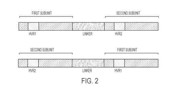
B2M12.
[0105] Fig. 2. The recombinant meganucleases of the disclosure comprise two
subunits,
wherein the first subunit comprising the HVR1 region binds to a first
recognition half-site
(e.g., B2M13, B2M5, B2M7, or B2M11) and the second subunit comprising the HVR2
region binds to a second recognition half-site (e.g., B2M14, B2M6, B2M8, or
B2M12). In
embodiments where the recombinant meganuclease is a single-chain meganuclease,
the first
subunit comprising the HVR1 region can be positioned as either the N-terminal
or C-terminal
subunit. Likewise, the second subunit comprising the HVR2 region can be
positioned as
either the N-terminal or C-terminal subunit.
[0106] Fig. 3. Schematic of reporter assay in CHO cells for evaluating
recombinant
meganucleases targeting recognition sequences found in the beta-2
microglobulin gene (SEQ
ID NO:1). For the recombinant meganucleases described herein, a CHO cell line
was
produced in which a reporter cassette was integrated stably into the genome of
the cell. The
reporter cassette comprised, in 5' to 3' order: an 5V40 Early Promoter; the 5'
2/3 of the GFP
gene; the recognition sequence for an engineered meganuclease of the
disclosure (e.g., the
B2M 13-14 recognition sequence, the B2M 5-6 recognition sequence, the B2M 7-8
recognition sequence, or the B2M 11-12 recognition sequence); the recognition
sequence for
the CHO-23/24 meganuclease (WO/2012/167192); and the 3' 2/3 of the GFP gene.
Cells
stably transfected with this cassette did not express GFP in the absence of a
DNA break-
inducing agent. Meganucleases were introduced by transduction of plasmid DNA
or mRNA
encoding each meganuclease. When a DNA break was induced at either of the
meganuclease
recognition sequences, the duplicated regions of the GFP gene recombined with
one another
to produce a functional GFP gene. The percentage of GFP-expressing cells could
then be
18
CA 03009637 2018-06-22
WO 2017/112859 PCT/US2016/068289
determined by flow cytometry as an indirect measure of the frequency of genome
cleavage by
the meganucleases.
[0107] Figs. 4A-4J. Efficiency of recombinant meganucleases for recognizing
and
cleaving recognition sequences in the human beta-2 microglobulin gene (SEQ ID
NO:1) in a
CHO cell reporter assay. Each of the recombinant meganucleases set forth in
SEQ ID
NOs:12-126 were engineered to target the B2M 13-14 recognition sequence (SEQ
ID NO:2),
the B2M 5-6 recognition sequence (SEQ ID NO:4), the B2M 7-8 recognition
sequence (SEQ
ID NO:6), or the B2M 11-12 recognition sequence (SEQ ID NO:8), and were
screened for
efficacy in the CHO cell reporter assay. The results shown provide the
percentage of GFP-
expressing cells observed in each assay, which indicates the efficacy of each
meganuclease
for cleaving a beta-2 microglobulin target recognition sequence or the CHO-
23/24
recognition sequence. A negative control (B2M bs) was further included in each
assay. A)
Meganucleases targeting the B2M 5-6 recognition sequence. B) Meganucleases
targeting the
B2M 7-8 recognition sequence. C) Meganucleases targeting the B2M 11-12
recognition
sequence. D)-J) Meganucleases targeting the B2M 13-14 recognition sequence.
[0108] Figs. 5A-5N. Efficiency of recombinant B2M 13-14 meganucleases for
inhibiting
cell-surface expression of beta-2 microglobulin in human T cells. 5A-5N) Donor
CD3+
human T cells were stimulated with anti-CD3 and anti-CD28 antibodies for 3
days, then
electroporated with mRNA encoding a given B2M 13-14 meganuclease. As a
positive
control, cells were mock electroporated. In an additional control for
electroporation
efficiency, cells were electroporated with mRNA encoding GFP. At 3 days post-
electroporation, cells were stained with an antibody recognizing beta-2
microglobulin and
analyzed by flow cytometry.
[0109] Figs. 6A-6J. Efficiency of recombinant B2M 13-14 meganucleases for
inhibiting
cell-surface expression of beta-2 microglobulin in human T cells. Additional
B2M 13-14
meganucleases were engineered in which the first meganuclease subunit remained
the same
as in B2M 13-14x.93, but the second meganuclease subunit contained new amino
acid
substitutions at positions contacting the B2M 13-14 recognition sequence. 6A-
6J) Donor
CD3+ human T cells were stimulated with anti-CD3 and anti-CD28 antibodies for
3 days,
then electroporated with mRNA encoding a given B2M 13-14 meganuclease (1 g)
using the
Amaxa 4D-Nucleofector (Lonza) according to the manufacturer's instructions.
B2M 13-
14x.93 QE was included to allow for comparison to previous variants. At 6 days
post-
19
CA 03009637 2018-06-22
WO 2017/112859 PCT/US2016/068289
electroporation, cells were stained with an antibody recognizing beta-2
microglobulin as well
as an antibody recognizing CD3. Data is presented by flow cytometry plots.
[0110] Figs. 7A-7H. Efficiency of recombinant B2M 13-14 meganucleases for
inhibiting
cell-surface expression of beta-2 microglobulin in human T cells. Further B2M
13-14
meganucleases were generated and evaluated for their ability to eliminate cell-
surface
expression of beta-2 microglobulin on human T cells. These nucleases were
based on B2M
13-14x.169. Amino acid substitutions were made in the first meganuclease
subunit to
introduce alternative base contacts, while the second meganuclease subunit
remained the
same as in B2M 13-14x.169. 7A-7H) Donor CD3 + human T cells were stimulated
with anti-
CD3 and anti-CD28 antibodies for 3 days, then electroporated with mRNA
encoding a given
B2M 13-14 meganuclease using the Amaxa 4D-Nucleofector. B2M 13-14x.202 was
included to allow for comparison to previous variants shown in Figs. 6A-6J.
Cells were
stained with an antibody recognizing beta-2 microglobulin as well as an
antibody recognizing
CD3. Flow cytometry data for the B2M 13-14 meganucleases that showed B2M
knockout
efficiency of >40% are shown.
[0111] Figs. 8A-8D. Double knockout of beta-2 microglobulin and T cell
receptor in
human T cells by simultaneous nucleofection of two mRNAs encoding different
meganucleases. Donor CD3 + human T cells were stimulated with anti-CD3 and
anti-CD28
antibodies for 2 days, then co-electroporated with mRNA encoding B2M 13-
14x.202 and
mRNA encoding TRC 1-2x.87 EE using the Amaxa 4D-Nucleofector. As controls,
human T
cells were mock electroporated or electroporated with mRNA encoding a single
meganuclease, either B2M 13-14x.202 or TRC 1-2x.87 EE. At 6 days post-
electroporation,
cells were stained with an antibody against CD3 and an antibody against B2M
and analyzed
by flow cytometry. A) Mock electroporated cells. B) TRC 1-2x.87 EE
nucleofected cells. C)
B2M 13-14x.202 nucleofected cells. D) Cells double nucleofected with B2M 13-
14x.202 and
TRC 1-2x.87 EE.
[0112] Figs. 9A-9D. Double knockout of beta-2 microglobulin and T cell
receptor in
human T cells by simultaneous nucleofection of two mRNAs encoding different
meganucleases. Donor CD3 + human T cells were stimulated with anti-CD3 and
anti-CD28
antibodies for 2 days, then co-electroporated with mRNA encoding B2M 13-
14x.169 and
mRNA encoding TRC 1-2x.87 EE using the Amaxa 4D-Nucleofector. As controls,
human T
cells were mock electroporated or electroporated with mRNA encoding a single
meganuclease, either B2M 13-14x.169 or TRC 1-2x.87 EE. At 6 days post-
electroporation,
CA 03009637 2018-06-22
WO 2017/112859
PCT/US2016/068289
cells were stained with an antibody against CD3 and an antibody against B2M
and analyzed
by flow cytometry. A) Mock nucleofected cells. B) TRC 1-2x.87 EE nucleofected
cells. C)
B2M 13-14x.169 nucleofected cells. D) Cells double nucleofected with B2M 13-
14x.169 and
TRC 1-2x.87 EE.
[0113] Figs. 10A-10C. Double knockout of beta-2 microglobulin and T cell
receptor in
human T cells by sequential nucleofection. Donor CD3 + human T cells were
stimulated with
anti-CD3 and anti-CD28 antibodies for 3 days, then electroporated with mRNA
encoding
B2M 13-14x.93 QE using the Amaxa 4D-Nucleofector. At 4 days post-
electroporation,
B2M-negative cells were enriched using a biotinylated anti-B2M antibody and a
human
biotin selection cocktail kit. The B2M-negative enriched cells were re-
stimulated with anti-
CD3 and anti-CD28 antibodies for 3 days, then electroporated with mRNA
encoding TRC 1-
2x.87 EE using the Amaxa 4D-Nucleofector. At 5 days post-electroporation,
cells were
stained with antibodies against B2M and TCR and analyzed by flow cytometry. A)
Mock
nucleofected cells. B) B2M 13-14x.93 QE nucleofected cells. C) Cells double
nucleofected
with B2M 13-14x.93 QE and TRC 1-2x.87 EE.
[0114] Figs. 11A-11C. Enrichment of a beta-2 microglobulin and T cell
receptor double
knockout population. Donor human peripheral blood mononuclear cells (PMBCs)
were
stimulated with anti-CD3 and anti-CD28 antibodies for 2 days, then
electroporated with
mRNA encoding B2M 13-14x.93 QE using the Amaxa 4D-Nucleofector. B2M-negative
cells were enriched, re-stimulated with anti-CD3 and anti-CD28 antibodies for
3 days, and
electroporated with mRNA encoding TRC 1-2x.87 EE. At 6 days post-
electroporation, CD3-
negative cells were enriched using a CD3 positive selection kit followed by
another
enrichment for B2M-negative cells using a biotinylated anti-B2M antibody and a
biotin
selection kit. Enriched cells were incubated 3 days in the presence of IL-2,
IL-7 and IL-15,
then stained with antibodies against B2M and CD3 and analyzed by flow
cytometry. A)
Non-nucleofected PBMCs stained with anti-CD3 antibody. B) Non-nucleofected
PBMCs
stained with anti-CD3 and anti-B2M antibodies. C) Enriched population of beta-
2
microglobulin and T cell receptor double knock cells stained with anti-CD3 and
anti-B2M
antibodies.
[0115] Figs. 12A-12H. Reduced allogenicity of B2M knockout T cells. The
allogenicity
of B2M knockout T cells was determined in a allogenic cytotoxicity assay using
T cells from
two donors (donor 36 and donor 75) and mismatched dendritic cells from another
donor.
Cytotoxicity was measured by VAD-FMK-FITC signal. A) Wild-type T cells;
effector and
21
CA 03009637 2018-06-22
WO 2017/112859 PCT/US2016/068289
donor cells from donor 36. B) Wild-type T cells; effector cells from donor 36,
target cells
from donor 75. C) Wild-type T cells; effector and donor cells from donor 75.
D) Wild-type
T cells; effector cells from donor 75, target cells from donor 36. E) B2M
knockout T cells;
effector and donor cells from donor 36. F) B2M knockout T cells; effector
cells from donor
36, target cells from donor 75. G) B2M knockout T cells; effector and donor
cells from
donor 75. H) B2M knockout T cells; effector cells from donor 75, target cells
from donor 36.
[0116] Fig. 13. Reduced allogenicity of B2M knockout T cells. Cytotoxicity
determined
by IFN-y secretion as measured by ELISA.
[0117] Fig. 14. Reduced allogenicity of B2M knockout T cells. Cytotoxicity
determined
by LDH secretion.
[0118] Figs. 15A-15N. Simultaneous knockout of TRC and B2M using
bicistronic
mRNA. Donor human T cells were electroporated with 1 g of TRC1-2x.87EE mRNA or
B2M13-14x.479 RNA. An additional sample of cells was electroporated with 1 g
each of
both individual nuclease mRNAs. As controls, human T cells were electroporated
in the
absence of mRNAs. At 3 and 7 days post-electroporation, cells that were
electroporated with
bicistronic mRNAs were stained with an antibody against CD3 (to determine TRC
knockdown) and an antibody against B2M and analyzed by flow cytometry. A) Mock-
treated. B) B2M 13-14x.479. C) TRC 1-2x.87 EE. D) TRC 1-2x.87 EE and B2M 13-
14x.479. E) B2M-IRES-TRC. F) B2M-E2A-TRC. G) B2M-F2A-TRC. H) B2M-P2A-
TRC. I) B2M-T2A-TRC. J) TRC-IRES-B2M. K) TRC-E2A-B2M. L) TRC-F2A-B2M. M)
TRC-P2A-B2M. N) TRC-T2A-B2M.
[0119] Figs. 16A-16P. Titration of bicistronic mRNA in T cells. B2M-IRES-
TRC,
B2M-T2A-TRC, TRC-P2A-B2M, or TRC-T2A-B2M mRNAs were introduced into donor
human T cells at increasing concentrations, and the percent knockdown of cell-
surface CD3
(indicated TRC knockdown) and B2M was determined. For comparison, donor human
T
cells were electroporated with 1 g of TRC1-2x.87EE or 1 g of B2M13-14x.479. In
addition,
donor human T cells were electroporated with both nucleases encoded on
separate RNA
molecules, using doses of 0.5 g of each nuclease or 1 g of each nuclease. As
controls,
human T cells were electroporated with no RNA. At 7 days post-electroporation,
cells were
enumerated and viability was assessed using trypan blue. Cells were stained
with an antibody
against CD3 (to determine TRC knockdown) and an antibody against B2M and
analyzed by
flow cytometry, as well as Ghost Dye 780 to exclude dead cells from analysis.
A) B2M-
IRES-TRC 1 jig. B) B2M-IRES-TRC 2 g. C) B2M-IRES-TRC 4 g. D) B2M-T2A-TRC
22
CA 03009637 2018-06-22
WO 2017/112859 PCT/US2016/068289
1 pig. E) B2M-T2A-TRC 2 g. F) B2M-T2A-TRC 4 g. G) TRC-P2A-B2M 1 pig. H)
TRC-P2A-B2M 2 g. I) TRC-P2A-B2M 4 g. J) TRC-T2A-B2M 1 pig. K) TRC-T2A-B2M
2 g. L) TRC-T2A-B2M 4 g. M) TRC 1-2x.87 EE. N) B2M 13-14x.479. 0) TRC 1-
2x.87
EE 0.5 g and B2M 13-14x.479 0.5 jig. P) TRC 1-2x.87 EE 1.0 g and B2M 13-
14x.479
1.0 g.
[0120] Figs. 17A-17H. Production of anti-CD19 CART cells using bicistronic
mRNA
and AAV. Bicistronic B2M-IRES-TRC was used in conjunction with an AAV vector
to
introduce an exogenous nucleic acid sequence, encoding a chimeric antigen
receptor, into the
genome of human T cells at the TRC 1-2 recognition sequence via homologous
recombination, while simultaneously knocking out cell-surface expression of
both the T cell
receptor and B2M. The AAV vector comprised a nucleic acid comprising the anti-
CD19
CAR coding sequence previously described, which was flanked by homology arms.
Expression of the CAR cassette was driven by a JeT promoter. As controls,
cells were
electroporated with 1 g of TRC1-2x87EE RNA prior to AAV transduction. In
addition,
B2M-IRES-TRC and TRC 1-2x.87 EE electroporated cells were mock transduced. At
3 and 6
days post-electroporation/transduction, edited cells were stained with an
antibody against
CD3 (to determine TRC knockdown) and an antibody against B2M, as well as a
biotinylated
recombinant CD19-Fc fusion protein to detect the CAR. Streptavidin-PE was used
as the
secondary detection reagent for CAR staining. CD3, B2m, and CAR levels were
assessed by
flow cytometry. A) Staining for CD3 (X axis) and B2M (Y axis) after
nucleofection with
TRC 1-2x.87 EE. B) Staining for CD3 (X axis) and B2M (Y axis) after
nucleofection with
B2M-IRES-TRC. C) Staining for CD3 (X axis) and CAR (Y axis) after
nucleofection with
TRC 1-2x.87 EE and mock transduction. D) Staining for CD3 (X axis) and CAR (Y
axis)
after nucleofection with B2M-IRES-TRC and mock transduction. E) Staining for
CD3 (X
axis) and CAR (Y axis) after nucleofection with TRC 1-2x.87 EE and
transduction with
AAV. F) Staining for CD3 (X axis) and CAR (Y axis) after nucleofection with
B2M-IRES-
TRC and transduction with AAV. G) Staining for B2M in cells nucleofected with
TRC 1-
2x.87 EE and transduced with AAV. H) Staining for B2M in cells nucleofected
with B2M-
IRES-TRC and transduced with AAV.
BRIEF DESCRIPTION OF THE SEQUENCES
[0121] SEQ ID NO:1 sets forth the nucleic acid sequence of the human beta-2
microglobulin gene.
23
CA 03009637 2018-06-22
WO 2017/112859
PCT/US2016/068289
[0122] SEQ ID NO:2 sets forth the nucleic acid sequence of the B2M 13-14
recognition
sequence (sense).
[0123] SEQ ID NO:3 sets forth the nucleic acid sequence of the B2M 13-14
recognition
sequence (anti-sense).
[0124] SEQ ID NO:4 sets forth the nucleic acid sequence of the B2M 5-6
recognition
sequence (sense).
[0125] SEQ ID NO:5 sets forth the nucleic acid sequence of the B2M 5-6
recognition
sequence (anti-sense).
[0126] SEQ ID NO:6 sets forth the nucleic acid sequence of the B2M 7-8
recognition
sequence (sense).
[0127] SEQ ID NO:7 sets forth the nucleic acid sequence of the B2M 7-8
recognition
sequence (anti-sense).
[0128] SEQ ID NO:8 sets forth the nucleic acid sequence of the B2M 11-12
recognition
sequence (sense).
[0129] SEQ ID NO:9 sets forth the nucleic acid sequence of the B2M 11-12
recognition
sequence (anti-sense).
[0130] SEQ ID NO:10 sets forth the amino acid sequence of the I-CreI
meganuclease.
[0131] SEQ ID NO:11 sets forth the amino acid sequence of the LAGLIDADG
motif
[0132] SEQ ID NO:12 sets forth the amino acid sequence of the B2M 13-
14x.479
meganuclease.
[0133] SEQ ID NO:13 sets forth the amino acid sequence of the B2M 13-
14x.287
meganuclease.
[0134] SEQ ID NO:14 sets forth the amino acid sequence of the B2M 13-
14x.377
meganuclease.
[0135] SEQ ID NO:15 sets forth the amino acid sequence of the B2M 13-
14x.169
meganuclease.
[0136] SEQ ID NO:16 sets forth the amino acid sequence of the B2M 13-
14x.202
meganuclease.
[0137] SEQ ID NO:17 sets forth the amino acid sequence of the B2M 13-14x.93
meganuclease.
[0138] SEQ ID NO:18 sets forth the amino acid sequence of the B2M 13-14x.93
QE
meganuclease.
24
CA 03009637 2018-06-22
WO 2017/112859 PCT/US2016/068289
[0139] SEQ ID NO:19 sets forth the amino acid sequence of the B2M 13-14x.93
EQ
meganuclease.
[0140] SEQ ID NO:20 sets forth the amino acid sequence of the B2M 13-14x.93
EE
meganuclease.
[0141] SEQ ID NO:21 sets forth the amino acid sequence of the B2M 13-14x.93
QQY66
meganuclease.
[0142] SEQ ID NO:22 sets forth the amino acid sequence of the B2M 13-14x.93
QQK66
meganuclease.
[0143] SEQ ID NO:23 sets forth the amino acid sequence of the B2M 13-14x.93
QQR66
meganuclease.
[0144] SEQ ID NO:24 sets forth the amino acid sequence of the B2M 13-14x.93
EEY66
meganuclease.
[0145] SEQ ID NO:25 sets forth the amino acid sequence of the B2M 13-14x.93
EEK66
meganuclease.
[0146] SEQ ID NO:26 sets forth the amino acid sequence of the B2M 13-14x.93
EER66
meganuclease.
[0147] SEQ ID NO:27 sets forth the amino acid sequence of the B2M 13-14x.93
EQY66
meganuclease.
[0148] SEQ ID NO:28 sets forth the amino acid sequence of the B2M 13-14x.93
EQK66
meganuclease.
[0149] SEQ ID NO:29 sets forth the amino acid sequence of the B2M 13-14x.93
EQR66
meganuclease.
[0150] SEQ ID NO:30 sets forth the amino acid sequence of the B2M 13-14x.3
meganuclease.
[0151] SEQ ID NO:31 sets forth the amino acid sequence of the B2M 13-14x.10
meganuclease.
[0152] SEQ ID NO:32 sets forth the amino acid sequence of the B2M 13-14x.14
meganuclease.
[0153] SEQ ID NO:33 sets forth the amino acid sequence of the B2M 13-14x.22
meganuclease.
[0154] SEQ ID NO:34 sets forth the amino acid sequence of the B2M 13-14x.67
meganuclease.
CA 03009637 2018-06-22
WO 2017/112859
PCT/US2016/068289
[0155] SEQ ID NO:35 sets forth the amino acid sequence of the B2M 13-14x.84
meganuclease.
[0156] SEQ ID NO:36 sets forth the amino acid sequence of the B2M 13-14x.85
meganuclease.
[0157] SEQ ID NO:37 sets forth the amino acid sequence of the B2M 13-14x.96
meganuclease.
[0158] SEQ ID NO:38 sets forth the amino acid sequence of the B2M 13-14x.97
meganuclease.
[0159] SEQ ID NO:39 sets forth the amino acid sequence of the B2M 13-
14x.102
meganuclease.
[0160] SEQ ID NO:40 sets forth the amino acid sequence of the B2M 13-
14x.105
meganuclease.
[0161] SEQ ID NO:41 sets forth the amino acid sequence of the B2M 13-
14x.106
meganuclease.
[0162] SEQ ID NO:42 sets forth the amino acid sequence of the B2M 13-
14x.115
meganuclease.
[0163] SEQ ID NO:43 sets forth the amino acid sequence of the B2M 13-
14x.139
meganuclease.
[0164] SEQ ID NO:44 sets forth the amino acid sequence of the B2M 13-
14x.141
meganuclease.
[0165] SEQ ID NO:45 sets forth the amino acid sequence of the B2M 13-
14x.146
meganuclease.
[0166] SEQ ID NO:46 sets forth the amino acid sequence of the B2M 13-
14x.162
meganuclease.
[0167] SEQ ID NO:47 sets forth the amino acid sequence of the B2M 13-
14x.165
meganuclease.
[0168] SEQ ID NO:48 sets forth the amino acid sequence of the B2M 13-
14x.178
meganuclease.
[0169] SEQ ID NO:49 sets forth the amino acid sequence of the B2M 13-
14x.182
meganuclease.
[0170] SEQ ID NO:50 sets forth the amino acid sequence of the B2M 13-
14x.198
meganuclease.
26
CA 03009637 2018-06-22
WO 2017/112859
PCT/US2016/068289
[0171] SEQ ID NO:51 sets forth the amino acid sequence of the B2M 13-
14x.199
meganuclease.
[0172] SEQ ID NO:52 sets forth the amino acid sequence of the B2M 13-
14x.207
meganuclease.
[0173] SEQ ID NO:53 sets forth the amino acid sequence of the B2M 13-
14x.222
meganuclease.
[0174] SEQ ID NO:54 sets forth the amino acid sequence of the B2M 13-
14x.245
meganuclease.
[0175] SEQ ID NO:55 sets forth the amino acid sequence of the B2M 13-
14x.255
meganuclease.
[0176] SEQ ID NO:56 sets forth the amino acid sequence of the B2M 13-
14x.259
meganuclease.
[0177] SEQ ID NO:57 sets forth the amino acid sequence of the B2M 13-
14x.275
meganuclease.
[0178] SEQ ID NO:58 sets forth the amino acid sequence of the B2M 13-
14x.280
meganuclease.
[0179] SEQ ID NO:59 sets forth the amino acid sequence of the B2M 13-
14x.281
meganuclease.
[0180] SEQ ID NO:60 sets forth the amino acid sequence of the B2M 13-
14x.283
meganuclease.
[0181] SEQ ID NO:61 sets forth the amino acid sequence of the B2M 13-
14x.285
meganuclease.
[0182] SEQ ID NO:62 sets forth the amino acid sequence of the B2M 13-
14x.286
meganuclease.
[0183] SEQ ID NO:63 sets forth the amino acid sequence of the B2M 13-
14x.295
meganuclease.
[0184] SEQ ID NO:64 sets forth the amino acid sequence of the B2M 13-
14x.301
meganuclease.
[0185] SEQ ID NO:65 sets forth the amino acid sequence of the B2M 13-
14x.306
meganuclease.
[0186] SEQ ID NO:66 sets forth the amino acid sequence of the B2M 13-
14x.317
meganuclease.
27
CA 03009637 2018-06-22
WO 2017/112859
PCT/US2016/068289
[0187] SEQ ID NO:67 sets forth the amino acid sequence of the B2M 13-
14x.325
meganuclease.
[0188] SEQ ID NO:68 sets forth the amino acid sequence of the B2M 13-
14x.335
meganuclease.
[0189] SEQ ID NO:69 sets forth the amino acid sequence of the B2M 13-
14x.338
meganuclease.
[0190] SEQ ID NO:70 sets forth the amino acid sequence of the B2M 13-
14x.347
meganuclease.
[0191] SEQ ID NO:71 sets forth the amino acid sequence of the B2M 13-
14x.361
meganuclease.
[0192] SEQ ID NO:72 sets forth the amino acid sequence of the B2M 13-
14x.362
meganuclease.
[0193] SEQ ID NO:73 sets forth the amino acid sequence of the B2M 13-
14x.365
meganuclease.
[0194] SEQ ID NO:74 sets forth the amino acid sequence of the B2M 13-
14x.369
meganuclease.
[0195] SEQ ID NO:75 sets forth the amino acid sequence of the B2M 13-
14x.371
meganuclease.
[0196] SEQ ID NO:76 sets forth the amino acid sequence of the B2M 13-
14x.372
meganuclease.
[0197] SEQ ID NO:77 sets forth the amino acid sequence of the B2M 13-
14x.375
meganuclease.
[0198] SEQ ID NO:78 sets forth the amino acid sequence of the B2M 13-
14x.378
meganuclease.
[0199] SEQ ID NO:79 sets forth the amino acid sequence of the B2M 13-
14x.385
meganuclease.
[0200] SEQ ID NO:80 sets forth the amino acid sequence of the B2M 13-
14x.392
meganuclease.
[0201] SEQ ID NO:81 sets forth the amino acid sequence of the B2M 13-
14x.432
meganuclease.
[0202] SEQ ID NO:82 sets forth the amino acid sequence of the B2M 13-
14x.433
meganuclease.
28
CA 03009637 2018-06-22
WO 2017/112859
PCT/US2016/068289
[0203] SEQ ID NO:83 sets forth the amino acid sequence of the B2M 13-
14x.440
meganuclease.
[0204] SEQ ID NO:84 sets forth the amino acid sequence of the B2M 13-
14x.449
meganuclease.
[0205] SEQ ID NO:85 sets forth the amino acid sequence of the B2M 13-
14x.456
meganuclease.
[0206] SEQ ID NO:86 sets forth the amino acid sequence of the B2M 13-
14x.457
meganuclease.
[0207] SEQ ID NO:87 sets forth the amino acid sequence of the B2M 13-
14x.459
meganuclease.
[0208] SEQ ID NO:88 sets forth the amino acid sequence of the B2M 13-
14x.464
meganuclease.
[0209] SEQ ID NO:89 sets forth the amino acid sequence of the B2M 13-
14x.465
meganuclease.
[0210] SEQ ID NO:90 sets forth the amino acid sequence of the B2M 13-
14x.470
meganuclease.
[0211] SEQ ID NO:91 sets forth the amino acid sequence of the B2M 13-
14x.471
meganuclease.
[0212] SEQ ID NO:92 sets forth the amino acid sequence of the B2M 13-
14x.540
meganuclease.
[0213] SEQ ID NO:93 sets forth the amino acid sequence of the B2M 13-
14x.543
meganuclease.
[0214] SEQ ID NO:94 sets forth the amino acid sequence of the B2M 13-
14x.551
meganuclease.
[0215] SEQ ID NO:95 sets forth the amino acid sequence of the B2M 13-
14x.554
meganuclease.
[0216] SEQ ID NO:96 sets forth the amino acid sequence of the B2M 13-
14x.556
meganuclease.
[0217] SEQ ID NO:97 sets forth the amino acid sequence of the B2M 13-14x.76
meganuclease.
[0218] SEQ ID NO:98 sets forth the amino acid sequence of the B2M 13-14x.82
meganuclease.
29
CA 03009637 2018-06-22
WO 2017/112859
PCT/US2016/068289
[0219] SEQ ID NO:99 sets forth the amino acid sequence of the B2M 13-14x.31
meganuclease.
[0220] SEQ ID NO:100 sets forth the amino acid sequence of the B2M 13-
14x.32
meganuclease.
[0221] SEQ ID NO:101 sets forth the amino acid sequence of the B2M 5-6x.14
meganuclease.
[0222] SEQ ID NO:102 sets forth the amino acid sequence of the B2M 5-6x.5
meganuclease.
[0223] SEQ ID NO:103 sets forth the amino acid sequence of the B2M 5-6x.6
meganuclease.
[0224] SEQ ID NO:104 sets forth the amino acid sequence of the B2M 5-6x.13
meganuclease.
[0225] SEQ ID NO:105 sets forth the amino acid sequence of the B2M 5-6x.22
meganuclease.
[0226] SEQ ID NO:106 sets forth the amino acid sequence of the B2M 5-6x.31
meganuclease.
[0227] SEQ ID NO:107 sets forth the amino acid sequence of the B2M 5-6x.69
meganuclease.
[0228] SEQ ID NO:108 sets forth the amino acid sequence of the B2M 5-6x.73
meganuclease.
[0229] SEQ ID NO:109 sets forth the amino acid sequence of the B2M 5-6x.85
meganuclease.
[0230] SEQ ID NO:110 sets forth the amino acid sequence of the B2M 5-6x.86
meganuclease.
[0231] SEQ ID NO:111 sets forth the amino acid sequence of the B2M 5-6x.91
meganuclease.
[0232] SEQ ID NO:112 sets forth the amino acid sequence of the B2M 5-6x.28
meganuclease.
[0233] SEQ ID NO:113 sets forth the amino acid sequence of the B2M 5-6x.3
meganuclease.
[0234] SEQ ID NO:114 sets forth the amino acid sequence of the B2M 7-8x.88
meganuclease.
CA 03009637 2018-06-22
WO 2017/112859
PCT/US2016/068289
[0235] SEQ ID NO:115 sets forth the amino acid sequence of the B2M 7-8x.7
meganuclease.
[0236] SEQ ID NO:116 sets forth the amino acid sequence of the B2M 7-8x.23
meganuclease.
[0237] SEQ ID NO:117 sets forth the amino acid sequence of the B2M 7-8x.30
meganuclease.
[0238] SEQ ID NO:118 sets forth the amino acid sequence of the B2M 7-8x.53
meganuclease.
[0239] SEQ ID NO:119 sets forth the amino acid sequence of the B2M 7-8x.2
meganuclease.
[0240] SEQ ID NO:120 sets forth the amino acid sequence of the B2M 7-8x.3
meganuclease.
[0241] SEQ ID NO:121 sets forth the amino acid sequence of the B2M 7-8x.6
meganuclease.
[0242] SEQ ID NO:122 sets forth the amino acid sequence of the B2M 7-8x.25
meganuclease.
[0243] SEQ ID NO:123 sets forth the amino acid sequence of the B2M 7-8x.78
meganuclease.
[0244] SEQ ID NO:124 sets forth the amino acid sequence of the B2M 7-8x.85
meganuclease.
[0245] SEQ ID NO:125 sets forth the amino acid sequence of the B2M 11-
12x.45
meganuclease.
[0246] SEQ ID NO:126 sets forth the amino acid sequence of the B2M 11-12x.2
meganuclease.
[0247] SEQ ID NO:127 sets forth the nucleic acid sequence of the human T
cell receptor
alpha constant region gene.
[0248] SEQ ID NO:128 sets forth the nucleic acid sequence of the TRC 1-2
recognition
sequence (sense).
[0249] SEQ ID NO:129 sets forth the nucleic acid sequence of the TRC 3-4
recognition
sequence (sense).
[0250] SEQ ID NO:130 sets forth the nucleic acid sequence of the TRC 7-8
recognition
sequence (sense).
31
CA 03009637 2018-06-22
WO 2017/112859
PCT/US2016/068289
[0251] SEQ ID NO:131 sets forth the amino acid sequence of the TRC 1-2x.87
EE
meganuclease.
[0252] SEQ ID NO:132 sets forth the B2M13 half-site binding subunit of the
B2M 13-
14x.479 meganuclease.
[0253] SEQ ID NO:133 sets forth the B2M13 half-site binding subunit of the
B2M 13-
14x.287 meganuclease.
[0254] SEQ ID NO:134 sets forth the B2M13 half-site binding subunit of the
B2M 13-
14x.377 meganuclease.
[0255] SEQ ID NO:135 sets forth the B2M13 half-site binding subunit of the
B2M 13-
14x.169 meganuclease.
[0256] SEQ ID NO:136 sets forth the B2M13 half-site binding subunit of the
B2M 13-
14x.202 meganuclease.
[0257] SEQ ID NO:137 sets forth the B2M13 half-site binding subunit of the
B2M 13-
14x.93 meganuclease.
[0258] SEQ ID NO:138 sets forth the B2M13 half-site binding subunit of the
B2M 13-
14x.93 QE meganuclease.
[0259] SEQ ID NO:139 sets forth the B2M13 half-site binding subunit of the
B2M 13-
14x.93 EQ meganuclease.
[0260] SEQ ID NO:140 sets forth the B2M13 half-site binding subunit of the
B2M 13-
14x.93 EE meganuclease.
[0261] SEQ ID NO:141 sets forth the B2M13 half-site binding subunit of the
B2M 13-
14x.93 QQY66 meganuclease.
[0262] SEQ ID NO:142 sets forth the B2M13 half-site binding subunit of the
B2M 13-
14x.93 QQK66 meganuclease.
[0263] SEQ ID NO:143 sets forth the B2M13 half-site binding subunit of the
B2M 13-
14x.93 QQR66 meganuclease.
[0264] SEQ ID NO:144 sets forth the B2M13 half-site binding subunit of the
B2M 13-
14x.93 EEY66 meganuclease.
[0265] SEQ ID NO:145 sets forth the B2M13 half-site binding subunit of the
B2M 13-
14x.93 EEK66 meganuclease.
[0266] SEQ ID NO:146 sets forth the B2M13 half-site binding subunit of the
B2M 13-
14x.93 EER66 meganuclease.
32
CA 03009637 2018-06-22
WO 2017/112859
PCT/US2016/068289
[0267] SEQ
ID NO:147 sets forth the B2M13 half-site binding subunit of the B2M 13-
14x.93 EQY66 meganuclease.
[0268] SEQ
ID NO:148 sets forth the B2M13 half-site binding subunit of the B2M 13-
14x.93 EQK66 meganuclease.
[0269] SEQ
ID NO:149 sets forth the B2M13 half-site binding subunit of the B2M 13-
14x.93 EQR66 meganuclease.
[0270] SEQ
ID NO:150 sets forth the B2M13 half-site binding subunit of the B2M 13-
14x.3 meganuclease.
[0271] SEQ
ID NO:151 sets forth the B2M13 half-site binding subunit of the B2M 13-
14x.10 meganuclease.
[0272] SEQ
ID NO:152 sets forth the B2M13 half-site binding subunit of the B2M 13-
14x.14 meganuclease.
[0273] SEQ
ID NO:153 sets forth the B2M13 half-site binding subunit of the B2M 13-
14x.22 meganuclease.
[0274] SEQ
ID NO:154 sets forth the B2M13 half-site binding subunit of the B2M 13-
14x.67 meganuclease.
[0275] SEQ
ID NO:155 sets forth the B2M13 half-site binding subunit of the B2M 13-
14x.84 meganuclease.
[0276] SEQ
ID NO:156 sets forth the B2M13 half-site binding subunit of the B2M 13-
14x.85 meganuclease.
[0277] SEQ
ID NO:157 sets forth the B2M13 half-site binding subunit of the B2M 13-
14x.96 meganuclease.
[0278] SEQ
ID NO:158 sets forth the B2M13 half-site binding subunit of the B2M 13-
14x.97 meganuclease.
[0279] SEQ
ID NO:159 sets forth the B2M13 half-site binding subunit of the B2M 13-
14x.102 meganuclease.
[0280] SEQ
ID NO:160 sets forth the B2M13 half-site binding subunit of the B2M 13-
14x.105 meganuclease.
[0281] SEQ
ID NO:161 sets forth the B2M13 half-site binding subunit of the B2M 13-
14x.106 meganuclease.
[0282] SEQ
ID NO:162 sets forth the B2M13 half-site binding subunit of the B2M 13-
14x.115 meganuclease.
33
CA 03009637 2018-06-22
WO 2017/112859
PCT/US2016/068289
[0283] SEQ
ID NO:163 sets forth the B2M13 half-site binding subunit of the B2M 13-
14x.139 meganuclease.
[0284] SEQ
ID NO:164 sets forth the B2M13 half-site binding subunit of the B2M 13-
14x.141 meganuclease.
[0285] SEQ
ID NO:165 sets forth the B2M13 half-site binding subunit of the B2M 13-
14x.146 meganuclease.
[0286] SEQ
ID NO:166 sets forth the B2M13 half-site binding subunit of the B2M 13-
14x.162 meganuclease.
[0287] SEQ
ID NO:167 sets forth the B2M13 half-site binding subunit of the B2M 13-
14x.165 meganuclease.
[0288] SEQ
ID NO:168 sets forth the B2M13 half-site binding subunit of the B2M 13-
14x.178 meganuclease.
[0289] SEQ
ID NO:169 sets forth the B2M13 half-site binding subunit of the B2M 13-
14x.182 meganuclease.
[0290] SEQ
ID NO:170 sets forth the B2M13 half-site binding subunit of the B2M 13-
14x.198 meganuclease.
[0291] SEQ
ID NO:171 sets forth the B2M13 half-site binding subunit of the B2M 13-
14x.199 meganuclease.
[0292] SEQ
ID NO:172 sets forth the B2M13 half-site binding subunit of the B2M 13-
14x.207 meganuclease.
[0293] SEQ
ID NO:173 sets forth the B2M13 half-site binding subunit of the B2M 13-
14x.222 meganuclease.
[0294] SEQ
ID NO:174 sets forth the B2M13 half-site binding subunit of the B2M 13-
14x.245 meganuclease.
[0295] SEQ
ID NO:175 sets forth the B2M13 half-site binding subunit of the B2M 13-
14x.255 meganuclease.
[0296] SEQ
ID NO:176 sets forth the B2M13 half-site binding subunit of the B2M 13-
14x.259 meganuclease.
[0297] SEQ
ID NO:177 sets forth the B2M13 half-site binding subunit of the B2M 13-
14x.275 meganuclease.
[0298] SEQ
ID NO:178 sets forth the B2M13 half-site binding subunit of the B2M 13-
14x.280 meganuclease.
34
CA 03009637 2018-06-22
WO 2017/112859
PCT/US2016/068289
[0299] SEQ
ID NO:179 sets forth the B2M13 half-site binding subunit of the B2M 13-
14x.281 meganuclease.
[0300] SEQ
ID NO:180 sets forth the B2M13 half-site binding subunit of the B2M 13-
14x.283 meganuclease.
[0301] SEQ
ID NO:181 sets forth the B2M13 half-site binding subunit of the B2M 13-
14x.285 meganuclease.
[0302] SEQ
ID NO:182 sets forth the B2M13 half-site binding subunit of the B2M 13-
14x.286 meganuclease.
[0303] SEQ
ID NO:183 sets forth the B2M13 half-site binding subunit of the B2M 13-
14x.295 meganuclease.
[0304] SEQ
ID NO:184 sets forth the B2M13 half-site binding subunit of the B2M 13-
14x.301 meganuclease.
[0305] SEQ
ID NO:185 sets forth the B2M13 half-site binding subunit of the B2M 13-
14x.306 meganuclease.
[0306] SEQ
ID NO:186 sets forth the B2M13 half-site binding subunit of the B2M 13-
14x.317 meganuclease.
[0307] SEQ
ID NO:187 sets forth the B2M13 half-site binding subunit of the B2M 13-
14x.325 meganuclease.
[0308] SEQ
ID NO:188 sets forth the B2M13 half-site binding subunit of the B2M 13-
14x.335 meganuclease.
[0309] SEQ
ID NO:189 sets forth the B2M13 half-site binding subunit of the B2M 13-
14x.338 meganuclease.
[0310] SEQ
ID NO:190 sets forth the B2M13 half-site binding subunit of the B2M 13-
14x.347 meganuclease.
[0311] SEQ
ID NO:191 sets forth the B2M13 half-site binding subunit of the B2M 13-
14x.361 meganuclease.
[0312] SEQ
ID NO:192 sets forth the B2M13 half-site binding subunit of the B2M 13-
14x.362 meganuclease.
[0313] SEQ
ID NO:193 sets forth the B2M13 half-site binding subunit of the B2M 13-
14x.365 meganuclease.
[0314] SEQ
ID NO:194 sets forth the B2M13 half-site binding subunit of the B2M 13-
14x.369 meganuclease.
CA 03009637 2018-06-22
WO 2017/112859
PCT/US2016/068289
[0315] SEQ
ID NO:195 sets forth the B2M13 half-site binding subunit of the B2M 13-
14x.371 meganuclease.
[0316] SEQ
ID NO:196 sets forth the B2M13 half-site binding subunit of the B2M 13-
14x.372 meganuclease.
[0317] SEQ
ID NO:197 sets forth the B2M13 half-site binding subunit of the B2M 13-
14x.375 meganuclease.
[0318] SEQ
ID NO:198 sets forth the B2M13 half-site binding subunit of the B2M 13-
14x.378 meganuclease.
[0319] SEQ
ID NO:199 sets forth the B2M13 half-site binding subunit of the B2M 13-
14x.385 meganuclease.
[0320] SEQ
ID NO:200 sets forth the B2M13 half-site binding subunit of the B2M 13-
14x.392 meganuclease.
[0321] SEQ
ID NO:201 sets forth the B2M13 half-site binding subunit of the B2M 13-
14x.432 meganuclease.
[0322] SEQ
ID NO:202 sets forth the B2M13 half-site binding subunit of the B2M 13-
14x.433 meganuclease.
[0323] SEQ
ID NO:203 sets forth the B2M13 half-site binding subunit of the B2M 13-
14x.440 meganuclease.
[0324] SEQ
ID NO:204 sets forth the B2M13 half-site binding subunit of the B2M 13-
14x.449 meganuclease.
[0325] SEQ
ID NO:205 sets forth the B2M13 half-site binding subunit of the B2M 13-
14x.456 meganuclease.
[0326] SEQ
ID NO:206 sets forth the B2M13 half-site binding subunit of the B2M 13-
14x.457 meganuclease.
[0327] SEQ
ID NO:207 sets forth the B2M13 half-site binding subunit of the B2M 13-
14x.459 meganuclease.
[0328] SEQ
ID NO:208 sets forth the B2M13 half-site binding subunit of the B2M 13-
14x.464 meganuclease.
[0329] SEQ
ID NO:209 sets forth the B2M13 half-site binding subunit of the B2M 13-
14x.465 meganuclease.
[0330] SEQ
ID NO:210 sets forth the B2M13 half-site binding subunit of the B2M 13-
14x.470 meganuclease.
36
CA 03009637 2018-06-22
WO 2017/112859
PCT/US2016/068289
[0331] SEQ
ID NO:211 sets forth the B2M13 half-site binding subunit of the B2M 13-
14x.471 meganuclease.
[0332] SEQ
ID NO:212 sets forth the B2M13 half-site binding subunit of the B2M 13-
14x.540 meganuclease.
[0333] SEQ
ID NO:213 sets forth the B2M13 half-site binding subunit of the B2M 13-
14x.543 meganuclease.
[0334] SEQ
ID NO:214 sets forth the B2M13 half-site binding subunit of the B2M 13-
14x.551 meganuclease.
[0335] SEQ
ID NO:215 sets forth the B2M13 half-site binding subunit of the B2M 13-
14x.554 meganuclease.
[0336] SEQ
ID NO:216 sets forth the B2M13 half-site binding subunit of the B2M 13-
14x.556 meganuclease.
[0337] SEQ
ID NO:217 sets forth the B2M13 half-site binding subunit of the B2M 13-
14x.76 meganuclease.
[0338] SEQ
ID NO:218 sets forth the B2M13 half-site binding subunit of the B2M 13-
14x.82 meganuclease.
[0339] SEQ
ID NO:219 sets forth the B2M13 half-site binding subunit of the B2M 13-
14x .31 meganuclease.
[0340] SEQ
ID NO:220 sets forth the B2M13 half-site binding subunit of the B2M 13-
14x.32 meganuclease.
[0341] SEQ
ID NO:221 sets forth the B2M14 half-site binding subunit of the B2M 13-
14x.479 meganuclease.
[0342] SEQ
ID NO:222 sets forth the B2M14 half-site binding subunit of the B2M 13-
14x.287 meganuclease.
[0343] SEQ
ID NO:223 sets forth the B2M14 half-site binding subunit of the B2M 13-
14x.377 meganuclease.
[0344] SEQ
ID NO:224 sets forth the B2M14 half-site binding subunit of the B2M 13-
14x.169 meganuclease.
[0345] SEQ
ID NO:225 sets forth the B2M14 half-site binding subunit of the B2M 13-
14x.202 meganuclease.
[0346] SEQ
ID NO:226 sets forth the B2M14 half-site binding subunit of the B2M 13-
14x.93 meganuclease.
37
CA 03009637 2018-06-22
WO 2017/112859
PCT/US2016/068289
[0347] SEQ
ID NO:227 sets forth the B2M14 half-site binding subunit of the B2M 13-
14x.93 QE meganuclease.
[0348] SEQ
ID NO:228 sets forth the B2M14 half-site binding subunit of the B2M 13-
14x.93 EQ meganuclease.
[0349] SEQ
ID NO:229 sets forth the B2M14 half-site binding subunit of the B2M 13-
14x.93 EE meganuclease.
[0350] SEQ
ID NO:230 sets forth the B2M14 half-site binding subunit of the B2M 13-
14x.93 QQY66 meganuclease.
[0351] SEQ
ID NO:231 sets forth the B2M14 half-site binding subunit of the B2M 13-
14x.93 QQK66 meganuclease.
[0352] SEQ
ID NO:232 sets forth the B2M14 half-site binding subunit of the B2M 13-
14x.93 QQR66 meganuclease.
[0353] SEQ
ID NO:233 sets forth the B2M14 half-site binding subunit of the B2M 13-
14x.93 EEY66 meganuclease.
[0354] SEQ
ID NO:234 sets forth the B2M14 half-site binding subunit of the B2M 13-
14x.93 EEK66 meganuclease.
[0355] SEQ
ID NO:235 sets forth the B2M14 half-site binding subunit of the B2M 13-
14x.93 EER66 meganuclease.
[0356] SEQ
ID NO:236 sets forth the B2M14 half-site binding subunit of the B2M 13-
14x.93 EQY66 meganuclease.
[0357] SEQ
ID NO:237 sets forth the B2M14 half-site binding subunit of the B2M 13-
14x.93 EQK66 meganuclease.
[0358] SEQ
ID NO:238 sets forth the B2M14 half-site binding subunit of the B2M 13-
14x.93 EQR66 meganuclease.
[0359] SEQ
ID NO:239 sets forth the B2M14 half-site binding subunit of the B2M 13-
14x.3 meganuclease.
[0360] SEQ
ID NO:240 sets forth the B2M14 half-site binding subunit of the B2M 13-
14x.10 meganuclease.
[0361] SEQ
ID NO:241 sets forth the B2M14 half-site binding subunit of the B2M 13-
14x.14 meganuclease.
[0362] SEQ
ID NO:242 sets forth the B2M14 half-site binding subunit of the B2M 13-
14x.22 meganuclease.
38
CA 03009637 2018-06-22
WO 2017/112859
PCT/US2016/068289
[0363] SEQ
ID NO:243 sets forth the B2M14 half-site binding subunit of the B2M 13-
14x.67 meganuclease.
[0364] SEQ
ID NO:244 sets forth the B2M14 half-site binding subunit of the B2M 13-
14x.84 meganuclease.
[0365] SEQ
ID NO:245 sets forth the B2M14 half-site binding subunit of the B2M 13-
14x.85 meganuclease.
[0366] SEQ
ID NO:246 sets forth the B2M14 half-site binding subunit of the B2M 13-
14x.96 meganuclease.
[0367] SEQ
ID NO:247 sets forth the B2M14 half-site binding subunit of the B2M 13-
14x.97 meganuclease.
[0368] SEQ
ID NO:248 sets forth the B2M14 half-site binding subunit of the B2M 13-
14x.102 meganuclease.
[0369] SEQ
ID NO:249 sets forth the B2M14 half-site binding subunit of the B2M 13-
14x.105 meganuclease.
[0370] SEQ
ID NO:250 sets forth the B2M14 half-site binding subunit of the B2M 13-
14x.106 meganuclease.
[0371] SEQ
ID NO:251 sets forth the B2M14 half-site binding subunit of the B2M 13-
14x.115 meganuclease.
[0372] SEQ
ID NO:252 sets forth the B2M14 half-site binding subunit of the B2M 13-
14x.139 meganuclease.
[0373] SEQ
ID NO:253 sets forth the B2M14 half-site binding subunit of the B2M 13-
14x.141 meganuclease.
[0374] SEQ
ID NO:254 sets forth the B2M14 half-site binding subunit of the B2M 13-
14x.146 meganuclease.
[0375] SEQ
ID NO:255 sets forth the B2M14 half-site binding subunit of the B2M 13-
14x.162 meganuclease.
[0376] SEQ
ID NO:256 sets forth the B2M14 half-site binding subunit of the B2M 13-
14x.165 meganuclease.
[0377] SEQ
ID NO:257 sets forth the B2M14 half-site binding subunit of the B2M 13-
14x.178 meganuclease.
[0378] SEQ
ID NO:258 sets forth the B2M14 half-site binding subunit of the B2M 13-
14x.182 meganuclease.
39
CA 03009637 2018-06-22
WO 2017/112859
PCT/US2016/068289
[0379] SEQ
ID NO:259 sets forth the B2M14 half-site binding subunit of the B2M 13-
14x.198 meganuclease.
[0380] SEQ
ID NO:260 sets forth the B2M14 half-site binding subunit of the B2M 13-
14x.199 meganuclease.
[0381] SEQ
ID NO:261 sets forth the B2M14 half-site binding subunit of the B2M 13-
14x.207 meganuclease.
[0382] SEQ
ID NO:262 sets forth the B2M14 half-site binding subunit of the B2M 13-
14x.222 meganuclease.
[0383] SEQ
ID NO:263 sets forth the B2M14 half-site binding subunit of the B2M 13-
14x.245 meganuclease.
[0384] SEQ
ID NO:264 sets forth the B2M14 half-site binding subunit of the B2M 13-
14x.255 meganuclease.
[0385] SEQ
ID NO:265 sets forth the B2M14 half-site binding subunit of the B2M 13-
14x.259 meganuclease.
[0386] SEQ
ID NO:266 sets forth the B2M14 half-site binding subunit of the B2M 13-
14x.275 meganuclease.
[0387] SEQ
ID NO:267 sets forth the B2M14 half-site binding subunit of the B2M 13-
14x.280 meganuclease.
[0388] SEQ
ID NO:268 sets forth the B2M14 half-site binding subunit of the B2M 13-
14x.281 meganuclease.
[0389] SEQ
ID NO:269 sets forth the B2M14 half-site binding subunit of the B2M 13-
14x.283 meganuclease.
[0390] SEQ
ID NO:270 sets forth the B2M14 half-site binding subunit of the B2M 13-
14x.285 meganuclease.
[0391] SEQ
ID NO:271 sets forth the B2M14 half-site binding subunit of the B2M 13-
14x.286 meganuclease.
[0392] SEQ
ID NO:272 sets forth the B2M14 half-site binding subunit of the B2M 13-
14x.295 meganuclease.
[0393] SEQ
ID NO:273 sets forth the B2M14 half-site binding subunit of the B2M 13-
14x.301 meganuclease.
[0394] SEQ
ID NO:274 sets forth the B2M14 half-site binding subunit of the B2M 13-
14x.306 meganuclease.
CA 03009637 2018-06-22
WO 2017/112859
PCT/US2016/068289
[0395] SEQ
ID NO:275 sets forth the B2M14 half-site binding subunit of the B2M 13-
14x.317 meganuclease.
[0396] SEQ
ID NO:276 sets forth the B2M14 half-site binding subunit of the B2M 13-
14x.325 meganuclease.
[0397] SEQ
ID NO:277 sets forth the B2M14 half-site binding subunit of the B2M 13-
14x.335 meganuclease.
[0398] SEQ
ID NO:278 sets forth the B2M14 half-site binding subunit of the B2M 13-
14x.338 meganuclease.
[0399] SEQ
ID NO:279 sets forth the B2M14 half-site binding subunit of the B2M 13-
14x.347 meganuclease.
[0400] SEQ
ID NO:280 sets forth the B2M14 half-site binding subunit of the B2M 13-
14x.361 meganuclease.
[0401] SEQ
ID NO:281 sets forth the B2M14 half-site binding subunit of the B2M 13-
14x.362 meganuclease.
[0402] SEQ
ID NO:282 sets forth the B2M14 half-site binding subunit of the B2M 13-
14x.365 meganuclease.
[0403] SEQ
ID NO:283 sets forth the B2M14 half-site binding subunit of the B2M 13-
14x.369 meganuclease.
[0404] SEQ
ID NO:284 sets forth the B2M14 half-site binding subunit of the B2M 13-
14x.371 meganuclease.
[0405] SEQ
ID NO:285 sets forth the B2M14 half-site binding subunit of the B2M 13-
14x.372 meganuclease.
[0406] SEQ
ID NO:286 sets forth the B2M14 half-site binding subunit of the B2M 13-
14x.375 meganuclease.
[0407] SEQ
ID NO:287 sets forth the B2M14 half-site binding subunit of the B2M 13-
14x.378 meganuclease.
[0408] SEQ
ID NO:288 sets forth the B2M14 half-site binding subunit of the B2M 13-
14x.385 meganuclease.
[0409] SEQ
ID NO:289 sets forth the B2M14 half-site binding subunit of the B2M 13-
14x.392 meganuclease.
[0410] SEQ
ID NO:290 sets forth the B2M14 half-site binding subunit of the B2M 13-
14x.432 meganuclease.
41
CA 03009637 2018-06-22
WO 2017/112859
PCT/US2016/068289
[0411] SEQ
ID NO:291 sets forth the B2M14 half-site binding subunit of the B2M 13-
14x.433 meganuclease.
[0412] SEQ
ID NO:292 sets forth the B2M14 half-site binding subunit of the B2M 13-
14x.440 meganuclease.
[0413] SEQ
ID NO:293 sets forth the B2M14 half-site binding subunit of the B2M 13-
14x.449 meganuclease.
[0414] SEQ
ID NO:294 sets forth the B2M14 half-site binding subunit of the B2M 13-
14x.456 meganuclease.
[0415] SEQ
ID NO:295 sets forth the B2M14 half-site binding subunit of the B2M 13-
14x.457 meganuclease.
[0416] SEQ
ID NO:296 sets forth the B2M14 half-site binding subunit of the B2M 13-
14x.459 meganuclease.
[0417] SEQ
ID NO:297 sets forth the B2M14 half-site binding subunit of the B2M 13-
14x.464 meganuclease.
[0418] SEQ
ID NO:298 sets forth the B2M14 half-site binding subunit of the B2M 13-
14x.465 meganuclease.
[0419] SEQ
ID NO:299 sets forth the B2M14 half-site binding subunit of the B2M 13-
14x.470 meganuclease.
[0420] SEQ
ID NO:300 sets forth the B2M14 half-site binding subunit of the B2M 13-
14x.471 meganuclease.
[0421] SEQ
ID NO:301 sets forth the B2M14 half-site binding subunit of the B2M 13-
14x.540 meganuclease.
[0422] SEQ
ID NO:302 sets forth the B2M14 half-site binding subunit of the B2M 13-
14x.543 meganuclease.
[0423] SEQ
ID NO:303 sets forth the B2M14 half-site binding subunit of the B2M 13-
14x.551 meganuclease.
[0424] SEQ
ID NO:304 sets forth the B2M14 half-site binding subunit of the B2M 13-
14x.554 meganuclease.
[0425] SEQ
ID NO:305 sets forth the B2M14 half-site binding subunit of the B2M 13-
14x.556 meganuclease.
[0426] SEQ
ID NO:306 sets forth the B2M14 half-site binding subunit of the B2M 13-
14x.76 meganuclease.
42
CA 03009637 2018-06-22
WO 2017/112859
PCT/US2016/068289
[0427] SEQ ID NO:307 sets forth the B2M14 half-site binding subunit of the
B2M 13-
14x.82 meganuclease.
[0428] SEQ ID NO:308 sets forth the B2M14 half-site binding subunit of the
B2M 13-
14x .31 meganuclease.
[0429] SEQ ID NO:309 sets forth the B2M14 half-site binding subunit of the
B2M 13-
14x.32 meganuclease.
[0430] SEQ ID NO:310 sets forth the B2M5 half-site binding subunit of the
B2M 5-
6x.14 meganuclease.
[0431] SEQ ID NO:311 sets forth the B2M5 half-site binding subunit of the
B2M 5-6x.5
meganuclease.
[0432] SEQ ID NO:312 sets forth the B2M5 half-site binding subunit of the
B2M 5-6x.6
meganuclease.
[0433] SEQ ID NO:313 sets forth the B2M5 half-site binding subunit of the
B2M 5-
6x.13 meganuclease.
[0434] SEQ ID NO:314 sets forth the B2M5 half-site binding subunit of the
B2M 5-
6x.22 meganuclease.
[0435] SEQ ID NO:315 sets forth the B2M5 half-site binding subunit of the
B2M 5-
6x.31 meganuclease.
[0436] SEQ ID NO:316 sets forth the B2M5 half-site binding subunit of the
B2M 5-
6x.69 meganuclease.
[0437] SEQ ID NO:317 sets forth the B2M5 half-site binding subunit of the
B2M 5-
6x.73 meganuclease.
[0438] SEQ ID NO:318 sets forth the B2M5 half-site binding subunit of the
B2M 5-
6x.85 meganuclease.
[0439] SEQ ID NO:319 sets forth the B2M5 half-site binding subunit of the
B2M 5-
6x.86 meganuclease.
[0440] SEQ ID NO:320 sets forth the B2M5 half-site binding subunit of the
B2M 5-
6x.91 meganuclease.
[0441] SEQ ID NO:321 sets forth the B2M5 half-site binding subunit of the
B2M 5-
6x.28 meganuclease.
[0442] SEQ ID NO:322 sets forth the B2M5 half-site binding subunit of the
B2M 5-6x.3
meganuclease.
43
CA 03009637 2018-06-22
WO 2017/112859
PCT/US2016/068289
[0443] SEQ ID NO:323 sets forth the B2M6 half-site binding subunit of the
B2M 5-
6x.14 meganuclease.
[0444] SEQ ID NO:324 sets forth the B2M6 half-site binding subunit of the
B2M 5-6x.5
meganuclease.
[0445] SEQ ID NO:325 sets forth the B2M6 half-site binding subunit of the
B2M 5-6x.6
meganuclease.
[0446] SEQ ID NO:326 sets forth the B2M6 half-site binding subunit of the
B2M 5-
6x.13 meganuclease.
[0447] SEQ ID NO:327 sets forth the B2M6 half-site binding subunit of the
B2M 5-
6x.22 meganuclease.
[0448] SEQ ID NO:328 sets forth the B2M6 half-site binding subunit of the
B2M 5-
6x.31 meganuclease.
[0449] SEQ ID NO:329 sets forth the B2M6 half-site binding subunit of the
B2M 5-
6x.69 meganuclease.
[0450] SEQ ID NO:330 sets forth the B2M6 half-site binding subunit of the
B2M 5-
6x.73 meganuclease.
[0451] SEQ ID NO:331 sets forth the B2M6 half-site binding subunit of the
B2M 5-
6x.85 meganuclease.
[0452] SEQ ID NO:332 sets forth the B2M6 half-site binding subunit of the
B2M 5-
6x.86 meganuclease.
[0453] SEQ ID NO:333 sets forth the B2M6 half-site binding subunit of the
B2M 5-
6x.91 meganuclease.
[0454] SEQ ID NO:334 sets forth the B2M6 half-site binding subunit of the
B2M 5-
6x.28 meganuclease.
[0455] SEQ ID NO:335 sets forth the B2M6 half-site binding subunit of the
B2M 5-6x.3
meganuclease.
[0456] SEQ ID NO:336 sets forth the B2M7 half-site binding subunit of the
B2M 7-
8x.88 meganuclease.
[0457] SEQ ID NO:337 sets forth the B2M7 half-site binding subunit of the
B2M 7-8x.7
meganuclease.
[0458] SEQ ID NO:338 sets forth the B2M7 half-site binding subunit of the
B2M 7-
8x.23 meganuclease.
44
CA 03009637 2018-06-22
WO 2017/112859
PCT/US2016/068289
[0459] SEQ ID NO:339 sets forth the B2M7 half-site binding subunit of the
B2M 7-
8x.30 meganuclease.
[0460] SEQ ID NO:340 sets forth the B2M7 half-site binding subunit of the
B2M 7-
8x.53 meganuclease.
[0461] SEQ ID NO:341 sets forth the B2M7 half-site binding subunit of the
B2M 7-8x.2
meganuclease.
[0462] SEQ ID NO:342 sets forth the B2M7 half-site binding subunit of the
B2M 7-8x.3
meganuclease.
[0463] SEQ ID NO:343 sets forth the B2M7 half-site binding subunit of the
B2M 7-8x.6
meganuclease.
[0464] SEQ ID NO:344 sets forth the B2M7 half-site binding subunit of the
B2M 7-
8x.25 meganuclease.
[0465] SEQ ID NO:345 sets forth the B2M7 half-site binding subunit of the
B2M 7-
8x.78 meganuclease.
[0466] SEQ ID NO:346 sets forth the B2M7 half-site binding subunit of the
B2M 7-
8x.85 meganuclease.
[0467] SEQ ID NO:347 sets forth the B2M8 half-site binding subunit of the
B2M 7-
8x.88 meganuclease.
[0468] SEQ ID NO:348 sets forth the B2M8 half-site binding subunit of the
B2M 7-8x.7
meganuclease.
[0469] SEQ ID NO:349 sets forth the B2M8 half-site binding subunit of the
B2M 7-
8x.23 meganuclease.
[0470] SEQ ID NO:350 sets forth the B2M8 half-site binding subunit of the
B2M 7-
8x.30 meganuclease.
[0471] SEQ ID NO:351 sets forth the B2M8 half-site binding subunit of the
B2M 7-
8x.53 meganuclease.
[0472] SEQ ID NO:352 sets forth the B2M8 half-site binding subunit of the
B2M 7-8x.2
meganuclease.
[0473] SEQ ID NO:353 sets forth the B2M8 half-site binding subunit of the
B2M 7-8x.3
meganuclease.
[0474] SEQ ID NO:354 sets forth the B2M8 half-site binding subunit of the
B2M 7-8x.6
meganuclease.
CA 03009637 2018-06-22
WO 2017/112859
PCT/US2016/068289
[0475] SEQ ID NO:355 sets forth the B2M8 half-site binding subunit of the
B2M 7-
8x.25 meganuclease.
[0476] SEQ ID NO:356 sets forth the B2M8 half-site binding subunit of the
B2M 7-
8x.78 meganuclease.
[0477] SEQ ID NO:357 sets forth the B2M8 half-site binding subunit of the
B2M 7-
8x.85 meganuclease.
[0478] SEQ ID NO:358 sets forth the B2M11 half-site binding subunit of the
B2M 11-
12x.45 meganuclease.
[0479] SEQ ID NO:359 sets forth the B2M11 half-site binding subunit of the
B2M 11-
12x.2 meganuclease.
[0480] SEQ ID NO:360 sets forth the B2M12 half-site binding subunit of the
B2M 11-
12x.45 meganuclease.
[0481] SEQ ID NO:361 sets forth the B2M12 half-site binding subunit of the
B2M 11-
12x.2 meganuclease.
[0482] SEQ ID NO:362 sets forth the nucleic acid sequence of an IRES
element.
[0483] SEQ ID NO:363 sets forth the nucleic acid sequence of a T2A element.
[0484] SEQ ID NO:364 sets forth the nucleic acid sequence of a P2A element.
[0485] SEQ ID NO:365 sets forth the nucleic acid sequence of a E2A element.
[0486] SEQ ID NO:366 sets forth the nucleic acid sequence of a F2A element.
[0487] SEQ ID NO:367 sets forth the nucleic acid sequence of a TRC-IRES-B2M
bicistronic mRNA.
[0488] SEQ ID NO:368 sets forth the nucleic acid sequence of a TRC-T2A-B2M
bicistronic mRNA.
[0489] SEQ ID NO:369 sets forth the nucleic acid sequence of a TRC-P2A-B2M
bicistronic mRNA.
[0490] SEQ ID NO:370 sets forth the nucleic acid sequence of a TRC-E2A-B2M
bicistronic mRNA.
[0491] SEQ ID NO:371 sets forth the nucleic acid sequence of a TRC-F2A-B2M
bicistronic mRNA.
[0492] SEQ ID NO:372 sets forth the nucleic acid sequence of a B2M-IRES-TRC
bicistronic mRNA.
[0493] SEQ ID NO:373 sets forth the nucleic acid sequence of a B2M-T2A-TRC
bicistronic mRNA.
46
CA 03009637 2018-06-22
WO 2017/112859 PCT/US2016/068289
[0494] SEQ ID NO:374 sets forth the nucleic acid sequence of a B2M-P2A-TRC
bicistronic mRNA.
[0495] SEQ ID NO:375 sets forth the nucleic acid sequence of a B2M-E2A-TRC
bicistronic mRNA.
[0496] SEQ ID NO:376 sets forth the nucleic acid sequence of a B2M-F2A-TRC
bicistronic mRNA.
DETAILED DESCRIPTION OF THE INVENTION
1.1 References and Definitions
[0497] The patent and scientific literature referred to herein establishes
knowledge that is
available to those of skill in the art. The issued US patents, allowed
applications, published
foreign applications, and references, including GenBank database sequences,
which are cited
herein are hereby incorporated by reference to the same extent as if each was
specifically and
individually indicated to be incorporated by reference.
[0498] The present disclosure is embodied in different forms and should not
be construed
as limited to the embodiments set forth herein. Rather, these embodiments are
provided so
that this disclosure will be thorough and complete, and will fully convey the
scope of the
disclosure to those skilled in the art. For example, features illustrated with
respect to one
embodiment can be incorporated into other embodiments, and features
illustrated with respect
to a particular embodiment can be deleted from that embodiment. In addition,
numerous
variations and additions to the embodiments suggested herein will be apparent
to those
skilled in the art in light of the instant disclosure, which do not depart
from the instant
disclosure.
[0499] Unless otherwise defined, all technical and scientific terms used
herein have the
same meaning as commonly understood by one of ordinary skill in the art to
which this
disclosure belongs. The terminology used in the description of the disclosure
herein is for the
purpose of describing particular embodiments only and is not intended to be
limiting of the
disclosure.
[0500] All publications, patent applications, patents, and other references
mentioned
herein are incorporated by reference herein in their entirety.
[0501] As used herein, "a," "an," or "the" can mean one or more than one.
For example,
"a" cell can mean a single cell or a multiplicity of cells.
[0502] As used herein, unless specifically indicated otherwise, the word
"or" is used in
the inclusive sense of "and/or" and not the exclusive sense of "either/or."
47
CA 03009637 2018-06-22
WO 2017/112859 PCT/US2016/068289
[0503] As used herein, the term "meganuclease" refers to an endonuclease
that binds
double-stranded DNA at a recognition sequence that is greater than 12 base
pairs. Preferably,
the recognition sequence for a meganuclease of the disclosure is 22 base
pairs. A
meganuclease is an endonuclease that is derived from I-CreI, and can refer to
an engineered
variant of I-CreI that has been modified relative to natural I-CreI with
respect to, for example,
DNA-binding specificity, DNA cleavage activity, DNA-binding affinity, or
dimerization
properties. Methods for producing such modified variants of I-CreI are known
in the art (e.g.
WO 2007/047859). A meganuclease as used herein binds to double-stranded DNA as
a
heterodimer. A meganuclease may also be a "single-chain meganuclease" in which
a pair of
DNA-binding domains are joined into a single polypeptide using a peptide
linker. The term
"homing endonuclease" is synonymous with the term "meganuclease."
Meganucleases of the
disclosure are substantially non-toxic when expressed in cells, particularly
in human T cells,
such that cells can be transfected and maintained at 37 C without observing
deleterious
effects on cell viability or significant reductions in meganuclease cleavage
activity when
measured using the methods described herein.
[0504] As used herein, the term "single-chain meganuclease" refers to a
polypeptide
comprising a pair of nuclease subunits joined by a linker. A single-chain
meganuclease has
the organization: N-terminal subunit ¨ Linker ¨ C-terminal subunit. The two
meganuclease
subunits will generally be non-identical in amino acid sequence and will
recognize non-
identical DNA sequences. Thus, single-chain meganucleases typically cleave
pseudo-
palindromic or non-palindromic recognition sequences. A single-chain
meganuclease is
referred to as a "single-chain heterodimer" or "single-chain heterodimeric
meganuclease"
although it is not, in fact, dimeric. For clarity, unless otherwise specified,
the term
"meganuclease" can refer to a dimeric or single-chain meganuclease.
[0505] As used herein, the term "linker" refers to an exogenous peptide
sequence used to
join two meganuclease subunits into a single polypeptide. A linker may have a
sequence that
is found in natural proteins, or is an artificial sequence that is not found
in any natural
protein. A linker is flexible and lacking in secondary structure or may have a
propensity to
form a specific three-dimensional structure under physiological conditions. A
linker can
include, without limitation, those encompassed by U.S. Patent No. 8,445,251.
In some
embodiments, a linker may have an amino acid sequence comprising residues 154-
195 of any
one of SEQ ID NOs:12-126.
48
CA 03009637 2018-06-22
WO 2017/112859 PCT/US2016/068289
[0506] As used herein, the term "TALEN" refers to an endonuclease
comprising a DNA-
binding domain comprising 16-22 TAL domain repeats fused to any portion of the
FokI
nuclease domain.
[0507] As used herein, the term "Compact TALEN" refers to an endonuclease
comprising a DNA-binding domain with 16-22 TAL domain repeats fused in any
orientation
to any portion of the I-TevI homing endonuclease.
[0508] As used herein, the term "zinc finger nuclease" or "ZFN" refers to a
chimeric
endonuclease comprising a zinc finger DNA-binding domain fused to the nuclease
domain of
the FokI restriction enzyme. The zinc finger domain can be redesigned through
rational or
experimental means to produce a protein which binds to a pre-determined DNA
sequence
¨18 basepairs in length, comprising a pair of nine basepair half-sites
separated by 2-10
basepairs. Cleavage by a zinc finger nuclease can create a blunt end or a 5'
overhand of
variable length (frequently four basepairs).
[0509] As used herein, the term "CRISPR" refers to a caspase-based
endonuclease
comprising a caspase, such as Cas9, and a guide RNA that directs DNA cleavage
of the
caspase by hybridizing to a recognition site in the genomic DNA.
[0510] As used herein, the term "megaTAL" refers to a single-chain nuclease
comprising
a transcription activator-like effector (TALE) DNA binding domain with an
engineered,
sequence-specific homing endonuclease.
[0511] As used herein, with respect to a protein, the term "recombinant"
means having an
altered amino acid sequence as a result of the application of genetic
engineering techniques to
nucleic acids which encode the protein, and cells or organisms which express
the protein.
With respect to a nucleic acid, the term "recombinant" means having an altered
nucleic acid
sequence as a result of the application of genetic engineering techniques.
Genetic
engineering techniques include, but are not limited to, PCR and DNA cloning
technologies;
transfection, transformation and other gene transfer technologies; homologous
recombination; site-directed mutagenesis; and gene fusion. In accordance with
this
definition, a protein having an amino acid sequence identical to a naturally-
occurring protein,
but produced by cloning and expression in a heterologous host, is not
considered
recombinant.
[0512] As used herein, the term "wild-type" refers to the most common
naturally
occurring allele (i.e., polynucleotide sequence) in the allele population of
the same type of
gene, wherein a polypeptide encoded by the wild-type allele has its original
functions. The
49
CA 03009637 2018-06-22
WO 2017/112859 PCT/US2016/068289
term "wild-type" also refers a polypeptide encoded by a wild-type allele. Wild-
type alleles
(i.e., polynucleotides) and polypeptides are distinguishable from mutant or
variant alleles and
polypeptides, which comprise one or more mutations and/or substitutions
relative to the wild-
type sequence(s). Whereas a wild-type allele or polypeptide can confer a
normal phenotype
in an organism, a mutant or variant allele or polypeptide can, in some
instances, confer an
altered phenotype. Wild-type nucleases are distinguishable from recombinant or
non-
naturally-occurring nucleases.
[0513] As used herein with respect to recombinant proteins, the term
"modification"
means any insertion, deletion or substitution of an amino acid residue in the
recombinant
sequence relative to a reference sequence (e.g., a wild-type or a native
sequence).
[0514] As used herein, the term "recognition sequence" refers to a DNA
sequence that is
bound and cleaved by an endonuclease. In the case of a meganuclease, a
recognition
sequence comprises a pair of inverted, 9 base pair "half sites" which are
separated by four
basepairs. In the case of a single-chain meganuclease, the N-terminal domain
of the protein
contacts a first half-site and the C-terminal domain of the protein contacts a
second half-site.
Cleavage by a meganuclease produces four base pair 3' "overhangs".
"Overhangs", or
"sticky ends" are short, single-stranded DNA segments that can be produced by
endonuclease
cleavage of a double-stranded DNA sequence. In the case of meganucleases and
single-chain
meganucleases derived from I-CreI, the overhang comprises bases 10-13 of the
22 base pair
recognition sequence. In the case of a Compact TALEN, the recognition sequence
comprises
a first CNNNGN sequence that is recognized by the I-TevI domain, followed by a
non-
specific spacer 4-16 basepairs in length, followed by a second sequence 16-22
bp in length
that is recognized by the TAL-effector domain (this sequence typically has a
5' T base).
Cleavage by a Compact TALEN produces two base pair 3' overhangs. In the case
of a
CRISPR, the recognition sequence is the sequence, typically 16-24 basepairs,
to which the
guide RNA binds to direct Cas9 cleavage. Cleavage by a CRISPR produced blunt
ends.
[0515] As used herein, the term "target site" or "target sequence" refers
to a region of the
chromosomal DNA of a cell comprising a recognition sequence for a nuclease.
[0516] As used herein, the term "DNA-binding affinity" or "binding
affinity" means the
tendency of a meganuclease to non-covalently associate with a reference DNA
molecule
(e.g., a recognition sequence or an arbitrary sequence). Binding affinity is
measured by a
dissociation constant, Ka. As used herein, a nuclease has "altered" binding
affinity if the Ka of
CA 03009637 2018-06-22
WO 2017/112859 PCT/US2016/068289
the nuclease for a reference recognition sequence is increased or decreased by
a statistically
significant (p<0.05) amount relative to a reference nuclease.
[0517] As used herein, the term "homologous recombination" or "FIR" refers
to the
natural, cellular process in which a double-stranded DNA-break is repaired
using a
homologous DNA sequence as the repair template (see, e.g. Cahill et at.
(2006), Front.
Biosci. 11:1958-1976). The homologous DNA sequence is an endogenous
chromosomal
sequence or an exogenous nucleic acid that was delivered to the cell.
[0518] As used herein, the term "non-homologous end-joining" or "NHEJ"
refers to the
natural, cellular process in which a double-stranded DNA-break is repaired by
the direct
joining of two non-homologous DNA segments (see, e.g. Cahill et at. (2006),
Front. Biosci.
11:1958-1976). DNA repair by non-homologous end-joining is error-prone and
frequently
results in the untemplated addition or deletion of DNA sequences at the site
of repair. In
some instances, cleavage at a target recognition sequence results in NHEJ at a
target
recognition site. Nuclease-induced cleavage of a target site in the coding
sequence of a gene
followed by DNA repair by NHEJ can introduce mutations into the coding
sequence, such as
frameshift mutations, that disrupt gene function. Thus, engineered nucleases
can be used to
effectively knock-out a gene in a population of cells.
[0519] As used herein, a "chimeric antigen receptor" or "CAR" refers to an
engineered
receptor that grafts specificity for an antigen onto an immune effector cell
(e.g., a human T
cell). A chimeric antigen receptor typically comprises an extracellular ligand-
binding domain
or moiety and an intracellular domain that comprises one or more stimulatory
domains. In
some embodiments, the extracellular ligand-binding domain or moiety can be in
the form of
single-chain variable fragments (scFvs) derived from a monoclonal antibody,
which provide
specificity for a particular epitope or antigen (e.g., an epitope or antigen
preferentially present
on the surface of a cancer cell or other disease-causing cell or particle).
The scFvs can be
attached via a linker sequence. The extracellular ligand-binding domain can be
specific for
any antigen or epitope of interest. In a particular embodiment, the ligand-
binding domain is
specific for CD19. In other embodiments, the scFvs can be humanized or fully
human. The
extracellular domain of a chimeric antigen receptor can also comprise an
autoantigen (see,
Payne et al. (2016) Science, Vol. 353 (6295): 179-184), which can be
recognized by
autoantigen-specific B cell receptors on B lymphocytes, thus directing T cells
to specifically
target and kill autoreactive B lymphocytes in antibody-mediated autoimmune
diseases. Such
51
CA 03009637 2018-06-22
WO 2017/112859 PCT/US2016/068289
CARs can be referred to as chimeric autoantibody receptors (CAARs), and their
use is
encompassed by the disclosure.
[0520] In some non-limiting embodiments, the extracellular ligand-binding
domain of the
CAR can have specificity for a tumor-associated surface antigen, such as CD19,
CD123,
CD22, CS1, CD20, ErbB2 (HER2/neu), FLT3R, carcinoembryonic antigen (CEA),
epithelial
cell adhesion molecule (EpCAM), epidermal growth factor receptor (EGFR), EGFR
variant
III (EGFRv111), CD30, CD40, CD44, CD44v6, disialoganglioside GD2, ductal-
epithelial
mucine, gp36, TAG-72, glycosphingolipids, glioma-associated antigen, B-human
chorionic
gonadotropin, alphafetoprotein (AFP), lectin-reactive AFP, thyroglobulin, RAGE-
1, MN-CA
IX, human telomerase reverse transcriptase, RU1, RU2 (AS), intestinal carboxyl
esterase,
mut hsp70-2, M-CSF, prostase, prostase specific antigen (PSA), PAP, NY-ESO-1,
LAGA-la,
p53, prostein, PSMA, surviving and telomerase, prostate-carcinoma tumor
antigen-1 (PCTA-
1), MAGE, ELF2M, neutrophil elastase, ephrin B2, insulin growth factor (IGF1)-
1, IGF-II,
IGFI receptor, mesothelin, a major histocompatibility complex (MHC) molecule
presenting a
tumor-specific peptide epitope, 5T4, ROR1, Nkp30, NKG2D, tumor stromal
antigens, the
extra domain A (EDA) and extra domain B (EDB) of fibronectin and the Al domain
of
tenascin-C (TnC Al) and fibroblast associated protein (fap); a lineage-
specific or tissue
specific antigen such as CD3, CD4, CD8, CD24, CD25, CD33, CD34, CD133, CD138,
CTLA-4, B7- 1 (CD80), B7-2 (CD86), endoglin, a major histocompatibility
complex (MHC)
molecule, BCMA (CD269, TNFRSF 17), or a virus-specific surface antigen such as
an HIV-
specific antigen (such as HIV gp120); an EBV-specific antigen, a CMV-specific
antigen, a
HPV-specific antigen, a Lasse Virus-specific antigen, an Influenza Virus-
specific antigen, as
well as any derivate or variant of these surface markers. In a particular
embodiment of the
invention, the ligand-binding domain is specific for CD19.
[0521] The intracellular stimulatory domain can include one or more
cytoplasmic
signaling domains which transmit an activation signal to the immune effector
cell following
antigen binding. The intracellular stimulatory domain can be any intracellular
stimulatory
domain of interest. Such cytoplasmic signaling domains can include, without
limitation,
CD3-zeta. The intracellular stimulatory domain can also include one or more
intracellular
co-stimulatory domains which transmit a proliferative and/or cell-survival
signal after ligand
binding. The intracellular co-stimulatory domain can be any intracellular co-
stimulatory
domain of interest. Such intracellular co-stimulatory domains can include,
without limitation,
a CD28 domain, a 4-1BB domain, an 0X40 domain, or a combination thereof A
chimeric
52
CA 03009637 2018-06-22
WO 2017/112859 PCT/US2016/068289
antigen receptor can further include additional structural elements, including
a
transmembrane domain which is attached to the extracellular ligand-binding
domain via a
hinge or spacer sequence.
[0522] As used herein, an "exogenous T cell receptor" or "exogenous TCR"
refers to a
TCR whose sequence is introduced into the genome of an immune effector cell
(e.g., a human
T cell) that may or may not endogenously express the TCR. Expression of an
exogenous
TCR on an immune effector cell can confer specificity for a specific epitope
or antigen (e.g.,
an epitope or antigen preferentially present on the surface of a cancer cell
or other disease-
causing cell or particle). Such exogenous T cell receptors can comprise alpha
and beta chains
or, alternatively, may comprise gamma and delta chains. Exogenous TCRs useful
in the
disclosure may have specificity to any antigen or epitope of interest.
[0523] As used herein, the term "reduced" refers to any reduction in the
expression of an
endogenous polypeptide (e.g., beta-2 microglobulin, an endogenous T cell
receptor, etc.) at
the cell surface of a genetically-modified cell or when compared to a control
cell. The term
"reduced" can also refer to a reduction in the percentage of cells in a
population of cells that
express an endogenous polypeptide at the cell surface when compared to a
population of
control cells. In either case, such a reduction is up to 5%, 10%, 20%, 30%,
40%, 50%, 60%,
70%, 80%, 90%, 95%, or up to 100%. Accordingly, the term "reduced" encompasses
both a
partial knockdown and a complete knockdown of an endogenous polypeptide.
[0524] As used herein with respect to both amino acid sequences and nucleic
acid
sequences, the terms "percent identity," "sequence identity," "percentage
similarity,"
"sequence similarity," and the like, refer to a measure of the degree of
similarity of two
sequences based upon an alignment of the sequences which maximizes similarity
between
aligned amino acid residues or nucleotides, and which is a function of the
number of identical
or similar residues or nucleotides, the number of total residues or
nucleotides, and the
presence and length of gaps in the sequence alignment. A variety of algorithms
and computer
programs are available for determining sequence similarity using standard
parameters. As
used herein, sequence similarity is measured using the BLASTp program for
amino acid
sequences and the BLASTn program for nucleic acid sequences, both of which are
available
through the National Center for Biotechnology Information
(www.ncbi.nlm.nih.gov), and are
described in, for example, Altschul et al. (1990), 1 Mol. Biol. 215:403-410;
Gish and States
(1993), Nature Genet. 3:266-272; Madden et al. (1996), Meth. Enzymol.266:131-
141;
Altschul et al. (1997), Nucleic Acids Res. 25:33 89-3402); Zhang et al.
(2000), 1 Comput.
53
CA 03009637 2018-06-22
WO 2017/112859 PCT/US2016/068289
Biol. 7(1-2):203-14. As used herein, percent similarity of two amino acid
sequences is the
score based upon the following parameters for the BLASTp algorithm: word
size=3; gap
opening penalty=-11; gap extension penalty=-1; and scoring matrix=BLOSUM62. As
used
herein, percent similarity of two nucleic acid sequences is the score based
upon the following
parameters for the BLASTn algorithm: word size=11; gap opening penalty=-5; gap
extension
penalty=-2; match reward=1; and mismatch penalty=-3.
[0525] As used herein with respect to modifications of two proteins or
amino acid
sequences, the term "corresponding to" is used to indicate that a specified
modification in the
first protein is a substitution of the same amino acid residue as in the
modification in the
second protein, and that the amino acid position of the modification in the
first proteins
corresponds to or aligns with the amino acid position of the modification in
the second
protein when the two proteins are subjected to standard sequence alignments
(e.g., using the
BLASTp program). Thus, the modification of residue "X" to amino acid "A" in
the first
protein will correspond to the modification of residue "Y" to amino acid "A"
in the second
protein if residues X and Y correspond to each other in a sequence alignment,
and despite the
fact that X and Y are different numbers.
[0526] As used herein, the term "recognition half-site," "recognition
sequence half-site,"
or simply "half-site" means a nucleic acid sequence in a double-stranded DNA
molecule
which is recognized by a monomer of a homodimeric or heterodimeric
meganuclease, or by
one subunit of a single-chain meganuclease.
[0527] As used herein, the term "hypervariable region" refers to a
localized sequence
within a meganuclease monomer or subunit that comprises amino acids with
relatively high
variability. A hypervariable region can comprise about 50-60 contiguous
residues, about 53-
57 contiguous residues, or preferably about 56 residues. In some embodiments,
the residues
of a hypervariable region may correspond to positions 24-79 or positions 215-
270 of any one
of SEQ ID NOs:12-126. A hypervariable region can comprise one or more residues
that
contact DNA bases in a recognition sequence and can be modified to alter base
preference of
the monomer or subunit. A hypervariable region can also comprise one or more
residues that
bind to the DNA backbone when the meganuclease associates with a double-
stranded DNA
recognition sequence. Such residues can be modified to alter the binding
affinity of the
meganuclease for the DNA backbone and the target recognition sequence. In
different
embodiments of the disclosure, a hypervariable region may comprise between
about 1-21
residues that exhibit variability and can be modified to influence base
preference and/or
54
CA 03009637 2018-06-22
WO 2017/112859 PCT/US2016/068289
DNA-binding affinity. In some embodiments, variable residues within a
hypervariable
region correspond to one or more of positions 24, 26, 28, 29, 30, 32, 33, 38,
40, 42, 44, 46,
48, 66, 68, 69, 70, 72, 73, 75, and 77 of any one of SEQ ID NOs:12-126. In
other
embodiments, variable residues within a hypervariable region correspond to one
or more of
positions 215, 217, 219, 220, 221, 223, 224, 229, 231, 233, 235, 237, 239,
248, 257, 259,
260, 261, 263, 264, 266, and 268 of any one of SEQ ID NOs:12-126.
[0528] As used herein, the terms "human beta-2 microglobulin gene," "B2M
gene," and
the like, are used interchangeably and refer to the human gene identified by
NCBI Gene ID
NO. 567 (Accession No. NG 012920.1), which is set forth in SEQ ID NO:1, as
well as
naturally-occurring variants of the human beta-2 microglobulin gene which
encode a
functional B2M polypeptide.
[0529] As used herein, the terms "T cell receptor alpha constant region
gene," "TCR
alpha constant region gene," and the like, are used interchangeably and refer
to the human
gene identified by NCBI Gene ID NO. 28755, which is set forth in SEQ ID
NO:127, as well
as naturally-occurring variants of the T cell receptor alpha constant region
gene which encode
a functional polypeptide.
[0530] The terms "recombinant DNA construct," "recombinant construct,"
"expression
cassette," "expression construct," "chimeric construct," "construct," and
"recombinant DNA
fragment" are used interchangeably herein and are nucleic acid fragments. A
recombinant
construct comprises an artificial combination of nucleic acid fragments,
including, without
limitation, regulatory and coding sequences that are not found together in
nature. For
example, a recombinant DNA construct may comprise regulatory sequences and
coding
sequences that are derived from different sources, or regulatory sequences and
coding
sequences derived from the same source and arranged in a manner different than
that found in
nature. Such a construct is used by itself or used in conjunction with a
vector.
[0531] As used herein, a "vector" or "recombinant DNA vector" is a
construct that
includes a replication system and sequences that are capable of transcription
and translation
of a polypeptide-encoding sequence in a given host cell. If a vector is used
then the choice of
vector is dependent upon the method that will be used to transform host cells
as is well
known to those skilled in the art. Vectors can include, without limitation,
plasmid vectors and
recombinant AAV vectors, or any other vector known in that art suitable for
delivering a gene
encoding a meganuclease of the disclosure to a target cell. The skilled
artisan is well aware of
the genetic elements that must be present on the vector in order to
successfully transform,
CA 03009637 2018-06-22
WO 2017/112859 PCT/US2016/068289
select and propagate host cells comprising any of the isolated nucleotides or
nucleic acid
sequences of the disclosure.
[0532] As used herein, a "vector" can also refer to a viral vector. Viral
vectors can
include, without limitation, retroviral vectors, lentiviral vectors,
adenoviral vectors, and
adeno-associated viral vectors (AAV).
[0533] As used herein, a "polycistronic" mRNA refers to a single messenger
RNA which
comprises two or more coding sequences (i.e., cistrons) and encodes more than
one protein.
A polycistronic mRNA can comprise any element known in the art to allow for
the
translation of two or more genes from the same mRNA molecule including, but
not limited
to, an IRES element, a T2A element, a P2A element, an E2A element, and an F2A
element.
[0534] As used herein, a "human T cell" or "T cell" refers to a T cell
isolated from a
human donor. Human T cells, and cells derived therefrom, include isolated T
cells that have
not been passaged in culture, T cells that have been passaged and maintained
under cell
culture conditions without immortalization, and T cells that have been
immortalized and is
maintained under cell culture conditions indefinitely.
[0535] As used herein, a "control" or "control cell" refers to a cell that
provides a
reference point for measuring changes in genotype or phenotype of a
genetically-modified
cell. A control cell may comprise, for example: (a) a wild-type cell, i.e., of
the same
genotype as the starting material for the genetic alteration which resulted in
the genetically-
modified cell; (b) a cell of the same genotype as the genetically-modified
cell but which has
been transformed with a null construct (i.e., with a construct which has no
known effect on
the trait of interest); or, (c) a cell genetically identical to the
genetically-modified cell but
which is not exposed to conditions, stimuli, or further genetic modifications
that would
induce expression of altered genotype or phenotype.
[0536] As used herein, the recitation of a numerical range for a variable
is intended to
convey that the disclosure is practiced with the variable equal to any of the
values within that
range. Thus, for a variable which is inherently discrete, the variable is
equal to any integer
value within the numerical range, including the end-points of the range.
Similarly, for a
variable which is inherently continuous, the variable is equal to any real
value within the
numerical range, including the end-points of the range. As an example, and
without
limitation, a variable which is described as having values between 0 and 2 can
take the values
0, 1 or 2 if the variable is inherently discrete, and can take the values 0.0,
0.1, 0.01, 0.001, or
any other real values 0 and 2 if the variable is inherently continuous.
56
CA 03009637 2018-06-22
WO 2017/112859 PCT/US2016/068289
2.1 Principle of the Invention
[0537] The present disclosure is based, in part, on the hypothesis that
engineered
nucleases is utilized to recognize and cleave recognition sequences found
within the human
beta-2 microglobulin gene (SEQ ID NO:1), such that NHEJ at the cleavage site
disrupts
expression of the beta-2 microglobulin polypeptide at the cell-surface, thus
interfering with
assembly and activation of endogenous MHC class I receptors encoded by HLA
genes.
Moreover, according to the disclosure, an exogenous polynucleotide sequence is
inserted into
the beta-2 microglobulin gene at the nuclease cleavage site, for example by
homologous
recombination. In some embodiments, the polynucleotide sequence comprises a
sequence of
interest that is concurrently expressed in the cell. Moreover, the engineered
nucleases of the
disclosure is used to knockout cell-surface expression of beta-2 microglobulin
in eukaryotic
cells that are genetically-modified to exhibit one or more additional
knockouts (e.g.,
knockout of an endogenous T cell receptor) and/or genetically-modified to
express one or
more polypeptides of interest (e.g., a chimeric antigen receptor or exogenous
T cell receptor).
Thus, in a preferred embodiment, the present disclosure allows for the
production of
genetically-modified eukaryotic cell, such as a T cell, that exhibits knockout
of both beta-2
microglobulin and an endogenous T cell receptor at the cell surface, while
concurrently
expressing a chimeric antigen receptor or exogenous T cell receptor. Such
cells can exhibit
reduced alloreactivity and/or reduced allogenicity when administered to a
subject.
2.2 Nucleases for Recognizing and Cleaving Recognition Sequences Within the
Human
Beta-2 Microglobulin Gene
[0538] It is known in the art that it is possible to use a site-specific
nuclease to make a
DNA break in the genome of a living cell, and that such a DNA break can result
in permanent
modification of the genome via mutagenic NHEJ repair or via homologous
recombination
with a transgenic DNA sequence. NHEJ can produce mutagenesis at the cleavage
site,
resulting in inactivation of the allele. NHEJ-associated mutagenesis may
inactivate an allele
via generation of early stop codons, frameshift mutations producing aberrant
non-functional
proteins, or could trigger mechanisms such as nonsense-mediated mRNA decay.
The use of
nucleases to induce mutagenesis via NHEJ can be used to target a specific
mutation or a
sequence present in a wild-type allele. The use of nucleases to induce a
double-strand break
in a target locus is known to stimulate homologous recombination, particularly
of transgenic
57
CA 03009637 2018-06-22
WO 2017/112859 PCT/US2016/068289
DNA sequences flanked by sequences that are homologous to the genomic target.
In this
manner, exogenous nucleic acid sequences can be inserted into a target locus.
Such
exogenous nucleic acids can encode, for example, a chimeric antigen receptor,
an exogenous
TCR, or any sequence or polypeptide of interest.
[0539] In different embodiments, a variety of different types of nuclease
are useful for
practicing the disclosure. In one embodiment, the disclosure is practiced
using recombinant
meganucleases. In another embodiment, the disclosure is practiced using a
CRISPR nuclease
or CRISPR Nickase. Methods for making CRISPRs and CRISPR Nickases that
recognize
pre-determined DNA sites are known in the art, for example Ran, et at. (2013)
Nat Protoc.
8:2281-308. In another embodiment, the disclosure is practiced using TALENs or
Compact
TALENs. Methods for making TALE domains that bind to pre-determined DNA sites
are
known in the art, for example Reyon et at. (2012) Nat Biotechnol. 30:460-5. In
a further
embodiment, the disclosure is practiced using megaTALs.
[0540] In preferred embodiments, the nucleases used to practice the
disclosure are single-
chain meganucleases. A single-chain meganuclease comprises an N-terminal
subunit and a
C-terminal subunit joined by a linker peptide. Each of the two domains
recognizes half of the
recognition sequence (i.e., a recognition half-site) and the site of DNA
cleavage is at the
middle of the recognition sequence near the interface of the two subunits. DNA
strand breaks
are offset by four base pairs such that DNA cleavage by a meganuclease
generates a pair of
four base pair, 3' single-strand overhangs.
[0541] In some examples, recombinant meganucleases of the disclosure have
been
engineered to recognize and cleave the B2M 13-14 recognition sequence (SEQ ID
NO:2).
Such recombinant meganucleases are collectively referred to herein as "B2M 13-
14
meganucleases." Exemplary B2M 13-14 meganucleases are provided in SEQ ID
NOs:12-
100.
[0542] In other examples, recombinant meganucleases of the disclosure have
been
engineered to recognize and cleave the B2M 5-6 recognition sequence (SEQ ID
NO:4). Such
recombinant meganucleases are collectively referred to herein as "B2M 5-6
meganucleases."
Exemplary B2M 5-6 meganucleases are provided in SEQ ID NOs:101-113.
[0543] In additional examples, recombinant meganucleases of the disclosure
have been
engineered to recognize and cleave the B2M 7-8 recognition sequence (SEQ ID
NO:6). Such
recombinant meganucleases are collectively referred to herein as "B2M 7-8
meganucleases."
Exemplary B2M 7-8 meganucleases are provided in SEQ ID NOs:114-124.
58
CA 03009637 2018-06-22
WO 2017/112859 PCT/US2016/068289
[0544] In further examples, recombinant meganucleases of the disclosure
have been
engineered to recognize and cleave the B2M 11-12 recognition sequence (SEQ ID
NO:8).
Such recombinant meganucleases are collectively referred to herein as "B2M 11-
12
meganucleases." Exemplary B2M 11-12 meganucleases are provided in SEQ ID
NOs:125
and 126.
[0545] Recombinant meganucleases of the disclosure comprise a first
subunit, comprising
a first hypervariable (HVR1) region, and a second subunit, comprising a second
hypervariable (HVR2) region. Further, the first subunit binds to a first
recognition half-site
in the recognition sequence (e.g., the B2M13, B2M5, B2M7, or B2M11 half-site),
and the
second subunit binds to a second recognition half-site in the recognition
sequence (e.g., the
B2M14, B2M6, B2M8, or B2M12 half-site). In embodiments where the recombinant
meganuclease is a single-chain meganuclease, the first and second subunits is
oriented such
that the first subunit, which comprises the HVR1 region and binds the first
half-site, is
positioned as the N-terminal subunit, and the second subunit, which comprises
the HVR2
region and binds the second half-site, is positioned as the C-terminal
subunit. In alternative
embodiments, the first and second subunits is oriented such that the first
subunit, which
comprises the HVR1 region and binds the first half-site, is positioned as the
C-terminal
subunit, and the second subunit, which comprises the HVR2 region and binds the
second
half-site, is positioned as the N-terminal subunit. Exemplary B2M 13-14
meganucleases of
the disclosure are provided in Table 1. Exemplary B2M 5-6 meganucleases of the
disclosure
are provided in Table 2. Exemplary B2M 7-8 meganucleases of the disclosure are
provided
in Table 3. Exemplary B2M 11-12 meganucleases of the disclosure are provided
in Table 4.
59
CA 03009637 2018-06-22
WO 2017/112859
PCT/US2016/068289
Table 1. Exemplary recombinant meganucleases engineered to recognize and
cleave the
B2M 13-14 recognition sequence (SEQ ID NO:2)
AA B2M13 B2M13 *B2M13 B2M14 B2M14 *B2M14
Meganuclease SEQ Subunit Subunit Subunit Subunit Subunit Subunit
ID Residues SEQ ID % Residues SEQ ID %
B2M 13-14x.479 12 198-344 132 100 7-153 221
100
B2M 13-14x.287 13 198-344 133 95.92 7-153 222
99.32
B2M 13-14x.377 14 198-344 134 94.56 7-153 223
97.96
B2M 13-14x.169 15 198-344 135 95.24 7-153 224
99.32
B2M 13-14x.202 16 198-344 136 95.24 7-153 225
97.96
B2M 13-14x.93 17 198-344 137 94.56 7-153 226 95.92
B2M 13-14x.93 QE 18 198-344 138 95.24 7-153 227 95.92
B2M 13-14x.93 EQ 19 198-344 139 94.56 7-153 228 96.6
B2M 13-14x.93 EE 20 198-344 140 95.24 7-153 229 96.6
B2M13-14x.93
21 198-344 141 94.56 7-153 230 95.92
QQY66
B2M13-14x.93
22 198-344 142 94.56 7-153 231 95.24
QQK66
B2M13-14x.93
23 198-344 143 94.56 7-153 232 95.24
QQR66
B2M13-14x.93
24 198-344 144 95.24 7-153 233 96.6
EEY66
B2M13-14x.93
25 198-344 145 95.24 7-153 234 95.92
EEK66
B2M13-14x.93
26 198-344 146 95.24 7-153 235 95.92
EER66
B2M13-14x.93
27 198-344 147 94.56 7-153 236 95.92
EQY66
B2M13-14x.93
28 198-344 148 94.56 7-153 237 95.92
EQK66
B2M13-14x.93
29 198-344 149 94.56 7-153 238 95.92
EQR66
B2M 13-14x.3 30 198-344 150 91.84 7-153 239 92.52
B2M13-14x.10 31 198-344 151 94.56 7-153 240 93.2
B2M13-14x.14 32 198-344 152 91.84 7-153 241 95.24
B2M 13-14x.22 33 198-344 153 93.88 7-153 242 93.2
B2M 13-14x.67 34 198-344 154 94.56 7-153 243 93.2
B2M 13-14x.84 35 198-344 155 93.88 7-153 244 94.56
B2M 13-14x.85 36 198-344 156 94.56 7-153 245 93.2
B2M 13-14x.96 37 198-344 157 95.24 7-153 246 96.6
B2M 13-14x.97 38 198-344 158 95.24 7-153 247 98.64
B2M 13-14x.102 39 198-344 159 95.24 7-153 248
98.64
B2M 13-14x.105 40 198-344 160 95.24 7-153 249
98.64
B2M 13-14x.106 41 198-344 161 95.24 7-153 250
97.96
B2M 13-14x.115 42 198-344 162 95.24 7-153 251
98.64
B2M 13-14x.139 43 198-344 163 95.24 7-153 252
95.92
B2M 13-14x.141 44 198-344 164 95.24 7-153 253
97.96
B2M 13-14x.146 45 198-344 165 95.24 7-153 254
96.6
B2M 13-14x.162 46 198-344 166 95.24 7-153 255
97.96
B2M 13-14x.165 47 198-344 167 95.24 7-153 256
99.32
B2M 13-14x.178 48 198-344 168 95.24 7-153 257
99.32
CA 03009637 2018-06-22
WO 2017/112859
PCT/US2016/068289
AA B2M13 B2M13 *B2M13 B2M14 B2M14 *B2M14
Meganuclease SEQ Subunit Subunit Subunit Subunit Subunit Subunit
ID Residues SEQ ID % Residues SEQ ID %
B2M 13-14x.182 49 198-344 169 95.24 7-153 258 98.64
B2M 13-14x.198 50 198-344 170 95.24 7-153 259 98.64
B2M 13-14x.199 51 198-344 171 95.24 7-153 260 97.96
B2M 13-14x.207 52 198-344 172 95.24 7-153 261 96.6
B2M 13-14x.222 53 198-344 173 95.24 7-153 262 98.64
B2M 13-14x.245 54 198-344 174 95.24 7-153 263 99.32
B2M 13-14x.255 55 198-344 175 95.24 7-153 264 99.32
B2M 13-14x.259 56 198-344 176 95.24 7-153 265 97.96
B2M 13-14x.275 57 198-344 177 95.24 7-153 266 100
B2M 13-14x.280 58 198-344 178 95.92 7-153 267 99.32
B2M 13-14x.281 59 198-344 179 94.56 7-153 268 99.32
B2M 13-14x.283 60 198-344 180 94.56 7-153 269 99.32
B2M 13-14x.285 61 198-344 181 95.24 7-153 270 99.32
B2M 13-14x.286 62 198-344 182 94.56 7-153 271 99.32
B2M 13-14x.295 63 198-344 183 96.6 7-153 272 99.32
B2M 13-14x.301 64 198-344 184 95.24 7-153 273 99.32
B2M 13-14x.306 65 198-344 185 95.24 7-153 274 99.32
B2M 13-14x.317 66 198-344 186 94.56 7-153 275 99.32
B2M 13-14x.325 67 198-344 187 95.24 7-153 276 99.32
B2M 13-14x.335 68 198-344 188 94.56 7-153 277 99.32
B2M 13-14x.338 69 198-344 189 95.24 7-153 278 99.32
B2M 13-14x.347 70 198-344 190 95.24 7-153 279 99.32
B2M 13-14x.361 71 198-344 191 95.24 7-153 280 99.32
B2M 13-14x.362 72 198-344 192 94.56 7-153 281 99.32
B2M 13-14x.365 73 198-344 193 95.24 7-153 282 99.32
B2M 13-14x.369 74 198-344 194 95.24 7-153 283 99.32
B2M 13-14x.371 75 198-344 195 94.56 7-153 284 99.32
B2M 13-14x.372 76 198-344 196 95.24 7-153 285 99.32
B2M 13-14x.375 77 198-344 197 95.92 7-153 286 97.96
B2M 13-14x.378 78 198-344 198 95.92 7-153 287 97.96
B2M 13-14x.385 79 198-344 199 95.92 7-153 288 97.96
B2M 13-14x.392 80 198-344 200 94.56 7-153 289 97.96
B2M 13-14x.432 81 198-344 201 96.6 7-153 290 97.96
B2M 13-14x.433 82 198-344 202 94.56 7-153 291 97.96
B2M 13-14x.440 83 198-344 203 94.56 7-153 292 97.96
B2M 13-14x.449 84 198-344 204 94.56 7-153 293 97.96
B2M 13-14x.456 85 198-344 205 94.56 7-153 294 97.96
B2M 13-14x.457 86 198-344 206 95.92 7-153 295 97.96
B2M 13-14x.459 87 198-344 207 95.24 7-153 296 97.96
B2M 13-14x.464 88 198-344 208 96.6 7-153 297 97.96
B2M 13-14x.465 89 198-344 209 96.6 7-153 298 97.96
B2M 13-14x.470 90 198-344 210 94.56 7-153 299 100
B2M13-14x.471 91 198-344 211 96.6 7-153 300 100
B2M13-14x.540 92 198-344 212 95.92 7-153 301 100
B2M13-14x.543 93 198-344 213 94.56 7-153 302 100
B2M13-14x.551 94 198-344 214 94.56 7-153 303 100
B2M 13-14x.554 95 198-344 215 95.92 7-153 304 100
B2M13-14x.556 96 198-344 216 94.56 7-153 305 100
61
CA 03009637 2018-06-22
WO 2017/112859
PCT/US2016/068289
AA B2M13 B2M13 *B2M13 B2M14 B2M14 *B2M14
Meganuclease SEQ Subunit Subunit Subunit Subunit Subunit Subunit
ID Residues SEQ ID % Residues SEQ ID %
B2M13-14x.76 97 7-153 217 91.16 198-344 306 91.84
B2M 13-14x.82 98 7-153 218 93.88 198-344 307 92.52
B2M 13-14x.31 99 7-153 219 89.8 198-344 308 91.84
B2M 13-14x.32 100 7-153 220 93.88 198-344 309 94.56
*"B2M13 Subunit %" and "B2M14 Subunit %" represent the amino acid sequence
identity between the
B2M13-binding and B2M14-binding subunit regions of each meganuclease and the
B2M13-binding and
B2M14-binding subunit regions, respectively, of the B2M 13-14x.479
meganuclease.
Table 2. Exemplary recombinant meganucleases engineered to recognize and
cleave the
B2M 5-6 recognition sequence (SEQ ID NO:4)
AA SEQ B2M5 B2M5 *B2M5 B2M6 B2M6 *B2M6
Meganuclease Subunit Subunit Subunit Subunit Subunit Subunit
B)
Residues SEQ ID % Residues SEQ ID %
B2M5-6x.14 101 7-153 310 100 198-344 323 100
B2M5-6x.5 102 7-153 311 94.56 198-344 324 90.48
B2M5-6x.6 103 7-153 312 100 198-344 325 92.52
B2M5-6x.13 104 7-153 313 93.2 198-344 326 93.88
B2M5-6x.22 105 7-153 314 91.16 198-344 327 93.2
B2M5-6x.31 106 7-153 315 92.52 198-344 328 92.52
B2M5-6x.69 107 7-153 316 93.2 198-344 329 93.88
B2M5-6x.73 108 7-153 317 93.2 198-344 330 93.2
B2M5-6x.85 109 7-153 318 91.16 198-344 331 92.52
B2M5-6x.86 110 7-153 319 100 198-344 332 91.16
B2M5-6x.91 111 7-153 320 92.52 198-344 333 93.88
B2M5-6x.28 112 198-344 321 91.16 7-153 334 93.2
B2M 5-6x.3 113 198-344 322 91.16 7-153 335 92.52
*"B2M5 Subunit %" and "B2M6 Subunit %" represent the amino acid sequence
identity between the B2M5-
binding and B2M6-binding subunit regions of each meganuclease and the B2M5-
binding and B2M6-binding
subunit regions, respectively, of the B2M 5-6x.14 meganuclease.
62
CA 03009637 2018-06-22
WO 2017/112859
PCT/US2016/068289
Table 3. Exemplary recombinant meganucleases engineered to recognize and
cleave the
B2M 7-8 recognition sequence (SEQ ID NO:6)
AA SEQ B2M7 B2M7 *B2M7 B2M8 B2M8 *B2M8
Meganuclease ID Subunit Subunit Subunit Subunit Subunit Subunit
Residues SEQ ID % Residues SEQ ID %
B2M7-8x.88 114 7-153 336 100 198-344 347 100
B2M7-8x.7 115 7-153 337 95.92 198-344 348 92.52
B2M7-8x.23 116 7-153 338 94.56 198-344 349 93.88
B2M7-8x.30 117 7-153 339 90.48 198-344 350 90.48
B2M7-8x.53 118 7-153 340 95.24 198-344 351 93.2
B2M7-8x.2 119 198-344 341 99.32 7-153 352 100
B2M7-8x.3 120 198-344 342 96.6 7-153 353 93.88
B2M7-8x.6 121 198-344 343 93.88 7-153 354 93.2
B2M7-8x.25 122 198-344 344 93.2 7-153 355 93.2
B2M 7-8x.78 123 198-344 345 95.92 7-153 356 96.6
B2M7-8x.85 124 198-344 346 96.6 7-153 357 93.88
*"B2M7 Subunit %" and "B2M8 Subunit %" represent the amino acid sequence
identity between the B2M7-
binding and B2M8-binding subunit regions of each meganuclease and the B2M7-
binding and B2M8-binding
subunit regions, respectively, of the B2M 7-8x.88 meganuclease.
Table 4. Exemplary recombinant meganucleases engineered to recognize and
cleave the
B2M 11-12 recognition sequence (SEQ ID NO:8)
AA B2M11 B2M11 *B2M11 B2M12 B2M12 *B2M12
Meganuclease SEQ Subunit Subunit Subunit Subunit Subunit Subunit
- ID Residues SEQ ID % Residues SEQ ID %
B2M 11-12x.45 125 7-153 358 100 198-344 360 100
B2M 11-12x.2 126 198-344 359 99 7-153 361 99
*"B2M11 Subunit %" and "B2M12 Subunit %" represent the amino acid sequence
identity between the
B2M11-binding and B2M12-binding subunit regions of each meganuclease and the
B2M11-binding and
B2M12-binding subunit regions, respectively, of the B2M 11-12x.45
meganuclease.
2.3 Methods for Producing Genetically-Modified Cells
[0546] The
disclosure provides methods for producing genetically-modified cells using
engineered nucleases that recognize and cleave recognition sequences found
within the
human beta-2 microglobulin gene (SEQ ID NO:1). Cleavage at such recognition
sequences
can allow for NHEJ at the cleavage site and disrupted expression of the beta-2
microglobulin
polypeptide, thus interfering with assembly and activation of endogenous MHC
class I
receptors encoded by HLA genes. Additionally, cleavage at such recognition
sequences can
63
CA 03009637 2018-06-22
WO 2017/112859 PCT/US2016/068289
further allow for homologous recombination of exogenous nucleic acid sequences
directly
into the beta-2 microglobulin gene.
[0547] In some aspects, the disclosure further provides methods for
producing
genetically-modified eukaryotic cells that have reduced cell-surface
expression of an
endogenous T cell receptor. Such methods utilize an endonuclease engineered to
recognize
and cleave a recognition sequence located in a gene encoding a component of an
endogenous
T cell receptor. Such a gene can include, without limitation, the gene
encoding the human T
cell receptor alpha constant region gene (SEQ ID NO:127). Endonucleases useful
in the
method can include, without limitation, recombinant meganucleases, CRISPRs,
TALENs,
compact TALENs, zinc finger nucleases (ZENs), megaTALs. In some embodiments,
the
endonuclease is a recombinant meganuclease, and the meganuclease recognition
sequence
comprises any one of SEQ ID NOs:128-130. Recombinant meganucleases useful for
recognizing and cleaving a recognition sequence in the human T cell receptor
alpha constant
region gene can include, without limitation, those disclosed in U.S.
Application Nos.
62/237,382 and 62/237,394. Cleavage at TCR recognition sequences can allow for
NHEJ at
the cleavage site and disrupted expression of the endogenous T cell receptor.
Additionally,
cleavage at TCR recognition sequences can further allow for homologous
recombination of
exogenous nucleic acid sequences directly into targeted gene.
[0548] Engineered nucleases of the disclosure is delivered into a cell in
the form of
protein or, preferably, as a nucleic acid encoding the engineered nuclease.
Such nucleic acid
is DNA (e.g., circular or linearized plasmid DNA or PCR products) or RNA. For
embodiments in which the engineered nuclease coding sequence is delivered in
DNA form, it
should be operably linked to a promoter to facilitate transcription of the
meganuclease gene.
Mammalian promoters suitable for the disclosure include constitutive promoters
such as the
cytomegalovirus early (CMV) promoter (Thomsen et at. (1984), Proc Natl Acad
Sci USA.
81(3):659-63) or the 5V40 early promoter (Benoist and Chambon (1981), Nature.
290(5804):304-10) as well as inducible promoters such as the tetracycline-
inducible promoter
(Dingermann et at. (1992), Mot Cell Biol. 12(9):4038-45).
[0549] In some embodiments, mRNA encoding the engineered nuclease is
delivered to
the cell because this reduces the likelihood that the gene encoding the
engineered nuclease
will integrate into the genome of the cell. Such mRNA encoding an engineered
nuclease is
produced using methods known in the art such as in vitro transcription. In
some embodiments,
64
CA 03009637 2018-06-22
WO 2017/112859 PCT/US2016/068289
the mRNA is capped using 7-methyl-guanosine. In some embodiments, the mRNA is
polyadenylated.
[0550] In particular embodiments, an mRNA encoding an engineered nuclease
of the
disclosure is a polycistronic mRNA encoding two or more nucleases which are
simultaneously expressed in the cell. A polycistronic mRNA can encode two or
more
nucleases of the disclosure which target different recognition sequences in
the same target
gene. Alternatively, a polycistronic mRNA can encode one or more nucleases of
the
disclosure and a second nuclease targeting a separate recognition sequence
positioned in the
same gene, or a second recognition sequence positioned in a second gene such
that cleavage
sites are produced in both genes. A polycistronic mRNA can comprise any
element known in
the art to allow for the translation of two genes (i.e., cistrons) from the
same mRNA molecule
including, but not limited to, an IRES element (e.g., SEQ ID NO:362), a T2A
element (e.g.,
SEQ ID NO:363), a P2A element (e.g., SEQ ID NO:364), an E2A element (e.g., SEQ
ID
NO:365), and an F2A element (e.g., SEQ ID NO:366). disclosure
[0551] Purified nuclease proteins can be delivered into cells to cleave
genomic DNA,
which allows for homologous recombination or non-homologous end-joining at the
cleavage
site with a sequence of interest, by a variety of different mechanisms known
in the art.
[0552] In some embodiments, engineered nuclease proteins, or DNA/mRNA
encoding
engineered nucleases, are coupled to a cell penetrating peptide or targeting
ligand to facilitate
cellular uptake. Examples of cell penetrating peptides known in the art
include poly-arginine
(Jearawiriyapaisarn, et al. (2008) Mot Ther. . 16:1624-9), TAT peptide from
the HIV virus
(Hudecz et at. (2005), Med. Res. Rev. 25: 679-736), MPG (Simeoni, et at.
(2003) Nucleic
Acids Res. 31:2717-2724), Pep-1 (Deshayes et al. (2004) Biochemistry 43: 7698-
7706, and
HSV-1 VP-22 (Deshayes et al. (2005) Cell Mot Life Sci. 62:1839-49. In an
alternative
embodiment, engineered nucleases, or DNA/mRNA encoding engineered nucleases,
are
coupled covalently or non-covalently to an antibody that recognizes a specific
cell-surface
receptor expressed on target cells such that the nuclease protein/DNA/mRNA
binds to and is
internalized by the target cells. Alternatively, engineered nuclease
protein/DNA/mRNA is
coupled covalently or non-covalently to the natural ligand (or a portion of
the natural ligand)
for such a cell-surface receptor. (McCall, et at. (2014) Tissue Barriers.
2(4):e944449; Dinda,
et at. (2013) Curr Pharm Biotechnol. 14:1264-74; Kang, et at. (2014) Curr
Pharm
Biotechnol. 15(3):220-30; Qian et at. (2014) Expert Opin Drug Metab Toxicol.
10(11):1491-
508).
CA 03009637 2018-06-22
WO 2017/112859 PCT/US2016/068289
[0553] In some embodiments, engineered nuclease proteins, or DNA/mRNA
encoding
engineered nucleases, are coupled covalently or, preferably, non-covalently to
a nanoparticle
or encapsulated within such a nanoparticle using methods known in the art
(Sharma, et at.
(2014) Biomed Res Int. 2014). A nanoparticle is a nanoscale delivery system
whose length
scale is <1 m, preferably <100 nm. Such nanoparticles is designed using a
core composed
of metal, lipid, polymer, or biological macromolecule, and multiple copies of
the recombinant
meganuclease proteins, mRNA, or DNA is attached to or encapsulated with the
nanoparticle
core. This increases the copy number of the protein/mRNA/DNA that is delivered
to each
cell and, so, increases the intracellular expression of each engineered
nuclease to maximize
the likelihood that the target recognition sequences will be cut. The surface
of such
nanoparticles is further modified with polymers or lipids (e.g., chitosan,
cationic polymers, or
cationic lipids) to form a core-shell nanoparticle whose surface confers
additional
functionalities to enhance cellular delivery and uptake of the payload (Jian
et at. (2012)
Biomaterials. 33(30): 7621-30). Nanoparticles may additionally be
advantageously coupled
to targeting molecules to direct the nanoparticle to the appropriate cell type
and/or increase
the likelihood of cellular uptake. Examples of such targeting molecules
include antibodies
specific for cell-surface receptors and the natural ligands (or portions of
the natural ligands)
for cell surface receptors.
[0554] In some embodiments, the engineered nucleases or DNA/mRNA encoding
the
engineered nucleases, are encapsulated within liposomes or complexed using
cationic lipids
(see, e.g., LipofectamineTM, Life Technologies Corp., Carlsbad, CA; Zuris et
at. (2015) Nat
Biotechnol. 33: 73-80; Mishra et al. (2011) J Drug Deliv. 2011:863734). The
liposome and
lipoplex formulations can protect the payload from degradation, and facilitate
cellular uptake
and delivery efficiency through fusion with and/or disruption of the cellular
membranes of
the cells.
[0555] In some embodiments, engineered nuclease proteins, or DNA/mRNA
encoding
engineered nucleases, are encapsulated within polymeric scaffolds (e.g., PLGA)
or
complexed using cationic polymers (e.g., PEI, PLL) (Tamboli et at. (2011) Ther
Deliv. 2(4):
523-536).
[0556] In some embodiments, engineered nuclease proteins, or DNA/mRNA
encoding
engineered nucleases, are combined with amphiphilic molecules that self-
assemble into
micelles (Tong et al. (2007) J Gene Med. 9(11): 956-66). Polymeric micelles
may include a
66
CA 03009637 2018-06-22
WO 2017/112859 PCT/US2016/068289
micellar shell formed with a hydrophilic polymer (e.g., polyethyleneglycol)
that can prevent
aggregation, mask charge interactions, and reduce nonspecific interactions
outside of the cell.
[0557] In some embodiments, engineered nuclease proteins, or DNA/mRNA
encoding
engineered nucleases, are formulated into an emulsion or a nanoemulsion (i.e.,
having an
average particle diameter of < mm) for delivery to the cell. The term
"emulsion" refers to,
without limitation, any oil-in-water, water-in-oil, water-in-oil-in-water, or
oil-in-water-in-oil
dispersions or droplets, including lipid structures that can form as a result
of hydrophobic
forces that drive apolar residues (e.g., long hydrocarbon chains) away from
water and polar
head groups toward water, when a water immiscible phase is mixed with an
aqueous phase.
These other lipid structures include, but are not limited to, unilamellar,
paucilamellar, and
multilamellar lipid vesicles, micelles, and lamellar phases. Emulsions are
composed of an
aqueous phase and a lipophilic phase (typically containing an oil and an
organic solvent).
Emulsions also frequently contain one or more surfactants. Nanoemulsion
formulations are
well known, e.g., as described in US Patent Application Nos. 2002/0045667 and
2004/0043041, and US Pat. Nos. 6,015,832, 6,506,803, 6,635,676, and 6,559,189,
each of
which is incorporated herein by reference in its entirety.
[0558] In some embodiments, engineered nuclease proteins, or DNA/mRNA
encoding
engineered nucleases, are covalently attached to, or non-covalently associated
with,
multifunctional polymer conjugates, DNA dendrimers, and polymeric dendrimers
(Mastorakos et at. (2015) Nanoscale. 7(9): 3845-56; Cheng et at. (2008) J
Pharm Sci. 97(1):
123-43). The dendrimer generation can control the payload capacity and size,
and can
provide a high payload capacity. Moreover, display of multiple surface groups
is leveraged
to improve stability and reduce nonspecific interactions.
[0559] In some embodiments, genes encoding an engineered nuclease are
introduced into
a cell using a viral vector. Such vectors are known in the art and include
retroviral vectors,
lentiviral vectors, adenoviral vectors, and adeno-associated virus (AAV)
vectors (reviewed in
Vannucci, et at. (2013 New Microbiol. 36:1-22). Recombinant AAV vectors useful
in the
disclosure can have any serotype that allows for transduction of the virus
into the cell and
insertion of the nuclease gene into the cell genome. In particular
embodiments, recombinant
AAV vectors have a serotype of AAV2 or AAV6. Recombinant AAV vectors can also
be
self-complementary such that they do not require second-strand DNA synthesis
in the host
cell (McCarty, et al. (2001) Gene Ther. 8:1248-54).
67
CA 03009637 2018-06-22
WO 2017/112859 PCT/US2016/068289
[0560] If the engineered nuclease genes are delivered in DNA form (e.g.
plasmid) and/or
via a viral vector (e.g. AAV) they must be operably linked to a promoter. In
some
embodiments, this is a viral promoter such as endogenous promoters from the
viral vector
(e.g. the LTR of a lentiviral vector) or the well-known cytomegalovirus- or
SV40 virus-early
promoters. In a preferred embodiment, nuclease genes are operably linked to a
promoter that
drives gene expression preferentially in the target cell (e.g., a human T
cell).
[0561] The disclosure further provides for the introduction of an exogenous
nucleic acid
into the cell, such that the exogenous nucleic acid sequence is inserted into
the beta-2
microglobulin gene at a nuclease cleavage site. In some embodiments, the
exogenous nucleic
acid comprises a 5' homology arm and a 3' homology arm to promote
recombination of the
nucleic acid sequence into the cell genome at the nuclease cleavage site.
[0562] Exogenous nucleic acids of the disclosure is introduced into the
cell by any of the
means previously discussed. In a particular embodiment, exogenous nucleic
acids are
introduced by way of a viral vector, preferably a recombinant AAV vector.
Recombinant
AAV vectors useful for introducing an exogenous nucleic acid can have any
serotype that
allows for transduction of the virus into the cell and insertion of the
exogenous nucleic acid
sequence into the cell genome. In particular embodiments, the recombinant AAV
vectors
have a serotype of AAV2 or AAV6. The recombinant AAV vectors can also be self-
complementary such that they do not require second-strand DNA synthesis in the
host cell.
[0563] In another particular embodiment, an exogenous nucleic acid is
introduced into
the cell using a single-stranded DNA template. The single-stranded DNA can
comprise the
exogenous nucleic acid and, in preferred embodiments, can comprise 5' and 3'
homology
arms to promote insertion of the nucleic acid sequence into the nuclease
cleavage site by
homologous recombination. The single-stranded DNA can further comprise a 5'
AAV
inverted terminal repeat (ITR) sequence 5' upstream of the 5' homology arm,
and a 3' AAV
ITR sequence 3' downstream of the 3' homology arm.
2.4 Pharmaceutical Compositions
[0564] In some embodiments, the disclosure provides a pharmaceutical
composition
comprising a genetically-modified cell, or a population of genetically-
modified cells, of the
disclosure and a pharmaceutical carrier. Such pharmaceutical compositions is
prepared in
accordance with known techniques. See, e.g., Remington, The Science And
Practice of
Pharmacy (21' ed. 2005). In the manufacture of a pharmaceutical formulation
according to
68
CA 03009637 2018-06-22
WO 2017/112859 PCT/US2016/068289
the disclosure, cells are typically admixed with a pharmaceutically acceptable
carrier and the
resulting composition is administered to a subject. The carrier must, of
course, be acceptable
in the sense of being compatible with any other ingredients in the formulation
and must not
be deleterious to the subject. In some embodiments, pharmaceutical
compositions of the
disclosure can further comprise one or more additional agents useful in the
treatment of a
disease in the subject. In additional embodiments, where the genetically-
modified cell is a
genetically-modified human T cell (or a cell derived therefrom),
pharmaceutical
compositions of the disclosure can further include biological molecules, such
as cytokines
(e.g., IL-2, IL-7, IL-15, and/or IL-21), which promote in vivo cell
proliferation and
engraftment. Pharmaceutical compositions comprising genetically-modified cells
of the
disclosure is administered in the same composition as an additional agent or
biological
molecule or, alternatively, is co-administered in separate compositions.
[0565] Pharmaceutical compositions of the disclosure is useful for treating
any disease
state that is targeted by T cell adoptive immunotherapy. In a particular
embodiment, the
pharmaceutical compositions of the disclosure are useful in the treatment of
cancer. Such
cancers can include, without limitation, carcinoma, lymphoma, sarcoma,
blastomas,
leukemia, cancers of B-cell origin, breast cancer, gastric cancer,
neuroblastoma,
osteosarcoma, lung cancer, melanoma, prostate cancer, colon cancer, renal cell
carcinoma,
ovarian cancer, rhabdomyo sarcoma, leukemia, and Hodgkin's lymphoma. In
certain
embodiments, cancers of B-cell origin include, without limitation, B-lineage
acute
lymphoblastic leukemia, B-cell chronic lymphocytic leukemia, and B-cell non-
Hodgkin's
lymphoma.
2.5 Methods for Producing Recombinant AAV Vectors
[0566] In some embodiments, the disclosure provides recombinant AAV vectors
for use
in the methods of the disclosure. Recombinant AAV vectors are typically
produced in
mammalian cell lines such as HEK-293. Because the viral cap and rep genes are
removed
from the vector to prevent its self-replication to make room for the
therapeutic gene(s) to be
delivered (e.g. the endonuclease gene), it is necessary to provide these in
trans in the
packaging cell line. In addition, it is necessary to provide the "helper"
(e.g. adenoviral)
components necessary to support replication (Cots D, Bosch A, Chillon M (2013)
Curr. Gene
Ther. . 13(5): 370-81). Frequently, recombinant AAV vectors are produced using
a triple-
transfection in which a cell line is transfected with a first plasmid encoding
the "helper"
69
CA 03009637 2018-06-22
WO 2017/112859 PCT/US2016/068289
components, a second plasmid comprising the cap and rep genes, and a third
plasmid
comprising the viral ITRs containing the intervening DNA sequence to be
packaged into the
virus. Viral particles comprising a genome (ITRs and intervening gene(s) of
interest) encased
in a capsid are then isolated from cells by freeze-thaw cycles, sonication,
detergent, or other
means known in the art. Particles are then purified using cesium-chloride
density gradient
centrifugation or affinity chromatography and subsequently delivered to the
gene(s) of
interest to cells, tissues, or an organism such as a human patient.
[0567] Because recombinant AAV particles are typically produced
(manufactured) in
cells, precautions must be taken in practicing the current disclosure to
ensure that the site-
specific endonuclease is NOT expressed in the packaging cells. Because the
viral genomes of
the disclosure comprise a recognition sequence for the endonuclease, any
endonuclease
expressed in the packaging cell line will be capable of cleaving the viral
genome before it is
packaged into viral particles. This will result in reduced packaging
efficiency and/or the
packaging of fragmented genomes. Several approaches are used to prevent
endonuclease
expression in the packaging cells, including:
1. The endonuclease is placed under the control of a tissue-specific
promoter that is
not active in the packaging cells. For example, if a viral vector is developed
for
delivery of (an) endonuclease gene(s) to muscle tissue, a muscle-specific
promoter
is used. Examples of muscle-specific promoters include C5-12 (Liu, et al.
(2004)
Hum Gene Ther. 15:783-92), the muscle-specific creatine kinase (MCK) promoter
(Yuasa, et al. (2002) Gene Ther. 9:1576-88), or the smooth muscle 22 (5M22)
promoter (Haase, et al. (2013) BMC Biotechnol. 13:49-54). Examples of CNS
(neuron)-specific promoters include the NSE, Synapsin, and MeCP2 promoters
(Lentz, et at. (2012) Neurobiol Dis. 48:179-88). Examples of liver-specific
promoters include albumin promoters ( such as Palb), human al-antitrypsin
(such
as PalAT), and hemopexin (such as Phpx) (Kramer, MG et at., (2003) Mol.
Therapy 7:375-85). Examples of eye-specific promoters include opsin, and
corneal epithelium-specific K12 promoters (Martin KRG, Klein RL, and Quigley
HA (2002)Methods (28): 267-75) (Tong Y, et al., (2007) J Gene Med, 9:956-66).
These promoters, or other tissue-specific promoters known in the art, are not
highly-active in HEK-293 cells and, thus, will not expected to yield
significant
levels of endonuclease gene expression in packaging cells when incorporated
into
viral vectors of the present disclosure. Similarly, the viral vectors of the
present
CA 03009637 2018-06-22
WO 2017/112859 PCT/US2016/068289
disclosure contemplate the use of other cell lines with the use of
incompatible
tissue specific promoters (i.e., the well-known HeLa cell line (human
epithelial
cell) and using the liver-specific hemopexin promoter). Other examples of
tissue
specific promoters include: synovial sarcomas PDZD4 (cerebellum), C6 (liver),
ASB5 (muscle), PPP1R12B (heart), SLC5Al2 (kidney), cholesterol regulation
APOM (liver), ADPRHL1 (heart), and monogenic malformation syndromes
TP73L (muscle). (Jacox E, et at., (2010) PLoS One v.5(8):e12274).
2. Alternatively, the vector is packaged in cells from a different species in
which the
endonuclease is not likely to be expressed. For example, viral particles is
produced in microbial, insect, or plant cells using mammalian promoters, such
as
the well-known cytomegalovirus- or SV40 virus-early promoters, which are not
active in the non-mammalian packaging cells. In a preferred embodiment, viral
particles are produced in insect cells using the baculovirus system as
described by
Gao, et al. (Gao, H., et at. (2007)1 Biotechnol. 131(2):138-43). An
endonuclease
under the control of a mammalian promoter is unlikely to be expressed in these
cells (Airenne, KJ, et at. (2013)Mot. Ther. 21(4):739-49). Moreover, insect
cells
utilize different mRNA splicing motifs than mammalian cells. Thus, it is
possible
to incorporate a mammalian intron, such as the human growth hormone (HGH)
intron or the SV40 large T antigen intron, into the coding sequence of an
endonuclease. Because these introns are not spliced efficiently from pre-mRNA
transcripts in insect cells, insect cells will not express a functional
endonuclease
and will package the full-length genome. In contrast, mammalian cells to which
the resulting recombinant AAV particles are delivered will properly splice the
pre-
mRNA and will express functional endonuclease protein. Haifeng Chen has
reported the use of the HGH and SV40 large T antigen introns to attenuate
expression of the toxic proteins barnase and diphtheria toxin fragment A in
insect
packaging cells, enabling the production of recombinant AAV vectors carrying
these toxin genes (Chen, H (2012) Mot Ther Nucleic Acids. 1(11): e57).
3. The endonuclease gene is operably linked to an inducible promoter such that
a
small-molecule inducer is required for endonuclease expression. Examples of
inducible promoters include the Tet-On system (Clontech; Chen H., et at.,
(2015)
BMC Biotechnol. 15(1):4)) and the RheoSwitch system (Intrexon; Sowa G., et
at.,
(2011) Spine, 36(10): E623-8). Both systems, as well as similar systems known
in
71
CA 03009637 2018-06-22
WO 2017/112859 PCT/US2016/068289
the art, rely on ligand-inducible transcription factors (variants of the Tet
Repressor
and Ecdysone receptor, respectively) that activate transcription in response
to a
small-molecule activator (Doxycycline or Ecdysone, respectively). Practicing
the
current disclosure using such ligand-inducible transcription activators
includes: 1)
placing the endonuclease gene under the control of a promoter that responds to
the
corresponding transcription factor, the endonuclease gene having (a) binding
site(s) for the transcription factor; and 2) including the gene encoding the
transcription factor in the packaged viral genome The latter step is necessary
because the endonuclease will not be expressed in the target cells or tissues
following recombinant AAV delivery if the transcription activator is not also
provided to the same cells. The transcription activator then induces
endonuclease
gene expression only in cells or tissues that are treated with the cognate
small-
molecule activator. This approach is advantageous because it enables
endonuclease gene expression to be regulated in a spatio-temporal manner by
selecting when and to which tissues the small-molecule inducer is delivered.
However, the requirement to include the inducer in the viral genome, which has
significantly limited carrying capacity, creates a drawback to this approach.
4. In another preferred embodiment, recombinant AAV particles are produced in
a
mammalian cell line that expresses a transcription repressor that prevents
expression of the endonuclease. Transcription repressors are known in the art
and
include the Tet-Repressor, the Lac-Repressor, the Cro repressor, and the
Lambda-
repressor. Many nuclear hormone receptors such as the ecdysone receptor also
act
as transcription repressors in the absence of their cognate hormone ligand. To
practice the current disclosure, packaging cells are transfected/transduced
with a
vector encoding a transcription repressor and the endonuclease gene in the
viral
genome (packaging vector) is operably linked to a promoter that is modified to
comprise binding sites for the repressor such that the repressor silences the
promoter. The gene encoding the transcription repressor is placed in a variety
of
positions. It is encoded on a separate vector; it is incorporated into the
packaging
vector outside of the ITR sequences; it is incorporated into the cap/rep
vector or
the adenoviral helper vector; or, most preferably, it is stably integrated
into the
genome of the packaging cell such that it is expressed constitutively. Methods
to
modify common mammalian promoters to incorporate transcription repressor sites
72
CA 03009637 2018-06-22
WO 2017/112859 PCT/US2016/068289
are known in the art. For example, Chang and Roninson modified the strong,
constitutive CMV and RSV promoters to comprise operators for the Lac repressor
and showed that gene expression from the modified promoters was greatly
attenuated in cells expressing the repressor (Chang BD, and Roninson D3 (1996)
Gene 183:137-42). The use of a non-human transcription repressor ensures that
transcription of the endonuclease gene will be repressed only in the packaging
cells expressing the repressor and not in target cells or tissues transduced
with the
resulting recombinant AAV vector.
2.6 Engineered Nuclease Variants
[0568] Embodiments of the disclosure encompass the engineered nucleases,
and
particularly the recombinant meganucleases, described herein, and variants
thereof Further
embodiments of the disclosure encompass isolated polynucleotides comprising a
nucleic acid
sequence encoding the recombinant meganucleases described herein, and variants
of such
polynucleotides.
[0569] As used herein, "variants" is intended to mean substantially similar
sequences. A
"variant" polypeptide is intended to mean a polypeptide derived from the
"native"
polypeptide by deletion or addition of one or more amino acids at one or more
internal sites
in the native protein and/or substitution of one or more amino acids at one or
more sites in the
native polypeptide. As used herein, a "native" polynucleotide or polypeptide
comprises a
parental sequence from which variants are derived. Variant polypeptides
encompassed by the
embodiments are biologically active. That is, they continue to possess the
desired biological
activity of the native protein; i.e., the ability to recognize and cleave
recognition sequences
found in the human beta-2 microglobulin gene (SEQ ID NO:1), including, for
example, the
B2M 13-14 recognition sequence (SEQ ID NO:2), the B2M 5-6 recognition sequence
(SEQ
ID NO:4), the B2M 7-8 recognition sequence (SEQ ID NO:6), and the B2M 11-12
recognition sequence (SEQ ID NO:8). Such variants may result, for example,
from human
manipulation. Biologically active variants of a native polypeptide of the
embodiments (e.g.,
SEQ ID NOs:12-126), or biologically active variants of the recognition half-
site binding
subunits described herein (e.g., SEQ ID NOs:132-361), will have at least about
40%, about
45%, about 50%, about 55%, about 60%, about 65%, about 70%, about 75%, about
80%,
about 85%, about 90%, about 91%, about 92%, about 93%, about 94%, about 95%,
about
96%, about 97%, about 98%, or about 99%, sequence identity to the amino acid
sequence of
73
CA 03009637 2018-06-22
WO 2017/112859 PCT/US2016/068289
the native polypeptide or native subunit, as determined by sequence alignment
programs and
parameters described elsewhere herein. A biologically active variant of a
polypeptide or
subunit of the embodiments may differ from that polypeptide or subunit by as
few as about 1-
40 amino acid residues, as few as about 1-20, as few as about 1-10, as few as
about 5, as few
as 4, 3, 2, or even 1 amino acid residue.
[0570] The polypeptides of the embodiments is altered in various ways
including amino
acid substitutions, deletions, truncations, and insertions. Methods for such
manipulations are
generally known in the art. For example, amino acid sequence variants are
prepared by
mutations in the DNA. Methods for mutagenesis and polynucleotide alterations
are well
known in the art. See, for example, Kunkel (1985) Proc. Natl. Acad. Sci. USA
82:488-492;
Kunkel et al. (1987) Methods in Enzymol. 154:367-382; U.S. Pat. No. 4,873,192;
Walker and
Gaastra, eds. (1983) Techniques in Molecular Biology (MacMillan Publishing
Company,
New York) and the references cited therein. Guidance as to appropriate amino
acid
substitutions that do not affect biological activity of the protein of
interest is found in the
model of Dayhoff et al. (1978) Atlas of Protein Sequence and Structure (Natl.
Biomed. Res.
Found., Washington, D.C.), herein incorporated by reference. Conservative
substitutions,
such as exchanging one amino acid with another having similar properties, is
optimal.
[0571] A substantial number of amino acid modifications to the DNA
recognition domain
of the wild-type I-CreI meganuclease have previously been identified (e.g.,
U.S. 8,021,867)
which, singly or in combination, result in recombinant meganucleases with
specificities
altered at individual bases within the DNA recognition sequence half-site,
such that the
resulting rationally-designed meganucleases have half-site specificities
different from the
wild-type enzyme. Table 5 provides potential substitutions that can be made in
a
recombinant meganuclease monomer or subunit to enhance specificity based on
the base
present at each half-site position (-1 through -9) of a recognition half-site.
Such substitutions
are incorporated into variants of the meganucleases disclosed herein.
74
CA 03009637 2018-06-22
WO 2017/112859
PCT/US2016/068289
Table 5
Favored Sense-Strand Base
Po sn. A C G T ATT A/C A/G C/T G/T A/G/T A/C/G/T
-1 Y75 R70* K70 Q70* T46* G70
L75* H75* E70* C70 A70
C75* R75* E75* L70 S70
Y139* H46* E46* Y75* G46*
C46* K46* D46* Q75*
A46* R46* H75*
H139
Q46*
H46*
-2 Q70 E70 H70 Q44* C44*
T44* D70 D44*
A44* K44* E44*
V44* R44*
144*
L44*
N44*
-3 Q68 E68 R68 M68 H68 Y68 K68
C24* F68 C68
124* K24* L68
R24* F68
-4 A26* E77 R77 S77 S26*
Q77 K26* E26* Q26*
-5 E42 R42 K28* C28* M66
Q42 K66
-6 Q40 E40 R40 C40 A40 S40
C28* R28* 140 A79 S28*
V40 A28*
C79 H28*
179
V79
Q28*
-7 N30* E38 K38 138 C38 H38
Q38 K30* R38 L38 N38
R30* E30* Q30*
-8 F33 E33 F33 L33 R32* R33
Y33 D33 H33 V33
133
F33
C33
-9 E32 R32 L32 D32 S32
CA 03009637 2018-06-22
WO 2017/112859 PCT/US2016/068289
Favored Sense-Strand Base
K32 V32 132 N32
A32 H32
C32 Q32
T32
[0572] For polynucleotides, a "variant" comprises a deletion and/or
addition of one or
more nucleotides at one or more sites within the native polynucleotide. One of
skill in the art
will recognize that variants of the nucleic acids of the embodiments will be
constructed such
that the open reading frame is maintained. For polynucleotides, conservative
variants include
those sequences that, because of the degeneracy of the genetic code, encode
the amino acid
sequence of one of the polypeptides of the embodiments. Variant
polynucleotides include
synthetically derived polynucleotides, such as those generated, for example,
by using site-
directed mutagenesis but which still encode a recombinant meganuclease of the
embodiments. Generally, variants of a particular polynucleotide of the
embodiments will
have at least about 40%, about 45%, about 50%, about 55%, about 60%, about
65%, about
70%, about 75%, about 80%, about 85%, about 90%, about 91%, about 92%, about
93%,
about 94%, about 95%, about 96%, about 97%, about 98%, about 99% or more
sequence
identity to that particular polynucleotide as determined by sequence alignment
programs and
parameters described elsewhere herein. Variants of a particular polynucleotide
of the
embodiments (i.e., the reference polynucleotide) can also be evaluated by
comparison of the
percent sequence identity between the polypeptide encoded by a variant
polynucleotide and
the polypeptide encoded by the reference polynucleotide.
[0573] The deletions, insertions, and substitutions of the protein
sequences encompassed
herein are not expected to produce radical changes in the characteristics of
the polypeptide.
However, when it is difficult to predict the exact effect of the substitution,
deletion, or
insertion in advance of doing so, one skilled in the art will appreciate that
the effect will be
evaluated by screening the polypeptide for its ability to preferentially
recognize and cleave
recognition sequences found within the human beta-2 microglobulin gene (SEQ ID
NO:1).
EXAMPLES
[0574] This disclosure is further illustrated by the following examples,
which should not
be construed as limiting. Those skilled in the art will recognize, or be able
to ascertain, using
no more than routine experimentation, numerous equivalents to the specific
substances and
76
CA 03009637 2018-06-22
WO 2017/112859
PCT/US2016/068289
procedures described herein. Such equivalents are intended to be encompassed
in the scope
of the claims that follow the examples below.
EXAMPLE 1
Characterization of Meganucleases That Recognize and Cleave B2M Recognition
Sequences
1. Meganucleases that recognize and cleave the B2M 13-14 recognition
sequence
[0575] Recombinant meganucleases (SEQ ID NOs:12-100), collectively referred
to
herein as "B2M 13-14 meganucleases," were engineered to recognize and cleave
the B2M
13-14 recognition sequence (SEQ ID NO:2), which is present in the human beta-2
microglobulin gene (SEQ ID NO:1). Each B2M 13-14 recombinant meganuclease
comprises
an N-terminal nuclease-localization signal derived from 5V40, a first
meganuclease subunit,
a linker sequence, and a second meganuclease subunit. A first subunit in each
B2M 13-14
meganuclease binds to the B2M13 recognition half-site of SEQ ID NO:2, while a
second
subunit binds to the
B2M14 recognition half-site (see Fig. 1).
[0576] B2M13-binding subunits and B2M14-binding subunits each comprise a 56
base
pair hypervariable region, referred to as HVR1 and HVR2, respectively. B2M13-
binding
subunits are highly conserved or, in many cases, identical outside of the HVR1
region except
at position 80 or position 271 (comprising a Q or E residue), and are highly
conserved within
the HVR1 region. Similarly, B2M14-binding subunits are also highly conserved
or, in many
cases, identical outside of the HVR2 region except at position 80 or position
271 (comprising
a Q or E residue). Like the HVR1 region, the HVR2 region is also highly
conserved.
[0577] The B2M13-binding regions of SEQ ID NOs:12-100 are provided as SEQ
ID
NOs:132-220, respectively. Each of SEQ ID NOs:132-220 share at least 90%
sequence
identity to SEQ ID NO:132, which is the B2M13-binding region of the
meganuclease B2M
13-14x.479 (SEQ ID NO:12). B2M14-binding regions of SEQ ID NOs:12-100 are
provided
as SEQ ID NOs:221-309, respectively. Each of SEQ ID NOs:221-309 share at least
90%
sequence identity to SEQ ID NO:221, which is the B2M14-binding region of the
meganuclease B2M 13-14x.479 (SEQ ID NO:12).
77
CA 03009637 2018-06-22
WO 2017/112859
PCT/US2016/068289
2. Meganucleases that recognize and cleave the B2M 5-6 recognition sequence
[0578] Recombinant meganucleases (SEQ ID NOs:101-113), collectively
referred to
herein as "B2M 5-6 meganucleases," were engineered to recognize and cleave the
B2M 5-6
recognition sequence (SEQ ID NO:4), which is present in the human beta-2
microglobulin
gene (SEQ ID NO:1). Each B2M 5-6 recombinant meganuclease comprises an N-
terminal
nuclease-localization signal derived from 5V40, a first meganuclease subunit,
a linker
sequence, and a second meganuclease subunit. A first subunit in each B2M 5-6
meganuclease binds to the B2M5 recognition half-site of SEQ ID NO:4, while a
second
subunit binds to the B2M6 recognition half-site (see Fig. 1).
[0579] B2M5-binding subunits and B2M6-binding subunits each comprise a 56
base pair
hypervariable region, referred to as HVR1 and HVR2, respectively. B2M5-binding
subunits
are highly conserved or, in many cases, identical outside of the HVR1 region
except at
position 80 or position 271 (comprising a Q or E residue), and are highly
conserved within
the HVR1 region. Similarly, B2M5-binding subunits are also highly conserved
or, in many
cases, identical outside of the HVR2 region except at position 80 or position
271 (comprising
a Q or E residue), and are highly conserved within the HVR2 region.
[0580] The B2M5-binding regions of SEQ ID NOs:101-113 are provided as SEQ
ID
NOs:310-322, respectively. Each of SEQ ID NOs:310-322 share at least 90%
sequence
identity to SEQ ID NO:310, which is the B2M5-binding region of the
meganuclease B2M 5-
6x.14 (SEQ ID NO:101). B2M6-binding regions of SEQ ID NOs:101-113 are provided
as
SEQ ID NOs:323-335, respectively. Each of SEQ ID NOs:323-335 share at least
90%
sequence identity to SEQ ID NO:323, which is the B2M6-binding region of the
meganuclease B2M 5-6x.14 (SEQ ID NO:101).
3. Meganucleases that recognize and cleave the B2M 7-8 recognition sequence
[0581] Recombinant meganucleases (SEQ ID NOs:114-124), collectively
referred to
herein as "B2M 7-8 meganucleases," were engineered to recognize and cleave the
B2M 7-8
recognition sequence (SEQ ID NO:6), which is present in the human beta-2
microglobulin
gene (SEQ ID NO:1). Each B2M 7-8 recombinant meganuclease comprises an N-
terminal
nuclease-localization signal derived from 5V40, a first meganuclease subunit,
a linker
sequence, and a second meganuclease subunit. A first subunit in each B2M 7-8
meganuclease binds to the B2M7 recognition half-site of SEQ ID NO:6, while a
second
subunit binds to the B2M8 recognition half-site (see Fig. 1).
78
CA 03009637 2018-06-22
WO 2017/112859 PCT/US2016/068289
[0582] B2M7-binding subunits and B2M8-binding subunits each comprise a 56
base pair
hypervariable region, referred to as HVR1 and HVR2, respectively. B2M7-binding
subunits
are highly conserved or, in many cases, identical outside of the HVR1 region
except at
position 80 or position 271 (comprising a Q or E residue), and are highly
conserved within
the HVR1 region. Similarly, B2M8-binding subunits are also highly conserved
or, in many
cases, identical outside of the HVR2 region except at position 80 or position
271 (comprising
a Q or E residue), and are highly conserved within the HVR2 region.
[0583] The B2M7-binding regions of SEQ ID NOs:114-124 are provided as SEQ
ID
NOs:336-346, respectively. Each of SEQ ID NOs:336-346 share at least 90%
sequence
identity to SEQ ID NO:336, which is the B2M7-binding region of the
meganuclease B2M 7-
8x.88 (SEQ ID NO:114). B2M8-binding regions of SEQ ID NOs:114-124 are provided
as
SEQ ID NOs:347-357, respectively. Each of SEQ ID NOs:347-357 share at least
90%
sequence identity to SEQ ID NO:347, which is the B2M8-binding region of the
meganuclease B2M 7-8x.88 (SEQ ID NO:114).
4. Meganucleases that recognize and cleave the B2M 11-12 recognition
sequence
[0584] Recombinant meganucleases (SEQ ID NOs:125 and 126), collectively
referred to
herein as "B2M 11-12 meganucleases," were engineered to recognize and cleave
the B2M
11-12 recognition sequence (SEQ ID NO:8), which is present in the human beta-2
microglobulin gene (SEQ ID NO:1). Each B2M 11-12 recombinant meganuclease
comprises
an N-terminal nuclease-localization signal derived from 5V40, a first
meganuclease subunit,
a linker sequence, and a second meganuclease subunit. A first subunit in each
B2M 11-12
meganuclease binds to the B2M11 recognition half-site of SEQ ID NO:8, while a
second
subunit binds to the B2M12 recognition half-site (see Fig. 1).
[0585] B2M11-binding subunits and B2M12-binding subunits each comprise a 56
base
pair hypervariable region, referred to as HVR1 and HVR2, respectively. B2M11-
binding
subunits are highly conserved or, in many cases, identical outside of the HVR1
region except
at position 80 or position 271 (comprising a Q or E residue), and are highly
conserved within
the HVR1 region. Similarly, B2M12-binding subunits are also highly conserved
or, in many
cases, identical outside of the HVR2 region except at position 80 or position
271 (comprising
a Q or E residue), and are highly conserved within the HVR2 region.
[0586] The B2M11-binding regions of SEQ ID NOs:125 and 126 are provided as
SEQ ID
NOs:358 and 359, respectively. SEQ ID NOs:358 and 359 share 99% sequence
identity.
79
CA 03009637 2018-06-22
WO 2017/112859
PCT/US2016/068289
B2M12-binding regions of SEQ ID NOs:125 and 126 are provided as SEQ ID NOs:360
and
361, respectively. SEQ ID NOs:360 and 361 share 99% sequence identity.
5. Cleavage of B2M recognition sequences in a CHO cell reporter assay
[0587] To determine whether B2M 13-14, B2M 5-6, B2M 7-8, and B2M 11-12
meganucleases could recognize and cleave their respective recognition
sequences (SEQ ID
NOs:2, 4, 6, and 8, respectively), each recombinant meganuclease was evaluated
using the
CHO cell reporter assay previously described (see WO/2012/167192 and Figs. 8A-
8D). To
perform the assays, CHO cell reporter lines were produced which carried a non-
functional
Green Fluorescent Protein (GFP) gene expression cassette integrated into the
genome of the
cells. The GFP gene in each cell line was interrupted by a pair of recognition
sequences such
that intracellular cleavage of either recognition sequence by a meganuclease
would stimulate
a homologous recombination event resulting in a functional GFP gene.
[0588] In CHO reporter cell lines developed for this study, one recognition
sequence
inserted into the GFP gene was the B2M 13-14 recognition sequence (SEQ ID
NO:2), the
B2M 5-6 recognition sequence (SEQ ID NO:4), the B2M 7-8 recognition sequence
(SEQ ID
NO:6), or the B2M 11-12 recognition sequence (SEQ ID NO:8). The second
recognition
sequence inserted into the GFP gene was a CHO-23/24 recognition sequence,
which is
recognized and cleaved by a control meganuclease called "CHO-23/24". CHO
reporter cells
comprising the B2M 13-14 recognition sequence and the CHO-23/24 recognition
sequence
are referred to herein as "B2M 13-14 cells." CHO reporter cells comprising the
B2M 5-6
recognition sequence and the CHO-23/24 recognition sequence are referred to
herein as
"B2M 5-6 cells." CHO reporter cells comprising the B2M 7-8 recognition
sequence and the
CHO-23/24 recognition sequence are referred to herein as "B2M 7-8 cells." CHO
reporter
cells comprising the B2M 11-12 recognition sequence and the CHO-23/24
recognition
sequence are referred to herein as "B2M 11-12 cells."
[0589] CHO reporter cells were transfected with plasmid DNA encoding their
corresponding recombinant meganucleases (e.g., B2M 13-14 cells were
transfected with
plasmid DNA encoding B2M 13-14 meganucleases) or encoding the CHO-23/34
meganuclease. In each assay, 4e5 CHO reporter cells were transfected with 50
ng of plasmid
DNA in a 96-well plate using Lipofectamine 2000 (ThermoFisher) according to
the
manufacturer's instructions. At 48 hours post-transfection, cells were
evaluated by flow
cytometry to determine the percentage of GFP-positive cells compared to an
untransfected
CA 03009637 2018-06-22
WO 2017/112859 PCT/US2016/068289
negative control (e.g., B2M 13-14bs). As shown in Figs. 4A-4J, all B2M 13-14,
B2M 5-6,
B2M 7-8, and B2M 11-12 meganucleases tested were found to produce GFP-positive
cells in
cell lines comprising their corresponding recognition sequence at frequencies
significantly
exceeding the negative control.
[0590] These studies demonstrated that B2M 13-14 meganucleases, B2M 5-6
meganucleases, B2M 11-12 meganucleases, and B2M 13-14 meganucleases
encompassed by
the disclosure can efficiently target and cleave their respective recognition
sequences in cells.
EXAMPLE 2
Suppression of Cell-Surface B2M Expression in T Cells
1. Suppression of B2M cell-surface expression in human T cells
[0591] This study demonstrated that a select number of B2M 13-14
meganucleases
encompassed by the disclosure could cleave the B2M 13-14 recognition sequence
in human T
cells obtained from a donor, resulting in suppression of B2M cell-surface
expression. To test
whether B2M meganucleases could cleave the B2M 13-14 recognitions sequence in
human T
cells, donor cells were stimulated with anti-CD3 and anti-CD28 antibodies for
3 days, then
electroporated with mRNA encoding a given B2M 13-14 meganuclease (111g) using
the
Amaxa 4D-Nucleofector (Lonza) according to the manufacturer's instructions. As
a positive
control, cells were mock electroporated. In an additional control for
electroporation
efficiency, cells were electroporated with mRNA encoding GFP (11.tg). At 3
days post-
electroporation, cells were stained with an antibody recognizing (3-2
microglobulin (BD
Biosciences) and analyzed by flow cytometry. Flow plots are shown in Figs. 5A-
5N and the
data are summarized in Table 6.
[0592] Positive control cells and GFP-electroporated cells stained
overwhelmingly
positive for B2M expression with 0.18% and 0.23% of the cells staining
negative,
respectively (Figs. 5A and 5C, and Table 6). Unstained control cells were
99.99% negative
for B2M staining (Fig. 5B and Table 6). Surprisingly, although all of the B2M
13-14
meganucleases tested were successful in the CHO reporter assay, B2M 13-14x.93
was the
only meganuclease that showed any indication that it could generate indels in
the B2M gene
and reduce B2M cell-surface expression, with 2.18% of cells staining negative
for B2M (Fig.
5N and Table 6). Cells electroporated with any of the other B2M 13-14
meganucleases tested
showed B2M-negative cells roughly equivalent to the mock-electroporated
control, ranging
81
CA 03009637 2018-06-22
WO 2017/112859 PCT/US2016/068289
from 0.01%-0.14% negative (Figs. 5A-5M and Table 6). These results indicated
that, for the
B2M gene, successful cleavage of a B2M recognition sequence in reporter cells
does not
assure cleavage of the recognition sequence in T cells and, subsequently,
reduced cell-surface
expression of B2M.
Table 6
Meganuclease % B2M Negative % B2M Positive
Positive Control 0.18% 99.82%
Unstained Control 99.99% 0.01%
GFP 0.23% 99.77%
B2M 13-14x.10 0.10% 99.90%
B2M 13-14x.32 0.03% 99.97%
B2M 13-14x.82 0.06% 99.94%
B2M 13-14x.84 0.01% 99.99%
B2M 13-14x.85 0.06% 99.94%
B2M 13-14x.3 0.00% 100.00%
B2M 13-14x.14 0.04% 99.96%
B2M 13-14x.22 0.06% 99.94%
B2M 13-14x.31 0.01% 99.99%
B2M 13-14x.76 0.14% 99.86%
B2M 13-14x.93 2.18% 97.82%
[0593] Since B2M 13-14x.93 was the only one of the B2M 13-14 meganucleases
that
demonstrated activity against the B2M recognition sequence in human T cells,
this
meganuclease was further modified to increase nuclease activity. The inventors
have
previously shown that mutations at amino acid position 80 and 66 of an I-CreI-
derived
meganuclease subunit (which also corresponds to positions 271 and 257,
respectively, of a
single-chain meganuclease) can dramatically impact nuclease activity,
presumably due to
non-specific interactions with the negatively-charged DNA backbone. Common
substitutions
include E or Q at amino acid position 80, and Y, K, or R at amino acid
position 66. Amino
acid position 80 can be changed in either the first meganuclease subunit
and/or the second
meganuclease subunit, generating the following possible combinations: E in
both
meganuclease subunits, Q in both meganuclease subunits, E in the first
meganuclease subunit
and Q in the second meganuclease subunit, or Q in the first meganuclease
subunit and E in
the second meganuclease subunit. Amino acid position 66 can be modified in
either
meganuclease subunit, but in this case is only modified in the first
meganuclease subunit.
82
CA 03009637 2018-06-22
WO 2017/112859 PCT/US2016/068289
The original B2M 13-14x.93 meganuclease had a Q at amino acid position 80 in
both
meganuclease subunits.
[0594] Table 7 shows B2M 13-14x.93 variants generated, with either E or Q
indicating
the amino acid at position 80 in the first and second meganuclease subunit,
respectively
followed by the amino acid substitution made at position 66 in the first
meganuclease
subunit. For example, B2M 13-14x.93 EQY66 indicates that amino acid 80 in the
first
meganuclease subunit is E, amino acid 80 (i.e., 271) in the second
meganuclease subunit is Q,
and amino acid 66 in the first meganuclease subunit is Y.
[0595] To test these variants of B2M 13-14x.93, donor human T cells were
stimulated
with anti-CD3 and anti-CD28 antibodies for 3 days, then electroporated with
mRNA
encoding a given B2M 13-14 meganuclease (l[tg) using the Amaxa 4D-Nucleofector
(Lonza)
according to the manufacturer's instructions. At 3 days post-electroporation,
cells were
stained with an antibody recognizing (3-2 microglobulin (BD Biosciences) and
analyzed by
flow cytometry. Flow cytometry results are summarized in Table 7. All B2M 13-
14x.93
variants were able to disrupt the B2M gene, with knockout efficiencies ranging
from 1.58%
to 37% (Table 7). The most active B2M 13-14x.93 variant was B2M 13-14x.93 QE,
which
resulted in 37% B2M-negative cells (Table 7). The next two most active B2M 13-
14x.93
meganuclease variants both had a Q at position 80 in the second meganuclease
subunit and a
Y at position 66 (B2M 13-14x.93 QQY66 and B2M 13-14x.93 EQY66). Interestingly,
most
of the variants were not markedly different from the original B2M 13-14x.93
meganuclease
(B2M 13-14x.93EQ, QQK66, QQR66, EEY66, EEK66, EER66, EQK66, and EQR66).
83
CA 03009637 2018-06-22
WO 2017/112859 PCT/US2016/068289
Table 7
Meganuclease % B2M Negative % B2M Positive
B2M 13-14x.93EE 18.3 81.7
B2M 13-14x.93QE 37 63
B2M 13-14x.93EQ 5.13 94.87
B2M 13-14x.93QQY66 19.2 80.8
B2M 13-14x.93QQK66 2.64 97.36
B2M 13-14x.93QQR66 5.05 94.95
B2M 13-14x.93EEY66 1.58 98.42
B2M 13-14x.93EEK66 3.84 96.16
B2M 13-14x.93EER66 7.07 92.93
B2M 13-14x.93EQY66 21.4 78.6
B2M 13-14x.93EQK66 6.81 93.19
B2M 13-14x.93EQR66 5.12 94.88
B2M 13-14x.93 3.95 96.05
[0596] While these substitutions at amino acid positions 80 and 66 resulted
in B2M 13-
14x.93 meganucleases that were more active against the B2M 13-14 recognition
sequence
than the original B2M 13-14x.93 meganuclease, further optimization was carried
out to
maximize the activity of the B2M 13-14 meganucleases. New B2M 13-14
meganucleases
were engineered in which the first meganuclease subunit remained the same as
in B2M 13-
14x.93, but the second meganuclease subunit contained new amino acid
substitutions at
positions contacting the B2M 13-14 recognition sequence.
[0597] To test these new B2M 13-14 variants, donor human T cells were
stimulated with
anti-CD3 and anti-CD28 antibodies for 3 days, then electroporated with mRNA
encoding a
given B2M 13-14 meganuclease (l[tg) using the Amaxa 4D-Nucleofector (Lonza)
according
to the manufacturer's instructions. B2M 13-14x.93 QE was included to allow for
comparison
to previous variants. At 6 days post-electroporation, cells were stained with
an antibody
recognizing (3-2 microglobulin (BD Biosciences) as well as an antibody
recognizing CD3
(BioLegend), a marker of T cells. Flow cytometry plots are shown in Figs. 6A-
6J.
[0598] In this experiment, B2M 13-14x.93QE generated 21.2% B2M-negative
cells,
compared to 0.49% in the non-electroporated control cells (Figs. 6B and 6A,
respectively).
Several of the new variants, including B2M 13-14x.97, B2M 13-14x.199, B2M 13-
14x.202,
B2M 13-14x.169, and B2M 13-14x.275 were significantly more active than B2M 13-
14x.93
QE, generating B2M knockout efficiencies as high as 58.4% (Fig. 6J).
84
CA 03009637 2018-06-22
WO 2017/112859 PCT/US2016/068289
[0599] A final group of B2M 13-14 meganucleases was generated and evaluated
for their
ability to eliminate cell-surface expression of B2M on human T cells. These
nucleases were
based on B2M 13-14x.169, one of the variants described above. Changes were
made in the
first meganuclease subunit to introduce alternative base contacts, while the
second
meganuclease subunit remained the same as in B2M 13-14x.169.
[0600] Donor human T cells were stimulated with anti-CD3 and anti-CD28
antibodies for
3 days, then electroporated with mRNA encoding a given B2M 13-14 meganuclease
(l[tg)
using the Amaxa 4D-Nucleofector (Lonza) according to the manufacturer's
instructions.
B2M 13-14x.202 was included to allow for comparison to previous variants shown
in Figs.
6A-6J. To look for the loss of B2M surface expression, cells were stained with
an antibody
recognizing (3-2 microglobulin (BD Biosciences) as well as an antibody
recognizing CD3
(BioLegend), a marker of T cells. Flow cytometry data for the entire panel of
variants at day
3 post-electroporation are summarized in Table 8, and flow plots of the B2M 13-
14
meganucleases that showed B2M knockout efficiency of >40% are shown in Figs.
7A-7H.
[0601] Similar to the previous experiment, B2M 13-14x.202 showed a B2M
knockout
efficiency of 60.9% (Table 8). Several of the B2M 13-14 variants tested showed
knockout
efficiencies greater than 40% (Figs. 7A-7H) and two variants, B2M 13-14x.287
and B2M 13-
14x.479, surpassed the efficiency of B2M 13-14x.202 with negative staining
populations of
75.7% and 67.2%, respectively.
CA 03009637 2018-06-22
WO 2017/112859 PCT/US2016/068289
Table 8
Meganuclease % B2M Negative % B2M Positive
B2M 13-14x.281 53.6 46.4
B2M 13-14x.283 49.3 50.7
B2M 13-14x.285 22 78
B2M 13-14x.286 44.6 55.4
B2M 13-14x.287 75.7 24.3
B2M 13-14x.288 18.8 81.2
B2M 13-14x.317 3.91 96.09
B2M 13-14x.325 14.4 85.6
B2M 13-14x.338 4.68 95.32
B2M 13-14x.362 37.8 62.2
B2M 13-14x.365 27.4 72.6
B2M 13-14x.371 22.6 77.4
B2M 13-14x.377 75.9 24.1
B2M 13-14x.378 3.54 96.46
B2M 13-14x.381 44.1 55.9
B2M 13-14x.448 17.1 82.9
B2M 13-14x.456 15.7 84.3
B2M 13-14x.457 19.7 80.3
B2M 13-14x.464 8.24 91.76
B2M 13-14x.465 30.8 69.2
B2M 13-14x.479 67.2 32.8
B2M 13-14x.556 11.8 88.2
B2M 13-14x.551 40.3 59.7
B2M 13-14x.202 60.9 39.1
[0602] The data presented above demonstrate the successful engineering of
meganucleases designed to target a double strand break at the beta-2
microglobulin gene and
the use of such meganucleases to generate mutations in the beta-2
microglobulin gene in
human T cells, resulting in knockout of the gene. Surprisingly, only one of
eleven B2M 13-
14 meganucleases that were successful in the CHO reporter assay was actually
able to
eliminate expression of the B2M gene. Further, the only one of the initial B2M
13-14
meganuclease that caused a deletion in the B2M gene, B2M 13-14x.93, did so
with very low
frequency (2.18%, Table 6). B2M 13-14x.93 was taken through several rounds of
redesign in
order to optimize its activity and specificity, eventually resulting in
several B2M 13-14
meganucleases that were capable of generating B2M knockouts with efficiencies
in the 60-
75% range (Table 8).
86
CA 03009637 2018-06-22
WO 2017/112859
PCT/US2016/068289
EXAMPLE 3
Double Knockout of Cell-Surface B2M and T Cell Receptor in T Cells
1. Double Knockout By Simultaneous Nucleofection
[0603] In some cases, it may be desirable to knockout both the beta-2
microglobulin gene
and a native T cell receptor (TCR). The inventors have previously described
meganucleases
designed to cause a double strand break in the T cell receptor alpha constant
gene (SEQ ID
NO:127) which, in turn, disrupts cell-surface expression of the endogenous
TCR. One such
meganuclease is referred to as TRC 1-2x.87 EE (SEQ ID NO:131), which targets
the
recognition sequence set forth in SEQ ID NO:128. Loss of the TCR can be
observed by
staining cells with an antibody against the CD3 protein, which is only
expressed on the
surface of cells if the TCR is expressed.
[0604] To test whether TRC 1-2x.87 EE and B2M 13-14 meganucleases could be
used to
generate a population of cells in which both the TRC gene and the B2M gene
were knocked
out, experiments were performed in which separate mRNAs encoding these
meganucleases
were delivered simultaneously to human T cells. In a first study, donor human
T cells were
stimulated with anti-CD3 and anti-CD28 antibodies for 2 days, then co-
electroporated with
mRNA encoding B2M 13-14x.202 (l[tg) and mRNA encoding TRC 1-2x.87 EE (l[tg)
using
the Amaxa 4D-Nucleofector (Lonza) according to the manufacturer's
instructions. As
controls, human T cells were mock electroporated or electroporated with mRNA
encoding a
single meganuclease, either B2M 13-14x.202 or TRC 1-2x.87 EE. At 6 days post-
electroporation, cells were stained with an antibody against CD3 and an
antibody against
B2M and analyzed by flow cytometry (Figs. 8A-8D). Cells that were
electroporated with
TRC 1-2x.87 EE alone were 57.6% TCR negative (Fig. 8B), compared to 2.35% in
the mock
electroporated cells (Fig. 8A), and cells that were electroporated with B2M 13-
14x.202 alone
were 49.4% B2M negative (Fig. 8C) compared to 0.72% in mock electroporated
cells (Fig.
8A). Cells that were co-electroporated with mRNA encoding B2M 13-14x.202 and
mRNA
encoding TRC 1-2x.87 EE show a clear population in which 21.5% of the cells
were both
negative for B2M and TCR expression (Fig. 8D), compared to 0.66% in mock
electroporated
cells (Fig. 8A). In cells that were co-electroporated with mRNA for both
meganucleases, the
single knockout efficiencies for TCR and B2M were 28.3% and 16.7%,
respectively.
87
CA 03009637 2018-06-22
WO 2017/112859
PCT/US2016/068289
[0605] In a second study, donor human T cells were stimulated with anti-CD3
and anti-
CD28 antibodies for 2 days, then co-electroporated with mRNA encoding B2M 13-
14x.169
(l[tg) and mRNA encoding TRC 1-2x.87 EE (l[tg) using the Amaxa 4D-Nucleofector
(Lonza) according to the manufacturer's instructions. As controls, human T
cells were mock
electroporated or electroporated with mRNA encoding a single meganuclease,
either B2M
13-14x.169 or TRC 1-2x.87 EE. At 6 days post-electroporation, cells were
stained with an
antibody against CD3 and an antibody against B2M and analyzed by flow
cytometry (Figs.
9A-9D). Cells that were electroporated with TRC 1-2x.87 EE alone were 57.6%
TCR
negative (Fig. 9B), compared to 2.35% in the mock electroporated cells (Fig.
9A), and cells
that were electroporated with B2M 13-14x.169 alone were 28.1% B2M negative
(Fig. 9C)
compared to 0.72% in mock electroporated cells (Fig. 9A). Cells that were co-
electroporated
with mRNA encoding B2M 13-14x.169 and mRNA encoding TRC 1-2x.87 EE show a
clear
population in which 15.4% of the cells were both negative for B2M and TCR
expression
(Fig. 9D), compared to 0.66% in mock electroporated cells (Fig. 9A). In cells
that were co-
electroporated with mRNA for both meganucleases, the single knockout
efficiencies for TCR
and B2M were 33.7% and 13.0%, respectively.
2. Double Knockout By Sequential Nucleofection
[0606] While simultaneous electroporation of human T cells with mRNA
encoding a
B2M 13-14 meganuclease and mRNA encoding the TRC 1-2x.87EE meganuclease is
effective in generating a B2M/TCR double-negative population, it may be useful
to generate
a double-knockout population using sequential electroporation of meganuclease
mRNA.
[0607] To test this, donor human T cells were stimulated with anti-CD3 and
anti-CD28
antibodies for 3 days, then electroporated with mRNA encoding B2M 13-14x.93 QE
(l[tg)
using the Amaxa 4D-Nucleofector (Lonza) according to the manufacturer's
instructions. 4
days post-electroporation, B2M-negative cells were enriched using a
biotinylated anti-B2M
antibody (BioLegend) and a human biotin selection cocktail kit (StemCell
technologies),
resulting in a population of cells that were 88.15% B2M negative (Fig. 10B) as
shown by
flow cytometry analysis after staining with an antibody against B2M. The B2M-
negative
enriched cells were re-stimulated with anti-CD3 and anti-CD28 antibodies for 3
days, then
electroporated with mRNA encoding TRC 1-2x.87 EE (l[tg) using the Amaxa 4D-
Nucleofector (Lonza) according to the manufacturer's instructions. At 5 days
post-
electroporation, cells were stained with antibodies against B2M and TCR and
analyzed by
88
CA 03009637 2018-06-22
WO 2017/112859
PCT/US2016/068289
flow cytometry (Fig. 10C). 31.67% of these cells were negative for surface
expression of
both B2M and TCR, compared to 0.58% of the starting population (Fig. 10A),
indicating that
sequential electroporation of cells with mRNA encoding B2M 13-14 and TRC 1-2
meganucleases is also an effective method to generate a B2M/TCR double-
negative
population of human T cells.
[0608] It was then determined whether a highly purified population of
B2M/TCR double-
negative cells could be enriched. In this study, donor human peripheral blood
mononuclear
cells (PMBCs) were stimulated with anti-CD3 and anti-CD28 antibodies for 2
days, then
electroporated with mRNA encoding B2M 13-14x.93 QE (111g) using the Amaxa 4D-
Nucleofector (Lonza) according to the manufacturer's instructions. B2M-
negative cells were
enriched as described above. Cells were then re-stimulated with anti-CD3 and
anti-CD28
antibodies for 3 days and electroporated with mRNA encoding TRC 1-2x.87 EE. 6
days
post-electroporation, CD3-negative cells were enriched using a CD3 positive
selection kit
(StemCell Technologies) followed by another enrichment for B2M-negative cells
using a
biotinylated anti-B2M antibody and a biotin selection kit (StemCell
Technologies). Enriched
cells were incubated 3 days in the presence of IL-2, IL-7 and IL-15, then
stained with
antibodies against B2M and CD3 and analyzed by flow cytometry (Figs. 11A-11C).
Figs.
11A and 11B show the starting PBMCs stained either with anti-CD3 alone (Fig.
11A) or anti-
CD3 and anti-B2M (Fig. 11B). Sequential electroporation with mRNA encoding B2M
13-
14, then mRNA encoding TRC 1-2x.87 EE followed by enrichment for both CD3- and
B2M-
negative cells resulted in a population that was 98.5% B2M/TCR double-negative
(Fig. 11C).
3. Production of an enriched and expanded population of B2M/TCR double
knockout T cells
[0609] Co-electroporation of mRNA encoding a B2M 13-14 meganuclease and
mRNA
encoding TRC 1-2x.87EE may allow for the production and generation of a
therapeutically
relevant amount (i.e. ,>10 million cells) of B2M/TCR double-negative human T
cells. To
generate >10 million B2M/TCR double-negative cells, human T cells will be
stimulated with
anti-CD3 and anti-CD28 antibodies for 3 days, then electroporated with mRNA
encoding
B2M 13-14x.479 and mRNA encoding TRC 1-2x.87EE using the Amaxa 4D-Nucleofector
(Lonza). In typical experiments, 1 million cells are electroporated for each
sample. To
produce a therapeutically relevant amount of B2M/TCR double-negative cells, as
many as 10
million cells will be electroporated. Following electroporation, cells will be
incubated with
89
CA 03009637 2018-06-22
WO 2017/112859 PCT/US2016/068289
media including IL-2 (30 ng/mL) and IL-7 (10 ng/mL) for 7 days. B2M/TCR double-
negative cells will be enriched using a CD3 positive selection kit (StemCell
Technologies)
followed by enrichment for B2M-negative cells using a biotinylated anti-B2M
antibody and a
biotin selection kit (StemCell Technologies). Purity will be assessed by flow
cytometry using
antibodies against B2M and CD3. B2M/TCR double-negative cells will be
incubated and
expanded with media including IL-2 (30 ng/mL) and IL-7 (10 ng/mL) for an
additional 7
days.
EXAMPLE 4
Reduced Allogenicity of B2M Knockout T Cells
1. Evaluation of B2M knockout T cells in cytotoxicity assay
[0610] The purpose of this study was to demonstrate whether B2M knockout T
cells
exhibit reduced allogenicity when compared to B2M-positive T cells.
[0611] Here, frozen PBMC vials from two mismatched donors were obtained
from
ImmunoSpot (C.T.L. ¨ Catalog # CTL-CP1 lot 20060906 (Donor 36) and 20110525
(Donor
75)). Their HLA class I typing appears in Table 9.
Table 9
Donor 36 Donor 75
Gene Allele 1 Allele2 Gene Allele 1 Allele2
HLA A 2 68 HLA A 33 68
HLA B 7 44 HLA B 14 48
HLA C w7 w7 HLA C w8 w8
[0612] The strategy was to prime T cells from each donor against allo-
antigens using
mismatched dendritic cells (DCs) (raised from the other donor). These allo-
sensitized T cells
served as effectors in cytotoxicity assays. Briefly, DCs were generated by
thawing frozen
PBMCs and culturing in X-VIVO 15 (Lonza) with 2% HABS. Cells were cultured in
a T75
flask and incubated for 1 hour to allow monocyte precursors to adhere. Non-
adherent cells
were removed and cultured separately in lOng/mL IL-2. Adherent cells were
cultured with
20mL of X-VIVO +2% HABS supplemented with 800 U/mL recombinant human (rh)GM-
CSF and 500 U/mL rhIL-4. Adherent DCs were harvested using enzyme-free
dissociation
CA 03009637 2018-06-22
WO 2017/112859 PCT/US2016/068289
buffer (Life Technologies). Harvested DCs were then co-cultured for 5 days
with
magnetically enriched CD8+ T cells at a 5:1 T cell:DC ratio.
[0613] In separate cultures T cells from each donor were edited with the
B2M 13-14x.479
meganuclease. B2M-negative and B2M-positive T cells served as the targets in
cytotoxicity
assays. Briefly, donor human T cells were stimulated with ImmunoCult, a
reagent purchased
from Stem Cell Technologies consisting of multimers of anti-CD3, anti-CD28,
and anti-CD2
antibodies. Stimulation was carried out for 3 days, prior to electroporation
with 1 jig of
B2M13-14x479 mRNA using the Amaxa 4D-Nucleofector (Lonza) according to the
manufacturer's instructions. As controls, human T cells were electroporated in
the absence
of mRNAs. At 6 days post-electroporation, cells that were electroporated with
B2M 13-
14x.479 mRNAs were enriched for B2M-negative cells with a biotinylated
antibody against
B2M and an anti-biotin magnetic separation kit (Stem Cell Technologies). B2M-
negative and
control B2M-positive cells were labeled with 2 M CellTrace Violet (Life
Technologies) in
accord with manufacturer's instructions.
[0614] To measure cytotoxicity, allo-sensitized T cells from Donor 36 were
cultured with
B2M-positive or B2M-negative CellTrace Violet-labeled T cells from either
Donor 36
(syngeneic controls) or Donor 75 (allogeneic samples). Co-cultures were also
carried out
using allo-sensitized T cells from Donor 75 and B2M-positive or B2M-negative
CellTrace
Violet-labeled T cells from either Donor 75 (syngeneic controls) or Donor 36
(allogeneic
samples). Co-cultures were carried out for 7 hours at a 5:1 effector:target
ratio. After 7 hours
of incubation, cells were labeled with VAD-FMK-FITC (CaspACE ¨ Promega) at the
vendor's recommended concentration as well as a fluorescent antibody against
B2M.
Replicate plates were cultured for 18 hours and supernatants were collected
for analyses of
secreted substances, such as IFNy (by ELISA, using a kit from BioLegend) and
lactate
dehydrogenase (LDH) (using a kit from Thermo-Fisher).
[0615] Targets were identified based on their CellTrace Violet signal, and
the frequency
with which they were killed by CD8+ T cells was assessed by their VAD-FMK-FITC
signal.
The results of the CTL assay are presented in Figs. 12A-12H. Allo-sensitized T
cells do not
induce significant VAD-FMK-FITC signal in syngeneic targets, although
allogeneic targets
are 24-27% VAD-FMK-FITC+, which is indicative that effector-generated perforin
A and
granzyme B are inducing apoptosis in mismatched targets. VAD-FMK-FITC signal
is only
detected in allogeneic cultures in which the target cells are B2M-sufficient.
B2M knockout
target cells are not killed by alloantigen-sensitized T cells.
91
CA 03009637 2018-06-22
WO 2017/112859 PCT/US2016/068289
[0616] This observation is supported by analyses of secreted substances in
18 hour
culture supernatants. Allogeneic T cell secretion of IFNy is reduced 66-75% in
co-cultures
containing B2M-negative targets compared to cultures containing B2M-positive
targets (Fig.
13). LDH release by killed target cells is likewise reduced when the target
cells lack B2M
expression (Fig. 14).
[0617] Therefore, it was observed that B2M knockout cells exhibit a reduced
susceptibility to killing by alloantigen-primed cytotoxic lymphocytes.
EXAMPLE 5
Expression of a Chimeric Antigen Receptor in TCR and B2M Double Knockout T
Cells
1. Recombinant AAV vectors
[0618] In this study, recombinant AAV vectors will be designed to introduce
an
exogenous nucleic acid sequence, encoding a chimeric antigen receptor, into
the genome of
human T cells at the TRC 1-2 recognition sequence (SEQ ID NO:128) via
homologous
recombination. Each recombinant AAV vector will be prepared using the triple-
transfection
protocol described previously. Recombinant AAV vectors prepared for this study
may be
self-complementary or single-stranded AAV vectors. In either case, the
recombinant AAV
vector will generally comprise sequences for a 5' ITR, a 5' homology arm, a
nucleic acid
sequence encoding a chimeric antigen receptor, an 5V40 poly(A) signal
sequence, a 3'
homology arm, and a 3' ITR. These studies will further include the use of an
AAV vector
encoding GFP (GFP-AAV), which will be incorporated as a positive control for
AAV
transduction efficiency.
2. Simultaneous introduction of a chimeric antigen receptor sequence into
the TRC
1-2 recognition sequence and knockout of B2M.
[0619] Studies will be conducted to determine the efficiency of knocking
out B2M while
simultaneously using recombinant AAV vectors in conjunction with TRC 1-2x.87EE
to insert
a chimeric antigen receptor sequence into the TCR alpha constant region gene.
Insertion of
the CAR into the TCR alpha constant region will eliminate expression of the
endogenous
TCR, so cells in which B2M is also knocked out by the B2M 13-14 meganuclease
will be
CAR-positive, B2M/TCR double-negative.
92
CA 03009637 2018-06-22
WO 2017/112859 PCT/US2016/068289
[0620] In general, human T cells will be co-electroporated with mRNA
encoding a B2M
13-14 meganuclease and mRNA encoding 1-2x.87 EE, then immediately transduced
with an
AAV vector encoding a CAR flanked by homology to the TRC 1-2 recognition site
locus.
The B2M 13-14 meganuclease will cause deletions in the B2M gene, resulting in
knockout of
B2M at the cell surface, while the TRC 1-2 meganuclease will cause a double
strand break at
the TRC 1-2 recognition site, stimulating recombination with the AAV vector
through
homologous recombination.
[0621] In these studies, human CD3+ T cells will be obtained and stimulated
with anti-
CD3 and anti-CD28 antibodies for 3 days, then co-electroporated with mRNA
encoding the
TRC 1-2x.87 EE meganuclease and mRNA encoding a B2M 13-14 meganuclease using
the
Amaxa 4D-Nucleofector (Lonza) according to the manufacturer's instructions.
Cells will be
immediately transduced with a recombinant AAV vector encoding a CAR flanked by
homology to the TRC 1-2 recognition site locus. The B2M 13-14 meganuclease
will cause
deletions in the B2M gene, resulting in knockout of B2M at the cell surface,
while the TRC
1-2 meganuclease will cause a double strand break at the TRC 1-2 recognition
site,
stimulating recombination with the AAV vector through homologous
recombination. To
confirm transduction efficiency, a separate group of meganuclease-transfected
human CD3+
T cells will be transduced with GFP-AAV (1e5 viral genomes per cell)
immediately after
transfection as described above. Cells will be analyzed by flow cytometry for
GFP
expression at 72 hours post-transduction to determine transduction efficiency.
[0622] As transduction-only controls, cells will be mock transfected (with
water) and
transduced with the recombinant AAV vector. For a meganuclease-only control,
cells will be
co-transfected with mRNA encoding TRC 1-2x.87 EE and mRNA encoding a B2M 13-14
meganuclease, then mock transduced (with water) immediately post-transfection.
[0623] Insertion of the chimeric antigen receptor sequence will be
confirmed by
sequencing of the cleavage site in the TCR alpha constant region gene. Cell-
surface
expression of the chimeric antigen receptor will be confirmed by flow
cytometry, using an
anti-Fab or, in specific cases, an anti-CD19 antibody. Knockout of the
endogenous T cell
receptor and B2M at the cell surface will be determined by flow cytometry as
previously
described.
93
CA 03009637 2018-06-22
WO 2017/112859 PCT/US2016/068289
EXAMPLE 6
Bicistronic mRNA Encoding Two Meganucleases Targeting Separate Recognition
Sequences
1. Design and evaluation of bicistronic mRNA variants
[0624] The purpose of this study was to evaluate the use of a bicistronic
mRNA which
simultaneously encodes two meganucleases for knockdown of multiple gene
targets in human
T cells. A number of variant mRNAs were designed using the TRC 1-2x.87 EE
meganuclease sequence and the B2M 13-14x.479 sequence in different
orientations, wherein
the sequences were separated by an IRES, T2A, P2A, E2A, or F2A sequence as
follows:
Table 10
SEQ ID NO: 5' Nuclease Peptide 3' Nuclease
367 TRC IRES B2M
368 TRC T2A B2M
369 TRC P2A B2M
370 TRC E2A B2M
371 TRC F2A B2M
372 B2M IRES TRC
373 B2M T2A TRC
374 B2M P2A TRC
375 B2M E2A TRC
376 B2M F2A TRC
[0625] In this study, donor human T cells were stimulated with ImmunoCult
(Stem Cell
Technologies) which consists of multimers of anti-CD3, anti-CD28, and anti-CD2
antibodies.
Stimulation was carried out for 3 days, prior to electroporation with 1 j_tg
of one of the
bicistronic mRNAs above using the Amaxa 4D-Nucleofector (Lonza) according to
the
manufacturer's instructions. Additionally, donor human T cells were
electroporated with liAg
of TRC1-2x.87EE mRNA or B2M13-14x.479 RNA. An additional sample of cells was
electroporated with 1 i_tg each of both individual nuclease mRNAs. As
controls, human T
cells were electroporated in the absence of mRNAs. At 3 and 7 days post-
electroporation,
cells that were electroporated with bicistronic mRNAs were stained with an
antibody against
94
CA 03009637 2018-06-22
WO 2017/112859 PCT/US2016/068289
CD3 (to determine TRC knockdown) and an antibody against B2M and analyzed by
flow
cytometry.
[0626] Assessment of cell number and viability was conducted on day 3 (not
shown) and
day 7 post-electroporation, as shown in Table 11. Cytometric analysis
identified cells in
which the TRAC gene was edited (TRC KO), the B2M gene was edited (B2M KO), or
both
genes were edited (dKO) (Figs. 15A-15N).
Table 11
Live cell # TRC B2M # dKO
d7 (x10^6) Viability KO% KO% dKO% (x10^6)
Mock 11.31 98 0 0 0
TRCx87EE 8.46 85 40 0 0
B2M13-14x479 4.86 76 0 50.8 0
TRC + B2M 3 89 39.4 36.9 17.1 0.513
TRC-IRES-B2M 5.97 88 31.2 32.2 11.3 0.675
B2M-IRES-TRC 5.01 82 31.1 44.8 16.3 0.817
B2M-E2A-TRC 5.64 87 24.8 44.8 12.6 0.711
B2M-F2A-TRC 6.42 83 20.6 31.9 8.13 0.522
B2M-P2A-TRC 6.66 87 21.9 37.8 9.18 0.611
B2M-T2A-TRC 6.96 88 25.1 45.1 12.8 0.891
TRC-E2A-B2M 8.52 87 22.9 28.7 7.4 0.630
TRC-F2A-B2M 9.15 92 18.4 29 6.66 0.609
TRC-P2A-B2M 7.68 85 30 28.2 10 0.768
TRC-T2A-B2M 9.12 93 28.4 24.8 8.14 0.742
[0627] As shown in Table 11 and Figs. 15A-15N, delivery of the two nuclease
RNAs
individually yielded the highest frequency of dKO cells (17.1%), followed by
B2M-IRES-
TRC (16.3%), B2M -T2A-TRC (12.8%), B2M -E2A-TRC (12.6%), and TRC-IRES- B2M
(11.3%). The remaining nucleases generated approximately half the frequency of
dKO cells
as the individual RNAs. The RNAs exhibited toxicity to the T cells in varying
degrees.
Further, while using each individual meganuclease gave the highest dK0%, the
total number
of dKO cells produced was highest when using B2M-T2A-TRC (0.891x10^6), B2M-
IRES-
TRC (0.817x10^6), and TRC-P2A-B2M (0.768x10^6).
CA 03009637 2018-06-22
WO 2017/112859 PCT/US2016/068289
[0628] These experiments clearly demonstrate the utility of using
bicistronic mRNA to
deliver two meganucleases to eukaryotic cells to simultaneously edit and/or
knockdown two
separate gene targets, in this case the TRC and B2M recognition sequences.
When
considering the viability and expansion of the cells over the 7 day culture
period, the cultures
containing the highest frequency of dKO cells were not necessarily the ones
containing the
largest number of dKO cells. B2M-T2A-TRC and B2M-IRES-TRC allowed the best
editing
of both genes and the expansion of viable, edited cells.
2. Titration of bicistronic mRNA
[0629] The purpose of this study was to determine the optimum concentration
of
bicistronic mRNAs for targeting the TRC and B2M recognition sequences in human
T cells.
As described in the previous Example, a number of bicistronic mRNAs were
developed
which comprised a TRC 1-2x.87 EE sequence and a B2M-13-14x.479 sequence for
simultaneous expression and targeting in a human T cell. Among those tested,
B2M-IRES-
TRC, B2M-T2A-TRC, TRC-P2A-B2M, and TRC-T2A-B2M were selected for further
evaluation.
[0630] Here, B2M-IRES-TRC, B2M-T2A-TRC, TRC-P2A-B2M, or TRC-T2A-B2M
mRNAs were introduced into donor human T cells at increasing concentrations,
and the
percent knockdown of cell-surface CD3 (indicated TRC knockdown) and B2M was
determined. Briefly, donor human T cells were stimulated with ImmunoCult for 3
days prior
to electroporation with 1, 2, or 4 jig of the B2M-IRES-TRC, B2M-T2A-TRC, TRC-
P2A-
B2M, or TRC-T2A-B2M mRNAs above using the Amaxa 4D-Nucleofector (Lonza)
according to the manufacturer's instructions. For comparison, donor human T
cells were
electroporated with l[tg of TRC1-2x.87 EE or liAg of B2M13-14x.479. In
addition, donor
human T cells were electroporated with both nucleases encoded on separate RNA
molecules,
using doses of 0.5[tg of each nuclease or liAg of each nuclease. As controls,
human T cells
were electroporated with no RNA. At 7 days post-electroporation, cells were
enumerated and
viability was assessed using trypan blue. Cells were stained with an antibody
against CD3 (to
determine TRC knockdown) and an antibody against B2M and analyzed by flow
cytometry,
as well as Ghost Dye 780 to exclude dead cells from analysis.
[0631] As shown in Figs 16A-16P and Table 12, increasing the amount of
bicistronic
mRNA electroporated into donor human T cells generally increased the frequency
of dKO
96
CA 03009637 2018-06-22
WO 2017/112859 PCT/US2016/068289
cells in culture but, in some cases, reduced the overall number of viable
cells present on day 7
post-electroporation.
Table 12
lug of live cell # Target cell Viability
Nuclease RNA (x10^6) dKO % # (%)
B2M-IRES-TRC 1 2.45 4.59 112455 79
B2M-IRES-TRC 2 2.35 9.34 219490 84
B2M-IRES-TRC 4 2.56 5.35 136960 82
B2M-T2A-TRC 1 1.92 4.07 78144 67
B2M-T2A-TRC 2 1.77 11.1 196470 79
B2M-T2A-TRC 4 2.23 13.4 298820 87
TRC-P2A-B2M 1 2.14 3.86 82604 83
TRC-P2A-B2M 2 2.39 7.82 186898 79
TRC-P2A-B2M 4 1.62 12.1 196020 73
TRC-T2A-B2M 1 2.23 6.65 148295 87
TRC-T2A-B2M 2 1.43 8.89 127127 83
TRC-T2A-B2M 4 1.01 11 111100 80
TRC 1 2.7 83
B2M 1 2.97 94
TRC+B2M 0.5+0.5 3.2 2.73 87360 85
TRC+B2M 1+1 3.05 9.19 280295 85
[0632] For B2M-IRES-TRC, higher doses of RNA were well-tolerated, but did
not
always increase the dKO cell frequency or number. For B2M-T2A-TRC, higher
amounts of
RNA were well tolerated and yielded a higher dKO frequency and more dKO cells.
This was
also the case for TRC-P2A-B2M, although cell viability was decreased at higher
RNA doses.
TRC-T2A-B2M appeared to be well tolerated at high doses, but evidently did not
allow for
robust cell expansion. In this experiment, B2M-T2A-TRC appears to generate a
slightly
higher frequency and number of dKO cells than electroporating T cells with the
two
nucleases on separate mRNA molecules.
97
CA 03009637 2018-06-22
WO 2017/112859 PCT/US2016/068289
[0633] Therefore, by increasing the amount of bicistronic mRNA, greater
frequencies and
numbers of dKO cells could be achieved. Specifically, 4 j_tg of B2M-T2A-TRC
yielded the
most target cells in this experiment, outperforming 1 i_tg of each of TRC and
B2M separately.
EXAMPLE 7
Production of anti-CD19 CAR T Cells Using Bicistronic mRNA and AAV
1. Electroporation of T cells with bicistronic mRNA and transduction
with AAV
[0634] The purpose of this study was to evaluate the use of bicistronic
mRNA for
producing CD19 CAR-T cells with double knockout of the T cell receptor and
B2M. As
described in the previous Examples, a number of bicistronic mRNAs were
developed which
comprised a TRC 1-2x.87 EE sequence and B2M 13-14x.479 sequence for
simultaneous
expression and targeting in a human T cell. Among those tested, B2M-IRES-TRC
was
selected for further evaluation after determining their optimal concentration.
[0635] Here, B2M-IRES-TRC was used in conjunction with an AAV vector to
introduce
an exogenous nucleic acid sequence, encoding a chimeric antigen receptor, into
the genome
of human T cells at the TRC 1-2 recognition sequence via homologous
recombination, while
simultaneously knocking out cell-surface expression of both the T cell
receptor and B2M.
The AAV vector comprised a nucleic acid comprising the anti-CD19 CAR coding
sequence
previously described, which was flanked by homology arms. Expression of the
CAR cassette
was driven by a JeT promoter. The AAV vector was prepared by Virovek (Hayward,
CA)
from a donor plasmid.
[0636] In these experiments, donor human T cells were obtained and
stimulated with
ImmunoCult (Stem Cell Technologies) for 3 days prior to electroporation. 3
g/1x106 cells
were then electroporated with the bicistronic B2M-IRES-TRC mRNA described
above using
the Amaxa 4D-Nucleofector (Lonza) according to the manufacturer's
instructions. Cells were
immediately transduced with a recombinant AAV vector encoding an anti-CD19 CAR
flanked by homology arms to the TRC 1-2 recognition site locus. As controls,
cells were
electroporated with l[tg of TRC1-2x87EE RNA prior to AAV transduction. In
addition,
B2M-IRES-TRC and TRC 1-2x.87 EE electroporated cells were mock transduced.
[0637] At 3 and 6 days post-electroporation/transduction, edited cells were
stained with
an antibody against CD3 (to determine TRC knockdown) and an antibody against
B2M, as
well as a biotinylated recombinant CD19-Fc fusion protein to detect the CAR.
Streptavidin-
98
CA 03009637 2018-06-22
WO 2017/112859
PCT/US2016/068289
PE was used as the secondary detection reagent for CAR staining. CD3, B2m, and
CAR
levels were assessed by flow cytometry.
2. Results
[0638] Cytometric measurements of CD3, B2M, and CAR expression levels were
performed at day 6 post-electroporation and appear in Figs. 17A-17H. TRC 1-
2x.87 EE
generated a population of CD3" events (67.3%) (Fig. 17A) but did not alter B2M
expression.
B2M-IRES-TRC generated populations lacking CD3 expression (17.3%), B2M
expression
(19.7%), or the expression of both markers (30.7%) (17B). Mock transduced
cultures were
used to set the baseline for CAR staining (17C and 17D), and CAR expression
was
determined for transduced cultures electroporated with either TRC 1-2x.87 EE
(22.2% CD3"
/CAR) (17E) or B2M-IRES-TRC (15.7% CD31CAR+) (17F). Gating on the CAR/CD3"
events shows that 67% of CAR T cells lacked cell-surface B2M expression in B2M-
IRES-
TRC cultures (17H).
3. Conclusions
[0639] The bicistronic mRNA was effective when used in combination with an
AAV for
producing CAR-T cells that are negative for cell-surface TRC and B2M.
99