Note: Descriptions are shown in the official language in which they were submitted.
CA 03009718 2018-06-26
WO 2017/118745 PCT/EP2017/050285
- 1 -
Anti-CD37 chimeric antigen receptors and immune cells expressing them
This invention relates to chimeric antigen receptors (CARs) against the
antigen
0D37 and their expression in immune effector cells to target cells expressing
0D37,
and particularly the use of such immune cells in treating B-cell cancers. The
invention
provides nucleic acid molecules encoding such CARs and vectors containing them
which may be used to modify immune effector cells to express the CAR. In
particular,
the CARs of the invention comprise an antigen-binding domain derived from a
particular
antibody, the antibody HH1.
lmmunotherapy using antibodies, particularly monoclonal antibodies, has
emerged in recent years as a safe and selective method for treating cancer and
other
diseases. Various extracellular cancer antigens have been identified but
antibodies
developed against a number of antigens expressed on the surface of B-cells,
e.g.
CD19, CD20 and 0D22, have particularly been successful in the treatment of B-
cell
malignancies. More recently the antigen 0D37 has been identified as an
attractive B-
cell target antigen, including but not only in patients not responding to anti-
CD20
(Rituximab) therapy. A very potent murine monoclonal anti-0D37 antibody has
been
isolated, antibody HH1 (Smeland etal., Scand. J. Immuno1.1985, 21(3), p205-
14). A
modified version of this antibody radiolabelled with Lu177 (BelutinTM; see
also WO
2011/092295) is presently undergoing phase 1 clinical trials for the treatment
of non-
Hodgkin lymphoma (NHL). Chimeric and humanized antibodies based on antibody
HH1
are described in WO 2013/088363.
As well as antibody therapies, cell-based cancer therapies have also been
developed using cytotoxic immune effector cells to target and kill cancer
cells (adoptive
cell transfer therapy, ACT). Whilst tumour-infiltrating CD8+ T-lymphocytes
(TILs) may
be isolated from a patient, expanded and re-introduced into the patient to
target and
trigger an immune response against the tumour, it has been found that T-cell
redirection, in which the patient's own T-cells are modified to express T-cell
receptors
(TcRs) against selected target cancer antigens (which may be identified from
particularly effective TILs) is a more promising approach. However, the
utility of this
approach is limited by the need to match the TcR introduced into the T-cell to
a
patient's immune type, as well as by the availability of suitable TcRs.
Accordingly, as an alternative to the use of TcRs, therapies involving the
expression of Chimeric Antigen Receptors (CARs) in T-cells or other immune
effector
.. cells, e.g. NK cells, have also been suggested and developed. CARs, now
widely
CA 03009718 2018-06-26
WO 2017/118745 PCT/EP2017/050285
- 2 -
known and described in the art, are fusion proteins comprising an antigen-
binding
domain, typically but not always derived from an antibody, linked to the
signalling
domain of the TcR complex (or equivalent), and can be used to direct T-cells
or other
immune effector cells against a tumour if a suitable antigen-binding domain or
antibody
is selected. Unlike a TcR, a CAR does not need to be MHC-matched to the
recipient.
Although CARs are now a well-known and practiced technology and the use of
immune cells expressing CARs represents an attractive and promising approach
to
cancer therapy, the design of an appropriate CAR is not always
straightforward. In
particular, with regard to the antigen-binding (antigen recognition) domain of
the CAR, it
cannot be predicted that a particular domain shown to have antigen binding
activity in
one particular context (e.g. in an antibody) will be effective when used in
the context of
a CAR (e.g. will be able to bind to the target antigen). Furthermore, the
antigen-binding
domain of a CAR is typically based on an scFv (single chain variable fragment)
and not
all antibodies make effective scFvs. A CAR construct generally comprises an
antigen-
binding domain, a hinge domain, which functions as a spacer to extend the
antigen-
binding domain away from the plasma membrane of the immune effector cell on
which
it is expressed, a transmembrane (TM) domain, an intracellular signalling
domain (e.g.
the signalling domain from the zeta chain of the CD3 molecule (CD34) of the
TcR
complex, or an equivalent) and optionally one or more co-stimulatory domains
which
may assist in signalling or functionality of the cell expressing the CAR. The
different
domains may be linked directly or by linkers. A variety of options are
available for these
different domains and linkers, and the selection of different domains, and/or
the
combination in which they are used, may affect the efficacy or functionality
of the CAR
when expressed on the surface of a cell, and its ability to bind to and/or be
effective
against (e.g. cytotoxic to) a target cell. Accordingly, not all CARs are
effective, or
equally effective, and the efficacy of a CAR directed against a particular
antigen (e.g.
comprising a particular antigen-binding domain, or derived from a particular
antibody or
scFv) may be dependent upon the precise domains, or combination of domains,
used,
or on the precise nature of the construct.
The present inventors have found that an effective CAR for use in adoptive
cell
transfer therapy against cells expressing 0D37 may be based on the specific
antibody
HH1, and more particularly on the variable region (VL and VH chains) of this
antibody
and specifically on the hypervariable regions or CDRs (complementarity
determining
regions) thereof. As will be described in more detail below, in more
particular
embodiments the CAR may comprise an antigen-binding domain based on, or
CA 03009718 2018-06-26
WO 2017/118745 PCT/EP2017/050285
- 3 -
comprising, the VI_ and VH chains of the HH1 antibody, in combination with a
particular
"signalling tail" comprising specific combinations of hinge, transmembrane, co-
stimulatory and intracellular signalling domains.
Accordingly, in a first aspect, the present invention provides a nucleic acid
molecule encoding a chimeric antigen receptor (CAR) directed against the
antigen
0D37, wherein said CAR when expressed on the surface of an immune effector
cell is
capable of binding to the antigen 0D37 expressed on a target cell surface and
comprises an antigen-binding domain comprising a VI_ sequence and a VH
sequence
each comprising three CDR sequences, wherein
a) CDRs 1,2 and 3 of the VI_ sequence have the sequences of SEQ ID NOs. 43,
44 and 35 respectively; and
b) CDRs 1, 2 and 3 of the VH sequence have the sequences of SEQ ID NOs. 45,
46 and 34 respectively, and
wherein one or more of said CDR sequences may optionally be modified by
substitution, addition or deletion of 1 to 3 amino acids.
The CDR sequences are, or correspond to, the CDR sequences contained in
the VI_ and VH sequences of SEQ ID NOs. 3 and 1 respectively. SEQ ID NOs. 3
and 1
represent the amino acid sequences of the VI_ and VH regions of antibody HH1,
respectively (SEQ ID NOs. 4 and 2, respectively, represent the nucleotide
sequences
encoding said amino acid sequences). SEQ ID NOs. 43, 44 and 35 correspond
respectively to CDRs 1, 2 and 3 lying at positions 27-32, 50-52 and 89-97 of
SEQ ID
NO. 3, and SEQ ID NOs. 45,46 and 34 correspond respectively to CDRs 1,2 and 3
lying at positions 26-33, 51-58 and 97-108 of SEQ ID NO. 1.
In a preferred embodiment CDR3 at least of the VI_ and VH sequences is
unmodified and preferably all of the CDRs are unmodified (i.e. have the amino
acid
sequences of SEQ ID NOs. 43, 44 and 35 (VL) and 45, 46 and 34 (VH)
respectively.
More particularly, the CAR when expressed on the surface of an immune
effector cell is capable of directing the immune effector cell against a
target cell
expressing 0D37. In other words, the immune cell is capable of directing its
effect or
function, e.g. its cytotoxic activity, against a said target cell,
particularly a target cancer
cell, e.g. a malignant B-cell.
As is known in the art, and is described further below, the VI_ and VH chains
of
an antibody each comprise 3 CDRs separated by framework regions which act as a
scaffold for the CDRs. Thus, the VI_ and VH sequences of a CAR of the
invention
comprise the CDR sequences of the VI_ and VH sequences of the HH1 antibody
CA 03009718 2018-06-26
WO 2017/118745 PCT/EP2017/050285
- 4 -
separated by framework regions. The framework regions may be those of the VI_
and VH
chains of the HH1 antibody, but need not be. Thus, the framework regions of
the VI_ and
VH chains of the HH1 antibody may be modified, which includes that they may be
substituted (thus the amino acid sequence of the framework regions may be
modified
and/or substituted), e.g. they may be humanised, as described in more detail
below. In
one particular embodiment, the invention provides a nucleic acid molecule
encoding a
chimeric antigen receptor (CAR) directed against the antigen 0D37, wherein
said CAR
when expressed on the surface of an immune effector cell is capable of binding
to the
antigen 0D37 expressed on a target cell surface and comprises an antigen-
binding
domain comprising the VI_ sequence of SEQ ID NO. 3 or an amino acid sequence
having at least 95 % sequence identity thereto, and the VH sequence of SEQ ID
NO. 1
or an amino acid sequence having at least 95 % sequence identity thereto.
In other embodiments, the framework regions of the VI_ and VH sequences are
modified, and the CAR may comprise an antigen-binding domain comprising a VI_
sequence having an amino acid sequence as shown in SEQ ID NO. 3, or an amino
acid
sequence having at least 60 % sequence identity thereto, and a VH sequence
having an
amino acid sequence as shown in SEQ ID NO.1, or an amino acid sequence having
at
least 60 % sequence identity thereto, preferably with the proviso that the CDR
sequences of SEQ ID NOs.43, 44, 35, 45, 46 and 34 are retained (i.e. are not
modified
or altered).
It will be understood, therefore, that in such embodiments, the CDR sequences
of the HH1 antibody are retained or substantially retained (i.e. they may
optionally be
modified within the constraints set out above, namely substitution, addition
or deletion
of 1 to 3 amino acids, such that the binding specificity of the HH1 antibody
is retained
(e.g. unaltered).
The antigen-binding domain is extracellular (i.e. when the CAR is expressed on
an immune effector cell). The CAR thus comprises an extracellular domain
comprising
an antigen-binding domain comprising the HH1-based VI_ and VH sequences as
defined
above. As will be described in more detail below, the extracellular domain may
also
comprise a signal sequence, more particularly a plasma membrane targeting
sequence,
and especially a plasma membrane targeting sequence based on the L-chain and
having or comprising SEQ ID NO. 6, or an amino acid sequence with at least 95
%
sequence identity thereto.
The nucleic acid molecule of the invention may be used to prepare immune
.. effector cells (more particularly modified immune effector cells) directed
against cells
CA 03009718 2018-06-26
WO 2017/118745 PCT/EP2017/050285
- 5 -
expressing 0D37. Such (modified) immune effector cells express the CAR on
their cell
surface and are capable of recognising, or binding to, a target cell
expressing 0D37,
e.g. a B-cell and in particular a cancerous or malignant B-cell or B-cell
tumour.
Accordingly, the nucleic acid molecule is such that an immune effector cell
expressing
said CAR (i.e. the CAR encoded by the nucleic acid molecule) is capable of
effector
activity (e.g. cytotoxic activity) against (e.g. killing) a target cell
expressing 0D37. A
modified immune effector cell is accordingly a genetically modified or
engineered
immune effector cell, or alternatively expressed an immune effector cell which
has been
transduced with a nucleic acid molecule of the invention.
The nucleic acid molecule may be introduced into an immune effector cell as
mRNA or as DNA for expression in the cell. Vectors may be used to transfer the
nucleic
acid molecule into the cell or to produce the nucleic acid for transfer (e.g.
to produce
mRNA for transfer, or to produce a nucleic acid molecule for preparation of an
expression vector for transfer into a cell).
Accordingly, a further aspect of the invention provides a vector comprising
the
nucleic acid molecule of the invention as defined herein.
The vector may for example be an mRNA expression vector, a cloning vector or
an expression vector for transfer into an immune cell e.g. a viral vector.
Another aspect of the invention provides an immune effector cell comprising a
nucleic acid molecule or vector of the invention as defined herein.
In preferred embodiments the immune effector cell may be a T-cell or an NK
cell.
Also provided is a method of generating a 0D37-specific immune effector cell,
said method comprising introducing a nucleic acid molecule or vector of the
invention
as defined herein into an immune effector cell.
Such a method may comprise stimulating the cell and inducing it to proliferate
before and/or after introducing the nucleic acid molecule or vector.
As noted above, immune effector cells of the invention have a utility in
therapy.
Accordingly, further aspects of the invention include:
a composition, particularly a therapeutic or pharmaceutical composition,
comprising the immune effector cell of the invention as defined herein and at
least one
physiologically acceptable carrier or excipient;
an immune effector cell or a composition of the invention as defined herein
for
use in therapy, particularly adoptive cell transfer therapy;
CA 03009718 2018-06-26
WO 2017/118745 PCT/EP2017/050285
- 6 -
an immune effector cell or a composition of the invention as defined herein
for
use in the treatment of cancer, particularly for the treatment of a B-cell
malignancy;
a method of treating cancer, particularly a B-cell malignancy, said method
comprising administering to a subject in need thereof an immune effector cell
or a
composition of the invention as defined herein, particularly an effective
amount of said
cell or composition; and
use of the immune effector cell of the invention as defined herein for the
manufacture of a medicament (or composition) for use in cancer therapy,
particularly for
treating a B-cell malignancy.
In the method of generating a 0D37-specific immune effector cell, the immune
effector cell which is modified by introduction of the nucleic acid molecule
of the
invention may be obtained from a subject to be treated (e.g. a subject with a
B-cell
malignancy). After modification of the immune effector cell, and optionally in
vitro
expansion, the modified immune effector cells expressing the CAR may be re-
introduced (i.e. administered) to the subject. Thus, autologous immune
effector cells
may be used in the therapeutic methods and uses of the invention.
Alternatively,
heterologous (i.e. donor or allogeneic, or syngeneic or xenogeneic) immune
effector
cells may be used.
An immune effector cell may be any immune cell capable of an immune
response against a target cell expressing 0D37. More particularly, the immune
effector
cell is capable of abrogating, damaging or deleting a target cell, i.e. of
reducing, or
inhibiting, the viability of a target cell, preferably killing a target cell
(in other words
rendering a target cell less or non-viable). The immune effector cell is thus
preferably a
cytotoxic immune effector cell.
The term "cytotoxic" is synonymous with "cytolytic" and is used herein to
refer to
a cell capable of inducing cell death by lysis or apoptosis in a target cell.
The term "immune effector cell" as used herein includes not only mature or
fully
differentiated immune effector cells but also precursor (or progenitor) cells
therefor,
including stem cells (more particularly haemopoietic stem cells, HSC), or
cells derived
from HSC. An immune effector cell may accordingly be a T-cell, NK cell, NKT
cell,
neutrophil, macrophage, or a cell derived from HSCs contained within the CD34+
population of cells derived from a haemopoietic tissue, e.g. from bone marrow,
cord
blood, or blood e.g. mobilised peripheral blood, which upon administration to
a subject
differentiate into mature immune effector cells. As will be described in more
detail
below, in preferred embodiments, the immune effector cell is a T-cell or an NK
cell.
CA 03009718 2018-06-26
WO 2017/118745 PCT/EP2017/050285
- 7 -
Primary cells, e.g. cells isolated from a subject to be treated or from a
donor subject
may be used, optionally with an intervening cell culture step (e.g. to expand
the cells) or
other cultured cells or cell lines (e.g. NK cell lines such as the NK92 cell
line).
The term "directed against the antigen 0D37" is synonymous with "specific for
0D37" or "anti-0D37", that is it means simply that the CAR is capable of
binding
specifically to 0D37. In particular, the antigen-binding domain of the CAR is
capable of
binding specifically to 0D37 (more particularly when the CAR is expressed on
the
surface of an immune effector cell). Specific binding may be distinguished
from non-
specific binding to a non-target antigen (in this case an antigen other than
0D37). Thus,
an immune effector cell expressing the CAR according to the present invention
is
redirected to bind specifically to and exhibit cytotoxicity to (e.g. kill) a
0D37-expressing
target cell. Alternatively expressed, the immune effector cell is modified to
redirect
cytotoxicity towards target cells expressing 0D37.
In an embodiment, specific binding to 0D37 may mean that the antigen-binding
domain (or CAR comprising the antigen-binding domain) binds to or associates
with
0D37 (or more particularly a target cell expressing 0D37 on its cell surface)
with an
affinity or Ka (i.e. equilibrium association constant) of greater than or
equal to about 105
M-1, e.g. at least 106M-1,107M-1, or 108M-1.
The binding of the antigen-binding domain of the CAR to its target antigen on
the surface of the target cell delivers an activation stimulus to the CAR-
containing cell,
resulting in induction of effector cell signalling pathways. Binding to target
antigen may
thereby trigger proliferation, cytokine production, phagocytosis, lytic
activity and/or
production of molecules that can mediate cell death of the target cell in an
MHC-
independent manner. Although CARs comprising an intracellular domain
comprising
solely a signalling domain from CD34 or FcRy may deliver a potent signal for
immune
cell activation and effector function they may not be sufficient to elicit
signals that
promote immune effector cell survival and expansion in the absence of a
concomitant
co-stimulatory signal. Accordingly, it may be preferred for the CAR to contain
one or
more co-stimulatory signalling domains.
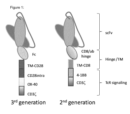
A CAR of the invention thus generally comprises 4, or preferably 5, domains as
follows:
(1) an antigen-binding domain, capable of binding specifically to CD37, that
comprises VL and VH sequences based or derived from SEQ ID NOs. 3 and 1 as
defined above;
CA 03009718 2018-06-26
WO 2017/118745 PCT/EP2017/050285
- 8 -
(2) a hinge domain that extends the antigen-binding domain away from the
surface of the immune effector cell;
(3) a transmembrane domain that anchors the CAR to the effector cell and links
the extracellular domain comprising the antigen-binding domain to the
intracellular
signalling domain;
(4) an intracellular domain comprising a signalling domain; and optionally or
preferably;
(5) one or more co-stimulatory signalling domains.
The CAR may further comprise (6) a signal sequence (i.e. a targeting domain),
and in particular a sequence which targets the CAR to the plasma membrane of
the
immune effector cell. This will generally be positioned next to or close to
the antigen-
binding domain, generally upstream of the antigen-binding domain, at the end
of the
CAR molecule/construct
It can thus be seen that the CAR may comprise an extracellular domain
comprising the antigen-binding domain and signal sequence, if present, linked
via a
hinge domain and transmembrane domain to an intracellular domain which
comprises
one or more signalling domains. In one aspect, the intracellular domain, or
the hinge,
transmembrane and intracellular domains, may be viewed as a "signalling tail"
in the
CAR construct. The order of the domains in the CAR construct is thus, N-
terminal to C-
terminal: extracellular domain ¨ hinge domain ¨ transmembrane domain ¨
intracellular
domain. Within the extracellular and intracellular domains the separate
domains may be
arranged in any order. Preferably however the order is signal sequence ¨
antigen-
binding domain in the extracellular domain. In one embodiment, in the
intracellular
domain the order may be co-stimulatory domain(s) ¨ intracellular signalling
domain(s).
In another embodiment, the order may be intracellular signalling domain(s) ¨
co-
stimulatory domain(s).
In the CAR of the present invention the "antigen-binding domain", which is
derived from the variable region sequences of the antibody HH1, may be
provided in
various formats, as long as it comprises the VI_ and VH sequences as defined
above. It
may accordingly be, or may correspond to, a natural or synthetic antibody
sequence.
Accordingly, the nucleotide sequence encoding the antigen-binding domain in
the
nucleic acid molecules of the invention may be derived from, or may correspond
to, a
natural sequence or may encode a genetically engineered product. Thus the
antigen-
binding domain may be (or more precisely may correspond to) a fragment of
antibody
CA 03009718 2018-06-26
WO 2017/118745
PCT/EP2017/050285
- 9 -
HH1 comprising the variable region (the antibody light chain and heavy chain
variable
regions; the VI_ and VH regions), e.g. a Fv or Fab or F(ab)2 or the light and
heavy chain
variable regions can be joined together in a single chain and in either
orientation (e.g.
VL-VH or VH-VL). The VI_ and/or VH sequences may be modified, as discussed
above. In
particular the framework regions may be modified (e.g. substituted, for
example to
humanise the antigen-binding domain).
In a preferred embodiment, the binding domain is a single chain antibody
(scFv)
derived from antibody HH1. The single chain antibody may be cloned using known
and
readily available techniques from the V region genes of the hybridoma
producing
antibody HH1. The hybridoma is described in Smeland etal. 1985 (supra). As
mentioned above the VI_ and/or VH sequences of the scFv may be modified.
"Single-chain Fv antibody" or "scFv" refers to an engineered antibody
consisting
of a light chain variable region (VL) and a heavy chain variable region (VH)
connected to
one another directly or via a peptide linker sequence.
"Heavy chain variable region" or "VH" refers to the fragment of the heavy
chain
of an antibody that contains three CDRs (complementarity determining regions)
interposed between flanking stretches known as framework regions, which are
more
highly conserved than the CDRs and form a scaffold to support the CDRs.
"Light chain variable region" or "VL" refers to the fragment of the light
chain of an
antibody that contains three CDRs interposed between framework regions.
"Fv" refers to the smallest fragment of an antibody to bear the complete
antigen-
binding site. An Fv fragment consists of the variable region of a single light
chain bound
to the variable region of a single heavy chain.
In one preferred embodiment the VI_ and VH are linked together by a linker
.. sequence. More precisely this may be referred to as a "variable region
linker
sequence", which is an amino acid sequence that connects a heavy chain
variable
region to a light chain variable region and provides a spacer function
compatible with
interaction of the two sub-binding domains so that the resulting polypeptide
retains a
specific binding affinity to the same target molecule as an antibody that
comprises the
same light and heavy chain variable regions. The linker sequence may be used
to
provide for appropriate spacing and conformation of the molecule.
Thus, in one embodiment the scFv comprises the VI_ sequence of SEQ ID NO. 3
or an amino acid sequence having at least 95 % sequence identity thereto
linked to the
VH sequence of SEQ ID NO. 1 or an amino acid sequence having at least 95 %
sequence identity thereto, preferably in the order VL-VH.
CA 03009718 2018-06-26
WO 2017/118745 PCT/EP2017/050285
- 10 -
In another embodiment the scFv comprises the VI_ sequence of SEQ ID NO. 3
or an amino acid sequence having at least 60 % sequence identity thereto
linked to the
VH sequence of SEQ ID NO.1 or an amino acid sequence having at least 60 %
sequence identity thereto, preferably in the order VL-VH. As noted above, this
is subject
to the proviso that the CDR sequences remain as defined above, and preferably
to the
proviso that the CDR sequences are unaltered.
More preferably, the VI_ sequence is linked to VH by a linker sequence. The
linker sequence may be between 1-30, more preferably 1-25, 1-22 or 1-20, amino
acids
long. The linker may be a flexible linker. Suitable linkers can be readily
selected and
can be of any of a suitable length, such as from 1 amino acid (e.g. Gly) to 20
amino
acids, from 2 amino acids to 15 amino acids, from 3 amino acids to 12 amino
acids,
including 4 amino acids to 10 amino acids, 5 amino acids to 9 amino acids, 6
amino
acids to 8 amino acids, or 7 amino acids to 8 amino acids, and may be 1, 2, 3,
4, 5, 6,
or 7 amino acids or longer.
Exemplary flexible linkers include glycine polymers (G)n, glycine-serine
polymers, where n is an integer of at least one, glycine-alanine polymers,
alanine-
serine polymers, and other flexible linkers known in the art. Glycine and
glycine-serine
polymers are relatively unstructured, and therefore may be able to serve as a
neutral
tether between domains of fusion proteins such as the CARs described herein.
In a representative embodiment the linker sequence may be (G4S)4 (SEQ ID
NO. 5). Thus in a representative embodiment the nucleic acid molecule of the
invention
may comprise a nucleotide sequence encoding the amino acid sequence of SEQ ID
NO. 16, comprising in order the VI_ of SEQ ID NO. 3, the linker of SEQ ID NO.
Sand the
VH of SEQ ID NO. 1, or a sequence having at least 95 % sequence identity
thereto.
The VI_ and VH regions may be encoded by nucleotide sequences comprising
the nucleotide sequences of SEQ ID NOs. 4 and 2 respectively, nucleotide
sequences
having at least 95 % nucleotide sequence identity thereto, or nucleotide
sequences
degenerate with SEQ ID NOs. 4 and 2.
In another embodiment The VI_ and VH regions may be encoded by nucleotide
sequences comprising the nucleotide sequences of SEQ ID NOs. 4 and 2
respectively,
or nucleotide sequences having at least 60 % nucleotide sequence identity
thereto. As
above, this is subject this is subject to the proviso that the CDR sequences
encoded by
the nucleotide sequences remain as defined above, and preferably to the
proviso that
the CDR sequences are unaltered.
CA 03009718 2018-06-26
WO 2017/118745 PCT/EP2017/050285
- 11 -
The VI_ and VH sequences may, if desired, be humanised by modifying one or
more of the framework regions to correspond to at least one human framework
region.
A "human framework region" refers to a wild type (i.e. naturally occurring)
framework
region of a human immunoglobulin variable region, an altered framework region
of a
human immunoglobulin variable region with less than about 50 % (e.g.
preferably less
than about 45%, 40%, 30%, 25%, 20%, 15%, 10%, 5%, or 1 %) of the amino acids
in the region deleted or substituted (e.g. with one or more amino acid
residues of a
nonhuman immunoglobulin framework region at corresponding positions), or an
altered
framework region of a nonhuman immunoglobulin variable region with less than
about
50 % (e.g. less than 45 %, 40 %, 30 %, 25 %, 20 %, 15 %, 10 %, or 5 %) of the
amino
acids in the region deleted or substituted (e.g. at positions of exposed
residues and/or
with one or more amino acid residues of a human immunoglobulin framework
region at
corresponding positions) so that, in one aspect, immunogenicity is reduced.
Thus, in a particular embodiment, the framework regions of the VI_ and VH
sequences of SEQ ID NOs. 3 and 1 respectively may be modified (more
specifically the
amino acid sequences of the framework regions may be modified), whilst
retaining, or
substantially retaining, the amino acid sequences of the CDRs.
Accordingly, in another embodiment, the framework regions of the VI_ and VH
sequences in the CAR have at least 60 % amino acid sequence identity to the
framework regions of SEQ ID NOs. 3 and 1 respectively.
The framework regions (FRs) of the VI_ sequence of SEQ ID NO. 3 lie at the
following amino acid positions: FR1 at positions 1-26; FR2 at positions 33-49,
FR3 at
positions 53-98 and FR4 at positions 98-107.
The framework regions of the VH sequence of SEQ ID NO. 1 lie at the following
amino acid positions: FR1 at positions 1-25; FR2 at positions 34-50, FR3 at
positions
59-96 and FR4 at positions 109-119.
SEQ ID NO. 47 shows the amino acid sequence of a modified VI_ sequence
having human framework regions and CDRs 1, 2 and 3 of SEQ ID NO. 3. SEQ ID
NO. 48 shows the amino acid sequence of a modified VH sequence having human
framework regions and CDRs 1, 2 and 3 of SEQ ID NO. 1. The corresponding
nucleotide sequences encoding SEQ ID NOs. 47 and 48 are shown in SEQ ID NOs.
49
and 50.
Amino acid sequences SEQ ID NOs. 47 and/or 48, or nucleotide sequences
SEQ ID NOs. 49 and/or 50, or sequences having at least 95 % sequence identity
CA 03009718 2018-06-26
WO 2017/118745 PCT/EP2017/050285
- 12 -
thereto, or nucleotide sequences degenerate with SEQ ID NOs. 49 and/or 50, may
be
used (i.e. contained or comprised) in the CARs or nucleic acid molecules of
the
invention.
As noted above, the CAR, and more particularly the extracellular domain
thereof, may also comprise a signal sequence (or targeting domain). Such a
sequence
will generally be provided at the N-terminal end of the molecule (construct)
and may
function to, co-translationally or post-translationally, direct transfer of
the molecule. In
particular, the signal sequence may be a sequence which targets the CAR to the
plasma membrane of the immune effector cell. This may be linked directly or
indirectly
(e.g. via a linker sequence) to the antigen-binding domain, generally upstream
of the
antigen-binding domain, at the N-terminal end of the CAR molecule/construct.
The
linker sequence may be a linker as described in connection with the variable
region
linker above. In one embodiment the signal sequence is linked directly to the
N-terminal
end of the antigen-binding domain, e.g. to the N-terminal end of the VI_
sequence (for
example SEQ ID NO. 3). In one preferred embodiment the signal sequence is, or
is
derived from, the L-chain having the sequence of SEQ ID NO. 6 or an amino acid
sequence having at least 95 % sequence identity thereto.
Accordingly, in a representative embodiment, the nucleic acid molecule encodes
(or comprises a nucleotide sequence encoding) a CAR comprising an
extracellular
domain which comprises or has the sequence of SEQ ID NO. 7 or an amino acid
sequence with at least 95 % sequence identity thereto, wherein SEQ ID NO. 7
comprises in order the L-chain of SEQ ID NO. 6, the VI_ of SEQ ID NO. 3, the
linker of
SEQ ID NO. Sand the VH of SEQ ID NO. 1.
An extracellular domain (or a scFv domain) having or comprising an amino acid
sequence as set out in SEQ ID NO. 7, or an amino acid sequence having at least
95 %
sequence identity thereto, may be encoded by a nucleic acid molecule having or
comprising a nucleotide sequence as set out in SEQ ID NO. 31 or 38, or a
nucleotide
sequence having at least 95 % sequence identity thereto, or a nucleotide
sequence
degenerate with SEQ ID NO. 31 or 38.
In a representative embodiment in which the VI_ and VH sequences are
humanised, the nucleic acid molecule encodes (or comprises a nucleotide
sequence
encoding) a CAR comprising an extracellular domain (or a scFv domain) which
comprises or has the sequence of SEQ ID NO. 53 or an amino acid sequence with
at
least 95 % sequence identity thereto, wherein SEQ ID NO. 53 comprises in order
the L-
CA 03009718 2018-06-26
WO 2017/118745 PCT/EP2017/050285
- 13 -
chain of SEQ ID NO. 6, the VI_ of SEQ ID NO. 47, the linker of SEQ ID NO. 5
and the
VH of SEQ ID NO. 48. An extracellular domain having or comprising an amino
acid
sequence as set out in SEQ ID NO. 53, or an amino acid sequence having at
least 95
% sequence identity thereto, may be encoded by a nucleic acid molecule having
or
comprising a nucleotide sequence as set out in SEQ ID NO. 54, or a nucleotide
sequence having at least 95 % sequence identity thereto, or a nucleotide
sequence
degenerate with SEQ ID NO. 54.
The antigen-binding domain of the CAR is generally followed by a hinge
domain. The hinge region in a CAR is generally between the transmembrane
domain
and the antigen-binding domain. In certain embodiments, a hinge region is an
immunoglobulin hinge region and may be a wild type immunoglobulin hinge region
or
an altered wild type immunoglobulin hinge region, for example a truncated
hinge region.
Other exemplary hinge regions which may be used include the hinge region
derived
from the extracellular regions of type 1 membrane proteins such as CD8a, CD4,
0D28
and CD7, which may be wild-type hinge regions from these molecules or may be
altered. Preferably the hinge region is, or is derived from, the hinge region
of human
CD8a, CD4, 0D28 or CD7.
An "altered wild type hinge region" or "altered hinge region" refers to (a) a
wild
type hinge region with up to 30 % amino acid changes (e.g. up to 25%, 20 %,
15%, 10
.. %, or 5 % amino acid changes e.g. substitutions or deletions), (b) a
portion of a wild
type hinge region that is at least 10 amino acids (e.g. at least 12, 13, 14 or
15 amino
acids) in length with up to 30 % amino acid changes (e.g. up to 25 %, 20 %, 15
%, 10
%, or 5 % amino acid changes, e.g. substitutions or deletions), or (c) a
portion of a wild
type hinge region that comprises the core hinge region (which may be 4, 5, 6,
7, 8, 9,
.. 10, 11, 12, 13, 14, or 15, or at least 4, 5, 6, 7, 8, 9, 10, 11, 12, 13,
14, or 15 amino acids
in length). When an altered wild type hinge region is interposed between and
connecting the 0D37-specific binding domain and another region (e.g. a
transmembrane domain) in the chimeric antigen receptors described herein, it
allows
the chimeric fusion protein to maintain specific binding to 0D37.
In certain embodiments, one or more cysteine residues in a wild type
immunoglobulin hinge region may be substituted by one or more other amino acid
residues (e.g. one or more serine residues). An altered immunoglobulin hinge
region
may alternatively or additionally have a proline residue of a wild type
immunoglobulin
hinge region substituted by another amino acid residue (e.g. a serine
residue).
CA 03009718 2018-06-26
WO 2017/118745 PCT/EP2017/050285
- 14 -
Hinge regions comprising the CH2 and CH3 constant region domains are
described in the art for use in CARs (for example the CH2CH3 hinge, referred
to as an
"Fe hinge" or "IgG hinge", as shown in SEQ ID NO. 40). However, it is
preferred that
when the hinge domain is based on or derived from an immunoglobulin it does
not
comprise a CH3 domain, e.g. it may comprise or consist of the CH2 domain or a
fragment or part thereof, without including CH3.
In one preferred embodiment the hinge domain has or comprises the amino acid
sequence of SEQ ID NO. 9 (which represents the hinge domain of CD8a) or an
amino
acid sequence having at least 95 % sequence identity thereto.
In another preferred embodiment the hinge domain has or comprises the amino
acid sequence of SEQ ID NO. 10 (which represents a shortened IgG hinge) or an
amino acid sequence having at least 95 % sequence identity thereto.
The hinge domain may be attached to the transmembrane domain by a linker
sequence, which may be a linker sequence as defined above. An exemplary linker
sequence is KDPK (SEQ ID NO. 11). A shortened IgG hinge with linker sequence
is
shown in SEQ ID NO. 21. Such a sequence, or a sequence having at least 95 %
sequence identity thereto, may be included in a CAR of the present invention.
More
particularly such a sequence may be included between the extracellular domain
(e.g.
the seFv part) and the transmembrane domain.
The transmembrane domain may be based on or derived from the
transmembrane domain of any transmembrane protein. Typically it may be, or may
be
derived from, a transmembrane domain from CD8a, 0D28, CD4, CD34 0D45, CD9,
CD16, 0D22, 0D33, 0D64, CD80, 0D86, 0D134, 0D137, and 0D154, preferably from
a human said protein. In one embodiment, the transmembrane domain may be, or
may
be derived from, a transmembrane domain from CD8a, 0D28, CD4, or CD34,
preferably from human 0D28, CD4, or CD34. In another embodiment the
transmembrane domain may be synthetic in which case it would comprise
predominantly hydrophobic residues such as leucine and valine.
In a preferred embodiment the transmembrane domain is the CD8a
transmembrane domain having the amino acid sequence of SEQ ID NO. 12 or an
amino acid sequence having at least 95 % sequence identity thereto.
In another embodiment the transmembrane domain may be the transmembrane
domain of human 0D28 having the amino acid sequence of SEQ ID NO. 17 or an
amino acid sequence having at least 95 % sequence identity thereto.
CA 03009718 2018-06-26
WO 2017/118745 PCT/EP2017/050285
- 15 -
The "intracellular signalling domain" refers to the part of the CAR protein
that
participates in transducing the message of effective CAR binding to a target
antigen
into the interior of the immune effector cell to elicit effector cell
function, e.g. activation,
cytokine production, proliferation and cytotoxic activity, including the
release of
cytotoxic factors to the CAR-bound target cell, or other cellular responses
elicited with
antigen binding to the extracellular CAR domain. The term "effector function"
refers to a
specialized function of the cell. Effector function of the T-cell, for
example, may be
cytolytic activity or help or activity including the secretion of a cytokine.
Thus, the term
"intracellular signalling domain" refers to the portion of a protein which
transduces the
effector function signal and that directs the cell to perform a specialized
function. While
the entire intracellular signalling domain can be employed, in many cases it
is not
necessary to use the entire domain. To the extent that a truncated portion of
an
intracellular signalling domain is used, such truncated portion may be used in
place of
the entire domain as long as it transduces the effector function signal. The
term
intracellular signalling domain is meant to include any truncated portion of
the
intracellular signalling domain sufficient to transduce effector function
signal. The
intracellular signalling domain is also known as the, "signal transduction
domain," and is
typically derived from portions of the human CD34 or FcRy chains.
Additionally, to allow or to augment full activation of the immune effector
cell the
CAR may be provided with a secondary, or co-stimulatory domain. Thus, the
intracellular signalling domain may initiate antigen dependent primary
activation (i.e.
may be a primary cytoplasmic signalling sequence) and the co-stimulatory
domain may
act in an antigen-independent manner to provide a secondary or co-stimulatory
signal
(secondary cytoplasmic signalling sequence(s)). Primary cytoplasmic signalling
sequences may regulate primary activation, including in an inhibitory way.
Primary
cytoplasmic signalling sequences that act in a co-stimulatory manner may
contain
signalling motifs which are known as immunoreceptor tyrosine-based activation
motifs
or ITAMs.
Examples of ITAM-containing primary cytoplasmic signalling sequences that
may be used in the invention include those derived from TCR4, FcRy, FcR[3,
CD3y,
CD36, CD3E, CD5, CD22, CD79a, CD79b and CD66d. In certain particular
embodiments, the intracellular signalling domain is derived from CD34 or FcRy,
preferably human CD34 or FcRy.
In a preferred representative embodiment the intracellular signalling domain
is
preferably a human CD34 domain, more preferably a human CD34 domain having the
CA 03009718 2018-06-26
WO 2017/118745 PCT/EP2017/050285
- 16 -
amino acid sequence of SEQ ID NO. 8 or an amino acid sequence having at least
95%
sequence identity thereto.
The term "co-stimulatory signalling domain" or "co-stimulatory domain", refers
to
the portion of the CAR comprising the intracellular domain of a co-stimulatory
molecule.
Co-stimulatory molecules are cell surface molecules other than antigen
receptors or Fc
receptors that provide a second signal required for efficient activation and
function of an
immune effector cell (e.g. a T-cell) upon binding to antigen. Examples of such
co-
stimulatory molecules include 0D27, 0D28, 4- IBB (0D137), 0X40 (0D134), CD30,
CD40, PD-1, ICOS (0D278), LFA-1 , CD2, CD7, LIGHT, NKD2C, B7-H2 and a ligand
i0 .. that specifically binds 0D83, more particularly the intracellular
domains of such
molecules. Preferably the molecules are human. Accordingly, while exemplary or
preferred co-stimulatory domains are derived from 4-i BB, 0D28 or 0X40
(CD134),
other co-stimulatory domains are contemplated for use with the CARs described
herein.
The co-stimulatory domains may be used singly or in combination (i.e. one or
more co-
stimulatory domains may be included. The inclusion of one or more co-
stimulatory
signalling domains may enhance the efficacy and expansion of immune effector
cells
expressing the CARs.
The intracellular signalling and co-stimulatory signalling domains may be
linked
in any order in tandem to the carboxyl terminus of the transmembrane domain.
In a preferred embodiment the co-stimulatory domain is the intracellular
domain
of 4-1BB having the amino acid sequence of SEQ ID NO. 13 or an amino acid
sequence having at least 95 % sequence identity thereto.
In another embodiment the co-stimulatory domain may be, or may include, the
intracellular domain of human 0D28 having the amino acid sequence of SEQ ID
NO. 18
or an amino acid sequence having at least 95 % sequence identity thereto
and/or an
0X40 (CD134) co-stimulatory domain having the amino acid sequence of SEQ ID
NO.
19 or an amino acid sequence having at least 95 % sequence identity thereto.
In a preferred embodiment of the invention, the CAR (or more particularly the
"signalling tail" thereof) comprises a hinge domain from CD8a or a truncated
IgG hinge
.. domain not including the CH3 domain, a CD8a transmembrane domain, a 4-i BB
co-
stimulatory domain and a CD34 intracellular signalling domain.
In other embodiments the CAR (or the "signalling tail" thereof) comprises a
hinge domain from CD8a or a truncated IgG hinge domain not including the CH3
domain, a 0D28 transmembrane domain, a 0D28 intracellular domain and/or 0X40
co-
stimulatory domain and a CD34 intracellular signalling domain.
CA 03009718 2018-06-26
WO 2017/118745
PCT/EP2017/050285
- 17 -
More particularly, the CAR comprises, preferably in the following order:
(i) a hinge domain being (a) the hinge domain of CD8a having the amino acid
sequence of SEQ ID NO. 9 or an amino acid sequence having at least 95 %
sequence
identity thereto; or (b) an IgG hinge domain having the amino acid sequence of
SEQ ID
NO. 10 or an amino acid sequence having at least 95 % sequence identity
thereto;
(ii) a CD8a transmembrane domain having the amino acid sequence of SEQ ID
NO. 12 or an amino acid sequence having at least 95 % sequence identity
thereto;
(iii) a co-stimulatory domain being the intracellular domain of 4-1BB
having the
amino acid sequence of SEQ ID NO. 13 or an amino acid sequence having at least
95
% sequence identity thereto; and
(iv) a human CD34 domain having the amino acid sequence of SEQ ID NO. 8 or
an
amino acid sequence having at least 95 % sequence identity thereto.
Further, in the CAR the hinge domain may be attached to the transmembrane
domain by means of a linker having the sequence KDPK (SEQ ID NO. 11). In
particular
embodiments the CAR may comprise the hinge-linker sequence of SEQ ID NO. 21 or
an amino acid sequence having at least 95 % sequence identity thereto.
In an alternative embodiment, the CAR (or the signalling tail thereof) may
comprise, preferably in the following order:
(i) a hinge domain being (a) the hinge domain of CD8a having the amino acid
sequence of SEQ ID NO. 9 or an amino acid sequence having at least 95%
sequence
identity thereto; or (b) an IgG hinge domain having the amino acid sequence of
SEQ ID
NO. 10 or an amino acid sequence having at least 95 % sequence identity
thereto;
(ii) a 0D28 transmembrane domain having the amino acid sequence of SEQ ID
NO. 17 or an amino acid sequence having at least 95 % sequence identity
thereto;
(iii) a co-stimulatory domain being the intracellular domain of 0D28 (0D28
intra)
having the amino acid sequence of SEQ ID NO. 18 or an amino acid sequence
having
at least 95 % sequence identity thereto and/or a co-stimulatory domain being
an 0X40
co-stimulatory domain having the amino acid sequence of SEQ ID NO. 19 or an
amino
acid sequence having at least 95 % sequence identity thereto;
(iv) a human CD34 domain having the amino acid sequence of SEQ ID NO. 8 or
an
amino acid sequence having at least 95 % sequence identity thereto.
Further in the CAR the hinge domain may be attached to the transmembrane
domain by means of a linker having the sequence KDPK (SEQ ID NO. 11). In
particular
embodiments the CAR may comprise the hinge-linker sequence of SEQ ID NO. 21 or
CA 03009718 2018-06-26
WO 2017/118745 PCT/EP2017/050285
- 18 -
an amino acid sequence having at least 95 % sequence identity thereto. Where
both
CD28intra and 0X40 co-stimulatory domains are present, they may be present in
either
order, but in one preferred embodiment they are present in the order CD28intra
¨
OX40.
Such a CAR according to the invention may include a scFv antigen-binding
domain as defined above and may further comprises a plasma membrane targeting
sequence having the sequence set out in SEQ ID NO. 6 or an amino acid sequence
having at least 95 % sequence identity thereto positioned upstream of the
scFv.
Thus the CAR of the invention may in certain representative embodiments
comprise, in addition to a signalling tail as defined above, an extracellular
domain
having the sequence of SEQ ID NO. 16 or 7 or a sequence having at least 95%
sequence identity thereto.
A representative CAR according to the present invention may thus have or
comprise the amino acid sequence of SEQ ID NO. 32 or 33, or an amino acid
having at
least 95 % sequence identity thereto.
A nucleic acid molecule of the invention may comprise the nucleotide sequence
of SEQ ID NO. 14 or SEQ ID NO. 15 or a nucleotide sequence having at least 95
%
sequence identity thereto, or a nucleotide sequence degenerate with SEQ ID NO.
14 or
SEQ ID NO. 15.
The present disclosure provides CAR polypeptides, and fragments thereof. The
terms "polypeptide" and "protein" are used interchangeably and mean a polymer
of
amino acids not limited to any particular length. The term does not exclude
modifications such as myristylation, sulfation, glycosylation, phosphorylation
and
addition or deletion of signal sequences. The terms "polypeptide" or "protein"
or
"peptide" mean one or more chains of amino acids, wherein each chain comprises
amino acids covalently linked by peptide bonds, and wherein said polypeptide
or
protein can comprise a plurality of chains non-covalently and/or covalently
linked
together by peptide bonds, having the sequence of native proteins, that is,
proteins
produced by naturally-occurring and specifically non-recombinant cells, or
genetically-
engineered or recombinant cells, and comprise molecules having the amino acid
sequence of the native protein, or molecules having deletions from, additions
to, and/or
substitutions of one or more amino acids of the native sequence. The terms
"polypeptide" and "protein" specifically encompass the CARs of the present
disclosure,
or sequences that have deletions from, additions to, and/or substitutions of
one or more
amino acid of a CAR as disclosed herein.
CA 03009718 2018-06-26
WO 2017/118745 PCT/EP2017/050285
- 19 -
As is clear from the above, the various domains of the CAR may comprise one
or more amino acid sequence modifications with respect to the native sequences
of the
molecules from which they are derived. For example, it may be desirable to
improve the
binding affinity and/or other biological properties of the CAR. For example,
amino acid
sequence variants of a CAR, or binding domain, or a stimulatory signalling
domain
thereof, may be prepared by introducing appropriate nucleotide changes into a
polynucleotide that encodes the CAR, or a domain thereof. Such modifications
include,
for example, deletions from, and/or insertions into and/or substitutions of,
residues
within the amino acid sequences of the CAR. Any combination of deletion,
insertion,
.. and substitution may be made to arrive at the final CAR, provided that the
final
construct possesses the desired characteristics, such as specific binding to
0D37 by
the binding domain, or increased signalling by the intracellular signalling
domain and/or
co-stimulatory domain. The amino acid changes also may alter post-
translational
processes of the CAR, such as changing the number or position of glycosylation
sites.
Any of the variations and modifications described above may be included in the
CARs
of the present invention.
In particular embodiments, the various domains of a CAR (other than the VI_
and
VH sequences) may have an amino acid sequence that is at least 80 % identical,
at
least 85 %, at least 90 %, at least 95 % identical, or at least 98 % or 99 %
identical, to
the native sequence of the domains of the proteins from which they are
derived. Thus,
in particular embodiments the domains may have an amino acid sequence that has
at
least 80, 85, 90, 95, 98 or 99 % sequence identity to any of SEQ ID NOs. 5, 8,
9, 10,
11, 12, 13, 17, 18, 19 or 21.
Alternatively or additionally, the VI_ and VH sequences of a CAR, or the
framework regions thereof, may have an amino acid sequence that is at least
60, 65 or
70 % identical to SEQ ID NOs. 3 and 1 respectively, or to the framework
regions
thereof, particularly with respect to SEQ ID NO. 3. In other embodiments the %
sequence identity may be higher, e.g. at least 75, 80, 85, 90, 95, 98 or 99 %
sequence
identity to SEQ ID NOs. 3 or 1, or to the framework regions thereof.
The nucleic acid molecule of the invention may be an isolated nucleic acid
molecule and may further include DNA or RNA or chemical derivatives of DNA or
RNA,
including molecules having a radioactive isotope or a chemical adduct such as
a
fluorophore, chromophore or biotin ("label"). Thus the nucleic acid may
comprise
modified nucleotides. Said modifications include base modifications such as
bromouridine, ribose modifications such as arabinoside and 2',3'-dideoxyribose
and
CA 03009718 2018-06-26
WO 2017/118745
PCT/EP2017/050285
- 20 -
internucleotide linkage modifications such as phosphorothioate,
phosphorodithioate,
phosphoroselenoate, phosphorodiselenoate, phosphoroanilothioate,
phoshoraniladate
and phosphoroamidate. The term "nucleic acid molecule" specifically includes
single
and double stranded forms of DNA and RNA.
In particular embodiments, nucleotide sequences in the nucleic acid molecule
encoding the various domains of a CAR (other than the VI_ and VH sequences)
may
have a nucleotide sequence having at least 75 %, 80 %, 85 %, 90 %, 95 %, 96 %,
97
%, 98 %, or 99 % or higher, sequence identity, to the native sequence of the
nucleic
acids from which they are derived or to a reference polynucleotide sequence
such as a
sequence encoding a said domain as described herein.
Alternatively or additionally, the nucleotide sequences encoding the VI_ and
VH
sequences of a CAR, or the framework regions thereof, may have a nucleic acid
sequence that is at least 60, 65 or 70 % identical to SEQ ID NOs. 4 and 2
respectively,
or to the framework regions thereof, particularly with respect to SEQ ID NO.
4. In other
embodiments the % sequence identity may be higher, e.g. at least 75, 80, 85,
90, 95,
98 or 99 % sequence identity to SEQ ID NOs. 4 or 2, or to the framework
regions
thereof.
Methods for determining sequence identity are well known in the art and any
convenient or available method may be used, e.g. BLAST analysis, e.g. using
standard
or default parameters.
When comparing polynucleotide sequences, two sequences are said to be
"identical" if the sequence of nucleotides in the two sequences is the same
when
aligned for maximum correspondence. Comparisons between two sequences are
typically performed by comparing the sequences over a comparison window to
identify
and compare local regions of sequence similarity. A "comparison window" refers
to a
segment of at least about 20 contiguous positions, usually 30 to about 75, 40
to about
50, or more in which a sequence may be compared to a reference sequence of the
same number of contiguous positions after the two sequences are optimally
aligned. In
particular, where % sequence identity is given herein with respect to a
particular
sequence, the % sequence identity is determined over the whole length of the
specified
sequence.
Optimal alignment of sequences for comparison may be conducted using the
Megalign program in the Lasergene suite of bioinformatics software (DNASTAR,
Inc.,
Madison, WI), using default parameters. Alternatively, optimal alignment of
sequences
CA 03009718 2018-06-26
WO 2017/118745 PCT/EP2017/050285
- 21 -
for comparison may be conducted by the local identity algorithm of Smith and
Waterman, Add. APL. Math 2:482 (1981), by the identity alignment algorithm of
Needleman and Wunsch, J. Mol. Biol. 48:443 (1970), by the search for
similarity
methods of Pearson and Lipman, Proc. Natl. Acad. Sci. USA 85: 2444 (1988), by
.. computerized implementations of these algorithms (GAP, BESTFIT, BLAST,
FASTA,
and TFASTA in the Wisconsin Genetics Software Package, Genetics Computer Group
(GCG), 575 Science Dr., Madison, WI), or by inspection.
One representative example of algorithms that are suitable for determining
percent sequence identity and sequence similarity are the BLAST and BLAST 2.0
algorithms, which are described in Altschul etal., Nucl. Acids Res. 25:3389-
3402
(1977), and Altschul eta!, J. Mol. Biol. 215:403-410 (1990), respectively.
BLAST and
BLAST 2.0 can be used, for example with the default parameters, to determine
percent
sequence identity among two or more nucleic acid molecules. Software for
performing
BLAST analyses is publicly available through the National Center for
Biotechnology
Information.
It will be appreciated by those of ordinary skill in the art that, as a result
of the
degeneracy of the genetic code, there are many nucleotide sequences that may
encode a CAR as described herein. By degenerate nucleotide sequences is meant
two
(or more) nucleotide sequences which encode the same protein (or protein
sequence),
specifically in the open reading frame of the reference nucleotide sequence
which
begins at position 1 (i.e. in which codon 1 of the encoding sequence
corresponds to
positions 1-3 of the reference nucleotide sequence). Thus for example a
nucleotide
sequence degenerate with SEQ ID NO. 2 is a nucleotide sequence which is
different to
SEQ ID NO.2 but which, due to the degeneracy of the genetic code, encodes the
same
protein sequence as SEQ ID NO. 2, i.e. the protein sequence of SEQ ID NO. 1.
Methods for modifying nucleotide sequences to introduce changes to the amino
acid sequences of the various domains are well known in the art e.g. methods
of
mutagenesis, such as site-specific mutagenesis, may be employed.
Likewise methods for preparing a nucleic acid molecule encoding the CAR are
also well known e.g. conventional polymerase chain reaction (PCR) cloning
techniques
can be used to construct the nucleic acid molecule. The nucleic acid molecule
can be
cloned into a general purpose cloning vector such as pENT (Gateway), pUC19,
pBR322, pBluescript vectors (Stratagene Inc.) or pCR TOPO from lnvitrogen
Inc. The
resultant nucleic acid construct (recombinant vector) carrying the nucleic
acid molecule
encoding the CAR can then be sub-cloned into expression vectors or viral
vectors for
CA 03009718 2018-06-26
WO 2017/118745 PCT/EP2017/050285
- 22 -
protein expression, e.g. in mammalian cells. This may be for preparation of
the CAR
protein, or for expression in immune effector cells, e.g. in human T-cells or
in NK cells
or cell lines. Further the nucleic acid may be introduced into mRNA expression
vectors
for production of mRNA encoding the CAR. The mRNA may then be transferred into
immune effector cells.
Thus, the nucleic acid molecule may be introduced into a cell in a vector or
as a
nucleic acid molecule or recombinant construct. Methods of heterologous gene
expression are known in the art, both in terms of construct/vector preparation
and in
terms of introducing the nucleic acid molecule (vector or construct) into the
cell. Thus,
.. promoters and/or other expression control sequences suitable for use with
mammalian
cells, in particular immune effector cells (e.g. lymphoid cells or NK cells),
and
appropriate vectors etc. (e.g. viral vectors) are well known in the art.
Vectors or constructs (nucleic acid molecules) may be introduced into a cell
of
the invention by a variety of means, including chemical transfection agents
(such as
calcium phosphate, branched organic compounds, liposomes or cationic
polymers),
electroporation, cell squeezing, sonoporation, optical transfection,
hydrodynamic
delivery, or viral transduction. In a preferred embodiment, a vector or
construct is
introduced by viral transduction. This may allow for more persistent
expression of the
CAR. However, in some situations, e.g. in clinical trials, or in some clinical
situations, it
may be desirable to have a more transient period of expression of CAR protein.
In such
a situation it may be desirable to deliver the nucleic acid molecule to the
immune
effector cell as mRNA. mRNA expression vectors for production of mRNA may be
prepared according to methods known in the art (e.g. using Gateway Technology)
and
are known in the art (e.g. pCIpA102, Sboe-Larssen eta!, 2002, J. lmmunol.
Methods
259, p 191-203 and pCIpA120-G, Walchli eta!, 2011, PLoS ONE 6(11) e27930).
The mRNA can be produced in vitro by e.g. in vitro transcription. The mRNA
may then be introduced into the immune effector cells, e.g. as naked mRNA,
e.g. by
electroporation (as described for example in Almasbak etal., Cytotherapy 2011,
13,
629-640, Rabinovich etal., Hum. Gene Ther., 2009, 20, 51-60 and Beatty et al.,
Cancer
lmmunol. Res. 2014, 2, 112-120. Alternatively, mRNA may be introduced by other
means such as by liposomes or cationic molecules etc. Heterologous nucleic
acid
molecules introduced into a cell may be expressed episomally, or may be
integrated
into the genome of the cell at a suitable locus.
CA 03009718 2018-06-26
WO 2017/118745
PCT/EP2017/050285
- 23 -
Thus the nucleic acid molecule may be introduced or inserted into a vector.
The
term "vector" as used herein refers to a vehicle into which the nucleic acid
molecule
may be introduced (e.g. be covalently inserted) so as to bring about the
expression of
the CAR protein or mRNA and/or the cloning of the nucleic acid molecule. The
vector
may accordingly be a cloning vector or an expression vectors.
The nucleic acid molecule may be inserted into a vector using any suitable
methods known in the art, for example, without limitation, the vector may be
digested
using appropriate restriction enzymes and then may be ligated with the nucleic
acid
molecule having matching restriction ends.
Expression vectors can contain a variety of control sequences, which refer to
nucleic acid sequences necessary for the transcription and possibly
translation of an
operatively linked coding sequence in a particular host cell. In addition to
control
sequences that govern transcription and translation, vectors may contain
additional
nucleic acid sequences that serve other functions, including for example for
replication,
selectable markers etc.
The expression vector should have the necessary 5' upstream and 3'
downstream regulatory elements such as promoter sequences such as CMV, PGK and
EF1 a promoters, ribosome recognition and binding TATA box, and 3' UTR AATAAA
(SEQ ID NO. 20) transcription termination sequence for the efficient gene
transcription
.. and translation in its respective host cell. Other suitable promoters
include the
constitutive promoter of simian virus 40 (SV40) early promoter, mouse mammary
tumour virus (MMTV), HIV LTR promoter, MoMuLV promoter, avian leukaemia virus
promoter, EBV immediate early promoter, and Rous sarcoma virus promoter. Human
gene promoters may also be used, including, but not limited to the actin
promoter, the
myosin promoter, the haemoglobin promoter, and the creatine kinase promoter.
In
certain embodiments inducible promoters are also contemplated as part of the
vectors
expressing the CAR. This provides a molecular switch capable of turning
expression of
the nucleic acid molecule on or off. Examples of inducible promoters include,
but are
not limited to a metallothionine promoter, a glucocorticoid promoter, a
progesterone
promoter, or a tetracycline promoter.
Further, the expression vector may contain 5' and 3' untranslated regulatory
sequences that may function as enhancer sequences, and/or terminator sequences
that can facilitate or enhance efficient transcription of the nucleic acid
molecule.
Examples of vectors are plasmid, autonomously replicating sequences, and
transposable elements. Additional exemplary vectors include, without
limitation,
CA 03009718 2018-06-26
WO 2017/118745 PCT/EP2017/050285
- 24 -
plasmids, phagemids, cosmids, artificial chromosomes such as yeast artificial
chromosome (YAC), bacterial artificial chromosome (BAC), or PI - derived
artificial
chromosome (PAC), bacteriophages such as lambda phage or MI 3 phage, and
animal
viruses. Examples of categories of animal viruses useful as vectors include,
without
limitation, retrovirus (including lentivirus), adenovirus, adeno-associated
virus,
herpesvirus (e.g. herpes simplex virus), poxvirus, baculovirus,
papillomavirus, and
papovavirus (e.g. SV40). Examples of expression vectors are pCIneo vectors
(Promega) for expression in mammalian cells; pLenti4/V5-DESTTm and pLenti6/V5-
DESTTm for lentivirus-mediated gene transfer and expression in mammalian
cells.
In certain embodiments viral vectors are preferred. A viral vector can be
derived
from retrovirus, lentivirus, or foamy virus. As used herein, the term, "viral
vector," refers
to a nucleic acid vector construct that includes at least one element of viral
origin and
has the capacity to be packaged into a viral vector particle. The viral vector
can contain
the nucleic acid molecule of the invention in place of nonessential viral
genes. The
vector and/or particle can be utilized for the purpose of transferring DNA,
RNA or other
nucleic acids into cells either in vitro or in vivo.
Accordingly, a further aspect of the invention includes a viral particle
comprising
a nucleic acid molecule as defined and described herein, or a preparation or
composition comprising such viral particles. Such a composition may also
contain at
least one physiologically acceptable carrier.
Numerous forms of viral vectors are known in the art. In certain embodiments,
the viral vector is a retroviral vector or a lentiviral vector. The vector may
be a self-
inactivating vector in which the right (3') LTR enhancer-promoter region,
known as the
U3 region, has been modified (e.g. by deletion or substitution) to prevent
viral
transcription beyond the first round of viral replication. Consequently, the
vectors are
capable of infecting and then integrating into the host genome only once, and
cannot be
passed further.
The retroviral vectors for use herein can be derived from any known
retrovirus,
e.g. type c retroviruses, such as Moloney murine sarcoma virus (MoMSV), Harvey
murine sarcoma virus (HaMuSV), murine mammary tumour virus (MuMTV), gibbon ape
leukaemia virus (GaLV), feline leukaemia virus (FLV), spumavirus, Friend,
Murine Stem
Cell Virus (MSCV) and Rous Sarcoma Virus (RSV), human T-cell leukaemia
viruses,
HTLV-1 and HTLV-2, and the lentiviral family of retroviruses, such as Human
Immunodeficiency Viruses, HIV-1, HIV-2, simian immunodeficiency virus (SIV),
feline
CA 03009718 2018-06-26
WO 2017/118745
PCT/EP2017/050285
- 25 -
immunodeficiency virus (Fly), equine immunodeficiency virus (Ely), and other
classes
of retroviruses.
A lentiviral vector is derived from a lentivirus, a group (or genus) of
retroviruses
that give rise to slowly developing disease. Viruses included within this
group include
HIV (human immunodeficiency virus; including HIV type 1, and HIV type 2).
A retroviral packaging cell line (typically a mammalian cell line) may be used
to
produce viral particles, which may then be used for transduction of the immune
effector
cells.
Illustrative viral vectors are described in W02002087341, W02002083080,
W02002082908, W02004000220 and W02004054512.
An exemplary retroviral vector as used in the Examples herein is pMP71 as
described in Walchli et al 2011. Other suitable vectors include pBABE, pWZL,
pMCs-
CAG, pMXs-CMV, pMXs-EF1a, pMXs-IRES, pMXs-SRa and pMYs-IRES.
It is within the scope of the invention to include gene segments that cause
the
immune effector cells of the invention, e.g. T-cells, to be susceptible to
negative
selection in vivo. By "negative selection" is meant that the infused cell can
be
eliminated as a result of a change in the in vivo condition of the individual.
The negative
selectable phenotype may result from the insertion of a gene that confers
sensitivity to
an administered agent, for example, a compound. Negative selectable genes are
known in the art, and include, inter alia, the following: the Herpes simplex
virus type I
thymidine kinase (HSV-I TK) gene (Wigler et al., Cell 11 (1):223-232, 1977)
which
confers ganciclovir sensitivity; the cellular
hypoxanthinephosphribosyltransferase
(HPRT) gene, the cellular adenine phosphoribosyltransferase (APRT) gene,
bacterial
cytosine deaminase, (Mullen et al., Proc. Natl. Acad. Sci. USA. 89:33-37
(1992)).
In some embodiments it may be useful to include in the genetically modified
immune effector cells, such as T-cells, a positive marker that enables the
selection of
cells of the negative selectable phenotype in vitro. The positive selectable
marker may
be a gene which, upon being introduced into the host cell expresses a dominant
phenotype permitting positive selection of cells carrying the gene. Genes of
this type
are known in the art, and include, inter alia, hygromycin-B phosphotransferase
gene
(hph) which confers resistance to hygromycin B, the amino glycoside
phosphotransferase gene (neo or aph) from Tn5 which codes for resistance to
the
antibiotic G418, the dihydrofolate reductase (DHFR) gene, the adenosine
daminase
gene (ADA), and the multi-drug resistance (MDR) gene.
CA 03009718 2018-06-26
WO 2017/118745 PCT/EP2017/050285
- 26 -
Preferably, the positive selectable marker and the negative selectable element
are
linked such that loss of the negative selectable element necessarily also is
accompanied by loss of the positive selectable marker. Even more preferably,
the
positive and negative selectable markers are fused so that loss of one
obligatorily leads
to loss of the other. An example of a fused polynucleotide that yields as an
expression
product a polypeptide that confers both the desired positive and negative
selection
features described above is a hygromycin phosphotransferase
thymidine kinase fusion gene (HyTK). Expression of this gene yields a
polypeptide that
confers hygromycin B resistance for positive selection in vitro, and
ganciclovir
sensitivity for negative selection in vivo. See Lupton S. D., et al, Mol. and
Cell. Biology
11:3374-3378, 1991.
For cloning of the nucleic acid molecule the vector may be introduced into a
host cell (e.g. an isolated host cell) and such "production host cells"
containing a
cloning vector of the invention may form a further aspect of the invention.
.. Suitable host cells can include, without limitation, prokaryotic cells,
fungal cells, yeast
cells, or higher eukaryotic cells such as mammalian cells. Suitable
prokaryotic cells for
this purpose include, without limitation, eubacteria, such as Gram-negative or
Gram-
positive organisms, for example, Enterobactehaceae such as Escherichia, e.g.
E. coli,
Enterobacter, Erwinia, Klebsiella, Proteus, Salmonella, e.g. Salmonella
typhimurium,
Serratia, e.g. Serratia marcescans, and Shigella, as well as Bacilli such as
B. subtilis
and B. licheniformis, Pseudomonas such as P. aeruginosa, and Streptomyces. A
production host cell may alternatively contain an mRNA expression vector
comprising
the nucleic acid molecule.
The nucleic acid molecules or vectors are introduced into a host cell (e.g. a
production host cell or an immune effector cell) using transfection and/or
transduction
techniques known in the art. As used herein, the terms, "transfection," and,
"transduction," refer to the processes by which an exogenous nucleic acid
sequence is
introduced into a host cell. The nucleic acid may be integrated into the host
cell DNA or
may be maintained extra-chromosomally. The nucleic acid may be maintained
transiently or a may be a stable introduction. Transfection may be
accomplished by a
variety of means known in the art including but not limited to calcium
phosphate-DNA
co-precipitation, DEAE-dextran-mediated transfection, polybrene-mediated
transfection,
electroporation, microinjection, liposome fusion, lipofection, protoplast
fusion, retroviral
infection, and biolistics. Transduction refers to the delivery of a gene(s)
using a viral or
retroviral vector by means of viral infection rather than by transfection. In
certain
CA 03009718 2018-06-26
WO 2017/118745
PCT/EP2017/050285
-27 -
embodiments, retroviral vectors are transduced by packaging the vectors into
viral
particles or virions prior to contact with a cell.
An "immune effector cell," is any cell of the immune system that has one or
more effector functions (e.g. cytotoxic cell killing activity, secretion of
cytokines,
induction of ADCC and/or CDC). Representative immune effector cells thus
include T-
lymphocytes, in particular cytotoxic T-cells (CTLs; CD8+ T-cells) and helper T-
cells
(HTLs; CD4+ T-cells). Other populations of T-cells are also useful herein, for
example
naive T-cells and memory T-cells. Other immune effector cells include NK
cells, NKT
cells, neutrophils, and macrophages. As noted above, immune effector cells
also
include progenitors of effector cells, wherein such progenitor cells can be
induced to
differentiate into an immune effector cells in vivo or in vitro.
T-cells, particularly CD8+ T-cells, and NK cells represent preferred immune
effector cells according to the invention.
The term "NK cell" refers to a large granular lymphocyte, being a cytotoxic
lymphocyte derived from the common lymphoid progenitor which does not
naturally
comprise an antigen-specific receptor (e.g. a T-cell receptor or a B-cell
receptor). NK
cells may be differentiated by their CD3-, 0D56+ phenotype. The term as used
herein
thus includes any known NK cell or any NK-like cell or any cell having the
characteristics of an NK cell. Thus primary NK cells may be used or in an
alternative
embodiment, a NK cell known in the art that has previously been isolated and
cultured
may be used. Thus a NK cell-line may be used. A number of different NK cells
are
known and reported in the literature and any of these could be used, or a cell-
line may
be prepared from a primary NK cell, for example by viral transformation (Vogel
et al.
2014, Leukemia 28:192-195). Suitable NK cells include (but are by no means
limited
to), in addition to NK-92, the NK-YS, NK-YT, MOTN-1, NKL, KHYG-1, HANK-1, or
NKG
cell lines. In a preferred embodiment, the cell is an NK-92 cell (Gong etal.
1994,
Leukemia 8:652-658), or a variant thereof. A number of different variants of
the original
NK-92 cells have been prepared and are described or available, including NK-92
variants which are non-immunogenic. Any such variants can be used and are
included
in the term "NK-92". Variants of other cell lines may also be used.
Where the immune effector cell is a non-autologous cell for therapeutic use
(i.e.
is a donor cell) it is preferred that it is non-immunogenic, such that it does
not, when
administered to a subject, generate an immune response which affects,
interferes with,
or prevents the use of the cells in therapy.
CA 03009718 2018-06-26
WO 2017/118745
PCT/EP2017/050285
- 28 -
NK cells may be naturally non-immunogenic, but NK cells or other immune
effector cells may be modified to be non-immunogenic. Naturally non-
immunogenic NK
cells will not express the MHC molecule or only weakly express the MHC
molecule, or
may express a non-functional MHC molecule which does not stimulate an
immunological response. Immune effector cells which would be immunogenic may
be
modified to eliminate expression of the MHC molecule, or to only weakly
express the
MHC molecule at their surface. Alternatively, such cells may be modified to
express a
non-functional MHC molecule.
Any means by which the expression of a functional MHC molecule is disrupted
is encompassed. Hence, this may include knocking out or knocking down a
molecule of
the MHC complex, and/or it may include a modification which prevents
appropriate
transport to and/or correct expression of an MHC molecule, or of the whole
complex, at
the cell surface.
In particular, the expression of one or more functional MHC class-I proteins
at
the surface of a cell of the invention may be disrupted. In one embodiment the
cells
may be human cells which are HLA-negative and accordingly cells in which the
expression of one or more HLA molecules is disrupted (e.g. knocked out), e.g.
molecules of the HLA MHC class I complex.
In a preferred embodiment, disruption of MHC class-I may be performed by
knocking out the gene encoding [32-microglobulin, a component of the mature
MHC
class-I complex. Expression of [32m may be eliminated through targeted
disruption of
the [32-microglobulin gene ([32m), for instance by site-directed mutagenesis
of the [32m
promoter (to inactivate the promoter), or within the gene encoding the [32m
protein to
introduce an inactivating mutation that prevents expression of the [32m
protein, e.g. a
frame-shift mutation or premature 'STOP' codon within the gene. Alternatively,
site-
directed mutagenesis may be used to generate non-functional [32m protein that
is not
capable of forming an active MHC protein at the cell surface. In this manner
the [32m
protein or MHC may be retained intracellularly, or may be present but non-
functional at
the cell surface.
Immune effector cells may alternatively be irradiated prior to being
administered
to a subject. Without wishing to be bound by theory, it is thought that the
irradiation of
cells results in the cells only being transiently present in a subject, thus
reducing the
time available for a subject's immune system to mount an immunological
response
against the cells. Whilst such cells may express a functional MHC molecule at
their cell
surface, they may also be considered to be non-immunogenic. Radiation may be
from
CA 03009718 2018-06-26
WO 2017/118745
PCT/EP2017/050285
- 29 -
any source of a, 13 or y radiation, or may be X-ray radiation or ultraviolet
light. A
radiation dose of 5-10 Gy may be sufficient to abrogate proliferation, however
other
suitable radiation doses may be 1-10, 2-10, 3-10, 4-10, 6-10, 7-10, 8-10 or 9-
10 Gy, or
higher doses such as 11, 12, 13, 14, 15 or 20 Gy. Alternatively, the cells may
be
modified to express a 'suicide gene', which allows the cells to be inducibly
killed or
prevented from replicating in response to an external stimulus.
Thus, an immune effector cell according to the invention may be modified to be
non-immunogenic by reducing its ability, or capacity, to proliferate, that is
by reducing
its proliferative capacity.
The modified immune effector cells of the invention may also be subject to
modification in other ways, for example to alter or modify other aspects of
cell function
or behaviour, and/or to express other proteins. For instance, the cells may be
modified
to express a homing receptor, or localisation receptor, which acts to target
or improve
the localisation of the cells to a particular tissue or location in the body.
The present invention provides methods for making the immune effector cells
which express the CAR as described herein. In one embodiment, the method
comprises transfecting or transducing immune effector cells isolated from
subject such
that the immune effector cells express one or more CAR as described herein. In
certain
embodiments, the immune effector cells are isolated from a subject and
modified by
introduction of the nucleic acid molecule without further manipulation in
vitro. Such cells
can then be directly re-administered into the subject. In further embodiments,
the
immune effector cells are first activated and stimulated to proliferate in
vitro prior to
being modified to express a CAR. In this regard, the immune effector cells may
be
cultured before or after being genetically modified (i.e. transduced or
transfected to
express a CAR as described herein).
T-cells can be obtained from a number of sources, including peripheral blood
mononuclear cells, bone marrow, lymph nodes tissue, cord blood, thymus issue,
tissue
from a site of infection, ascites, pleural effusion, spleen tissue, and
tumours. In certain
embodiments, T-cells can be obtained from a unit of blood collected from the
subject
using any number of techniques known to the skilled person, such as FICOLLTM
separation. In one embodiment, cells from the circulating blood of a subject
are
obtained by apheresis. The apheresis product typically contains lymphocytes,
including
T-cells, monocytes, granulocytes, B-cells, other nucleated white blood cells,
red blood
cells, and platelets. In one embodiment, the cells collected by apheresis may
be
washed to remove the plasma fraction and to place the cells in an appropriate
buffer or
CA 03009718 2018-06-26
WO 2017/118745 PCT/EP2017/050285
- 30 -
media for subsequent processing. In one embodiment of the invention, the cells
are
washed with PBS. In an alternative embodiment, the washed solution lacks
calcium
and/or magnesium or may lack many if not all divalent cations. As would be
appreciated
by those of ordinary skill in the art, a washing step may be accomplished by
methods
known to those in the art, such as by using a semi-automated flow-through
centrifuge.
For example, the Cobe 2991 cell processor, the Baxter CytoMate, or the like.
After
washing, the cells may be resuspended in a variety of biocompatible buffers or
other
saline solution with or without buffer. In certain embodiments, the
undesirable
components of the apheresis sample may be removed in the cell directly
resuspended
culture media.
In certain embodiments, T-cells are isolated from peripheral blood mononuclear
cells (PBMCs) by lysing the red blood cells and depleting the monocytes, for
example,
by centrifugation through a PERCOLLTM gradient. A specific subpopulation of T-
cells,
such as 0D28+, CD4+, CD8+, CD45RA+, and CD45R0+ T-cells, can be further
isolated by positive or negative selection techniques. For example, enrichment
of a T-
cell population by negative selection can be accomplished with a combination
of
antibodies directed to surface markers unique to the negatively selected
cells. One
method for use herein is cell sorting and/or selection via negative magnetic
immunoadherence or flow cytometry that uses a cocktail of monoclonal
antibodies
directed to cell surface markers present on the cells negatively selected. For
example,
to enrich for CD4+ cells by negative selection, a monoclonal antibody cocktail
typically
includes antibodies to CD14, CD20, CDIIb, CD16, HLA-DR, and CD8. Flow
cytometry
and cell sorting may also be used to isolate cell populations of interest for
use in the
present invention.
PBMC may be used directly for genetic modification using methods as
described herein. In certain embodiments, after isolation of PBMC, T-
lymphocytes are
further isolated and in certain embodiments, both cytotoxic and helper T-
lymphocytes
can be sorted into naive, memory, and effector T-cell subpopulations either
before or
after genetic modification and/or expansion. CD8+ cells can be obtained by
using
standard methods. In some embodiments, CD8+ cells are further sorted into
naive,
central memory, and effector cells by identifying cell surface antigens that
are
associated with each of those types of CD8+ cells. In embodiments, memory T-
cells
are present in both CD62L+ and CD62L- subsets of CD8+ peripheral blood
lymphocytes. PBMC are sorted into CD62L-/CD8+ and CD62L+/CD8+ fractions after
staining with anti-CD8 and anti-CD62L antibodies. In some embodiments, the
CA 03009718 2018-06-26
WO 2017/118745 PCT/EP2017/050285
- 31 -
expression of phenotypic markers of central memory TOM include 0D45R0, 0D62L,
00R7, 0D28, 0D3, and 0D127 and are negative for granzyme B. In some
embodiments, central memory T-cells are 0D45R0+, 0D62L+, 0D8+ T-cells. In some
embodiments, effector T-cells are negative for 0D62L, 00R7, 0D28, and 0D127,
and
positive for granzyme B and perforin. In some embodiments, naive 0D8+ T-
lymphocytes are characterized by the expression of phenotypic markers of naive
T-cells
including 0D62L, 00R7, 0D28, 0D3, 0D127, and CD45RA.
The immune effector cells, such as T-cells, can be modified following
isolation,
or the immune effector cells can be activated and expanded (or differentiated
in the
case of progenitors) in vitro prior to being modified. In another embodiment,
the
immune effector cells, such as T-cells, are modified by introduction of the
nucleic acid
molecules and then are activated and expanded in vitro. Methods for activating
and
expanding T-cells are known in the art and are described, for example, in
US6905874;
US6867041; US6797514; W02012079000. Generally, such methods include
contacting PBMC or isolated T-cells with a stimulatory agent and co-
stimulatory agent,
such as anti-0D3 and anti-0D28 antibodies, generally attached to a bead or
other
surface, in a culture medium with appropriate cytokines, such as IL-2. Anti-
0D3 and
anti-0D28 antibodies attached to the same bead serve as a "surrogate" antigen
presenting cell (APO). In other embodiments, the T-cells may be activated and
stimulated to proliferate with feeder cells and appropriate antibodies and
cytokines
using methods such as those described in US6040177; US5827642; and
W02012129514.
In one embodiment, 0D34+ cells are transduced or transfected with a nucleic
acid molecule in accordance with the invention. In certain embodiments, the
modified
(e.g. transfected or transduced) 0D34+ cells differentiate into mature immune
effector
cells in vivo following administration into a subject, generally the subject
from whom the
cells were originally isolated. In another embodiment, CD34+ cells may be
stimulated in
vitro prior to or after introduction of the nucleic acid molecule, with one or
more of the
following cytokines: Flt-3 ligand (FL), stem cell factor (SF), megakaryocyte
growth and
differentiation factor (TPO), IL-3 and IL-6 according to the methods known in
the art.
The invention provides a modified immune effector cell for use in the
treatment
of cancer, the modified immune effector cell expressing a CAR as disclosed
herein. For
example, the modified immune effector cells may be prepared from peripheral
blood
mononuclear cells (PBMCs) obtained from a patient diagnosed with B-cell
malignancy.
CA 03009718 2018-06-26
WO 2017/118745 PCT/EP2017/050285
- 32 -
Standard procedures may be used for storage, e.g. cryopreservation, of the
modified immune effector cells and/or preparation for use in a human or other
subject.
The CAR-expressing immune effector cells can be utilized in methods and
compositions for adoptive immunotherapy in accordance with known techniques
In some embodiments, the cells are formulated by first harvesting them from
their
culture medium, and then washing and concentrating the cells in a medium and
container system suitable for administration (a "pharmaceutically acceptable"
carrier) in
a treatment-effective amount. Suitable infusion medium can be any isotonic
medium
formulation, typically normal saline, Normosol R (Abbott) or Plasma-Lyte A
(Baxter), but
also 5 % dextrose in water or Ringer's lactate can be utilized. The infusion
medium can
be supplemented with human serum albumin.
A treatment-effective amount of cells in the composition is at least 2 cells (
for
example, at least 1 CD8+ central memory T-cell and at least 1 CD4+ helper T-
cell
subset) or is more typically greater than 102 cells, and up to 106, up to and
including 108
or 109 cells and can be more than 1019 cells. The number of cells will depend
upon the
ultimate use for which the composition is intended as will the type of cells
included
therein. For uses provided herein, the cells are generally in a volume of a
litre or less,
500 ml or less, even 250 ml or 100 ml or less. Hence the density of the
desired cells is
typically greater than 106 cells/ml and generally is greater than 107
cells/ml, generally
108 cells/ml or greater. The clinically relevant number of immune cells can be
apportioned into multiple infusions that cumulatively equal or exceed 105, 1
06, 1 07, 1 08,
109, 1019, 1011, or 1012 cells. For example, 2, 3, 4, 5, 6 or more separate
infusions may
be administered to a patient, at intervals of 24 or 48 hours, or every 3, 4,
5, 6 or 7 days.
Infusions may also be spaced at weekly, fortnightly or monthly intervals, or
intervals of
6 weeks or 2, 3, 4, 5, or 6 months. It is also possible that yearly infusions
may be
administered. In some aspects of the present invention, since all the infused
cells are
redirected to a particular target antigen (namely CD37), lower numbers of
cells, in the
range of 106-108/kilogram (106-1011 per patient) may be administered. The cell
compositions may be administered multiple times at dosages within these
ranges. If
desired, the treatment may also include administration of mitogens (e.g. PHA)
or
lymphokines, cytokines, and/or chemokines (e.g. IFN-y, IL-2, IL-12, TNF-alpha,
IL-18,
and TNF-beta, GM-CSF, IL-4, IL-13, Flt3-L, RANTES, MrPTa, etc.) to enhance
induction of the immune response.
The CAR expressing immune effector cells of the present invention may be
administered either alone, or as a pharmaceutical composition in combination
with
CA 03009718 2018-06-26
WO 2017/118745 PCT/EP2017/050285
- 33 -
diluents and/or with other components such as IL-2 or other cytokines or cell
populations. Briefly, pharmaceutical compositions of the present invention may
comprise a CAR-expressing immune effector cell population, such as T-cells, as
described herein, in combination with one or more pharmaceutically or
physiologically
acceptable carriers, diluents or excipients. Such compositions may comprise
buffers
such as neutral buffered saline, phosphate buffered saline and the like;
carbohydrates
such as glucose, mannose, sucrose or dextrans, mannitol; proteins;
polypeptides or
amino acids such as glycine; antioxidants; chelating agents such as EDTA or
glutathione; adjuvants (e.g. aluminium hydroxide); and preservatives.
Compositions of
.. the present invention are preferably formulated for intravenous
administration.
As noted elsewhere with regard to in vivo selectable markers for use in the
vectors encoding the CAR, adverse events may be minimized by transducing the
immune effector cells containing CAR with a suicide gene, such as inducible
caspase 9
or a thymidine kinase, before, after or at the same time, as the cells are
modified with
nucleic acid molecule of the present invention.
The liquid pharmaceutical compositions, whether they be solutions, suspensions
or other like form, may include one or more of the following: sterile diluents
such as
water for injection, saline solution, preferably physiological saline,
Ringer's solution,
isotonic sodium chloride, fixed oils such as synthetic mono- or diglycerides
which may
serve as the solvent or suspending medium, polyethylene glycols, glycerin,
propylene
glycol or other solvents; antibacterial agents such as benzyl alcohol or
methyl paraben;
antioxidants such as ascorbic acid or sodium bisulfite; chelating agents such
as
ethylenediaminetetraacetic acid; buffers such as acetates, citrates or
phosphates and
agents for the adjustment of tonicity such as sodium chloride or dextrose. The
.. parenteral preparation can be enclosed in ampoules, disposable syringes or
multiple
dose vials made of glass or plastic. An injectable pharmaceutical composition
is
preferably sterile.
Pharmaceutical compositions of the present invention may be administered in a
manner appropriate to the disease to be treated (or prevented). The quantity
and
frequency of administration will be determined by such factors as the
condition of the
patient, and the type and severity of the patient's disease, although
appropriate
dosages may be determined by clinical trials.
The immune response induced in a subject by administering CAR expressing
immune effector cells described herein may include cellular immune responses
.. mediated by cytotoxic T-cells capable of killing infected cells, regulatory
T-cells, and
CA 03009718 2018-06-26
WO 2017/118745 PCT/EP2017/050285
- 34 -
helper T-cell responses. Humoral immune responses, mediated primarily by
helper T-
cells capable of activating B-cells thus leading to antibody production, may
also be
induced.
When an "effective amount" is indicated, the precise amount of the
compositions to be administered can be determined by a physician with
consideration
of individual differences in age, weight, extent of malignancy, and general
condition of
the patient (subject). The optimal dosage and treatment regime for a
particular patient
can readily be determined by one skilled in the art of medicine by monitoring
the subject
for signs of disease and adjusting the treatment accordingly.
Thus the present invention provides for the treatment of a subject diagnosed
with or suspected of having, or at risk of developing, a 0D37-expressing
cancer. In
particular the cancer is a B-cell malignancy, or a haematological malignancy,
such as a
leukaemia or lymphoma, e.g. chronic lymophocytic leukaemia (CLL), acute
lymphoblastic leukaemia (ALL), hairy cell leukaemia (HCL), B-cell non-Hodgkin
lymphoma (B cell NHL), lymphoplasmacytic lymphoma, multiple myeloma or other
similar malignancies, or any other 0D37-expressing cancer, such as ovarian
cancer or
urothelial cancer.
The CAR-expressing immune effector cells may be administered in combination
with one or more other therapeutic agents, which may include any other known
cancer
treatments, such as radiation therapy, chemotherapy, transplantation,
immunotherapy,
hormone therapy, photodynamic therapy, etc. The compositions may also be
administered in combination with antibiotics or other therapeutic agents,
including e.g.
cytokines (e.g. IL-1, IL-2, IL-3, IL-4, IL-5, IL-6, IL-7, IL-8, IL-9, IL-10,
IL-11, IL-12, IL-13,
IL-15 and IL-17), growth factors, steroids, NSAIDs, DMARDs, anti-
inflammatories,
analgesics, chemotherapeutics (e.g. monomethyl auristatin E, fludarabine,
gemcitabine,
capecitabine, methotrexate, taxol, taxotere, mercaptopurine, thioguanine,
hydroxyurea,
cytarabine, cyclophosphamide, ifosfamide, nitrosoureas, cisplatin,
carboplatin,
oxaliplatin, mitomycin, dacarbazine, procarbazine, etoposide, teniposide,
campathecins, bleomycin, doxorubicin, idarubicin, daunorubicin, dactinomycin,
plicamycin, mitoxantrone, L-asparaginase, 5-fluorouracil), radiotherapeutics,
immune
checkpoint inhibitors (e.g. Tremelimumab, 1pilimumab, Nivolumab, MK-3475,
Urelumab,
Bavituximab, MPDL3280A, MEDI4736), small molecule inhibitors or other active
and
ancillary agents.
The term 'target cell' refers to any cell which is to be killed or abrogated
by the
modified immune effector cells of the invention. As noted above, it will be
generally be
CA 03009718 2018-06-26
WO 2017/118745 PCT/EP2017/050285
- 35 -
a 0D37-expressing cancer cell, preferably a malignant B-cell or a cell
associated with a
B-cell malignancy. The immune effector cells of the invention may accordingly
be used
in B-cell depletion therapies.
Cancer is defined broadly herein to include any neoplastic condition, whether
malignant, pre-malignant or non-malignant. Generally, however, it may be a
malignant
condition. Both solid and non-solid tumours are included and the term "cancer
cell" may
be taken as synonymous with "tumour cell".
In one embodiment of the present invention the cells may be administered to a
subject directly intravenously. In an alternative embodiment the cells may
administered
directly into a tumour via intratumoural injection.
The subject to be treated using the methods and cells of the present invention
may be any species of mammal. For instance, the subject may be any species of
domestic pet, such as a mouse, rat, gerbil, rabbit, guinea pig, hamster, cat
or dog, or
livestock, such as a goat, sheep, pig, cow or horse. In a further preferred
embodiment
of the invention the subject may be a primate, such as a monkey, gibbon,
gorilla, orang-
utang, chimpanzee or bonobo. However, in a preferred embodiment of the
invention the
subject is a human.
It is contemplated that immune effector cells for use in the present invention
may be obtained from any species of mammal, however, in a preferred embodiment
the
immune effector cells will be from the same species of mammal as the subject
to be
treated.
The present invention may be more fully understood from the Examples below
and in reference to the drawings, in which:
Figure 1 shows an overview of two different CAR constructs made and tested in
Example 1. On the left is an initial 3rd generation CAR construct: the
extracellular region
is composed of a scFv (single chain variable fragment) domain and a Fc domain
(from
an IgG1 antibody) which forms the hinge, the transmembrane region is composed
of
the transmembrane domain of the CD28 receptor (TM-CD28), and the intracellular
region is composed of a fusion of the intracellular domains of three
signalling receptors:
CD28, OX-40 and CD34. Upon expression, this type of design showed some
cytotoxicity to the expressing cells due to high signalling in steady state,
and the Fc
hinge was bound by Fc Receptor-expressing cells (macrophages, NK cells) which
inhibit the CAR effect. This design may be modified to a second generation CAR
(as
shown on the right) by modifying the signalling tail to contain only two
signalling
CA 03009718 2018-06-26
WO 2017/118745 PCT/EP2017/050285
- 36 -
domains (here the intracellular domains of 4-1BB and CD34) and the Fc domain
may be
replaced by either a CD8 hinge or a short "antibody" hinge carrying the
dimerization
cysteines. Due to its relative ease of detection, a 3rd generation CAR was
used in
preliminary experiments (Figs. 2 and 3)
Figure 2 shows 3rd generation CAR expression on the surface of T-cells
following
retroviral transfection. Expression of 3 CARs is shown: anti-0D37-(HH1)CAR
(aCD37CAR graph), anti-CD19-(iE-1)CAR (aCD19CAR graph) and anti-CD19-
(FMC63)CAR. Expression was measured by identification of cells expressing the
Fc
hinge domain by flow cytometry. Mock transduced cells were used as a negative
control.
Figure 3 shows the results of a functional assay of redirected effector cells.
When the
redirected effector cells are stimulated, cytotoxic granules are released and
the cells
become CD107a positive. Thus CD107a levels correspond to the level of effector
cell
activation.
Top: the anti-0D37-(HH1)CAR is active and specific. The anti-0D37-(HH1)CAR
was compared to the clinical anti-CD19-(fmc63)CAR in a functional assay, where
retrovirally redirected T-cells were incubated with double positive target
cells (the
Mantle cell lymphoma B-cell line "Mino"), double negative targets (HEK cells:
human
embryonic kidney cells), or 0D37 positive HEK cells (HEK cells transfected
with a
0D37-encoding cDNA). The figure shows that the anti-CD37-(HH1)CAR is specific
and
as potent as a clinical CAR to stimulate redirected T-cells upon target
recognition.
Bottom: NK-92 cells were transduced with the indicated 2nd generation
constructs or not transduced (NT) and were incubated with target cells
expressing the
indicated antigens. Specific recognition of the respective targets of the
indicated CARs
was demonstrated.
Figure 4 shows expression of 2nd generation CARs using (A) retrovirus or (B)
mRNA
electroporation. Cells were stained 5 days (A) or 12 hours (B) after
transduction/transfection using an anti-mouse Fab antibody. In (B), CAR
constructs (as
defined in Table 6) were analysed in T-cells obtained from two donors.
Expression was
only visible from constructs 756 and 757 (anti-CD19-(iE-1)CAR and anti-CD37-
(HH1)CAR, respectively, both with CD8 hinges). Importantly, expression from
constructs 755 and 758 (anti-CD19-(fmc63)CAR with CD8 and Ab hinges,
respectively)
CA 03009718 2018-06-26
WO 2017/118745 PCT/EP2017/050285
- 37 -
will not be detected by anti-mouse Fab antibody, since these CARs have been
humanised.
Figure 5 shows the results of a functional assay (using CD107a as a
degranulation
marker) in which the redirected T-cells shown in Figure 4 were tested for
activity
against cells expressing their target proteins. The constructs carried by the
T-cells are
defined in Table 6. Redirection of CD4+ and CD8+ T-cells from the two donors
were
individually analysed according to CD107a cell surface expression. Target
cells are
K562 cells (0D37-/CD19-) and Mino cells (0D37+/CD19+). As shown, CD4+ T-cells
might not react in all donors, and CD37CAR with the ab-hinge does not redirect
either
the CD4+ or CD8+ T-cells, suggesting that the hinge is important for proper
0D37
recognition. This is not the case for CD19CAR, which recognises CD19 with both
designs (i.e. with the Ab-hinge and the CD8 hinge). It should be noted that
activity
detection such as shown in this figure is more sensitive than direct protein
labeling.
Figure 6 shows that CAR redirected effector cells kill their targets. (A) NK-
92 cells
expressing the indicated construct were incubated with Europium-labelled
target cells
(BL41 CD19+/CD37+) for two hours, and Europium release was detected as an
indicator of target cell lysis. Non-transfected (NT) NK cells were used as a
negative
control. (B) The same experiment was run over 48 hours and the presence of
remaining
BL41 cells was monitored by flow cytometry.
Figure 7 shows that CAR redirected T-cells are fully functional. (A) A panel
of B-cells
was used to stimulate the redirected T-cells, and as targets for cell killing
thereby. The
target B-cells were 0D37 and CD19 stained. (B) Cytokine release (IFNy, TNFa
and IL-
2) from CAR redirected T-cells in response to U2932 cell exposure was
monitored. (C)
T-cells isolated from PBMC were electroporated with anti-0D37-(HH1)CAR or anti-
.. CD19-(fmc63)CAR (indicated by triangles and squares, respectively) and
tested for
their killing ability against a panel of B-cells. The negative control
(circles) was the
same T-cells without CAR constructs. The experiment was run over 24 hours.
EXAMPLES
Example 1
This shows the design and construction and testing of anti-0D37 CARs according
to
the present invention.
CA 03009718 2018-06-26
WO 2017/118745 PCT/EP2017/050285
- 38 -
MATERIALS AND METHODS
0D37 CARs were designed to contain an antigen-binding part (scFv), in addition
to a signaling part of the TCR (CD34), and signaling parts of co-receptors
(0D28,
CD137/4-1BB and/or 0X40). Importantly, the combination of signaling domains
included in the CAR is critical for its performance. This is also true for the
hinge domain
connecting the scFy and the transmembrane region (CD8 hinge, antibody hinge or
CH2CH3 hinge).
5'-RACE and Antibody Sequence Determination:
The hybridoma producing the HH1 antibody was described in (Smeland eta!,
1985). The HH1 antibody is specific for the B-cell marker 0D37. One million
hybridoma
cells were pelleted and deep frozen at -80 C. RNA was prepared using
Stratagene
RNA kit (Absolutely RNA Miniprep kit, 400800) with a starting volume of 350 pl
lysis
buffer. Final elution was done in 50 pl and the yield was less than 100 ng/pl.
cDNA was prepared using oligo dT primers in the following mix: 1 p.I oligo dT
50
p.M (Invitrogen, 18418-020), 1 p.I dNTP 10 mM (Roche, 11969064001), 12 p.I RNA
(<1
pg), 1 p.I DTT, 0.5 p.I RNAsin (Stratagene, N2511) and 1 p.I SuperScriptTM
III Reverse
Transcriptase (Invitrogen, 18080-044) and incubated at 50 C for 1 hr. RNAsin
was
inactivated for 15' at 60 C. RNA was then removed by RNaseH treatment (21 p.I
cDNA+1 p.I RNaseH (NEB, M02975) at 37 C for 20 min). Quality of cDNA was then
checked by actin PCR amplification (0.5 p.I cDNA, 1.5 p.I each primer
(GCTCCGGCATGTGCAA (SEQ ID NO. 41), AGGATCTTCATGAGGTAGT (SEQ ID
NO. 42)), 1.5 p.I dNTP, 1 p.I Taq DNA polymerase, 5 pi 10x buffer, 38.5 pi
H20) under
the following conditions: 3' 94 C, 30x(30" 94 C, 30" 53 C and 1' 72 C), 7' 72
C and 4 C
co.
cDNA was then cleaned and precipitated as follows: 22 p.I cDNA, 0.5 p.I
Glycogen (Fermentas, R0561), 2.3 p.I NaAc 3M pH 5.6,65 p.I Et-OH 100% and
incubated at -20 C for 20 min, spun down (10' at >10000 x g at 4 C). Pellet
was
washed with 200 p.I Et0H 70% and dried. Finally, cDNA was resuspended in 11
p.I
dH20.
3' terminal dC tailing of cDNA was performed as follows: 10 p.I of the
purified
cDNA from the previous step was incubated for 1 min at 95 C and chilled on ice
before
addition of other reagents: 10 p.I template, 4 p.I RB 5x, 4 p.I CoCl2, 1
dCTP (10 mM,
CA 03009718 2018-06-26
WO 2017/118745
PCT/EP2017/050285
- 39 -
Invitrogen, 10217-016) and 1 11.1TdT (400U, Roche, 03333566001) and incubated
for
15 min at 37 C.
dC-tailed cDNA was then precipitated as previously (22 .1 cDNA, 0.5 .1
Glycogen (Fermentas, R0561), 2.1 11.1NaAc 3M pH 5.6,65 .1 Et-OH 100% and
incubated at -20 C for 20 min, spun down (10' at >10000 x g at 4 C). Pellet
was
washed with 200 11.1Et0H 70 % and dried. Finally cDNA was resuspended in 24
.1
dH20.
Antibody amplification was performed in two steps by nested PCR using the
following mix: 1 .1 cDNA (dC-tailed for the 1St PCR and 1St amplification
product for the
second PCR), 1.5 11.1 of each primer (see Tables 1 and 2 below), 1.5 11.1dNTP
(Roche,
11969064001), 1 .1 Titanium Taq DNA Polymerase (Clontech, 639208), 5 .110x
buffer,
38.5 .1 H20 in the following conditions (same conditions for both PCRs, only
primers
changed): 494 C, 27x(1' 94 C, 1' 53 C and 1' 72 C), 772 C and 4 C co.
Table 1 1st PCR
PRIMERS Forward Reverse
Heavy chain CACCGGGIIGGGIIGGGII GGTCACTGTCACTGGCTCAG
amplification (SEQ ID NO. 22) (G1, SEQ ID NO. 23)
TAGAGGTCAGACTGCAGGACA
(G2a, SEQ ID NO. 24)
GAGTTCCAAGTCACAGTCACTG
(G2b, SEQ ID NO. 25)
Light chain CACCGGGIIGGGIIGGGII GCCATCAATCTTCCACTTGACA
amplification (SEQ ID NO. 22) (IgK, SEQ ID NO. 26)
Table 2 2nd PCR
PRIMERS Forward Reverse
Heavy chain CACCGGGIIGGGIIGGGII CTTGACCAGGCATCCTAGAGTCA
amplification (SEQ ID NO. 22) (G2a, SEQ ID NO. 27)
ATACGCGTGTTTGCAGCAGATCCAG
GG (G1, SEQ ID NO. 28)
ATACGCGTAGTTGTATCTCCACACCC
AGG (G2b, SEQ ID NO. 29)
Light chain CACCGGGIIGGGIIGGGII CAAGAAGCACACGACTGAGGC
amplification (SEQ ID NO. 22) (SEQ ID NO. 30)
CA 03009718 2018-06-26
WO 2017/118745 PCT/EP2017/050285
- 40 -
PCR fragments from the 2nd PCR were gel-purified and cloned into pGEM
vector (Stratagene, A1360). Bacteria were selected and blue/white screening
was
performed. Sequences were analyzed using the IMGT online antibody analysis
tool
(http://www.imgt.org/) and the anti-0D37 antibody composition was found to be
the
following:
VH (Musmus IGHV15135*01, CDR1: GYSFTDYN (SEQ ID NO. 45); CDR2: IDPYNGDT
(SEQ ID NO. 46); CDR3: ARSPYGHYAMDY (SEQ ID NO. 34)) and VI_ (Musmus
IGKV6-25*01, CDR1: QDVSTA (SEQ ID NO. 43); CDR2: WAS (SEQ ID NO. 44);
CDR3: RQHYSTPFT (SEQ ID NO. 35)).
Further alignment to the IMGT database identified the somatic mutations and
the sequences of the V chains, as indicated below:
VH (SEQ ID NO. 36): 3 mutations: G57D/559T/M81I
MEWSWIFLFLLSGTTGVHSEilQLQQSGPELVKPGASVKVSCKASGYSFTDYNMYWV
KQSHGKSLEWIGYIDPYNGD57-1T59YNQKFKGKATLTVDKSSSTAF181HLNSLTSEDSA
VYYCARSPYGHYAMDYWGQGTSVTVSSAKTTPPSVYPLAPGSAAQTRV
VI_ (SEQ ID NO. 37): 6 mutations: F1OL/M11L/A34D/Y49N/V78M/Q89R
MAFVFVFLWL5GVDGD11VMTQ5HKL10L115T5VGDRV5IT0KA5QDV5TAVD34WYQQ
KPGQSPKLLIN49WASTRHTGVPDRFTGSGSGTDYTLTISSM78QAEDLALYYCR89QHY
STPFTFGSGTKLEIKRADAAPTVSIFPPSSEQLTSGGASVVCFLNH
Subscript numbers identify the position of the previous residue. Number 1
identifies the
first residue of the mature protein following leader peptide cleavage.
CAR design and subcloning:
Based on the cDNA sequence, a single chain antibody was designed following
this scaffold: L-chain ¨ Vrchain ¨ (G45)4 ¨ VH-chain. The amino acid sequence
of this
HH1 anti-0D37 antibody-derived scFv is shown in SEQ ID NO. 7. The leader
sequence
has the sequence of SEQ ID NO. 6, the Vrchain section has the sequence of SEQ
ID
NO. 3, the (G45)4 linker has the sequence of SEQ ID NO. 5, and the VH-chain
section
has the sequence of SEQ ID NO. 1.
CA 03009718 2018-06-26
WO 2017/118745 PCT/EP2017/050285
- 41 -
Signal seguence-VL-finker-VH
A synthetic gene was generated having the nucleotide sequence of SEQ ID NO.
38.
This scaffold was inserted into a third generation CAR retroviral expression
vector
(pSFG.aCD19fmc63-HCH2CH3-CD280XZ, Pule etal. 2005, Mol. Ther. 12(5): p933-
941) using compatible restriction sites from which aCD19fmc63 had been
removed. For
this, Ncol and BamHI restriction sites were used, which are present at the 5'
and 3'
ends respectively of SEQ ID NO. 38. The final product of this plasmid is a
0D37-CAR-
3rd generation, with a CH2CH3 hinge, a 0D28 TM domain and a 0D28 ¨ 0X40 ¨ CD34
signaling domain, the 3rd generation CAR having the amino acid sequence of SEQ
ID
NO. 39.
The various domains of this CAR are as follows (from N-terminus to
C-terminus):
scFv with L-chain leader sequence, as shown in SEQ ID NO. 7; CH2CH3 hinge
domain
as shown in SEQ ID NO. 40; CD28 TM domain as shown in SEQ ID NO. 17; CD28
intra-cellular domain as shown in SEQ ID NO. 18; OX-40 co-stimulatory domain
as
shown in SEQ ID NO. 19; and CD34 signalling domain as shown in SEQ ID NO. 8.
This construct was used to validate binding of the scFv to CD37 and the
signalling from the CAR, but demonstrated some toxicity in expressing cells.
An
alternative 3rd generation CAR construct has the amino acid sequence of SEQ ID
NO. 51, wherein the CH2CH3 hinge domain and the CD28 TM domain of the
construct
represented by SEQ ID NO. 39 are switched for the CD8 hinge and TM domains
respectively. The CD8 hinge domain has the sequence shown in SEQ ID NO. 9 and
the
CD8 TM domain has the sequence shown in SEQ ID NO. 12. SEQ ID NO. 52
represents the DNA sequence for the construct of SEQ ID NO. 51. However, 2nd
generation CAR constructs were designed and constructed for further studies.
In order to generate the new second generation CAR constructs the described
CD37-binding scFv was fused to two different second generation signalling
tails (see
below). These tails were generated with compatible cloning ends so the CD37
antibody
HH1 scFv could be extracted and subcloned in-frame into them using Ncol and
BamH1.
All the constructs were first subcloned into pENTR vector (Invitrogen) and
then
recombined using the Gateway technology into either a retroviral expression
vector
CA 03009718 2018-06-26
WO 2017/118745 PCT/EP2017/050285
- 42 -
(MP71-Gateway) or an mRNA synthesis vector (pCIpA102-Gateway). These
expression vectors are described in Walchli et al, 2011.
Complete CAR constructs:
scFv ¨ CD8-hinge ¨ CD8-TM ¨ 4-1 BB ¨ CD34, designated "8843". This construct
comprises domains as follows (from N-terminus to C-terminus): the scFy of SEQ
ID
NO. 7; the CD8 hinge of SEQ ID NO. 9; the CD8 TM-domain of SEQ ID NO. 12; the
4-
1BB co-stimulatory domain of SEQ ID NO. 13; and the CD34 signaling domain of
SEQ
ID NO. 8. The complete amino acid sequence of CAR 8843 is shown in SEQ ID NO.
32, and the complete DNA sequence encoding this CAR is shown in SEQ ID NO. 14.
scFy ¨ ab-hinge-linker ¨ CD8-TM ¨ 4-1BB ¨ CD34, designated "ab843". This
construct
comprises domains as follows (from N-terminus to C-terminus): the scFy of SEQ
ID
NO. 7; the ab-hinge¨linker of SEQ ID NO. 21; the CD8 TM-domain of SEQ ID NO.
12;
the 4-1BB co-stimulatory domain of SEQ ID NO. 13; and the CD34 signaling
domain of
SEQ ID NO. 8. The complete amino acid sequence of CAR ab843 is shown in SEQ ID
NO. 33, and the complete DNA sequence encoding this CAR is shown in SEQ ID NO.
15.
An overview of different CAR designs is presented in Figure 1.
CAR expression:
Retroviral particles of pSFG.aCD37HCH2CH3-CD280XZ (encoding the 3rd
generation CAR) were prepared as follows: HEK-Phoenix (HEK-P, our collection)
were
grown in DMEM (PAA) supplemented with 10 A HyClone FCS (HyClone) and 1 A
antibiotic-antimicotic (penicillin/streptomycin, PIS, PAA). Viral particles
were produced
using HEK-P cells transfected using Fugene-6 (Roche) with retroviral packaging
vectors and the expression vector. After 24 hours of incubation at 37 C,
medium was
replaced with DMEM 1 % FCS and cells were incubated at 32 C. Supernatants were
harvested after 24 and 48 hours.
PBMCs isolated from healthy donors were cultured and activated in X-
VIVOTM 20 media supplemented with 5 % human serum and 100 U/ml IL2 (R&D
Systems) for 48 hours in a 24-well plate pre-coated with anti-CD3 (OKT-3) and
anti-
CD28 antibodies (BD Biosciences). After two days of culture, PBMCs were
harvested.
Spinoculation of T-cells from PBMC was performed with 1 ml of retroviral
supernatant in
CA 03009718 2018-06-26
WO 2017/118745 PCT/EP2017/050285
- 43 -
a 12-well culture non-treated plate (Nunc A/S) pre-coated with retronectin (20
mg/mL,
Takara Bio.). Spinoculation was repeated once, 1 day after the 1St
spinoculation. On
day 7 post-transduction, PBMCs were used for experiments. The same protocol
was
followed to express the 2nd generation CAR, except that it was cloned into an
MP71-
gateway adapted vector (see Walchli et al, 2011).
mRNA was prepared following the standard protocol:
Reagents:
Table 3
Name Product Reference
CutSmart Buffer NEB, B7204S
Mfel-HF NEB, R3589L
Nuclease Free Water (NFW) Sigma, W4502
Notl-HF NEB, R3189L
Flash Gel DNA Cassette Lonza, 57023
Flash Gel Loading Dye Lonza, 50463
Flash Gel DNA Marker 100-4000bp Lonza, 50473
Wizard SV Gel and PCR Clean-Up System Promega, A9281
Molecular AccuGENE Lonza, 51200
RiboMAX Large Scale RNA Production System Promega, P1300
ARCA Tr-Link, N-703
MEGAclear Ambion, AM1908
Flash Gel RNA Cassette Lonza, 57027
Flash Gel RNA Marker Lonza, 50577
Formaldehyde Sample Buffer Lonza, 50569
Ethidium Bromide lnvitrogen, 15585-011
Gel Loading Dye NEB, B70245
1 kb DNA ladder NEB, N3232L
CA 03009718 2018-06-26
WO 2017/118745 PCT/EP2017/050285
- 44 -
Reaction mix for linearization of template DNA:
Table 4
Reagent Volume
MQ water (to 500 pl) X pl
Cut smart buffer (x10) 50 pl
Mfel-HF 15 pl
Plasmid DNA (100 pg) Y pl
TOTAL 500 pl
100 pg Plasmid DNA was digested, enough for 500 pl mRNA synthesis (40 pg/200
pl synthesis) by incubation for 4 hrs at 37 C, followed by:
1. Inactivation of enzyme activity by placing tube in heating block, 65 C, 15
mins.
2. Proceeding with purification (or storage at -20 C).
Purification of linearised template DNA:
1. Wizard SV Gel and PCR Clean-Up System by Promega were used.
2. An equal volume of Membrane Binding Solution was added to the DNA and
mixed well.
Binding of DNA
3. SV Minicolumn was inserted into Collection tube.
4. Dissolved mixture (1000 pl) was transferred to the Minicolumn assembly x2,
then incubated at RT for 1 minute.
5. Minicolumn assembly was centrifuged at 16,000 x g for 1 min. Flow-through
was
discarded and the Minicolumn re-inserted into the Collection Tube.
Washing
6. 700 pl Membrane Wash Solution (with Et0H added) was added to the
Minicolumn. The Minicolumn assembly was centrifuged at 16,000 x g for 1 min.
Flow-through was discarded and the Minicolumn reinserted into the Collection
Tube.
7. Step 6 was repeated with 500 pl Membrane Wash Solution. Centrifugation was
performed at 16,000 x g for 1 min. Flow-through was discarded, and the column
centrifuged for another 5 min at 16,000 x g.
8. The Minicolumn was carefully transferred to a clean 1.5 ml microcentrifuge
tube.
CA 03009718 2018-06-26
WO 2017/118745
PCT/EP2017/050285
- 45 -
9. 50 pl NFW was added to the Minicolumn. Minicolumn was then incubated at RT
for 1 min, then centrifuged at 16,000 x g for 1 min.
10. Minicolumn was discarded.
11. DNA concentration was measured using NanoDrop ND-1000
Spectrophotometer.
In Vitro Transcription:
Reaction mix for mRNA Synthesis:
Table 5
Reagent Volume
Nuclease Free Water (to 100 pl) X ul
rATP 7.5 ul
rCTP 7.5 ul
rUTP 7.5 ul
rGTP 2.3 ul
ARCA Cap 9.0 ul
T7 Buffer* 20.0 ul
Template DNA (50 ng/pl) Y ul
T7 Enzyme Mix 10.0 Ill
Total 100 (pi)
* Buffer was heated to 37 C to dissolve precipitated material and mixed
regularly for
complete dissolution. Buffer was kept at RT while setting up the reaction.
1. Mixture was mixed with a pipette and incubated at 37 C for 5hrs.
2. 5 pl RQ1 RNase-free DNase (1 U/p1) (Promega) was added per 100 pl reaction
volume, mixed well and incubated for another 20 mins at 37 C.
3. Mixture was stored at -20 C overnight.
mRNA Isolation
mRNA was isolated using MEGAclear KIT from Ambion.
If sample volume was less than 100 pl, sample was brought to 100 pl with
Elution
Solution and mixed gently.
1. 350 pl of Binding Solution was added per 100 pl sample and mixed gently.
CA 03009718 2018-06-26
WO 2017/118745
PCT/EP2017/050285
- 46 -
2. 250 p1100 % ethanol was added per 100 pl sample and mixed gently.
3. Sample was applied to the filter:
a) A Filter Cartridge was inserted into a Collection and Elution Tube.
b) The RNA mixture was applied to the Filter Cartridge.
c) The Filter Cartridge was centrifuged at 10,000-15,000 x g for 1 min.
d) The flow-through was discarded.
4. The Filter Cartridge was washed with 3 x 500 pl Wash Solution.
a. 500 pl Wash Solution was applied to the Filter Cartridge. This was then
centrifuged at 15,000 x g for 1 min and the flow-through discarded.
b. Step 'a' was repeated twice.
c. A final centrifugation step was performed to remove the last traces of Wash
Solution (1 min for 15,000 x g).
5. RNA was eluted from the filter with 50 pl Elution Solution by
centrifugation.
T-cell electroporation was carried out as in Walchli et al, 2011.
RESULTS
Retroviral particles were prepared and the constructs were expressed in NK-92
cells (a commonly used cell line for CAR study) or in T-cells isolated from
peripheral
blood from a healthy donor. Expression was analysed by staining the cells with
a
specific antibody against a domain present in the three constructs (anti-
Hinge) followed
by flow cytometry. As shown in Figure 2, in both cell types, the CARs were
well
expressed, suggesting that our scFvs were well folded and could generate
membrane
proteins that were expressed in lymphocytes.
The ability of these CARs to redirect T-cells to specifically target NHL B-
cell
lines was further tested. Activation was monitored by cell surface expression
of
CD107a, a degranulation marker which is present only upon killer cell
activation. When
cells are activated, CD107a can be detected by flow cytometry, whereas in
steady-
state, its presence is not visible. Redirected T-cells were tested against the
NHL cell
line Mino (CD37/CD19 positive) and the non-B-cell line HEK (CD37/CD19
negative)
either expressing CD37 cDNA or not. This control was performed to verify the
specificity of the anti-CD37 CAR. As shown in Figure 3, only cells expressing
CD37
endogenously (Mino) or artificially (transfected HEK cells) could stimulate
anti-CD37
redirected T-cells, suggesting that stimulation was CD37-specific. When T-
cells
expressed anti-CD19 CAR, only Mino cells were detected, but not HEK cells.
Taken
CA 03009718 2018-06-26
WO 2017/118745
PCT/EP2017/050285
-47 -
together, the present data validate the use of our novel approach, using the
antigen-
binding parts of our in-house anti-0D37 antibody to construct efficient CARs.
This is to
our knowledge the first example of a functional anti-0D37 CAR.
Example 2
mRNA Transduction of 0D37 CAR:
Construct codes:
Table 6
Construct CAR
755 aCD19 FM63 (CD8 hinge)
756 aCD19 IKF (CD8 hinge)
757 aCD37 (CD8 hinge)
758 aCD19 FM63 (Ab hinge)
759 aCD19 IKF (Ab hinge)
760 aCD37 (Ab hinge)
CAR Expression 12 hours After Electroporation:
Testing cells for Fab expression:
200 pl isolated T-cells were washed once, resuspended in 10 pl anti-Fab
antibody
(Goat F(ab')2 Anti-Mouse IgG (Fab')2 (Biotin), Abcam 98657) and incubated for
20 min
at RT. They were then washed once more. Added 5 pl Streptavidin APC in Flow
buffer,
incubated for 10 min at RT. Cells were washed a final time, then resuspended
in 180 pl
Flow Buffer and expression analysed by flow cytometry. Results are shown in
Figure
4B. Expression of 755 and 758 should not be visible because the FM63 is
humanised.
Only the CD8 hinge constructs (756 (CD19IKF CAR) and 757 (CD37 CAR)) were
visible, indicating they are probably more stable.
Functional assay:
EFFECTOR: CAR transfected T-cells.
TARGETS: Mino (CD37 and CD19 positive);
K562 (CD37 and CD19 negative).
1. Cells were centrifuged and their supernatant removed.
CA 03009718 2018-06-26
WO 2017/118745 PCT/EP2017/050285
-48-
2. 200 pl serum free CellGro with 5 pl CD107a was added to each well, with 2
pl
1:10 dilution of GolgiPlug TM and GolgiStopTM.
3. Cells were incubated for 5 hrs
4. Responses of both CD4+ and CD8+ T-cells were monitored.
Staining for Flow Cytometry
1. Plate was spun at 350 x g for 5 mins.
2. Supernatant was removed.
3. Cells were washed once with 150 pl Flow Buffer (2 % FCS, 1 mM EDTA in
PBS).
4. Cells were resuspended in 10 pl Ab master mix: 4 pl anti-CD8 PE-Cy7 and 4
pl
anti-CD4 PE + 2 pl Flow Buffer.
5. Cells were incubated at RT for 15 min.
6. Cells were washed once with 150 pl Flow Buffer.
7. Cells were incubated at RT for 10 min.
8. Plate was centrifuged at 350 x g for 5 min.
9. Supernatant was discarded and cells were resuspended in 180 pl Flow Buffer.
Proportions of CD107a+ cells are shown in Figure 5. T-cells obtained from 2
different donors were tested. Donor variation is apparent: CD4+ cells from
Donor 1
were not redirected, whereas those from Donor 2 gave a decent response.
CD37 CARs (757 and 760, CD8 and Ab hinge, respectively) only work in the
CD8 hinge format; this is not the case for CD19 CARs (both FM63 and IKF). This
may
be due to differences in the targets' positions. CD37 CAR with Ab hinge worked
in the
retroviral format, so this is probably a dose effect.
Example 3
Killing assay using Europium and monitoring of BL41 presence by flow
cytometry:
The Europium killing assay is performed by loading target cells with BATDA (an
acetoxymethyl ester of a fluorescence enhancing ligand: bis (acetoxymethyl)
2,2':6',2"-
terpyridine-6,6"-dicarboxylate). Hydrophobic BATDA quickly penetrates the cell
membrane. Within the cell, the ester bonds are hydrolysed by acetyl esterases
to form
the hydrophilic ligand TDA (2,2':6',2"-terpyridine-6,6"-dicarboxylic acid)
which cannot
pass through the membrane. After cytolysis the TDA ligand is released from the
cells
CA 03009718 2018-06-26
WO 2017/118745 PCT/EP2017/050285
- 49 -
into a Europium solution. The Eu forms a highly fluorescent and stable chelate
with the
TDA ligand (EuTDA). The fluorescent signal is measured, its intensity
correlating
directly with the number of lysed cells.
MATERIALS AND METHODS
Reagents:
Name Product Nr. Lot #
RPM! phenol red free Life Technologies 1723502
#11835-063
BAT DA Perkin Elmer 2093024
#C136-100
Europium solution Perkin Elmer 642061
#C135-100
Triton-x 100 Sigma #T8787
Protocol:
All reagents were stored at 4 C but brought to room temperature before use.
Cell Loading
= Target cells: BL41 (a CD19+/0D37+ B-cell line)
= Target cells per well: 7500
1. Target cells were washed once in phenol red-free RPM! medium.
2. The concentration of target cells was adjusted to 2 x 10⁶/ml.
3. 2 pl of BATDA reagent was added and the cells then incubated at 37 C for 15
mins.
4. Cells were washed twice with 5 ml PBS.
5. After the final wash, cells were resuspended to 7.5 x 10⁴/ml.
The loaded target cells must not be incubated or left standing at this point.
It is
necessary to proceed immediately to the next step in the assay.
Preparation of Effector Cells
= Effector cells: NK-92 stably expressing anti-CD19-(IKF)CAR or anti-0D37-
(HH1)CAR. Untransduced NK-92 cells were used as a control
CA 03009718 2018-06-26
WO 2017/118745 PCT/EP2017/050285
- 50 -
= Effector:Target (E:T) ratios used: 10:1 (75 000:7500), 1:1 (7500:7500)
1. NK-92 cells were harvested and counted.
2. NK-92 cells were resuspended to desired concentration: 7.5 x 10⁵/m1 or
7.5 x 10⁴/ml.
Assay
1. 100 pl of loaded target cells (7500 cells) was pipetted into a round-
bottomed
sterile plate.
2. 100 pl effector cells (7500 or 75 000 cells) was added.
3. Control wells were set up for detection of background, spontaneous
release and
maximum release (see definitions below).
4. Plate was incubated at 37 C for 2 hours.
5. Plate was centrifuged for 5 min at 500 x g.
6. 50 pl of the supernatant was transferred to a flat-bottomed white plate.
7. 200 pl of the Europium Solution was added.
8. Mixture was shaken at 250 rpm for 15 min at RT.
9. Fluorescence was measured using a VICTOR microplate fluorometer with the
Europium programme.
10. Specific release was calculated using the formula below:
Experimental release (counts) ¨ Spontaneous release (counts) x 100
Maximum release (counts) ¨ Spontaneous release (counts)
Background = medium without cells: one aliquot of the loaded target cells was
taken
immediately after dilution in culture medium and not incubated. The cells were
centrifuged and 100 pl of the supernatant was pipetted into the wells, and 100
pl of cell
culture medium was added.
Spontaneous release = target cells without effector cells: 100 pl target cells
were
incubated with 100 pl cell culture medium instead of effector cells during the
assay.
Maximum release = lysed target cells: 100 pl target cells was incubated with
100 pL of
cell culture medium supplemented with 1 % Triton.
CA 03009718 2018-06-26
WO 2017/118745
PCT/EP2017/050285
- 51 -
RESULTS
The killing assay was run as indicated using NK-92 cells transduced with the
indicated CAR constructs. Non-transduced (NT) NK-92 cells were used as a
control. As
shown in Figure 6A, the killing of BL41 cells (a Burkitt's lymphoma B-cell
line) was
specific and dependent on the expression of the CAR constructs. Since the
Europium
assay is a short-term procedure (2 hours), the presence of target cells after
24 and 48
hours was also monitored by direct flow cytometry (detection of specific B-
cell markers
and exclusion of NK-92 by NK cell markers). As shown in Figure 6B, although NK-
92
cells are naturally able to kill BL41 cells (as shown by the reduction in BL41
cell number
when exposed to NT NK-92 cells), killing of BL41 cells is significantly
enhanced by
expression of the CARs.
Luciferase-Based Killing Assay:
MATERIALS AND METHODS
mRNA Trans fection of T-cells
Before transfection, three T25 flasks were prepared with 15 ml complete
CellGro DC medium (including 5% Human Serum, Gentamycin, 0.01 M HEPES, 1.25
mg/ml N-acetyl-cysteine (Mucomyst)) plus 100 [Jim! IL-2, and placed in a CO2
incubator
at 37 C. Transfected cells were transferred directly into these flasks
immediately after
electroporation.
1. One frozen vial of T-cells (previously expanded from PBMCs) was thawed.
2. T-cells were incubated for 10 min at RT in a 50 ml tube with 20 ml complete
CellGro DC medium plus 100 [Jim! IL-2 and 20 pg/ml DNase I.
3. T-cells were centrifuged at 300 x g for 10 min. The pellet was resuspended
in
10 ml CellGro DC medium plus 100 [Jim! IL-2 and transferred into a T25 flask
4. The T-cells were incubated for 2 hours in a CO2 incubator at 37 C
5. The T-cells were transferred into a 50 ml tube and washed twice with
CellGro
DC medium (no supplements)
6. The viable cells were counted
7. The cell concentration was adjusted to 10 x 106 cells in 144 pl CellGro DC
medium (no supplements) for each transfection (leave the cells on ice)
8. 16 pg mRNA was pipetted into an Eppendorf tube and the total volume made up
to 16 pl.
CA 03009718 2018-06-26
WO 2017/118745 PCT/EP2017/050285
- 52 -
RNA RNA conc. RNA Volume Water Total Volume
(16 pg total) Added
Mock 16p 16p1
Construct 755 (CD19) 1.2 pg/pl 13.3 pl 2.7 pl 16 pl
Construct 757 (CD37) 2.2 pg/pl 7.3 pl 8.7 pl 16 pl
A 144 pl T-cell resuspension was added to each Eppendorf tube containing an
RNA
sample
9. Each cell/DNA mix was carefully transferred into an electroporation cuvette
without introducing bubbles and making sure that the cells deposited across
the
bottom of the cuvette. Electroporation was performed using a BTX ECM 630
electroporator with the following conditions: 500 V and time constant of 2
milliseconds. One cuvette was electroporated at a time; each cuvette was
washed with 800 pl medium after electroporation.
10. The electroporated cells were transferred with a pipette into the T25
flasks and
incubated at 37 C for translation of the transfected mRNAs.
Assay
1. Target cells were washed once with assay medium.
2. The concentration of target cells was adjusted to about 3 x 10⁵ cells/ml
with
culture medium. 75 pg/ml D-firefly luciferin potassium salt was added and the
cells
immediately seeded into the 96-well plate (100 pl each well).
3. The effector cells were washed once with assay medium.
4. The concentration of effector cells was adjusted to about 3 x 10⁶ cells/ml
with
culture medium and 100 pl was added to each well.
5. Control wells were set up for detection of background, spontaneous
release and
maximum release (as defined above).
6. The cells were incubated for 2, 4, 6, 8, 10 or 24 hours in a humidified 5 %
CO2
atmosphere at 37 C.
7. Bioluminescence (as relative light units (RLU)) was measured with a
luminator.
8. Specific lysis was calculated using the formula below:
CA 03009718 2018-06-26
WO 2017/118745
PCT/EP2017/050285
- 53 -
Experimental death RLU (average) ¨ Spontaneous death RLU (average) x 100
Maximum death RLU (average) ¨ Spontaneous death RLU (average)
RESULTS
The expression of CD19 and 0D37 in different B-cell lines was monitored by
flow cytometry (Figure 7A). U2932 cells were identified as most highly
expressing
0D37. T-cells were mock electroporated or electroporated with mRNA encoding
anti-
CD19-(fmc63)CAR or anti-0D37-(HH1)CAR and incubated for 5 hours with U2932
cells. Cytokine production was detected by flow cytometry (Figure 7B). All
cell lines
tested in Figure 7A were then used in the luciferase-based killing assay. To
this end,
target cells were retrovirally transduced to stably express luciferase gene
and the killing
assay performed as described above. The results are shown in Figure 70. As can
be
seen, all B-cell lines were successfully killed by T-cells expressing the
CARs.
Significant killing by the control, non-transduced T-cells was seen only with
the MINO
cell line showing that CAR expression enables killing of the B-cell lines by
the effector
T-cells. It is notable that the greatest difference in killing efficacy
between the T-cells
expressing the anti-0D27 CAR and the anti-CD19 CAR was seen with the U2932
cells,
which cell line demonstrated highest expression of 0D37 and lowest expression
of
CD19 of all those tested.