Note: Descriptions are shown in the official language in which they were submitted.
CA 03009843 2018-06-26
WO 2017/132503 PCT/US2017/015325
FETAL SUPPORT TISSUE PRODUCTS AND METHODS OF USE
CROSS REFERENCE TO RELATED APPLICATIONS
[0001] This application claims benefit of priority to U.S. Provisional
Application No.
62/288,881, filed on January 29, 2016 which is incorporated by reference
herein in its entirety.
SUMMARY OF THE INVENTION
[0002] Disclosed herein, in certain embodiments, are methods of treating a
complex wound in
an individual in need thereof, comprising: administering to a complex wound in
an individual, a
therapeutically effective amount of a fetal support tissue product. In some
embodiments, the
complex wound is an ulcer, a lower extremity ulcer, a foot ulcer, a chronic
foot ulcer, a pressure
sore, or an ischemic wound. In some embodiments, the complex wound comprises
exposed
bone. In some embodiments, the complex wound comprises bone loss. In some
embodiments,
the method further comprises debriding the complex wound. In some embodiments,
the
debriding is surgical debridement. In some embodiments, the method further
comprises
resecting bone. In some embodiments, resecting the bone is performed until
healthy bone is
reached. In some embodiments, resecting the bone is performed to substantially
remove necrotic
or diseased bone. In some embodiments, the method further comprises opening
the cortex of
exposed bone. In some embodiments, the method further comprises administering
a second fetal
support tissue product to the complex wound. In some embodiments, the method
further
comprises covering the fetal support tissue product with a dressing,
antimicrobial dressing,
antimicrobial alginate dressing, compression dressing, metipel wound contact
layer, gauze,
patch, substrate, backing, covering, bandage, or a combination thereof In some
embodiments,
the method further comprises administering a treatment selected from the group
consisting of
antibiotics, hyperbaric oxygen therapy, revascularization therapy, and
combinations thereof In
some embodiments, the individual has osteomyelitis. In some embodiments, the
fetal support
tissue product is derived from placental amniotic membrane, umbilical cord,
umbilical cord
amniotic membrane, chorion, amnion-chorion, placenta, or any combination
thereof In some
embodiments, the fetal support tissue product is ground, pulverized,
morselized, a graft, a sheet,
a powder, a gel, a homogenate, or an extract. In some embodiments, the fetal
support tissue
product is aseptically processed or terminally-sterilized. In some
embodiments, the fetal support
tissue product is a graft. In some embodiments, the fetal support tissue
product is a substantially-
flattened sheet. In some embodiments, the fetal support tissue product is from
human, non-
human primate, cow, or pig. In some embodiments, the fetal support tissue
product is an
-1-
CA 03009843 2018-06-26
WO 2017/132503 PCT/US2017/015325
umbilical cord product. In some embodiments, the umbilical cord product
comprises umbilical
cord amniotic membrane. In some embodiments, the umbilical cord product
comprises
Wharton's Jelly. In some embodiments, the umbilical cord product is
substantially free of blood.
In some embodiments, the umbilical cord product lacks an umbilical cord vein
and umbilical
cord arteries.
[0003] Disclosed herein, in certain embodiments, are methods of treating a
complex lower
extremity ulcer in an individual in need thereof, comprising: administering to
a complex lower
extremity ulcer in the individual a therapeutically effective amount of a
fetal support tissue
product. In some embodiments, the method further comprises debriding the
ulcer. In some
embodiments, the debriding is surgical debridement. In some embodiments, the
ulcer comprises
exposed bone. In some embodiments, the ulcer comprises bone loss. In some
embodiments, the
ulcer comprises necrotic soft tissue, necrotic bone, or a combination thereof
In some
embodiments, the method further comprises resecting the bone. In some
embodiments, resecting
the bone is performed until healthy bone is reached. In some embodiments,
resecting the bone is
performed to substantially remove necrotic or diseased bone. In some
embodiments, the method
further comprises opening the cortex of exposed bone. In some embodiments, the
method further
comprises administering a second fetal support tissue product to the ulcer. In
some
embodiments, the method further comprises covering the fetal support tissue
product with a
dressing, antimicrobial dressing, antimicrobial alginate dressing, compression
dressing, metipel
wound contact layer, gauze, patch, substrate, backing, covering, bandage, or a
combination
thereof In some embodiments, the method further comprises administering a
treatment selected
from the group consisting of antibiotics, hyperbaric oxygen therapy,
revascularization therapy,
and combinations thereof In some embodiments, the fetal support tissue product
is derived from
placental amniotic membrane, umbilical cord, umbilical cord amniotic membrane,
chorion,
amnion-chorion, placenta, or any combination thereof In some embodiments, the
fetal support
tissue product is ground, pulverized, morselized, a graft, a sheet a powder, a
gel, a homogenate,
or an extract. In some embodiments, the fetal support tissue product is
aseptically processed or
terminally-sterilized. In some embodiments, the fetal support tissue product
is a graft. In some
embodiments, the fetal support tissue product is a substantially-flattened
sheet. In some
embodiments, the fetal support tissue product is from human, non-human
primate, cow or pig. In
some embodiments, the fetal support tissue product is an umbilical cord
product. In some
embodiments, the umbilical cord product comprises umbilical cord amniotic
membrane. In some
embodiments, the umbilical cord product further comprises Wharton's Jelly. In
some
embodiments, the umbilical cord product is substantially free of blood. In
some embodiments,
the umbilical cord product lacks an umbilical cord vein and umbilical cord
arteries.
-2-
CA 03009843 2018-06-26
WO 2017/132503 PCT/US2017/015325
[0004] Disclosed herein, in certain embodiments, are methods of repairing a
spina bifida defect
in an individual in need thereof, comprising: administering to a spina bifida
defect in the
individual a therapeutically effective amount of an umbilical cord product to
repair the defect. In
some embodiments, the individual is a fetus in utero. In some embodiments, the
umbilical cord
product is sutured in place. In some embodiments, the repair comprises
regenerating epidermal,
dermal, and subcutaneous layers. In some embodiments, the umbilical cord
product is ground,
pulverized, morselized, a graft, a sheet, a powder, a gel, a homogenate, or an
extract. In some
embodiments, the umbilical cord product is aseptically processed or terminally-
sterilized. In
some embodiments, the umbilical cord product is a graft. In some embodiments,
the umbilical
cord product is a substantially-flattened sheet. In some embodiments, the
umbilical cord product
is from human, non-human primate, cow or pig. In some embodiments, the
umbilical cord
product comprises umbilical cord amniotic membrane. In some embodiments, the
umbilical cord
product further comprises Wharton's Jelly. In some embodiments, the umbilical
cord product is
substantially free of blood. In some embodiments, the umbilical cord product
lacks an umbilical
cord vein and umbilical cord arteries.
[0005] Disclosed herein, in certain embodiments, are methods of reducing or
preventing scar
formation from granulation tissue in an individual in need thereof,
comprising: administering to
granulation tissue in the individual a therapeutically effective amount of a
fetal support tissue
product thereby reducing or preventing scar formation. In some embodiments,
the granulation
tissue arises during healing of damaged tissue. In some embodiments, the
damaged tissue is the
result of a burn, a wound, an injury, an ulcer, or surgery. In some
embodiments, the damaged
tissue is skin, bone, muscle, tendon, cartilage, ligament, soft tissue, or a
joint. In some
embodiments, the method further comprises administering a second fetal support
tissue product
to the granulation tissue. In some embodiments, the method further comprises
covering the fetal
support tissue product with a dressing, antimicrobial dressing, antimicrobial
alginate dressing,
compression dressing, metipel wound contact layer, gauze, patch, substrate,
backing, covering,
bandage, or a combination thereof In some embodiments, the fetal support
tissue product is
derived from placental amniotic membrane, umbilical cord, umbilical cord
amniotic membrane,
chorion, amnion-chorion, placenta, or any combination thereof In some
embodiments, the fetal
support tissue product is ground, pulverized, morselized, a graft, a sheet, a
powder, a gel, a
homogenate, or an extract. In some embodiments, the fetal support tissue
product is aseptically
processed or terminally-sterilized. In some embodiments, the fetal support
tissue product is a
graft. In some embodiments, the fetal support tissue product is a
substantially-flattened sheet. In
some embodiments, the fetal support tissue product is from human, non-human
primate, cow or
pig. In some embodiments, the fetal support tissue product is an umbilical
cord product. In some
-3-
CA 03009843 2018-06-26
WO 2017/132503 PCT/US2017/015325
embodiments, the umbilical cord product comprises umbilical cord amniotic
membrane. In some
embodiments, the umbilical cord product comprises Wharton's Jelly. In some
embodiments, the
umbilical cord product is substantially free of blood. In some embodiments,
the umbilical cord
product lacks an umbilical cord vein and umbilical cord arteries.
BRIEF DESCRIPTION OF THE DRAWINGS
[0006] The novel features of the invention are set forth with particularity in
the appended
claims. A better understanding of the features and advantages of the present
invention will be
obtained by reference to the following detailed description that sets forth
illustrative
embodiments, in which the principles of the invention are utilized, and the
accompanying
drawings of which:
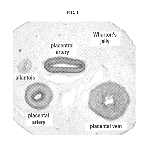
[0007] FIG. 1 exemplifies a cross-section of an umbilical cord (UC).
[0008] FIG. 2 illustrates time to wound closure for the 26 wounds that showed
complete healing
to determine the percentage of wounds healed over time, via a Kaplan-Meier
analysis.
[0009] FIGS. 3A-3B illustrate wound closure based on initial wound area. FIG.
3A illustrates,
for the 26 wounds that achieved complete healing, wounds separated into
quartiles based on the
initial wound area. FIG. 3B illustrates the total time to achieve complete
wound closure for each
respective quartile. Although there was a significant difference in the
initial wound size, i.e.,
*p<0.05 vs. Q 1; Ap<0.05 vs. Q2; #p<0.05 vs. Q3 (FIG. 3A), there is no
difference in the mean
time to achieve wound closure, p>0.05 when compared among the four quartiles
(FIG. 3B).
DETAILED DESCRIPTION OF THE INVENTION
[0010] Provided herein are methods of treating complex wounds. Complex chronic
wounds
constitute life-threatening and severely debilitating conditions. Complex
wounds may occur in
patients requiring long periods of hospitalization with limited mobility for
treating chronic
illness (e.g., pressure sores or bed sores) and result in higher mortality and
lower quality of life.
Complex wounds due to venous stasis ulceration cause considerable morbidity
and poor quality
of life. Complex wounds may occur in patients having an autoimmune disease or
under
immunosuppressive therapy (e.g., vasculitis resulting in extensive ulcers) can
cause longer
hospitalization time and rising costs of treatment. Fournier's gangrene is
another complex wound
and is characterized by an infectious necrotizing fasciitis of the perineum
and genital regions
caused by a mixture of aerobic and anaerobic organisms. The mortality rate
from this infection
can be as high as 67%.
[0011] Non-healing diabetic foot ulcers (DFU), for example, have become a
significant strain on
-4-
CA 03009843 2018-06-26
WO 2017/132503 PCT/US2017/015325
healthcare systems around the world. The World Health Organization estimates
that 347 million
people worldwide suffer from diabetes and according to the US Centers of
Disease Control,
there were 25.8 million Americans in 2010 that have diabetes. Diabetic persons
have
approximately 25% risk of developing a foot ulcer in their lifetime with an
estimated annual
incidence rate of 2%.
[0012] Osteomyelitis and the exposure of bone and/or tendon, muscle, joint
capsule are
prevalent and serious complications of diabetic foot ulcers. Osteomyelitis
refers to the
inflammation or infection of the bone and is a condition that complicates
approximately 20% of
diabetic foot ulcers. Therefore, it is estimated that each year in the U.S.,
100,000 people suffer
from diabetic foot ulcers complicated by underlying osteomyelitis. Deep and
large ulcers
particularly those with exposed bone are more likely to be complicated by
osteomyelitis. Nearly
all diabetic foot ulcers with underlying osteomyelitis result from contiguous
spread of infection
from adjacent soft tissue to the cortical bone and/or bone marrow.
[0013] The prognosis of such complex non-healing diabetic foot ulcers is
generally poor.
Diabetic foot ulcers with exposed bone and with osteomyelitis are at high risk
for delayed/non-
healing of the ulcers, recurrence of ulcers and increased likelihood of
amputation. Non-healing
ulcers compromise the dermal first line of defense, making the patient to be
susceptible to
infection and non-infective tissue loss. Infection of the ulcer is often the
event that prompts
hospitalization and that leads to amputation. When the infection of the ulcer
progresses to
become severe or limb threatening, the amputation rate has been reported to be
as high as 51%.
Over 65,000 non-traumatic lower-limb amputations are performed in the U.S. for
people with
diabetes annually. The risk of amputation increases by four times when the
foot ulcer is
complicated by osteomyelitis compared to soft tissue infection alone.
Unfortunately, after one
major lower extremity amputation, the 5-year survival rate is estimated to be
50%, worse than
those of most malignancies and second only to that of lung cancer. Moreover,
once amputation
occurs, 50% of the patients will develop an ulcer in the contralateral limb
within 5 years. For
amputation survivors, day-to-day functioning is greatly impaired. Many cannot
walk, with or
without the use of a cane or walker. A study found that in 2010, 22.8% of
patients undergoing
amputation of a lower extremity in the United States were readmitted to the
hospital within 30
days, the highest rate of re-admission among the procedures considered in the
study. Moreover,
even with the best of medical care, amputation and its aftermath are traumatic
experiences that
can be expected to produce depression as the patient copes with the social and
financial
consequences of disfigurement and loss of function. Collectively, one can
envision a grave
picture of the seriousness of the complex non-healing foot ulcers of high risk
that may lead to
amputation in this country and worldwide.
-5-
CA 03009843 2018-06-26
WO 2017/132503 PCT/US2017/015325
[0014] The primary treatment goal of managing complex non-healing diabetic
foot ulcers of
high risk with a clinical suspicion of osteomyelitis that have exposed bone
and/or tendon,
muscle, joint capsule is to close the ulcer as expeditiously as possible,
thereby reducing the risk
of further wound related complications such as increased severity of infection
that may lead to
amputation. Current medical therapies include local wound care (e.g. wound
dressing
application and debridement), pain relief, pressure relief (off-loading) and
treatment of infection.
Additional new technologies have also been implemented such as vacuum
extraction devices,
hyperbaric oxygen treatment, and sound-wave technology. New advances in wound
care
products include advanced skin substitutes and recombinant growth factors such
as platelet-
derived growth factor (PDGF). None of the advanced skin substitute products,
however, are
indicated for treating complex ulcers presenting with osteomyelitis. In
addition, the vast
majority have not been demonstrated to be safe or effective in the treatment
of complex non-
healing diabetic foot ulcers that have a depth exhibiting exposure of bone
and/or tendon, muscle,
joint capsule. At least some of these products are not indicated for ulcers
with tendon, muscle,
capsule or bone exposure, and are contraindicated for use on clinically
infected wounds.
Furthermore, nearly all of these advanced skin substitutes require
"engraftment" or "graft take."
[0015] The presently claimed methods do not depend on the fetal support tissue
product
functioning as a scaffold and its engraftment depending on vascularization or
host tissue/cell
integration when applied on the wound bed. Hence, while not wishing to be
bound by any
particular theory, the fetal support tissue products (e.g., umbilical cord
products) may employ a
healing mechanism different from that of conventional advanced skin
substitutes. In contrast to
many of currently available therapies that are targeted to treat specific
actions of a condition, for
example, silver dressings are intended to specifically manage infection and
PDGFs are intended
to stimulate angiogenesis, the fetal support tissue products (e.g., umbilical
cord products) exert
multi-modal actions including anti-inflammatory, anti-scarring, and
regenerative effects in
different types of cells.
[0016] Complex wounds are often chronic and non-healing and provide additional
treatment
challenges when infection and necrotic tissue are present or occur in elderly
or
immunocompromised patients, or those having other chronic illnesses that
contribute to poor
healing (e.g., diabetes, immune system deficiency, arterial or venous
insufficiency, chronic
obstructive pulmonary disease, or paraplegia or quadriplegia). The present
methods provide an
improved treatment for complex wounds. As provided herein in a first exemplary
study, 26 of
27 complex wounds were completely healed following administration of a fetal
support tissue to
the complex wound (Example 1). In a second exemplary study provided herein, a
patient with a
complex wound of the scalp involving tissue and bone necrosis following
surgery and radiation
-6-
CA 03009843 2018-06-26
WO 2017/132503 PCT/US2017/015325
therapy, treated with a fetal support tissue exhibited healing of the soft
tissue and stimulation of
bone regrowth. Thus, the presently disclosed methods address this serious and
potentially fatal
condition that has become a worldwide public health concern and presents a
significant unmet
medical need.
[0017] Disclosed herein, in certain embodiments, are methods of treating a
complex wound in
an individual in need thereof, comprising: applying a fetal support tissue
product to a complex
wound in the individual in an amount effective to treat the complex wound.
[0018] Disclosed herein, in certain embodiments, are methods of treating a
complex lower
extremity ulcer in an individual in need thereof, comprising: applying a fetal
support tissue
product to a complex lower extremity ulcer in the individual in an amount
effective to treat the
complex lower extremity ulcer.
[0019] Disclosed herein, in certain embodiments, are methods of repairing a
spina bifida
defect in an individual in need thereof, comprising: applying an umbilical
cord product to a
spina bifida defect in the individual in an amount effective to repair the
defect.
[0020] Disclosed herein, in certain embodiments, are methods of reducing or
preventing scar
formation from granulation tissue in an individual in need thereof,
comprising: applying a fetal
support tissue product to granulation tissue in the individual in an amount
effective to reduce or
prevent scar formation.
Certain Definitions
[0021] As used herein, "fetal support tissue product" means any isolated
product derived from
tissue used to support the development of a fetus. Examples of fetal support
tissue products
include, but are not limited to, (i) placental amniotic membrane (PAM), or
substantially isolated
PAM, (ii) umbilical cord amniotic membrane (UCAM) or substantially isolated
UCAM, (iii)
chorion or substantially isolated chorion, (iv) amnion-chorion or
substantially isolated amnion-
chorion, (v) placenta or substantially isolated placenta, (vi) umbilical cord
or substantially
isolated umbilical cord, or (vii) any combinations thereof In some
embodiments, the fetal
support tissue is selected from the group consisting of placental amniotic
membrane (PAM),
umbilical cord amniotic membrane (UCAM), chorion, amnion-chorion, placenta,
umbilical cord,
and any combinations thereof In some embodiments, the fetal support tissue
comprises
umbilical cord. Fetal support tissue products include any form of the fetal
support tissue,
including cryopreserved, terminally-sterilized, lyophilized fetal support
tissue or powders
resulting from grinding fetal support tissue. In some embodiments, the fetal
support tissue
product is ground, pulverized, morselized, a graft, a sheet, a powder, a gel,
a homogenate, an
extract, or a terminally-sterilized product. In some embodiments, the fetal
support tissue
product is a graft.
-7-
CA 03009843 2018-06-26
WO 2017/132503 PCT/US2017/015325
[0022] As used herein, "human tissue" means any tissue derived from a human
body. In some
embodiments, the human tissue is a fetal support tissue selected from the
group consisting of
placental amniotic membrane, umbilical cord, umbilical cord amniotic membrane,
chorion,
amnion-chorion, placenta, or any combination thereof
[0023] As used herein, the phrase "granulation tissue" refers to new tissue
and tiny blood
vessels that form on the surfaces of a wound during the healing process. In
some embodiments,
granulation tissue exhibits a bumpy or granular surface containing outgrowths
of new
capillaries. In some embodiments, granulation tissue grows from the base of a
wound and is
able to fill wounds of almost any size. In some embodiments, the fetal support
tissue products
disclosed herein are applied to granulation tissue to prevent or reduce the
formation of scar
tissue from the granulation tissue. In some embodiments, the fetal support
tissue products
disclosed herein are applied to granulation tissue to promote tissue
regeneration wound repair.
In some embodiments, hypergranulation prevents epithelization and the healing
process is
arrested.
[0024] As used herein, a "complex wound" refers to a wound that has exposed
bone, muscle,
tendon, joint capsule or a combination thereof In some embodiments, the
complex wound
comprises exposed bone. In some embodiments, the complex wound comprises loss
of bone. In
some embodiments, bone loss is due to necrosis. In some embodiments, the
complex wound
includes necrosis of soft tissue, bone, or a combination thereof Complex
wounds are generally
difficult to heal and highly susceptible to infection of the skin, muscle, and
tendon, and
predisposes the patient to a risk of osteomyelitis. Complex wounds are at
greater risk of
resulting in amputation, particularly when associated with ischemia or
infection. In some
embodiments, the complex wound is an ulcer, a lower extremity ulcer, a foot
ulcer, a chronic
foot ulcer, or an ischemic wound. In some embodiments, the complex wound is a
pressure sore.
In some embodiments, the complex wound is a venous stasis ulcer or an ulcer
due to vasculitis.
In some embodiments, the complex wound is ischemic. In some embodiments, the
complex
wound involves a wound of the scalp, skull, dura, or a combination thereof In
some
embodiments, the complex wound is associated with infection. In some
embodiments, the
complex wound is associated with osteomyelitis. In some embodiments, the
complex wound is
ischemic and infected.
[0025] A "simple wound" as used herein refers to a wound of the skin with
little or no damage
to underlying tissues such as muscle, tendon, joint or bone.
[0026] As used herein, "graft" means a matrix of proteins (e.g., collagen and
elastin) and
glycans (e.g., dermatan, hyaluronan, and chondroitin) that is used to replace
damaged,
-8-
CA 03009843 2018-06-26
WO 2017/132503 PCT/US2017/015325
compromised, or missing tissue. In certain instances, the matrix is laid down
and host cells
gradually integrate into the matrix.
[0027] As used herein, "minimal manipulation" means (1) for structural tissue,
processing that
does not alter the original relevant characteristics of the tissue relating to
the tissue's utility for
reconstruction, repair, or replacement; and (2) for cells or nonstructural
tissues, processing that
does not alter the relevant biological characteristics of cells or tissues.
[0028] As used herein, "processing" means any activity performed on a fetal
support tissue
product, other than recovery, donor screening, donor testing, storage,
labeling, packaging, or
distribution, such as testing for microorganisms, preparation, sterilization,
steps to inactivate or
remove adventitious agents, preservation for storage, and removal from
storage.
[0029] As used herein, "sheet" means any continuous expanse or surface. In
some embodiments,
a sheet of a fetal support tissue product is substantially flattened. In some
embodiments, a sheet
of a fetal support tissue product is flat. In some embodiments, a sheet of
fetal support tissue
product is tubular. The sheet can be any shape or size suitable for the wound
to be treated. In
some embodiments, the sheet is a square, circle, triangle, or rectangle.
[0030] As used herein, the term "subject" is used to mean any animal,
preferably a mammal,
including a human or non-human. The terms patient, subject, and individual are
used
interchangeably. None of the terms are to be interpreted as requiring the
supervision of a
medical professional (e.g., a doctor, nurse, physician's assistant, orderly,
hospice worker).
[0031] "Substantially isolated" or "isolated" when used in the context of a
fetal support tissue
product means that the fetal support tissue product is separated from most
other non-fetal
support tissue materials (e.g., other tissues, red blood cells, veins,
arteries) derived from the
original source organism.
[0032] As used herein, the phrase "wherein the biological and structural
integrity of the isolated
fetal support tissue product is substantially preserved" means that when
compared to the
biological activity and structural integrity of fresh UC, the biological
activity and structural
integrity of the isolated UC has only decreased by about 5%, about 10%, about
15%, about 20%,
about 25%, about 30%, about 35%, about 40%, about 50%, or about 60%.
[0033] The term "fresh fetal support tissue" refers to fetal support tissue
that is less than 10 days
old following birth, and which is in substantially the same form as it was
following birth.
[0034] As used herein, "biological activity" means the activity of
polypeptides and
polysaccharides of the fetal support tissue. In some embodiments, the
biological activity of
polypeptides and polysaccharides found in fetal support tissue is anti-
inflammatory, anti-
scarring, anti-angiogenic, or anti-adhesion. In some embodiments, the
biological activity is the
biological activity of HC-HA/PTX3 complex in the fetal support tissue. In some
embodiments,
-9-
CA 03009843 2018-06-26
WO 2017/132503 PCT/US2017/015325
the biological activity of HC-HA/PTX3 complex in the fetal support tissue is
substantially
preserved. In some embodiments, the activity of polypeptides and
polysaccharides found in
fetal support tissue is promoting wound healing. In some embodiments, the
activity of
polypeptides and polysaccharides found in fetal support tissue is preventing
scarring. In some
embodiments, the activity of polypeptides and polysaccharides found in fetal
support tissue is
reducing inflammation.
[0035] As used herein, "structural integrity" means the integrity of stroma
and basement
membrane that make up the fetal support tissue product. In some embodiments,
the structural
integrity of the fetal support tissue product results in suture pull out
strength.
[0036] The terms "treat," "treating" or "treatment," as used herein, include
alleviating, abating or
ameliorating a disease or condition symptoms, preventing additional symptoms,
ameliorating or
preventing the underlying metabolic causes of symptoms, inhibiting the disease
or condition,
e.g., arresting the development of the disease or condition, relieving the
disease or condition,
causing regression of the disease or condition, relieving a condition caused
by the disease or
condition, or stopping the symptoms of the disease or condition either
prophylactically and/or
therapeutically. In some embodiments, treating a wound, such as a complex
wound or complex
lower extremity ulcer, refers to promoting wound closure. In some embodiments,
treating a
wound such as a complex wound or complex lower extremity ulcer refers to
complete wound
healing. In some embodiments, complete wound healing refers to 100% re-
epithelialization of
the wound area. In some embodiments, treating a wound, such as a complex wound
or complex
lower extremity ulcer, refers to promoting the generation new bone, tendon,
muscle, and skin.
In some embodiments, treating a wound, such as a complex wound or complex
lower extremity
ulcer, refers to promoting the generation of bone, tendon, muscle, and skin so
that the wound is
closed. In some embodiments, treating a wound, such as a complex wound or
complex lower
extremity ulcer, refers to avoiding or minimizing the need for amputation of
an affected
extremity.
Fetal Support Tissue Products
[0037] As used herein, the term "product" refers ground, pulverized,
morselized, a graft, a sheet,
a powder, a gel, a homogenate, an extract, or a terminally-sterilized product
derived from a fetal
support tissue. In some embodiments, the fetal support tissue product is a
graft. . In some
embodiments, the fetal support tissue product is a sheet. In some embodiments,
the fetal support
tissue product is derived from placental amniotic membrane, umbilical cord,
umbilical cord
amniotic membrane, chorion, amnion-chorion, placenta, or any combination
thereof
[0038] In some embodiments, the fetal support tissue product is an umbilical
cord product. In
some embodiments, the umbilical cord product comprises umbilical cord amniotic
membrane
-10-
CA 03009843 2018-06-26
WO 2017/132503 PCT/US2017/015325
and at least some Wharton's jelly. In some embodiments, the umbilical cord
product lacks
umbilical cord vein and arteries.
[0039] As used herein, "placental amniotic membrane" (PAM) means amniotic
membrane
derived from the placenta. In some embodiments, the PAM is substantially
isolated.
[0040] As used herein, "umbilical cord" means the organ that connects a
developing fetus to the
placenta. The umbilical cord is made up of amniotic membrane (UCAM), Wharton's
Jelly, and
blood vessels. The UCAM functions to regulate the fluid pressure within the
UC. For a cross-
sectional view of an umbilical cord, see FIG. 1. As used herein, "Wharton's
Jelly" means a
gelatinous substance within the umbilical cord, largely made up of
mucopolysaccharides
(hyaluronic acid and chondroitin sulfate). The umbilical cord further
comprises two arteries (the
umbilical arteries) and one vein (the umbilical vein), buried within the
Wharton's jelly. In certain
instances, an umbilical vein supplies a developing fetus with oxygenated blood
from the
placenta. In certain instances, an umbilical artery returns deoxygenated blood
to the placenta.
[0041] As used herein, "umbilical cord amniotic membrane" (UCAM) means
amniotic
membrane derived from the umbilical cord. It reduces inflammation, reduces
angiogenesis,
reduces scarring, and reduces adhesion. UCAM is a translucent membrane. The
UCAM has
multiple layers: an epithelial layer; a basement membrane; a compact layer; a
fibroblast layer;
and a spongy layer. Further, the basement membrane of the UCAM serves as a
natural niche for
stem cells. It lacks blood vessels or a direct blood supply. In some
embodiments, the UCAM is
substantially isolated. In some embodiments, the UCAM further comprises
Wharton's Jelly. In
some embodiments, the UCAM further comprises at least a portion of Wharton's
Jelly. In some
embodiments, the UCAM comprises blood vessels and/or arteries. In some
embodiments, the
UCAM comprises Wharton's Jelly and blood vessels and/or arteries.
[0042] As used herein, "placenta" means the organ that connects a developing
fetus to the
maternal uterine wall to allow nutrient uptake, waste elimination, and gas
exchange via the
maternal blood supply. The placenta is composed of three layers. The innermost
placental layer
surrounding the fetus is called amnion. The allantois is the middle layer of
the placenta (derived
from the embryonic hindgut); blood vessels originating from the umbilicus
traverse this
membrane. The outermost layer of the placenta, the chorion, comes into contact
with the
endometrium. The chorion and allantois fuse to form the chorioallantoic
membrane.
[0043] As used herein, "chorion" means the membrane formed by extraembryonic
mesoderm
and the two layers of trophoblast. The chorionic villi emerge from the
chorion, invade the
endometrium, and allow transfer of nutrients from maternal blood to fetal
blood. The chorion
consists of two layers: an outer layer formed by the trophoblast, and an inner
layer formed by
the somatic mesoderm; the amnion is in contact with the latter. The
trophoblast is made up of an
-11-
CA 03009843 2018-06-26
WO 2017/132503 PCT/US2017/015325
internal layer of cubical or prismatic cells, the cytotrophoblast or layer of
Langhans, and an
external layer of richly nucleated protoplasm devoid of cell boundaries, the
syncytiotrophoblast.
The avascular amnion is adherent to the inner layer of the chorion.
[0044] As used herein, "amnion-chorion" means a product comprising amnion and
chorion. In
some embodiments, the amnion and the chorion are not separated (i.e., the
amnion is naturally
adherent to the inner layer of the chorion). In some embodiments, the amnion
is initially
separated from the chorion and later combined with the chorion during
processing.
Generation of UC Products
[0045] In some embodiments, the fetal support tissue products are UC products.
In some
embodiments, the UC products comprise: isolated UC tissue that does not
comprise a vein or an
artery. In some embodiments, the UC products comprise: isolated UC tissue that
does not
comprise a vein or an artery, a cell with metabolic activity, active HIV-1,
active HIV-2, active
HTLV-1, active hepatitis B, active hepatitis C, active West Nile Virus, active
cytomegalovirus,
active human transmissible spongiform encephalopathy, or active Treponema
pallidum, wherein
the natural structural integrity of the UC product is substantially preserved
for at least 15 days
after initial procurement. In some embodiments, the UC product comprises
umbilical cord
amniotic membrane and Wharton's Jelly. In some embodiments, the biological
activity of HC-
HA/PTX3 complex in the UC product is substantially preserved. In some
embodiments, the
biological activity of HC-HA/PTX3 complex in the UC product is substantially
preserved for at
least 15 days. In some embodiments, the biological and structural integrity of
the UC product is
substantially preserved for at least 20 days after initial procurement. In
some embodiments, the
biological and structural integrity of the UC product is substantially
preserved for at least 25
days after initial procurement. In some embodiments, the biological and
structural integrity of
the UC product is substantially preserved for at least 30 days after initial
procurement. In some
embodiments, the biological and structural integrity of the UC product is
substantially preserved
for at least 35 days after initial procurement. In some embodiments, the
biological and structural
integrity of the UC product is substantially preserved for at least 40 days
after initial
procurement. In some embodiments, the biological and structural integrity of
the UC product is
substantially preserved for at least 45 days after initial procurement. In
some embodiments, the
biological and structural integrity of the UC product is substantially
preserved for at least 50
days after initial procurement. In some embodiments, the biological and
structural integrity of
the UC product is substantially preserved for at least 55 days after initial
procurement. In some
embodiments, the biological and structural integrity of the UC product is
substantially preserved
for at least 60 days after initial procurement. In some embodiments, the
biological and structural
integrity of the UC product is substantially preserved for at least 90 days
after initial
-12-
CA 03009843 2018-06-26
WO 2017/132503 PCT/US2017/015325
procurement. In some embodiments, the biological and structural integrity of
the UC product is
substantially preserved for at least 180 days after initial procurement. In
some embodiments, the
biological and structural integrity of the UC product is substantially
preserved for at least 1 year
after initial procurement. In some embodiments, the biological and structural
integrity of the
UC product is substantially preserved for at least 2 years after initial
procurement. In some
embodiments, the biological and structural integrity of the UC product is
substantially preserved
for at least 3 years after initial procurement. In some embodiments, the
biological and structural
integrity of the UC product is substantially preserved for at least 4 years
after initial
procurement. In some embodiments, the biological and structural integrity of
the UC product is
substantially preserved for at least 5 years after initial procurement.
[0046] Further disclosed herein, in certain embodiments, a method of producing
a UC product,
comprising: obtaining pre-frozen umbilical cord, and removing the umbilical
vein and umbilical
arteries, wherein the structural integrity of the UC product is substantially
preserved for at least
15 days after processing. In some embodiments, substantially all of the blood
is removed from
the umbilical cord product. In some embodiments, the umbilical cord is
processed by thawing
pre-frozen umbilical cord, removing the umbilical vein and umbilical arteries,
and removing
substantially all of the blood from the umbilical cord. In some embodiments,
the biological and
structural integrity of the UC product is substantially preserved for at least
20 days after
processing. In some embodiments, the biological and structural integrity of
the UC product is
substantially preserved for at least 25 days after processing. In some
embodiments, the
biological and structural integrity of the UC product is substantially
preserved for at least 30
days after processing. In some embodiments, the biological and structural
integrity of the UC
product is substantially preserved for at least 35 days after processing. In
some embodiments,
the biological and structural integrity of the UC product is substantially
preserved for at least 40
days after processing. In some embodiments, the biological and structural
integrity of the UC
product is substantially preserved for at least 45 days after processing. In
some embodiments,
the biological and structural integrity of the UC product is substantially
preserved for at least 50
days after processing. In some embodiments, the biological and structural
integrity of the UC
product is substantially preserved for at least 55 days after processing. In
some embodiments,
the biological and structural integrity of the UC product is substantially
preserved for at least 60
days after processing. In some embodiments, the biological and structural
integrity of the UC
product is substantially preserved for at least 90 days after processing. In
some embodiments,
the biological and structural integrity of the UC product is substantially
preserved for at least
180 days after processing. In some embodiments, the biological and structural
integrity of the
UC product is substantially preserved for at least 1 year after processing. In
some embodiments,
-13-
CA 03009843 2018-06-26
WO 2017/132503 PCT/US2017/015325
the biological and structural integrity of the UC product is substantially
preserved for at least 2
years after processing. In some embodiments, the biological and structural
integrity of the UC
product is substantially preserved for at least 3 years after processing. In
some embodiments,
the biological and structural integrity of the UC product is substantially
preserved for at least 4
years after processing. In some embodiments, the biological and structural
integrity of the UC
product is substantially preserved for at least 5 years after processing. In
some embodiments, at
least a portion of the Wharton's Jelly is removed. Umbilical cord is recovered
from any suitable
source (e.g., a hospital or tissue bank). In some embodiments, umbilical cord
is obtained from a
mammal. In some embodiments, umbilical cord is obtained from a human, a non-
human
primate, a cow or a pig.
[0047] The umbilical cord product is kept at -80 C until donor and specimen
eligibility has been
determined. In some embodiments, storing the UC product at -80 C kills
substantially all cells
found in the UC. In some embodiments, storing the UC product at -80 C kills
substantially all
cells found in the UC product while maintaining or increasing the biological
activity of the UC
product (e.g., its anti-inflammatory, anti-scarring, anti-antigenic, and anti-
adhesion properties)
relative to fresh (i.e., non-frozen) UC. In some embodiments, storing the UC
product at -80 C
results in the loss of metabolic activity in substantially all cells found in
the UC. In some
embodiments, storing the UC at -80 C results in the loss of metabolic activity
in substantially all
cells found in the UC while maintaining or increasing the biological activity
of the UCAM (e.g.,
its anti-inflammatory, anti-scarring, anti-antigenic, and anti-adhesion
properties) relative to fresh
(i.e., non-frozen) UC. In some embodiments, the UC is dried. In some
embodiments, the UC is
not dehydrated.
Processing of UC products
[0048] All processing is done following Good Tissue Practices (GTP) to ensure
that no
contaminants are introduced into the UC product.
[0049] The umbilical cord is tested for HIV-1, HIV-2, HTLV-1, hepatitis B and
C, West Nile
virus, cytomegalovirus, human transmissible spongiform encephalopathy (e.g.,
Creutzfeldt-
Jakob disease) and Treponema pallidum using FDA licensed screening test. Any
indication that
the tissue is contaminated with HIV-1, HIV-2, HTLV-1, hepatitis B and C, West
Nile virus, or
cytomegalovirus results in the immediate quarantine and subsequent destruction
of the tissue
specimen.
[0050] Further, the donor's medical records are examined for risk factors for
and clinical
evidence of hepatitis B, hepatitis C, or HIV infection. Any indication that
the donor has risk
factors for, and/or clinical evidence of, infection with HIV-1, HIV-2, HTLV-1,
hepatitis B and
C, West Nile virus, cytomegalovirus, human transmissible spongiform
encephalopathy (e.g.,
-14-
CA 03009843 2018-06-26
WO 2017/132503 PCT/US2017/015325
Creutzfeldt-Jakob disease) and Treponema pallidum results in the immediate
quarantine and
subsequent destruction of the tissue specimen.
[0051] In some embodiments, the UC is frozen. In some embodiments, the UC is
not frozen. If
the UC is not frozen, it is processed as described below immediately.
[0052] In some embodiments, substantially all of the blood is removed from the
UC (e.g., from
any arteries and veins found in the UC, and blood that has infiltrated into
the tissue). In some
embodiments, substantially all of the blood is removed from the UC before the
UC is frozen. In
some embodiments, blood is not removed from the UC. In some embodiments, blood
is not
removed from the UC before the UC is frozen. In some embodiments, the blood is
substantially
removed after the UC has been frozen.
[0053] In some embodiments, the umbilical cord tissue is washed with buffer
with agitation to
remove excess blood and tissue. In some embodiments, the umbilical cord tissue
is soaked with
buffer with agitation to remove excess blood and tissue. In some embodiments,
washing or
soaking with agitation reduces the wash time. In some embodiments, the buffer
wash solution is
exchanged for fresh buffer solution. In some embodiments, the umbilical cord
tissue is soaked
in isotonic solution and the solution is exchanged. In some embodiments, the
umbilical cord is
washed with an isotonic buffer or tissue culture media. In some embodiments,
the UC is washed
with saline. In some embodiments, the UC is washed with PBS. In some
embodiments, the UC
is washed with 1X PBS. In some embodiments, the UC is washed with a TRIS-
buffered saline.
In some embodiments, the UC is washed with a HEPES ¨buffered saline. In some
embodiments,
the UC is washed with Ringer's solution. In some embodiments, the UC is washed
with
Hartmann's solution. In some embodiments, the UC is washed with EBSS. In some
embodiments, the UC is washed with HBSS. In some embodiments, the UC is washed
with
Tyrode's Salt Solution. In some embodiments, the UC is washed with Gey's
Balanced Salt
Solution. In some embodiments, the UC is washed with DMEM. In some
embodiments, the UC
is washed with EMEM. In some embodiments, the UC is washed with GMEM. In some
embodiments, the UC is washed with RPMI.
[0054] In some embodiments, the UC is cut into multiple sections (e.g., using
a scalpel). The
size of the sections depends on the desired use of the UC product derived from
the UC. In some
embodiments, a section of the umbilical cord is cut longitudinally (e.g.,
using a scalpel or
scissors) to open the UC. In some embodiments, the section of the UC is not
cut into halves. In
some embodiments, the section of the UC is cut into two halves. In some
embodiments,
additional cuts are made in the Wharton's Jelly to help flatten out the UC.
[0055] In some embodiments, the cut UC tissue is optionally washed again with
buffer to further
remove excess blood and tissue.
-15-
CA 03009843 2018-06-26
WO 2017/132503 PCT/US2017/015325
[0056] In some embodiments, the UC is fastened onto a substrate (e.g., a
styrofoam board) using
any suitable method (e.g., it is fastened with needles or pins (e.g., T
pins)). In some
embodiments, both ends of the umbilical cord are fastened to the substrate. In
some
embodiments, only one end is attached to the substrate. In some embodiments,
the UC is
stabilized with a substrate (e.g., absorbent towel cloth, drape). In some
embodiments, the UC is
oriented such that the inside face of the UC (e.g., the face comprising the
Wharton's Jelly) is
facing up while the outside face (i.e., the face comprising UCAM) is facing
the substrate. If one
end of the umbilical cord is left free, in some embodiments, the free end of
the umbilical cord is
held (e.g., with a clamp, hemostats or a set of forceps (e.g., wide serrated
tip forceps)) while part
or all of the Wharton's Jelly is removed. Alternatively, in some embodiments,
both ends of the
UC are left free.
[0057] The umbilical cord comprises two arteries (the umbilical arteries) and
one vein (the
umbilical vein). In some embodiments, the vein and arteries are removed from
the UC. In
certain instances, the vein and arteries are surrounded (or suspended or
buried) within the
Wharton's Jelly. In some embodiments, the vein and arteries are removed
concurrently with the
removal of the Wharton's Jelly. In some embodiments, the vein and arteries are
peeled (or
pulled) from the umbilical cord (e.g., using a set of forceps). In some
embodiments, the vein and
arteries are cut away (e.g., shaved) from the umbilical cord in sections. In
some embodiments, a
rotoblator removes the vein and arteries concurrently with the Wharton's
Jelly. In some
embodiments, a liposuction machine is utilized to remove the vein and arteries
concurrently with
the Wharton's Jelly. In some embodiments, a vein stripper is utilized to
remove the vein and
arteries concurrently with the Wharton's Jelly. In some embodiments, a liquid
under high
pressure removes the vein and arteries concurrently with the Wharton's Jelly.
In some
embodiments, a brush removes the vein and arteries concurrently with the
Wharton's Jelly. In
some embodiments, a surgical dermatome removes the vein and arteries
concurrently with the
Wharton's Jelly.
[0058] The desired thickness of the UC product determines how much of the
Wharton's Jelly is
removed. In some embodiments, the umbilical cord is contacted with a buffer to
facilitate
separation of the Wharton's Jelly and the UCAM. In some embodiments, the
Wharton's Jelly is
peeled from the UC in layers (e.g., using a set of forceps, hemostats). In
some embodiments, the
Wharton's Jelly is cut away (e.g., shaved) from the UC in sections. In some
embodiments, a
rotoblator (i.e., a catheter attached to a drill with a diamond coated burr)
is utilized to remove
the Wharton's Jelly. In some embodiments, a liposuction machine is utilized to
remove the
Wharton's Jelly. In some embodiments, a liquid under high pressure is applied
to remove the
Wharton's Jelly. In some embodiments, a brush is utilized to remove the
Wharton's Jelly (e.g., a
-16-
CA 03009843 2018-06-26
WO 2017/132503 PCT/US2017/015325
mechanized brush rotating under high speed). In some embodiments, a surgical
dermatome is
utilized to remove the Wharton's Jelly.
[0059] In some embodiments, the UC product comprises isolated umbilical cord
amniotic
membrane (UCAM). In certain instances, the UCAM comprises proteins, glycans,
protein-
glycan complexes (e.g., a complex of hyaluronic acid and a heavy chain of IaI
and PTX3) and
enzymes that promote tissue repair. For example, the stroma of UCAM contains
growth factors,
anti-angiogenic and anti-inflammatory proteins, as well as natural inhibitors
to various
proteases. In some embodiments, proteins and enzymes found in the UCAM diffuse
out of the
UC and into the surrounding tissue. In some embodiments, the UCAM is isolated
by removing
all of the Wharton's Jelly and umbilical vessels from the UC, leaving the
UCAM. In some
embodiments, the umbilical cord is contacted with a buffer to facilitate
separation of the
Wharton's Jelly and the UCAM. In some embodiments, the Wharton's Jelly is
peeled from the
UC in layers (e.g., using a set of forceps, hemostats). In some embodiments,
the Wharton's Jelly
is cut away (e.g., shaved) from the UC in sections. In some embodiments, a
rotoblator (i.e., a
catheter attached to a drill with a diamond coated burr) is utilized to remove
the Wharton's Jelly.
In some embodiments, a liposuction machine is utilized to remove the Wharton's
Jelly. In some
embodiments, a liquid under high pressure is applied to remove the Wharton's
Jelly. In some
embodiments, a brush is utilized to remove the Wharton's Jelly (e.g., a
mechanized brush
rotating under high speed). In some embodiments, a surgical dermatome is
utilized to remove
the Wharton's Jelly. In some embodiments, UCAM is removed directly from the
tubular
umbilical cord. In some embodiments, UCAM is shaved off of the umbilical cord.
In some
embodiments, UCAM is shaved off of the umbilical cord using any suitable
method. In some
embodiments, UCAM is shaved off of the umbilical cord using a shaver or a
surgical
dermatome. After substantially pure UCAM has been obtained, the UCAM is
optionally washed
with buffer to remove excess blood and tissue.
[0060] In some embodiments, the UC product comprises UCAM as a scaffold, and a
plurality of
cells integrated into the scaffold. In some embodiments, the cells are
embryonic stem cells,
mesenchymal stem cells or adult lineage-committed stem cells or differentiated
epidermal cells
(e.g., to treat a burn or a surgical incision in the skin). In some
embodiments, the cells are
mesothelial cells (e.g., to treat to a wound (e.g., surgical incision) in an
internal organ).
[0061] In some embodiments, the use is a homologous use (e.g., a functional
homologous use or
a structural homologous use). In some embodiments, the UC product is minimally
manipulated.
In some embodiments, the UC product does not comprise another article, except
for water,
crystalloids, or a sterilizing, preserving, or storage agent. In some
embodiments, the UC product
-17-
CA 03009843 2018-06-26
WO 2017/132503 PCT/US2017/015325
does not have a systemic effect and is not dependent upon the metabolic
activity of living cells
for its primary function.
[0062] In some embodiments, the UC products are in any suitable shape (e.g., a
square, a circle,
a triangle, a rectangle). In some embodiments, the UC product is generated
from a sheet of UC.
In some embodiments, the sheet is flat. In some embodiments, the sheet is
tubular.
[0063] The size of the UC product depends on the desired use of the UC
product. In some
embodiments, the UC product is cut into multiple sections (e.g., using a
scalpel). In some
embodiments, the UC product is divided into sections that are about 1.0 cm x
about 0.25 cm. In
some embodiments, the UC product is divided into sections that are about 1.0
cm x about 0.5
cm. In some embodiments, the UC product is divided into sections that are
about 1.0 cm x about
0.75 cm. In some embodiments, the UC product is divided into sections that are
about 1 cm x
about 1 cm. In some embodiments, the UC product is divided into sections that
are about 1 cm x
about 2 cm. In some embodiments, the UC product is divided into sections that
are about 1 cm x
about 3 cm. In some embodiments, the UC product is divided into sections that
are about 1 cm x
about 4 cm. In some embodiments, the UC product is divided into sections that
are about 1 cm x
about 5 cm. In some embodiments, the UC product is divided into sections that
are about 1 cm x
about 6 cm. In some embodiments, the UC product is divided into sections that
are about 2 cm x
about 2 cm. In some embodiments, the UC product is divided into sections that
are about 2 cm x
about 3 cm. In some embodiments, the UC product is divided into sections that
are about 2 cm x
about 4 cm. In some embodiments, the UC product is divided into sections that
are about 2 cm x
about 5 cm. In some embodiments, the UC product is divided into sections that
are about 2 cm x
about 6 cm. In some embodiments, the UC product is divided into sections that
are about 3 cm x
about 3 cm. In some embodiments, the UC product is divided into sections that
are about 3 cm x
about 4 cm. In some embodiments, the UC product is divided into sections that
are about 3 cm x
about 5 cm. In some embodiments, the UC product is divided into sections that
are about 3 cm x
about 6 cm. In some embodiments, the UC product is divided into sections that
are about 4 cm x
about 4 cm. In some embodiments, the UC product is divided into sections that
are about 4 cm x
about 5 cm. In some embodiments, the UC product is divided into sections that
are about 4 cm x
about 6 cm. In some embodiments, the UC product is divided into sections that
are about 5 cm x
about 5 cm. In some embodiments, the UC product is divided into sections that
are about 5 cm x
about 6 cm. In some embodiments, the UC product is divided into sections that
are about 6 cm x
about 6 cm. In some embodiments, the UC product is divided into sections that
are about 8 cm x
about 1 cm. In some embodiments, the UC product is divided into sections that
are about 8 cm x
about 2 cm. In some embodiments, the UC product is divided into sections that
are about 8 cm x
about 3 cm. In some embodiments, the UC product is divided into sections that
are about 8 cm x
-18-
CA 03009843 2018-06-26
WO 2017/132503 PCT/US2017/015325
about 4 cm. In some embodiments, the UC product is divided into sections that
are about 8 cm x
about 5 cm. In some embodiments, the UC product is divided into sections that
are about 8 cm x
about 6 cm. In some embodiments, the UC product is divided into sections that
are about 10 cm
x about 10 cm. In some embodiments, the UC product is divided into sections
that are about 12
cm x about 10 cm. In some embodiments, the UC product is divided into sections
that are about
15 cm x about 10 cm. In some embodiments, the UC product is divided into
sections that are
about 20 cm x about 10 cm. In some embodiments, the UC product is divided into
sections that
are about 25 cm x about 10 cm. In some embodiments, the UC product is divided
into sections
that are about 30 cm x about 10 cm.
[0064] In some embodiments, the UC product is contacted with a buffer to
remove substantially
all remaining red blood cells. In some embodiments, the UC product is
contacted with an
isotonic buffer. In some embodiments, the UC product is contacted with saline.
In some
embodiments, the UC product is contacted with PBS. In some embodiments, the UC
product is
contacted with PBS 1X. In some embodiments, the UC product is contacted with
Ringer's
solution. In some embodiments, the UC product is contacted with Hartmann's
solution. In some
embodiments, the UC product is contacted with a TRIS-buffered saline. In some
embodiments,
the UC product is contacted with a HEPES-buffered saline. In some embodiments,
the UC
product is contacted with EBSS. In some embodiments, the UC product is
contacted with HBSS.
In some embodiments, the UC product is contacted with Tyrode's salt Solution.
In some
embodiments, the UC product is contacted with Gey's Balanced Salt Solution. In
some
embodiments, the UC product is contacted with DMEM. In some embodiments, the
UC product
is contacted with EMEM. In some embodiments, the UC product is contacted with
GMEM. In
some embodiments, the UC product is contacted with RPMI.
[0065] In some embodiments, the UC product is contacted with buffer under
agitation to remove
substantially all remaining red blood cells. In some embodiments, the UC
product is contacted
with a buffer for 10 minutes. In some embodiments, the UC product is contacted
with a buffer
for 15 minutes. In some embodiments, the UC product is contacted with a buffer
for 20 minutes.
In some embodiments, the UC product is contacted with a buffer for 25 minutes.
In some
embodiments, the UC product is contacted with a buffer for 30 minutes. In some
embodiments,
the UC product is contacted with a buffer for 35 minutes. In some embodiments,
the UC product
is contacted with a buffer for 40 minutes. In some embodiments, the UC product
is contacted
with a buffer for 45 minutes. In some embodiments, the UC product is contacted
with a buffer
for 50 minutes. In some embodiments, the UC product is contacted with a buffer
for 55 minutes.
In some embodiments, the UC product is contacted with a buffer for 60 minutes.
In some
embodiments, the UC product is contacted with a buffer for 2 hours. In some
embodiments, the
-19-
CA 03009843 2018-06-26
WO 2017/132503 PCT/US2017/015325
UC product is contacted with a buffer for 3 hours. In some embodiments, the UC
product is
contacted with a buffer for 4 hours. In some embodiments, the UC product is
contacted with a
buffer for 5 hours. In some embodiments, the UC product is contacted with a
buffer for 6 hours.
In some embodiments, the UC product is contacted with a buffer for 6 hours. In
some
embodiments, the UC product is contacted with a buffer for 10 hours. In some
embodiments, the
UC product is contacted with a buffer for 12 hours. In some embodiments, the
UC product is
contacted with a buffer for 18 hours. In some embodiments, the UC product is
contacted with a
buffer for 24 hours. In some embodiments, the UC product is contacted with a
buffer for 2 days.
In some embodiments, the UC product is contacted with a buffer for 3 days. In
some
embodiments, the UC product is contacted with a buffer for 4 days. In some
embodiments, the
UC product is contacted with a buffer for 5 days. In some embodiments, the UC
product is
contacted with a buffer for 6 days. In some embodiments, the UC product is
contacted with a
buffer for 7 days. In some embodiments, the UC product is contacted with a
buffer for 10 days.
In some embodiments, the UC product is contacted with a buffer for 14 days. In
some
embodiments, the UC product is contacted with a buffer for 21 days. In some
embodiments, the
UC product is contacted with a buffer for 30 days. In some embodiments, the
buffer is
optionally changed during the contacting (e.g., when the rate at which red
blood cells diffuse
from the UC sheets slows). In some embodiments, a magnetic stirrer is added
during the
contacting. In some embodiments, adding (and activating) a magnetic stirrer
increases the rate at
which the red blood cells diffuse from the UC sheets.
Processing to generate pulverized fetal support tissue product
[0066] In some embodiments, isolated fetal support tissue product is used to
generate a
pulverized fetal support tissue product. As used herein, "pulverized fetal
support tissue product"
means a fetal support tissue product comprising tissue that has been broken up
(or,
disassociated). In some embodiments, the pulverized fetal support tissue
product is a dry
powder. In some embodiments, the pulverized fetal support tissue product is
further processed
into a solution, suspension or emulsion by mixing the fetal support tissue
powder with a carrier.
In some embodiments, the pulverized fetal support tissue product is formulated
into a cream,
lotion, ointment, paste, gel, film or paint. In some embodiments, the
pulverized fetal support
tissue product is contacted with a patch or wound dressing.
[0067] In some embodiments, the isolated fetal support tissue is pulverized by
any suitable
method. In some embodiments, the isolated fetal support tissue is pulverized
by use of a
pulverizer (e.g., a Bessman Tissue Pulverizer, a Biospec biopulverizer, or a
Covaris CryoPrep).
In some embodiments, the isolated fetal support tissue is pulverized by use of
a tissue grinder
(e.g., a Potter-Elvehjem grinder or a Wheaton Overhead Stirrer). In some
embodiments, the
-20-
CA 03009843 2018-06-26
WO 2017/132503 PCT/US2017/015325
isolated fetal support tissue is pulverized by use of a sonicator. In some
embodiments, the
isolated fetal support tissue is pulverized by use of a bead beater. In some
embodiments, the
isolated fetal support tissue is pulverized by use of a freezer/mill (e.g., a
SPEX SamplePrep
Freezer/Mill or a Retch Ball Mill). In some embodiments, the isolated fetal
support tissue is
pulverized by use of a pestle and mortar. In some embodiments, the isolated
fetal support tissue
is pulverized by manual use of a pestle and mortar.
[0068] In some embodiments, the isolated fetal support tissue is optionally
lyophilized before
being pulverized. In some embodiments, the isolated fetal support tissue is
lyophilized by any
suitable method (e.g., exposure to a liquid gas, placement in a freezer). In
some embodiments,
the isolated fetal support tissue is placed in the vacuum chamber of a
lyophilization device until
all or substantially all fluid (e.g., water) has been removed. In some
embodiments, the isolated
fetal support tissue is lyophilized following freezing (e.g., exposure to a
temperature below 0 C,
-20 C, -40 C, -50 C, -60 C, -70 C, -75 C, -80 C, -90 C, or -100 C).
Storage of the fetal support tissue product
[0069] In some embodiments, the fetal support tissue product is stored for
later use. In some
embodiments, storing the fetal support tissue product does not destroy the
integrity of the fetal
support tissue extracellular matrix. In some embodiments, the fetal support
tissue product is
lyophilized. In some embodiments, the fetal support tissue product is stored
in any suitable
storage medium. In some embodiments, the fetal support tissue product is
stored in 50% DMEM
+ 50% Glycerol. In some embodiments, the fetal support tissue product is
stored in 10%, 20%,
30%, 40%, 50%, 60%, 70%, 80%, 90% or 100% glycerol. In some embodiments, the
fetal
support tissue product is stored in10%, 20%, 30%, 40%, 50%, 60%, 70%, 80%, 90%
or 100%
propylene glycol.
[0070] In some embodiments, the fetal support tissue product is optionally
contacted with a
substrate (i.e., a supportive backing). In some embodiments, the fetal support
tissue product is
not contacted with a substrate. In some embodiments, the fetal support tissue
product is
orientated such that the fetal support tissue product is in contact with the
substrate. In some
embodiments, the fetal support tissue product is orientated such that the
stroma is in contact with
the substrate. In some embodiments the fetal support tissue product is
orientated such that the
epithelial side is in contact with the substrate.
[0071] In some embodiments, the fetal support tissue product is attached to
the substrate. In
some embodiments, the substrate is nitrocellulose paper (NC). In some
embodiments, the
substrate is nylon membrane (NM). In some embodiments, the substrate is
polyethersulfone
membrane (PES).
-21-
CA 03009843 2018-06-26
WO 2017/132503 PCT/US2017/015325
Cryopreservation
[0072] In some embodiments, the fetal support tissue product is frozen for
cryopreservation. In
some embodiments, cryopreserving the fetal support tissue product does not
destroy the integrity
of the fetal support tissue extracellular matrix. In some embodiments, the
fetal support tissue
product is exposed to a liquid gas (e.g., liquid nitrogen or liquid hydrogen).
In some
embodiments, the fetal support tissue product is exposed to liquid nitrogen.
In some
embodiments, the fetal support tissue product does not contact the liquid gas.
In some
embodiments, the fetal support tissue product is placed in a container and the
container is
contacted with liquid gas. In some embodiments, the fetal support tissue
product is exposed to
the liquid gas until the fetal support tissue product is frozen.
Lyophilization
[0073] In some embodiments, the fetal support tissue product is lyophilized.
In some
embodiments, the fetal support tissue product is lyophilized following
freezing. In some
embodiments, the fetal support tissue product is lyophilized following
freezing by any suitable
method (e.g., exposure to a liquid gas, placement in a freezer). In some
embodiments, the fetal
support tissue product is frozen by exposure to a temperature below about 0 C.
In some
embodiments, the fetal support tissue product is frozen by exposure to a
temperature below
about -20 C. In some embodiments, the fetal support tissue product is frozen
by exposure to a
temperature below about -40 C. In some embodiments, the fetal support tissue
product is frozen
by exposure to a temperature below about -50 C. In some embodiments, the fetal
support tissue
product is frozen by exposure to a temperature below about -60 C. In some
embodiments, the
fetal support tissue product is frozen by exposure to a temperature below
about -70 C. In some
embodiments, the fetal support tissue product is frozen by exposure to a
temperature below
about -75 C. In some embodiments, the fetal support tissue product is frozen
by exposure to a
temperature below about -80 C. In some embodiments, the fetal support tissue
product is frozen
by exposure to a temperature below about -90 C. In some embodiments, the fetal
support tissue
product is frozen by exposure to a temperature below about -100 C. In some
embodiments, the
fetal support tissue product is frozen by exposure to a liquid gas.
[0074] In some embodiments, the cryopreserved fetal support tissue product is
lyophilized. In
some embodiments, the cryopreserved fetal support tissue product is placed in
the vacuum
chamber of a lyophilization device until all or substantially all fluid (e.g.,
water) has been
removed.
Sterilization
[0075] In some embodiments, the fetal support tissue product is subject to
terminal sterilization
by any suitable (e.g., medically acceptable) method. In some embodiments, the
lyophilized fetal
-22-
CA 03009843 2018-06-26
WO 2017/132503 PCT/US2017/015325
support tissue product is exposed to gamma radiation for a period of time
sufficient to sterilize
the fetal support tissue product. In some embodiments, the lyophilized fetal
support tissue
product is exposed to gamma radiation at 25 kGy for a period of time
sufficient to sterilize the
fetal support tissue product. In some embodiments, the lyophilized fetal
support tissue product is
exposed to an electron beam for a period of time sufficient to sterilize the
fetal support tissue
product. In some embodiments, the lyophilized fetal support tissue product is
exposed to X-ray
radiation for a period of time sufficient to sterilize the fetal support
tissue product. In some
embodiments, the lyophilized fetal support tissue product is exposed to UV
radiation for a
period of time sufficient to sterilize the fetal support tissue product.
Rehydration
[0076] In some embodiments, the fetal support tissue product is partially or
fully rehydrated. In
some embodiments, the fetal support tissue product is rehydrated by contacting
the fetal support
tissue product with a buffer or with water. In some embodiments, the fetal
support tissue product
is contacted with an isotonic buffer. In some embodiments, the fetal support
tissue is contacted
with saline. In some embodiments, the fetal support tissue product is
contacted with PBS. In
some embodiments, the fetal support tissue product is contacted with Ringer's
solution. In some
embodiments, the fetal support tissue product is contacted with Hartmann's
solution. In some
embodiments, the fetal support tissue product is contacted with a TRIS-
buffered saline. In some
embodiments, the fetal support tissue product is contacted with a HEPES-
buffered saline; 50%
DMEM + 50% Glycerol; 10%, 20%, 30%, 40%, 50%, 60%, 70%, 80%, 90% or 100%
glycerol;
and/or 10%, 20%, 30%, 40%, 50%, 60%, 70%, 80%, 90% or 100% propylene glycol.
[0077] In some embodiments, the fetal support tissue product is contacted with
a buffer for 10
minutes. In some embodiments, the fetal support tissue product is contacted
with a buffer for 15
minutes. In some embodiments, the fetal support tissue product is contacted
with a buffer for 20
minutes. In some embodiments, the fetal support tissue product is contacted
with a buffer for 25
minutes. In some embodiments, the fetal support tissue product is contacted
with a buffer for 30
minutes. In some embodiments, the fetal support tissue product is contacted
with a buffer for 35
minutes. In some embodiments, the fetal support tissue product is contacted
with a buffer for 40
minutes. In some embodiments, the fetal support tissue product is contacted
with a buffer for 45
minutes. In some embodiments, the fetal support tissue product is contacted
with a buffer for 50
minutes. In some embodiments, the fetal support tissue product is contacted
with a buffer for 55
minutes. In some embodiments, the fetal support tissue product is contacted
with a buffer for 60
minutes. In some embodiments, the fetal support tissue product is contacted
with a buffer for 2
hours. In some embodiments, the fetal support tissue product is contacted with
a buffer for 3
hours. In some embodiments, the fetal support tissue product is contacted with
a buffer for 4
-23-
CA 03009843 2018-06-26
WO 2017/132503 PCT/US2017/015325
hours. In some embodiments, the fetal support tissue product is contacted with
a buffer for 5
hours. In some embodiments, the fetal support tissue product is contacted with
a buffer for 6
hours. In some embodiments, the fetal support tissue product is contacted with
a buffer for 6
hours. In some embodiments, the fetal support tissue product is contacted with
a buffer for 10
hours. In some embodiments, the fetal support tissue product is contacted with
a buffer for 12
hours. In some embodiments, the fetal support tissue product is contacted with
a buffer for 18
hours. In some embodiments, the fetal support tissue product is contacted with
a buffer for 24
hours.
Methods of Use
Treating complex wounds
[0078] Disclosed herein, in certain embodiments, are methods of treating a
complex wound in
an individual in need thereof, comprising: administering to a complex wound in
the individual a
therapeutically effective amount of a fetal support tissue product. In some
embodiments, the
complex wound is an ulcer, a lower extremity ulcer, a foot ulcer, a chronic
foot ulcer, or an
ischemic wound. In some embodiments, the complex wound is a wound of the
scalp, skull, dura,
or combination thereof In some embodiments, the complex wound comprises
exposed bone. In
some embodiments, the complex wound is ischemic. In some embodiments, the
complex
wound is infected. In some embodiments, the complex wound is ischemic and
infected. In some
embodiments, the method further comprises debriding the complex wound. In some
embodiments, the debriding is surgical debridement. In some embodiments, the
method further
comprises resecting bone. In some embodiments, the resecting the bone is
performed until
healthy bone is reached. In some embodiments, the resecting the bone is
performed to
substantially remove necrotic or diseased bone. In some embodiments, the
method further
comprises opening the cortex of exposed bone. In some embodiments, the opening
the cortex
comprises making holes in the cortical bone to the trabecular bone. In some
embodiments, the
method further comprises monitoring healing of the wound. In some embodiments,
the method
further comprises administering a second fetal support tissue product to the
wound. In some
embodiments, the method further comprises administering a second fetal support
tissue product
to the exposed bone. In some embodiments, administering a second fetal support
tissue product
to the exposed bone comprises injecting the second fetal support tissue into
the exposed bone. In
some embodiments, the method further comprises covering the fetal support
tissue product with
a dressing, antimicrobial dressing, antimicrobial alginate dressing,
compression dressing,
metipel wound contact layer, gauze, patch, substrate, backing, covering,
bandage, or a
combination thereof In some embodiments, the method further comprising
administering a
treatment selected from the group consisting of antibiotics, hyperbaric oxygen
therapy,
-24-
CA 03009843 2018-06-26
WO 2017/132503 PCT/US2017/015325
revascularization therapy, and combinations thereof In some embodiments, the
individual has
osteomyelitis. In some embodiments, the fetal support tissue product is
derived from placental
amniotic membrane, umbilical cord, umbilical cord amniotic membrane, chorion,
amnion-
chorion, placenta, or any combination thereof In some embodiments, the fetal
support tissue
product is derived umbilical cord. In some embodiments, the fetal support
tissue product is
ground, pulverized, morselized, a graft, a powder, a gel, a homogenate, an
extract, or a
terminally-sterilized product. In some embodiments, the fetal support tissue
product is a graft.
In some embodiments, the fetal support tissue product is a substantially-
flattened sheet. In some
embodiments, the fetal support tissue product is from human, non-human
primate, cow or pig. In
some embodiments, the fetal support tissue product is substantially free of
blood.
[0079] In some embodiments, the fetal support tissue product is an umbilical
cord product. In
some embodiments, the umbilical cord product comprises umbilical cord amniotic
membrane. In
some embodiments, the umbilical cord product further comprises Wharton's
Jelly. In some
embodiments, the umbilical cord product is substantially free of blood. In
some embodiments,
the umbilical cord product lacks an umbilical cord vein and umbilical cord
arteries. In some
embodiments, the umbilical cord product is ground, pulverized, morselized, a
graft, a powder, a
gel, a homogenate, or an extract. In some embodiments, the umbilical cord
product is a graft. In
some embodiments, the umbilical cord product is a substantially-flattened
sheet. In some
embodiments, the umbilical cord product is from human umbilical cord, non-
human primate
umbilical cord, cow umbilical cord or pig umbilical cord.
Treating complex lower extremity ulcer
[0080] Disclosed herein, in certain embodiments, are methods of treating a
complex lower
extremity ulcer in an individual in need thereof, comprising: administering to
a complex lower
extremity ulcer in the individual a therapeutically effective amount of a
fetal support tissue
product to treat the complex lower extremity ulcer. In some embodiments, the
ulcer is a foot
ulcer, a chronic ulcer, a diabetic foot ulcer, an arterial insufficiency
ulcer, a venous stasis (VS)
ulcer, a neurotrophic ulcer, or an arterial (ischemic) ulcer. In some
embodiments, the complex
wound comprises exposed bone. In some embodiments, the method further
comprises debriding
the ulcer. In some embodiments, the debriding is surgical debridement. In some
embodiments,
the method further comprises resecting the bone. In some embodiments, the
resecting the bone is
performed until healthy bone is reached. In some embodiments, the resecting
the bone is
performed to substantially remove necrotic or diseased bone. In some
embodiments, the method
further comprises opening the cortex of exposed bone. In some embodiments, the
opening the
cortex comprises making holes in the cortical bone to the trabecular bone. In
some
embodiments, the method further comprises monitoring healing of the ulcer. In
some
-25-
CA 03009843 2018-06-26
WO 2017/132503 PCT/US2017/015325
embodiments, the method further comprises administering a second fetal support
tissue product
to the ulcer. In some embodiments, the method further comprises administering
a second fetal
support tissue product to the exposed bone. In some embodiments, administering
a second fetal
support tissue product to the exposed bone comprises injecting the second
fetal support tissue
into the exposed bone. In some embodiments, the method further comprises
covering the fetal
support tissue product with a dressing, antimicrobial dressing, antimicrobial
alginate dressing,
compression dressing, metipel wound contact layer, gauze, patch, substrate,
backing, covering,
bandage, or a combination thereof In some embodiments, the method further
comprises
administering a treatment selected from the group consisting of antibiotics,
hyperbaric oxygen
therapy, revascularization therapy, and combinations thereof In some
embodiments, the fetal
support tissue product is derived from placental amniotic membrane, umbilical
cord, umbilical
cord amniotic membrane, chorion, amnion-chorion, placenta, or any combination
thereof In
some embodiments, the fetal support tissue product is derived umbilical cord.
In some
embodiments, the fetal support tissue product is ground, pulverized,
morselized, a graft, a
powder, a gel, a homogenate, an extract, or a terminally-sterilized product.
In some
embodiments, the fetal support tissue product is a graft. In some embodiments,
the fetal support
tissue product is a substantially-flattened sheet. In some embodiments, the
fetal support tissue
product is from human, non-human primate, cow or pig. In some embodiments, the
fetal support
tissue product is substantially free of blood.
[0081] In some embodiments, the fetal support tissue product is an umbilical
cord product. In
some embodiments, the umbilical cord product comprises umbilical cord amniotic
membrane. In
some embodiments, the umbilical cord product further comprises Wharton's
Jelly. In some
embodiments, the umbilical cord product is substantially free of blood. In
some embodiments,
the umbilical cord product lacks an umbilical cord vein and umbilical cord
arteries. In some
embodiments, the umbilical cord product is ground, pulverized, morselized, a
graft, a powder, a
gel, a homogenate, or an extract. In some embodiments, the umbilical cord
product is a graft. In
some embodiments, the umbilical cord product is a substantially-flattened
sheet. In some
embodiments, the umbilical cord product is from human umbilical cord, non-
human primate
umbilical cord, cow umbilical cord or pig umbilical cord.
Treating spina bifida
[0082] Spina bifida is a birth defect of the neural tube involving incomplete
closing of the
backbone and the membranes around the spinal cord. Spina bifida can be
classified as spina
bifida occulta or spina bifida cystica. Spina bifida occulta is the mildest
form of spina bifida in
which one or more vertebrae fail to properly form, and usually only visibly
manifests as a
dimple, tuft of hair, or red mark on the back. Spina bifida cystica is a more
severe form and
-26-
CA 03009843 2018-06-26
WO 2017/132503 PCT/US2017/015325
takes the form of either a cyst containing meninges (meningocele), a cyst
containing both
meninges and spinal cord (meningomyelocele) or only spinal cord (myleocele,
also known as
myeloschisis).
[0083] Disclosed herein, in certain embodiments, are methods of repairing a
spina bifida defect
in an individual in need thereof comprising administering to a spina bifida
defect in the
individual a therapeutically effective amount of an umbilical cord product to
repair the defect. In
some embodiments, the spina bifida defect is spina bifida cystica defect. In
some embodiments,
the spina bifida cystica defect is a meningocele defect, a meningomylocele
defect, or a myeocele
defect. In some embodiments, the individual is a fetus in utero or a neonate.
In some
embodiments, the individual is a fetus in utero. In some embodiments, the
umbilical cord
product is sutured in place. In some embodiments, the repair comprises
regenerating epidermal,
dermal, and subcutaneous layers. In some embodiments, the administering an
umbilical cord
product comprises in utero surgery. In some embodiments, the administering an
umbilical cord
product comprises surgery carried out within 12, 24, or 48 hours after birth.
[0084] In some embodiments, the umbilical cord product comprises umbilical
cord amniotic
membrane. In some embodiments, the umbilical cord product further comprises
Wharton's Jelly.
In some embodiments, the umbilical cord product is substantially free of
blood. In some
embodiments, the umbilical cord product lacks an umbilical cord vein and
umbilical cord
arteries. In some embodiments, the umbilical cord product is ground,
pulverized, morselized, a
graft, a sheet, a powder, a gel, a homogenate, or an extract. In some
embodiments, the umbilical
cord product is a graft. In some embodiments, the umbilical cord product is a
substantially-
flattened sheet. In some embodiments, the umbilical cord product is
aseptically processed or
terminally-sterilized. In some embodiments, the umbilical cord product is from
human umbilical
cord, non-human primate umbilical cord, cow umbilical cord or pig umbilical
cord.
Reducing or preventing scar formation from granulation tissue
[0085] Disclosed herein, in certain embodiments, are methods of reducing or
preventing_scar
formation from granulation tissue in an individual in need thereof,
comprising: administering to
granulation tissue in the individual a therapeutically effective amount of a
fetal support tissue
product to reduce or prevent scar formation. In some embodiments, the
granulation tissue arises
during healing of damaged tissue. In some embodiments, the damaged tissue is
the result of a
burn, a wound, an injury, an ulcer, or surgery. In some embodiments, damaged
tissue is skin,
bone, muscle, tendon, cartilage, ligament, soft tissue, or a joint. In some
embodiments, the
method further comprises monitoring healing of the damaged tissue. In some
embodiments, the
method further comprises administering a second fetal support tissue product
to the granulation
tissue. In some embodiments, the method further comprises covering the fetal
support tissue
-27-
CA 03009843 2018-06-26
WO 2017/132503 PCT/US2017/015325
product with a dressing, antimicrobial dressing, antimicrobial alginate
dressing, compression
dressing, metipel wound contact layer, gauze, patch, substrate, backing,
covering, bandage, or a
combination thereof
[0086] In some embodiments, the fetal support tissue product is derived from
placental amniotic
membrane, umbilical cord, umbilical cord amniotic membrane, chorion, amnion-
chorion,
placenta, or any combination thereof In some embodiments, the fetal support
tissue product is
ground, pulverized, morselized, a graft, a powder, a gel, a homogenate, an
extract, or a
terminally-sterilized product. In some embodiments, the fetal support tissue
product is a graft.
In some embodiments, the fetal support tissue product is a substantially-
flattened sheet. In some
embodiments, the fetal support tissue product is from human, non-human
primate, cow or pig. In
some embodiments, the fetal support tissue product is substantially free of
blood.
[0087] In some embodiments, the fetal support tissue product is an umbilical
cord product. In
some embodiments, the umbilical cord product comprises umbilical cord amniotic
membrane. In
some embodiments, the umbilical cord product further comprises Wharton's
Jelly. In some
embodiments, the umbilical cord product is substantially free of blood. In
some embodiments,
the umbilical cord product lacks an umbilical cord vein and umbilical cord
arteries. In some
embodiments, the umbilical cord product is ground, pulverized, morselized, a
graft, a powder, a
gel, a homogenate, or an extract. In some embodiments, the umbilical cord
product is a graft. In
some embodiments, the umbilical cord product is a substantially-flattened
sheet. In some
embodiments, the umbilical cord product is from human umbilical cord, non-
human primate
umbilical cord, cow umbilical cord or pig umbilical cord.
EXAMPLES
Example 1: A single center, retrospective study of cryopreserved umbilical
cord to
promote healing of complex foot ulcers in patients with underlying
osteomyelitis
[0088] A retrospective review was performed to assess healing of 31 patients
presenting with 33
complex foot ulcers with a confirmed histopathological diagnosis of
osteomyelitis treated by the
same surgeon at a single wound care center by sharp debridement, resection of
affected bone,
open cortex and application of cryopreserved umbilical cord (cUC).
Methods
Clinical data retrieval
[0089] This retrospective review was conducted after approval by the
Institutional Review
Board of the Barnabas Health System (West Orange, NJ) to capture, verify, and
subsequently
analyze all relevant clinical data on 31 eligible patients that had been
managed by the same
surgeon between January 2013 and December 2014 at the Wound Care Center at
Clara Maass
-28-
CA 03009843 2018-06-26
WO 2017/132503 PCT/US2017/015325
Medical Center to determine the safety and effectiveness of cUC in promoting
the wound
healing of chronic complex foot ulcers. The clinical data included demographic
information,
past and present medical history including co-morbidities such as diabetes,
hypertension,
peripheral vascular disease, renal disease, and cardiovascular diseases,
previous treatments and
prior amputations. Special attention was given to assess the extent of the
index ulcer by
verifying the exposure of bone, tendon, muscle, or joint capsule, initial
wound area (cm2),
wound location, presence of soft tissue infection and ischemia. In addition,
data were also
retrieved regarding assessment of wound measurement and photography to
document the
changes in the ulcer during the entire follow-up period.
Study patients
[0090] All study patients suffered from chronic, non-healing foot ulcers that
demonstrated
exposed bone, tendon, muscle, or joint capsule. These complex wounds were all
associated with
the clinical diagnosis of osteomyelitis confirmed by bone biopsy with positive
microbial cultures
and histopathological evidence of bone tissue containing lymphocytes or plasma
cells.
Standard evaluation and management
[0091] Following a full initial medical evaluation, all patients presented
with ulcers complicated
by osteomyelitis that were greater than 3 weeks in duration, larger than 2 cm
wide and 3 mm
deep, positive probe-to-bone or exposed bone. Patients were referred for an
infectious disease
consult while obtaining baseline X-ray and blood tests including complete
blood count, C-
reactive protein, erythrocyte sedimentation rate, and alkaline phosphatase.
Those presenting with
suspicion of peripheral vascular disease were also referred for a vascular
consult. Patients
cleared of notable vascular involvement with a high clinical suspicion of
osteomyelitis (i.e.,
elevated white blood cell counts, C-reactive protein, erythrocyte
sedimentation rate, or alkaline
phosphatase) as well as radiographic evidence indicative of bone necrosis,
were immediately
scheduled for treatment as follows: in the operating room, all ulcers
underwent sharp surgical
debridement and the necrotic bone was resected when deemed medically necessary
by the
surgeon. For patients with forefoot ulcers, bone resection was performed until
healthy bone was
reached based on physical characteristics. For mid- and rear-foot ulcers, bone
resection was
performed in a "piecemeal" fashion with the intent of excising the majority of
the diseased bone
tissue while simultaneously preserving as much length as possible. Following
resection, bone
biopsy was performed to obtain a sample for microbial culture and
histopathological
confirmation of osteomyelitis. A small margin (-2 mm) of clinically presumed
healthy bone was
also obtained and submitted for pathological analysis to ensure the complete
removal of diseased
bone, as well as to guide the duration of systemic antibiotic administration.
Afterward, any
-29-
CA 03009843 2018-06-26
WO 2017/132503 PCT/US2017/015325
exposed bone received an open cortex procedure using either a 6-2k-wire or
surgical
debridement device (Misonix. Inc., Farmingdale, NY) to create ¨3 (depending on
the wound
size) equidistant holes through the cortical bone to the underlying trabecular
bone. This allowed
for access to pluripotent adult progenitor stem cells to aid in repair as
previously described. The
exposed surgical wound was then completely covered by one layer of cUC (NEOX
CORD 1K,
Amniox Medical, Inc., Atlanta, GA) that was held in place with either tissue
adhesive
(Indermil tissue adhesive, United States Surgical Corporation, CT) or
staples. All wounds
were then covered with a standard non-adherent dressing (Xeroform dressing,
DeRoyal
Industries, Inc., Powell, TN) followed by compression dressing (CobanTM 2
layer compression
system, 3M, Co., St. Paul, MN) and a variety of off-loading devices. All
patients initially
received oral broad spectrum antibiotics until the bone culture results were
available to guide
subsequent administration of microbe-specific antibiotics. Patients with
forefoot ulcers were
discharged with an initial two-week antibiotic regimen pending the result of
the bone margin
histopathology. If the bone biopsy was free of any involvement, antibiotics
were discontinued,
but if the margin was not clear, the antibiotic therapy was continued for an
additional 4-6 weeks.
Patients with mid- or rear-foot osteomyelitis were discharged with continuous
IV antibiotics for
a 6-8 week duration.
[0092] All patients were discharged from the hospital once they were
clinically stable, and
returned to the wound clinic for weekly wound monitoring including dressing
changes that
followed the above standard of care with the addition of silver sulfadiazine
(Silvadene , Pfizer
Inc., New York, NY) to high colonization. If wound progression appeared to
stall, an additional
cUC application was applied during which time sharp debridement and open bone
cortex were
also performed. Patients received other treatments such as hyperbaric oxygen
therapy and
revascularization therapy if indicated by the study surgeon/PI.
Outcome measures
[0093] Wound area was determined by using a ruler to measure the length and
width of the
wound. Complete wound healing was defined as 100% re-epithelialization as
determined by the
investigator. For those wounds achieving complete healing, the total time
needed to achieve
initial wound closure was assessed. In addition to wound closure, the
relationship between the
initial wound area and the time needed to achieve closure was assessed by
subdividing the initial
wound area into quartiles as previously described. The mean wound area and the
mean time to
achieve wound closure were compared between quartiles using an un-paired t-
test; p-values <
0.05 between groups were considered to be statistically significant.
-30-
CA 03009843 2018-06-26
WO 2017/132503 PCT/US2017/015325
Results
[0094] The patients had multiple co-morbidities including diabetes,
hypertension, peripheral
vascular disease, renal failure, and coronary artery disease. The ulcers were
mostly ischemic,
over half were gangrenous and some received prior partial amputation and
revascularization
attempts. The average ulcer size was 15.6 17.7 cm2(0.4 - 73.95 cm2). Overall,
26 wounds
achieved complete closure (78.8%). Five patients were lost to follow up and
one patient expired
during the course of treatment, not believed to be treatment related,
resulting in a healing rate in
patients not lost to follow up of 96.3% with an average of 1.24 applications
of cUC. Although
16 ulcers were recommended for amputation at presentation, two patients
achieved complete
wound closure using cUC without the need for amputation, one patient
eventually received a
below- knee amputation, and the remaining 13 wounds received partial digit
resection.
Clinical features
[0095] A total of 31 patients presenting with 33 foot ulcers were identified
for inclusion in the
study. A summary of their clinical data is provided in Table 1. There were 26
males and 5
females with an average age of 58.3 12.9 years. The majority of patients
treated were
Caucasian (12/31) or African-American (10/31). Overall, these patients
presented with multiple
co-morbidities, among which the most significant were diabetes (26/31),
hypertension (23/31),
peripheral vascular disease (16/31), renal failure (12/31), and coronary
artery disease (9/31). In
addition, 24/33 wounds (72.2%) were clinically judged as ischemic in the
affected limb and
17/33 wounds (51.5%) had gangrene on the affected extremity.
Table 1. Summary of patient clinical data.
Gender Male 26 (83.9%)
Female 5(16.1%)
Age Median 57 (range: 35 ¨ 90)
Mean 58.3 12.9
Ethnicity Caucasian 12/31 (38.7%)
African-American 10/31 (32.3%)
Hispanic 6/31 (19.3%)
Other 3/31 (9.7%)
Significant Co-morbidities ____ Diabetes 26/31 (83.9%)
Hypertension 23/31 (74.2%)
Peripheral Vascular Disease 16/31 (51.6%)
Renal Failure 12/31 (38.7%)
Coronary Artery Disease 9/31 (29%)
Ischemia in affected limb 24/33 (72.7%)
(wounds)
Gangrene (wounds) 17/33 (51.5%)
-31-
CA 03009843 2018-06-26
WO 2017/132503 PCT/US2017/015325
Initial wound area 16.68 18.07 cm
Wound duration 4 weeks ¨ 1 year 7 months
Wound exposure Muscle, tendon, ligament, 27/33 (81.8%)
_______________________ bone
Osteomyelitis 33/33 (100%)
Wounds healed 26/27 (96.3%)
Average time to healing 16.02 9.25 weeks (range: 4 ¨ 44 weeks)
Average # applications of 1.24 0.44
cUC
[0096] All ulcers included in the study were chronic, non-healing wounds
lasting for a duration
of greater than 3 weeks and up to 1 year and 7 months. The average wound area
for all wounds
at the time of application of cUC was 15.6 17.7 cm2 (0.4 - 73.95 cm2). At
initial presentation,
9/33 ulcers (27.3%) were rearfoot, 21/33 ulcers (63.6%) were forefoot
primarily associated with
the digits, one ulcer was plantar, and 2 ulcers were non-healing Chopart
amputation sites. All 33
ulcers were considered "complex" with exposed tendon, muscle, joint capsule or
bone, and
27/33 ulcers (81.8%) demonstrated exposure of all four tissues. In addition,
all patients had a
diagnosis of osteomyelitis confirmed by bone biopsy showing the
histopathological presence of
inflammatory cells in the bone marrow and positive microbial cultures. Of 33
wounds, 16
(48.5%) presented with a recommendation for amputation by either the referring
physician.
Wound healing after application of cUC
[0097] All 33 complex ulcers underwent sharp debridement, resection of
necrotic bone with
biopsy, open cortex and application of cUC without complications. A total of
26 wounds
achieved complete wound closure as evidenced by complete epithelialization,
resulting in an
overall healing rate of 78.8%. Following initial application of cUC, five
patients were lost to
follow up post-cUC application and one patient expired due to causes not
believed to be related
to the study product. For the 27 wounds not lost to follow-up, the overall
healing rate was
96.3%. For the 26 wounds that completely healed, the average time to wound
closure was 16.0
9.3 weeks (range: 4 ¨ 44 weeks) (FIG. 2). Twenty-one of the 26 wounds which
healed received
a single application of cUC, and 2 applications of cUC were needed to achieve
healing of the
remaining 5 wounds, with the second application occurring between 4 ¨ 10 weeks
after the
initial application. The patient with the non-healing wound underwent a below-
knee amputation
due to wound complications and other co-morbidities.
[0098] To determine if initial wound area had any impact on the time to reach
complete wound
closure, the wounds were separated into four quartiles based on their initial
area (0-25%, 25-
50%, 50-75%, and 75-100%) of 2.2 0.9, 5.8 1.6, 13.8 3.9, and 43.0 23.1
cm2,
respectively (FIG. 3A). As expected, there was a statistical difference among
wound size in
-32-
CA 03009843 2018-06-26
WO 2017/132503 PCT/US2017/015325
these four quartiles. However, the mean time to achieve wound closure was
found to be similar
without any statistically significant difference among them (FIG. 3B).
[0099] Although 16 ulcers were recommended for amputation at presentation, two
patients
achieved complete wound closure using cUC without the need for amputation. As
stated above,
one patient (6.25%) eventually received a below-knee amputation after a single
cUC application.
The remaining 13 wounds received partial digit resection, to a lesser extent
than what was
initially recommended.
Representative Cases
Case study #1: 2nd and 3rd metatarsal wound with gangrene
[00100] A 52 year-old female presented with several co-morbidities
including type II
diabetes, diabetic neuropathy, peripheral vascular disease, end stage renal
disease, hypertension,
hypercholesterolemia, and coronary artery disease. She was referred by the
Infectious Diseases
department of the hospital with a recommendation to amputate the 2' and 3rd
digits while
presenting with a forefoot ulcer on the left 2' and 3rd metatarsals with
exposed muscle, tendon,
ligament, and bone with gangrene. Following sharp debridement, bone resection,
bone biopsy
and open cortex procedure, cUC was applied to completely cover the index
ulcer. Bone biopsy
confirmed the diagnosis of osteomyelitis. At six weeks, the wound was reduced
in size by
approximately 50%. At seven weeks, the wound healing progression was noted to
have stalled,
and a second application of cUC was applied after debridement and open cortex
surgical
procedure. At 12 weeks, the index ulcer achieved complete epithelialization
saving both the 2'
and 3rd digits without the need for any amputation. During the entire follow
up period of 32
weeks, there was no recurrence.
Case study #2. Wound post-4th ray amputation
[00101] A 57 year-old male with diabetes, cellulitis, and ischemia
presented with an open
wound following left 4th toe amputation with exposed bone, tendon, muscle, and
ligament. After
sharp debridement, bone resection, bone biopsy and open cortex procedure, cUC
was applied to
completely cover the wound. At 4 days and two weeks after application, the cUC
could be
observed filled with blood. However, the cUC remnant was left in place over
the wound bed,
and the wound continued to heal, achieving complete re-epithelialization by
week 13.
Case study #3 ft ray amputation
[00102] A 63 year-old male with a history of type I diabetes and
peripheral vascular
disease presented with an open wound in the midfoot following first ray
amputation. In addition
-33-
CA 03009843 2018-06-26
WO 2017/132503 PCT/US2017/015325
to osteomyelitis, gangrene was present on the affected limb. After sharp
debridement, bone
resection, bone biopsy, and open cortex procedure, the wound was covered by
cUC. At 7 weeks,
the wound displayed a 44% reduction in size. At 9 weeks, another application
of cUC was
performed following sharp debridement and open cortex surgical procedure. The
index wound
continued to show improvement at 11 weeks and went on to complete healing at
15 weeks. The
wound remained healed with no recurrence at 28 weeks.
Example 2: Cryopreserved human umbilical cord (HUC) vs. biocellulose film
(BSCF) for
antenatal spina bifida repair
[00103] Two patches, HUC and BCF sutured over spina bifida (SB) lesions in
a retinoic
acid (RA) rat model, were compared for regenerative ingrowth of native cells
and associated
inflammatory response.
[00104] Pregnant time-dated Sprague-Dawley rats were gavaged with RA
(40mg/kg) on
gestational day 10 (GD10) to induce SB in pups. Laparotomy and hysterotomy
were performed
on GD20 and HUC (n=11) or BCF (n=10) sutured over the spinal defect. Patches
placed into the
amniotic cavity without suturing over the lesion were controls. 30-34 hours
after grafting pups
were harvested and formalin fixed. H&E and Trichrome staining assessed
cellular migration into
the patches. Immunohistochemistry was performed to demonstrate the nature of
the cellular
migration. Native cell markers evaluated were CK 5/6 (epidermal), GFAP
(astrocytes) and vWF
(endothelial). Inflammatory markers were CD3 (lymphocytes), MPO (neutrophils),
and F4-80
(macrophages). Four high power fields (hpf) of all patches and surrounding
exudates were
evaluated and Image-J software was used to quantify cells.
[00105] Pup survival was equal: HUC 8/11, BCF 7/10, (p=0.9). Histology
showed cellular
migration in all HUC patches applied over lesions (median:38 [range:13-102]
cells/hpf)
compared to none in BCF patches (Figure; p<0.001). CK 5/6 positive cells were
noted migrating
over the HUC patch surface (4-7ce11s/hpf): GFAP positive cells were noted on
the HUC patch
surface adjacent to the lesion (3-11 cells/hpf); vWF positive cells were noted
in the HUC patch
(5-15cells/hpf). No CK 5/6, GFAP or vWF positive cells were noted in BCF
patches (p=0.03).
HUC patches showed minimal MPO (2%[0-7%]), CD3 (7%[3-12%]) and F4-80 (0%)
positive
cells. Exudates in HUC treated pups had fewer MPO (0%[0-9%] vs 17%[0-39%];
p<0.01) and
CD3 (7%[0-13%] vs 15%[0-22%]; p<0.01) positive cells compared to BCF and
demonstrated no
difference in F4-80. Both HUC and BCF control patches demonstrated no
infiltrate.
-34-
CA 03009843 2018-06-26
WO 2017/132503 PCT/US2017/015325
[00106] Example 3: Cryopreserved human umbilical cord for in-utero
myeloschisis
repair
[00107] Described herein is a case of large lumbosacral myeloschisis with
Chiari II
malformation that underwent in-utero spina bifida repair at 23 weeks of
gestation. The skin
defect was closed using cryopreserved human umbilical cord patch following the
primary
closure of meningeal layers. The pregnancy was uncomplicated and the delivery
occurred at 37
weeks by elective C-section. The repair site was intact with no evidence of
cutaneous
cerebrospinal fluid leakage. The skin regenerated into the patch after
delivery over a period of 4
weeks. There was complete reversal of Chiari II malformation, normal lateral
ventricles, normal
sensory and motor response in the lower extremities, normal voiding
cystourethrogram.
[00108] A 21 year old G1P0-0-0 was referred to the Fetal Center at
Children's Memorial
Hermann Hospital with myeloschisis of the fetus at 21 6/7 weeks gestation for
possible in-utero
spina bifida repair. On evaluation with both ultrasound and MRI, the skin
defect extended from
L3 level to S4 with no identifiable meningeal sac. The overall dimensions of
the lesion
measured 1.6 cm by 2.7 cm. The posterior superior iliac spine was prominent
through the defect.
She underwent an amniocentesis with the findings of a 46XX karyotype, elevated
maternal
serum alpha-fetoprotein at 8.25 multiples of median and a positive
acetylcholinesterase. The
fetal MRI showed a grade III Chiari II malformation with effacement of the
cisterna magna and
fourth ventricle and herniation of the cerebellar tonsils to the level of C2-
C3. The left lateral
ventricle measured 7.1 mm and the right measured 8.1 mm. Both lower
extremities had good
movement and there was no talipes. The patient met all the Management of
Myelomeningocele
Study (MOMS) for in-utero spina bifida repair. Due to the large size of the
myeloschisis with
the relationship of the skin margins to the posterior superior iliac spine,
the need of using a patch
to cover the skin was discussed. The patient was offered the HUC patch as an
alternative to the
usual Allodermg after approval from the Fetal Therapy Board and the
Institutional Review
Board at University of Texas Medical School at Hosuton, TX
[00109] At 24 weeks of gestation, the surgery was conducted similar to the
MOMs study
protocol. Briefly, the patient underwent laparotomy under general anesthesia,
followed by
exposure of the uterus. The fetus was positioned so that the spina bifida
lesion was close to the
possible site of hysterotomy. Uterine entry was made between two full
thickness stay sutures.
The incision was extended using absorbable staples (Covidien, Dublin,
Ireland).The fetal spina
bifida defect site was exposed. A continuous intrauterine infusion of Lactated
Ringers solution
with Nafcillin lg/L was infused through a foley catheter. Fetal anesthesia
consisting of fentanyl
20 ug/kg and paralytic agent (vecuronium 400 ug/kg) was administered
intramuscularly. The
-35-
CA 03009843 2018-06-26
WO 2017/132503 PCT/US2017/015325
defect was confirmed to be a myeloschisis with meningeal layers extending
beyond the edges of
the lesion with protruding posterior superior iliac spines. The skin lesion
measured 5 cm x 6 cm.
An incision was made at the junction of meninges and skin. The meningeal
layers were
dissected off the fascia and closed primary approach in the midline using 6-0
Monocryl suture
(Ethicon,Somerville, NJ). The skin edges were sutured to an HUC patch using 6-
0 Monocryl
suture in a running locking (towards the patch) fashion, circumferentially.
The fetus was
repositioned in the uterus. Amnioinfusion was performed to refill the uterine
cavity. The uterine
incision was closed using 0-0 Prolene sutures with multiple stay sutures and a
running suture to
reapproximate the edges. The laparotomy was closed in the usual fashion. Post-
operatively, the
patient had an uneventful course and was discharged home on post-operative day
#5. The post-
natal course was uncomplicated. On weekly ultrasound examination, the lesion
site was noted to
be covered with tissue with a thickness measuring 3-4 mm. There was a fluid-
filled space noted
posterior to the spinal cord and below the tissue at the repair site.
[00110] At 37 1/7 weeks gestation, the patient was electively delivery by C-
section. The lesion
site appeared completely covered with no evidence of cutaneous CSF leakage.
The patch
appeared semi-transparent with visible clear fluid below it; the dural closure
could be seen
through the patch. The defect size measured 6 cm x 5 cm. The anterior
frontenelle was soft; the
head circumference measured 33.5 cm. There was symmetric 5/5 power in all
proximal muscle
groups in the legs. There was slight right dorsiflexion weakness initially
that improved at the
time of discharge. Sensation to pin testing was noted in all lumbar
dermatomes, 51 and perinatal
region. The voiding cystourethrogram was normal and the post-void residual
volume measuring
<5 ml. The neonate was placed in supine position with lower spine elevated as
a precautionary
measure for 2 weeks. During the interim period, there was rapid regeneration
of the skin into the
graft. Wound care consisted of daily changed with a non-adherent dressing.
[00111] On day #1, the patch appeared opaque with increased vascularity in the
periphery. On
day #7, vascularization and epithelialization continued at the peripheral
margins. There was a
coat of fibrin deposited over the center of the patch. On day #14, there was
complete
epithelialization of the defect except for a 2 cm x 1.5 cm in the lower part
of the defect where
there was vascular tissue covered with fibrin. Keratinization was noted to be
proceeding from
the periphery to the central portion of the defect over to the epithelialized
tissue. On day #21,
there was central area of granulation tissue measuring 1 cm x 1 cm surrounded
by
epithelialization and keratinization. On day #28, the skin had completely
healed over with
keratinization except for 3 x 5 mm area that was still epithelializing.
-36-
CA 03009843 2018-06-26
WO 2017/132503 PCT/US2017/015325
[00112] Head ultrasound and MItI was performed on day #21. There was complete
reversal of
the Chiari II malformation, with atrophic changes in the left cerebellar
hemisphere consistent
with stigmata of hindbrain herniation prior to the surgery. The lateral
ventricles measured within
normal limits. There was a tissue bridge measuring 4-5 mm in thickness
covering the defect site.
There was no fluid filled space in the regenerated skin. There was a fluid
space posterior to the
spinal cord and the thecal layers. T1/T2 weighted images showed hypodense
fibrous tissue,
suggestive of tethering, between the spinal cord at the upper edge of the
repair to the posterior
thecal coverings of the spinal canal. The tethering adhesion measured 1 mm in
thickness at the
level of L3. The conus terminated at the level of L4. There was no tethering
of the conus.
Dysraphism of the posterior spinal elements was seen at L2-S1. The skin
overlying the defect
appeared intact without a definite subcutaneous tract.
[00113] The neonate was discharged home on day #22. The head circumference at
discharge
remained at 33 cm with a soft and scaphoid shaped anterior frontenelle. The
lower extremities
had normal movement. Neonatal urodynamic testing was within normal limits and
the anal
reflex was normal.
[00114] In this case, the primary objective of the patch was to create a water-
tight and effective
barrier between the spinal cord and the amniotic fluid which was successful.
The HUC patch
also showed reversal of Chiari II malformation and preservation of the lower
extremities
neurological function at birth. The lack of epithelialization and
keratinization in utero was a
surprising finding. Additionally, the rapid ingrowth of vascularization,
epithelialization and
keratinization after delivery was remarkable, which we were able to evidence
on a daily basis.
The presence of cerebrospinal fluid between the patch and dural closure and
demonstration of
fluctuance of anterior frontenelle supports the possibility of incomplete
meningeal sealing.
However, after the complete healing of the patch, there was no fluid space
found between the
repair site and meninges on the MItI. This supports the water-tight healing.
Example 4: Manufacturing process of cUC
[00115] cUC is aseptically processed in compliance with current Good Tissue
Practices (cGTP)
from donated human placental tissue after determination of donor eligibility
and placenta/cord
suitability.
[00116] Upon receipt of a tissue shipment, the shipping container is stored in
a designated
freezer.
[00117] Frozen tissue is thawed for processing. The UC is isolated from the
placenta and
opened. The arteries, vein, and a portion of the Wharton's Jelly that is
associated with the blood
-37-
CA 03009843 2018-06-26
WO 2017/132503 PCT/US2017/015325
vessels are removed, leaving the AM and remainder of the Wharton's Jelly. The
blood is
removed by soaking and swirling in solution and manual gentle removal.
[00118] The tissue is cut to achieve the designated product sizes.
[00119] Each unit of tissue is aseptically packaged in storage medium a
sterile clear plastic peel
pouch for a single application.
Example 5: Use of a fetal support tissue product to reduce or prevent scar
formation in
granulation tissue
[00120] An individual having a wound exhibiting granulation tissue is
identified. A fetal
support tissue product is prepared. The fetal support tissue product applied
to the granulation
tissue. A protective covering is place over the fetal support tissue product.
Example 6: Use of a fetal support tissue product to repair a gingival wound
[00121] An individual in need of repair of a gingival wound is identified.
A fetal support
tissue product is prepared. The wound is debrided as necessary. The fetal
support tissue product
is placed over the wound. A protective covering is place over the fetal
support tissue product.
Example 7: Use of a fetal support tissue product to repair a damaged joint
cavity
[00122] An individual in need of repair of a damaged joint cavity is
identified. A fetal
support tissue product is prepared. The joint cavity is prepared. The fetal
support tissue product
is placed over the joint cavity. A protective covering is place over the fetal
support tissue
product.
Example 8: Use of a fetal support tissue product to treat a complex foot ulcer
[00123] An individual having a complex foot ulcer is identified. A fetal
support tissue
product is prepared. The ulcer is debrided as necessary. Bone is resected as
necessary.
Optionally, an open cortex procedure is performed. The fetal support tissue
product is placed
over the ulcer. A protective covering is place over the fetal support tissue
product.
Example 9: Cryopreserved umbilical cord (cUC) treatment of radiation wound
post
melanoma removal involving soft tissue and bone
[00124] The patient was an 87-year old male with a history of renal
transplantation due to
polycystic kidney disease and most recently melanoma on his scalp. Surgical
removal revealed
the cancer had penetrated the bone. The melanoma's proximity to the brain did
not allow for it
all to be removed surgically, so the patient underwent radiation treatment.
The radiation
resulted in necrosis of the skull and a significant wound with exposed brain
dura.
-38-
CA 03009843 2018-06-26
WO 2017/132503 PCT/US2017/015325
[00125] A 4x3 cm cUC graft (Neoxlk; NX-10-4030) was cut into 6 strips and
placed in
an asterisk pattern over the wound and covered with a non-adhering dressing
(Adaptic touch).
Within 4 weeks the graft had absorbed and the wound bed had begun to
granulate. 16 weeks
after the initial application of the cUC graft, 100 mg of a particulate form
of the tissue (CR-FL-
100mg; Clarix Flo) was injected into the bone to stimulate progress in the
cranial margins. 21
weeks from the initial cUC application, progress was noted both in
epithelialization and cranial
margins. Therefore, another 100 mg of particulate cUC (Clarix Flo) was
injected into the bone
and a second 4x3 cm cUC tissue graft (NX-10-4030) was placed along the wound
borders to
progress this wound to complete healing.
[00126] This case study demonstrated the unique healing capabilities of
cUC.
Significantly, this patient's age, compromised renal function, and exposure to
radiation created
an extremely challenging wound environment. However, the introduction of cUC
into the
wound not only advanced the soft tissue of the wound to complete healing, but
also showed the
ability to stimulate bone regrowth, which was a novel observation.
[00127] While preferred embodiments of the present invention have been shown
and described
herein, it will be obvious to those skilled in the art that such embodiments
are provided by way
of example only. Numerous variations, changes, and substitutions will now
occur to those
skilled in the art without departing from the invention. It should be
understood that various
alternatives to the embodiments of the invention described herein may be
employed in practicing
the invention. It is intended that the following claims define the scope of
the invention and that
methods and structures within the scope of these claims and their equivalents
be covered
thereby.
-39-