Note: Descriptions are shown in the official language in which they were submitted.
WO 2017/117464 PCT/US2016/069336
ANTIBODIES AND CONJUGATES THEREOF
INCORPORATION BY REFERENCE TO ANY PRIORITY APPLICATIONS
[0001] Any and all applications for which a. foreign or domestic
priority claim is
identified in the Application Data Sheet as filed with the present application
are hereby
incorporated by reference under 37 CFR 1.57.
SEQUENCE LISTING
[00021 The present application is being filed along with a Sequence
Listing in
electronic format. The Sequence Listing is provided as a file entitled
KDIAK004.txt, created
December 28, 2016, which is 18,231 bytes in size. The information in the
electronic format
of the Sequence Listing is incorporated herein by reference in its entirety.
FIELD OF THE INVENTION
[0003] The present invention relates to antibodies and conjugates
thereof and
methods of using and manufacturing said antibodies, conjugates thereof, and
other protein
conjugates.
BACKGROUND
[0004] Diabetic retinopathy is a leading cause of blindness in
people between the
ages of about 20 to 64 years of age. Engelgau M, Geiss L, Saaddine J, Boyle j,
et al. 2004.
The Evolving Diabetes Burden in the United States. Ann of Int Med. 140 (11):
945-951. In
the United States, diabetic retinopathy accounts for some 12% of new cases of
blindness.
'Typically, in cases of diabetic retinopathy, retinal blood vessels will swell
and leak fluid into
the rear of the eye. Hyperglycemia induces intramural and thickening of the
basement
membrane, resulting in leaky or permeable blood vessels.
[00051 In diabetic retinopathy, changes in blood glucose level cause
changes to
retinal blood vessels. All people with diabetes mellitus are at risk. The
longer a person has
-1-
CA 3010056 2018-06-28
WO 2017/117464 PCT/US2016/069336
diabetes, the higher their risk of developing some ocular problem. Between 40
to 45 percent
of Americans diagnosed with diabetes have some stage of diabetic retinopathy.
Causes and
Risk Factors. Diabetic Retinopathy. United States National Library of
Medicine. 15
September 2009.
[0006] Diabetic retinopathy is first exhibited in the development of
microaneurysms in the retina. Microaneurysms occur when there is a swelling of
capillaries
(very small blood vessels) that feed the retina. The presence of relatively
small numbers of
microaneurysrns will not usually cause problems with vision. However, if the
retinopathy
develops to later stages, there are significant chances of vision loss. Such
early stage
retinopathy are referred to as background diabetic retinopathy or non-
proliferative diabetic
retinopathy (NPDR), While NPDR patients are generally asymptomatic, early
detection of
retinopathy is crucial because if the disease proceeds to later stages,
significant vision loss is
very likely.
[0007] In the next stage of diabetic retinopathy, neovascularization
occurs in the
back of the eye (proliferative diabetic retinopathy). The neova.sculature is
leaky and the
vessels can burst, followed by bleeding and resulting in blurred or obscured
vision. Due to
lack of oxygen in the eye, still further neovascularization occurs. Blood
vessels grow along
the retina and in the vitreous humor. As these vessels burst, there is further
bleeding and the
retina can be badly damaged or destroyed. The accumulation of fluid in the
macula due to
leaking blood vessels is called diabetic macular edema. Many patients with
diabetic
retinopathy will develop diabetic macular edema.
[0008] There are generally three treatment pathways for patients
with diabetic
retinopathy: laser surgery, injection of corticosteroids and injection of anti-
VEGF agents (e.g.
AVASTINC.0(bevacizumab), LUCENTISOD(ranibizumah) and Eylea (aflibercept)).
While
laser surgery is generally effective in treating diabetic retinopathy, retinal
damage induced by
the laser is a frequent side effect. Steroid preparations such as
triamcinolone acetonide have
been administered via intravitreal injection to treat diabetic retinopathy.
However, to treat
diabetic retinopathy, the steroid solutions must be frequently administered.
Moreover,
intravitreal treatment with steroids has been associated with cataracts,
steroid-induced
glaucoma and endophthalmitis.
-2-
CA 3010056 2018-06-28
WO 2017/117464 PCT/US2016/069336
[0009] Another way to treat diabetic retinopathy is the intravitreal
injection of
anti-VEGF agents. In this regard, LUCENTISMranibizumab) and EYLEAMaflibercept)
have been recently approved for treatment of diabetic retinopathy in patients
with diabetic
macular edema. VEGF-directed therapies are effective not just for diabetic
retinopathy, but
also for Age-Related Macular Degeneration (AMD), such as neovascular (wet)
AMD.
SUMMARY OF THE INVENTION
[0010] Provided herein is an antibody conjugate of an anti-VEGF-A
antibody
bonded at a cysteine outside a variable region of the antibody to a
phosphorylcholine
containing polymer, wherein said cysteine has been added via recombinant DNA
technology.
Optionally the anti-VEGF-A antibody comprises a light chain and a heavy chain
and the
heavy chain comprises an Fe region. Optionally, the anti-VEGF-A antibody is an
immunoglobulin G (1gG). Optionally, the cysteine is in the Fc region of the
heavy chain.
Optionally, the anti-VEGF-A heavy chain comprises CDRHI: GYDETHYGMN (SEQ ID
NO: 9), CDRH2: WINTYTGEPTYAADFKR (SEQ ID NO: 10), and CDRH3:
YPYYYGTSHWYFDV (SEQ ID NO: 11), and position 221 (via sequential counting as
in
SEQ ID NO. 3) is T, and the anti-VEGF-A light chain comprises CDR11:
SASQDISNYLN
(SEQ ID NO: 12), CDRL2: FTSSLHS (SEQ ID NO: 13), and CDR.13: QQYSTVPWT (SEQ
ID NO: 14), and K.abat position 4 is L. Optionally, the anti-VEGF-A heavy
chain isotype is
human IgGi.
[0011] Optionally, the heavy chain constant domain of the anti-VEGF-
A antibody
IgG1 has one or more mutations relative to an IgGi constant domain to modulate
effector
function. The mutations are optionally to one or more of the following amino
acid positions
(EU numbering): E233X, 1234X, L235X, G236X, 0237X, A327X, A330X, and P33 IX
wherein X is any natural or unnatural amino acid. Optionally, the mutations
are selected
from the group consisting of (EU numbering): E233P, L234V, 1.234A, L235A,
G237A,
A327G, A330S, and P33 1.S. Optionally, the mutations are (EU numbering) L234A,
L235A,
and G237A. In some embodiments, the effector function is decreased. In some
embodiments, CDC, ADCC, and/or ADC' is decreased at least 10, 20, 30, 40, 50,
60, 70, or
more percent. In some emboditnents, CDC is mediated by Fc binding to CIA, ADCC
and
CA 3010056 2018-06-28
WO 2017/117464 PCT/US2016/069336
ADCP are. mediated by Fc binding to various Fe gamma receptors and each of
these binding
interactions is decreased at least 10, 20, 30, 40, 50, 60, 70, or more
percent.
[0012] The cysteine residue is optionally in the anti-VEGF-A heavy
chain and is
optionally Q347C (EU numbering) or L443C (EU numbering). Optionally, the anti-
VEGF-A
heavy chain is SEQ ID NO. I and the sequence of the anti-VEGILA light chain is
SEQ ID
NO. 2. Optionally, the cysteine is 1,443C (EU numbering).
10013] Optionally, the phosphorylcholine containing polymer
comprises 2-
(methacryloyloxyethyl)-2'-(trimethylammonium)ethyl phosphate (MPC) monomers as
set
forth below:
H2C
=c\\----cH3
0
0
LCH
CH3
+ I
CH
Such that the polymer comprises the following repeating units:
0
0
0
0 \
CH3
_________________________________________ N CH3
CH3 ;
-4-
CA 3 0 1 0 056 2 0 1 8-0 6-2 8
WO 2017/117464 PCT/US2016/069336
where n is an integer from 1 to 3000 and the wavy lines indicate the points of
attachment
between monomer units in the polymer.
[0014] The polymer optionally has three or more arms, or is
synthesized with an
initiator comprising 3 or more polymer initiation sites. Optionally, the
polymer has 2, 3, 4, 5,
6, 7, 8, 9, 10, 11 or 12 arms, or is synthesized with an initiator comprising
2, 3, 4, 5, 6, 7, 8,
9, 10, 11 or 12 polymer initiation sites. Optionally, the polymer has 2, 3, 6,
or 9 arms, or is -
synthesized with an initiator comprising 2, 3, 6 or 9 polymer initiation
sites. Optionally, the
polymer has 9 arms, or is synthesized with an initiator comprising 9 polymer
initiation sites.
1-001.51 Optionally, the polymer has a molecular weight between about
300,000
and about 1,750,000 Da, as measured by size exclusion chromatography ¨ multi
angle light
scattering (hereinafter "SEC-MAUS"). Optionally, the polymer has a molecular
weight
between about 500,000 and about 1,000,000 Da. Optionally, the polymer has a
molecular
weight of between about 600,000 to about 800,000 Da.
[0016] Optionally, the antibody conjugate is purified and the
polymer is
polydisperse. Optionally, the polymer has a polydispersity value (PDI) of less
than 1.2. In
some embodiments, any of the conjugate solutions provided herein can have a
PolyDispersity
Index (pDt) that is equal to or less than 1.8, for example, less than or equal
to: 1.7, 1.6, 1.5,
1.4, 1.3, 1.2, 1.1, 1.
[0017] Optionally, an antibody conjugate comprising an anti-VEGF-A
immunoglobulin G (IgG) bonded to a polymer, which polymer comprises MPC
monomers,
wherein the sequence of the anti-VEGF-A heavy chain is SEQ ID NO. 1, and the
sequence of
the anti-VEGF-A light chain is SEQ ID NO. 2, and wherein the antibody is
bonded only at
C449 in SEQ ID NO. 1 to the polymer. Optionally, the polymer has 9 arms; and
the polymer
has a molecular weight of between about 600,000 to about 800,000 Da.
[0018] Optionally, an antibody conjugate comprising an anti-VEGF- A
immunoglobulin G (IgG) bonded to a polymer, which polymer comprises MPC
monomers,
Wherein the sequence of the anti-VEGF-A heavy chain is SEQ ID NO. 1, and the
sequence of
the anti-VEGF-A light chain is SEQ ID NO. 2, and wherein the antibody is
bonded only at
C443 (EU numbering) to the polymer. Optionally, the polymer has 9 arms; and
the polymer
has a molecular weight of between about 600,000 to about 800,000 Da.
[0019] Optionally, the antibody conjugate has the following
structure:
-5-
CA 3010056 2018-06-28
WO 2017/117464
PCT/US2016/069336
+
9
L H H
X CH, 0 j H3C H3c-n3
,
iCir
0
n
- n4
0)---,-,H2
PC NH
(
0
---r 0 ,NH 0
' >4C
4 0 H3C
0 ii H
HN
0 ,h,
n6- - 0 PC
PC X
tO
<I
0 i
X,9
H,C 'n7
0
0 Ch( H
3
-- '--NPC
PCS L
wherein: each heavy chain of the anti-VEGF-A antibody is denoted by the letter
H, and each
light chain of the anti-VEGF-A antibody is denoted by the letter L; the
polymer is bonded to
the anti-VEGF-A antibody through the sulfhydryl of C449 in SEQ ID NO: 1, which
bond is
0 oH,
II -cHs
py.-7--Ø------õ,"
o- cH3
depicted on one of the heavy chains; PC is , where the curvy line
indicates the point of attachment to the rest of the polymer; where X=a) OR
where R=11,
Methyl, ethyl, propyl, isopropyl, b) H, or c) any halide, including Br; and n
I, n2, n3, n4, n5,
n6, n7, n8 and n9 are the same or different such that the sum of nl, n2, n3,
n4, n5, n6, n7, n8
and n9 is 2500 plus or minus 15%. Optionally, ril , n2, n3, n4, n5, n6, n7, n8
and n9 are
independently integers from 0 to 3000. Optionally, ni, n2, n3, n4, n5, n6, n7,
n8 and n9 are
independently integers from 0 to 500. In some embodiments, X=OR, where R is a
sugar, an
-6-
CA 3 0 1 0 056 2 0 1 8-0 6-2 8
WO 2017/117464 PCT/US2016/069336
aminoalkyl, mono-substituted, poly-substituted or unsubstituted variants of
the following
residues: saturated C1 -C24 alkyl, unsaturated C2 -G,4 alkenyl or C2 -C24
alkynyl, acyl,
acyloxy, alkyloxycarbonyloxy, aryloxycarbonyloxy, cycloalkyl, cycloalken!,71,
alkoxy,
cycloalkoxy, aryl, heteroaryl, aryialkoxy carbonyl, alkoxy carbonylacyl,
amino,
aminocarbonyl, aminocarboyloxy, nitro, azido, phenyl, hydroxy, alkyithio,
arylthio,
oxysulfonyl, carboxy, cyano, and halogenated alkyl including polyhalogenated
alkyl, --CO--
0--R7, carbonyl --CCO--R7, --00--NR8R9, --(CH2)11--COOR7, --CO--(CH) n--COOR7,
--
(CH2) n--NR8R9, ester, alkoxycarbonyl, aryloxycarbonyl, wherein n is an
integer from I to 6,
wherein each R7, R8 and R, is separately selected from the group consisting of
a hydrogen
atom, halogen atom, mono-substituted, poly-substituted or unsubstituted
variants of the
following residues: saturated C1- C24 alkyl, unsaturated C2 -C24 alkenyl or C2-
C24 alkynyl,
acyl, acyloxy, alkyloxycarbonyloxy, aryloxycarbonyloxy, cycloalkyl,
cycloalkenyl, alkoxy,
cycloalkoxy, aryl, heteroaryl, arylalkoxy carbonyl, alkoxy carbonylacyl,
amino,
aminocarbonyl, aminocarboyloxy, nitro, azido, phenyl, hydroxy, alkylthio,
arylthio,
oxysulfonyl, carboxy, cya.no, and halogenated alkyl including polyhalogenated
alkyl, a 5-
membered ring, and a 6-membered ring.
[0020] Optionally, the antibody conjugate has the following
structure:
-7-
CA 3010056 2018-06-28
WO 2017/117464
PCT/US2016/069336
+r (CH))3
0
1.,.\H /400#40000e/H j-0-
L
4-
S
H3C---t,0H, 0 eH 0
ro CH3 c;H
HC H>'3
CY.n iCHr
5 0
\ 0 x
\ CH3
PC NH
/ (
r_:17.,0
0 NH 0 ( ji,,,,3 cc W ,
t. ,.....
14 H H
6 6 H
0 Hae,,,
, a
H3C, 0_,
_...,
HN' PC < t n6--õ1 .. PC O .. I .. A 0
0 )
413:\ro
n9 /CH3 CH1 f H,C NtInX7
-S5-----
o
o cHA H3C, jr--=
,_,,,
-8-L(A(0-----\,
PC PC
X 0
wherein: each heavy chain of the anti-VEGF-A antibody is denoted by the letter
H, and each
light chain of the anti-VE(117-A antibody is denoted by the letter L; the
polymer is bonded to
the anti-VEGF-A antibody through the sulfhydryl of C443 (EU numbering), which
bond is
0 II al, 1 -
id-,
0 c
-
depicted on one of the heavy chains; PC is , where the curvy line
indicates the point of attachment to the rest of the polymer; where X=a) OR
where R,41,
Methyl, ethyl, propyl, isopropyl, b) H, or c) any halide, including Br; and
nl, n2, n3, n4, n5,
n6, n7, n8 and n9 are the same or different such that the sum of nl, n2, n3,
n4, n5, n6, n7, n8
and n9 is 2500 plus or minus 15%. Optionally, n1 , n2, n3, n4, n5, n6, n7, 118
and n9 are
independently integers from 0 to 3000. Optionally, ni, n2, n3, n4, n5, n6, n7,
n8 and n9 are
independently integers from 0 to 500. In some embodiments, X=OR, where R is a
sugar, an
-8-
CA 3010056 2018-06-28
WO 2017/117464 PCT/US2016/069336
aminoalkyl, mono-substituted, poly-substituted or unsubstituted variants of
the following
residues: saturated CI -C24 alkyl, unsaturated C2 -C24 alkenyl or C2 -C24
alkynyl, acyl,
acyloxy, alkyloxycarbonyloxy, aryloxycarbonyloxy, cycloalkyl, cycloalketr,71,
alkoxy,
cycloalkoxy, aryl, heteroaryl, aryialkoxy carbonyl, alkoxy carbonylacyl,
amino,
aminocarbonyl, aminocarboyloxy, nitro, azido, phenyl, hydroxy, alkylthio,
arylthio,
oxysulfortyl, cm-boxy, cyano, and halogenated alkyl including polyhalogenated
alkyl, --CO--
0--R7, carbonyl --CCO--R7, --
(C112),--COOR7, --CO--(CH) n--COOR7, --
(CH2) n-NR8R9, ester, alkoxycarbonyl, aryloxycarbonyl, wherein n is an integer
from 1 to 6,
wherein each R7, R8 and R, is separately selected from the group consisting of
a hydrogen
atom, halogen atom, mono-substituted, poly-substituted or unsubstituted
variants of the
following residues: saturated C24
alkyl, unsaturated C2 -C24 alkenyl or C).- C24 alkynyl,
acyl, acyloxy, alkyloxycarbonyloxy, aryloxycarbonyloxy, cycloalkyl,
cycloalkenyl, alkoxy,
cycloalkoxy, aryl, heteroaryl, arylalkoxy carbonyl, alkoxy carbonylacyl,
amino,
aminocarbonyl, aminocarboyloxy, nitro, azido, phenyl, hydroxy, alkylthio,
arylthio,
oxysulfonyl, carboxy, cyano, and halogenated alkyl including polyhalogenated
alkyl, a 5-
membered ring, and a 6-membered ring.
[0021] In some
embodiments an antibody conjugate as described above in a
liquid solution is provided. Optionally, the liquid solution has a
pharmaceutically acceptable
carrier.
[0022] In some
embodiments an anti-VEGF-A antibody which comprises a heavy
chain and a light chain, wherein is provided. The heavy chain comprises
CDR111:
GYDFTHYGMN (SEQ ID NO: 9), CDRH2: WINTYTGEPTYAADFKR (SEQ ID NO: 10),
and CDRH3: YPYYYGTSHWYEDV (SEQ ID NO: 11), and position 221 (via sequential
counting as in SEQ ID NO. 3) is T and the light chain comprises CDRO:
SASQD1SNYLN
(SEQ ID NO: 12), CDR12: FTSSLIIS (SEQ ID NO: 13), and CDRL3: QQYSTVP\VT (SEQ
ID NO: 14), and Kabat position 4 is L. Optionally, the heavy chain isotype is
IgGI, wherein
the IgGI constant domain comprises one or more of the following mutations to
modulate
effector function (EU numbering): E233P, L234V, L234A, L235A, G237A, A327G,
A3305,
and P331S. Optionally, the antibody has (EU numbering) L234A, L235A, and
G237A.
Optionally, the sequence of the anti-VEGF-A heavy chain is SEQ ID NO. I and
sequence of
the anti-VEGF-A light chain is SEQ ID NO. 2.
-9-
CA 3 0 1 0 056 2 0 1 8-0 6-2 8
WO 2017/117464 PCT/US2016/069336
[0023] In some embodiments a method for treatment or prophylaxis of
an ocular
disease is provided. The method comprises administering an antibody conjugate
as described
above, or the pharmaceutical composition as described above. Optionally, the
the ocular
disease is selected from the group consisting of diabetic retinopathy,
choroidal
neovascularization (CNV), age-related macular degeneration (AMD), diabetic
macular
edema (DME), pathological myopia, von Hippel-Lindau disease, histoplasmosis of
the eye,
central retinal vein occlusion (CRVO), branched central retinal vein occlusion
(BRVO),
corneal neovascularization, retinal neovascularization, retinopathy of
prematurity (ROP),
subconjunctival hemorrhage, and hypertensive retinopathy. Optionally, the
disease is
diabetic retinopathy.
[0024] In sonic embodiments a method of making an antibody conjugate
comprising an anti-VEGF-A antibody conjugated to a phosphorylcholine
containing polymer
is provided. The method comprises the step of: conjugating an anti-VEGF-A
antibody to a
phosphorylcholine containing polymer; wherein the anti-VEGF-A antibody
comprises a
cysteine residue added via recombinant DNA technology and wherein the cysteine
is outside
a variable region of the antibody; wherein the phosphorylcholine containing
polymer
comprises a sulthydryl specific reacting group selected from the group
consisting of a
maleimide, a vinylsulfone, an orthopyridyl-disulfide, and an iodoacetamide;
and wherein the
sultbydryl specific reacting group on the phosphorylcholine containing polymer
reacts with
the cysteine residue on the anti-VEGF-A antibody to make the antibody
conjugate.
100251 Optionally, the anti-VEGF-A antibody is an immunoglobulin U
(IgG) and
the cysteine is in the Fc region of the antibody. Optionally, the anti-VE(iF-A
antibody
comprises a light chain and a heavy chain, wherein the anti-VEGF-A antibody
heavy chain
comprises CDRH1: GYDETHYGMN (SEQ ID NO: 9), CDRH2: WINTYTGEPTYAADFKR
(SEQ ID NO: 10), and CDR03: YPYYYGISHWYEDV (SEQ ID NO: 11), and position 221
(via sequential counting as in SEQ ID NO. 3) is T, and the anti-VEGF-A
antibody light chain
comprises CDRL1: SASQDISNYLN (SEQ ID NO: 12), CDRO: FTSSLIIS (SEQ ID NO:
13), and CDRO: QQYSTVPWT (SEQ ID NO: 14), and Kabat position 4 is L.
Optionally,
the anti-VEGF-A antibody heavy chain isotype is IgGl.
100261 Optionally, the IgG1 constant domain has one or more
mutations relative
to an IgG1 constant domain to modulate effector function. Optionally, the
mutations are to
-10-
CA 3010056 2018-06-28
WO 2017/117464 PCT/US2016/069336
one or more of the following amino acid positions (EU numbering): E233X,
L234X, L235X,
G236X, G237X, G236X, D270X, K322X, A327X, P329X, A330X, A330X, P331X, and
P33 lx, wherein X is any natural or non-natural amino acid. Optionally, the
mutations are
selected from the group consisting of (ELI numbering) E233P, L234V, L234A,
L235A,
G237A, A327G, A330S, and P331S. Optionally, the mutations are (EU numbering)
L234A,
1.235 A, and G237A.
[0027] Optionally, the cysteine residue added by recombinant DNA
technology is
selected from the group consisting of Q347C (EU numbering) and L443C (EU
numbering).
Optionally, the cysteine residue added by recombinant DNA technology is L443C
(EU
numbering). Optionally, the sequence of the anti-VEGF-A heavy chain is SEQ ID
NO. 1
and sequence of the anti-VEGF-A light chain is SEQ ID NO. 2.
[0028] Optionally, the phosphorylcholine containing polymer
comprises 2-
(meth.acryloyloxyethyl)-2'-(trimethylammonium)ethyl phosphate (MPC) monomers
as set
forth below:
H2C
(7"..
0 cH3
LCH3
+
CH3
Such that the polymer comprises the following repeating units:
-11-
CA 3010056 2018-06-28
WO 2017/117464 PCT/US2016/069336
CH3
0
0
0
0
CH3
0- __________________________________ \
,N-CH
CH3
where 11 is an integer from 1 to 3000 and the wavy lines indicate the points
of attachment
between monomer units in the polymer.
[0029]
Optionally, the polymer has three or more arms. Optionally, the polymer
has 2, 3, 4, 5, 6, 7, 8, 9, 10, 11 or 12 arms. Optionally, the polymer has 2,
3, 6, or 9 arms.
Optionally, the polymer has 9 arms.
[00301
Optionally, the polymer has a molecular weight between about 300,000
and 1,750,000 Da. Optionally, the polymer has a molecular weight between about
500,000
and 1,000,000 Da. Optionally, the polymer has a molecular weight of between
about
600,000 to 800,000 Da.
[0031]
Optionally, the method has a further step comprising the step of contacting
the anti-VEGF-A antibody with a thiol reductant under conditions that produce
a reduced
cysteine sulfhydryl group to produce a reduced anti-VEGF-A antibody in which
all cysteine
residues are reduced. Optionally, the thiol reductant is selected from the
group consisting of
Tris[2-carboxyethyl]phosphine hydrochloride (TCEP), dithiothreitol (DTT),
dithioerythritol
(DTE), sodium borohydride (NaBI-14), sodium cyanoborohydride (NaCNBI-13),
mercaptoethanol (BME), cysteine hydrochloride, and cysteine.
Optionally, the thiol
reductant is TCEP. Optionally, the thiol reductant is between 1 and 100 fold
molar excess
relative to the concentration of the anti-VEGF-A antibody. Optionally, the
thiol reductant is
between 20 to 50 fold molar excess relative to the concentration of the anti-
VEGF-A
antibody.
-12-
CA 3 01 0 056 2 01 8-0 6-2 8
WO 2017/117464 PCT/US2016/069336
[0032]
Optionally, the method further comprising the steps of removing the thiol
reductant from the reduced anti-VEGF-A antibody, and treating the reduced anti-
VEGRA
antibody with an oxidizing agent. Optionally, the oxidizing agent is air,
aqueous CuSO4, or
dehydroascorbic acid (DHAA). Optionally, the method further comprises the step
of
purifying the antibody conjugate.
[0033]
Optionally, the antibody conjugate is purified using a technique selected
from the group consisting of ion exchange chromatography, hydrophobic
interaction
chromatography, size exclusion chromatography, affinity chromatography, and
combinations
thereof. Optionally, the purified antibody conjugate retains at least 20%
biological activity
relative to an unconjugated anti-VEGF-A antibody. Optionally, the purified
antibody
conjugate retains at least 50% biological activity relative to an unconjugated
anti-VEGF-A
antibody. Optionally, the purified antibody conjugate retains at least 90%
biological activity
relative to an unconjugated anti-VEGF-A antibody. Optionally, the purified
antibody
conjugate has an increased half-life relative to an unconjugated anti-VEGF-A
antibody.
Optionally, the purified antibody conjugate has at least a 1.5 fold increase
in half-life relative
to an unconjugated anti-VEGF- A antibody.
[0034]
Optionally, the method further comprises the step of polymerizing, a free
radically polyinerizable phosphorylcholine containing monomer in a
polymerization
medium to provide the phosphoryleholine containing polymer, the medium
comprising: the
radically polymerizable phosphorylcholine containing monomer; a transition
metal catalyst
wherein Mt is a transition metal, q is the maximum oxidation state of the
metal and q-
1 is the oxidation state of the metal, wherein the metal can act as a
catalyst, wherein the
transition metal catalyst is supplied as a salt of the form Mt`qii)'X'40,
wherein X' is a
counterion or group, or wherein the transition metal catalyst is supplied in
situ by providing
the inactive metal salt at its highest oxidation state Mt`14-X'5 together with
a reducing agent
that is capable of reducing the transition metal from the oxidized inactive
state to the reduced
active state; a ligand; and an initiator.
[0035]
Optionally, the radically polymerizable phosphorylcholine containing
monomer is
-13-
CA 3010056 2018-06-28
WO 2017/117464 PCT/US2016/069336
H2C
0
X
0
0 ri
0
Y
I--'*-R4
R3
wherein R1 is H or C1.6 alkyl; R2, R3, R4 are each methyl; and X and Y are
each 2.
[00361
Optionally, Mt is selected from the group consisting of Cu, Fe, Rif, Cr,
Mo, W, Mn, Rh, Re, Co, V, Zn, Au, and Ag. Optionally, the metal catalyst is
supplied as a
04-
salt of the form Mt(q-1)+X'(0). Optionally, 11/11(Q = is selected from the
group consisting of
Fe2-', R.u2+, Cr2", Mo2+, Mn3",
Rh, Re2", Coir, V2", Zn+, Au, and Ag+ and X' is
selected from the group consisting of halogen, C1.6 alkoxy, (SO4)1/2,
(PO4)113, (R7PO4)112,
(R7 7PO4), triflate, hexaluorophosphate, methanesulfonate, arylsulfonate, CN
and R.7CO2,
where R7 is 1-1 or a straight or branched C1.6 alkyl group which may be
substituted from I to
times with a halogen. Optionally, M,1-) is Cu'+ and X' is Br. Optionally,
supplied in situ. Optionally, 114111+Xq is CuBr,.
[0037]
Optionally, the reducing agent is an inorganic compound. Optionally, the
inorganic compound is selected from the group consisting of a sulfur compound
of a low
oxidation level, sodium hydrogen sulfite, sodium sulfite, an inorganic salt
comprising a metal
ion, a metal, hydrazine hydrate, and derivatives of such compounds.
[0038]
Optionally, the reducing agent is a metal. Optionally, the reducing agent
is Cu .
[0039]
Optionally, the reducing agent is an organic compound. Optionally, the
organic compound is selected from the group consisting of alkylthiols,
mercaptoethanol, or
carbonyl compounds that can be easily enolized, ascorbic acid, acetyl
acetonate,
-14-
CA 3 01 0 056 2 01 8-0 6-2 8
WO 2017/117464 PCT/US2016/069336
camphosulfonic acid, hydroxy acetone, reducing sugars, monosaccharides,
glucose,
aldehydes, and derivatives of such organic compounds.
[0040]
Optionally, the ligand is selected from the group consisting of 2,2.-
bipyridine, 4,4',4"-
tris(5-nony1)-
2,2`:6',2"-terpyridine, N,N,N',N',N"-Pentamethyldiethylenetriamine,
Hex am ethyl triethylenetetramine, Tris(2-
dimethylaminoethyl)amine, N,N-bis(2-
pyridylmethyl)octadecylamine, N,N,N',1\1`-tetraR2-
pyridal)methyllethylenediamine, tris[(2-
pyridyl)methyl]amine, tris(2-aminoethyl)amine,
tris(2-bis(3-butoxy-3-
oxopropyl)aminoethyl)amine, tris(2-bis(3-(2-ethylhexox y)-3-
oxopropyl)aminoethypamine,
and Tris(2-bis(3-dodecoxy-3-oxopropyparninoethyparnine. Optionally, the ligand
is 2,2'-
bipyridine.
[0041] Optionally, the initiator has
the structure:
R1-R2-E-R3)
wherein R1 is a nucleophilic reactive group. R2 comprises a linker, and R3
comprises a
polymer synthesis initiator moiety having the structure
0
wherein R4 and R5 and are the same or different and are selected from the
group consistitw
of alkyl, substituted alkyl, alkylene, alkoxy, carboxyalkyl, haloalkyl,
cycloalkyl, cyclic alkyl
ether, alkenyl, alkenylene, alkynyl, alkynylene, cycloalkylene,
heterocycloalkyl,
heterocycloalkylene, ary], arylene, arylene-oxy, heteroaryl, amino, amido, and
any
combination thereof; Z is a halogen or CN; and s is an integer between 1 and
20.
[0042]
Optionally, Z is Br and R4 and R5 are each methyl. Optionally, R1 is
selected from the group consisting of NH?-, and
5311-. Optionally, RI is N112-.
Otionally, R2 is
alkyl, substituted alkyl, alkylene, alkoxy, carboxyalkyl, haloalkyl,
cycloalkyl, cyclic alkyl ether, alkenyl, alkenylene, alkynyl, alkynylene,
cycloalkylene,
heterocycloalkyl, heterocycloalkylene, aryl, arylene, arylene-oxy, heteroaryl,
amino, amid ,
and any combination thereof. Optionally, R2 is
-15-
CA 3010056 2018-06-28
WO 2017/117464 PCT/US2016/069336
wherein X and Y are the same or different and are integers from 1-20.
[0043] Optionally, X and Y are each 4. Optionally, R3 further
comprises
R6
_07
08
wherein R6, R7 and R8 are the same or different and are selected from the
group consisting
of
0 CH3
0
CH3
..-11<CH3
CH3
0
0
H3C-7\1.
H3C z
and
-16-
CA 3010056 2018-06-28
WO 2017/117464
PCT/US2016/069336
H3C
0 0
0 0
3
1\ 0 27 CH3
wherein Z is NCS, F, Cl, Br or I. Optionally, Z is Br. Optionally, R6, R7 and
R8 are each
cH3
H:3c
_______________________________________________ 0 H3C CH3
0>-.7(CH,
[0044] Optionally, the initiator has
the structure
-17-
CA 3 01 0 056 2 01 8-0 6-2 8
WO 2017/117464 PCT/US2016/069336
H3c,
cH3
0
CH, CH3
0 0
Hi(ir H3C
0 0
7
FiN ________________________________________________ H3 C
CH3
0
0
CH3
0
0 N C 4 H2 N
3
-0
0
8 HN H3 C
0>/-
NH
0
?cy
0 H3C
0 0
r,
H3 C
_____________________________________________ CH3 cH3
Cl-I3
wherein A and B are the same or diffemet and are integers from 2 to 12 and Z
is any halide,
such as Br. Optionally, A and B are each 4.
00451 Optionally, the method further comprising the step of
reacting the polymer
with a maleimide reagent to provide a polymer having a terminal maleimide.
Optionally, the
malcimide compound is
0
N 0)13
0
0
-18-
CA 3010056 2018-06-28
WO 2017/117464 PCT/US2016/069336
[00461 In some embodiments, the options provided herein avoid one or
more
issues with other methods or forms of therapy. For example, they can reduce
the frequency
of administration to less than once a month intravitreal injection, as such
intravitreal
injections can be painful and require a clinical setting. Furthermore,
diabetic retinopathy
patients whose vision is relatively unimpaired could be resistant to monthly
intravitreal
injections and thus may go untreated under other forms of therapy. There is
thus a need for
diabetic retinopathy treatments with less frequent dosing. In this context, a
treatment for
diabetic retinopathy could be used early in the disease to prevent progression
of the disease
and associated vision threatening events.
[0047] In some embodiments, the antibody conjugate comprises (1) an
anti-
VEGF-A antibody and (2) a phosphorylcholine containing polymer. The polymer is
covalently bonded to the antibody at a cysteine outside a variable region of
the antibody and
said cysteine has been added via recombinant DNA technology.
[00481 In some embodiments, the anti-VEGF-A antibody heavy chain
comprises
CDR111: GYDETHYGMN (SEQ ID NO: 9), CDRH2: WINTYTGEPTYANDFKR (SEQ ID
NO: 10), and CDR113: YPYYYGTSIIWYEDV (SEQ ID NO: 11), and the anti-VEGF-A
light
chain comprises CDR4,1.: SASQDISNYLN (SEQ ID NO: 12), CDRO: FTSSLI-IS (SEQ ID
NO: 13), and CDRO: WY:SIVE:VT (SEQ ID NO: 14).
[0049] In sonic embodiments, the polymer conjugated to the antibody
has a
molecular weight between about 300,000 and about 1,750,000 Da as measured by
size
exclusion chromatography multi angle light scattering (hereinafter "SEC-
MALS").
[0050] In some embodiments, the anti-VEGF-A antibody comprises a
heavy
chain and a light chain. The heavy chain comprises CDR1-m1: GYDFTHYGMN (SEQ ID
NO:
9), CDRH2: WINTYTGEPTYAADEKR (SEQ ID NO: 10), and CDRH3:
YPYYYGISIIWYEDY (SEQ ID NO: 11) and the light chain comprises CDR1,1:
SASQDISNYLN (SEQ ID NO: 12), CDR12: FTSSLI-IS (SEQ ID NO: 13), and CDR13:
QQYSTVPWT (SEQ ID NO: 14), and the heavy chain isotype is igGl. The ligG1
constant
domain comprises one or more of the following mutations to reduce effector
function (ELI
numbering): E233P, L234V, L234A, .L235A, G237A, A327G, A330S, and P331S.
[0051] in some embodiments, any of the methods can employ an anti-
VEGF-A
antibody that comprises a light chain and a heavy chain, wherein the antiNEGE-
A antibody
-19-
CA 3010056 2018-06-28
WO 2017/117464 PCT/US2016/069336
heavy chain comprises CDRHI: GYDFTHYGMN (SEQ ID NO: 9), CDRH2:
WINTITGEPTYAADEKR (SEQ ID NO: 10), and CDR113: YPYYYGTSHWYEDV (SEQ
ID NO: 11), and the anti-VEGF-A antibody light chain comprises CDRLI:
SASQDISNYLN
(SEQ ID NO: 12), CDR12: Frssi.ns (SEQ ID NO: 13), and CDR13: QQYSTVPWT (SEQ
ID NO: 14).
100521 In some embodiments, the antibody comprises a heavy chain
amino acid
variable region that comprises SEQ ID NO I and a light chain amino acid
variable region
that comprises SEQ ID NO, 2.
100531 In some embodiments, the antibody is a human IgGl, and the
heavy chain
constant domain comprises one or more mutations that reduce an immune-mediated
effector
function.
[0054] In some embodiments, the antibody is further conjugated to a
polymer to
form a bioconjugate, and wherein the bioconjugate has a molecular weight
between about
450,000 and 1,900,000 Daltons.
[0055] In some embodiments, the PolyDispersity Index (PM) is equal
to or less
than 1.5.
[0056] In some embodiments, the antibody that binds to VEGF-A
comprises a
CDR01 that is the CDR11 in SEQ ID NO: l; a CDR112 that is the CDR142 in SEQ ID
NO: 1;
a CDR113 that is the CDR113 in SEQ ID NO: 1; a CDRLI that is the CDRL1 in SEQ
ID NO: 2;
a CDRL2 that is the CDRL2 in SEQ ID NO: 2; a CDR1:3 that is the CDRL3 in SEQ
ID NO: 2;
at least one of the following mutations: L234A, L235A, and G237A (EU
numbering); and at
least one of the following mutations: Q347C (EU numbering) or L443C (EU
numbering).
[0057] In some embodiments, the antibody comprises all three of the
following
mutations (EU numbering) L234A, L235A, and G237A, and wherein the antibody
comprises
L443C (EU numbering).
[0058] In some embodiments, a process for preparing a conjugated
protein
comprises reducing one or more cysteines in a protein to form a decapped
protein in a
solution, reoxidizing the decapped protein to restore at least one disulfide
linkage in the
reduced protein while ensuring that an engineered cysteine residue in the
protein remains in a
free thiol form to thereby form a reoxidized decapped protein in the solution,
and adding at
least one excipient to the solution, wherein the excipient reduces a polymer
induced protein
-?0-
CA 3010056 2018-06-28
WO 2017/117464 PCT/US2016/069336
precipitation. The process further includes adding a polymer to the solution,
and conjugating
the polymer to the reoxidized &capped protein at the engineered Q,,,steine
residue to form a
conjugated protein. In some embodiments, the protein is an antibody, an
antibody protein
fusion or a binding fragment thereof. In some embodiments, the excipient is an
acid or a
base. In some embodiments, the excipient is selected from the group consisting
of at least
one of: a detergent, a sugar, and a charged amino acid. In sonic embodiments,
reaction of a
polymer with the reduced protein occurs under aqueous conditions between pH
6.0 to pH 8.5.
In some embodiments, an amount of the reduced protein is less than an amount
of the
polymer. In some embodiments, the polymer is conjugated to the protein at 2-37
degrees
Celsius. In some embodiments, the process further comprises the process of
contacting a
solution comprising the conjugated protein to an ion exchange medium or
hydrophobic
interaction chromatography or affinity chromatography medium. In some
embodiments, the
ion exchange medium or hydrophobic interaction chromatography or affinity
chromatography medium separates the conjugated protein from the free polymer
and from
the reoxidized decapped protein. In some embodiments, the polymer comprises a
zwitterion.
In some embodiments, the polymer comprises a phosphorylcholine. In some
embodiments,
the polymer comprises a PEG linker bridging a center of a polymer branching
point to the
inaleimide functional group.
[0059] In some embodiments, an anti-VEGF antibody conjugate is
provided that
is capable of blocking at least 90% of an interaction between a VEGF ligand
and a VEGF-
receptor.
[00601 In some embodiments, an anti-VEGF antibody conjugate is
provided that
blocks at least 95% of an interaction between a VEGF ligand and a VEGF-
receptor.
[0061] In some embodiments, an anti-VEGF antibody is provided that
blocks at
least 90:c of an interaction between a VEGF ligand and a VEGF-receptor.
BRIEF DESCRIPTION OF THE DRAWINGS
[0062] FIG. I shows Compound L.
[0063] FIG. 2 shows Compound K.
[0064] FIG. 3 shows the synthesis of 0G1802 from R3707.
[0065] FIG. 4 shows 0G1786.
-71-
CA 3 01 0 056 2 01 8-0 6-2 8
WO 2017/117464 PCT/US2016/069336
[0066] HG. 5 shows the synthesis of 0G1546 from 0G1550.
[0067] FIG. 6 shows the synthesis of 0G1784 from 0G1546 and 061563.
[0068] HG. 7 shows the synthesis of 0G1405 from 0G1784.
[0069] FIG. 8 shows the synthesis of OG 1785 from 0G1405.
[0070] FIG. 9 shows the synthesis of 0G1786 from 0G1785.
[0071] FIG. 10 shows 061802.
[0072] FIG. 11 shows Compound E.
[0073] FIG. 12 depicts some embodiments of anti-VEGF-A heavy chain
with
certain effector function mutations and 1.443C (EU numbering, which is
position 449 in SEQ
ID NO. 1).
[0074] FIG. 13 depicts some embodiments of an anti-VEGF-A light
chain (SEQ
ID NO. 2).
[0075] FIG. 14 depicts some embodiments of a Bevaizurnab heavy chain
(SEQ.
ID NO. 3).
[0076] FIG. 15 depicts some embodiments of a Bevaizumab light chain
(SEQ. ID
NO. 4).
[0077] FIG. 16 depicts some embodiments of a Ranibizumiab heavy
chain (SEQ
ID NO. 5).
[0078] FIG. 17 depicts some embodiments of a Ranibizumab light chain
(SEQ ID
NO. 6).
[0079] FIG. 18 depicts some embodiments of a method for preparing an
antibody
conjugate.
[0080] FIG. 19 depicts Ion Exchanger analysis (A280 absorbance) of
reactions A
through G.
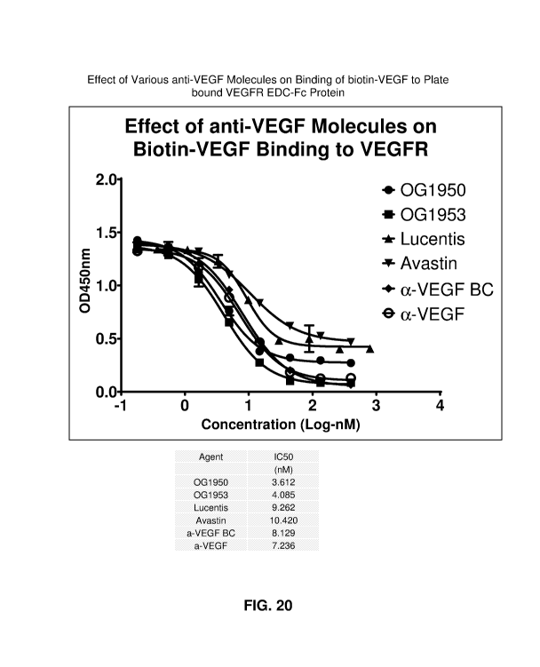
[0081] FIG. 20 depicts the effect of various anti-VEGF molecules on
binding of
biotin-VEGF to plate bound .VEGFR ECD-Fc protein, and their IC50 values.
[0082] FIG. 21 depicts the 061950 binding affinity to VEGF measured
by
BIAcore single cycle kinetics.
[0083] FIG. 22 depicts binding of the 0G1950 to Fe gamma receptor 1.
[0084] FIG. 23 depicts binding of the 0G1950 to Fc gamma receptor
Ilia.
[0085] FIG. 24 depicts binding of QG 1950 to human complement
protein Clq.
CA 3010056 2018-06-28
WO 2017/117464 PCT/US2016/069336
I-00861 FIG. 25 depicts the results of a proliferation assay
(including IC50 values).
[0087] FIG. 26 depicts the results of single cycle kinetics of VEGF
binding to
anti- V EGF agents.
[00881 FIG. 27 depicts some embodiments of nucleic acid sequences
encoding
heavy and light chain variable regions.
[0089] FIG. 28 depicts the screening results after incubation of
various samples
(of various excipients) in polymer solution (OG J.802) for 20 hours at 2-8
degrees Centigrade.
DETAILED DESCRIPTION OF THE PREFERRED EMBODIMENT
[0090] Provided herein are anti-VEGF-A antibodies. In some
embodiments,
these antibodies can be conjugated to a half-life extending moiety. In some
embodiments,
the conjugate can be used for the treatment of certain conditions, such as
diabetic retinopathy
and/or age-related macular degeneration.
[0091] Further provided herein are methods for preparing conjugate
compositions
of antibodies (of any type of antibody). In some embodiments, these methods
allow for
lower aggregate formation or higher efficiency of formation of the desired
antibody
conjugate.
[0092] These and additional embodiments are provided below,
following the
definition section.
DEFINITIONS
[0093] A "neovascular disorder" is a disorder or disease state
characterized by
altered, dysregulated or unregulated angiogenesis. Examples of neovascular
disorders
include ne.oplastic transformation (e.g. cancer) and ocular neovascular
disorders including
diabetic retinopathy and age-related macular degeneration.
[0094] An "ocular neovascular" disorder is a disorder characterized
by altered,
dysregulated or unregulated angiogenesis in the eye of a patient. Such
disorders include
optic disc neovascularization. iris neov ascii] ari za ti on, retinal neov
ascu I ariza ti on, choroidal
neo vascul arization, corneal Deo vasculari zation, vitreal
neovascularization. glaucoma, pann us,
pterygium, macular edema, diabetic retinopathy, diabetic macular edema,
vascular
_73_
CA 3010056 2018-06-28
WO 2017/117464 PCT/US2016/069336
retinopathy, retinal degeneration, uveitis, inflammatory diseases of the
retina, and
proliferative vitreoretinopathy.
[0095] The term antibody includes intact antibodies and binding
fragments
thereof. A binding fragment refers to a molecule other than an intact antibody
that comprises
a portion of an intact antibody that binds the antigen to which the intact
antibody binds.
Examples of binding fragments include Fv, Fab', Fab'-SH, 17(abI)2; diabodies;
linear
antibodies; single-chain antibody molecules (e.g. scFv); and multispecific
antibodies formed
from antibody fragments. scFv antibodies are described in Houston JS. 1991.
Methods in
Enzyrn.ol. 203:46-96. In addition, antibody fragments comprise single chain
polypeptides
having the characteristics of a VH domain, namely being able to assemble
together with a VI.
domain, or of a VI., domain, namely being able to assemble together with a VH
domain to a
functional antigen binding site and thereby providing the antigen binding
property of full
length antibodies.
[0096] Specific binding of an antibody to its target antigen(s)
means an affinity of
at least 106, 107, 1.08, 109, or le M-1. Specific binding is detectably higher
in magnitude and
distinguishable from non-specific binding occurring to at least one unrelated
target. Specific
binding can be the result of formation of bonds between particular functional
groups or
particular spatial fit (e.g., lock and key type) whereas nonspecific binding
is usually the result
of van der Waals forces. Specific binding does not however necessarily imply
that an
antibody or fusion protein binds one and only one target.
[00971 A basic antibody structural unit is a tetramer of subunits.
Each tetramer
includes two identical pairs of polypeptide chains, each pair having one
"light" (about 25
kDa) and one "heavy" chain (about 50-70 kDa). The amino-terminal portion of
each chain
includes a variable region of about 100 to 110 or more amino acids primarily
responsible for
antigen recognition. This variable region is initially expressed linked to a
cleavable signal
peptide. The variable region without the signal peptide is sometimes referred
to as a mature
variable region. Thus, for example, a light chain mature variable region means
a light chain
variable region without the light chain signal peptide. However, reference to
a variable
region does not mean that a signal sequence is necessarily present; and in
fact signal
sequences are cleaved once the antibodies or fusion proteins have been
expressed and
secreted. A pair of heavy and light chain variable regions defines a binding
region of an
-14-
CA 3 0 1 0 056 2 0 1 8-0 6-2 8
WO 2017/117464 PCT/US2016/069336
antibody. The carboxy-terminal portion of the light and heavy chains
respectively defines
light and heavy chain constant regions. The heavy chain constant region is
primarily
responsible for effector function. In IgG antibodies, the heavy chain constant
region is
divided into CHI, hinge, CH2, and CH3 regions. The CHI region binds to the
light chain
constant region by disulfide and noncovalent bonding. The hinge region
provides flexibility
between the binding and effector regions of an antibody and also provides
sites for
intermolecular disulfide bonding between the two heavy chain constant regions
in a tetram.er
subunit. The CH2 and CH3 regions are the primary site ()-1. effector functions
and FcR.
binding.
[00981 Light chains are classified as either kappa or lambda. Heavy
chains are
classified as gamma, mu, alpha, delta, or epsilon, and define the antibody's
isotype as IgG,
IgD and IgE, respectively. Within light and heavy chains, the variable and
constant regions are joined by a "J" segment of about 12 or more amino acids,
with the heavy
chain also including a "D" segment of about 10 or more amino acids. (See
generally,
Fundamental Immunology (Paul, W., ed., 2nd ed. Raven Press, N.Y., 1989), Ch.
7)
(incorporated by reference in its entirety for all purposes).
100991 The mature variable regions of each light/heavy chain pair
form the
antibody binding site. Thus, an intact antibody has two binding sites, i.e.,
is divalent. In
natural antibodies, the binding sites are the same. However, bispecific
antibodies can be
made in which the two binding sites are different (see, e.g., Songsivilai S.
Lachmann PC.
1990. Bispecific antibody: a tool for diagnosis and treatment of disease. Clin
Exp immunol.
79:315-321; Kostelny SA, Cole MS, Tso Ff. 1992. Formation of bispecific
antibody by the
use of leucine zippers. J Immunol. 148: 1547-1553). The variable regions all
exhibit the
same general structure of relatively conserved framework regions (FR) joined
by three
hypervariable regions, also called complementarity determining regions or
CDRs. The.
CDRs from the two chains of each pair are aligned by the framework regions,
enabling
binding to a specific epitope. From N-terminal to C-terminal, both light and
heavy chains
comprise the domains FR1, CDRI, FR2, CDR2, FR3, CDR3 and FR4. For convenience,
the
variable heavy CDRs can be referred to as CDRH1, CDRH2 and CDRH3; the variable
light
chain CDRs can be referred to as CDRLI, CDR1,2 and CDR1,3. The assignment of
amino
acids to each domain is in accordance with the definitions of Rabat EA, et al.
1987 and 1991.
-25-
CA 3010056 2018-06-28
WO 2017/117464 PCT/US2016/069336
Sequences of Proteins of immunological Interest (National Institutes of
Health, Bethesda,
MD) or Chothia C, Lesk AM. 1987. Canonical Structures for the Hypervariable
Regions of
Immunoglobulins. J Mol Biol 196:901-917; Chothia C, et al. 1989. Conformations
of
imnaunoglobulin Hypervariable Regions. Nature 342:877-883. Kabat also provides
a widely
used numbering convention (Kabat numbering) in which corresponding residues
between
different heavy chain variable regions or between different light chain
variable regions are
assigned the same number. Although Kabat numbering can be used for antibody
constant
regions, EU numbering is more commonly used, as is the case in this
application. Although
specific sequences are provided for exemplary antibodies disclosed herein, it
will be
appreciated that after expression of protein chains one to several amino acids
at the amino or
carboxy terminus of the light and/or heavy chain, particularly a heavy chain C-
terminal
lysine residue, may be missing or derivatized in a proportion or all of the
molecules.
10100] The term "epitope" refers to a site on an antigen to which an
antibody or
extracellular trap segment binds. An epitope on a protein can be formed from
contiguous
amino acids or noncontiguous amino acids juxtaposed by tertiary folding of one
or more
proteins. Epitopes formed from contiguous amino acids (also known as linear
epitopes) are
typically retained on exposure to denaturing solvents whereas epitopes formed
by tertiary
folding (also known as
10101] conformational epitopes) are typically lost on treatment with
denaturing
solvents. An epitope typically includes at least 3, and more usually, at least
5 or 8-10 amino
acids in a unique spatial conformation. Methods of determining spatial
conformation of
epitopes include, for example, x-ray crystallography and 2-dimensional nuclear
magnetic
resonance. See, e.g., Epitope Mapping Protocols, in Methods in Molecular
Biology, Vol. 66,
Glenn E. Morris, Ed. (1996).
[0102] Antibodies that recognize the same or overlapping epitopes
can be
identified in a simple immunoassay showing the ability of one antibody to
compete with the
binding of another antibody to a target antigen. The epitope of an antibody
can also be
defined by X-ray crystallography of the antibody (or Fab fragment) hound to
its antigen to
identify contact residues.
[0103] Alternatively, two antibodies have the same epitope if all
amino acid
mutations in the antigen that reduce or eliminate binding of one antibody
reduce or eliminate
CA 301 005 6 2 01 8-0 6-2 8
WO 2017/117464 PCT/US2016/069336
binding of the other. Two antibodies have overlapping epitopes if some amino
acid
mutations that reduce or eliminate binding of one antibody reduce or eliminate
binding of the
other.
[0104]
Competition between antibodies is determined by an assay in which an
antibody under test inhibits specific binding of a reference antibody to a
common antigen
(see, e.g., Jungharts et al., Cancer Res. 50: 1495, 1990). A test antibody
competes with a
reference antibody if an excess of a test antibody (e.g., at least 2x, 5x,
10x, 20x or 100x)
inhibits binding of the reference antibody by at least 50%. In sonic
embodiments the test
antibody inhibits binding of the reference antibody by 75%, 90%, or 99% as
measured in a
competitive binding assay.
Antibodies identified by competition assay (competing
antibodies) include antibodies binding to the same epitope as the reference
antibody and
antibodies binding to an adjacent epitope sufficiently proximal to the epitope
bound by the
reference antibody for steric hindrance to occur.
[0105] The
term "patient" includes human and other mammalian subjects that
receive either prophylactic or therapeutic treatment.
[0106] For
purposes of classifying amino acids substitutions as conservative or
nonconservative, amino acids are grouped as follows: Group I (hydrophobic side
chains):
met, ala, val, len, ile; Group II (neutral hydrophilic side chains): cys, ser,
thr; Group 111
(acidic side chains): asp, glu; Group IV (basic side chains): asn, gin, his,
lys, arg; Group V
(residues influencing chain orientation): gly, pro; and Group VI (aromatic
side chains): trp,
tyr, phe. Conservative substitutions involve substitutions between amino acids
in the same
class. Non-conservative substitutions constitute exchanging a member of one of
these classes
for a member of another.
[0107]
Percentage sequence identities are determined with antibody sequences
maximally aligned by the Kabat numbering convention for a variable region or
EU
numbering for a constant region. After alignment, if a subject antibody region
(e.g., the
entire mature variable region of a heavy or light chain) is being compared
with the same
region of a reference antibody, the percentage sequence identity between the
subject and
reference antibody regions is the number of positions occupied by the same
amino acid in
both the subject and reference antibody region divided by the total number of
aligned
positions of the two regions, with gaps not counted, multiplied by 100 to
convert to
CA 3010056 2018-06-28
WO 2017/117464 PCT/US2016/069336
percentage. Sequence identities of other sequences can be determined by
aligning sequences
using algorithms, such as BESTFIT, FAS-FA, and TFASTA in the Wisconsin
Genetics
Software Package Release 7.0, Genetics Computer Group, 575 Science Dr.,
Madison, WI,
using default gap parameters, or by inspection, and the best alignment (i.e.,
resulting in the
highest percentage of sequence similarity over a comparison window).
Percentage of
sequence identity is calculated by comparing two optimally aligned sequences
over a window
of comparison, determining the number of positions at which the identical
residues occurs in
both sequences to yield the number of matched positions, dividing the number
of matched
positions by the total number of positions in the window of comparison (i.e.,
the window
size), and multiplying the result by 100 to yield the percentage of sequence
identity.
[0108]
Compositions or methods "comprising" one or more recited elements may
include other elements not specifically recited. For example, a composition
that comprises
antibody may contain the antibody alone or in combination with other
ingredients.
[0109] The
term "antibody-dependent cellular cytotoxicity", or A DCC, is a
mechanism for inducing cell death that depends upon the interaction of
antibody-coated
target cells (i.e., cells with bound antibody) with immune cells possessing
lytic activity (also
referred to as effector cells). Such
effector cells include natural killer cells,
monocytes/macrophages and neutrophils. ADCC is triggered by interactions
between the Fc
region of an antibody bound to a cell and Fcy receptors, particularly FcyRI
and FcyR111, on
immune effector cells such as neutrophils, macrophages and natural killer
cells. The target
cell is eliminated by phagocytosis or lysis, depending on the type of
mediating effector cell.
Death of the antibody-coated target cell occurs as a result of effector cell
activity.
[01101 The
term opsonization also known as "antibody-dependent cellular
phagocytosis", or ADCP, refers to the process by which antibody-coated cells
are
internalized, either in whole or in part, by phagocytic immune cells (e.g.,
macroPhages,
neutrophils and dendritic cells) that bind to an immunoglobulin Fe region.
The term "complement-dependent cytotoxicity" or CDC refers to a
mechanism for inducing cell death in which an Fe effector domain(s) of a
target-bound
antibody activates a series of enzymatic reactions culminating in the
formation of holes in the
target cell membrane. Typically, antigen-antibody complexes such as those on
antibody-
coated target cells bind and activate complement component Clq which in turn
activates the
CA 3 01 0 056 2 01 8-0 6-2 8
WO 2017/117464 PCT/US2016/069336
complement cascade leading to target cell death. Activation of complement may
also result
in deposition of complement components on the target cell surface that
facilitate ADCC by
binding complement receptors (e.g., CR3) on leukocytes.
101121 A humanized antibody is a genetically engineered antibody in
which the
CDRs from a non-human "donor" antibody are grafted into human "acceptor"
antibody
sequences (see, e.g., Queen, US 5,530,101 and 5,585,089; Winter, US 5,225,539,
Carter, US
6,407,213, Adair, US 5,859,205 6,881,557, Foote, US 6,881,557). The acceptor
antibody
sequences can be, for example, a mature human antibody sequence, a composite
of such
sequences, a consensus sequence of human antibody sequences, or a germline
region
sequence. Thus, a humanized antibody is an antibody having some or all CDRs
entirely or
substantially from a donor antibody and varia.ble region framework sequences
and constant
regions, if present, entirely or substantially from human antibody sequences.
Similarly a
humanized heavy chain has at least one, two and usually all three CDRs
entirely or
substantially from a donor antibody heavy chain, and a heavy chain variable
region
framework sequence and heavy chain constant region, if present, substantially
from human
heavy chain variable region framework and constant region sequences. Similarly
a
humanized light chain has at least one, two and usually all three CDRs
entirely or
substantially from a donor antibody light chain, and a light chain variable
region framework
sequence and light chain constant region, if present, substantially from human
light chain
variable region framework and constant region sequences. Other than nanobodies
and dAbs,
a humanized antibody comprises a humanized heavy chain and a humanized light
chain. A
CDR in a humanized antibody is substantially from a corresponding CDR in a non-
human
antibody when at least 85%, 90%, 95% or 100% of corresponding residues (as
defined by
Kabat) are identical between the respective CDRs. The variable region
framework sequences
of an antibody chain or the constant region of an antibody chain are
substantially from a
human variable region framework sequence or human constant region respectively
when at
least 85, 90, 95 or 100% of corresponding residues defined by Kabat are
identical.
[01131 Although humanized antibodies often incorporate all six CDRs
(which can
be as defined by Kabat) from a mouse antibody, they can also be made with less
than all
CDRs (e.g., at least 3, 4, or 5 CDRs from a mouse antibody) (e.g., De Pascalis
R, Iwahashi
M, Tamura M, et al. 2002. Grafting "Abbreviated" Complementary-Determining
Regions
CA 3010056 2018-06-28
WO 2017/117464 PCT/US2016/069336
Containing Specificity-Determining Residues Essential for Ligand Contact to
Engineer a
Less Immunogenic Humanized Monoclonal Antibody. I Immunol. 169:3076-3084;
Vajdos
FE, Adams CW, Breece TN, Presta LG, de Vos AM, Sidhu, SS. 2002. Comprehensive
functional maps of the antigen-binding site of an anti-ErbB2 antibody obtained
with shotgun
scanning mutagenesis. J Mol Biol. 320: 415-428; Iwahashi M, IVIiienic DE,
Padian EA, et al.
1999. CDR substitutions of a humanized monoclonal antibody (CC49):
Contributions of
individual CDRs to antigen binding and immunogenicity. Mol Immunol. 36:1079-
1091;
Tamura M, Milenic DE, Iwahashi M, et al. 2000. Structural correlates of an
anticarcinoma
antibody: Identification of specifici(y-determining regions (SDRs) and
development of a
minimally immunogenic antibody variant by retention of SDRs only. I Immunol.
164:1432-
1441).
[0114] A chimeric antibody is an antibody in which the mature
variable regions
of light and heavy chains of a non-human antibody (e.g., a mouse) are combined
with human
light and heavy chain constant regions. Such antibodies substantially or
entirely retain the
binding specificity of the mouse antibody, and are about two-thirds human
sequence.
[0115] A veneered antibody is a type of humanized antibody that
retains some
and usually all of the CDRs and some of the non-human variable region
framework residues
of a non-human antibody but replaces other variable region framework residues
that may
contribute to B- or T-cell epitopes, for example exposed residues (PadIan EA.
1991. A
possible procedure for reducing the immunogenicity of antibody variable
domains while
preserving their ligand-binding properties. Mol Immunol. 28:489-98) with
residues from the
corresponding positions of a human antibody sequence. The result is an
antibody in which
the CDRs are entirely or substantially from a non-human antibody and the
variable region
frameworks of the non-human antibody are made more human-like by the
substitutions. A
human antibody can be isolated from a human, or otherwise result from
expression of human
immunoglobulin genes (e.g., in a transgenic mouse, in vitro or by phage
display). Methods
for producing human antibodies include the trioma method of Ostberg L, Pursch
E. 1983.
Human x (mouse x human) hybridomas stably producing human antibodies.
Ilybridoma
2:361-367; Ostberg, U.S. Patent No. 4,634,664; and Engleman et al., US Patent
4,634,666,
use of transgenic mice including human immunoglobulin genes (see, e.g.,
Lonberg et al.,
W093/122.27 (1993); US 5,877,397, US 5,874,299, US 5,814,318, US 5,789,650, US
-30-
CA 3010056 2018-06-28
WO 2017/117464 PCT/US2016/069336
5,770,429, US 5,661,016, US 5,633,425, US 5,625,126, US 5,569,825, US
5,545,806, Nature
148, 1547-1553 (1994), Nature Biotechnology 14, 826 (1996), Kucherlapati, WO
91/10741
(1991) and phage display methods (see, .e.g. Dower et al., WO 91/17271 and
McCafferty et
al., WO 92/01047, US 5,877,218, US 5,871,907, US 5,858,657, US 5,837,242, US
5,733,743
and US 5,565,332.
[0116] "Polymer" refers to a series of monomer groups linked
together. A
polymer is composed of multiple units of a single monomer (a homopolymer) or
different
monomers (a heteropolymer). High MW polymers are prepared from monomers that
include, but are not limited to, acrylates, methacrylates, acrylarnides,
methacrylamides,
styrenes, vinyl-pyridine, vinyl-pyrrolidone and vinyl esters such as vinyl
acetate. Additional
monomers are useful in high MW polymers . When two different monomers are
used, the
two monomers are called "comonomers," meaning that the different monomers are
copolymerized to form a single polymer. The polymer can be linear or branched.
When the
polymer is branched, each polymer chain is referred to as a "polymer arm." The
end of the
polymer arm linked to the initiator moiety is the proximal end, and the
growing-chain end of
the polymer arm is the distal end. On the growing chain-end of the polymer
arm, the
polymer arm end group can be the radical scavenger, or another group.
[0117] "Initiator" refers to a compound capable of initiating a
polymerization'
using monomers or comonomers. The polymerization can be a conventional free
radical
polymerization or a controlled/living" radical polymerization, such as Atom
Transfer
Radical Polymerization (ATRP), Reversible Addition-Fragmentation-Termination
(RAFT)
polymerization or nitroxide mediated polymerization (NMP). The polymerization
can be a
"pseudo" controlled polymerization, such as degenerative transfer. When the
initiator is
suitable for ATRP, it contains a labile bond which can be homolytically
cleaved to form an
initiator fragment, 1, being a radical capable of initiating a radical
polymerization, and a
radical scavenger, I', which reacts with the radical of the growing polymer
chain to
reversibly terminate the polymerization. The radical scavenger l' is typically
a halogen, but
can also he an organic moiety, such as a nitrite. In some embodiments , the
initiator contains
one of more 2-bromoisobutyrate groups as sites for polymerization via ATRP.
[0118] A "chemical linker" refers to a chemical moiety that links two
groups
together, such as a half-life extending moiety and a protein. The linker can
be cleavable or
-31-
CA 3010056 2018-06-28
WO 2017/117464 PCT/US2016/069336
non-cleavable. Cleavable linkers can be hydrolyzable, enzymatically cleavable,
pfl sensitive,
photolabile, or disulfide linkers, among others. Other linkers include
homobifunctional and
heterobifunctional linkers. A "linking group" is a functional group capable of
forming a
covalent linkage consisting of one or more bonds to a bioactive agent. Non-
limiting
examples include those illustrated in Table 1 of W02013059137 (incorporated by
reference).
[0119] The term "reactive group" refers to a group that is capable
of reacting with
another chemical group to form a covalent bond, i.e. is covalently reactive
under suitable
reaction conditions, and generally represents a point of attachment for
another substance.
The reactive group is a moiety, such as maleimide or succinimidyl ester, is
capable of
chemically reacting with a functional group on a different moiety to form a
covalent linkage.
Reactive groups generally include nucleophiles, electrophiles and
photoactivatable groups.
[0120] "Phosphorylcholine," also denoted as "PC," refers to the
following:
0
I I
.-0-P-0 N tC111\3
where * denotes the point of attachment. The phosphorylcholine is a
zwitterionic group and
includes salts (such as inner salts), and protonated and deprotonated forms
thereof.
[0121] "Phosphorylcholine containing polymer" is a polymer that
contains
phosphorylcholine. "Zwitterion containing polymer" refers to a polymer that
contains a
zwitterion.
[0122] Poly(acryloyloxyethyl phosphorylcholine) containing polymer
refers to a
polymer containing 2-(acryloyloxy)ethyl-2-(trimethylammonium)ethyl phosphate
(1-IEA-PC
shown below in Example 6) as monomer.
-32-
CA 3 0 1 0 056 2 0 1 8-0 6-2 8
WO 2017/117464 PCT/US2016/069336
[0123] Poly(methacrvloyloxyethyl phosphorylcholine) containing
polymer refers
to a polymer containing 2-(methacryloyloxy)ethy1-2-(trimethylammonium)ethyl
phosphate
(HEMA-PC or MPC) as monomer (see below):
H C
2
CE-13
0
- \No
CH3
LCH3
# I
CI-13
[0124] As used herein, "MPC" and "I-IEMA-PC" are interchangeable.
[0125] Molecular weight" in the context of the polymer can be
expressed as either
a number average molecular weight, or a weight average molecular weight or a
peak
molecular weight. Unless otherwise indicated, all references to molecular
weight herein refer
to the peak molecular weight. These molecular weight determinations, number
average
(Mn), weight average (Mw) and peak (Mp), can be measured using size exclusion
chromatography or other liquid chromatography techniques. Other methods for
measuring
molecular weight values can also be used, such as the use of end-group
analysis or the
measurement of colligative properties (e.g., freezing-point depression,
boiling-point
elevation, or osmotic pressure) to determine number average molecular weight,
or the use of
light scattering techniques, ultracentrifugation or viscometry to determine
weight average
molecular weight. In some embodiments, the molecular weight is measured by SEC-
MALS
(size exclusion chromatography -- multi angle light scattering). In some
embodiments, the
polymeric reagents are typically polydisperse (i.e., number average molecular
weight and
weight average molecular weight of the polymers are not equal), and can
possess low
polydispersity values of, for example, less than about 1.5, as judged, for
example, by the PDI
value derived from the SEC-MALS measurement, In some embodiments, the
-33-
CA 3010056 2018-06-28
WO 2017/117464 PCT/US2016/069336
polydispersities (PDI) are in the range of about 1.4 to about 1.2. In some
embodiments the
PD1 is less than about 1.15, 1.10, 1.05, or 1.03.
[0126] The phrase "a" or "an" entity refers to one or more of that
entity; for
example, a compound refers to one or more compounds or at least one compound.
As such,
the terms "a" (or "an"), "one or more", and "at least one" can be used
interchangeably herein.
[0127] "About" means variation one might see in measurements taken
among
different instruments, samples, and sample preparations.
[0128] "Protected," "protected form," "protecting group" and
"protective group"
refer to the presence of a group (i.e., the protecting group) that prevents or
blocks reaction of
a particular chemically reactive functional group in a molecule under certain
reaction
conditions. Protecting groups vary depending upon the type of chemically
reactive group
being protected as well as the reaction conditions to be employed and the
presence of
additional reactive or protecting groups in the molecule, if any. Suitable
protecting groups
include those such as found in the treatise by Greene et al., "Protective
Groups In Organic
Synthesis," 3'd Edition, John Wiley and Sons, Inc., New York, 1999.
[0129] "Alkyl" refers to a straight or branched, saturated,
aliphatic radical having
the number of carbon atoms indicated. For example, C,-C6 alkyl includes, but
is not limited
to, methyl, ethyl, propyl, isopropyl, butyl, isobutyl, sec-butyl, tert-butyl,
pentyl, isopentyl,
hexyl, etc. Other alkyl groups include, but are not limited to heptyl, octyl,
nonyl, decyl, etc.
Alkyl can include any number of carbons, such as 1-2, 1-3, 1-4, 1-5, 1-6, 1-7,
1-8, 1-9, 1-10,
2-3, 2-4, 2-5, 2-6, 3-4, 3-5, 3-6, 4-5, 4-6 and 5-6. The alkyl group is
typically monovalent,
but can be divalent, such as when the alkyl group links two moieties together.
[0130] The term "lower" referred to above and hereinafter in
connection with
organic radicals or compounds respectively defines a compound or radical which
can be
branched or unbranched with up to and including 7 or up to and including 4 and
(as
unbranched) one or two carbon atoms.
[0131] "Alkylene" refers to an alkyl group, as defined above,
linking at least two
other groups, i.e., a divalent hydrocarbon radical. The two moieties linked to
the alkylene
can be linked to the same atom or different atoms of the alkylene. For
instance, a straight
chain alkylene can be the bivalent radical of -(CH2),,, where n is 1, 2, 3, 4,
5 or 6. Alkylene
-34-
CA 301 005 6 2 01 8-0 6-2 8
WO 2017/117464 PCT/11S2016/069336
groups include, but are not limited to, methylene, ethylene, propylene,
isopropylene,
butylene, isobutylene, sec-butylene, pentylene and hexylene.
[0132]
Substituents for the alkyl and heteroalkyl radicals (including those groups
often referred to as alkylene, alkenyl, heteroalkylene, heteroalkenyl,
alkynyl, cycloalkyl,
heterocycloalkyl, cycloalkenyl, and heterocycloalkenyl) can be a variety of
groups selected
from: -OR', =0,
=N-OR', -NR'R", -SR', -halogen, -SiR'R"R", -0C(0)R', -C(0)R', -CO2R', -
CONR'R",
OC(0)NR'R", -NR"C(0)R', -NR'-C(0)NR."R", -NR"C(0)2R', -NH-C(NW)=NH, -NR.'C(
NH7)=NH, -S(0)R',
-S(0)2R', -S(0)2NR'R", -CN and -NO2 in a number
ranging from zero to (2m'+ I). where m' is the total number of carbon atoms in
such radical.
R', R" and R" each independently refer to hydrogen, unsubstituted (C1-C8)alkyl
and
heteroalkyl, unsubstituted aryl, aryl substituted with 1-3 halogens,
unsubstituted alkyl, alkoxy
or thioalkoxy groups, or aryl-(Ci-C4)alkyl groups. When R' and R" are attached
to the same
nitrogen atom, they can be combined with the nitrogen atom to form a 5-, 6-,
or 7-membered
ring. For example, -NR'R" is meant to include 1-pyrrolidinyl and 4-
morpholinyl. The term
"alkyl" is include groups such as haloalkyl (e.g., -CF3 and -CI-I2CF3) and
acyl
(e.g., -C(0)Cf-13, -C(0)CF3, -C(0)0-1120C1-13, and the like). In some
embodiments, the
substituted alkyl and heteroalkyl groups have from I to 4 substituents. In
some embodiments,
the substituted akyl and heteroalkyl groups have I, 2 or 3 substituents.
Exceptions are those
perhalo alkyl groups (e.g., pentafluoroethyl and the like) .
[0133]
Substituents for the alkyl and heteroalkyl radicals (including those groups
often referred to as alkylene, alkenyl, heteroalkylene, heteroalkenyl,
alkynyl, cycloalkyl,
heterocycloalkyl, cycloalkenyl, and heterocycloalkenyl) can be one or more of
a variety of
Prom's selected from, but not limited to: -OR',
=0, =NR',
=N-OR', -NR'R", -SR', -halogen, -SiR'R"R", -0C(0)R', -C(0)R', -CO2R', -
CONR'R", -0
C(0)NR'R", -NR"C(0)R', -NW-C(0)NR"R", -NR"C(0)2R', -NR-C(NR.R"R'")=NR'". -N
R-C(NR'R")=NR'", -S(0)R', -S(0)2R', -S(0)2NICR", -NRSO2R', -CN and ....NO2 in
a
number ranging from zero to (2m'+1), where m' is the total number of carbon
atoms in such
radical. R', R", R" and R'" each independently refer to hydrogen, substituted
or
unsubstituted heteroalkyl, substituted or unsubstituted aryl, e.g., aryl
substituted with 1-3
halogens, substituted or unsubstituted alkyl, alkoxy or thioalkoxy groups, or
arylalkyl groups.
-35-
CA 3 0 1 0 056 2 0 1 8-0 6-2 8
WO 2017/117464 PCT/US2016/069336
When a compound includes more than one R group, for example, each of the R
groups is
independently selected as are each R', R", R" and R"" groups when more than
one of these
groups is present. When R' and R" are attached to the same nitrogen atom, they
can be
combined with the nitrogen atom to form a 5-, 6-, or 7-membered ring. For
example, -NR'R" is meant to include, but not be limited to, 1-pyrrolidinyl and
4-morpholinyl. From the above discussion of substituents, one of skill in the
art will
understand that the term "alkyl" is meant to include groups including carbon
atoms bound to
groups other than hydrogen groups, such as haloalkyl (e.g., -CF 3 and ¨CH2CF3)
and acyl
(e.g., -C(0)CH, -C(0)CF3, -C(0)CR2OCH3, and the like).
[0134] "Alkoxy" refers to alkyl group having an oxygen atom that
either connects
the alkoxy group to the point of attachment or is linked to two carbons of the
alkoxy group.
Alkoxy groups include, for example, methoxy, ethoxy, propoxy, iso-propoxy,
butoxy,
2-butoxy, iso-butoxy, sec-butoxy, tert-butoxy, pentoxy, hexoxy, etc. The
alkoxy groups can
be further substituted with a variety of substituents described within. For
example, the
alkoxy groups can be substituted with halogens to form a "halo-alkoxy" group.
[0135] "Carboxyalkyl" means an alkyl group (as defined herein)
substituted with
a carboxy group. The term "carboxycycloalkyl" means a cycloalkyl group (as
defined
herein) substituted with a carboxy group. The term alkoxyalkyl means an alkyl
group (as
defined herein) substituted with an alkoxy group. The term "carboxy" employed
herein
refers to carboxylic acids and their esters.
[0136] "Haloalkyl" refers to alkyl as defined above where some or
all of the
hydrogen atoms are substituted with halogen atoms. Halogen (halo) represents
chloro or
fluoro, but may also be bromo or iodo. For example, haloalkyl includes
trifluoromethyl,
fluoromethyl, 1,2,3,4,5-pentafluoro-phenyl, etc. The term "perfluoro" defines
a compound
or radical which has all available hydrogens that are replaced with fluorine.
For example,
perfluorophenvl refers to 1,2,3,4,5-pentafluorophenyl, perfluoromethyl refers
to
1,1,1-trifluoromethyl, and perfluoromethoxy refers to 1-trifluoromethoxy.
[0137] "Fluoro-substituted alkyl" refers to an alkyl group where
one, some, or all
hydrogen atoms have been replaced by fluorine.
[01381 "Cytokine" is a member of a group of protein signaling
molecules that
may participate in cell-cell communication in immune and inflammatory
responses.
-36-
CA 3010056 2018-06-28
WO 2017/117464 PCT/US2016/069336
Cytokines are typically small, water-soluble glycoproteins that have a mass of
about 8-35
kDa.
[0139] "Cycloalkyl" refers to a cyclic hydrocarbon group that
contains from
about 3 to 12, from 3 to 10, or from 3 to 7 endocyclic carbon atoms.
Cycloalkyl groups
include fused, bridged and spiro ring structures.
[0140] "Endocyclic" refers to an atom or group of atoms which
comprise part of a
cyclic ring structure.
[0141] "Exocyclic" refers to an atom or group of atoms which are
attached but do
not define the cyclic ring structure.
[0142] "Cyclic alkyl ether" refers to a 4 or 5 member cyclic alkyl
group having 3
or 4 endocyclic carbon atoms and 1 endocyclic oxygen or sulfur atom (e.g.,
oxetane, thietane,
tetrahydrotbran, tetrahydrothiopherie); or a 6 to 7 member cyclic alkyl group
having I or 2
endocyclic oxygen or sulfur atoms (e.g., tetrahydropyran, 1,3-dioxane, 1,4-
dioxane,
tetrahydrothiopyran, 1,4-dithiane, 1,4-exathiane).
[0143] "Alkenyl" refers to either a straight chain or branched
hydrocarbon of 2 to
6 carbon atoms, having at least one double bond. Examples of alkenyl groups
include, but
are not limited to, vinyl, properly], isopropenyl, 1-butenyl, 2-butenyl,
isobutenyl, butadienyl,
1-pentenyl, 2-pentenyl, isopentenyl, 1,3-pentadienyl, 1,4-pentadienyl, 1-
hexenyl, 2-hexenyl,
3-hexenyl, 1,3-hexadienyl, 1,4-hexadienyl, 1.,5-hexadienyl, 2,4-hexadienyl, or
1,3,5-hexatrienyl. Alkenyl groups can also have from 2 to 3, 2 to 4, 2 to 5, 3
to 4, 3 to 5, 3 to
6, 4 to 5, 4 to 6 and 5 to 6 carbons. The alkenyl group is typically
monovalent, but can be
divalent, such as when the alkenyl group links two moieties together.
[0144] "Alkenylene" refers to an alkenyl group, as defined above,
linking at least
two other groups, i.e., a divalent hydrocarbon radical. The two moieties
linked to the
alkenylene can be linked to the same atom or different atoms of the
alkenylene. Alkenylene
groups include, but are not limited to, ethenylene, propenylene,
isopropenylene, butenylene,
isobutenylene, sec-butenylene, pentenylene and hexenylene.
[0145] "Alkynyl" refers to either a straight chain or branched
hydrocarbon of 2 to
6 carbon atoms, having at least one triple bond. Examples of alkynyl groups
include, but are
not limited to, acetylenyl, propynyl, 1-butynyi, 2-butynyl, isobutynyl, sec-
butynyl,
butadiynyl, 1-pentynyi, 2-pentynyl, isopentynyl, 1,3 -pentadiynyl, 1,4-
pentadiynyl,
-37--
CA 3010056 2018-06-28
WO 2017/117464 PCT/US2016/069336
1-hexynyl, 2-hexynyl, 3-hexynyl, 1,3-hexadiynyl, 1,4-hexadiynyl, 1,5-
hexadlynyl,
2,4-hexadiynyl, or 1,3,5-hexatriynyl. Alkynyl groups can also have from 2 to
3, 2 to 4, 2 to
5, 3 to 4, 3 to 5, 3 to 6, 4 to 5, 4 to 6 and 5 to 6 carbons. The alkynyl
group is typically
monovalent, but can be divalent, such as when the alkynyl group links two
moieties together.
[0146] "Alkynylene" refers to an alkynyl group, as defined above,
linking at least
two other groups, i.e., a divalent hydrocarbon radical. The two moieties
linked to the
alkynylene can be linked to the same atom or different atoms of the
alkynylene. Alkynylene
groups include, but are not limited to, ethynylene, propynylene, butynylcne,
sec-butynylenc.,
pentynylene and hexynylene.
[0147] "Cycloalkyl" refers to a saturated or partially unsaturated,
monocyclic,
fused bicyclic or bridged polycyclic ring assembly containing from 3 to 12
ring atoms, or the
nurnber of atoms indicated. Monocyclic rings include, for example,
cyclopropyl, cyclobutyl,
cyclopentyl, cyclohexyl, and cyclooctyl. Bicyclic and polycyclic rings
include, for example,
norbornane, decah.ydronaphthalene and a.datnantane. For example, Cmcycloalkyl
includes
eyclopropyl, cyclobutyl, cyclopentyl, eyclohexyl, cyclooctyl, and norbornane.
[0148] "Cycloalkylene" refers to a cycloalkyl group, as defined
above, linking at
least two other groups, i.e., a divalent hydrocarbon radical. The two moieties
linked to the
cycloalkylene can be linked to the same atom or different atoms of the
cycloalkylene.
Cycloalkylene groups include, but are not limited to, cyclopropylene,
cyclobutylene,
cyclopentylene, cyclohexylene, and cyclooctylene.
[0149] "Heterocycloalkyl" refers to a ring system having from 3 ring
members to
about 20 ring members and from 1 to about 5 heteroatoms such as N, 0 and S.
Additional
heteroatoms can also be useful, including, but not limited to, B, Al, Si and
P. The
heteroatoms can also be oxidized, such as, but not limited to, -S(0)- and -
S(0)2-. For
example, heterocycle includes, but is not limited to, tetrahydrofuranyl,
tetrahydrotbiophenyl,
morpholino, pyrrolidinyl, pyrrolinyl, imidazolidinyl, imidazolinyl,
pyrazolidinyl, pyrazolinyl,
piperazinyl, piperidinyl, indolinyl, quinuclidinyl and 1,4-dioxa-8-aza-
spiro[4.5]dec-8-yl.
[0150] "Heterocycloalkylene" refers to a heterocyclalkyl group, as
defined above,
linking at least two other groups. The two moieties linked to the
heterocycloalkylenc can be
linked to the same atom or different atoms of the heterocycloalkylene.
-38-
CA 3010056 2018-06-28
WO 2017/117464 PCT/US2016/069336
[0151] "Aryl"
refers to a monocyclic or fused bicyclic, tricyclic or greater,
aromatic ring assembly containing 6 to 16 ring carbon atoms. For example, aryl
may be
phenyl, benzyl or naphthyl. "Arylene" means a divalent radical derived from an
aryl group.
Aryl groups can be mono-, di- or tri-substituted by one, two or three radicals
selected from
alkyl, alkoxy, aryl, hydroxy, halogen, cyano, amino, amino-alkyl,
trifluoromethyl,
alkylenedioxy and oxy-C2-C3-alkylene; all of which are optionally further
substituted, for
instance as hereinbefore defined; or 1- or 2-naphthyl; or 1- or 2-
phenanthrenyl.
Alkylenedioxy is a divalent substitute attached to two adjacent carbon atoms
of phenyl, e.g.
methylenedioxy or ethylenedioxy. Oxy-C2-C3-alkylene is also a divalent
substituent attached
to two adjacent carbon atoms of phe,nyl, e.g. oxyethylene or oxypropylene. An
example for
oxy- C2-C3-alkylene-phenyl is 2,3-dihydrobenzofuran-5-yl.
[0152] In some
embodiments the aryl is naphthyl, phenyl or phenyl mono- or
disubstituted by alkoxy, phenyl, halogen, alkyl or trifluoromethyl, especially
phenyl or
phenyl-mono- or disubstituted by alkoxy, halogen or trifluoromethyl, and in
particular
phenyl.
[0153] Examples
of substituted phenyl groups as R are, e.g. 4-chlorophen-1.-yl,
3,4-dichlorophen- I -yl, 4-methox yphen-l-yl, 4-methylphen-l-yl, 4-am
inomethylphen-l-yl,
4-methoxyethylaminomethylphen- , 4-
hydroxyethylaminomethylphen-l-yl,
4-h ydrox yeth yl-(rne th ino me
thylphen -1-yl, 3-aminomethylphen-1-yl,
4-N -acet ylaminomethylphen-l-yi , 4-aminophen-l-yl, 3-aminophen-1-yl, 2-
aminophen-l-yl,
4-ph enyl-phen-l-yl, 4-(imidazol-1-y1)-phenyl, 4-
(imidazol-1-ylmethyll-phen-1-yl,
4-(morpholi n-l-ylmethyl)-phen -1-y1 ,
4-(2-methoxyethylaminomethyl)-phen-1 -y1 and 4-
(pyrrolidin-1-ylmethyl)-phen-l-yl,
4-(thiopheny1)-phen-1-yl, 4-(3-thi ophen y1)- phen-l-yl, 4-(4-methylpiperazin-
1-y1)-phen-1-yl,
and 4-(piperidinyI)-phenyl and 4-(pyridiny1)-phenyl optionally substituted in
the heterocyclic
ring.
[0154]
"Arylene" refers to an aryl group, as defined above, linking at least two
other groups. The two moieties linked to the arylene are linked to different
atoms of the
arylene. Arylene groups include, but are not limited to, phenylene.
-39-
CA 3 01 0 056 2 01 8-0 6-2 8
WO 2017/117464 PCT/US2016/069336
[0155] "Arylene-
oxy" refers to an arylene group, as defined above, where One of
the moieties linked to the arylene is linked through an oxygen atom. Arylene-
oxy groups
include, but are not limited to, phenyiene-oxy.
[01561
Similarly, substituents for the aryl and heteroaryl groups are varied and are
selected
from: -halogen, -OR', -0C(0)R.', -NR'R", -SR', -R', -CN, -NO2, -CO2R', -
CONR'R", -C(0
-0C(0)NR.'R", -NR"C(0)R', -
NR7C(0)2W,
,-NR'-C(0)NR"R", -NH-C(NH2)=NH, -NH-
C(NH2)=NR.', -S(0)R', -S(
0)2R', -S(0)2NR' R.", -N3, -CH(Ph)2, perfluoro(Q-C4)alkoxy, and perfluoro(C1-
C4)alkyl, in a
number ranging from zero to the total number of open valences on the aromatic
ring system;
and where R', R" and R" are independently selected from hydrogen, (C1-C8)a1kyl
and
heteroalkyl, unsubstituted aryl and heteroaryl, (unsubstituted aryl)-(C1-
C4)alkyl, and
(unsubstituted arypoxy-(Ci-C4)alkyl.
[0157] Two of
the substituents on adjacent atoms of the aryl or heteroaryl ring
may optionally be replaced with a substituent of the formula -T-C(0)-(CH7)q-U-
, wherein T
and U are independently -NH-, -0-, -CI-12- or a single bond, and q is an
integer of from 0 to 2.
Alternatively, two of the substituents on adjacent atoms of the aryl or
heteroaryl ring may
optionally be replaced with a substituent of the formula -A-(CH2),-B-, wherein
A and B are
independently -CH7-, -0-, -NH-, -S-, -S(0)-, -S(0)2-, -S(0)2NR'- or a single
bond, and r is
an integer of from t to 3. One of the single bonds of the new ring so formed
may optionally
be replaced with a double bond. Alternatively, two of the substituents on
adjacent atoms of
the aryl or heteroaryl ring may optionally be replaced with a substituent of
the
formula -(C1-12),-X-(CH2)t-, where s and t are independently integers of from
0 to 3, and X
is -0-, -NR'-, -S-, -S(0)-, -S(0)7-, or -S(0)2NR'-. The substituent R'
in -NR'- and -S(0)2NW- is selected from hydrogen or unsubstituted (Cn-
C6)alkyl.
[0158]
"Heteroaryl" refers to a monocyclic or fused bicyclic or tricyclic aromatic
ring assembly containing 5 to 16 ring atoms, Where from 1 to 4 of the ring
atoms are a
heteroatom each N, 0 or S. For example, heteroaryl includes pyridyl, indolyl,
indazolyl,
quinoxalinyl, quinolinyl, isoquinolinyl, benzothienyl, benzofuranyl, furanyl,
pyrrolyl,
thiazolyl, benzothiazolyl, oxazolyl, isoxazolyl, triazolyl, tetrazolyi,
pyrazolyl, imidazolyl,
thienyl, or any other radicals substituted, especially mono- or di-
substituted, by e.g. alkyl,
-40-
CA 3010056 2018-06-28
WO 2017/117464 PCT/US2016/069336
nitro or halogen. Pyridyl represents 2-, 3- or 4-pyridyl, advantageously 2- or
3-pyridyl.
Thienyl represents 2- or 3-thienvl. In some embodiments, quinolinyl represents
2-, 3- or
4-quinolinyl. In some embodiments, isoquinolinyl represents 1-, 3- or 4-
isoquinolinyl. In
some embodiments, benzopyranyl, benzothiopyranyl can represent 3-benzopyranyl
or
3-benzothiopyranyl, respectively. In some embodiments, thiazolyi can represent
2- or
4-thiazolyl. In some embodiments, triazolyl can be 1-, 2- or 5-(1,2,4-
triazoly1). In some
embodiments, tetrazolyl can be 5-tetrazolyl.
[0159] In some embodiments, heteroaryl is pyridyl, indolyl,
quinolinyl, pyrrolyl,
thiazolyl, isoxazolyl, triazolyl, tetrazolyl, pyraz,olyl, imidazolyl,
thiertyl, furanyl,
benzothia.zolyl, benzofuranyl, i soqiimolmyl, benzothienyl, oxazolyl,
indazolyl, or any of the
radicals substituted, especially mono- or di-substituted.
[0160] The term "heteroalkyl" refers to an alkyl group having from 1
to 3
heteroatoms such as N, 0 and S. Additional heteroatom.s can also be useful,
including, but
not limited to, 13, Al, Si and P. The heteroatoms can also be oxidized, such
as, but not
limited to, -5(0)- and -S(0)2-. For example, heteroalkyl can include ethers,
thioethers,
alkyl-amines and alkyl-thiols.
[0161] The term "heteroalkylene" refers to a heteroalkyl group, as
defined above,
linking at least two other groups. The two moieties linked to the
heteroalkylene can be
linked to the same atom or different atoms of the heteroalkylene.
[0162] "Electrophile" refers to an ion or atom or collection of
atoms, which may
be ionic, having an electrophilic center, Le., a center that is electron
seeking, capable of
reacting with a nucleophile. An electrophile (or electrophilic reagent) is a
reagent that forms
a bond to its reaction partner (the nucleophile) by accepting both bonding
electrons from that
reaction partner.
[0163] "Nucleophile" refers to an ion or atom or collection of
atoms, which may
be ionic, having a nucleophilic center, i.e., a center that is seeking an
electrophilic center or
capable of reacting with an electrophile. A nucleophile (or nucleophilic
reagent) is a reagent
that forms a bond to its reaction partner (the electrophile) by donating both
bonding
electrons. A "nucleophilic group" refers to a nucleophile after it has reacted
with a reactive
group. Non limiting examples include amino, hydroxyl, alkoxy, haloalkoxy and
the like.
[0164] "Maleimido" refers to a pyrrole-2,5-dione-1-y1 group having
the structure:
-41-
CA 301 0 056 2 01 8-0 6-2 8
WO 2017/117464 PCT/US2016/069336
0
0
[0165] which upon reaction with a sulfhydryl (e.g., a thio alkyl)
forms
an -S-malei.mido group having the structure
where "=" indicates the point of attachment for the maleimido group and
"'indicates the
point of attachment of the sulfur atom the thiol to the remainder of the
original sulfhydryl
bearing group.
[01661 For the purpose of this disclosure, "naturally occurring
amino acids"
found in proteins and polypeptides are L-alanine, L-arginine, L-asparagine, L-
aspartic acid,
L-cysteine, L-glutamine, L-glutamic acid, L-glycine, L-histidine, L-
isoleuci.ne, L-leucine,
L-lysine, L-methionine, L-phenylalanine, LTroline, L-serine, L-threonine, L-
tryptophan,
L-tyrosine, and or L-valine. "Non-naturally occurring amino acids" found in
proteins are any
-42-
CA 3010056 2018-06-28
WO 2017/117464 PCT/US2016/069336
amino acid other than those recited as naturally occurring amino acids. Non-
naturally
occurring amino acids include, without limitation, the D isomers of the
naturally occurring
amino acids, and mixtures of D and L isomers of the naturally occurring amino
acids. Other
amino acids, such as N-alpha- methyl amino acids (e.g. sarcosine), 4-
hydroxyproline,
desmosine, isodesmosine, 5-hydroxylvsine, epsilon-N-methyllysine, 3-
methylhistidine,
although found in naturally occurring proteins, are considered to be non-
naturally occurring
amino acids found in proteins for the purpose of this disclosure as they are
generally
introduced by means other than ribosomal translation of mRNA.
[0167] "Linear" in reference to the geometry, architecture or
overall structure of a
polymer, refers to polymer having a single polymer arm.
[0168] "Branched," in reference to the geometry, architecture or
overall structure
of a polymer, refers to a polymer having 2 or more polymer "arms" extending
from a core
structure contained within an initiator. The initiator may be employed in an
atom transfer
radical polymerization (ATR.P) reaction. A branched polymer may possess 2
polymer chains
(arms), 3 polymer arms, 4 polymer arms, 5 polymer arms, 6 polymer arms, 7
polymer anus, 8
polymer arms, 9 polymer arms or more. Each polymer arm extends from a polymer
initiation
site Each polymer initiation site is capable of being a site for the growth of
a polymer chain
by the addition of monomers. For example and not by way of limitation, using
ATRP, the
site of polymer initiation on an initiator is typically an organic halide
undergoing a reversible
redox process catalyzed by a transition metal compound such as cuprous halide.
In some
embodiments, the halide is a bromine.
[0169] "Pharmaceutically acceptable excipient" refers to an
excipient that can be
included in compositions and that causes no significant adverse toxicological
effect on the
patient and is approved or approvable by the FDA for therapeutic use,
particularly in humans.
Non-limiting examples of pharmaceutically acceptable excipients include water,
NaCl,
normal saline solutions, lactated Ringer's, normal sucrose, normal glucose and
the like.
[0170] Therapeutic proteins are administered in an effective regime
meaning a
dosage, route of administration and frequency of administration that delays
the onset, reduces
the severity, inhibits further deterioration, and/or ameliorates at least one
sign or symptom of
a disorder. If a patient is already suffering from a disorder, the regime can
be referred to as a
therapeutically effective regime. If the patient is at elevated risk of the
disorder relative to
-43-
CA 3 01 0 056 2 01 8-0 6-2 8
WO 2017/117464 PCT/US2016/069336
the general population but is not yet experiencing symptoms, the regime can be
referred to as
a prophylactically effective regime. In some instances, therapeutic or
prophylactic efficacy
can be observed in an individual patient relative to historical controls or
past experience in
the same patient. In other
instances, therapeutic or prophylactic efficacy can be
demonstrated in a preclinical or clinical trial in a population of treated
patients relative to a
control population of untreated patients.
[0171] The
"biological half-life" of a substance is a pharmacokinetic parameter
which specifies the time required for one half of the substance to be removed
from a tissue or
an organism following introduction of the substance.
[0172] "0G1786"
is a 9-ami initiator used for polymer synthesis with the
structure shown in FIG. 30, which depicts that salt form of 061786 with
trifluororacetic
acid. 061786 may be used as other salts are used or as the free base.
[0173] "061801"
is an approximately (-1-1- 15%) 750 k.Da polymer (either by Mn
or Mp) made using 061786 as an intiator for ATRP synthesis using the monomer
HEMA-
PC.
[0174] "061802"
is 061801 with a tnaleimide functionality added and is shown
in FIG. 36 wherein each of flj, It', n3, n4, n5,116, n7, ns and n9 is an
integer (positive) (from 0
U p to about 3000) such that the total molecular weight of the polymer is (Mw)
750,000
15% daltons.
[0175] Multi-
angle light scattering (MALS) is a technique of analyzing
macromolecules where the laser light impinges on the molecule, the oscillating
electric field
of the light induces an oscillating dipole within it. This oscillating dipole
will re-radiate light
and can be measured using a MALS detector such as Wyatt miniDai,vn TREOS. The
intensity of the radiated light depends on the magnitude of the dipole induced
in the
macromolecule which in turn is proportional to the polarizability of the
macromolecule, the
larger the induced dipole, and hence, the greater the intensity of the
scattered light.
Therefore, in order to analyze the scattering from a solution of such
macromolecules, one
should know their polarizability relative to the surrounding medium (e.g., the
solvent). This
may be determined from a measurement of the change, An, of the solution's
refractive
index n with the molecular concentration change, Ac, by measuring the dn1dc
(=AniAc) value
using a Wyatt Optilab T-rEX differential refractometer. Two molar weight
parameters that
-44-
CA 3010056 2018-06-28
WO 2017/117464 PCT/US2016/069336
MALS determination employ are number average molecular weight (Mn) and weight
average molecular weight (Mw) where the polydispersity index (PIM) equals Mw
divided by
Mn. SEC also allows another average molecular weight determination of the peak
molecular
weight Mp which is defined as the molecular weight of the highest peak at the
SEC.
[01761 The PD1 is used as a measure of the broadness of a molecular
weight
distribution of a polymer and bioconjugate which is derived from conjugation
of a discrete
protein (e.g. 0G1950) to a polydisperse biopolymer (e.g., 0G1802). For a
protein sample,
its polydispersity is close to 1.0 due to the fact that it is a product of
translation where every
protein molecule in a solution is expected to have almost the same length and
molar mass. In
contrast, due to the polydisperse nature of the biopolyrner where the various
length of
polymer chains are synthesized during the polymerization process, it is very
important to
determine the PD1 of the sample as one of its quality attribute for narrow
distribution of
molecular weight.
[01.77] Size exclusion chromatography (SEC) is a chromatography
technique in
which molecules in solution are separated by their size. Typically an aqueous
solution is
applied to transport the sample through the column which is packed with resins
of various
pore sizes. The resin is expected to be inert to the analyte when passing
through the column
and the analytes separate from each other based on their unique size and the
pore size
characteristics of the selected column.
[0178] Coupling the SEC with MALS or SEC/MALS provides accurate
distribution of molar mass and size (root mean square radius) as opposed to
relying on a set
of SEC calibration standards. This type of arrangement has many advantages
over traditional
column calibration methods. Since the light scattering and concentration are
measured for
each eluting fraction, the molar mass and size can be determined independently
of the elution
position. This is particularly relevant for species with non-globular shaped
macromolecules
such as the biopolymers (0G1802) or bioconjugates (0G1953); such species
typically do not
elute in a manner that might be described by a set of column calibration
standards.
[0179] In some embodiments, a SEC/MALS analysis includes a Waters
HPLC
system with Alliance 2695 solvent delivery module and Waters 2996 Photodiole
Array
Detector equipped with a Shodex SEC-HPLC column (7.8x300mm). This is connected
online with a Wyatt miniDawn TREOS and Wyatt Optilab T-rEX differential
refractometer.
-45-
CA 3 0 1 0 056 2 0 1 8-0 6-2 8
WO 2017/117464 PCT/US2016/069336
The Empower software from Waters can be used to control the Waters HPLC system
and the
ASTRA V 6.1.7.16 software from Wyatt can be used to acquire the MALS data from
the
Wyatt miniDawn TREOS, dn/dc data from the T-rEX detector and the mass recovery
data
using the A280 absorbance signal from the Waters 2996 Photodiole Array
detector. SEC can
be carried out at linlimin in IncPBS pH 7.4, upon sample injection, the MALS
and RI signals
can be analyzed by the ASTRA software for determination of absolute molar mass
(Nip, Mw,
Mn) and polydisperse index (PDT). In addition, the calculation also involves
the input dn/dc
values for polymer and protein as 0.142 and 0.183, respectively. For 0G1953
bioconjugates
dn/dc value, the dn/dc is calculated based on the weighted MW of the polymer
and the
protein to be about 0.148 using the formula below:
Conjugate dn/dc = 0.142 x [ MIN polymer l(MW polymeri-MW proteinfl+ 0.183 x
[MWprotein l(MW polynier+MW protein)]
[0180] Where MW polymer for 0G1802 is 800 kDa and the MW protein for
0G1950 is 146 kDa.
General
[0181] Provided herein are anti-VEGF antibodies and conjugates
thereof. In
some embodiments, the antibodies themselves are different from other anti-VEGF
agents and
provide superior results over other anti-VEGF agents. In some embodiments, the
anti-VE(3F
antibody conjugate displays a surprising superiority over other antibodies
and/or the
expectation of the activity other antibody conjugates.
[0182] Historically, conjugating a molecule to a protein often
resulted in a
decrease in the protein's binding interaction to its intended target. In some
embodiments of
the present disclosure, when conjugating to a location that is outside of the
active site, the
same level of decrease as might have been expected is not necessarily
Observed. The
evidence provided herein shows the opposite effect as to what may have been
expected. In
some embodiments, and without intending to be limited by theory, the conjugate
can be
superior to the antibody alone. For example, the interaction of a ligand and
its specific
receptor is often driven through the stereospecific interaction of the ligand
and the receptor,
as directed by the interactions of the hydrophilic amino acids on the ligand
with the
hydrophilic amino acids on the receptor, and water molecules are front and
center in those
interactions. At the same time, this hydrophilic stereospecificity is further
enhanced by de-
-46-
CA 3 0 1 0 056 2 0 1 8-0 6-2 8
WO 2017/117464 PCT/US2016/069336
emphasizing and/or suppressing non-specific hydrophobic interactions that
might generally
be mediated/created by hydrophobic-to-hydrophobic amino acids.
[0183] In some embodiments, an anti-VEGF antibody conjugate is
provided that
is capable of blocking at least 906_, of an interaction between a VEGF ligand
("VEGFL") and
a VEGF-receptor ("VEGFR"). For example, it can block at least 90, 91, 92, 93,
94, 95, 96,
97, 98, 99, or effectively all of the interaction between VEGFR and VEGFL. In
some
embodiments, the noted blocking occurs at saturating concentrations. In some
embodiments,
an anti-VEGF antibody conjugate is provided that blocks at least 95% of an
interaction
between a VEGF ligand and a VEGF-receptor. As an example of such superiority
of
blocking, see FIG. 20, regarding the ability of 0G1953 (and antibody conjugate
provided
herein) to block to a higher degree than Lucentis (ranibizumab) or
.Avastin@(bevacizumab)
or even the antibody 0G1950 (unconjugated). Indeed, this result was unexpected
in that
while the addition of a polymer to an antibody (to form an antibody
conjugate), could be
expected to have some or no detrimental impact on binding/activity of the
antibody, it was
unexpected that it would actually improve the blocking ability of the antibody
in this manner.
[0184] In some embodiments, the antibodies or conjugates thereof
inhibit at least
70, 80, 85, 86, 87, 88, 89, 90, 91, 92, 93, 94, 95, 96, 97, 98, 99, or 100% of
the activity
and/or interaction between VEGFR and VEGFL. In some embodiments, the 1050
value can
be 0.1, 1, 2, 3, 4, 5, 6, 7 , 8, 9, 10, 20, 30, 40, 50, 100 rtNI or less than
any one or more of the
preceding values. In some embodiments, the KD can be 2*10A-13, 1*10A-13, 1*10A-
12,
1*10A-11, 1*10-AI0M or less than any one of the preceding values. In some
embodiments,
the IC50 value can be 1, 5, 10, 20, 30, 40, 50, 60, 70 80, 90, 100, 200, 300,
400, 500, 600,
700, 800, 900, 1,000, 1,100, 1,200, 1,300, or less than any one of the
preceding values.
[0185] in some embodiments, an anti-VEGF antibody is provided that
blocks at
least 90% of an interaction between a VEGF ligand and a VEGF-receptor. For
example, it
can block at least 91, 92, 93, 94, 95, 96, 97, 98, 99, or effectively all of
the interaction
between VEGFR and VEGFL. As an example of such superiority of blocking, see
FIG. 20,
regarding the ability of 0G1950 (and antibody provided herein) to block to a
higher degree
than Lucentis (ranibizumab) or AvastinC0(bevacizumab).
[01861 In some embodiments, other antibodies, such as Lucentis
E0(ranibizumab)
or Avastin60(bevacizumab) can be conjugated to one or more of the polymers as
described
-47-
CA 3010056 2018-06-28
WO 2017/117464 PCT/US2016/069336
herein, by one or more of the processes described herein. In some embodiments,
any
antibody, or fragment thereof, can be conjugated to one or more of the
polymers as described
herein, by one or more of the processes described herein.
[0187] In some embodiments the antibody comprises a heavy chain amino
acid
variable region that comprises SEQ ID NO 1 and a light chain amino acid
variable region
that comprises SEQ. ID NO. 2. In some embodiments, the antibody is conjugated
to one or
more of the polymers provided herein. In some embodiments, the conjugated
antibody is at
least 90 , identical to SEQ ID NO: 1 and/or 2. In some embodiments, the
antibody contains
the 6 CDRs within SEQ ID NO:1 and SEQ ID NO: 2, as well as a point mutation of
1õ443C
(EU numbering, or 449C in SEQ ID NO: 1). In some embodiments, the conjugated
antibody
is at least 90% identical to SEQ ID NO: I and/or 2 and includes the following
mutations:
1,234A, 1,235A, and G237A (EU numbering), and at least one of the following
mutations:
Q347C (EU numbering)or 1,443C (EU numbering).
[0188] In some embodiments an antibody that binds to VEGF-A is
provided. The
antibody comprises: a CDRHI that is the CDRH1 in SEQ ID NO: 1, a CDR-2 that is
the
CDRH2 in SEQ ID NO: 1, a CDR13 that is the CDR113 in SEQ ID NO: 1, a CDRL1
that is
the CDR.Ll. in SEQ ID NO: 2, a CDRL2 that is the CDR12 in SEQ ID NO: 2, a
CDR13 that is
the CDRL3 in SEQ ID NO: 2, at least one of the following mutations: Iõ234A,
1.235A, and
G237A (EU numbering), and at least one of the following mutations: Q347C (EU
numbering) or 1,443C (EU numbering).
[0189] As will be appreciated by one of skill in the art, inlight of
the present
specification, any of the antibodies provided herein can be conjugated to any
of the polymers
provided herein andlor any antibody provided herein can have a cysteine added
such that it
allows for site specific conjugation to a plymer.
101901 "VEGF" or "vascular endothelial growth factor" is a human
vascular
endothelial growth factor that affects angiogenesis or an angiogenic process.
In particular,
the term VEGF means any member of the class of growth factors that (i) bind to
a VEGF
receptor such as VEGFR-1 (Flt-1), VEGFR-2 (KDR/Flk-1), or VEGER-3 (FLT-4);
(ii)
activates a tyrosine kinase activity associated with the VEGF receptor; and
(iii) thereby
affects angiogenesis or an angiogenic process.
-48-
CA 3 01 0 056 2 01 8-0 6-2 8
WO 2017/117464 PCT/US2016/069336
[0191] The -VEGF family of factors is made up of five related
glycoproteins:
VEGF-A (also known as VPE), -B, -C, -D and PGF (placental growth factor). Of
these,
VEGF-A is the most well studied and is the target of anti-angiogenic therapy.
Ferrara et al,
(2003) Nat. Med. 9:669-676. VEGF-A exists as a number of different isotypes
which are
generated both by alternative splicing and proteolysis: VEGF-A206, VEGF-A189,
VEGF-A165,
and VEGF-AnH. The isoforms differ in their ability to bind heparin and non-
signaling
binding proteins called neuropilins. The isoforms are all biologically active
as dimers.
[0192] The various effects of VEGF are mediated by the binding of a
VEGF, e.g.,
VEGF-A (P15692), -B (P49766), -C (P49767) and ¨D (Q43915), to receptor
tyrosine kinases
(RTKs). The VEGF family receptors belong to class V RTKs and each carry seven
Ig-like
domains in the extracellular domain (ECD). In humans, VEGF binds to three
types of RTKs:
VEGFR-1 (Flt-1) (P17948), VEGFR-2 (KDR, Flk-1) (P935968) and -VEGFR-3 (Flt-4)
(P35916). Unless otherwise apparent from the context reference to a VEGF means
any of
VEGF-A, -B, -C , ¨D, and PGF, in any of the natural isoforms or natural
variants or induced
variants having at least 90, 95, 98 or 99% or 100% sequence identity to a
natural form. In
some embodiments, such VEG1Fs are human VEGFs. Likewise reference to a VEGFR.
means
any of VEGR- I, R-2 or R-3, including any natural isoform or natural variant,
or an induced
variant having at least 90, 95, 98 or 99% or 100% sequence identity to a
natural sequences.
[0193] VEGF antagonist therapies have been approved for the treatment
of
certain cancers and wet AMD. Bevacizumab (AVASTIN, Genentech/Roche) is a
humanized
mouse monoclonal antibody that binds to and neutralizes human VEGF, in
particular to all
isoforms of VEGF-A and to bioactive proteolytic fragments of VEGF-A. See,
e.g., Ferrara
N. Hillan KJ, Gerber HP, Novotny W. 2004. Discovery and development of
bevacizumab, an
anti-VEGF antibody for treating cancer. Nat Rev Drug Discov. 3(5):391-400.
Bevacizumab
has been approved fir the treatment of certain cancers. The protein sequence
of the heavy
and light chains of bevacizumab (DrugBank DB00112) are set forth in SEQ ID NO.
3
(heavy) and SEQ ID NO. 4 (light).
[0194] Bevacizumab variable light chain CDRs are CDRL1 : SASQDISNYLN
(SEQ ID NO: 12), CDR12: Frssuis (SEQ ID NO: 13) and CDR13: QQYSTVPWI= (SEQ
ID NO: 14). Bevacizumab variable heavy chain CDRs are CDRH1: GYTFTNYGMN,
CDRH2: WINTY TGEPTYAADFKR (SEQ ID NO: 10), and CDRH3:
_49_
CA 3010056 2018-06-28
WO 2017/117464 PCT/US2016/069336
YPHYYGSSHWYEDV. CDRs are defined by Kabat except CDRH1 uses the composite
Kabat/Chothia definition. In some embodiments, a cysteine can be added to the
Bevacizumab sequence and the antibody (and/or a variant that includes the 6
CDRs of
Bevacizumab) can be conjugated to any one or more of the polymers provided
herein.
[01951 Another anti-VEGF molecule, derived from the same mouse
monoclonal
antibody as bevacizumab has been approved as a treatment for wet .AMD:
ranibizumab
(LUCENTIS (ranibizumab), Genentech/Roche). Ranibizurnab is an antibody
fragment or
Fab. Ranibizumab was produced by affinity maturation of the variable heavy and
light
chains of bevacizumab. The sequence of the heavy and light chains of
ranibizumab (as
published by Novartis) is set forth in SEQ. ID NO. 5 and 6 respectively. In
some
embodiments, a cysteine can be added to the ranibizurnab sequence and the
antibody (and/or
a variant that includes the 6 CDRs of ranibizumab) can be conjugated to any
one or more of
the polymers provided herein.
[01961 The Ranibizumab CDRS are the same as Bevacizumab except where
an
improvement was added after affinity maturation: Ranibizumab variable light
chain CDRs
are CD-Rnl: SASQDISNYLN (SEQ ID NO: 12), CDRL2: FTSSUIS (SEQ ID NO: 13) and
CDRO: QQYSTVPWT (SEQ. ID NO: 14). Ranibizumab variable heavy chain CDRs are
CDR11: GYDETHYGMN (SEQ ID NO: 9), CDR112: WINTYTGEPTYAADFKR (SEQ ID
NO: 10), and CDR13: YPYYYGTSFIWYEDV (SEQ ID NO: 11).
[0197] In some embodiments, an antibody conjugate is presented having
an anti-
VEGF-A antibody bonded at a cysteine outside a variable region of the antibody
to a
phosphorylcholine containing polymer, wherein the cysteine has been added via
recombinant
DNA technology. In some embodiments, the polymer is bonded to a single
cysteine. In
some embodiments, "added by recombinant DNA technology" means that the
cysteine
residue replaces a non-cysteine amino acid that occurs in the same position in
a known or
existing antibody or in a consensus antibody sequence. Thus, for example where
the
antibody is an IgG1 and the heavy chain possess a leucine at EU position 443,
the leucine is
replaced via recombinant DNA technology with a cysteine (1,443C, EU-
numbering, or 449C
in SEQ ID NO: 1). Correspondingly, the native igGi sequence at EU position 347
is Q
(glutamine) and the Q is replaced with cysteine via recombinant DNA technology
to yield
Q347C.
-50-
CA 3010056 2018-06-28
WO 2017/117464 PCT/US2016/069336
[0198] In some
embodiments, the anti-VEGF-A antibody comprises a light chain
and a heavy chain where the heavy chain has an Fc region. In some embodiments,
the
cysteine is in the Fc region and the anti-VEGF-A antibody is an immunoglobulin
U (1gG).
In some embodiments, the anti-VEGF-A heavy chain has CDRHI: GYDETHYGMN (SEQ
ID NO: 9), CDRH2: WINTYTGEPTYAADFKR (SEQ ID NO: 10), and CDR1-13:
YPYYYGTSHWYFDV (SEQ ID NO: Ii), and position 221 (via sequential counting as
in
SEQ ID NO. 3) is T, and the anti-VEGF-A light chain has CDR11: SASQDISNYLN
(SEQ
ID NO: 12), CDR12: FTSSLHS (SEQ ID NO: 13), and CDRO: QQYSTVPWT (SEQ ID
NO: 14), and Kabat position 4 is L.
[0199] In some
embodiments, the anti-VEGF-A heavy chain isotype is IgGl. In
some embodiments, the IgG1 constant dornain has one or more mutations relative
to an IgGi
constant domain (e.g. constant region of SEQ ID NO. 3) to modulate effector
function. In
some embodiments, the effector function mutations are one or more of the
following: (EU
numbering) E23.3X, 1,234X, 1,235X, G236X, G237X, A327X, A330X, and P331X
wherein
X is any natural or unnatural amino acid. In some embodiments, the mutations
are selected
from the group consisting of (EU numbering): E233P, 1,234V, L234A, 1,235A,
G237A,
A327G, A330S, and P3315. In some embodiments, antibody conjugate has the
following
mutations (EU numbering): 1,234A, 1,235A, and G237A.
[0200] In some
embodiments, the cysteine residue is in the anti-VEGF-A heavy
chain and is Q347C (EU numbering) or L443C (EU- numbering). In some
embodiments, the
cysteine residue is L443C (EU numbering, or 449C in SEQ ID NO: 1). In some
embodiments, the sequence of the anti-VEGF-A heavy chain is SEQ ID NO. 1 and
the
sequence of the anti-VEGF-A light chain is SEQ ID NO. 2.
[0201] In some
embodiments, the phosphorylcholine containing polymer
comprises 2-(meth acryloylox yet hyl)-2'-(trimethylammon ium)eth yl phosphate
(NI PC)
monomers as set forth below:
-51-
CA 3 01 0 056 2 01 8-0 6-2 8
WO 2017/117464 PCT/US2016/069336
H2C
CH3
Nsb
CH3
Ai¨CH3
+
[02021 Such that the polymer comprises the following repeating units:
CH3
0
0
¨o-\
CH3
________________________________________ N CH3
CH3 ;
where n is an integer from 1 to 3000 and the wavy lines indicate the points of
attachment
between monomer units in the polymer.
I02031 In some embodiments, the polymer has three or more arms, or is
synthesized with an initiator comprising 3 or more polymer initiation sites.
In some
embodiments, the polymer has 2, 3, 4, 5, 6, 7, 8, 9, 1.0, 11 or 12 arms, or is
synthesized with
an initiator comprising 2, 3, 4, 5, 6, 7, 8, 9, 10, 1.1, or 12 polymer
initiation sites. More
preferably, the polymer has 3, 6, or 9 arms, or is synthesized with an
initiator comprising 3,
6, or 9 polymer initiation sites. In some embodiments, the polymer has 9 arms,
or is
synthesized with an initiator comprising 9 polymer initiation sites.
-52-
CA 301 0 056 2 01 8-0 6-2 8
WO 2017/117464 PCT/US2016/069336
[0204] In some
embodiments, the polymer that is added has a molecular weight
between about 300,000 and about 1,750,000 Da (SEC-MALs). In some embodiments,
the
polymer has a molecular weight between about 500,000 and about 1,000,000 Da.
In some
embodiments, the polymer has a molecular weight of between about 600,000 to
about
900,000 Da. In some embodiments, the polymer has a molecular weight of between
about
750,000 to about 850,000 Da. In some embodiments, the polymer has a molecular
weight of
between about 800,000 to about 850,000 Da. In some embodiments, the polymer
has a
molecular weight of between about 750,000 to about 800,000 Da.
[0205] In some
embodiments, any of the antibodies described herein can be
further conjugated to a polymer to form a bioconjugate. The molecular weight
of the
bioconjugate (in total, SEC-MALs) can be between about 350,000 and 2,000,000
Daltons,
for example, between about 450,000 and 1,900,000 Daltons, between about
550,000 and
1,800,000 Daltons, between about 650,000 and 1,700,000 Da:twits, between about
750,000
and 1,600,000 Dollops, between about 850,000 and 1,500,000 Dollops, between
about
900,000 and 1,400,000 Daltons, between about 950,000 and 1,300,000 Daltons,
between
about 900,000 and 1,000,000 Daltons, between about 1,000,000 and 1,300,000
Daltons,
between about 850,000 and 1,300,000 Daltons, between about 850,000 and
1,000,000
Daltons, and between about 1,000,000 and 1,200,000 Daltons.
[0206] In some embodiments, the antibody conjugate is purified.
In some
embodiments, the polymer is aspect of the antibody conjugate is polydisperse,
i.e. the
polymer PD] is not LO. In some
embodiments, the PD1 is less than 1.5. In some
embodiments, the PDI is less than 1.4. In some embodiments, the PD] is less
than 1.3. In
some embodiments the PDI is less than 1.2. In some embodiments the PDI is less
than 1.1.
[0207] In some
embodiments, the antibody conjugate has an anti-VEGF-A
immunoglobulin G (1gG) bonded to a polymer, which polymer comprises MPC
monomers,
wherein the sequence of the anti--VEGF-A heavy chain is SEQ ID NO. 1, and the
sequence of
the anti-VEGF-A light chain is SEQ ID NO. 2, and wherein the antibody is
bonded only at
C449 in SEQ ID NO. I to the polymer. In some embodiments, the polymer has 9
arms and
has a molecular weight of between about 600,000 to about 1,000,000 Da.
[0208] In some
embodiments, the antibody conjugate has an anti-VEGF-A
immunoglobulin G (IgG) bonded to a polymer, which polymer comprises MPC
monomers,
-53-
CA 301 0 056 201 8-0 6-28
WO 2017/117464 PCT/US2016/069336
wherein the sequence of the anti-VEGF-A heavy chain is SEQ ID NO. 1, and the
sequence of
the anti-VEGF-A light chain is SEQ ID NO. 2, and wherein the antibody is
bonded only at
C443 (EU numbering, or 449C in SEQ ID NO: 1) to the polymer. In some
embodiments, the
polymer has 9 arms and has a molecular weight of between about 600,000 to
about
1,000,000 Da.
[0209] in some embodiments, the antibody conjugate has the following
structure:
= -
1INCH,),
L H H P -
9 x
L µ
PC:\,,,-, )-qõ--"
H3C-l--
C1-13 9 CH 'DSc)
o o )ni CH3
H3C fi,c/ n3
e Q, x i4
? 0-------- PC
PC NH C"I
(
0 -
\ \
0 /
fll, o il
_ õa, ,
6 6 H
H3C.,1¨
CHa
) H,C
HN\ PC X 0
7-0
S
\O 0 0
n9 CH' CH, I = ri
RIC, , n7
= 1-1,t,
io Q
0'. cirri
) 1-1,C,õ2, 0
-,-..---\,,
Pei n8" X t PC
0
wherein: each heavy chain of the anti-VEGF-A antibody is denoted by the letter
H, and each
light chain of the anti-VEGF-A antibody is denoted by the letter L;
the polymer is bonded to the anti-VEGF-A antibody through the sulfhydryl of
C449 of SEQ
ID NO: 1, which bond is depicted on one of the heavy chains; PC is,
-54-
CA 3010056 2018-06-28
WO 2017/117464 PCT/1JS2016/069336
CH3
C Fi
0-
where the curvy line indicates the point of attachment to the rest of
the polymer; wherein X=a) OR where R=I4, Methyl, ethyl, propyl, isopropyl, b)
H, or c) any
halide, including Br; and nl, n2, n3, n4, n5, n6, n7, n8 and n9 are the same
or different such
that the sum of Ti l, n2, n3, n4, n5, n6, n6, n7, n8 and n9 is 2500 plus or
minus 10%. In
certain embodiments, nl, n2, n3, n4, n5, n6, n7, n8 and n9 are the same or
different and are
integers from 0 to 3000. In certain embodiments, n1 , n2, n3, n4, n5, n6, n7,
n8 and n9 are the
same or different and are integers from 0 to 500. In some embodiments, X=OR,
where R is a
sugar, an aminoalkyl, mono-substituted, poly-substituted or unsubstituted
variants of the
following residues: saturated C1 -C24 alkyl, unsaturated C., -C24 alkenyl or
C2 -C24 alkynyl,
acyl, acyloxy, alkyloxycarbonyloxy, aryloxycarbonyloxy, cycloalkyl,
cycloalkenyl, alkoxy,
cycloalkoxy, aryl, heteroaryl, arylalkoxy carbonyl, alkoxy carbonylacyl,
amino,
aminocarbonyl, aminocarboyloxy, nitro, azido, phenyl, hydroxy, alkylthio,
arylthio,
oxysulfonyl, carboxy, cyano, and halogenated alkyl including polyhalogenated
alkyl, --CO--
0--R7, carbonyl --CCO-R7, --CO-NR8R9, --CO--
(C14) 11--COOR7, --
(CH2) ,---NR8R9, ester, alkoxycarbonyl, aryloxycarbonyl, wherein n is an
integer from I to 6,
wherein each R7, Rs and R9 is separately selected from the group consisting of
a hydrogen
atom, halogen atom, mono-substituted, poly-substituted or unsubstituted
variants of the
following residues: saturated CI- C24 alkyl, unsaturated C2 -C24 alkenyl or
C24 alkynyl,
acyl, acyloxy, alkyloxycarbonyloxy, aryloxycarbonyloxy, cycloalkyl,
cycloalkenyl, alkoxy,
cycloalkoxy, aryl, heteroaryl, arylalkoxy carbonyl, alkoxy carbonylacyl,
amino,
aminocarbonyl, arninocarboyloxy, nitro, azido, phenyl, hydroxy, alkylthio,
arylthio,
oxysulfonyl, carboxy, cyano, and halogenated alkyl including polyhalogenated
alkyl, a 5-
membered ring, and a 6-membered ring.
[02101 In some
embodiments, the antibody conjugate has the following structure:
-55-
CA 3010056 2018-06-28
WO 2017/117464 PCT/US2016/069336
+P (CH,),
0
Lx11 1-1 i-o-
L o x =P\
P cs,F0
H,C, H3r1N 'n3
X'ers\_14 0,
("0 191
5 0
PC NH CH,
(fC
0-4
b
9 a/
NH 0
0 ( j CH,
0 6 H 4 1
HN
<
X 0
\o 0)
H2C H,C n7
n9 CH, cH3f =-)4x
x 1...,(0 0
tõ...).
,S L
X 0
wherein: each heavy chain of the anti-VEGF-A antibody is denoted by the letter
H, and each
light chain of the anti-VEGF-A antibody is denoted by the letter L;
the polymer is bonded to the anti-VEGF-A antbiody through the sulthydryl of
C443 (Eli
numbering, or 449C in SEQ ID NO: 1), which bond is depicted on one of the
heavy chains;
0 CH:
li
PC is, where the curvy line indicates the point of
attachment to the
rest of the polymer; wherein X----,a) OR where R=H, Methyl, ethyl, propyl,
isopropyl, b) H, or
c) any halide, including Br; and ni, n2, n3, n4, n5, n6, n7, n8 and n9 are the
same or different.
such that the sum of nl, n2, n3, n4, n5, n6, n6, n7, n8 and n9 is 2500 plus or
minus 10%. In
certain embodiments, nl, n2, n3, n4, n5, no, n7, n8 and n9 are the same or
different and are
integers from 0 to 3000. In certain embodiments, nl, n2, n3, n4, n5, n6, n7,
n8 and n9 are the
-56-
CA 3010056 2018-06-28
WO 2017/117464 PCT/US2016/069336
same or different and are integers from 0 to 500. In some embodiments, X=OR,
where R is a
sugar, an aminoalkyl, mono-substituted, poly-substituted or unsubstituted
variants of the
following residues: saturated CI -C24 alkyl, unsaturated C, -C24 alkenyl or C,
-C24 alkynyl,
acyl, acyloxy, alkyloxycarbonyloxy, aryloxycarbonyloxy, cycloalkyl,
cycloalkenyl, alkoxy,
cycloalkoxy, aryl, heteroaryl, arylalkoxy carbonyl, alkoxy carbonylacyl,
amino,
aminocarbonyl, aminocarboyloxy, nitro, azido, phenyl, hydroxy, alkylthio,
arylthio,
oxysulfonyl, carboxy, cyano, and halogenated alkyl including polyhalogenated
alkyl, --CO--
0--R7, carbonyl --CCO¨R7, --CO¨NR8R9, --(CH,)õ--COOR7, --CO--(CH) n--COOR.,, --
(CH2) n---NR-5R9, ester, alkoxycarbonyl, aryloxycarbonyl, wherein n is an
integer from 1 to 6,
wherein each R7, Rs and R9 is separately selected from the group consisting of
a hydrogen
atom, halogen atom, mono-substituted, poly-substituted or unsubstituted
variants of the
following residues: saturated C1- C24 alkyl, unsaturated C, -C,4 alkenyl or C2-
C24 alkynyl,
acyl, acyloxy, alkyloxycarbonyloxy, aryloxycarbonyloxy, cycloalkyl,
cycloalkenyl, alkoxy,
cycloalkoxy, aryl, heteroaryl, arylalkox y carbonyl, alkoxy carbon ylacyl,
amino,
atninocarbonyl, aminocarboyloxy, nitro, azido, phenyl, hydroxy, alkylthio,
arylthio,
oxysulfonyl, carboxy, cyano, and halogenated alkyl including polyhalogenated
alkyl, a 5-
membered ring, and a 6-membered ring.
[0211] In some embodiments, the antibody conjugate is present in a
liquid
formulation. In some embodiments, the antibody conjugate is combined with
a
pharmaceutically acceptable carrier.
[0212] In some embodiments, an anti-VEGF-A antibody is presented.
The anti-
VEGF-A antibody heavy chain has at least the following CDR sequences: CDRR1:
GYDFTHYGMN (SEQ ID NO: 9), CDRR2: WINTYTGEPTYAADFKR (SEQ ID NO: 10),
and CDRH3: YPYYYGTSHWYFDV (SEQ ID NO: 11). In some embodiments, the anti-
VEGF-A heavy chain has those CDRs and in addition has threonine (T) at
position 221 (via
sequential counting as in SEQ ID NO. 3). In some embodiments, the anti-VEGF-A
light
chain has at least the following CDRs: CDRL1: SASQDISNYLN (SEQ ID NO: 12),
CDRL2:
FFSSLHS (SEQ ID NO: 13) and CDR1,3: QQYSTVPWT (SEQ ID NO: 14). In some
embodiments, the anti-VEGF-A antibody has those CDRs and in addition has
lencine (L) at
Kabat position 4. In some embodiments, the isotype of the anti-VEGF-A antibody
heavy
-57-
CA 3010056 2018-06-28
WO 2017/117464 PCT/US2016/069336
chain, is IgGi and has a CEn hinge, CH2 and Cf11 domains. In some embodiments
the light
chain isotype is kappa.
[0213] In some embodiments, the IgGi domain of the anti-VEGF-A
antibody has
one or more mutations to modulate effector function, such as ADCC, ADCP, and
CDC. In
some embodiments, the IgGi mutations reduce effector function. In some
embodiments the
amino acids to use for effector function mutations include (EU numbering)
E233X, 1,234X,
1.235X, G236X, G237X, G236X, D270X, K322X, A327X, P329X, .A330X, A330X, P33
IX,
and P33 IX, in which X is any natural or non-natural amino acid. In some
embodiments, the
mutations include one or more of the following: E233P, 1,234V, 1,234A,
1,235.A, G237A.,
A327G, A3305 and P331S (EU numbering). In some embodiments, the anti-VEGF-A
heavy
chain has the following mutations (EU numbering): 1,234A, 1.235A and G237A. In
some
embodiments, the number of effector function mutations relative to a natural
human IgG1
sequence is no more than 10. In some embodiments the number of effector
function
mutations relatative to a natural human IgGI sequence is no more than 5, 4, 3,
2 or I. In
some embodiments, the antibody has decreased Fe gamma binding and/or
complement Clq
binding, such that the antibody's ability to result in an effector function is
decreased. This
can be especially advantageous for ophthalmic indications/disorders.
[0214] In sonic embodiments, the anti-VEGIF-A antibody comprises one
or more
of the following amino acid mutations: I.,234A, 1.,235A, G237A (EU numbering),
and 1_,443C
(EU numbering, or 449C in SEQ ID NO: 1).
[0215] In some embodiments, the anti-VEGF-A antibody is or is part
of a human
immunoglobulin G (IgG1).
[0216] In some embodiments, the VE6E-A antibody comprises a heavy
chain
constant domain that comprises one or more mutations that reduce an immune-
mediated
effector function.
[0217] in some embodiments an anti-VEGF-A antibody is provided. The
anti..
VEGF-antibody comprises a heavy chain that comprises a CDRH1 comprising the
sequence
GYDFTHYGMN (SEQ ID NO: 9), a CDRH2 comprising the
sequence WINTYTGEPTYAADFKR (SEQ ID NO: 10), a CDRH3 comprising the sequence
YPYYYGTSHWYFDV (SEQ ID NO: 11), a CDR1,1 comprising the sequence
-58-
CA 3010056 2018-06-28
WO 2017/117464 PCT/US2016/069336
SASQDISNYLN (SEQ ID NO: 12), a CDRO comprising the sequence FISSLHS (SEQ ID
NO: 13), and a CDR.L3 comprising the sequence QQYSTVPWT (SEQ ID NO: 14).
[0218] Alternatively, the IgG domain can be IgG2, IgG3 or IgG4 or a
composite
in which a constant regions is formed from more than one of these isotypes
(e.g., CH1 region
from IgG2 or IgG4, hinge, CH2 and CH3 regions from IgG1). Such domains can
contain
mutations to reduce and/or modulate effector function at one or more of the
ELI position
mentioned for IgGl. Human IgG2 and IgG4 have reduced effector functions
relative to
human IgGi and IgG3.
[0219] The anti-VEGF-A heavy chain has a cysteine residue added as a
mutation
by recombinant DNA technology which can be used to conjugate a half-life
extending
moiety. In some embodiments, the mutation is (EU numbering) Q347C (EU
numbering)
and/or 1-443C (EU numbering, or 449C in SEQ ID NO: 1). In some embodiments,
the
mutation is L443C (EU numbering, or 449C in SEQ ID NO: 1). In some
embodiments, the
stoichiometry of antibody to polymer is 1:1; in other words, a conjugate has
one molecule of
antibody conjugated to one molecule of polymer.
[0220] The half-life of the anti-VEGF-A antibodies can be extended
by
attachment of a "half-life ("half life") extending moieties" or "half-life
("half life") extending
groups". Half-life extending moieties include peptides and proteins which can
be expressed
in frame with the biological drug of issue (or conjugated chemically depending
on the
situation) and various polymers which can be attached or conjugated to one or
more amino
acid side chain or end functionalities such as -SH, -OH, -COOK -CONII2, -N112,
or one or
more N- and/or 0-glycan structures. Half-life extending moieties generally act
to increase
the in vivo circulatory half-life of biologic drugs.
[0221] Examples of peptide/protein half-life extending moieties
include Fc fusion
(Capon D'1, Chamow SM, Mordenti 1, et al. Designing CD4 immunoadhesions for
AIDS
therapy. Nature. 1989. 337:525-31), human serum albumin (HAS) fusion (Yeh P,
Landais D,
Lemaitre M, et at. Design of yeast-secreted albumin derivatives for human
therapy:
biological and antiviral properties of a serum albumin-CD4 genetic conjugate.
Proc Nati
Acad Sci USA. 1992. 89:1904-08 ), carbox!,7 terminal peptide (crio) fusion
(Fares FA,
Suganuma N. Nishimori K, et al. Design of a long-acting follitropin agonist by
fusing the C-
terminal sequence of the chorionic gonadotropin beta subunit to the
follitropin beta subunit.
-59-
CA 301 005 6 2 01 8-0 6-2 8
WO 2017/117464 PCT/US2016/069336
Proc Nad Acad Sci USA. 1992. 89:4304-08), genetic fusion of non-exact repeat
peptide
sequence (XTEN) fusion (Schellenberger V, Wang CW, Geething NC, et al. A
recombinant
polypeptide extends the in vivo half-life of peptides and proteins in a
tunable manner. Nat
Biotechnol. 2009. 27:1186-90), elastin like peptide (ELPylation) (MCpherson
DT, Morrow
C, Minehan DS, et al. Production and purification of a recombinant elastomeric
polypeptide,
G(VPGVG1.9-VPGV, from Escheriachia co/i. Biotechnol Prog. 1992. 8:347-52),
human
transferrin fusion (Prior CP. Lai C-H, Sadehghi H et al. Modified transferrin
fusion proteins.
Patent W02004/020405. 2004), proline-alanine-serine (PASylation) (Skerra A,
Theobald 1,
Schlapsky M. Biological active proteins having increased in vivo and/or vitro
stability. Patent
W02008/155134 Al. 2008), homo-amino acid polymer (HAPylation) (Schlapschy M,
Theobald 1, Mack H, et al. Fusion of a recombinant antibody fragment with a
homo-amino
acid polymer: effects on biophysical properties and prolonged plasma half-
life. Protein Eng
Des Sel. 2007. 20:273-84) and gelatin like protein (GLK) fusion (Huang Y-S,
Wen X-F, Zaro
JL, et al. Engineering a pharmacologically superior form of granulocyte-colony-
stimulating-
factor by fusion with gelatin-like protein polymer. Eur J. Pharrn Biopharm.
2010. 72:435-41).
[0222] Examples of polymer half-life extending moieties include
polyethylene
glycol (PEG), branched PEG, PolyPEG (Warwick Effect Polymers; Coventry, UK),
polysialic acid (PSA), starch, hydroxylethyl starch (HES), hydroxyalkyl starch
(HAS),
carbohydrate, polysaccharides, pullulane, chitosan, hyaluronic acid,
chondroitin sulfate,
dermatan sulfate, dextran, carboxymethyl-dextran, polyalkylene oxide (PAO),
polyalkylene
glycol (PAG), polypropylene glycol (PP(1), polyoxazoline,
polyacryloylmorpholine,
polyvinyl alcohol (PVA), polycarboxylate, polyvinylpyrrolidone,
polyphosphazene,
polyoxazOline, polyethylene-co-maleic acid anyhydride, polystyrene-co-maleic
acid
anhydride, poly(1-hydroxymethyethylene hydroxymethylformal) (PEW), a
zwitterionic
polymer, a phosphorylcholine containing polymer and a polymer comprising MPC,
Poly
(Glyx-Sery), Hyaluronic acid (HA), Heparosan polymers (HEP), Fleximers,
Dextran, and
Poly-sialic acids (PSA).
[0223] In one embodiment a half-life extending moiety can be
conjugated to an
antibody via free amino groups of the protein using N-hydroxysuccinirnide
(NHS) esters.
Reagents targeting conjugation to amine groups can randomly react to c-amine
group of
lysines, a-amine group of N-terminal amino acids, and Es-amine group of
histidines.
-60-
CA 3 0 1 0 056 2 0 1 8-0 6-2 8
WO 2017/117464 PCT/US2016/069336
[02241 However, the anti-VEGF-A antibodies disclosed herein have many
amine
groups available for polymer conjugation. Conjugation of polymers to free
amino groups,
thus, might negatively impact the ability of the antibody proteins to bind to
VEGE
[02251 In some embodiments, a half-life extending moiety is coupled
to one or
more free SH groups using any appropriate thiol-reactive chemistry including,
without
limitation, maleimide chemistry, or the coupling of polymer hydrazides or
polymer amines to
carbohydrate moieties of the antibody after prior oxidation. In some
embodiments
m.aleimide coupling is used In some embodiments, coupling occurs at cysteines
naturally
present or introduced via genetic engineering.
[0226] In some embodiments, polymers are covalently attached to
cysteine
residues introduced into anti-VEGF-A antibodies by site directed mutagenesis.
In some
embodiments, the cysteine residues are employed in the Fe portion of the
antibody. In some
embodiments, the sites to introduce cysteine residues into an Fe region are
provided in WO
2013/093809, US 7,521,541, WO 2008/020827, US 8,008,453, US 8,455,622 and
US2012/0213705, incorporated herein by reference for all purposes. In some
embodiments,
the cysteine mutations are Q347C (EU numbering) and [-443C referring to the
human IgG
heavy chain by EU numbering.
[0227] In some embodiments, conjugates of antibody and high MW
polymers
serving as half-life extenders are provided. In some embodiments, a conjugate
comprises an
antibody that is coupled to a zwitterionic polymer wherein the polymer is
formed from one or
more monomer units and wherein at least one monomer unit has a zwitterionic
group is
provided. In some embodiments, the zwitterionic group is phosphorylcholine.
102281 In some embodiments, one of the monomer units is HEMA-PC. In
some
embodiments, a polymer is synthesized from a single monomer which is HEMA-PC.
102291 In some embodiments, some antibody conjugates have 2, 3, or
more
polymer arms wherein the monomer is HEMA-PC. In some embodiments, the
conjugates
have 2, 3, 4, 5, 6, 7, 8, 9, 10, 11, or 12 polymer arms wherein the monomer is
HEMA-PC. In
some embodiments, the conjugates have 3, 6 or 9 arms. In some embodiments, the
conjugate
has 9 arms.
[0230] In some embodiments, polymer-antibody conjugates have a
polymer
portion with a molecular weight of between 100,000 and 1,500,000 Da. In some
-61-
CA 3010056 2018-06-28
WO 2017/117464 PCT/US2016/069336
embodiments, the conjugate has a polymer portion with a molecular weight
between 500,000
and 1,000,000 Da. In some embodiments, the conjugate has a polymer portion
with a
molecular weight between 600,000 to 800,000 Da. In some embodiments, the
conjugate has
a polymer portion with a molecular weight between 600,000 and 850,000 Da and
has 9 arms.
When a molecular weight is given for an antibody conjugated to a polymer, the
molecular
weight will be the addition of the molecular weight of the protein, including
any
carbohydrate moieties associated therewith, and the molecular weight of the
polymer.
[02311 In some embodiments, an anti-VEGF-A antibody has a HEMA-PC
polymer which has a molecular weight measured by Mw of between about 100 kDa
and 1650
kDa is provided. In some embodiments, the molecular weight of the polymer as
measured
by Mw is between about 500 kDa and 1000 kDa. In some embodiments, the
molecular
weight of the polymer as measured by Mw is between about 600 kDa to about 900
kDa. In
some embodiments, the polymer molecular weight as measured by Mw is 750 kDa
plus or
minus 15%.
[0232] In some embodiments, the polymer is made from an initiator
suitable for
ATRP having one or more polymer initiation sites. In some embodiments, the
polymer
initiation site has a 2-bromoisobutyrate site. In some embodiments, the
initiator has 3 or
more polymer initiation sites. In some embodiments, the initiator has 3, 4, 5,
6, 7, 8, 9, 10,
11 or 12 polymer initiation sites. In some embodiments, the initiator has 3, 6
or 9 polymer
initiation sites. In some embodiments, the initiator has 9 polymer initiation
sites. In some
embodiments, the initiator is OG1.786.
[0233] The anti-VEGF-A antibodies can be produced by recombinant
expression
including (i) the production of recombinant DNA by genetic engineering, (ii)
introducing
recombinant DNA into prokaryotic or eukaryotic cells by, for example and
without
limitation, transfection, electroporation or microinjection, (iii) cultivating
the transformed
cells, (iv) expressing antibody, e.g. constitutively or on induction, and (v)
isolating the
antibody, e.g. from the culture medium or by harvesting the transformed cells,
in order to (vi)
obtain purified antibody.
[02341 The anti-VEGF-A antibodies can be produced by expression in a
suitable
prokaryotic or eukaryotic host system characterized by producing a
pharmacologically
acceptable antibody molecule. Examples of eukaryotic cells are mammalian
cells, such as
-62-
CA 3010056 2018-06-28
WO 2017/117464 PCT/US2016/069336
CHO, COS, HEK 293, BHK, SK-Hip, and HepG2. Other suitable expression systems
are
prokaryotic (e.g., E. coli with pETIBL21 expression system), yeast
(Saccharom,,,ces
cerevisiae and/or Pichia pastoris systems), and insect cells.
[0235] A wide variety of vectors can be used for the preparation of
the antibodies
disclosed herein and are selected from eukaryotic and prokaryotic expression
vectors.
Examples of vectors for prokaryotic expression include plasmids such as, and
without
limitation, preset, pet, and pad, wherein the promoters used in prokaryotic
expression vectors
include one or more of, and without limitation, lac, trc, trp, recA, or
araB.AD. Examples of
vectors for eukaryotic expression include: (i) for expression in yeast,
vectors such as, and
without limitation, pAO, pPIC, pYES, or pMET, using promoters such as, and
without
limitation, A0X1, GAP, GAL!, or AUG1; (ii) for expression in insect cells,
vectors such as
and without limitation, pMT, pAc5, pIB, pMIB, or pBAC, using promoters such as
and
without limitation PH, p1.0, MTõNc5, Op1E2, g.p64, or polh, and (iii) for
expression in
mammalian cells, vectors such as, and without limitation, pSVL, pCMV, pRc/RSV,
pcDNA3, or pBPV, and vectors derived from, in one aspect, viral systems such
as and
without limitation vaccinia virus, adeno-associated viruses, herpes viruses,
or retroviruses,
using promoters such as and without limitation CMV, SV40, EF-1, UbC, RSV, ADV,
BPV,
and beta-actin.
Method of Conjugating Proteins to Polymers
[0236] in some embodiments, a method is presented of preparing a
therapeutic
protein-half life extending moiety conjugate having the step of conjugating a
therapeutic
protein which has a cysteine residue added via recombinant DNA technology to a
half-life
extending moiety having a sullhydryl specific reacting group selected from the
group
consisting of maleimide, vinylsulfones, orthopyridyl-disulfides, and
iodoacetamides to
provide the therapeutic protein-half life extending moiety conjugate.
[0237] In some embodiments a method of preparing the 0G1953 antibody
conjugate from 0G1950 is provided. As shown in Ha 18, the method comprises
reducing
the 0G1950 protein with a 50x molar excess of the TCEP reducing agent (FIG.
18). After
reduction, the antiobody is oxidized to produce a decapped 0G1950 antibody
where the
inter- and intra- light and heavy chain disulfide bonds naturally occurring in
the antibody are
formed, but the engineered Cysteine on the heavy chain position L443C (EU
numbering, or
-63-
CA 301 005 6 2 01 8-0 6-2 8
WO 2017/117464 PCT/US2016/069336
449C in SEC) ID NO: 1) remains to be decapped (FIG. 18). The 0G1950 is then
conjugated
by adding an excipient and adding 540x molar excess of a maleimide biopolymer.
(FIG. 18).
The biopolymer links to the 061950 antibody through a covalent thiolether
linkage (FIG.
18). After conjugation, the 0G19503 antibody conjugate is purified with both
unconjugated
antibody and polymer removed (FIG. 18).
[0238] The protein and process described above can be varied as well.
Thus, in
some embodiments, a process for preparing a conjugated protein (which need not
be an
antibody or an anti-VEGF antibody) is provided. The process includes reducing
one or more
cysteines in a protein to form a decapped protein in a solution. After
reducing the one or
more cysteines the decapped protein is reoxidized to restore at least one
disulfide linkage in
the reduced protein while ensuring that an engineered cysteine residue in the
protein remains
in a free Olio] form to form a reoxidized decapped protein in the solution. At
least one
excipient is then added to the solution. The excipient reduces a polymer
induced protein
precipitation. After the excipient is added, a polymer is added to the
solution, which is
conjugated to the reoxidized decapped protein at the engineered cysteine
residue to form a
conjugated protein.
10239I In some embodiments, the molar excess of the reducing agent
can be
altered to any amount that functions. In some embodiments 10, 20, 30, 40, 50,
60, 70, 80,
90x molar excess of the reducing agent (which need not be TCEP in all
embodiments) can be
employed. In some embodiments, any antibody (therapeutic or otherwise) can be
employed.
In some embodiments, I, 2, 3, 4, 5, 6, 7, 8, 9, 10, 11, 12, 13, 14, 15x molar
excess of a
maleimide biopolymer can be employed. In some embodiments, there is an excess
of
decapped protein to polymer. In some embodiments, the amount of the reduced
protein is
less than the amount of the polymer. In some embodiments, the amount of the
reduced
protein is 90%, 80%, 70%, 60%, 50%, 40%, 30%, 20%, 10%, 5%, 4%, 3%, 2%, 1% of
the
amount of the polymer. In some embodiments, 10-15 times as much polymer is
used as
protein. In some embodiments the amount of the reduced antibody is greater
than the amount
of the polymer. In some embodiments the amount of the polymer is greater than
the amount
of the reduced antibody.
[02401 in some embodiments, the purification step is optional.
-64-
CA 3010056 2018-06-28
WO 2017/117464 PCT/US2016/069336
[02411] In some embodiments, the method of making an antibody
conjugate
comprises conjugating an anti-VEGF-A antibody to a phosphorylcholine
containing polymer.
In some embodiments the method comprises the steps of conjugating an anti-VEGF-
A
antibody to a phosphorylcholine containing polymer. The anti-VEGF-A antibody
comprises
an amino residue added via recombinant DNA technology. In some enibodiments,
the added
amino acid residue is a cysteine residue. In some embodiments, the cysteine
residue is
added outside a variable region of the antibody. The cysteine residue can be
added to either
the heavy chain or light chain of the antibody.
[0242] In some embodiments, the polymer comprises or consists of a
phosphorylcholine containing polymer. In some embodiments, the
phosphorylcholine
containing polymer comprises a sulfhydryl specific reacting group selected
from the group
consisting of a maleimide, a vinylsulfone, an orthopyridyl-disulfide, and an
iodoacetamide.
In some embodiments, the sulfhydry] specific reacting group on the
phosphorylcholine
containing polymer reacts with the cysteine residue on the anti-VEGF-A
antibody to make
the antibody conjugate.
[0243] In some embodiments, the protein to be conjugated can be an
antibody, an
antibody protein fusion, or a binding fragment thereof. In some embodiments,
the protein is
not an antibody but is an enzyme, a ligand, a receptor, or other protein or
mutants or variants
thereof. In some embodiments, the native protein contains at least one
disulfide bond and at
least one non-native cysteine.
[0244] In some embodiments, the excipient can be an acid or a base.
In some
embodiments, the excipient is a detergent, a sugar, or a charged amino acid.
In some
embodiments, the excipient assists in keeping the protein in solution during
the conjugation
to the polymer. In some embodiments, the excipient is added to the solution
containing the
protein, prior to the addition of the polymer to the solution that contains
the protein.
[0245] In some embodiments, the reaction occurs under aqueous
conditions
between about pH 5 to about prl 9. In some embodiments, the reaction occurs
between 6.0
and 8.5, between 6.5 and 8.0 or between 7.0 and 7.5.
[0246] In some embodiments, the polymer is conjugated to the protein
at 2-37
degrees Celsius. In some embodiments, the conjugation occurs at 0-40 degrees
Celsius, 5-35
degrees Celsius, 10-30 degrees Celsius, and 15-25 degrees Celsius.
-65-
CA 3010056 2018-06-28
WO 2017/117464 PCT/US2016/069336
[0247] In some embodiments, the conjugated proteins described herein
can be
contacted to an ion exchange medium or hydrophobic interaction chromatography
or affinity
chromatography medium for purification (to remove the conjugated from the
unconjugated).
In some embodiments, the ion exchange medium, hydrophobic interaction
chromatography,
and/or affinity chromatography medium separates the conjugated protein from
the free
polymer and from the reoxidized decapped protein.
[0248] In some embodiments, the processes described herein and
outlined in FIG.
18 invovles an excipient that is capable of facilitating and/or maintaining a
solubility system.
In some embodiments, the process allows the solution to maintain the
solubility of the two
components meant to interact. This can include the solubility of the protein
and the polymer
and then the end conjugate as well. In some embodimentsm, without the
excipient approach,
the issue can be that while the protein it is soluble, when the biopolymer is
added, the
solubility of the solution (e.g., protein) drops and and it
crashes/precipitates out of solution.
Of course, when the protein crashes out, it is not available to conjugate
efficiently with the
biopolymer. Thus, an excipient can be employed to maintain the solubility of
the protein in
the presence of the biopolymer so the two can couple to form the protein
conjugate (or as
depicted in FIG. 18, an antibody conjugate). This also allows for the
solubility of the
conjugate to be maintained.
[0249] In some embodiments, the polymers disclosed herein can
comprise one or
more of the following: a zwitterion, a phosphorylcholine, or a PEG linker
bridging a center
of a polymer branching point to the maleimide functional group. In some
embodiments, any
of the polymers provided herein can be added to a protein via the methods
provided herein.
[0250] In some embodiments, any of the proteins provided herein can
be
conjugated to any of the polymers provided herein via one or more of the
methods provided
herein.
[0251] in some embodiments, the process(es) provided herein allow(s)
for larger
scale processing to make and purify protein and/or antibody conjugates. In
some
embodiments, the volume employed is at least 1 liter, for example 1, 10, 100,
1,000, 5,000,
10,000, liters or more. In some embodiments, the amount of the antibody
conjugate
produced and/or purified can be 0.1, 1, 10, 100, 1000, or more grams.
-66-
CA 3 0 1 0 056 2 0 1 8-0 6-2 8
WO 2017/117464 PCT/US2016/069336
10252] in some
embodiments, the therapeutic protein may be any of the anti-
VEGF-A antibodies described herein having a cysteine residue added via
recombinant DNA
technology. In some embodiments, the anti-VEGF antibody heavy chain has the
following
CDRs: CDRH1: GYDFTHYGMN (SEQ ID NO: 9), CDR142: WINTYTGEPTYAADFKR
(SEQ ID NO: 10), and CDR1.13: .YPYYYGTSHWYEDV (SEQ ID NO: 11). The heavy chain
can also have threonine (T) at position 221 (via sequential counting as in SEQ
ID NO. 3). In
some embodiments, the anti-VEGF light chain has the following CDRs: CDRLI:
SASQDISNYLN (SEQ ID NO: 12), CDRO: FTSSLHS (SEQ ID NO: 13), and CDRL3:
QQYSTVPWT (SEQ ID NO: 14). The anti-VEGF-A light chain can also have leucine
(L) at
Kabat position 4.
[0253] In some
embodiments, the anti-VEGF-A antibody is IgGl. In sonic
embodiments, the heavy chain has one or more mutations to modulate effector
function. In
some embodiments, the mutations are to one or more of the following amino acid
positions
(EU numbering): E233, L234, L235, G236, G237, A327, A330, and P331. In some
embodiments, the mutations are selected from the group consisting of: E233P,
1.234V,
1,234A, L235A, G237A, A327G, A330S and P33 ES (EU numbering). In some
embodiments, the mutations are (EU numbering) 1,234A, 1.235A and G237A.
[0254] In some
embodiments, the cysteine residue added to the therapeutic
protein via recombinant DNA technology should not be involved in Cys-Cys
disulfide bond
pairing. In this regard, therapeutic proteins may be dimeric. So for example,
an intact anti-
VEGF-A antibody has two light chains and two heavy chains. If a Cys residue is
introduced
into the heavy chain for instance, the intact antibody will have two such
introduced cysteines
at identical positions and the possibility exists that these cysteine residues
will form intra-
chain disulfide bonds. if the introduced cysteine residues form Cys-Cys
disulfide bonds or
have a propensity to do so, that introduced Cys residue will not be useful for
conjugation. it
is know in the art how to avoid positions in the heavy and light chains that
will give rise to
intra-chain disulfide pairing. See, e.g., U.S. Patent Application No.
2015/0158952.
[0255] in some
embodiments, the cysteine residue introduced via recombinant
DNA technology is selected from the group consisting of (EU numbering) Q347C
and
L443C. In some embodiments, the cysteine residue is L443C (EU numbering, or
449C in
SEQ ID NO: 1). In some embodiments, the heavy chain the antibody has the amino
acid
-67-
CA 3 01 0 056 2 01 8-0 6-2 8
WO 2017/117464 PCT/US2016/069336
sequence set forth in SEQ ID NO. 1 and the light chain has the amino acid
sequence of SEQ
ID NO. 2.
[0256] In some embodiments, the sulfhydral specific reacting group
is maleimide.
[0257] In some embodiments, the half-life extending moiety is
selected from the
group consisting of polyethylene glycol (PEG), branched PEG, PolyPEG (Warwick
Effect
Polymers; Coventry, UK), polysialic acid (PSA), starch, hydroxylethyl starch
(HES),
hydroxyalkyl starch (HAS), carbohydrate, polysaccharides, pullulane, chitosan,
hyaluronic
acid, chondroitin sulfate, dermatan sulfate, d.extran, carboxymethyl-dextran,
polyalkylene
oxide (PAO), polyalkylene glycol (PAG), polypropylene glycol (PPG),
polyoxazoline,
polyacryloylmorpholine, polyvinyl alcohol (PVA), polycarboxylate,
polyvinylpyrrolidone,
polyphosphazene, polyoxazoline, polyethylene-co-maleic acid anyhydride,
polystyrene-co-
tnaleic acid anhydride, poly(l-hydroxymethyethylene hydroxymethylfortnal)
(PHF), a
zwitterionic polymer, a phosphorylcholine containing polymer and a polymer
comprising 2-
me thacryloylox y -2' -eth.yltri methyl ammoni um phosphate (MPC).
[0258] In some embodiments, the half-life extending moiety is a
zwitterionic
polymer. S In some embodiments, the zwitterion is phosphorylcholine, i.e.
a
phosphorylcholine containing polymer. In some embodiments, the polymer is
composed of
MPC units.
[0259] In some embodiments, the MPC polymer has three or more arms.
In
some embodiments, the MPC polymer has 2, 3, 4, 5, 6, 7, 8, 9, 10, 11 or 12
arms. In some
embodiments, the MPC polymer has 3, 6, or 9 arms. In some embodiments, the MPC
polymer has 9 arms. In some embodiments, the polymer is synthesized with an
initiator
comprising 2, 3, 4, 5, 6, 7, 8, 9, 10, 11, 12, or more polymer initiation
sites
[0260] In some embodiments, the MPC polymer has a molecular weight
between
about 300,000 and 1,750,000 Da. In some embodiments, the MPC polymer has a
molecular
weight between about 500,000 and 1,000,000 Da or between about 600,000 to
900,000 Da.
[02611 In some embodiments, the method of preparing a therapeutic
protein-half
life extending moiety conjugate has an additional step of contacting the
therapeutic protein
with a thiol reductant under conditions that produce a reduced cysteine
sulfhydryl group. As
discussed above, it is preferable that the cysteine residue added via
recombinant DNA
technology are unpaired, i.e. are not involved in Cys-Cys intra chain
disulfide bonds or are
-68-
CA 3010056 2018-06-28
WO 2017/117464 PCT/US2016/069336
not substantially involved in such bonding. However, Cys residues which are
not involved in
such Cys-Cys disulfide bonding and are free for conjugation are known to react
with with
free cysteine in the culture media to form disulfide adducts. See, e.g., WO
2009/052249. A
cysteine so derivatized will not be available for conjugation. To free the
newly added
cysteine from the disulfide adduct, the protein after purification is treated
with a reducing
agent, e.g., dithiothreitol. However, such treatment with a reducing agent
will reduce all of
the cysteine residues in the therapeutic protein, including native cysteines
many of which are
involved in inter and intra chain Cys-Cys disulfides bonds. The native Cys-Cys
disulfides
are generally crucial to protein stability and activity and they should be
reformed. In some
embodiments, all native (e.g., inter and intra) Cys-Cys disulfides are
reformed.
[0262] To
reform native inter and intra-chain disulfide residues, after reduction to
remove the cysteine disulfide adducts, the therapeutic protein is exposed to
oxidizing
conditions and/or oxidizing agents for a prescribed period of time, e.g.,
overnight. In some
embodiments, ambient air exposure overnight can be used to achieve reformation
of the
native disulfide bonds. In some embodiments, an oxidizing agent is employed to
restore the
native disulfides. In some
embodiments, the oxiding agent is selected from the group
consisting (if acqueous CuSO4 and dehydroascorbie acid (Dl-IAA). In some
embodiments,
the oxidizing agent is DHAA. In some embodiments, the range of DI-IAA used is
in the
range of 5-30 equivalents. In some embodiments, the range is 10-20
equivalents. In some
embodiments, the range is 15 equivalents.
[0263] In some
embodiments, the thiol reductant is selected from the group
consisting of: Tris[2-carboxyehtyliphosphine hydrochloride (MEP),
dithiothreitol (DTI),
dithioervthritol (DTE), sodium borohydride (NaBH4), sodium cyanoborohydride
(NaCNBH3), 13-mercaptoethanol (BME), cysteine hydrochloride and cysteine. In
some
embodiments, the thiol reductant is TCER
[0264] in some
embodiments, the thiol reductant concentration is between 1 and
100 fold molar excess relative to the therapeutic protein concentration. In
some
embodiments, the thiol reductant concentration is between 20 to 50 fold molar
excess relative
to the therapeutic protein concentration. In some
embodiments, the thiol reductant is
removed following incubation with the therapeutic protein prior to oxidation
of the
therapeutic protein.
-69-
CA 3 0 1 0 056 2 0 1 8-0 6-2 8
WO 2017/117464 PCT/US2016/069336
02651 In some embodiments, the method for conjugating a therapeutic
protein to
a half-life extending moiety has a further step of purifying the therapeutic
protein conjugate
after conjugation. In some embodiments, the therapeutic protein conjugate is
purified using a
technique selected from the group consisting of ion exchange chromatography,
hydrophobic
interaction chromatography, size exclusion chromatography, and affinity
chromatography or
combinations thereof.
[0266] In some embodiments, the therapeutic protein conjugate retains
at least
20% biological activity relative to unconjugated therapeutic protein. In some
embodiments,
the therapeutic protein conjugate retains at least 50% biological activity
relative to
unconjugated therapeutic protein. In some embodiments, the therapeutic protein
conjugate
retains at least 90% biological activity relative to native therapeutic
protein.
[0267] in some embodiments, the therapeutic protein conjugate has an
increased
half-life relative to unconjugated therapeutic protein. In some embodiments,
the therapeutic
protein conjugate has at least a 1.5 fold increase in half-life relative to
unconjugated
therapeutic protein. In some. embodiments, the therapeutic protein conjugate
has at least a 5
fold increase in half-life relative to unconjugated therapeutic protein.
102681 In some embodiments, the zwitterionic polymer of the method of
conjugating a therapeutic protein to a half-life extending moiety is a
radically polymerizable
monomer having a zwitterionc group and the method has a further step of
polymerizing the
free radically polymerizable zwitterionic monomer in a polymerization medium
to provide a
polymer, the medium comprising: the radically polymerizable zwitterionic
monomer; a
transition metal catalyst M,(4-1÷- wherein M, is a transition metal, q is a
higher oxidation state
of the metal and q-1 is a lower oxidation state of the metal, wherein the
metal catalyst is
supplied as a salt of the form Me1-in-X',0) wherein X' is a counterion or
group or the
transition metal catalyst is supplied in situ by providing the inactive metal
salt at its higher
oxidation state M,11+X'," together with a reducing agent that is capable of
reducing the
transition metal from the oxidized inactive state to the reduced active state;
a ligand; and an
initiator.
0269] To function as an AIR!' transition metal catalyst, the
transition metal
should have at least two readily accessible oxidation states separated by one
electron, a
higher oxidation state and a lower oxidation state. In ATM', a reversible
redox reaction
-70-
CA 3 0 1 0 056 2 0 1 8-0 6-2 8
WO 2017/117464 PCT/US2016/069336
results in the transition metal catalyst cycling between the higher oxidation
state and the
lower oxidation state while the polymer chains cycle between having
propagating chain ends
and dormant chain ends. See, e.g., U.S. Patent No. 7,893,173.
[0270] In some embodiments, the radically polymerizable zwitterionic
monomer
is selected from the group consisting of
H2Cx0R1 H2C R1 H2C R1 H2C
\R1
0 hiNX0
z ) 1?
¨ CH2)
2
CH ) zw
n
n
Z W
wherein RI is H or C1_6 alkyl, ZW is a zwitterion and n is an integer from 1-
6.
[0271] In some embodiments, the radically polymerizable monomer is
H2c
cH2)x
o_p
\o
N
-*R4
R3
wherein RI is H or C1_6 alkyl, R2, R3, R4 are the same or different and are H
or C1.4alkyl and
X and Y are the same or different and are integers from 1-6. In some
embodiments, RI, R2,
R3 and R4 are each methyl and X and Y are each 2.
-71-
CA 3010056 2018-06-28
WO 2017/117464 PCT/US2016/069336
[02721 In some embodiments, the radically polymerizable monomer is
H2C
0 ________________________________
0
( CH2) x
( CH2) 1,
_
R4
wherein RI is H or Ci_6alkyl, R2 and R3 are the same or different and are H or
Ci_4alkyl, R4
is PO4-, SO3- or CO2- and X and Y are the same or different and are integers
from 1-6. In
some embodiments , R1, R2 and R3 are methyl, R4 is PO4- and X and Y are each
2.
[0273] In some embodiments, the monomer is
C H2
0
0
)X
0
'5
N
R(+.NR4
wherein R1 is H or C1.6alkyl, R2, R3 anti R4 are the same or ditTerent and are
H or Ct.:talky],
R5 is 130:1-, SO3- or CO2- and X and Y are the same or different and are
integers from 1-6.
In some embodiments, RI, R2, R3 and R4 are methyl, R5 is 1304- and X and Y are
2.
[0274] In some embodiments, the transition metal Mt is selected from
the group
consisting of Cu, Fe, Ru, Cr, Mo, W, Mn, Rh, Re, Co, V, Zn, Au, and Ag. In
some
-72-
CA 301 0 056 2 01 8-0 6-2 8
WO 2017/117464 PCT/US2016/069336
embodiments, the metal catalyst is supplied as a salt of the form Meci-
14X'(q_1). M1(c1-1)+ is
selected from the group consisting of Cub, Fe2+, Run, Cr2+, 1,4021. w2-F,
mn3f.
Rh3+, Re2+,
Co, \12.1., Zn+, Au+, and Ag+ and X' is selected from the group consisting of
halogen, C1_6
alkoxy, (SO4)1/2, (.PO4)113, (11(7P041/2, (R72PO4), triflate,
hexaluorophosphate,
methanesulfonate, aryisulfonate, CN and R7CO2, where R7 is H or a straight or
branched Ci_
6 alkyl group which may be substituted from 1 to 5 times with a halogen. In
some
embodiments, M.,(1)+ is Cul+ and X' is Br.
[0275] In some
embodiments. A41(q-n+ is supplied in situ. In some embodiments,
Mtg+Xci is CuBm. In some embodiments, the reducing agent is an inorganic
compound. In
some embodiments, the reducing agent is selected from the group consisting of
a sulfur
compound of a low oxidation level, sodium hydrogen sulfite, an inorganic salt
comprising a
metal ion, a metal, hydrazine hydrate and derivatives of such compounds. In
some
embodiments, the reducing agent is a metal, In some embodiments, the reducing
agent is
Cu .
[0276] In some
embodiments, the reducing agent is an organic compound. In
some embodiments, the organic compound is selected from the group consisting
of
alkylthiols, mercaptoethanol, or carbonyl compounds that can be easily
enolized, ascorbic
acid, acetyl acetonate, camphosulfonic acid, hydroxy acetone, reducing sugars,
monosaccharides, glucose, aldehydes, and derivatives of such organic
compounds.
[0277] In some
embodiments, the ligand is selected from the group consisting of
2,2'-bipyridine, 4,4-
dinony1-2,2'-dipyridy1, 4,4',4"-tris(5-
nony1)-2,2':6',2÷-terpyridine, N,N,N',N',N"-Pentamethyldiethylenetriami ne,
1,1 ,4,7, 1 0, 1 0-
Hex a meth yltri ethyl enete tramine, Tris(2-
dimethylaminoethyl)amine, N,N-bis(2-
pyridylmethypoctadecylamine, N,N,1\1',N'-tetra[(2-
pyridal)methyl]ethylenediamine, tris[(2-
pyridypmethyl[amine, tris(2-aminoethyl)ami ne, tri s(2-
bis(3 -butox y-3-
oxopropyl)aminoethyl)amine, tris(2-
bis(3 -(2-ethylhexoxy)-3-oxopropyl)aminoethyl)amine
and Tris(2-bis(3-dodecoxy-3-oxopropyl)aminoethypamine. In some
embodiments, the
ligand is 2,2'-bipyridine.
[02781 In some embodiments the initiator has the structure:
01--R2 R3),
-73-
CA 3010056 2018-06-28
WO 2017/117464 PCT/US2016/069336
wherein RI is a nucleophilic reactive group, R2 comprises a linker, and R3
comprises a
polymer synthesis initiator moiety having the structure
0
wherein R4 and R5 and are the same or different and are selected from the
group consisting
of alkyl, substituted alkyl, alkylene, alkoxy, carboxyalkyl, haloalkyl,
cycloalkyl, cyclic alkyl
ether, alkenyl, alkenylene, alkynyl, alkynylene, cycloalkylene,
beterocycloalkyl,
heterocycloalkylene, aryl, arylene, arylene-oxy, heteroaryl, amino, amido or
any combination
thereof; Z is a halogen or CN; and s is an integer between I and 20.
1-02791 In some embodiments, Z is Br and R4 and R5 are each methyl. In
some
embodiments. RI is selected from the group consisting of NH-, OH-, and SH-.
[02801 In some embodiments R2 is alkyl, substituted alkyl, alkylene,
alkoxy,
carboxyalkyl, haloalkyl, cycloalkyl, cyclic alkyl ether, alkenyl, alkenylene,
alkynyl,
alkynylene, cycloalkylene, heterocycloalkyl, heterocycloalkylene, aryl,
arylene, arylene-oxy,
heteroaryl, amino, amido or any combination thereof. In some embodiments, R2
is
fi
N
X
wherein X and Y are the same or different and are integers from 1-20. In
some
embodiments. X and Y are each 4.
[0281] In some embodiments. R3 is
R6
_____________________________________ R7
R8
wherein R6, R7 and R8 are the same or different and are selected from the
group consisting
of
-74-
CA 3 0 1 0 056 2 0 1 8-0 6-2 8
WO 2017/117464 PCT/US2016/069336
Z
\-"N '
y"k=CH,
0 CH3
,
0
0 0113
.7
H CH,
.,
0
H,,, C _...70
H3C z
and
CH3
H: C,\?, (
Z
0 0
)1,,,....... 0
H H 0 H3C CH3
>7( 0 CH3
wherein Z is NCS, F, Cl, Br or I. In some embodiments, Z is Br and R6, R7 and
R8 are each
CH3
H30...
Z
0 ________________________________________________ 0
0õ....L...õ,0 %.)
H H ________________________ 0 H3C CH3
0 CH3
-75-
CA 3010056 2018-06-28
WO 2017/117464 PCT/US2016/069336
[0282] In some embodiments, the initiator has the structure:
2
113c
D13
o
o
z
z
ii,c H3c
o o
z
HN H3:-,:
0 _________________________ 0H3
0
(
0 0
H
0 0 CH,
0 -
NH
H H 0
CH,
0
8 HN H,C
) \
NI -I
o> \ 0
CH3
0
Z
-0 H3 C
0
H:1C
Z _____________________________________________ CH, cH3
Z
Cl-I3
wherein A and B are the same or differnet and are integers from 2 to 12 and Z
is any halide,
for example Br. In some embodiments, A and B are each 4.
[0283] In some embodiments, the method further has the step of
reacting the
polymer with a maleimide reagent to provide a polymer having a terminal
maleimide. In
some embodiments, the maleimide compound is
-76-
CA 3010056 2018-06-28
WO 2017/117464 PCT/US2016/069336
0
cf. 0
N
0 0
0
Method of Treatment
[0284] In some embodiments, a method is presented for the treatment
or
prophylaxis of an ocular disease having the step of administering a
therapeutic protein
selected from the group consisting of an anti-VEGF-A antibody (and conjugates
thereof). In
some embodiments, any one or more of the antibodies or antibody conjugates
provided
herein can be used as treatment and/or prophylaxis for an ocular disease. The
method
includes administering to the subject any one or more of the antibodies or
antibody
conjugates provided herein.
[0285I In some embodiments a method for treatment or prophylaxis of
an ocular
disease is provided. The method comprises administering an effective dose of
any of the an
antibody and/or antibody conjugates described herein to a subject in need
thereof. In some
embodiments, the disease can be age-related macular degeneration (AMD) or
diabetic
macular edema (DI\4E). In some embodiments, the disease can be wet AMD.
[0286] In some embodiments, the ocular disease is selected from one
or more of
the group consisting of diabetic retinopathy, choroidal neovascularization (CN
V), age-related
macular degeneration (AMD), diabetic macular edema (DME), pathological myopia,
von
Hippel-Lindau disease, histoplasmosis of the eye, central retinal vein
occlusion (CRVO),
branched central retinal vein occlusion (BRVO), corneal neovascularization,
retinal
neovascularization, retinopathy of prematurity (ROP), subconjunctival
hemorrhage, and
hypertensive retinopathy, in some embodiments, the ocular disease is diabetic
retinopathy.
[0287] In some embodiments, the antibody or antibody conjugate is
administered
no more frequently than once a month. In some embodiments, the antibody or
conjugate
thereof is administered two times per month or weekly. In some embodiments,
the antibody
or conjugate thereof is administered once every two months, once every three
months, once
every four months, once every five months, once every six months, once every
seven months,
-77-
CA 3010056 2018-06-28
WO 2017/117464 PCT/US2016/069336
once every eight months, once every nine months, once every ten months, once
every eleven
months, or once every twelve months.
[0288] In some
embodiments, one or more of the compositions provided herein
can allow for a reduction in the consequences of high treatment burdens from
the use of
intravitreal injection of anti-VEGF agents for the treatment of the wet
(proliferative) form of
age related macular degeneration (AMD). Real world outcomes for patients with
wet AMD
lag behind the clinical outcomes demonstrated in the phase 3 clinical studies
such. as the
MARINA and ANCHOR studies with Dicentis(Wranibizumab) and the VIEW 1 and VIEW
2
studies with Eylea0(aflibercept). An anti-VEGF therapeutic with a longer
ocular residence
time such that it can be administered less frequently and therefore with a
more patient-
tolerable profile can bring real world outcomes closer to phase 3 clinical
outcomes for more
patients.
[0289] In some
embodiments, compounds, including antibody conjugates and
anti-VEGF-A antibodies described herein are used to treat patients who have
background or
nonproliferative diabetic retinopathy but have little or no vision impairment.
In some
embodiments, such patients are dosed less than once a month via intravitreal
injection. In
some embodiments, such patients are dosed six times a year. In some
embodiments, such
pateints are dosed no more than four times a year. In some embodiments, the
patients are
dose no more than three times a year. In some embodiments, the patients are
dosed no more
than twice a year. In some embodiments, the patients are dosed no more than
once a year.
In some embodiments, the subject receives the antibody or antibody conjugate
via intravitreal
injection.
10290] The
therapeutic proteins (e.g., both antibodies and antibody conjugates)
described herein can be employed by expression of such polypeptides in vivo in
a patient,
i.e., gene therapy.
[0291] There
are two major approaches to getting the nucleic acid (optionally
contained in a vector) into the patient's cells: in vivo and ex vivo. For in
vivo delivery the
nucleic acid is injected directly into the patient, usually at the sites where
the therapeutic
protein is required, i.e., where biological activity of the therapeutic
protein is needed. For ex
vivo treatment, the patient's cells are removed, the nucleic acid is
introduced into these
isolated cells, and the modified cells are administered to the patient either
directly or, for
-78-
CA 301 0 056 2 01 8-0 6-2 8
WO 2017/117464 PCT/US2016/069336
example, encapsulated within porous membranes that are implanted into the
patient (see, e.g
U.S. Pat. Nos. 4,892,538 and 5,283,187). There are a variety of techniques
available for
introducing nucleic acids into viable cells. The techniques vary depending
upon whether the
nucleic acid is transferred into cultured cells in vitro, or transferred in
vivo in the cells of the
intended host. Techniques suitable for the transfer of nucleic acid into
mammalian cells in
vitro include the use of liposomes, electroporation, microinjection,
transduction, cell fusion,
DEAE-dextran, the calcium phosphate precipitation method, etc. Transduction
involves the
association of a replication-defective, recombinant viral (including
retroviral) particle with a
cellular receptor, followed by introduction of the nucleic acids contained by
the particle into
the cell. A commonly used vector for ex vivo delivery of the gene is a
retrovirus.
[0292] In some
embodiments, the in vivo nucleic acid transfer techniques include
transfection with viral or non-viral vectors (such as adenovirus, lentivirus,
Herpes simplex I
virus, or adeno-associated virus (AAV)) and lipid-based systems (useful lipids
for lipid-
mediated transfer of the gene are, for example, DOTMA, DOPE, and DC-Chol; see,
e.g.,
Tonkinison et al., Cancer Investigation, 14(1): 54-65 (1996)). In some
embodiments the
vectors for use in gene therapy are viruses, which include adenoviruses, AAV,
lentiviruses,
or retroviruses. A viral vector such as a retroviral vector includes at least
one transcriptional
promoter/enhancer or locus-defining element(s), or other elements that control
gene
expression by other means such as alternate splicing, nuclear RNA export, or
post-
translational modification of messenger. In addition, a viral vector such as a
retroviral vector
includes a nucleic acid molecule that, when transcribed in the presence of a
gene encoding
the therapeutic protein, is operably linked thereto and acts as a translation
initiation sequence.
Such vector constructs also include a packaging signal, long terminal repeats
(LTRs) or
portions thereof, and positive and negative strand primer binding sites
appropriate to the
virus used (if these are not already present in the viral vector). In
addition, such vector
typically includes a signal sequence for secretion of the PRO polypeptide from
a host cell in
which it is placed. In some
embodiments, the signal sequence for this purpose is a
mammalian signal sequence. In some embodiments, the signal is the native
signal sequence
for the therapeutic protein. Optionally, the vector construct may also include
a signal that
directs polyadenylation, as well as one or more restriction sites and a
translation termination
sequence. By way of example, such vectors will typically include a 5' LTR, a
tRNA binding
-79-
CA 3010056 2018-06-28
WO 2017/117464 PCT/US2016/069336
site, a packaging signal, an origin of second-strand DNA synthesis, and a 3'
LTR or a portion
thereof. Other vectors can be used that are non-viral, such as cationic
lipids, polylysine, and
dendrimers.
[02931 In some situations, it is desirable to provide the nucleic
acid source with
an agent that targets the target cells, such as an antibody specific for a
cell-surface membrane
protein or the target cell, a ligand for a receptor on the target cell, etc.
Where liposomes are
employed, proteins that bind to a ce]l-surface membrane protein associated
with endocytosis
may be used for targeting and/or to facilitate uptake, e.g., capsid proteins
or fragments
thereof tropic for a particular cell type, antibodies for proteins that
undergo internalization in
cycling, and proteins that target intracellular localization and enhance
intracellular half-life.
The technique of receptor-mediated endocytosis is described, for example, by
Wu et al., J.
Biol. Chem .,262: 4429-4432 (1987); and Wagner et al., Proc. Natl. Acad. Sci.
USA, 87:
3410-3414(1990). For a review of the currently known gene marking and gene
therapy
protocols, see, Anderson et al., Science, 256: 808-813 (1992). See also WO
93/25673 and
the references cited therein.
[02941 Suitable gene therapy and methods for making retroviral
particles and
structural proteins can be found in, e.g., U.S. Pat. No. 5,681,746.
[0295] In some embodiments, a method for treatment or prophylaxis of
an ocular
disease in a mammal is presented in which a nucleic acid molecule that encodes
a therapeutic
protein selected from the group consisting of an anti-VEGF-A antibody is
administered. In
some embodiments, the nucleic acid is set forth in FIG. 27.
[02961 In some embodiments, the heavy chain is that set forth in SEQ
ID NO. I
and the light chain is that set forth in SEQ ID NO. 2. In some embodiments,
the nucleic acid
molecule is administered via ex vivo gene therapy.
[0297] Therapeutic proteins can be incorporated into a pharmaceutical
composition with a pharmaceutically acceptable excipient. Pharmaceutical
compositions
adapted for oral administration may be presented as discrete units such as
capsules, as
solutions, syrups or suspensions (in aqueous or non--aqueous liquids; or as
edible foams or
whips; or as emulsions). Suitable excipients for tablets or hard gelatine
capsules include
lactose, maize starch or derivatives thereof, stearic acid or salts thereof.
Suitable excipients
for use with soft gelatine capsules include for example vegetable oils, waxes,
fats, semi-solid,
-80-
CA 301 0 056 2 01 8-0 6-2 8
WO 2017/117464 PCT/US2016/069336
or liquid polyols etc. For the preparation of solutions and syrups, excipients
which may be
used include for example water, polyols and sugars. For the preparation of
suspensions oils
(e.g. vegetable oils) may be used to provide oil-in--water or water in oil
suspensions.
[0298] Pharmaceutical compositions can be adapted for nasal
administration
wherein the excipient is a solid include a coarse powder having a particle
size for example in
the range 20 to 500 microns which is administered in the manner in which snuff
is taken, i.e.
by rapid inhalation through the nasal passage from a container of the powder
held close up to
the nose. Suitable compositions wherein the excipient is a liquid, for
administration as a
nasal spray or as nasal drops, include aqueous or oil solutions of the active
ingredient.
Pharmaceutical compositions adapted for administration by inhalation include
fine particle
dusts or mists which may be generated by means of various types of metered
dose
pressurized aerosols, nebulizers or insufflators.
[02991 Pharmaceutical compositions adapted for parenteral
administration include
aqueous and non-aqueous sterile injection solution which may contain anti-
oxidants, buffers,
bacteriostats and solutes which render the formulation substantially isotonic
with the blood of
the intended recipient; and aqueous and non-aqueous sterile suspensions which
may include
suspending agents and thickening agents. Excipients which may be used for
injectable
solutions include water, alcohols, polyols, glycerine and vegetable oils, for
example. The
compositions may be presented in unit-dose or multi-dose containers, for
example sealed
ampoules and vials, and may be stored in a freeze-dried (lyophilized)
condition requiring
only the addition of the sterile liquid carried, for example water for
injections, immediately
prior to use. Extemporaneous injection solutions and suspensions may be
prepared from
sterile powders, granules and tablets. Pharmaceutical compositions can be
substantially
isotonic, implying an osmolality of about 250-400 mOsm/kg water.
[03001 The pharmaceutical compositions may contain preserving agents,
solubilizing agents, stabilizing agents, wetting agents, emulsifiers,
sweeteners, colorants,
odorants, salts (substances may themselves be provided in the form of a
pharmaceutically
acceptable salt), buffers, coating agents or antioxidants. They may also
contain
therapeutically active agents in addition to the substance. 'The
pharmaceutical compositions
may be employed in combination with one or more pharmaceutically acceptable
excipients.
-81-
CA 3 01 0 056 2 01 8-0 6-2 8
WO 2017/117464 PCT/US2016/069336
Such excipients may include, but are not limited to, saline, buffered saline
(such as phosphate
buffered saline), dextrose, liposomes, water, glycerol, ethanol and
combinations thereof.
[0301] The antibodies and pharmaceutical compositions containing them
may be
administered in an effective regime for treating or prophylaxis of a patient's
disease
including, for instance, administration by oral, intravitreal, intravenous,
subcutaneous,
intramuscular, intraosseous, intranasal, topical, intraperitoneal, and
intralesional
administration. Parenteral infusions include intramuscular, intravenous,
intraarterial,
intraperitoneal, or subcutaneous administration or routes among others. In
therapy or as a
prophylactic, the active agent may be administered to an individual as an
injectable
composition, for example as a sterile aqueous dispersion In some embodiments
the agent is
isotonic or substantially isotonic.
[0302] For administration to mammals, and particularly humans, it is
expected
that the dosage of the active agent is from 0.01 rug/kg body weight, typically
around 1
mg/kg. The physician can determine the actual dosage most suitable for an
individual which
depends on factors including the age, weight, sex and response of the
individual, the disease
or disorder being treated and the age and condition of the individual being
treated. The
above dosages are exemplary of the average case. There can, of course, be
instances where
higher or lower dosages are merited. ln some embodiments, the dosage can be
0.5 to 20
mg/eye, e.g., 1, 2, 3, 4, 5, 6, 7, 8,9, 10, 11, 12, 13, 14, 15, 16, 17, 18, or
19 mg.
[0303] This dosage may be repeated as often as appropriate (e.g.,
weekly,
fortnightly, monthly, once every two months, quarterly, twice a year, yearly).
If side effects
develop the amount and/or frequency of the dosage can be reduced, in
accordance with
normal clinical practice. In one embodiment, the pharmaceutical composition
may be
administered once every one to thirty days. In one embodiment, the
pharmaceutical
composition may be administered twice every thirty days. In one embodiment,
the
pharmaceutical composition may be administered once a week.
[0304] The antibodies and pharmaceutical compositions can be employed
alone
or in conjunction with other compounds, such as therapeutic compounds or
molecules, e.g.
anti-inflammatory drugs, analgesics or antibiotics. Such
administration with other
compounds may be simultaneous, separate or sequential. The components may be
prepared
in the form of a kit which may comprise instructions as appropriate.
-82-
CA 3010056 2018-06-28
WO 2017/117464 PCT/US2016/069336
[0305] The antibodies and pharmaceutical compositions disclosed
herein can be
used for treatment or prophylaxis of disease, particularly the ocular diseases
or conditions
described herein.
[03061 So used, the conjugates are typically formulated for and
administered by
ocular, intraocnlar, and/or intravitreal injection, and/or juxtascieral
injection, and/or
subretinal injection and/or subtenon injection, and/or superchoroidal
injection and/or
subconjunctival and/or topical administration in the form of eye drops and/or
ointment. Such
antibodies and compositions can be delivered by a variety of methods, e.g.
intravitreally as a
device and/or a depot that allows for slow release of the compound into the
vitreous,
including those described in references such as Intraocular Drug Delivery,
Jaffe, Ashton, and
Pearson, editors, Taylor & Francis (March 2006). In one example, a device may
be in the
form of a minipump and/or a matrix and/or a passive diffusion system and/or
encapsulated
cells that release the compound for a prolonged period of time (Intraocular
Drug Delivery,
Jaffe, Ashton, and Pearson, editors, Taylor & Francis (March 2006).
103071 Formulations for ocular, intraoeular or intravitreal
administration can be
prepared by methods and using ingredients known in the art. A main requirement
for
efficient treatment is proper penetration through the eye. Unlike diseases of
the front of the
eye, where drugs can be delivered topically, retinal diseases require a more
site-specific
approach. Eye drops and ointments rarely penetrate the back of the eye, and
the blood-ocular
barrier hinders penetration of systemically administered drugs into ocular
tissue.
Accordingly, usually the method of choice for drug delivery to treat retinal
disease, such as
AMD and CNV, is direct intravitreal injection. Intravitrial injections are
usually repeated at
intervals which depend on the patient's condition, and the properties and half-
life of the drug
delivered.
[0308] Therapeutic antibodies and related conjugates generally are
placed into a
container having a sterile access port, for example, an intravenous solution
bag or vial having
a stopper pierceable by a hypodermic injection needle. Such compositions may
also be
supplied in the form of pre-filled syringes.
[03091 A "stable" formulation is one in which the protein or protein
conjugated to
a polymer of other half-life extending moiety therein essentially retains its
physical stability
and/or chemical stability and/or biological activity upon storage. By
''stable" is also meant a
-83-
CA 3010056 2018-06-28
WO 2017/117464 PCT/1JS2016/069336
formulation which exhibits little or no signs of instability, including
aggregation and/or
deamidation. For example, the formulations provided may remain stable for at
least two
year, when stored as indicated at a temperature of 5-8 C.
[0310] Various analytical techniques for measuring protein stability
are available
in the art and are reviewed in Peptide and Protein Drug Delivery, 247-301
(Vincent Lee ed.,
New York, N.Y., 1991) and Jones, 1993 Adv. Drug Delivery Rev. 10: 29-90, for
examples.
Stability can be measured at a selected temperature for a selected time
period. In some
embodiments the storage of the formulations is stable for at least 6 months,
12 months, 12-
18 months, or for 2 or more years.
[03111 A protein, such as an antibody or fragment thereof, "retains
its physical
stability" in a pharmaceutical formulation if it shows no signs of
aggregation, precipitation,
deamidation and/or denaturation upon visual examination of color and/or
clarity, or as
measured by UV light scattering or by size exclusion chromatography.
[0312] A protein "retains its chemical stability" in a pharmaceutical
formulation,
If the chemical stability at a given time is such that the protein is
considered to still retain its
biological activity. Chemical stability can be assessed by detecting and
quantifying
chemically altered forms of the protein. Chemical alteration may involve size
modification
(e.g., clipping), which can be evaluated using size exclusion chromatography.
SDS-PAGE
and/or matrix-assisted laser desorption ionization/time-of-flight mass
spectrometry
(MALDITTOF MS), for examples. Other types of chemical alteration include
charge
alteration (e.g,., occurring as a result of deamidation), which can be
evaluated by ion-
exchange chromatography, for example. An antibody "retains its biological
activity" in a
pharmaceutical formulation, if the biological activity of the antibody at a
given time is within
about 10% (within the errors of the assay) of the biological activity
exhibited at the time the
pharmaceutical formulation was prepared as determined in an antigen binding
assay, for
example.
[0313] A protein-polymer conjugate "retains its chemical stability"
the chemical
bond between the protein and the polymer is maintained intact, e.g., it is not
hydrolyzed or
otherwise disrupted. The protein part of the conjugate retains its chemical
stability as
described above.
-84-
CA 3 01 0 056 2 01 8-0 6-2 8
WO 2017/117464 PCT/US2016/069336
[0314] By
"isotonic" is meant that the formulation of interest has essentially the
same osmotic pressure as human blood or the vitreous for intravitreal
injections. Isotonic
formulations will generally have an osmotic pressure from about 250 to 400
mOsm.
Isotonicity can be measured using a vapor pressure or ice-freezing type
osmometer, for
example.
[0315] As used
herein, "buffer" refers to a buffered solution that resists changes
in pH by the action of its acid-base conjugate components. In some
embodiments, the buffer
has a pH from about 3.0 to about 8.0; for example from about 4.5 to 8; or
about pH 6 to
about 7.5; or about 6.0 to about 7.0, or about 6.5-7.0, or about pH 7.0 to
about 7.5; or about
7.1 to about 7.4. A pH of any point in between the above ranges is also
contemplated.
[0316] In some
embodiments, "PBS" phosphate buffered saline, iris based
buffers and histidine based buffers are used.
[0317] In some
embodiments, the PBS buffer is made up of at least Na2HPO4,
KII2P0,1 and NaCl adjusted so as to provide the appropriate pH. In some
embodiments, the
buffer may contain other pharmaceutical excipients such as KC' and other
salts, detergents
and/or preservatives so as to provide a stable storage solution.
[0318] A
"preservative" is a compound which can be included in the formulation
to essentially reduce bacterial action therein, thus facilitating the
production of a multi-use
formulation, for example. Examples
of potential preservatives include
octadecyldimethylbenzyl ammonium chloride, hexatnethonium chloride,
benzalkonitrm
chloride (a mixture of alkylbenzyldimethylammonium chlorides in which the
alkyl groups
are long-chain compounds), and benzethonium chloride. Other types of
preservatives
include aromatic alcohols such as phenol, butyl and benzyl alcohol, alkyl
parabens such as
methyl or propyl paraben, catechol, resorcinol, cyclohexanol, 3-pentanol, and
m-cresol.
[0319] In some
embodiments, formulations, to be safe for human use or for
animal testing, should have sufficiently low levels of endotoxin.
"Endotoxin" is
lipopolysaccharide (LPS) derived from the cell membrane of Gram-negative
bacteria.
Endotoxin is composed of a hydrophilic polysaccharide moiety covalently linked
to a
hydrophobic lipid moiety (lipid A). Raetz CR, Ulevitch RI, Wright SD, Sibley
CH, Ding A,
Nathan CF. 1991. Gram-negative endotoxin: an extraordinary lipid with profound
effects on
eukaryotic signal transduction. FASEB J. 5(12):2652-2660. Lipid A is
responsible for most
-85-
CA 3010056 2018-06-28
WO 2017/117464 PCT/US2016/069336
of the biological activities of endotoxin, i.e., its toxicity. Endotoxins are
shed in large
amount upon bacterial cell death as well as during growth and division. They
are highly
heat-stable and are not destroyed under regular sterilizing conditions.
Extreme treatments
with heat or pH, e.g., 180-250 C and over 0.1 M of acid or base must be used
(Petsch D,
Anspach F. 2000. Endotoxin removal from protein solutions. J Biotechnol. 76:
97-119).
Such conditions of course would be highly detrimental to biological drugs.
[0320] In the biotech and pharmaceutical industries, it is possible
to find
endotoxin during both production processes and in final products. As bacteria
can grow in
nutrient poor media, including water, saline and buffers, endotoxins are
prevalent unless
precautions are taken. Endotoxin injection into an animal or human causes a
wide variety of
pathophysiological effects, including endotoxin shock, tissue injury and even
death.
Ogikubo Y, Ogikubo Y, Norimatsu M, Noda K. Takahashi J, Inotsume M, Tsuchiya
NI,
Tamura Y. 2004. Evaluation of the bacterial endotoxin test for quantifications
of endotoxin
contamination of porcine vaccines. Biologics 32:88-93.
103211 Pyrogenic reactions and shock are induced in mammals upon
intravenous
injection of endotoxin at low concentrations (1 ng/tulL) (Fiske ,TM, Ross A,
VanDerMeid RIK,
McMichael IC, Arurnugham. 2001. Method for reducing endotoxin in Moraxella
catarrhalis
UspA2 protein preparations. J Chrom B. 753:269-278). The maximum level of
endotoxin for
intravenous applications of pharmaceutical and biologic product is set to 5
endotoxin units
(ELT) per kg of body weight per hour by all pharmacopoeias (Daneshiam M,
Guenther A,
Wendel A, Hartung T, Von Aulock S. 2006. In vitro pyrogen test for toxic or
immunomodulatory drugs. J Immunol Method 313:169-1751. ELI is a measurement of
the
biological activity of an endotoxin. For example, 100 pg of the standard
endotoxin EC-5 and
120 pg of endotoxin from Escherichia coil 01.1.1:B4 have activity of 1 EU
(Hirayama C,
Sakata M. 2002. Chromatographic removal of endotoxin from protein solutions by
polymer
particles. J Chrom B 781:419-432). Meeting this threshold level has always
been a challenge
in biological research and pharmaceutical industry (Berthold W. Walter J.
1994. Protein
Purification: Aspects of Processes for Pharmaceutical Products. Biologicals
22:135-150;
Petsch D, Anspach FB. 2000. Endotoxin removal from protein solutions. J
Biotech 76:97-
119).
-86-
CA 3010056 2018-06-28
WO 2017/117464 PCT/US2016/069336
I-03221 The presence of endotoxin in drugs to be delivered via
intravitreal
injection is of particular concern. Intravitreal injection of drug
(penicillin) was first
performed in 1945 by Rycroft. Rycroft BW. 1945. Penicillin and the control of
deep intra-
ocular infection. British J Ophthalmol 29 (2): 57-87. The vitreous is a
chamber where high
level of drug can be introduced and maintained for relatively long periods of
time. The
concentration of drug that can be achieved via intravitreal injection far
exceeds what can be
generated by topical administration or by systemic administration (e.g.
intravenous).
[0323] One of the most dangerous complications potentially arising
from
intravitreal injections is endophthalmitis. Endophthalmitis falls into two
classes: infectious
and sterile. Infectious endophthalmitis is generally cause by bacteria, fungi
or parasites. The
symptoms of infectious endophthalmitis include severe pain, loss of vision,
and redness of
the conjunctiva and the underlying episclera. Infectious endophthalmitis
requires urgent
diagnosis and treatment. Possible treatments include intravitreal injection of
antibiotics and
pars plana vitrectorny in some cases. Enucleation may be called for to remove
a blind and
painful eye. See, e.g., Christy NE, Sommer A. 1979. Antibiotic prophylaxis of
postoperative
endophthalmitis. Ann Ophthalmol 11(8): 1261-1265.
10324] Sterile endophthalmitis in contrast does not involve an
infectious agent
and can be defined as the acute intraocular inflammation of the vitreous
cavity that resolves
without the need of intravitreal antibiotics and/or vitreoretinal surgery. If
a vitreous
microbiological study has been done, it needs to be negative culture proven to
sustain a
diagnosis of sterile endophthalmitis. Marticorena J, Romano V. Gomez-Ulla F.
2012 "Sterile
Endophthalmitis after Intravitreal Injections" cVled Intim. 928123.
[0325] It has been observed that intravitreal injection of biological
drugs
contaminated with endotoxin can result in sterile endophthalmitis.
Marticorena, et al.
Bevacizumab (Avastin) is approved by the Food and Drug Administration for the
treatment
of glioblastoma and of metastatic colorectal cancer, advanced nonsquamous non-
small-cell
lung cancer and metastatic kidney cancer. Bevacizumab is also widely used off
label as a
treatment for wet AMD. Bevacizumab comes from the manufacturer as a 100 mg/4
ml. This
solution cannot be directly used for intravitreal injection and should be
compounded by a
pharmacist. Clusters of sterile endophthalmitis have been observed and are
theorized to be
-87-
CA 301 0 056 2 01 8-0 6-2 8
WO 2017/117464 PCT/US2016/069336
cause by inadvertent contamination of bevacizumab by endotoxin by the
compounding
pharmacist.
[0326] Given the dire clinical results of intravitreal injection of
endotoxin, the
total amount of endotoxin that can be given to a patient via intravitreal
dosing is highly
limited. In some embodiments, a solution having an antibody or antibody-
conjugate is
provided having an endotoxin level that does not exceed 5.0 EU/ml. In some
embodiments,
the endotoxin level does not exceed 1.0 EU/rnl. In some embodiments, the
endotoxin level
does not exceed 0.5 EU/ml. In some embodiments, the endotoxin level does not
exceed 0.2
EU/ml. In some embodiments, the endotoxin level does not exceed 2, 1, 0.5,
0.2, 0.1, 0.09,
0.08, 0.07, 0.06, 0.05, 0.04, 0.03, 0.02 or 0.01
103271 Two commonly used FDA-approved tests for the presence of
endotoxin
are the rabbit pyrogen test and Limulus Amoebodyte Lysate (LAL) assay (Hoffman
S, et at
2005. International validation of novel pyrogen tests based on human
monocytoid cells J.
Irnmunol. Methods 298:161-173; Ding JL, Ho BA. 2001. New era in pyrogen
testing.
Biotech. 19:277-281). The rabbit pyrogen test was developed in the 1920s and
involves
monitoring the temperature rise in a rabbit injected with a test solution.
However, use of the
rabbit pyrogen test has greatly diminished over the years due to expense and
long turnaround
time. Much more common is the LAI. test. LAL is derived from the blood of a
horseshoe
crab and clots upon exposure to endotoxin.
[0328] One of the simplest LAL assays is the LAL gel-clot assay.
Essentially, the
LAL clotting assay is combined with a serial dilution of the sample in
question. Formation
of the gel is proportional to the amount of endotoxin in the sample. Serial
dilutions are
prepared from the sample and each dilution assayed for its ability to form LAL
gel. At some
point a negative reaction is contained. The amount of endotoxin in the
original sample can
be estimated from the dilution assay.
[0329] Other LAL tests have also been developed, including the
turbidimetric
LAL assay (Ong KG, Lelan JM, Zeng KF, Barrett G, Aourob M, Grimes CA. 2006. A
rapid
highly-sensitive endotoxin detection system. Biosensors and Bioelectronics
21:2270-2274)
and the chromogenic LAL assay (Haishima Y, Hasegawa C, Yagami T, Tsuchiya T,
Matsuda
R, Hayashi Y. 2003. Estimation of uncertainty in kinetic-colorimetric assay of
bacterial
-88-
CA 3010056 2018-06-28
WO 2017/117464 PCT/US2016/069336
endotoxins. J Pharm Biomed Analysis. 32:495-503). The turbidimetric and
chromogenic
assays are much more sensitive and quantitative than the simple gel-clot
dilution assay.
[03301 In some embodiments a method of reducing the amount of
endotoxin in a
composition having an antibody disclosed herein is provided. The method having
the steps of
contacting the composition with an affinity chromatography resin that binds to
the antibody;
eluting the antibody from the affinity chromatography resin to form an
affinity
chromatography eluent having the antagonist; contacting the affinity
chromatography eluent
with an ion-exchange resin that binds the antibody; and eluting the antibody
from the ion-
exchange resin, wherein the antibody eluted from the ion-exchange resin is
substantially free
from endotoxin.
[03311 The above method for reducing the amount of endotoxin, or
other method
or process recited herein, can be performed in the order described in the
steps above or it can
optionally be performed by varying the order of the steps or even repeating
one or more of
the steps. In one embodiment, the method of reducing the amount of endotoxin
in a
composition is performed in the order of the described steps. In some
embodiments, the
affinity chromatography resin contacting, washing and eluting steps are
repeated in the same
order more than one time before contacting the affinity chromatography einem
with the ion
exchange resin. The method can also include a filtering step using, for
example, a 0.1
micron, 0.22 micron, or 0.44 micron filter, that can be performed on either
one or more of the
eluents removed after each resin binding step.
[0332] In certain instances, the steps of contacting the composition
with affinity
chromatography resin, washing and eluting the antibody from the affinity
chromatography
resin can be repeated more than one time before contacting the first eluent
with an ion-
exchange resin. In one embodiment, the affinity chromatography resin comprises
a
recombinant Protein A
[0333] (rProteinA") resin. One example of a suitable recombinant
Protein A
resin is rProteinA Sepharose HOD resin (Amersham, Piscataway, N.J.). In
another
embodiment, a suitable affinity chromatography resin would comprise a protein
G
chromatography resin. In other embodiments, a suitable affinity chromatography
resin
comprises a mixed Protein A/Protein Ci resin. In other embodiments, a suitable
affinity
chromatography resin comprises a hydrophobic charge induction resin that
comprises a 4-
-89-
CA 3010056 2018-06-28
WO 2017/117464 PCT/US2016/069336
mercaptoethylmidine ligand such as a MEP HyperCol@ resin (BioSepra, Cergy,
Saint
Christophe, France).
[0334] In some
embodiments, the ion exchange resin comprises an anion--
exchange resin. As will be known by the person skilled in the art, ion
exchangers may be
based on various materials with respect to the matrix as well as to the
attached charged
groups. For example, the following matrices may be used, in which the
materials mentioned
may be more or less cross-linked: MacroCap Q (GE Healthcare Biosciences,
Piscataway,
NJ), agarose based (such as Sepharose CL-6B , Sepharose Fast Flow and
Sepharose High
Performance TO), cellulose based (such as DEAF. Sephacel ), dextran based
(such as
Sephadex(f9), silica based and synthetic polymer based. For the anion exchange
resin, the
charged groups, which are covalently attached to the matrix, may, for example,
be
diethylaminoethyl, quaternary aminoethyl, and/ or quaternary ammonium. In
some
embodiments the anion-exchange resin comprises a quaternary amine group. An
exemplarily
anion-exchange resin that has a quaternary amine group for binding the anti-M-
CSF antibody
is a Q Sepharose resin (Amersham, Piscataway, N.J.).
[0335] In other
aspects, if the endotoxin levels are higher than desired after
subjecting the composition to the afirrementioned anion-exchange
chromatography step, the
composition may in the alternative be subjected to a cation exchange resin. In
some
embodiments, any endotoxin in the composition should have a differential
binding to the ion-
exchange resin than the protein in question to allow purification of the
protein from the
endotoxin. In this regard, endotoxin is negatively charged and will generally
bind to an
anion exchange resin. If both the protein and the endotoxin bind to the anion
exchange resin,
purification of one from the other may be effectuated by using a salt gradient
to elute the two
into different fractions. The relative binding of the protein to a particular
resin May also be
effected by changing the pH of the buffer relative to the pl of the protein.
In some
embodiments, cation-exchange chromatography is the sole ion-exchange
chromatography
employed.
[0336] In some
embodiments, if the endotoxin levels are too high after the anion
exchange resin, the composition may be further subjected to a second ion-
exchange step, for
example, by contacting the compositions with a cation exchange resin and
followed by a
wash step, then elution from the ion-exchange resin. In someembodiments, the
cation
-90-
CA 3010056 2018-06-28
WO 2017/117464 PCT/US2016/069336
exchange resin comprises a sulfonic group for binding. Exemplary cation
exchange resins
are SP Sepharose resin FE (Amersham, Piscataway, N.J.) Poros XS (CEX) (Life
Technology, Grand Island, New York).
[0337] In some embodiments, after the solution of antibody protein is
produced
having the specified level of endotoxin, there are a number of steps prior to
final formulation
of the protein. In some embodiments, a half-life extending moiety is
conjugated to the
protein. The conjugate is then formulated into a final drug formulation which
is injected into
the patients. In some embodiments, the conjugate is again purified on an ion-
exchange resin
which can be a cation-exchange resin. In other embodiments, the protein is
formulated. In
all cases, normal laboratory procedures should be employed to prevent the
introduction of
endotoxin contaminants into the protein sample or into the protein-polymer
conjugate.
EXAMPLES
Example 1. Route 1 Synthesis of 0G1802.
[0338] A first route for the synthesis of OG1802 is as follows.
First. TFA/amine
salt initiator (Compound L) having the structure shown in FIG. I was
synthesized as
follows.
[0339] First, Compound K, having the structure shown in FIG. 2 was
synthesized
as follows. Into a 200 mt. round bottom flask under nitrogen was placed
Compound J
(0G1563) (1.9 g, 2.67 mmol, 3.3 equiv)
-91-
CA 301 0 056 2 01 8-0 6-2 8
WO 2017/117464 PCT/US2016/069336
F 3C
Br
0 .
0 0 -0
.0 Ur13
\sõ NF Br
0 0
kinr
a,/
H1C
Br
COMPOUND J.
and Compound E (0.525 g, 0.81 mmol, 1.0 equiv) (see FIG. 11) followed by
dimethylformamide (10 mL) then diisopmpylethylamine (2.5 mL, 14.6 mmoll, 18
equiv).
The flask was cooled to 0 C using an ice bath. To this was added
propylphosphonic
anhydride solution (50 wt. (.7c in ethyl acetate, 2.5 mL, 4.04 mmol, 5 equiv)
over -6 minutes.
[0340] The reaction was warmed to room temperature and stirred for 15
minutes.
The reaction was quenched by adding water (20 mL), saturated aqueous sodium
bicarbonate
(20 mL) and ethyl acetate (100 mL). The organic layer was separated and the
aqueous layer
extracted with ethyl acetate (75 mL). The combined organic layers were washed
with
saturated aqueous sodium bicarbonate (30 mL), 0.5 M aqueous citric acid (40
mi..), water (25
ml,), and saturated aqueous sodium chloride (40 rrd,), then dried (sodium
sulfate), filtered
and concentrated under vacuum. The residue which was used without =further
purification
resulted in 2.0 v (0.80 mmol, 99%) of Compound K.
H NMR (400 MHz DMSO-d6): 6 = 1.36 (s, 911, OCCH3), 1.90 (s, 5414,
CC(C113)2Br),
2.31 (t, J = 7.2 Hz, 6H, CCH2CH2NH), 2.98 (d, J = 5.6 Hz, 6H, CCH2NH), 3.04
(q, J= 6.0
Hz, 2H, OCH2CH2NH). 3.18 (s, 2H, 00-12C), 3.3-3.37 (m, 811, CH2), 3.47-3.55
(m, 12H,
-92-
CA 3 01 0 056 2 01 8-0 6-2 8
WO 2017/117464 PCT/US2016/069336
CH2), 3.58 (s, 6H, OCH2C), 3.87 (s, 6H, 0=CCH20), 4.27 (s, 1811, CCH20C=0),
6.74 (hr
t, 1H, CH2NHC=0), 7.69 (t, J = 6.8 Hz, 311, CH2NHC=0), 7.84 (t, J = 6.0 Hz,
3H,
CH2NHC=0).
LC-MS (ES, nVz): RM+211-boc)/2j+ Calcd for (C84H136Br9N7033+2H-Boc)/2 =
1196.6;
Found 1196.6.
[0341] Next Compound L (FIG. 1) was synthesized as follows: into a
100 mi,
round bottom under nitrogen was added Compound K (2.0 g, 0.8 mmol),
dichloromethane
(10 mL) followed by trifluoroacetic acid (5 miL). The reaction was stirred at
room
temperature for 30 minutes. The reaction was concentrated under a vacuum. The
reaction
was diluted using dichloromethane (10 mL) and concentrated under a vacuum. The
residue
was dissolved using acetonitrile (10 nit), filtered through a syringe filter
(Acrodisc CR25,
PN 4225T) and loaded onto a preparatory 1-1PIX: column and eluted with 60%
acetonitrile in
water (with 0.1% trifluoroacetic acid) up to 98% acetonitrile (with 0.16,
trifluoroa.cetic acid).
The tubes containing product were pooled, concentrated under vacuum, frozen
and placed on
a lyophilizer. This resulted in 990 rugs (0.4 mmol, 50% over 2 steps) Compound
L as a
white powder.
IH NMR (400 MHz DMSO-d6): 5 = 1.90 (s, 54H, CC(CI-13)2Br), 2.31 (t, J = 7.2
Hz, 61-1,
C.:0-12CH2NH), 2.97-3.0 (m, 8H, CCH2NH and OCH2CH2NH), 3.17 (s, 2H, OC,H12C),
3.3
(q, 61-1, CH2CH2NHC=0), 3.4-3.59 (m, 20H, C1-12), 3.87 (s, 6H, 0=CCIRO), 4.27
(s, 18H,
CaI20C=0), 7.69-7.84 (m, 91-1, both CH2IDIC=0 and NH3+).
LC-MS (ES, miz): [(M+211)/2]+ Calcd for (C84H136Br9N7033+2H)/2 = 1196.6; Found
1197.4.
[0342] Next, compound L was used as an initiator to synthesize MPC
polymer.
Initiator is typically prepared as a stock solution in DMF of about 100 mg/mL.
The initiator
and the ligand (2,2"-bipyridy1) were introduced into a Schlenk tube. The
resultant solution
was cooled to -78 C using a dry ice/acetone mixture, and was degassed under
vacuum for
10min. The tube was refilled under Argon and the catalyst (CuBr unless
otherwise
indicated), kept under Argon, was introduced into the Schlenck tube (the Molar
ratio of atom
bromine on the initiator/catalyst (CuBr)/ligand was kept at 1/1/2). The
solution became dark
brown immediately. The Schlenk tube was sealed and immediately purged by
applying a
short cycle vacuum/Argon. A solution of HEMA-PC was prepared by mixing a
defined
-93-
CA 301 0 056 2 01 8-0 6-2 8
WO 2017/117464 PCT/US2016/069336
quantity of monomer, prepared in a glovebox kept under nitrogen, with 200
proof degassed
ethanol. The monomer solution was added drop wise into the Schlenk tube (via
cannula)
(and homogenized by light stirring). The temperature was maintained at -78 C.
A thorough
vacuum was applied to the reaction mixture for at least 10 to 15 min. until
bubbling from the
solution ceased. The tube was then refilled with Argon and warmed to room
temperature. The solution was stirred, and as the polymerization proceeded,
the solution
became viscous. After 3 to 8 hours or just left overnight, the reaction was
quenched by direct
exposure to air in order to oxidize Cu (I) to Cu (II), the mixture became blue-
green in color,
and was passed through a silica column in order to remove the copper catalyst.
The collected
solution was concentrated by rotary evaporation and the resulting mixture was
either
precipitated with tetrahydrofuran or dialyzed against water followed by freeze
drying to yield
a free-flowing white powder. Table 1.1 below sets forth polymer data for
polymer
employing compound L as an initiator.
TABLE 1.1
Theor. e)MW
Polymer ID No. Initiator Mn(kDa) Mo(kDa) PDI
(kD
500 130 L 490 530 1.1
750 150 L 645 750 1.1
[0343] Next, the maleimide Mal-PEG4-PFP ester was snapped on (as set
forth in
FIG. 29) to the 750 kDa polymer referred to above to provide 0G1802. Into a 20
triL vial
was placed Polymer R3707 (750 kDa polymer made using L as initiator, 515mg)
and
dissolved using ethanol (4.0mL) after stirring for 40 minutes. To this was
added a 1%
solution of 4-methylmorpholine in acetonitrile (22u1). In a separate vial was
dissolved Mal-
PEG4-PFP (1.97mg) in acetonitrile (1.0mL) and this solution was added to the
polymer
solution over ¨2 minute at room temperature and the resulting solution was
stirred for
overnight. The reaction was diluted with 0.1% aqueous trifluoroacetic acid (2
mL) (pH ¨5)
followed by water (-12 mL), filtered through a syringe filter (Acrodisc Supor,
PN 4612) and
placed evenly into 3 Amicon centrifuge membrane dialysis tubes (30,000 mwco).
The tubes
were diluted and mixed with water (-5 mL each), placed into centrifuge (rpm
3200) for 25
-94-
CA 301 005 6 2 01 8-0 6-2 8
WO 2017/117464 PCT/US2016/069336
minutes. The filtrate is removed for analysis while the retentate is diluted
and mixed with
water (-10 mlitube). The centrifuge procedure repeated 5 more times, after
which the
retentate is removed and placed into a vial. The Amicon membrane tubes were
rinsed with
water (2 x ¨2 mL each tube) and this combined with the retentate. The
retentate solution was
filtered through a syringe filter (Acrodisc Supor, PN 4612), frozen and placed
on a
lyophilizer. This resulted in 485 mgs as a white powder.
Example 2. Synthesis of Initiator 0G1786
[03441 0G1786 is the nine-arm initiator for polymer synthesis used
as a precursor
in the synthesis of 0G1802. Each arm is terminated with a 2-bromoisobutyrate
which is
capable of initiating polymerization under ATRP. 0G1786 is a salt of trifluoro
acetic acid
(TFA) as shown in FIG. 30. 0G1786 is prepared as follows. First, OGI.550 is
reacted with
TFA (trifluoro acetic acid) to produce OG1.546 as depicted in FIG. 31.
[0345] In a I L round bottom flask equipped with ,a magnetic stir
bar and an
addition funnel was added OGI550 (14.8 g), methyl tert-butyl ether (MTBE) (350
ml) and
water (30 ml). The mixture was stirred to dissolve the 0G1550, then cooled in
an ice bath.
To this mixture was added a solution of trifluoroacetic acid (4.9 ml) in water
(90 ml)
dropwise over 90 minutes. After addition is complete the mixture was stirred
an additional
15 minutes then removed from the ice bath and allowed to warm to room
temperature. The
mixture was stirred (after removal from the ice bath) for a further 4-5 hours,
until tic showed
¨5% starting material remaining, and the pH of the aqueous was between 3 and 4
(pH paper).
[0346] The mixture was partitioned. The MTBE layer was washed with
water
(30 m1). Combine aqueous layers then the aqueous extracted with MTBE (150 ml).
This
second MTBE phase was washed with water (30 m1). The combined aqueous layers
were
washed with a third portion of MTBE (100 nil). The third MBTE phase was washed
with
water (25 ml). The aqueous layers were again combined (-250 ml, pH ¨4, by pH
paper).
[0347] The product was collected by lyophilization. 11.5 g white
solid was
obtained. This material is extremely hygroscopic, so best handled under
nitrogen. The
product was confirmed by LCMS.
[0348] The prepared 0G1546 was then reacted with 0G1563 to yield OG
1784
(as depicted in FIG. 32).
-95-
CA 3010056 2018-06-28
WO 2017/117464 PCT/US2016/069336
[0349] In a 250 ml flask under nitrogen equipped with a stir bar was
added
0G1546 (hygroscopic, 9.0 g), followed by N,N-dimethylformamide (110 ml). The
mixture
was stirred at room temperature until all 0G1546 dissolved (about 15 minutes),
then 0G1563
(29.9 g) was added, and the mixture stirred a further 3 minutes until the
0G1563 had also
been dissolved. The resulting solution was cooled in an ice bath, and N,N-
diisopropylethylamine (37.6 ml) was added over 3 minutes, followed by
propylphosphonic
anhydride (T3P), 50% in ethyl acetate (34.5 ml) dropwise over 5 minutes (T3P
addition is
exothermic). After T3P addition was complete, the flask was removed from the
cooling bath
and allowed to reach room temperature. Samples were then taken at 5 minute
intervals for
LCMS analysis. The reaction showed very light yellow/tan color.
[0350] After 20 minutes the reaction was cooled again in an ice bath
and 5 ml
water added. The mixture was then removed from the cooling bath and a further
50 ml water
portion added, followed by 50 ml 0.5 M citric acid then isopropylacetate (300
m1). The
mixture was partitioned. The aqueous phase (-300 ml) was extracted with
additional
isopropyl acetate (150 ml). The aqueous phase was AQ1 for FIPLC test. The
combined
organics were washed with aqueous citric acid (115 ml, 65 mM, which was the
mixture of 15
nil of 0.5 M citric acid plus 100 ml water), and the aqueous phase was AQ2 (pH-
3). The
organic phase was washed with water/saturated sodium chloride (100 m1/25 ml),
and the
aqueous phase was AQ3 (p1-1-3). The organic phase was finally washed with
saturated
sodium chloride (100 ml), and the aqueous phase was AQ4. None of the AQ
fractions
contained any significant product (data not provided). The organic phase
confirmed the
product via LCMS. The product was dried over sodium sulfate (80 g), filtered
and rinsed
with isopropyl acetate (75 ml), and concentrated on a rotary evaporator to a
tan oil (33.2 g).
The crude was stored overnight under nitrogen.
[0351] The next day the crude was allowed to come to room
temperature, then
dissolved in acetonitrile/water (46 m1/12 ml) and filtered using an HPLC
filter disk (Cole-
Parmer PTFE, 0.2 um, product number 02915-20). The filtrate was split into
three equal
portions and purified in three runs.
[0352] The filtrate was loaded onto a R.ediSep Rf Gold C18 column
(275 g, SN
69-2203-339, Lot# 24126-611Y) equilibrated with 50% acetonitrile/water. The
material was
eluted at 100 mi/min using the following gradient (solvent A: water, solvent
B: acetonitrile).
-96-
CA 3 01 0 056 2 01 8-0 6-2 8
WO 2017/117464 PCT/US2016/069336
All the relevant fractions were checked by HPLC. The fractions adjudged to be
pure enough
were pooled (from all three runs) and concentrated (bath temperature kept at
about 20 C) on
rotovap, then partitioned between dichloromethane (100 ml) and water (5
ml)/saturated
sodium chloride (25 m1). The aqueous was extracted twice more with
dichloromethane (2 x
30 m1). The combined organics were dried over sodium sulfate (35 g), filtered,
rinsed with
DCM (30 ml), and concentrated. The product and purity were confirmed by II,CMS
methods.
The isolated yield and the purity of the R5172 and R5228 lots are shown in
Table 2.1.
TABLE 2.1
0G1784 lot R5172 R5228
0G1546 used 5.3g 9.0 g
0G1563 used 17.6g 29.9g
Isolated yield 53% 58%
Purity (aia 210 nm) 99.3% 100.0%
103531 Next OG1405 was prepared from 0G1784 as depicted in FIG. 33.
In a
500 ml round bottom flask equipped with a magnetic stir bar was added 0G1784
(20.9 g),
followed by dichloromethane (50 ml) then tritluoroacetic acid (20 ml). The
mixture was
stirred at room temperature and ITU'. analysis showed complete deprotection in
23 minutes.
The mixture was concentrated on a rotary evaporator, redissolved in
dichloromethane (25 ml)
and re-concentrated, then redissolved in acetonitrile (25 ml) and re-
concentrated. The
product was confirmed by LCMS. The material from above (0G1405, 34.5 g, assume
21.0 g
as quantitative yield) was used as a crude oil in the next step. No
purification is needed.
[0354] Next, 0G1405 was reacted with 0G1402 to prepare 0G1785 as set
forth
in FIG. 34. In a 500 ml flask under nitrogen equipped with a stir bar was
placed 0G1402
(5.5 g), followed by acetonitrile (70 ml), then N,N-diisopropylethylamine
(26.3 ml) and T3P
solution (see above) (7.9 m1). The solution was stirred at room temperature
for 30 minutes,
then cooled in an ice water bath and a solution of 0G1405 (crude oil from
above, 34.5 g) in
acetonitrile (70 ml) added. The mixture was warmed to room temperature. After
20 minutes
the reaction was cooled in an ice water bath and quenched with water (5 m1).
The mixture
-97-
CA 3010056 2018-06-28
WO 2017/117464 PCT/US2016/069336
was then concentrated under vacuum using a rotary evaporator to half volume.
Samples
were taken for LCMS.
[0355] More water (50 ml), followed by 0.5 M citric acid (75 ml) and
isopropyl
acetate (175 ml) was added. The mixture was partitioned in 5 minutes. The
aqueous was
extracted with additional isopropyl acetate (50mL). The combined organics were
washed
with aqueous citric acid (0.13 M, 30 ml, consist of 10 ml of 0.5 M citric acid
and 20 ml
water). The organics were then washed with the mixture of saturated sodium
chloride (25
ml) and water (25 ml), then finally washed with the saturated sodium chloride
(25 ml). They
were then dried over sodium sulfate (124 g), filtered and rinsed with
isopropyl acetate (30
ml), and concentrated under rotary evaporator to a tan oil (27.3 g). Samples
were taken for
LENTS analysis.
[0356] The oil was dissolved in acetonitrile/water (3:1, 15 ml/5m1),
filtered
through an HPLC filter disk (Cole-Parmer PUT, membrane 0.2 pm, product number
02915-
20) and split into three equal portions, each of which were individually
purified as follows.
[0357] Portions were loaded onto Redi-Sep Gold CI8 column (275 g, SN
¨ 69-
2203-339, Lot 241234-611W) equilibrated at 50% solvent B (acetonitrile)/50%
solvent A
(water). The material was then purified by reverse phase 'PLC with a solvent
A:
water/solvent B: acetonitrile gradient. Appropriate fractions were pooled and
partitioned
between dichloromethane (150 ml) and water (5 ml)/saturated sodium chloride
(25 m1). The
aqueous was extracted twice with dichloromethane (2 x 50 MD. Combined organics
were
dried over sodium sulfate (60g), filtered and rinsed with dichloromethane (40
ml) and
concentrated. Structure and purity were confirmed by various analytics
including LCMS:
0G1785 was isolated as a foamy solid (R5329, 19.0 g, 83% yield, 95.1% purity
(a/a 210
nm), stored under nitrogen at 4 C.
[0358] Next, the tert-butyloxycarbonyl protecting group on 0G1785 was
removed using trifluoroacetic acid (TFA) to produce 0G1786 as depicted in FIG.
35.
Example 3. Synthesis of Polymer 0G1801
[0359] Polymer 0G1801 is made first from the initiator 0G1786. OG1801
has
an amine functionality, which is more stable (than maleimide) during polymer
synthesis. To
synthesize polymer 0G1801, a modified version of ATRP is used wherein the
copper species
(Cu(1)) is generated in situ by adding metallic copper to Cu (II). Starting
materials and
-98-
CA 301 0 056 2 01 8-0 6-2 8
WO 2017/117464 PCT/11S2016/069336
reagents needed in the reaction are calculated based on batch input of the
monomer (HEMA-
PC) 0G47, as well as the targeted molecular weight (MW).
[0360] Weighed 50 g monomer 0G47 in glove box and added 200 mL of
degassed Et0H to dissolve the monomer at room temperature; sampled for monomer
concentration test. Weighed Cu (II), Bpy, Cu(0) in a 500 mL flask; purged with
Argon,
while adding monomer solution to the flask; sealed the flask with stopper and
vacuumed for
25 min until no bubbles. The reaction changed color gradually from light green
to dark
green, then to light brown; weighed -200 mg of initiator OG1786 in glove box,
and
dissolved in -2000 uL of DMF under room temperature to make 100 rn.g/mL stock
solution;
sampled for initiator concentration and purity test; added the initiator
solution to the flask
under Argon. The reaction solution became dark brown and started thickening
over time;
sealed the system and let the reaction occur over 2 days.
[0361] 0G1801 was then prepared for addition of the maleimide and
catalyst
(copper) was removed as follows: A prepacked -RediSepOD Rf normal phase silica
column is
used to remove the catalyst. The size of the column is chosen based on the
copper amount in
the reaction mixture. For instance, a 330 g column (Cat. # 69-2203-330, Column
size 330g,
CV=443 mL) was used for a 50 g batch of 0G180L Teflon tubing is used for all
the
connection as Et0I-1 is the elute solvent.
[0362] After copper removal, all the fractions were transferred to a
round bottom
flask in batches, and evaporated the Et01-1 by rotary evaporator at 45-50 'V
at reduced
pressure to dryness. In this step, Et0H volume collected from condensation was
monitored
to make sure Et01-4 removal was > 90%. The polymer was dissolved in 250 mL of
WH and
filtered using a 0.2 um filter. It resulted in a clear to light yellow polymer
solution at -150
mg/mL. The solution could be stored at 2-8, 'C up to 3 month before use.
Example 4. Synthesis of Polymer OG1802
[0363] Starting materials and reagents needed in the reaction are
calculated based
on batch input of 0G1801. The linker is 3-maleimidopropionic acid, NHS ester.
Added 30
ml of 0.5 M sodium phosphate (in WH, pH 8) to 50 g polymer solution (-150
mg/mL). Let
stir for 1 mm; pH was 8.0 by pH paper. Weighed 204.8 mg of linker and
dissolved in DMF
4.1 triL to make 50 mg/mL stock sin. Added linker solution dropwise 815 uL per
minute to
the polymer sin with strong stirring. Took 5 min to added 4095 uL of linker
solution.
-99-
CA 3010056 2018-06-28
WO 2017/117464
PCT/US2016/069336
Reacted at room temperature for 30 min. Quenched reaction with 20 mL of 5%
acetic acid to
achieve a final pH of 5. Filtered the solution using IL vacuum filter (0.2
urn).
[0364] 0G1802
(see FIG. 36) is then purified as follows: Milipore cross flow
cassettes are used for polymer purification in aqueous system. Started with
concentrating the
polymer solution to 250 mL (- 200 mg/mL). Added the fresh WM from reservoir,
and
adjusted the flow rate of the fresh WFI feed to the same as the permeate (-
2mUmin). The
LIFIDF was set up at 2-8 C overnight. Typically 2.5 L of W1F1 was used (10x
volume ratio
to the polymer solution). A sample of retente was collected for purity test.
The targeted
purity was > 98%. Filtered the polymer solution by 0.2 i.t1\4 IL filter
bottle. The polymer
solution could be stored at 2-8 C for up to 3 month before conjugation.
Example 5. Alternative Phosphorykholine Polymers
[0.365] A HEA-PC
polymer was synthesized as described below. HEA-PC (2-
(acryloyloxy)ethy1-2-(trimethylammonium)ethyl phosphate), which is an acrylate
as opposed
to the methacrylate HEMA-PC described above, has the following structure:
H3C
H3VN\ Ps,
4/ 0
CH3 0
0
HEA-PC
[0366] HEA-PC
was polymerized to the initiator shown in Example 1 as
compound L.
TABLE 5.1
Reactant Name Amount MW
Initiator Compound L (see above) 1.65 mg 2505.5
Monomer HEA-PC 0.461 g I 281.24
I
-100-
CA 3 0 1 0 056 2 0 1 8-0 6-2 8
WO 2017/117464 PciluS2016/069336
Reactant Name Amount MW
Catalyst Cu (I) Bromide 1.2 mg 143.45
Ligand Iris [2-(dimethylamino)ethyl]amine 2.73 mg 230.39
(Me6TREN)
Solvent A N,N-Dimethylformamide (DMF) 21.85 pi 73.09
Solvent B Water 0.7 ml 18.02
Solvent C Methanol 0.7 ml 32.04
103671 Prepared a stock solution of initiator at 200mg/mL by
dissolving 2.2 mg of
initiator in 11 pl of dry DMF and a 200mg/mi solution of ligand by dissolving
4.6 mg of
Me6TREN in 23 pL of dry DMF. Dispense 8.25 pl of the stock solution of
initiator and 13.6
pl of the ligand into a tube. Degas at -78 C for 5 rim then refill with Argon
and add 1.2 mg
of CuBr. Degas and refill with Argon. Add a stock solution of HEA-PC in
methanol (weigh
out 0.461 g of HEA-PC and dissolve it in 0.5 mL of methanol) to the solution
inside the
reactor at -78 C. Rinse the vial with 200 pl of methanol and add it inside the
reactor at
78 C and then 0.5 mt. of distilled water then another 200 pi of water. Degas
thoroughly until
no bubbling is seen and all heterogeneity disappears (solid particulates
dissolve or
disappear). Refill with 4 psi of Argon and ld the reaction to proceed at RT
for an hour. The
reaction was already viscous. The reaction was allowed to proceed for about
one hour. A
solution of bipyrindine in methanol (5mg in 0.5uL) was added. Another 2-3 ml
of methanol
was added and the catalyst was allowed to oxidize overnight at 4 C. Conversion
determined
by 1H NMR was estimated to be 94%.
103681 The next day the polymer was dialyzed and subjected to
SEC/MALS
analysis using Shodex SB806M:HQ column (7.8x300mm) in lx PBS pH 7.4 at I
ml/min,
giving a PDI of 1.157, Mn of 723.5 kDa, Mp of 820.4 kDa and Mw of 837.2 kDa
(before
dialysis PDI is 1.12, Mn = 695 kDa, Mp 778 kDa). Next a maleimide
functionality was
added to the polymer so that it could be conjugate to a protein.
103691 Next, the maleimide Mal-PECF4-PFP (see Example l above) ester
was
snapped on to the HEA-PC polymer as shown in Example 1. The resulting
maleimide
-101-
CA 3010056 2018-06-28
WO 2017/117464 PCT/US2016/069336
functionalized HEA-PC polymer can then he conjugated to sulfhydryl groups as
discussed
herein for HEMA-PC polymers,
[0370] An acrylamide PC polymer was also made using the monomer 2-
(acrylamyHethyl-2-(trimethylammonium)ethyl phosphate (Am-PC), having the
following
structure:
CH3 0 0
HqC \\ 0 H2 N4 p"-- .7.-µ=-,..:,,-;--
H3C H
Am-PC
[0371] The Am-PC was used for polymerization employing a 3 arm
initiator (a
TFA salt) having the structure:
CH3
C,,,k 1 .
1 ________________________________________ Sr
) 1 r
F Br 0 1-i4C u
F F
CH3 õ
il.t'l'I rA NH-
i....."-=,,,,',..õ."-=,,o,---,,õ,..- z
!
H
HO- -O n
Br __ , v
</
--1 k, ,
[03721 The synthesis of the Am-PC polymer was conducted as follows:
TABLE 5.2
Reactant Name/Identity Amount MW
Initiator 3-arm initiator (see above) 2.2 mg 885.35
Monomer Am-PC 0.5 g 280.26
-102-
CA 3010056 2018-06-28
WO 2017/117464 PCT/11S2016/069336
Reactant Name/Identity Amount MW
Catalyst (I) Copper (I) Bromide 1 mg 143.45
Catalyst (II) Copper (II) Bromide 0.2 mg 223.35
Ligand Tris[2-(dimethylamino)ethyllamine 3.94 mg 230.39
(Me6TREN)
Solvent A N,N-Dimethylformamide (DMF) 31.7 p1 73.09
Solvent B Water 1 ml 18.02
Solvent C Methanol 1 ml 32.04
[0373] A stock solution of ligand at 200mg/mL was prepared by
dissolving 9 mg
of Me6TREN in 45uL of dry DMF. Add 19.7uL of the stock solution to a reaction
vessel.
Prepare a stock solution of initiator at 200mg/int, by dissolving 6.5 mg of
material in 32.5E11,
of DMF. Add I luL of the initiator stock solution to the ligand from above.
Degas for 5 nut.
Add I mg of CuBr. Prepared a stock solution of CuBr2 at 200mg/mL by dissolving
4mg
CuBr2 in 20 [di, of MM. Add 0.5g of monomer (AmPC) to 1 int, of methanol (slow
dissolution/viscous solution), followed by luL of the stock solution of CuBr2.
Add the
monomer solution dropwise to the reaction mixture above. Rinse with 1 mL of
water. Degas
the reaction mixture thoroughly (freeze-thaw). Let the reaction proceed for 24
hours.
[0374] Afterwards the Am-PC polymer may be dialyzed. The molecular
weight
of the above polymer was determined by SEC/MALS: Mn is 215kDa, Mp: 250kDa, PDI
is
1.17. Conversion was estimated by IH NIVIR to be 94%. A maleimide
functionality can be
added to the Am-PC polymer as discussed above for 1-IEMA-PC and flEA-PC.
Maleimide
functionalized Am-PC polymer can be conjugated to a protein as described
above.
Exampk 6. Reverse .E1limn's Assay for Calculating Free Makimide in a Compound
[0375] After addition of the maleimide functionality to polymer
0G1801 to form
OG1802 (see above), an Ellman's assay was used to determine the amount of
functional
maleimide (i.e. conjugatable) in a sample. Thiol converted Ellman's reagent
(DTNB) to
TNB- then to TNB2- in water at neutral and alkaline p1-I, which gave off a
yellow color
(measured at 412nm). A standard curve was established with eysteine. Since the
maleimide
-103-
CA 3010056 2018-06-28
WO 2017/117464 PCT/US2016/069336
reacts with thiol, this assay actually measured the thiol (cysteine) left. The
inhibition was
calculated as the (original thiol thiol left after maleimide polymer
addition)/ (original thiol)
and is expressed as a percentage.
[0376] Reagents Employed in Assay: A standard curve was prepared
using the
cysteine from 62.5pM to 2pM. Polymer stock solutions were prepared by
dissolving the
powder in I xPBS pH7.4 (reaction buffer) and mixing thoroughly. An equal molar
of
polymer and cysteine solutions were mixed and allowed to react at 27 C for 30
minutes. The
1501iM of DTNB solution was added into the cysteine standards and
polymer/cysteine
reactions and the color was developed at 27 C for 5 minutes. OD at 412mn was
read on the
Spectrama.x plate reader and percent inhibition was calculated with the
Softmax Pro software
and the cysteine standard curve.
Example 7. Protein Sequence of antibody (0G1950) comprising an anti-VEGF-A
antibody heavy chain with an L443C (Ell numbering, or 44.9C in SEQ ID NO:
1)mutation
and an anti-VEGFA-antibody light chain
[0377] An anti-VEGF-A antibody with an 1_443C (EU numbering) mutation
having the sequence set forth below in SEQ ID NO. 1 (FIG. 12) (heavy chain)
was cloned.
An anti-VEGF-A antibody light chain having the sequence set forth in SEQ ID
NO. 2 (FIG.
13) below was cloned.
Example 8a. Purification and Decapping of 0G1950
[0378] The 0G1.950 heavy and light chains may be cloned into
expression
plasmids and transfeeted into CHO cells. Cells can be grown up in appropriate
media and
harvested. 0G1950 may be purified using techniques described above. The 0G1950
cysteine at position 443 (1443C (EU numbering)) residue is typically "capped"
or oxidized
by chemicals in the cell culture media and is not available for conjugation.
In this regard,
purified 0G1950 may be subjected to a decapping (i.e. reducing) procedure to
remove the
cap arid enable the free (i.e those not involved in Cys-Cys disulfide bonds)
cysteine residue
to be conjugated to the maleirnide functionality of a polymer. Decapping may
be done by
mixing purified 0G1950 protein with a 30x molar excess for I hour at 25 C of
the reducing
agent TCEP (3,3',3"-Phosphanetriyltripropanoic acid). The reduction reaction
with TCEP
may be monitored by SDS-PAGE. Following denaturation, the 0G1950 protein maybe
-1.04-
CA 3010056 2018-06-28
WO 2017/117464 PCT/11S2016/069336
washed by UFtIF using a PeIlion XL Ultrafiltration Cassette with 20mM Tris
pH7.5, 150mM
NaCi, 0.5mM TCEP buffer to remove the cap. The TCEP reagent may then be
removed in
the same UHF setup with 20rnM Tris pH7.5, 150mM NaCi. Reduced 0G1950 may then
be
allowed to refold using air (ambient oxygen) which again is followed by SDS-
PAGE as an
assay
Example 8b. Purification and Decapping of 0G1950
[03791 The 0GI950 heavy and light chains may be cloned into
expression
plasmids and transfected into CHO cells. Cells can be grown up in appropriate
media and
harvested. 0G1950 may be purified using techniques described above. The 0G1950
cysteine at position 443 (I/443C (EU numbering)) residue is typically "capped"
or oxidized
by chemicals in the cell culture media and is not available for conjugation.
In this regard,
purified 0G1950 may be subjected to a decapping (i.e. reducing) procedure to
remove the
cap and enable the free (i.e. those not involved in Cys-Cys disulfide bonds)
cysteine residue
to be conjugated to the maleimide functionality of a polymer. Decapping may be
done by
mixing purified 0G1950 protein with a 30x molar excess for 1 hour at 25 C of
the reducing
agent TCEP (3,3',3"-Phosphanetriyltripropanoic acid). The reduction reaction
with TCEP
may be monitored by SDS-PAGE. Following reduction, the 0G1950 protein can be
washed
by Ultrafiltration/Diafiltration (UF/DF) system using a Pellicon XL
Ultrafiltration Cassette
with 30 kDa MWCO membrane from Millipore with 20mM Tris pH 7.5, 150mM NaCI,
0.5mM TCEP buffer to remove the cap and the excess TCEP. The residual TCEP
reagent
may then be removed in the same UF/DF setup with 20mM Tris pH7.5. 150mM NaCl.
Reduced 0G1950 can then be allowed to reoxidize using dHAA at ambient
temperature for 1
hour followed by UF/DF for removal of dHAA to form decapped 0G1.950. The
decapping
status is monitored by SDS-PAGE assay.
Example 9. Conjugation of 0G1950 to MPC Polymer
[0380] Decapped 0G1950 may be conjugated to polymer OG1802. An
excess of
0G1802 is used (10-20 fold molar excess). Conjugation can be monitored by SDS-
PAGE
and driven to near completion. 0G1950 conjugate may be purified via cation
exchanger
chromatography and buffer exchanged into the formulation buffer by UF/DF.
Polymer-
0G1950 conjugate may be purified chromatographically as described above.
-105-
CA 3010056 2018-06-28
WO 2017/117464 PCT/US2016/069336
Example 10. 0G1950 SPR Binding Kinetics
[0381] This Example illustrates binding of 0G1950 to VEGF-165 in
single cycle
kinetics BlAcorem experiment
[0382] SPR interaction analysis of 0G1950 to human VEGF-165 was
performed
on a BlAcore.rm T200 system (GE Healthcare) equipped with a protein A chip (GE
Healthcare). A single-cycle kinetics method was implemented. Antibody was
captured at 25
pgimle in HBS-EP buffer (0.01 M 4-(2-hydroxyethy1)-1-piperazineethanesulfonic
acid, 0.15
M sodium chloride, and 0.05% polysorbate 20) with a pulse of 25 s at a flow
rate of 10
ullmin. Subsequently, recombinant human VEGF-165 (R&D Systems) was applied at
various concentrations with a pulse of 60 s at a flow rate of 30 p1/ruin and a
final dissociation
time of 1000 s. The experiment was carried out at 25 C. The surface was
regenerated with 10
mM glycine pH 1.7 for 1 min at a flow rate of 50 Umin. Binding kinetics
analysis was
performed using the Biacore T200 evaluation software with responses globally
fit to a 1:1
interaction. Results are summarized in Table 10.1 and FIG. 21.
TABLE 10.1. BINDING KINETICS OF 0G1950 TO VEGF-165
ka (1/Ms) kd (1/s) KD(M)
0G1950 3.35E+05 6.77E-08 2.02E-13
=
0G1950 exhibits <1 pM KB in a 1:1 binding fit model (beyond instrument
sensitivity).
Example 11 - Conjugation and purification of 0G1953 using cation exchanger
chromatography
[0383] 0G1950 protein expression and protein preparation: 0(3195(1
protein
was expressed in mammalian GSCHOK1 expression system followed by purification
using a
Protein A affinity column. The purity of the Protein A column purified 0G1950
was over
90% based on size exclusion chromatography and SDS-PAGE. The engineered
specific
cysteine residue of 0G1950 was available for thiol conjugation to the 0G1802
biopolymer.
The thiol reacting chemical group of 0G1802 was to react to form stable
covalent linkage,
which forms the 0G1953 bioconjugate. To accomplish this, 1 mg of 0G1950 was
fully
reduced with Tris-(2-Carboxyethy1)phosphine, Hydrochloride (TCEP) reducing
agent
-106-
CA 3 01 0 056 2 01 8-0 6-2 8
WO 2017/117464
PCT/US2016/069336
followed by removal of TCEP via buffer exchange using a 30KDa spin
concentrator. The
fully reduced 061950 protein was then allowed to reoxidize to ensure all
expected protein
disulfide linkage was formed except the engineered cysteine residue (decap
0G1950),
which remained in reduced form for conjugation to the biopolymer via thiol
specific
conjugation chemistry.
[03841 Conjugation reaction: The conjugation reactions were performed
by
mixing 100ug of deczp 061950 with 1.5x molar excess of 0G1802 biopolymer with
final
protein concentration at 2 niginni, in Tris buffer (20mM Tris, 100mM NaC1, pH
7.5).
Various additives were evaluated in different conjugation reactions as shown
in Table 11.1.
in order to compare their impact on the conjugation efficiency. All reactions
were setup in a
fixed volume and incubated at 4 C for 20 hours. Table 11.1 and Figure 19 shows
ion
Exchanger analysis (A280 absorbance) and fractionation of the conjugation
reactions A
through G. Reaction A contained buffer only; reaction B contained 0G1950
antibody only;
reaction C contained 0G1950 antibody + OG1802 polymer; reaction D contained
0G1950
antibody + OG1802 polymer + sucrose; reaction E contained 0G1950 antibody +
OG1802
polymer + trehalose; reaction F contained OG1950 antibody + 0G1802 polymer +
glutamic
acid; reaction G contained 0G1950 antibody + 0G1802 polymer 4- aspartic acid.
Reactions
B-G were started with 100ug 0G1950 protein input with a fixed total reaction
volume. Upon
reaction completion, equal volumes of B-G reactions were injected for 1EX
analysis. The
conjugation efficiency comparison is shown in Figure 19. Peak l(P1) represents
the excess
biopolyrner (0G1802) that was not conjugated to the protein and unable to bind
to the ion
exchanger column and therefore eluted as unbound fraction in each reaction;
Peak 2 (P2)
represents the antibody polymer bioconjugate in each reaction; Peak 3 (P3)
represents the
free antibody that was not conjugated to the polymer in each reaction.
TABLE 11.1
(P2) (P3)
Conjugation
Samples 0G1953 Conjugate
0G1950 Protein efficiency
(Au x Second) (AU x Second)
P2/[P3 of 18]
A Buffer blank injection NA NA NA
-107-
CA 3010056 2018-06-28
WO 2017/117464 PCT/US2016/069336
B 0G1950 protein control 0 410129 NA
C 0G1950 +0G1802 biopolymer 37334 125884 9%
D C +Sucrose 198064 110356 48%
E C + Trehalose 202682 139313 49%
F C Glutamic acid 30401 220577 7%
G C +Aspartic acid 29054 175266 7%
[0385] As can be seen from the results above, various excipients
allowed for
significantly higher conjugation efficiency. Without intending to be limited
by theory, such
excipients which assist in maintaining the solubility of the ingredients help
the conjugation
efficiency.
[0386] Cation exchanger analysis of the 0G1953 conjugation reaction:
Upon
conjugation reaction completion, 5u1 of each reaction mixture was diluted 3
fold with a
column equilibration buffer (20mM sodium acetate pH 5.5) for cation exchanger
chromatography (1EX) analysis and separation using a Shodex SP-825 HPLC
column. 1EX
was performed under bind and elution mode where the reaction mixture was first
diluted to
lower the salt concentration so both conjugate and unreacted protein would
bind to the
column, followed by a salt gradient elution where increasing NaCI
concentration resulted in
elution of the conjugate (0G1953) and unconjugated free protein (0G1950) at
different
retention times due to the differential charge variation. TEX analysis
results, as shown in
Figure 19 show that the 0G1953 conjugate (peak P2) separated very well from
the unreacted
free polymer (0G1802) as shown in peak P1 and unreacted free protein (0G1950)
as shown
in P3.
[0387] Scale up purification of the 0G1953 conjugate for activity
analysis:
Conjugation reactions D and E were pooled and further separated with the 1EX
where the
conjugate Peak (P2) eluted fractions were collected and concentrated for
activity analysis.
-108-
CA 301 0 056 2 01 8-0 6-2 8
WO 2017/117464 PCT/US2016/069336
Example 12 - Effect of anti-VEGF Molecules on Biotin- VEGF Binding to VEGFR
using
EWA.
[03881 The
abilities of 0G1950, 0G1953 and other anti-VEGF molecules to
inhibit binding of Biotin-VEGF-165 to VEGFR were tested on an ELISA assay. 96
well
ELISA plates were coated with luglmL recombinant VEGF R1-Fc protein (R&D
systems
part# 321-FL-050). Plates were Incubated o/n at RT. Plates were then washed
and blocked
with blocking buffer (1% BSA in lx PBS, pH7.4) for > 90 min with gentle
shaking. 3-fold
dilution series of test samples were made. Starting from 400nM samples were
mixed with
biotin-VEGF (R&D systems, part # custom06). The final concentration of biotin-
VEGF
was 4rig/mL (100pM). The top concentration of sample dilution series was
85ug/mL. After
dilutions were made, samples were incubated at RT for >=30min and then washed
3x. After
washing, 100uL of the sample/biotin-VEGF mixture was transferred to each well.
Plates
were incubated for >=90 min at RT with gentle shaking. After incubation 100u1
of the
1:1500 diluted SA-IIRP (R&D systems, part# A7906) was added to each well.
Incubated
plates were protected from light for approximately 20 min. Plates were then
washed 3x.
After washing 100u1 TMB substrate (R&D systems, part 41)1(998) was added.
Plates were
incubated and protected from light for approximately 30 min. Color development
was
monitored. Color development was stopped by adding 50u1 of stop solution.
Plates were
read at 450nm. Results are shown in FIG. 20
[0389] These
results show that the 0G1953 antibody/conjugate reached a higher
maximal inhibition of Biotin-VEGF binding to VEGFR when compared to 0G1950
(the
unconjugated antibody) as well as the commercially available VEGF inhibitors
Lucentis (ranibizumab) and Avastin0D(bevacizunnab). In a typical VEGF ligand
VEGF
receptor binding assay, addition of 0G1953 results in inhibition 97-98% of
VEGF ligand
binding to the VEGF receptor. This is compared to adding 0G1950, which results
in
inhibition of 75-87% of VEGF ligand/receptor binding and adding Lucentis
(ranibizumab)
or Avastin6D(bevacizumab), each of which only results in inhibition of 68-78%
of VEGF
ligand/receptor binding.
Furthermore, previous studies have shown that adding either
0G1950, Lucentis (ranibizumab), or Avastin (bevacizumab) at concentrations of
400nM
does not result in 100% inhibition of binding of the VEGF ligand to the VEGF
receptor.
-109-
CA 3010056 2018-06-28
WO 2017/117464 PCT/US2016/069336
[0390] FIG. 20 also shows that the other antibody conjugate anti-VEGF
Bio
Conjugate (anti-VEGF BC) reached a higher maximal inhibition of binding of
VEGF to the
VEGF receptor when compared to the antibody alone (anti-VEGF). Together these
results
show that (i) the 0G1953 antibody conjugate is more effective at inhibiting
VEGF ligand
binding to the VEGF receptor when compared to currently available VEGF
inhibitors and (ii)
conjugating VEGF antibodies at a site outside of the region of the active site
can result
increased inhibition of VEGF ligand binding to the VEGF receptor.
[0391] In some embodiments, provided herein are anti-VEGF antibody
conjugates that display a superior (or at least equal) level of blocking
ability, as compared to
the anti-VEGF antibody alone.
Example 13 - Method of determining binding of 0G1950 to Fe gamma receptor land
lila
[03921 Binding kinetics experiments were performed at 25 C. using a
BIAcore
1200. ,An anti-his antibody was immobilized on a CM5 chip. Histidine-tagged
FcyRI and
FeyRiila at a concentration of 0.51.1g/flit., prepared in HBS-EP buffer (0.01M
HEPES p1117.4,
0.15M NaCI, 3mM EDTA, 0.005% Tween-20) were injected independently for 60-s
using a
flow rate of 54,/min in the active flow cell only. Antibody candidate and
Ava.stin (bevacizumab), used as a positive control, were then injected over
the reference and
active flow cell using 60-s injections at 30p Lind..., applying single-cycle
kinetics method.
Antibody concentrations in the range of 0.48 to 300nM were used for FcyRI and
7.8nM to
2000nTvl for FcyRilla. Following each run, flow cells were regenerated with a
60s injection
of lOrtiM glycine pH 1.7 using a flow-rate of 50u/mL. Data was double
referenced, using
subtraction of both reference flow cell and blank cycles. Analysis was
performed using BIA
evaluation software. Results are shown in Figures 22 and 23. Results show that
0G1950
showed no significant binding to either Fe gamma receptor I and lila in this
assay.
Exampk 14 - Method of determining binding of 0G1950 to human complement
protein
Clq
103931 Complement engagement liabilities were assessed by Clq ELBA
binding.
The antibody panel was titrated 1:2 from a top concentration of 1 Oug/ml, in
1xPBS for
overnight coating at 4C. Plates were then blocked After a 2 hour blocking step
in 1%BSA.
Purified human Cl.q was then applied at 5ug/mL in 1% BSA for 2 hours at room
temperature
-110-
CA 3010056 2018-06-28
WO 2017/117464 PCT/US2016/069336
followed by detection with HRP-coMugated anti-human Clq antibody and TMB
development. Results are shown in figure 24. Results show that Clq has more
binding
affinity for Avastin (bevacizumab) relative to 0G1950 at antibody
concentrations between
tig/mL and 0.625 ug/mL.
(103941 in some
embodiments, OGI950 has less than 10% of the binding of that of
A vasti n. ID some
embodiments, OG It 950 has less binding to C q than
AvastinTAbevacizumab).
Example 15 ¨ Effect of anti-VEGF Agents to VEGF stimulated IFIRMVEC
Proliferation
[0395] Human
retinal microvascular endothelial cell (HuRMVEC) proliferation
assays were performed as follows: cells were maintained in CSC complete medium
(cell
systems, #4Zo-500) supplemented with 2% of CultureBoost (cell systems, 44CB-
500) and
0.2% of Bac-off (cell systems, #4Z0-643), seeded in 96-well plates in assay
medium (serum.
free medium (cell systems, 44Z3-500-S) with 5% FBS) at density of 10,000 cells
per well.
VEGF inhibitors were first added at the indicated concentrations to each well.
Thirty
minutes later, VEGF 165 (R&D systems, #293-VE-500/CF) was added to a final
concentration of 1.3nM. After 3 days, cells were incubated with WST-1 cell
proliferation
assay reagent and read at OD450nM.
[0396] Results
demonstrated that two independent preps. of 0G1953 (0G1953A
and 0G1 953B) both show potent inhibition to the HuRNVEC proliferation. Both
maximal
inhibition and 1050 of OG1953 is comparable to that of antibody alone 0G1950
in this
assay. Maximal inhibition of 0G1953 is also significantly better than that of
Avastin0D(bevacizumab) and Eylea (allibercept). The results (including IC50
values and
their comparison with Lucentis0D(ranibizurnab), Eylea
(aflibercept), and
Avastin0D(bevacizumab)) are shown in FIG. 25.
Example 16 ¨ Single Cycle Kinetics (SCK) of VEGF Binding to Anti-VEGF Agents
captured on a Protein A Chip at 25 Degrees
[03971 Binding
kinetics was performed at 25 C using a BIAcore T200 on
Avastin0(bevacizumab), 0G1950 and 0G1953. Briefly, anti-VEGF agents were
captured on
a Protein A chip (GE). lug/ml 0G1950 and AvastinCiD(bevacizumab) were flowed
at
25u1/min for 2 mins. 10ug/m1 0G1953 was flowed at 10u1s/min for 10mins. VEGF
- Ill-
CA 3 0 1 0 056 2 0 1 8-0 6-2 8
WO 2017/117464 PCT/US2016/069336
(recombinant; R&D systems) was flowed over captured antibodies for 240 seconds
contact
time each at 0.56nM, 1.67nM, 5nM, 1511M, and 45nM for single cycle kinetics,
and
dissociated for 30 minutes. Analysis was performed using BiaEvaluation
software (GE). All
sensorgrams were double reference subtracted and fit using a 1:1 Langmuir
binding model.
Off-rate of these anti-VE(3F agents might be under-estimated in this
experiment, due to the
disassociation between anti-VEGF agents and Protein A capture.
[0398] The results are
presented in FIG. 26, including the calculated KD, ka, and
kd values
Example 17¨ Excipient screening experiment for prevention of 0G1802 polymer
induced
IgG1 precipitation
[0399] Using the standard
conjugation reaction process setup, the 0G1950
conjugation reaction mixture was found to be cloudy with precipitate
immediately present
upon mixing. Further investigation revealed the precipitate was the protein
itself, which in
turn resulted in poor conjugation efficiency observed via either SDS-PAGE or
ion exchanger
analysis as the protein was lost by precipitation instead of participating in
the conjugation
reaction.
[0400] Initial
troubleshooting experiments performed revealed conditions that did
not result in a clear reaction solution included (1) reduction of polymer
molar excess ratio
from over 10 to less than 5; (2) preadjusting the reaction solution p1-I to
more acidic (e.g. pH
5) or basic (e.g. pH 8.5) from the standard neutral pH range (e.g. pH 6.5-
7.5); (3) testing of
other IgG1 protein samples with similar or different isoelectric point (pI) as
compared to
0G1950; (4) buffer exchanged the sample storage buffer into IxPBS pH 7.4 or
20mM Tris
buffer pH 7.4, 100mM NaCl; and (5) preadjusting the 0G1802 solution with 20mM
Tris
buffer 7.4.
[04011 Protein is known to
carry net surface charge that helps protein solubility in
aqueous solution. The amino acids are referred to as hydrophilic amino acids
which include
arginine, lysine, aspartic acid, and glutamic acid. At neutral pH 7 the side
chains of these
amino acids carry charges ------------------------------------------- positive
for arginine and lysine, negative for aspartic acid, and
glutamic acid. Altering the solution pH could modulate the intrinsic protein
solubility which
is therefore in some of the troubleshoot experiments mentioned above such as
(2) this
approach was applied. In theory, proteins solubility in aqueous solution
differs depending on
- f I 2-
CA 3010056 2018-06-28
WO 2017/117464 PCT/US2016/069336
the level of hydrophobic or hydrophilic properties of the surface. Proteins
with surfaces that
have greater hydrophobic properties will readily precipitate. The addition of
ions (e.g. NaCi
or other salt) creates an electron shielding effect that nullifies some
activity between water
particles and the protein, reducing solubility as the proteins bind with each
other and begin to
aggregate. In the current situation, it was hypothesized that the biopolymer
directly or
indirectly modulates the protein surface charge and/or exposed surfaces in a
manner that
promotes the intermolecular hydrophobic interactions which results in protein.
precipitation.
[0402] Excipients that were determined to modulate protein solubility
include the
following categories (i) detergents including neutral detergent (e.g. 0.1-1%
po1ysorbate20 or
tween20) or charged detergent (e.g. 0.1-1% Sodium Dodecyl Sulfate (SDS)) iii)
sugars (e..
6% Trehalose or 6% sucrose) (iii) negatively charged amino acids (e.g. 0.03-
1mM glutamic
acid or 0.03-1mM aspartic acid) or positively charged amino acids (e.g. 1-
100mM lysine or
arginine) (iv) chaotropic agents or denaturants (e.g. 1-100ml`s4 urea,
guanidine hydrochloride
analog or 1-100mM arginine) (v) polyethylene glycol (e.gØ03-1mM PEG8000);
and (vi)
organic solvent (e.g. 20% ethanol).
[04103] In a further design of experiment (DOE) study, various
excipients from
each category mentioned above were selected based on their compatibility to
the
pharmaceutical manufacturing for human injectable use. In addition, extreme
acidic 01 at 4
and basic pH at 9 were also included in such evaluation. A standard IgG I
protein sample
was selected for such evaluation which did not contain an engineered cysteine
to minimize
the potential interference of such unpaired cysteine residue complicating the
precipitation
observation. Table 17.1 depicted such matrix. The shaded code/rows are
conditions that
result in clear solution while the unshaded rows are the ones that result in
various degree of
cloudiness or precipitation.
TABLE 17.1
code Excipient Reaction buffer
------ ----
M Tris pH 7,
1 0G189835782_5xR7473 (control)
201m .4
00mM NaC1
- - - 5mg/m135782.5xR7473_pH4 sodium acetate
3 5mg/m1.35782_5xR7473_pH9 sodium carbonate
-113-
CA 3010056 2018-06-28
WO 2017/117464 PCT/US2016/069336
code Excipient Reaction buffer
. .
: ......... 5mg/mL_R5782_5xR7473_1% l'weenZO Tris 20mM Naa
5mg/mL_R5782_5xR7473_0,1%I'weeri20 Tris 20mM NaCi
5mg/mL_R5782_5xR7473_1% 605 Tris 20mM Naa
7 5mg/mL_R5782_5xR7473_0,1% SDS Tris 20mM Naa
. --- a 5mg/mL_R5782_5xR7473_6% Sucro,..e Tris 20mM NaCi
-
" 9 5mg/mL_R5782_5xR7473_6% TrehaloSe Tris 20mM NaCi
: - : ;
5mg/m1 R5782_5xR7473_0,03mm Aspartc.
Tris 20mM Naa
5mg/m _ L_R5782_5xR74731mM Aparrlc.
- - Tris 20mM Naa
:
12
:r---5---rn-g-Tml_115782_5xR7473_0,03mNi Gutamic.
õ Iris 20mM NaCi
- : - 5mg/m1. _ 35782_5xR74731mM Gutamic .
- , Tris 20mM Naa
a:cia
14 5mg/ml_R5782_5xR7473_1.mM Argiaine Tris 20mM NaCi
037,7.57.' - '777:77,5,7
:311 5mg/mL_R5782_5xR7473_100znM Argnie Iris 20mM NaCi
-
......... ..
16 5mg/mL_R5782_5xR7473_1mM Urea Tris 20mM NaCi
-
17 5mg/mL_R5782_5xR7473_100nM Urea Iris 20mM NaCi
:
18 5mg/mL_R5782_5xR7473 jõ03mM PEG DO Iris 20mM Naa
19 5mg/mL_R5782_5xR7473_1mM PEG8000 Tris 20mM NaCi
20 5mg/rnl_R5782_5xR7473_2.0% Et.OH Tris 20mM NaCi
21 5mg/m I_OG 1931_R8399_5xR7473_01. Iris 20mM NaCi
5mg/m L_OG 1931_R8399_5xR7473_1 mM
22 Iris 20mM NaCi
Aspartic urid
23
5mg/mL 0G1931_R8399_SxR _ 74731mM
Tris 20mM Naa
jk.riP
24 8mg/mL_0G1321_R7587_5xR7473_01 Tris 20mM NaCi
25 5mg/mL_R5782_5xR7473_1mM Lyne Tris 20mM NaCi
26 5mg/mL_R5782_5xR7473_100mM Lyn e Tris 20mM NaCi
[0404] The resulting precipitated solutions (or non-precipitated
solutions) are
shown in FIG. 28.
-114-
CA 3010056 2018 -06 -28
WO 2017/117464 PCT/US2016/069336
[04051 All patent filings, websites, other publications, accession
numbers and the
like cited above or below are incorporated by reference in their entirety for
all purposes to the
same extent as if each individual item were specifically and individually
indicated to be so
incorporated by reference. If different versions of a sequence are associated
with an
accession number at different times, the version associated with the accession
number at the
effective filing date of this application is meant. The effective filing date
means the earlier of
the actual filing date or filing date of a priority application referring to
the accession number
if applicable. Likewise if different versions of a publication, website or the
like are
published at different times, the version most recently published at the
effective filing date of
the application is meant unless otherwise indicated. Any feature, step,
element, embodiment,
or aspect disclosed herein can be used in combination with any other unless
specifically
indicated otherwise. Although some embodiments have been described in some
detail by
way of illustration and example for purposes of clarity and understanding, it
will be apparent
that certain changes and modifications may be practiced within the scope of
the appended
claims.
-1 1 5-
CA 3 0 1 0 056 2 0 1 8-0 6-2 8