Note: Descriptions are shown in the official language in which they were submitted.
CA 03010164 2018-06-28
WO 2017/115361 PCT/IL2016/051386
DEVICE, SYSTEM AND METHOD FOR NON-INVASIVE MONITORING
OF PHYSIOLOGICAL MEASUREMENTS
FIELD OF THE INVENTION
[001] The present invention relates to non-invasive physiological
measurements. More
particularly, the present invention relates to wearable devices, systems and
methods for non-
invasive monitoring and analyzing of physiological measurements.
BACKGROUND OF THE INVENTION
[002] Many people periodically undergo physical checks in order to monitor any
change in their
health. For instance taking periodic (e.g., monthly, quarterly) blood tests to
check cholesterol
levels in the blood, or daily glucose tests with a dedicated device (typically
requiring skin
puncturing) so as to monitor the glucose levels in the blood.
[003] Since all of these tests are invasive and sometimes painful to the
patient, a need arises for
a non-invasive solution that could allow users to continuously monitor their
physiological
characteristics as well as identify trends and changes in the levels of the
measured parameters in
the blood. Some commercially available products allow non-invasive
measurements of
physiological signs such as pulse or temperature, however these solutions are
not very accurate
and there is no available solution to replace the current invasive
measurements, capable of
measuring blood components levels in a non-invasive manner.
SUMMARY OF THE INVENTION
[004] There is thus provided, in accordance with some embodiments of the
invention, a
monitoring device adapted to be removably attachable to a subject's body, the
device including a
measuring unit with at least two light emitting sources, at least one sensor,
to detect light beams
emitted from the at least two light emitting source, and a controller, coupled
to the measuring
unit, and configured to measure and analyze physiological signs of the
subject. In some
embodiments, the monitoring device may be wearable.
[005] In some embodiments, a first sampling frequency may be used for a first
measured
physiological characteristic, and a second sampling frequency may be used for
a second
measured physiological characteristic. In some embodiments, the at least one
sensor may be a
light sensor configured to detect light beams emitted from the at least one
light emitting source
that are reflected from a subcutaneous tissue of the subject.
1
CA 03010164 2018-06-28
WO 2017/115361 PCT/IL2016/051386
[006] In some embodiments, at least one light emitting source of the at least
two light emitting
sources, may operate at a different wavelength than at least another light
emitting source of the
at least two light emitting sources. In some embodiments, the number of
different wavelengths
may be determined based on the substance to be measured. In some embodiments,
light beams
may be emitted from each light emitting source in predetermined time
intervals.
[007] In some embodiments, at least one light emitting source may be a
polarized light source
configured to emit light beams with a predetermined polarization, and wherein
at least one
sensor may be a polarized light sensor configured to detect reflection of the
polarized light
beams, wherein the polarized sensor has a different polarization than the
polarized light source.
[008] In some embodiments, the device may further include a communication
module,
configured to allow communication with external computerized devices. In some
embodiments,
the device may further include a power storage unit.
[009] In some embodiments, the communication module may be configured to allow
wireless
communication. In some embodiments, the device may further include a memory
module
configured to store measurement data to be sent to an external computerized
device. In some
embodiments, the device may further include a pressure sensor to indicate
excessive pressure on
the skin of the subject.
[010] There is thus provided, in accordance with some embodiments of the
invention, a system
for non-invasive monitoring of physiological measurements of a subject, the
system including at
least one monitoring device, to detect changes in measured physiological
signals, the monitoring
device comprising at least one measuring unit, wherein each measuring unit
includes at least two
light emitting sources, at least one sensor, to detect light beams emitted
from the at least two
light emitting source, and a computerized device, in communication with the at
least one
monitoring device, the computerized device to receive data from monitoring
device. In some
embodiments, the monitoring device may be configured to be removably
attachable to the
subject's body.
[011] In some embodiments, the communication between the monitoring device and
the
computerized device may be wireless. In some embodiments, at least one light
emitting source
of the at least two light emitting sources may operate at a different
wavelength than at least
another light emitting source of the at least two light emitting sources.
[012] In some embodiments, the number of different wavelengths may be
determined based on
the substance to be measured. In some embodiments, a first wavelength and/or
frequency may
be used for a first measured physiological characteristic, and a second
frequency may be used
for a second measured physiological characteristic.
2
CA 03010164 2018-06-28
WO 2017/115361 PCT/IL2016/051386
[013] In some embodiments, the measurement wavelength and/or frequency may
correspond to
changes in measured physiological signals. In some embodiments, the
computerized device may
be selected from a group consisting of a mobile phone, a tablet, a personal
computer, and a
mobile computer. In some embodiments, data may be transferred between the
monitoring device
and the computerized device via a communication module. In some embodiments,
data may be
transferred wirelessly. In some embodiments, data may be transferred between
the monitoring
device and the computerized device in predetermined time intervals.
[014] In some embodiments, the computerized device may include a display with
a user
interface. In some embodiments, the measuring unit may further include an
ultrasound unit
configured to determine skin tissue thickness. In some embodiments, the system
may further
include a data analyzing facility, in communication with the computerized
device, the data
analyzing facility to analyze measured physiological signals for at least one
subject.
[015] In some embodiments, the ultrasound unit may be further configured to
determine array
proximity to blood vessels under the skin of the subject. In some embodiments,
the system may
further include an acoustic sensor to provide acoustic data to the
computerized device to be
combined with optical data from the light emitting sources.
[016] In some embodiments, the system may include at least one sensor to
detect light reflected
from the skin of the subject, and at least one sensor to detect light
transmitted through the skin of
the subject.
[017] There is thus provided, in accordance with some embodiments of the
invention, a method
of non-invasive monitoring of physiological measurements of a subject, the
method including
emitting light beams towards the skin of the subject, with at least two light
sources, sampling the
physiological signals of the subject, with at least one light sensor, based on
detected reflected
light beams, and issuing an alert upon detection of a change in measured
physiological signals
exceeding a predetermined threshold.
[018] In some embodiments, sampling the physiological signals of the subject
may be carried
out repetitively every predefined time period. In some embodiments, a first
frequency may be
used for a first measured physiological characteristic, and a second frequency
may be used for a
second measured physiological characteristic. In some embodiments, the method
may further
include comparing two consecutive measurements to detect a change.
[019] In some embodiments, the method may further include calibrating
intensities of light
sources emitting light to be reflected from a known blood vessel and detected
thereon. In some
embodiments, the method may further include receiving an indication on
position on the skin of
3
CA 03010164 2018-06-28
WO 2017/115361 PCT/IL2016/051386
the subject that is proximal to a blood vessel. In some embodiments, the
indication may be
received upon measurement of a pulse signal.
[020] In some embodiments, the method may further include comparing data of
emitted light
beam and detected light beam, and providing an indication on radiation
absorption by the blood
based on the comparison. In some embodiments, the method may further include
directing each
emitted light beam in a predefined direction. In some embodiments, the method
may further
include adjusting the light source to be aligned with an adjacent blood
vessel.
[021] In some embodiments, the method may further include adjusting the
wavelength of at
least one emitted light beam. In some embodiments, the sampling may be
initiated upon
detection of contact with the skin of the subject. In some embodiments, the
method may further
include checking if sampled data is within a predetermined range, and issuing
an alert if the
sampled data exceeds the predetermined range.
[022] In some embodiments, the method may further include comparing the
sampled data to at
least one stored data set. In some embodiments, the method may further include
checking if the
wavelength of the emitted light beams is within a predetermined range. In some
embodiments,
the method may further include adjusting the wavelength of the emitted light
beams. In some
embodiments, the method may further include monitoring thickness of skin
tissue of the subject.
In some embodiments, the method may further include associating each subject
to a personal
reflectance coefficient, and adjusting the sampled data based on the personal
reflectance
coefficient.
[023] In some embodiments, the method may further include associating each
subject to a
personal reflectance coefficient, comparing two consecutive measurements to
detect a change,
and applying a compensation function for readings with the detected change
based on the
personal reflectance coefficient.
BRIEF DESCRIPTION OF THE DRAWINGS
[024] The subject matter regarded as the invention is particularly pointed out
and distinctly
claimed in the concluding portion of the specification. The invention,
however, both as to
organization and method of operation, together with objects, features, and
advantages thereof,
may best be understood by reference to the following detailed description when
read with the
accompanying drawings in which:
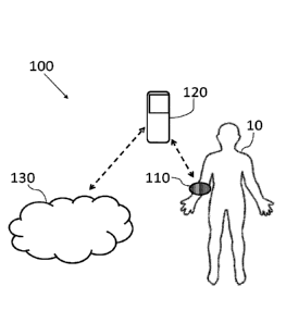
[025] Fig. 1 schematically illustrates a non-invasive monitoring system,
according to some
embodiments of the present invention;
4
CA 03010164 2018-06-28
WO 2017/115361 PCT/IL2016/051386
[026] Fig. 2 schematically illustrates a measuring unit for a wearable
monitoring device,
according to some embodiments of the present invention;
[027] Fig. 3A schematically illustrates a cross-sectional view of the wearable
monitoring device
with the measuring unit as worn by the subject, according to some embodiments
of the present
invention;
[028] Fig. 3B schematically illustrates a cross-sectional view of the
measuring unit coupled and
adjacent to the subject, according to some embodiments of the present
invention;
[029] Fig. 4 shows a flowchart for a method of non-invasive monitoring of
physiological
measurements, according to some embodiments of the present invention;
[030] Fig. 5 shows a flowchart for a method of analyzing non-invasive
monitoring of
physiological measurements, according to some embodiment of the present
invention;
[031] Fig. 6 shows a flowchart for a method of compensating blood parameter
readings,
according to some embodiment of the present invention;
[032] Fig. 7 schematically illustrates a cross-sectional view of a measuring
unit with an
embedded ultrasonic unit coupled and adjacent to the subject, according to
some embodiment of
the present invention;
[033] Fig. 8A shows a Clarke Error Grid chart for a first measurement with a
non-invasive
monitoring system, according to some embodiments of the present invention;
[034] Fig. 8B shows a Clarke Error Grid chart for a second measurement with a
non-invasive
monitoring system, according to some embodiments of the present invention;
[035] Fig. 9A shows absorption measurements with a non-invasive monitoring
system for
Albumin, according to some embodiments of the present invention;
[036] Fig. 9B shows absorption measurements with a non-invasive monitoring
system for low
density lipoprotein (LDL), according to some embodiments of the present
invention; and
[037] Fig. 9C shows absorption measurements with a non-invasive monitoring
system for very
low density lipoprotein (vLDL), according to some embodiments of the present
invention.
[038] It will be appreciated that, for simplicity and clarity of illustration,
elements shown in the
figures have not necessarily been drawn to scale. For example, the dimensions
of some of the
elements may be exaggerated relative to other elements for clarity. Further,
where considered
appropriate, reference numerals may be repeated among the figures to indicate
corresponding or
analogous elements.
CA 03010164 2018-06-28
WO 2017/115361 PCT/IL2016/051386
DETAILED DESCRIPTION OF THE PRESENT INVENTION
[039] In the following detailed description, numerous specific details are set
forth in order to
provide a thorough understanding of the invention. However, it will be
understood by those
skilled in the art that the invention may be practiced without these specific
details. In other
instances, well-known methods, procedures, and components modules, units
and/or circuits have
not been described in detail so as not to obscure the invention. Some features
or elements
described with respect to one embodiment may be combined with features or
elements described
with respect to other embodiments. For the sake of clarity, discussion of same
or similar features
or elements may not be repeated.
[040] Although embodiments of the invention are not limited in this regard,
discussions
utilizing terms such as, for example, "processing," "computing,"
"calculating," "determining,"
"establishing", "analyzing", "checking", or the like, may refer to
operation(s) and/or process(es)
of a computer, a computing platform, a computing system, or other electronic
computing device,
that manipulates and/or transforms data represented as physical (e.g.
electronic) quantities within
the computer's registers and/or memories into other data similarly represented
as physical
quantities within the computer's registers and/or memories or other
information non-transitory
storage medium that may store instructions to perform operations and/or
processes. Although
embodiments of the invention are not limited in this regard, the terms
"plurality" and "a
plurality" as used herein may include, for example, "multiple" or "two or
more". The terms
"plurality" or "a plurality" may be used throughout the specification to
describe two or more
components, devices, elements, units, parameters, or the like. The term set
when used herein
may include one or more items. Unless explicitly stated, the method
embodiments described
herein are not constrained to a particular order or sequence. Additionally,
some of the
described method embodiments or elements thereof can occur or be performed
simultaneously,
at the same point in time, or concurrently.
[041] Reference is now made to Fig. 1, which schematically illustrates a non-
invasive
monitoring system (wherein the direction of dashed arrows may indicate the
direction of
information flow), generally designated 100, according to some embodiments of
the invention.
The non-invasive monitoring system 100 is designed to allow continuous and/or
repetitive non-
invasive monitoring of a subject 10, using a wearable monitoring device 110.
The wearable
monitoring device 110 may be wearable on a limb of the subject 10, or
alternatively on other
parts of the body (e.g. on a finger, on the ear, etc.).
[042] It should be appreciated that wearable device 110 may collect continuous
data on the
physiological signals (e.g., pulse, blood components levels, etc.) of the
subject 10, as long as
6
CA 03010164 2018-06-28
WO 2017/115361 PCT/IL2016/051386
device 110 is worn by the subject 10, and therefore wearable device 110 may
provide ongoing
data such that changes in measured physiological signals may be detected. In
some
embodiments, wearable device 110 may collect the data when wearable device 110
is worn by
the user, and provide the collected data to user even when device 110 is not
worn by the subject
10.
[043] According to some embodiments, wearable device 110 may be configured to
sample the
physiological signals of subject 10 repetitively every predefined time period.
In some
embodiments, the frequency of sampling may be equal to or higher than Nyquist
rate. When
wearable monitoring device 110 is configured to measure non-invasively various
physiological
characteristics of the subject 10, different frequencies of sampling may be
used for each
measured physiological characteristic.
[044] According to some embodiments, monitoring system 100 may further include
a
computerized device 120 (e.g. a processor in the vicinity of subject 10), that
is configured to
receive data from wearable monitoring device 110 and may allow processing
(e.g. with a
processor) of the received data thereof. In some embodiments, computerized
device 120 may be
or may include, for example, a mobile phone, a tablet, a personal computer, a
mobile computer,
or any other suitable computing device 120. For example, system 100 as
described herein may
include one or more devices such as computerized device 120.
[045] According to some embodiments, monitored data may be transferred from
computerized
device 120 to wearable monitoring device 110, and vice versa, via a compatible
communication
module (e.g. via Wi-Fi, Bluetooth, NFC, etc.). For example, a user 10 wearing
wearable
monitoring device 110 and also operating a mobile phone, may utilize the
mobile phone as
computerized device 120 in order to transfer data to and from wearable
monitoring device 110
via wired and/or wireless communication.
[046] In some embodiments, wearable monitoring device 110 may include a
measuring unit
(200 in Fig. 2) with a dedicated controller that may be configured to measure
physiological signs
of subject 10 (further described with reference to Fig. 2, 3A and 3B
hereinafter), a
communication module configured to allow communication with computerized
device 120 via,
for example, wireless communication, and a power storage unit (e.g. a
battery). Computerized
device 120 may include compatible components that are configured to allow the
data transfer to
and from the wearable monitoring device 110, and to allow processing of data
received from
wearable monitoring device 110. For instance, computerized device 120 may
include a
compatible communication module, a display (e.g. with a user interface), and a
processor
7
CA 03010164 2018-06-28
WO 2017/115361 PCT/IL2016/051386
capable of processing and monitoring the physiological data of subject 10
measured by
monitoring device 110.
[047] Computerized device 120 may have, according to some embodiments, a
dedicated user
interface (e.g. with a dedicated algorithm installed thereon) so as to display
real-time
measurements to subject 10. Thus, the user may receive alerts and/or updates
regarding the
physiological signs that were measured by the wearable monitoring device 110.
In some
embodiments, computerized device 120 may issue an alert upon detection of a
change in
measured physiological signals exceeding a predetermined threshold.
[048] In some embodiments, monitoring system 100 may further include a data
analyzing
facility 130, e.g. such as one or more server computers in communication with
one or more
wearable devices. The data analyzing facility 130 may include a computerized
device with a
dedicated database for processing and analyzing measurement data from one or
more subjects
10, such as physiological signals, blood parameters (e.g. medication
concentration in the blood,
blood chemistry etc.) and the like.
[049] Such a data analyzing facility 130 may be adapted to carry out at least
one of big data
analysis, machine learning, and data mining tasks. Thus, data analyzing
facility 130 may analyze
physiological signals from multiple subjects and thereby deduce desirable
ranges and trends for
physiological measurements and medical insights, as further described
hereinafter.
[050] In some embodiments, the analyzing and processing of the measured data
may be carried
out on a dedicated processor embedded into monitoring device 110.
[051] In some embodiments, the dedicated measuring algorithm may provide
predetermined
time intervals for performing measurements and/or time intervals for sending
data from
wearable monitoring device 110 to computerized device 120. Furthermore, the
measuring
algorithm may also provide predetermined time intervals for sending data from
computerized
device 120 to data analyzing facility 130. In some embodiments, these
predetermined time
intervals may be altered by the user (e.g., subject 10, a caregiver and the
like) and/or by the
dedicated measuring algorithm.
[052] For instance, monitoring system 100 may receive data from wearable
monitoring device
110 indicating a sharp rise in the glucose level in the blood, so that the
dedicated measuring
algorithm may automatically increase the frequency and/or duration of the time
intervals for
performing measurements so as to gather additional information prior to
sending an alert to
subject 10. In some embodiments, the time intervals between measurements may
be reduced, for
instance upon receiving data from wearable monitoring device 110 indicating a
drop in the
glucose level in the blood.
8
CA 03010164 2018-06-28
WO 2017/115361 PCT/IL2016/051386
[053] In some embodiments, the monitoring system 100 may perform self-
optimization by
learning the behavior of the subject 10. For instance, learning specific time
periods during the
day when the subject 10 engages in sport activity, affecting the expected
values of measured
physiological signs (e.g. pulse, blood pressure and the like) and storing data
for such time
periods in a dedicated memory and/or database. With such optimization,
monitoring system 100
may only perform measurements that give actual information on the subject in a
"relaxed state"
(e.g., where the subject does not perform any physical activity), as well as
saving electrical
power for the wearable monitoring device 110, since redundant measurements are
reduced or
completely prevented.
[054] In some embodiments, wearable monitoring device 110 may further include
a memory
module that is configured to store measurement data to be sent at a later
time. This feature may
allow the system to save electrical power by sending data only at
predetermined intervals. In
some embodiments, only a predetermined type of data may be stored on this
memory module,
for instance storing a record of daily glucose level measurements.
[055] Reference is now made to Fig. 2, which schematically illustrates a
measuring unit 200 for
a wearable monitoring device, according to some embodiments of the present
invention.
Measuring unit 200 may include at least one sensor 210 and at least one light
emitting source
220. In some embodiments, measuring unit 200 is adjacent to and in contact
with the skin of
subject 10 so as to reduce noises from the environment. It should be noted
that with light beams
emitted from the at least one light emitting source 220 towards subject 10,
the wearable
monitoring device may perform optical measurements (e.g. with at least one
sensor 210) that are
non-invasive.
[056] In some embodiments, multiple measuring units 200 may be employed (for
instance as an
array) in order to allow simultaneous monitoring of several blood vessels of
the subject 10. For
example, an array of three adjacent measuring units 200. In some embodiments,
the monitoring
system 100 may operate different light emitting sources 220 within the
multiple measuring units
200 in order to achieve optimal measurements (for example initiating only
sources 220 closest to
detected blood vessels). Such an array may allow measurements in parts of the
body having
different distributions of blood vessels, whereby the monitoring system 100
may choose to
operate a particular measuring unit 200 that is closest to a blood vessel. In
some embodiments,
each measuring unit 200 may be operated in a different wavelength, for
instance in order to
allow simultaneous measurements of different features (e.g. glucose, insulin,
LDL, VLDL and
Albumin). In some embodiments, measurements of different substances (e.g.
glucose and
Albumin) may be carried out with different wavelengths and/or different number
of
9
CA 03010164 2018-06-28
WO 2017/115361 PCT/IL2016/051386
wavelengths. For example, measurements of glucose may require each measuring
unit 200 to be
operated in eight different wavelengths, while measurements of Albumin may
require each
measuring unit 200 to be operated in six other wavelengths.
[057] The light emitted from the at least one light emitting source 220 (e.g.
LED), to be
reflected from a subcutaneous tissue (e.g. from blood in a blood vessel in the
subcutaneous
tissue) of the subject, and then detected by the at least one sensor 210 may
be, according to some
embodiments, in the Infra-Red or near Infra-Red (IR) spectrum. In some
embodiments, Short
Wave IR (SWIR) imaging is utilized for measuring physiological signals from
the blood of
subject 10. The SWIR waveband runs from the lower edge of the near IR region
at 900nm up to
2500 nm, and may be utilized for inspection of blood and blood components in
blood vessels of
the subject 10. It should be noted that if required, the range of the SWIR
waveband may be
increased.
[058] It should be noted that the measuring unit 200 may be provided in
various configurations,
and in some embodiments a single sensor 210 is surrounded by a plurality of
light emitting
sources 220 (as for example illustrated in Fig. 2). Other configuration may
also employ a
plurality of sensors 210 and light emitting sources. For example, a plurality
of sensors 210,
where each sensor 210 is surrounded by a plurality of light emitting sources
220 and where at
least two sensors 210 may share at least one light emitting source 220.
[059] In some embodiment, each light emitting source 220, or sub-sets (e.g.
pairs, triplets etc.)
of light emitting sources 220 may emit light in a different predetermined
wavelength.
[060] In some embodiment, each light emitting source 220, or sub-set of light
emitting sources
220, may emit light in a different time and/or in a different frequency, such
that not all light
emitting sources 220 emit light simultaneously. This may provide additional
information on the
reflected tissue when the time intervals between the emissions of light beams
is known.
[061] According to some embodiments, the frequency of sampling by each light
emitting
source 220, or by each sub-set of light emitting sources 220, may be equal to
or higher than
Nyquist rate of the measured physiological signal.
[062] In some embodiments, polarized optical means may be utilized in order to
increase the
accuracy in the optical measurements. Specifically, emitting light beams with
a predetermined
polarization and receiving these beams with a substantially different
polarization, for instance
with dedicated filters, may improve the signal to noise ratio in the
measurements. Furthermore,
such polarizing may also provide improved indication on the penetration of the
light beam into
the tissue as noises from the external skin layer may be reduced while only
signals from the
beam reflected from the blood is measured.
CA 03010164 2018-06-28
WO 2017/115361 PCT/IL2016/051386
[063] In some embodiments, at least one light emitting source may be a
polarized light source
configured to emit light beams with at least one predetermined polarization
(e.g. in an
alternating mode such that at least one emitted light beam is not polarized),
and wherein at least
one sensor may be a polarized light sensor configured to detect reflection of
the polarized light
beams, wherein the polarized sensor has a different polarization than the
polarized light source.
[064] In some embodiments, other sensors may also be utilized. For example
acoustic
ultrasound sensors, as well as photoacoustic sensors, terahertz sensors, RF
sensors, microwave
sensors and corresponding energy sources.
[065] In some embodiments, the light emitting source (e.g. LED) may be
operated in pulse
width modulation (PWM) mode with a configurable duty cycle. As may be apparent
to one of
ordinary skill in the art, the pulse width of a duty cycle may be as small as
about ¨0.01%. In
some embodiments, the pulse width may also be changed during measurements by
data
analyzing facility 130 corresponding to the actual readings.
[066] In addition, in some embodiments, at least one sensor 210 may be
synchronized with at
least one light emitting source 220 by applying a band pass filter that blocks
all information not
correlated to the PWM switching frequency. Such a band pass filter may be
adjustable to allow
frequency change during operation, for example as calculated by analyzing
facility 130.
[067] Reference is now made to Figs. 3A-3B, which show wearable monitoring
device 110
coupled to the body of the subject 10. Fig. 3A schematically illustrates a
cross-sectional view of
wearable monitoring device 110 with measuring unit 200 worn on a limb of
subject 10,
generally designated 300, according to some embodiments of the present
invention.
[068] In some embodiments, wearable monitoring device 110 encompasses a
portion of the
body of the subject 10 (e.g. a finger or the wrist, or leg), wherein the
emitted light is reflected
back from a blood vessel (e.g. as shown in Fig. 3B). Alternatively, in other
embodiments, the
wearable monitoring device 110 may be clipped onto a different portion of the
body (e.g. the
ear), wherein the emitted light passes through that portion and is transmitted
to the sensor on the
other side of the wearable monitoring device 110. According to some
embodiments, at least one
wearable monitoring device 110 may simultaneously detect with at least one
sensor light
reflected back from a blood vessel and/or detect light transmitted through a
portion of the body
of the subject 10.
[069] For example, measuring unit 200 may be embedded into a band like
wearable monitoring
device 110, which is worn on the wrist of the subject 10. Thus, the measuring
unit 200 may be
sufficiently adjacent to the skin to prevent exposure of sensor 210 (in Fig.
2) to ambient light,
11
CA 03010164 2018-06-28
WO 2017/115361 PCT/IL2016/051386
and to detect light beams (with the sensor 210) emitted from the light
emitting source 220 and
reflected from blood in a blood vessel.
[070] In some embodiments, the wearable monitoring device 110 may include a
bracket that is
capable of sensing whether the wearable monitoring device 110 is in an
operable state. For
example, a bracket that triggers the wearable monitoring device 110 to
commence monitoring
once appropriate contact with subject 10 is achieved (e.g. once a wrist band
is secured onto the
wrist of a user).
[071] In some embodiments, multiple wearable monitoring devices 110 may be
employed by
the same subject 10 in order to provide accurate measurement with comparison
to measurements
from different parts of the body. For example, a first wearable monitoring
device 110 worn on
the wrist, and a second wearable monitoring device 110 worn on the leg.
[072] In some embodiments, wearable monitoring device 110 may further include
a pressure
sensor that is configured to indicate excessive pressure on the skin of the
subject 10. This may
be performed so as to allow adjustment of wearable monitoring device 110
fastener (e.g. to
reduce or increase the pressure), for instance by manipulating the coupling of
the wearable
monitoring device 110 to the skin (e.g. physically moving the monitoring
device 110), in order
to adjust the pressure on the skin and thereby reduce noise from the
measurements.
[073] It should be noted that in order to receive an accurate measurement, the
sensor 210 of the
measuring unit 200 should be adjacent to the skin of the subject 10, such that
it may measure
light signals that are reflected from a blood vessel. In some embodiments, the
signal intensity
from each light emitting source 220 may indicate the proximity to a blood
vessel, based on
previously calibrated light sources that were placed over a known blood
vessel. Thus, a
processor of monitoring system 100 may select only some of the light emitting
sources 220 that
are proximal to the blood vessel to perform the optical measurements (i.e.
emit the light beams).
In some embodiments, once the monitoring system 100 receives an indication on
a position that
is proximal to a blood vessel then the light emitting sources 220 may receive
a signal to initiate
the measurements.
[074] As may be apparent to one of ordinary skill in the art, light in
specific wavelengths
between 400-2500nm (e.g. 417nm, 545nm, or 578nm), reflected from subcutaneous
tissue (e.g.
reflected from blood inside a blood vessel) and light reflected from tissue
above a blood vessel
have different intensities since light reflected from tissue above a blood
vessel have a weaker
reflection due to higher light absorption in water content. Therefore, it may
be possible to
determine a threshold for determining position of measuring unit 200 being
over a blood vessel.
12
CA 03010164 2018-06-28
WO 2017/115361 PCT/IL2016/051386
[075] In some embodiments, the monitoring system 100 may further include
positioning
correction indicators (not shown) that are adapted to allow the user to
correctly place measuring
unit 200 over a blood vessel. For instance, displaying to the user how to move
monitoring device
110 to improve positioning of light emitting sources 220 to optimize
reflections to the sensor.
[076] In some embodiments, the measurements of a pulse signal may provide
indication that the
measuring unit 200 is in a proper position, when a sufficiently strong pulse
signal is received. A
sufficiently strong pulse signal may refer to a signal above a predefined
threshold. In some
embodiments, the monitoring system 100 may be utilized for measurement of
medication
concentration and/or existence of medication in the blood, for instance by
monitoring a different
range of wavelengths.
[077] According to some embodiments, the wearable monitoring device may be
provided as a
patch (or sticker) that is removably attached to the skin of the object while
having the same
features as described above, and capable of monitoring the object. Such a
patch-like monitoring
device may be particularly helpful to users that wish to perform measurements
at various
locations on the body, and for instance without wearing a wearable devices on
their limbs.
[078] Fig. 3B schematically illustrates a cross-sectional view of the
measuring unit 200 coupled
and adjacent to the subject 10 (wherein the direction of the dashed arrows
indicates the direction
of the light beams), according to some embodiments of the present invention.
[079] Measuring unit 200 may be placed adjacent to the skin 30 of subject 10
(in Fig. 1). A light
beam may then be emitted (e.g. periodically every 5 minutes, every 10 minutes,
or at any other
time or frequency) from the light emitting source 220 and into the skin such
that it penetrates the
skin and may be reflected back from blood vessel 35 towards sensor 210. The
difference in the
data between the emitted beam and the received (reflected) beam may provide an
indication on
the radiation (e.g., light) absorption by the blood in blood vessel 35 and
thus may indicate
characteristics and blood measurements of the blood inside blood vessel 35, in
a non-invasive
procedure. In some embodiments, each light emitting source 220 may be provided
with an
optical collimator (or reflector) so as to allow directing the light beam
emitted by each light
source 220 in a specific predefined direction.
[080] For example, such measurements may provide an indication for a "health
matrix" with
continuous glucose monitoring, dehydration monitoring, blood lipids, vitamins,
calories, pulse,
PWV (Pulse wave velocity) blood pressure, and also an indication of
medications,
pharmaceuticals and other chemicals in the blood stream of the subject. It is
appreciated that in
order to provide an alert to the subject regarding for example, glucose
measurements, it may be
sufficient to indicate a (predetermined minimal) change in the level and/or
trend of glucose in
13
CA 03010164 2018-06-28
WO 2017/115361 PCT/IL2016/051386
the blood, such that a precise and accurate measurement is not always
required. Thus, the system
may continuously or repetitively monitor the glucose levels and/or glucose
trends and indicate
upon measuring a change. In some embodiments, the system may perform a
continues
measurement or multiple measurements only upon indication of a significant
change such that
power is saved and the system operates in "low energy consumption" mode.
[081] In some embodiments, the orientation of the emitted light beam may be
controlled, for
instance with dedicated beam controlling elements of angle and position, so as
to allow control
of the depth of the penetration of the beam that corresponds to the relative
angle of emission.
[082] In some embodiments, data analyzing (e.g. by the computerized device) of
such non-
invasive measurements may provide a prediction regarding at least one of the
following:
diabetes (e.g., through glucose levels monitoring), dehydration (e.g., through
water level,
cortisol, blood albumin level, urea level, and skin temperature monitoring),
medication
compliance (e.g., Depalept, Plavix clopidogrel, cyclosporine Anti-hypertensive
drugs,
Metformin¨Glucophage, Lipitor-Statins, Cannabinoids based drugs, etc.),
Creatine kinase,
Cardiac troponin, Hs CRP-C reactive protein, Cholesterol LDL/HDL,
Triglycerides, and blood
lipids such as high-density lipoprotein (HDL), low-density lipoprotein (LDL),
and very-low-
density lipoprotein (VLDL). In some embodiments, such non-invasive
measurements may
provide a prediction regarding Cholesterol lowering medications (e.g. Statins,
Niatzin, and
fibrates) and/or blood pressure lowering medications (e.g. thiazides) and/or
diabetes lowering
medications (e.g. biguanidins, glitazones, sulfinyl urea, and insulin).
[083] In some embodiments, such measurements may allow issuing alerts before
life
threatening conditions, such as heart abnormality or stroke, by continuously
monitoring and
collecting personal data to detect changes for instance in low density
lipoprotein (LDL),
Albumin, glucose, etc.
[084] In some embodiments, the optical measurements (e.g. light emitting diode
(LED)) for
glucose may be performed with wavelengths in the range of about ¨900nm-2500nm.
In some
embodiments, a glucose measurement may also be performed with acoustic means
(e.g. an
ultrasound sensor) whereby the sensor may receive a sound (e.g. ultrasound)
waves reflected
from the blood in the subject's blood vessels. In some embodiments, other or
additional
substances may be measured within the blood using such acoustic means, and/or
medicament
concentrations may also be measured using such acoustic means.
[085] In some embodiments, the optical measurements (e.g. LED) for Oxygen
saturation may
be performed with additional wavelengths of about ¨660nm and/or about ¨910nm
with a
comparison of oxygenized and deoxygenized hemoglobin.
14
CA 03010164 2018-06-28
WO 2017/115361 PCT/IL2016/051386
[086] In some embodiments, the monitoring system may be pre-calibrated prior
to initial
operation with a specific subject, based on average measurements from multiple
users or
alternatively calibrated for a specific subject, for instance compared to
measurements from other
devices (e.g. calibrating glucose measurement's versus a commercially
available glucometer).
[087] In some embodiments, the optical measurements (e.g. LED) for lipids and
cholesterol
may be performed with wavelengths of about ¨930nm, ¨1210nm, ¨1400nm, ¨1730nm
and/or
¨1760nm.
[088] In some embodiments, the optical measurements (e.g. LED) for hemoglobin
may be
performed with additional wavelengths of about ¨400nm, ¨815nm, and/or ¨950nm
checking the
red cells level in the blood.
[089] In some embodiments, the optical measurements (e.g. LED) for Bilirubin
may be
performed with wavelengths of about ¨460nm, and/or ¨585nm, and/or ¨650nm
checking the
Bilirubin level in the blood as well as liver functioning.
[090] It should be appreciated that hematocrit measurements (i.e. percentage
of red blood cells
volume in total blood volume) may differ between men and women, for instance
being 42-52%
in men and 36-48% in women. Thus, the values of different parameters in the
blood may be
adjusted for the specific subject group of the user (e.g. for a female user)
since the concentration
of a particular substance may be different, thereby providing substantially
different values.
[091] In some embodiments, the optical measurements (e.g. LED) for a
substantially constant
parameter, such as Albumin, total serum proteins, globulins or any combination
of thereof, may
be performed, checking the constant parameter's level in the blood. In some
embodiments, the
optical measurements (e.g. LED) for alcohol may be performed with wavelengths
of about
¨1250-2500nm checking the Ethanol level in the blood.
[092] Reference is now made to Fig. 4, which shows a flowchart of a method of
non-invasive
monitoring of physiological measurements. According to some embodiments, the
wearable
monitoring device 110 may be initiated 410 (e.g. by a processor of
computerized device 120),
for instance upon detecting contact (e.g. with a pressure sensor or any other
element sensitive to
contact and/or pressure) with the skin of the user (as described above), in
order to commence the
measurements.
[093] According to some embodiments, at least one light emitting source 220
may emit light
beams 420 in the direction of the skin of the user, to be reflected from a
blood vessel (e.g., by the
content of the blood vessel) and then received 430 by at least one sensor 210.
In some
embodiments, the light beams are transmitted through the tissue (including the
blood vessels
therein) of the user and then received by the sensor 210.
CA 03010164 2018-06-28
WO 2017/115361 PCT/IL2016/051386
[094] According to some embodiments, the wearable monitoring device 110 may be
calibrated
prior to initial use (as described above) such that the received light beams
may provide an
indication whether a blood vessel is detected 440, in order to filter
measurements that were not
carried out with proper positioning over a blood vessel. In case that the
received signal does not
provide an indication of a measurement from a blood vessel, the monitoring
device may be
adjusted 450 so as to better align the at least one light emitting source 220
with an adjacent
blood vessel. In some embodiments, such adjustment 450 may be carried out by
the user slightly
moving the wearable monitoring device 110 along the skin, or alternatively
carried out
electrically by adjustment of the illumination angle of the light emitting
sources 220 and/or
selection of at least one light emitting source 220 that provides signals from
a blood vessel. Once
the wearable monitoring device 110 is properly adjusted 450, the at least one
light emitting
source 220 may emit light beams 420 again in order to commence a new
measurement until a
suitable blood vessel measurement is detected.
[095] For example, if five light emitting sources 220 sequentially emit light
beams towards the
skin and the received signal indicates that only the first two are adjacent to
a blood vessel
(according to the light absorption and reflection from and/or transmission
through blood vessels
with respect to the absorption and reflection/transmission of tissue not
including blood vessels),
only information from these two light emitting sources 220 may be employed for
the
measurement.
[096] According to some embodiments, in case that the received signal does not
provide an
indication of a measurement from a blood vessel, at least one of the light
emitting sources 220
may be adjusted to emit light at a different wavelength. In some embodiments,
if only some of
the light emitting sources 220 are utilized to detect a blood vessel then at
least one other light
emitting source 220 (at a different position on the wearable monitoring device
110) may be
utilized to emit light so as to detect a blood vessel from a different
position of the light emitting
source 220 (e.g. an LED) on the wearable monitoring device 110.
[097] In some embodiments, if such adjustment of the measurement does not
allow detection of
a blood vessel, for instance after a predetermined number of periodic tests
(e.g. five tests), then
an alert may be provided to the user.
[098] In case that the received signal provides an indication of a measurement
from a blood
vessel, the measured data may be analyzed 460 in order to monitor the
physiological signals of
the user. For example, analyzing the received data to check a change in
glucose level, whereby
such analyzing may be based on previous calibration (as described above).
16
CA 03010164 2018-06-28
WO 2017/115361 PCT/IL2016/051386
[099] In some embodiments, the analyzing 460 of the measurements may be
carried out at a
computerized device 120 and/or at a data analyzing facility 130 (as described
with reference to
Fig. 1).
[100] Reference is now made to Fig. 5, which shows a flowchart of a method of
monitoring and
analyzing non-invasive physiological measurements. According to some
embodiments, the
analyzing 460 of the data measured by the wearable monitoring device 110 may
be carried out
to determine whether the physiological signals of the object 10 are within the
desired range, for
instance by calibrating physiological signals it may be possible to determine
a desired range for
the received signals and/or the desired range may correspond to data gathered
from multiple
subjects and/or the desired range may correspond to a specific range provided
(e.g. by health
officials) for particular substances in the blood.
[101] After a blood vessel is detected 440, the measured data of the object 10
may be collected
510 for analyzing by the monitoring system 100. For instance, the measured
data may be
collected and stored in a dedicated memory unit embedded into the wearable
monitoring device
110. The collected data may then be transferred to the computerized device 120
and/or the data
analyzing facility 130 for further analyzing.
[102] In some embodiments, the collected data may be compared 520 to at least
one stored data
set in order to determine whether the collected data is within a normal range
530. According to
some embodiments, the stored data may be stored at a dedicated memory unit at
the wearable
monitoring device 110 and/or at the computerized device 120 with data from
previous
measurements of the object 10. For example, the collected data may be compared
to a threshold
value and/or the deviation from a threshold value may be calculated in order
to compare the
deviation to a desired deviation range.
[103] After sufficient measurements of the object 10 are collected, for
instance during
calibration, a normal (or desired) range for the measurements may be
determined, such that new
measurements may be compared 520 to the normal range 530. For example,
performing
calibration for glucose measurements to establish a desired range for a
particular object 10.
[104] In some embodiments, the stored data for comparison includes average
measurement data
taken from measurements of the general public, whereby averaging values for a
large group of
people may provide a normal range for comparison.
[105] According to some embodiments, the stored data may be used to improve
the glucose
level estimation accuracy, by comparing to stored temporal patterns within the
optical
measurements and corresponding glucose levels, and deriving low-dimensional
representations
of the corresponding optical measurements, best adhering to the glucose
levels.
17
CA 03010164 2018-06-28
WO 2017/115361 PCT/IL2016/051386
[106] Moreover, the measured glucose levels may be used by the data analyzing
facility 130
(e.g. cloud based), to apply statistical inference to the stored data of
multiple users, in order to
detect temporal patterns common to large numbers of users, and relating to
physical activities.
[107] In case that the collected data is within the desired range 530, the
system may check if the
frequency is at a normal (or predetermined) range 540. If the frequency is at
a normal value 540
the measured data then may be stored 590 and aggregated for future
measurements. If the
frequency is not normal 540, then the system may return 580 the frequency to
normal and also
store 590 and aggregate the measured data for future measurements.
[108] Otherwise, in case that the collected data is not within the desired
range 530, the system
may check if the frequency is at a normal (or predetermined) range 540. If the
frequency is at a
normal value 540 the system may change the frequency 570, for instance
increasing the
frequency of the measurements when a sharp rise (e.g. with a change of about
10-20% between
measurement periods) in a physiological feature is detected, and then collect
data 510 with the
new measurement frequency.
[109] If the frequency is not normal 540, then an alert may be sent 560 to the
user (e.g. on a
display) and also the collected data may be stored 590 for future reference
together with
collection of new measurement data 510 with the new measurement frequency. For
example, a
first measurement gives an indication that a measured glucose level is out of
the desired range
(i.e., lower or higher than predefined upper and lower limits) so the
measurement frequency is
increased and an alert is sent to the user. At the second measurement the
glucose level is still out
of the desired range, and at the third measurements the glucose level returns
to normal range so
that measurement frequency may be reduced to normal level. In some
embodiments, an alert
may also be created when the data return to the normal range.
[110] For example, two computational tests are applied to the spectral
measurements. In the
first test, the dynamics of the glucose levels are compared to those computed
using a low-
dimensional embedding of the spectral measurements. It follows that the
spectral measurements
follows closely the dynamics and trends of the glucose levels. In particular,
as the number of
samples increases (as on the third day of experiments) the parameterization
accuracy is
improved. Thus, it follows that the parameterization accuracy can be improved
by using more
test subjects and also a larger database. With the number of optical
measurements increasing,
and also corresponding reference glucose measurements, more elaborate
numerical models may
be derived. Thus, improving the glucose readings accuracy. In particular, when
a large dataset of
such measurements is accumulated, data mining and statistical inference
techniques may be
applied.
18
CA 03010164 2018-06-28
WO 2017/115361 PCT/IL2016/051386
[111] According to some embodiments, each user may be associated with a
personal reflectance
coefficient during calibration of the system. Such a personal reflectance
coefficient may provide
personal calibration data regarding parameters that may influence optical
readings and thereby
allow personalization of the measurements, as further described hereinafter.
It should be
appreciated that since each user has unique physical conditions, each user may
need their own
personal adjustments for the optical readings. These adjustments may therefore
enable setting a
personal offset in relation to average values that are previously stored in
the database.
[112] It should be noted that Infra-Red (IR) reflection in the human body may
be affected by
various parameters. User dependent parameters may include parameters affecting
the optical
reflection, such as different hand and/or skin thickness, different skin
complexions, thickness
and diameter of blood vessels, and thickness and composition of different skin
layers. Other user
dependent parameters may affect (or alter) the results of the measurement,
such as hair on the
wrist, skin temperature, user activity level (e.g. causing sweating), and
dehydration level.
[113] Environmental dependent parameters may also affect the result, such as
the amount of
external light radiation combined with light beams of the measurement, the
type of external light
radiation, the angle and distance between the IR light source and the blood
vessel, the diversity
of IR light sources (e.g. additional IR light sources with a different
wavelength), light source
decay during operation, and possible water presence (e.g. in ambient air or on
the body).
[114] According to some embodiments, certain reference parameters in the blood
cycle, that
may be considered as constant, may be defined as a reference level for each
user (e.g. during
calibration). Thus, comparison to that reference level may indicate changes in
the measured
parameters of the individual user. It should be appreciated that since the
values of these
reference parameters may change in a relatively narrow range (and in a slow
rate), these
reference parameters may be utilized for calculation of the relevant personal
reflectance
coefficient for each user.
[115] It should be noted that the personal reflectance coefficient may
represent for each user
reference parameters (being relatively constant) that influence light
transmittance, reflectance
and absorbance through tissue of the body. These reference parameters do not
vary, since they
correspond to skin, tissue and blood vessel structure that is a part of the
optical path of which the
light beams from the light source go in and are reflected, absorbed or
transmitted back towards
the detector, for example as shown in Fig. 3B.
[116] In some embodiments, substantially constant bloodstream components (e.g.
Albumin,
serum proteins, globulins and any combination thereof) may be considered as a
"mirror with
known reflection index" reflecting back the light (e.g. IR light), through
skin, blood and blood
19
CA 03010164 2018-06-28
WO 2017/115361 PCT/IL2016/051386
vessels, with a known (or constant) optical behavior. Such reference
parameters (with known
optical behavior) may be therefore defined for each user, for instance during
calibration, such
that reflectance, absorbance and transmittance of light during glucose
measurements may be
determined. For example, two possible blood parameters may be chosen as
Albumin and CO2
level dissolved in the blood (associated with blood vessels blueish color),
which can be
considered as constant with healthy users. Changes in such substantially
constant parameters as
Albumin may occur in healthy people, for instance during three weeks, in a
narrow range of
about 3.5-5 gr/dl. The amount of CO2 dissolved in blood may stay constant as
long as the user is
not suffering from any serious distress or illness, so a change in dissolved
CO2 levels may
therefore indicate a distress in a particular user.
1117] It should be appreciated that since Albumin (or other constant
parameter) values do not
vary dramatically for each user, these values may be utilized to constantly
recalculate the
personal reflectance coefficient for each user. Thus, all varying parameters
within the blood may
be compensated for with the known value for Albumin (or other serum proteins),
whereby the
other parameters may be compared to a reference level for the known values.
For instance if a
user moves the device on the hand, the angle and distance between the light
source and the
blood vessel may change and therefore the optical reading may be different.
Therefore it may be
possible to calculate a compensation function based on changes in the Albumin
(and/or other
blood serum proteins) reading, so as to derive that the user moved the device,
and thus applying
the compensation function on reading of other parameters (e.g. of glucose), as
further described
in Fig. 6. In some embodiments, the base value of Albumin (and/or other blood
serum proteins)
for this user may be accordingly derived from previously acquired databases
based on the cluster
of data that this particular user belongs to, or from previous results of
general blood test that are
available at medical databases.
1118] Reference is now made to Fig. 6, which shows a flowchart of a method of
compensating
blood parameter readings, according to some embodiments. Initially, a
measurement 610 may be
carried out for a particular substantially constant parameter in the blood
(such as Albumin). In
some embodiments, a change may be calculated 620 (e.g. for Albumin) relative
to a reference
value for that parameter (e.g. Albumin), for instance reference value may be
derived from a
database of average values for populations having similar characteristics
(e.g. for elderly
women).
1119] In some embodiments, the reference value may be derived from a reference
conversion
lookup table (e.g. initially created in laboratory conditions) associating an
optical reading of a
particular substance with the actual value. For example, having a sample of a
known amount of
CA 03010164 2018-06-28
WO 2017/115361 PCT/IL2016/051386
Albumin (or glucose) in a known concentration (e.g. 4 gr/d1), that is
irradiated with IR radiation
in a known wavelength. Thus, future optical reading of the same (or similar)
wavelength should
be converted to the actual value of the measured parameter, e.g. Albumin or
glucose, so as to
allow pre usage calibration as well as future calibration after measurements.
In some
embodiments, an additional reference lookup table may also be provided (e.g.
initially created in
laboratory conditions) for tissues of different groups of patients, for
instance provide a reference
tissue of a Hispanic woman with a predefined skin color, that may be used to
convert optical IR
readings to actual values of the tissue.
[120] Then, a compensation function may be calculated 630 based on the
previously calculated
change 620 in the constant parameter (e.g. Albumin) reading. Finally, the
compensation function
may be applied 640 on readings of other parameters (e.g. on glucose) in order
to normalize the
readings of those parameters. Thus, due to a known value of a particular
reference parameter
(e.g. Albumin), other parameters may be calibrated in order to provide a
reading that is not
affected by other factors. It should be noted that while Albumin is described
herein, any other
substantially constant parameter may be similarly calculated and applied for
the calibration
process. It should be further appreciated that more than one substantially
constant parameter
may be used.
[121] Reference is now made to Fig. 7, which schematically illustrates a cross-
sectional view of
the measuring unit 700 with an embedded ultrasonic unit 720 coupled and
adjacent to the subject
(wherein the direction of the dashed arrows indicates the direction of the
light beams), according
to an embodiment of the present invention. According to some embodiments,
monitoring of
thickness of a user's skin tissue may be allowed with positioning of non-
invasive monitoring
system 100 above a blood vessel.
[122] Measuring unit 700 may be placed adjacent to the skin 30 of the subject.
A light beam
may then be emitted from the light emitting source 220 and into the skin such
that it penetrates
the skin and may be reflected back from blood vessel 35 towards sensor 210.
The difference in
the data between the emitted beam and the received (reflected) beam may
provide an indication
on the radiation absorption by the blood in blood vessel 35 and thus may
indicate characteristics
and blood measurements of the blood inside blood vessel 35, in a non-invasive
procedure. In
some embodiments, measuring unit 700 may further include at least one
ultrasonic unit 720
capable of transmitting and receiving ultrasound signals.
[123] It may be appreciated that an ultrasonic transducer may include a set of
crystals which
may transmit and receive ultrasound signals derived from changes in their
magnetic field. In
some embodiments, an array of such crystals may be embedded into non-invasive
monitoring
21
CA 03010164 2018-06-28
WO 2017/115361 PCT/IL2016/051386
system 100, for example embedded into measuring unit 700. The ultrasonic
transducer may also
turn these signals into electrical currents. Different ultrasonic signals may
penetrate and
propagate through skin tissue, and may be reflected back to the transducer
depending among
other parameters on the operational frequency, and/or on tissue water content,
and/or on tissue
density. In some embodiments, signals of reflected ultrasound waves may be
separated
according to the depth of tissue from where these waves are reflected from. In
some
embodiments, a set of piezo crystals arranged in a predetermined pattern may
be used for
continuously measuring the skin thickness above blood vessels, for example in
the lower part of
the wrist, and thereby determine the position of a transducer and/or its
proximity to blood
vessels.
[124] In some embodiments, such a set of crystals may be positioned such that
in predetermined
time periods and/or every time the monitoring device is removed and/or
replaced, the set of
crystals may transmit and receive ultrasonic signals through the skin. These
signals may
determine skin tissue thickness underneath the transducers array and/or
determine transducer
array proximity to blood vessels under the measured skin.
[125] In some embodiments, determination of desired parameters from monitoring
of signals
reflected from blood vessels, for instance using non-invasive monitoring
system 100, may be
accomplished with at least one of predefined arrangement of crystals, and/or
predefined distance
between them, and/or predefined frequency.
[126] In some embodiments, the signal to noise ratio, for instance with non-
invasive monitoring
system 100 for glucose measurements, with IR signals reflected from human
tissue, may be at a
ratio of 150 to 1, where sources of noise may be for example background
lighting.
[127] Reference is now made to Figs. 8A-8B, which show a Clarke Error Grid
chart for a first
and a second measurement with non-invasive monitoring system 100. A test has
been initially
conducted for monitoring of glucose for a test group of eight users receiving
50 grams of
glucose after fasting for ten hours. The monitoring have been measured for
about 4-6 hours
about every 30 minutes, wherein non-invasive monitoring system 100 has been
compared to
monitoring with commercially available invasive glucometers (e.g.
"AccuCheck"Tm) as
reference for a given blood test. The results of this test are shown in Fig.
8A with a Clarke Error
Grid (CEG), where compatibility of over 96 percent has been observed in zone
"A" for
measurements with non-invasive monitoring system compared to FDA approved
glucometers.
[128] In a second test experiment, monitoring of glucose for a test group of
eight users has been
conducted, wherein each member of the test group received about 2000
milligrams of a pain
relieving medication containing Acetaminophen (e.g. administered with a pill
of "AcamorTm),
22
CA 03010164 2018-06-28
WO 2017/115361 PCT/IL2016/051386
after users of the test group have been fasting for ten hours. It may be
appreciated that
Acetaminophen exhibits a unique spectral signal with optical measurements, and
therefore can
be detected with non-invasive monitoring system 100. The results of this test
are shown in Fig.
8B with a Clarke Error Grid (CEG), where compatibility of over 97 percent has
been observed
in zone "A" for measurements with non-invasive monitoring system compared to
FDA
approved glucometers.
[129] It may be appreciated, for instance from a Clarke Error Grid chart in
Fig. 8B, that using
the monitoring device, as described above, glucose monitoring may not be
affected by presence
of Acetaminophen since the optical properties of glucose in the blood may not
be affected by the
presence of Acetaminophen, for example in contrast to reading by a
commercially available
continuous glucose monitoring (CGM, for example such as "Dexcom platinum G4")
which is
sensitive to Acetaminophen presence in the blood.
[130] In some embodiments, the amount of Acetaminophen may be monitored and/or
measured, due to the unique spectral signal of Acetaminophen, so as to serve
as a monitoring
unit for acetaminophen, where such measurement may allow determination of
accurate reading
of other substances in the blood, for instance glucose. Therefore, monitoring,
for instance with
non-invasive monitoring system 100, may be carried out even with users
receiving medications
and still provide substantially accurate monitoring thereby enhancing
compliance of monitoring
with various medications.
[131] Reference is now made to Figs. 9A-9C, which show absorption measurements
with non-
invasive monitoring system 100, for Albumin, LDL and vLDL respectively. It
should be noted
that these absorption measurements with non-invasive monitoring system 100, as
shown in Figs.
9A-9C, are carried out on predetermined samples with known concentrations
(e.g., with test
tubes containing blood sample with known Albumin concentration) so that they
may be used for
calibration of absorption values.
[132] A test has been conducted for Albumin absorption, as shown in Fig. 9A,
for four different
concentrations of Albumin in blood. Line 910 indicates concentration of
¨1g/dL, line 920
indicates concentration of ¨3g/dL, line 930 indicates concentration of ¨5g/dL,
line 940 indicates
concentration of ¨10g/dL for Albumin.
[133] A test has been conducted for LDL absorption, as shown in Fig. 9B. Line
960 indicates
measurements with concentration of ¨500 microgram/milliliter, line 962
indicates measurements
with concentration of ¨700 microgram/milliliter, and line 964 indicates
measurements with
concentration of ¨1200 microgram/milliliter.
23
CA 03010164 2018-06-28
WO 2017/115361 PCT/IL2016/051386
[134] A test has been conducted for vLDL absorption, as shown in Fig. 9C. Line
970 indicates
measurements with concentration of ¨50 microgram/milliliter, line 972
indicates measurements
with concentration of ¨100 microgram/milliliter, line 974 indicates
measurements with
concentration of ¨150 microgram/milliliter, line 976 indicates measurements
with concentration
of ¨250 microgram/milliliter, and line 978 indicates measurements with
concentration of ¨300
microgram/milliliter.
[135] As may be appreciated by one of ordinary skill in the art, Figs. 9A-9C
show that there is a
direct correlation between the concentration and the absorption values, where
changes between
positive and negative peaks indicate different wavelength bands (as the scale
can be normalized
for each test). With such measurements on samples with known concentration
values, each
absorption value (e.g., at a peak for a specific wavelength) may be correlated
to the
corresponding concentration of the substance. Thus, future measurements with
non-invasive
monitoring system 100 on a subject indicating a particular absorption value
(e.g., for LDL) may
be correlated to the corresponding concentration of that substance in the
blood of the subject.
[136] Unless explicitly stated, the method embodiments described herein are
not constrained to
a particular order in time or chronological sequence. Additionally, some of
the
described method elements may be skipped, or they may be repeated, during a
sequence of
operations of a method.
[137] Various embodiments have been presented. Each of these embodiments may
of course
include features from other embodiments presented, and embodiments not
specifically described
may include various features described herein.
24