Note: Descriptions are shown in the official language in which they were submitted.
CA 03010418 2018-06-29
WO 2017/127742
PCT/US2017/014427
VARIANT BASED DISEASE DIAGNOSTICS AND TRACKING
CROSS REFERENCE TO RELATED APPLICATIONS
[0001] This application claims priority benefit of the filing date of US
Provisional Patent
Application Serial No. 62/286,103, filed on January 22, 2016, the disclosure
of which application
is herein incorporated by reference in its entirety.
FIELD OF THE INVENTION
[0002] Aspects of the invention relate to methods for tracking patient
health using patient
mutation signatures and telomere specific tandem repeat sequences.
BACKGROUND
[0003] Cancer is a devastating disease affecting millions of individuals
every year. The disease
is characterized by a complex lineage of genomic alterations, or mutations,
manifesting as intra-
and inter-tumor genetic heterogeneity. See, e.g., Knudson Proc Natl Acad Sci,
68: 820-823
(1971); Gerlinger et al., N Engl J Med, 366: 883-892 (2012); Campbell et al.,
Proc Natl Acad
Sci, 105: 13081-13086 (2008); Robbins et al., Nature Medicine, 19: 747-752
(2013); Murtaza et
al., Nature Communications, 6:8760 (Nov 2015); and Hong et al., Nature
Communications
6:6605 (Apr 2015).
[0004] Some alterations are causal and drive tumor progression, while other
events have little
functional consequence and are known as passenger mutations. The accumulation
of alterations
is observed as genetic heterogeneity within a tumor and/or between tumors in
individual patients
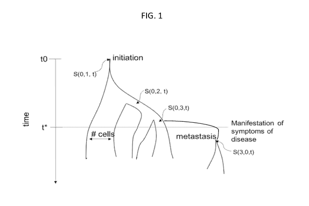
and between patients. An example of this can be seen in FIG. 1, wherein the
lineage of an
initiating tumor cell is shown. The ancestral cell arises at time tO and
genetically distinct sub-
populations (subclones) arise during cell division adding new branches to the
tree. The relative
population size of each subclone is represented by the width of each branch.
Over time three
subclones are generated S(0,1), S(0,2), and S(0,3), each distinguished by its
own set of somatic
alterations. If no reversion mutations occur and there is no recombination,
the mutations can be
represented as a nested tree object (e.g. S(0,1) contained in S(0,3)). A
metastasis S(3,0) is
derived from rapidly expanding subclone S(3,0). In this particular example,
the number of cells
1
CA 03010418 2018-06-29
WO 2017/127742
PCT/US2017/014427
in S(0,2) decreases, the number of cell in S(0,1) remains stable, and the
number of cells in S(0,3)
increases.
[0005] However, somatic genetic heterogeneity generates two challenges in
tumor classification:
tumors undergo rapid evolution through time and, despite arising in the same
tissue in two+
individuals, the tumors can be genetically distinct with different prognosis
and treatment
response.
[0006] Genetic tests that focus on single loci, such as the KRAS mutation
status test, have
demonstrated utility in therapy choice, such as informing the decision whether
to use tyrosine
kinase inhibitors. See, e.g., Plesec et al., Adv. Anal Pathol. (2009).
However, single locus tests
are inadequate to capture the genetic heterogeneity in cancer and therefore
have limited utility in
classification. Some studies have assessed heterogeneity using multi-region
sequencing at one
point in time, while others have tracked pre-defined mutations through time.
Accordingly, there
is a need to develop a method to create a tumor classification signature from
sampling part or all
of the genetic variation in a patient through time.
SUMMARY
[0007] Aspects of the invention relate to methods for tracking patient
health by longitudinally
tracking genetic variants in patients, such that it is possible to provide a
tumor, or mutation,
classification signature. Longitudinal tracking improves the ability to detect
minimal residual
disease (MRD; the small number of cells that remain in the patient after
treatment and/or during
remission) and/or treatment response at an early stage, both of which can help
guide treatment
decisions and guard against missing different intra-/inter-tumor responses in
a patient. Systems
and methods of the invention relate to identifying and tracking the genetic
diversity in individual
tumors and/or patients in order to predict and understand treatment resistance
and to generate
neo-antigens that can be targets of host immune response. These alterations
represent a
discriminating and fundamental signature of the tumor that can ultimately be
used to classify the
tumor and predict progression and treatment efficacy.
[0008] In accordance with methods of the invention, mutation signatures can
be created from
sampling part or all of the genetic variation in a patient through time. These
longitudinal
signatures can then be used to classify patient status against one or more
databases of known
healthy and sick individuals' signatures. As each additional patient's
signature and health status
2
CA 03010418 2018-06-29
WO 2017/127742
PCT/US2017/014427
is refined overtime, the next patient benefits from the improved
discriminatory power of the
classification database.
[0009] According to one embodiment of the invention, the health status of a
patient can be
tracked by creating a mutation signature for a patient. The mutation signature
is determined from
a number of variables including a total number of observed variants in a
nucleic acid sample of
the patient, a sequence context factor for each of the observed variants,
allele frequency of each
of the observed variants, nucleic acid polymer fragment size, inferred DNA
replication timing,
chromatin structure (e.g., open v. closed chromatin structure), DNA
methylation status, inter-
mutation distance, predicted functional consequence of mutations, estimates of
selection (e.g.,
the ratio of non-synonymous to synonymous mutations in a patient), and variant
type
classification. The mutation signature for the patient is then compared to a
reference database
containing mutation signatures of patients with known health statuses, wherein
a diagnosis or
therapy can be determined for the patient. Variant type classifications can
include telomeric
sequence copy number variation, chromosomal instability, translocation,
inversion, insertion,
deletion, loss of heterozygosity, amplification, kataegis, and microsatellite
instability.
[00010] In one aspect of the invention, a longitudinal mutation signature can
be determined for
the patient by comparing a plurality of mutation signatures for the patient
over time to a
reference database, wherein the reference database also contains longitudinal
mutation signatures
of patients with known health statuses, before determining a diagnosis or
therapy. In some
embodiments, a longitudinal mutation signature comprises a first mutation
signature for the
patient from a first time point, and a second mutation signature for the
patient from a second time
point. In some embodiments, the first time point is before a treatment and the
second time point
is after the treatment. In some embodiments, the treatment comprises a tumor
resection surgery.
In some embodiments, the treatment comprises administration of an anti-cancer
therapeutic
agent.
[00011] In another aspect of the invention, a health status for the patient is
obtained and added to
the database along with the mutation signature of the patient. Information
from the patient, such
as age, gender, race, ethnicity, family disease history (e.g., the presence of
Lynch syndrome,
inherited BRCA 1/2 mutations, etc.), weight, body mass index, height, prior
and/or concurrent
infections, environmental exposures, and smoking history can be obtained and
also compared to
the one or more databases of patients with known health statuses. Furthermore,
gene product
3
CA 03010418 2018-06-29
WO 2017/127742
PCT/US2017/014427
levels, such as protein biomarker levels, can also be obtained from the
patient and compared to
levels of patients with known health statuses in the one or more databases of
patients with known
health statuses.
[00012] In order to determine the mutation signature from the nucleic acids of
a patient, a sample
can be obtained from the patient. The sample can comprise, e.g., a tissue
sample, a body fluid, a
cell sample, or a stool sample. In certain embodiments, a sample comprises a
body fluid, such as
whole blood, saliva, tears, sweat, sputum, or urine. In some embodiments, only
a portion of the
whole blood, such as blood plasma or cell free nucleic acid is used. In other
embodiments, the
sample is a tissue sample, such as a formalin-fixed paraffin-embedded (FFPE)
tissue sample, a
fresh frozen (FF) tissue sample, or a combination thereof
[00013] Methods of the invention can also be used to determine intra-tumor or
inter-tumor
heterogeneity from observed variants over time. Furthermore, treatment
efficacy can also be
determined by monitoring observed variants over time before and after
treatment of the patient.
In this manner, the patient can be monitored for minimal residual disease.
[00014] In another embodiment, patient health can be tracked by performing an
assay on nucleic
acid obtained from a patient to determine telomere specific tandem repeat
sequences, creating a
telomere integrity score comprising a frequency distribution of telomere
tandem repeats,
producing a longitudinal trajectory of the telomere integrity score of nucleic
acid obtained from
the patient at two or more time points, comparing the longitudinal trajectory
to a reference
database containing longitudinal trajectories of patients with known health
statuses, and
determining a diagnosis or therapy for the patient.
[00015] In one aspect of the invention, the cell free nucleic acid is obtained
from a body fluid,
such as whole blood, saliva, tears, sweat, sputum, and urine. When the body
fluid is whole
blood, a portion of the whole blood, such as plasma can be used.
[00016] In another aspect of the invention, a health status for the patient is
obtained and added to
the database along with the longitudinal trajectory of the patient.
Information from the patient,
such as age, gender, race, ethnicity, family disease history, weight, body
mass index, height,
prior and/or concurrent infections, environmental exposures, and smoking
history can be
obtained and also compared to the one or more databases of patients with known
health statuses.
Gene product levels, such as protein biomarker levels, can also be obtained
from the patient and
compared to levels of patients with known health statuses in the one or more
databases of
4
CA 03010418 2018-06-29
WO 2017/127742
PCT/US2017/014427
patients with known health statuses. Furthermore, a TERT promoter mutation
profile can be
obtained from the patient and compared to TERT promoter mutation profiles in
one or more
databases of patients with known health statuses.
[00017] The frequency distribution of telomere tandem repeats can also be
normalized. This can
be done by comparing the frequency distribution to a control sequence having
the same
proportions of individual nucleobases as the telomere specific tandem repeat
sequences. The
frequency distribution can also be normalized by comparing the frequency
distribution of
telomere tandem repeats to a reference database of frequency distributions.
[00018] In one aspect of the invention, the assay can be sequencing, such as
whole genome
sequencing. The sequencing can also be targeted sequencing such as targeted
PCR amplification
or hybrid capture using selectable oligonucleotides.
[00019] In another aspect, the telomere specific tandem repeat sequences can
be identified
through alignment to a telomeric reference sequence or analysis of k-mer
frequencies.
BRIEF DESCRIPTION OF THE DRAWINGS
[00020] FIG. 1 shows the lineage of an initiating tumor cell through time.
[00021] FIG. 2 is a chart depicting the depth of sequence coverage of whole
genome sequencing
(WGS) from cfDNA versus tissue biopsy.
[00022] FIG. 3 is a chart depicting whole genome sequencing (WGS) identified
mutations
stratified by triplet sequence context from a metastatic melanoma cancer
patient (PT0001). The
first and second panels show the identified mutations at a first time point
and a second time
point, respectively. The second time point was taken after multiple therapy
regimens. The third
panel shows the relative change in frequency between time points.
[00023] FIG. 4 is a chart showing the allele frequency of validated tumor
mutations in a thoracic
cancer patient before and after resection surgery.
[00024] FIGS. 5A-K are charts showing the allele frequency of 100 somatic
mutations in protein
coding regions over a treatment period in a metastatic melanoma cancer
patient.
[00025] FIG. 6 is a flow chart depicting a method in accordance with an
embodiment of the
invention.
CA 03010418 2018-06-29
WO 2017/127742
PCT/US2017/014427
[00026] FIG. 7 is a chart showing the empirical distribution of the number of
whole genome
sequencing reads from cfDNA containing repeated telomeric sequences from a
melanoma cancer
patient PT0001.
[00027] FIG. 8 is a diagram of a system in accordance with embodiments of the
invention.
[00028] FIG. 9 is a graph showing somatic variant allele frequencies measured
in a colorectal
cancer (CRC) patient before and after surgical tumor excision.
[00029] FIG. 10 is a graph showing somatic variant allele frequencies measured
in a CRC patient
before and after surgical tumor excision.
[00030] FIG. 11 is a graph showing somatic variant allele frequencies measured
in a CRC patient
before and after surgical tumor excision. The tree on the right hand side
represents a potential
underlying lineage of cancer cells in the patient; the tree is consistent with
allele frequency
trajectories under surgery.
[00031] FIG. 12 is a collection of bar graphs that show allele frequencies of
microsatellite repeats
from cfDNA sequencing from different patients.
[00032] FIG. 13 is a collection of bar graphs that show allele frequencies of
microsatellite repeats
from cfDNA and genomic DNA sequencing for various sample types including
cancer patients
and synthetic controls using WGS and targeted sequencing.
[00033] FIG. 14 is a collection of bioanalyzer traces that show fragment size
of extracted cfDNA
in base pairs.
[00034] FIG. 15 is a collection of bioanalyzer traces that show cfDNA library
fragment size in
base pairs prior to PCR amplification.
[00035] FIG. 16 is a collection of bioanalyzer traces that show cfDNA library
fragment size in
base pairs after 8 cycles of PCR amplification.
[00036] FIG. 17 is a collection of bioanalyzer traces that show cfDNA library
fragment size in
base pairs after 12 cycles of PCR and clean-up.
[00037] FIG. 18 is a time-course representation of a time course of disease
progression for the
patient, and shows treatment, observations and sample collection time points.
[00038] FIG. 19A is a panel of pileup views of sequencing reads from PT0001 at
the core
promoter of telomerase reverse transcriptase (TERT). Four panels (top to
bottom): white blood
cell derived genomic DNA sequencing, cfDNA sequencing at time point 1, cfDNA
time point 2
6
CA 03010418 2018-06-29
WO 2017/127742
PCT/US2017/014427
sequencing, and tumor biopsy sequencing. Between dashed vertical lines A
letters represent the
reverse-complement of mutant alleles copies of a known activating C>T
mutation.
[00039] FIG. 19B is a table that summarizes the data in FIG. 19A, showing the
read counts at
chr5: 129,250 for the indicated samples.
[00040] FIGS. 20A-C provide summary tables of colorectal cancer patient
information and
predicted disease recurrence from cfDNA analysis.
DETAILED DESCRIPTION
[00041] Methods of the invention involve longitudinally tracking multiple
somatic alterations,
such that it may be possible to guard against missing different intra-/inter-
tumor responses in a
patient and improve the ability to detect minimal residual disease and/or
treatment response. This
can be accomplished through the creation of a mutation signature or signatures
and/or the
creation of a telomere integrity score determined from nucleic acid obtained
from a patient, both
of which can be longitudinally tracked.
[00042] The methods initially involve obtaining a sample, e.g., a tissue or
body fluid that is
suspected to include a cancer-associated gene or gene product. The sample may
be collected in
any clinically acceptable manner. A tissue is a mass of connected cells and/or
extracellular
matrix material, e.g., skin tissue, hair, nails, endometrial tissue, nasal
passage tissue, CNS tissue,
neural tissue, eye tissue, liver tissue, kidney tissue, placental tissue,
mammary gland tissue,
placental tissue, gastrointestinal tissue, musculoskeletal tissue,
genitourinary tissue, bone
marrow, and the like, derived from, for example, a human or other mammal and
includes the
connecting material and the liquid material in association with the cells
and/or tissues. The tissue
can be prepared and provided as any one of the tissue samples types known in
the art, such as,
for example and not limitation, formalin-fixed paraffin-embedded (FFPE) and
fresh frozen (FF)
tissue samples.
[00043] A body fluid is a liquid material derived from, for example, a human
or other mammal.
Such body fluids include, but are not limited to, mucous, blood, plasma,
serum, serum
derivatives, bile, maternal blood, phlegm, saliva, sweat, tears, sputum,
amniotic fluid, menstrual
fluid, urine, and cerebrospinal fluid (C SF), such as lumbar or ventricular
CSF. A sample may
also be a fine needle aspirate or biopsied tissue. A sample also may be media
containing cells or
biological material. A sample may also be a blood clot, for example, a blood
clot that has been
7
CA 03010418 2018-06-29
WO 2017/127742
PCT/US2017/014427
obtained from whole blood after the serum has been removed. A sample may also
be stool. In
certain embodiments, the sample is drawn whole blood. In one aspect, only a
portion of whole
blood is used, such as plasma, red blood cells, white blood cells, and
platelets.
[00044] The sample can include nucleic acid not only from the subject from
which the sample
was taken, but also from other species such as viral DNA/RNA. Nucleic acid can
be extracted
from the sample according to methods known in the art. See for example,
Maniatis, et al.,
Molecular Cloning: A Laboratory Manual, Cold Spring Harbor, N.Y., pp. 280-281,
1982, the
contents of which are incorporated by reference herein in their entirety. In
certain embodiments,
cell free nucleic acid is extracted from the sample.
[00045] In some embodiments, cell free DNA (cfDNA) is extracted from the
sample. Cell free
DNA are short base nuclear-derived DNA fragments present in several bodily
fluids (e.g.
plasma, stool, urine). See, e.g., Mouliere and Rosenfeld, PNAS 112(11): 3178-
3179 (Mar 2015);
Jiang et al., PNAS (Mar 2015); and Mouliere et al., Mol Oncol, 8(5):927-41
(2014). Tumor
derived circulating tumor DNA (ctDNA) constitutes a minority population of
cfDNA, in some
embodiments, varying up to about 50%. In some embodiments, ctDNA varies
depending on
tumor stage and tumor type. In some embodiments, ctDNA varies from about
0.001% up to
about 30%, such as about 0.01% up to about 20%, such as about 0.01% up to
about 10%. The
covariates of ctDNA are not fully understood, but appear to be positively
correlated with tumor
type, tumor size, and tumor stage. E.g., Bettegowda et al, Sci Trans Med,
2014; Newmann et al,
Nat Med, 2014. Despite the challenges associated with the low population of
ctDNA in cfDNA,
tumor variants have been identified in ctDNA across a wide span of cancers.
E.g., Bettegowda et
al, Sci Trans Med, 2014. Furthermore, analysis of cfDNA versus tumor biopsy is
less invasive
and methods for analyzing, such as sequencing, enable the identification of
sub-clonal
heterogeneity. Analysis of cfDNA also provides for more uniform genome-wide
sequencing
coverage than with a tissue tumor biopsy, as shown in FIG. 2.
[00046] An exemplary procedure for preparing nucleic acid from blood follows.
Blood may be
collected in 10m1 EDTA tubes (available, for example, from Becton Dickinson).
Streck cfDNA
tubes (Streck, Inc., Omaha, Nebraska) can be used to minimize contamination
through chemical
fixation of nucleated cells but little contamination from genomic DNA is
observed when samples
are processed within 2 hours or less as in some embodiments. Beginning with a
blood sample,
plasma may be extracted by centrifugation at 3000rpm for 10 minutes at room
temperature minus
8
CA 03010418 2018-06-29
WO 2017/127742
PCT/US2017/014427
brake. Plasma may then be transferred to 1.5m1 tubes in lml aliquots and
centrifuged again at
7000rpm for 10 minutes at room temperature. Supernatants can then be
transferred to new 1.5m1
tubes. At this stage, samples can be stored at -80 C. In certain embodiments,
samples can be
stored at the plasma stage for later processing as plasma may be more stable
than storing
extracted cfDNA.
[00047] Plasma DNA can be extracted using any suitable technique. For example,
in some
embodiments, plasma DNA can be extracted using one or more commercially
available assays,
for example, the Qiagen QIAmp Circulating Nucleic Acid kit (Qiagen N.Y., Venlo
Netherlands).
In certain embodiments, the following modified elution strategy may be used.
DNA may be
extracted using the Qiagen QIAmp circulating nucleic acid kit following the
manufacturer's
instructions (maximum amount of plasma allowed per column is 5m1). If cfDNA is
being
extracted from plasma where the blood was collected in Streck tubes, the
reaction time with
proteinase K may be doubled from 30 min to 60 min. Preferably, as large a
volume as possible
should be used (i.e., 5mL). In various embodiments, a two-step elution may be
used to maximize
cfDNA yield. First, DNA can be eluted using 30 1 of buffer AVE for each
column. A minimal
amount of buffer necessary to completely cover the membrane can be used in
elution in order to
increase cfDNA concentration. By decreasing dilution with a small amount of
buffer,
downstream desiccation of samples can be avoided to prevent melting of double
stranded DNA
or material loss. Subsequently, about 30 1 of buffer for each column can be
eluted. In some
embodiments, a second elution may be used to increase DNA yield.
[00048] In certain embodiments, a genomic sample is collected from a subject
followed by
enrichment for genetic regions or genetic fragments of interest. For example,
in some
embodiments, a sample can be enriched by hybridization to a nucleotide array
comprising
cancer-related genes or gene fragments of interest. In some embodiments, a
sample can be
enriched for genes of interest (e.g., cancer-associated genes) using other
methods known in the
art, such as hybrid capture. See, for example, Lapidus (U.S. patent number
7,666,593), the
content of which is incorporated by reference herein in its entirety. In one
hybrid capture
method, a solution-based hybridization method is used that includes the use of
biotinylated
oligonucleotides and streptavidin coated magnetic beads. See, e.g., Duncavage
et al., J Mol
Diagn. 13(3): 325-333 (2011); and Newman et al., Nat Med. 20(5): 548-554
(2014).
9
CA 03010418 2018-06-29
WO 2017/127742
PCT/US2017/014427
[00049] Isolation of nucleic acid from a sample in accordance with the methods
of the invention
can be done according to any method known in the art. For example, RNA may be
isolated from
eukaryotic cells by procedures that involve lysis of the cells and
denaturation of the proteins
contained therein. Tissue of interest includes gametic cells, gonadal tissue,
endometrial tissue,
fertilized embryos, and placenta. RNA may be isolated from fluids of interest
by procedures that
involve denaturation of the proteins contained therein. Fluids of interest
include those fluids
listed above. Additional steps may be employed to remove DNA. Cell lysis may
be
accomplished with a nonionic detergent, followed by microcentrifugation to
remove the nuclei
and hence the bulk of the cellular DNA. In one embodiment, RNA is extracted
from cells of the
various types of interest using guanidinium thiocyanate lysis followed by CsC1
centrifugation to
separate the RNA from DNA (Chirgwin et al., Biochemistry 18:5294-5299 (1979)).
Poly(A)+
RNA is selected by selection with oligo-dT cellulose (see Sambrook et al.,
MOLECULAR
CLONING--A LABORATORY MANUAL (2ND ED.), Vols. 1-3, Cold Spring Harbor
Laboratory, Cold Spring Harbor, N.Y. (1989). Alternatively, separation of RNA
from DNA can
be accomplished by organic extraction, for example, with hot phenol or phenol/
chloroform/
isoamyl alcohol. If desired, RNase inhibitors may be added to the lysis
buffer. Likewise, for
certain cell types, it may be desirable to add a protein
denaturation/digestion step to the protocol.
[00050] Once the nucleic acid has been extracted, it can be assayed to
determine genetic variants.
The terms "variants", "variations", and "mutations" as used interchangeably
herein refer to
genetic sequences that are different from a wild type or control sequence. Any
assay known in
the art may be used to determine presence or absence of a genetic variation.
Conventional
methods can be used, such as those employed to make and use nucleic acid
arrays, amplification
primers, hybridization probes, and can be found in standard laboratory manuals
such as: Genome
Analysis: A Laboratory Manual Series (Vols. I-IV), Cold Spring Harbor
Laboratory Press; PCR
Primer: A Laboratory Manual, Cold Spring Harbor Laboratory Press; and
Sambrook, J et al.,
(2001) Molecular Cloning: A Laboratory Manual, 2nd ed. (Vols. 1-3), Cold
Spring Harbor
Laboratory Press. Custom nucleic acid arrays are commercially available from,
e.g., Affymetrix
(Santa Clara, CA), Applied Biosystems (Foster City, CA), and Agilent
Technologies (Santa
Clara, CA).
[00051] In some embodiments of the invention, nucleic acids are sequenced in
order to detect
variants (i.e., mutations) in the nucleic acid. The nucleic acid can include a
plurality of nucleic
CA 03010418 2018-06-29
WO 2017/127742
PCT/US2017/014427
acids derived from a plurality of genetic elements. Methods of detecting
sequence variants are
known in the art, and sequence variants can be detected by any sequencing
method known in the
art, e.g., ensemble sequencing (wherein consensus sequencing is conducted by
integrating
sequencing/PCR errors across PCR duplicates) or single molecule sequencing.
[00052] Sequencing may be by any method known in the art. DNA sequencing
techniques include
classic dideoxy sequencing reactions (Sanger method) using labeled terminators
or primers and
gel separation in slab or capillary, sequencing by synthesis using reversibly
terminated labeled
nucleotides, pyrosequencing, 454 sequencing, allele specific hybridization to
a library of labeled
oligonucleotide probes, sequencing by synthesis using allele specific
hybridization to a library of
labeled clones that is followed by ligation, real time monitoring of the
incorporation of labeled
nucleotides during a polymerization step, polony sequencing, and SOLiD
sequencing.
Sequencing of separated molecules has more recently been demonstrated by
sequential or single
extension reactions using polymerases or ligases as well as by single or
sequential differential
hybridizations with libraries of probes.
[00053] One conventional method to perform sequencing is by chain termination
and gel
separation, as described by Sanger et al., Proc Natl. Acad. Sci. U S A,
74(12): 5463 67 (1977).
Another conventional sequencing method involves chemical degradation of
nucleic acid
fragments. See, Maxam et al., Proc. Natl. Acad. Sci., 74: 560 564 (1977).
Methods have also
been developed based upon sequencing by hybridization. See, e.g., Harris et
al., (U.S. patent
application number 2009/0156412). The content of each reference is
incorporated by reference
herein in its entirety.
[00054] A sequencing technique that can be used in the methods of the provided
invention
includes, for example, Helicos True Single Molecule Sequencing (tSMS) (Harris
T. D. et al.
(2008) Science 320:106-109). Further description of tSMS is shown for example
in Lapidus et al.
(U.S. patent number 7,169,560), Lapidus et al. (U.S. patent application number
2009/0191565),
Quake et al. (U.S. patent number 6,818,395), Harris (U.S. patent number
7,282,337), Quake et al.
(U.S. patent application number 2002/0164629), and Braslaysky, et al., PNAS
(USA), 100:
3960-3964 (2003), the contents of each of these references is incorporated by
reference herein in
its entirety.
[00055] Another example of a DNA sequencing technique that can be used in the
methods of the
provided invention is 454 sequencing (Roche) (Margulies, M et al. 2005,
Nature, 437, 376-380).
11
CA 03010418 2018-06-29
WO 2017/127742
PCT/US2017/014427
Another example of a DNA sequencing technique that can be used in the methods
of the
provided invention is SOLiD technology (Applied Biosystems). Another example
of a DNA
sequencing technique that can be used in the methods of the provided invention
is Ion Torrent
sequencing (U.S. patent application numbers 2009/0026082, 2009/0127589,
2010/0035252,
2010/0137143, 2010/0188073, 2010/0197507, 2010/0282617, 2010/0300559),
2010/0300895,
2010/0301398, and 2010/0304982), the content of each of which is incorporated
by reference
herein in its entirety.
[00056] In some embodiments, the sequencing technology is Illumina sequencing.
Illumina
sequencing is based on the amplification of DNA on a solid surface using fold-
back PCR and
anchored primers. Genomic DNA can be fragmented, or in the case of cfDNA,
fragmentation is
not needed due to the already short fragments. Adapters are ligated to the 5'
and 3' ends of the
fragments. DNA fragments that are attached to the surface of flow cell
channels are extended and
bridge amplified. The fragments become double stranded, and the double
stranded molecules are
denatured. Multiple cycles of the solid-phase amplification followed by
denaturation can create
several million clusters of approximately 1,000 copies of single-stranded DNA
molecules of the
same template in each channel of the flow cell. Primers, DNA polymerase and
four fluorophore-
labeled, reversibly terminating nucleotides are used to perform sequential
sequencing. After
nucleotide incorporation, a laser is used to excite the fluorophores, and an
image is captured and
the identity of the first base is recorded. The 3' terminators and
fluorophores from each
incorporated base are removed and the incorporation, detection and
identification steps are
repeated.
[00057] Another example of a sequencing technology that can be used in the
methods of the
provided invention includes the single molecule, real-time (SMRT) technology
of Pacific
Biosciences. Yet another example of a sequencing technique that can be used in
the methods of
the provided invention is nanopore sequencing (Soni G V and Meller A. (2007)
Clin Chem 53:
1996-2001). Another example of a sequencing technique that can be used in the
methods of the
provided invention involves using a chemical-sensitive field effect transistor
(chemFET) array to
sequence DNA (for example, as described in US Patent Application Publication
No.
20090026082). Another example of a sequencing technique that can be used in
the methods of
the provided invention involves using a electron microscope (Moudrianakis E.
N. and Beer M.
Proc Natl Acad Sci USA. 1965 March; 53:564-71).
12
CA 03010418 2018-06-29
WO 2017/127742
PCT/US2017/014427
[00058] If the nucleic acid from the sample is degraded or only a minimal
amount of nucleic acid
can be obtained from the sample, PCR can be performed on the nucleic acid in
order to obtain a
sufficient amount of nucleic acid for sequencing (See, e.g., Mullis et al.
U.S. patent number
4,683,195, the contents of which are incorporated by reference herein in its
entirety).
[00059] While the combination of the potential sequence of genetic variants
and their sequence of
occurrence, in addition to their relative frequency allows for essentially
infinite combinations,
the creation of a mutation signature is made practical by establishing a
framework within which
to classify the variants.
[00060] In some embodiments, variations are measured at a single point in time
to determine a
mutation signature for a patient. In some embodiments, variations are
longitudinally tracked over
time to facilitate the generation of a longitudinal mutation signature for a
patient. For example, in
some embodiments, two or more samples can be collected from a patient over
time, and the
collected samples can be used to generate a longitudinal mutation signature
for the patient. In
some embodiments, a first sample is collected at a first time point and a
second sample is
collected at a second time point. Research has demonstrated that cfDNA can
have a clearance
time ranging from about 15 mins up to several hours, depending on the rate of
clearance (Forte
VA, et al., The potential for liquid biopsies in the precision medical
treatment of breast
cancer, Cancer Biology &Medicine. 2016; 13(1): 19-40. doi :10.28092/j .i
ssn.2095-
3941.2016.0007. Accordingly, in some embodiments, the first and second time
points are
separated by an amount of time that ranges from about 15 minutes up to about
25 years, such as
about 30 minutes, such as about 1, 2, 3, 4, 5, 6, 7, 8, 9, 10, 11, 12, 13, 14,
15, 16, 17, 18, 19,20,
21, 22, 23, or about 24 hours, such as about 1, 2, 3, 4, 5, 10, 15, 20, 25 or
about 30 days, or such
as about 1, 2, 3, 4, 5, 6, 7, 8,9, 10, 11, or 12 months, or such as about 1,
1.5, 2, 2.5, 3, 3.5, 4, 4.5,
5, 5.5, 6, 6.5, 7, 7.5, 8, 8.5, 9, 9.5, 10, 10.5, 11, 11.5, 12, 12.5, 13,
13.5, 14, 14.5, 15, 15.5, 16,
16.5, 17, 17.5, 18, 18.5, 19, 19.5, 20, 20.5, 21, 21.5, 22, 22.5, 23, 23.5,
24, 24.5 or about 25
years.
[00061] In some embodiments, the first time point is before the inception of
treatment, and the
second time point is after the inception of treatment. In some embodiments,
the first time point is
before the inception of treatment, and the second time point is after the
completion of treatment.
In some embodiments, the first time point is before a tumor resection surgery,
and the second
time point is after the tumor resection surgery. In some embodiments, the
first time point is
13
CA 03010418 2018-06-29
WO 2017/127742
PCT/US2017/014427
before a tumor resection surgery, and the second time point is about 5, 10,
15, 20, 25, or 30 days
after the tumor resection surgery. In some embodiments, the first time point
is before a tumor
resection surgery, and the second time point is about 1, 2, 3, 4, 5, 6, 7, 8,
9, 10, 11 or 12 months
after the tumor resection surgery. In some embodiments, the first time point
is before a tumor
resection surgery, and the second time point is about 1, 2, 3, 4, 5, 6, 7, 8,
9, or about 10 years
after the tumor resection surgery.
[00062] In some embodiments, one or more changes of a mutational signature
before and after
administration of a treatment can be used to identify patient populations that
respond better or
worse to the treatment, according to a mutational signature classification. As
such, tracking
mutational signatures over time can be used to identify cases where therapy is
ineffective, and to
identify cases where a change in therapeutic intervention may be needed (e.g.,
administration of
a different therapy may be needed).
[00063] In certain embodiments, a longitudinal mutation signature comprises a
plurality of
different time points, wherein a first time point is before the inception of
treatment, and a
plurality of additional time points are collected at specific time intervals
following treatment,
e.g., about 1, 2, 3, 4, 5, 6, 7, 8, 9, 10, 11 or 12 months following
treatment. In some
embodiments, a treatment comprises a tumor resection surgery with curative
intent. In some
embodiments, a treatment comprises administration of a therapeutic agent. In
some
embodiments, a therapeutic agent is an anti-cancer therapeutic agent.
[00064] In some embodiments, a longitudinal mutation signature comprises a
plurality of different
time points, wherein a first time point is before a tumor resection surgery
with curative intent,
and a plurality of additional time points are collected at specific time
points following the tumor
resection surgery, e.g., about 1, 2, 3, 4, 5, 6, 7, 8, 9, 10, 11 or 12 months
or more following tumor
resection surgery, such as about 1, 2, 3, 4, 5, 6, 7, 8, 9, or 10 years
following tumor resection
surgery. In some embodiments, a longitudinal mutation signature comprises a
plurality of
different time points, wherein a first time point is before administration of
an anti-cancer
therapeutic agent, and a plurality of additional time points are collected at
specific time points
following the administration of the anti-cancer therapeutic agent, e.g., about
1, 2, 3, 4, 5, 6, 7, 8,
9, 10, 11 or 12 months or more following administration of the anti-cancer
therapeutic agent,
such as about 1, 2, 3, 4, 5, 6, 7, 8, 9, or 10 years following administration
of the anti-cancer
therapeutic agent.
14
CA 03010418 2018-06-29
WO 2017/127742
PCT/US2017/014427
[00065] Aspects of the methods include collecting mutation signatures over
time from
asymptomatic patients to facilitate early detection of cancer and/or to
predict risk levels
associated with developing cancer. In some embodiments, a mutational signature
is built up over
multiple time points for an asymptomatic patient. The mutational signature can
be used to
estimate cancer or disease risk by, e.g., determining a status of certain
genetic markers (e.g.,
BRCA germline status and somatic status) and/or the presence or absence of a
cancer (e.g., a
somatic mutation signature that is consistent with the presence or absence of
a cancer) and/or a
molecular classification of a cancer (e.g., a somatic signature coupled with a
germline status
determination).
[00066] Variables used in the creation of a mutation signature in accordance
with embodiments of
the invention include, but are not limited to, the total number of observed
genetic variants, or
alterations, the sequence context in which the variants occur, the prevalence
of the mutation
relative to other somatic mutations or to the germline genome, the type of
genetic alteration, one
or more fragmentation patterns of cfDNA fragments (e.g., a cfDNA fragment size
distribution
pattern, and/or the location of fragment start and end points), chromatin
structure (e.g., open v.
closed chromatin structure), methylation status, and inter-mutation distance
(e.g., clustering of
mutations.
[00067]
Sequence context refers to the nucleotides surrounding the mutation. See,
e.g., Sung et
al., "Asymmetric Context-Dependent Mutation Patterns Revealed Through Mutation-
Accumulation Experiments," Mol. Biol. Evol., Apr 2015. By including the
sequence context in
which the variant occurs, mutation signatures with the same substitutions, but
within different
sequence context can be differentiated. For example, the genetic signature
associated with UV
damage evidences an increased number of C>T mutations with triplet context
dependence (e.g.,
the substitution and the nucleotides 3' and 5' to the mutation). See
Alexandrov et al. 2013. In
some embodiments, the sequence context can include at least one, two, three,
four, five, six,
seven, eight, nine, ten, or more nucleotides on either or both of the
positions 3' and 5' to the
mutation. In some embodiments, a sequence context includes at least one
nucleotide 3' and at
least one nucleotide 5' to the mutation. In some embodiments, a mutation
signature can take into
account a strand on which a mutation occurs. For example, in some embodiments,
a mutation can
be more prevalent on a transcribed strand versus a non-transcribed strand. See
Alexandrov at
page 6.
CA 03010418 2018-06-29
WO 2017/127742
PCT/US2017/014427
[00068] Longitudinal trajectories can be analyzed as the evolution of
alterations stratified by
sequence context. For example, FIG. 3 displays the dynamic melanoma mutation
signature from
cfDNA using whole genome sequencing (WGS) applied to a metastatic melanoma
patient with a
targetable somatic BRAF mutation V600R. Using WGS, mutations were identified
and stratified
by triplet context (Ntimepoint1-24377, Nttmepoint2-35036). Samples were taken
and analyzed using
WGS at a first timepoint prior to a course of treatment, as shown in the first
panel, and then
again at a second timepoint subsequent to a course of treatment, as shown in
the second panel
(95% Cl, bootstrapping). The observed profile was concordant with Type 2
melanoma reported
by Alexandrov et al (2013) (cited herein) and is compatible with UV induced
DNA damage. The
profile exhibits abundant C>T mutations, as shown in the C>T column of FIG. 3.
The relative
change in frequency between time points was then calculated, as shown in the
third panel, with
the stars representing significant changes (p <0.05, FET). As can be seen,
over the course of one
year of treatment with vemurafenib (targeting BRAF) and ipilimumab (anti-CTLA4
checkpoint
inhibitor), a systematic and consistent decrease in T>C mutations is observed
in a patient
evidencing with melanoma. The systemic and consistent change in the relative
frequency of this
mutation suggests potential differential response between sub-clones and or
metastases in the
patient. See, e.g., Venn et al., "Genome-wide cfDNA Sequencing of Melanoma
Progression,"
presented at the BioTrinity 2015 Conference in London on May 12, 2015, herein
incorporated by
reference in its entirety.
[00069] Furthermore, the prevalence of mutations is highly variable between
and even within
cancer types. For instance, certain childhood cancers are associated with the
fewest mutations
and cancers related to chronic exposures that cause mutations are associated
with the highest
number of mutations. See, e.g., Alexandrov at page 221. Furthermore, the
prevalence of one
mutation is variable with respect to other somatic mutations within a type of
cancer. In
accordance with the methods described herein, the prevalence of a mutation is
measured by the
variant allele frequency. The frequency, or prevalence, can then be compared
to other mutations
or to the germline genome (e.g., the ratio of circulating tumor DNA (ctDNA) to
cell free DNA
(cfDNA)).
[00070]
Variant allele frequency is the relative frequency of an allele at a
particular locus in a
population. For example, to calculate an allele frequency across a population
of individuals, one
would calculate the fraction of all occurrences of an allele in i chromosomes
in the population of
16
CA 03010418 2018-06-29
WO 2017/127742
PCT/US2017/014427
N individuals with ploidy n, and the total number of chromosome copies across
the population,
represented by the following equation: Allele frequency = i/(nN).
[00071] For the frequency of a somatic mutation allele within an individual,
the frequency is
calculated as a quotient of the observed mutant allele copies (dividend) by
the non-mutant allele
copies in the individual. In some embodiments, the observed frequencies can be
corrected for
ploidy, noise-rates, and/or sub-clonal complexity.
[00072] FIGS. 5A-K shows the allele frequency trajectories of 100 somatic
mutations through the
course of treatment. The variants were tracked using amplicon-based sequencing
of cfDNA
samples on PGM (Life Tech). Loci were assigned one of 8 clusters based on
hierarchical
clustering (Euclidean distance). Treatment cycles of vemurafenib (first two
rectangles in the
"Treatment" row, located above the x-axis) and ipilimumab (third rectangle in
the "Treatment"
row, located above the x-axis) are indicated in FIGS. 5A-K. Also shown are the
tumor diameters
for prevascular lymph node ("Prevascular LN" row, located above the x-axis)
and paratracheal
lymph node ("Paratracheal LN" row. located above the x-axis) obtained using CT
imaging. Here,
by tracking the allelic frequencies of the somatic mutations, it would have
been possible to see
early on that treatment with ipilimumab was ineffective. An increase in
allelic frequencies was
detectable 88 days before the third CT imaging scan. Variant allele frequency
trajectory was
highly correlated (86% Pearson correlation) with aggregated imaged lymph node
diameter.
[00073] Furthermore, the type of genetic variation will also contribute to the
classification of the
tumor. Examples of genetic variations that can be used to classify tumors
include, but are not
limited to, telemoric sequence copy number status (explained in further detail
below), single
nucleotide polymorphism(s), chromosome instability, translocations,
inversions, insertions,
deletions, loss of heterozygosity, amplifications, kateagis (hyper mutation
localized to small
genomic regions; See Alexandrov), and microsatellite instability.
[00074] In addition to observed genetic variants, the classification can
also include the
determination of one or more gene product biomarkers, which provide different
transformations
of the underlying genomic information. A biomarker generally refers to a
molecule that acts as
an indicator of a biological state. In some embodiments, a gene product can be
an RNA molecule
or a protein.
[00075] Protein biomarkers in accordance with embodiments of the invention can
include those
proteins involved in oncogenesis, angiogenesis, development, differentiation,
proliferation,
17
CA 03010418 2018-06-29
WO 2017/127742
PCT/US2017/014427
apoptosis, hematopoiesis, immune and hormonal responses, cell signaling,
nucleotide function,
hydrolysis, cellular homing, cell cycle and structure, the acute phase
response and hormonal
control. See e.g., Polanski and Anderson, "A List of Candidate Cancer
Biomarkers for Targeted
Proteomics," Biomark Insights, 1:1-48 (2007). Examples of cancer protein
biomarkers approved
by the FDA and encompassed by the present invention include, but are not
limited to, CEA
(carcinicenbryonic antigens); Her-2/neu; Bladder Tumor Antigen; Thyroglobulin;
Alpha-
fetoprotein; PSA; CA 125; CA 19.9; CA 15.3; leptin, prolactic, osteopontin and
IGF-II; CD98,
fascin, sPIgR, and 14-3-3 eta; Troponin I, and B-type natriuretic peptide. See
Id; and Dawson et
al., N Engl J Med 368:1199/1209 (March 2013).
[00076] Any assay known in the art can be used to analyze a gene product. In
certain
embodiments, an assay involves determining an amount of a gene product and
comparing the
determined amount to a reference. In one embodiment, a level of one or more
protein biomarkers
is obtained from a sample from the patient. The level obtained from the
patient is then compared
to a database of patient information of patients with known health statuses.
[00077] Methods of detecting levels of gene products (e.g., RNA or protein)
are known in the art.
Commonly used methods known in the art for the quantification of mRNA
expression in a
sample include northern blotting and in situ hybridization (Parker & Barnes,
Methods in
Molecular Biology 106:247 283 (1999), the contents of which are incorporated
by reference
herein in their entirety); RNAse protection assays (Hod, Biotechniques 13:852
854 (1992), the
contents of which are incorporated by reference herein in their entirety); and
PCR-based
methods, such as reverse transcription polymerase chain reaction (RT-PCR)
(Weis et al., Trends
in Genetics 8:263 264 (1992), the contents of which are incorporated by
reference herein in their
entirety). Alternatively, antibodies can be employed that can recognize
specific duplexes,
including RNA duplexes, DNA-RNA hybrid duplexes, or DNA-protein duplexes.
Other methods
known in the art for measuring gene expression (e.g., RNA or protein amounts)
are shown in
Yeatman et al. (U.S. patent application number 2006/0195269), the content of
which is hereby
incorporated by reference in its entirety.
[00078] The terms "differentially expressed gene" or "differential gene
expression" refer to a
gene whose expression is activated to a higher or lower level in a subject
suffering from a
disease, such as cancer, relative to its expression in a normal or control
subject. These terms also
include genes whose expression is activated to a higher or lower level at
different stages of the
18
CA 03010418 2018-06-29
WO 2017/127742
PCT/US2017/014427
same disease. It is also understood that a differentially expressed gene may
be either activated or
inhibited at the nucleic acid level or protein level, or may be subject to
alternative splicing to
result in a different polypeptide product. Such differences may be evidenced
by a change in
mRNA levels, surface expression, secretion or other partitioning of a
polypeptide, for example.
[00079] Differential gene expression can include a comparison of expression
between two or
more genes or their gene products, or a comparison of the ratios of the
expression between two
or more genes or their gene products, or even a comparison of two differently
processed products
of the same gene, which differ between normal subjects and subjects suffering
from a disorder,
such as infertility, or between various stages of the same disorder.
Differential expression
includes both quantitative, as well as qualitative, differences in the
temporal or cellular
expression pattern in a gene or its expression products. Differential gene
expression (increases
and decreases in expression) is based upon percent or fold changes over
expression in normal
cells. Increases may be of 1, 5, 10, 20, 30, 40, 50, 60, 70, 80, 90, 100, 120,
140, 160, 180, or
200% relative to expression levels in normal cells. Alternatively, fold
increases may be of 1, 1.5,
2, 2.5, 3, 3.5, 4, 4.5, 5, 5.5, 6, 6.5, 7, 7.5, 8, 8.5, 9, 9.5, or 10 fold
over expression levels in
normal cells. Decreases may be of 1, 5, 10, 20, 30, 40, 50, 55, 60, 65, 70,
75, 80, 82, 84, 86, 88,
90, 92, 94, 96, 98, 99 or 100% relative to expression levels in normal cells.
[00080] In certain embodiments, reverse transcriptase PCR (RT-PCR) is used to
measure gene
expression. RT-PCR is a quantitative method that can be used to compare mRNA
levels in
different sample populations to characterize patterns of gene expression, to
discriminate between
closely related mRNAs, and to analyze RNA structure.
[00081] In another embodiment, a MassARRAY-based gene expression profiling
method is used
to measure gene expression. For further details see, e.g. Ding and Cantor,
Proc. Natl. Acad. Sci.
USA 100:3059 3064 (2003). Further PCR-based techniques include, for example,
differential
display (Liang and Pardee, Science 257:967 971 (1992)); amplified fragment
length
polymorphism (iAFLP) (Kawamoto et al., Genome Res. 12:1305 1312 (1999));
BeadArrayTM
technology (Illumina, San Diego, Calif; Oliphant et al., Discovery of Markers
for Disease
(Supplement to Biotechniques), June 2002; Ferguson et al., Analytical
Chemistry 72:5618
(2000)); BeadsArray for Detection of Gene Expression (BADGE), using the
commercially
available Luminex100 LabMAP system and multiple color-coded microspheres
(Luminex Corp.,
Austin, Tex.) in a rapid assay for gene expression (Yang et al., Genome Res.
11:1888 1898
19
CA 03010418 2018-06-29
WO 2017/127742
PCT/US2017/014427
(2001)); and high coverage expression profiling (HiCEP) analysis (Fukumura et
al., Nucl. Acids.
Res. 31(16) e94 (2003)). The contents of each of which are incorporated by
reference herein in
their entirety.
[00082] In certain embodiments, differential gene expression can also be
identified, or confirmed
using a microarray technique. In this method, polynucleotide sequences of
interest (including
cDNAs and oligonucleotides) are plated, or arrayed, on a microchip substrate.
The arrayed
sequences are then hybridized with specific DNA probes from cells or tissues
of interest.
Methods for making microarrays and determining gene product expression (e.g.,
RNA or
protein) are shown in Yeatman et al. (U.S. patent application number
2006/0195269), the content
of which is incorporated by reference herein in its entirety.
[00083] Alternatively, protein levels can be determined by constructing an
antibody microarray
in which binding sites comprise immobilized, preferably monoclonal, antibodies
specific to a
plurality of protein species encoded by the cell genome. Preferably,
antibodies are present for a
substantial fraction of the proteins of interest. Methods for making
monoclonal antibodies are
well known (see, e.g., Harlow and Lane, 1988, ANTIBODIES: A LABORATORY MANUAL,
Cold Spring Harbor, N.Y., which is incorporated in its entirety for all
purposes).
[00084] Alternatively, levels of transcripts of marker genes in a number of
tissue specimens may
be characterized using a "tissue array" (Kononen et al., Nat. Med 4(7):844-7
(1998)). In a tissue
array, multiple tissue samples are assessed on the same microarray. The arrays
allow in situ
detection of RNA and protein levels; consecutive sections allow the analysis
of multiple samples
simultaneously.
[00085] In some embodiments, Serial Analysis of Gene Expression (SAGE) is used
to measure
gene expression. For more details see, e.g. Velculescu et al., Science 270:484
487 (1995); and
Velculescu et al., Cell 88:243 51 (1997, the contents of each of which are
incorporated by
reference herein in their entirety).
[00086] In some embodiments, Massively Parallel Signature Sequencing (NIPSS)
is used to
measure gene expression. See e.g., Brenner et al., Nature Biotechnology 18:630
634 (2000).
[00087] Immunohistochemistry methods are also suitable for detecting the
expression levels of
the gene products of the present invention. Thus, antibodies (monoclonal or
polyclonal) or
antisera, such as polyclonal antisera, specific for each marker are used to
detect expression. The
antibodies can be detected by direct labeling of the antibodies themselves,
for example, with
CA 03010418 2018-06-29
WO 2017/127742
PCT/US2017/014427
radioactive labels, fluorescent labels, hapten labels such as, biotin, or an
enzyme such as horse
radish peroxidase or alkaline phosphatase. Alternatively, unlabeled primary
antibody is used in
conjunction with a labeled secondary antibody, comprising antisera, polyclonal
antisera or a
monoclonal antibody specific for the primary antibody. Immunohistochemistry
protocols and
kits are well known in the art and are commercially available.
[00088] In certain embodiments, a proteomics approach is used to measure gene
expression. A
proteome refers to the totality of the proteins present in a sample (e.g.
tissue, organism, or cell
culture) at a certain point of time. Proteomics includes, among other things,
study of the global
changes of protein expression in a sample (also referred to as expression
proteomics). Proteomics
typically includes the following steps: (1) separation of individual proteins
in a sample by 2-D
gel electrophoresis (2-D PAGE); (2) identification of the individual proteins
recovered from the
gel, e.g. my mass spectrometry or N-terminal sequencing, and (3) analysis of
the data using
bioinformatics. Proteomics methods are valuable supplements to other methods
of gene
expression profiling, and can be used, alone or in combination with other
methods, to detect the
products of the prognostic markers of the present invention.
[00089] In some embodiments, mass spectrometry (MS) analysis can be used alone
or in
combination with other methods (e.g., immunoassays or RNA measuring assays) to
determine
the presence and/or quantity of the one or more biomarkers disclosed herein in
a biological
sample. Methods for utilizing MS analysis, including MALDI-TOF MS and ESI-MS,
to detect
the presence and quantity of biomarker peptides in biological samples are
known in the art. See
for example U.S. Pat. Nos. 6,925,389; 6,989,100; and 6,890,763 for further
guidance, each of
which is incorporated by reference herein in their entirety.
[00090] In one aspect of the invention, the methods comprise the incorporation
of patient
information that can be used as covariates to assist in the classification.
Non-limiting examples
of patient information that can be incorporated include: age, gender, race,
ethnicity, family
disease history, weight, body mass index, height, prior and concurrent
infections (e.g., HPV,
HCV, EBV and HEW-6), environmental exposure(s) to potential toxins (e.g.,
asbestos exposure,
ingestion of BPA from plastics, etc.), alcohol intake, smoking history,
cholesterol level, drug use
(illegal or legal), sleep patterns, diet, stress, and exercise history.
[00091] Patient information can be obtained by any means known in the art. In
some
embodiments, patient information can be obtained from a questionnaire
completed by the patient.
21
CA 03010418 2018-06-29
WO 2017/127742
PCT/US2017/014427
Information can also be obtained from the medical history of the patient, as
well as the medical
history of blood relatives and other family members. Medical history
information can be
obtained through analysis of electronic medical records, paper medical
records, a series of
questions about medical history included in the questionnaire, or a
combination thereof. In some
embodiments, patient information can be obtained by analyzing a sample
collected from the
patient, sexual partners of the patient, blood relatives of the patient, or a
combination thereof In
some embodiments, a sample can include human tissue or bodily fluid.
[00092] In some embodiments, a health outcome is assessed for each patient.
Health outcome can
include one or more diagnoses of diseases or disorders and the stage or
progression of the one or
more diseases or disorders, or the outcome can be that the patient is
otherwise healthy.
Diagnoses are typically made by a medical practitioner/clinician and can be
based on
symptomatic, or clinical, observations, and/or laboratory results.
[00093] In accordance with some embodiments of the methods of the invention,
and as shown in
FIG. 6, patient data, which includes the observed genetic alterations,
biomarker signatures,
patient covariate information, and health outcomes, can be collected at
various points in time.
This data is used to generate a mutation signature (e.g. "classification
signature", as shown in
FIG. 6) for the patient. The mutation signature is then compared to a database
of healthy and sick
individuals to compute the health status of that individual.
[00094] As the patient is followed through time, the computed health status
can be refined
according to the one or more databases or according to clinical information on
the patient. This
allows new disease signatures to be identified and refined through time. The
classification
database benefits from a network effect in that the discriminatory power of
the one or more
databases improves with each added patient and as patients are followed over
time. As each
additional patient's classification signature and health status is refined
over time, that information
can be entered into the one or more databases, such that the discriminatory
power of the
classification database(s) is improved.
[00095] As shown in FIG. 6, based on information obtained from public
databases as well as
information obtained from a database containing information observed directly
from patients,
classification signatures can be calculated. For example, the information
obtained from public
databases can initially be used to determine mutation signatures for both
healthy and sick
individuals. These signatures are to be stored in one database or can be
stored in individual
22
CA 03010418 2018-06-29
WO 2017/127742
PCT/US2017/014427
databases. When genetic data and other patient information is observed, and/or
obtained from a
patient (e.g., observed genetic variants, protein biomarker levels, clinician-
determined health
outcomes, and patient information, as discussed above), a mutation signature
is created in
accordance with embodiments of the methods of the invention. The information,
data, and
mutation signatures can be contained in a separate patient database or in
multiple databases. The
mutation signature of a patient can be pulled from the patient database(s) and
compared to the
database(s) of mutation signatures of healthy and sick individuals. The
patient can then be
assigned to either one of a category of healthy or sick individuals.
Optionally, signatures can be
weighted based on whether they were computed from public database information
or observed
directly from a patient. Over time, information from public databases and the
patient information
database(s) are used to inform the mutation signatures of healthy and sick
individuals.
[00096] In some embodiments, information obtained directly from a patient is
entered into a
database(s) at each time point in which the information is obtained. These
entries are used to
create a longitudinal trajectory, or signature, such that the mutation
signatures at each time point
can be analyzed and compared to the mutation signatures in one or more
database(s) of healthy
and sick individuals over a period of time to determine a longitudinal
mutation signature for a
patient. Furthermore, the longitudinal signature for both the patient and the
disease state can be
refined over time as each observation(s) from a patient at a point in time is
compared and added
to the one or more databases of sick and healthy individuals.
[00097] In some embodiments, a computed health status for a patient can be
determined using the
mutation signature for the patient and based on comparison of that signature
to a database(s) of
mutation signatures for healthy and sick individuals. As noted above, the
health status
computation can incorporate the health outcome of the patient, as determined
by a medical
practitioner/clinician at various points in time. Both clinician-determined
health outcomes and
continued comparison to the database(s) of mutations signatures of healthy and
sick individuals
serve to refine the computed health status of a patient over time.
[00098] Refinement of mutation signatures and patient health statuses in
accordance with the
methods of the invention allows for the early detection of disease process,
including intra-tumor
and inter-tumor heterogeneity, and/or identification of minimal residual
disease after treatment
that otherwise would not be detectable using means currently employed in the
art. Early
23
CA 03010418 2018-06-29
WO 2017/127742
PCT/US2017/014427
detection is important in that it affords the opportunity for curative surgery
and/or treatment,
rather than detecting the disease at a more advanced stage, such as in
metastases.
[00099] In some embodiments, the methods of the invention can be used to track
the aging
process in an individual. Mutations are observed in individuals as they age;
these mutations not
necessarily resulting in a cancerous development. By tracking a patient's
genetic variations,
phenotypic traits and environmental exposures, biomarker levels, and medical
practitioner/clinician-determined health outcomes, longitudinal classification
signatures (e.g.
somatic burden scores) as they relate to aging can be created and refined.
[000100] As discussed above, tumors can be classified based, in part, on the
type of genetic
alteration, such as telomeric sequence copy number status. The telomeric
sequence copy number
status can also be used on its own to determine a diagnosis and/or proposed
therapy for the
patient, or the status can be combined with one or more of patient
information, gene product
biomarkers, and health outcomes, as discussed above with respect to the
classification signature.
[000101] Telomeres are complex structures of DNA sequence and associated
proteins that cap the
ends of chromosomes and are critical for the maintenance of genome integrity.
A telomeric DNA
sequence is composed of repeated DNA motifs that vary between organisms. In
humans,
telomeres are typically 3 ¨ 18 kilobases of (TTAGGG)n tandem repeats which are
gradually
eroded with cell doublings. Telomere sequence attrition leads to cell
senescence of that cell.
[000102] Attrition is compensated by telomerase, a ribonucleotide-protein
complex with reverse
transcriptase activity that adds TTAGGG repeats on to the 3' DNA end of
chromosomes using
its RNA component as a template. Telomerase is not usually expressed in
somatic cells, but is
present in stem cells and immortalized cells.
[000103] Reactivation of telomerase reverse transcriptase functionality is
considered a fundamental
step in oncogenesis (this enzyme is overexpressed in 85 ¨ 90% of tumor cells).
E.g., Akincilar et
al., Cell Mole Life Sci, 2016. Other forms of telomere lengthening, such as
alternative telomere
lengthening, have also been observed in cancer patients. Consequently, there
has been much
interest in using telomere tandem repeat copy number as a biomarker of disease
and aging.
[000104] There are several methods to detect dysregulation of telomeres. These
include the use of
polymerase chain reaction (PCR), restriction enzyme digestion, ligation of
radiolabeled
oligonucleotides, direct detection of telomerase activity, and
immunohistochemistry techniques.
Recently, methods have been described to estimate telomere length from WGS of
genomic
24
CA 03010418 2018-06-29
WO 2017/127742
PCT/US2017/014427
DNA. See, e.g., Zhihao Ding et al., Estimating telomere length from whole
genome sequence
data. Nucl. Acids Res. (14 May 2014) 42 (9): e75 first published online March
7, 2014
doi:10.1093/nar/gku181; Nersisyan Let al., (2015) Compute!: Computation of
Mean Telomere
Length from Whole-Genome Next-Generation Sequencing Data. PLOS ONE 10(4):
e0125201.
doi: 10.1371/j ournal.pone.0125201; and Lars Feuerbach et al., TelomereHunter:
telomere
content estimation and characterization from whole genome sequencing data.
2016. bioRxiv
065532; doi: https://doi.org/10.1101/065532.
[000105] However, all the above-described approaches are limited in that they
have only been
applied in cross-sectional studies versus longitudinal studies. Much
contradiction exists in the
literature from cross-sectional cohort studies in different disease, disorder,
and aging studies.
Furthermore, the described approaches have only been applied to genomic DNA
from peripheral
blood mononuclear cells (PBMC). Such an approach only reflects telomere
integrity in the
leukocyte lineage.
[000106] In contrast, methods in accordance with embodiments of the present
invention estimate
telomere length from cell free nucleic acid (e.g., DNA, RNA) in a patient over
time. The use of
cell free nucleic acid to estimate telomere length reflects the consensus
telomere integrity across
all tissues in an individual, and not just a specific population, such as
occurs with the use of
PBMCs.
[000107] In accordance with one embodiment of the invention, telemore
integrity from cell free
DNA (cfDNA) is inferred by computing an integrity score from the sequencing of
cfDNA. Any
suitable method for sequencing cfDNA can be used in accordance with
embodiments of the
invention. For example, WGS can be used to sequence cfDNA. Such a method can
be preferred
due to the strong impact of GC content on PCR amplification bias and hybrid
capture.
Alternatively, a telomere integrity score can be computed by sequencing cfDNA
that has been
enriched for a specific telomere sequence or sequences, otherwise known as
targeted sequencing.
Telomeric sequencing can be enriched using PCR-amplification, hybrid capture,
small molecules
that bind to telomeric sequences, G-quadruplex signatures, or ChIP-seq with
antibodies against
telomere associated proteins.
[000108] In some embodiments, a plurality of sequences can be aligned, using
various alignment
methods, such as those described in Zhihao Ding et al., Estimating telomere
length from whole
genome sequence data. Nucl. Acids Res. (14 May 2014) 42 (9): e75 first
published online March
CA 03010418 2018-06-29
WO 2017/127742
PCT/US2017/014427
7, 2014 doi:10.1093/nar/gku181; and Nersisyan Let al., (2015) Compute!:
Computation of Mean
Telomere Length from Whole-Genome Next-Generation Sequencing Data. PLOS ONE
10(4):
e0125201. doi: 10.1371/j ournal.pone.0125201, both of which are incorporated
herein by
reference in their entirety. Both Ding and Nersisyan use whole-genome next
generation
sequencing (NGS) to generate short reads. In Ding, telomeric length is
calculated by the TelSeq
algorithm using the formula /¨tksc, where / is mean telomere length, tk is the
abundance of
telomeric reads, s is the fraction of all reads with GC composition between
48% and 52%, and c
is a constant for the genome length divided by the number of telomere ends.
Ding at page 2. In
Nersisyan, the short reads are used as input in the Computel algorithm, which
are then mapped to
a telomeric index that is built based on a user-defined telomeric repeat
pattern and read length.
The Computel algorithm then calculates the mean telomere length based on the
ratio of telomeric
and reference genome coverage, the number of chromosome, and the read length.
Nersisyan at
pages 2-4 and Nersisyan 's Figure. 1.
[000109] It is also to be understood that other methods for identifying
telomeric sequences known
in the art can also be used in carrying out the subject methods. These
include, but are not limited
to, analysis of k-mer frequencies from de-novo assembly methods. See e.g., Li
et al., Genome
Res 20(2): 265-272 (2010); and Liu et al., published online at
http://arxiv.org/abs/1308.2012
(2013), both of which are incorporated herein in their entirety. Using any of
the above methods,
sequence reads can be interrogated (directly or indirectly) for telomere
specific tandem repeats.
[000110] In some embodiments, telomeric frequencies can be normalized for each
individual. In
one embodiment, the frequencies are normalized using the frequencies of
control sequences that
have the same proportion of individual nucleotides as the telomere-specific
tandem repeat
sequence. For example, the frequencies of a TTAGGG tandem repeat can be
normalized using
the frequencies of control sequences having the same A, C, G, and T
proportions at the
TTAGGG tandem repeat, but with a permuted sequence. In some embodiments,
frequencies can
be normalized by comparing a determined frequency distribution to a reference
database of
frequency distributions. In each case, the controls provide a reference
frequency to which
observed telomere frequencies can be compared and for which variation in the
input amount of
DNA can be accounted.
[000111] In some embodiments, an integrity score can be created once a
telomere specific tandem
repeat sequence is determined. The integrity score can contain a frequency
distribution of
26
CA 03010418 2018-06-29
WO 2017/127742
PCT/US2017/014427
telomere tandem repeat sequences as a function of repeat length. As with the
classification
signature discussed above, stratification can be done by sequence context, for
example, by
identifying sequences adjacent to telomeres on each chromosome arm. The
topology of this
distribution at any point in time, or its change between time points can be
used as an identifying
feature. FIG. 7 shows the empirical distribution of the number of whole genome
sequencing
reads from cfDNA containing repeated telomeric sequences from a melanoma
cancer patient.
Presented are two time points during treatment, identified by arrows. For each
time point, the
number of reads is calculated for each sequencing lane.
[000112] As with the classification signature discussed above, a longitudinal
trajectory can also be
constructed from the telomere integrity scores for each patient. This
trajectory can then be
compared to longitudinal trajectories contained in one or more databases of
patients with known
health statuses to determine a diagnosis and potential therapy. Furthermore,
as discussed above
with respect to the classification signature, patient information, gene
product biomarkers, and
health outcomes can also be integrated with the integrity score.
[000113] Information that can be obtained from the patient, to be used as, for
example, covariates,
can include but is not limited to age, gender, race, ethnicity, family disease
history, weight, body
mass index, height, prior and concurrent infections (e.g., HPV, HCV, EBV and
HEIV-6),
environmental exposures to potential toxins (e.g. asbestos exposure, ingestion
of BPA from
plastics, etc.), alcohol intake, smoking history, cholesterol level, drug use
(illegal or legal), sleep
patterns, diet, stress, and exercise history. This information can then be
compared to the one or
more databases of patients with known health statuses.
[000114] Other covariates can include but are not limited to the input
nucleotide mass and assay
dynamic range. Alternatively or additionally the genetic background of the
patient, such as the
patient's TERT promoter mutation profile, can be included as a covariate.
[000115] Gene product biomarkers in accordance with methods of this invention
can include
protein expression levels. An example of a preferred protein is the telomerase
protein. The level
of the biomarker can be obtained from the patient according to any assay
method known in the
art, as described above. Once obtained, the level can be compared to the
database of patients
with known health statuses.
[000116] Aspects of the invention described herein can be performed using any
type of computing
device, such as a computer, that includes a processor, e.g., a central
processing unit, or any
27
CA 03010418 2018-06-29
WO 2017/127742
PCT/US2017/014427
combination of computing devices where each device performs at least part of
the process or
method. In some embodiments, systems and methods described herein may be
performed with a
handheld device, e.g., a smart tablet, or a smart phone, or a specialty device
produced for the
system.
[000117] Methods of the invention can be performed using software, hardware,
firmware,
hardwiring, or combinations of any of these. Features implementing functions
can also be
physically located at various positions, including being distributed such that
portions of functions
are implemented at different physical locations (e.g., imaging apparatus in
one room and host
workstation in another, or in separate buildings, for example, with wireless
or wired
connections).
[000118] Processors suitable for the execution of computer programs include,
by way of example,
both general and special purpose microprocessors, and any one or more
processor of any kind of
digital computer. Generally, a processor will receive instructions and data
from a read-only
memory or a random access memory or both. The essential elements of a computer
are a
processor for executing instructions and one or more memory devices for
storing instructions and
data. Generally, a computer will also include, or be operatively coupled to
receive data from or
transfer data to, or both, one or more mass storage devices for storing data,
e.g., magnetic,
magneto-optical disks, or optical disks. Information carriers suitable for
embodying computer
program instructions and data include all forms of non-volatile memory,
including, by way of
example, semiconductor memory devices, (e.g., EPROM, EEPROM, solid state drive
(SSD), and
flash memory devices); magnetic disks, (e.g., internal hard disks or removable
disks); magneto-
optical disks; and optical disks (e.g., CD and DVD disks). The processor and
the memory can be
supplemented by, or incorporated in, special purpose logic circuitry.
[000119] To provide for interaction with a user, the subject matter described
herein can be
implemented on a computer having an I/0 device, e.g., a CRT, LCD, LED, or
projection device
for displaying information to the user and an input or output device such as a
keyboard and a
pointing device, (e.g., a mouse or a trackball), by which the user can provide
input to the
computer. Other kinds of devices can be used to provide for interaction with a
user as well. For
example, feedback provided to the user can be any form of sensory feedback,
(e.g., visual
feedback, auditory feedback, or tactile feedback), and input from the user can
be received in any
form, including acoustic, speech, or tactile input.
28
CA 03010418 2018-06-29
WO 2017/127742
PCT/US2017/014427
[000120] The subject matter described herein can be implemented in a computing
system that
includes a back-end component (e.g., a data server), a middleware component
(e.g., an
application server), or a front-end component (e.g., a client computer having
a graphical user
interface or a web browser through which a user can interact with an
implementation of the
subject matter described herein), or any combination of such back-end,
middleware, and front-
end components. The components of the system can be interconnected through
network by any
form or medium of digital data communication, e.g., a communication network.
For example, the
reference set of data may be stored at a remote location and the computer
communicates across a
network to access the reference set to compare data derived from the female
subject to the
reference set. In other embodiments, however, the reference set is stored
locally within the
computer and the computer accesses the reference set within the CPU to compare
subject data to
the reference set. Examples of communication networks include cell network
(e.g., 3G or 4G), a
local area network (LAN), and a wide area network (WAN), e.g., the Internet.
[000121] The subject matter described herein can be implemented as one or more
computer
program products, such as one or more computer programs tangibly embodied in
an information
carrier (e.g., in a non-transitory computer-readable medium) for execution by,
or to control the
operation of, data processing apparatus (e.g., a programmable processor, a
computer, or multiple
computers). A computer program (also known as a program, software, software
application, app,
macro, or code) can be written in any form of programming language, including
compiled or
interpreted languages (e.g., C, C++, Peri), and it can be deployed in any
form, including as a
stand-alone program or as a module, component, subroutine, or other unit
suitable for use in a
computing environment. Systems and methods of the invention can include
instructions written
in any suitable programming language known in the art, including, without
limitation, C, C++,
Perl, Java, ActiveX, HTML5, Visual Basic, or JavaScript.
[000122] A computer program does not necessarily correspond to a file. A
program can be stored
in a file or a portion of a file that holds other programs or data, in a
single file dedicated to the
program in question, or in multiple coordinated files (e.g., files that store
one or more modules,
sub-programs, or portions of code). A computer program can be deployed to be
executed on one
computer or on multiple computers at one site or distributed across multiple
sites and
interconnected by a communication network.
29
CA 03010418 2018-06-29
WO 2017/127742
PCT/US2017/014427
[000123] A file can be a digital file, for example, stored on a hard drive,
SSD, CD, or other
tangible, non-transitory medium. A file can be sent from one device to another
over a network
(e.g., as packets being sent from a server to a client, for example, through a
Network Interface
Card, modem, wireless card, or similar).
[000124] Writing a file according to the invention involves transforming a
tangible, non-transitory
computer-readable medium, for example, by adding, removing, or rearranging
particles (e.g.,
with a net charge or dipole moment into patterns of magnetization by
read/write heads), the
patterns then representing new collocations of information about objective
physical phenomena
desired by, and useful to, the user. In some embodiments, writing involves a
physical
transformation of material in tangible, non-transitory computer readable media
(e.g., with certain
optical properties so that optical read/write devices can then read the new
and useful collocation
of information, e.g., burning a CD-ROM). In some embodiments, writing a file
includes
transforming a physical flash memory apparatus such as NAND flash memory
device and storing
information by transforming physical elements in an array of memory cells made
from floating-
gate transistors. Methods of writing a file are well-known in the art and, for
example, can be
invoked manually or automatically by a program or by a save command from
software or a write
command from a programming language.
[000125] Suitable computing devices typically include mass memory, at least
one graphical user
interface, at least one display device, and typically include communication
between devices. The
mass memory illustrates a type of computer-readable media, namely computer
storage media.
Computer storage media may include volatile, nonvolatile, removable, and non-
removable media
implemented in any method or technology for storage of information, such as
computer readable
instructions, data structures, program modules, or other data. Examples of
computer storage
media include RAM, ROM, EEPROM, flash memory, or other memory technology, CD-
ROM,
digital versatile disks (DVD) or other optical storage, magnetic cassettes,
magnetic tape,
magnetic disk storage or other magnetic storage devices, Radiofrequency
Identification tags or
chips, or any other medium which can be used to store the desired information
and which can be
accessed by a computing device.
[000126] Functions described above can be implemented using software,
hardware, firmware,
hardwiring, or combinations of any of these. Any of the software can be
physically located at
CA 03010418 2018-06-29
WO 2017/127742
PCT/US2017/014427
various positions, including being distributed such that portions of the
functions are implemented
at different physical locations.
[000127] As one skilled in the art would recognize as necessary or best-suited
for performance of
the methods of the invention, a computer system 501 for implementing some or
all of the
described inventive methods can include one or more processors (e.g., a
central processing unit
(CPU) a graphics processing unit (GPU), or both), main memory and static
memory, which
communicate with each other via a bus.
[000128] FIG. 8 provides a diagram of a system 501 according to embodiments of
the invention.
System 501 may include an analysis instrument 503 which may be, for example, a
sequencing
instrument. Instrument 503 includes a data acquisition module 505 to obtain
results data such as
sequence read data. Instrument 503 may optionally include or be operably
coupled to its own,
e.g., dedicated, analysis computer 533 (including an input/output mechanism,
one or more
processor, and memory). Additionally or alternatively, instrument 503 may be
operably coupled
to a server 513 or computer 549 (e.g., laptop, desktop, or tablet) via a
network 509.
[000129] Computer 549 includes one or more processors and memory as well as an
input/output
mechanism. Where methods of the invention employ a client/server architecture,
steps of
methods of the invention may be performed using the server 513, which includes
one or more of
processors and memory, capable of obtaining data, instructions, etc., or
providing results via an
interface module or providing results as a file. The server 513 may be engaged
over the network
509 by the computer 549 or the terminal 567, or the server 513 may be directly
connected to the
terminal 567, which can include one or more processors and memory, as well as
an input/output
mechanism.
[000130] In system 501, each computer preferably includes at least one
processor coupled to a
memory and at least one input/output (I/0) mechanism.
[000131] A processor will generally include a chip, such as a single core or
multi-core chip, to
provide a central processing unit (CPU). A process may be provided by a chip
from Intel or
AMD.
[000132] Memory can include one or more machine-readable devices on which is
stored one or
more sets of instructions (e.g., software) which, when executed by the
processor(s) of any one of
the disclosed computers can accomplish some or all of the methodologies or
functions described
herein. The software may also reside, completely or at least partially, within
the main memory
31
CA 03010418 2018-06-29
WO 2017/127742
PCT/US2017/014427
and/or within the processor during execution thereof by the computer system.
Preferably, each
computer includes a non-transitory memory such as a solid state drive, flash
drive, disk drive,
hard drive, etc.
[000133] While the machine-readable devices can in an exemplary embodiment be
a single
medium, the term "machine-readable device" should be taken to include a single
medium or
multiple media (e.g., a centralized or distributed database, and/or associated
caches and servers)
that store the one or more sets of instructions and/or data. These terms shall
also be taken to
include any medium or media that are capable of storing, encoding, or holding
a set of
instructions for execution by the machine and that cause the machine to
perform any one or more
of the methodologies of the present invention. These terms shall accordingly
be taken to include,
but not be limited to one or more solid-state memories (e.g., subscriber
identity module (SIM)
card, secure digital card (SD card), micro SD card, or solid-state drive (S
SD)), optical and
magnetic media, and/or any other tangible storage medium or media.
[000134] A computer of the invention will generally include one or more I/0
device such as, for
example, one or more of a video display unit (e.g., a liquid crystal display
(LCD) or a cathode
ray tube (CRT)), an alphanumeric input device (e.g., a keyboard), a cursor
control device (e.g., a
mouse), a disk drive unit, a signal generation device (e.g., a speaker), a
touchscreen, an
accelerometer, a microphone, a cellular radio frequency antenna, and a network
interface device,
which can be, for example, a network interface card (NIC), Wi-Fi card, or
cellular modem.
[000135] Any of the software can be physically located at various positions,
including being
distributed such that portions of the functions are implemented at different
physical locations.
[000136] Additionally, systems of the invention can be provided to include
reference data. Any
suitable genomic data may be stored for use within the system. Examples
include, but are not
limited to: comprehensive, multi-dimensional maps of the key genomic changes
in major types
and subtypes of cancer from The Cancer Genome Atlas (TCGA); a catalog of
genomic
abnormalities from The International Cancer Genome Consortium (ICGC); a
catalog of somatic
mutations in cancer from COSMIC; the latest builds of the human genome and
other popular
model organisms; up-to-date reference SNPs from dbSNP; gold standard indels
from the 1000
Genomes Project and the Broad Institute; exome capture kit annotations from
Illumina, Agilent,
Nimblegen, and Ion Torrent; transcript annotations; small test data for
experimenting with
pipelines (e.g., for new users).
32
CA 03010418 2018-06-29
WO 2017/127742
PCT/US2017/014427
[000137] In some embodiments, data is made available within the context of a
database 580
included in the system. Any suitable database structure may be used including
relational
databases, object-oriented databases, and others. In some embodiments,
reference data is stored
in a relational database such as a "not-only SQL" (NoSQL) database. In certain
embodiments, a
graph database is included within systems of the invention. It is also to be
understood that
database 580 is not limited to one database; multiple databases can be
included in the system.
For example, database 580 can include two, three, four, five, six, seven,
eight, nine, ten, fifteen,
twenty, or more databases, including any integer of databases therein, in
accordance with
embodiments of the invention. For example, one database can contain public
reference data, a
second database can contain observed genetic variants, gene product biomarker
levels, clinically
assessed health outcomes and patient information from a patient, a third
database can contain
variant signatures of healthy individuals, and a fourth database can contain
variant signatures of
sick individuals. In another embodiment, the observed genetic variants, gene
product biomarker
levels, clinically assessed health outcomes and patient information can each
be contained in a
separate database. In yet another embodiment, the variant signatures of
healthy and sick
individuals are contained in one database. It is to be understood that any
other configuration of
databases with respect to the data contained therein is also contemplated by
the methods
described herein.
[000138] Cancer is a disease characterized by a complex lineage of genomic
alterations, depicted
schematically in FIG. 1. The processes of somatic mutation and somatic
recombination generate
genetic diversity within the tumor cell lineage. These alterations represent a
discriminating and
fundamental signature of the tumor ¨ a molecular barcode.
[000139] Some alterations are causal, driving tumor progression, while other
events have little
functional consequence and are known as passenger mutations. The accumulation
of alterations
is observed as genetic heterogeneity within a tumor and/or between tumors in
individual patients
and between patients. Genetic diversity is an important contributor to
treatment resistance, but
also can generate neo-antigens that can be targets of host immune response.
[000140] Somatic genetic heterogeneity generates two challenges in tumor
classification: tumors
undergo rapid evolution through time; and, despite arising in the same tissue
in two or more
individuals, can be genetically distinct, with different prognoses and
treatment response. FIG. 1
depicts the lineage of an initiating tumor cell. The ancestral cell arises at
time to, with genetically
33
CA 03010418 2018-06-29
WO 2017/127742
PCT/US2017/014427
distinct sub-populations (subclones) arising during cell division and adding
new branches to the
tree. The relative population size of each subclone is represented by the
width of each branch.
Over time, three subclones are generated S(0,1), S(0,2), and S(0,3), each
distinguished by its set
of somatic alterations. If no reversion mutations occur and there is no
recombination
(recombination makes graphs), the mutations can be represented as a nested
tree object (e.g.
S(0,1) contained in S(0,3)). A metastasis S(3,0) is derived from rapidly
expanding subclone
S(3,0). The number of cells in S(0,2) decreases, contrastingly S(0,1) remains
stable, and S(0,3)
increases. Consequently, the frequency of a mutant allele will be a function
of its relative
frequency within and between subclones and healthy tissue. With the
measurement of barcodes,
the tumor can be detected before the manifestation of symptoms that only arise
after multiple
subclones have arisen.
[000141] Genetic tests, such as KRAS or BRAF mutation status, have
demonstrated utility in
therapy choice, for example, in cases such as the depicted case (PT0001, where
BRAF
mutational status was used to select vemurafenib therapy), informing the
decision whether to use
tyrosine kinase inhibitors. However, single locus tests are inadequate to
capture the genetic
heterogeneity in cancer, and therefore have limited utility in classification.
Some studies have
assessed heterogeneity using multi-region sequencing, while others have
tracked pre-defined
mutations through time.
[000142] Aspects of the present invention include a method to create a tumor
classification
signature from sampling part or all of the genetic variation in a patient
through time. This
longitudinal signature can be used to classify patient status against a
database of signatures
collected from known healthy and sick individuals. As each additional
patient's signature and
health status is refined over time, the next patient benefits from the
improved discriminatory
power of the classification database (FIG. 6). In FIG. 6, a flow chart depicts
the transformation
of observed genetic alterations, biomarker signatures, patient information and
health outcome to
generate a classification barcode for a hypothetical patient. Once determined,
the classification
barcode is then used to compute a health status according to a database of
healthy and sick
individuals. As the patient is followed through time, the health status can be
refined according to
the database, and/or according to clinical information from the patient. This
allows new disease
signatures to be identified and refined through time. The classification
database benefits from a
34
CA 03010418 2018-06-29
WO 2017/127742
PCT/US2017/014427
network effect in that the discriminatory power of the database improves with
each added
patient, and as patients are followed over time.
[000143] In a practical sense, the combinatorial complexity of the potential
sequence of alterations
and their sequence of occurrence, further compounded by their relative
frequency, is infinite.
Therefore, the construction of an abstract representation, the barcode
trajectory, is required to
make comparisons between previously observed cases and the patient under
consideration.
[000144] Non-limiting examples of variables, or features, that can be used for
classification
include: the total number of observed variants; the sequence context in which
the variants occur
(e.g. UV damage has a signature of increased C>T mutations with triplet
context dependence
(Alexandrov et al., (2013) Signatures of mutational processes in human cancer.
Nature,
500(7463), 415-421. http://doi.org/10.1038/nature12477); the prevalence (e.g.,
variant allele
frequency) of the mutation relative to other somatic mutations, or to the
germline genome (e.g.,
the ratio of circulating tumor DNA to cfDNA); the type of genetic alteration;
telomeric sequence
copy number status; chromosome instability; translocations; inversions;
insertions; deletions;
loss of heterogzygosity; amplifications; and microsatellite instability.
[000145] These genomic variables can be combined with protein biomarkers, e.g.
CEA, or RNA
signatures, which provide different transformations of the underling genomic
information. From
a database of observed signature trajectories, patient covariates (e.g., age,
gender, smoking
history), and health outcome (explanatory variable), an individual's tumor can
be classified, its
prognosis inferred, and potential therapeutic interventions can be inferred.
[000146] Cancer is characterized by a lineage of genetic aberrations
manifesting as intra- and inter-
tumor genetic heterogeneity. This diversity underpins treatment resistance,
while also generating
the reservoir of neo-epitope targets for cancer immunotherapies. Hence,
constructing a measure
of heterogeneity has important utility in patient care. Aspects of the present
invention include
methods for monitoring global treatment response by identifying and tracking
heterogeneity
signatures in a patient. Tracking multiple somatic alterations can help to
guard against missing
different intra- and/or inter-tumor responses in a patient, which improves the
ability to detect
minimal residual disease or treatment response (FIG. 10). For example,
clustering can be
accomplished using frequency-domain and/or time-domain methods. The clinical
impact of
tumor heterogeneity is that if only one trajectory is tracked, a clinician may
conclude a partial
response. However, in the depicted case, the patient presented with penile,
liver and lung
CA 03010418 2018-06-29
WO 2017/127742
PCT/US2017/014427
metastases at a follow-up that took place 6 months after surgery. Initial
classification pT3 NO
(0/14) MO LO VO RO (wherein "pT3" indicates pathological staging with stage
number 3; "NO"
indicates the number of positive lymph nodes is zero (of 14 tested); "MO"
indicates zero
metastasis; "LO" indicates zero lymphatic vessel invasion; and "RO" indicates
no residual tumor
after resection). The depicted trajectories are compatible with, and
demonstrate the presence of,
at least one metastasis at the time of surgery. despite residual tumor
classification of no residual
tumor and neither positive lymph nodes nor metastasis.
[000147] Furthermore, trajectories can be analyzed as the evolution of
alterations stratified by
sequence context. FIG. 3 depicts a dynamic melanoma mutation signature
obtained from whole
genome sequencing (WGS) of cfDNA from a patient. FIG. 3 shows that over the
course of 1 year
of treatment with vemurafenib and ipilimumab, a systematic and consistent
decrease in T>C
mutations is observed, suggesting potential differential response between sub-
clones and/or
metastases in the patient. FIG. 3 depicts WGS-identified mutations stratified
by triplet context
(Ntimepoint1-24377, Nttmepoint2-35036). The observed profile is concordant
with a Type 2 profile
reported by Alexandrov et al. (2013), Signatures of mutational processes in
human cancer.
Nature, 500(7463), 415-421. http://doi.org/10.1038/nature12477, compatible
with UV-induced
DNA damage (abundant C>T, see upper right hand side insert). The first and
second panels show
mutational cfDNA WGS (bootstrapping, 95% CI). The third panel shows relative
change in
frequency between time points, stars represent significant changes (p <0.05,
FET).
[000148] Aspects of the invention include methods for detecting cancer using
telomere motif copy
number from cfDNA. Telomeres are complex structures of DNA sequence and
associated
proteins that cap the ends of chromosomes and are critical for the maintenance
of genome
integrity. A telomeric DNA sequence is composed of repeated DNA motifs that
vary between
organisms. In humans, telomeres are typically 3 ¨ 18 kilobases of (TTAGGG)n
tandem repeats
that are gradually eroded with cell doublings. Telomere sequence attrition
leads to cell
senescence of that cell.
[000149] Attrition is compensated by telomerase, a ribonucleotide-protein
complex with reverse
transcriptase activity that adds TTAGGG repeats on to the 3' DNA end of
chromosomes using
its RNA component as a template. Telomerase is not usually expressed in
somatic cells, but is
present in stem cells and immortalized cells. Reactivation of telomerase
reverse transcriptase
functionality is considered a fundamental step in oncogenesis (this enzyme is
overexpressed in
36
CA 03010418 2018-06-29
WO 2017/127742
PCT/US2017/014427
85 ¨ 90% of tumor cells). Other forms of telomere lengthening, such as
alternative telomere
lengthening, have also been observed. Consequently, there has been much
interest in using
telomere tandem repeat copy number as a biomarker of aging and disease.
[000150] There are several methods for detecting dysregulation of telomeres,
e.g., using PCR,
restriction enzyme digestion, ligation of radiolabeled oligonucleotides,
direct detection of
telomerase activity, and immunohistochemistry techniques. Recently, methods
have been
described to estimate telomere length from WGS of genomic DNA (Ding et al.,
2014; Nersisyan
et al., 2015, both cited above).
[000151] However, all the above-described approaches have the limitation in
that they have been
described for a) cross-sectional studies and b) have been applied to genomic
DNA from PBMCs,
which is only a reflection of telomere integrity in the leukocyte lineage.
There is much
contradiction in the literature from cross-sectional cohort studies in
different disease, disorder,
and aging studies. Estimating telomere length from cfDNA reflects the
consensus telomere
integrity across all tissues in an individual. Aspects of the present
invention include methods for
constructing an inferred telomere integrity score from sequencing of cfDNA. In
some
embodiments, a telomere integrity score is computed from whole genome
sequencing (WGS) of
cfDNA. Due to the strong impact of GC content on PCR amplification bias and
hybrid capture,
WGS can provide more accurate results. In some embodiments, a telomere
integrity score is
computed from sequencing cfDNA that has been enriched for a telomeric sequence
(i.e., targeted
sequencing). Telomeric sequences can be enriched using PCR-amplification,
hybrid capture
using selectable oligonucleotides (e.g., biotinylated), using small molecules
that bind to a
telomeric sequence and/or G-quadruplex structures, or using ChIP-seq with
antibodies against
telomere associated proteins.
[000152] In some embodiments, a telomeric sequence can be identified using
alignment-based
methods, as described by Ding et al. (2014) or Nersisyan et al. (2015), both
cited above, or
through the analysis of k-mer frequencies from de-novo assembly methods known
to the art. In
both instances, sequencing reads are interrogated (either directly or
indirectly) for telomere-
specific tandem repeats. Telomere frequencies can be normalized per individual
using the
frequency of control sequences that have the same A, C, G, and T proportions
at the TTAGGG
tandem repeat, but with permuted sequence, or by targeting a unique homozygous
locus in the
37
CA 03010418 2018-06-29
WO 2017/127742
PCT/US2017/014427
genome. These controls provide a reference frequency against which telomere
frequencies can be
evaluated, and to account for variation in the input amount of DNA.
[000153] Aspects of the invention include methods of constructing a
longitudinal trajectory of the
telomere integrity score for each patient. The trajectory of the individual
can then be classified
against a reference database of other individuals who have known health
outcomes. The integrity
score can contain the frequency distribution of telomere tandem repeats as a
function of repeat
length, potentially stratified by an identifying sequence adjacent to
telomeres on each
chromosome arm. The topology of this distribution at any point in time, or its
change between
time points, can be used as an identifying feature.
[000154] In one preferred embodiment, the subject methods involve isolating
cfDNA from a blood
plasma sample of a patient, and performing sequencing of the cfDNA using
Illumina sequencing.
INCORPORATION BY REFERENCE
[000155] References and citations to other documents, such as patents, patent
applications, patent
publications, journals, books, papers, web contents, have been made throughout
this disclosure.
All such documents are hereby incorporated herein by reference in their
entirety for all purposes.
EQUIVALENTS
[000156] Various modifications of the invention and many further embodiments
thereof, in
addition to those shown and described herein, will become apparent to those
skilled in the art
from the full contents of this document, including references to the
scientific and patent literature
cited herein. The subject matter herein contains important information,
exemplification and
guidance that can be adapted to the practice of this invention in its various
embodiments and
equivalents thereof
[000157] The following examples are offered for illustrative purposes only,
and are not intended to
limit the scope of the present invention in any way. While several embodiments
have been
provided in the present disclosure, it should be understood that the disclosed
systems and
methods might be embodied in many other specific forms without departing from
the spirit or
scope of the present disclosure. The present examples are to be considered as
illustrative and not
restrictive, and the intention is not to be limited to the details given
herein. Various examples of
38
CA 03010418 2018-06-29
WO 2017/127742
PCT/US2017/014427
changes, substitutions, and alterations are ascertainable by one skilled in
the art and could be
made without departing from the spirit and scope disclosed herein.
[000158] All references cited throughout the specification are expressly
incorporated by reference
herein.
Materials and Methods
[000159] The following materials and methods were used in the examples
described below.
[000160] Design: cfDNA from blood samples from 10 colorectal cancer patients
was sequenced
before and after surgery (listed in Table 1), one renal cancer patient (#004)
and one breast cancer
patient (#009). Patient clinical information is reported in Table 1.
[000161] Sequencing Method: Sequencing libraries were generated from 70ng of
cfDNA that was
isolated from pre- and post-surgery blood plasma samples per protocol
SENTRYSEQ version 1.
The protocol is comprised of seven stages: plasma separation from blood,
extraction of cfDNA
from plasma, sequencing library preparation, quality control checks, PCR-
amplification, target
enrichment, and sequencing. Stages are described in order.
[000162] Blood was collected in 10 mL EDTA tubes and the samples processed to
separate plasma
within 2 hours of blood draw in order to minimize contamination from genomic
DNA. Plasma
was extracted using centrifugation: first, blood was centrifuged at 3000 rpm
for 10 minutes at
room temperature minus brake; second, plasma was transferred to 1.5 mL tubes
in 1 mL aliquots
and run second spin at 7000 rpm for 10 minutes at room temperature. The
supernatant was then
transferred to new 1.5 mL tubes, which can be stored at -80 C. Cell free DNA
was extracted
using Qiagen QIAmp Circulating Nucleic Acid kit, with modifications to the
elution protocol to
maximize cfDNA yield from input material. cfDNA was extracted using the Qiagen
QIAmp
circulating nucleic acid kit following the manufacturer's instructions
(maximum amount of
plasma allowed per column is 5 mL). If cfDNA was being extracted from plasma
where the
blood was collected in Streck tubes, the reaction time with proteinase K was
doubled from 30
min to 60 min. If there was enough material, the maximum allowed volume of 5
mL was filled.
The protocol was then modified to a two-step elution to maximize cfDNA yield:
first, DNA is
eluted using 30 1 of buffer AVE for each column (official protocol 20 ¨ 150
uL). The amount of
buffer used was minimized in the elution while ensuring complete coverage of
the membrane.
39
CA 03010418 2018-06-29
WO 2017/127742
PCT/US2017/014427
This limits dilution to maximize cfDNA concentration, avoiding the requirement
for downstream
desiccation of samples, which can cause double stranded DNA melting and or
material loss.
Second, 30 uL of buffer AVE was eluted for each column. The second elution
increases DNA
yield as shown in Table 1. Additional elutions must be balanced by decreasing
the final DNA
concentration in the elution. Elutions are then combined. Quantify extracted
DNA in triplicate
using the Qubit DNA HS kit. Modify Illumina's TruSeq Nano kit (Part # 15041110
Rev. B) for
WGS. Vendor supplied Illumina's TruSeq Nano kit was used to prepare libraries.
This kit is
designed for whole genome sequencing, but the reagent stoichiometry and
incubation times were
modified to increase the number of molecules with correct sequencing adapter
ligation through
the process (library conversion efficiency). No fragmentation of sampled DNA
(e.g., sonication)
was performed, since cfDNA is already fragmented (average length of cfDNA
population about
167 bases and the distribution of fragment sizes varies from individual to
individual). No SPRI
bead cleanup steps before End Repair to minimize loss of cfDNA. This
eliminates the risk of
ethanol carry over into PCR; ethanol is a well-known inhibitor of PCR and it
is challenging to
remove all Ethanol droplets before SPRI beads start to crack. Also results in
less hands on time.
Adjusted Illumina reagent volumes based on the estimated number of DNA
fragments in the
sample by factor A to account for the different number of cfDNA fragments N
irelative to the
fragments from sonicated genomic DNA Ng specified in TruSeq Nano protocol.
Adjustment
applied to reagents using in End Repair, 3' End Adenylation, and Adapter
Ligation steps. The
number N i of molecules in population i is calculated by dividing the mass of
the population ml
by the product of the average molecular weight of one dideoxyribonucleotide (w
= 6.5E+11 ng /
mole) and the average number of bases in each molecule L i, then multiplying
this value by
Avogadro's constant Ni = m 1/(wx Li) xN A. The adjustment factor A is the
quotient of NJ
divided by Ng, A = m j7m g xL g/L j: The Illumina TruSeq Nano kit protocol
which specifies
mg= 100 ng of input DNA, and specified sonication to fragment length of L g=
350 bases. To
maximize the number of cfDNA fragments that have at least two Y-shaped
Illumina sequencing
adapters ligated for paired-end sequencing, the adapter ligation reaction time
was increased to 16
hours and the kinetic energy of the molecules in solution was decreased using
a lower incubation
temperature of 16 C.
[000163] Adapter ligation resulted in 'stacking', after PCR amplification the
multiple stacked
adapters were converted to single adapter copies on each end of the molecules
through steric
CA 03010418 2018-06-29
WO 2017/127742
PCT/US2017/014427
hindrance. Samples were cleaned up using SPRI sample purification beads at a
ratio of 1:1.6 and
then 1:1 of sample:beads, which was optimized to remove free adapters. At the
final step of the
library preparation, the mixture was eluted into a recommended volume of 27.5
1. This
concludes the library preparation step.
[000164] Next, the fragment size distribution of the DNA population was
recorded using a
Bioanalyzer (or equivalent) instrument. Readouts from this machine before and
after PCR
amplification of the sequencing library show that stacked adapters occur and
are effectively
resolved through PCR, resulting in a higher yield of molecules that are
compatible with paired-
end sequencing, which are refered to as "sequencable" molecules. For fragment
size
determination, 1 1 of cfDNA was input to identify average fragment length pre-
and post-library
preparation. The distribution of cfDNA molecule lengths prior to sequencing
library preparation
can be approximated as sampling from a Normal distribution, X_pre
N(p,_pre,a^2), with mean
length IA 0 of about 150 ¨ 180 bases, and sample variance GA2. The
distribution of molecule
lengths post library preparation, X_post, can be approximated as a
superposition of Normal
distributions shifted by the number of ligated sequencing adapters, each
sequencing adapter has
fixed length A, which is usually 60 bases for Illumina platforms (P5 and P7
adapters). Molecules
that can be sequenced (sequencable) have at least 1 adapter ligated to each
end of the cfDNA
fragment, thus having a mean of IA 0,+kA, where k>2. As described in the
Adapter Ligation
section, if the library is PCR amplified, sequencable molecules are generated
if the number of
ligated adapters, k, is at least 2: X_post-1 (k=0)^4 Y kxN((Jt_pre+kA),GA2 ) ,
k ENO,
_
where Y k is the weight of the contribution of molecules with k adapters
ligated. After PCR
amplification using P5 and P7 PCR primers the population is dominated by the
population
[t_pre+2A.
[000165] Next, the library is quantified. The mass of the library is
quantified using a Kapa Library
Quantification Kit (Kapa Biosystems). Quantification is important in
determining library yield
through the library preparation process and for calculating the reaction
volumes for subsequent
steps in the protocol. Kapa HiFi Hotstart amplification (Kapa Biosystems,
KR0370-v5.13) was
used for amplification. High fidelity PCR enzymes with robust performance
across GC content
are used. High Fidelity enzymes such as Kapa HiFi Hotstart have 100X lower
error rates that
Taq. The level of duplicate reads impacts the total amount of required
sequencing. A simulation
engine was used to assess the optimal over-amplification factor to detect
variants at specified
41
CA 03010418 2018-06-29
WO 2017/127742
PCT/US2017/014427
frequencies, jointly incorporating losses during library prep, induced errors,
and the calling
algorithm dependencies. The ratio of reads to underlying original molecules in
an ensemble was
referred to as the Over-amplification Factor. To calculate the number of
samples that can be
analysed on one sequencing run, the following formula is applied:
(samples/run)1(reads/run )
((# genome equivalents/sample )x(panel size)x(overamplification
factor))/average library
molecule length]. This ensures efficient utilization of each sequencing run,
while ensuring that
there are enough reads for ensembles to be represented in the sequencing. The
PCR amplification
is as follows. The number of PCR cycles required to achieve desired redundancy
is calculated
using a model fit to previous PCR runs. First calculate PCR efficiency by
fitting exponential
model to known input amount of cfDNA. Then using the estimated parameters
calculate total
number of amplifications required to achieve desired overamplification
(average number of PCR
duplicates per original input molecule). 20u1 of each sample was used in the
subsequent 8 cycle
PCR: 25 uL of KAPA HiFi Mastermix 25uL, 2.5 uL of 10uM Forward primer, 2.5 uL
of 10uM
Reverse primer, 20 uL of Template DNA.
[000166] Samples were cleaned up using Sample purification beads at a ratio of
1:1.6 and eluted
into a volume of 22 uL. lul was run on a Bioanalyzer and 3 uL was used to
quantify library
concentration by qPCR in triplicate.
[000167] Next, Pull-down by hybridization capture was performed. Mutation
hotspots were
identified across cancer types and combined with models of determinants of
hybrid capture
performance using DT's protocol (DNA Probe Hybridization and Target Capture,
version 2.0)
to design a custom hybrid capture panel. To further optimize performance of
the hybrid capture
panel, the stoichiometric ratio of hybrid capture probes to the input
sequencing library was
optimized. The input amount of probe was decreased under the hypothesis that
limiting the input
probe amount would decrease off target pulldown thereby increasing
specificity. The capture
was observed to be fairly robust to hybrid capture probe concentration. The
incubation time for
hybrid capture was from 4 to 16 hours at 60C incubation temperature. The
combined
bioinformatics optimization and reaction condition optimization increased
yield to 47% with and
on target rate of 80% and uniformity of 1.6 (estimated as maximum fold change
in sequencing
depth of reads for 95% of the sequenced population). Since each molecule was
represented by
approximately 8 copies, on average 4 copies were retained across the panel,
given the consistent
coverage uniformity.
42
CA 03010418 2018-06-29
WO 2017/127742
PCT/US2017/014427
[000168] The protocol for the hybrid capture is specified below: 500ng of
prepared sequencing
library, 5 ug Cot-1 DNA and 1 uL of each Universal oligo were dried down in
the speedvac. It is
essential that the library is not dried out as this melts DNA. Resuspend
contents of tube in 2X
7.5u1 hybridization buffer, 3 uL hybridization component and 2.5 uL nuclease
free water.
Resuspended material was incubated in a thermocycler at 95 C for 10 minutes.
Added 3 pmol
Lockdown Xgen probe (IDT, CA) to the solution. Incubated hybridization
reaction at 60 C for
16hr. Followed IDT protocol for binding target to streptavidin beads and wash
steps. For each
sample, an aliquot was taken and quantified by qPCR followed by 12 cycles of
PCR on 20u1 of
library (same conditions as described above). Carried out clean-up with
Agencourt Ampure XP
beads. Final library is eluted into 22 uL IDTE. luL then run on a Bioanalyzer
to determine size
distribution and quantified by qPCR using P5, P7 primers in triplicate.
Sequencing. Samples
were diluted to 2 nM initially and then a final concentration of 19 pM in 600
uL was loaded onto
the HiSeq. This results in optimal cluster generation on HiSeq2500. However,
if desired cluster
generation is 850-1000 K/mm2 on a rapid run is not obtained the loading
concentration may
have to be varied.
Table 1: cfDNA yield from second elution from the Qiagen QIAmp Circulating
Nucleic Acid kit.
cfDNA samples from six melanoma patients. Elution volumes were both 30uL of
AVE.
Plasma volume
Sample ID (mL) Elution 1 (ng) Elution 2 (ng)
Plasma 009 3 12.63 5.22
Plasma 010 3 11.76 6.12
Plasma 045 3 21 4.14
Plasma 020 3 20.94 5.7
Plasma 062 3 17.1 5.88
Plasma 063 3 18.9 6.6
[000169] This protocol applies PCR amplification to create multiple copies of
original cfDNA
molecules, followed by a hybridization capture step that utilizes pulldown
capture enrichment of
43
CA 03010418 2018-06-29
WO 2017/127742
PCT/US2017/014427
targeted genomic regions. Samples were paired-end sequenced on a HiSeq2500
instrument
(IIlumina, CA) in HT mode.
[000170] Sequencing Data: Pre- and post-surgery samples are identified by
numeric sample IDs
with "pre" or "post" suffix. FASTQ files were downloaded from BaseSpace. Reads
were aligned
to a human reference genome, the "1000 Genomes Human Reference Genome", (build
37) using
sample BWA (version 0.7.8). Alignment BAMs were sorted, merged and indexed
using
Samtools (version 1.2) and Picard (version 1.111). Sequencing summary
statistics were
generated using Picard (version 1.111). Candidate somatic variants were called
from alignments
in cfDNA. A list of nine samples and their characteristics are provided in
Table 2, below.
Table 2: List of samples.
Patient Post-surgery 16-DEC-15
ID No. 126 base PE reads
Reads GB
041 273,997,742 17.71
210 401,354,832 26.20
225 340,101,890 22.56
030 348,268,676 22.44
034 304,033,890 19.71
015 341,846,560 21.89
088 296,138,376 19.06
187 371,512,946 23.95
196 405,343,732 26.51
Examples:
Example 1: Detection of disease recurrence and presence of metastases using
cfDNA somatic
variant frequency trajectories in patient ID No. 034
[000171] Colorectal cancer (CRC) patient ID No. 034 underwent surgery with
curative intent. Pre-
and post-surgery blood samples were collected, and cfDNA therein was sequenced
as described
above. Pre-surgery sequencing data revealed thirteen detected somatic
variants. The allele
frequency of all thirteen detected somatic variants decreased to non-
detectable levels in the post-
surgery sample, indicating complete resection of the tumor. The results are
showing in FIG. 9.
FIG. 9 shows the change in the fraction of reads containing a somatic mutation
pre- and post-
44
CA 03010418 2018-06-29
WO 2017/127742
PCT/US2017/014427
surgery. Each circle and connecting line represents an individual somatic
mutation. Genes with
mutations that have computationally inferred functional impact are identified.
One identified
mutation was in TGFBR2, with high functional impact. Researchers have
investigated whether
inactivation of TGF-beta receptors is a mechanism by which human colon cancer
cells lose
responsiveness to TGF-beta. See, e.g., Markowitz et al. (1995), Inactivation
of the type a TGF-B
receptor in colon cancer cells with microsatellite instability, Science 268
(1995): 1336-1338.
Results have demonstrated that the TGFBR2 gene was inactivated in a subset of
colon cancer
cell lines (referred to as RER+, for "replication errors positive"),
exhibiting microsatellite
instability, but not in RER(-) cells.
Example 2: Detection of disease recurrence and presence of metastases using
cfDNA somatic
variant frequency trajectories in patient ID No. 020
[000172] Colorectal cancer (CRC) patient ID No. 020 underwent surgery with
curative intent. Pre-
and post-surgery blood samples were collected, and cfDNA therein was sequenced
as described
above. Pre-surgery sequencing data revealed a plurality of detected somatic
variants. However,
after surgery, the allele frequencies of the detected somatic variants did not
decrease to non-
detectable levels (in contrast to patient 034, Example 1). Results are shown
in FIG. 10. FIG. 10
shows the change in the number of reads containing a somatic mutation pre- and
post-surgery.
Each circle and connecting line represents an individual somatic mutation.
Genes with mutations
that have computationally inferred functional impact are identified.
[000173] Nine months after surgery, a secondary tumor was detected. After 12
months, liver and
lung metastases were detected. The trajectories on the graph in FIG. 10
indicate that the primary
tumor and the metastases were clonally uniform, meaning they contained the
same allele
frequencies of the same somatic variants. These results demonstrate the value
of using cfDNA
sequencing analysis to determine a trajectory of a given somatic mutation when
sampling
multiple tumors within a patient. For this patient, the trajectories indicated
that residual disease
was still present.
CA 03010418 2018-06-29
WO 2017/127742
PCT/US2017/014427
Example 3: Detection of disease recurrence and presence of metastases using
cfDNA somatic
variant frequency trajectories in patient ID No. 187
[000174] Colorectal cancer (CRC) patient ID No. 187 underwent surgery with
curative intent. Pre-
and post-surgery blood samples were collected, and cfDNA therein was sequenced
as described
above. Patient 187 displayed a diverse trajectory response for 9 somatic
variants pre- and post-
surgery that reflects a history of metastatic development in missed metastatic
colorectal cancer.
Results are shown in FIG. 11. FIG. 11 shows the change in the number of reads
containing a
somatic mutation pre- and post-surgery. Each circle and connecting line
represents an individual
somatic mutation. Genes with mutations that have computationally inferred
functional impact are
identified.
[000175] Clinical history at a plurality of different time points is provided
in the top panel of FIG.
11. The treating surgeon did not know of the metastases prior to surgery with
curative intent.
Three allele frequency clusters in the pre-surgery cfDNA sample indicate the
presence of three
distinct cancer cell populations. A differential change in allele frequency
after surgery confirmed
that the three clusters arose from three different tumor populations. The tree
in the right-hand
panel of FIG. 11 represents a potential underlying lineage of the cancer cells
within the patient,
with time progressing from top to bottom, and the left most lineage in the
tree representing a
tumor that was surgically resected. The ancestral mutation in PXDNL is
identified as the
frequency approaches the frequency of mutations in the middle lineage. The far
right lineage
does not share any tumor mutations with the resected tumor, and therefore is
unchanged after
surgery, indicating that residual disease is still present.
Example 4: Microsatellite instability (MSI) in cfDNA samples
[000176] Analysis of cfDNA from a patient with microsatellite instability
(MSI) using the lob STR
program (Gymrek et al., (2012), lob STR: a short tandem repeat profiler for
personal genomes,
Genome research 22.6 (2012): 1154-1162.) showed evidence of more than two
alleles at the
locus chr20:3,345,703 (FIG. 12, Panel B), demonstrating that MSI can be
directly observed in
cfDNA. In contrast, a sample from a cancer patient with no MSI (negative
control) shows no
evidence for MSI in cfDNA. Differences in frequency in FIG. 12, Panel A can be
driven by
differential hybridization capture efficiency of non-reference repeat
elements. Microsatellite
46
CA 03010418 2018-06-29
WO 2017/127742
PCT/US2017/014427
instability is identified as short tandem repeats (STRs) that are present in
more than 2 alleles in
cfDNA.
[000177] FIG. 12, Panels A & B show the distribution of (TGC)n repeat allele
frequencies at
chr20:3,3345,703 (Human Reference B37) in a patient that has no evidence for
MSI through
clinical testing (FIG. 12, Panel A), and a patient that has confirmed MSI
through clinical testing
(FIG. 12, Panel B). The Y-axis represents relative change in repeat number: a
repeat number of
zero represents the same repeat number as observed in the human genome
reference, whereas
values less than zero represent a decrease in the number of copies
(deletions), and values greater
than zero represent a relative increase in the number of repeat copies at that
locus.
[000178] FIG. 13 shows data that exemplifies increased variance in the
inferred STR repeat
number after PCR-amplification and hybridization capture. The data presented
are inferred STR
copy number on sequencing data from four cfDNA samples and a genomic DNA
sample from
peripheral blood mononuclear cells (PBMCs). Panel A: PCR-free whole genome
sequencing
(WGS) of cfDNA from a metastatic melanoma patient; Panel B: PCR-free WGS of
PBMC
genomic DNA from the same metastatic melanoma patient; Panel C: SENTRYSEQ
applied to
healthy donor "A" DNA (30 nanograms of input DNA; Panel D: SENTRYSEQ applied
to
healthy donor "B" DNA (30 nanograms of input); and Panel E: a mixture of
healthy donor "A"
to healthy donor "B" at a ratio of 1:1000(20 nanograms of input).
Example 5: Analysis of cfDNA fragment size distribution
[000179] Three sequencing libraries were run on a HiSeq 2500 instrument in
rapid run mode across
2 flow cells. The libraries were prepared from cfDNA samples that were
obtained from cancer
patient ID Nos. 009, 031 and 045 prior to commencement of treatment. Seventy
nanograms of
extracted cfDNA were used in the SENTRYSEQ library preparation protocol (see
Materials and
Methods section, above). The concentrations as determined by Qubit
quantification are provided
in Table 3, below:
47
CA 03010418 2018-06-29
WO 2017/127742
PCT/US2017/014427
Table 3: Amount of cfDNA extracted from samples.
Patient ID Cancer Conc Conc (ng/ml Total DNA
No: Type: (ng/ul): plasma): extracted (ng):
009 Thoracic 0.7 10.0 120.6
031 Colorectal 0.8 11.9 142.2
045 Breast 0.40 6.0 72
[000180] Each sample was run on a bioanalyzer instrument to determine the
fragment size
distribution of the cfDNA. The results are shown in FIG. 14, Panels A, B, and
C, which provide
bioanalyzer traces showing fragment size of extracted cfDNA in base pairs. A
characteristic
cfDNA peak at approximately 167 bp is present in all samples; however, the
sample from patient
ID No: 009 appears to have contributions from longer fragment lengths,
possibly suggesting
contamination from white blood cell genomic DNA.
[000181] End repair and A-tailing were carried out as described in the
SENTRYSEQ protocol.
Ligation of adapters was carried out using a modified protocol of 16 C for 16
hours. Samples
were cleaned up using sample purification beads at a sample-to-bead ratio of
1:1.6, and then
again at a ratio of 1:1. Sample was then eluted into 27.5 !IL of resuspension
buffer. One
microliter of each sample was run on a bioanalyzer instrument to determine the
fragment size
distribution of the cfDNA. The results are shown in FIG. 15, Panels A, B, and
C, which provide
bioanalyzer traces showing library fragment size prior to PCR amplification.
The observed mode
of tri-modal distribution is compatible with adapter stacking (cfDNA fragment
length + (4 x
adapter length)).
[000182] Amplification was conducted on each sample. Twenty microliters of
each sample was
used in an 8 cycle PCR reaction. Reaction components are provided in Table 4,
below:
48
CA 03010418 2018-06-29
WO 2017/127742
PCT/US2017/014427
Table 4: 8 cycle PCR reaction mixture components.
Component Volume (ul)
KAPA HiFi
Mastermix 25
10uM Forward
primer 2.5
10uM Reverse
primer 2.5
Template DNA 20
[000183] Following the PCR reaction, samples were cleaned up using sample
purification beads at
a ratio of 1:1.6, and eluted into a volume of 22 L. One microliter of each
sample was then run
on a bioanalyzer instrument and 3 !IL of each sample was used to quantify the
library
concentration by quantitative PCR (qPCR) in triplicate. Results are provided
in FIG. 16, Panels
A, B, and C, which shows library fragment sizes after 8 cycles PCR
amplification. The observed
mode of tri-modal distribution was shifted towards fragment length + (2 x
adapter length). This
evidences the resolution of daisy-chained y-shaped sequencing adapters during
PCR by steric
hindrance, resulting in a majority population of sequencing-compatible
molecules with one
adapter on each end of the molecule. The concentration of each library
following 8 cycles of
PCR amplification is provided in Table 5, below:
Table 5: Library concentrations after 8 cycles of PCR amplification.
Library name Concentration of library
post 8 cycle PCR (nM)
009 397
031 472
045 449
[000184] Pull-down and hybridization were then performed on each library. Five
hundred ng of
cfDNA from each library, 50 tg of Cot-1 DNA, and 1 !IL of each universal oligo
were dried
down in a speedvac. The contents of each tube were re-suspended in 2X 7.5 !IL
hybridization
buffer, 3 tL hybridization component, and 2.5 !IL nuclease-free water. The re-
suspended
material was incubated in a thermocycler at 95 C for 10 minutes, and then 3
pico moles of
49
CA 03010418 2018-06-29
WO 2017/127742
PCT/US2017/014427
Lockdown probes (IDT, Iowa) was added. Two-hundred kilobases of target regions
were
identified using a panel selector that maximized the number of expected
patient mutations within
TCGA and COSMIC databases using cross-fold validation and accounting for
reference
sequence uniqueness. The panel optimization method identifies recurrent
somatic mutations
using a greedy approach. First, somatic variant calls are obtained from
external and/or internal
cancer genomics datasets. Second, genomic regions are weighted based on a
model of predicted
enrichment performance. Third, the greedy optimization identifies panel
regions that maximize
the total number of expected mutations in the observed data under the
constraint of a certain total
panel size and/or pre-specified regions or variants of interest. Fourth, the
designed panel is
optionally evaluated in a cross-fold validation framework to account guard
against overfitting to
the observed training data. These and other related techniques are described
in US Provisional
Patent Application No. 62/286,110, the disclosure of which is herein
incorporated by reference in
its entirety. Lockdown probes were then ordered that covered these target
regions. The
hybridization mixture was incubated at 600 for 16 hours, and an IDT protocol
was then used to
bind target to streptavidin beads and wash away unbound target. For each
sample, an aliquot was
taken and quantified by qPCR. The results are provided in Table 6, below:
Table 6: Library concentrations post pull-down.
Library name Concentration of library pre
12 cycle PCR (pM)
009 0.7
031 1.6
045 1.2
[000185] Twelve cycles of PCR were then conducted on a 20 !IL sample from each
library. Clean-
up was carried out with Agencourt ampure XP beads according to standard
procedure. The final
library was then eluted into 22 !IL of IDTE, and samples from each library
were run on a
bioanalyzer to determine the cfDNA fragment size distribution. The samples
were also
quantified using qPCR in triplicate. The results are provided in FIG. 17,
Panels A & B, which
show the cfDNA fragment size distribution for the 009 and 031 libraries. The
final library
concentrations, as determined by qPCR, are shown in Table 7, below:
CA 03010418 2018-06-29
WO 2017/127742
PCT/US2017/014427
Table 7: Final library concentrations.
Library name Conc of library post
12 cycle PCR (nM)
009 4.1
031 5.6
045 3.9
[000186] Sequencing was conducted on each library sample. Samples were diluted
to 2 nM
initially, and then to a final concentration of 13.5 pM. 600 [tL of each
sample was then loaded
into a HiSeq instrument. Desired cluster generation was 850-1000 K/mm2 on a
rapid run.
Observed cluster generation was very low at 120 K/mm2 on both flow cells,
resulting in the run
being aborted. The run was repeated using one flow cell, and using 600 [tL of
sample at a
concentration of 20 pM. From this run, observed cluster generation was 1031
K/mm2 and 926
K/mm2 on lanes 1 and 2, respectively. From these results, it was determined
that 600 [tL of
sample at a concentration of 20 pM in the HiSeq instrument in rapid run mode
provided optimal
results.
Example 6: Identification of cancer recurrence in colorectal cancer patients
undergoing
surgical resection with curative intent
[000187] Clinical information was collected for fifteen colorectal cancer
patients that underwent
surgical resection with curative intent. Three patients had clinically
confirmed recurrence within
the study period. Ten patients were found to have metastatic cancer after
surgery. Somatic
trajectory tracking was used to identify both confirmed recurrence cases and
two additional
predicted cancer recurrences. Patient information and predicted recurrence
from cIDNA analysis
are provided in FIGS. 20A-C. The results demonstrate that somatic trajectory
tracking in
accordance with methods of the invention can be used to detect disease
recurrence and/or MRD.
Example 7: Genome-wide cfDNA sequencing of melanoma progression
[000188] A single metastatic melanoma patient was tracked over the course of
disease progression.
A biopsy of the tumor was conducted early in the disease progression, and
formalin-fixed
paraffin-embedded (FFPE) samples were prepared for analysis. Serial plasma
collection and CT
imaging was conducting as the disease progressed. FIG. 18 illustrates a time
course of disease
progression for the patient, and shows treatment, observations and sample
collection time points.
51
CA 03010418 2018-06-29
WO 2017/127742
PCT/US2017/014427
[000189] Samples were analyzed to compare the probative value of cfDNA and
FFPE samples.
The results are provided in FIG. 2. FFPE blocks are widely used, preserving
tissue morphology
but damaging nucleic acids. The most common artifacts are C>T base
substitutions caused by
deamination of cytosine bases, and strand breaks. C>T base substitutions
induce false signals of
somatic point mutations, while deamination of cytosine bases increases
variance in the genome-
wide coverage of template molecules. The results of this study indicate that
coverage uniformity
is severely impacted by FFPE sample preparation, as compared to cfDNA
analysis. For example,
FIG. 2 shows that cfDNA WGS has much superior sequencing uniformity compared
to FFPE
WGS. If coverage was perfectly uniform across the genome, traces would track
the diagonal.
Deviation from the diagonal indicates non-uniformity.
[000190] FIG. 3 provides a dynamic melanoma mutation signature obtained by
cfDNA WGS.
WGS-identified mutations stratified by triplet context (Ntimepoint1=24377,
Ntimepoint2=35036) are shown. The observed profile is concordant with a Type 2
profile
reported by Alexandrov et al., (2013) Signatures of mutational processes in
human cancer,
Nature 500.7463 (2013): 415-421, compatible with UV-induced DNA damage
(abundant C>T).
The first and second panels show mutational cfDNA WGS (bootstrapping, 95% CI).
The third
panel shows relative change in frequency between time points, stars represent
significant
changes (p <0.05, FET). FIG. 3 illustrates an example of a time-based
progression of a
melanoma mutation signature.
[000191] FIG. 19 illustrates transcription activating C>T mutation in the core
promoter of
telomerase reverse transcriptase (TERT). The mutation generates a consensus
binding site for
ETS transcription factors, resulting in a 2-4 fold increased transcription
versus wild-type
promoter status as reported by Huang et al., (2013), Highly recurrent TERT
promoter mutations
in human melanoma, Science 339.6122 (2013): 957-959.
[000192] FIGS. 5A-K illustrate the allele frequency trajectories of 100
somatic mutations over a
course of treatment. Variants were tracked by amplicon-based sequencing of
cfDNA samples on
PGM (Life Tech). Loci were assigned one of 8 clusters based on hierarchical
clustering
(Euclidean distance) of variant allele frequency (VAF) trajectories measured
across 10 time
points. Alternative time series clustering approaches known in the art can be
used to cluster VAF
trajectories and optionally including functional annotation of variants in the
clustering procedure.
Treatment cycles of vemurafenib (first two rectangles in the "Treatment" row,
located above the
52
CA 03010418 2018-06-29
WO 2017/127742
PCT/US2017/014427
x-axis) and ipilimumab (third rectangle in the "Treatment" row, located above
the x-axis) are
indicated in FIGS. 5A-K. Also shown are the tumor diameters for prevascular
lymph node
("Prevascular LN" row, located above the x-axis) and paratracheal lymph node
("Paratracheal
LN" row, located above the x-axis) obtained using CT imaging.
[000193] FIG. 5A shows all 100 variants plotted together on the same chart.
[000194] FIG. 5B shows 54 somatic mutations.
[000195] FIG. 5C shows 1 somatic mutation (C1orf43).
[000196] FIG. 5D shows 24 somatic mutations including BRAF V600R.
[000197] FIG. 5E shows 10 somatic mutations. This population of low frequency
variants do not
increase with increasing tumor burden. As such, this population is interpreted
to comprise non-
tumor-derived somatic mutations. False positive results could also generate
variants of this
nature, but in illustrated example, these variants (mutations) were validated
across two different
sequencing technologies, thereby reducing the likelihood that they are the
result of false positive
results.
[000198] FIG. 5F shows 3 somatic mutations. (ADAMDEC1; CSMDI; BFSP1).
[000199] FIG. 5G shows the trajectory of a single somatic variant that is not
associated with the
trajectories of the other 100 tracked variants (BLACE). This variant does not
track with other
mutation trajectories under treatment. Accordingly, this variant is
interpreted to be a non-tumor-
derived somatic mutation.
[000200] FIG. 5H shows 4 somatic mutations. These somatic variants tend to
have the highest
VAF at any given time point (CSMD1; PKHD1L1; CSMD3; UNCSD).
[000201] FIG. 5I shows 2 somatic mutations (ST18; ADAM2).
[000202] FIG. 5J shows 1 somatic mutation (TRPS1).
[000203] FIG. 5K shows the VAF trajectory of a single clinically actionable
somatic
nonsynonymous variant BRAF V600R driver mutation. The BRAF V600R mutation is
predicted
to be sensitive to the BRAF inhibitor vemurafenib McArthur, Grant A., et al.
"Safety and
efficacy of vemurafenib in BRAF V600E and BRAF V600K mutation-positive
melanoma
(BRIM-3): extended follow-up of a phase 3, randomised, open-label study." The
lancet oncology
15.3 (2014): 323-332. WGS of cfDNA identified the activating mutation BRAF
V600R at 6%
VAF, concordant with a VAF of 5% estimated from amplicon sequencing on a PGM
instrument.
Under vemurafenib treatment the BRAF V600R mutation VAF drops to non-
detectable levels
53
CA 03010418 2018-06-29
WO 2017/127742
PCT/US2017/014427
using the amplicon sequencing approach. CT imaging shows a decrease in tumor
volume during
the same time period. Importantly, other tracked mutations continued at
detectable levels during
treatment showing the value of tracking a plurality of somatic variants to
improve estimates of
treatment response. This is further demonstrated in detecting lack of response
to checkpoint
inhibition therapy in the patient (variant BRAF V600R).
[000204] By tracking the allelic frequencies of the somatic mutations from
cfDNA, it would have
been possible to see early on that treatment with ipilimumab was ineffective
in this patient. An
increase in allelic frequencies is detectable 88 days before the third CT
imaging scan. The variant
allele frequency trajectory was highly correlated (86% Pearson correlation)
with aggregated
imaged node diameter. By tracking a plurality of allelic frequencies over
time, the subject
methods facilitate detection of a variety of different responses by a patient,
including, but not
limited to, disease progression and response to therapy. For example, as
demonstrated by FIGS.
5A-K, a diminishing response to therapy was observed in the patient, and the
aggregate allelic
frequency of somatic mutations increased as the disease progressed. In some
embodiments, a
correlation is observed between tumor size and the allelic frequencies of the
aggregated somatic
mutations. Accordingly, in the depicted example, the subject methods
facilitated monitoring of
disease progression as well as response to therapy by tracking both individual
mutations as well
as the aggregate allelic frequency of mutations over time.
[000205] This example demonstrates that cfDNA WGS is readily applicable to
patients with high
system tumor burden, and enables comprehensive evaluation of clonal genomic
evolution
associated with treatment response and resistance. In addition, cfDNA results
in more uniform
WGS libraries than libraries from FFPE biopsies.
[000206] While the present invention has been described with reference to the
specific
embodiments thereof, it should be understood by those skilled in the art that
various changes
may be made and equivalents may be substituted without departing from the true
spirit and scope
of the invention. In addition, many modifications may be made to adapt to a
particular situation,
material, composition of matter, process, process step or steps, to the
objective, spirit and scope
of the present invention. All such modifications are intended to be within the
scope of the claims
appended hereto.
54