Note: Descriptions are shown in the official language in which they were submitted.
CA 03010421 2018-06-29
CUCURBITANE TETRACYCLIC TRITERPENOID COMPOUNDS FOR
APPLICATION IN TREATING PULMONARY FIBROSIS
TECHNICAL FIELD
[0001] The disclosure belongs to the technical field of prevention and
treatment of
pulmonary fibrosis, and relates to an application of cucurbitane tetracyclic
triterpenoid compounds in preparation of drugs and/or health products for
preventing
and/or treating pulmonary fibrosis.
BACKGROUD OF THE PRESENT INVENTION
[0002] Pulmonary fibrosis (PF) is a chronic interstitial lung disease
characterized by
infiltration of inflammatory cells such as lymphocytes and macrophages in lung
mesenchyme, proliferation of fibroblasts, and deposition of fibrous connective
tissues
in lung mesenchyme. Pulmonary fibrosis is caused by a variety of pathogenic
factors
both inside and outside lungs, is a consequence that a series of chronic lung
injuries or
diseases are developed to be in a late stage, and seriously threatens human
health. The
causes of pulmonary fibrosis include hereditary immunologic dysfunction, virus
infection, drugs and chemicals, radioactive rays, air pollution (haze, smoke,
dust) and
other factors. With a complex pathophysiological process, pulmonary fibrosis
is
dominated by pulmonary inflammation with significant inflammatory cellular
infiltration and diffuse thickened pulmonary alveolar wall in an early stage.
In the
middle and advanced stage, a large amount of collagen fibers are generated and
deposited, with alveolar deformation and atresia, and the normal lung tissue
structure
is damaged with loss of functions. The main clinical manifestations of
pulmonary
fibrosis include stimulating dry cough, restrictive ventilatory dysfunction,
progressive
dyspnea, reduction of diffusion function, hypoxemia and the like. In recent
years, due
to increase of incidence year by year, pulmonary fibrosis is still a disease
with high
mortality rate, and lacks effective treatment means in clinic. The traditional
therapeutic drugs are still mainly anti-inflammatory, immunosuppressive and
anticoagulant, and glucocorticoid, cyclophosphamide, cyclosporine, colchicine
and
penicillamine are clinically used but have poor efficacy and large side
effects.
Therefore, it is an urgent need for the current medicine that effective
pulmonary
fibrosis-resisting drugs are found.
[0003] Related studies have shown that in a body, many cytokines can influence
CA 03010421 2018-06-29
formation and development of pulmonary fibrosis, such as a transforming growth
factor-01 (TGF-01), a connective tissue growth factor (CTGF) and a platelet
derived
growth factor (PDGF) that can promote formation of fibrosis, and a tumor
necrosis
factor-a (TNF-a), interleukin-6 (IL-6) and interleukin-17 (IL-17) that
participate in
local injury and inflammatory reaction. The interaction between these
cytokines and
interaction between these cytokines and inflammatory cells as well as lung
tissue cells
aggravate inflammation or immune injury of lung tissues, stimulate
proliferation and
differentiation of fibroblasts, promote generation and deposition of an
extracellular
matrix and play an important role in the process of pulmonary fibrosis. TGF-01
plays
an important role, is capable of promoting phenotypic transformation of
myofibroblasts, stimulating synthesis and secretion of a plurality of
cytokines and
regulating proliferation and differentiation of cells and the like, is a
potent fibrosing
factor, can stimulate synthesis of cells and secrete the extracellular matrix
(ECM), and
can also change activities of degrading enzyme components of the matrix to
directly
aggravate the deposition of ECM. In the pathogenesis of pulmonary fibrosis,
activation and proliferation of myofibroblasts play a key role and a large
amount of
pro-fibrogenic factors are released at the same time, so as to increase
expression of
smooth muscle actin a-SMA and accumulation of collagens, thereby resulting in
the
deposition of ECM and eventually causing pulmonary fibrosis. The development
of
drugs for treating pulmonary fibrosis with inhibition of pro-fibrogenic
factors as a
starting point is a research hotspot at present.
[0004] Lung lipid peroxidation injury is also called ventilator-induced lung
injury,
and meanwhile is possibly one of mechanisms that macrophages are activated and
cytokines are released so as to promote formation of pulmonary fibrosis.
Malondialdehyde (MDA) is one of main products generated by peroxidation of
lipid
in a body. The level of MDA can indirectly reflect the ability of the body to
eliminate
oxygen free radicals and the severity degree of cells attacked by free
radicals. In case
of over-high concentration of nitric oxide (NO), excessive toxic intermediates
are
generated to aggravate injury of cells; generation of a huge amount of NO in
the lung
tissue has an effect of aggravating the lung injury and promoting the
proliferation of
myofibroblasts. The metabolic disorder of these oxidation-related indexes in
the body
= promotes the occurrence and progress of pulmonary fibrosis, and
regulation of
balance of these indexes is beneficial to treatment of pulmonary fibrosis.
[0005] The animal model for bleomycin-induced mice pulmonary fibrosis through
2
CA 03010421 2018-06-29
tracheal administration is a classic animal model for study on drugs for
pulmonary
fibrosis at home and abroad. The pulmonary fibrosis animal model replicated by
this
method is similar to a human pulmonary fibrosis model, and can induce
generation of
tissue inflammation and fibrosis in a short time to cause imbalance of
oxidation and
antioxidation in the body, and increase the expression of pro-inflammatory
factor IL-6
and pro-fibrotic markers TGF-f31 and a-SMA and the like, thereby leading
extracellular matrix deposition and fibrous tissue proliferation to form
pulmonary
fibrosis.
[0006] At present, drugs for treating pulmonary fibrosis are mainly
glucocorticoid,
cytotoxic drugs, immunosuppressants, and immunomodulators. However, no
specific
drugs are available. Therefore, it is necessary to develop a novel efficient
and safe
drug for treating pulmonary fibrosis.
[0007] So far, there are no reports about the application of cucurbitane
tetracyclic
triterpenoid compounds in treating pulmonary fibrosis.
SUMMARY OF PRESENT INVENTION
[0008] The disclosure discloses a new use of cucurbitane tetracyclic
triterpenoid
compounds in preparation of drugs and/or health products for preventing and/or
treating pulmonary fibrosis.
[0009] The technical solutions of the disclosure are as follows:
[0010] The disclosure discloses an application of cucurbitane tetracyclic
triterpenoid
compounds in preparation of drugs and/or health products for preventing and/or
treating pulmonary fibrosis.
[0011] The structural formula of the cucurbitane tetracyclic triterpenoid
compounds
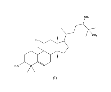
is as shown in a formula I:
oR2
OR4
Ri
111111.
R30
3
CA 03010421 2018-06-29
Formula I
wherein, RI is hydroxyl or carbonyl; R2, R3 and R4 are hydrogen or glycosyl;
glycosyl is hexapyranose, pentapyranose, hexafuranose, pentafuranose, or
diglycosyl,
triglycosyl and tetraglycosyl formed therefrom.
[0012] Preferably, in the formula I, RI is hydroxyl.
[0013] Preferably, in the formula I, R4 is hydrogen.
[0014] Preferably, in the formula I, R2 is hydrogen, R3 is hydrogen or
glycosyl;
more preferably, the glycosyl is hexapyranose, or diglycosyl, triglycosyl and
tetraglycosyl formed therefrom; more preferably, the glycosyl is diglycosyl or
triglycosyl of hexapyranose.
[0015] Furthermore, the structural formula of the cucurbitane tetracyclic
triterpenoid
compounds is as follows: mogroside IIIe (formula II), mogroside III Al
(formula III),
mogroside IVe (formula IV), mogroside V (formula V), mogroside II A2 (formula
VI),
mogroside I El (formula VII), 11-oxo-mogroside V (formula VIII) or mogrol
(formula IX):
HO
0
0
HO
MO
4111111 0
0 1111401 0
OH
0
OH
off
Formula II,
4
CA 03010421 2018-06-29
OH
HO
OH
HO 0
OH
0
OH
0
0 0
0
OH
OH
1-10
HO
OH
HO
HO
Formula III,
OH
HO
HO OH
0
OH
0
OH
0
0
OH
OH
HOOO
HO
OH
O HO H
OH
Formula IV,
CA 03010421 2018-06-29
OH
HO
HO OH
0
OH
0
OH
0
0
0
0
OH
HO OH
HO
OH
OH
HO
HOoO
HOOH
OH
Formula V,
OH
OH
F10
OH
HOOH
O.
O. HO 0 0
HOOH
OH
Formula VI,
6
CA 03010421 2018-06-29
OH
OH
HO,....,.., HO
HOõ....,õ.õ..õ
0
HOO
OH
Formula VII,
OH
HO
O
HO H 0
OH
0
OH
0
0 0
0
OH OH
HO
0
OH OH
HO
HO,,__,....,..,.......õõOH
HO00
HOOH
OH
Formula VIII, or
01-1
OH
HO O.
Se
HO
7
CA 03010421 2018-06-29
Formula IX.
[0016] More preferably, the structural formula of the cucurbitane tetracyclic
triterpenoid compound is formula II, formula IV or formula IX.
[0017] In the application of the aforementioned cucurbitane tetracyclic
triterpenoid
compounds in preparation of drugs and/or health products for preventing and/or
treating pulmonary fibrosis, a compound as shown in formula I and a
combination
thereof as well as other human-acceptable pharmaceutical adjuvants are
prepared into
tablets, granules, decoctions or capsules.
[0018] The cucurbitane tetracyclic triterpenoid compounds are drugs and/or
health
products capable of reducing the accumulation volume of collagens in pulmonary
fibrosis mesenchyme.
[0019] The cucurbitane tetracyclic triterpenoid compounds are drugs and/or
health
products capable of relieving inflammation, inhibiting collagen formation and
protecting lung tissue against pulmonary fibrosis.
[0020] The cucurbitane tetracyclic triterpenoid compounds are drugs and/or
health
products capable of resisting pulmonary fibrosis by resisting inflammation and
inhibiting alveolar epithelial-mesenchymal transition.
[0021] Beneficial effects
[0022] 1. Pulmonary fibrosis is a common pathological change generated when
multiple lung diseases or lung injuries are developed into be in a late stage.
A clinical
study result shows that the survival rate of patients after pulmonary fibrosis
is treated
with glucocorticoid has no significant change, and no clear therapeutic drugs
are
available at present. The cucurbitane tetracyclic triterpenoid compounds of
the
disclosure are prepared from the traditional Chinese medicine momordica
grosvenori
capable of moistening lung for arresting cough. At present, there are no any
studies to
report that mogroside components can be used for treatment of pulmonary
fibrosis.
The inventors have demonstrated that mogroside IIIe, mogroside IVe and
aglycone-mogrol of mogroside components can significantly improve
bleomycin-induced mice pulmonary fibrosis via in-vivo experiments. Mogroside
mogroside IVe and mogrol of the disclosure have good stability and can be used
for
preparing drugs for treating corresponding diseases.
[0023] 2. Specifically, the experimental results of the disclosure show that,
in the
mogroside IIIe administration group of Example 1, Masson staining pathological
8
CA 03010421 2018-06-29
sections show that the degree of pulmonary fibrosis in the administration
groups is
obviously improved, and the number of leukocytes in the bronchoalveolar fluid
of the
mogroside tile administration group is significantly lower than that in the
model
group. On the 28d after administration, the content of TNF-cc in the lung
tissue and
contents of HYP and TGF-131 reflecting collagen deposition are significantly
lower
than those in the model group (P<0.05 or P<0.01). It indicates that mogroside
tile has
a certain therapeutic effect on pulmonary fibrosis at different stages, and
the in-vivo
experiment proves that mogroside Itte has the effects of relieving the degree
of
inflammation and inhibiting the formation of collagen to protect the lung
tissue.
[0024] The in-vitro experiment indicates that mogroside Itte has an
anti-inflammatory effect. Within a range of 10-100uM, mogroside Me is of dose
dependence to inhibit the level of NO in LPS-induced RAW264.7 cells. This
result
also confirms that mogroside Ilk has the effect of inhibiting epithelial-
mesenchymal
transition of TGF-131-inducted Type II alveolar epithelial cells A549, and
discloses
that mogroside Ille can take a medicine use of resisting pulmonary fibrosis by
resisting inflammation and inhibiting alveolar epithelial-mesenchymal
transition.
Therefore, it illustrates that mogroside tile has a certain therapeutic effect
on
pulmonary fibrosis of model mice.
[0025] 3. The experimental materials involved in the disclosure are derived
from
original plants. The original plants have wide range, low cost, clear extract
activity
and wide practical value.
DESCRIPTION OF THE DRAWINGS
[0026] Fig. 1 is a schematic diagram showing change trend of mogroside Ille on
body weights of bleomycin-induced pulmonary fibrosis model mice; compared with
the control group, #p<0.05; compared with the model group, *p<0.05, "p<0.01;
positive control drug: prednisone acetate;
[0027] Fig. 2 is a schematic diagram showing influence of mogroside Me on
change
of lung coefficients of bleomycin-induced pulmonary fibrosis model mice;
compared
with the control group, " p<0.001; compared with the model group, *p<0.05,
"p<0.01; positive control drug: prednisone acetate;
[0028] Fig. 3 is a schematic diagram showing influence of mogroside Me on lung
tissue fibrosis degrees of each group of bleomycin-induced pulmonary fibrosis
model
mice (Masson staining); compared with the control group, "p<0.01; compared
with
9
CA 03010421 2018-06-29
the model group, *p<0.05, "p<0.01; positive control drug: prednisone acetate;
[0029] Fig. 4 is a schematic diagram showing influence of mogroside Ille on
contents of HYP in lung tissues of bleomycin-induced pulmonary fibrosis model
mice;
compared with the control group, #p<0.05; compared with the model group,
*p<0.05;
[0030] Fig. 5 is a schematic diagram showing influence of mogroside Me on
a-SMA expression level in TGF-I31-induced human alveoli type II epithelial
cells.
compared with the control group, " p<0.01; compared with the model group,
*p<0.05;
[0031] Fig. 6 is a schematic diagram showing change trends of mogroside IVe on
changes of body weights of bleomycin-induced pulmonary fibrosis model mice;
compared with the control group, # p<0.05; compared with the model group,
*p<0.05,
**p<0.01; positive control drug: prednisone acetate;
[0032] Fig. 7 is a schematic diagram showing influence of mogroside IVe on
change
of lung coefficients of bleomycin-induced pulmonary fibrosis model mice;
compared
with the control group, " p<0.001; compared with the model group, *p<0.05,
**p<0.01; positive control drug: prednisone acetate;
[0033] Fig. 8 is a schematic diagram showing influence of mogroside IVe on
pulmonary fibrosis degrees of each group of bleomycin-induced pulmonary
fibrosis
model mice (Masson staining); compared with the control group, "p<0.01;
compared
with the model group, *p<0.05, "p<0.01; positive control drug: prednisone
acetate;
[0034] Fig. 9 is a schematic diagram showing change trend of mogrol on body
weights of bleomycin-induced pulmonary fibrosis model mice; compared with the
control group, #p<0.05; compared with the model group, *p<0.05, "p<0.01;
positive
control drug: prednisone acetate; and
[0035] Fig. 10 is a schematic diagram showing influence of mogrol on pulmonary
fibrosis degrees of each group of bleomycin-induced pulmonary fibrosis model
mice
(Masson staining); compared with the control group, "p<0.01; compared with the
model group, *p<0.05, **p<0.01; positive control drug: prednisone acetate.
DETAILED DESCRIPTION OF PREFERRED EMBODIMENTS
[0036] The disclosure discloses an application of the cucurbitane tetracyclic
triterpenoid compound as shown in formula I in preparation of drugs and/or
health
products for preventing and/or treating pulmonary fibrosis. In the formula I,
RI is
hydroxyl or carbonyl; R2, R3 and R4 are hydrogen or glycosyl; glycosyl is
CA 03010421 2018-06-29
hexapyranose, pentapyranose, hexafuranose, pentafuranose, or diglycosyl,
triglycosyl
and tetraglycosyl formed therefrom.
=
HO 0
0 OH
H = H
1111111111
=
0 40401 0
Off
HO 0
Formula I
[0037] Preferably, in the formula I, R1 is hydroxyl.
[0038] Preferably, in the formula I, R4 is hydrogen.
[0039] Preferably, in the formula I, R2 is hydrogen, R3 is hydrogen or
glycosyl;
more preferably, the glycosyl is hexapyranose or diglycosyl, triglycosyl or
tetraglycosyl formed therefrom; more preferably, the glycosyl is diglycosyl or
triglycosyl of hexapyranose.
[0040] Preferably, in the formula I, R2 is glycosyl, R3 is hydrogen or
glycosyl; more
preferably, the glycosyl is hexapyranose or diglycosyl, triglycosyl or
tetraglycosyl
formed therefrom; more preferably, the glycosyl is diglycosyl or triglycosyl
of
hexapyranose.
[0041] Furthermore, the mogroside compounds are selected from one of the
following compounds: mogroside Ille (formula II), mogroside III Al (formula
III),
mogroside IVe (formula IV), mogroside V (formula V), mogroside II A2 (formula
VI),
mogroside I El (formula VII), 11-oxo-mogroside V (formula VIII) or mogrol
(formula IX):
11
CA 03010421 2018-06-29
HO 0
0 cii
HO Hi
0
=
0 0
HO
OH
Formula II,
OH
HO
OH
HO 0
OH
0
OH
0
0 0
0
O
OH HO H
HO
OH
HO
HO
Formula III,
12
CA 03010421 2018-06-29
OH
HO
HO OH
0
OH
0
OH
0
0
OH
OH
HO
OH
HOOH
H0(3C)0/0
HOOH
OH
Formula IV,
OH
HO
HO OH
0
OH
0
OH
0
0
0
0
OH
HO HO OH
OH
OH
HO
HOOH
OH
Formula V.
13
CA 03010421 2018-06-29
OH
OH
HO
OH
HOOH
0 0
HOOH
OH
Formula VI,
OH
HO
OH
HO
0
HOO
OH
Formula VII,
14
CA 03010421 2018-06-29
OH
HO
HO OH
0
OH
0
OH
0 0
0
0
OH
HO OH
HOOH
OH OH
HO
H'D H
HO 00
OH
Formula VIII, or
OH
OH
HO
HO ee
Formula IX.
[0042] More preferably, the cucurbitane tetracyclic triterpenoid compound is
mogroside We (formula II), mogroside IVe (formula IV) or mogrol (formula IX).
[0043] In the application of the cucurbitane- tetracyclic triterpenoid
compounds in
preparation of drugs and/or health products for preventing and/or treating
pulmonary
CA 03010421 2018-06-29
fibrosis, a compound as shown in the formula I and a combination thereof as
well as
other human-acceptable pharmaceutical adjuvants are prepared into tablets,
granules,
decoctions or capsules.
[0044] The drugs and/or health products are capable of reducing accumulation
volume of collagen in pulmonary fibrosis mesenchyme.
[0045] The drugs and/or health products are capable of relieving inflammation,
inhibiting collagen formation and protecting lung tissue against pulmonary
fibrosis.
[0046] The drugs and/or health products are capable of resisting pulmonary
fibrosis
by resisting inflammation and inhibiting alveolar epithelial-mesenchymal
transition.
[0047] According to the disclosure, studies demonstrate that the mogroside
compounds can improve bleomycin-induced mice pulmonary fibrosis. In-vivo
experiments are performed to observe the pathological sections of lung tissue
after
Masson staining, determine the lung coefficients, body weight change and end
body
weights of each group, and detect the number of leukocytes and contents of TNF-
a in
the bronchoalveolar fluid of each group of mice at different stages, and the
contents of
HYP and TGF-pl and the expression of a-SMA in lung tissue. The in-vitro
experiments investigate that the mogroside compounds can significantly inhibit
the
NO release of LPS-induced mice macrophages RAW264.7 and TGF-P1- type II
human lung type II epithelial-mesenchymal transition, and reduce the high
expression
of a-SMA after epithelial-mesenchymal transition after induction. The
experimental
results show that the mogroside compounds in the disclosure can improve the
degrees
pulmonary fibrosis of lung tissues in model mice, reduce collagen deposition
in model
lung tissue, improve epithelial-mesenchymal transition, and show a therapeutic
effect
on the mice pulmonary fibrosis model, and have a new medicine use for treating
pulmonary fibrosis.
[0048] Embodiments of the disclosure will be further described in combination
with
examples below, but the disclosure is not limited to the scopes of these
examples.
[0049] In the disclosure, mogroside Me (Example 1), mogroside IVe (Example 2)
and mogrol (Example 3) are taken as examples to verify partial pharmacodynamic
tests and results of mogroside compounds against pulmonary fibrosis.
[0050] In the disclosure, the preparation of mogroside IIIe, mogroside IVe and
mogrol is as follows:
[0051] Mogroside V contained in total mogrosides extracted from natural
grosvenor
16
CA 03010421 2018-06-29
momordica is the highest in content. After total mogrosides are hydrolyzed
using
f3-glucosidase, extracts containing different proportions of mogroside
components can
be obtained according to enzymolysis time. Mogrol is the aglycone of mogroside
compounds, and can be obtained by acid hydrolysis.
[0052] Mogroside Me is obtained from commercially available momordica
grosvenori extracts via macroporous resin separation and high pressure
reversed-phase preparative chromatography separation, and has a purity of 95%
or
more. Alternatively, by reference to a method for preparing mogroside IV in
Chinese
Patent 2010105610299, commercially available momordica grosvenori extracts are
hydrolyzed using p-glucosidase, and then macroporous resin separation and high
pressure reversed-phase preparative chromatography separation are carried out
to
obtain mogroside IIIe with a purity of 95% or more.
[0053] Mogroside IVe is obtained from commercially available momordica
grosvenori extracts via macroporous resin separation and high pressure
reversed-phase preparative chromatography separation, and has a purity of 95%
or
more. Alternatively, by reference to a method for preparing mogroside IV in
Chinese
Patent 2010105610299, commercially available momordica grosvenori extracts are
hydrolyzed using 13-glucosidase, and then macroporous resin separation and
high
pressure reversed-phase preparative chromatography separation are carried out
to
obtain mogroside IV with a purity of 95% or more.
[0054] Mogrol is obtained by hydrolyzing commercially available momordica
grosvenori extracts with 5% sulfuric acid and purifying with silica gel, and
has a
purity of 95% or more.
Example 1 Improvement of mogroside Me on bleomycin-induced mice
pulmonary fibrosis
[0055] The above mogroside Ille is selected to be subjected to in-vivo
pharmacodynamical research. Injection of a bleomycin-induced mice pulmonary
fibrosis model is a commonly used method in the world, and such model is
similar to
a human pulmonary interstitial fibrosis model.
[0056] 1.1 Experimental method
[0057] 100 male ICR mice with a body weight of 25-30g are provided by
Comparative Medicine Center of Yangzhou University.
17
CA 03010421 2018-06-29
[0058] 20 mice are used as a blank control group (control group in Fig. 1),
and the
other 80 mice are used for modeling. All the above mice are anesthetized by
virtue of
intraperitoneal injection of 4% chloral hydrate, with injection volume of 10
ml/kg.
After anesthesia, each mouse is fixed and the neck of the mouse is
disinfected; the
skin of its neck is cut longitudinally with scissors to expose the trachea;
the syringe is
inserted into the trachea. The mice in the blank control group are injected
with normal
saline, and the remaining mice are injected with bleomycin (5mg/kg); then the
mouse
plate is quickly erected and rotated, and the wound of each mouse is sutured
after
observing the breathing condition of the mouse, and 1-2 drops of penicillin
injection
are dropped at the suture. The postoperative mouse is placed back in a dry and
clean
mouse cage for rest, and then is normally fed after it revives approximately 1-
2h later.
[0059] Starting from the day 7 after modeling, the other 80 mice are randomly
divided into a model group, a positive drug (prednisone acetate) group, a high-
dose
mogroside Ille (50mg/kg, mogroside IlIe-H) group, a low-dose mogroside Tile
group
(10mg/kg, mogroside Ille-L), 20 mice in each group.
[0060] The mice in the blank control group and the model group are subjected
to
stomach perfusion with normal saline every day, the mice in the positive drug
group
are subjected to stomach perfusion with 6.67mg/kg/d prednisone acetate, the
mice in
the high-dose/low-dose mogroside Ille groups are subjected to stomach
perfusion
with 50mg/kg/d and 10mg/kg/d respectively for continuous 28 days, and are
killed on
the day 14 and the day 28. The each mouse is recorded in body weight, the lung
tissue
is taken out via dissection, cleaned with ice normal saline, sucked dry with
absorbent
paper and weighed, and then a lung coefficient is calculated, i.e., lung
coefficient=lung weight (mg)/body weight (g). The left lung is put into 4%
neutral
formaldehyde for fixation, gradually dehydrated with alcohol, transparentized
with
xylene, immersed in wax, embedded in paraffin, then routinely sectioned and
subjected to Masson staining, so that the lung tissue morphology, lung injury
condition and pulmonary fibrosis degree are observed. Other lung lobes are
individually stored for determination of HYP content.
[0061] All data are expressed as mean SD(x s). SPSS 11.5 statistical software
is
used for processing. Statistical analysis is performed using one-way ANOVA.
P<0.05
represents this group has statistical significance. =
[0062] 1.2 Experimental results
[0063] 1.2.1 Influence of mogroside Me on body weights of model mice
18
CA 03010421 2018-06-29
[0064] Compared with the blank control group, the body weights of the mice in
the
model group are decreased significantly and all have statistical significance
(P<0.01
or 0.05). Compared with the body weight in the model group mice, the body
weights
of mice in the high-dose mogroside Me group, low-dose mogroside Ilk group and
positive drug (prednisone acetate) group are obviously increased with
statistical
significance (P<0.01) after 14 and 28 days of administration. It indicates
that
mogroside file can improve the physique of bleomycin-induced pulmonary
fibrosis
mice to different degrees at the doses of 20mg/kg and 10mg/kg and slow down
the
weight loss rate of the pulmonary fibrosis model mice, and the results are as
shown in
Fig.l.
[0065] 1.2.2 Influence of mogroside Me on the number of leukocytes in the
bronchoalveolar fluid of the model mice
[0066] For mice lung tissue injury caused by bleomycin, the number of
leukocytes is
increased, especially neutrophil infiltration causes alveolar inflammation,
and
inflammatory cells release inflammatory mediators NO, TNF-oc and various
cytokines,
thereby on one hand, aggravating lung tissue injury and on the other hand
promoting
excessive increase of collagen through various growth factors. This experiment
is
performed in accordance with requirement specification of a kit. The number of
leukocytes in the bronchoalveolar lavage fluid of the mice in each group is
detected
by a colorimetric method on the day 14 (as shown in Table 1). Neutrophils and
lymphocytes are obviously accumulated in the bronchoalveolar lavage fluid of
the
mice on the day 14 after treatment with bleomycin (BLM). Compared with the
blank
control group, the total number of cells and the number of neutrophils in the
bronchoalveolar lavage fluid of the model group are both obviously increased
(compared with the blank control group, "P<0.0 I). After treatment with
mogroside
life, the total number of cells and the number of neutrophils in the
bronchoalveolar
lavage fluid are obviously reduced than those in the individually
administrated model
group (compared with the model group, *P<0.05). It indicates that mogroside
IIIe is
capable of reducing the effusion of BLM-induced inflammatory cells.
19
CA 03010421 2018-06-29
Table 1
The total number of cells and the number of neutrophils in bleomycin-induced
mice
bronchoalveolar lavage fluid
Group Total number of cells Number of neutrophils
Blank control group 11.2113.98 1.0210.21
Model group 56.98123.76" 18.3219.02"
Mogroside IlIe-H group 32.81118.53* 4.2814.52*
(50mg/kg)
[0067] 1.2.3 Influence of mogroside Hie on lung coefficients of model mice
[0068] Compared with the blank control group, the lung coefficients of mice in
the
model group are significantly increased and have statistical significance
(P<0.01 or
0.05); compared with the lung coefficients of mice in the model group, after
administration for 14 days and 28 days, the lung coefficients of the mogroside
IlIe
drug group and the prednisone acetate group are both obviously decreased and
have
statistical significance (P<0.01) . Compared with the blank control group, the
lung
coefficients of mice in the model group are obviously increased and have
statistical
significance (P<0.01 or 0.05); compared with the body weights of mice in the
model
group, after administration for 14 days and 21 days, the lung coefficients of
high-dose
and low-dose mogroside IlIe groups and positive drug (prednisone acetate)
group are
all obviously decreased and have statistical significance (P<0.01). It
indicates that
mogroside We can improve bleomycin-induced mice pulmonary fibrosis to
different
degrees and relieve the development degree of mice pulmonary fibrosis at the
doses
of 20mg/k and 10mg/kg (see Fig. 2).
[0069] 1.2.4 Influence of mogroside Me on lung tissues of model mice
[0070] A pathological tissue section of lung on the day 28 is subjected to
Masson
staining, and a result indicates that lung tissue structures of mice in the
blank control
group are complete and clear, pulmonary alveoli septum is not thickened, an
alveolar
space is transparent and does not contain obvious effusion, and no fibroblasts
are
proliferated; a few of collagenous fibers stained into blue can be seen in the
lung
tissues of mice in the blank control group, which are main components of an
extracellular matrix. The alveoli structures of mice in the model group are
damaged,
pulmonary alveoli septum is widened, a large amount of inflammatory cells are
CA 03010421 2018-06-29
infiltrated into acute fibrocyte hyperplasia, a large amount of collagens are
deposited,
pulmonary fibrosis is formed, a large amount of dense collagenous fibers dyed
into
blue, which are deposited in a sarciniform or a sheet form, can be seen after
Masson
staining, and all of them basically meet features of pulmonary fibrosis,
thereby
indicating that an experiment mice pulmonary fibrosis model is successfully
prepared.
After treatment with mogroside Ille, it can be seen that the lung tissue
structures of
mice are complete and clear, pulmonary alveoli septum is slightly thickened,
and
fibroblast hyperplasia degree is lower than that in the model group. After
treatment
with positive drug prednisone acetate, pulmonary alveoli septum of mice in the
positive group is wider, the alveolar space is narrowed, multiple fibroblasts
are
proliferated, and the degree of the lesion is relieved than that in the model
group.
Compared with the model group, the fibrosis degrees of various administration
groups
and positive drug groups are all relieved (see FIG. 3).
[0071] 1.2.5 Influence of mogroside Hie on HYP contents of lung tissues of
model mice lung tissue
[0072] Hydrooxyproline (HPY) is an amino acid obtained through protein
hydrolysis of connective tissues, accounts for about 14% of the weight of
collagen
and plays a crucial role in stability of collagen. Since the collagen is an
only protein
having high HYP content, determination of HYP content can reflect total amount
change of tissue collagen. The content of HYP in the lung tissue is detected
adopting a
digestion method. Compared with the blank control group, the HYP content of
the
lung tissue in the model group is obviously increased (P<0.01) at the day 28;
compared with the model group, the drug can obviously reduce the content of
HYP in
the lung tissue (P<0.05). It indicates that mogroside Me can improve
bleomycin-induced mice pulmonary fibrosis to different degrees at the dose of
50mg/kg, mogroside 'Ile can reduce the content of collagenous fibers of the
model
tissue and slow down the development degree of pulmonary fibrosis of mice in
the
model mice (as shown in Fig. 4).
[0073] 1.2.6 Influence of mogroside Hie on a-SMA level of TGF-131-induced
human pulmonary alveoli type II epithelial cells
[0074] Alveolar epithelial cells can obtain mesenchymal cell phenotypes
through an
epithelial cell-mesenchyme transformation (EMT) process to serve as an
important
source of myofibroblast and muscle fiber metrocyte. In this new mode,
epithelial
21
CA 03010421 2018-06-29
cell-mesenchyme transformation of pulmonary alveoli should be considered as
one of
key links of fibrosis. In mature cells, injury can induce transformation of
epithelial
cells into mesenchymal cell phenotype, thus facilitating the fibrosis of many
organs.
Fibroblasts and muscle fiber metrocytes differentiated from epithelial cells
often
change through form (for example, change from a cubical cell form to a strip
form or
a fusiform), obtaining of specific marker of the fibroblast or the muscle
fiber
metrocyte (for example, ot-SMA) and loss of the epithelial character marker
(for
example, epithelial cell cadherin ( E-cadherin and closely linked protein) are
fussed
together with these epithelial tissues. Through an in-vitro test, A549 cells
are induced
with TGF-131 to generate epithelial cell-mesenchyme transformation (EMT), the
a-SMA expression level of actin is analyzed adopting a Western blot method,
and
study meaning of mogroside Ille on pathogensis of pulmonary fibrosis in a
signal
transduction pathway of an EMT process is discussed.
[0075] In the disclosure, well grown A549 cells subcultured by 2-4 generations
perform 1 x10⁵ passage and then are divided into four groups, and serum-free
DMEM
culture solution is added to perform starvation for 12 hours, so that cells
are in the
same growth level. For the model group, TGF-f31 having a concentration of
5ng/mL is
added in a serum-free culture medium; for the positive drug group and
mogroside Ille
group (1001.iM), TGF-I31 having a concentration of 5ng/mL and corresponding
drugs
are added. After culture for 48 hours, change of lung epithelial cells is
observed under
an inverted microspcope, influence of TGF-131 on an expression level of a
marker
protein ct-SMA for transforming A549 epithelial cells into mesenchymal cells
is
detected with Western blot. An experiment result indicates that compared with
the
blank control group, the expression of mesenchymal cell marker et-SMA is
up-regulated after TGF-I31 is added in A549 cells, and mogroside Ille can
significantly inhibit a-SMA caused by TGF-131 under the concentration of 50pM
(see
Fig. 5).
[0076] 1.2.7 Discussion:
[0077] Compared with the model group, high-dose/low-dose mogroside Ille groups
can obviously reduce the index of lung, the content of HYP in the lung tissue
from the
high-dose group is obviously reduced (p<0.05), and a pathological result shows
that
the lung tissue structure of the mogroside Ille drug group is obviously
improved, the
pulmonary alveoli structure is damaged and the thickening degree of alveolar
septum
22
CA 03010421 2018-06-29
is obviously alleviated, inflammatory cell infiltration is reduced, and
collagenous fiber
content is reduced. Compared with the blank control group, the number of
neutrophils
in bronchoalveolar lavage fluid of mice in the model group is significantly
increased,
indicating that bleomycin causes inflammatory reaction in the model group so
as to
initiate in-vivo inflammatory cascade reaction. However, after the drug is
administrated, inflammation and fibrosis degrees of mice are reduced to
different
degrees, and the content of HYP in the lung tissue is significantly reduced,
indicating
that the drug can protect lung cells from being injured so as to prevent and
treat
pulmonary fibrosis. An in-vitro experiment result shows that mogroside Ille
can
inhibit TGF131-induced epithelial-mesenchymal transition at a concentration of
50)AM and reduce lung fibroblast marker a-SMA.
[0078] In summary, in-vivo and in-vitro experiment results show that mogroside
IIIe
can improve inflammation and fibrosis degrees of lung tissues in the bleomycin
mice
pulmonary fibrosis model and generation of collagen in the lung tissue, and
effectively inhibits human pulmonary alveoli type 11 epithelial-mesenchymal
transition caused by the cell growth factor TGF-131, and mogroside Me has a
new use
for treating pulmonary fibrosis.
Example 2 Improvement of mogroside IVe on mice pulmonary fibrosis caused by
bleomycin induction
[0079] The above mogroside IVe (formula IV) is selected to carry out in-vivo
pharmacodynamic study.
[0080] 2.1 Experiment method
[0081] 100 male ICR mice with a body weight of 25-30g are provided by
Comparative Medicine Center of Yangzhou University.
[0082] 20 mice are used as a blank control group, and the other 80 mice are
used for
modeling. All the above mice are anesthetized by virtue of intraperitoneal
injection of
4% chloral hydrate, with injection volume of 10 ml/kg. After anesthesia, each
mouse
is fixed and the neck of the mouse is disinfected. The skin of its neck is cut
longitudinally with scissors, and fascia and muscles are longitudinally and
bluntly tore
with tweezers to expose the trachea. The syringe is inserted into the trachea.
The mice
in the blank control group are injected with normal saline, and the remaining
mice are
injected with bleomycin (5mg/kg); then the mouse plate is quickly erected and
rotated,
23
CA 03010421 2018-06-29
the breathing condition of the mouse is observed, the wound of the neck is
disinfected
with 75% alcohol cotton after being rotated and then sutured, and 1-2 drops of
penicillin injection are dropped at the suture. The postoperative mouse is
placed back
in a dry and clean mouse cage for rest, and then is normally fed after it
revives
approximately 1-2h later.
[0083] Starting from the day 7 after modeling, the other 80 mice are divided
into a
model group, a positive drug (prednisone acetate) group, a high-dose mogroside
IVe
group (50mg/kg, mogroside IVe-H) and a low-dose mogroside IVe group (50mg/kg,
mogroside IVe-L), 20 mice in each group.
[0084] The mice in the blank control group and the model group are subjected
to
stomach perfusion with normal saline every day, the mice in the positive drug
group
are subjected to stomach perfusion with 6.67mg/kg/d prednisone acetate, the
mice in
the high-dose/low-dose mogroside IVe groups are subjected to stomach perfusion
with
50mg/kg/d (high-dose group) and 20mg/kg/d (low-dose group) respectively for
continuous 28 days, and the body weights are weighed and recorded; and the
mice are
killed on the day 28. The lung tissue is taken out via dissection, and a lung
coefficient
is calculated, i.e., lung coefficient = lung weight (mg)/body weight (g). The
left lung
is put into 4% neutral formaldehyde for fixation, gradually dehydrated with
alcohol,
transparentized with xylene, immersed in wax, embedded in paraffin, then
routinely
sectioned and subjected to Masson staining, so that the lung tissue
morphology, lung
injury condition and pulmonary fibrosis degree are observed.
[0085] All data are expressed as mean SD(x s). SPSS11.5 statistical software
is
used for processing, statistical analysis is performed using one-way ANOVA,
P<0.05
represents this group has statistical significance.
[0086] 2.2 Experiment result
[0087] 2.2.1 Influence of mogroside IVe on body weights of pulmonary
fibrosis model mice
[0088] Compared with the blank control group, the body weights of mice in the
model group are obviously decreased and have statistical significance (P<0.01
or
0.05); compared with the body weights of mice in the model group, the body
weights
of the high-dose/low-dose mogroside IVe groups and positive drug (prednisone
acetate) groups are all obviously increased after administration for 14 days
and 28
days and have statistical significance (P<0.01). It indicates that mogroside
IVe can
improve the physique of bleomycin-induced mice pulmonary fibrosis to different
24
CA 03010421 2018-06-29
degrees at the doses of 50mg/kg and 20mg/kg and relieve the reduction degrees
of the
body weights of the lung model mice (see FIG. 6).
[0089] 2.2.2 Influence of mogroside IVe on the number of leukocytes in
bronchoalveolar lavage fluid of the model mice
[0090] For tissue injury caused by bleomycin, the number of leukocytes is
increased,
especially, neutrephil infiltration causes pulmonary alveoli inflammation, and
inflammatory cells release inflammatory mediators NO, TNF-a and various
cytokines,
thereby on one hand, aggravating lung tissue injury and on the other hand
promoting
excessive increase of collagen through various growth factors. This experiment
is
performed in accordance with requirement specification of a kit. The number of
leukocytes in the bronchoalveolar lavage fluid of the mice in each group is
detected
by a colorimetric method on the day 14 (as shown in Table 2). Neutrophils and
lymphocytes are obviously accumulated in the bronchoalveolar lavage fluid of
the
mice on the day 14 after treatment with bleomycin (BLM). Compared with the
blank
control group, the total number of cells and the number of neutrophils in the
bronchoalveolar lavage fluid of the model group are both obviously increased
(compared with the blank control group, "P<0.01). After treatment, the total
number
of cells and the number of neutrophils in the bronchoalveolar lavage fluid are
obviously reduced than those in the individually administrated model group
(compared with the model group, *P<0.05). It indicates that mogroside IVe is
capable
of reducing the effusion of BLM-induced inflammatory cells.
Table 2
Influence on the total number of cells and the number of the neutrophils in
bronchoalveolar lavage fluid of mice after bleomycin induction ( x104, )
CA 03010421 2018-06-29
Group Total No. of cells No. of neutrophils
Blank control group 12.2613.74 1.3610.23
Model group 58.93121.47" 19.26/9.65"
Mogroside IVe high-dose group 30.84116.49* 3.9713.89*
(50mg/kg)
[0091] 2.2.3 Influence of mogroside IVe on lung coefficients of model mice
[0092] The lung coefficient is measured on the day 28 after modeling. Compared
with the blank control group, the lung coefficients of mice in the model group
are
obviously increased and have statistical significance (P<0.01 or 0.05);
compared with
the lung coefficients of mice in the model group, the lung coefficients of the
mogroside IVe drug group and the prednisone acetate group are all obviously
reduced
and have statistical significance (P<0.01). Compared with the blank control
group, the
lung coefficients of mice in the model group are obviously increased and have
statistical significance (P<0.01 or 0.05); compared with the body weights of
mice in
the model group, the lung coefficients of the high-dose/low-dose mogroside IVe
groups and the positive drug (prednisone acetate) group are all obviously
reduced and
have statistical significance (P<0.01). It indicates that mogroside IVe can
improve
bleomycin-induced mice pulmonary fibrosis to different degrees at the doses of
50mg/kg and 20mg/kg and relieve the development degree of the mice pulmonary
fibrosis (see Fig. 7).
[0093] 2.2.4 Influence of mogroside IVe on lung tissues of the model mice
[0094] A pathological tissue section is subjected to Masson staining, and a
result
indicates that lung tissue structures of mice in the blank control group are
complete
and clear, pulmonary alveoli septum is not thickened, an alveolar space is
transparent,
and no fibroblasts are proliferated; a few of collagenous fibers stained into
blue can be
seen in the lung tissues of mice in the blank control group, which are main
components of an extracellular matrix. The alveoli structures of mice in the
model
group are damaged, pulmonary alveoli septum is widened, a large amount of
collagens are deposited, pulmonary fibrosis is formed, a large amount of dense
collagenous fibers stained into blue, which are deposited in a sarciniform or
a sheet
form, can be seen after Masson staining, and all of them basically meet
features of
pulmonary fibrosis, thereby indicating that an experiment mice pulmonary
fibrosis
model is successfully prepared. After treatment with mogroside IVe, it can be
seen
26
CA 03010421 2018-06-29
that the lung tissue structures of mice are complete and clear, pulmonary
alveoli
septum is slightly thickened, and fibroblast hyperplasia degree is lower than
that in
the model group. Compared with the model group, the fibrosis degrees of
various
administration groups and positive drug groups are all relieved (see FIG. 8).
[0095] 2.2.5 Discussion:
[0096] Compared with the model group, high-dose/low-dose mogroside We groups
can obviously reduce lung index, pathological results show that lung tissue
structures
of the high-dose/low-dose mogroside IVe groups and mogrol drug groups are
obviously improved. Compared with the blank control group, the number of
neutrophils in the bronchoalveolar lavage fluid of mice in the model group is
significantly increased, indicating that in the model group, bleomycin causes
inflammatory reaction, thereby initiating in-vivo inflammatory cascade
reaction. After
the drug is administrated, inflammation and fibrosis degrees of mice are
relieved to
different degrees, indicating that the drug can protect lung cells from being
damaged
so as to prevent and treat pulmonary fibrosis. An in-vivo experiment result
indicates
that mogroside IVe can improve lung tissue inflammation and pulmonary fibrosis
degree in a bleomycin mice pulmonary fibrosis model and generation of
collagens in
lung tissue, and mogroside IVe has a new use of treating pulmonary fibrosis.
Example 3 Improvement of mogrol on bleomycin-induced mice pulmonary
fibrosis
[0097] Mogrol (formula IX) is selected to carry out the following in-vivo
pharmacodynamic study.
[0098] 3.1 Experiment method
[0099] 100 male ICR mice with a body weight of 25-30g are provided by
Comparative Medicine Center of Yangzhou University.
[00100] 20 mice are used as a blank control group, and the other 80 mice are
used for
modeling. All the above mice are anesthetized by virtue of intraperitoneal
injection of
4% chloral hydrate, with injection volume of 10 ml/kg. After anesthesia, each
mouse
is fixed and the neck of the mouse is disinfected; the skin of its neck is cut
longitudinally with scissors, and fascia and muscles are longitudinally and
bluntly tore
with tweezers to expose the trachea; the syringe is inserted into the trachea.
The mice
in the blank control group are injected with normal saline, and the other mice
are
27
CA 03010421 2018-06-29
injected with bleomycin (5mg/kg); then the mouse plate is quickly erected and
rotated,
the breathing condition of the mouse is observed, the wound of the neck is
disinfected
with 75% alcohol cotton after being rotated and then sutured, and 1-2 drops of
penicillin injection are dropped at the suture. The postoperative mouse is
placed back
in a dry and clean mouse cage for rest, and then is normally fed after it
revives
approximately 1-2h later. Starting from the day 7 after modeling, the other
mice are
randomly divided into a model group, a positive drug (prednisone acetate)
group, a
high-dose mogrol group (50mg/kg, mogrol-H) and a low-dose mogrol group
(20mg/kg, mogrol-L), 20 mice in each group.
[00101] The mice in the blank control group and the model group are subjected
to
stomach perfusion with normal saline every day, the mice in the positive drug
group
are subjected to stomach perfusion with 7.0mg/kg/d prednisone acetate, the
mice in
the high-dose/low-dose mogrol groups are subjected to stomach perfusion for
continuous 28 days, and the body weights are weighed and recorded; and the
mice are
killed on the day 28. The lung tissue is taken out via dissection, fixed in 4%
neutral
formaldehyde, gradually dehydrated with alcohol, transparentized with xylene,
immersed in wax, embedded in paraffin, then routinely sectioned and subjected
to
Masson staining, so that the lung tissue morphology, lung injury condition and
pulmonary fibrosis degree are observed.
[00102] All data are expressed as mean SD(x+s). SPSS11.5 statistical
software is
used for processing, statistic analysis is performed adopting one-way ANOVA,
and
P<0.05 represents this group has significant difference.
[00103] 3.2 Experiment result
[00104] 3.2.1 Influence of mogrol on body weights of pulmonary fibrosis
model mice
[00105] Compared with the body weights of mice in the model group, the body
weights of high-dose/low-dose mogrol groups and positive drug (prednisone
acetate)
group are all obviously increased. It indicates that mogrol can improve the
physique
of bleomycin-induced mice pulmonary fibrosis to different degrees at the doses
of
50mg/kg and 20mg/kg and relive the reduction degrees of the body weights of
the
model mice (see FIG. 9).
[00106] 3.2.2 Influence of mogrol on the number of leukocytes in the
bronchoalveolar lavage fluid of model mice
[00107] For mice lung tissue injury caused by bleomycin, the number of
leucocytes is
28
CA 03010421 2018-06-29
increased, especially neutrophil infiltration causes pulmonary alveoli
inflammation,
inflammatory cells release inflammatory mediators NO, TNF-or and various
cytokines,
thereby on one hand, aggravating lung tissue injury and on the other hand
promoting
excessive increase of collagen through various growth factors. This experiment
is
performed in accordance with requirement specification of a kit. The number of
leukocytes in the bronchoalveolar lavage fluid of the mice in each group is
detected
by a colorimetric method on the day 14 (the results are as shown in Table 3).
Compared with the blank control group, the total number of cells and the
number of
neutrophils in the bronchoalveolar lavage fluid of mice in the model group are
both
obviously increased (compared with the blank control group, "P<0.01). After
treatment with mogrol (50mg/kg, the total number of cells and the number of
neutrophils in the bronchoalveolar lavage fluid are obviously reduced than
those in
the individually administrated model group (compared with the model group,
*P<0.05). It indicates that mogrol is capable of reducing effusion of BLM-
induced
inflammatory cells.
Table 3
Influence on the total number of the cells and the number of the neutrophils
in
bleomycin-induced mice bronchoalveolar lavage fluid ( x104, n=5)
Group Total No. of cells No. of neutrophils
Blank control group 13.16+3.74 1.96+0.65
Model group 65.13+21.57" 18.26+9.45"
high-dose mogrol group 34.72+17.83** 4.16+4.26**
(50mg/kg)
[00108] 3.2.3 Influence of mogrol on lung tissues of model mice
[00109] A few of collagenous fibers stained into blue can be seen in the lung
tissues
of mice in the blank control group, which are main components of an
extracellular
matrix. The pulmonary alveoli structures of mice in the model group are
damaged,
pulmonary alveoli septum is widened, a large amount of collagens are
deposited,
pulmonary fibrosis is formed, a large amount of dense collagenous fibers
stained into
blue, which are deposited in a sarciniform or a sheet form, are seen after
Masson
staining, and all of them basically meet features of pulmonary fibrosis,
indicating that
an experiment mice pulmonary fibrosis model is successfully prepared. After
29
CA 03010421 2018-06-29
treatment with high-dose/low-dose mogrol (50, 20mg/kg), it can be seen that
the lung
tissue structures of mice are complete and clear, pulmonary alveoli septum is
thickened, and fibroblast hyperplasia degree is lower than that in the model
group.
Compared with the model group, fibrosis degrees of various administration
groups are
all reduced (see FIG. 10).
[00110] 3.2.5 Discussion:
[00111] Compared with the model group, high-dose/low-dose mogrol groups can
obviously reduce lung index, and pathological results show that the lung
tissue
structure of the mogrol drug group is obviously improved. Compared with the
blank
control group, the number of neutrophils in the bronchoalveolar lavage fluid
of mice
in the model group is significantly increased, indicating that in the model
group,
bleomycin causes inflammatory reaction; and after the drug is administrated,
inflammation and fibrosis degrees of mice are relieved to different degrees,
indicating
that the drug can protect lung cells from being injured so as to prevent and
treat
pulmonary fibrosis. As an aglucon of a cucurbitane tetracylic triterpenoid
compound,
mogrol has common nuclear parent in this compound structure skeleton. An in-
vivo
experiment result discloses that the in-vivo experiment result shows that
mogrol can
improve the pulmonary fibrosis degree of the lung tissue in a bleomycin mice
pulmonary fibrosis model, indicating a new use of this component in
preparation of a
drug for preventing or treating pulmonary fibrosis.