Note: Descriptions are shown in the official language in which they were submitted.
CA 03010895 2018-07-06
WO 2017/127684
PCT/US2017/014343
METHODS FOR RAPID ANTIMICROBIAL SUSCEPTIBILITY TESTING
RELATED APPLICATIONS
This application claims priority to and benefit of U.S. Provisional Patent
Application
No. 62/281,698, filed January 21, 2016; U.S. Provisional Patent Application
No. 62/298,821,
.. filed February 23, 2016; U.S. Provisional Patent Application No.
62/326,545, filed April 22,
2016; U.S. Provisional Patent Application No. 62/338,376, filed May 18, 2016;
U.S.
Provisional Patent Application No. 62/370,579, filed August 3, 2016; and U.S.
Provisional
Patent Application No. 62/383,198, filed September 2, 2016. The contents of
the
aforementioned patent applications are incorporated herein by reference in
their entireties.
BACKGROUND OF THE INVENTION
Antimicrobial-resistant microbial infections are associated with poor clinical
outcomes including increased morbidity, mortality, and healthcare costs among
infected
patients. The prevalence of these organisms in such facilities in the United
States has steadily
increased over the last 30 years. Phenotypic antimicrobial susceptibility
testing (AST) of
microorganisms is critical for informing physicians of appropriate therapeutic
regimens.
Using current methods, AST determination typically requires a minimum of eight
hours,
rendering it an overnight process due to shift work in many clinical
microbiology
laboratories. While awaiting a determination from current AST methods,
patients are often
administered broad-spectrum antimicrobials which often have significant
detrimental effects
on patient health and/or contribute to the growing antimicrobial resistance
epidemic.
Furthermore, this time delay to getting accurate antimicrobial treatment
information increases
patient stays in hospitals, thereby increasing costs and inconvenience to the
patient.
Accordingly, a need exists for a method that rapidly determines antimicrobial
susceptibility of a microbial infection. The method described here is further
advantageous in
that it addresses this need in a cost-effective manner because it is
compatible with existing
assay hardware components.
SUMMARY OF THE INVENTION
The present invention permits rapid determination of antibiotic susceptibility
of
microbial infections. The invention is based in part upon the surprising
discovery of non-
specific surface binding assays that provide accurate and rapid Antimicrobial
Susceptibility
CA 03010895 2018-07-06
WO 2017/127684
PCT/US2017/014343
Testing (AST) determinations in fewer than twelve hours ¨ and, specifically,
under four
hours. The present invention ("Fast-AST") provides accurate results that are
consistent with
results obtained using the Clinical Laboratory Standards Institute (CLSI)
reference methods
when tested with multiple antimicrobials and on a plurality of microorganisms;
however, the
present invention takes significantly less time to obtain results than the
CLSI methods.
Moreover, the present invention accurately differentiates an antimicrobial's
MIC for
clinically-relevant microbial strains that are resistant to one or more
antimicrobials and the
antimicrobial's MIC for strains of the same microorganism that are sensitive
to the
antimicrobials. Furthermore, the present invention may include signaling
agents (e.g.,
Europium compounds) that are bound to microorganisms non-specifically rather
than
specifically (e.g., via chemically conserved groups or biochemically conserved
binding sites
on microorganisms), thereby expanding the generalization of the present
invention to any
microorganism and allowing onset of an appropriate treatment without first
needing to
identify the particular infectious microorganism. Also, the present invention
permits signal
amplification such that microbes may be rapidly detected at lower
concentrations, e.g., from a
dilute culture of microorganisms or via a patient's biological sample.
Additionally, the
present invention may use Europium formulations as chemical moiety, thereby
expanding the
dynamic range of the methods and allowing for more accurate determinations
from a range of
microbial samples. Finally, the present invention is compatible with existing
equipment,
thereby enabling rapid adoption in current clinical laboratories. Accordingly,
the present
invention, in a greatly reduced amount of time and expense, relative to
standard methods, can
provide a patient with an appropriate treatment regimen, i.e., a specific
antimicrobial and at a
particular dosage. Thus, the present invention will improve patient outcomes,
lower hospital
costs, and help reduce further evolution of antimicrobial resistant
microorganisms; thus, the
present invention represents a significant breakthrough in the AST field.
An aspect of the present invention is a method for determining antimicrobial
susceptibility of microorganisms. The method includes steps of incubating a
liquid
suspension of microorganisms in the presence of an antimicrobial and a
signaling agent,
which is capable of binding to a surface of the microorganisms, under
conditions that
promote growth of the microorganisms; separating the microorganisms bound by
the
signaling agent from the unbound signaling agent; and determining signal
levels associated
with the microorganisms as compared to one or more controls.
Another aspect of the present invention is a method for determining
antimicrobial
susceptibility of microorganisms. The method includes steps of incubating a
liquid
2
CA 03010895 2018-07-06
WO 2017/127684
PCT/US2017/014343
suspension of microorganisms in the presence of an antimicrobial under
conditions that
promote growth of the microorganisms; adding a signaling agent capable of
binding to a
surface of the microorganisms; separating the microorganisms bound by the
signaling agent
from the unbound signaling agent; and determining signal levels associated
with the
microorganisms as compared to one or more controls.
Yet another aspect of the present invention is a method for determining
antimicrobial
susceptibility of microorganisms. The method includes steps of incubating a
liquid
suspension of microorganisms in a cartridge including a plurality of chambers,
each chamber
containing one or more antimicrobials, under conditions that promote growth of
the
microorganisms; adding a signaling agent, which is capable of binding to a
surface of the
microorganisms, to the plurality of chambers; removing unbound signaling
agent; and
determining signaling levels in the plurality of chambers as compared to one
or more
controls.
An aspect of the present invention is a method for determining antimicrobial
susceptibility of microorganisms. The method includes incubating
microorganisms in the
presence of an antimicrobial and a signaling agent, which includes a signal
amplifier and one
or more chemical moieties capable of binding non-specifically to a surface of
the
microorganisms, under conditions that promote growth of the microorganisms;
separating the
microorganisms bound by the signaling agent from the unbound signaling agent;
and
determining signal levels associated with the microorganisms as compared to
one or more
controls.
Another aspect of the present invention is a method for determining
antimicrobial
susceptibility of microorganisms. The method includes incubating
microorganisms in the
presence of an antimicrobial under conditions that promote growth of the
microorganisms;
adding a signaling agent including a signal amplifier and one or more chemical
moieties
capable of binding non-specifically to a surface of the microorganisms;
separating the
microorganisms bound by the signaling agent from the unbound signaling agent;
and
determining signal levels associated with the microorganisms as compared to
one or more
controls.
Yet another aspect of the present invention is a kit for determining
antimicrobial
susceptibility of microorganisms. The kit includes a signaling agent capable
of binding to a
surface of the intact microorganisms of interest; a solution for incubating a
sample containing
microorganisms; and one or more reagents for generating signals from the
signaling agent.
3
CA 03010895 2018-07-06
WO 2017/127684
PCT/US2017/014343
Any aspect or embodiment described herein can be combined with any other
aspect or
embodiment as disclosed herein. While the disclosure has been described in
conjunction with
the detailed description thereof, the foregoing description is intended to
illustrate and not
limit the scope of the disclosure, which is defined by the scope of the
appended claims. Other
aspects, advantages, and modifications are within the scope of the following
claims.
The patent and scientific literature referred to herein establishes the
knowledge that is
available to those with skill in the art. All United States patents and
published or unpublished
United States patent applications cited herein are incorporated by reference.
All published
foreign patents and patent applications cited herein are hereby incorporated
by reference. All
other published references, documents, manuscripts and scientific literature
cited herein are
hereby incorporated by reference.
Other features and advantages of the invention will be apparent from the
Drawings
and the following detailed description and claims.
BRIEF DESCRIPTION OF THE DRAWINGS
The above and further features will be more clearly appreciated from the
following
detailed description when taken in conjunction with the accompanying drawings.
The
drawings however are for illustration purposes only; not for limitation.
FIG. 1 is a schematic showing generalized steps of the present invention.
FIG. 2A to FIG. 2D are graphs and illustrations showing key features of
aspects of
the present invention ("fast-AST").
FIG. 3 is a schematic comparing steps required in currently-used Antimicrobial
Susceptibility Testing (AST) systems and aspects of the present invention.
FIG. 4 is a graph showing the time delay required to obtain results using a
currently-
used AST system (i.e., BioMerieux's Vitek2).
FIG. 5 is a graph showing a comparison of a minimum inhibitory concentration
(MIC) determination of clindamycin on Staphylococcus aureus (ATCC strain
29213) using
the present invention ("fast-AST" technique) and a standard, overnight growth
followed by
optical density (OD) reading at 600 nm. The data shown for the "fast-AST" is
from the five
minute point after the start of incubation with detection solution and
represents the average
and standard deviations of four wells, with values for each assay type
relative to an
antimicrobial-free control.
4
CA 03010895 2018-07-06
WO 2017/127684
PCT/US2017/014343
FIG. 6 is a graph showing a comparison of a MIC determination of ceftazidime
on
Pseudomonas aeruginosa (ATCC strain 27853) using the present invention ("fast-
AST"
technique) and a standard, overnight growth followed by optical density (OD)
reading at 600
nm. The data shown represents the average and standard deviations of four
wells, with
.. values for each assay type relative to an antimicrobial-free control.
FIG. 7 is a graph showing a MIC determination comparison using the present
invention ("fast-AST") for two strains of Pseudomonas aeruginosa: a
susceptible strain
(ATCC strain 27853) and a resistant strain (ATCC strain BAA-2108). The data
shown
represents the average of four wells, with values for each assay relative to
an antimicrobial-
free control.
FIG. 8 is a table summarizing data from Example 2. It compares MIC calls by
the
"fast-AST" technique with those of the standard overnight 0D600 procedure. The
data shown
represents the average of four wells, with values for each assay type relative
to an
antimicrobial-free control.
FIG. 9 is a graph showing a comparison of raw optical signal vs.
Staphylococcus
aureus concentration (in CFU/ml) for two techniques: the present invention
("fast-AST"
amplification technique) and the standard optical density at 600 nm technique
(0D600).
FIG. 10 is a graph showing the MIC results for the present invention (the
"fast-AST"
method) compared to the Clinical Laboratory Standards Institute (CLSI)
reference method for
seven pathogenic bacterial species.
FIG. 11 is a table identifying the bacteria, antimicrobials, and the signaling
agent/chemical moiety used in Example 4.
FIG. 12A to FIG. 12C are tables showing representative SensiTitre results for
S.
aureus (FIG. 12A) and K pneumonia (FIG. 12B). Data is not presented using the
present
.. invention for the nitrofurantonin S. aureus experiment because only two
wells were dedicated
to this antimicrobial.
FIG. 13A to FIG. 13C is a graph showing a comparison between MIC results
obtained by the present invention and the CLSI reference method for the
antimicrobials
oxacillin (FIG. 13A), vancomycin (FIG. 13B), and levofloxacin (FIG. 13C) on S.
aureus
.. clinical strains.
FIG. 14A to FIG. 14D is a graph showing a comparison between MIC results
obtained by the present invention and the CLSI reference method for the
antimicrobials
ampicillin (FIG. 14A), ciprofloxacin (FIG. 14B), imipenem (FIG. 14C), and
gentamicin
(FIG. 14D) on E. coli clinical strains.
5
CA 03010895 2018-07-06
WO 2017/127684
PCT/US2017/014343
FIG. 15 is a graph showing that the present invention ("fast-AST")
consistently
produces MIC results similar to those obtained by the CLSI standard reference
method over
the course of one month on the same clinical species of S. aureus.
FIG. 16 to FIG. 23 are graphs comparing sensitives for a plurality of
antimicrobials
for a chemically sensitive E. coli strain ("QC 25922") and a clinically-
relevant antimicrobial-
resistant strain ("Clinical"). The antimicrobials used are Imipenem (FIG. 16);
Ampicillin
(FIG. 17); Ceftazidime (FIG. 18); Gentamicin (FIG. 19); Levofloxacin (FIG.
20);
Trimethethoprim/Sulfamethoxazole (SXT) (FIG. 21); Ciprofloxacin (FIG. 22); and
Cetriaxone (FIG. 23).
FIG. 24 to FIG. 26 are graphs comparing sensitives for a plurality of
antimicrobials
for a chemically sensitive S. aureus ("QC strain 29213") strain. The
antimicrobials used are
Vancomycin (FIG. 24); Penicillin (FIG. 25); and Teicoplanin (FIG. 26).
FIG. 27 and FIG. 28 are graphs showing MIC results using a method of the
present
invention directly on a clinical sample and compared to clinical results
obtained with a
Beckman-Coulter MicroScan Walkaway (in which a sub-culturing step is performed
prior to
overnight growth).
FIG. 29 is a graph showing the detected fluorescence (via a signaling agent
comprising Europium and use of wheat germ agglutinin, which specifically binds
gram
positive bacteria) of a gram-positive bacterial solution relative to the
concentration of bacteria
in the solution.
FIG. 30 is a graph showing the detected fluorescence (via a signaling agent
comprising Europium and use of Polymixin B, which specifically binds gram
negative
bacteria) of a gram-negative bacterial solution relative to the concentration
of bacteria in the
solution.
FIG. 31 are graphs that compare MIC values obtained when an antibody-bound
Europium formulation is used as signaling agent to MIC values obtained when an
antibody-
horse radish peroxidase (HRP) is used as signaling agent. The MIC for SXT for
this clinical
S. aureus strain was < 0.5 jig/ml by CLSI overnight method.
FIG. 32 are graphs showing the relative fluorescence units (RFU) obtained for
specific bacterial concentrations for two Europium formulations that are non-
specifically
bound to bacterial surfaces.
FIG. 33 are graphs showing the RFU obtained for specific bacterial
concentrations of
two bacterial species for two Europium formulations that are non-specifically
bound to
bacterial surfaces.
6
CA 03010895 2018-07-06
WO 2017/127684
PCT/US2017/014343
FIG. 34 and FIG. 35 are graphs showing the RFU obtained for specific bacterial
concentrations of various bacterial species for a Europium formulation that is
non-
specifically bound to bacterial surfaces.
FIG. 36A to FIG. 36C are graphs showing the RFU obtained for specific
bacterial
concentrations of various bacterial species for a Europium formulation that is
non-
specifically bound to bacterial surfaces when using various washes comprising
glutaraldehyde.
FIG. 37 are graphs showing the RFU obtained for specific bacterial
concentrations of
E. coli for a Europium formulation that is non-specifically bound to bacterial
surfaces using a
two-step process comprising NH2-PEG-Biotin followed by streptavidin-europium
(Eu-SAv).
FIG. 38 is a graph showing the RFU obtained for specific bacterial
concentrations of
E. coli for a Europium formulation that is non-specifically bound to bacterial
surfaces using a
two-step process comprising NHS-LC-LC-Biotin followed by Eu-SAv.
FIG. 39 is a schematic that illustrates the confounding effect that
filamentous growth
has on volumetric-based determinations of microorganism's antimicrobial
susceptibilities.
Susceptible bacteria entering filamentous growth may appear falsely resistant
due to their
increased volume.
FIG. 40 is a schematic that illustrates a method for minimizing the
interference of
filamentous microorganisms in AST determinations.
FIG. 41 is a graph showing the result of an assay using signaling agent
comprising a
fluorescent nanoparticle for E. coli and ampicillin-resistant E. coli treated
with and without
100 pg/ml ampicillin (a concentration well above the MIC).
FIG. 42 is a graph showing the result of an assay using signaling agent
comprising a
fluorescent nanoparticle for E. coli and with varying ampicillin
concentrations. Error bars
shows the standard deviation of three replicates.
FIG. 43 is a graph showing the ability of E. coli-functionalized magnetic
beads are
capable of binding and isolating intact bacterium from a solution.
FIG. 44 is a graph showing the number of intact bacteria that are isolated by
functionalized magnetic beads from solutions comprising varying amounts of an
antimicrobial.
FIG. 45 includes graphs comparing the number of intact bacteria that are
isolated by
centrifugation versus functionalized magnetic beads from solutions comprising
varying
amounts of an antimicrobial (here, vancomycin "VAN"). The MIC for VAN for this
clinical
S. aureus strain was 8 ug/m1 by CLSI overnight method.
7
CA 03010895 2018-07-06
WO 2017/127684
PCT/US2017/014343
FIG. 46A to FIG. 46C show tetra-amino metalorganic ligand (TAML ) nanolabel
design and performance. FIG. 46A is a schematic showing nanolabel
constituents.
FIG. 46B is a graph showing catalytic comparison of HRP and TAML (inset); the
dashed
lines are linear best fits for each dataset with R2 of 0.997 for HRP and 0.987
for TAML.
FIG. 46C is a graph showing the TAML nanolabel vs. HRP comparison for C.
difficile Toxin
A immunoassay. In FIG. 46B and FIG. 46C, the signals were normalized to "1" at
zero
concentration, errors were propagated, and error bars represent 1 standard
deviation.
Experiments were repeated three times in triplicate with similar results.
DEFINITIONS
In order for the present invention to be more readily understood, certain
terms are first
defined below. Additional definitions for the following terms and other terms
are set forth
throughout the Specification.
As used in this Specification and the appended claims, the singular forms "a,"
"an"
and "the" include plural referents unless the context clearly dictates
otherwise.
Unless specifically stated or obvious from context, as used herein, the term
"or" is
understood to be inclusive and covers both "or" and "and".
The terms "e.g.," and "i.e." as used herein, are used merely by way of
example,
without limitation intended, and should not be construed as referring only
those items
explicitly enumerated in the specification.
The terms "one or more", "at least one", "more than one", and the like are
understood
to include but not be limited to at least 1, 2, 3, 4, 5, 6, 7, 8, 9, 10, 11,
12, 13, 14, 15, 16, 17,
18, 19 20, 21, 22, 23, 24, 25, 26, 27, 28, 29, 30, 31, 32, 33, 34, 35, 36, 37,
38, 39, 40, 41, 42,
43, 44, 45, 46, 47, 48, 49, 50, 51, 52, 53, 54, 55, 56, 57, 58, 59, 60, 61,
62, 63, 64, 65, 66, 67,
68, 69, 70, 71, 72, 73, 74, 75, 76, 77, 78, 79, 80, 81, 82, 83, 84, 85, 86,
87, 88, 89, 90, 91, 92,
93, 94, 95, 96, 97, 98, 99, 100, 101, 102, 103, 104, 105, 106, 107, 108, 109,
110, 111, 112,
113, 114, 115, 116, 117, 118, 119, 120, 121, 122, 123, 124, 125, 126, 127,
128, 129, 130,
131, 132, 133, 134, 135, 136, 137, 138, 139, 140, 141, 142, 143, 144, 145,
146, 147, 148, 149
or 150, 200, 300, 400, 500, 600, 700, 800, 900, 1000, 2000, 3000, 4000, 5000
or more and
any number in between.
Conversely, the term "no more than" includes each value less than the stated
value.
For example, "no more than 100 nucleotides" includes 100, 99, 98, 97, 96, 95,
94, 93, 92, 91,
90, 89, 88, 87, 86, 85, 84, 83, 82, 81, 80, 79, 78, 77, 76, 75, 74, 73, 72,
71, 70, 69, 68, 67, 66,
8
CA 03010895 2018-07-06
WO 2017/127684
PCT/US2017/014343
65, 64, 63, 62, 61, 60, 59, 58, 57, 56, 55, 54, 53, 52, 51, 50, 49, 48, 47,
46, 45, 44, 43, 42, 41,
40, 39, 38, 37, 36, 35, 34, 33, 32, 31, 30, 29, 28, 27, 26, 25, 24, 23, 22,
21, 20, 19, 18, 17, 16,
15, 14, 13, 12, 11, 10, 9, 8, 7, 6, 5, 4, 3, 2, 1, and 0 nucleotides.
The terms "plurality", "at least two", "two or more", "at least second", and
the like,
are understood to include but not limited to at least 2, 3, 4, 5, 6, 7, 8, 9,
10, 11, 12, 13, 14, 15,
16, 17, 18, 19 20, 21, 22, 23, 24, 25, 26, 27, 28, 29, 30, 31, 32, 33, 34, 35,
36, 37, 38, 39, 40,
41, 42, 43, 44, 45, 46, 47, 48, 49, 50, 51, 52, 53, 54, 55, 56, 57, 58, 59,
60, 61, 62, 63, 64, 65,
66, 67, 68, 69, 70, 71, 72, 73, 74, 75, 76, 77, 78, 79, 80, 81, 82, 83, 84,
85, 86, 87, 88, 89, 90,
91, 92, 93, 94, 95, 96, 97, 98, 99, 100, 101, 102, 103, 104, 105, 106, 107,
108, 109, 110, 111,
112, 113, 114, 115, 116, 117, 118, 119, 120, 121, 122, 123, 124, 125, 126,
127, 128, 129,
130, 131, 132, 133, 134, 135, 136, 137, 138, 139, 140, 141, 142, 143, 144,
145, 146, 147,
148, 149 or 150, 200, 300, 400, 500, 600, 700, 800, 900, 1000, 2000, 3000,
4000, 5000 or
more and any number in between.
Throughout the specification the word "comprising," or variations such as
"comprises" or "comprising," will be understood to imply the inclusion of a
stated element,
integer or step, or group of elements, integers or steps, but not the
exclusion of any other
element, integer or step, or group of elements, integers or steps.
Unless specifically stated or obvious from context, as used herein, the term
"about" is
understood as within a range of normal tolerance in the art, for example
within 2 standard
deviations of the mean. "About" can be understood to be within 10%, 9%, 8%,
7%, 6%, 5%,
4%, 3%, 2%, 1%, 0.5%, 0.1%, 0.05%, 0.01%, or 0.001% of the stated value.
Unless
otherwise clear from the context, all numerical values provided herein are
modified by the
term "about".
A surface can be an external surface of cell wall, cell envelope, plasma
membrane, or
cell capsule; internal surface of cell wall, cell envelope, plasma membrane,
or cell capsule; or
within a cell wall, cell envelope, plasma membrane, or cell capsule. The
surface may include
structures of the cell projecting extracellularly, including but not limited
to cilium, pilus, and
flagellum. The surface may include an organelle. The surface may include
transmembrane
proteins, cell-wall proteins, extracellular proteins, intracellular proteins,
extracellular-
associated polysaccharides, intracellular-associated polysaccharides,
extracellular lipids,
intracellular lipids, membrane lipids, cell-wall lipids, proteins,
polysaccharides, and/or lipids
integral to or associated with a cell envelop. The surface may include a
nucleic acid.
9
CA 03010895 2018-07-06
WO 2017/127684
PCT/US2017/014343
The surface may include a biomolecule to which the signaling agent binds or
associates. Exemplary biomolecules include peptidoglycans, mureins,
mannoproteins,
porins, beta-glucans, chitin, glycoproteins, polysaccharides,
lipopolysaccharides,
lipooligosaccharides, lipoproteins, endotoxins, lipoteichoic acids, teichoic
acids, lipid A,
carbohydrate binding domains, efflux pumps, other cell-wall and/or cell-
membrane
associated proteins, other anionic phospholipids, and a combination thereof.
Growth, as in growth of microorganisms, includes a proliferation in number, an
increase in length, an increase in volume, and/or an increase in nucleic acid
and/or protein
content of the microorganisms.
Controls may include antimicrobials for which the microorganism are not
susceptible.
As examples, if the assay is used to determine the susceptibility of gram-
positive bacteria,
then the controls (and the test incubations) may include one or more
antimicrobials that target
gram-negative bacteria and if the assay is used to determine the
susceptibility of eukaryotic
microorganisms, the control (and the test incubations) may include one or more
antibacterial
antimicrobials.
A control may be a positive control measured from microorganisms under
otherwise
identical conditions but without antimicrobials or with one or more
antimicrobials for which
the microorganisms are not susceptible.
A control may be measured from microorganisms under otherwise identical
conditions but without nutrients.
A control may be measured from microorganisms under otherwise identical
conditions with one or more toxins known to inhibit growth of the
microorganisms.
Controls may be historic controls. Here, the test incubations may be performed
after
control incubations have been performed.
Alternately, controls may be performed in a cartridge distinct from the
cartridge
comprising the test incubations.
By "processed" is meant a step that isolates microorganisms from a biological
sample,
a step that increases the concentration of microorganisms obtained from a
biological sample,
and/or a step that increases the number of microorganisms obtained from a
biological sample,
e.g., by culturing the microorganisms under conditions that promote
proliferation of the
microorganisms.
Compounds of this invention include those described generally herein, and are
further
illustrated by the classes, subclasses, and species disclosed herein. As used
herein, the
CA 03010895 2018-07-06
WO 2017/127684
PCT/US2017/014343
following definitions shall apply unless otherwise indicated. For purposes of
this invention,
the chemical elements are identified in accordance with the Periodic Table of
the Elements,
CAS version, Handbook of Chemistry and Physics, 75th Ed. Additionally, general
principles
of organic chemistry are described in "Organic Chemistry", Thomas Sorrell,
University
Science Books, Sausalito: 1999, and "March's Advanced Organic Chemistry", 5th
Ed., Ed.:
Smith, M.B. and March, J., John Wiley & Sons, New York: 2001, the entire
contents of
which are hereby incorporated by reference.
The term "heterocycle", "heterocyclyl", or "heterocyclic" as used herein means
nonaromatic, monocyclic, bicyclic, or tricyclic ring systems in which one or
more ring
members are an independently selected heteroatom. In some embodiments, the
"heterocycle", "heterocyclyl", or "heterocyclic" group has three to fourteen
ring members in
which one or more ring members is a heteroatom independently selected from
oxygen, sulfur,
nitrogen, or phosphorus, and each ring in the system contains 3 to 7 ring
members.
The term "heteroatom" refers to one or more of oxygen, sulfur, nitrogen,
phosphorus,
and silicon (including, any oxidized form of nitrogen, sulfur, phosphorus, or
silicon; the
quaternized form of any basic nitrogen or; a substitutable nitrogen of a
heterocyclic ring, for
example N (as in 3,4-dihydro-2H-pyrroly1), NH (as in pyrrolidinyl) or NR+ (as
in N-
substituted pyrrolidinyl)).
The term "alkoxy", or "thioalkyl", as used herein, refers to an alkyl group,
as
previously defined, attached through an oxygen ("alkoxy") or sulfur
("thioalkyl") atom.
The terms "haloalkyl", "haloalkenyl", "haloaliphatic", and "haloalkoxy" mean
alkyl,
alkenyl or alkoxy, as the case may be, substituted with one or more halogen
atoms. This term
includes perfluorinated alkyl groups, such as ¨CF3 and -CF2CF3.
The terms "halogen", "halo", and "hal" mean F, Cl, Br, or I.
The terms "aryl" and "ar-", used alone or as part of a larger moiety, e.g.,
"aralkyl",
"aralkoxy", or "aryloxyalkyl", refer to an optionally substituted C6-
14aromatic hydrocarbon
moiety comprising one to three aromatic rings. For example, the aryl group is
a C6-10aryl
group (i.e., phenyl and naphthyl). Aryl groups include, without limitation,
optionally
substituted phenyl, naphthyl, or anthracenyl. The terms "aryl" and "ar-", as
used herein, also
include groups in which an aryl ring is fused to one or more cycloaliphatic
rings to form an
optionally substituted cyclic structure such as a tetrahydronaphthyl, indenyl,
or indanyl ring.
The term "aryl" may be used interchangeably with the terms "aryl group", "aryl
ring", and
"aromatic ring".
11
CA 03010895 2018-07-06
WO 2017/127684
PCT/US2017/014343
The compounds of this invention can exist in free form for treatment, or where
appropriate, as a pharmaceutically acceptable salt.
As used herein, the term "aromatic" includes aryl and heteroaryl groups as
described
generally below and herein
The term "aliphatic" or "aliphatic group", as used herein, means an optionally
substituted straight-chain or branched C1-12 hydrocarbon which is completely
saturated or
which contains one or more units of unsaturation. For example, suitable
aliphatic groups
include optionally substituted linear or branched alkyl, alkenyl, and alkynyl
groups. Unless
otherwise specified, in various embodiments, aliphatic groups have 1-12, 1-10,
1-8, 1-6, 1-
4, 1-3, or 1-2 carbon atoms. It is apparent to a skilled person in the art
that in some
embodiments, the "aliphatic" group described herein can be bivalent.
The term "alkyl", used alone or as part of a larger moiety, refers to a
saturated,
optionally substituted straight or branched chain hydrocarbon group having 1-
12, 1-10, 1-8,
1-6, 1-4, 1-3, or 1-2 carbon atoms.
The term "alkenyl", used alone or as part of a larger moiety, refers to an
optionally
substituted straight or branched chain hydrocarbon group having at least one
double bond and
having 2-12, 2-10, 2-8, 2-6, 2-4, or 2-3 carbon atoms.
The term "alkynyl", used alone or as part of a larger moiety, refers to an
optionally
substituted straight or branched chain hydrocarbon group having at least one
triple bond and
having 2-12, 2-10, 2-8, 2-6, 2-4, or 2-3 carbon atoms.
Unless otherwise stated, structures depicted herein are also meant to include
all
isomeric (e.g., enantiomeric, diastereomeric, and geometric (or
conformational)) forms of the
structure; for example, the R and S configurations for each asymmetric center,
(Z) and (E)
double bond isomers, and (Z) and (E) conformational isomers. Therefore, single
stereochemical isomers as well as enantiomeric, diastereomeric, and geometric
(or
conformational) mixtures of the present compounds are within the scope of the
invention.
Unless otherwise stated, all tautomeric forms of the compounds of the
invention are within
the scope of the invention. Additionally, unless otherwise stated, structures
depicted herein
are also meant to include compounds that differ only in the presence of one or
more
isotopically enriched atoms. For example, compounds having the present
structures where
there is a replacement of hydrogen by deuterium or tritium, or a replacement
of a carbon by a
13C- or 14C-enriched carbon are within the scope of this invention. Such
compounds are
useful, as a nonlimiting example, as analytical tools or probes in biological
assays:
12
CA 03010895 2018-07-06
WO 2017/127684
PCT/US2017/014343
It is to be understood that, when a disclosed compound has at least one chiral
center,
the present invention encompasses one enantiomer of inhibitor free from the
corresponding
optical isomer, racemic mixture of the inhibitor and mixtures enriched in one
enantiomer
relative to its corresponding optical isomer. When a mixture is enriched in
one enantiomer
relative to its optical isomers, the mixture contains, for example, an
enantiomeric excess of at
least 50%, 75%, 90%, 95% 99% or 99.5%.
Unless otherwise defined, all technical and scientific terms used herein have
the same
meaning as commonly understood by one of ordinary skill in the art to which
this application
belongs and as commonly used in the art to which this application belongs;
such art is
incorporated by reference in its entirety. In the case of conflict, the
present Specification,
including definitions, will control.
DETAILED DESCRIPTION OF THE INVENTION
The present invention permits rapid determination of antibiotic susceptibility
of
microbial infections. The invention is based in part upon the surprising
discovery of non-
specific surface binding assays that provide accurate and rapid Antimicrobial
Susceptibility
Testing (AST) determinations in fewer than twelve hours ¨ and, specifically,
under four
hours. The present invention ("fast-AST") provides accurate results that are
consistent with
results obtained using the Clinical Laboratory Standards Institute (CLSI)
reference methods
and when tested with multiple antimicrobials and on a plurality of
microorganisms; however,
the present invention takes significantly less time to obtain results than the
CLSI methods.
Moreover, the present invention accurately differentiates an antimicrobial's
MIC for
clinically-relevant microbial strains that are resistant to one or more
antimicrobials and the
antimicrobial's MIC for strains of the same microorganism that are sensitive
to the
antimicrobials. Furthermore, the present invention may include signaling
agents (e.g.,
Europium compounds) that are bound to microorganisms non-specifically rather
than
specifically (e.g., via chemically conserved groups or biochemically conserved
binding sites
on microorganisms), thereby expanding the generalization of the present
invention to any
microorganism and allowing onset of an appropriate treatment without first
needing to
identify the particular infectious microorganism. Also, the present invention
permits signal
amplification such that microbes may be rapidly detected at lower
concentrations, e.g., from a
dilute culture of microorganisms or via a patient's biological sample.
Additionally, the
present invention may use Europium formulations as chemical moiety, thereby
expanding the
13
CA 03010895 2018-07-06
WO 2017/127684
PCT/US2017/014343
dynamic range of the methods and allowing for more accurate determinations
from a range of
microbial samples. Finally, the present invention is compatible with existing
equipment,
thereby enabling rapid adoption in current clinical laboratories. Accordingly,
the present
invention, in a greatly reduced amount of time and expense, relative to
standard methods, can
provide a patient with an appropriate treatment regimen, i.e., a specific
antimicrobial and at a
particular dosage. Thus, the present invention will improve patient outcomes,
lower hospital
costs, and help reduce further evolution of antimicrobial resistant
microorganisms; thus, the
present invention represents a significant breakthrough in the AST field.
Aspects of the present invention deliver accurate, low-cost phenotypic AST
results by
chemically amplifying microorganism surfaces. This novel approach offers two
primary
advances over currently-used methods: 1) Quantification of microorganism
growth by
determining relative surface area, which overcomes limitations of current
platforms with
regard to filamentous growth regimes, as are well known to those skilled in
the art; and 2)
Microorganism amplification with optimal sensitivity in the 1 x10³ to lx108
CFU/ml range
using standard optical detection equipment.
As disclosed herein (e.g., in the Examples), the present invention has been
shown to
deliver equivalent results to the gold-standard for a broad range of
microorganism species,
including all six (Enterococcus faecium, Staphylococcus aureus, Klebsiella
pneumoniae,
Acinetobacter baumannii, Pseudomonas aeruginosa, and Enterobacter species)
("ESKAPE")
pathogens. Because of the generality of the present invention, it is flexible,
in that it can be
easily and cheaply adapted to new microorganism species strains and diagnostic
tests.
The present invention provides low-cost, phenotypic ASTs from standard
microbial
colony isolates or from direct-from-positive blood samples, in less than 8
hours, preferably
less than 5 hours. This allows standard clinical microbiology laboratories
same-shift,
phenotypic AST results. The below working Examples demonstrate that chemical
amplification of microorganism surfaces produces accurate minimum inhibitory
concentration (MIC) and breakpoint calls in less than four hours. This will
shorten current
wait times by over twenty hours and will match direct-from-positive blood
culture MALDI-
TOF identifications currently nearing FDA trials, as well as direct-from-
positive blood
culture multiplex PCR identification platforms that have already obtained FDA
clearance.
This design enables the present invention ("fast-AST" platform) to break the
traditional speed
vs. cost tradeoff. The present invention is compatible both with standard
microplate formats
(e.g., having 6, 12, 24, 48, 96, 384, or 1536 wells) and conventional optical
detectors.
14
CA 03010895 2018-07-06
WO 2017/127684
PCT/US2017/014343
Identification and antimicrobial susceptibility testing (AST) of the invading
pathogen
with speed and accuracy allow for timely administration of the most effective
therapeutic
agent. Such treatment ameliorates the infection, decreases length of stay for
hospitalized
patients, and diminishes the time patients are subject to broad spectrum
antimicrobials, the
latter contributing the global epidemic of antimicrobial resistance. In
contrast, the currently-
accepted over thirty hour wait for microorganism identification and
susceptibility results
necessitates overuse of broad-spectrum antimicrobials and longer than
necessary patient stay.
For this reason, the Presidential Advisory Council on Combating Antibiotic-
Resistant
Bacteria recently made the development and use of rapid diagnostics for the
detection of
antibiotic resistant bacteria one of its main goals.
The present invention, which generates more rapid and accurate AST
determinations, may provide actual cost benefits of over S2,000 per patient.
These value
points include the more easily quantifiable (reduced length of stay and
expensive treatments)
and the more intangible, difficult to value (patient mortality and societal
impacts from
improved antimicrobial stewardship). Some of these intangible values, such as
the value of
antimicrobial stewardship, may become more quantified as regulatory bodies
start to impose
costs on hospitals for not adopting more rigorous antimicrobials stewardship
programs. In
September 2014, California Senate Bill 1311 was signed into law, further
requiring hospitals
to adopt and implement an antimicrobial stewardship policy in accordance with
guidelines
established by federal government and professional organizations, and to
establish a
physician-supervised multidisciplinary antimicrobial stewardship committee
with at least one
physician or pharmacist who has undergone specific training related to
stewardship. In June
2016, the Centers for Medicare and Medicaid Systems (CMS) used proposed roles
promoting
antimicrobial stewardship in hospitals, with many industry experts expecting
financial
incentives to be implemented in the coming two years. The present invention
will further the
government's and the healthcare industry's goals of better antimicrobial
stewardship.
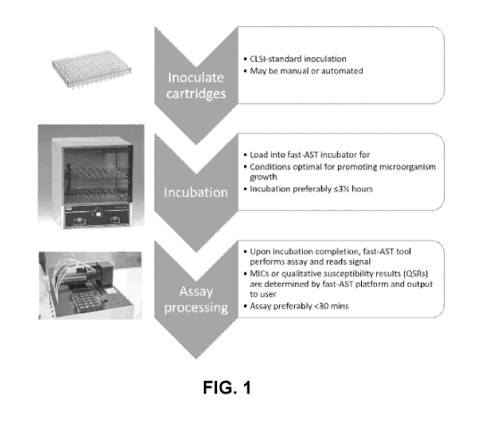
Generalized steps of aspects of the present invention are shown in FIG. 1.
Images in
FIG. 1 show an aspect with distinct process steps; however, aspects of the
present invention
may be automated.
FIG. 2A to FIG. 2D show features of aspects of the present invention. FIG. 2A
shows a detection sensitivity range for three representative pathogens. Dashed
lines show
zero-concentration signal levels. FIG. 2B shows the "Crocodile" (Titertek-
Berthold)
automated fast-AST prototype platform which may be used in the present
invention.
CA 03010895 2018-07-06
WO 2017/127684
PCT/US2017/014343
FIG. 2C is a schematic showing anionic bacteria interacting with cationic
nanolabels and
polymers. The decreased solubility of the resulting neutral complexes allows
magnetic beads
to bind. FIG. 2D shows data for S. aureus with a SensiTitre Gram Positive
panel
(GPALL3F) showing a bacteriostatic (clindamycin) and bactericidal (penicillin)
antimicrobial
results relative to the high-growth and "frozen-in-time (FIT)" controls.
As is known to those skilled in the art, AST platforms may yield minimum
inhibitory
concentration (MIC) results and/or qualitative susceptibility results (QSRs)
for each
antimicrobial tested. MICs are commonly known to be the lowest concentration
of
antimicrobial that inhibits microorganism growth and provides physicians with
dosing
information. QSRs may also provide physicians with similar dosing information
but may not
provide a numerical MIC. AST assays are predominantly configured to test
multiple
antimicrobials in parallel for each obtained biological sample. In order to
produce MIC or
QSR results, dilution series are required for each antimicrobial. Thus, for
liquid-based ASTs,
termed "broth microdilution" by the CLSI, assays are commonly performed in
cartridges
and/or microplates, which enable parallel testing of different antimicrobials
at different
concentrations.
Long times to obtain an AST determination result in incomplete information
being
delivered to physicians. These long times often prevent the identification of
rates of
antimicrobial efficacy, or kill kinetics. This additional information may be
important for
informing treatment. Current ASTs, which are not determined until over six
hours (and
generally over twelve hours) after treatment commences, often lose the ability
to discern
differences between the rate of antimicrobial efficacy: an antimicrobial that
kills a
microorganism instantly looks the same after twelve hours as one that killed
it within four
hours.
Table 1 estimates the effects of different treatments on the number of
bacteria after a
two- hour incubation. Assuming a thirty minute doubling time, untreated
controls should
increase by sixteen-fold. Treatment groups with a "potent" antimicrobial
(defined as one
having efficacy against the bacteria, for example) above the MIC should result
in minimal
microorganism growth and, in the case of bactericidal antimicrobials, death of
the
microorganism. Thus, fewer bacteria are expected than the starting
concentration. Treatment
groups with a "potent" antimicrobial below its MIC should result in
microorganism growth
equal to or lesser than the no-antimicrobial control. Slow-acting
antimicrobials, defined in
this case as those requiring more than two hours to kill the bacteria (e.g.,
as it the case for
16
CA 03010895 2018-07-06
WO 2017/127684
PCT/US2017/014343
bacteriostatic antimicrobials) will produce a signal between the starting
concentration and the
sixteen-fold increase.
Table 1
Potent
No No
Step antimicrobial Step
antimicrobial antimicrobial
at conc.
Starting bacteria
5x10⁵ 5x10⁵ 5x10⁵ 5x10⁵
concentration
Estimated
bacteria
concentration 5x10⁵ to
8x10⁶ <8x10⁶ <5x10⁵
after 2 hours, <8x10⁶
with 30 mm.
doubling time
The starting concentration of bacteria of 5x10⁵ CFU/ml is given in the
American
Society for Microbiology's "Manual of Antimicrobial Susceptibility Testing" 0
2005, with
Marie B. Coyle as the coordinating editor, for the broth micro dilution
technique. Since each
well contains approx. 100 uL, there are approx. 5x10⁴ bacteria per well.
Standard fluorescent
dyes begin to be quantifiable at approx. 0.1 nM concentrations, which
correspond to
approximately 1.2x101 molecules. Thus, for a thirty minute doubling time
bacteria to be
visible after two hours, each individual bacterium would have to be labeled
with 1.5x10⁴
fluorescent molecules. Practical considerations, such as fluorescent
background and non-
specific binding, may increase this number by orders of magnitude. In order to
enable
compatibility with standard optical detectors, it may thus be advantageous to
use a chemical
and/or biochemical amplifier that produces a detectable signal at lower
concentrations.
Without wishing to be bound by theory, the present invention is based in part
on the
principle of broth micro dilution. A culture to be assessed is diluted, most
preferably to 1-
10x105 CFU/ml, and introduced to wells containing different antimicrobials at
different
concentrations, such that MICs can be determined for an appropriate panel of
antimicrobials.
The plate is then introduced into an incubator at the appropriate temperature,
most preferably
31-37 C, and under appropriate conditions, most preferably aerobic, for
growing bacteria.
During this time, the microorganism can grow.
17
CA 03010895 2018-07-06
WO 2017/127684
PCT/US2017/014343
The broth may be cation-adjusted Mueller Hinton broth and may contain
additional
supplements known by those skilled in the art to be advantageous for microbial
growth, such
as lysed horse blood, and/or for determining antimicrobial efficacies, such as
high sodium
chloride concentrations. The microplates may be agitated during this growth
period, which
.. may be advantageous for dispersing nutrients and/or gas exchange and/or
antimicrobials in
each well and/or decreasing biofilm formation.
Within zero to eight hours of the AST onset (most preferably zero to four
hours), a
known quantity of signaling agent is added to each well. Adding reagents
(including signal
generators) may be performed by an automated instrument or a semi-automated
instrument or
may be performed manually.
Signaling agents (which may be referred to as "sticky-amps") comprise a moiety
capable of binding to a microorganism (e.g., an antibody and/or a lectin that
bind to a
microorganism surface, a charged moiety and/or a functional moiety that non-
specifically
binds to the microorganism surface) and a chemical moiety capable of providing
a signal or
contributing to production of a signal (e.g., an enzyme chemiluminophore, and
lanthanide
chelate). Exemplary enzymes include horseradish peroxidase, alkaline
phosphatase, acetyl
cholinesterase, glucose oxidase, beta-D-galactosidase, beta-lactamase, and a
combination
thereof.
As used herein, signal generator may include one or more chemical moieties
(i.e.,
"signal generators") conjugated to one or more "microorganism receptors."
Signal generators
include, but are not limited to, one or more catalysts (including enzymes,
metal-oxide
nanoparticles, organometallic catalysts, nanoparticles designed for signal
amplification (such
as those described in the U.S. Provisional Applications to which the present
application
claims priority and incorporates by reference in their entireties),
bacteriophages comprising
signal generating elements, fluorophores (including organic fluorophores,
europium, or
ruthenium(II), rhenium(I), palladium(II), platinum(II)-containing
organometallics), and/or
colorimetric dyes (including organic "stains"). Combinations of the above may
be used, such
as nanoparticles, dendrimers, and/or other nanoscale structures with enzymes,
fluorophores,
and/or organometallic molecules.
The chemical moiety may be conjugated to a signaling agent before contacting
the
signaling agent to a microorganism, while the signaling agent is initially
contacted to a
microorganism, or after the signaling agent has contacted a microorganism.
When the signaling agents are added to AST dilutions containing a
microorganism,
signaling agent receptors (e.g., moieties that can bind specifically or non-
specifically to a
18
CA 03010895 2018-07-06
WO 2017/127684
PCT/US2017/014343
microorganism) associate with microorganism surfaces. Thus, the more intact
microorganisms, for example, there are in solution, the greater the number of
signaling agents
that will be associated with these bacteria. Consequently, there is an inverse
relationship
between the number of intact bacteria and the number of signaling agents that
are "free" in
solution, as defined by those not bound to intact bacteria. Note that free
signaling agents may
be bound to soluble microbial components if, for example, microorganisms lyse
in response
to antimicrobial treatment.
The number of signaling agents that associate with and/or intercalate into
microorganism surfaces is proportional to the microorganism surface area.
Microorganism
surface area is strongly associated with truly resistant microorganisms. In
particular, in the
case of microorganisms that swell or elongate in response to MIC- and sub-MIC
concentrations of antimicrobials (e.g., filament forming bacteria), metabolic
and/or
volumetric identifications are known to give false susceptibility profiles for
"rapid" AST time
points, defined as those less than six hours. To overcome this limitation, the
present
invention translates microorganism surface area (rather than volume) into a
measurable
signal, most preferably an optical signal. The present methods are able to
accurately
determine microorganism resistance profiles in less than six hours.
In order to separate signaling agents associated with and/or intercalated into
microorganisms from free signaling agents, it may be necessary to perform one
or more
.. separation and/or competitive binding steps. Such steps include, but are
not limited to,
centrifugation (e.g., with a g-force >500 x g), filtration (e.g., via a filter
having pores smaller
than or equal to 0.45 microns, and preferably smaller than or equal to 0.2
microns),
electrophoresis, and/or magnetic capture; such steps are well-known to those
skilled in the
art.
In order to promote signaling agent binding and/or reduce background, it may
further
be advantageous, before adding signaling agents, to separate microorganisms
from the liquid
in which they were suspended during incubation. Such separations may include
but are not
limited to, centrifugation, filtration, electrophoresis, and/or magnetic
capture.
When these data are compared across treatment groups, microbial resistance
profiles
may be determined, using steps similar to currently-used AST determinations.
Additionally,
these data may enable determination of rates of antimicrobial efficacy, or
kill kinetics.
Signaling agents may be added together with microorganisms and/or
antimicrobials,
such that they are present for the entire AST incubation period. This total
period may be up
to twenty-four hours but is preferably within eight hours and more preferably
within five
19
CA 03010895 2018-07-06
WO 2017/127684
PCT/US2017/014343
hours. Alternatively, signaling agents may be added to microorganisms and
antimicrobial
after a prescribed incubation period. This period may be up to twenty-four
hours but is
preferably within eight hours and more preferably within four hours.
Signaling agents are designed to associate with and/or intercalate in
microorganism
surfaces, including walls and/or membranes. Signaling agents designed for
association
comprise binding moieties including, but are not limited to, one or more
antibodies, lectins,
other proteins, small molecules with one or more charged chemical groups,
small molecules
with one or more functional chemical groups, phages, glycoproteins, peptides,
aptamers,
charged small molecules, small molecules with fixed charges, charged polymers,
charged
polymers with fixed charges, hydrophobic small molecules, charged peptide,
charged
peptides with fixed charges, peptides with alternating hydrophilic and
hydrophobic regions,
and/or small molecule ligands, which may or may not be organometallic
complexes.
Molecules designed for microorganism association are well-known to those
skilled in the art.
Signaling agents may remain bound to microorganisms and/or may be
internalized, thus all
associations are included. Signaling agents designed for intercalation may
include, but are not
limited to, small hydrophobic molecules, hydrophobic peptides, and/or peptides
with
alternating hydrophobic and hydrophilic regions. Molecules designed for
microorganism
intercalation are well-known to those skilled in the art. Signaling agents may
further be
specific to one or more types of microorganisms. Signaling agents may have
multiple
receptors. These may enhance binding and/or enable simultaneous binding to two
or more
microorganisms, which may further serve to "agglutinate" bacteria. Prior to or
concurrently
with the addition of signaling agents it may be advantageous to adjust the
solution pH. This
may be beneficial for enhancing charge-charge interactions between
microorganisms and
signaling agents. The anionic charge of microorganisms may be increased by
titrating the
solution pH above neutral (more basic). It may thus be beneficial to utilize
moieties with one
or more fixed, cationic charges.
It is noteworthy that the signaling agent may specifically bind to a
microorganism
(e.g., an antibody that specifically binds to a microorganism species or a
strain of
microorganism) or my non-specifically binds to a microorganism (e.g., by a
generic covalent
or non-covalent bond formation and another non-specific chemical association
known in the
art).
It is preferred that signaling agents bind native microorganism surfaces.
Alternately, chemicals and/or biochemicals which are capable of associating
with
signaling agents may be added to the liquid in which the microorganisms are
suspended
CA 03010895 2018-07-06
WO 2017/127684
PCT/US2017/014343
during growth, such that chemicals and/or biochemicals are incorporated into
microorganisms during incubation. This may serve to enhance signaling agent
association
with microorganisms. In alternative embodiments, the signaling agents
themselves may be
present in the liquid in which the microorganisms are suspended during
incubation and may
be incorporated into microorganisms during growth.
Preferably the signaling agents comprise an amplifier signal generator, such
that the
signal from each intact microorganism may be amplified beyond the number of
signaling
agents associated with each microorganism. For example, the enzyme horseradish
peroxidase (HRP) is known to be able to amplify signals >1x10⁴-fold. Thus, if
one hundred
HRP molecules are bound to each microorganism surface, an amplification of 106
may be
achieved. This may increase the speed with which AST determinations may be
made by
enabling discrimination of microorganism concentrations that cannot otherwise
be
differentiated. Use of Europium formulations similarly provides signal
amplification.
Alternatively, the signaling agents may comprise optical dye precursors known
to
those skilled in the art as "membrane dyes" that are designed to greatly
increase fluorescence
emission upon intercalation into a hydrophobic region, such as a cell
membrane. Assays
designed with these signaling agents may require microorganisms to be
concentrated into a
smaller volume, approaching a plane, to produce sufficient signals so as to be
easily optically
measured. Interfering species may require the use of near-IR fluorophores.
Potential separation techniques include, but are not limited to, filtering
(e.g., via a
filter having pores smaller than or equal to 0.45 microns, preferably smaller
than or equal to
0.2 microns), centrifugation (e.g., with a g-force >500 x g), electrophoresis,
dielectrophoresis, and magnetic capture. These techniques are employed to
separate
signaling agents associated with microorganisms, which are stuck in a filter,
pelleted in a
centrifuge, and/or separated electrophoretically and/or magnetically, from
those free in
solution. Free signaling agents pass through a filter ("filtrate"), remain in
solution after
centrifugation or magnetic separation ("supernatant"), and/or run separately
electrophoretically. Centrifugation may be standard, density gradient, or
differential
centrifugation. Magnetic separation may require the addition of one or more
magnetic
particles specifically targeted to associate with or bind to microorganisms.
These may be
added prior to or concurrently with signaling agent addition.
Such separation techniques may also isolate microorganisms that change
morphology
in response to an antimicrobial treatment and may confound a determination. An
example of
such a microorganism is filamentous bacteria which initially elongate in
response to
21
CA 03010895 2018-07-06
WO 2017/127684
PCT/US2017/014343
antimicrobial treatment. This growth regime is known to those skilled in the
art. Isolating
and excluding filamentous bacteria from an assay, using a herein-described
separation
technique, will increase the accuracy of the obtained results.
Microorganism separation may be enhanced through the association of particles
with
microorganism species. For example, in the case of magnetic separation,
magnetic beads
may associate with microorganisms (specifically or non-specifically). Moieties
present on
magnetic bead surfaces may bind the same surface (or a biomolecule thereof) of
microorganisms as the singling agent or different surface (or a biomolecule
thereof). The
magnetic beads may have the same and/or different moieties as the signaling
agents. For
example, if a signaling agent comprises an antibody that binds to E. coli,
then a magnetic
bead may be functionalized with the same antibody. In other examples, the
signaling agent
may include a motif that binds a microorganism and the magnetic bead is
functionalized to
non-specifically bind to microorganisms.
The one or more binding moieties associated with the magnetic beads may be
identical of different to the chemical moiety associated with or of the
signaling agent.
The one or more binding moieties associated with the magnetic beads may bind
the
microorganisms prior to, simultaneously with, or subsequent to the biding of
the signaling
agent with the microorganism.
The one or more binding moieties associated with the magnetic beads may
associate
with one or more polymers that precipitate microorganisms. The one or more
polymers that
precipitate microorganisms may cationic. The one or more polymers that
precipitate
microorganisms may be poly(ethylene glycol).
Magnetic beads, as are known to those skilled in the art, may range in size
from 20
nm to 20 microns.
After separation, one or more assays to determine the number of signaling
agents
remaining after microorganism separation and/or the number of signaling agents
removed
during microorganism separation ("free" signaling agents) may be performed.
Performing an
assay for free signaling agents provides a signal inversely proportional to
microorganism
concentration. In this case signaling agents associated with microorganisms
may be either
bound to or internalized by microorganisms. Alternatively, an assay may be
performed for
signaling agents associated with microorganisms. In this case, unless
microorganisms are
specifically lysed, only bound signaling agents will contribute to the signal.
In order to maximize separation efficiency, i.e., minimize the number of free
signaling
agents remaining, one or more washing steps may be performed. These may be
continuous,
22
CA 03010895 2018-07-06
WO 2017/127684
PCT/US2017/014343
as in the cases of filtering, magnetic capture, or electrophoresis, and/or
discrete, as in the
cases of centrifugation or magnetic capture.
In alternative embodiments, signaling agents may not require washing. This may
be
the case when "membrane dye" signaling agents are used. Molecules not
intercalated into
microorganism membranes have significantly lower optical activities than
intercalated
species, thus washing may not be required.
One or more washes may be performed before signaling agents are added to the
microorganisms. These washes may, for example, remove interfering species
present in the
liquid in which the microorganisms were suspended during incubation.
In embodiments, no wash is performed.
Signal development may require the addition of a "development solution." For
signaling agents comprising catalysts, the development solution may comprise
one or more
signal precursors that can be converted to an optically and/or electrically
active signaling
molecule. For signaling agents comprising encapsulated molecules, such as
within
nanoparticles, the development solution may comprise one or more reagents to
release the
encapsulated species. At a specified time after addition of the development
solution, a
colorimetric and/or electrochemical signal may be measured. Such signals
include, but are
not limited to, absorbance, fluorescence, time-resolved fluorescence,
chemiluminescence,
electrochemiluminescence, amperometric, voltammetric, impedance, and/or
impedance
spectroscopy. The data may then be compared to determine ASTs and MICs,
similarly to
current AST protocols.
In embodiments, determining signal levels includes measuring the signal levels
associated with intact microorganisms. Alternately or additionally,
determining signal levels
includes measuring the signal levels not associated with intact
microorganisms.
These processes may be performed directly from cultures, sub-cultures,
positive blood
cultures, samples. Treatments to concentrate microorganisms and/or remove
potential
interfering species may be performed prior to AST or prior to signaling agent
addition.
Signaling agents may also be used with plate-based methods for AST
determination,
such as gradient diffusion. They may be added simultaneously upon
microorganism addition
to plates or following a set incubation period. Spatial information for the
optical and/or
electrical signal is important in these cases. With this approach an assay for
intact
microorganism-bound signaling agents may be preferable in order to retain
spatial
information. In this case, one or more wash steps may be performed prior to
the addition of
the development solution in order to remove free signaling agents.
23
CA 03010895 2018-07-06
WO 2017/127684
PCT/US2017/014343
In embodiments, no wash is performed.
Alternatively, signaling agents may be designed to be up-taken by bacteria,
e.g.,
which may be achieved through the use of bacteriophages. In such methods, an
assay for free
signaling agents is performed.
Alternatively, a blot-transfer approach, such as is standard with
nitrocellulose paper,
may be used to transfer bacteria or free signaling agents and a spatial assay
then performed
on the blotted paper.
Separation step(s) may not be required if the signaling agent produces a
signal upon
binding. Alternatively, a separation step-free process may be achieved if the
signaling agent
becomes susceptible or resistant to a specific developer solution constituent
upon binding.
Final MIC and/or QSR output data may be interpreted by a user directly from
the data
produced by the assays described herein. Alternatively, these data may be
processed by one
or more algorithms to yield MICs and/or QSRs. Reported MIC and/or QSR values
may be
derived from one or more of the assays described herein or may be derived from
one or more
of the assays described herein together with one or more known assays for
microorganism
growth including, but not limited to, metabolic dye indicator assays, pH
indicator assays,
nucleic acid assays, and ATP assays.
Methods of the present invention
An aspect of the present invention is a method for determining antimicrobial
susceptibility of microorganisms. The method includes steps of incubating a
liquid
suspension of microorganisms in the presence of an antimicrobial and a
signaling agent under
conditions that promote growth of the microorganisms, wherein the signaling
agent is capable
of binding to a surface of the microorganisms; separating the microorganisms
bound by the
signaling agent from the unbound signaling agent; and determining signal
levels associated
with the microorganisms as compared to one or more controls, thereby
determining the
antimicrobial susceptibility of the microorganisms.
Another aspect of the present invention is a method for determining
antimicrobial
susceptibility of microorganisms. The method includes steps of incubating a
liquid
suspension of microorganisms in the presence of an antimicrobial under
conditions that
promote growth of the microorganisms; adding a signaling agent capable of
binding to a
surface of the microorganisms; separating the microorganisms bound by the
signaling agent
from the unbound signaling agent; and determining signal levels associated
with the
microorganisms as compared to one or more controls, thereby determining the
antimicrobial
24
CA 03010895 2018-07-06
WO 2017/127684
PCT/US2017/014343
susceptibility of the microorganisms. In embodiments, adding the signaling
agent occurs
prior to or during the incubating step or adding the signaling agent occurs
after the incubating
step.
Another aspect of the present invention is a method for determining
antimicrobial
susceptibility of microorganisms. The method includes steps of incubating a
liquid
suspension of microorganisms in a cartridge comprising a plurality of
chambers, each
chamber containing one or more antimicrobials, under conditions that promote
growth of the
microorganisms; adding a signaling agent to the plurality of chambers, wherein
the signaling
agent is capable of binding to a surface of the microorganisms; removing
unbound signaling
agent; and determining signaling levels in the plurality of chambers as
compared to one or
more controls, thereby determining the susceptibility of microorganisms to the
one or more
antimicrobials. In embodiments, the cartridge further includes one or more
control chambers
(e.g., at least 2, 4, 6, 8, 12, 24, 48, 96, 192, 384, 1536 or more chambers)
that do not contain
antimicrobials or one or more antimicrobials for which the microorganisms are
not
.. susceptible.
In embodiments of an above aspect, binding to a surface of the microorganisms
is
non-specific, e.g., comprising a non-covalent interaction and via forming a
covalent bond.
In embodiments of an above aspect, the signaling agent may include a chemical
and/or biochemical group capable of binding a surface of the microorganisms,
wherein the
surface comprises one or more of membranes, walls, proteins, organelles,
saccharides, lipids,
cell envelope, and/or nucleic acids.
In embodiments of an above aspect, the signaling agent may include a chemical
and/or biochemical group capable of binding a biomolecule of the surface of
the
microorganisms, wherein the surface biomolecule is selected from
peptidoglycans, mureins,
mannoproteins, porins, beta-glucans, chitin, glycoproteins, polysaccharides,
lipopolysaccharides, lipooligosaccharides, lipoproteins, endotoxins,
lipoteichoic acids,
teichoic acids, lipid A, carbohydrate binding domains, efflux pumps, other
cell-wall and/or
cell-membrane associated proteins, other anionic phospholipids, and a
combination thereof.
In embodiments of an above aspect, the signaling agent may include a signal
amplifier
and one or more chemical moieties capable of binding non-specifically to a
surface of the
microorganisms.
Another aspect of the present invention is a method for determining
antimicrobial
susceptibility of microorganisms. The method includes incubating
microorganisms in the
presence of an antimicrobial and a signaling agent under conditions that
promote growth of
CA 03010895 2018-07-06
WO 2017/127684
PCT/US2017/014343
the microorganisms, wherein the signaling agent comprises a signal amplifier
and one or
more chemical moieties capable of binding non-specifically to a surface of the
microorganisms; separating the microorganisms bound by the signaling agent
from the
unbound signaling agent; and determining signal levels associated with the
microorganisms
as compared to one or more controls, thereby determining the antimicrobial
susceptibility of
the microorganisms.
Another aspect of the present invention is a method for determining
antimicrobial
susceptibility of microorganisms. The method includes incubating
microorganisms in the
presence of an antimicrobial under conditions that promote growth of the
microorganisms;
adding a signaling agent comprising a signal amplifier and one or more
chemical moieties
capable of binding non-specifically to a surface of the microorganisms;
separating the
microorganisms bound by the signaling agent from the unbound signaling agent;
and
determining signal levels associated with the microorganisms as compared to
one or more
controls, thereby determining the antimicrobial susceptibility of the
microorganisms. In
embodiments, the signaling agent occurs prior to, at the beginning of, or
during the
incubating step, preferably during the incubating step. In embodiments, the
microorganisms
are incubated in a liquid suspension.
In embodiments of an above aspect, the liquid suspension may be prepared by
inoculating a liquid media with a microbial isolate grown from a biological
sample.
In embodiments of an above aspect, the liquid suspension of microorganisms may
be
prepared from an unprocessed biological sample, e.g., an unprocessed
biological sample has
not undergone a culturing step.
In embodiments of an above aspect, the liquid suspension of microorganisms may
be
prepared from a cultured or processed biological sample.
In embodiments of an above aspect, the biological sample is selected from
blood,
cerebrospinal fluid, urine, stool, vaginal, sputum, bronchoalveolar lavage,
throat,
nasal/wound swabs, and a combination thereof.
In embodiments of an above aspect, the method does not involve a step of
capturing
microorganisms on a solid surface prior to or during incubation.
In embodiments of an above aspect, the method does not include a step of
growing
microorganisms on a solid surface during or subsequent to the incubating step.
In embodiments of an above aspect, the incubating may include agitating the
liquid
suspension of microorganisms.
26
CA 03010895 2018-07-06
WO 2017/127684
PCT/US2017/014343
In embodiments of an above aspect, the liquid suspension of microorganisms may
be
agitated by means of mechanical, acoustic, and/or magnetic agitation
continuously or
discretely during the incubating.
In embodiments of an above aspect, the incubating occurs at 31-37 C.Comparison
of
the present invention to currently-used AST systems
The present invention is superior to currently-used AST methods, in part
because it
provides accurate AST results in significantly less time.
Three automated AST systems that are currently used in clinics are
BioMerieux's
Vitek2, Beckman Dickinson's Phoenix, and Beckman-Coulter' s MicroScan. A
comparison
.. between steps in currently-used AST systems and the present invention are
shown in FIG. 3.
The processes described in this invention may be performed in at least two
modes. The first
is for standard isolates, with no changes to current laboratory workflows. The
second is
direct from positive blood cultures.
For standard isolate processing, the present invention is compatible with
existing
.. clinical laboratory workflows and, thus, requires no changes. As shown in
"step 8" of
FIG. 3, current susceptibility (AST) testing is performed after colony
isolation (step 6) and
microorganism concentration standardization (step 7). In this workflow, the
present invention
would replace current systems at "step 8." Because AST results with the
present invention
are available within a healthcare worker's the shift (<5 hours), utility may
increase the speed
with which patients receive optimized therapies (step 9) by up to one day in
practice.
Automation may be designed to include "step 7" and, potentially, additional
steps in the
workflow. Such automation is known to those skilled in the art. Note that FIG.
3 illustrates
the workflow for blood samples. Many sample types, such as urine and swabs,
may be
streaked directly on plates (step 4). In this case, the gram stain may be
performed at step 6.
For a blood test, the currently-used AST systems require an obtained blood
culture to
become detectibly positive (which takes ten or more hours), followed by a sub-
culture step
(of at least twelve hours), and then an AST test (which requires a minimum of
eight hours):
this totals over forty-eight hours, depending on the pathogen, and often takes
greater than
three days in practice. In most workflows, the identification of the organism
happens after
.. the sub-culture step and increasingly done by mass spectrometry. Since both
identification
and AST results are required for clinicians or pharmacists to prescribe proper
targeted
antimicrobials, this delay to AST results directly extends the duration of
broad-spectrum
27
CA 03010895 2018-07-06
WO 2017/127684
PCT/US2017/014343
antimicrobial treatment. Furthermore, the wait is often compounded by one-
shift operation
common in many clinical microbiology laboratories.
Alternatively, the present invention may be used directly from positive blood
cultures. After the standard step 1 and step 2 of blood draw and
incubation/culture, if the
culture is positive, the culture bottle would move directly to a microorganism
isolation (step
3) and then into an automated system (step 4). The present invention can be
fully automated,
requiring a technician to only load the system with a standard cartridge with
microorganism
dilutions and then initiate the four-hour "fast-AST" process. The lab
technician would then
receive the same standard phenotypic results for AST of a minimum inhibitory
concentration
("MIC"). However, the streamlined process would reduce the time to AST by over
twenty-
four hours in theory, and potentially two days in practice, and simplify lab
workflow.
The currently used AST systems perform variants of the Clinical Laboratory
Standards Institute (CLSI) broth microdilution procedure. Bacteria are
inoculated into
multiple wells in parallel, each of which contains one (or more)
antimicrobials at a known
concentration and a nutrient broth. Wells are inoculated at 5 x 10⁵ CFU/ml to
ensure bacteria
are in the log growth phase, important for detecting accurate responses to
antimicrobials.
Microorganism detection is then performed visually.
The slow speed of phenotypic AST testing is due, in part, to its reliance on
microorganism growth in order to produce a detectable optical signal. Bacteria
cannot be
quantified by optical density measurements below a concentration of ¨1 x 10⁸
CFU/ml,
rendering the CLSI starting concentration invisible for a minimum of eight
doubling times.
Since differentiation of microorganism growth in the slowest-growing well from
the no-
growth well is critical for minimum inhibitory concentration (MIC)
determinations,
significantly longer times are required. As is known to those skilled in the
art, some existing
platforms overcome these growth issues by including metabolic probes in the
liquid in which
the microorganisms are suspended during incubation. However, inclusion of
these probes
may miss growth regimes, such as filamentous growth, and may impact the
accuracy of
results.
Once the AST-specific steps have commenced, the currently-used AST systems
still
.. typically require over eight hours to report results for simple, highly-
susceptible bacteria and
require over ten hours for pathogens with complex resistance profiles or slow
growth
kinetics, see FIG. 4.
28
CA 03010895 2018-07-06
WO 2017/127684
PCT/US2017/014343
Additionally, the currently-used automated AST systems suffer from two
shortcomings that prevent the reporting of accurate results in less than six
hours: 1) very
major errors, inaccurate "susceptible" calls for truly-resistant strains; and
2) major errors,
inaccurate "resistant" calls for truly-susceptible strains. Indeed, these
issues have required
BioMerieux and BD to revise their initial four-hour speed claims for the
Vitek2 and
Phoenix .
The existence of very major errors is explained in part by the metabolic
energy
expended by the microorganisms in achieving antimicrobial resistance.
Resistant
microorganisms may alter energy expenditures in response to antimicrobials,
confounding the
results of metabolic probes around the MIC. These may also result from the
present of
additives, such as redox indicators, in the growth media. The prevalence of
major errors is
due primarily to filamentous growth of certain bacteria. This growth regime is
a common
antimicrobial response amongst gram-negative bacteria, in particular to cell-
wall-acting
antimicrobials, such as 0-lactams. Filamentous bacteria continue to replicate
their internal
contents but do not septate. Thus, again, metabolic probes give erroneous near-
MIC results.
The removal of filamentous bacteria was shown to significantly reduce major
errors for the
AST method.
Measurements of relative microorganism surface area, as used in the present
invention, overcome the pitfalls of metabolic probes for AST. First, since
relative surface
area is not confounded by shifts in metabolic activity, fast-AST enables
rapid, accurate
resistance calls. Second, surface area measurements prevent over-resistance
calls. In
contrast to volumetric measurements obtained with metabolic probes of the
currently-used
AST systems, surface area measurements enable accurate differentiation between
true
resistance and filamentous growth. As illustrated in the schematic of FIG. 39,
volumes of
resistant and susceptible filamentous bacteria are difficult to distinguish.
But the lack of
septation creates a filamentous surface area significantly lower than that of
truly resistant
bacteria. Thus, by amplifying each bacteria's surface area, the present
invention is able to
accurately call four-hour, 0-lactam (ampicillin) MICs for E. coli samples
(see, the below
Examples). As illustrated in FIG. 39, the surface area differential between
elongation and
"true" resistance approaches 2/3, which may be detected with an amplified
signal.
Patient
As used herein, the term "patient" (also interchangeably referred to as "host"
or
"subject") refers to any host that can serve as a source of one or more of the
biological
29
CA 03010895 2018-07-06
WO 2017/127684
PCT/US2017/014343
samples or specimens as discussed herein. In certain aspects, the donor will
be a vertebrate
animal, which is intended to denote any animal species (and preferably, a
mammalian species
such as a human being). In certain embodiments, a "patient" refers to any
animal host,
including but not limited to, human and non-human primates, avians, reptiles,
amphibians,
bovines, canines, caprines, cavities, corvines, epines, equines, felines,
hircines, lapines,
leporines, lupines, ovines, porcines, racines, vulpines, and the like,
including, without
limitation, domesticated livestock, herding or migratory animals or birds,
exotics or
zoological specimens, as well as companion animals, pets, and any animal under
the care of a
veterinary practitioner.
Biological samples
The biological sample is any sample that contains a microorganism, e.g., a
bacterium
and a fungal cell.
Exemplary biological samples include, but are not limited to, whole blood,
plasma,
serum, sputum, urine, stool, white blood cells, red blood cells, buffy coat,
tears, mucus,
saliva, semen, vaginal fluids, lymphatic fluid, amniotic fluid, spinal or
cerebrospinal fluid,
peritoneal effusions, pleural effusions, exudates, punctates, epithelial
smears, biopsies, bone
marrow samples, fluids from cysts or abscesses, synovial fluid, vitreous or
aqueous humor,
eye washes or aspirates, bronchoalveolar lavage, bronchial lavage, or
pulmonary lavage, lung
aspirates, and organs and tissues, including but not limited to, liver,
spleen, kidney, lung,
intestine, brain, heart, muscle, pancreas, and the like, swabs (including,
without limitation,
wound swabs, buccal swabs, throat swabs, vaginal swabs, urethral swabs,
cervical swabs,
rectal swabs, lesion swabs, abscess swabs, nasopharyngeal swabs, and the
like), and any
combination thereof. Also included are bacteria cultures or bacteria isolates,
fungal cultures
or fungal isolates. The ordinary-skilled artisan will also appreciate that
isolates, extracts, or
materials obtained from any of the above exemplary biological samples are also
within the
scope of the invention.
Microorganisms obtained from a biological sample may be cultured or otherwise
processed as is routinely performed in the art.
Exemplary Microorganisms
As used herein, infection is meant to include any infectious agent of a
microbial
origin, e.g., a bacterium, a fungal cell, an archaeon, and a protozoan. In
preferred examples,
the infectious agent is a bacterium, e.g., a gram-positive bacterium, a gram-
negative
CA 03010895 2018-07-06
WO 2017/127684
PCT/US2017/014343
bacterium, and an atypical bacteria. The term "antimicrobial resistant
microorganism" is a
microorganism (e.g., bacterium, fungus, archeaon, and protozoan) that is
resistant to one or
more distinct antimicrobials, i.e., anti-bacterial drugs, antifungal drugs,
anti-archaea
medications, and anti-protozoan drugs.
The microorganisms (e.g., a liquid suspension of microorganisms) may include
one
strain of microorganism. The microorganisms may include one species of
microorganism.
The microorganisms may include more than one strain of microorganism. The
microorganisms may include one order of microorganism. The microorganisms may
include
one class of microorganism. The microorganisms may include one family of
microorganism.
The microorganisms may include one kingdom of microorganism.
The microorganisms (e.g., a liquid suspension of microorganisms) may include
more
than one strain of microorganism. The microorganisms may include more than one
species
of microorganism. The microorganisms may include more than one genus of
microorganism.
The microorganisms may include more than one order of microorganism. The
microorganisms may include more than one class of microorganism. The
microorganisms
may include more than one family of microorganism. The microorganisms may
include more
than one kingdom of microorganism.
The microorganism may be a bacterium. Examples of bacterium include and are
not
limited to Acetobacter aura ntius, Acinetobacter bitumen, Acinetobacter spp.,
Actinomyces
israelii, Actinomyces spp., Aerococcus spp., Agrobacterium radiobacter,
Agrobacterium
tumefaciens, Anaplasma, Anaplasma phagocytophilum, Azorhizobium caulinodans,
Azotobacter vinelandii, Bacillus, Bacillus anthracis, Bacillus brevis,
Bacillus cereus, Bacillus
fusiformis, Bacillus licheniformis, Bacillus megaterium, Bacillus mycoides,
Bacillus spp.,
Bacillus stearothermophilus, Bacillus subtilis, Bacillus Thuringiensis,
Bacteroides,
Bacteroides fragilis, Bacteroides gingivalis, Bacteroides melaninogenicus
(also known as
Prevotella melaninogenica), Bartonella, Bartonella henselae, Bartonella
quintana,
Bartonella spp., Bordetella, Bordetella bronchiseptica, Bordetella pertussis,
Bordetella spp.,
Borrelia burgdorferi, Brucella, Brucella abortus, Brucella melitensis,
Brucella spp., Brucella
suis, Burkholderia, Burkholderia cepacia, Burkholderia mallei, Burkholderia
pseudomallei,
Calymmatobacterium granulomatis, Campylobacter, Campylobacter coli,
Campylobacter
fetus, Campylobacter jejuni, Campylobacter pylori, Campylobacter spp.,
Chlamydia,
Chlamydia spp., Chlamydia trachomatis, Chlamydophila, Chlamydophila pneumoniae
(previously called Chlamydia pneumoniae), Chlamydophila psittaci (previously
called
Chlamydia psittaci), Chlamydophila spp., Clostridium, Clostridium botulinum,
Clostridium
31
CA 03010895 2018-07-06
WO 2017/127684
PCT/US2017/014343
difficile, Clostridium perfringens (previously called Clostridium welchii),
Clostridium spp.,
Clostridium tetani, Corynebacterium, Corynebacterium diphtheriae,
Corynebacterium
fusiforme, Corynebacterium spp., Coxiella burnetii, Ehrlichia chaffeensis,
Ehrlichia spp.,
Enterobacter cloacae, Enterobacter spp., Enterococcus, Enterococcus avium,
Enterococcus
durans, Enterococcus faecalis, Enterococcus faecium, Enterococcus galllinarum,
Enterococcus maloratus, Enterococcus spp., Escherichia coli, Francisella spp.,
Francisella
tularensis, Fusobacterium nucleatum, Gardenerella spp., Gardnerella vaginalis,
Haemophilius spp., Haemophilus, Haemophilus ducreyi, Haemophilus influenzae,
Haemophilus parainfluenzae, Haemophilus pertussis, Haemophilus vaginalis,
Helicobacter
pylori, Helicobacter spp., Klebsiella pneumoniae, Klebsiella spp.,
Lactobacillus,
Lactobacillus acidophilus, Lactobacillus bulgaricus, Lactobacillus casei,
Lactobacillus spp.,
Lactococcus lactis, Legionella pneumophila, Legionella spp., Leptospira spp.,
Listeria
monocytogenes, Listeria spp., Methanobacterium extroquens, Microbacterium
multiforme,
Micrococcus luteus, Moraxella catarrhalis, Mycobacterium, Mycobacterium avium,
Mycobacterium bovis, Mycobacterium diphtheriae, Mycobacterium intracellulare,
Mycobacterium leprae, Mycobacterium lepraemurium, Mycobacterium phlei,
Mycobacterium
smegmatis, Mycobacterium spp., Mycobacterium tuberculosis, Mycoplasma,
Mycoplasma
fermentans, Mycoplasma genitalium, Mycoplasma hominis, Mycoplasma penetrans,
Mycoplasma pneumoniae, Mycoplasma spp., Neisseria, Neisseria gonorrhoeae,
Neisseria
meningitidis, Neisseria spp., Nocardia spp., Pasteurella, Pasteurella
multocida, Pasteurella
spp., Pasteurella tularensis, Peptostreptococcus, Porphyromonas gingivalis,
Prevotella
melaninogenica (previously called Bacteroides melaninogenicus), Proteus spp.,
Pseudomonas aeruginosa, Pseudomonas spp., Rhizobium radiobacter, Rickettsia,
Rickettsia
prowazekii, Rickettsia psittaci, Rickettsia quintana, Rickettsia rickettsii,
Rickettsia spp.,
Rickettsia trachomae, Rochalimaea, Rochalimaea henselae, Rochalimaea quintana,
Rothia
dentocariosa, Salmonella, Salmonella enteritidis, Salmonella spp., Salmonella
typhi,
Salmonella typhimurium, Serratia marcescens, Shigella dysenteriae, Shigella
spp., Spirillum
volutans, Staphylococcus, Staphylococcus aureus, Staphylococcus epidermidis,
Staphylococcus spp., Stenotrophomonas maltophilia, Stenotrophomonas spp.,
Streptococcus,
Streptococcus agalactiae, Streptococcus avium, Streptococcus bovis,
Streptococcus cricetus,
Streptococcus faceium, Streptococcus faecalis, Streptococcus ferus,
Streptococcus
gallinarum, Streptococcus lactis, Streptococcus mitior, Streptococcus mitis,
Streptococcus
mutans, Streptococcus oralis, Streptococcus pneumoniae, Streptococcus pyo
genes,
Streptococcus rattus, Streptococcus salivarius, Streptococcus sanguis,
Streptococcus
32
CA 03010895 2018-07-06
WO 2017/127684
PCT/US2017/014343
sobrinus, Streptococcus spp., Treponema, Treponema denticola, Treponema
pallidum,
Treponema spp., Ureaplasma spp., Vibrio, Vibrio cholerae, Vibrio comma, Vibrio
parahaemolyticus, Vibrio spp., Vibrio vulnificus, viridans streptococci,
Wolbachia, Yersinia,
Yersinia enterocolitica, Yersinia pestis, Yersinia pseudotuberculosis., and
Yersinia spp.
The microorganism may be a fungus. Examples of fungi include and are not
limited
to Aspergillus spp., Blastomyces spp., Candida spp., Cladosporium,
Coccidioides spp.,
Cryptococcus spp., Exserohilum, fusarium, Histoplasma spp., Issatchenkia spp.,
mucormycetes, Pneumocystis spp., ringworm, scedosporium, Sporothrix, and
Stachybotrys
spp.
The microorganism may be a protozoan. Examples of protozoan include and are
not
limited to Entamoeba histolytica, Plasmodium spp., Giardia lamblia, and
Trypanosoma
brucei.
Exemplary Antimicrobials
When the microorganism is a bacterium, exemplary antimicrobials include
Amikacin,
Aminoglycoside, Aminoglycoside amoxicillin, Aminoglycosides, Amoxicillin,
Amoxicillin/clavulanate, Ampicillin, Ampicillin/sulbactam, Antitoxin,
Arsphenamine,
Azithromycin, Azlocillin, Aztreonam, 0-lactam, Bacitracin, Capreomycin,
Carbapenems,
Carbenicillin, Cefaclor, Cefadroxil, Cefalexin, Cefalothin, Cefalotin,
Cefamandole,
Cefazolin, Cefdinir, Cefditoren, Cefepime, Cefixime, Cefoperazone, Cefotaxime,
Cefoxitin,
.. Cefpodoxime, Cefprozil, Ceftaroline, Ceftaroline fosamil, Ceftazidime,
Ceftibuten,
Ceftizoxime, Ceftobiprole, Ceftriaxone, Cefuroxime, Cephalosporin,
Chloramphenicol,
Chloramphenicol(Bs), Ciprofloxacin, Clarithromycin, Clindamycin, Clofazimine,
Cloxacillin, Colistin, Co-trimoxazole, Cycloserine, Dalbavancin, Dapsone,
Daptomycin,
Demeclocycline, Dicloxacillin, Dirithromycin, Doripenem, Doxycycline,
Enoxacin,
Ertapenem, Erythromycin, Ethambutol, Ethambutol(Bs), Ethionamide,
Flucloxacillin,
Fluoroquinolone, Fluoroquinolones, Fosfomycin, Furazolidone, Fusidic acid,
Gatifloxacin,
Geldanamycin, Gemifloxacin, Gentamicin, Grepafloxacin, Herbimycin,
Imipenem/Cilastatin,
Isoniazid, Kanamycin, Levofloxacin, Lincomycin, Linezolid, Lomefloxacin,
Loracarbef,
Macrolides, Mafenide, Meropenem, Methicillin, Metronidazole, Mezlocillin,
Minocycline,
Moxifloxacin, Mupirocin, Nafcillin, Nafcillinõ Nalidixic acid, Neomycin,
Netilmicin,
Nitrofurantoin(B s), Norfloxacin, Ofloxacin, Oritavancin, Oxacillin,
Oxytetracycline,
Paromomycin, Penicillin, Penicillin G, Penicillin V, Piperacillin,
Piperacillin/tazobactam,
Platensimycin, Polymyxin B, Posizolid, Pyrazinamide,
Quinupristin/Dalfopristin, Radezolid,
33
CA 03010895 2018-07-06
WO 2017/127684
PCT/US2017/014343
Raxibacumab, Rifabutin, Rifampicin, Rifampin, Rifapentine, Rifaximin,
Roxithromycin,
Silver sulfadiazine, Sparfloxacin, Spectinomycin, Spectinomycin(Bs),
Spiramycin,
Streptogramins, Streptomycin, Sulbactam, Sulfacetamide, Sulfadiazine,
Sulfadimethoxine,
Sulfamethizole, Sulfamethoxazole, Sulfanilimide, Sulfasalazine, Sulfisoxazole,
Sulfonamidochrysoidine, Tedizolid, Teicoplanin, Teixobactin, Telavancin,
Telithromycin,
Temafloxacin, Temocillin, Tetracycline, Thiamphenicol, ticarcillin,
Ticarcillin/clavulanate,
Ticarcillin/clavulanic acid, Tigecycline, Tigecycline(Bs), Tinidazole,
TMP/SMX,
Tobramycin, Torezolid, Trimethoprim(Bs), Trimethoprim-Sulfamethoxazole,
Troleandomycin, Trovafloxacin, Vancomycin, and generics thereof or a variant
thereof.
Antimicrobials whose interactions with the microorganism affect and are
affected by
the negative charges on the microorganism surface include: polycationic
aminoglycosides,
which upon binding the cell surface displace Mg2+ ions, which bridge lipid
membrane
components, thereby disrupting the outer membrane and enhancing drug uptake;
cationic
polymyxins (colistin and polymyxin B), whose binding to the microorganism cell
is also
dependent on the membrane's negative charge and for which both mutational and
plasmid-
mediated resistance occurs by reducing membrane negative charge; and
daptomycin, a
lipopeptide that resembles host innate immune response cationic antimicrobial
peptides and
requires Ca2+ and phosphatidyl glycerol for its membrane-disrupting mechanism
of action
and for which resistance can also involve alteration in cell surface charge.
When the microorganism is a fungus, exemplary antimicrobials include 5-
fluorocytosine, Abafungin, Albaconazole, Allylamines, Amphotericin B, Ancobon,
Anidulafungin, Azole, Balsam of Peru, Benzoic acid, Bifonazole, Butoconazole,
Candicidin,
Caspofungin, Ciclopirox, Clotrimazole, Cresemba, Crystal violet, Diflucan,
Echinocandins,
Econazole, Efinaconazole, Epoxiconazole, Fenticonazole, Filipin, Fluconazole,
Flucytosine,
Grifulvin V, Griseofulvin, Gris-Peg, Haloprogin, Hamycin, Imidazoles,
Isavuconazole,
isavuconazonium, Isoconazole, Itraconazole, Ketoconazole, Lamisil,
Luliconazole,
Micafungin, Miconazole, Natamycin, Noxafil, Nystatin, Omoconazole, Onmel,
Oravig,
Oxiconazole, Posaconazole, Propiconazole, Ravuconazole, Rimocidin,
Sertaconazole,
Sporanox, Sulconazole, Terbinafine, Terconazole, Thiazoles, Thiocarbamate
antifungal,
Tioconazole, Tolnaftate, Triazoles, Undecylenic acid, Vfend, Voriconazole, and
generics
thereof or a variant thereof.
When the microorganism is a protozoan, exemplary antimicrobials include 8-
Aminoquinoline, Acetarsol, Agents against amoebozoa, Ailanthone, Amodiaquine,
Amphotericin B, Amprolium, Antitrichomonal agent, Aplasmomycin, Arsthinol,
Artelinic
34
CA 03010895 2018-07-06
WO 2017/127684
PCT/US2017/014343
acid, Artemether, Artemether/lumefantrine, Artemisinin, Artemotil, Arterolane,
Artesunate,
Artesunate/amodiaquine, Atovaquone, Atovaquone/proguanil, Azanidazole,
Azithromycin,
Benznidazole, Broxyquinoline, Buparvaquone, Carbarsone, Carnidazole,
Chiniofon,
Chloroquine, Chlorproguanil, Chlorproguanil/dapsone,
Chlorproguanil/dapsone/artesunate,
Chlorquinaldol, Chromalveolate antiparasitics, Cinchona, Cipargamin, Clazuril,
Clefamide,
Clioquinol, Coccidiostat, Codinaeopsin, Cotrifazid, Cryptolepine, Cycloguanil,
Dehydroemetine, Difetarsone, Dihydroartemisinin, Diloxanide, Diminazen,
Disulfiram,
Doxycycline, Eflomithine, ELQ-300, Emetine, Etofamide, Excavata
antiparasitics,
Fumagillin, Furazolidone, Glycobiarsol, GNF6702, Halofantrine,
Hydroxychloroquine,
Imidocarb, Ipronidazole, Jesuit's bark, KAF156, Lumefantrine, Maduramicin,
Mefloquine,
Megazol, Meglumine antimoniate, Melarsoprol, Mepacrine, Metronidazole,
Miltefosine,
Neurolenin B, Nicarbazin, Nifurtimox, Nimorazole, Nitarsone, Nitidine,
Nitrofural,
Olivacine, Ornidazole, Oroidin, Pamaquine, Paromomycin, Pentamidine,
Pentavalent
antimonial, Phanquinone, Phenamidine, Piperaquine, Primaquine, Proguanil,
Project 523,
.. Propenidazole, Pyrimethamine, Pyronaridine, Quinfamide, Quinine,
Ronidazole, Schedula
Romana, SCYX-7158, Secnidazole, Semapimod, Sodium stibogluconate,
Spiroindolone,
Sulfadoxine, Sulfadoxine-Pyrimethamine, Sulfalene, Suramin, Tafenoquine,
Teclozan,
Tenonitrozole, Tilbroquinol, Tinidazole, Trimetrexate, Trypanocidal agent,
Warburg's
tincture, and generics thereof or a variant thereof.
An antimicrobial may be a drug that operates by a mechanism similar to a
herein-
recited drug.
Other antimicrobial drugs known in the art may be used in the present
invention.
Liquid suspensions
A liquid suspension of microorganisms may by agitated using mechanical,
acoustic,
and/or magnetic agitation. Examples of mechanical agitation include shaking or
rocking
and/or use of stir bars, stir paddles, stir blades, and/or stir propellers or
impellers.
The microorganism separation is performed by centrifugation (e.g., with a g-
force
>500 x g), magnetic separation, filtration (e.g., via a filter having pores
smaller than or equal
to 0.45 microns, and preferably smaller than or equal to 0.2 microns),
electrophoresis,
dielectrophoresis, precipitation, agglutination, or any combination thereof.
The liquid may include a growth media, such as cation-adjusted Mueller Hinton
broth.
This media may contain one or more additives, known to those skilled in the
art to promote
microorganism growth, and stability. In addition to different antimicrobials,
different test
CA 03010895 2018-07-06
WO 2017/127684
PCT/US2017/014343
wells may contain one or more additives known to improve AST accuracy for
specific
antimicrobials. For example, additional sodium chloride may be added to tests
comprising
oxacillin and additional calcium may be added to tests comprising daptomycin.
Cartridges
The type of cartridge is not limited. A cartridge is a container that is
capable of
holding and allowing growth of a liquid suspension of microorganisms. Non-
limited
examples of a cartridge include a culture flask, a culture dish, a petri dish,
a bioassay dish, a
culture tube, a test tube, a microfuge tube, a bottle, a microwell plate, a
multiwell plate, a
microtiter plate, a microplate. The cartridge may contain one chamber. The
cartridge may
.. include a plurality of chambers each chamber being a space capable of
holding a liquid
suspension in physical isolation from another space; an example of a chamber
is a well in a
multiwall plate. The cartridge may comprise 1, 2, 3, 4, 5, 6, 7, 8, 9, 10, 11,
12, 24, 48, 96,
192, 384, 1536, or more chambers, and any number of chambers in between.
Optical device
Any optical device (e.g., microscope, microplate reader) with a number of
varying
features is capable of detecting a signal is useful in the present invention.
For instance: broad
spectrum lamp (e.g., xenon), narrow spectrum lamps, laser, LED, multi-photon,
confocal or
total-internal reflection illumination can be used for excitation. Cameras
(single or multiple),
single or arrays (1D or 2D) of photodiodes, avalanche photodiodes, CMOS or CCD
sensors,
solid-state photomultipliers (e.g. silicon photomultipliers), and/or
Photomultiplier tube
(single or multiple) with either filter-based or grating-based spectral
resolution (one or more
spectrally resolved emission wavelengths) are possible on the detection side.
Kits
The terms "kits" and "systems," as used herein in the present invention, are
intended
to refer to such things as combinations of multiple signaling agents with one
or more other
types of elements or components (e.g., other types of biochemical reagents,
signal detection
reagents, controls (i.e., positive and negative controls, e.g., chemically
sensitive/resistant
microorganisms), separation means (e.g., filters and magnetic beads),
containers, packages
such as packaging intended for commercial sale, substrates/cartridges to which
.. microorganism suspensions can be cultured, processed, or contained,
electronic hardware
components, and software recorded on a non-transitory processor-readable
medium).
36
CA 03010895 2018-07-06
WO 2017/127684
PCT/US2017/014343
Another aspect of the present invention is a kit for determining antimicrobial
susceptibility of microorganisms. The kit includes a signaling agent capable
of binding to a
surface of the intact microorganisms of interest; a solution for incubating a
sample containing
microorganisms; and one or more reagents for generating signals from the
signaling agent.
In embodiments, the signaling agent is associated with one or more binding
moieties
capable of binding directly or indirectly to the intact microorganisms of
interest.
In embodiments, the one or more binding moieties are selected from antibodies,
lectins, natural and/or synthetic peptides, synthetic and/or natural ligands,
synthetic and/or
natural polymers, synthetic and/or natural glycopolymers, carbohydrade-binding
proteins
and/or polymers, glycoprotein-binding proteins and/or polymers, charged small
molecules,
other proteins, bacteriophages, and/or aptamers.
In embodiments, the one or more binding moieties may be a polyclonal and/or
monoclonal antibody.
In embodiments, the one or more binding moieties may be a synthetic and/or
natural
ligand and/or peptide. The ligand and/or peptide may be selected from bis(zinc-
dipicolylamine), TAT peptide, serine proteases, cathelicidins, cationic
dextrins, cationic
cyclodextrins, salicylic acid, lysine, and a combinations thereof.
In embodiments, the one or more binding moieties may be a synthetic and/or
natural
polymer and/or glycopolymer. The natural and/or synthetic polymer may be
amylopectin,
Poly(N-l3-(dimethylamino)propyll methacrylamide), poly(ethyleneimine), poly-L-
lysine,
poly[2-(N,N-dimethylamino)ethyl methacrylatel, and combinations thereof. The
natural
and/or synthetic polymer and/or glycopolymer my include moieties including,
but not limited
to, chitosan, gelatin, dextran, trehalose, cellulose, mannose, cationic
dextrans and
cyclodextrans, or combinations thereof including, but not limited to, co-
block, graft, and
alternating polymers.
In embodiments, the one or more binding moieties may include a glycoprotein
selected from mannose-binding lectin, other lectins, annexins, and
combinations thereof.
In embodiments, the one or more binding moieties may include two or more
binding
moieties.
In embodiments, the one or more binding moieties may bind directly or
indirectly to
one or more biomolecules present on the microorganism surface. Exemplary
biomolecules
include peptidoglycans, mureins, mannoproteins, porins, beta-glucans, chitin,
glycoproteins,
polysaccharides, lipopolysaccharides, lipooligosaccharides, lipoproteins,
endotoxins,
lipoteichoic acids, teichoic acids, lipid A, carbohydrade binding domains,
efflux pumps, other
37
CA 03010895 2018-07-06
WO 2017/127684
PCT/US2017/014343
cell-wall and/or cell-membrane associated proteins, other anionic
phospholipids, and a
combination thereof.
In embodiments, the binding moiety is a nanoparticle.
In embodiments, the binding moiety is a bacteriophage.
In embodiments, the one or more binding moieties may bind to one or more
biomolecules specifically.
In embodiments, the one or more binding moieties may bind to one or more
species-
specific biomolecules.
In embodiments, the one or more binding moieties may bind to the
microorganisms
non-specifically, e.g., via a non-covalent interaction and via forming a
covalent bond.
In embodiments, the kit further includes magnetic beads to magnetically
separate the
microorganisms from the supernatant.
In embodiments, the magnetic beads are associated with one or more binding
moieties
that bind to microorganisms. The one or more binding moieties associated with
the magnetic
beads may identical to those associated with the signaling agents. The one or
more binding
moieties associated with the magnetic beads may be different than those
associated with the
signaling agents.
In embodiments, the magnetic beads have diameters ranging between 20 nm to 20
microns.
In embodiments, the kit further includes one or more ions or small molecules
to
enhance the binding between the binding moieties and the microorganism.
In embodiments, the solution comprises <0.15 M salt.
In embodiments, the kit further includes a microorganism binding agent, and
wherein
the binding moiety binds to the microorganisms indirectly via the
microorganism binding
agent. The binding moiety may be conjugated to streptavidin, neutravidin, or
avidin and the
microorganism binding agent may be biotinylated. The binding moiety may be an
antibody
that binds to a species specific Fc domain and the microorganism binding agent
may be an
antibody capable of binding to the microorganisms with the species specific Fc
domain.
In embodiments, the signaling agent may include one or more of a
chemiluminophore,
a catalyst, or an enzyme. The enzyme may be at least one of horseradish
peroxidase, alkaline
phosphatase, acetyl cholinesterase, glucose oxidase, beta-D-galactosidase,
beta-lactamase,
and combinations thereof. The catalyst may be an organometallic compound.
In embodiments, the signaling agent is provided in a form of a nanoparticle,
e.g., the
signaling agent is encapsulated within a nanoparticle. The nanoparticle may be
dissociable,
38
CA 03010895 2018-07-06
WO 2017/127684
PCT/US2017/014343
which may include a metal oxide; the metal oxide may be or include iron oxide,
cesium
oxide, and /or cerium oxide.
In embodiments, zero, one, or two washes are performed prior to determining
signal
levels.
In embodiments, zero, one, or two washes are performed prior to addition of
the
signaling agents.
In embodiments, the kit further includes a developer reagent to produce a
measurable
signal.
In embodiments, the one or more reagents include reagents for a catalytic
reaction and
a reagent that stops the catalytic reaction.
In embodiments, the kit further comprises a device for measuring signal, e.g.,
an
optical and/or electrical signal. The optical measurement may be fluorescent,
time-resolved
fluorescent, absorbent, and/or luminescent.
In embodiments, the kit further comprises a multiwell plate, e.g., a 24-well,
96-well,
192-well, or 384-well plate.
In embodiments, the further contains instructions for using the kit to perform
a herein-
disclosed method. The kit may additionally contain instructions for performing
steps
conducted prior to or subsequent to one or more methods as described herein.
In one embodiment, kits are provided which contain the necessary reagents to
carry
out one or more methods as described herein or reagents necessary to carry out
steps prior to
or subsequent to one or more methods as described herein.
Treatment Methods
As used herein, the terms "treat," "treating," "treatment," and the like refer
to
reducing or ameliorating a disorder and/or a symptom associated therewith. It
will be
appreciated that, although not precluded, treating a disorder or condition
does not require that
the disorder, condition or symptoms associated therewith be completely
eliminated. Treating
may include a health care professional or diagnostic scientist making a
recommendation to a
subject for a desired course of action or treatment regimen, e.g., a
prescription.
As used herein, the terms "prevent," "preventing," "prevention," "prophylactic
treatment" and the like refer to reducing the probability of developing a
disorder or condition
in a subject, who does not have, but is at risk of or susceptible to
developing a disorder or
condition.
39
CA 03010895 2018-07-06
WO 2017/127684
PCT/US2017/014343
The term "methods of treating" includes methods of managing, and when used in
connection with the infections microbial organism or infection, includes the
amelioration,
elimination, reduction, prevention, or other relief or management from the
detrimental effects
of the infections microbe.
As used herein, the terms "drug", "medication", "therapeutic", "active agent",
"therapeutic compound", "composition", or "compound" are used interchangeably
and refer
to any chemical entity, pharmaceutical, drug, biological, botanical, and the
like that can be
used to treat or prevent a disease, illness, condition, or disorder of bodily
function. A drug
may comprise both known and potentially therapeutic compounds. A drug may be
determined to be therapeutic by screening using the screening known to those
having
ordinary skill in the art. A "known therapeutic compound", "drug", or
"medication" refers to
a therapeutic compound that has been shown (e.g., through animal trials or
prior experience
with administration to humans) to be effective in such treatment. A
"therapeutic regimen"
relates to a treatment comprising a "drug", "medication", "therapeutic",
"active agent",
"therapeutic compound", "composition", or "compound" as disclosed herein
and/or a
treatment comprising behavioral modification by the subject and/or a treatment
comprising a
surgical means.
An antimicrobial, e.g., an antibiotic, is an agent capable of killing a
microorganism or
inhibiting the growth of a microorganism.
Signaling Agents and Chemical Attachment
In embodiments, the invention features a signaling agent capable of binding to
the
surface of a microorganism. In embodiments, said binding is non-specific. In
embodiments,
said binding is specific.
In embodiments, a signaling agent is present during an incubating step of a
method
described herein. In embodiments, a signaling agent is present after an
incubating step of a
method described herein.
In embodiments, binding comprises the formation of a covalent bond. In
embodiments, a signaling agent is capable of binding to the surface of a
microorganism,
wherein said binding comprises the formation of a covalent bond. In
embodiments, a method
as described herein results in the formation of a covalent bond between a
group on a
microorganism surface (e.g., via a reactive group such as an electrophilic or
nucleophilic
group as described herein) and a signaling agent as described herein. In
embodiments, a
signaling agent has formed a covalent bond to the surface of a microorganism.
CA 03010895 2018-07-06
WO 2017/127684
PCT/US2017/014343
In embodiments, binding comprises the formation of a non-covalent interaction.
In
embodiments, a signaling agent is capable of binding to the surface of a
microorganism,
wherein said binding comprises the formation of a non-covalent interaction. In
embodiments,
a method as described herein results in the formation of non-covalent
interaction between a
group on a microorganism surface (e.g., via a reactive group such as an
electrophilic or
nucleophilic group as described herein) and a signaling agent as described
herein. In
embodiments, a signaling agent has formed a non-covalent interaction with the
surface of a
microorganism.
In embodiments, a non-covalent interaction comprises: ionic interaction, ion-
ion
interaction, dipole-dipole interaction, ion-dipole interaction, electrostatic
interaction, London
dispersion, van der Waals interaction, hydrogen bonding, 7E- it interaction,
hydrophobic
interaction, or any combination thereof. In embodiments, a non-covalent
interaction is: ionic
interaction, ion-ion interaction, dipole-dipole interaction, ion-dipole
interaction, electrostatic
interaction, London dispersion, van der Waals interaction, hydrogen bonding,
7E- it
interaction, hydrophobic interaction, or any combination thereof.
In embodiments, a non-covalent interaction comprises ionic interactions, van
der
Waals interactions, hydrophobic interactions, 7E-7E interactions, or hydrogen
bonding, or any
combination thereof. In embodiments, a non-covalent interaction comprises
ionic interaction,
van der Waals interaction, hydrogen bonding, or 7E- it interaction, or any
combination thereof.
In embodiments, a signaling agent capable of binding to a microorganism's
surface
comprises a group (e.g., a chemical or biochemical group) capable of binding
microorganism
membranes, walls, proteins, organelles, saccharides, lipids, cell envelope, or
nucleic acids, or
any combination thereof. In embodiments, a signaling agent capable of binding
to a
microorganism's surface comprises a chemical group (e.g., a nucleophilic group
or an
electrophilic group) capable of binding microorganism membranes, walls,
proteins,
organelles, saccharides, lipids, cell envelope, or nucleic acids, or any
combination thereof. In
embodiments, a signaling agent capable of binding to a microorganism's surface
comprises a
biochemical group capable of binding microorganism membranes, walls, proteins,
organelles,
saccharides, lipids, cell envelope, or nucleic acids, or any combination
thereof.
In embodiments, the surface may include a biomolecule to which the signaling
agent
binds or associates. Exemplary biomolecules include peptidoglycans, mureins,
mannoproteins, porins, beta-glucans, chitin, glycoproteins, polysaccharides,
lipopolysaccharides, lipooligosaccharides, lipoproteins, endotoxins,
lipoteichoic acids,
41
CA 03010895 2018-07-06
WO 2017/127684
PCT/US2017/014343
teichoic acids, lipid A, carbohydrate binding domains, efflux pumps, other
cell-wall and/or
cell-membrane associated proteins, other anionic phospholipids, and a
combination thereof.
In embodiments, a signaling agent capable of binding to a microorganism's
surface
comprises a biochemical group capable of binding microorganism membranes,
walls,
proteins, organelles, saccharides, lipids, cell envelope, or nucleic acids, or
any combination
thereof.
In embodiments, a signaling agent capable of binding to a microorganism's
surface
comprises a chemical group (e.g., a nucleophilic or electrophilic functional
group) capable of
binding microorganism membranes, walls, proteins, organelles, saccharides,
lipids, cell
envelope, or nucleic acids, or any combination thereof. In embodiments, said
chemical group
is a nucleophilic functional group. In embodiments, said chemical group is an
electrophilic
functional group.
In embodiments, a signaling agent is a biochemical signaling agent. In
embodiments,
a biochemical signaling agent comprises a biomolecule such as an antibody,
ligand, protein,
aptamer, ss-DNA, ss-RNA, or ss-PNA).
In embodiments, a signaling agent is a chemical signaling agent. In
embodiments, a
chemical signaling agent is a chemical compound (e.g., a synthetic chemical
compound). In
embodiments, a chemical signaling agent does not comprise a biomolecule such
as an
antibody, ligand, protein, aptamer, ss-DNA, ss-RNA, or ss-PNA).
In embodiments, a signaling agent capable of binding to a microorganism's
surface
comprises
a linker group L; and
an amplifier group (e.g., an amplifier group 104 that is a chemical or
biochemical amplifier).
In embodiments, an amplifier group is an amplifier group 104, which is a
chemical or
biochemical amplifier. In embodiments, an amplifier group 104 is a chemical
amplifier. In
embodiments, an amplifier group 104 is a biochemical amplifier.
In embodiments, a signaling agent is a chemical compound. In embodiments, a
chemical compound comprises a chemical amplifier group such as those described
herein).
In embodiments, a linker group L comprises the conserved (Fc) region of an
antibody.
In embodiments, a linker group L is capable of forming a covalent bond to an
amplifier group (e.g., an amplifier group 104 that is a chemical or
biochemical amplifier).
In embodiments, a linker group L forms a covalent bond to a signal amplifier
group
(e.g., an amplifier group 104 that is a chemical or biochemical amplifier).
42
CA 03010895 2018-07-06
WO 2017/127684
PCT/US2017/014343
In embodiments, a linker group L is capable of forming one or more non-
covalent
interactions to an amplifier group (e.g., an amplifier group 104 that is a
chemical or
biochemical amplifier).
In embodiments, a linker group L forms one or more non-covalent interactions
to an
amplifier group (e.g., an amplifier group 104 that is a chemical or
biochemical amplifier).
In embodiments, a linker group L comprises a group (e.g., a chemical or
biochemical
group) capable of binding a microorganism surface. In embodiments, a linker
group L
comprises a group (e.g., a chemical or biochemical group) that binds a
microorganism
surface.
In embodiments, a linker group L comprises a group (e.g., a chemical or
biochemical
group) that is capable of forming a covalent bond to a microorganism's
surface. In
embodiments, a linker group L comprises a group (e.g., a chemical or
biochemical group)
that forms a covalent bond to a microorganism's surface.
In embodiments, a linker group L comprises a group (e.g., a chemical or
biochemical
group) that is capable of forming one or more non-covalent interactions with a
microorganism's surface. In embodiments, a linker group L comprises a group
(e.g., a
chemical or biochemical group) that forms one or more non-covalent
interactions with a
microorganism's surface.
In embodiments, a linker group L comprises a chemical moiety 101, wherein said
chemical moiety is capable of forming a non-covalent interaction with the
surface of a
microorganism. In embodiments, a linker group L comprises a chemical moiety
101, wherein
said chemical moiety is capable of forming a covalent bond with the surface of
a
microorganism. In embodiments, a linker group L comprises a chemical moiety
101, wherein
said chemical moiety forms a non-covalent interaction with the surface of a
microorganism.
In embodiments, a linker group L comprises a chemical moiety 101, wherein said
chemical
moiety forms a covalent bond with the surface of a microorganism.
In embodiments, a linker group L comprises a spacer moiety 102. In
embodiments,
spacer moiety 102 is covalently attached to chemical moiety 101 and/or to
chemical moiety
103. In embodiments, spacer moiety 102 is covalently attached to chemical
moiety 101. In
embodiments, spacer moiety 102 is covalently attached to chemical moiety 103.
In
embodiments, spacer moiety 102 is covalently attached to chemical moiety 101
and to
chemical moiety 103. In embodiments, spacer moiety 102 forms a non-covalent
interaction
with chemical moiety 101 and/or with chemical moiety 103. In embodiments,
spacer moiety
102 forms a non-covalent interaction with chemical moiety 101. In embodiments,
spacer
43
CA 03010895 2018-07-06
WO 2017/127684
PCT/US2017/014343
moiety 102 forms a non-covalent interaction with chemical moiety 103. In
embodiments,
spacer moiety 102 forms a non-covalent interaction with chemical moiety 101
and with
chemical moiety 103.
In embodiments, a linker group L comprises a chemical moiety 103, wherein said
chemical moiety is capable of forming a covalent bond to an amplifier group
(e.g., an
amplifier group 104 that is a chemical or biochemical amplifier). In
embodiments, a linker
group L comprises a chemical moiety 103, wherein said chemical moiety has
formed a
covalent bond to an amplifier group (e.g., an amplifier group 104 that is a
chemical or
biochemical amplifier). In embodiments, a linker group L comprises a chemical
moiety 103,
wherein said chemical moiety is capable of forming a non-covalent interaction
with an
amplifier group (e.g., an amplifier group 104 that is a chemical or
biochemical amplifier). In
embodiments, a linker group L comprises a chemical moiety 103, wherein said
chemical
moiety has formed a non-covalent interaction with an amplifier group (e.g., an
amplifier
group 104 that is a chemical or biochemical amplifier).
In embodiments, a signaling agent is a chemical compound comprising a linker
group
L that comprises:
a chemical moiety 101, wherein said chemical moiety is capable of forming a
covalent bond or a non-covalent interaction with the surface of the
microorganisms;
a spacer moiety 102, wherein spacer moiety is covalently attached to chemical
moiety
.. 101 and to chemical moiety 103; and
a chemical moiety 103, wherein said chemical moiety has formed or can form a
covalent bond to an amplifier group 104 that is a chemical or biochemical
amplifier.
In embodiments, a signaling agent is a chemical compound comprising a linker
group
L that comprises:
a chemical moiety 101, wherein said chemical moiety is capable of forming a
covalent bond or a non-covalent interaction with the surface of a
microorganism;
a spacer moiety 102, wherein spacer moiety is covalently attached to chemical
moiety
101 and to chemical moiety 103; and
a chemical moiety 103, wherein said chemical moiety has formed or can form a
non-
covalent interaction with an amplifier group 104 that is a chemical or
biochemical amplifier.
In embodiments, a linker group comprises one chemical moiety 101. In
embodiments, a linker group comprises more than one chemical moiety 101 (e.g.,
a linker
group comprises 1, 2, 3, 4, 5, or 6 chemical moieties 101).
44
CA 03010895 2018-07-06
WO 2017/127684
PCT/US2017/014343
In embodiments, a linker group comprises one spacer moiety 102. In
embodiments, a
linker group comprises more than one spacer moiety 102 (e.g., a linker group
comprises 1, 2,
3, 4, 5, or 6 spacer moieties 102).
In embodiments, a linker group comprises one chemical moiety 103. In
embodiments, a linker group comprises more than one chemical moiety 103 (e.g.,
a linker
group comprises 1, 2, 3, 4, 5, or 6 chemical moieties 103).
In embodiments, a linker group comprises: one chemical moiety 101, one spacer
moiety 102, and one chemical moiety 103. In embodiments, a linker group
consists of: one
chemical moiety 101, one spacer moiety 102, and one chemical moiety 103.
In embodiments, a linker group has the structure of substructure (I):
-101-102-103- , (I)
wherein
"101" represents a chemical moiety 101;
"102" represents a spacer moiety 102; and
"103" represents a chemical moiety 103.
In embodiments, a chemical moiety 101 is capable of forming a covalent bond
with
the surface of a microorganism.
In embodiments, a chemical moiety 101 is capable of forming a covalent bond
with
the surface of a microorganism in the presence of one or more agents that
promote coupling
(also referred to herein as coupling agents).
In embodiments, agents that promote coupling include glutaraldehyde,
formaldehyde,
paraformaldehyde, 1-ethyl-3-(3-dimethylaminopropyl)carbodiimide (EDC), N,N'-
dicyclohexylcarbodiimide (DCC), N-cyclohexyl-N'-(2-
morpholinoethyl)carbodiimide-
methyl-p-toluenesulfonate (CMC), diisopropylcarbodiimide (DIC), (1-
lbis(dimethylamino)methylenel-1H-1,2,3-triazolol4,5-blpyridinium 3-oxid
hexafluorophosphate) (HATU), Woodward's Reagent, /V,N'-carbonyl diimidazole, N-
hydroysuccinimide (NHS), or N-hydroxysulfosuccinimide (sulfo-NHS), or any
combination
thereof.
In embodiments, agents that promote coupling include aldehydes, acrylates,
amides,
imides, anhydrides, chlorotriazines, epoxides, isocyanates, isothiocyanates,
organic acids,
monomers, polymers, silanes, or silcates, or any combination thereof.
In embodiments, agents that promote coupling include a carbodiimide, a
phosphonium salt, or an ammonium salt, or any combination thereof.
CA 03010895 2018-07-06
WO 2017/127684
PCT/US2017/014343
In embodiments agents that promote coupling include glutaraldehyde, N-(3-
dimethylaminopropy1)-N' -ethylcarbonate (EDC), (1-
lbis(dimethylamino)methylenel-1H-
1,2,3-triazolol4,5-blpyridinium 3-oxid hexafluorophosphate) (HATU), (0-
benzotriazol-1-yl-
N,N,N ,N -tetramethyluronium hexafluorophosphate) (HBTU), N-
.. hydroxysuccinimide (NHS), /V,N'-dicyclohexylcarbodiimide (DCC),
diisopropylcarbodiimide
(DIC), hydroxy-3,4-dihydro-4-oxo-1,2,3-benzotriazine (HOOBt),
hydroxybenzotriazole (HOB T), 1-hydroxy-7-azabenzotriazole (HOAt), (N-(3-
dimethylaminopropy1)-N' -ethylcarbodiimide (EDAC), 4-(N,N-
dimethylamino)pyridine
(DMAP), benzotriazol-1-yloxy-tris(dimethylamino)- phosphonium
hexafluorophosphate
(BOP), benzotriazol-l-yloxy-tripyrrolidino-phosphonium hexafluorophosphate
(PyB OP),
bromo-tripyrrolidino-phosphonium hexafluorophosphate (PyBrOP), 7-aza-
benzotriazol-1-
yloxy-tripyrrolidinophosphonium hexafluorophosphate (PyA0P), ethyl
cyano(hydroxyimino)acetato-02 )- tri-(1-pyrrolidiny1)-phosphonium
hexafluorophosphate
(PyOxim), 3-(diethoxy-phosphoryloxy)-1,2,3-benzoldl triazin-4(3H)-one (DEPBT),
2-(6-
chloro-1H-benzotriazol-1-y1)- N,N,N',N'-tetramethylaminium hexafluorophosphate
(HCTU),
N-R5-chloro-1H-benzotriazol-1-y1)- dimethylamino-morpholinol-uronium
hexafluorophosphate N-oxide (HDMC), 1-l1-(cyano-2-ethoxy-2-
oxoethylideneaminooxy)-
dimethylamino-morpholinol- uronium hexafluorophosphate (COMU), 2-(1-oxy-
pyridin-2-
y1)-1,1,3,3-tetramethylisothiouronium tetrafluoroborate (TOTT),
.. tetramethylfluoroformamidinium hexafluorophosphate (TFFH), N-Ethoxycarbony1-
2-ethoxy-
1,2-dihydroquinoline (EEDQ), 2-propanephosphonic acid anhydride (PPA),
triphosgene,
1,1'-carbonyldiimidazole (CDI), R6-nitrobenzotriazol-1-
yl)oxyltris(pyrrolidino)phosphonium hexafluorophosphate (PyNOP), ll6-
(trifluoromethyl)benzotriazol-1-ylloxyltris(pyrroli-dino)phosphonium
hexafluorophosphate
(PyFOP), ll4-nitro-6-(trifluoromethyl)benzotriazol-1-
ylloxyltris(pyrrolidino)phosphonium
hexafluorophosphate (PyNFOP), R6-nitrobenzo-triazol-1-yl)oxyltris(dimethyl-
amino)phosphonium hexafluorophosphate (NOP), 1-fl-naphthalenesulfonyloxy
benzotriazole
(NSBt), 1-0-naphthalenesulfonyloxy-6-nitrobenzotriazole (N-NSBt),
tetramethylfluoroformamidinium hexafluorophosphate (TFFH),
bis(tetramethylene)fluoroformamidinium hexafluorophosphate (BTFFH), 1,3-
dimethy1-2-
fluoro-4,5-dihydro-1H-imidazolium hexafluorophosphate (DFIH), Cyanuric
chloride (CC), or
2,4-dichloro-6-methoxy-1,3,5-triazine (DCMT), and 2-chloro-4,6-dimethoxy-1,3,5-
triazine
(CDMT), or any combination thereof.
46
CA 03010895 2018-07-06
WO 2017/127684
PCT/US2017/014343
In embodiments, agents that promote coupling include EDC, HATU, HBTU, NHS,
DCC, HOBT, or PyBOP, or any combination thereof.
In embodiments, agents that promote coupling include EDC, DCC, CMC, DIC, or
HATU, or any combination thereof.
In embodiments, agents that promote coupling include glutaraldehyde,
formaldehyde,
or paraformaldehyde, or any combination thereof.
In embodiments, a chemical moiety 101 is capable of forming a non-covalent
interaction with the surface of a microorganism (e.g., any non-covalent
interaction described
herein). In embodiments, a non-covalent interaction comprises: ionic, ion-ion,
dipole-dipole,
ion-dipole, electrostatic, London dispersion, van der Waals, hydrogen bonding,
or 7E- it, or
any combination thereof.
In embodiments, a chemical moiety 101 is capable of forming a non-covalent
interaction with the surface of a microorganism, wherein said non-covalent
interaction
comprises ionic interactions, van der Waals interactions, hydrophobic
interactions, 7E-7E
interactions, or hydrogen bonding, or any combination thereof.
In embodiments, a chemical moiety 101 comprises a nucleophilic functional
group.
In embodiments, a chemical moiety 101 comprises a group formed from a
nucleophilic
functional group.
In embodiments, a nucleophilic functional group is: amino, amido, hydrazino,
hydroxyamino, hydroxy, or thio. In embodiments, a nucleophilic functional
group is: amino,
hydrazino, hydroxyamino, or thio. In embodiments, a nucleophilic functional
group
comprises: amino, hydrazino, hydroxyamino, hydroxy, or thio. In embodiments, a
nucleophilic functional group is carboxamide, N-hydroxycarboxamide, carboxyl
hydrazide,
or guanidino.
In embodiments, a nucleophilic functional group is ¨NH2, -NHNH2, -CONHOH,
-CONHNH2,¨ONH2, -OH, or -SH. In embodiments, a nucleophilic functional group
is ¨NH2,
-NHNH2, -CONHNH2, or ¨ONH2.
In embodiments, a chemical moiety 101 comprises an electrophilic functional
group.
In embodiments, a chemical moiety 101 comprises a group formed from an
electrophilic functional group.
In embodiments, an electrophilic functional group comprises an aldehyde, a
ketone, a
carboxylic acid, a carboxylic ester, a carboxylic acid halide (e.g., acetyl
chloride), or a
carboxylic acid anhydride (e.g., acetic anhydride).
47
CA 03010895 2018-07-06
WO 2017/127684
PCT/US2017/014343
In embodiments, an electrophilic functional group comprises an aldehyde, an a-
halo
ketone, a maleimide, a succinimide, a hydroxysuccinimide, an isothiocyanate,
an isocyanate,
an acyl azide, a sulfonyl chloride, a tosylate ester, a glyoxal, an epoxide,
an oxirane, a
carbonate, an imidoester, an anhydride, a fluorophenyl ester, a hydroxymethyl
phosphine
derivative, a carbonate, a haloacetyl, a chlorotriazine, a haloacetyl, an
alkyl halide, an
aziridine, or an acryloyl derivative. In embodiments, an electrophilic
functional group is an
aldehyde, an a-halo ketone, a maleimide, a succinimide, a hydroxysuccinimide,
an
isothiocyanate, an isocyanate, an acyl azide, a sulfonyl chloride, a tosylate
ester, a glyoxal, an
epoxide, an oxirane, a carbonate, an imidoester, an anhydride, a fluorophenyl
ester, a
hydroxymethyl phosphine derivative, a carbonate, a haloacetyl, a
chlorotriazine, a haloacetyl,
an alkyl halide, an aziridine, or an acryloyl derivative.
In embodiments, an electrophilic functional group comprises an aldehyde, an a-
halo
ketone, a maleimide, a succinimide, or a hydroxysuccinimide group.
In embodiments, an electrophilic functional group comprises ¨CHO,
0
NI 0 0 II *
Or_s_r1 0 0__r0 A A NN
-C(0)CH2I, 0 0
0*C*N*
, or S"
In embodiments, an electrophilic functional group comprises ¨CHO,
0
-C(0)CH2I, \=/ , or
In embodiments, chemical moiety 101 comprises a group that is alkyl, alkenyl,
alkynyl, phenyl, heteroaryl, haloalkyl, hydroxy, carbonyl, acyl halide,
alkoxycarbonylloxy,
carboxy, haloketone, alkoxy, alkoxyol (hemiacetal or) hemiketal, dialkoxy
(e.g., ketal or
acetal), trialkoxy(orthoether), carbamoyl, amino, ammonio, imino, imido,
succinamido,
maleidido, hydroxysuccinamido, biotin, D-Biotin, azido, azo, cyanate,
isocyanato, nitroxy,
cyano, isocyano, nitrosooxy, nitro, nitroso, oxime, sulfanyl, sulfinyl,
sulfonyl, sulfino, sulfo,
thiocyanato, isothiocyanato, thioyl, phosphate, or boronate.
In embodiments, spacer moiety 102 is hydrophobic. In embodiments, spacer
moiety
102 is hydrophilic.
In embodiments, spacer moiety 102 is peptidic (e.g., derived from peptide
linkages).
48
CA 03010895 2018-07-06
WO 2017/127684
PCT/US2017/014343
In embodiments, spacer moiety 102 comprises inorganic linkages. In
embodiments,
spacer moiety 102 comprises organic linkages. In embodiments, spacer moiety
102
comprises only organic linkages.
In embodiments, spacer moiety 102 is oligomeric. In embodiments, spacer moiety
102 is polymeric. In embodiments, spacer moiety 102 comprises segments (e.g.,
1 to about
300, 1 to about 200, 1 to about 100, 1 to about 50, 1 to about 25, or 1 to
about 10, or 1, 2, 3,
4, 5, 6, 7, 8, 9, or 10 segments) of methylene (-CH2-), ethylene glycol (-
CH2CH20-),
iminoethylene (-CH2CH2NH-), vinyl alcohol (-CH2CHOH-),õ lactic acid (-CH(CH3)-
C(0)-0-
), acrylic acid (-CH2CH2(CO2H)-), methacrylic acid (-CH2C(CH3)(CO2H)-1, or
methyl
methacrylate (-CH2C(CH3)(CO2CH3)-)-
*/*
In embodiments, spacer moiety 102 comprises a segment that is "n
*,of* *!'y
rn H P , or HO
In embodiments, n, m, p, and q independently is an integer of 1 to about 300
(e.g., 1
to about 200, 1 to about 100, 1 to about 50, 1 to about 25, or 1 to about 10).
In embodiments,
each of n, m, p, and q is independently 1, 2, 3, 4, 5, 6, 7õ 9, or 10.
0 0*
In embodiments, spacer moiety 102 comprises R'. In embodiments, R'
is independently hydrogen or a group that is Ci-C12 alkyl, C2-C12 alkenyl, or
C2-C12 alkynyl.
In embodiments, o is an integer of 1 to about 300 (e.g., 1 to about 200, 1 to
about 100, 1 to
about 50, 1 to about 25, or 1 to about 10). In embodiments, o is independently
1, 2, 3, 4, 5, 6,
7, 8, 9, or 10.
0
yt.,*
0 R
In embodiments, spacer moiety 102 comprises . In
embodiments, R is independently hydrogen or a group that is Ci-C12 alkyl, C2-
C12 alkenyl, or
C2-C12 alkynyl. In embodiments, r is an integer of 1 to about 300 (e.g., 1 to
about 200, 1 to
49
CA 03010895 2018-07-06
WO 2017/127684
PCT/US2017/014343
about 100, 1 to about 50, 1 to about 25, or 1 to about 10). In embodiments, r
is independently
1, 2, 3, 4, 5, 6, 7, 8, 9, or 10.
,N
s
In embodiments, spacer moiety 102 comprises R R . In embodiments, R is
independently hydrogen or a group that is Ci-C12 alkyl, C2-C12 alkenyl, or C2-
C12 alkynyl. In
embodiments, s is an integer of 1 to about 300 (e.g., 1 to about 200, 1 to
about 100, 1 to about
50, 1 to about 25, or 1 to about 10). In embodiments, s is independently 1, 2,
3, 4, 5, 6, 7, 8,
9, or 10.
*-F,=Nµ
o B 4131-*
-N,
In embodiments, spacer moiety 102 comprises t . In embodiments, t
is
an integer of 1 to about 300 (e.g., 1 to about 200, 1 to about 100, 1 to about
50, 1 to about 25,
.. or 1 to about 10). In embodiments, t is independently 1, 2, 3, 4, 5, 6, 7,
8, 9, or 10.
0
11, A*
II H
In embodiments, spacer moiety 102 comprises: 0
In embodiments, spacer moiety 102 is a polymer comprising repeating groups,
comprising alkyl, alkoxy, ester, acrylic, amino, hydroxyl, or acyl hydrazine
functional
.. groups, or any combination thereof.
H* *(-1 *
In embodiments, the spacer moiety 102 is: m
N*
H , or HO q , wherein n, m, p, and q are as defined herein
In embodiments, each of n, m, o, p, q, r, s, or t independently is an integer
of 1 to 100,
of 10 to 90, of 10 to 80, of 10 to 70, of 10 to 60, of 10 to 50, of 10 to 40,
of 10 to 30, of 10 to
.. 20, or of 1 to 10.
In embodiments, a chemical moiety 103 comprises a group that is a nucleophilic
functional group.
In embodiments, a chemical moiety 103 comprises a group formed from a
nucleophilic functional group.
CA 03010895 2018-07-06
WO 2017/127684
PCT/US2017/014343
In embodiments, a nucleophilic functional group is: amino, amido, hydrazino,
hydroxyamino, hydroxy, or thio. In embodiments, a nucleophilic functional
group is: amino,
hydrazino, hydroxyamino, or thio.
In embodiments, a nucleophilic functional group comprises: amino, hydrazino,
hydroxyamino, hydroxy, or thio. In embodiments, a nucleophilic functional
group is
carboxamide, N-hydroxycarboxamide, carboxyl hydrazide, or guanidino.
In embodiments, a nucleophilic functional group is ¨NH2, -NHNH2, -CONHOH,
-CONHNH2,¨ONH2, -OH, or -SH. In embodiments, a nucleophilic functional group
is ¨NH2,
-NHNH2, -CONHNH2, or ¨ONH2.
In embodiments, a chemical moiety 103 comprises a group that is a
electrophilic
functional group.
In embodiments, a chemical moiety 103 comprises a group formed from a
electrophilic functional group.
In embodiments, an electrophilic functional group comprises an aldehyde, a
ketone, a
carboxylic acid, a carboxylic ester, a carboxylic acid halide (e.g., acetyl
chloride), or a
carboxylic acid anhydride (e.g., acetic anhydride).
In embodiments, an electrophilic functional group comprises an aldehyde, an a-
halo
ketone, a maleimide, a succinimide, a hydroxysuccinimide, an isothiocyanate,
an isocyanate,
an acyl azide, a sulfonyl chloride, a tosylate ester, a glyoxal, an epoxide,
an oxirane, a
carbonate, an imidoester, an anhydride, a fluorophenyl ester, a hydroxymethyl
phosphine
derivative, a carbonate, a haloacetyl, a chlorotriazine, a haloacetyl, an
alkyl halide, an
aziridine, or an acryloyl derivative. In embodiments, an electrophilic
functional group is an
aldehyde, an a-halo ketone, a maleimide, a succinimide, a hydroxysuccinimide,
an
isothiocyanate, an isocyanate, an acyl azide, a sulfonyl chloride, a tosylate
ester, a glyoxal, an
epoxide, an oxirane, a carbonate, an imidoester, an anhydride, a fluorophenyl
ester, a
hydroxymethyl phosphine derivative, a carbonate, a haloacetyl, a
chlorotriazine, a haloacetyl,
an alkyl halide, an aziridine, or an acryloyl derivative.
In embodiments, an electrophilic functional group comprises an aldehyde, an a-
halo
ketone, a maleimide, a succinimide, or a hydroxysuccinimide group.
51
CA 03010895 2018-07-06
WO 2017/127684
PCT/US2017/014343
In embodiments, an electrophilic functional group comprises ¨CHO,
o _ro CI rCI
0
NI 0 0
A A N
CN
-C(0)CH2I, 0 0 0*
*
, or S"
In embodiments, an electrophilic functional group comprises ¨CHO,
1 0
-C(0)CH2I, \¨=/ , or
In embodiments, a chemical moiety 103 comprises a chemical structure that is
carbonyl, alkenyl, alkynyl, hydroxyl, amino, thiol, maleimide, succinimide,
hydroxysuccinimide, biotinyl, anhydride, chlorotriazine, epoxide, isocyanate,
or
isothiocyanate. In embodiments, said group that is carbonyl, alkenyl, alkynyl,
hydroxyl,
amino, thiol, maleimide, succinimide, hydroxysuccinimide, biotinyl, anhydride,
chlorotriazine, epoxide, isocyanate, or isothiocyanate is capable of forming a
covalent bond
to an amplifier group (e.g., an amplifier group 104). In embodiments, said
group that is
carbonyl, alkenyl, alkynyl, hydroxyl, amino, thiol, maleimide, succinimide,
hydroxysuccinimide, biotinyl, anhydride, chlorotriazine, epoxide, isocyanate,
or
isothiocyanate is capable of forming non-covalent interaction with an
amplifier group (e.g.,
an amplifier group 104).
In embodiments, a chemical moiety 103 is formed from a chemical structure
comprising a group that is carbonyl, alkenyl, alkynyl, hydroxyl, amino, thiol,
maleimide,
succinimide, hydroxysuccinimide, biotinyl, anhydride, chlorotriazine, epoxide,
isocyanate, or
isothiocyanate. In embodiments, said group that is carbonyl, alkenyl, alkynyl,
hydroxyl,
amino, thiol, maleimide, succinimide, hydroxysuccinimide, biotinyl, anhydride,
chlorotriazine, epoxide, isocyanate, or isothiocyanate has formed a covalent
bond to an
amplifier group (e.g., an amplifier group 104). In embodiments, said group
that is carbonyl,
alkenyl, alkynyl, hydroxyl, amino, thiol, maleimide, succinimide,
hydroxysuccinimide,
biotinyl, anhydride, chlorotriazine, epoxide, isocyanate, or isothiocyanate
has formed a non-
covalent interaction with an amplifier group (e.g., an amplifier group 104).
In embodiments, a chemical moiety 103 comprises a group that is carbonyl,
alkenyl,
alkynyl, hydroxyl, amino, thiol, maleimide, succinimide, hydroxysuccinimide,
or biotinyl.
52
CA 03010895 2018-07-06
WO 2017/127684
PCT/US2017/014343
In embodiments, a chemical moiety 103 comprises a carbonyl, alkenyl, alkynyl,
hydroxyl, amino, thiol, maleimide, succinimide, hydroxysuccinimide, or
biotinyl functional
group.
0
In embodiments, a chemical moiety 103 comprises:
0 H 0
0/ =e0 0 0 HN50"
SH \=/
ClyNyCl
0 0
*ArIA
NN
N
or S*C*IµL*
In embodiments, a chemical moiety 103 comprises a group formed from a chemical
structure comprising a group that is carbonyl, alkenyl, alkynyl, hydroxyl,
amino, thiol,
maleimide, succinimide, hydroxysuccinimide, or biotinyl functional group.
In embodiments, a linker group L has the structure of substructure (II):
. .
X R/N,
(II)
wherein
X represents a chemical moiety 101 (e.g., any chemical moiety 101 as described
herein;
R represents a spacer moiety 102 (e.g., any spacer moiety 102 as described
herein);
Y represents a chemical moiety 103 (e.g., any chemical moiety 103 as described
herein); and
each of j and k independently is an integer of 0 to 100.
53
CA 03010895 2018-07-06
WO 2017/127684
PCT/US2017/014343
H
*
,k ,r1s1,NH2 *\
In embodiments, Xis NH2, 0
H I 0
*NI,ri cisi O o/NIo x i
ClyNyc I
NI, N N_
0 0*C * =
*1.*
*H* *,of* -0? *,Ny
In embodiments, R is n m , R' , H P ,
¨
Fit ,FI
R 0
' H W H
N, ,N,B=N\
*¨Nµ IB¨*
* * *1* %'B I,
0 R i \ s H H
HO 0 ¨r , R R ,or - -t ,
wherein each of n, m, o, p, q, r, s, or t is as described herein (e.g., an
integer of 1 to about
300).
NI
0=r0
0 * * *
=,,,
In embodiments, Y is
*1:3 NH 0
i
N
HN6H 0 0
0 A A
S * 0 *
CIrNrCI
* 14
NyN *\_7 *
0 , 141-120 'Is1H2, *\O¨NH2, 0*C*N*, or
...Nõ,
C' *
S =
54
CA 03010895 2018-07-06
WO 2017/127684
PCT/US2017/014343
In embodiments, X is capable of forming a covalent bond to a microorganism's
surface. In embodiments, X forms a covalent bond to a microorganism's surface.
In embodiments, X is capable of forming one or more non-covalent interactions
with
a microorganism's surface. In embodiments, X forms one or more non-covalent
interactions
with a microorganism's surface.
In embodiments, Y is capable of forming a covalent bond to an amplifier group
104
(e.g., a chemical or biochemical amplifier). In embodiments, Y forms a
covalent bond to an
amplifier group such as an amplifier group 104 (e.g., a chemical or
biochemical amplifier.)
In embodiments, Y is capable of forming one or more non-covalent interactions
to an
amplifier group 104 (e.g., a chemical or biochemical amplifier). In
embodiments, Y forms
one or more non-covalent interactions to an amplifier group such as an
amplifier group 104
(e.g., a chemical or biochemical amplifier.)
/-PEG-0Me
HN
0
HNeNH
In embodiments, a linker group L is: 0 (13),
0 0
0
H\1\
HN-PEG HN-PEG_T
11i
0 0
HN
HNC-"?
eNH )r-NH
0 (14), 0
_/--SH
HN-PEG-NH2 HN-PEG
HNC-"? HN/q
eNH eNH
(15), 0 (16), 0 (17),
CA 03010895 2018-07-06
WO 2017/127684
PCT/US2017/014343
HN-PEG
0
HN),rNH
O (18),
NH
HN-PEG-NH
r 0
HNJ
eNH
O (19),
0 0, /0
O-N
__________________ 00
HNeNH
O (20), WGA-Biotin, PolymixinB-Biotin, monoclonal
antibody, polyclonal antibody, biotinylated monoclonal antibody, biotinylated
polyclonal
antibody, europium chelate-antibody, horseradish peroxidase-conjugated
antibody, and
antibody variants (e.g., Fab: fragment, antigen-binding (one arm); F(ab')2:
fragment, antigen-
binding, including hinge region (both arms); Fab': fragment, antigen-binding,
including hinge
region (one arm); scFv: single-chain variable fragment; di-scFv: dimeric
single-chain variable
fragment; sdAb: single-domain antibody; Bispecific monoclonal antibodies;
trifunctional
antibody; and BiTE: bi-specific T-cell engager),
Exemplary amplifier groups include those described in, e.g., International
Publication
No. WO 2016/015027 and in International Application No. PCT/US16/42589, each
of which
is incorporated by reference in its entirety.
In embodiments, an amplifier group (e.g., an amplifier group 104) comprises a
catalyst, a fluorophore, or a colormetric dye. In embodiments, an amplifier
group (e.g., an
amplifier group 104) is a catalyst, a fluorophore, or a colormetric dye.
56
CA 03010895 2018-07-06
WO 2017/127684
PCT/US2017/014343
In embodiments, an amplifier group (e.g., an amplifier group 104) comprises an
enzyme, a catalyst, or a nanoparticle. In embodiments, an amplifier group
(e.g., an amplifier
group 104) is an enzyme, a catalyst, or a nanoparticle.
In embodiments, a chemical amplifier group comprises a catalyst, a
fluorophore, a
nanoparticle, or a colormetric dye. In embodiments, a chemical amplifier group
is a catalyst,
a fluorophore, a nanoparticle, or a colormetric dye.
In embodiments, an amplifier group (e.g., an amplifier group 104) comprises a
catalyst. In embodiments, an amplifier group (e.g., an amplifier group 104) is
a catalyst.
In embodiments, an amplifier group (e.g., an amplifier group 104) comprises a
fluorophore. In embodiments, an amplifier group (e.g., an amplifier group 104)
is a
fluorophore. Exemplary fluorophores include those described in Table 1 of
International
Application No. PCT/US16/42589, which is incorporated by reference in its
entirety.
In embodiments, an amplifier group (e.g., an amplifier group 104) comprises a
colormetric dye. In embodiments, an amplifier group (e.g., an amplifier group
104) is a
colormetric dye.
In embodiments, an amplifier group (e.g., an amplifier group 104) comprises an
enzyme. In embodiments, an amplifier group (e.g., an amplifier group 104) is
an enzyme.
In embodiments, an amplifier group (e.g., an amplifier group 104) comprises a
nanoparticle. In embodiments, an amplifier group (e.g., an amplifier group
104) is a
nanoparticle.
In embodiments, an amplifier group (e.g., an amplifier group 104) comprises a
lanthanide.
In embodiments, an amplifier group (e.g., an amplifier group 104) comprises a
lanthanide that is europium, strontium, terbium, samarium, or dysprosium. In
embodiments,
an amplifier group (e.g., an amplifier group 104) comprises a lanthanide
selected from the
group consisting of: europium, strontium, terbium, samarium, and dysprosium.
In embodiments, an amplifier group (e.g., an amplifier group 104) comprises an
organic fluorophore.
In embodiments, an amplifier group (e.g., an amplifier group 104) comprises a
fluorophore that is a coordination complex.
In embodiments, an amplifier group (e.g., an amplifier group 104) comprises a
europium coordination complex. In embodiments, a coordination complex is a
europium
coordination complex. In embodiments, an amplifier group (e.g., an amplifier
group 104)
comprises a ruthenium coordination complex. In embodiments, a coordination
complex is a
57
CA 03010895 2018-07-06
WO 2017/127684
PCT/US2017/014343
ruthenium coordination complex. In embodiments, an amplifier group (e.g., an
amplifier
group 104) comprises a rhenium coordination complex. In embodiments, a
coordination
complex is a rhenium coordination complex. In embodiments, an amplifier group
(e.g., an
amplifier group 104) comprises a palladium coordination complex. In
embodiments, a
coordination complex is a palladium coordination complex. In embodiments, an
amplifier
group (e.g., an amplifier group 104) comprises a platinum coordination
complex. In
embodiments, a coordination complex is a platinum coordination complex.
In embodiments, an amplifier group (e.g., an amplifier group 104) comprises a
chemiluminophore, a quantum dot, an enzyme, an iron coordination catalyst, a
europium
coordination complex, a ruthenium coordination complex, a rhenium coordination
complex, a
palladium coordination complex, a platinum coordination complex, a samarium
coordination
complex, a terbium coordination complex, or a dysprosium coordination complex.
In embodiments, an amplifier group (e.g., an amplifier group 104) comprises a
chemiluminophore. In embodiments, an amplifier group (e.g., an amplifier group
104)
comprises a quantum dot. In embodiments, an amplifier group (e.g., an
amplifier group 104)
comprises an enzyme. In embodiments, an amplifier group (e.g., an amplifier
group 104)
comprises an iron coordination catalyst. In embodiments, an amplifier group
(e.g., an
amplifier group 104) comprises a europium coordination complex. In
embodiments, an
amplifier group (e.g., an amplifier group 104) comprises a ruthenium
coordination complex.
In embodiments, an amplifier group (e.g., an amplifier group 104) comprises a
rhenium
coordination complex. In embodiments, an amplifier group (e.g., an amplifier
group 104)
comprises a palladium coordination complex. In embodiments, an amplifier group
(e.g., an
amplifier group 104) comprises a platinum coordination complex. In
embodiments, an
amplifier group (e.g., an amplifier group 104) comprises a samarium
coordination complex.
In embodiments, an amplifier group (e.g., an amplifier group 104) comprises a
terbium
coordination complex. In embodiments, an amplifier group (e.g., an amplifier
group 104)
comprises a dysprosium coordination complex.
58
CA 03010895 2018-07-06
WO 2017/127684 PCT/US2017/014343
In embodiments, an amplifier group 104 comprises a moiety that is:
0 0 0
H2N
IPW IWO
--. N
/ \ \ i / / \ \
-- ' ' (III), (IV), or
1 = ¨
N/ Is/¨
N
( (1. ( )
COO- COO- C00-000-(V).
In embodiments, an amplifier group 104 comprises a moiety that is:
lei
I I
R
oCk 1 .....\ I
= = .. p - 0
,N F),'
/ N Xl%117:7\.....--0-R
I N'Eu'N,
\_li H
0 OH
(VI);
59
CA 03010895 2018-07-06
WO 2017/127684
PCT/US2017/014343
A
0
I01
0,1,0
<
I N'"14
110 N
6' \
II
040;
o\
* (N-Ellsiu\' ______________
0\
(V.);
N
N N
N N
(IX); or
CA 03010895 2018-07-06
WO 2017/127684
PCT/US2017/014343
0
0-
Eu
/ N = /
(X).
In embodiments, an amplifier group 104 is a catalyst or enzyme. In
embodiments, an
amplifier group is horseradish peroxidase, alkaline phosphatase, acetyl
cholinesterase,
glucose oxidase, beta-D-galactosidase, or beta-lactamase.
In embodiments, amplifier group 104 is horseradish peroxidase.
In embodiments, amplifier group 104 is a fluorophore or colormetric dye.
Suitable fluorophores and colormetric dyes are well known to those skilled in
the art
and are described in The Molecular Probes Handbook: A Guide to Fluorescent
Probes and
Labeling Technologies, 11th Ed. (2010) and Gomes, Fernandes, and Lima J.
Biochem.
Biophys. Methods 65 (2005) pp 45-80, which are herein incorporated by
reference in their
entirety. Exemplary fluorophores also include those described in, e.g.,
International
Publication No. WO 2016/015027 and in International Application No.
PCT/U516/42589,
each of which is incorporated by reference in its entirety.
Examples of suitable fluorophore or colormetric dyes include, but are not
limited to,
ethidium bromide, propidium iodide, SYTOX green, phenanthridines, acridines,
indoles,
imidazoles, cyanine, TOTO, TO-PRO, SYTO, 5-carboxy-2,7-dichlorofluorescein , 5-
Carboxyfluorescein (5-FAM), 5-Carboxynapthofluorescein, 5-
Carboxytetramethylrhodamine
(5-TAMRA), 5-FAM (5-Carboxyfluorescein), 5-HAT (Hydroxy Tryptamine), 5-ROX
(carboxy-X-rhodamine), 6-Carboxyrhodamine 6G, 7-Amino-4-methylcoumarin, 7-
Aminoactinomycin D (7-AAD), 7-Hydroxy-4-methylcoumarin, 9-Amino-6-chloro-2-
methoxyacridine, ACMA (9-Amino-6-chloro-2- methoxyacridine), Acridines, Alexa
Fluors,
Alizarin, Allophycocyanin (APC), AMCA (Aminomethylcoumarin), Bodipy, Carboxy-X-
rhodamine, Catecholamine, Fluorescein (FITC), Hydroxycoumarin, Lissamine
Rhodamine,
61
CA 03010895 2018-07-06
WO 2017/127684
PCT/US2017/014343
Monobromobimane, Oregon Green, Phycoerythrin, SYTO, Thiadicarbocyanine
(DiSC3),
Thioflavin, X-Rhodamine, C or TetramethylRodamineIsoThioCyanate.
In embodiments, amplifier group 104 is an organometallic compound, transition
metal
complex, or coordination complex. Exemplary examples are described in but not
limited to
EP 0 180492, EP 0 321 353, EP 0 539435, EP 0 539 477, EP 0 569496, EP139675,
EP64484, US 4,283,382, US 4,565,790, US 4,719,182, US 4,735,907, US 4,808,541,
US 4,927,923, US 5,162,508, US 5,220,012, US 5,324,825, US 5,346,996, US
5,373,093,
US 5,432,101, US 5,457,185, US 5,512,493, US 5,527,684, US 5,534,622, US
5,627,074,
US 5,696,240, US 6,100,394, US 6,340,744, US 6,524,727, US 6,717,354, US
7,067,320,
US 7,364,597, US 7,393,599, US 7,456,023, US 7,465,747, US 7,625,930, US
7,854,919,
US 7,910,088, US 7,955,859, US 7,968,904, US 8,007,926, US 8,012,609, US
8,017,254,
US 8,018,145, US 8,048,659, US 8,067,100, US 8,129,897, US 8,174,001, US
8,183,586,
US 8,193,174, US 8,221,719, US 8,288,763, US 8,362,691, US 8,383,249, US
8,492,783,
US 8,632,753, US 8,663,603, US 8,722,881, US 8,754,206, US 8,890,402, US
8,969,862,
US 9,012,034, US 9,056,138, US 9,118,028, US 9,133,205, US 9,187,690, US
9,193,746,
US 9,312,496, US 9,337,432, US 9,343,685, US 9,391,288, and US 9,537,107,
which are
incorporated by reference in their entirety. Exemplary organometallic
compounds, transition
metal complexes, or coordination complexes also include those described in,
e.g.,
International Publication No. WO 2016/015027 and in International Application
No.
PCT/US16/42589, each of which is incorporated by reference in its entirety.
In embodiments, amplifier group 104 is a lanthanide coordination complex.
In embodiments, a lanthanide coordination complex is a complex between a
lanthanide (e.g., Eu or Tb) and a tetradentate ligand.
In embodiments, a lanthanide coordination complex is a complex between a
lanthanide (e.g., Eu or Tb) and a cryptate ligand.
In embodiments, amplifier group 104 is a coordination complex of Lanthanum
(La),
Cerium (Ce), Praseodymium (Pr), Neodymium (Pm), Samarium (Sm), Europium (Eu),
Gadolinium (Gd), Terbium (Tb), Dysprosium (Dy), Holmium (Ho), Erbium (Er),
Thulium
(Tm), Ytterbium (Yb), Lutetium (Lu), Ruthenium (Ru), Rhodium (Rh), Palladium
(Pd),
Osmium (Os), Iridium (Ir), or Platinum (Pt).
In embodiments, amplifier group 104 is a coordination complex of a rare earth
metal
collectively refers to 17 elements consisting of a group of 15 elements from
lanthanum
having an atomic number of 57 to lutetium having an atomic number of 71
(lanthanides), and
two additional elements consisting of scandium having an atomic number of 21
and yttrium
62
CA 03010895 2018-07-06
WO 2017/127684 PCT/US2017/014343
having an atomic number of 39. Specific examples of rare earth metals include
europium,
terbium, lanthanum, cerium, praseodymium, neodymium, promethium, samarium,
gadolinium, dysprosium, holmium, erbium, thulium, ytterbium, lutetium,
scandium and
yttrium, with europium and terbium being preferable, and europium being more
preferable.
In embodiments, amplifier group 104 is a coordination complex of a lanthanide
(e.g.,
Europium or Terbium) with diethylenetriaminetetraacetic acid or a cryptate
ligand.
In embodiments, amplifier group 104 is a coordination complex of a lanthanide
(e.g.,
Europium or Terbium) with diethylenetriaminetetraacetic acid.
In embodiments, amplifier group 104 is a coordination complex of a lanthanide
(e.g.,
.. Europium or Terbium) with a cryptate ligand.
In embodiments, a signaling agent (e.g., a chemical signaling agent) comprises
or is
formed from:
0 0 H
0 0
/
N 0
N N
/
N
(1)
Eu-cryptate-maleimide
0 0 11
Eu\
0 0
0
0
N 0
N******\
QJ
N
(2);
Eu-cryptate-NHS
63
CA 03010895 2018-07-06
WO 2017/127684
PCT/US2017/014343
H 0 0 H
H2N/s---/
NH2
N
Isr"\0_1\ N.õ/N
/
N N,
/ (3) ;
Eu-cryptate-diamine
SCN
N
r ro".r
coo- coo- coo-coo- (4) ;
Eu-N1-ITC (Delfia)
CI
)¨N H
N =
=N
CI) 1 1
coo- coo- coo-coo- (5);
E u-N 1-DTA
H 2 N 1100
COO- COO- COO- C00- (6) ;
Eu-N1-amino
N =
17-1 N
0 r ("low(
coo- coo- coo-coo- (7);
Eu-N1-iodoacetannido
64
CA 03010895 2018-07-06
WO 2017/127684
PCT/US2017/014343
/
I
I N I
(NN)
Iftli.'
I N I
rayrNI 1
H2N 0
illifl
101
N N NH2
I N I
I / (9);
yayrN 1
SCN
I.
litHiel
0
N .".=.'.:.:-.- N NCS
I I
I /
(10);
7F3c I H
/ 0 E tli N al NI -
VS
c.
-0 Nisi N
I . 0
3
CA 03010895 2018-07-06
WO 2017/127684 PCT/US2017/014343
7F3C \ I
, 0
N N
3 (12).
In embodiments, a signaling agent may comprise one or more paramagnetic metal
chelates in order to form a contrast agent. Preferred paramagnetic metal ions
have atomic
numbers 21-29, 42, 44, or 57-83. This includes ions of the transition metal or
lanthanide
series which have one, and more preferably five or more, unpaired electrons
and a magnetic
moment of at least 1.7 Bohr magneton. Preferred paramagnetic metals include
chromium
(III), manganese (II), manganese (III), iron (II), iron (III), cobalt (II),
nickel (II), copper (II),
praseodymium (III), neodymium (III), samarium (III), gadolinium (III), terbium
(III),
dysprosium (III), holmium (III), erbium (III), europium (III) and ytterbium
(III).
Additionally, a signaling agent of the present invention may also comprise one
or more
superparamagnetic particles:
In embodiments, a signaling agent may comprise one or more metals that are
included
in a metal complex along with or as a part of a fluorescent compound: The
metal complex
includes metal complexes having Al, Zn, Be, or the like; a rare-earth metal
such as Tb, Eu, or
Dy; or a transition metal such as Pt or Jr as a central metal, and having an
oxadiazole,
thiadiazole, phenylpyridine, phenylbenzimidazole, or quinoline structure as a
ligand, such as
aluminum quinolinol complexes, benzoquinolinol beryllium complexes,
benzoxazole zinc
complexes, benzothiazole zinc complexes, azomethyl zinc complexes, porphyrin
zinc
complexes, and europium complexes.
In embodiments, a signaling agent may comprise a luminophore (donor) which
features high luminescence quantum efficiency and long luminescence decay time
(>100 ns).
Preferred luminophores are cationic, metalorganic complexes of palladium,
rhodium,
platinum, ruthenium, osmium, rare earths (in particular, europium and
lanthanum). The
organic portion of these metalorganic complexes may consist, for example, of
ligands from
the group of porphyrins, bipyridyls, phenanthrolines or other heterocyclical
compounds.
In embodiments, a signaling agent capable of binding a microorganism surface
comprises an antibody (e.g., monoclonal or polyclonal), modified antibodies
(e.g.,
biotinylated monoclonal antibody, biotinylated polyclonal antibody, europium
chelate-
66
CA 03010895 2018-07-06
WO 2017/127684
PCT/US2017/014343
antibody, horseradish peroxidase-conjugated antibody), antibody variants
(e.g., Fab:
fragment, antigen-binding (one arm); F(ab')2 fragment, antigen-binding,
including hinge
region (both arms); Fab': fragment, antigen-binding, including hinge region
(one arm); scFv:
single-chain variable fragment; di-scFv: dimeric single-chain variable
fragment; sdAb:
single-domain antibody; Bispecific monoclonal antibodies; trifunctional
antibody; and BiTE:
bi-specific T-cell engager), WGA-Biotin, PolymixinB-Biotin, lectin, natural
peptide,
synthetic peptides, synthetic and/or natural ligands, synthetic and/or natural
polymers,
synthetic and/or natural glycopolymers, carbohydrate-binding proteins and/or
polymers,
glycoprotein-binding proteins and/or polymers, charged small molecules, other
proteins,
bacteriophages, and/or aptamers.
As used herein, the term "antibody" referrers to any herein-described type of
antibody, modified antibody, or antibody fragment or type of antibody,
modified antibody, or
antibody fragment as known in the art. Thus, "a signaling agent comprising an
antibody"
includes, as examples, a signaling agent comprising an unmodified monoclonal
antibody, a
Fab fragment, and a trifunctional antibody.
In embodiments, a signaling agent capable of binding a microorganism surface
comprises a lanthanide coordination complex, biotin, antibody, and/or an
enzyme.
In embodiments, a signaling agent capable of binding a microorganism surface
comprises or is formed from a structure comprising an antibody, lectin,
natural peptide,
synthetic peptides, synthetic and/or natural ligands, synthetic and/or natural
polymers,
synthetic and/or natural glycopolymers, carbohydrate-binding proteins and/or
polymers,
glycoprotein-binding proteins and/or polymers, charged small molecules, other
proteins,
bacteriophages, and/or aptamers.
In embodiments, a signaling agent capable of binding a microorganism surface
comprises an amplifier group 104 that comprises a lanthanide coordination
complex, and/or
an enzyme and streptavidin and/or an antibody and/or aptamer.
In embodiments, a signaling agent capable of binding a microorganism surface
comprises a binding moiety comprising a polyclonal and/or monoclonal antibody.
In embodiments, a signaling agent capable of binding a microorganism surface
comprises a binding moiety comprising a modified antibody. Exemplary modified
antibodies
include a biotinylated monoclonal antibody, biotinylated polyclonal antibody,
a europium
chelate-antibody, and a horseradish peroxidase-conjugated antibody.
In embodiments, a signaling agent capable of binding a microorganism surface
comprises a binding moiety comprising an antibody variant. Exemplary antibody
variants
67
CA 03010895 2018-07-06
WO 2017/127684
PCT/US2017/014343
include Fab: fragment, antigen-binding (one arm); F(ab')2 fragment, antigen-
binding,
including hinge region (both arms); Fab': fragment, antigen-binding, including
hinge region
(one arm); scFv: single-chain variable fragment; di-scFv: dimeric single-chain
variable
fragment; sdAb: single-domain antibody; Bispecific monoclonal antibodies;
trifunctional
antibody; and BiTE: bi-specific T-cell engager),
In embodiments, a signaling agent capable of binding a microorganism surface
comprises WGA-Biotin or PolymixinB-Biotin.
In embodiments, a signaling agent capable of binding a microorganism surface
comprises a binding moiety comprising a synthetic and/or natural ligand and/or
peptide.
In embodiments, a ligand and/or peptide is selected from bis(zinc-
dipicolylamine),
TAT peptide, serine proteases, cathelicidins, cationic dextrins, cationic
cyclodextrins,
salicylic acid, lysine, and combinations thereof.
In embodiments, a signaling agent capable of binding a microorganism surface
comprises a binding moiety comprising a synthetic and/or natural polymer
and/or
glycopolymer.
In embodiments, a natural and/or synthetic polymer is linear or branched and
selected
from amylopectin, Poly(N43-(dimethylamino)propyll methacrylamide),
poly(ethyleneimine),
poly-L-lysine, poly[24/V,N-dimethylaminolethyl methacrylatel, and combinations
thereof.
In embodiments, a natural and/or synthetic polymer and/or glycopolymer
comprises
moieties including, but not limited to, chitosan, gelatin, dextran, trehalose,
cellulose,
mannose, cationic dextrans and cyclodextrans, quaternary amines, pyridinium
tribromides,
histidine, lysine, cysteine, arginine, sulfoniums, phosphoniums, or
combinations thereof
including, but not limited to, co-block, graft, and alternating polymers.
In embodiments, a signaling agent capable of binding a microorganism surface
comprises a binding moiety comprising a glycoprotein selected from mannose-
binding lectin,
other lectins, annexins, and combinations thereof.
In embodiments, a signaling agent capable of binding to a microorganism
surface
comprises:
an antibody; and
a europium coordination complex.
In embodiments, a signaling agent capable of binding to a microorganism
surface
comprises a linker group L that comprises NH2-PEG-Biotin (2K), NH2-PEG-Biotin
(4K),
sulfo-NHS-Biotin, WGA-Biotin, or polymixinB-Biotin.
68
CA 03010895 2018-07-06
WO 2017/127684 PCT/US2017/014343
In embodiments, a signaling agent capable of binding to a microorganism
surface
comprises a Europium complex comprises:
0 H 0
HO 0 \ 0 \
/ \ .........
geika Weiiiniii
====\ Isi"SrN, /.."-N re\ Isii"M N, /....--N N
\ /
- (III), \ /(IV) or
NNN
z I rn
C00- COO- C00-000" (V).
In embodiments, a signaling agent capable of binding to a microorganism
surface
comprises a Europium complex comprises:
0
I I
13
0,!,\ I,0
...p
N P,'
/ N Xii17.1.>õ...0-R
I N.EU.N,
\
\-li El
1101 OH
;or
69
CA 03010895 2018-07-06
WO 2017/127684
PCT/US2017/014343
A
0
I I
0.1,0
p A:0
N
\ N7%
N'¨u-N
\
II
0 0
=
As disclosed in the below working Examples and throughout the Specification
and
.. Drawings, the present invention provides, at least:
= >89.9% MIC agreement ( 1 dilution) between presently-disclosed methods
and CLSI
standard with no major/very major errors for seventy-five strains of twelve
bacterial
species (including 0-lactams with gram-negative rods);
= Equivalent MICs between the presently-disclosed method for direct-from-
positive-
blood culture and CLSI standard blood culture sample processing
= Detection of gram-positive and negative species down to 2x10³ CFU/ml;
= Non-specific binding of a microorganism by a signaling agent;
= Use of Europium formulations;
= Semi-automated device use with data output.
Additional teaching relevant to the present invention are described in one or
more of
the following: EP139675; EP64484; US 2013/0217063; US 2014/0278136; US
CA 03010895 2018-07-06
WO 2017/127684
PCT/US2017/014343
2014/0323340; US 2014/0363817; US 2015/0064703; US 2015/0337351; US
2016/0010138;
US 3,798,320; US 4,565,790; US 4,647,536; US 4,808,541; US 4,927,923; US
5,457,185;
US 5,489,401; US 5,512,493; US 5,527,684; US 5,627,074; US 5,665,554; US
5,695,946;
US 6,284,470; US 6,385,272; US 6,844,028; US 7,341,841; US 7,629,029; US
7,868,144;
US 8,178,602; US 8,895,255; PCT/U52016/042589; and WO/2016015027 each of which
is
incorporated herein by reference in their entireties.
Any of the above aspects and embodiments can be combined with any other aspect
or
embodiment as disclosed in the Drawings, in the Summary of the Invention,
and/or in the
Detailed Description, including the below Examples.
Although methods and materials similar or equivalent to those described herein
can be
used in the practice or testing of the present invention, suitable methods and
materials are
described below. All publications, patent applications, patents, and other
references
mentioned herein are incorporated by reference in their entirety. The
references cited herein
are not admitted to be prior art to the claimed invention. In addition, the
materials, methods,
.. and examples are illustrative only and are not intended to be limiting.
EXAMPLES
Example 1: The present invention provides rapid and accurate determination of
an
antimicrobial's minimum inhibitory concentration (MIC)
In this example, the present invention for rapidly determining an
antimicrobial's
minimum inhibitory concentration (MIC) against Staphylococcus aureus or
Pseudomonas
aeruginosa was compared to a standard method requiring an overnight incubation
of a S.
aureus or P. aeruginosa.
A culture of Staphylococcus aureus (ATCC strain 29213) was grown using Mueller-
Hinton (MH) broth overnight at 37 C with vigorous shaking. Concurrently, two
sterile 96-
well microplates were prepared with serial dilutions of clindamycin from 32
ug/m1 to 0.125
jig/ml and a no-clindamycin control, all in MH broth. The S. aureus
concentration from the
overnight culture was then set to 5x10⁵ CFU/ml using the McFarland standard
technique for
optical density readings at 600 nm. The microplates, each well containing 200
uL were
inoculated with prepared dilutions of antimicrobial and incubated at 37 C for
3.5 hours for
determining antimicrobial susceptibly using a herein-disclosed invention
(e.g., the "fast-
AST" technique) or incubated at 37 C overnight (>12 hours) for the 0D600
control. The
"fast-AST" microplate was removed from the shaking incubator after 4 hours and
a
71
CA 03010895 2018-07-06
WO 2017/127684
PCT/US2017/014343
horseradish peroxidase (HRP) conjugate of a polyclonal rabbit-anti-S. aureus
antibody
(Fitzgerald Industries International, Inc.) was added to each well. The plate
was then shaken
at room temperature for 20 minutes to allow binding, and afterwards, the
microplate was
centrifuged at 4,000 x g in order to pellet the remaining intact bacteria. The
MH broth was
then aspirated and sterile broth added for a total of 3 washes. After the
final aspiration, a
stabilized development solution consisting of 3,3',5,5'-tetramethylbenzidine
(TMB) and
hydrogen peroxide (ThermoFisher) was added, and the optical density at 650 nm
and 450 nm
were monitored for 10 minutes with a microplate reader (Vmax, Molecular
Devices). After
an overnight incubation, the 0D600 control microplate was removed from the
incubator, and
the optical density at 600 nm was read directly (Vmax, Molecular Devices).
Finally, the MIC
was determined per CLSI standards from the data, as shown in FIG. 5. The MIC
determined
by both techniques is the same: 0.125 jig/ml
Similarly, a culture of Pseudomonas aeruginosa (ATCC strain 27853) was grown
using MH broth overnight at 37 C with vigorous shaking. Concurrently, two
sterile 96-well
microplates were prepared with serial dilutions of ceftazidime from 32 jig/m1
to 0.125 jig/ml
and a no-ceftazidime control, all in MH broth. The P. aeruginosa concentration
from the
overnight culture was then set to 5x10⁵ CFU/ml using the McFarland standard
technique for
optical density readings at 600 nm. These microplates, each well containing
200 uL, were
inoculated with the prepared antimicrobial dilutions and incubated at 37 C for
3.5 hours for
determining antimicrobial susceptibly using a herein-disclosed invention or
incubated at
37 C overnight (>12 hours) for the 0D600 control. The "fast-AST" microplate
was removed
from the shaking incubator after 4 hours and a solution of a horseradish
peroxidase (HRP)
conjugate of a polyclonal rabbit-anti-P. aeruginosa antibody (Abcam) was added
to each
well. The plate was then shaken at room temperature for 20 minutes to allow
binding, and
.. afterwards, the microplate was centrifuged at 4,000 x g in order to pellet
the remaining intact
bacteria. The MH broth was then aspirated and sterile broth added for a total
of 3 washes.
After the final aspiration, a stabilized development solution consisting of
3,3',5,5'-TMB and
hydrogen peroxide (ThermoFisher) was added, and the optical density at 650 nm
and 450 nm
were monitored for 10 minutes with a microplate reader (Vmax, Molecular
Devices). The
.. data shown in FIG. 6 depicts from the 5 minute point after the start of
incubation with
detection solution. After an overnight incubation, the 0D600 control
microplate was removed
from the incubator, and the optical density at 600 nm was read directly (Vmax,
Molecular
Devices). Finally, the MIC was determined per CLSI standards from the data, as
shown in
FIG. 6. The MIC determined by both techniques is the same: 4 jig/ml.
72
CA 03010895 2018-07-06
WO 2017/127684
PCT/US2017/014343
The accuracy of the present invention in determining MIC is clearly
demonstrated in
that the slope of the downward region of the data (in FIG. 5 and in FIG. 6) is
nearly identical
between the present invention and the overnight cultures.
These data show that the present invention is accurately able to determine an
antimicrobials' MIC which is at least as accurate as a standard method
requiring an overnight
incubation of a bacterial culture.
Example 2: The present invention provides rapid and accurate determination of
an
antimicrobial's minimum inhibitory concentration (MIC) for antimicrobial-
resistant
bacteria.
In this example, the present invention was used to compare the MICs for
P. aeruginosa, S. aureus, and E. coli strains that are antimicrobial resistant
to, respectively,
the MICs for P. aeruginosa, S. aureus, and E. coli strains that are
antimicrobial-sensitive.
The MIC for the susceptible P. aeruginosa strain, ATCC 27853, was determined
as
described in Example 1, with the key difference that imipenem was used as the
antimicrobial
(a serial dilution from 32 ug/m1 to 0.125 ug/m1 was used). The MIC for the
resistant P.
aeruginosa strain, ATCC BAA-2108, was determined similarly. The same 96-well
microplate was used for both strains, with 48 wells dedicated to each strain.
The experiment
was repeated 3 times with similar results, and the resulting data are shown in
FIG. 7. The
MIC for the susceptible strain is 2 jig/m1; the MIC for the resistant strain
is 32 jig/ml.
For S. aureus, the same procedure as described above in Example 1 was used,
with
the exceptions that methicillin was used as the antimicrobial and a resistant
strain, ATCC
43300, was used. For E. coli, the same procedure as described above for P.
aeruginosa was
used, except E. coli susceptible (25922) and resistant (35218) strains were
used and
ampicillin was used as the antimicrobial, and a HRP-conjugate of a polyclonal
rabbit-anti-E.
coli antibody (Abcam) was used as the chemical moiety that allows the
signaling agent to
bind the bacterium. The "fast-AST" values after a five-minute incubation with
detection
solution were compared with 0D600 overnight controls, and the data is compiled
in FIG. 8.
These data show that the present invention is accurately able to differentiate
an
antimicrobial's MIC for a strain of bacteria that is resistant to the
antimicrobial and a strain of
the same bacteria that is sensitive to the antimicrobial.
73
CA 03010895 2018-07-06
WO 2017/127684
PCT/US2017/014343
Example 3: The present invention provides a detectible signal at microbial
concentration that is two-hundred fold less concentrated than is required for
a standard
method
In this example, the microbial concentration required to provide a detectable
signal in
present invention for was compared to a standard method requiring an overnight
incubation
of a S. aureus.
S. aureus was cultured overnight as described in Example 1. A serial dilution
of the
overnight colony was made in a 96-well microplate and the absorbance was read
at 600 nm.
These values were compared with McFarland standards to obtain the bacteria
concentration
in CFU/ml. The quantifiable region of the curve is shown, in FIG. 9 (0D600);
the experiment
was repeated three times with similar results. A similar dilution series of S.
aureus was
treated with S. aureus-specific signaling agents for 20 mm, as described in
Example 2.
Following Example 2's procedure, the microorganisms were centrifuged and
washed three
times, and a detection solution was added.
In FIG. 9, the resulting absorbance is shown vs. the McFarland standard-
determined
S. aureus concentration. The "fast-AST" signal is visible from the starting
bacteria
concentration of a Clinical Laboratory Standards Institute (CLSI)-standard AST
experiment
(i.e., 5x10⁵ CFU/ml), as shown by the arrow. In contrast, the optical signal
does not enable
accurate quantification until ¨108 CFU/ml.
These data show that the present invention is able to provide a detectible and
usable
signal at microbial culture concentration that is two-hundred fold less
concentration than that
required for a standard method.
Example 4: The present invention provides MICs values similar to those
obtained from
the CLSI reference method across multiple species and strains of pathogenic
bacteria,
yet in significantly less time than required for the CLSI method.
In this example, the present invention for rapidly determining an
antimicrobial's MIC
for a plurality of pathogenic bacteria was compared to the Clinical Laboratory
Standards
Institute (CLSI) method.
As shown in FIG. 10, MIC determinations for six bacteria were obtained after
3.5-
hour incubations, whereas the CLSI AST reference method determinations were
obtained
after sixteen hour incubations (for ampicillin-treated cultures) or twenty-
four hour
incubations (for oxacillin-treated cultures). The drug, signaling
agent/chemical moiety
("antibody-HRP conjugate"), and bacteria strains are listed in FIG. 11. A for
the signaling
agent/chemical moiety, wheat germ agglutinin (WGA) HRP conjugate was used for
S.
74
CA 03010895 2018-07-06
WO 2017/127684
PCT/US2017/014343
epidermidis testing, and the "fast-AST" assay follows the procedure of Example
2 above. All
clinical isolates were de-identified samples and were sub-cultured a minimum
of two times
before use. A total of eighty-seven individual samples were tested, including,
but not limited
to, the following bacterial species: E. coli, S. aureus, P. aeruginosa, K
pneumoniae, E.
faecalis, Coagulase-Negative Staphylococci, P. mirabilis, E. faecium, E.
clocae, and A.
baumannii. It is noteworthy that the bacterial species tested in this example
(except P.
mirabilis) are together responsible for >90% of positive blood cultures in
many clinical
laboratories. Thus, the present invention has clear clinical relevance to
human infectious
diseases. The MIC values between the present invention and the CLSI method are
highly
similar, yet the present invention requires a three and half hour incubation
whereas the CLSI
method requires sixteen hour or twenty-four hour incubations.
These data show that the present invention is accurately able to determine an
antimicrobials' MIC which is at least as accurate as the CLSI method, yet
takes significantly
less time to determine the MIC; thus, the present invention greatly reduces
time before a
patient is provided an appropriate treatment regimen, i.e., a specific
antimicrobial and at a
particular dosage.
Example 5: With S. aureus and K pneumoniae samples, across a wide variety of
antimicrobials, the present invention provides MICs values similar to those
obtained
from the CLSI reference method, yet in significantly less time.
In this example, the present invention was used to rapidly determine a
plurality of
antimicrobials' MICs when treating S. aureus (a gram-positive bacterium) or K
pneumoniae
(a gram-negative bacterium) and compared to MIC values obtained by the CLSI
method.
Commercial, full-panel dried antimicrobial plates, SensiTitre (ThermoFisher)
were
used in the method of the present invention as described above in Example 2,
where bacterial
viability was assessed at four hours. Representative S. aureus and K.
pneumoniae results are
shown in FIG. 12A to FIG. 12C. There was excellent agreement between MIC
values
obtained from the present invention "fast-AST" and the CLSI-obtained results
for all
experiments except for the erythromycin experiment with S. aureus and the
tetracycline and
imipenem experiments with K pneumonia; however, according to the FDA, the
discrepancies
between the present invention and the CLSI results are "minor errors", see,
FIG. 12C.
These data show that the present invention is accurately able to determine,
for two
dissimilar bacterial species, a plurality of antimicrobials' MIC which are at
least as accurate
as the CLSI method, yet takes significantly less time to determine the MIC;
thus, the present
CA 03010895 2018-07-06
WO 2017/127684
PCT/US2017/014343
invention greatly reduces time before a patient is provided an appropriate
treatment regimen,
i.e., a specific antimicrobial and at a particular dosage.
Example 6: Using multiple S. aureus and E. coli clinical strains, across a
wide variety of
antimicrobials, the present invention provides MICs values similar to those
obtained
from the CLSI reference method, yet in significantly less time.
In this example, the present invention was used to rapidly determine a
plurality of
antimicrobials' MICs when treating S. aureus (a gram-positive bacterium) or E.
coli (a gram-
negative bacterium) and compared to MIC values obtained by the CLSI method.
As in Example 5, SensiTitre plate (ThermoFisher) was used to perform these
experiments. The same procedure was used as described in Example 2, except 50
pL of
inoculum was added to each well, according to ThermoFisher's instructions. The
CLSI
reference method was performed for twenty-four hours (oxacillin and
vancomycin) and
eighteen hours (levofloxacin) and for all experiments using the present
invention ("fast-AST"
method) was performed in four hours (including a three and half hour
incubation). Results
are shown in FIG. 13A to FIG. 13C and FIG. 14A to FIG. 14D. The dark lines in
FIG. 13A
to FIG. 13C and FIG. 14A to FIG. 14D show the CLSI breakpoints for each
antimicrobial.
Essential Agreement "EA" and Categorical Agreement "CA" are defined as by the
FDA in
their Class II Guidance Document for Automated AST Systems. Additionally, for
one
clinical species of S. aureus, multiple "fast-AST" assays and CLSI standard
reference assays,
as described above, were run over the course of one month to determine
consistency of
results; see FIG. 15.
These data show that the present invention ("fast-AST" procedure) provides
consistent results with the CLSI reference method when tested with multiple
antimicrobials
on S. aureus and E. coli clinical strains, yet takes significantly less time
to determine the
MIC; thus, the present invention greatly reduces time before a patient is
provided an
appropriate treatment regimen, i.e., a specific antimicrobial and at a
particular dosage.
Example 7: The present invention provides rapid and accurate determination of
a
plurality of antimicrobials' MICs for an antimicrobial-resistant bacterium.
In this example, the present invention was used to determine the MICs for a
plurality
of antimicrobials for E. coli strains that are antimicrobial resistant to the
MICs for E. coli
strains that are antimicrobial-sensitive.
76
CA 03010895 2018-07-06
WO 2017/127684
PCT/US2017/014343
Both Escherichia coli (QC strain, ATCC 25922) and clinical resistant E. coli
("Clinical") were cultured under standard sterile conditions in Mueller-Hinton
(MH) broth
overnight at 37 C with shaking. The E. coli concentration from the overnight
culture was
then set to 5x10⁵ CFU/ml using the McFarland standard technique for optical
density
readings at 600 nm. Concurrently, two sterile 96-well microplates were
prepared with serial
dilutions of a specified antimicrobial (see below) and a no-antimicrobial
(saline) control, all
in MH broth. The microplates, each well containing 200 uL, were inoculated
with the
prepared antimicrobial dilutions and incubated at 37 C for 3 hours, 45 minutes
for
determination using the present invention (the "fast-AST" technique). The
"fast-AST"
microplates were removed from the shaking incubator after 3 hours, 45 minutes
and
centrifuged for 2.5 minutes at 2500g in order to pellet. The MH broth was then
aspirated and
100 L of water was added to each well of both microplates. Then, 10 L of the
chemical
moiety (here, Europium-Cryptate formulation) was added to each well (to
20ng/well) and
10 L of 5% Glutaraldehyde (as the signaling agent) was added to each well. The
two
microplates were then shaken at 300rpm for 30 minutes. After, both plates were
centrifuged
for 2.5 minutes at 2500g to pellet. The solution was aspirated and a wash of
200 L PBS-
tween was added to each well, followed by a centrifugation to pellet. After
aspiration of
solution, a second identical wash of 200 L PBS-tween occurred, followed by a
final
centrifugation to pellet. The plate was then read using time resolved
fluorescence on a
BioTek H1 plate reader. This process was carried out with the following
antimicrobial
preparations: Imipenem at diluted concentrations from 8 jig/m1 to 0.12 jig/ml
(FIG. 16);
Ampicillin at diluted concentrations from 32 jig/ml to 0.25 jig/m1 (FIG. 17);
Ceftazidime at
diluted concentrations from 32 jig/m1 to 0.03 jig/ml (FIG. 18); Gentamicin at
diluted
concentrations from 16 jig/m1 to 0.06 jig/ml (FIG. 19); Levofloxacin at
diluted
concentrations from 8 jig/m1 to 0.06 jig/m1 (FIG. 20);
Trimethethoprim/Sulfamethoxazole
(SXT) at diluted concentrations from 32 jig/m1 to 0.5 jig/ml (FIG. 21);
Ciprofloxacin at
diluted concentrations from 4 jig/ml to 0.015 jig/ml (FIG. 22); and Cetriaxone
at diluted
concentrations from 64 jig/m1 to 0.12 jig/ml (FIG. 23).
As seen in FIG. 16 to FIG. 23, Escherichia coli (QC strain, ATCC 25922) and
the
clinical resistant E. coli ("Clinical") had similar MICs for Imipenem,
Ceftazidime,
Gentamicin, Levofloxacin, Ciprofloxacin, and Cetriaxone whereas the two
strains had
dissimilar MICs for Ampicillin and Trimethethoprim/Sulfamethoxazole (SXT).
Accordingly,
the data shows that clinical resistant E. coli strain is resistant to
Ampicillin and
Trimethethoprim/Sulfamethoxazole (SXT). Thus, if a patient presents with an
infection with
77
CA 03010895 2018-07-06
WO 2017/127684
PCT/US2017/014343
this (or a similar strain), Ampicillin and Trimethethoprim/Sulfamethoxazole
(SXT) should
not be administered; instead, Imipenem, Ceftazidime, Gentamicin, Levofloxacin,
Ciprofloxacin, and Cetriaxone should be administered.
These data show that the present invention is accurately able to differentiate
an
antimicrobial's MIC for a clinically-relevant strain of bacteria that is
resistant to one or more
antimicrobials and the antimicrobial's MIC for a strain of the same bacteria
that is sensitive
to the antimicrobial; thus, the present invention, in a greatly reduced amount
of time relative
to standard methods, can provide a patient with an appropriate treatment
regimen, i.e., a
specific antimicrobial and at a particular dosage.
Example 8: The present invention provides rapid and accurate determination of
a
plurality of antimicrobials' MICs for an antimicrobial-sensitive bacterium.
In this example, the present invention was used to determine the MICs for a
plurality
of antimicrobials for an S. aureus strain that is antimicrobial sensitive.
S. aureus (QC strain 29213) was cultured under standard sterile conditions in
Mueller-Hinton (MH) broth overnight at 37 C with shaking. The S. aureus
concentration
from the overnight culture was then set to 5x10⁵ CFU/ml using the McFarland
standard
technique for optical density readings at 600 nm. Concurrently, two sterile 96-
well
microplates were prepared with serial dilutions of a specified antimicrobial
(see below) and a
no-antimicrobial (saline) control, all in MH broth.
The microplates, each well containing 100 uL, were inoculated with the
prepared
antimicrobial dilutions and incubated at 37 C for 3 hours, 45 minutes for
determination using
the present invention (the "fast-AST" technique). The "fast-AST" microplates
were removed
from the shaking incubator after 3 hours, 45 minutes and centrifuged for 2.5
minutes at
2500xg in order to pellet. The MH broth was then aspirated and 100 L of 25 mM
PBS was
added to each well of both microplates. Then, 10 L of the chemical moiety
(here, Europium-
Cryptate formulation) was added to each well (to 20ng/well) and 10 L of 0.005%
Glutaraldehyde (as the signaling agent) was added to each well. The two
microplates were
then shaken at 300 rpm for 30 minutes. After, both plates were centrifuged for
2.5 minutes at
2500xg to pellet. The solution was aspirated and a wash of 200 L PBS-tween was
added to
each well, followed by a centrifugation to pellet. After aspiration of
solution, a second
identical wash of 200 L PBS-tween occurred, followed by a final centrifugation
to pellet.
200 L PBS-tween was added to each well. The plate was then read using time
resolved
78
CA 03010895 2018-07-06
WO 2017/127684
PCT/US2017/014343
fluorescence on a BioTek H1 plate reader. This process was carried out with
the following
antimicrobial preparations: Vancomycin at diluted concentrations from 32 ug/m1
to 0.25
ug/m1 (FIG. 24); Penicillin at diluted concentrations from 8 ug/m1 to 0.0625
ug/m1
(FIG. 25); and Teicoplanin at diluted concentrations from 16 ug/m1 to 0.0125
ug/m1
(FIG. 26).
As seen in FIG. 24 to FIG. 26, S. aureus (QC strain 29213) the present
invention
determined MICs that were similar to those obtained from a standard CLSI
reference method:
Vancomycin: 0.5-2 ug/m1; Penicillin: 0.25-2 jig/ml; and Teicoplanin 0.25-1
jig/ml.
These data show that the present invention is accurately able to determine a
plurality
of antimicrobial's MICs; thus, the present invention, in a greatly reduced
amount of time
relative to standard methods, can provide a patient with an appropriate
treatment regimen,
i.e., a specific antimicrobial and at a particular dosage.
Example 9: The present invention provides rapid and accurate determination of
an
antimicrobial's MIC directly from blood culture samples and without the need
for sub-
culturing and an overnight growth incubation.
In this example, the present invention was used for rapidly determining an
antimicrobial's MIC directly from a blood culture sample.
One clinical sample for each of E. coli, S. aureus, and K pneumoniae were
obtained.
The isolates were shipped on agar slants, sub-cultured, and stored at -80 C.
The samples
were removed from the freezer, allowed to warm to room temperature, and
streaked on a 5%
sheep blood-trypic soy agar (TSA) petri dish (ThermoFisher). The petri dish
was placed in
an incubator at 35 C overnight. A single colony was picked and the streaking
process was
repeated on a new plate, followed by a second, 35 C overnight incubation. A
total of three to
five colonies were picked and dispersed into 1 mL of sterile saline (Hardy
Diagnostics) and
the concentration was determined by optical density measurement at 600 nm
(Molecular
Devices M2). The sample was diluted in two steps to 2 CFU/ml in 40 mL of
sterile cation
adjusted Mueller Hinton Broth (MHB, Hardy Diagnostics) in a covered flask.
The flask was loaded into a shaker incubator overnight at 35 C to mimic the
performance of a BD BACTEC blood culture system. The flask was put at 4 C
after 10
hours, at which point the E. coli concentration was determined to be ¨1x10⁸
CFU/ml. This is
the approximate concentration at which commercial blood culture systems, such
as the BD
BACTEC and bioMerieux BacT/Alert, register positive blood cultures. The 10-
hour
79
CA 03010895 2018-07-06
WO 2017/127684
PCT/US2017/014343
incubation time was determined by streaking blood culture samples on 5% sheep
blood TSA-
petri dishes, incubating these at 35 C overnight, and determining the colony
count.
The sub-culture "control" sample was taken by streaking this "positive" blood
culture
onto a TSA plate and incubating overnight at 35 C. A standard CLSI broth
microdilution
reference method was then performed, as described previously.
Centrifugation-based separation was then performed by following the SepsiTyper
(Bruker Daltonics) protocol. Briefly, 1 mL of lysis buffer (Bruker Daltonics)
was added to 5
mL of the MHB broth with lx108 CFU/ml E. coli. The mixture was aliquoted into
six
microfuge tubes, vortexed for 10 seconds, and then spun at 13,000 rpm for 2
mm. The
.. supernatant was removed and discarded, 1 mL of washing buffer (Bruker) was
added to each
tube, and the tubes were centrifuged at 13,000 rpm for 1 mm. The supernatant
was again
removed and discarded. Each pellet was resuspended in 500 uL of sterile saline
by pipetting
up-and-down. The solutions were mixed and the bacteria concentration was
determined
using a Promega BactitreGloTM bacteria cell viability kit.
The samples were diluted into MHB at a concentration of ¨5x10⁵ CFU/ml. A "fast-
AST" assay (as described in Example 2) was then performed, and the MIC
determinations
were compared. The "fast-AST" method on clinical samples provided similar MIC
values as
a standard method which requires sub-culturing, see, FIG. 27 and FIG. 28.
These data show that the present invention ("fast-AST" procedure), when used
directly on clinical samples, provides consistent results with a standard MIC-
determining
method which requires a sub-culturing step prior to an overnight growth; thus,
the present
invention greatly reduces time before a patient is provided an appropriate
treatment regimen,
i.e., a specific antimicrobial and at a particular dosage.
Example 10: Streptavidin conjugated to Europium binds to biotinylated wheat
germ
agglutinin, which specifically binds to Gram positive bacteria.
In this example, Europium was used as a chemical moiety in signaling agents
that
comprise wheat germ agglutinin, which specifically binds to gram-positive
bacteria.
Bacteria (S. aureus) were inoculated across a 96-well plate in concentrations
ranging
from 1x10⁵ to 1x10⁹ in MES buffer at pH 6. To each well containing the
bacteria, and the
corresponding control wells, 2 lig of biotinylated wheat germ agglutinin
(Sigma) was added
and the reaction solution was allowed to incubate for 15 minutes in order to
facilitate the
labeling of the exterior of the bacteria within the well with the chosen
reporter. Then, a
CA 03010895 2018-07-06
WO 2017/127684
PCT/US2017/014343
commercially-available streptavidin-Europium (e.g., from Perkin-Elmer) was
added to a final
concentration per well of 0.4 ug/ml. After incubation for a further 15 mm, the
test plate was
centrifuged, using a Thermo Scientific Heraeus Multifuge X3, at a speed of
2500 x g for 2.5
minutes in order to pellet the bacteria in the bottom of the plate while
leaving any
unassociated reporter in the supernatant. The plate was then aspirated, using
a BioTek
Multiflo X plate washer, to remove the supernatant and unreacted reporter,
before the
addition of wash buffer. This wash procedure was repeated two additional times
in order to
thoroughly remove any unreacted reporter. Finally, to the aspirated wells, was
added the
reading buffer before the addition of Delfia Enhancement Solution. The plate
was then
incubated for 15 minutes to allow for the europium enhancement before
measurement of the
europium using time resolved fluorescence on a BioTek H1 plate reader, as
shown in
FIG. 29.
These data show that the use of Europium as a chemical moiety in a signaling
agent is
accurately able to quantify bacterial concentrations in a solution.
Example 11: Streptavidin conjugated to Europium binds to biotinylated
Polymixin B,
which specifically binds to Gram negative bacteria.
In this example, Europium was used as a chemical moiety in signaling agents
that
comprise Polymixin B, which specifically binds to gram-negative bacteria.
Bacteria (E coli) were inoculated across a 96-well plate in concentrations
ranging
from 1x10⁵ to 1x10⁹ in MES buffer at pH 6. To each well containing the
bacteria, and the
corresponding control wells, biotinylated polymixin (Hycult Biosciences) was
added for a
final dilution of 1:200 and the reaction solution was allowed to incubate for
15 minutes in
order to facilitate the labeling of the exterior of the bacteria within the
well with the chosen
reporter. Then, a commercially-available streptavidin-Europium (e.g., from
Perkin-Elmer)
was added to a final concentration per well of 0.4 ug/ml. After incubation for
a further 15
min, the test plate was centrifuged, using a Thermo Scientific Heraeus
Multifuge X3, at a
speed of 2500 x g for 2.5 minutes in order to pellet the bacteria in the
bottom of the plate
while leaving any unassociated reporter in the supernatant. The plate was then
aspirated,
using a BioTek Multiflo X plate washer, to remove the supernatant and
unreacted reporter,
before the addition of wash buffer. This wash procedure was repeated two
additional times in
order to thoroughly remove any unreacted reporter. Finally, to the aspirated
wells, was added
the reading buffer before the addition of Delfia Enhancement Solution. The
plate was then
81
CA 03010895 2018-07-06
WO 2017/127684
PCT/US2017/014343
incubated for 15 minutes to allow for the europium enhancement before
measurement of the
europium using time resolved fluorescence on a BioTek H1 plate reader, as
shown in
FIG. 30.
These data show that the use of Europium as a chemical moiety in a signaling
agent is
.. accurately able to quantify bacterial concentrations in a solution.
Example 12: Europium detector provides larger signal range and, therefore,
more
accurate MIC data.
This example compared the ability of Europium and HRP, as chemical moieties in
signaling agents (comprising an antibody that specifically binds to bacteria)
to accurately
determining MICs.
Using 96-well plates containing cation-adjusted Mueller Hinton broth and
appropriate
antimicrobial dilutions, bacteria were prepared by diluting colonies into
saline to reach a
McFarland value of 0.5, which was verified using a spectrophotometer. This was
diluted
.. 1:20 into saline and 10 ul of inoculum was added to each well. Bacterial
antimicrobial
testing plates were incubated at 35 C, shaking at 150 rpm for 3 hours and 45
minutes. After
this incubation, cationic magnetic beads and anti-S. aureus antibodies
(conjugated to either
horseradish peroxidase or Europium; custom conjugation performed by Cisbio
Assays) were
added to each well and incubated for 20 minutes. Using an automated plate
washer, magnetic
.. beads were captured and the contents of each well were washed three times
with PBS-
Tween20 (0.1%). Then, wells were imaged directly using time resolved
fluorescence
(Europium) or TMB was added and allowed to incubate for 15 minutes, after
which the
reaction was stopped by addition of 1 M sulfuric acid and absorbance at 450 nm
was
measured for each well.
As shown in FIG. 31, using Europium as chemical moiety determined SXT's MIC
more accurately than the MIC determined using HRP as chemical moiety.
These data show that the use of Europium as a chemical moiety in a signaling
agent is
accurately able to determine an antimicrobial's MIC.
.. Example 13. Embodiments of Europium formulations, which non-specifically
label
bacteria, are effective at detecting bacteria and quantifying bacteria
concentrations.
In this example, Europium formulations are non-specifically bound to bacteria.
Bacteria (E coli) were inoculated across a 96-well plate in concentrations
ranging
from 1 e5 to 1e9 in MES buffer at pH 6 (Europium Cryptate-diamine) or HEPES pH
7.5
82
CA 03010895 2018-07-06
WO 2017/127684
PCT/US2017/014343
(EuropiumN 1-amino). To each well containing the bacteria, and the
corresponding control
wells, EuropiumCryptate-diamine (Compound (3); Cisbio) or Europium NI-amino
(Compound (6); PerkinElmer) was added at 66 ng/well, then EDC/NHS (at 0.1 and
0.3
mg/mi) or glutaraldehyde (0.5% final concentration) were added as indicated.
I-1 0 0 H
H2N NH2
N
N N,
/ (3) ;
Europium-cryptate-diamine (Eu-Cryptate-diamine)
H2N
rN/--=
N
10fir
COO' COO' COO-COO- (6) ;
Europium-NI-amino (Eu-Ni-amino)
The reaction solution was allowed to incubate for 30 minutes in order to
facilitate the
labeling of the exterior of the bacteria within the well with the chosen
reporter. Then, the test
plate was centrifuged, using a Thermo Scientific Heraeus Multifuge X3, at a
speed of 2500 x
g for 2.5 minutes in order to pellet the bacteria in the bottom of the plate
while leaving any
unassociated reporter in the supernatant. The plate was then aspirated, using
a BioTek
Multiflo X plate washer, to remove the supernatant and unreacted reporter,
before the
addition of wash buffer. This wash procedure was repeated one (Eu-Cryptate-
diamine) or
two (EuropiumN1-amino) additional times in order to thoroughly remove any
unreacted
reporter. Wells containing Europium Cryptate-diamine were reconstituted in
reading buffer
and read using time resolved fluorescence on a BioTek H1 plate reader, as
shown in FIG. 32.
Finally, to the aspirated wells treated with Europium NI-amino, was added the
reading buffer
before the addition of Delfia Enhancement Solution. The plate was then
incubated for 15
minutes to allow for the europium enhancement before measurement of the
europium using
time resolved fluorescence on a BioTek H1 plate reader, as shown in FIG. 32.
83
CA 03010895 2018-07-06
WO 2017/127684
PCT/US2017/014343
These data show that Europium formulations are accurately able to quantify
bacterial
concentrations in a solution when the Europium formulations are non-
specifically bound to
bacteria.
Example 14. Europium can be attached to amines via isothiocyanate or to
carboxylic
acids via NH2 for non-specifically labeling bacteria when quantifying bacteria
concentrations.
In this example, Europium formulations are non-specifically bound to bacteria.
Klebsiella pneumoniae or E. coli were inoculated across a 96-well plate in
concentrations ranging from 1x10⁵ to 1x10⁹ in MES buffer at pH 6 (Europium
Cryptate-
diamine; Compound (3)) or HEPES pH 7.5 (EuropiumITC; Compound (4)). To each
well
containing the bacteria, and the corresponding control wells, Europium
Cryptate-diamine
(Cisbio) or Europium ITC (PerkinElmer) was added at 66 ng/well, then EDC/NHS
(at 0.1 and
0.3 mg/mi) to wells containing Europium Cryptate.
H 0 0 01
/....../Nr....) rss. \.......\
H2 N NH2
/ \
geoõõ,g
N N..._
(3) ;
Europium-cryptate-diamine (Eu-Cryptate-diamine)
SCN 11 /¨ N /¨
z
[ (E
fl
)
coo- coo- coo-coo- (4) ;
Eu-N1-ITC (Delfia)
The reaction solution was allowed to incubate for 30 minutes in order to
facilitate the
labeling of the exterior of the bacteria within the well with the chosen
reporter. Then, the test
plate was centrifuged, using a Thermo Scientific Heraeus Multifuge X3, at a
speed of 2500 x
g for 2.5 minutes in order to pellet the bacteria in the bottom of the plate
while leaving any
unassociated reporter in the supernatant. The plate was then aspirated, using
a BioTek
Multiflo X plate washer, to remove the supernatant and unreacted reporter,
before the
addition of wash buffer. This wash procedure was repeated one
(EuropiumCryptate-diamine)
or two (PerkinElmer) additional times in order to thoroughly remove any
unreacted reporter.
84
CA 03010895 2018-07-06
WO 2017/127684
PCT/US2017/014343
Wells containing EuropiumCryptate-diamine were reconstituted in reading buffer
and read
using time resolved fluorescence on a BioTek H1 plate reader, as shown in FIG.
33. Finally,
to the aspirated wells treated with Eu-N1, was added the reading buffer before
the addition of
Delfia Enhancement Solution. The plate was then incubated for 15 minutes to
allow for the
europium enhancement before measurement of the europium using time resolved
fluorescence on a BioTek H1 plate reader, as shown in FIG. 33.
These data show that Europium formulations are accurately able to quantify
bacterial
concentrations in a solution when the Europium formulations are non-
specifically bound to
bacteria.
Example 15: Glutaraldehyde can be used to non-specifically link Europium-
Cryptate to
the bacterial surface.
In this example, Europium formulations are non-specifically bound to bacteria
with
glutaraldehyde.
Klebsiella pneumoniae, E coli, or Staph aureus were inoculated across a 96-
well plate
in concentrations ranging from 1x10⁵ to lx109 in MES buffer at pH 6. To each
well
containing the bacteria, and the corresponding control wells, Europium
Cryptate-diamine
(Compound (3); Cisbio) was added at 66 ng/well, then a 5% solution of
glutaraldehyde to
wells containing Europium Cryptate.
H 0 0 01
H2N N H2
N I
/
/ (3) ;
Europium-cryptate-diamine (Eu-Cryptate-diamine)
The reaction solution was allowed to incubate for 30 minutes in order to
facilitate the
labeling of the exterior of the bacteria within the well with the chosen
reporter. Then, the test
plate was centrifuged, using a Thermo Scientific Heraeus Multifuge X3, at a
speed of 2500 x
g for 2.5 minutes in order to pellet the bacteria in the bottom of the plate
while leaving any
unassociated reporter in the supernatant. The plate was then aspirated, using
a BioTek
CA 03010895 2018-07-06
WO 2017/127684
PCT/US2017/014343
Multiflo X plate washer, to remove the supernatant and unreacted reporter,
before the
addition of wash buffer. This wash procedure was repeated once to thoroughly
remove any
unreacted reporter. Wells containing EuropiumCryptate-diamine were
reconstituted in
reading buffer and read using time resolved fluorescence on a BioTek H1 plate
reader, as
shown in FIG. 34.
These data show that Europium formulations are accurately able to quantify
bacterial
concentrations in a solution when the Europium formulations are non-
specifically bound to
bacteria.
Example 16: EDC/NHS can be used to non-specifically couple Europium-Cryptate
to
the bacterial surface.
In this example, Europium formulations are non-specifically bound to bacteria
with
EDC/NHS.
Klebsiella pneumoniae or E coli were inoculated across a 96-well plate in
.. concentrations ranging from 1x10⁵ to 1x10⁹ in MES buffer at pH 6. To each
well containing
the bacteria, and the corresponding control wells, EuropiumCryptate-diamine
(Compound
(3); Cisbio) was added at 66 ng/well, then EDC/NHS (at 0.1 and 0.3 mg/mi) to
wells
containing Europium Cryptate.
0 0
H2N NH2
N
N N
(3) ;
Europium-cryptate-diamine (Eu-Cryptate-diamine)
The reaction solution was allowed to incubate for 30 minutes in order to
facilitate the
labeling of the exterior of the bacteria within the well with the chosen
reporter. Then, the test
plate was centrifuged, using a Thermo Scientific Heraeus Multifuge X3, at a
speed of 2500 x
g for 2.5 minutes in order to pellet the bacteria in the bottom of the plate
while leaving any
unassociated reporter in the supernatant. The plate was then aspirated, using
a BioTek
Multiflo X plate washer, to remove the supernatant and unreacted reporter,
before the
addition of wash buffer. This wash procedure was repeated once to thoroughly
remove any
86
CA 03010895 2018-07-06
WO 2017/127684
PCT/US2017/014343
unreacted reporter. Wells containing EuropiumCryptate-diamine were
reconstituted in
reading buffer and read using time resolved fluorescence on a BioTek H1 plate
reader, as
shown in FIG. 35.
These data show that Europium formulations are accurately able to quantify
bacterial
concentrations in a solution when the Europium formulations are non-
specifically bound to
bacteria.
Example 17: Effect of glutaraldehyde wash cycles on non-specifically, cryptate
labeled
bacteria.
In this example, Europium formulations are non-specifically bound to bacteria
with
various washes comprising glutaraldehyde.
E coli or Staph aureus were inoculated across a 96-well plate in
concentrations
ranging from lx105 to 1x10⁹ in MES buffer at pH 6. To each well containing the
bacteria,
and the corresponding control wells, EuropiumCryptate-diamine (Compound (3);
Cisbio) was
added at 66 ng/well, then a 5% solution of glutaraldehyde to wells containing
Europium
Cryptate.
H 0 0 H
H2N//
NH2
\
N
gEt.rig
N.00 N N
N,
/ (3) ;
Europium-cryptate-diamine (Eu-Cryptate-diamine)
The reaction solution was allowed to incubate for 30 minutes in order to
facilitate the
labeling of the exterior of the bacteria within the well with the chosen
reporter. Then, the test
plate was centrifuged, using a Thermo Scientific Heraeus Multifuge X3, at a
speed of 2500 x
g for 2.5 minutes in order to pellet the bacteria in the bottom of the plate
while leaving any
unassociated reporter in the supernatant. The plate was then aspirated, using
a BioTek
Multiflo X plate washer, to remove the supernatant and unreacted reporter,
before the
addition of wash buffer. This wash procedure was repeated twice to investigate
the effect of
multiple washes on overall data quality. Wells containing EuropiumCryptate-
diamine were
87
CA 03010895 2018-07-06
WO 2017/127684
PCT/US2017/014343
reconstituted in reading buffer and read using time resolved fluorescence on a
BioTek H1
plate reader, as shown in FIG. 36A to FIG. 36C.
These data show that Europium formulations can be non-specifically bound to
bacteria using various washes comprising glutaraldehyde.
Example 18: A two-step tagging process using NH2-PEG-Biotin followed by
streptavidin-europium (Eu-SAv) can non-specifically label bacteria.
In this example, Europium formulations are non-specifically bound to bacteria
using a
two-step process.
E coli was inoculated across a 96-well plate in concentrations ranging from
1x10⁵ to
1x10⁹ in MES buffer at pH 6. To each well containing the bacteria, and the
corresponding
control wells, Amine-PEG-Biotin (Laysan Bio) was added at lmg/well, then
EDC/NHS (at
0.1 and 0.3 mg/mi) to wells containing Amine-PEG-Biotin. The reaction solution
was
allowed to incubate for 15 minutes in order to facilitate the
functionalization of the exterior of
the bacteria within the well with the biotin species. To each reaction well,
streptavidin-
europium (Eu-SAv) (PerkinElmer) was added at 400ng/well. The reaction solution
was
allowed to incubate for 15 minutes in order to facilitate the coupling between
the biotin and
streptavidin. Then, the test plate was centrifuged, using a Thermo Scientific
Heraeus
Multifuge X3, at a speed of 2500 x g for 2.5 minutes in order to pellet the
bacteria in the
bottom of the plate while leaving any unassociated reporter in the
supernatant. The plate was
then aspirated, using a BioTek Multiflo X plate washer, to remove the
supernatant and
unreacted reporter, before the addition of wash buffer. This wash procedure
was repeated
twice to investigate the effect of multiple washes on overall data quality.
Wells containing
Eu-SAv were reconstituted in reading buffer and read using time resolved
fluorescence on a
BioTek H1 plate reader, as shown in FIG. 37.
These data show that Europium formulations can be non-specifically bound to
bacteria using a two-step process comprising NH2-PEG-Biotin followed by Eu-
SAv.
Example 19. A two-step bacteria tagging process with NHS-LC-LC-Biotin followed
by
Eu-SAv can non-specifically label bacteria.
In this example, Europium formulations are non-specifically bound to bacteria
using
another two-step process.
88
CA 03010895 2018-07-06
WO 2017/127684
PCT/US2017/014343
E coli was inoculated across a 96-well plate in concentrations ranging from ix
i0 to
1x10⁹ in MES buffer at pH 6. To each well containing the bacteria, and the
corresponding
control wells, Amine-LC-LC-Biotin (Thermo-Fisher) was added at lmg/well, then
EDC/NHS
(at 0.1 and 0.3 mg/mi) to wells containing Amine-PEG-Biotin. The reaction
solution was
allowed to incubate for 15 minutes in order to facilitate the
functionalization of the exterior of
the bacteria within the well with the biotin species. To each reaction well,
streptavidin-
europium (Perkin-Elmer) was added at 400ng/well. The reaction solution was
allowed to
incubate for 15 minutes in order to facilitate the coupling between the biotin
and streptavidin.
Then, the test plate was centrifuged, using a Thermo Scientific Heraeus
Multifuge X3, at a
speed of 2500 x g for 2.5 minutes in order to pellet the bacteria in the
bottom of the plate
while leaving any unassociated reporter in the supernatant. The plate was then
aspirated,
using a BioTek Multiflo X plate washer, to remove the supernatant and
unreacted reporter,
before the addition of wash buffer. This wash procedure was repeated twice to
investigate
the effect of multiple washes on overall data quality. Wells containing
EuropiumCryptate-
diamine were reconstituted in reading buffer and read using time resolved
fluorescence on a
BioTek H1 plate reader, as shown in FIG. 38.
These data show that Europium formulations can be non-specifically bound to
bacteria using a two-step process comprising NHS-LC-LC-Biotin followed by Eu-
SAv.
Example 20: Filamentous bacteria can be isolated from a solution using a
filter system
having pores ranging in size from >0.2 microns to <10 microns; thereby
providing more
accurate chemical sensitivity data.
This Example illustrates an embodiment using filtering to exclude bacteria
that have
undergone filamentous growth in response to antimicrobial treatment.
Gram negative rods in particular first undergo filamentous growth in response
to sub-
inhibitory concentrations of cell wall-acting antimicrobials (such as beta
lactams). Although
these will eventually be inhibited, metabolic "volume" approaches have
significant difficultly
in distinguishing antimicrobial resistant bacteria from bacteria that have
undergone
filamentous growth. When not considered, such filamentous growth improperly
identifies a
bacterium as more resistant than it really is. Difficulty in determining
antimicrobial
resistance using a "volume" approach is seen in FIG. 39.
In order to avoid this, in one embodiment, at the end of an incubation period,
each
broth microdilution is loaded into a filter comprising one or more pre-
determined pore sizes
89
CA 03010895 2018-07-06
WO 2017/127684
PCT/US2017/014343
(see FIG. 40). The pore sizes are chosen such that a plurality of "normal"
bacteria is able to
pass through the filter, but filamentous bacteria greater than a certain
length are trapped. The
pore size may be >0.2 microns and <10 microns.
This filter may be designed for parallel sample processing, such as a 96-, 384-
, or
1586-well plate. A filter may be applied during the AST process, as shown in
FIG. 40.
This example further illustrates the key advantage of designing a rapid AST
platform
that determines intact bacteria presence by surface area as opposed to the
conventional
metabolic approach, which is essentially a volumetric measurement.
Example 21: Methods for preparing and using a signaling agent comprising a
fluorescent nanoparticle.
In this example, methods for preparing and using a signaling agent comprising
a
fluorescent nanoparticle are described.
First, 20 mg of fluorescein dilaurate (FL-DL) were weighed into a clear glass
scintillation vial and 1000 mg of Ethanol were added. The FL-DL was then
dissolved in the
vial via vortexing. Afterwards, 10 mg of DSPE-PEG-2k-amine (Laysan Bio) were
added to
this mixture and dissolved by vortexing. Separately, 40 g of DI water was
weighed in a
beaker, and a stir-bar was added. The beaker was then placed on a magnetic
stirrer and
stirred at 200 RPM. Next, the Ethanol solution was added to the beaker in a
drop-wise
fashion, and the subsequent solution was then introduced into a Microfluidics
homogenizer
and processed one time at 6000 psi. 200 gm of DI water was then added to the
resulting
mixture and Tangential Flow Filtration (TFF) was used to purify and
concentrate the
nanoparticles about 12-fold to 20 mL and were then collected in a glass
scintillation vial.
This collected nanoparticle formulation was filtered through a 0.2 um filter,
and nanoparticle
size and concentration were determined by NanoSight (Malvern), with an average
size
reading of 102 nm.
The fluorophore-comprising nanoparticles were functionalized with positively
charged small molecules to form a signaling agent useful in the present
invention.
Both Escherichia coli (ATCC 11303) and ampicillin-resistant E. coli (ATCC
39936) were cultured under standard sterile conditions in LB broth at 37 C.
Concentrations were determined by measuring absorbance at 600 nm (McFarland),
and
a concentration of 5x10⁵ CFU/ml was set by dilution. Ampicillin was then
weighed into
sterile water and added in appropriate concentrations to sterile 3 mL
microfuge tubes.
Bacteria and nanoparticles at a concentration of 8x10⁸ nanoparticles/ml were
both
CA 03010895 2018-07-06
WO 2017/127684
PCT/US2017/014343
added to these ampicillin-coated sterile microfuge tubes. The tubes were then
capped
and placed at 37 C for 1.5 hours with continuous shaking, after which they
were opened
and each was passed through a 0.2 pin filter. Next, 100 L of each filtrate was
added to
a well of a 96-well plate and 150 L of a development solution (5%
tetramethylammonium hydroxide in ethanol) was added to each well. After 5
minutes
the plate was read at 490 nm excitation/530 nm emission in a SpectraMax M2
microplate reader (Molecular Devices).
FIG. 41 shows the result of an assay using the above-prepared signaling agent
comprising a nanoparticle. Here, Escherichia coli and ampicillin-resistant E.
coli
treated with and without 100 pg/ml ampicillin, a concentration well above the
MIC. An
assay for free signaling agents, which are not associated with intact
bacteria, was
performed after intact bacteria were removed by filtration through a 0.2 pm
filter.
Control groups containing no ampicillin show low fluorescent signals as does
the
ampicillin-resistant E. coli group treated with ampicillin. The E. coli
treated with
ampicillin above the MIC show a significant increase in fluorescence,
indicating
efficacy of this antimicrobial.
FIG. 42 shows the result of an assay for E. coli for varying ampicillin
concentrations. The fluorescent signal is low when the ampicillin
concentration is
below the MIC, ¨15 pg/ml, and it rises at this value, indicating the efficacy
of the
antimicrobial in this range
Example 22: The present invention can be performed using magnetic beads to
isolate
intact bacteria.
In this example, magnetic beads, which are associated with an agent capable of
binging intact bacteria, are used to isolate intact bacteria from a solution.
Magnetic beads reactive to E. coli were prepared from N-hydroxysuccinimidyl
ester-
activated 1 micron magnetic beads according to the manufacturer's instructions
(ThermoFisher). Briefly, the supplied beads were magnetically captured and the
storage
solution was aspirated. The beads were then washed with ice-cold 0.1 M
hydrochloric acid,
followed by the addition of a polyclonal goal-anti-lipopolysaccharide (LPS)
antibody
(Antibodies-Online Inc.). The reaction was shaken for 2 hours, with vortexing
every 5
minutes for the first 30 minutes, per the manufacturer's instructions. The
beads were then
washed thoroughly and stored in phosphate buffered saline, pH 7.4, at 4 C
until use.
91
CA 03010895 2018-07-06
WO 2017/127684
PCT/US2017/014343
Signaling agents comprise a moiety capable of binding to a microorganism
(e.g., an
antibody that binds to E. coli) and a chemical moiety capable of providing a
signal or
contributing to production of a signal (e.g., horseradish peroxidase (HRP)).
The anti-LPS magnetic beads and anti-E. coli signaling agents were added
simultaneously to a McFarland standard-determined dilution series of E. coli
in MH broth.
The reaction was allowed to proceed for 20 mm, followed by magnetic bead
capture with a
96-well microplate magnetic stand (V&P Scientific). The wells were washed
three times,
followed by the addition of the detection solution described in Example 1. The
optical
densities at 450 nm and 650 nm were read for 10 mm. The value at 5 mm is
plotted in
FIG. 43.
The procedure of Example 1 was then used through the addition of signaling
agents
and functionalized magnetic beads. However, here, the S. aureus strain was
ATCC 12600
and three antimicrobials were used: certazidime, oxacillin, and vancomycin.
After the incubation period, magnetic beads with fixed cationic charges of 0.5
um size
(ChemiCell Fluidmag) were added concurrently with the signaling agents. The pH
was
adjusted to ¨8.4 by the addition of 50 uL of borate buffer. The microplate was
agitated on an
orbital shaker for 20 minutes. The microplate was then placed on a magnetic
capture plate
comprising 24 neodymium N52 magnetics. The MH broth was then aspirated and PBS
with
0.1% Tween-20 added for a total of three washes. After the final aspiration, a
stabilized
development solution consisting of 3,3',5,5'-tetramethylbenzidine (TMB) and
hydrogen
peroxide (ThermoFisher) was added, and the optical density at 650 nm and 450
nm were
monitored for ten minutes with a microplate reader (Vmax, Molecular Devices).
The data
shown in FIG. 44 depicts from the five-minute point after the start of
incubation with
detection solution. As expected increasing amounts of the antimicrobials,
which cause
bacterial cell lysis, reduces the number of intact bacteria.
These data show that functionalized magnetic particles can capture intact
bacteria and
enable quantification of the intact bacteria (when bound to a signaling agent)
following an
antimicrobial treatment. Such magnetic capture may be used together with or in
place of
other separation techniques in order to collect intact bacteria for use in the
present invention.
Example 23: Centrifugation of bacterial solutions provides more accurate
counts of
intact bacteria when compared to isolation of intact bacteria by
functionalized magnetic
beads.
92
CA 03010895 2018-07-06
WO 2017/127684
PCT/US2017/014343
In this example, the method for isolating intact bacteria using functionalized
magnetic
beads (as described in Example 21+) is compared to a method for isolating
intact bacteria
using centrifugation.
Capture of intact bacteria using functionalized magnetic beads was performed
as
described above. For the centrifugation data, bacteria were washed three times
by a process
of centrifugation at 2500 x g for 2.5 minutes, manually aspiration, and
addition of PBS-
Tween. Magnetic beads were not used for centrifugation washes. Bacteria were
treated with
varying concentrations of vancomycin ("VAN").
FIG. 45 shows that centrifugation provides much higher and more accurate
bacterial
numbers and MICs than isolation by functionalized magnetic beads.
These data show that centrifugation of bacterial cultures for isolating intact
bacteria is
superior to methods using functionalized magnetic particles. Isolation using
centrifugation
may be used together with (e.g. magnetic isolation) or in place of other
separation techniques
in order to collect intact bacteria for use in the present invention.
Example 24: Chemical amplification via TAML nanoparticle amplifier allows for
signaling with optimal sensitivity in the 1x10³ to 1x10⁸ CFU/ml range using
standard
optical detection equipment.
In this example, methods for preparing and using a signaling agent comprising
tetra-
amino metalorganic ligand (TAML ) catalysts are described.
Chemical amplification in enabled with a proprietary nanoparticle amplifier,
which
adapts cutting-edge nanoparticle formulation techniques from drug delivery and
a small-
molecule catalyst from green chemistry. Each "nanolabel" comprises >6x10⁴
densely packed
iron-containing, tetra-amino metalorganic ligand (TAML ) catalysts shielded by
a polymer
.. shell functionalized with specific ligands (FIG. 46). Each TAML molecule
has molar
activities within 5-fold those of horseradish peroxidase, a gold-standard
immunoassay
enzyme label (FIG. 46B). After specific binding, nanolabels are chemically
triggered to
release their contents into solution, enabling homogeneous catalysis of the
optical signal.
The high number of catalysts per binding event enables quantification of as
few as 200 intact
bacteria (FIG. 46A) and 100-fold sensitivity enhancements over standard enzyme
immunoassays (FIG. 46C). In addition to obviating the need for the development
of new
detection technologies, standard optical detection enables compatibility with
standard dried
antimicrobial panel microplates, such as SensiTitre plates.
93