Note: Descriptions are shown in the official language in which they were submitted.
Catheter with Fibonacci Distributed Electrodes
COPYRIGHT NOTICE
[0001] A portion of the disclosure of this patent document contains mate-
rial that is subject to copyright protection. The copyright owner has no
objection
to the facsimile reproduction by anyone of the patent document or the patent
disclosure, as it appears in the Patent and Trademark Office patent file or
rec-
ords, but otherwise reserves all copyright rights whatsoever.
BACKGROUND OF THE INVENTION
1. Field of the Invention.
[0002] This invention relates to detecting, measuring or recording bioe-
lectric signals of the body. More particularly, this invention relates to
analysis of
electrical signals of the heart for diagnostic purposes.
2. Description of the Related Art.
[0003] Cardiac arrhythmias such as atrial fibrillation are an important
cause of morbidity and death. Commonly assigned U.S. Pat. No. 5,546,951, and
U.S. Pat. No. 6,690,963, both issued to Ben Haim, and PCT application WO
96/05768, all of which are incorporated herein by reference, disclose methods
for sensing an electrical property of heart tissue, for example, local
activation
time, as a function of the precise location within the heart. Data are
acquired
with one or more catheters having electrical and location sensors in their
distal
tips, which are advanced into the heart. Methods of creating a map of the elec-
trical activity of the heart based on these data are disclosed in commonly as-
signed U.S. Pat. No. 6,226,542, and U.S. Pat. No. 6,301,496, both issued to
Reisfeld, which are incorporated herein by reference.
[0004] As indicated in these patents, location and electrical activity are
typically initially measured on about 10 to about 20 points on the interior
surface
of the heart. These data points are then generally sufficient to generate a
prelim-
inary reconstruction or map of the cardiac surface. The preliminary map is
often
combined with data taken at additional points in order to generate a more com-
prehensive map of the heart's electrical activity. Indeed, in clinical
settings, it is
1 of 22
CA 3010948 2018-07-10
not uncommon to accumulate data at 100 or more sites to generate a detailed,
comprehensive map of heart chamber electrical activity. The generated de-
tailed map may then serve as the basis for deciding on a therapeutic course of
action, for example, tissue ablation, to alter the propagation of the heart's
elec-
trical activity and to restore normal heart rhythm.
[0005] Activation time differs from point to point in the endocardium due
to the time required for conduction of electrical impulses through the heart
mus-
cle. The direction of this electrical conduction at any point in the heart is
conven-
tionally represented by an activation vector, which is normal to an
isoelectric
activation front, both of which may be derived from a map of activation time.
The
rate of propagation of the activation front through any point in the
endocardium
may be represented as a velocity vector. The trajectory of points on the
cardiac
surface may be used to infer motion characteristics such as the contractility
of
the tissue. As disclosed in U.S. Patent No. 5,738,096, issued to Ben Haim,
which is
incorporated herein in its entirety by reference, maps depicting such motion
characteristics may be constructed when the trajectory information is sampled
at a sufficient number of points in the heart.
[0006] Mapping the activation front and conduction fields aids the physi-
cian in identifying and diagnosing abnormalities, such as ventricular and
atrial
tachycardia and ventricular and atrial fibrillation, which result from areas
of im-
paired electrical propagation in the heart tissue.
[0007] Localized defects in the heart's conduction of activation signals
may be identified by observing phenomena such as multiple activation fronts,
abnormal concentrations of activation vectors, or changes in the velocity
vector
or deviation of the vector from normal values. Examples of such defects
include
re-entrant areas, which may be associated with signal patterns known as com-
plex fractionated electrograms. Once a defect is located by such mapping, it
may be ablated (if it is functioning abnormally) or otherwise treated to
restore
the normal function of the heart insofar as is possible.
[0008] Electrical activity at a point in the heart is typically measured by
advancing a catheter containing an electrical sensor at or near its distal tip
to
that point in the heart, contacting the tissue with the sensor and acquiring
data at
that point. One drawback with mapping a cardiac chamber using a catheter con-
2 of 22
CA 3010948 2018-07-10
taming only a single, distal tip electrode is the long period of time required
to
accumulate data on a point-by-point basis over the requisite number of points
required for a detailed map of the chamber as a whole. Accordingly, multiple-
electrode catheters have been developed to simultaneously measure electrical
activity, such as local activation times (LAT) at multiple sampled points in
the
heart chamber.
SUMMARY OF THE INVENTION
[0009] Typically, in order to measure conduction velocity of the elec-
tropotential at a selected point in a heart chamber, two electrodes are posi-
tioned at the point in the heart chamber at a known distance from each other,
and the time difference between the occurrence of the electropotential at each
electrode is measured. The speed of propagation is then simply the dis-
tance/time. However, this method requires that the line joining the two elec-
trodes corresponds to the direction of travel of the electropotential, and
this di-
rection may not be known.
[0010] Embodiments of the invention use a catheter with multiple elec-
trodes. The catheter may be a two dimensional catheter, such as a PentaRayTM
catheter with multiple splines, or a three-dimensional catheter such as a
balloon
or basket catheter. The catheter has a central electrode, which may be posi-
tioned on a selected point. The spatial distribution of the electrodes
surrounding
the central electrode is set to correspond to a Fibonacci array, and the elec-
trodes are then placed on splines (for the two-dimensional catheter) or a bal-
loon/basket for the three-dimensional catheter. The inventors have noticed
that
electrodes distributed on splines configured as Fibonacci spirals have the
prop-
erty that certain triangles defined by the electrodes are approximately
equilat-
eral. Because the electrodes define approximately equilateral triangles, the
val-
ue of the conduction velocity of the electropotential can be determined, and
the
accuracy of the determination is approximately invariant regardless of the di-
rection of the propagation of the electropotential. Typically this is done
within
about 2.0 mm of the central electrode. However the neighborhood can be from
0.5 to 3.0 mm.
3 of 22
CA 3010948 2018-07-10
[0011] There is provided according to embodiments of the invention a
catheter having at least one lumen extending longitudinally therethrough and a
mapping assembly having a plurality of splines mounted at the distal portion
of
the catheter body. Each of the splines has a proximal end disposed at the
distal
portion of the catheter body and a distal end and configured as a Fibonacci
spi-
ral arm that diverges outwardly from the proximal end. The splines each have a
support arm with shape memory, a non-conductive covering in surrounding re-
lation to the support arm, at least one location sensor mounted at or near the
dis-
tal end, a plurality of electrodes mounted in surrounding relation to the
.. non-conductive covering, and a plurality of electrode lead wires extending
within the non-conductive covering. Each electrode lead wire is attached to a
corresponding one of the electrodes.
[0012] According to a further aspect of the catheter, the electrodes are
disposed at distances from the proximal end of the respective splines that cor-
respond to a Fibonacci sequence.
[0013] According to one aspect of the catheter, the splines further com-
prise a tip electrode mounted at or near the respective distal end thereof,
elec-
trically isolated from the support arm.
[0014] According to yet another aspect of the catheter, the mapping as-
sembly is moveable between an expanded arrangement, in which each of the
splines extends radially outward from the catheter body, and a collapsed ar-
rangement, in which each of the splines is disposed generally along a
longitudi-
nal axis of the catheter body.
[0015] According to another aspect of the catheter, the splines are dis-
posed on an expandable balloon, and the distal ends thereof bend in the ex-
panded arrangement and converge at a central point.
[0016] According to still another aspect of the catheter, the splines com-
prise a first set of spiral arms having a left-directed curvature and a second
set
of spiral arms having a right-directed curvature that intersect the first set
of spi-
.. ral arms.
[0017] According to yet another aspect of the catheter, the distal ends of
the splines converge at a central point in the expanded arrangement to define
a
basket.
4 of 22
CA 3010948 2018-07-10
[0018] According to a further aspect of the catheter, a tip electrode is
disposed at the central point.
[0019] According to still another aspect of the catheter, the mapping as-
sembly has eight splines.
[0020] According to an additional aspect of the catheter, the mapping as-
sembly has 12 splines.
[0021] According to yet another aspect of the catheter, a group of elec-
trodes of neighboring splines are disposed so as to define approximately equi-
lateral triangles, wherein a deviation from equilaterality of the triangles
does not
exceed 20%.
[0022] There is further provided according to embodiments of the inven-
tion a method, which is carried out by introducing a catheter into a heart to
be
mapped. The catheter has at least one lumen extending longitudinally
therethrough and a mapping assembly having a plurality of splines mounted at
the distal portion of the catheter body. Each of the splines has a proximal
end
disposed at the distal portion of the catheter body and a distal end and
config-
ured as a Fibonacci spiral arm that diverges outwardly from the proximal end.
The splines each have a support arm with shape memory, a non-conductive
covering in surrounding relation to the support arm, at least one location
sensor
mounted at or near the distal end, a plurality of electrodes mounted in
surround-
ing relation to the non-conductive covering, and a plurality of electrode lead
wires extending within the non-conductive covering. Each electrode lead wire
is attached to a corresponding one of the electrodes. The method is further
car-
ried out by positioning the mapping assembly so that at least one electrode
from
each spine is in contact with a respective location in the heart, and
recording re-
spective electrical data from the at least one electrode.
BRIEF DESCRIPTION OF THE SEVERAL VIEWS OF THE DRAWINGS
[0023] For a better understanding of the present invention, reference is
made to the detailed description of the invention, by way of example, which is
to
be read in conjunction with the following drawings, wherein like elements are
given like reference numerals, and wherein:
5 of 22
CA 3010948 2018-07-10
[0024] Fig. 1 is a pictorial illustration of a system for evaluating
electrical
activity in a heart of a living subject in accordance with an embodiment of
the
invention;
[0025] Fig. 2 is a diagram illustrating construction a Fibonacci spiral,
which can be employed in an embodiment of the invention;
[0026] Figs. 3A and 3B, referred to collectively as Fig. 3, comprise a
schematic diagram of a layout of a catheter comprising multiple splines in ac-
cordance with an embodiment of the invention;
[0027] Fig. 4 is a diagram illustrating a layout of Fibonacci spirals in ac-
cordance with an embodiment of the invention.
[0028] Fig. 5 is a schematic side elevation of a catheter in accordance
with an embodiment of the invention;
[0029] Fig. 6, which is a sectional schematic view of a portion of the cath-
eter of Fig. 5 taken through line 6-6 in accordance with an embodiment of the
invention;
[0030] Fig. 7 is a longitudinal sectional view of one of the splines in the
catheter shown in Fig. 5 in accordance with an embodiment of the invention;
[0031] Fig. 8 is a schematic diagram of a partially unfolded catheter in
accordance with an embodiment of the invention;
[0032] Fig. 9 is a diagram illustrating the calculation of velocity vectors
for an electrical wave on a triangular mesh in accordance with an embodiment
of the invention; and
[0033] Fig. 10 is a schematic diagram of a spline assembly in accordance
with an embodiment of the invention.
DETAILED DESCRIPTION OF THE INVENTION
[0034] In the following description, numerous specific details are set
forth in order to provide a thorough understanding of the various principles
of
the present invention. It will be apparent to one skilled in the art, however,
that
not all these details are necessarily needed for practicing the present
invention.
In this instance, well-known circuits, control logic, and the details of
computer
program instructions for conventional algorithms and processes have not been
shown in detail in order not to obscure the general concepts unnecessarily.
6 of 22
CA 3010948 2018-07-10
[0035] Documents incorporated by reference herein are to be consid-
ered an integral part of the application except that, to the extent that any
terms
are defined in these incorporated documents in a manner that conflicts with
def-
initions made explicitly or implicitly in the present specification, only the
defini-
.. tions in the present specification should be considered.
Overview.
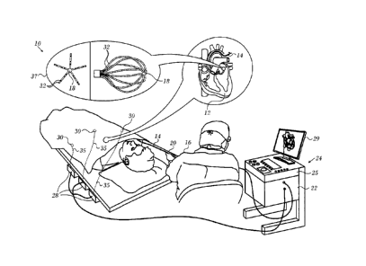
[0036] Turning now to the drawings, reference is initially made to Fig. 1,
which is a pictorial illustration of a system 10 for performing diagnostic and
therapeutic procedures on a heart 12 of a living subject, which is constructed
and operative in accordance with a disclosed embodiment of the invention. The
system comprises a catheter 14, which is percutaneously inserted by an opera-
tor 16 through the patient's vascular system into a chamber or vascular
structure
of the heart 12. The operator 16, who is typically a physician, brings the
cathe-
ter's distal tip 18 into contact with the heart wall, for example, at an
ablation tar-
get site. Electrical activation maps may be prepared, according to the methods
disclosed in U.S. Patent Nos. 6,226,542, and 6,301,496, and in commonly as-
signed U.S. Patent No. 6,892,091, whose disclosures are herein incorporated by
reference.
[0037] The system 10 may comprise a general purpose or embedded
computer processor, which is programmed with suitable software for carrying
out the functions described hereinbelow. Thus, although portions of the sys-
tem 10 shown in other drawing figures herein are shown as comprising a num-
ber of separate functional blocks, these blocks are not necessarily separate
physical entities, but rather may represent, for example, different computing
tasks or data objects stored in a memory that is accessible to the processor.
These tasks may be carried out in software running on a single processor, or
on
multiple processors. The software may be provided to the processor or proces-
sors on tangible non-transitory media, such as CD-ROM or non-volatile memory.
Alternatively or additionally, the system 10 may comprise a digital signal pro-
cessor or hard-wired logic. One commercial product embodying elements of
the system 10 is available as the CARTO 3 System, available from Biosense
Webster, Inc., 3333 Diamond Canyon Road, Diamond Bar, CA 91765. This sys-
7 of 22
CA 3010948 2018-07-10
tern may be modified by those skilled in the art to embody the principles of
the
invention described herein.
[0038] Areas determined to be abnormal, for example by evaluation of
the electrical activation maps, can be ablated by application of thermal
energy,
e.g., by passage of radiofrequency electrical current through wires in the
cathe-
ter to one or more electrodes at the distal tip 18, which apply the
radiofrequen-
cy energy to the myocardium. The energy is absorbed in the tissue, heating it
to
a point (typically above 50 C) at which it permanently loses its electrical
excit-
ability. When successful, this procedure creates non-conducting lesions in the
cardiac tissue, which disrupt the abnormal electrical pathway causing the ar-
rhythmia. The principles of the invention can be applied to different heart
chambers to diagnose and treat many different cardiac arrhythmias.
[0039] The catheter 14 typically comprises a handle 20, having suitable
controls on the handle to enable the operator 16 to steer, position and orient
the
distal end of the catheter as desired for the ablation. To aid the operator
16, the
distal portion of the catheter 14 contains position sensors (not shown) that
pro-
vide signals to a processor 22, located in a console 24. The processor 22 may
fulfill several processing functions as described below.
[0040] The catheter 14 is a multi-electrode catheter, which can be a bal-
loon or basket catheter as shown in the right portion of balloon 37, or a
spline
catheter as shown in the left portion. In any case there are multiple elec-
trodes 32, which are used as sensing electrodes and have known locations on
the basket or spline, and known relationships to one another. Thus, once the
catheter is located in the heart, for example by constructing a current
position
map, the location of each of the electrodes 32 in the heart is known. One
method
for generation of a current position map is described in commonly assigned
U.S.
Patent No. 8,478,383 to Bar-Tal et aL, which is herein incorporated by
reference.
[0041] Electrical signals can be conveyed to and from the heart 12 from
the electrodes 32 located at or near the distal tip 18 of the catheter 14 via
ca-
ble 34 to the console 24. Pacing signals and other control signals may be con-
veyed from the console 24 through the cable 34 and the electrodes 32 to the
heart 12.
8 of 22
CA 3010948 2018-07-10
[0042] Wire connections 35 link the console 24 with body surface elec-
trodes 30 and other components of a positioning sub-system for measuring loca-
tion and orientation coordinates of the catheter 14. The processor 22, or
another
processor (not shown) may be an element of the positioning subsystem. The
electrodes 32 and the body surface electrodes 30 may be used to measure tis-
sue impedance at the ablation site as taught in U.S. Patent No. 7,536,218,
issued
to Govari et al., which is herein incorporated by reference. A temperature sen-
sor (not shown), typically a thermocouple or thermistor, may be mounted near
the distal tip 18 of the catheter 14.
[0043] The console 24 typically contains one or more ablation power
generators 25. The catheter 14 may be adapted to conduct ablative energy to
the heart using any known ablation technique, e.g., radiofrequency energy, ul-
trasound energy, and laser-produced light energy. Such methods are disclosed
in commonly assigned U.S. Patent Nos. 6,814,733, 6,997,924, and 7,156,816,
which are herein incorporated by reference.
[0044] In one embodiment, the positioning subsystem comprises a mag-
netic position tracking arrangement that determines the position and
orientation
of the catheter 14 by generating magnetic fields in a predefined working vol-
ume and sensing these fields at the catheter, using field generating coils 28.
A
suitable positioning subsystem is described in U.S. Patent No. 7,756,576,
which
is hereby incorporated by reference, and in the above-noted U.S. Patent
No. 7,536,218.
[0045] As noted above, the catheter 14 is coupled to the console 24,
which enables the operator 16 to observe and regulate the functions of the
cath-
eter 14. Console 24 includes a processor, preferably a computer with appropri-
ate signal processing circuits. The processor is coupled to drive a monitor
29.
The signal processing circuits typically receive, amplify, filter and digitize
sig-
nals from the catheter 14, including signals generated by the above-noted sen-
sors and a plurality of location sensing electrodes (not shown) located
distally in
the catheter 14. The digitized signals are received and used by the console 24
and the positioning system to compute the position and orientation of the
cathe-
ter 14 and to analyze the electrical signals from the electrodes as described
in
further detail below.
9 of 22
CA 3010948 2018-07-10
[0046] Typically, the system 10 includes other elements, which are not
shown in the figures for the sake of simplicity. For example, the system 10
may
include an electrocardiogram (ECG) monitor, coupled to receive signals from
one or more body surface electrodes, so as to provide an ECG synchronization
signal to the console 24. As mentioned above, the system 10 typically also in-
cludes a reference position sensor, either on an externally applied reference
patch attached to the exterior of the subject's body, or on an internally-
placed
catheter, which is inserted into the heart 12 and maintained in a fixed
position
relative to the heart 12. The system 10 may receive image data from an
external
imaging modality, such as an MRI unit or the like and includes image
processors
that can be incorporated in or invoked by the processor 22 for generating and
displaying images.
Fibonacci sequence.
[0047] Wikipedia briefly describes Fibonacci numbers: In mathematics,
the Fibonacci numbers are the numbers in the following integer sequence,
called the Fibonacci sequence, and characterized by the fact that every number
after the first two is the sum of the two preceding ones:
1, 1, 2, 3, 5, 8, 13, 21, 34, 55, 89, 144.
[0048] Reference is now made to Fig. 2, which is a diagram illustrating
one well-known method of approximating a logarithmic spiral by forming a til-
ing with squares whose side lengths are successive Fibonacci numbers begin-
ning at a point of origin. Spiral 40, formed of circular arcs passing through
the
corners of the squares, e.g., corners 42, 44 approaches a golden spiral as the
spiral diverges outwardly from the origin, because the ratio of each element
in
the Fibonacci sequence to the preceding element converges on Phi, 1.618,
known as the golden ratio, as the series progresses. For example, the series
1,
1, 2, 3, 5, 8 and 13 produce ratios of 1, 2, 1.5, 1.67, 1.6 and 1.625,
respectively.
[0049] Reference is now made to Fig. 3, which is a schematic diagram of
a layout 46 of a catheter comprising multiple splines 48 in accordance with an
embodiment of the invention. In embodiments of a 3-dimensional balloon cathe-
ter, the splines may be constructed on the surface as Fibonacci spiral forms,
as
described with respect to Fig. 2, the splines 48 all converging at a central
point
10 of 22
CA 3010948 2018-07-10
50 and preferably equally distributed about the circumference of the balloon.
Electrodes 52 are disposed along the splines 48. The respective distances be-
tween electrodes 52 from along each spline increase according to the Fibonacci
sequence. In this embodiment the spacing of electrodes 52 is given in table
54.
Eight sets of coefficients of spline equation 56 are given in table 58,
applicable
to the eight splines shown in the layout 46. In the layout 46 many
approximately
equilateral triangles are defined. For example, electrodes at points 60, 62,
64
define such a triangle, as do electrodes at points 66, 68, 70. In practice a
devia-
tion from equilaterality of 20% can be tolerated.
[0050] Reference is now made to Fig. 4, which is a diagram illustrating a
layout with two intersecting sets of Fibonacci spirals having opposite
curvatures
in accordance with an embodiment of the invention, one set having a left-
directed curvature and the other set having a right-directed curvature. The
terms "left-directed" and "right-directed" are used arbitrarily herein to
distin-
guish the curvatures of the spirals. For example spirals 72, 74 are in
different
sets. Electrodes are disposed at the intersection of the sets, such as elec-
trode 76, which is at an intersection of the spiral 72 and its neighboring spi-
ral 74. Similarly, electrode 78 is located at the intersection of spirals 72,
80. Elec-
trodes 78, 76, 82, all on intersections of neighboring spirals define a
triangle,
which is approximately equilateral. The two sets of spirals can be constructed
using the equation 56 (Fig. 3) with an appropriate change in direction The op-
posing splines form a weave, which when incorporated into a balloon assembly
of a catheter advantageously tends to stabilize the balloon surface. Such
balloon
assemblies can be constructed according to the teachings of commonly as-
signed U.S. Patent Application Publication No. 2016/0324571, entitled Spring
Loaded Balloon, which is herein incorporated by reference.
Construction.
[0051] Reference is now made to Fig. 5, which is a schematic side eleva-
tion of a catheter 84 in accordance with an embodiment of the invention. Cathe-
ter body 86 comprises an elongated tubular construction having a single, axial
or central lumen 88, but can optionally have multiple lumens along all or part
of
its length if desired. The catheter body 86 is flexible, i.e., bendable, but
sub-
11 of 22
CA 3010948 2018-07-10
stantially non-compressible along its length. The catheter body 86 can be of
any
suitable construction and made of any suitable material.
[0052] Reference is made to Fig. 6, which is a sectional schematic view of
a portion of the catheter of Fig. 5, taken through line 6-6 in accordance with
an
embodiment of the invention. Mounted in the distal end of the lumen 88 of the
catheter body 86 is a spine mounting assembly 90.
[0053] One construction of the catheter body 86 comprises an outer wall
92 made of polyurethane or pebax (polyether block amide). The outer wall 92
comprises an imbedded braided mesh of stainless steel or the like, as is gener-
ally known in the art, to increase torsional stiffness of the catheter body 86
so
that, when a control handle 94 is rotated, the distal end of the catheter body
86
rotates in a corresponding manner.
[0054] The length of the catheter body 86 is not critical, but preferably
ranges from about 190 cm to about 120 cm, and more preferably is about 110
.. cm. The outer diameter of the catheter body 86 is also not critical, but is
prefer-
ably no more than about 8 french, more preferably about 7 french. Likewise,
the
thickness of the outer wall 92 is not critical, but is preferably thin enough
so that
the central lumen 88 can accommodate puller wires, lead wires, sensor cables
and any other wires, cables or tubes. If desired, the inner surface of the
outer
.. wall 92 is lined with a stiffening tube (not shown) to provide improved
torsional
stability. An example of a catheter body construction suitable for use in
connec-
tion with the present invention is described and depicted in U.S. Pat. No.
6,064,905, the entire disclosure of which is incorporated herein by reference.
[0055] In the depicted embodiment, a mapping assembly 96 comprises
eight splines 98, which are configured as Fibonacci spirals, as described
above.
Each of the splines 98 has a proximal end attached at the distal end of the
cathe-
ter body 86 and a free distal end, i.e., the distal end is not attached to any
of the
other splines, to the catheter body, or to any other structure that confines
movement of the distal end. Each of the splines 98 contains a support arm 100
.. comprising a metal or plastic material that has shape memory, such that the
support arm 100 forms an initial shape when no external forces are applied,
forms a deflected shape when an external force is applied, and returns to its
ii-
12 of 22
CA 3010948 2018-07-10
tial shape when the external force is released. In a preferred embodiment, the
support arm 100 comprises a superelastic material, for example a nickel-
titanium alloy, such as Nitinol. Each of the splines 98 also comprises a non-
conductive covering 102 in surrounding relation to the support arm 100. In a
preferred embodiment, the non-conductive covering 102 comprises a biocom-
patible plastic tubing, such as a polyurethane or polyimide tubing.
[0056] A first non-conducting tube 104 is disposed between an outer
mounting ring 106 and the support arm 100, and a second non-conducting tube
108 is disposed between the support arm 100 and a mounting structure 110. The
non-conducting tubes 104, 108, which may be polyimide tubes, ensure that each
support arm 100 remains electrically isolated. In addition, a mounting ring
inner
tube 112 is secured within the mounting structure 110. The mounting ring inner
tube 112 preferably comprises a non-conducting material such as polyimide.
The mounting ring inner tube 112 defines a mounting ring lumen 114 through
which electrode lead wires 116 and sensor cables 118 extend.
[0057] As will be recognized by one skilled in the art, the number of
splines 98 can vary as desired depending on the particular application, so
that
the catheter has at least two splines, preferably at least three splines, more
preferably at least eight splines and as many as 12 or more splines. As de-
scribed in more detail below, the splines 98 are moveable between an expand-
ed arrangement, wherein, for example, each spline spirals outwardly from the
catheter body 86, or the splines 98 may be arranged in a collapsed arrange-
ment, wherein, for example, each spline is disposed generally along a longitu-
dinal axis of the catheter body 86 so that the splines are capable of fitting
within
a lumen of a guiding sheath.
[0058] Reference is made to Fig. 7, which is a longitudinal sectional view
of one of the splines 98 (Fig. 5), in accordance with an embodiment of the
inven-
tion. Each of the splines 98 carries at least one electrode mounted along its
length, disposed as described above. In the depicted embodiment, a tip elec-
trode 120 may be mounted on a distal end of each non-conductive covering 102
and at least one ring electrode 122 is mounted on each non-conductive cover-
ing 102, preferably on the distal end of the non-conductive covering 102. In a
bipolar arrangement, the ring electrode 122 is used as a reference electrode.
13 of 22
CA 3010948 2018-07-10
The distance between the tip electrode and ring electrode preferably ranges
from about 0.5 mm to about 2 mm. In an alternative bipolar arrangement (not
shown), the tip electrode 120 is eliminated and at least two ring electrodes
122
are mounted on each non-conductive covering 102, preferably on the distal end
of the non-conductive covering 102. Another alternative embodiment (not
shown), is a unipolar arrangement, in which the tip electrode 120 is mounted
on
the distal end of each non-conductive covering 102, with one or more reference
ring electrodes mounted on the distal end of the catheter body 86, or one or
more reference electrodes attached outside the body of the patient (e.g., in
the
form of a patch). In an alternative unipolar arrangement, a ring electrode 122
mounted on each non-conductive covering 102, preferably on the distal end of
the non-conductive covering 102, is used instead of tip electrode 120.
[0059] Each of the splines 98 may also include at least one location sen-
sor 124. The location sensor 124 is mounted near the distal end of each spine.
In
the depicted embodiment, where each spline 98 comprises tip electrode 120.
The location sensor 124 is mounted such that the distal end of the location
sensor
124 is secured within its corresponding tip electrode 120, while the proximate
end of the location sensor 124 extends into the distal end of the non-
conductive
covering 102. Each location sensor 124 is used to determine the coordinates of
its corresponding tip electrode 120 at each instant when the tip electrode 120
is
being used to collect an electrical mapping data point. As a result, both
electri-
cal and locational data can be obtained for each data point that is mapped. If
the
spline 98 carries at least one ring electrode 28 but does not include the tip
elec-
trode 120, the location sensor 124 is mounted near the distal end of the non-
conductive covering 102, preferably as close to the distal end of the spline
98 as
possible or in a plane concentric with the ring electrode 122.
[0060] Each location sensor 124 is connected to a corresponding sensor
cable 118. Each sensor cable 118 extends through the non-conductive covering
102, catheter body 86 and control handle 94 and out the proximal end of the
control handle 94.
[0061] Each tip electrode 120 has an exposed length preferably ranging
from about 0.5 mm to about 4 mm, more preferably from about 0.5 mm to about
2 mm, still more preferably about 1 mm. Each ring electrode 122 has a length
14 of 22
CA 3010948 2018-07-10
preferably up to about 2 mm, more preferably from about 0.5 mm to about 1
mm.
[0062] Each tip electrode 120 and each ring electrode 122 is electrically
connected to an electrode lead wire 116, which in turn is electrically
connected
to a connector 126 (Fig. 5). The connector 126 is connected to an appropriate
mapping or monitoring system (not shown). Each electrode lead wire 116 ex-
tends from the connector 126, through the control handle 94, through the
central
lumen 88 in the catheter body 86, and into the non-conductive covering 102 of
the splines 98 where it is attached to its corresponding tip electrode 120 or
ring
electrode 122. Each lead wire 116, which includes a non-conductive coating
over almost all of its length, is attached to its corresponding tip electrode
120 or
ring electrode 122 by any suitable method.
[0063] Additional details of the construction of the catheter are found in
commonly assigned U.S. Patent Application Publication No. 20060276703, which
is herein incorporated by reference.
[0064] Reference is now made to Fig. 8, which is a schematic diagram of
a partially unfolded catheter 128 in accordance with an embodiment of the in-
vention. Spiral assembly 130 may be provided with a distal locking element,
such as a hook 132. The assembly 130 is attached to a rotator shaft 134, which
is
inserted through a lumen 136 of the catheter 128. As the assembly 130 extends
beyond distal end 138 of the catheter 128 it eventually encounters a wall of
the
atrium, and is held in place against the wall by the hook 132. Deployment of
the
assembly 130 is then completed by retracting and concurrently turning the rota-
tor shaft 134, causing the assembly 130 to assume an expanded spiral configura-
tion as shown in Fig. 5.
Velocity Vector Calculations.
[0065] The approximately equilateral triangles defined by the electrodes
in Fig. 3 can be regarded as a triangular mesh. Reference is now made to Fig.
9,
which is a diagram illustrating the calculation of velocity vectors for an
electrical
wave on a triangular mesh in accordance with an embodiment of the invention.
Triangle 140 has edges, including edges 142, 144. A velocity vector exists at
each edge. For example, the velocity vector 17: for edge 142 is given by
15 of 22
CA 3010948 2018-07-10
(112
V12 = Eq. (1)
(iut2 ¨ /at")
where d12 is the distance between vertices 146, 148 of triangle 140, and lati
and
1at2 are the activation times at vertices 146, 148. The velocity vector for
edge 144
is calculated in like manner.
[0066] The velocity '17. through triangle 140 is the sum of velocities along
edges 142, 144:
= Vi2+17-13 EcI= (2).
[0067] Because the electrodes define approximately equilateral trian-
gles, the accuracy of the determination of the velocity vector is
approximately
invariant regardless of the direction of the electrical propagation. Further
de-
tails on evaluating propagation through the heart, which can be advantageously
accomplished using the principles of the present invention may be found in
commonly assigned Application No. 15/086,220, entitled Mapping of Atrial Fi-
brillation, which is herein incorporated by reference.
Alternate Embodiment. =
[0068] In this embodiment 3-dimensional splines formed as Fibonacci
spirals are realized in a balloon catheter. Except for the spiral arrangement
of
the splines, a catheter of this sort can be constructed and introduced conven-
tionally, as described, for example in commonly assigned U.S. Patent Applica-
tion Publication Nos. 20160175041 entitled Balloon for Ablation around
Pulmonary
Veins, 20160324571 entitled Spring-Loaded Balloon, and U.S. Patent No.
9,352,134 entitled Segmented Balloon Catheter, which are all herein incorpo-
rated by reference.
[0069] Reference is now made to Fig. 10, which is a schematic diagram of
a 3-dimensional spline assembly 150, in accordance with an embodiment of the
invention. Splines 152, configured as Fibonacci spirals as described above,
but
now stretched to form a 3-dimensional surface, extend from a central loca-
tion 154, which may comprise a central electrode, along the surface of the as-
sembly 150. The splines 152, when distorted in this manner, form a sphere or
an
ellipsoid with a major diameter generally aligned with the axial dimension of
the
16 of 22
CA 3010948 2018-07-10
assembly 150. The 2-dimensional spline arrangement shown in Fig. 4 may be
deformed to form such a 3-dimensional structure.
[0070] In alternate embodiments the splines 152 may be adhered to the
exterior surface, the interior surface, or be embedded within the substance of
a
balloon. Alternatively the splines may form a basket catheter as is known in
the
art. Electrodes (not shown) are distributed on the splines at distances from
the
central location 154 corresponding to the Fibonacci sequence.
[0071] It will be appreciated by persons skilled in the art that the present
invention is not limited to what has been particularly shown and described
hereinabove. Rather, the scope of the present invention includes both
combinations and sub-combinations of the various features described
hereinabove, as well as variations and modifications thereof that are not in
the
prior art, which would occur to persons skilled in the art upon reading the
foregoing description.
17 of 22
CA 3010948 2018-07-10