Note: Descriptions are shown in the official language in which they were submitted.
CA 03011080 2018-07-10
WO 2017/137844 PCT/IB2017/000183
TITLE
SIGNAL TRIMMING AND FALSE POSITIVE REDUCTION OF
POST-SEGMENTATION SWALLOWING ACCELEROMETRY DATA
BACKGROUND
[0001] The present disclosure generally relates to methods and devices for
quantifying
swallowing function. More specifically, the present disclosure relates to
methods in which
adaptive trimming and/or false positive reduction improve the accuracy of
automatically
segmented swallowing accelerometry data.
[0002] Any difficulty in the process of transferring food or liquid from
the mouth to the
stomach is referred to as dysphagia or swallowing disorder. Dysphagia
negatively affects the
quality of life of patients through an increased risk of aspiration (the entry
of material into the
airway below the true vocal folds). Causes of dysphagia include: changes in
the anatomical
structures necessary for swallowing, as a consequence of surgery, cancer,
trauma, or inflammation;
genetic malformations of the swallowing apparatus; and neurological
impairments due to stroke,
Parkinson's disease, cerebral palsy, and acquired brain injury.
[0003] The videofluoroscopic swallowing study (VFSS) is the gold standard
for the diagnosis
of dysphagia. During this procedure, patients swallow different consistencies
of barium-coated
liquid and food and are exposed to ionizing radiation. In addition, expensive
equipment and
specialized clinicians are required for VFSS and are not available in every
medical establishment.
As a result, VFSS cannot be performed on a day-to-day basis.
[0004] As a non-invasive adjunct to VFSS, swallowing accelerometry has been
introduced,
deploying a dual-axis accelerometer on the surface of the patient's neck
slightly below the
laryngeal prominence (commonly known as Adam's apple) to measure epidermal
vibrations
accompanying swallowing, in two anatomical directions: anterior-posterior (A-
P) and
superior-inferior (S-I). A dual-axis accelerometer provides more information
relating to
swallowing than either axis alone. Quantitative evaluations of the recorded
signals obtained from
the accelerometer are possible through digital signal processing. Significant
correlation between
the peak laryngeal movement and the A-P acceleration signal have been
reported, while
hyolaryngeal excursion has been implicated as the primary physiological source
of the
two-dimensional signal via semi-partial correlations.
[0005] The accelerometry data can be manually segmented into distinct
swallowing events that
can be individually classified. Manual segmentation may be applied to
accelerometry data, for
example, upon visual inspection of the data (e.g. identification of the start
of each swallowing
1
CA 03011080 2018-07-10
WO 2017/137844 PCT/IB2017/000183
event, which may be readily and systematically recognized by an operator of
the device).
However, automatic segmentation using algorithms facilitates segmentation of
larger collections
of data. Larger volumes of accelerometry data necessitate an automatic method
to mitigate human
error due to fatigue or oversight and to ensure consistent segmentation
criteria.
SUMMARY
[0006] The present inventors discovered that automatic segmentation of
acceleration signals in
anterior-posterior and superior-inferior anatomical directions may be too
liberal, admitting pre-
and post-swallowing activity while also giving rise to false positive, non-
swallow segments. These
segmentation shortcomings adversely affect feature extraction and ultimately
classification of
swallowing function. As set forth in the experimental example disclosed
herein, the present
inventors found that adaptively trimming the swallow segments (e.g., using a
kernel density
estimation-based algorithm) and/or performing false positive reduction (e.g.,
energy-based and/or
noise-floor) significantly mitigates these segmentation shortcomings.
[0007] Accordingly, in a general embodiment, the present disclosure
provides a method of
method of swallowing impairment detection, the method comprising subjecting
swallowing
segments and non-swallowing segments of vibrational data to processing by a
processing module. The
processing is selected from the group consisting of adaptive trimming, false
positive reduction, and a
combination thereof. The vibrational data (i) represents swallowing activity,
(ii) is from a sensor
positioned externally on the throat of a patient and operatively connected to
the processing module, and
(iii) is associated with at least one axis selected from the group consisting
of an anterior-posterior axis
and a superior-inferior axis.
[0008] In an embodiment, the processing comprises adaptive trimming for
each of the at least
one axis, the adaptive trimming comprising forming trimmed segments from the
swallowing and
the non-swallowing segments, each of the trimmed segments comprise a portion
of the respective
segment corresponding to physiological vibrations associated with swallowing
and exclude a
portion of the respective segment corresponding to pre-swallow and post-
swallow signal
fluctuations.
[0009] The adaptive trimming can comprise, for each of the at least one
axis: using kernel
density estimation to obtain probability distributions for the swallowing
segments and the
non-swallowing segments; determining an energy threshold based at least
partially on the
probability distributions; and the excluded portion is identified based on an
energy difference of
the excluded portion falling below the energy threshold. The method can
comprise setting a
probability cut-off for the probability distributions to adjust an extent of
the adaptive trimming.
2
CA 03011080 2018-07-10
WO 2017/137844 PCT/IB2017/000183
[0010] In an embodiment, the processing comprises false positive reduction
selected from the
group consisting of energy-based false positive reduction, noise floor-based
false positive
reduction, and a combination thereof The energy-based false positive reduction
can comprise
determining, for each of the least one axis, an axial energy-based bolus-
specific threshold and
discarding the swallowing segments having a maximum energy value less than the
respective
bolus-specific threshold. The noise floor-based false positive reduction can
comprise generating
an amplitude histogram of the vibrational data, using the amplitude histogram
to determine an axial
threshold for each of the at least one axis, and discarding the swallowing
segments having a noise
range greater than the respective axial threshold. The method can comprise
adjusting an axial
threshold to control a balance between removal of false positives and loss of
true positives.
[0011] In an embodiment, the processing module receives the vibrational
data from the sensor
and automatically forms the swallowing segments and the non-swallowing
segments from the
vibrational data.
[0012] In an embodiment, the sensor is selected from the group consisting
of a single-axis
accelerometer and a dual-axis accelerometer.
[0013] In an embodiment, the method comprises classifying the swallowing
segments and the
non-swallowing segments as normal swallowing or a possible swallowing
impairment after the
processing of the swallowing and non-swallowing segments, and the processing
module performs
the classifying. The method can comprise generating an output representing the
classification, the
processing module generates the output. The possible swallowing impairment can
comprise at
least one of a swallowing safety impairment or a swallowing efficiency
impairment. The possible
swallowing impairment can comprise penetration or aspiration, and the
processing module can
further classify the swallowing event as indicative of one of a safe event and
an unsafe event.
[0014] In another embodiment, the present disclosure provides an apparatus
for quantifying
swallowing function. The apparatus comprises: a sensor configured to be
positioned on the throat
of a patient and acquire vibrational data representing swallowing activity and
associated with at
least one axis selected from the group consisting of an anterior-posterior
axis and a
superior-inferior axis; and a processing module operatively connected to the
sensor and configured
to subject swallowing segments and non-swallowing segments of the vibrational
data to processing
selected from the group consisting of adaptive trimming, false positive
reduction, and a
combination thereof
[0015] In an embodiment, the apparatus comprises an output component
selected from a
display, a speaker, and a combination thereof, the processing module
configured to classify the
swallowing segments and the non-swallowing segments as normal swallowing or a
possible
swallowing impairment after the processing of the swallowing segments and the
non-swallowing
3
CA 03011080 2018-07-10
WO 2017/137844 PCT/IB2017/000183
segments, the processing module configured to use the output component to
indicate the
classification visually and/or audibly.
[0016] In an embodiment, the processing module is operatively connected to
the sensor by at
least one of a wired connection or a wireless connection.
[0017] In an embodiment, the processing module is configured to receive the
vibrational data
from the sensor and automatically form the swallowing segments and the non-
swallowing
segments from the vibrational data.
[0018] In another embodiment, the present disclosure provides a method of
treating dysphagia
in a patient, the method comprising: positioning a sensor externally on the
throat of the patient, the
sensor acquiring vibrational data representing swallowing activity and
associated with at least one
axis selected from the group consisting of an anterior-posterior axis and a
superior-inferior axis,
the sensor operatively connected to a processing module subjecting swallowing
segments and
non-swallowing segments of the vibrational data to processing selected from
the group consisting
of adaptive trimming, false positive reduction, and a combination thereof, the
processing module
generating an output indicative of a classification of the vibrational data;
and adjusting a feeding
administered to the patient based on the classification.
[0019] In an embodiment, the adjusting of the feeding is selected from the
group consisting of
changing a consistency of the feeding, changing a type of food in the feeding,
changing a size of a
portion of the feeding administered to the patient, changing a frequency at
which portions of the
feeding are administered to the patient, and combinations thereof
[0020] An advantage of one or more embodiments provided by the present
disclosure is to
overcome drawbacks of known techniques for swallowing impairment detection.
[0021] Another advantage of one or more embodiments provided by the present
disclosure is
to reduce inclusion of pre- and post-swallowing activity in automatically
segmented swallowing
accelerometry data.
[0022] A further advantage of one or more embodiments provided by the
present disclosure is
to reduce false positive, non-swallow segments in automatically segmented
swallowing
accelerometry data.
[0023] Yet another advantage of one or more embodiments provided by the
present disclosure
is to improve feature extraction in automatically segmented swallowing
accelerometry data.
[0024] Another advantage of one or more embodiments provided by the present
disclosure is
to improve classification of swallowing function based on automatically
segmented swallowing
accelerometry data.
[0025] A further advantage of one or more embodiments provided by the
present disclosure is
to minimize loss of true positives (e.g., at most a moderate loss) while
significantly reducing the
4
CA 03011080 2018-07-10
WO 2017/137844 PCT/IB2017/000183
number of false positive swallow segments in classification of swallowing
function based on
automatically segmented swallowing accelerometry data.
[0026] Yet another advantage of one or more embodiments provided by the
present disclosure
is to enable a user to control algorithmic thresholds to adjust the balance
between false positive
reduction and loss of true positives in classification of swallowing function
based on automatically
segmented swallowing accelerometry data.
[0027] Another advantage of one or more embodiments provided by the present
disclosure is
to classify swallows in greater detail than is possible in known methods.
[0028] A further advantage of one or more embodiments provided by the
present disclosure is
to extract individual swallows with a higher accuracy rate than is possible in
known methods.
[0029] Additional features and advantages are described herein, and will be
apparent from, the
following Detailed Description and the Figures.
BRIEF DESCRIPTION OF THE FIGURES
[0030] FIG. 1 is diagram showing the axes of acceleration in the anterior-
posterior and
superior-inferior directions.
[0031] FIG. 2 is a schematic diagram of an embodiment of a swallowing
impairment detection
device in operation.
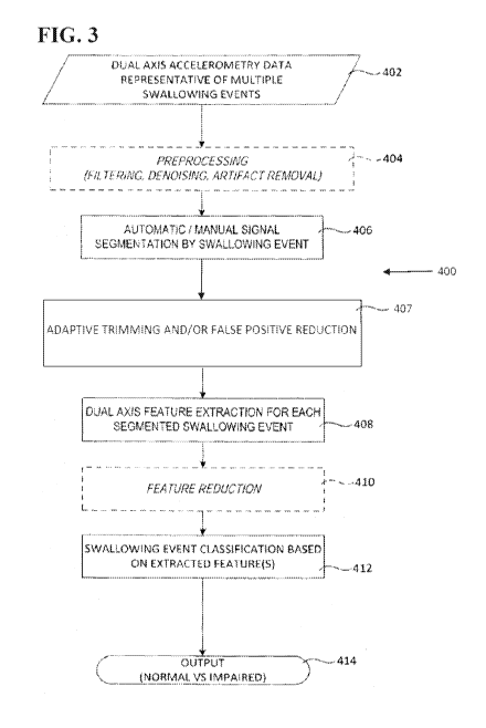
[0032] FIG. 3 is a flowchart of a dual axis accelerometry data processing
method for
implementation by a swallowing impairment detection device.
[0033] FIG. 4 is a schematic diagram of an accelerometry signal in which
False Positive (FP),
True Positive (TP) and False Negative (FN) segments are defined.
[0034] FIGS. 5a-5e are graphs showing swallow trimming based on dual-
directional energy
differences in the experimental example disclosed herein.
[0035] FIG. 6 is a table showing estimation of the scalars AAP and As/ in
the experimental
example disclosed herein.
[0036] FIG. 7a is a graph showing maximum windowed energy of boluses
(crosses) and their
constituent candidate swallows in the experimental example disclosed herein.
[0037] FIG. 7b is a graph showing true positive (TP) and false positive
(FP) changes after
applying energy-based FP reduction in the experimental example disclosed
herein.
[0038] FIG. 8a is a graph showing a raw A-P bolus signal in the
experimental example
disclosed herein. The vertical lines indicate the VFSS-identified swallow
onsets.
[0039] FIG. 8b is a graph showing segmentation with a FP case (first
rectangle) in the
experimental example disclosed herein. The rectangles identify the segmented
swallows.
CA 03011080 2018-07-10
WO 2017/137844 PCT/IB2017/000183
[0040] FIG. 8c is a graph showing segmentation after FP removal in the
experimental example
disclosed herein. The rectangles identify the segmented swallows.
DETAILED DESCRIPTION
[0041] As used in this disclosure and the appended claims, the singular
forms "a," "an" and
"the" include plural referents unless the context clearly dictates otherwise.
As used herein, "about"
is understood to refer to numbers in a range of numerals, for example the
range of -10% to +10% of
the referenced number, preferably -5% to +5% of the referenced number, more
preferably -1% to
+1% of the referenced number, most preferably -0.1% to +0.1% of the referenced
number.
Moreover, all numerical ranges herein should be understood to include all
integers, whole or
fractions, within the range.
[0042] The words "comprise," "comprises" and "comprising" are to be
interpreted inclusively
rather than exclusively. Likewise, the terms "include," "including" and "or"
should all be
construed to be inclusive, unless such a construction is clearly prohibited
from the context. A
disclosure of a device "comprising" several components does not require that
the components are
physically attached to each other in all embodiments.
[0043] Nevertheless, the devices disclosed herein may lack any element that
is not specifically
disclosed. Thus, a disclosure of an embodiment using the term "comprising"
includes a disclosure
of embodiments "consisting essentially of' and "consisting of' the components
identified.
Similarly, the methods disclosed herein may lack any step that is not
specifically disclosed herein.
Thus, a disclosure of an embodiment using the term "comprising" includes a
disclosure of
embodiments "consisting essentially of' and "consisting of' the steps
identified.
[0044] The term "and/or" used in the context of "X and/or Y" should be
interpreted as "X," or
"Y," or "X and Y." Where used herein, the terms "example" and "such as,"
particularly when
followed by a listing of terms, are merely exemplary and illustrative and
should not be deemed to
be exclusive or comprehensive. Any embodiment disclosed herein can be combined
with any
other embodiment disclosed herein unless explicitly stated otherwise.
[0045] An aspect of the present disclosure is a method of processing
segmented dual-axis
accelerometry signals for the indication of problematic swallowing events,
such as dysphagia or
aspiration. Non-limiting examples of such methods include a method of
quantifying swallowing
function and a method of swallowing impairment detection. Another aspect of
the present
disclosure is a device that implements one or more steps of the method.
[0046] In some embodiments, the method and the device can be employed in
the apparatus
and/or the method for detecting aspiration disclosed in U.S. Patent No.
7,749,177 to Chau et al., the
method and/or the system of segmentation and time duration analysis of dual-
axis swallowing
6
CA 03011080 2018-07-10
WO 2017/137844 PCT/IB2017/000183
accelerometry signals disclosed in U.S. Patent App. Publ. No. 8,267,875 to
Chau et al., the system
and/or the method for detecting swallowing activity disclosed in U.S. Patent
No. 9,138,171 to
Chau et al., or the method and/or the device for swallowing impairment
detection disclosed in U.S.
Patent App. Publ. No. 2014/0228714 to Chau et al., each of which is
incorporated herein by
reference in its entirety.
[0047] As used herein, "aspiration" is entry of food or drink into the
trachea (windpipe) and
lungs and can occur during swallowing and/or after swallowing (post-
deglutitive aspiration).
Post-deglutitive aspiration generally occurs as a result of pharyngeal residue
that remains in the
pharynx after swallowing.
[0048] As discussed in greater detail hereafter, the device may include a
sensor configured to
produce signals indicating swallowing activities (e.g., a single axis
accelerometer or a dual axis
accelerometer). The sensor may be positioned externally on the neck of a
human, preferably
anterior to the cricoid cartilage of the neck. A variety of means may be
applied to position the
sensor and to hold the sensor in such position, for example double-sided tape.
Preferably the
positioning of the sensor is such that the axes of acceleration are aligned to
the anterior-posterior
and super-inferior directions 10, as shown in FIG. 1.
[0049] FIG. 2 generally illustrates a non-limiting example of a device 100
for use in
swallowing impairment detection. The device 100 can comprise a sensor 102
(e.g., a single axis
accelerometer or a dual axis accelerometer) to be attached in a throat area of
a candidate for
acquiring dual axis accelerometry data and/or signals during swallowing, for
example illustrative
S-I acceleration signal 104. Accelerometry data may include, but is not
limited to, throat vibration
signals acquired along the anterior-posterior axis (A-P) and/or the superior-
inferior axis (S-0. The
sensor 102 can be any accelerometer known to one of skill in this art, for
example an EMT 25-C
single axis accelerometer or an ADXL322 dual axis accelerometer, and the
present disclosure is
not limited to a specific embodiment of the sensor 102.
[0050] The sensor 102 can be operatively coupled to a processing module 106
configured to
process the acquired data for swallowing impairment detection, for example
aspiration detection
and/or detection of other swallowing impairments such as swallowing
inefficiencies. The
processing module 106 can be a distinctly implemented device operatively
coupled to the sensor
102 for communication of data thereto, for example, by one or more data
communication media
such as wires, cables, optical fibres, and the like and/or by one or more
wireless data transfer
protocols. In some embodiments, the processing module 106 may be implemented
integrally with
the sensor 102.
[0051] The signal acquisition by the sensor 102 and the processing of the
signal by the
processing module 106, which are described in greater detail hereafter, are
generally discussed in
7
CA 03011080 2018-07-10
WO 2017/137844 PCT/IB2017/000183
the context of the preferred embodiment in which a dual axis accelerometer is
used to obtain both
A-P and S-I vibrational data. However, the present disclosure also encompasses
embodiments in
which a single axis accelerometer is used. In this regard, the disclosures
regarding the data from
the corresponding single axis (A-P or S-I) which are provided in the context
of a dual axis
accelerometer can also be applied to embodiments in which a single axis
accelerometer is used.
For example, processing of A-P data in the context of a dual axis
accelerometer can be applied
similarly to A-P data obtained by a single axis accelerometer (e.g., in the
absence of S-I data), and
processing of S-I data in the context of a dual axis accelerometer can be
applied similarly to S-I
data obtained by a single axis accelerometer (e.g., in the absence of A-P
data). Further in this
regard, the reduced inclusion of pre- and post-swallowing activity in
automatically segmented
swallowing accelerometry data can be achieved by both single axis and dual
axis embodiments.
[0052] FIG.
3 generally illustrates a non-limiting example of a method 400 of swallowing
impairment detection, wherein optional steps in this embodiment are shown in
dashed-line boxes.
At Step 402, accelerometry data ("raw data") can be acquired from multiple
swallowing events, for
example by an accelerometer such as sensor 102. At Step 404, the accelerometry
data can
optionally be processed to condition the accelerometry data and thus
facilitate further processing
thereof For example, the accelerometry data may be filtered, denoised and/or
processed for signal
artifact removal ("preprocessed data").
[0053] At
Step 406, the accelerometry data (either raw or preprocessed) can then be
automatically or manually segmented into distinct swallowing events.
Preferably the
accelerometry data is automatically segmented. In an embodiment, the
accelerometry data is
automatically segmented as disclosed in U.S. Patent No. 8,267,875 to Chau et
al., the entirety of
which is incorporated herein by reference as noted above. For example, the
automatic
segmentation can comprise applying fuzzy c-means optimization to the data
determine the time
boundaries for each of the swallowing and non-swallowing segments.
Additionally or
alternatively, manual segmentation may be applied, for example by visual
inspection of the data.
The method 400 is not limited to a specific embodiment of the segmented data.
[0054] At
Step 407, the segmented accelerometry data can be subjected to adaptive signal
trimming and/or false positive reduction. The resultant event-specific data
can then be processed
for dual axis feature extraction at Step 408, and optionally processed for
feature reduction at Step
410. In embodiments where the data is single-axis data, the extracted features
preferably comprise
one or more of stationarity, normality and dispersion ratio. In embodiments
where the data is
dual-axis data, the extracted features preferably comprise a log energy of
vibrational data acquired
along the A-P axis and an entropy of vibrational data acquired along the S-I
axis, for example a log
energy of each level of an 18 level 5ym8 wavelet decomposition of the
vibrational data acquired
8
CA 03011080 2018-07-10
WO 2017/137844 PCT/IB2017/000183
along the A-P axis and an entropy of each level of a 12 level sym8 wavelet
decomposition of said
vibrational data acquired along the S-I axis. The present disclosure is not
limited to a specific
embodiment of the extracted features.
[0055] Each
swallowing event can then be classified based on the extracted features at
Step
412. In an embodiment where the data is single-axis data, the classification
is preferably
performed using a radial basis function neural network implemented by the
processing module 106
to classify swallowing events in real-time, as either swallows or aspirations.
In an embodiment
where the data is dual-axis data, the classification is preferably performed
by comparing the
extracted features with preset classification criteria defined by features
previously extracted and
classified from a known training data set, for example as a function of a
distance of the extracted
features from the classification criteria (e.g., discriminant analysis using
Mahalanobis distances
with stratified covariance estimates). The present disclosure is not limited
to a specific
embodiment of the classifying process.
[0056] The
classification can be used to determine and output which swallowing event
represented a normal swallowing event as compared to a penetration, an
aspiration, a swallowing
safety impairment and/or an swallowing efficiency impairment at Step 414.
In some
embodiments, the swallowing event can be further classified as a safe event or
an unsafe event.
[0057] For
example, the processing module 106 and/or a device associated with the 106 can
comprise a display that identifies a swallow or an aspiration using images
such as text, icons,
colors, lights turned on and off, and the like. Alternatively or additionally,
the processing module
106 and/or a device associated with the processing module 106 can comprise a
speaker that
identifies a swallow or an aspiration using auditory signals. The present
disclosure is not limited to
a specific embodiment of the output, and the output can be any means by which
the classification
of the swallowing event is identified to a user of the device 100, such as a
clinician or a patient.
[0058] The
output may then be utilized in screening/diagnosing the tested candidate and
providing appropriate treatment, further testing, and/or proposed dietary or
other related
restrictions thereto until further assessment and/or treatment may be applied.
For example,
adjustments to feedings can be based on changing consistency or type of food
and/or the size
and/or frequency of mouthfuls being offered to the patient.
[0059]
Alternative types of vibration sensors other than accelerometers can be used
with
appropriate modifications to be the sensor 102. For example, a sensor can
measure displacement
(e.g, a microphone), while the processing module 106 records displacement
signals over time. As
another example, a sensor can measure velocity, while the processing module
106 records velocity
signals over time. Such signals can then be converted into acceleration
signals and processed as
9
CA 03011080 2018-07-10
WO 2017/137844 PCT/IB2017/000183
disclosed herein and/or by other techniques of feature extraction and
classification appropriate for
the type of received signal.
[0060] As noted above, Step 407 comprises subjecting the segmented
accelerometry data to
adaptive signal trimming and/or false positive reduction, and preferred
embodiments of these
processes are disclosed below.
[0061] Adaptive Signal Trimming
[0062] Adaptive signal trimming can trim the segmented accelerometry data
so that the
resultant data comprises the portion of the signal corresponding to the
physiological vibrations
associated with swallowing, while excluding the pre- and post-swallow signal
fluctuations.
Preferably the trimmed data consists of the portion of the signal
corresponding to the physiological
vibrations associated with swallowing.
[0063] The adaptive signal trimming can comprise determining the base
energy (Ebaõ) within a
window of a predetermined number of samples w (e.g., w = 500) that are
centered at the location of
the peak amplitude value of the segmented swallow (S):
13+7
VaPse = GAP)2 (Equation 1)
where 1AP is the A-P signal, p is the peak index, and w is the window size.
[0064] Ls can be the length of the initially segmented swallow, and a
corresponding
non-swallow segment NS can be defined as the segment of length Ls with the
minimum signal
energy within a predetermined time period of the beginning of the signal
(e.g., the first 10 seconds
of the calibration signal, given that typical swallows are approximately 1
second in duration).
[0065] Then the adaptive trimming can comprise moving the window w by a
predetermined
sample increment s (e.g., s = 50 samples) along the swallow and non-swallow
segments (e.g., with
90% overlap). Then the adaptive trimming can comprise determining the energy
differences
between the base energy Ebaõ and the energy within the moving windows. For the
A-P signal,
these differences are preferably determined as:
AEgP (j) = IgaPse EtP U) 1 j [Ls __________________ w sl (Equation
2)
AEkic (j) = IVaPse (Equation 3)
where EtP and Of are the energy differences of the swallow and non-swallow
segments,
respectively, and
w+(j-1)s
Et(j) = (jAP)2, 1 j [Ls __________________ ¨ w sl
(Equation 4)
i=1+(j-1)s
[0066] Preferably, Eilf (j) is similarly defined using the non-swallow
segment NS.
CA 03011080 2018-07-10
WO 2017/137844 PCT/IB2017/000183
[0067] The adaptive trimming can further comprise applying the above
formulation to the S-I
signal. Nevertheless, in some embodiments (e.g., those using a single axis
accelerometer), the
above formulation is applied to only one of the A-P axis or the S-I axis.
[0068] The adaptive trimming can further comprise determining the
probability density of
energy differences for both swallow and non-swallow segments from their
respective histograms
using kernel density estimation, for example as disclosed by M. Di Marzio and
C.C. Taylor in
"Kernel density classification and boosting: an L2 analysis," Statistics and
Computing
15(2):113-123 (April 2005). Additionally or alternatively, other methods of
determining the
probability density of the energy differences can be used.
[0069] In embodiments using kernel density classification, x, denotes the
histogram bin counts
of energy difference values i = 1,... ,N. The estimated kernel density of
energy differences d(x) is:
N
d(x) = _v K
________________________________________ (Equation 5)
Nh h
i=t
where K is the kernel function, Nis the number of energy difference
distribution bins, and h is the
kernel smoothing bandwidth. Given the versatile estimation capabilities of a
Gaussian mixture, a
Gaussian kernel can be adopted:
- xi) 1 (x-x)2
K ____________________ h
= ¨ e 2h2 (Equation 6)
[0070] The adaptive trimming can comprise estimating the bandwidth of the
kernel:
h = 1.06o-N- (Equation 7)
where a is the standard deviation of the energy differences.
[0071] C(x) E {swallow, non-swallow} can represent the predicted label for
an energy
difference x. The adaptive trimming can comprise determining the probability
of an energy
difference x belonging to the swallow class:
Psas(x)
P(C(x) = swallow IX = x) = (Equation 8)
Psas(x) + PNsaNs(x)
where as(x) and aNs(x) are the estimated densities for swallow and non-swallow
segments,
while ps = pNs = 0.5 are the swallow and non-swallow priors, respectively.
[0072] The adaptive trimming can comprise applying the above formulation to
both A-P and
S-I signals. Again, in some embodiments (e.g., those using a single axis
accelerometer), the above
formulation is applied to only one of the A-P axis or the S-I axis.
[0073] The adaptive trimming can comprise obtaining probability
distributions for swallow
and non-swallow segments. Setting a probability cut-off can obtain energy
thresholds T4P and Ts7
for each channel. The higher the probability cut-off, the more aggressive the
trimming.
11
CA 03011080 2018-07-10
WO 2017/137844 PCT/IB2017/000183
Preferably, trimming the swallow segments comprises identifying the location
of the peak
amplitude, then shifting overlapping windows of size w to the left and to the
right of the peak by
increments of size s, and calculating the energy difference within each window
w. Bilaterally,
windowed segments with energy difference below the threshold can be removed
from the
candidate swallow segment.
[0074] False Positive Reduction
[0075] The relevant performance metrics for false positive reduction are
discussed hereafter.
FIG. 4 defines False Positive (FP), True Positive (TP) and False Negative
(F1V) segments. In this
figure, vertical lines correspond to VFSS-demarcated swallow onsets, and
rectangles denote
candidate swallow segments. True Positive (TP) refers to an automatically
segmented swallow
candidate where a videofluoroscopy-demarcated swallow onset precedes or falls
within the
candidate segment boundaries. False Positive (FP) refers to an automatically
segmented swallow
candidate that does not have a videofluoroscopy-demarcated swallow onset
neither within nor
preceding the candidate segment boundaries. False Negative (F1V) occurs when
no swallows are
segmented for a particular videofluoroscopic swallow onset.
[0076] "TP change" refers to the percent change in the number of TP cases,
i.e., TP change =
(TP new - TP existing)/( TP existing). "FP change" refers to the percent
change in the number of FP
segments. Recall (R), also known as sensitivity, measures the proportion of
swallow segments that
are correctly identified, i.e., R = TP/(TP + FN). Precision (P) is a measure
of fidelity and equals
one minus the FP rate, i.e., P = TP/(TP + FP). Precision (P) is also known as
the positive
predictive value. Harmonic average (F) is a combined measure of recall and
precision, i.e., F = (2
RP)/(P +R).
[0077] The false positive reduction preferably comprises energy-based false
positive reduction
and/or noise floor-based false positive reduction, each of which is explained
in turn hereafter.
[0078] Energy-based False Positive Reduction
[0079] Energy-based false positive reduction is based on adaptive energy-
based thresholding.
The energy-based false positive reduction can comprise deriving, for each
axis, a bolus-specific
threshold Th based on the axial energy of the bolus:
TZ,IP = AAP X VP
where -VP is the maximum energy calculated within a moving window of a
predetermined
number of samples (e.g., 500 samples) on the A-P channel for a specific bolus
b, and AAP is a
data-dependent scalar.
[0080] The energy-based false positive reduction can comprise determining
Tg/ for the S-I
channel, using the same procedure. For each candidate swallow, the energy can
be estimated
within overlapping windows (e.g., 50% overlapping), each of predetermined
number of samples
12
CA 03011080 2018-07-10
WO 2017/137844 PCT/IB2017/000183
(e.g., 500 samples each). The energy-based false positive reduction can
comprise discarding the
candidate swallow if the maximum energy value across these windows was less
than the
corresponding bolus-adaptive threshold (either Te,P and Ti'). The scalars AAP
and As/ can be
estimated by the following approaches, namely energy ratio and maximum energy
difference.
[0081] For the energy ratio approach to scalar estimation, segmental
scalars for each channel
(41: and An) can be determined for each swallow segment as:
E
= an = s/
d 41 (Equation 9)
b,E -4111P ' w SI
where i > 1 indexes the number of the detected swallow segment within bolus b,
and per and
are the maximum axial energies of the ith swallow segment of bolus b.
[0082] The denominators are the maximum energies over the entire bolus. As
above, all
energies are preferably estimated within a moving window of predetermined
number of samples
(e.g., 500 samples) with a predetermined overlap (e.g., 50%). The scalars for
the A-P and S-I
channels (AAP and Asi) can then be estimated as a linear combination of the
average (mean) and
standard deviation (std) of the candidate scalars. For example, in an
embodiment, an estimate for
the A-P scalar with j = 0, 1, 2 can be:
MP = mean()t) ¨j x std(ilf) (Equation 10)
[0083] The maximum energy difference approach to scalar estimation is based
on the
recognition that FP segments generally have lower maximum windowed energy than
TP segments.
An energy difference approach was thus devised in which the maximum energy
difference for
bolus b can be defined as:
(VP, = ¨ Ef, (Equation 11)
where -44,P is the maximum windowed energy of bolus b, and 4,1P, = minE. AAP
can be defined
as the set of that satisfy the following:
AAP= [sr,' V b : seP, > max b(8 x 131 (Equation 12)
where fl E (0,1] is an empirically tuned scalar to suit the characteristics of
the signals of interest. In
this set, there exists swallow segment i' within bolus b', for which the
energy ratio, 41:, defined in
Equation 9 is maximized:
[3! b', 1' I AAbr > .. c AAP, i E lb, if E /0 (Equation 13)
where lb and IL are the sets of candidate swallow indices for boluses b and
b', respectively.
Finally, the scalar AAP is set as the energy ratio of swallow segment within
bolus b': AAP
[0084] AS/ can be estimated by following the same procedure for the S-I
axis.
[0085] Noise-floor False Positive Reduction
13
CA 03011080 2018-07-10
WO 2017/137844 PCT/IB2017/000183
[0086] Noise-floor false positive reduction comprises only accepting
candidates whose range
exceeds that of the noise floor. In embodiments where both energy-based false
positive reduction
and noise-floor false positive reduction are used, the noise-floor false
positive reduction can
further reduce false positives. Most of the noise is typically low energy, so
the noise-floor
algorithm can comprise determining the amplitude histogram of the bolus
signal. Therefore, the
noise-floor false positive reduction can comprise estimating the range of the
noise signal as a x
where a is initially the bolus variance and a is a scalar multiplier (i.e.,
assuming that the noise
resided within p + ao- and II - ao-). The axial thresholds are then determined
as:
TAP = aAP x cop, and Ts' = as' x as' (Equation 14)
[0087] Preferably the noise-floor false positive reduction comprises re-
estimating the noise
signal range each time a swallow is detected and "removed" from the bolus.
[0088] To estimate the optimum values for OCAP and asi, the following
criterion function can be
considered:
i(aAp asi) = nTp(aAP, aS/) nFp(aAP, aS/) (Equation 15)
where TP and FP are the number of nTp and nFp cases, expressed as a function
of A-P and S-I
scalars(4P
, as') respectively.
[0089] The optimal A-P and S-I scalars can be given by:
aAP* aSI* = argmaxi (Equation 16)
AP SI
a ,a
[0090] The energy and noise-floor false positive reduction methods are
preferably applied in
parallel on segmented, preprocessed data. In an embodiment, only candidate
segments identified
as valid by at least one of the two false positive reduction methods is
admitted.
[0091] Another aspect of the present disclosure is a method of treating
dysphagia. The term
"treat" includes both prophylactic or preventive treatment (that prevent
and/or slow the
development of dysphagia) and curative, therapeutic or disease-modifying
treatment, including
therapeutic measures that cure, slow down, lessen symptoms of, and/or halt
progression of
dysphagia; and treatment of patients at risk of dysphagia, for example
patients having another
disease or medical condition that increase their risk of dysphagia relative to
a healthy individual of
similar characteristics (age, gender, geographic location, and the like). The
term does not
necessarily imply that a subject is treated until total recovery. The term
"treat" also refers to the
maintenance and/or promotion of health in an individual not suffering from
dysphagia but who
may be susceptible to the development of dysphagia. The term "treat" also
includes the
potentiation or otherwise enhancement of one or more primary prophylactic or
therapeutic
measures. The term "treat" further includes the dietary management of
dysphagia or the dietary
14
CA 03011080 2018-07-10
WO 2017/137844 PCT/IB2017/000183
management for prophylaxis or prevention of dysphagia. A treatment can be
conducted by a
patient, a clinician and/or any other individual or entity.
[0092] The method of treating dysphagia comprises using any embodiment of
the device 100
disclosed herein and/or performing any embodiment of the method 400 disclosed
herein. For
example, the method of treating dysphagia can comprise positioning a sensor
externally on the
throat of the patient, the sensor acquiring vibrational data that represents
swallowing activity and
associated with at least one axis selected from the group consisting of an
anterior-posterior axis and
a superior-inferior axis. The sensor is preferably operatively connected to a
processing module
subjecting swallowing segments and non-swallowing segments of the vibrational
data to
processing selected from the group consisting of adaptive trimming, false
positive reduction, and a
combination thereof, and the processing module generates an output indicative
of a classification
of the vibrational data.
[0093] The method can further comprise adjusting a feeding administered to
the patient based
on the classification, for example by changing a consistency of the feeding,
changing a type of food
in the feeding, changing a size of a portion of the feeding administered to
the patient, changing a
frequency at which portions of the feeding are administered to the patient, or
combinations thereof
[0094] In an embodiment, the method prevents aspiration pneumonia from
dysphagia.
[0095] In an embodiment, the dysphagia is oral pharyngeal dysphagia
associated with a
condition selected from the group consisting of cancer, cancer chemotherapy,
cancer radiotherapy,
surgery for oral cancer, surgery for throat cancer, a stroke, a brain injury,
a progressive
neuromuscular disease, neurodegenerative diseases, an elderly age of the
patient, and
combinations thereof As used herein, an "elderly" human is a person with a
chronological age of
65 years or older.
[0096] EXAMPLE
[0097] The following experimental example presents scientific data
developing and
supporting the concept of adaptive trimming and/or false positive reduction
improving the
accuracy of automatically segmented swallowing accelerometry data.
[0098] Dual-axes acceleration signals were acquired using a biaxial
accelerometer
(ADXL327) with sensitivity of 2g from 264 consenting adult participants
referred to VFSS. The
protocol was approved by the research ethics boards of the participating
hospitals. The signals
were collected via a two-channel custom USB audio interface, consisting of a
high-pass filter with
0.1 Hz cut-off to remove the DC or the gravity component from the signals and
a low-pass filter
with 3 kHz cutoff for each channel. The signals from each axis were sampled at
10 kHz with 12-bit
resolution. Data were stored by a custom Lab VIEW program running on a laptop
for subsequent
CA 03011080 2018-07-10
WO 2017/137844 PCT/IB2017/000183
offline analysis. Participants were asked to perform a calibration task, which
included rest,
coughing, and counting. Participants were then instructed to take 6 sips of
water followed by 6 sips
of barium-coated liquids of different consistencies. The acceleration signals
were recorded
concurrent to the videofluoroscopy recordings. The signals were annotated by
speech language
pathologists. Over 3,000 usable boluses were identified.
[0099] Preprocessing and swallow segmentation were performed as follows.
Signals were
preprocessed by de-noising, head movement removal, and speech removal. High
frequency noise
was further suppressed via wavelet packet decomposition with a 4-level
discrete Meyer wavelet
and shannon entropy A-P and S-I variance signals were computed by estimating
the sample
variance within windows of size 200 data points, shifted along each of the AP
and SI signals with
50% overlap. The swallows were then segmented by subjecting the variance
signals to a sequential
fuzzy c-means algorithm. However, automatic segmentation by this method, as
well as by neural
network or quadratic variation, tends to yield segment boundaries that are too
lenient, admitting
non-swallow activity pre- and post-swallow (FIG. 5e). Likewise, segmentation
is prone to
identify non-swallow artifacts, resulting in false positives segments (FIG.
8b). To address both of
these issues, the present inventors designed the algorithms disclosed above
and experimentally
utilized as follows.
[00100] Adaptive swallow trimming was performed by calculating the base energy
Ebase
within a window of size w = 500 samples centred at the location of the peak
amplitude value of the
segmented swallow S, according to Equation 1. A corresponding non-swallow
segment NS was
defined as the segment of length Ls with the minimum signal energy within the
first 10 seconds of
the calibration signal. Then the window slides by an increment of s = 50
samples along the
swallow and non-swallow segments with 90% overlap, and the energy differences
between the
base energy and the energy within the moving windows were calculated according
to Equations
2-4. FIG. 5a depicts an example of these energy differences for one swallow.
The same
formulation was applied to the S-I signal.
[00101] The probability density of energy differences for both swallow and non-
swallow
segments were then estimated from their respective histograms (FIG. 5b) using
kernel density
estimation, according to Equations 5-7. FIG. Sc shows the probability density
estimations of both
swallow and non-swallow segments. Again, the above formulation was applied to
both AP and SI
signals.
[00102] Integrating the densities, the probability distributions for swallow
and non-swallow
segments were obtained. By setting a probability cutoff, the energy thresholds
T4P and Ts7 were
determined for each channel. A probability cutoff 0.9 was determined to be
suitable for the
problem at hand, as exemplified in FIG. 5d, where Tsi = 1.26 x 10⁶. The
vertical green line marks
16
CA 03011080 2018-07-10
WO 2017/137844 PCT/IB2017/000183
the energy difference where the swallowing class probability exceeds 0.9. This
energy threshold is
also plotted on FIGS. 5b and Sc.
[00103] FIG. 5e illustrates the S-I signal of a swallow segment. The black
dashed rectangle
marks the trimming boundary achieved considering only the S-I channel. In
order to select the
same portion of the A-P and S-I signals, the present inventors adopted two
approaches. The first
approach selected the left- and right-most boundaries of the A-P and S-I
segments (marked by the
green rectangle). The second approach calculated the midpoint of the two
boundaries of A-P and
S-I segments (marked by a red rectangle).
[00104] Energy-based false positive reduction was performed. For each axis, a
bolus-specific
threshold Th was derived based on the axial energy of the bolus:
TZ,IP = AAP X VP
where pe,P was the maximum energy calculated within a moving window of size
500 samples on
the A-P channel for a specific bolus b, and AAP was a data-dependent scalar.
[00105] Tr was be determined using the same procedure, but for the S-I
channel. For each
candidate swallow, the energy was estimated within 50% overlapping windows of
500 samples.
The candidate swallow was discarded if the maximum energy value across these
windows was less
than the corresponding bolus-adaptive threshold (either Te,P and Ti'). The
scalars AAP and As/
were estimated by energy ratio and maximum energy difference approaches.
[00106] For the energy ratio approach, segmental scalars for each channel (41:
and An) were
calculated according to Equation 9. As above, all energies in this study were
estimated within a
500-sample moving window with 50% overlap. The scalars for the A-P and S-I
channels (AAP and
Asi) were then estimated according to Equation 10. Rows 2-4 of the table in
FIG. 6 document the
effect of these scalar estimates on false positive reduction metrics.
[00107] FIG. 7a portrays the maximum energy of 30 randomly selected boluses
(crosses), and
their TP (open circles) and FP (dots) swallow segments. As shown in the
figure, FP segments
generally have lower maximum windowed energy than TP segments. Therefore, the
scalar AAP
was estimated by the energy difference approach in Equations 11-13, with the
scalar AAP set as the
energy ratio of swallow segment i' within bolus b': AP= Asi
was estimated by following
the same procedure for the S-I axis. FIG. 6 summarizes different estimations
of these scalars.
[00108] FIG. 7b portrays FP and TP changes for different values of scalar AAP.
The vertical
line delineates the scalars (AAP = 0.079, As/ = 0.078) that yielded the
highest harmonic average,
decreasing false positives by 11.5% with minimal change to the true positives.
[00109] To further reduce false positives, only candidates whose range
exceeded that of the
noise floor were accepted. This algorithm first computes the amplitude
histogram of the bolus
17
CA 03011080 2018-07-10
WO 2017/137844 PCT/IB2017/000183
signal. The axial thresholds were then determined according to Equation 14.
The noise signal
range was re-estimated each time a swallow was detected and "removed" from the
bolus.
[00110] The optimum values for OtAP and as/ were estimated according to
Equations 15 and 16.
aAP* = 7 and aS/* = 4 led to a 74% FP reduction with only a 12% decrease in TP
cases. FIG. 8
exemplifies a case where a FP swallow segment was removed after the
application of this
noise-floor FP reduction algorithm. The energy and noise-floor false positive
reduction methods
were applied in parallel on segmented, preprocessed data.
[00111] Only candidate segments that were identified as valid by at least one
of the two FP
reduction methods were admitted. If the loss of TPs was capped at 20%, the
proposed methods led
to a dramatic reduction in FPs (-85.4%) while sacrificing only 15.1% of TPs
(AAP = 0.458, As/ =
0.326, OCAP* = 7, and asy* 4).
[00112] In conclusion, the combined effect of the proposed energy and noise-
floor methods was
a definitive decrease in the number of false positives post-segmentation. The
balance between FP
reduction and loss of TPs can be fine-tuned according to the specific
accelerometric application by
tuning the axial thresholds.
[00113] It should be understood that various changes and modifications to the
presently
preferred embodiments described herein will be apparent to those skilled in
the art. Such changes
and modifications can be made without departing from the spirit and scope of
the present subject
matter and without diminishing its intended advantages. It is therefore
intended that such changes
and modifications be covered by the appended claims.
18