Note: Descriptions are shown in the official language in which they were submitted.
88202-1D1
APPARATUS, SYSTEM AND METHOD FOR PROVIDING LASER STEERING AND
FOCUSING FOR INCISION, EXCISION AND ABLATION OF TISSUE IN MINIMALLY-
INVASIVE SURGERY
[0001]
FIELD OF THE DISCLOSURE
[0002] The present disclosure relates generally to apparatus, system and
method for
providing laser steering and focusing for incision, excision and/or ablation
of tissue in
minimally-invasive surgery.
BACKGROUND INFORMATION
[0003] Minimally invasive surgical techniques can offer the potential for
reliable cancer
control with minimal impact on post treatment function of the diseased organ.
There have
been certain advances in providing instrumentation for minimally invasive
surgery of many
diseases. Although the use of CO2 lasers has become well established and can
be
considered to be as effective and precise scalpel, it is likely still largely
limited to
operations where the surgeon has unobstructed access to the tissue. (See,
e.g., Polanyi,
Bredemei.Hc et al. 1970; Jako 1972; Mihashi, Jako et al. 1976; Garden, Obanion
et al.
1988). A particular advantage of the CO2 laser over other lasers can be that
it can be
readily absorbed by water, which is the primary component of most biological
tissues. This
can facilitate minimal thermal spread and injury to adjoining normal tissue,
making the CO2
laser especially useful for surgery near critical anatomical structures, for
example.
[0004] The CO2 laser can also be used to seal small blood vessels and
lymphatics, likely
minimizing bleeding and risk of lymphatic metastases from tumors. With the
appropriate
surgical optics, the tissue interaction of the CO2 laser can be used
advantageously for a
precise excision of a tumor with minimal injury to normal tissue so as to
likely preserve
function without compromising the cure. However, an exemplary disadvantage of
the CO2
laser can be related to its beam's likely inability to travel in any medium
other than air.
Since the CO2 laser beam is likely unable to be transmitted along glass or
conventional
optical fibers, its use has probably been generally restricted to "line-of-
sight applications,
in which it
-1-
CA 3011108 2018-07-11
WO 2011/032165
PCT/US2010/048807
can be passed down a hollow, air-filled, straight rigid instrument or
endoscope. Thus,
endoscopic applications of this technique and the CO2 laser has likely been
restricted to
treatment of tumors of the mouth, pharynx, larynx and cervix, for example.
[0005] Further, a delivery of any type of surgical laser light into a
body cavity by means
fiber optics has likely been limited to use in the near field, e.g., by
bringing the distal tip of
the fiberoptic close to the tissue in order to keep the power density high. It
can be very
difficult to facilitate a flexible, variable and accurate maneuvering of such
laser beam.
[0006] Instrumentation for endoscopic applications of the CO2 lasers and
other surgical
lasers has undergone refinement and improvement, but access to the larynx and
pharynx in
certain patients with adverse anatomic features has likely continued to pose a
problem. This
limitation of the conventional technology can be largely responsible for the
potential benefits
of certain surgery being denied to a large number of patients, such as
patients whose tumors
can be relatively difficult to access for surgical resection with endoscopic
CO2 laser
instrumentation, for example. Consequently, many of these patients have been
treated
using non-surgical options, including radiation with or without chemotherapy,
to avoid the
potentially devastating effects that conventional surgery can have on a
patient's quality of
life. However, the use of such non-surgical "organ preserving" approaches can
likely often
cause permanent and significant side effects that can drastically alter the
lives of patients
who survive after treatment.
[0007] Currently, one of the more widely used delivery methods for the CO2
lasers (and
other lasers) in surgery is likely a "line-of-sight" system that may include a
laser source that
can deliver energy to a micromanipulator coupled to an operating microscope
via an
articulated arm. For example, a hollow core fiber optic delivery systems for
CO2 surgical
lasers which can facilitate providing a laser beam into a confined space has
been described
by Hart Temelkuran et al. (See, e.g., Temelkuran, Hart et al. 2002). As
described, the fiber
can transmit the light from the laser source to its distal end that can be
used as a "laser
scalpel." However, the use of the fiber delivery techniques are likely not
ideal as they can
have some of the limitations of line-of-sight technologies. Additionally,
fiber delivery
techniques can introduce certain other problems.
[0008] For example, similarly to line-of-sight delivery techniques, it can
be important to
externally manipulate an apparatus using fiber delivery techniques if it is to
be used in
confined spaces. Additionally, because the laser beam exiting the fiber can
rapidly diverge,
- 2 -
CA 3011108 2018-07-11
WO 2011/032165
PCT/US2010/048807
the fiber likely should be precisely placed near the tissue in order to incise
or ablate the
tissue. If the fiber is placed too far away (e.g., over one millimeter), the
power density can
likely drop, and the laser scalpel can become ineffective. However, if the
fiber tip touches
the tissue, it can burn and/or become obstructed. Further, a precise
manipulation of the
working end of the fiber inside a body cavity can be challenging for the
endoscopic surgeon
due to the difficulty of maintaining a consistent depth of incision with the
laser directed
through a hand held fiber moving over an uneven tissue surface in a confined
closed space.
Moreover, a complex electro-mechanical system should likely need to be
provided for the
laser beam to be controlled remotely.
[0009] Certain scanners having dimensions that can likely be appropriate
for endoscopic
use have been described. (See, e.g., Fountain and Knopp 1992; Dohi, Sakuma et
al. 2003;
Wu, Conry et al. 2006; and Tsia, Goda et al. 2009). Many of these devices can
be
instruments that have likely been initially designed specifically for
endoscopic imaging, and
only subsequently were considered for use in performing tissue modification
and altered to
accordingly. However, the technical requirements of imaging scanners and
surgical laser
scanners are generally not the same, but rather can be very different. While
imaging
scanners generally can require regular scanning patterns to generate the
image, surgical
laser scanners generally can utilize random and precise variations of the
scanners to
address the discrete adjacent and distant points that can be involved in a
typical laser
surgery pattern. Thus, conventional apparatuses provided for surgery are
described as
having the optics and mechanical control of the scanners external to the body.
(See, e.g.,
Fountain and Knopp 1992). Endoscopic devices have been described with optics
designed
to be inserted into the body, but with the mechanical control external to the
body. (See, e.g.,
Dohi, Sakuma et al. 2003; Wu, Conry et al. 2006). These systems have certain
limitations
and associated problems such as spatial and temporal inaccuracies associated
with the
remote transmission of positioning forces from the external motors to the
internal optics.
Additionally, an imaging apparatus can be provided that can be used for laser
surgery, in
principle, purportedly without mechanical movements and that can be
internalized. (See,
e.g., Tsia, Goda et al. 2009). However, this device requires a tunable laser,
and thus would
likely not be able to work with surgical lasers like a CO2 laser, for example.
[0010] Accordingly, there may be a need to address and/or overcome at
least some of
the above-described deficiencies and limitations, and to provide exemplary
embodiments of
arrangement and method according to the present disclosure as described in
further detailed
herein.
- 3 -
CA 3011108 2018-07-11
WO 2011/032165
PCT/US2010/048807
SUMMARY OF EXEMPLARY EMBODIMENTS OF THE DISCLOSURE
[0011]
Indeed, one of the objects of certain exemplary embodiments of the present
disclosure can be to address the exemplary problems described herein above,
and/or to
overcome the exemplary deficiencies commonly associated with the prior art as,
e.g.,
described herein. Accordingly, for example, provided and described herein are
certain
exemplary embodiments of exemplary apparatus, system, procedure and method
according
to the present disclosure which can be used for providing laser steering and
focusing for
incision, excision and/or ablation of tissue in minimally-invasive surgery.
[0012]
According to one exemplary embodiment of the present disclosure, an exemplary
apparatus is provided that can include at least one optical element which can
be configured
to refract and/or diffract light provided in at least one structure which can
be configured to be
inserted into a body, where at least one of the optical element(s) is
structured to receive the
light at a first angle and generate a refracted and/or diffracted light at a
second angle which
can be different from the first angle relative to an optical axis (e.g., the
straight line which
passes through the center of the optical element). An exemplary actuating
arrangement
which can be configured to control at least one of the optical element(s) to
change the
second angle of the refracted and/or diffracted light can also be provided and
situated at
least partially within the at least one structure, for example. The at least
one light can be a
laser light, and the second angle can be uniform. At least two of the optical
elements can be
structured to generate the refracted and/or diffracted light.
[0013]
According to certain exemplary embodiments of the present disclosure, at least
one of the exemplary element(s) can be a wedge or prism, and/or a grating,
such as a
grating of variable spatial frequency, an acoustooptical grating, a fixed
grating, a holographic
transmission grating, a blazed grating, etc. The exemplary arrangenneni can be
further
configured to control at least one of the optical element(s) to change the
second angle of the
refracted and/or diffracted light and a uniform third angle of the refracted
and/or diffracted
light which is different from the uniform second angle. The
exemplary actuating
arrangement can be controlled manually, mechanically, electrically,
electromechanically
and/or remotely. For example, the exemplary actuating arrangement can be
controlled at
least partially by a mechanical and/or an electromechanical arrangement.
[0014] An
exemplary fiber optic configuration which can be configured to provide the at
least one light therethrough can also be provided in accordance with certain
exemplary
embodiments of the present disclosure. For example, the exemplary fiber optic
configuration
- 4
CA 3011108 2018-07-11
WO 2011/032165
PCT/US2010/048807
can be configured to deliver the light to at least one of the optical
element(s). At least one
lens which is optically associated with at least one of the optical element(s)
can further be
provided, such as in an optical path between the optical elements, between the
optical
element(s) and the fiber optic configuration or after the optical element(s).
[0015] According to certain exemplary embodiments of the present
disclosure, a further
arrangement can be provided which can be configured to provide the laser light
and which
can be controlled so as to modify a depth of a delivery of the refracted
and/or diffracted light
to a target tissue in the exemplary structure(s). For example, the further
arrangement can
include an ablation laser, an incision laser and/or an excision laser. A
receiving
arrangement can also be provided which can be structured to be provided in the
body and
configured to receive at least one further light, such as a visual light, from
a target tissue in
the exemplary structure(s). For example, the receiving arrangement can include
at least one
light detector, which can be a charged-coupled device (CCD), a fiberoptic
bundle and/or a
complementary metal oxide semiconductor (CMOS) detector, for example, and be
configured to provide at least one image of the target tissue.
[0016]
Further, an additional arrangement can be provided which can be configured to
provide the further light to the target tissue and which can be structured to
be situated within
the body. A particular arrangement can also be provided which can facilitate a
visual control
of an application of the light by, e.g., (i) a user control and/or (ii)
automatically. Additionally,
an external configuration, which can be configured to deliver the light to the
structure(s), can
be provided and situated externally from the body. The external configuration
can be further
configured to provide multiple frequencies of the light sequentially in time
and/or at the same
time. For example, the external configuration can be or include a tunable
laser arrangement.
[0017]
According to certain exemplary embodiments of the present disclosure, at least
one dispersive element can be provided which can be configured to deflect
and/or refract the
light at a particular wavelength dependent angle in a radial direction during
a rotation of the
dispersive element(s) to move the light in a rotational motion. For example,
the external
configuration can be configured to vary the wavelength of the light so as to
move the light
radially and/or to be rotated so as to move the light.
[0018] According to another exemplary embodiment of the present disclosure,
an
exemplary apparatus is provided that can include, inter elle, a plurality of
optical elements
which can be configured to reflect light, such as laser light, and which can
be provided in at
- 5 -
CA 3011108 2018-07-11
WO 2011/032165
PCT/US2010/048807
least one structure which can be configured to be inserted into a body. For
example, a first
configuration of the optical elements can be positioned or controlled to
receive the light at a
first angle and generate a first reflected light at a second angle which can
be different from
the first angle relative to an optical axis. A second configuration of the
optical elements can
.. be structured to receive the first reflected light and generate a second
reflected light at a
third angle which can be different from the second angle relative to the
optical axis.
[0019] An actuating arrangement which can be configured to control the
first
configuration and/or the second configuration of the optical elements to
change the second
angle and/or the third angle of the light can also be provided and situated at
least partially
within the structure(s). A third configuration of the optical elements can be
structured and/or
controlled to receive the second reflected light and generate a third
reflected light at a fourth
angle which can be different from the third angle relative to the optical
axis. A fourth
configuration of the optical elements can be structured and/or controlled to
receive the third
reflected light and generate a fourth reflected light toward the body at a
fifth angle which is
different from the fourth angle relative to the optical axis. The actuating
arrangement can be
further configured to control at least one of the optical elements to change
the second angle
of the reflected light and the third angle of the reflected light relative to
the optical axis,
wherein the first and/or second angles can be uniform. The actuating
arrangement can be
controlled at least one of manually, mechanically, electrically,
electromechanically or
remotely, such as at least partially by a mechanical arrangement, for example.
[0020] The second configuration can be further structured to generate the
second
reflected light so as to have a cylindrical shape. The first configuration
and/or the second
configuration can be or include at least one section which can have a conical
shape. For
example, the first configuration can be or include a conical mirror, and the
second
configuration can be or include a conical section mirror. The third
configuration and/or the
fourth configuration can be or include at least one section which can have a
parabolic shape.
For example, the third configuration can be or include a parabolic section
mirror, and the
fourth configuration can be or include a parabolic mirror.
[0021] According to yet another exemplary embodiment of the present
disclosure,
provided is an exemplary process for providing laser steering and focusing,
which can
include, inter alia, defining a pattern to irradiate at least one section in a
body, and
controlling at least one optical element provided in a housing to refract
and/or diffract at least
one light based on the pattern using an actuating arrangement. The housing can
be
- 6 -
CA 3011108 2018-07-11
WO 2011/032165
PCT/US2010/048807
structured to be inserted into the body, and/or the actuating arrangement can
be structured
to be inserted into a body.
[0022] The exemplary process can also include, inter al/a, providing the
light, controlling
at least one of the optical elements to change an angle of the refracted
and/or diffracted light
and controlling the at least one light so as to modify a delivery of the
refracted and/or
diffracted light to a target tissue in the structure(s). According to certain
exemplary
embodiments of the present disclosure, the exemplary process can further
include, inter alia,
monitoring at least one position and/or orientation of at least one of the
optical elements or
the refracted and/or diffracted light, generating at least one signal based on
the position
and/or the orientation, and controlling the position and/or the orientation of
at least one of the
optical elements based on signal(s).
[0023] According to still yet another exemplary embodiment of the present
disclosure,
provided is an exemplary computer-accessible medium, which can be non-
transitory, and
which can have stored thereon computer executable instructions for providing
laser steering
and focusing, which, when executed by a hardware processing arrangement,
configure the
hardware processing arrangement to perform certain procedures, such as, e.g.,
defining a
pattern to irradiate at least one section in a body, and controlling at least
one optical element
provided in a housing to refract and/or diffract at least one light based on
the pattern using
an actuating arrangement. The housing and/or the actuating arrangement can be
structured
to be inserted into the body.
[0024] The exemplary processing arrangement can be further configured to
control a
source arrangement to provide the at least one light, to control at least one
of the optical
elements to change an angle of refracted and/or diffracted light with respect
to the optical
axis, and/or control a source arrangement by modifying at least one
characteristic of the light
so as to modify a position of a delivery of the refracted and/or diffracted
light to a target
location in the body. Additionally, according to certain exemplary embodiments
of the
present disclosure, the exemplary processing arrangement can be further
configured to
monitor a position and/or orientation of at least one of the optical elements
or the refracted
and/or diffracted light, generate at least one signal based on the position
and/or the
orientation, and control the position and/or the orientation of at least one
of the optical
elements based on the signal(s).
- 7 -
CA 3011108 2018-07-11
88202-1D1
[0025] According to still yet another exemplary embodiment of the present
disclosure, an
exemplary process can be facilitated for steering and/or focusing a laser on
or to a target
tissue within a body, which can include, inter alia, locating the target
tissue within the body
from a position within the body and establishing a position of the device
relative to the
target tissue using a particular arrangement that has a housing that is
inserted into the
body. The exemplary process can also include generating control data by
tracing over an
image of the tissue a path to be cut with at least one electro-magnetic
radiation, defining
an area to be effected by the at least one electro-magnetic radiation, and/or
controlling the
position of the at least one electro-magnetic radiation in real time. Further,
the exemplary
process can include, with at least one actuating arrangement provided in the
housing,
controlling at least one optical element provided in the housing, based on the
control data,
to refract and/or diffract at least one light. The at least one light can be a
laser light, and
the path can be based on a predetermined pattern.
[0026] According to still yet another exemplary embodiment of the present
disclosure, an
exemplary computer-accessible medium can be provided, which can be non-
transitory,
and which can have stored thereon computer executable instructions for
steering and/or
focusing a laser on or to a target tissue within a body, which, when executed
by a
hardware processing arrangement, configure the hardware processing arrangement
to
perform certain procedures, such as, e.g. , locating the target tissue within
the body from a
position within the body and establishing a position of the device relative to
the target
tissue using a particular arrangement that has a housing that is inserted into
the body. The
exemplary procedure executed using the computer-executable instructions can
further
include generating control data by tracing over an image of the tissue a path
to be cut with
at least one electro-magnetic radiation, defining an area to be effected by
the at least one
electro-magnetic radiation, and/or controlling the position of the at least
one electro-
magnetic radiation in real time. Further, the exemplary instructions can
configure the
processing arrangement to with at least one actuating arrangement provided in
the
housing, control at least one optical element provided in a housing, based on
the control
data, to at least one of refract or diffract at least one light, for example.
The at least one
light can be a laser light, and the path can be based on a predetermined
pattern.
-8-
CA 3011108 2018-07-11
88202-1D1
[0026A] According to another exemplary embodiment of the present disclosure,
an
endoscopic laser scalpel system is provided, comprising: a laser source
providing laser
light for at least one of incision, excision, and ablation of tissue in
minimally invasive
.. surgery; a laryngoscope or endoscope having a distal end configured to be
inserted into a
body cavity to deliver the laser light to a target tissue therein; and an
endoscopic head
mounted on the distal end of the laryngoscope or endoscope, the endoscopic
head
comprising: an optical element arrangement comprising at least four optical
elements each
optical element comprising a reflective optical element configured to reflect
light, wherein a
first set of optical elements is configured to receive an incident laser beam
comprising the
laser light from the laser source and expand it into a hollow cylindrical beam
of light wider
than the incident laser beam, and wherein a second set of optical elements is
structured to
receive the hollow cylindrical beam of light and focus it into a reflected
beam directed to
the target tissue.
[0027] These and other objects, features and advantages of the exemplary
embodiment of
the present disclosure will become apparent upon reading the following
detailed
description of the exemplary embodiments of the present disclosure, when taken
in
conjunction with the appended claims.
-8a-
CA 3011108 2019-05-01
WO 20111032165
PCT/US2010/048807
BRIEF DESCRIPTION OF THE DRAWINGS
[0028] For a more complete understanding of the present disclosure and
its advantages,
reference is now made to the following description, taken in conjunction with
the
accompanying drawings, in which:
[0029] Figure 1 is a diagram of an endoscopic laser scalpel system in
accordance with
an exemplary embodiment of the present disclosure;
[0030] Figure 2 is a perspective view of a laser scanning endoscopic head
of the
exemplary laser scalpel system of Figure 1 in accordance with a first
exemplary embodiment
of the present disclosure;
[0031] Figure 3A is a side view of optical elements that can be included in
the exemplary
embodiment of the laser scanning endoscopic head illustrated in Figure 2;
[0032] Figure 3B is an illustration of an exemplary scanning geometry
generated using
the exemplary optical elements of Figure 3A;
[0033] Figure 4A is a sketch of optical elements that can be included in
the laser
scanning endoscopic head according to a second exemplary embodiment of the
present
disclosure;
[0034] Figure 4B is an illustration of an exemplary scanning geometry
generated by the
exemplary optical elements of Figure 4A;
[0035] Figure 5A is a sketch of a particular optical element that can be
included in the
laser scanning endoscopic head according to a third exemplary embodiment of
the present
disclosure;
[0036] Figure 5B is an illustration of an exemplary scanning geometry
generated by the
exemplary optical element of Figure 5A;
[0037] Figure 6A is a sketch of still other optical elements that can be
included in the
laser scanning endoscopic head according to a fourth exemplary embodiment of
the present
disclosure;
[0038] Figure 6B is an illustration of an exemplary scanning geometry
generated by the
exemplary optical elements of Figure 6A;
- 9 -
CA 3011108 2018-07-11
W02011/032165
PCT/US2010/048807
[0039] Figure 7A is an illustration of further optical elements that can
be included in the
laser scanning endoscopic head according to a fifth exemplary embodiment of
the present
disclosure;
[0040] Figure 7B is an illustration of an exemplary scanning geometry
generated by the
exemplary optical elements of Figure TA;
[0041] Figure 8A is an illustration of yet another optical configuration
that can be
included in the laser scanning endoscopic head according to a sixth exemplary
embodiment
of the present disclosure;
[0042] Figure 8B is an illustration of an exemplary scanning geometry
generated the
.. exemplary optical element of Figure 8A;
[0043] Figure 9A are illustrations of an endoscopic laser scalpel device
and a cutout
view at a head section thereof, in accordance with an exemplary embodiment of
the present
disclosure;
[0044] Figure 9B are illustrations of the endoscopic laser scalpel device
and a cutout
view of at the head section thereof, in accordance with another exemplary
embodiment of
the present disclosure;
[0045] Figure 10 is a side cross-sectional view of a laser scanning
endoscopic head
along with representative dimensions thereof in accordance with an exemplary
embodiment
of the present disclosure;
[0046] Figure 11 is a perspective view of a servo controlled positioning
system in
accordance with an exemplary embodiment of the present disclosure;
[0047] Figure 12A is an illustration of an exemplary scanner geometry in
accordance
with an exemplary embodiment of the present disclosure;
[0048] Figure 12B is an illustration of an exemplary scanner geometry in
accordance
with another exemplary embodiment of the present disclosure;
[0049] Figure 13A is an exemplary image of an exemplary scanning pattern
generated
by a device or an arrangement in accordance with an exemplary embodiment of
the present
disclosure;
- 10 -
CA 3011108 2018-07-11
WO 2011/032165
PCT/US2010/048807
[0050] Figure 13B is an illustration of an exemplary scanner geometry in
accordance
with yet another exemplary embodiment of the present disclosure;
[0051] Figure 13C is an illustration of a further exemplary scanning
pattern in
accordance with an exemplary embodiment of the present disclosure;
[0052] Figure 14 is a side view of a device in accordance with a further
exemplary
embodiment of the present disclosure;
[0053] Figure 15 is an illustration of a block diagram of a system in
accordance with still
another exemplary embodiment of the present disclosure;
[0054] Figure 16 is a flow diagram of an exemplary procedure in
accordance with certain
exemplary embodiments of the present disclosure; and
[0055] Figure 17 is a flow diagram of an exemplary procedure in
accordance with further
exemplary embodiments of the present disclosure.
[0056] Throughout the figures, the same reference numerals and
characters, unless
otherwise stated, are used to denote like features, elements, components or
portions of the
illustrated embodiments. Moreover, while the subject disclosure will now be
described in
detail with reference to the figures, it is done so in connection with the
illustrative
embodiments. It is intended that changes and modifications can be made to the
described
embodiments without departing from the true scope and spirit of the subject
disclosure.
DETAILED DESCRIPTION OF EXEMPLARY EMBODIMENTS
[0057] To address and/or overcome at least some of the above-described
deficiencies,
exemplary embodiments of the device, arrangement, apparatus, non-transitory
computer-
accessible medium and method can be provided in according to the present
disclosure. For
example, it is possible to introduce into the body cavity a small exemplary
scanner that can
be controlled remotely by the surgeon to guide the laser. The laser can be
delivered to the
body cavity and scanned over the tissue using, e.g., two or more of small,
rotating optical
wedges. One of the advantages of using one exemplary prism arrangement, e.g.,
which can
be referred to as a Risley prism pair, is that it can be made with a very
small profile, e.g., not
much larger than the diameter of the optical fiber (under 10 mm), and that it
can be placed at
the distal end of an endoscope. One or more lenses can be used to focus the
light and
improve the power density delivered to the tissue and/or adjust the depth of
operation of the
-11 -
CA 3011108 2018-07-11
WO 2011/032165
PCT/US2010/048807
device. Using such exemplary embodiment of the arrangement/system can
facilitate a
precise manipulation of the laser delivered into relatively inaccessible body
cavities by a
flexible or rigid conduit. This exemplary embodiment can also be utilized with
an articulated
arm and/or a straight, line-of-sight laser delivery procedure/arrangement.
[0058] For example, an exemplary embodiment of the system can include an
imaging
device e.g., a video camera which can be provided next, near and/or fixed to
the scanners to
provide a live endoscopic image to the user (e.g., the surgeon), who can trace
a graphic
over the video to establish a particular scanning path on the image of the
tissue. The path
can then be translated into appropriate wedge movements to produce a scan with
appropriate speed and trajectory on the tissue. Alternatively or in addition,
the user can
control the laser path and delivery directly through a pointer or joystick (or
mouse, touch-
screen, digital pen, track ball, etc.) from the video stream as displayed on a
video monitor.
The exemplary system can also be configured or structured to vary the focus of
the beam on
the tissue to accommodate different depths of operation, thus facilitating a
real-time three-
dimensional control of the laser in the body cavity.
[0059] An
exemplary laser delivery using the exemplary embodiments of the devices,
systems, apparatus, non-transitory computer-accessible medium and arrangements
according to the present disclosure can be provided so as to implement and/or
utilize
multiple configurations. For example, it is possible to include one or more
ducts and/or
channels, including, e.g., a duct for a photonic bandgap, and/or a hollowcore
fiber for the
CO2 laser delivery. It is also possible to include a direct, line-of-sight
configuration for
delivery of the laser and/or utilize an articulated arm delivery mechanism
with certain
exemplary systems according to the present disclosure. For
example, exemplary
configurations of the devices, systems, apparatus, non-transitory computer-
accessible
medium and arrangement according to the present disclosure can provide for
depth
adjustment to be performed by, e.g., moving a negative lens to control the
focus. Certain
exemplary configurations can be independent of some or all optical fibers. For
example,
certain exemplary embodiments of the devices, systems, apparatus, non-
transitory
computer-accessible medium and arrangement according to the present disclosure
can
utilize a variety of certain electro-magnetic radiation sources which can
operate with some or
all of the wavelengths that can be used in performing surgeries, including,
e.g., Nd:YAG @
1064nm, Argon and Krypton Ion @ 488nm, and 684nm, etc.). Further, according to
certain
exemplary embodiments of the present disclosure, the endoscope head and body
can be
- 12 -
CA 3011108 2018-07-11
WO 2011/032165
PCT/US2010/048807
composed of biocompatible materials, which can facilitate relatively easy
cleaning and
sterilization, for example.
[0060] Figure 1 shows a diagram of an endoscopic laser scalpel system in
accordance
with an exemplary embodiment of the present disclosure. The exemplary
endoscopic laser
scalpel system illustrated in Figure 1 can include a laser scanning endoscopic
head 100
mounted on the distal end of a laryngoscope or endoscope 105. Exemplary
embodiments of
a laser scanning endoscopic head of a laryngoscope or endoscope are further
described
herein with reference to Figures 2-11. An exemplary imaging device in the
endoscopic head
100 can relay a still, live or moving endoscopic image of the tissue in a body
cavity 110
through a video stream to a processor 115 that can display the image, e.g., in
a user-
readable format on a monitor and/or screen 120. A user (e.g., a surgeon) can
utilize an
input device 125, which can be a graphical input device (e.g., mouse,
joystick, touch screen,
digital pen, track ball, etc.), that can be used to overlay the live image
with a graphical
representation of the path of a desired laser cut. A computer control
arrangement 130 can
be programmed and/or activated to convert Cartesian coordinates of a graphic
path on the
display into angular coordinates for a scanner that can be sent to a scanner
control 135.
The scanner control 135 can transform the coordinates into electrical command
signals that
can be transmitted to the motors in an endoscopic head 100. The scanner
control 135 can
also activate a laser 140 and control the laser light intensity delivered to
the scanners
through an optical fiber 145 or another laser delivery system, for example.
[0061] According to certain exemplary embodiments of the present
disclosure, the
motors can be activated in response to the movement of the graphical input
device in real
time, to provide direct control of the scanners by the user, for example. It
is also possible to
utilize a rigid operating laryngoscope instead of a flexible endoscope, such
as the
endoscope 105. As shown in Figure 1, the exemplary system according to the
present
disclosure can include an eiectrooptical attenuator that can be controlled by
a computer
control, such as computer control 130, and used to modulate the intensity of a
laser (e.g.,
laser 140). The optical fiber 145 can be a single mode optical fiber.
Alternatively, optical
fiber 145 can be a multi mode optical fiber. Further, for example, optical
fiber 145 can be a
hollowcore or photonic bandgap optical fiber. In accordance with certain
exemplary
embodiments of the present disclosure, direct delivery of the laser, or
delivery of the laser
through an articulated arm, to the endoscopic scanning head 100 can be used in
conjunction
with or instead of using the optical fiber 145. It is also possible to use a
radio frequency (or
radio frequencies, including a band thereof) to interface the scanner control
unit 135 with a
- 13 -
CA 3011108 2018-07-11
WO 2011/032165
PCT/US2010/048807
local processor located in the scanning head 100. Additionally, it is possible
to use a local
source of power, such as a battery, that can be located in (or near) the
scanning head 100 to
provide power to operate the device. Further, it is possible to incorporate or
include the
laser source 140 into or within the endoscopic scanning head 100. Thus, in
accordance with
certain exemplary embodiments of the present disclosure, it is possible to
operate the
exemplary device without any physical connections to the exterior of the body;
or, if using an
external laser source, with the only physical connection to the exterior of
the body being for
the delivery of the laser light.
[0062] Figure 2 shows a perspective view of a laser scanning endoscopic
head of the
exemplary laser scalpel system of Figure 1 in accordance with a first
exemplary embodiment
of the present disclosure. For example, the laser scanning endoscopic head
shown in
Figure 2 can have one or more exemplary transmissive elements 210 and 225
(e.g.,
refractive and/or diffractive elements), aligned with their centers on an
optical axis 200. At
least one light beam 206 can be delivered by a light delivery
mechanism/arrangement 206
incident on the first element 210 at a first angle dp1 with respect to the
optical axis 200. As
illustrated in Figure 2, the light beam 205 can be refracted or diffracted by
the exemplary first
element 210, and emerge at a different second angle c2 with respect to the
optical axis 200.
The light beam 205 can also emerge from the first element 210 with a rotation
angle and/or
azimuthal angle around the optical axis that is dependent on the rotation
angle cp1 of the first
element 210 around the optical axis 200. The rotation angle cp1 can be
actuated on and/or
controlled by a motor 215 and an angular position sensor 220 in a servo
control positioning
arrangement, for example. The exemplary motor 215 can be provided in the
endoscopic
head, or adjacent thereto, according to one exemplary embodiment.
[0063] As further illustrated in Figure 2, the exemplary second element
225 can receive
the light at the first angle 032 with respect to the optical axis 200, and can
induce a further
refraction or diffraction of the light beam 205 such that the resultant light
beam 205 emerges
at a third angle cb3 with respect to the optical axis, and with a rotation
and/or azimuthal angle
around the optical axis 200. According to this exemplary arrangement, the
rotation angle
91 around the optical axis 200 at which the light beam 205 emerges from the
second
element 225 is dependent on another rotation angle 92 of the second device 225
which can
be actuated and/or controlled by a second motor 230 and a second angular
position sensor
235, operated in a servo control positioning configuration, for example. Thus,
the angle of
the light emerging from the second element 225 can have the following
dependencies:
cp3(cp14142) according to the exemplary arrangement illustrated in Figure 2.
- 14 -
CA 3011108 2018-07-11
WO 2011/032165
PCT/US2010/048807
[0064] Additionally, an exemplary focusing element 240 can be included in
the
exemplary system shown in Figure 2. In particular, the exemplary arrangement
illustrated in
Figure 2 can be configured so that focusing element 240 follows the two
elements 210, 225
to focus the light beam 205 onto the target tissue in the body cavity, for
example. The
components illustrated in Figure 2 and described herein can be housed or
situated in a
structure and/or housing 250 which can be configured, sized and/or structured
to be inserted
into a body cavity of a person, animal or any other creature for which the
exemplary device
in accordance with the present disclosure can be utilized.
[0065] Figure 3A shows a functional illustration of a laser scanning
endoscopic head of
the exemplary laser scalpel system of Figure 1 in accordance with a first
exemplary
embodiment of the present disclosure. Exemplary optical elements 300, 310 can
each be
optical wedges or prisms, or diffraction gratings, or any combination and/or
hybrid thereof,
for example. According to certain exemplary embodiments of the laser scanning
endoscopic
head, the optical elements 210, 225 illustrated in Figure 2 can be the same as
or
.. substantially similar to optical elements 300 and 310 illustrated in Figure
3.
[0066] Figure 3B shows an illustration of an exemplary scanning pattern
320 generated
using the exemplary optical elements of Figure 3A. As illustrated in Figure
3B, the scanning
pattern 320 (e.g., a scanned area) can be defined by the light beam 205
emerging from the
second element 225, and thus is dependent on the rotation angles cpl,T2 of the
optical
elements 210, 225, respectively.
[0067] Figure 4A shows a side view of further optical elements that can
be included in
the laser scanning endoscopic head according to a second exemplary embodiment
of the
present disclosure. The exemplary optical elements 400, 410 illustrated in
Figure 4A can be
used in an exemplary device having the same or similar basic configuration as
the
exemplary laser scanning endoscopic head shown in Figure 2. However, according
to the
exemplary embodiment of the device illustrated in Figure 4A, the first optical
element 400
can be a fixed (e.g., non-rotating) grating with a variable pitch. For
example, a first optical
element 400 can be an acousto-optical device or a liquid crystal device. The
optical element
400 can modify the second angle q32 of the diffracted light beam by varying
the grating pitch
v. As also illustrated in Figure 4A, the second optical element 410 can be an
optical wedge
or prism, or diffraction grating that can rotate about the optical axis to
induce the refraction
and/or the diffraction of the diffracted light beam. Exemplary optical
elements 400,410 can
be aligned with their centers on the optical axis.
- 15 -
CA 3011108 2018-07-11
WO 2011/032165
PCT/US2010/048807
[0068] Figure 4B shows an illustration of an exemplary scanning pattern
420 generated
by the exemplary optical elements 400, 410 of Figure 4A. As illustrated in
Figure 4B, the
resulting scanning pattern 420 can be an ellipse with the long axis orthogonal
to the ruling of
the fixed grating of the first optical element 400.
[0069] Figure 5A shows a side view of a particular optical layout that can
be included in
the laser scanning endoscopic head according to a third exemplary embodiment
of the
present disclosure. Based on a basic configuration similar or that of the
exemplary laser
scanning endoscopic head illustrated in Figure 2, an exemplary device in
accordance with
the present disclosure can have a single optical element 500, as illustrated
in Figure 5A. For
example, the optical element 500 can be a single rotating diffraction grating
of variable pitch,
such as an acousto-optical device or a liquid crystal device. The diffracted
angle (or
elevation angle) clo2 can be varied by modifying the pitch of the grating v.
The angle around
the optical axis (or azimuthal angle) can be varied by rotating the optical
element 500 by an
angle cp.
[0070] Figure 5B an illustration of an exemplary scanning pattern 520
generated by the
exemplary optical element 500 of Figure 5A. As illustrated in Figure 5B, the
scanning
pattern 520 resulting from the exemplary embodiment of a device according to
the present
disclosure illustrated in Figure 5A can be a circle. The radius of the circle
530 can be
dependent on the grating pitch of the optical element 500.
[0071] Figure 6A shows a side view of still another optical element
arrangement that can
be included in the laser scanning endoscopic head according to a fourth
exemplary
embodiment of the present disclosure. The exemplary optical elements 600, 610
illustrated
in Figure 6A can also be used in an exemplary device having the same or
similar basic
configuration as that of the exemplary laser scanning endoscopic head
illustrated in Figure
2. However, according to the exemplary embodiment of the device shown in
Figure 6A, the
optical elements 600, 610 can both be fixed (e.g., non-rotating) and
orthogonal diffraction
gratings of variable pitch v1 and v2, respectively, such as acousto-optical
devices or liquid
crystal devices. As illustrated in Figure 6A, the diffracted angle cp3 of the
light beam
emerging from optical element 610 can be dependent on the incident angle c01
of the light
beam and the grating pitches v1 and v2 of the optical elements 600 and 610,
respectively.
Thus, for example, in accordance with certain exemplary embodiments of the
present
disclosure, it is possible to modify the position of the delivery of the
refracted and/or
diffracted light to the target location in the body by modifying and/or
controlling the pitch of
- 16 -
CA 3011108 2018-07-11
WO 2011/032165
PCT/US2010/048807
the grating(s) without using any actuators, motors and/or sensors to modify
and/or control
the position and/or orientation of the optical element(s).
[0072] Figure 6B shows an illustration of an exemplary scanning pattern
620 generated
by the exemplary optical elements 600, 610 of Figure 6A. As illustrated in
Figure 6B, the
resulting scanning pattern 620 of this embodiment is a rectangle with
Cartesian coordinates
dependent on the pitches v1 and v2 of the two gratings of optical elements 600
and 610,
respectively.
[0073] Further, in accordance with certain exemplary embodiments of the
present
disclosure, it is possible to modify the position of the delivery of the
refracted and/or
.. diffracted light to the target location in the body by modifying and/or
controlling at least one
characteristic of the incident light beam, such as the frequency and/or
wavelength of the
light, e.g., without using any actuators, motors and/or sensors to modify
and/or control the
position and/or orientation of the optical element(s).
[0074] Figure 7A shows a side view of a further optical arrangement that
can be included
in the laser scanning endoscopic head according to a fifth exemplary
embodiment of the
present disclosure. The exemplary optical elements 700, 710 illustrated in
Figure 7A can be
used in an exemplary device having the same or similar basic configuration as
that of the
exemplary laser scanning endoscopic head illustrated in Figure 2. Similarly to
the
configuration illustrated in Figure 4A, the first optical element 700 can be a
fixed (e.g., non-
rotating) grating with a variable pitch, and the second optical element 710
can be an optical
wedge or prism, or a diffraction grating of fixed pitch. Similarly to the
configuration illustrated
in Figure 4A, the second optical element 710 can rotate with respect to the
optical axis to
induce a refraction or diffraction of the diffracted beam. Unlike the
exemplary configuration
illustrated in Figure 4A, however, according to the exemplary embodiment
illustrated in
Figure 7A, the second angle d32 (elevation angle) of the diffracted light beam
can be varied
by modifying the frequency and/or wavelength of the incident light beam.
[0075] Figure 7B shows an illustration of still further exemplary
scanning pattern 720
generated by the exemplary optical elements 700, 710 of Figure 7A. As
illustrated in Figure
76, the resulting scanning pattern 720 of the exemplary embodiment of a device
according
to the present disclosure illustrated in Figure 7A can be an ellipse, with the
long axis being
orthogonal to the grating direction, for example.
- 17 -
CA 3011108 2018-07-11
WO 2011/032165 PCT/US2010/048807
[0076] Figure
8A shows a side view of yet another optical arrangement that can be
included in the laser scanning endoscopic head according to a sixth exemplary
embodiment
of the present disclosure. The exemplary embodiment of a device according to
the present
disclosure illustrated in Figure 8A has a similar configuration as the
exemplary embodiment
.. of the exemplary device illustrated in Figure 7A, with an incident light
beam of variable
frequency/wavelength. Unlike the exemplary configuration illustrated in Figure
7A, however,
the exemplary configuration illustrated in Figure 8A has a single optical
element 800, which
can be an optical wedge or prism, or a diffraction grating of fixed pitch that
can rotate about
the optical axis to induce a rotation azumuthal angle of the diffracted light
beam.
[0077] Figure 88 is an illustration of an exemplary scanning pattern 820
generated the
exemplary optical element 800 of Figure 8A. As illustrated in Figure 8B, the
resulting
scanning pattern 820 of the exemplary embodiment of a device according to the
present
disclosure illustrated in Figure 8A can be circular.
[0078] Figure
9A shows illustrations of an endoscopic laser scalpel device and a cutout
view at a head section thereof, in accordance with an exemplary embodiment of
the present
disclosure. As illustrated in Figure 9, the exemplary device includes a hollow
core optical
fiber 945 that can deliver the CO2 infrared laser light to the endoscopic
scanning head 900.
The exemplary device can be configured such that the laser and/or light can
first be
expanded with a negative lens 905 and then collimated by a positive lens 910
to fill the
aperture of scanning optical prisms or wedges 915, 960. The first scanning
optical prism or
wedge 915 can be mounted on a rigid ring (or ring mount) 920 that may be held
in place by
an array of bearings 925. Ring 920 can be rotated by an ultrasonic motor 930,
for example.
A magnetic ring 935 can be attached to the rigid ring 920, and the position of
the magnetic
ring 935 can be measured and/or monitored, such as by a hall sensor array 940.
An
ultrasonic motor 930 and the hall position sensor 940 can be controlled in a
servo loop
arrangement by, e.g., a local processor 950 in response to movement commands
that can
be delivered from a scanner control unit, such as the scanner control unit 135
illustrated in
Figure 1. The movement commands can be delivered via a signal bus 955 or a
wide variety
of other wired and/or wireless communication systems and protocols.
=
[0079] As further illustrated in Figure 9A, a second wedge or prism electro-
opto-
mechanical unit/arrangement 960 can follow (e.g., be located further away from
the light
source than) the first mechanical unit, and may include a second prism,
mounting ring,
bearing array, ring magnet, and hall position sensor. The
second mechanical
- 18 -
CA 3011108 2018-07-11
WO 2011/032165
PCT/US2010/048807
unit/arrangement 960 can also be under servo control by the local processor
950. A
focusing positive lens 965 can interface the endoscopic head to the body
cavity, and can
define the working distance, field of operation and resolution of the
endoscopic scanning
head_
[0080] According to the exemplary embodiment of the endoscopic laser
scalpel system,
device, apparatus and arrangement shown in Figure 9A, some or all of the
scanner optics
can be composed of zinc selenide (ZnSe) and configured for use with CO2 laser
light. While
one having ordinary skill in the art can understand in view of the teachings
herein that other
materials can be used, the use of ZnSe can be preferred because of its
relatively low bulk
absorption coefficient and good antireflection properties, in comparison to
other materials
that can be used.
[0081] The endoscopic head can include a fiberscope 970 and/or other
endoscopic
imaging device adjacent to, and with a fixed relation to, the scanner. The
fiberscope 970 can
be configured to provide or facilitate the illumination to the tissue sample
in the body cavity,
as well as relay a live video image to the external video processor and image
display, such
as the video processor 115 and the display 120 illustrated in Figure 1, for
example. Data
and/or information associated with the video image can also be stored in a
storage
arrangement and/or storage device, which can include a hardware non-transitory
computer-
accessible medium for subsequent display and/or processing, for example.
[0082] According to certain exemplary embodiments of the system, device,
apparatus
and arrangement of the present disclosure, one or more additional channels
configured to be
parallel (or substantially parallel) to the fiberscope 970 can be provided in
the endoscopic
head. Such additional channel(s) can vary in size and/or cross-sectional
shape, and may be
configured and used to provide delivery and/or removal of liquids, gasses
and/or small solids
from the body cavity, for example. For example, the additional channel(s) can
also be
configured and/or used for insertion and removal of other surgical
instruments, devices,
tools, detectors and/or sensors, etc.
[0083] According to the exemplary embodiment of the system, device,
apparatus and
arrangement illustrated in Figure 9A, the endoscopic head can be mounted at
the end of a
Kleinstasser or Steiner operating laryngoscope 975 with a moveable coupling
980 that can
provide a panning motion of the endoscopic scanning head through a pulley
system 985 and
an external manipulating lever lock 990. It is also possible to mount the
endoscopic
- 19 -
CA 3011108 2018-07-11
WO 2011/032165 PCT/IJS2010/048807
scanning head on a flexible endoscope, such as a gastroendoscope or a
sigmoidoscope. As
an alternative to using a fiberscope (or in conjunction therewith), an imaging
sensor, such as
a CCD or CMOS imaging chip, can be incorporated into the exemplary scanning
head to
provide the live image of the tissue, for example. A separate illumination
path also can be
.. provided in the exemplary scanning head 900.
[0084] Further, according to another exemplary embodiment of the present
disclosure it
is possible to incorporate two or more imaging devices (e.g., detector chips,
imaging
devices, and/or fiberscopes) in the exemplary scanning head to produce a
stereoscopic
image of the tissue in the body cavity that can be used to provide greater
control of the
.. device to the user (e.g., operating surgeon). According to such exemplary
embodiments, the
front focusing lens can be adjusted to modify the working distance, field of
operation, and
laser spot size of the scanning head, for example. It is also possible using
certain exemplary
embodiments of the system, device, apparatus and arrangement according to the
present
disclosure to be configured or structured for positioning the scanning head
900 remotely
through an electromechanical arrangement so as to provide for the use of such
exemplary
embodiments of the system, device, apparatus and arrangement in telemedicine,
for
example. Communication interfaces can be used to facilitate real-time direct
communication
and/or communication via the Internet, for example, so as to facilitate a user
located off-site
to remotely control and/or use the exemplary system, device, apparatus and
arrangement.
The implementation of simulations and/or pre-programmed procedures can also be
used to
overcome any time-delays that can result from the user being located off-site
and otherwise
pose a possible difficulty in the performance of certain time-critical
operations. It also is
=
possible to incorporate or include an accelerometer and/or a stabilization
system in the
scanner head to compensate for any unwanted movement during operation, for
example.
[0085] Figure 9B shows illustrations of the endoscopic laser scalpel device
901 and a
cutout view of at the head section 902 thereof, in accordance with another
exemplary
embodiment of the present disclosure. A hollow-core fiber for CO2 laser
delivery can be
used for the endoscopic laser scalpel device 901 according to certain
exemplary
embodiments of the present disclosure. It is also possible to include and/or
utilize other
elements/components/arrangements in addition to the scanning optics, and such
other
elements/components/arrangements can be located, e.g., adjacent to the
scanning optics.
For example, these other elements/components/arrangements can include, e.g.,
(a) an
illumination channel 903 (e.g., optical fiber or LED), (b) a video channel 904
(e.g., which can
include CCD and/or fiber bundle), (c) a laser delivery channel 905 (e.g.,
single or multimode
- 20 -
CA 3011108 2018-07-11
WO 2011/032165 PCT/US2010/048807
optical fiber, articulated arm delivery configuration or line-of-sight
delivery configuration), (d)
an electromechanical control channel 906 that can be used for two or more
separate and
independent optical scanning elements 907, 908, (e) other co-axial optical
beam shaping
and focusing elements 909 (e.g., focusing and/or correction optics), (f) a
remote control
system that can be used for positioning and/or orientating the endoscope head
(e.g., which
can include angulation control wires and pulleys), and (g) additional
channels, working ducts
and/or instruments that can be used for, e.g., delivery and/or removal of
fluids, gasses, small
solids and/or other instruments, devices and tools.
(0086] As shown in Figure 9B, the rotation and control of the optical
elements 907, 908
(e.g., optical wedges and/or prisms) can be located or provided in the
endoscope head 902
according to certain exemplary embodiments of the present disclosure, and
configured/structured to perform certain exemplary methods and procedures in
accordance
with further exemplary embodiments of the present disclosure for
miniaturization and
precision, for example. This can be achieved, e.g., using micro-motors and/or
actuators
located in the endoscope head 902 that can be controlled remotely. Because
optical
elements 907, 908 may have little mass or weight, it is possible to use small
actuators in the
endoscopic head 902, and maintain accuracy and speed of the exemplary device,
system,
apparatus and/or arrangement according to the exemplary embodiment of the
present
disclosure. For example, an exemplary mechanical system can be used to control
simple
and complex movements and/or rotations of the wedges 907, 908, including,
e.g.,
accelerations, reversals and repetitions, in response to a user's (e.g.,
surgeon's) interaction
with a video image and the pointing device. Exemplary imaging and illumination
channels
can be provided to facilitate a retrieval and generation of an endoscopic
image of, e.g., 200
lines (or better) of the field in front of the endoscope head to a depth of
approximately 2 cm -
10 cm, for example. The mathematical relationship between the Cartesian space
of an
image screen and the dual-angle space of exemplary optical elements which
should be
understood by those having ordinary skill in the art can facilitate exemplary
results.
[0087] Additionally, according to certain exemplary embodiments of the
present
disclosure, a light detector and/or analyzer can be incorporated in the
scanner head.
Information and/or data can be obtained from the light detector and displayed
in real-time,
processed and/or stored in a storage arrangement and/or storage device, and
used in real-
time or subsequently to analyze the scattered light from the surgical laser
and/or the light
= reflected from the tissue. Such exemplary data, information and/or
analysis can be used to
- 21 -
CA 3011108 2018-07-11
WO 2011/032165 PCT/US2010/048807
assess and/or review the performance of the laser device, system, method,
arrangement
and/or apparatus and effects of the laser surgery, for example.
[0088] While certain types of optical elements have been described
herein, one having
ordinary skill in the art should appreciate in view of the teachings of the
present disclosure
that relatively lower-cost Germanium optics in the scanner can be used in
accordance with
certain exemplary embodiments of the present disclosure. It is also possible
to use a
combination of a laser control and various different optics. For example,
Table 1 shows
exemplary numerical data and parameters for exemplary combinations of laser
control and
different optics in accordance with certain exemplary embodiments of with the
present
disclosure. One having ordinary skill in the art should appreciate, based on
the teachings of
the present disclosure, that other exemplary combinations of laser control and
different
optics can be used in accordance with certain exemplary embodiments of the
present
disclosure.
Type Comment Curvature Thickness Glass Semi-Diameter
Parameter 1 Parameter 2
0.0000 STANDARD 0.0000 inf 0.0000 0.0000
0.0000
1.0000 PARAXIAL 0.0000 75.0000 0.1500 -5.0000
1.0000
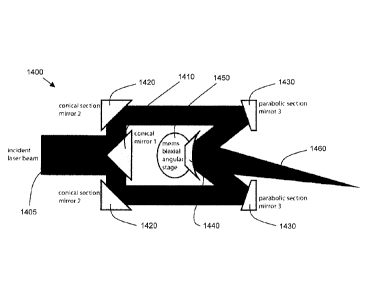
2.0000 TILTSURF WEDGE 0.0000 2.7200 ZNSE 6.3500 -0.0524
.. 0.0000
3.0000 STANDARD 0.0000 0.5000 6.3500 0.0000
0.0000
4.0000 STANDARD wedge 0.0000 2.7200 ZNSE 6.3500 0.0000
0.0000
5.0000 TILTSURF 0.0000 1.0000 6.3500 0.0524
0.0000
6.0000 STANDARD laser rsrch opt 0.0000 1.6000 ZNSE
7.6200 -- 0.0000 -- 0.0000
7.0000 STANDARD LX-0620-Z-ET1.5 -0.0140 0.0000 7.6200 0.0000
0.0000
8.0000 STANDARD 0.0000 128.4603 2.5718 0.0000
0.0000
9.0000 STANDARD 0.0000 0.0000 18.4306 0.0000
0.0000
Table 1: Exemplary numerical data and parameters for exemplary
combinations of laser control and different optics in accordance with the
present disclosure
[0089] Figure 10 shows a side cross-sectional view of a laser scanning
endoscopic head
1000 along with representative dimensions thereof in accordance with an
exemplary
embodiment of the present disclosure, in which the diameter of the scanner
optics can
define the numerical aperture of the surgical laser beam. For example, as
illustrated in
Figure 10, a diameter of the scanner optics 1010 according to an exemplary
embodiment of
the present disclosure can be approximately 8 mm. It is also possible for the
diameter of the
scanner optics 1010 to be in the range of approximately 2 mm to 14 mm. Other
exemplary
ranges for the diameter of the scanner optics 1010 can be 4 mm to 12 mm, 6 mm
to 10 mm
(or approximations thereof), etc. It should be understood that the diameter of
the scanner
optics 1010 can be smaller than 2 mm or larger than 15mm in accordance with
certain
- 22 -
CA 3011108 2018-07-11
WO 2011/032165
PCT/US2010/048807
exemplary embodiments of the present disclosure. With the diameter of the
scanner optics
1010 being approximately 8 mm, an aperture diameter can be approximately 5
rnm, for
example.
[0090] As further illustrated in Figure 10, the diameter 1020 of the head
1000 can be
dependent on other elements and/or components in the exemplary system in
addition to the
optics, such as, e.g., imaging, illumination and instrument channels, motors,
processors,
controls systems, etc. For example, the diameter 1020 of the head 1000 can be
approximately 16 mm, as illustrated in Figure 10. It is also possible for the
diameter 1020 of
the head 1000 to be in a range of approximately 8 mm to 24 mm. For example,
the diameter
1020 of the head 1000 can also be 10 mm to 22 mm, 12 mm to 20 mm (or
approximations
thereof), etc. it should be understood that the diameter 1020 of the head 1000
can be
smaller than 8 mm or larger than 24 mm in accordance with certain exemplary
embodiments
of the present disclosure. The diameter 1020 of the head 1000 can depend on
several
factors, including the application(s) for which the system is to be used, the
features (e.g.,
channels) to be included in the head, and the associated manufacturing
feasibility and
expense, for example. Accordingly, considering precision manufacturing and
technologies
related to producing the optics and other elements that can be included in an
exemplary
system and device according to the present disclosure, it may be possible to
produce heads
having continuously smaller diameters will be possible and is thus considered
to be in
accordance with the present disclosure.
[0091] The length of the head 1000 can depend on the optical design, the
selection,
and/or the configuration of the optical elements used to generate the scan.
For example, a
length 1030 of the head 1000 can be approximately 17 mm, as illustrated in
Figure 10. It is
also possible for the length 1030 of the head 1000 to be in a range of
approximately 9 mm to
25 mm. Further, the length 1030 of the head 1000 can be 11 mm to 23 mm, 13 mm
to 21
mm (or approximations thereof), etc. For example, the length 1030 can be
smaller than 9
mm or larger than 25 mm in accordance with certain exemplary embodiments of
the present
disclosure.
[0092] Figure 11 shows a perspective view of a servo controlled
positioning system in
accordance with an exemplary embodiment of the present disclosure. The
exemplary
scanner illustrated in Figure 11 can include one or more optical fibers 1145
that can be
configured to deliver the light to the exemplary system, device, apparatus,
arrangement, etc.
Two or more lenses 1100, 1105 can expand and/or collimate the light beam to
fill the
- 23 -
CA 3011108 2018-07-11
WO 2011/032165
PCT/US2010/048807
aperture of the scanner elements. A first scanning wedge 1110 can be mounted
on a
mounting ring 1115 with a magnetic ring 1120 fixed concentrically with respect
thereto. An
ultrasonic motor 1125 can be configured to rotate the mounting ring 1115 with
the direction
and acceleration being controlled by a microprocessor/frequency generator
arrangement
1130. An array of four or more orthogonal magnetic detectors 1135 can relay
(and/or
communicate) the rotational position of the magnetic ring to the arrangement
1130. The
ultrasonic motor 1125 and the detector(s) 1135 can be connected to the
arrangement 1130
through a signal bus 1140 in a servo control arrangement, for example. It is
also possible to
use other communication system(s), configuration(s) and/or protocol(s) that
can be either
wired and/or wireless to connect the ultrasonic motor 1125 and the detector(s)
1135 with the
arrangement 1130. A second scanner unit/arrangement may be provided which can
have a
second optical wedge 1150, a further mounting ring 1155, another magnetic ring
1160,
another ultrasonic motor 1165 and position sensor array 1170, which are
provided in the
endoscopic head. A focusing lens 1175 can establish the resolution, working
distance and
working field diameter of the scanner, for example. The arrangement 1130 can
be
connected via a signal bus 1180 to an external scanner control, such as the
computer
control arrangement 130 illustrated in Figure 1.
[0093] Figure 12A shows an illustration of a geometrical sketch 1200
representing a
scanning pattern generated by an exemplary optical element and/or arrangement
in
accordance with an exemplary embodiment of the present disclosure, which can
be based
on an exemplary model. For example, the exemplary geometric sketch 1200
illustrated in
Figure 12A can be generated using the exemplary device, arrangement, system,
apparatus,
etc. according to the present disclosure that can include two or more optical
elements, such
as, e.g., optical elements 300, 310 illustrated in Figure 3A, or an exemplary
model thereof.
The optical wedges and/or corresponding model that can be used to generate the
geometrical sketch 1200 can include, e.g., two wedges 1110, 1150 shown in
Figure 11.
Exemplary variables that can be used in a corresponding exemplary model are
shown in
Figure 12A, for example.
[0094] Figure 12B shows an exemplary geometrical sketch 1220 with the
variables that
can be used in a model in accordance with an exemplary embodiment of the
present
disclosure. The exemplary geometrical sketch 1220 can be the same or
substantially similar
to the geometrical sketch 1200 illustrated in Figure 12A, It should be
understood that the
geometries and/or patterns used to generate and/or can be modeled by the two
exemplary
sketches 1200, 1220, respectively, can be different, as illustrated by a
comparison of Figures
- 24 -
CA 3011108 2018-07-11
WO 2011/032165
PCT/US2010/048807
12A and 12B, for example. While the geometrical sketch 1220 illustrated in
Figure 12B can
be generated by and/or represent the model of operation of the exemplary
device that can
be the same or substantially similar to the exemplary device used to generate
the scanning
pattern and/or be represented by the sketch 1200 (e.g., using two or more
optical wedges),
the geometries of the geometrical sketches 1200, 1220 with respect to the
angles can be
different from one another. For example, as illustrated in Figure 12B, there
can be a gap
1230 resulting from different angles, which can be modeled by the variable CT,
representing
the distance between the optical elements used to generate a scanning pattern
corresponding to geometrical sketch 1220. As illustrated in Figure 12A, in
this exemplary
embodiment, a gap 1230 does not exist and o- is not represented in the
geometrical sketch
1200.
[0095] For
example, the following exemplary equations can be used with a model in
accordance with an exemplary embodiment of the present disclosure, such as the
exemplary
models corresponding to the exemplary sketches 1200, 1220 as illustrated in
Figures 12A
and 12B.
= (n, ¨1) x A
= (n2 ¨1) x õg2
Vr2
2 2 2
r3 = ¨ x sin (91 ¨ 92) x cos(9i ¨ 92)
X = x cOs + cr x cos 91 + r3 x cos 92
R 11x2 4. y2
{ a tan(Y/X), X> 0;
0 =
TC-Eatan(Y X), X< 0
where r, is the radial displacement from the axis. fi, is the wedge angle, a
is the distance
between the two wedges, R and 6 are the radius and angle of target points in
polar
coordinates, X and Y are the positions in Cartesian coordinates, and ni is the
refractive index
of the wedges.
[0096] Figure
13A is an exemplary image 1300 of an exemplary scanning pattern 1305
generated by a device in accordance with an exemplary embodiment of the
present
- 25 -
CA 3011108 2018-07-11
WO 2011/032165
PCT/US2010/048807
disclosure. To generate the exemplary scanning pattern 1305, an exemplary
software
arrangement (e.g., a set of computer-executable instruction) is provided that
can be stored
on a hardware computer-accessible medium which, when executed, configure a
hardware
processing arrangement to execute procedures to facilitate and/or control the
operation of
the exemplary system using optical devices via the controller to plot and
trace an irradiation
path corresponding to the scanning pattern 1305 illustrated in Figure 13A.
[0097] For example, image 1305 can be a photograph of a trace made on a
piece of
thermal paper by a laser beam scanned and controlled by an exemplary device
made and
used in accordance with an exemplary embodiment of the present disclosure. The
exemplary trace can be of a previously programmed path established in the
computer
control 130 illustrated in Figure 1 and executed by the scanner control 135
illustrated in
Figure 1.
[0098] Provided herewith in the Appendix is exemplary code that can be
used for
performing an exemplary procedure in accordance with the present disclosure
which can,
e.g., generate the scanning pattern 1305 shown in the exemplary image 1300 of
Figure 13A.
[0099] For example, the exemplary procedures illustrated in the Appendix
can be used
with a CO2 laser being delivered through a hollow core or photonic bandgap
fiber. Such
exemplary arrangement and/or system can be coupled with, e.g., a motor
controller and
industrial Zn-Se optics for CO2 lasers. Certain exemplary motor control
equations can be
utilized via a computer software and/or programming language such as Matlab,
for example.
As described herein above, it is possible to use a fiber delivery
configuration for the laser, as
well as a direct laser delivery procedure.
[00100] According to such exemplary embodiment, the exemplary relationship
between
the desired points in the video image space and the rotational position of the
optical
elements can be represented by the following equations in a similar manner as
described
herein above with reference to Figure 12A. For example, if ri = r2 = r, the
angles for each
point (x,y) can be calculated by:
,( R R
co, = 0 ¨ cos- co, = TC x = Rcos(6) y = R sin(0)
.2r .2r where and
[00101] For example, such exemplary equations (or functions) can provide
absolute
angles of each wedge for each point in a scanning pattern space, which can
then be
- 26 -
CA 3011108 2018-07-11
WO 2011/032165
PCT/US2010/048807
converted to relative angles that each wedge can rotate to obtain to the
desired point from
the previous point (e.g., to obtain an angle between 35 and 37 , a wedge can
rotate about
2 from a starting point of 35 ). According to the exemplary procedures of
this exemplary
embodiment of the present disclosure, the exemplary angles can then be
verified by certain
conditions to ensure that the shortest path is taken every time an wedge is
rotated from one
point to another point (e.g., to ensure that a wedge rotates -5 as opposed to
355 ). The
exemplary angles are then fed into the `MotorRotateRelative' function (as
illustrated in the
exemplary code and procedures provided in the Appendix) which sends the motor
rotation
commands to an exemplary motor controller. Further details of the exemplary
procedures
.. and programming instructions are provided in enclosed Appendix, which
include comments
providing descriptions of the functions and commands used in accordance with
certain
exemplary embodiments of the present disclosure.
[00102] Figure 13B shows an illustrations of an exemplary optical wedge or
prism
scanner 1310 which can be used in accordance with an exemplary embodiment of
the
present disclosure. As described herein above, the scanner in the exemplary
system can
include, e.g., two or more co-axial circular optical wedges and/or prisms that
can each bend
(refract) a laser beam or other light by an angle. This bend angle 1315 can
depend on
wedge parameters and wavelength of the laser or light, for example. For small
angles, the
relationship can be represented by, e.g., a¨On(2) where a is the bend angle
(known as the
refracted angle), 0 is the angle of the wedge or prism, and n(2) is the
optical index of
refraction of the wedge or prism material that can be dependent on the
wavelength A of the
light. For example, according to certain exemplary embodiments of the present
disclosure, it
is possible to use zinc selenide (ZnSe) as an optical material with an index
of 2.34 at the
10.6 pm wavelength of a CO2 laser.
[00103] Figure 13C shows an illustration of a further exemplary scanning
pattern 1320 in
accordance with an exemplary embodiment of the present disclosure. For
example, by
rotating one wedge or prism, a light beam can be scanned in a circular motion
at a fixed
angle and, depending on the distance to the target, can generate a scanned
circle 1321
having, e.g., a fixed diameter. If two or more wedges or prisms are used in
series with one
another, the light beam can be scanned over any point in a circular area 1325
with a
diameter equal to twice the diameter of the scanned circle 1321 resulting from
the single
wedge scan, for example.
- 27 -
CA 3011108 2018-07-11
WO 2011/032165
PCT/US2010/048807
[00104] The first wedge can rotate the light beam by an angle (p1 to provide
the light
beam to the second prism, which can then produce a second angle 92 for the
light beam. If
the two angles are equal and opposite from one another, then the light beam
would provide
little or no net angular change. If the angles are equal and oriented in the
same direction as
one another, the light beam will likely undergo double (or approximately
double) the angular
change of a single wedge. A characteristic of this type of the exemplary
scanning system
can be that the periphery of the corresponding scanned area can be uniquely
defined by a
single pair of angles. For example, every point in the circular area can be
defined by two
pairs of angles, and the center can be defined by infinite pairs of angles.
Thus, to scan a
straight line from one side of the circle, through the center and to the other
side, the wedges
can be moved simultaneously in opposite directions, for example. Such
exemplary optical
device/arrangement can be used for accurate optical alignment, and can be
effectively and
readily operated by a variety of motor and control systems, including, e.g.,
servo control
and/or electromechanical motors and systems
[00105] Figure 14 shows a side view of another device/arrangement 1400 in
accordance
with a further exemplary embodiment of the present disclosure. As illustrated
in Figure 14,
the exemplary device/arrangement 1400 can be used to expand an incident laser
beam
1405 with a conical mirror 1410 and a conical section mirror 1420 into a wider
hollow
cylindrical (or tubular) beam of light, for example. The light beam can then
be focused wrth a
parabolic section mirror 1430 onto a parabolic mirror 1440, e.g., on the same
or substantially
the same axis with the incident laser beam 1405. The parabolic mirror 1440 can
be
mounted on a biaxial angular stage to control the direction of a reflected
beam 1460. One of
the advantages of the exemplary embodiment illustrated in Figure 14 is that
the exemplary
device/arrangement 1400 can maintain a relatively small profile with respect
to a relatively
large aperture. For example, it is possible to utilize angular components of
higher spatial
frequencies without using central or low frequency components for a relatively
large
exemplary focused beam angular spectrum.
[00106] Figure 15 shows an exemplary block diagram of an exemplary embodiment
of a
system according to the present disclosure. For example, an exemplary
procedure in
accordance with the present disclosure can be performed by a processing
arrangement
and/or a computing arrangement 1510. Such processing/computing arrangement
1510 can
be, e.g., entirely or a part of, or include, but not limited to, a
computer/processor 1520 that
can include, e.g., one or more microprocessors, and use instructions stored on
a computer-
accessible medium (e.g., RAM, ROM, hard drive, or other storage device).
- 28 -
CA 3011108 2018-07-11
WO 2011/032165
PCT/US2010/048807
[00107] As shown in Figure 15, e.g., a computer-accessible medium 1530 (e.g.,
as
described herein above, a storage device such as a hard disk, floppy disk,
memory stick,
CD-ROM, RAM, ROM, etc., or a collection/combination thereof) can be provided
(e.g., in
communication with the processing arrangement 1510). The computer-accessible
medium
1530 can contain executable instructions 1540 thereon. In addition or
alternatively, a
storage arrangement 1550 can be provided separately from the computer-
accessible
medium 1530, which can provide the instructions to the processing arrangement
1510 so as
to configure the processing arrangement to execute certain exemplary
procedures,
processes and methods, as described herein above, for example.
[00108] Further, the exemplary processing arrangement 1510 can be provided
with or
include an input/output arrangement 1570, which can include, e.g., a wired
network, a
wireless network, the internet, an intranet, a data collection probe, a
sensor, etc. As shown
in Figure 15, the exemplary processing arrangement (computing arrangement)
1510 can be
in communication with an exemplary display arrangement 1560, which, according
to certain
exemplary embodiments of the present disclosure, can be a touch-screen
configured for
inputting information to the processing arrangement in addition to outputting
information from
the processing arrangement, for example. Further, the exemplary display 1560
and/or a
storage arrangement 1550 can be used to display and/or store data in a user-
accessible
format and/or user-readable format. The exemplary procedure described herein
above can
be performed using the exemplary system shown in Figure 15 and described
herein.
[00109] Figure 16 shows a flow diagram of a procedure in accordance with
certain
exemplary embodiments of the present disclosure. As shown in Figure 16, the
exemplary
procedure can be executed on and/or by, e.g., the processing/computing
arrangement 1510
of Figure 15, and can be stored by a hardware computer-accessible medium,
which can
include the storage arrangement 1550 of Figure 15. The processing/computing
arrangement
1510 can be or included in the computer control 130 of Figure 1, for example.
The
processing/computing arrangement 1510 can access the storage arrangement 1550
to
obtain the instructions which can be used to configure the
processing/computing
arrangement 1510 to execute such exemplary procedure. For example, starting at
subprocess 1610, in accordance with certain exemplary embodiments of the
present
disclosure, the exemplary processing/computing arrangement 1510 can, in
subprocess
1620, define a pattern to irradiate at least one section in a body. In
subprocess 1630, the
exemplary processing/computing arrangement 1510 can control at least one
optical element
provided in a housing to refract and/or diffract light based on the pattern
using an actuating
- 29 -
CA 3011108 2018-07-11
WO 2011/032165
PCT/1JS2010/048807
arrangement. The housing and/or actuating arrangement can be structured to be
inserted
into the body, for example.
[00110] Figure 17 shows a flow diagram of another procedure in accordance with
certain
exemplary embodiments of the present disclosure. As shown in Figure 17, the
exemplary
procedure can be executed, at least in part, on and/or by, e.g., the
processing/computing
arrangement 1510 illustrated in Figure 15, and can be stored by a hardware
computer-
accessible medium, which can include the storage arrangement 1550 of Figure
15. The
processing/computing arrangement 1510 can be or included in the computer
control 130 of
Figure 1, for example. The processing/computing arrangement 1510 can access
the storage
arrangement 1550 to obtain the instructions which can be used to configure the
processing/computing arrangement 1510 to execute such exemplary procedure.
[00111] For example, starting at subprocess 1710, a device (e.g.,
endoscope) in
accordance with an exemplary embodiment of the present disclosure can locate
target tissue
within a body from a position within the body. In subprocess 1730, the
exemplary
processing/computing arrangement 1510 can establish a position of the device
relative to
the target tissue using a particular arrangement that has a housing that is
inserted into the
body, for example. Then, the exemplary processing/computing arrangement 1510
can
generate control data by (i) tracing over an image of the tissue a path to be
cut with at least
one electro-magnetic radiation in subprocess 1740, (ii) defining an area to be
effected by the
at least one electro-magnetic radiation in subprocess 1750, and/or (iii) c
controlling the
position of the at least one electro-magnetic radiation in real time in
subprocess 1760. The
exemplary processing/computing arrangement 1510 can then, in subprocess 1770,
with at
least one actuating arrangement provided in the housing, control at least one
optical element
provided in the housing, based on the control data, to refract and/or diffract
at least one light.
The exemplary path can be based on a pattern in accordance with certain
exemplary
embodiments of the present disclosure, and the housing and/or actuating
arrangement can
be structured to be inserted into the body, for example.
EXEMPLARY APPLICATIONS
[00112] While the exemplary embodiment of the steering and focusing
.. system/arrangement according to the present disclosure can be used to
improve a minimally
invasive surgical treatment of cancers of the head and neck, it is possible to
use such
exemplary systems/arrangements to treat diseases of other anatomical regions
and to work
- 30 -
CA 3011108 2018-07-11
88202-1D1
with other types of laser wavelengths. Other exemplary areas of application
can include
laparoscopic, gastrointestinal, urologic and thoracoscopic minimally invasive
surgery.
[00113] The foregoing merely illustrates the principles of the
disclosure. Various
modifications and alterations to the described embodiments will be apparent to
those
skilled in the art in view of the teachings herein, and especially in the
appended claims. It
will thus be appreciated that those skilled in the art will be able to devise
numerous
systems, arrangements, and methods which, although not explicitly shown or
described
herein, embody the principles of the disclosure. For the purpose of further
supporting and
enabling the present disclosure, the reader is invited to refer to the
publications and
references referred to above. It should be understood that the exemplary
procedures
described herein can be stored on any computer accessible medium, including a
hard
drive, RAM, ROM, removable disks, CD-ROM, memory stick, etc., and executed by
a
processing arrangement which can be a microprocessor, mini, macro, mainframe,
etc.
-31-
CA 3011108 2019-05-01
WO 2011/032165
PCT/1JS2010/048807
APPENDIX
I. Script file used to entirely run the system by setting parameters, defining
the desired laser
path, and sending the necessary commands to each motor. Calls the various
functions
listed on the following pages. (ControlMotorMASTER.m):
de; clear all; close all
% matlab function to reset serial port
instrreset
s=serialccoml ', 'Terminator', 'CR');
fopen(s);
it:, Stefan's TMCM-3-10 initialization routine
Motorinitialization(s)
% pause prevents serial port from choking
pause(0.05)
Stei'an's TIV1CM-310 horning routine
MotorHome(s)
pause(0.05)
% Defining triangle path for laser to follow:
% % Change "k" value in loop below to 121 when using this smalle: triancie
% x(1.30)={-3:0.1:-0.1]:
% x(31:60)=[0:0.1:2.9];
%, x(61:121 )=fiipir(i-3:0.1:3J):
% y(1.30)=4-1_5:0.11 .4]:
% y(31:60)=1ip1ra-1.4:0.1:1.5]):
a/,.> y(61:121)=-1.5:
% plotting triangular path for reference
plot(x.y)
Change "k" value in loop below to 201 when using this larger triangle
x(1:50)=4-5:0.1:-0.1];
x(51:100)[0:0.1:4.9];
x(101:201)=fliplr([-5:0.1:51);
y(1:50)=[-2.5:0.1:2.4];
- 32 -
CA 3011108 2018-07-11
WO 2011/032165
PCT/US2010/048807
y(51:100)=fliplr([-2.4:0.1:2.5]);
y(101:201)=-2.5;
it plotting trianguiar path for reference
A, plot(x,y)
as homing switches are set to 12 o'ciock, we move wedges to
% center point and call that 0 degrees for both
MotorRotateRelative(s,-90,-90);
absth1=(0.9*round((-90/0.9)*-4.2857)/-4.2857)+90;
absth2=absth1;
"/10 defining maximum radius of each circle (2*r is the maximum radius for the
=!;, two combined circies)--NOTE: This will late:- need to change to a
function
% of the distance from the wedges to the target plane.
r=3;
% now we move the wedges to the desired points
"i'lo NOTE: change the max k value to match the number of points in the
triangle defined above
for k=1:201
if scirt(x(k)^2+y(k)^2)<=2*r % check to make sure inside the field
% For testing purposes, show x and y values.
% X=x(k)
% Y=y(k)
% convert points to angles, with our own conversion function
[th1 th2]=xy2th1th2(x(k),y(k),r);
Settno conditions to pick shortest path
rotth 1=th1-absth 1;
rotth2=th2-absth2;
if rotth1>180
rotth 1=rotth 1-360;
elseif rotth1<(-180)
rotth1=rotth 1+360;
end
if rotth2>180
rotth2=rotth2-360;
elseif rotth2<(-180)
- 33 -
CA 3011108 2018-07-11
WO 2011/032165 PCT/US2010/048807
rotth2=rotth2+360;
end
Stefan 's move weddes function
[relth1 relth2]=MotorRotateRelative(s,rotth1,rotth2);
update current annular position
absth1=absth1+relth 1 ;
absth2=absth2+relth2;
'X) Pause to allow 2nd motor to reach its desHrecl point before the
% first motor starts rotating to the next point
pause(0.1)
else
out of range'
end
end
II. Motor initialization function to set motor parameters. Sends commands to
the motor
driver/controller. (Motorinitialization.m):
function [1= Motorinitialization(s)
%Motor Settings l=unction
MC0=['ASAP 6, 0, 40013]; a/oMax Current to Motors 0 and 1 (0.8A)
MC1I'ASAP 6. 1, 40013];
MPS0=1ASAP 4, 0, 50' 13]; %Max Positioning Speed for Motors 0 and 1
MPS14ASAP 4, 1, 50' 131;
MSRO4A3AP 140, 0, 1' 13]; l/ofVlicrosteo Resolution for Motors 0 and 1 - Hail-
stepped
MSR1:--[ASAP 140, 1, 1' 13];
for a=1:length(MCO)
fwrite(s,1nt8(MCO(a)))
end
out=fscanf(s);
out=fscanf(s);
pause(0.05)
for b=1:length(MC1)
fwrite(s,1nt8(MC1(b)))
- 34 -
CA 3011108 2018-07-11
WO 2011/032165
PCT/US2010/048807
end
out=fscanf(s);
out=fscanf(s);
pause(0.05)
for c=1:length(MPSO)
fwrite(s,int8(MPSO(c)))
end
out=fscanf(s);
out=fscanf(s);
pause(0.05)
for d=1:length(MPS1)
fwrite(s,int8(MPS1(d)))
end
out=fscanf(s);
out=fscanf(s);
pause(0.05)
for e=1:length(MSR0)
fwrite(s,1nt8(MSR0(e)))
end
out=fscanf(s);
out=fscanf(s);
pause(0.05)
for f=1:length(MSR1)
fwrite(s,int8(MSR1(f)))
end
out=fscanf(s);
out=fscanf(s);
pause(0.05)
HI. Motor homing function which zeros the motor position. (MotorHome.m)
function []=MotorHome(s)
'Yo Motor Homing Function for TMCM-300
- 35 -
CA 3011108 2018-07-11
WO 2011/032165
PCT/US2010/048807
c.!ir; For limit switches connected witn NC terminal to 'L and
!:., common terminal to 'GND' on the TIVICM-310 board
create reference search start strings
RFS0a=['ARFS START, 0131;
RFS1ar-[ARFS START, 1' 13];
To create referE,,rce search status strings
RFS0b4ARFS STATUS, 0131;
RFS1tor[ARFS STATUS, 1' 13];
To ci-eate end condition string
endcondition=1nt8([3A 100 313]);
D.4) send the refernce search start commands and clear replies
for a=1:length(RFSDa)
fwrite(s,int8(RFS0a(a)))
end
out=fscanf(s);
out=fscanf(s);
pause(0.05)
for a=1:length(RFS1 a)
fwrite(s,int8(RFS1a(a)))
end
out=fscanf(s);
out=fscanf(s);
pause(0.05)
ciii) send the reference search start commands and get replies
for a=1:length(RFS0b)
fwrite(s,int8(RFS0b(a)))
end
outCOM0=int8(fscanf(s));
outRET0=1nt8(fscanf(s));
pause(0.05)
for a=1:length(RFS1b)
fwrite(s,int8(RFS1b(a)))
- 35 -
CA 3011108 2018-07-11
WO 2011/032165 PCT/US2010/048807
end
outCOM1=int8(fscanf(s));
outRET1=1nt8(fscanf(s));
pause(0.05)
cornpal.e replies to endcondition
while outRET0(8)¨=48
for a=1:length(RFS0b)
fwrite(s,1nt8(RFS0b(a)))
end
outCOM0=1nt8(fscanf(s));
outRET0=int8(fscanf(s));
pause(0.05)
end
while outRET1(8)¨=48
for a=1:length(RFS1b)
fwrite(s,1nt8(RFS1b(a)))
end
outCOM1=int8(fscanf(s));
outRET1=int8(fscanf(s));
pause(0.05)
end
IV. Motor relative rotation function that sends the necessary commands to move
each motor
relative to its current position. (MotorRotateRelative.m)
function [realthl, rea1th2]=MotorRotateRelative(s, thetal , theta2)
%Function to rotate motor by an angular ammounT
s is the serial port object.
thetal and 1heta2 are the angles
110Defice Command Strings to send to TiV1C1v1-310
= vo convert angles to half steps - (0.9 is half stepped)
% the fraction 4.2857 is the ratio between the two pulleys
steps1=round((theta1/0.9)*(-60/14));
steps2=round((theta2/0.9)*(60/14));
- 37 -
CA 3011108 2018-07-11
WO 2011/032165
PCT/US2010/048807
outstr0=['AMVP PEL, 0, ' int2str(steps1) 13];
outstr1=['AMVP RE L, 1, ' int2str(steps2) 13];
realthl=steps1*0.9/(-60/14);
realth2=steps2*0.9/(60/14);
%Writing Relative Positioning Strings to TMCM-310
for i=1:length(outstrO)
fwrite(s,int8(outstrO(i)))
end
The following returns are necessary in order for the homing function
'to work properly. This ls because the sent and returned strings build up
%and must be scanned after each command in order to get the return string
%corresponding to each command.
out=fscanf(s);
out=fscanf(s);
pause(0.05) :ll.,The pause is necessary in order for the board to accept both
commands
without error.
for j=1:length(outstr1)
fwrite(s,int8(outstrl
end
out=fscanf(s);
out=fscanf(s);
pause(0.05)
V. Function to convert (x,y) point to angles of rotation for each wedge.
(xy2th1th2.m)
`)/-0 This function converts the input 'x and 'y' co-ordinates to angles
thetal
To and 1heta2. This program always gives the value of the angle from the
position it is at. i.e. considering that point as the origin.
function [theta 1,theta2]=xy2th1th2(x,y,r)
n=sqrt((x^2)+(y^2));
if n<=(2*r)
%The value of variable b and the formula for theta1 and theta2 was
%mathematically calculated.
b = acosd(sqrt(xA2+y^2)/(2*r));
- 38 -
CA 3011108 2018-07-11
WO 2011/032165
PCT/US2010/048807
thetal ((a1an2(y,x)*180)/p0+ b;
theta2 = ((atan2(y,x)*180)/pi)- b;
else
thetal =('The values of x and y are out at the maneuvering limits')
theta2('The values oi x and y are out o; the maneuvering lirnits')
end
- 39 -
CA 3011108 2018-07-11