Note: Descriptions are shown in the official language in which they were submitted.
CA 03011240 2018-07-11
WO 2017/124060 PCT/US2017/013611
SYSTEMS AND METHODS FOR ADHERING VESSELS
CROSS-REFERENCE TO RELATED APPLICATIONS
[0001] This application claims the benefit of U.S. Provisional Application No.
62/279,642,
filed January 15, 2016, and titled "SYSTEMS AND METHODS FOR ADHERING VESSELS,"
which is hereby incorporated by reference in its entirety.
FIELD
[0002] The current invention relates to systems and methods for adhering
tubular structures
within the body, such as vessels.
BACKGROUND
[0003] The devices, systems, and methods described here may be used to enhance
mechanical
adhesion between tubular structures within the body. In some instances, this
may be desirable to
improve mechanical adhesion between tubular structures used to form fistulas.
A fistula is
generally a passageway formed between two internal organs. Forming a fistula
between two
tubular structures, such as blood vessels, can have one or more beneficial
functions. For
example, the formation of a fistula between an artery and a vein may provide
access to the
vasculature for hemodialysis patients. Specifically, forming a fistula between
an artery and a
vein allows blood to flow quickly between the vessels while bypassing the
capillaries. In other
instances, a fistula may be formed between two veins to form a veno-venous
fistula. Generally,
fistula formation requires surgical dissection of a target vein, and
transecting and moving the
vein for surgical anastomosis to the artery. It may therefore be useful to
find improved ways to
form a fistula between two blood vessels.
BRIEF SUMMARY
[0004] Described here are devices, systems, and methods for adhering two or
more tubular
structures together. The tubular structures may be any suitable tubular
structure, such as an
artery, vein, duct, digestive tract, and so forth. For example, the devices,
systems, and methods
disclosed herein may increase mechanical adhesion between two blood vessels
such as an artery
and a vein, a vein and a vein, an artery and an artery, or between a duct and
a duct, a digestive
1
CA 03011240 2018-07-11
WO 2017/124060 PCT/US2017/013611
tract and a digestive tract, and the like.
[0005] Generally, a method for adhering tubular structures described herein
comprises
advancing a first catheter into a first tubular structure and a second
catheter into a second tubular
structure. The first catheter may comprise a first adhesion element and the
second catheter may
comprise a second adhesion element. The first adhesion element element may be
aligned with
the second adhesion element. The first and second tubular structures may be
adhered by heating
tissue between the two adhesion elements.
[0006] In some variations, a method of adhering vessels together comprises
advancing a first
catheter comprising a first magnetic adhesion element into a first blood
vessel and a second
catheter comprising a second magnetic adhesion element into a second blood
vessel. The first
magnetic adhesion element may be aligned with the second magnetic adhesion
element. In some
variations, the first magnetic adhesion element may be aligned with the second
magnetic
adhesion element by fluoroscopically visualizing at least a portion of the
first and second
catheters. In some variations, the first and/or second catheters may comprise
one or more
rotational indicators. The rotational indicators may be fluoroscopically
visualized to align the
catheters. Tissue of the first and second blood vessels may be compressed
between the first and
second magnetic adhesion elements. An adhesion (weld) may be formed between
the first blood
vessel to the second blood vessel by using the magnetic adhesion elements to
heat tissue of the
first and second blood vessels between the magnetic adhesion elements. In some
variations, the
tissue may be heated by delivery of radiofrequency energy from the magnetic
adhesion elements.
In other variations, the tissue may be heated by ohmic heating of the magnetic
adhesion
elements. In these variations the magnetic adhesion elements may comprise a
resistor. Heating
may occur over a single cycle, or a plurality of cycles. In some variations,
impedance between
the magnetic adhesion elements may be monitored before, after, or during
energy delivery.
Additionally or alternatively, tissue temperature may be monitored before,
after, or during
energy delivery. In some variations, a second adhesion may be formed between
the first blood
vessel and the second blood vessel. The formed adhesion(s) may in some
instances have a width
between about 0.1 mm and about 15 mm, and a length between about 0.1 mm and
about 10 cm.
In some variations, a fistula may be formed through the adhesion. For example,
a fistula may be
formed through the adhesion using the magnetic adhesion elements.
[0007] In some variations, a system for adhering two tubular structures
together comprises a
2
CA 03011240 2018-07-11
WO 2017/124060 PCT/US2017/013611
first catheter comprising a first adhesion element and a second catheter
comprising a second
adhesion element. The first adhesion element may be magnetic and may comprise
a flat contact
surface. The second adhesion element may be magnetic and comprise a flat
contact surface. A
power source may be connected to the first and second adhesion elements. In
some variations,
the first adhesion element may be located at a distal end of the first
catheter, and the second
adhesion element may be located at a distal end of the second catheter. The
first and/or second
adhesion elements may be coated with one or more layers of a fluoropolymer.
Additionally or
alternatively, the first and/or second magnetic adhesion elements may comprise
surface
insulation. In some variations, the first adhesion element may define a
recess. In some of these
variations, the second adhesion element may comprise a protrusion
complementary to the recess.
In some variations, the first and/or second catheter may comprise a rotational
indicator.
[0008] In some variations, the first catheter may comprise a proximal portion
and a distal
portion, wherein the largest cross-sectional dimension of the distal portion
is larger than the
largest cross-sectional dimension of the proximal portion. The first adhesion
element may be
located on the distal portion. Additionally or alternatively, the second
catheter may comprise a
proximal portion and a distal portion, wherein the largest cross-sectional
dimension of the distal
portion is larger than the largest cross-sectional dimension of the proximal
portion. The second
adhesion element may be located on the distal portion. In some variations, at
least one of the
pushability, flexibility, or torquability of the first and/or second catheter
may be adjustable. For
example, the first catheter may comprise a distal portion, an inner proximal
portion, and an outer
proximal portion. The outer proximal portion may be slidable relative to the
distal portion. The
distal portion may have a retracted configuration and an extended
configuration. The distal
portion may be configured to extend away from the outer proximal portion when
moved from the
retracted configuration to the extended configuration.
BRIEF DESCRIPTION OF THE DRAWINGS
[0009] FIG. 1 is a block diagram of an illustrative variation of a system.
[0010] FIGS. 2A-2D are views of an illustrative system described here in
vasculature.
[0011] FIGS. 3-4 are perspective views of a distal portion of an illustrative
variation of a
catheter comprising an adhesion element described here.
3
CA 03011240 2018-07-11
WO 2017/124060 PCT/US2017/013611
[0012] FIGS. 5A-5F are perspective views of variations of adhesion elements.
[0013] FIGS. 6A-6C are cross-sectional side views of variations of adhesion
elements.
[0014] FIGS. 7-8 are perspective views of distal portions of variations of
catheters comprising
adhesion elements described here.
[0015] FIGS. 9A-9E are side views (FIGS. 9A, 9B, 9D) and perspective views
(FIGS. 9C, 9E)
of distal portions of a catheter in retracted (FIGS. 9A-9C) and extended
(FIGS. 9D-9E)
configurations.
[0016] FIG. 10 is a perspective view of an illustrative variation of a distal
portion of a catheter
comprising a separate magnet and adhesion element.
[0017] FIGS. 11A-11B are perspective views of an illustrative variation of a
system described
here.
[0018] FIGS. 12A-12B are cross-sectional and plan views, respectively, of
adhered vessels.
[0019] FIGS. 13A-13B are cross-sectional and plan views of a vessel comprising
a weld and a
fistula.
DETAILED DESCRIPTION
[0020] Generally described here are devices, systems, and methods for
increasing mechanical
adhesion between tubular structures, such as blood vessels, and in some
instances forming a weld
between the structures to adhere the two structures together. Generally, to
adhere two tubular
structures together, a system comprising multiple catheters may be advanced in
a minimally
invasive fashion (e.g., for blood vessels, via the vasculature) to a target
location and used to
adhere the tubular structures together. In some examples, the tubular
structures may comprise
blood vessels such as two arteries, two veins, or a vein and an artery.
[0021] Generally, each catheter may comprise an adhesion element. An adhesion
element may
comprise an element capable of adhering tissue, either alone or in combination
with another
adhesion element. An adhesion element may be configured to adhere tissue
together by heating
the tissue. In some variations, an adhesion element may heat issue by
delivering electrical energy
4
CA 03011240 2018-07-11
WO 2017/124060 PCT/US2017/013611
to the tissue. In some of these variations, the adhesion element may comprise
a magnet
configured to heat tissue by delivering electrical current, as described in
more detail herein. In
other variations, an adhesion element may be heated through resistive heating,
which may in turn
heat tissue. In yet other variations, an adhesion element may heat tissue
using laser energy. For
example, a catheter may comprise a fiber optic filament coupled to a laser,
such that the adhesion
element may be configured to direct laser energy to heat tissue. In yet
another variation, an
adhesion element may deliver ultrasonic energy to heat tissue. In such a
variation, the adhesion
element may comprise a piezoelectric element configured to use ultrasonic
vibration to induce
heating.
[0022] A first catheter comprising an adhesion element may be placed at a
target location in a
first tubular structure, and a second catheter comprising an adhesion element
may be placed at a
target location in a second tubular structure. The catheters may be aligned
relative to each other
using the adhesion elements, coaption regions, and/or visual alignment aids,
as described in more
detail herein. For example, when the tubular structures are blood vessels, a
first catheter may be
placed in a first blood vessel, and a second catheter may be placed in a
second blood vessel,
where the first and second vessels are in proximity to each other, and the two
catheters may be
aligned to coapt the two vessels. Tissue heating due to one or more adhesion
elements may
adhere tissue of the first tubular structure to tissue of the second tubular
structure. For example,
current applied to blood vessel walls may denature proteins in each vessel,
which may cause
them to adhere together. Adhesion may be performed before, during, or after
other procedures,
such as fistula formation, as described in more detail herein.
I. SYSTEMS
[0023] Generally, the systems described here are configured to adhere tubular
structures in the
body, such as blood vessels. In some variations, the systems comprise two
catheters each
comprising one or more adhesion elements. An adhesion element may comprise an
element
capable of adhering tissue, either alone or in combination with another
adhesion element, and
may be configured to adhere tissue together by heating the tissue. In some
variations, an
adhesion element may heat issue by delivering electrical current to the
tissue, while in other
variations an adhesion element may heat tissue by delivering laser or
ultrasonic energy to the
tissue. In yet other variations, an adhesion element may be resistively
heated, which may in turn
heat tissue. In some variations, an adhesion element may comprise a magnet
configured to
CA 03011240 2018-07-11
WO 2017/124060 PCT/US2017/013611
deliver energy to tissue, although in other variations it may comprise a non-
magnetic element.
[0024] The adhesion elements may be configured to be delivered to target
locations in tubular
structures (e.g., blood vessels) via catheters. When an adhesion element is
configured to heat
tissue by delivering electrical energy, it may comprise a contact surface
configured to contact
tissue (e.g., a blood vessel wall) or fluid (e.g., blood). When the contact
surface is in contact with
tissue and/or fluid at the target location, it may supply current to and/or
carry current from the
tissue and/or fluid. This may result in heat, which in turn may facilitate
adhesion of one portion
of tissue to another. More particularly, current applied to the adhesion
elements may be
configured to heat and/or desiccate tissue to mechanically adhere the vessels
together through
protein denaturation. In some instances, tissue may be thermally welded
together by applying a
coagulation current to an electrode to denature connective tissue proteins and
thereby increase
adhesion between tissue planes. In some instances, the denatured proteins from
each vessel may
intertwine to fuse together and/or shrink the vessel. In some variations,
thermal denaturing and
welding may modify the vessel without removing material as occurs when
ablating tissue.
[0025] FIG. 1 is a block diagram of one variation of a system comprising a
first catheter (100)
and a second catheter (104). The first catheter (100) may comprise a first
adhesion element (102)
and the second catheter (104) may comprise a second adhesion element (106). At
least a portion
of the adhesion elements may be exposed to the surrounding environment (i.e.,
may not be fully
encompassed by the catheters). The adhesion elements (102, 106) may comprise
electrically
conductive magnets, as described in more detail herein, although they need not
be magnetic. In
use, the first catheter (100) and the second catheter (104) may be placed in
first and second
tubular structures (e.g., blood vessels), respectively, wherein the tubular
structures are adjacent,
and the adhesion elements (102, 106) may interact to adhere the outer wall of
the first structure
(e.g., blood vessel) to the outer wall of the second structure (e.g., blood
vessel). In some
variations, the adhesion elements may adhere the tissue by delivering
electrical energy to the
tissue. As such, a proximal end of each catheter (100, 104) may be connected
to a power supply
(110) by respective connections (108). The power supply (110) may further
comprise a controller
(not shown) for controlling energy delivery to the catheters (100, 104). The
power supply (110)
may be an AC or DC power supply. The power supply (110) may output current to
heat and/or
desiccate tissue.
[0026] In some variations, the adhesion elements (102, 106) may each deliver
electrical energy
6
CA 03011240 2018-07-11
WO 2017/124060 PCT/US2017/013611
to heat tissue. For example, each adhesion elements (102, 106) may be
connected to an active
output of the power supply (110) to deliver current and thus heat adjacent
tissue. As such, the
adhesion elements (102, 106) may simultaneously heat tissue from opposing
sides. A ground pad
(e.g., a large metal plate or flexible metalized pad) affixed to the patient
may be connected to a
return terminal of the power supply. In other variations, the first adhesion
element (102) may be
connected to the active output of the power supply (110) and the second
adhesion element (106)
may be connected to the return terminal. In yet other variations, the first
adhesion element (102)
may be connected to an output of the power supply, and the second adhesion
element (106) may
be floating, that is, not directly connected to any output of the power
supply, in a focused
monopolar configuration.
[0027] It should be appreciated that in other variations, only the first
catheter may be
connected to the power supply in a monopolar configuration. For example, only
the first
adhesion element may heat tissue, while the second adhesion element may
mechanically
contribute to tissue adhesion by pressing tissue toward the first adhesion
element while the first
adhesion element heats tissue. In such a configuration, the first adhesion
element may be
connected to an active output of a power supply, and a ground pad affixed to
the patient may be
connected to a return terminal of the power supply. The second adhesion
element may not be
connected to the power supply but may oppose the first adhesion element and
compress tissue
between the two adhesion elements, promoting heating and adhesion.
[0028] The catheters and adhesion elements may be configured to coapt with
each other and to
compress tissue between the adhesion elements in order to adhere tissue when
it is heated. In
some instances, a system may comprise first and second catheters each having
one or more
magnets, such that magnets of the first catheter may be attracted to magnets
of the second
catheter to bring the catheters in closer approximation. In some variations,
the adhesion elements
themselves may be magnets and may be configured to be attracted to each other.
As such, the
adhesion elements may promote axial and/or rotational alignment of the
catheters. Additionally
or alternatively, the catheters may comprise coaption regions comprising
magnets, and/or may
comprise one or more visual alignment aids, to promote tissue coaption as well
as axial and/or
rotational alignment of the catheters. For instance, a rotational indicator
may allow catheter
alignment to be visualized under fluoroscopy, such that a user may manipulate
the catheters into
a desired position. In some variations, the catheters may also be configured
to promote the ability
7
CA 03011240 2018-07-11
WO 2017/124060 PCT/US2017/013611
of an adhesion element to press into tissue, as described in more detail
herein. The catheters may
have the same configuration of elements, or may have different and/or
complementary
configurations of elements.
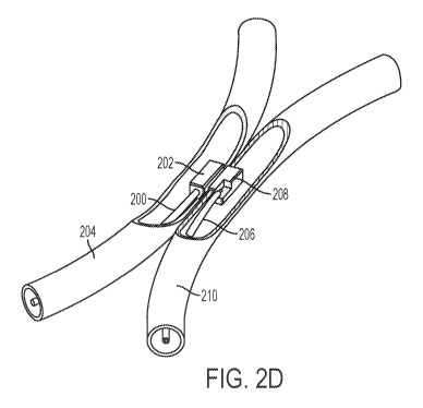
[0029] FIGS. 2A-2C show an exemplary system comprising two catheters each
comprising an
adhesion element comprising a magnet. A first catheter (200) comprising an
adhesion element
(202) is shown located within a first blood vessel (204). A second catheter
(206) comprising an
adhesion element (208) is shown located within a second blood vessel (210).
The shape and
material of the adhesion elements may help to align one catheter in a first
vessel with another
catheter in a second vessel, and may help to ensure optimal heating and
adhesion. In the variation
shown in FIGS. 2A-2D, each of the adhesion elements (202, 208) comprises a
rectangular
magnet. The adhesion elements (202, 208) may be configured to be attracted to
each other when
in proper axial and rotational alignment. FIGS. 2A-2B show the catheters (200,
206) placed in
the vessels without coaption due to magnetic attraction, while FIGS. 2C-2D
show the catheters
coapted due to magnetic attraction between the adhesion elements (202, 208).
When the
catheters are coapted as in FIGS. 2C-2D, the walls of vessels (204, 210) may
be compressed
between the two adhesion elements (202, 208). That is, during alignment of the
catheters (200,
206), the attractive magnetic forces of the adhesion elements (202, 208) may
bring the catheters
(200, 206) and blood vessels (204, 210) into closer approximation, as shown in
FIGS. 2C-2D.
This compression may aid with heating and adhesion. Each of the adhesion
elements (202, 208)
may comprise a flat contact surface configured to be contact with the interior
vessel wall when
the catheters are coapted across the vessel walls.
[0030] The adhesion elements (202, 208) may comprise a conductive material and
may be
connected to a power supply configured to provide electrical current for
heating tissue. FIG. 3 is
a perspective view of a distal end of the catheter (200) and the adhesion
element (202), shown
with a portion of the adhesion element cut away to depict the interior of the
adhesion element. As
described above, the adhesion element (202) shown may be a magnet that may
coapt with
another magnet of another catheter (e.g., adhesion element (208) of catheter
(206)) to compress
tissue therebetween. The adhesion element (202) may comprise one or more flat
contact surfaces
(214) for providing flush contact with tissue to be heated. Furthermore, in
addition to bringing
the catheters closer together and compressing tissue, a flat contact surface,
such as contact
surface (214), may allow a lateral magnetic coaption force to be generated and
translated into an
8
CA 03011240 2018-07-11
WO 2017/124060 PCT/US2017/013611
aligning torque, which may aid rotational alignment, as well as axial
alignment, with an adhesion
element of a second catheter. A wire (212) may be located within the catheter
(200) and may
electrically couple the adhesion element (202) to a power source such as an
external power
supply. This may allow an external power supply to energize the adhesion
element (202). For
example, returning to FIGS. 2A-2D, once the adhesion elements (202, 208) are
coapted at a
desired location, one or more of the adhesion elements may be energized to
apply heat to the
vessels (204, 210). The heat applied to the compressed vessel tissue (204,
210) may denature
proteins in a manner to adhere the vessels together and may form a weld, which
may increase the
mechanical strength of the vessels. Upon completion of welding between the
blood vessels (204,
210), the catheters (200, 206) may be removed.
[0031] When heat is applied to the compressed vessels using the devices,
systems, and
methods described herein, heating tissue to 70 C may result in denaturation.
In some variations,
the delivered energy may be constant, while in other variations it may be
modulated. In some
variations, the tissue may be heated by the delivery of radiofrequency energy
to tissue. The
power source may deliver energy having any suitable waveform to the tissue via
the adhesion
elements, such as but not limited to waveform having a sinusoidal or square
shape. Electrical
energy delivered to tissue may have a peak voltage below the ionization
threshold of the tissue.
For example, when the waveform is a sinusoidal waveform, the peak voltage may
in some
variations be below about 150 V. It should also be appreciated that in other
variations, current
need not travel through tissue in order to heat the tissue. For example, the
tissue may be heated
through ohmic heating of the adhesion elements. For example, each adhesion
element may
comprise a resistor resulting in ohmic heating. In these instances, either AC
or DC current may
be used. In yet other variations, the tissue may be heated through laser or
ultrasonic energy
delivery.
[0032] In some variations, one or more surfaces of an adhesion element may be
coated with a
material that may facilitate removal of the adhesion element from a tissue
surface after heating.
For example, one or more surfaces of an adhesion element may be coated with
one or more
layers of PTFE, parylene, silicone, or another fluoropolymer. FIG. 4 is a
perspective view of
another variation of a catheter (400) comprising an adhesion element (402)
comprising a
rectangular magnet, and a wire (404) extending through the catheter (400) to
electrically couple
to the adhesion element (402) to a power supply. A surface of the adhesion
element (402) may
9
CA 03011240 2018-07-11
WO 2017/124060 PCT/US2017/013611
comprise a coating (406), wherein the coating comprises a material that may
facilitate the
removal of the adhesion element (402) from a tissue surface without sticking.
For example, the
coating (406) may comprise one or more layers of PTFE, parylene, silicone, or
another
fluoropolymer. In some variations, the coating (406) may additionally or
alternatively comprise a
jacket of material for enhanced biocompatibility or electrical conductivity,
such as one or more
of gold, platinum, and titanium. In some variations, the adhesion element
(402) may be
additionally or alternatively partially coated with an insulative coating in
order to insulate certain
surfaces and leave other surfaces exposed for electrical conduction to the
tissue. This may direct
and/or isolate energy delivery to a specific region of tissue and/or in a
specific shape.
[0033] Generally, catheters in the coapted state, as shown in FIGS. 2C-2D for
example, may
sandwich the tissue interposed between their surfaces with a desired pressure
as determined by
the size, shape, and material composition of the adhesion elements. Adhesion
elements having a
flat contact surface, as shown in FIGS. 2A-4 for example, may promote
rotational alignment and
may better compress tissue for adhesion. These flat surfaces may help to
naturally align the
adhesion elements with each other, as two flat surfaces may generate a greater
aligning torque
for a given amount of rotational misalignment than two curved surfaces. For
example, in some
instances, the aligning torque generated between flat magnetic surfaces at 5
degrees of
misalignment is at least approximately 18 times stronger than that of the
aligning torque between
magnetic cylinders.
[0034] FIGS. 5A-5E show a variety of possible shapes for adhesion elements,
each comprising
a flat contact surface. For example, an adhesion element (500) may have a
square cross-section
(FIG. 5A); an adhesion element (502) may have a triangular cross-section (FIG.
5B); an adhesion
element (504) may have a hexagonal cross-section (FIG. 5C); an adhesion
element (506) may
have an a rectangular cross-section (FIG. 5D); an adhesion element (508) may
have a semi-
circular cross-section (FIG. 5E), or the like to provide a flat contact
surface for an adhesion
element.
[0035] However, in other variations, the adhesion elements may not comprise
flat contact
surfaces. For example, FIG. 5F illustrates an adhesion element (510) having a
circular cross-
section. As another example, FIGS. 6A-6B illustrate cross-sectional views of
two adhesion
element pairs, where one adhesion element is configured to be located in a
first tubular structure
(e.g., a blood vessel), and a second adhesion element is configured to be
located in a second
CA 03011240 2018-07-11
WO 2017/124060 PCT/US2017/013611
tubular structure (e.g., a second blood vessel). The adhesion elements shown
there may comprise
one or more protrusions and recesses, where the protrusions and recesses may
be
complementary. These protrusions and recesses may form an indent in tissue
interposed between
the adhesion elements. Pairs of adhesion elements having complementary shapes,
as shown in
FIGS. 6A-6B, may allow greater pressure to be applied to tissue between the
two adhesion
elements. In some variations, the protrusions (608, 610) may have the shape of
a block (see
adhesion element (600) in FIG. 6A) or a rigid fin (see adhesion element (604)
in FIG. 6B) or
point. The recesses (612, 614) of adhesion elements (602, 606) may have
complementary shapes
to the protrusions (608, 610). In other variations, a pair of adhesion
elements may have the same
shape. For example, FIG. 6C illustrates a cross-sectional view of a pair of
matching adhesion
elements (616, 618), each having a raised perimeter and a recessed central
region (620, 622). A
similar adhesion element having a raised rectangular perimeter and a recessed
central region is
shown in FIG. 8, described in more detail herein. In some variations, the
protruding member may
have a hollow interior for decreasing the surface area of the protruding
member so that a first
current may be applied to adhere tissue and a second current may be supplied
to cut an opening
through the tissue.
[0036] The adhesion elements described herein may be attached to catheters, as
shown for
example in FIGS. 2A-2D. Generally, the systems may comprise a first catheter
for placement in
a first tubular structure (e.g., a blood vessel) and a second catheter for
placement in a second
tubular structure (e.g., a blood vessel), where each catheter may comprise at
least one adhesion
element. The catheters may have any suitable diameter. For intravascular use,
for example, the
catheters may be about 4 French, about 5.7 French, about 6.1 French, about 7
French, about 8.3
French, between about 4 French and about 9 French, between about 4 French and
about 7
French, between about 4 French and about 6 French, or the like. In the
variation shown in FIGS.
2A-2D, the widest dimension of the adhesion elements (202, 208) is greater
than the diameter of
the catheters (200, 206). This may allow the contact surfaces of the adhesion
elements (202, 208)
to more easily contact tissue.
[0037] In other variations, the catheters described herein may be configured
to promote the
ability of an adhesion element to press into tissue. For example, the adhesion
element may be
located on a portion of a catheter having a greater diameter than an adjacent
portion of a catheter.
This may allow the contact surfaces of the adhesion elements to more easily
contact tissue. For
11
CA 03011240 2018-07-11
WO 2017/124060 PCT/US2017/013611
example, FIG. 7 is a perspective view of a variation of a catheter (700)
having an adhesion
element (706) embedded within a portion of the catheter and having an exposed
flat contact
surface. Catheter (700) comprises a proximal portion (702), a distal portion
(704), and an
adhesion element (706) disposed on the distal portion (704) and having a flat
contact surface. As
shown, the largest cross-sectional dimension of the distal portion (704)
comprising the adhesion
element (706) is larger than the largest cross-sectional dimension of the
proximal portion (702)
of the catheter. As such, the adhesion element (706) may be able to press into
tissue without
obstruction from contact between the proximal portion (702) of the catheter
(700) and tissue, for
example, a vessel wall. FIG. 8 illustrates another catheter (800) comprising a
proximal portion
(802) and a distal portion (804), where the distal portion (804) has a larger
cross-sectional
dimension than the proximal portion (802). The catheter (800) further
comprises an adhesion
element (806) disposed on the distal portion (804), where the adhesion element
(806) comprises
a raised perimeter with a central rectangular recess. That is, the adhesion
element (806) may
define an opening such that, for example, tissue indented against the adhesion
element (806)
forms a rectangular indent around the perimeter formed by the raised portion
of the adhesion
element (806). Energy may be supplied to activate the adhesion element (806)
to adhere two
vessels together. In some variations, an opposing adhesion element on a second
catheter may be
configured to fit within the central rectangular recess, which may allow for
increased pressure
application to tissue located between the adhesion element (806) and the
opposing adhesion
element.
[0038] In some variations, the catheters described herein may be configured to
have adjustable
stiffness, for example in the event that an increase in pushability,
flexibility, or torquability may
be desired. For example, FIGS. 9A-9E show side and perspective views of
variations of a
catheter (900) similar to catheter (700) comprising a proximal portion (904)
and a distal portion
(906) comprising an adhesion element (908) having a flat contact surface. The
distal portion
(906) may be fixedly connected to an inner proximal portion (904) and may have
a larger cross-
sectional dimension than the inner proximal portion (904). The inner proximal
portion (904) may
be slidable within an outer proximal portion (902), where the outer proximal
portion comprises a
tubular shape. As such, the distal portion (906) and part of the inner
proximal portion (904) may
be configured to extend distally from the outer proximal portion (902) between
a retracted
position (FIGS. 9A-9C) and an extended configuration (FIGS. 9D-9E). In the
extended
configuration shown in FIGS. 9D- 9E, the distal portion (906) of the catheter
(900) may have
12
CA 03011240 2018-07-11
WO 2017/124060 PCT/US2017/013611
increased ability to deform and/or press into tissue when the adhesion element
(908) is attracted
to a corresponding adhesion element of another catheter, since deformation of
the inner proximal
portion (904) is not limited by the outer proximal portion (902). In the
retracted configuration
shown in FIGS. 9A-9C, the distal portion (906) of the catheter (900) may have
increased
pushability, since deformation of the inner proximal portion (904) is limited
by the outer
proximal portion (902). In this way, either of the extended or retracted
configurations (or an
intermediate configuration between the extended or retracted configurations)
may be selected
based on one more requirements related to pushability, flexibility, and/or
torquability.
[0039] In FIGS. 2A-4 and 7-9E, the adhesion elements are shown at or near a
distal end of the
catheters. However, it should be appreciated that adhesion elements may be
located along any
suitable portion of the catheters described herein (e.g., a distal end, an
intermediate portion, or
combinations thereof). It should also be appreciated that a catheter may have
any suitable
number (e.g., zero, one, two, three, or four or more) and combination of
adhesion elements. In
variations in which a catheter comprises two or more adhesion elements,
multiple adhesion
elements may be used to create multiple adhesion regions, either
simultaneously or sequentially.
In other variations, multiple adhesion elements may interact to form a single
adhesion region.
[0040] Furthermore, in other variations, an adhesion element need not comprise
a magnet.
While magnetic adhesion elements may help to compress tissue between two
catheters, in some
variations the adhesion elements may comprise any material suitable for
heating tissue to cause
adhesion. For example, the adhesion elements may comprise any suitable
conductive material. In
some of these variations, the catheters described herein may comprise one or
more adhesion
elements and one or more separate alignment features to assist in coaption and
rotational and/or
axial alignment of the catheters relative to each other. In some of these
variations, alignment
features may assist a user in manual positioning of the catheters. Generally,
in these variations,
the catheters may comprise at least one of a flat coaption surface, a magnet,
and a rotational
indicator. Combinations of one or more of these elements may improve the
ability of a user to
orient and align catheters rotationally. For instance, the catheters described
herein may comprise
one or more adhesion elements and a separate coaption region comprising one or
more magnets
to promote coaption and alignment. For example, a catheter may comprise a
magnet and a
separate adhesion element comprising an electrode. FIG. 10 is a perspective
view of such a
variation of a catheter (1000) comprising a magnet (1002) and a separate
electrode (1006). The
13
CA 03011240 2018-07-11
WO 2017/124060 PCT/US2017/013611
magnet (1002) may be separated from the electrode (1006) by an electrical
and/or thermal
insulator (1004). The thermal insulator may comprise, for example, polyimide,
PEEK, PTFE,
and/or ceramic. In this configuration, the magnet (1002) may act to promote
tissue compression
between the flat contact surface of the adhesion element (electrode (1006))
and an adhesion
element in an adjacent vessel and may promote proper alignment between the
adhesion elements,
while the electrode (1006) may act as the adhesion element. In other
variations, the electrode
(1006) may be disposed directly on the magnet (1002).
[0041] FIGS. 11A-11B illustrate another variation of a system in vasculature
comprising a first
catheter (1100) in a first blood vessel (1106) and a second catheter (1108) in
a second blood
vessel (1114). The first catheter (1100) may comprise a first adhesion element
(1102) that may
be a non-magnetic adhesion element, such as a non-magnetic electrode, and the
second catheter
(1108) may comprise a second adhesion element (1110) that may be non-magnetic
adhesion
element, such as a non-magnetic electrode. It should be appreciated that in
other variations, the
first and second adhesion elements (1102, 1110) may comprise magnetic
electrodes. The
adhesion elements (1102, 1110) may be connected via electrical leads (1116,
1118) to a power
source (not shown), as described in more detail herein. The first catheter
(1100) may further
comprise a first coaption region (1104) comprising one or more magnets that
may be distal and
proximal to the first adhesion element (1102). The second catheter (1108) may
further comprise
a second coaption region (1112) comprising one or more magnets that may be
distal and
proximal to the second adhesion element (1110). Generally, the magnets may be
configured to be
attracted to one or more magnetic fields (e.g., produced by one or more
magnets of the other
catheter). The magnets may help to align or otherwise reposition the catheters
(1000, 1108) when
placed in the vasculature. Once the first and second catheters (1000, 1108)
have been positioned,
the attractive magnetic forces may also act to maintain the relative positions
of the catheters
(1000, 1108). When the first and second catheters (1000, 1108) are placed in
respective blood
vessels (1106, 1114), tissue positioned between the blood vessels and/or
limited compliance of
the blood vessels may limit the extent to which the magnets of the first and
second catheters
bring the first and second catheters toward each other. The magnets may
additionally or
alternatively help to ensure that the catheters (1000, 1108) are in proper
axial and/or rotational
alignment relative to each other. Such axial and/or rotational alignment of
the catheters (1000,
1108) may also facilitate alignment of the adhesion elements (1102, 1110)
relative to a target
location for vessel adhesion.
14
CA 03011240 2018-07-11
WO 2017/124060 PCT/US2017/013611
[0042] It should be appreciated that the catheters of the systems described
here may comprise
one or more magnets, and each catheter may comprise any number of individual
magnets (e.g.,
one, two, three, four, five, six, seven, or eight or more, etc.). In some
variations in which a
catheter comprises multiple magnets, one or more magnets may act as adhesion
elements and be
configured to heat tissue (e.g., through delivery of electrical current),
while one or more other
magnets may not be configured to heat tissue. In variations in which a
catheter comprises a
plurality of magnets, these magnets may be grouped into one or more magnet
arrays. The
magnets may be located inside and/or outside of a catheter body. The magnets
may be positioned
at any suitable location along the length of the catheter. Generally, the
dimensions of the
magnets described herein may be selected based on the size of the catheters
carrying the
magnets, which in turn may be selected based on the anatomical dimensions of
the blood vessels
through which the catheters may be advanced. For example, if the catheter is
to be advanced
through a blood vessel having an internal diameter of about 3 mm, it may be
desirable to
configure any magnet to be less than about 3 mm at the widest part of its
cross-section, to reduce
the risk of injury to vessel walls during advancement and manipulation of the
catheter. Each
magnet may have any suitable length (e.g., about 5 mm, about 10 mm, about 15
mm, about 20
mm, or the like), although it should be appreciated that in some instances
longer magnets may
limit the flexibility of the catheter to maneuver through tissue.
[0043] The magnets described here throughout may be permanent magnets
comprising one or
more hard magnetic materials, such as but not limited to alloys of rare earth
elements (e.g.,
samarium-cobalt magnets or neodymium magnets, such as N52 magnets) or alnico.
In some
variations, the magnets may comprise anisotropic magnets; in other variations,
the magnets may
comprise isotropic magnetics. In some variations, the magnets may be formed
from compressed
powder. In some variations, a portion of the magnets (e.g., a permeable
backing) may comprise
one or more soft magnetic materials, such as but not limited to iron, cobalt,
nickel, or ferrite.
When the magnets are configured to deliver electrical current to tissue, the
magnets may
comprise conductive material and/or comprise a conductive coating. When the
magnets are
located within the catheter, as in FIGS. 11A-11B for example, given the
limitations on magnet
size, it may be desirable in some instances to use magnets configured to
produce magnetic fields
that increase the magnetic force that can be generated with a magnet of a
given size. For
example, in some variations the system may comprise one or more of the magnets
described in
U.S. Patent Application Serial No. 14/214,503, filed on March 14, 2014, and
titled "FISTULA
CA 03011240 2018-07-11
WO 2017/124060 PCT/US2017/013611
FORMULATION DEVICES AND METHODS THEREFOR," and/or U.S. Patent Application
Serial No. 14/657,997, filed on March 13, 2015, and titled "FISTULA FORMATION
DEVICES
AND METHODS THEREFOR," each of which is hereby incorporated by reference in
its
entirety.
[0044] It should be appreciated that while some of the systems described here
comprise a first
catheter and a second catheter each comprising one or more permanent magnets
(which may or
may not be configured to heat tissue), in other variations either the first or
second catheter may
comprise ferromagnetic elements (i.e., elements attracted to but not
generating a permanent
magnetic field). For example, in some variations, the first catheter may
include only one or more
ferromagnetic elements while the second catheter comprises one or more
permanent magnets. In
other variations, the second catheter may include only one or more
ferromagnetic elements while
the first catheter comprises one or more permanent magnets. However, in other
variations, one or
both of the first and second catheters may include any suitable combination of
ferromagnetic,
permanent, and/or other suitable kinds of magnets.
[0045] Returning to FIG. 11A-11B, these figures illustrate the catheters
(1100, 1108) advanced
through respective vessels (1106, 1114). When the catheters (1100, 1108) are
brought together,
the attractive magnetic forces of the magnets within the coaption regions
(1104, 1112) may bring
the catheters (1100, 1108) and blood vessels (1106, 1114) in closer
approximation, as shown in
FIG. 11B. In variations where the adhesion elements are magnetic, the adhesion
elements may
also bring the catheters together. One or more of the adhesion elements (1102,
1108) may then
be energized so as to apply heat to the vessels, as described in more detail
herein.
[0046] The systems described herein may further comprise one or more
additional alignment
features to help ensure that the catheters are axially and/or rotationally
aligned prior to heating
the tissue to achieve adhesion. For example, one or both of the first and
second catheters may
comprise a visual alignment aid for indirectly visualizing the alignment of a
catheter within a
tubular structure or relative to another catheter, such as via fluoroscopy,
during positioning
and/or alignment thereof
[0047] In some variations, the visual alignment aid may comprise a rotational
indicator. A
rotational indicator may serve as a visual marker for guiding rotational
alignment of two
catheters as viewed under fluoroscopy. The rotational indicators of each
catheter may be used to
16
CA 03011240 2018-07-11
WO 2017/124060 PCT/US2017/013611
rotationally and/or axially position the catheters such that that one or more
adhesion elements are
properly positioned to adhere tissue. Generally, a rotational indicator may be
configured such
that its rotational orientation is discernable in a two-dimensional
fluoroscopic image. A
rotational indicator may comprise a radiopaque portion. The first catheter may
include a first
radiopaque portion and the second catheter may include a corresponding second
radiopaque
portion. An X-ray beam may fluoroscopically image an orientation of the first
radiopaque
portion and the second radiopaque portion, and the image may be shown on a
display for a user.
The user may then manipulate one or both of the catheters to align the
catheters. A rotational
indicator may be provided along any suitable portion of the catheter. In some
variations, the
rotational indicator may comprise any radiopaque metal, such as tungsten,
platinum iridium,
stainless steel, titanium, as well as a tungsten filled polymer, zirconia
ceramic, or any suitable
radiopaque material. In some variations, the rotational indicator may comprise
a radiopaque film.
Rotational indicators suitable for use in the catheters described herein are
discussed in more
detail in U.S. Patent Application Serial No. filed concurrently herewith,
titled
"DEVICES AND METHODS FOR FORMING A FISTULA" and claiming the benefit of U.S.
Provisional Application No. 62/399,471, filed September 25, 2016, and U.S.
Provisional
Application No. 62/279,603, filed January 15, 2016, which is hereby
incorporated by reference
in its entirety.
METHODS
[0048] Also described here are methods for adhering tissue of two tubular
structures, such as
two blood vessels. When the tubular structures comprise blood vessels, the two
blood vessels
may be two closely-associated blood vessels, such as a vein and an artery, two
veins, two
arteries, or the like. Generally, when the tubular structures are blood
vessels, the methods
described here comprise accessing a first blood vessel with a first catheter
having features as
described herein, and advancing the first catheter to a target location within
the first blood vessel.
A second blood vessel may be accessed with a second catheter having features
as described
herein, and the second catheter may be advanced to a target location within
the second vessel.
After the vessels are brought toward each other and aligned, one or more
adhesion elements may
be activated to heat and denature tissue to fuse tissue together and form an
adhesion between the
two vessels. The catheters may then be removed. In some variations, a fistula
may be formed
through a portion of the welded tissue. In some instances, a fistula may be
formed using the
17
CA 03011240 2018-07-11
WO 2017/124060 PCT/US2017/013611
devices, systems, and methods in U.S. Patent Application Serial No.
13/298,169, filed on
November 16, 2011, and titled "DEVICES AND METHODS FOR FORMING A FISTULA,"
which is hereby incorporated by reference in its entirety, and in U.S. Patent
Application Serial
No. ____ filed concurrently herewith, titled "DEVICES AND METHODS FOR FORMING
A
FISTULA" and claiming the benefit of U.S. Provisional Application No.
62/399,471, filed
September 25, 2016, and U.S. Provisional Application No. 62/279,603, filed
January 15, 2016,
which was previously incorporated by reference in its entirety, while in other
variations, the
devices and systems described herein may be used to form a fistula.
[0049] When the tubular structures are blood vessels, advancement of one or
more catheters
through a vessel to a target site is not particularly limited. In some
variations, a first catheter is
advanced into an artery, and a second catheter is advanced into a vein. In
other variations, a first
catheter is advanced into a first vein, and a second catheter is advanced into
a second vein. In
still other variations, a first catheter is advanced into a first artery and a
second catheter is
advanced into a second artery. In some variations, a first catheter is
advanced into a vein, and the
second catheter is advanced into an artery. The first and/or second catheters
may be advanced
over a guidewire or in any suitable manner and may or may not occur under
indirect
visualization (e.g., via fluoroscopy, X-ray, or ultrasound).
[0050] In some variations, each of the first or second catheters may comprise
one or more
adhesion elements as described herein. The adhesion elements may or may not be
magnetic. In
some variations, aligning the first and second catheters may comprise axial
and/or rotational
alignment of the adhesion elements. In variations where both the first and
second catheters
comprise adhesion elements, the catheters may be oriented to align these
adhesion elements. The
catheters may be aligned in any suitable manner. In some variations, magnetic
adhesion elements
may generate an attractive force between the first and second catheters, which
may pull the
catheters toward each other. In these or other variations, separate coaption
regions may comprise
one or more magnets configured to generate an attracted force between the
first and second
catheters.
[0051] Additionally or alternatively, the catheter systems described herein
may comprise one
or more rotational indicators allowing for indirect visualization of catheter
alignment such as
through fluoroscopy. In variations where the first and/or second catheters
comprise one or more
rotational indicators, such as those described herein, the markers may be
viewed (e.g., via
18
CA 03011240 2018-07-11
WO 2017/124060 PCT/US2017/013611
fluoroscopy, X-ray, or the like) to ensure that the catheters have the proper
axial and/or radial
orientation relative to each other. For example, the catheter and rotational
indicators may be
visualized fluoroscopically during alignment of the catheters, and in some
cases from at least
advancement steps through alignment of the catheters. The user may view the
rotational
indicators in a fluoroscopic image to determine a rotational alignment of the
catheters and may
rotate the catheters until alignment is achieved. When the catheters are
viewed as axially aligned
based on the position of the rotational indicators or another portion of the
catheters, the user may
bring the catheters into close approximation.
[0052] Once the catheters are aligned, one or more adhesion elements may be
activated to
adhere tissue in vessels. As shown in FIG. 12A, one or more adhesion elements
may form a
thermal weld (1206) between a first vessel (1202) and a second vessel (1204).
FIG. 12B is a plan
view of the vessel (1202) having a formed weld (1206) in the shape of the
adhesion element in
contact with the vessel (1202). In some instances, tissue may be heated to
form a thermal weld
between the intimal, medial, and/or adventitia of the vessels (1202, 1204).
The weld (1206) may
form a hermetic seal between the vessels, thereby preventing pressurized fluid
from ingress or
egress through the weld plane. The weld may also be strong enough to prevent
the vessels from
being pulled apart under forces that may be applied due to bodily function or
motion. In other
instances, the weld (1206) may be able to withstand internal hydraulic
pressure from dissecting
the vessels apart, as discussed in further detail herein. In some variations,
the weld may have a
width of about 0.1 mm to about 15 mm and a length ranging from about 0.1 mm to
about 10 cm,
although the weld length may vary from this range. In some variations, a
plurality of discrete
welds may be produced by a single catheter system using a plurality of
adhesion elements.
[0053] The adhesion elements may adhere tissue by heating the tissue. In some
variations, the
adhesion elements may heat tissue by delivering radiofrequency energy. In
other variations, the
adhesion elements may be heated through ohmic heating, which may in turn heat
tissue. In yet
other variations, the adhesion elements may deliver laser energy to heat
tissue, or may deliver
ultrasonic energy to heat tissue.
[0054] In some variations, the systems discussed herein may comprise an
electrosurgical
controller coupled to one or more adhesion elements for controlling tissue
adhesion. A controller
may control the energy delivery to one more adhesion elements to heat tissue
based on the
selected adhesion parameters. Adhesion parameters may include an energy
waveform shape,
19
CA 03011240 2018-07-11
WO 2017/124060 PCT/US2017/013611
frequency, amplitude, duration, and so forth. For example, in one non-limiting
variation, a
controller may be configured to deliver a waveform having a frequency between
about 300 kHz
and about 500 kHz, with a peak voltage of between about 120 V and about 140 V.
In some
variations, a controller may be configured to deliver a waveform having a
frequency of about
400 kHz, with a peak voltage of about 130 V. The waveform may have any
suitable shape, such
as a sinusoidal or square shape. The controller may modulate one or more
parameters to achieve
a desired heating profile. For example, the controller may modulate one or
more of the peak
voltage or duty cycle of the waveform. In some variations, the electrosurgical
controller may
deliver energy for a predetermined duration to achieve the intended adhesion.
In other variations,
the strength of an adhesion cycle may be limited in power and/or duration so
as to perform a
plurality of adhesion cycles. In this manner, the thermal effects of heating
may be dispersed over
a longer period of time so as to limit collateral thermal injury to the
vessel.
[0055] In some variations, tissue parameters may be measured and analyzed in
order to
determine one or more adhesion parameters. The electrosurgical controller may
in some
instances monitor the impedance during energy delivery to determine a rate of
tissue heating. In
other instances, termination of adhesion may occur after measuring a
predetermined impedance
or a predetermined rate of change of impedance. In order to measure impedance,
the system may
comprise an impedance metering circuit such as a bipolar sensing circuit with
each adhesion
element serving as an element. To measure impedance, low power DC or
alternating voltage may
be applied to the adhesion elements. The resulting current and/or phase may
then be measured to
determine impedance. Additionally or alternatively, impedance may also be
measured during a
thermal adhesion period by measuring the impedance in a bipolar or monopolar
circuit. In this
manner, a single heating cycle may be performed without interrupting the
energy delivery cycle
to measure impedance. Impedances measured before, after, and/or during an
adhesion sequence
may determine the level of vessel modification provided. Additionally or
alternatively, a catheter
may further comprise a thermocouple or thermistor to monitor tissue
temperature as an
additional input signal for controlling adhesion by the electrosurgical
controller. In some
variations, one or more impedance measurements or tissue temperature
measurements may be
outputted to a user as one or more of visual and audio feedback. For example,
the system may
output an impedance value on a display meter coupled to the catheters.
Impedance values may be
output as audio tones. In other variations, impedance measurements or tissue
temperature
information may be provided to the electrosurgical controller to automatically
adjust or stop
CA 03011240 2018-07-11
WO 2017/124060 PCT/US2017/013611
current delivery. For example, tissue measurements indicating that the
temperature has reached
70 C may indicate that protein denaturation has been achieved.
[0056] After tissue adhesion is performed using one or more adhesion elements,
in some
variations a fenestration between the two tubular structures may optionally be
formed. In some
variations, a fenestration may be formed using different devices, such as a
different catheter
system. For example, a fistula between the two vessels may be formed using a
system and
method as described in U.S. Patent Application Serial No. 13/298,169, filed on
November 16,
2011, and titled "DEVICES AND METHODS FOR FORMING A FISTULA," and in U.S.
Patent Application Serial No. filed concurrently herewith, titled "DEVICES
AND
METHODS FOR FORMING A FISTULA" and claiming the benefit of U.S. Provisional
Application No. 62/399,471, filed September 25, 2016, and U.S. Provisional
Application No.
62/279,603, filed January 15, 2016, each of which was previously incorporated
by reference in
its entirety. For example, a fistula may be formed using a system comprising a
first catheter and
a second catheter. The first catheter may comprise a catheter body, one or
more magnetic
elements, and a fistula-forming element. The second catheter may comprise a
catheter body, one
or more magnetic elements, and may optionally comprise a fistula-forming
element. In some
variations, the fistula-forming element may comprise an electrode configured
to move between a
low-profile configuration and an extended configuration in which it extends
radially away from
the catheter body. In some variations the fistula-forming element may be
spring-biased toward
the extended configuration, i.e., may be configured to self-expand from the
low-profile to the
extended configuration, and may be held in the low-profile configuration
during placement, for
example by an external radially inward force on the electrode from a the
catheter body or a
vessel wall during delivery.
[0057] In other variations, a fenestration may be formed using the same
catheters but using a
separate fistula-forming element. For example, the separate fistula-forming
element may be
axially displaced along the catheter from the adhesion element, or as another
example, a separate
fistula-forming element may be located within an adhesion element. In the case
of a fistula
formed between blood vessels, hemostasis may be created without the need for a
separate device
or structure (e.g., a suture, stent, shunt, or the like) connecting or joining
the blood vessels. In yet
other variations, a fenestration may be formed by further activating the
adhesion elements to
bore through, perforate, or otherwise create a passageway between the two
structures (e.g., blood
21
CA 03011240 2018-07-11
WO 2017/124060 PCT/US2017/013611
vessels such that blood may flow directly between the two adjoining blood
vessels). In some
variations in which a fenestration is formed, a first current may be applied
to the adhesion
element to adhere tissue together while a second current may be applied to
form an opening
through the tissue in the shape of the adhesion element. For example, the
waveform may be
modified to have an increased peak voltage. For example, the peak voltage may
be increased to
reach an ionization threshold. In one non-limiting example, the peak voltage
may be increased to
about 180 V. In other variations, energy supplied to an adhesion element for a
first time period
may adhere the two vessels together while continued heating for a second time
period beyond the
first time period may form a fistula. In one non-limiting example, the first
time period may be up
to about 10 seconds, while the second time period may be up to an additional
about 5 seconds.
[0058] In variations in which a fistula is formed between the two vessels
after adhesion, the
weld may maintain adhesion of the two attached vessels when the fistula is
subsequently formed
in the weld. In other words, a weld may prevent pressurized fluids traveling
through the fistula
from breaching the hermetic seal. In this way, the weld may prevent
extravasation or leaking of
fluids and thus may provide an enhanced fistula. FIG. 13A shows a cross-
sectional view of a
thermal weld (1306) surrounding a fistula (1308) between a first vessel (1302)
and a second
vessel (1304). FIG. 13B shows a plan view of the vessel (1302) and weld (1306)
and a fistula
(1308) formed therethrough to provide fluid communication through the fistula
(1308) while
maintaining a perimeter of welded tissue (1306) to prevent fluid leakage.
[0059] Although the foregoing variations have, for the purposes of clarity and
understanding,
been described in some detail by of illustration and example, it will be
apparent that certain
changes and modifications may be practiced, and are intended to fall within
the scope of the
appended claims. Additionally, it should be understood that the components and
characteristics
of the devices and methods described herein may be used in any appropriate
combination. The
description of certain elements or characteristics with respect to a specific
figure are not intended
to be limiting or nor should they be interpreted to suggest that the element
cannot be used in
combination with any of the other described elements.
22