Note: Descriptions are shown in the official language in which they were submitted.
CA 03011458 2018-07-13
WO 2017/123910
PCT/US2017/013386
Genome Editing for Treating Glioblastoma
CLAIM OF PRIORITY
This application claims the benefit of U.S. Provisional Application Serial No.
62/278,732, filed on January 14, 2016. The entire contents of the foregoing
are
incorporated herein by reference.
SEQUENCE LISTING
The instant application contains a Sequence Listing which has been submitted
electronically in ASCII format and is hereby incorporated by reference in its
entirety.
Said ASCII copy, created on January 5, 2017, is named 29618_5T25.txt and is
140,526 bytes in size.
FEDERALLY SPONSORED RESEARCH OR DEVELOPMENT
This invention was made with Government support under Grant No.
RO1CA138734 awarded by the National Institutes of Health. The Government has
certain rights in the invention.
TECHNICAL FIELD
The present invention relates, at least in part, to methods of treating
glioma,
e.g., astrocytoma, oligodendroglioma, or glioblastoma, using CRISPR/Cas
mediated
genome editing of one or both of microRNA 10b (miR-10b) and/or 10a (miR-10a).
BACKGROUND
A glioma is a primary central nervous system (CNS) tumor that arises from
.. glial cells. Gliomas can include astrocytoma, oligodendroglioma, or
glioblastoma
multiforme (GBM) tumors. GBM, the most common malignant brain tumor, is a
heterogeneous mixture of poorly- or un-differentiated neoplastic astrocytes
typically
present in the subcortical white matter of the cerebral hemispheres. GBM
remains
one of the most lethal human diseases as even patients treated with optimal
therapy
only have a median survival of about one year, a measure which has only
marginally
improved over the past 25 years. There is an urgent need for new molecular
targets,
concepts, and approaches to treating this disease.
1
CA 03011458 2018-07-13
WO 2017/123910
PCT/US2017/013386
SUMMARY
Gliomas such as glioblastoma (GBM) brain tumors remain among the most
lethal and incurable human diseases. Oncogenic microRNA-10b (miR-10b) is
strongly and universally up-regulated in GBM and other gliomas (see Gabriely
et al.,
Cancer Res 71: 3563-72, 2011, Teplyuk et al., Oncotarget. 2015 Feb
28;6(6):3770-
83), and its inhibition by antisense oligonucleotides (ASO) reduces the growth
of
heterogeneous glioma cells; miR-10b, therefore, represents a unique
therapeutic target
for treating gliomas including GBM. The present inventors explored the effects
of
miR-10b gene editing on gliomas such as GBM. Using Clustered Regularly
Interspaced Short Palindromic Repeats (CRISPR)/Cas9 system, the effects of miR-
10b gene editing on the growth of cultured human glioma cells, tumor-
initiating stem-
like cells, and mouse GBM xenografts as well as oncogene-induced
transformation of
normal astrocytes were investigated. As shown herein, glioma cells and GBM are
strictly "addicted" to miR-10b, and miR-10b gene ablation is lethal for glioma
cell
cultures and established intracranial tumors. miR-10b loss-of-function
mutations lead
to the death of glioma but not other cancer cell lines. Escaped proliferative
clones of
GBM cells edited in the miR-10b locus were not detected. Finally, neoplastic
transformation of normal astrocytes was abolished by the miR-10b-editing
vectors. In
addition, miR-10a can be targeted as well as miR-10b. There is a single
nucleotide
difference between miR-10b and 10a, therefore they are expected to target the
same
genes and be largely functionally redundant; thus the present methods can
include
targeting miR-10a as an alternative or in addition to miR-10b. The present
data show
that sgRNA-1 (targets both 10a and 10b) and sgRNA-3 (targets 10b more
specifically)
both kill glioma cells. This disclosure, therefore, demonstrates the
feasibility of gene
editing for brain tumors in vivo and provides virus-mediated miR-10a/10b gene
ablation as a therapeutic approach that permanently eliminates the key
regulator
essential for tumor growth and survival.
Thus, provided herein are methods for treating a subject who has cancer, e.g.,
a glioma, e.g., an astrocytoma, oligodendroglioma, or glioblastoma multiforme
(GBM) tumor. The methods include administering to the subject a
therapeutically
effective amount of a Clustered Regularly Interspaced Short Palindromic
Repeats
(CRISPR) microRNA-10a/microRNA-10b (miR-10a/10b) editing complex
2
CA 03011458 2018-07-13
WO 2017/123910
PCT/US2017/013386
comprising a CRISPR Associated Protein 9 (Cas9) and at least one guide RNA
targeting one or both of miR-10a or miR-10b.
In some embodiments, the methods include administering a Cas9 protein. In
some embodiments, the Cas9 protein is in a complex with the guide RNA. In some
embodiments, the Cas9 protein is administered with a nucleic acid encoding at
least
one guide RNA targeting one or both of miR-10a or miR-10b.
In some embodiments, the methods include administering a nucleic acid
encoding the Cas9 protein. In some embodiments, the nucleic acid encoding the
Cas9
protein is administered in a viral vector, e.g., a viral vector selected from
the group
consisting of recombinant retroviruses, adenovirus, adeno-associated virus,
and
lentivirus.
In some embodiments, the methods include administering a nucleic acid
encoding at least one guide RNA targeting one or both of miR-10a or miR-10b.
In
some embodiments, the guide RNA targets only miR-10b, or specifically targets
miR-
10b. In some embodiments, the guide RNA targets both miR-10a.
In some embodiments, the nucleic acid encoding the guide RNA is
administered in a viral vector, e.g., a viral vector selected from the group
consisting of
recombinant retroviruses, adenovirus, adeno-associated virus, and lentivirus.
In some embodiments, the nucleic acid encoding the Cas9 protein and the
nucleic acid encoding the guide RNA are administered in and expressed from the
same viral vector. In some embodiments, the viral vector is selected from the
group
consisting of recombinant retroviruses, adenovirus, adeno-associated virus,
and
lentivirus.
In some embodiments, the methods include administering a guide RNA
targeting miR-10b, miR-10a, or both miR-10a and miR-10b, or a pool of guide
RNAs
targeting 10a and/or 10b.
In some embodiments, the Cas9 is Streptococcus thermophilus (ST) Cas9
(StCas9); Treponema denticola (TD) (TdCas9); Streptococcus pyogenes (SP)
(SpCas9); Staphylococcus aureus (SA) Cas9 (SaCas9); or Neisseria meningitidis
(NM) Cas9 (NmCas9), or a variant thereof
In some embodiments, the Cas9 is SpCas9 or a variant of SpCas9 selected
from the group consisting of SpCas9 D 1135E variant; SpCas9 VRER variant;
SpCas9
EQR variant; and SpCas9 VQR variant.
3
CA 03011458 2018-07-13
WO 2017/123910
PCT/US2017/013386
In some embodiments, the guide RNA targeting miR-10b is complementary to
17-20 nucleotides of SEQ ID NO:1 or 24, and/or the guide RNA targeting miR-10a
is
complementary to 17-20 nucleotides of SEQ ID NO:25 or 26.
In some embodiments, the CRISPR miR-10a/10b editing complex is
administered systemically, locally to a tumor, or locally to the site of a
tumor after
complete or partial surgical resection.
In some embodiments, the CRISPR miR-10a/10b editing complex is
administered intrathecally.
In some embodiments, the CRISPR miR-10a/10b editing complex is
administered in a composition comprising a biodegradable, biocompatible
polymer.
In some embodiments, the biodegradable, biocompatible polymer is selected
from the group consisting of collagen, ethylene vinyl acetate, polyanhydrides,
polyglycolic acid, collagen, polyorthoesters, polyethyleneglycol-coated
liposomes,
and polylactic acid.
In some embodiments, the subject has a glioma, e.g., an astrocytoma,
oligodendroglioma, or glioblastoma multiforme (GBM) tumor.
In some embodiments, the subject has breast cancer or colorectal cancer, and
the therapeutically effective amount reduces risk of metastasis, e.g., reduces
motility/migration of metastasis.
Also provided herein are Clustered Regularly Interspaced Short Palindromic
Repeats (CRISPR) microRNA-10a/microRNA-10b (miR-10a/10b) editing complexes,
comprising a CRISPR Associated Protein 9 (Cas9) and at least one guide RNA
targeting one or both of miR-10a or miR-10b, for use in the treatment cancer,
e.g., a
glioma, e.g., an astrocytoma, oligodendroglioma, or glioblastoma multiforme
(GBM)
tumor. The complexes can comprise protein and nucleic acids, or just nucleic
acids.
In some embodiments, the CRISPR miR-10a/10b editing complex is
administered as, or formulated to be administered as, a Cas9 protein and guide
RNA,
e.g., wherein the Cas9 protein is in a complex with the guide RNA, or wherein
the
Cas9 protein is administered with a nucleic acid encoding at least one guide
RNA
targeting one or both of miR-10a or miR-10b.
In some embodiments, the Cas9 protein is administered as, or formulated to be
administered as, a nucleic acid comprising a sequence encoding a Cas9 protein,
e.g.,
4
CA 03011458 2018-07-13
WO 2017/123910
PCT/US2017/013386
in a viral vector, e.g., a viral vector selected from the group consisting of
recombinant
retroviruses, adenovirus, adeno-associated virus, and lentivirus.
In some embodiments, the CRISPR miR-10a/10b editing complex is
administered as, or formulated to be administered as, a nucleic acid
comprising a
sequence encoding at least one guide RNA targeting one or both of miR-10a or
miR-
10b .
In some embodiments, the nucleic acid comprising a sequence encoding the
guide RNA is administered, or formulated to be administered, in a viral
vector, e.g., a
viral vector selected from the group consisting of recombinant retroviruses,
adenovirus, adeno-associated virus, and lentivirus.
In some embodiments, the CRISPR miR-10a/10b editing complex is
administered as, or formulated to be administered as, a single nucleic acid,
preferably
a viral vector, comprising a sequence encoding the Cas9 protein and a sequence
encoding the guide RNA, and the Cas9 protein and the guide RNA are expressed
from
the same nucleic acid. In some embodiments, the nucleic acid is a viral vector
selected from the group consisting of recombinant retroviruses, adenovirus,
adeno-
associated virus, and lentivirus.
In some embodiments, the complex is administered as, or formulated to be
administered as, a guide RNA targeting miR-10b.
In some embodiments, the Cas9 is Streptococcus thermophilus (ST) Cas9
(StCas9); Treponema denticola (TD) (TdCas9); Streptococcus pyogenes (SP)
(SpCas9); Staphylococcus aureus (SA) Cas9 (SaCas9); or Neisseria meningitidis
(NM) Cas9 (NmCas9), or a variant thereof. In some embodiments, the Cas9 is
SpCas9 or a variant of SpCas9 selected from the group consisting of SpCas9
D1135E
variant; SpCas9 VRER variant; SpCas9 EQR variant; and SpCas9 VQR variant.
In some embodiments, the guide RNA targeting miR-10b is complementary to
17-20 nucleotides of SEQ ID NO:1 or 24, and/or the guide RNA targeting miR-10a
is
complementary to 17-20 nucleotides of SEQ ID NO:25 or 26.
In some embodiments, the CRISPR miR-10a/10b editing complex is
formulated to be administered systemically, locally to a tumor, or locally to
the site of
a tumor after complete or partial surgical resection.
In some embodiments, the CRISPR miR-10a/10b editing complex is
formulated to be administered intrathecally.
5
CA 03011458 2018-07-13
WO 2017/123910
PCT/US2017/013386
In some embodiments, the CRISPR miR-10a/10b editing complex is
formulated to be administered in a composition comprising a biodegradable,
biocompatible polymer. In some embodiments, the biodegradable, biocompatible
polymer is selected from the group consisting of collagen, ethylene vinyl
acetate,
polyanhydrides, polyglycolic acid, collagen, polyorthoesters,
polyethyleneglycol-
coated liposomes, and polylactic acid.
In some embodiments, the subject has a glioma, e.g., an astrocytoma,
oligodendroglioma, or glioblastoma multiforme (GBM).
In some embodiments, the subject has metastatic cancer, e.g., breast cancer or
colorectal cancer, and the present methods reduce risk of metastasis.
As used herein, a sgRNA (Single guide RNA) is a RNA, preferably a synthetic
RNA, composed of a targeting sequence and scaffold sequence derived from
endogenous bacterial crRNA and tracrRNA; it is used to target Cas9 to a
specific
genomic locus in genome engineering experiments. The sgRNA can be administered
or formulated, e.g., as a synthetic RNA, or as a nucleic acid comprising a
sequence
encoding the gRNA, which is then expressed in the target cells. "Cas9" refers
to
CRISPR Associated Protein; the Cas9 nuclease is the active enzyme for the Type
II
CRISPR system. "nCas9" refers to a Cas9 that has one of the two nuclease
domains
inactivated, i.e., either the RuvC or HNH domain. nCas9 is capable of cleaving
only
one strand of target DNA (a "nickase"). "PAM" is a Protospacer Adjacent Motif
and
is necessary for Cas9 to bind target DNA; Must immediately follow the target
sequence. The Cas9 can be administered or formulated, e.g., as a protein
(e.g., a
recombinant protein), or as a nucleic acid comprising a sequence encoding the
Cas9
protein, which is then expressed in the target cells.
Unless otherwise defined, all technical and scientific terms used herein have
the same meaning as commonly understood by one of ordinary skill in the art to
which this invention belongs. Methods and materials are described herein for
use in
the present invention; other, suitable methods and materials known in the art
can also
be used. The materials, methods, and examples are illustrative only and not
intended
to be limiting. All publications, patent applications, patents, sequences,
database
entries, and other references mentioned herein are incorporated by reference
in their
entirety. In case of conflict, the present specification, including
definitions, will
control.
6
CA 03011458 2018-07-13
WO 2017/123910
PCT/US2017/013386
Other features and advantages of the invention will be apparent from the
following detailed description and figures, and from the claims.
DESCRIPTION OF DRAWINGS
Figure 1A-C. miR-10b gene is specifically edited by CRISPR-Cas9
(A) Design of alternative sgRNA guides for CRISPR/Cas9 miR-10b editing.
The closely related hsa-pre-miR-10b (SEQ ID NO:96) and hsa-pre-miR-10a (SEQ ID
NO:97) are aligned. The respective mature sequences are marked in Italic.
sgRNA
Gl-G3 are marked by horizontal arrows, and the corresponding PAMs are shown in
boxes. sgRNA G1 and G2 were designed to target the mature miR-10b, and G3 ¨
its
precursor pre-miR-10b. (B) CRISPR-Cas9 mediated editing of miR-10b locus in
LN229 glioma cells, 48 hours post-transfection. The efficiency of miR-10b gene
editing with alternative sgRNAs was estimated by Surveyor cleavage assay and
bands
densitometry (left panel). Cleavage products, indicative of the edited gene,
are marked
with an arrowhead. miR-10b editing results in a significant down-regulation of
mature
miR-10b expression (right panel). miR-10b/a levels were analyzed by Taqman qRT-
PCR and normalized to the geometrical mean of unaffected miR-99a, miR-125a,
and
miR-148a. Error bars depict SEM, n = 6, *P <0.01, **P <0.005, Student's t
test. (C)
Assessment of putative off-target effects. Bioinformatically predicted off-
targets with
a maximum of 3 mismatches for sgRNA Gl, G2, and G3 (Table 1). miR-10a
represents the major off-target as it differs from miR-10b by a single
nucleotide.
Surveyor cleavage assay depicts miR-10a editing by sgRNA G1 but not G2 or G3,
and the lack of editing of other top predicted genes.
Figures 2A-G CRISPR-Cas9 targeting reveals that miR-10b expression is
essential for glioma viability
(A) miR-10b is efficiently edited in heterogeneous human glioma cell lines
and GSC, but not in the non-expressing normal astrocytes and MCF7 cells, as
determined by Surveyor assay. Efficient editing of other miRNAs in MCF7 cells
is
shown as a control. (B) Editing of miR-21, miR-139 and miR-107 results in
significant down-regulation of the corresponding mature miRNAs, as analyzed by
qRT-PCR. The data was normalized to the geometrical mean of three unaffected
miRNAs (miR-99a, miR-125a, and miR-148a). Error bars depict SEM, n = 6, *P <
0.005, Student's t test. (C) miR-10b gene editing reduces viability of glioma
cells, as
determined by WST1 assays 48 hours post-transfections for glioma lines, and 5
days
7
CA 03011458 2018-07-13
WO 2017/123910
PCT/US2017/013386
post-transfections for GSCs. n = 6, *P < 0.001, Student's t test. (D)
Viability of miR-
10b-edited glioma LN229 and U251 cells (edited by lentiviral CRISPR/Cas9,
guided
by either G1 or G3 sgRNAs) is rescued by the miR-10b mimic transfected at
25nM,
as monitored by WST1 assays 48 hours post-transfection, n = 6, *P < 0.05. (E)
miR-
10b does not affect the viability of breast cancer cell lines MDA-MB-231 and
MCF7,
as determined by WST1 assays. n = 6, *P < 0.001. (F) qRT-PCR analysis
demonstrates negligible miR-10b expression in primary astrocytes and MCF7
cells.
The data was normalized to the geometrical mean of unaffected miR-99a, miR-
125a,
and miR-148a. Error bars depict SEM, n = 6, *P < 0.05, **P <0.001, Student's t
test
(G) qRT-PCR analysis of established miR-10b targets BIM, CDKN1A/p21 and
CDKN2A/p16, PTBP2, and DGCR14 demonstrates their de-repression in edited
LN229 cells. mRNA expression levels were normalized to the geometrical mean of
three unaffected genes (GAPDH. 18S rRNA and SERAC1). Error bars depict SEM, n
= 6, *P < 0.05, Student's t test.
Figures 3A-C. CRISPR-Cas9 editing reveals that miR-10b expression is
essential for glioma viability
(A) Light microscopy images of glioma cells transfected either with the
control empty vector or miR-10b targeting vectors demonstrate the appearance
of
floating apoptotic cells in the edited cultures (upper panels). Schematic view
of the
analysis of miR-10b DNA locus in the floating cells. The DNA was isolated from
the
sgRNA nGl/G3¨targeted cultures and the miR-10b genomic locus amplified and
sequenced. The sequencing results reveal a range of miR-10b mutants, with 17
out of
20 clones mutated in miR-10b locus. (B) Surveyor cleavage assay of the
attached and
floating populations of LN229 and U251 glioma cells demonstrates that miR-10b
is
edited preferentially in floating cells, whereas the unedited cells remain
attached. (C)
miR-10b levels are reduced in the floating apoptotic but not in the attached
viable
LN229 cells.
Figures 4A-E. Intratumoral injections of lentiviral miR-10b editing
vectors (105 TU) strongly impair tumor growth of established orthotopic LN229
.. GBM
(A) Immunohistochemistry of brain sections exhibits specific Cas9 staining in
the tumor areas, marked by the mCherry fluorescence. (B) Western blot analysis
(top
panel) and Surveyor cleavage assay (bottom panel) demonstrate,
correspondingly,
8
CA 03011458 2018-07-13
WO 2017/123910
PCT/US2017/013386
Cas9 expression and efficient miR-10b editing in infected tumor xenografts but
not
control tumors 3 days after infections with G1 and G3 sgRNAs. Cleavage
products,
indicative of the edited miR-10b gene, are marked with an arrowhead. (C) Tumor
growth was monitored by luciferase imaging in vivo. There were 6-7 mice per
group
at the treatment initiation, and each dot represents an animal/tumor. The
insert
illustrates tumor imaging in representative animals. *P <0.005 by unpaired
ANOVA
test. (D) H&E histology and mCherry fluorescence of the LN229 intracranial GBM
demonstrate markedly reduced tumors in G1 and G3 sgRNAs-targeted groups. Scale
bar = 500 [im for H&E, 200 [im for IF. "T" indicates tumor and "B" - brain
tissue. (E)
miR-10b gene editing helps maintain the body weight in mice bearing
intracranial
tumors. N = 6 animals per group. *P < 0.005, Student's t test.
Figures 5A-H. Lentivirus-mediated miR-10b gene editing abolishes
neoplastic transformation of oncogene-induced astrocytes
(A) Transductions of human and mouse primary astrocytes and neurons with
miR-10b editing lentivirus at the MOI levels that led to similar levels of
Cas9
expression, as assessed by Western blot with Cas9 antibody (low panel), does
not
cause miR-10b gene editing. 100% of glioma LN229 cells were Cas9-positive in
these
conditions. Human Brain Microvascular Endothelial Cells (HBMECs) were edited
in
miR-10b gene by high-titer virus with low efficiency (11% versus 53% in glioma
cells, at 10-fold higher viral titer). The relative MOI required for similar
Cas9
expression in these cells is indicated. (B) miR-10b gene editing reduces the
viability
of glioma cells but not human and mouse primary astrocytes, neurons, and
HBMEC,
as determined by WST1 assays 48 hours post-transduction. Transduction
conditions
and MOI match those utilized in panel A. n = 6, *P < 0.001, Student's t test.
(C) miR-
10b levels in mouse primary astrocytes induced for transformation by H-
RasG12V/Ad-El, and subsequently transduced with miR-10b-editing vectors for
two
weeks, as determined by qRT-PCR and normalized to the geometrical mean of
unaffected miR-99a, miR-125a, and miR-148a. (D) Transformed primary astrocytes
exhibit the reduced levels of miR-10b targets p21, p16, BIM, and PTBP2,
relative to
the corresponding naive cultures. qRT-PCR data was normalized to the
geometrical
mean of three unaffected genes (GAPDH. 18S rRNA and SERAC1).Error bars depict
SEM, n = 3, *P <0.05 Student's t test. (E) miR-10b editing reduces the number
of
transformed colonies. Crystal violet staining and quantification of the
colonies are
9
CA 03011458 2018-07-13
WO 2017/123910
PCT/US2017/013386
shown two weeks after infections with miR-10b-editing vectors. (F)
Transformed,
miR-10b-expressing mouse astrocytes become editable in miR-10b locus. (G) miR-
10b editing of transformed astrocytes induces cell death, similarly to the
effect on
glioma cell lines. The scale bar = 20 um (H) Relative miR-10b levels in glioma
and
various brain-derived cell types were assessed by qRT-PCR analysis and the
data was
normalized to the geometrical mean of unaffected miR-99a, miR-125a, and miR-
148a
n = 6, *P <0.001 Student's t test.
Figure 6: miR-10b editing with G1-G3 sgRNAs does not affect the
expression of adjacent HOXD4 and HOXD3 genes. Schematic presentation of
1() miR-10b located upstream of the HOXD4 and embedded in the first intron
separating
two non-coding exons of HOXD3. Expression levels of HOXD3 and HOXD4
mRNAs were examined in LN229 glioma cells 48 hours after transfections with Gl-
G3 sgRNAs or double sgRNA guide nG1/G3.
Figure 7. CRISPR-Cas9/G3 mediated editing of miR-10b reduces
migration of MDA-MB-231 cells as indicated by the scratch motility assay. The
cell
viability was not affected.
Figures 8A-8B. Lentivirus-mediated miR-10b CRISPR-Cas9 editing
reduces (A) miR-10b levels and (B) glioma cell viability as monitored by qRT-
PCR
and WST1 assays, respectively.
Figure 9. Functional validation of lentivirus nCas9 in LN229 cells
demonstrates efficient editing guided by a pair of sgRNAs targeting both
strands
(sgRNA nG1/G3), but not individual G1 or G3 sgRNAs.
Figures 10A-B. Intratumoral injections of lentiviral miR-10b editing
nCas9 "nickase" vectors (3x10⁵ TU) strongly impair the growth of established
orthotopic GBM8. A. Tumor growth was monitored by luciferase imaging in vivo.
There were 7-8 mice per group at the treatment initiation, and each dot
represents an
animal. The insert illustrates tumor imaging in representative animals. *P
<0.05, **P
<0.005 by Student's t-test. B. miR-10b gene editing helps maintain the body
weight
in mice bearing intracranial tumors. n= 7-8 animals per group. *P < 0.005.
Figure 11. Transduction of normal mouse and human primary neuroglial
cultures with lentiviral miR-10b editing CRISPR/Cas9 vectors at 3x10⁵ TU does
not
result in miR-10b gene editing. Western blot analysis (lower panel)
demonstrates the
corresponding Cas9 expression at 48h post-transduction.
CA 03011458 2018-07-13
WO 2017/123910
PCT/US2017/013386
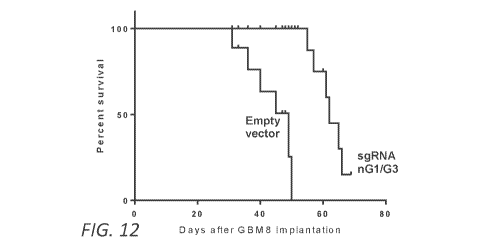
Figure 12. miR-10b editing significantly extends animal survival in
orthotopic GBM models. miR-10b editing (by lentivirus) significantly extended
animal survival, analyzed by Kaplan-Meier plot. N = 8 mice per group. P =
0.0001 by
log-rank (Mantel-Cox) test.
Figure 13. miR-10b editing as therapeutic approach for human
Glioblastoma. An exemplary illustration showing miR-10b editing using CRISPR
Cas9. miR-10b gene ablation leads to eradication of GBM, but is uneditable in
and
thus does not affect normal brain cells.
DETAILED DESCRIPTION
Mounting evidence indicates that glioma and GBM growth and invasiveness
are closely regulated by miRNAs (reviewed in 1). Micro RNA 10b (miR-10b) is
embedded within the HOXD genomic locus and implicated in proliferation,
invasion,
and metastasis of various types of malignancies including gliomas such as GBM
(reviewed in 2, 3). miR-10b is especially notable in brain tumors due to its
unique
expression pattern: while virtually undetectable in the normal brain, it
becomes
extremely abundant in the majority of low and high-grade gliomas across all
subtypes,
as well as metastatic brain tumors (3-6). Breast cancer patients with brain
metastases
have significantly higher miR-10b levels compared to patients with metastases
in
other organs (7, 8). Inhibition of miR-10b by chemically modified antisense
oligonucleotides (ASO) reduces growth and invasion of cultured glioma cells
(4, 9),
and metastasis in aggressive cancer models (10, 11). Recent work on highly
invasive
and aggressive intracranial glioma models demonstrated that ASO inhibitors of
miR-
10b reduce GBM growth in mice (12). However, the effects observed in the
orthotopic GBM models were transient, with disease relapse due to both low-
efficiency uptake and non-uniform distribution of the ASO in intracranial GBM.
There are only a few examples of true onco-miR dependencies known for
cancer cells. The present data indicates that high expression of the WT miR-
10b gene
is essential for glioma, whereas loss-of-function mutations lead to the
lethality of
heterogeneous glioma cells and tumor-initiating GSC. Alternative sgRNA guides
targeting either miR-10b alone or together with its closely related paralog
miR-10a
produced a diverse range of mutants, none of which were viable. The loss-of-
function
mutations in miR-10b alone were sufficient to cause the lethality, validating
the key
role of miR-10b in the sustained growth and survival of glioma. Specifically,
mutated
11
CA 03011458 2018-07-13
WO 2017/123910
PCT/US2017/013386
nCas9 guided by the G1/G3 sgRNAs had detrimental effects on glioma cells by
reducing the levels of miR-10b and without affecting the levels of miR-10a
gene,
suggesting the efficacy of the miR-10b single-gene targeting approach for GBM.
Since miR-10a and miR-10b differ in one nucleotide and are largely
functionally
redundant, the relative efficacy of miR-10a targeting remains to be evaluated.
Of note,
miR-10b is expressed in normal extracranial tissues; nevertheless, its
activity in these
tissues seems to be dispensable as the initial analysis of miR-10b knock-out
mice has
no apparent pathological phenotype (MirlObtmlmtm/Mmjax; MMRRC Stock No:
36061-JAX). Glioma addiction to miR-10b appears, therefore, truly as a tumor-
trait, probably associated with de-repression of the gene in the brain
microenvironment where it is normally silenced. A unique onco-miR-dependence
of
glioma and GBM also suggests that the tumor could be eradicated by targeting a
single miRNA gene.
Administration of synthetic miR-10b inhibitors caused potent but transient
effects on orthotopic GBM in aggressive GSC-based models (12). This may have
been due to both poor uptake and distribution of the ASO in intracranial GBM,
and
dilution of the drug in the actively growing tumor. Gene editing, based on
permanent
miR-10b inactivation, may provide an alternative strategy, eliminating the
need for
continuous delivery of anti-miRs to intracranial brain tumors and improving
the
efficacy of tumor cell destruction. Interestingly, even moderately efficient
miR-10b
gene editing of GBM8 glioma stem cells led to disaggregation and massive death
of
glioma spheres, suggesting that disruption of this core cell population may
have
detrimental effects on the tumor growth. Using lentiviral CRISPR-Cas9
targeting, the
effects of miR-10b ablation were examined on highly aggressive human GBM
xenografts. Remarkably efficient Cas9 expression and miR-10b editing
throughout the
tumor resulted in the permanent ablation of miR-10b and near-eradication of
orthotopic GBM tumors. The data suggest that less-than-100% efficient editing
and
miR-10b ablation is sufficient for potent inhibition of GBM growth. The
lentiviral
editing vector used herein caused strong effects on glioma growth both in
vitro and in
vivo. The effects may appear stronger in vivo due to the longer duration of
the
experiment; however, miR-10b editing in cultures also resulted in death of the
entire
population, when analyzed over longer time. Overall, these data provide proof-
of-
12
CA 03011458 2018-07-13
WO 2017/123910
PCT/US2017/013386
principle for the single-target gene editing based therapeutic strategy for
malignant
gliomas, and may also apply to other miR-10b dependent metastatic cancers (11,
21).
A limitation of the CRISPR-Cas9 technology, and particularly its clinical
application, is associated with its restricted specificity (reviewed in 22).
Bioinformatics analysis suggested only a few potential high-ranked protein-
coding
off-targets (mismatched target sites) for the designed miR-10b SpCas9 sgRNAs
(Fig.
1C and Table 1); none of them appeared to be actually edited in the present
experiments.
Table 1 ¨ Predicted Off-Targets for the Designed miR-10b SpCas9 sgRNAs
Top predicted off-targets mismatche(s) Locus
& position(s)
sgRNA miRNA-10a 1MMs [11] Chr16:+64668475
G1 Asparagine amidohydrolase (NTAN1) 3MMs [9:11:12] Chrl 6:-
15151834
Sulfotransferase (ST1A3) 3MMs [5:9:13]
Chr2:+108886423
miRNA-10a 3MMs [1:2:15]
Chr16:+64668475
sgRNA Cytosolic pu rine 5'-nucleotidase (5NTC) 3MMs [2:6:13] Chrl
0:+104928080
G2
Phosphodiesteras eta-1 (PLCH1) 3MMs [411:13]
Chr3:+155214975
Leucine-rich repeat transmembrane protein (FLRT2) 3MMs [2:7:9]
Chr14:-87587093
sgRNA
G3 Collagen alpha-a1 (Ill) chain preportein (COL3A1) 3MMs
[4:1015] Chr2:-189649274
To start evaluating the therapeutic potential of miR-10b editing in the brain
and assess
its safety, the effects of miR-10b ablation were tested in the normal cells of
brain
tumor microenvironment in vitro. Major cell types of the brain, including
neurons,
astrocytes, microglia, and neuroprogenitors express very low, or undetectable
levels
of miR-10b, while exhibiting low levels of miR-10a (4). Although CRISPR-Cas9
system can target genes in any cell type including postmitotic neurons (23,
24), the
efficacy of editing genes that are not actively transcribed in a specific
cellular context,
and might be less accessible by Cas9-sgRNA due to their epigenetic state and
chromatin structure, is presently unknown and expected to be low (25). The
present
data indicated that CRISPR-Cas9 plasmid- and virus-mediated miR-10b targeting
did
not cause locus editing in normal brain cells and did not affect the viability
of mouse
primary astrocytes or neurons (Figs. 2A and 5A); neither did the miR-10b ASO
inhibitors (4). Additional experiments on human MCF7 cells that express only
negligible miR-10b levels also demonstrated the lack of miR-10b editing and no
visible phenotypic effects, despite the efficient editing of other highly
expressed
miRNAs in these cells (Fig. 2A). This data suggested that miR-10b is not
edited in
13
CA 03011458 2018-07-13
WO 2017/123910
PCT/US2017/013386
normal neuroglial and other non-expressing cells due to the compact chromatin
structure of the locus, and not merely lower efficiency of transfection or
transduction.
Of note, human brain-derived microvascular endothelial cells do express
substantial
levels of miR-10b. While the functional role of miR-10b in these cells
requires further
investigation, the present results demonstrate that miR-10b gene editing
(which is less
efficient than in glioma cells) does not affect their phenotype. Importantly,
the lack of
toxicity for normal brain cells suggests a reasonable therapeutic window for
miR-10b
editing in glioma in vivo, further validates the high targeting specificity,
and paves the
way for its clinical development.
The present methods of miR-10b-editing viral therapy for glioma, e.g., GBM,
patients can include at least a one-time treatment with local administration
of the viral
vector to the surgical bed, immediately after tumor resection. The lentiviral
vectors
utilized in the present experiments in vivo transduce dividing as well as
quiescent
cells. This can be viewed as a major advantage for cancer gene therapy in
general, as
within a short treatment window most tumor cells (and especially GSC) do not
divide.
Since miR-10b editing prevents neoplastic transformation of astrocytes and
selectively eradicates the transforming cells (Fig. 5), in addition to
malignant tumor
cells this approach may target the brain cells undergoing early stages of
gliomagenesis. Therapeutic gene editing using high viral titers applied
locally to the
surgical cavity, may also prove to be effective for targeting infiltrating
tumor cells
(26, 27).
An advantage of a locally applied lentivirus pseudotyped with the VSV-G
glycoprotein is its inactivation by human serum (28) that would reduce
systemic
effects. Although the application of human lentiviral gene therapy is hampered
by the
risk of carcinogenesis by random proviral integration into the genome of
normal
somatic cells, future studies should determine if this risk is acceptable for
local glioma
treatment, given the lack of efficacious drugs and poor life expectancy of
patients
with the disease. Importantly, the identification of Staphylococcus aureus
(SaCas9)
and other smaller Cas9 enzymes that can be packaged into adeno-associated
viral
vectors highly stable and effective in vivo (29-31), easily produced, approved
by FDA
for other applications, and tested in multiple clinical trials, paves new
avenues for
therapeutic gene editing. Further optimization of the targeting vectors with
increased
tropism for glioma cells, as well as in-depth investigation of potential
neurotoxic
14
CA 03011458 2018-07-13
WO 2017/123910
PCT/US2017/013386
effects have to be performed before clinical applications of this promising
new
strategy.
Methods of Treatment
The methods described herein include methods for the treatment of glioma,
e.g., GBM, astrocytoma or oligodendroglioma, e.g., a glioma that has increased
levels
of miR-10b expression (increased as compared to normal tissue or to other
gliomas).
Generally, the methods include administering a therapeutically effective
amount of a
miR-10b gene editing complex as described herein, to a subject who is in need
of, or
who has been determined to be in need of, such treatment.
As used in this context, to "treat" means to ameliorate at least one clinical
parameter of the glioma; thus, in some embodiments, administration of a
therapeutically effective amount of a compound described herein for the
treatment of
glioma results in a reduction in tumor size; a reduction in tumor growth rate;
a
reduction in risk of tumor regrowth or recurrence; an improved prognosis; or
an
increase in survival time. In some embodiments, the treatment improves one or
more
symptoms of the glioma.
In some embodiments, the subject has a cancer that may become metastatic,
e.g., breast cancer or colorectal cancer, e.g., wherein metastasis is
associated with
miR-10b. Metastasis has been shown to be linked to miR-10b in a number of
cancers;
see, e.g., Ma et al., Tumour invasion and metastasis initiated by microRNA-10b
in
breast cancer. Nature 449: 682-8 (2007); Li et al., microRNA expression
profiles in
human colorectal cancers with brain metastases. Oncol Lett 3: 346-50 (2012);
Ahmad
et al., Up-regulation of microRNA-10b is associated with the development of
breast
cancer brain metastasis. Am J Transl Res 6: 384-90 (2014); Parrella et al.,
Evaluation
of microRNA-10b prognostic significance in a prospective cohort of breast
cancer
patients. Mol Cancer 13: 142 (2014); Lu et al., The association between
abnormal
microRNA-10b expression and cancer risk: a meta-analysis. Sci Rep 4: 7498
(2014).
The methods described herein can be used to reduce the risk or likelihood that
the
subject who has cancer, e.g., breast or colorectal cancer, will develop
metastatic
disease, e.g., a brain metastasis.
A diagnosis of a glioma (e.g., identification of a subject with glioma, e.g.,
GBM, astrocytoma or oligodendroglioma) can be made based on methods known in
the art. Gliomas such as GBM commonly presents with symptoms that include the
CA 03011458 2018-07-13
WO 2017/123910
PCT/US2017/013386
following: progressive neurologic deficit; motor weakness; headache;
generalized
symptoms of increased intracranial pressure or neurologic symptoms including
headaches, nausea and vomiting, memory loss, personality changes, confusion,
and
cognitive impairment; and seizures. Focal signs can include hemiparesis,
sensory loss,
visual loss, and aphasia. A diagnosis is typically made based on imaging
studies
including computed tomography (CT), magnetic resonance imaging (MRI), with
and/or without contrast; positron emission tomography (PET); and/or magnetic
resonance spectroscopy (MRS); a biopsy can be done to confirm the diagnosis,
usually during surgical resection or using a stereotactic needle biopsy.
Presently, standard therapy includes maximal surgical resection (preferably
gross total resection), radiotherapy, and concomitant and adjuvant
chemotherapy, e.g.,
with temozolomide, nitrosoureas (e.g., carmustine [BCNU]), MGMT inhibitors
(e.g.,
06-benzylguanine); platinum-containing agents, e.g., cisplatin; anti-VEGF
agents,
e.g., bevacizumab (alone or with irinotecan); and tyrosine kinase inhibitors
(e.g.,
gefitinib, erlotinib).
CRISPR miR-10a/10b Gene Editing Complexes
The present methods include the use of CRISPR miR-10b gene editing
complexes. The methods can include the use of expression vectors for in vivo
transfection and expression of a Cas9 protein and suitable guide RNAs
targeting miR-
10b. Alternatively, or in addition, the methods can include the use of
purified Cas9
proteins complexed with suitable guide RNAs targeting miR-10b.
In addition, miR-10a can be targeted as well as miR-10b. There is a single
nucleotide difference between miR-10b and 10a, therefore they are expected to
target
the same genes and be largely functionally redundant; thus the present methods
can
include targeting miR-10a as an alternative or in addition to miR-10b. The
present
data show that sgRNA-1 (targets both 10a and 10b) and sgRNA-3 (targets 10b
more
specifically) both kill glioma cells.
Nucleic Acids Encoding a CRISPR miR-10a/10b Gene Editing Complex
The present methods include the delivery of nucleic acids encoding a CRISPR
miR-10b gene editing complex. The gene editing complex includes a Cas9 editing
enzyme and one or more guide RNAs directing the editing enzyme to miR-10b. In
16
CA 03011458 2018-07-13
WO 2017/123910
PCT/US2017/013386
some embodiments, the guide RNA may also direct the editing enzyme to miR-10a,
e.g., to miR-10a/10b (i.e., one or both of miR-10a and miR-10b).
Guide RNAs directing the editing enzyme to miR-10a/10b
The gene editing complex also includes guide RNAs directing the editing
enzyme to one or both of miR-10a and miR-10b, i.e., comprising a sequence that
is
complementary to the sequence of a nucleic acid encoding miR-10a or miR-10b,
and
that include a PAM sequence that is targetable by the co-administered Cas9
editing
enzyme. In some embodiments, the precursor sequence is targeted by the guide
RNA., i.e., comprising a sequence that is complementary to the sequence of a
nucleic
acid encoding miR-10a or miR-10b. In some embodiments, the precursor sequence
is
targeted by the guide RNA.
The gene encoding the human miR-10b precursor is at nucleotides
176,150,303-176,150,412 of chromosome 2 (see GenBank Acc. No. NC_000002.12).
The sequence of human gene coding for pre-microRNA-10b (MIR10B) (GenBank
Acc. No. NR 029609.1) is 110 nucleotides, as follows:
CCAGAGGTTGTAACGTTGTCTATATATACCCTGTAGAACCGAATTTGTGTGG
TATCCGTATAGTCACAGATTCGATTCTAGGGGAATATATGGTCGATGCAAAA
ACTTCA (SEQ ID NO:1). Exemplary miRlOb target sequences are shown in Tables
A-C. An exemplary miR-10b primary transcript (pri-miR-10b), Homo sapiens
chromosome 2: position start 176136921-End 176173102, is provided as SEQ ID
NO:24 and can also be targeted. This is an exemplary sequence as the primary
transcript of miR-10b (pri-miR-10b) is not well defined. Most likely, based on
unpublished RNA sequencing data, it starts close to the HoxD antisense RNA2
and
ends at the HOXD antisense growth-associated long non-coding RNA, transcript
variant 10. SEQ ID NO: 24 can be considered as a single pri-miR-10b
transcript, e.g.,
HOXD cluster antisense RNA 2 > pri-miR-10b < HOXD antisense growth-associated
long non-coding RNA, transcript variant 10. Additional sgRNAs targeting the
sequence of pri-miR-10b (e.g., SEQ ID NO:24) and its promoter could be
designed
and utilized.
The gene encoding the human miR-10a precursor is at nucleotides 48,579,947-
48,579,838 of chromosome 17 (see GenBank Acc. No. NC_000017.11). The
sequence of human gene coding for pre-microRNA-10b (MIR10B) (GenBank Acc.
No. NR 029609.1) is 110 nucleotides, as follows:
17
CA 03011458 2018-07-13
WO 2017/123910
PCT/US2017/013386
GATCTGTCTGTCTTCTGTATATACCCTGTAGATCCGAATTTGTGTAAGGAATT
TTGTGGTCACAAATTCGTATCTAGGGGAATATGTAGTTGACATAAACACTCC
GCTCT (SEQ ID NO:25). An exemplary miR-10a primary transcript (pri-miR-10a),
Homo sapiens chromosome 17: position start 48,548,870 to 48,590,369, provided
in
SEQ ID NO:26 can also be targeted. This is an exemplary sequence as the
primary
transcript of miR-10a (pri-miR-10a) is not well defined. Most likely, based on
RNA
sequencing data, it starts close to the HOXB cluster antisense RNA 3
transcript and
ends at the HOXB cluster antisense RNA 1. SEQ ID NO:26 can be considered as a
single pri-miR-10a transcript, and additional sgRNAs targeting the sequence of
pri-
miR-10a and its promoter could be designed and utilized.
Therefore, additional sgRNAs targeting the sequence of pri-miR-10a (e.g.,
SEQ ID NO:26) and its promoter could be designed and utilized. In some
embodiments, sgRNAs targeting sequence that is identical, or at least 80%,
85%,
90%, 95%, or 99% identical between miR-10a and miR-10b are used, e.g., sgRNAs
that target both miR-10a and miR-10b encoding sequences.
Table A ¨ miRlOb target sequences, genome editing by SpCas9 from
Streptococcus pyogenes (PAM: 5'-NGG-3')
GC
SEQ Cleavage
Contents
ID Position (%, w/o
sgRNA Target (5 to 3') NO: Position (%) Direction PAM)
ATAGACAACGTTACAACCTCTGG 2 1 5.5 - 40.0
¨ 4- ¨ ¨ -4-
CACACAAATTCGGTTCTACAGGG 3 29 31.2 - 40.0
............................................ .. ......................
CCTGTAGAACCGAATTTGTGTGG 4 30 42.2 + 45.0
CCACACAAATTCGGTTCTACAGG 5 30 32.1 - 45.0
4- ¨ ¨ -4-
ATACGGATACCACACAAATTCGG 6 39 40.4 - 35.0
............................................ .. ......................
GAATCGAATCTGTGACTATACGG 7 56 56.0 - 35.0
ATAGTCACAGATTCGATTCTAGG 8 60 69.7 + 35.0
¨ ¨ 4-
TAGTCACAGATTCGATTCTAGGG 9 61 70.6 + 35.0
............................................ .. ......................
AGTCACAGATTCGATTCTAGGGG 10 62 71.6 + 40.0
TTCGATTCTAGGGGAATATATGG 11 71 79.8 + 35.0
18
CA 03011458 2018-07-13
WO 2017/123910 PCT/US2017/013386
Table B - miRlOb target sequences, genome editing by SpCas9 from
Staphylococcus aureus (PAM: 5'-NNGRRT-'3, (R=A or G)
.................................................................. ,
GC
Cleavage Contents
SEQ ID Position (%, w/o
sgRNA Target (5 to 3') NO: Position (%) Direction PAM)
TCTATATATACCCTGTAGAACCGAAT 12 19 32.1 + 30.0
ACCACACAAATTCGGTTCTACAGGGT 13 28 33.0 - 40.0
........................................... i .....
AGAATCGAATCTGTGACTATACGGAT 14 54 56.9 - 35.0
¨ -4-
AGTCACAGATTCGATTCTAGGGGAAT 15 62 71.6 + 40.0
GACCATATATTCCCCTAGAATCGAAT 16 70 71.6 - 40.0
........................................... i .....
GCATCGACCATATATTCCCCTAGAAT 17 75 76.1 - 50.0
------------------------------------- .. -------------------------
Table C - miRlOb target sequences, genome editing by SpCas9 from Neisseria
meningitides (PAM: 5'-NNNNGMTT-3' (M = A or C))
GC
SEQ Cleavage Contents
ID Position (%, w/o
sgRNA Target (5' to 3') NO: Position (%) Direction PAM)
GAATTTGTGTGGTATCCGTATAGTC
18 41 56.0 + 37.5
ACAGATT
................................ , .....
TGTGTGGTATCCGTATAGTCACAG
19 46 60.6 +
ATTCGATT 45.8
_
¨ ¨
TTGTGTGGTATCCGTATAGTCACA
20 45 56.0 + 40.0
GATT
TGGTATCCGTATAGTCACAGATTC 21 50 60.6 + 45.0
GATT
Other Cas9s from other species can also be used, including those shown in
Table D. Suitable target sequences for use with those Cas9s can readily be
determined using known methods.
19
CA 03011458 2018-07-13
WO 2017/123910
PCT/US2017/013386
Table D. Additional Cas9s from various species
Species/Variant of Cas9 PAM Sequence
SpCas9 D1135E variant NGG (reduced NAG binding)
SpCas9 VRER variant NGCG
SpCas9 EQR variant NGAG
SpCas9 VQR variant NGAN or NGNG
Streptococcus thermophilus (ST) NNAGAAW
Treponema denticola (TD) NAAAAC
Streptococcus pyogenes (SP); SpCas9 NGG
Staphylococcus aureus (SA); SaCas9 NNGRRT or NNGRR(N)
Neisseria meningitidis (NM) NNNNGATT
Cas9 editing enzymes
The methods include the delivery of Cas9 editing enzymes to the cancer cells.
The editing enzymes can include one or more of SpCas9 D1135E variant; SpCas9
VRER variant; SpCas9 EQR variant; SpCas9 VQR variant; Streptococcus
thermophilus (ST) Cas9 (StCas9); Treponema denticola (TD) (TdCas9);
Streptococcus pyogenes (SP) (SpCas9); Staphylococcus aureus (SA) Cas9
(SaCas9);
or Neisseria meningitidis (NM) Cas9 (NmCas9), as well as variants thereof that
are at
least 80%, 85%, 90%, 95%, 99% or 100% identical thereto that retain at least
one
function of the parent case, e.g., the ability to complex with a gRNA, bind to
target
DNA specified by the gRNA, and alter the sequence of the target DNA.
To determine the percent identity of two sequences, the sequences are aligned
for optimal comparison purposes (gaps are introduced in one or both of a first
and a
second amino acid or nucleic acid sequence as required for optimal alignment,
and
non-homologous sequences can be disregarded for comparison purposes). The
length
of a reference sequence aligned for comparison purposes is at least 80% (in
some
embodiments, about 85%, 90%, 95%, or 100% of the length of the reference
sequence) is aligned. The nucleotides or residues at corresponding positions
are then
compared. When a position in the first sequence is occupied by the same
nucleotide
or residue as the corresponding position in the second sequence, then the
molecules
are identical at that position. The percent identity between the two sequences
is a
function of the number of identical positions shared by the sequences, taking
into
CA 03011458 2018-07-13
WO 2017/123910
PCT/US2017/013386
account the number of gaps, and the length of each gap, which need to be
introduced
for optimal alignment of the two sequences.
The comparison of sequences and determination of percent identity between
two sequences can be accomplished using a mathematical algorithm. For example,
the percent identity between two amino acid sequences can be determined using
the
Needleman and Wunsch ((1970) J. Mol. Biol. 48:444-453) algorithm which has
been
incorporated into the GAP program in the GCG software package, using a Blossum
62 scoring matrix with a gap penalty of 12, a gap extend penalty of 4, and a
frameshift
gap penalty of 5.
The PAM sequences of these Cas9s are listed in Table D, above. The
sequences of the Cas9s are known in the art; see, e.g., Kleinstiver et al.,
Nature. 2015
Jul 23; 523(7561): 481-485; WO 2016/141224; US 9,512,446; US-2014-0295557;
WO 2014/204578; and WO 2014/144761. The methods can also include the use of
the other previously described variants of the SpCas9 platform (e.g.,
truncated
sgRNAs (Tsai et al., Nat Biotechnol 33, 187-197 (2015); Fu et al., Nat
Biotechnol 32,
279-284 (2014)), nickase mutations (Mali et al., Nat Biotechnol 31, 833-838
(2013);
Ran et al., Cell 154, 1380-1389 (2013)), FokI-dCas9 fusions (Guilinger et al.,
Nat
Biotechnol 32, 577-582 (2014); Tsai et al., Nat Biotechnol 32, 569-576 (2014);
W02014144288).
The SpCas9 wild type sequence is as follows:
MDKKYS I GLDI GTNSVGWAVI T DEYKVP S KKFKVLGNT DRHS I KKNL I GALL FDS
GETAEATRLKRTAR
RRYTRRKNRI CYLQEI FSNEMAKVDDS FFHRLEES FLVEEDKKHERHP I FGNIVDEVAYHEKYPT I YHL
RKKLVDS T DKADLRL I YLALAHMI KFRGHFL I EGDLNP DNS DVDKL FIQLVQTYNQL FEENP I
NAS GVD
AKAI L SARL S KS RRLENL IAQL P GEKKNGL FGNLIAL S LGLT PNEKSNEDLAEDAKLQL S KDT
YDDDLD
NLLAQIGDQYADLFLAAKNLSDAI LL S DI LRVNTEITKAPLSASMIKRYDEHHQDLTLLKALVRQQLPE
KYKEI FFDQSKNGYAGYI DGGASQEEFYKFI KP I LEKMDGT EELLVKLNREDLLRKQRT FDNGS I PHQI
HLGELHAI LRRQEDFYPFLKDNREKI EKI LT FRI PYYVGP LARGNS RFAWMT RKS EET I T PWN
FEEVVD
KGASAQS Fl ERMTNEDKNLPNEKVLPKHSLLYEYFTVYNELTKVKYVTEGMRKPAELSGEQKKAIVDLL
FKTNRKVTVKQLKEDYFKKI ECFDSVEI S GVEDRFNAS LGTYHDLLKI I KDKDFLDNEENEDI LEDIVL
T LT L FEDREMI EERLKTYAHL FDDKVMKQLKRRRYT GWGRL SRKL INGI RDKQ S GKT I
LDFLKSDGFAN
RNFMQLI HDDS LT FKEDI QKAQVS GQGDS LHEHIANLAGS PAIKKGI LQTVKVVDELVKVMGRHKPENI
VI EMARENQTTQKGQKNSRERMKRI EEGIKELGSQI LKEHPVENTQLQNEKLYLYYLQNGRDMYVDQEL
DINRLSDYDVDHIVPQS FLKDDS I DNKVLT RS DKNRGKS DNVP SEEVVKKMKNYWRQLLNAKL ITQRKF
DNLTKAERGGLSELDKAGFIKRQLVETRQITKHVAQI LDS RMNTKYDENDKL I REVKVI T LKS KLVSDF
RKD FQ FYKVRE I NNYHHAHDAYLNAVVGTAL I KKYP KLE S E FVYGDYKVYDVRKMIAKS EQEI
GKATAK
YFFYSNIMNFFKTEI T LANGEI RKRP L I ETNGETGEIVWDKGRDFATVRKVL SMPQVNIVKKT EVQT GG
FS KES I L P KRNS DKL IARKKDWDP KKYGGFDS PTVAYSVLVVAKVEKGKSKKLKSVKELLGIT IMERS
S
FEKNP I DFLEAKGYKEVKKDL I I KL P KYS L FELENGRKRMLASAGELQKGNELAL P S
KYVNFLYLASHY
EKLKGSPEDNEQKQLFVEQHKHYLDEI I EQI S EFS KRVI LADANLDKVL SAYNKHRDKP I REQAENI I
H
LFTLTNLGAPAAFKYFDTTIDRKRYTSTKEVLDATLIHQSITGLYETRIDLSQLGGD (SEQ ID
NO: 22)
21
CA 03011458 2018-07-13
WO 2017/123910
PCT/US2017/013386
The SaCas9 wild type sequence is as follows:
MKRNYILGLDI GIT SVGYGI I DYET RDVI DAGVRL FKEANVENNEGRRS KRGARRLKRRRRHRIQRVKK
LL FDYNLLT DHS EL S GINPYEARVKGL SQKL S EEEFSAALLHLAKRRGVHNVNEVEEDT GNEL
STKEQI
SRNSKALEEKYVAELQLERLKKDGEVRGS INRFKTSDYVKEAKQLLKVQKAYHQLDQS FIDTYIDLLET
RRTYYEGPGEGS PFGWKDIKEWYEMLMGHCTYFPEELRSVKYAYNADLYNALNDLNNLVITRDENEKLE
YYEKFQI I ENVFKQKKKPTLKQIAKEI LVNEEDIKGYRVTSTGKPEFTNLKVYHDIKDITARKEI I ENA
ELLDQIAKI LT I YQS S EDI QEELTNLNSELTQEEI EQI SNLKGYT GTHNL S LKAINL I
LDELWHTNDNQ
IAI FNRLKLVPKKVDLSQQKEI PTTLVDDFI LS PVVKRS FI QS I KVINAI I KKYGLPNDI I I
ELAREKN
SKDAQKMINEMQKRNRQTNERI EEI I RTT GKENAKYL I EKIKLHDMQEGKCLYSLEAI PLEDLLNNPFN
YEVDHI I PRSVS FDNS FNNKVLVKQEENSKKGNRTPFQYLS S S DS KI SYETFKKHILNLAKGKGRI S
KT
KKEYLLEERDINRFSVQKDFINRNLVDTRYATRGLMNLLRSYFRVNNLDVKVKS INGGFTS FL RRKWKF
KKERNKGYKHHAEDAL I IANADFI FKEWKKLDKAKKVMENQMFEEKQAESMPEI ETEQEYKEI FIT PHQ
I KHI KDFKDYKYSHRVDKKPNREL INDTLYS T RKDDKGNT L IVNNLNGLYDKDNDKLKKL INK S P
EKLL
MYHHDPQTYQKLKL IMEQYGDEKNP LYKYYEET GNYLTKYS KKDNGPVI KKI KYYGNKLNAHL DI T DDY
PNSRNKVVKLSLKPYRFDVYLDNGVYKFVTVKNLDVIKKENYYEVNSKCYEEAKKLKKISNQAEFIASF
YNNDLIKINGELYRVIGVNNDLLNRIEVNMIDITYREYLENMNDKRPPRIIKTIASKTQSIKKYSTDIL
GNLYEVKSKKHPQIIKKG (SEQ ID NO:23)
See also Hou, Z. et al. Efficient genome engineering in human pluripotent
stem cells using Cas9 from Neisseria meningitidis. Proc Natl Acad Sci U S A
(2013);
Fonfara, I. et al. Phylogeny of Cas9 determines functional exchangeability of
dual-
RNA and Cas9 among orthologous type II CRISPR-Cas systems. Nucleic Acids Res
42, 2577-2590 (2014); Esvelt, K.M. et al. Orthogonal Cas9 proteins for RNA-
guided
gene regulation and editing. Nat Methods 10, 1116-1121 (2013); Cong, L. et al.
Multiplex genome engineering using CRISPR/Cas systems. Science 339, 819-823
(2013); Horvath, P. et al. Diversity, activity, and evolution of CRISPR loci
in
Streptococcus thermophilus. J Bacteriol 190, 1401-1412 (2008).
As noted above, the Cas9 can be delivered as a purified protein (e.g., a
recombinantly produced purified protein, prefolded and optionally complexed
with
the sgRNA) or as a nucleic acid encoding the Cas9, e.g., an expression
construct.
Purified Cas9 proteins can be produced using methods known in the art, e.g.,
expressed in prokaryotic or eukaryotic cells and purified using standard
methodology.
See, e.g., Liang et al., Journal of Biotechnology 208:44-53 (2015); Kim et
al.,
Genome Res. 2014 Jun; 24(6): 1012-1019. Efficiency of protein delivery can be
enhanced, e.g., using electroporation (see, e.g., Wang et al., Journal of
Genetics and
Genomics 43(5):319-327 (2016)); cationic or lipophilic carriers (see, e.g., Yu
et al.,
Biotechnol Lett. 2016; 38: 919-929; Zuris et al., Nat Biotechnol. 33(1):73-80
(2015));
or even lentiviral packaging particles (see, e.g., Choi et al., Gene Therapy
23, 627-633
(2016)).
22
CA 03011458 2018-07-13
WO 2017/123910
PCT/US2017/013386
Expression Constructs
Expression constructs encoding one or both of guide RNAs and/or Cas9
editing enzymes can be administered in any effective carrier, e.g., any
formulation or
composition capable of effectively delivering the component gene to cells in
vivo.
Approaches include insertion of the gene in viral vectors, including
recombinant
retroviruses, alenovirus, adeno-associated virus, lentivirus, and herpes
simplex virus-
1, or recombinant bacterial or eukaryotic plasmids. Viral vectors transfect
cells
directly; plasmid DNA can be delivered naked or with the help of, for example,
cationic liposomes (lipofectamine) or derivatized (e.g., antibody conjugated),
polylysine conjugates, gramacidin S, artificial viral envelopes or other such
intracellular carriers, as well as direct injection of the gene construct or
CaPO4
precipitation carried out in vivo.
A preferred approach for in vivo introduction of nucleic acid into a cell is
by
use of a viral vector containing nucleic acid, e.g., a cDNA. Infection of
cells with a
viral vector has the advantage that a large proportion of the targeted cells
can receive
the nucleic acid. Additionally, molecules encoded within the viral vector,
e.g., by a
cDNA contained in the viral vector, are expressed efficiently in cells that
have taken
up viral vector nucleic acid.
Retrovirus vectors and adeno-associated virus vectors can be used as a
recombinant gene delivery system for the transfer of exogenous genes in vivo,
particularly into humans. These vectors provide efficient delivery of genes
into cells,
and the transferred nucleic acids are stably integrated into the chromosomal
DNA of
the host. The development of specialized cell lines (termed "packaging cells")
which
produce only replication-defective retroviruses has increased the utility of
retroviruses
for gene therapy, and defective retroviruses are characterized for use in gene
transfer
for gene therapy purposes (for a review see Miller, Blood 76:271 (1990)). A
replication defective retrovirus can be packaged into virions, which can be
used to
infect a target cell through the use of a helper virus by standard techniques.
Protocols
for producing recombinant retroviruses and for infecting cells in vitro or in
vivo with
such viruses can be found in Ausubel, et al., eds., Current Protocols in
Molecular
Biology, Greene Publishing Associates, (1989), Sections 9.10-9.14, and other
standard
laboratory manuals. Examples of suitable retroviruses include pLJ, pZIP, pWE
and
pEM which are known to those skilled in the art. Examples of suitable
packaging
23
CA 03011458 2018-07-13
WO 2017/123910
PCT/US2017/013386
virus lines for preparing both ecotropic and amphotropic retroviral systems
include
TCrip, TCre, T2 and tIlAm. Retroviruses have been used to introduce a variety
of
genes into many different cell types, including epithelial cells, in vitro
and/or in vivo
(see for example Eglitis, et al. (1985) Science 230:1395-1398; Danos and
Mulligan
(1988) Proc. Natl. Acad. Sci. USA 85:6460-6464; Wilson et al. (1988) Proc.
Natl.
Acad. Sci. USA 85:3014-3018; Armentano et al. (1990) Proc. Natl. Acad. Sci.
USA
87:6141-6145; Huber et al. (1991) Proc. Natl. Acad. Sci. USA 88:8039-8043;
Ferry et
al. (1991) Proc. Natl. Acad. Sci. USA 88:8377-8381; Chowdhury et al. (1991)
Science 254:1802-1805; van Beusechem et al. (1992) Proc. Natl. Acad. Sci. USA
1() 89:7640-7644; Kay et al. (1992) Human Gene Therapy 3:641-647; Dai et
al. (1992)
Proc. Natl. Acad. Sci. USA 89:10892-10895; Hwu et al. (1993) J. Immunol.
150:4104-4115; U.S. Patent No. 4,868,116; U.S. Patent No. 4,980,286; PCT
Application WO 89/07136; PCT Application WO 89/02468; PCT Application WO
89/05345; and PCT Application WO 92/07573).
As demonstrated herein, a lentiviral CRISPR-Cas9 targeting system provided
high and tumor-specific expression of Cas9, the corresponding high miR-10a/10b
editing efficacy in tumor tissues, while lacking general toxicity or
neurotoxicity.
Lentiviral vectors transduce dividing as well as quiescent cells. This can be
viewed as
a major advantage with respect to gene therapy for tumors in general, as
within a short
treatment window most tumor cells (and especially GSC) do not divide.
Therapeutic
use of the lentiviral editing approach can be a legitimate alternative to
other viral
systems, as high viral titers can be produced, nonproliferating cells that are
especially
abundant in the walls of the tumor cavity after surgery can be transduced, and
transduction efficacies are very high. An additional advantage of a locally
applied
vesicular stomatitis virus glycoprotein (VSV-G) pseudotyped lentivirus is its
inactivation by human serum that would reduce systemic effects. To further
reduce
neurotrophism, and enhance selective tropism for glioma and GSC, the commonly
bound envelope glycoprotein of VSV can be replaced with a more selective
variant
glycoprotein of lymphocytic choriomeningitis virus (LCMV-GP). LCMV-GP is not
cytotoxic when injected locally or systemically, can be packaged with other
components of the CRISPR-Cas9 system, and efficiently transduces solid glioma
tissues as well as infiltrating tumor cells.
24
CA 03011458 2018-07-13
WO 2017/123910
PCT/US2017/013386
Another viral gene delivery system useful in the present methods utilizes
adenovirus-derived vectors. The genome of an adenovirus can be manipulated,
such
that it encodes and expresses a gene product of interest but is inactivated in
terms of
its ability to replicate in a normal lytic viral life cycle. See, for example,
Berkner et
al., BioTechniques 6:616 (1988); Rosenfeld et al., Science 252:431-434 (1991);
and
Rosenfeld et al., Cell 68:143-155 (1992). Suitable adenoviral vectors derived
from
the adenovirus strain Ad type 5 d1324 or other strains of adenovirus (e.g.,
Ad2, Ad3,
or Ad7 etc.) are known to those skilled in the art. Recombinant adenoviruses
can be
advantageous in certain circumstances, in that they are not capable of
infecting non-
dividing cells and can be used to infect a wide variety of cell types,
including
epithelial cells (Rosenfeld et al., (1992) supra). Furthermore, the virus
particle is
relatively stable and amenable to purification and concentration, and as
above, can be
modified so as to affect the spectrum of infectivity. Additionally, introduced
adenoviral DNA (and foreign DNA contained therein) is not integrated into the
genome of a host cell but remains episomal, thereby avoiding potential
problems that
can occur as a result of insertional mutagenesis in situ, where introduced DNA
becomes integrated into the host genome (e.g., retroviral DNA). Moreover, the
carrying capacity of the adenoviral genome for foreign DNA is large (up to 8
kilobases) relative to other gene delivery vectors (Berkner et al., supra; Haj-
Ahmand
and Graham, J. Virol. 57:267 (1986).
miR-10a/10b genome-editing vectors based on recombinant Adenovirus-5
(Ad5): Ad5 have many advantages for this purpose, including non-integration,
lack of
insertional mutagenesis, high-efficiency transduction, and accommodation of
large
expression cassettes; these vectors have also been utilized in multiple
clinical trials.
In some embodiments, an Ad5-CRISPR/Cas9nD10A-10b (or 10a) vector expressing a
pair of G1 and G3 sgRNAs under the control of a U6 promoter and containing the
expression system for CRISPR/Cas9nD1OA can be used. Other combinations of
CRISPR/Cas9 systems (enzymes and corresponding sgRNAs) could be utilized based
on Ad5; for example, the vectors can be replication-defective, wherein ElA and
ElB
genes are replaced by an expression cassette. In addition to ElA, ElB, the
vectors can
be deleted for E3 and E4, to avoid leaky expression of other early as well as
late
adenoviral genes, thus avoiding an inflammatory response. These vectors can be
produced in complementing cell lines that express ElA, ElB, and E4 proteins.
CA 03011458 2018-07-13
WO 2017/123910
PCT/US2017/013386
Helper-dependent (HDAd) vectors can also be produced with all adenoviral
sequences deleted except the origin of DNA replication at each end of the
viral DNA
along with packaging signal at 5-prime end of the genome downstream of the
left
packaging signal. HDAd vectors are constructed and propagated in the presence
of a
replication-competent helper adenovirus that provides the required early and
late
proteins necessary for replication.
Yet another viral vector system useful for delivery of nucleic acids is the
adeno-associated virus (AAV). Adeno-associated virus is a naturally occurring
defective virus that requires another virus, such as an adenovirus or a herpes
virus, as
a helper virus for efficient replication and a productive life cycle. (For a
review see
Muzyczka et al., Curr. Topics in Micro. and Immuno1.158:97-129 (1992). It is
also
one of the few viruses that may integrate its DNA into non-dividing cells, and
exhibits
a high frequency of stable integration (see for example Flotte et al., Am. J.
Respir.
Cell. Mol. Biol. 7:349-356 (1992); Samulski et al., J. Virol. 63:3822-3828
(1989); and
McLaughlin et al., J. Virol. 62:1963-1973 (1989). Vectors containing as little
as 300
base pairs of AAV can be packaged and can integrate. Space for exogenous DNA
is
limited to about 4.5 kb. An AAV vector such as that described in Tratschin et
al.,
Mol. Cell. Biol. 5:3251-3260 (1985) can be used to introduce DNA into cells. A
variety of nucleic acids have been introduced into different cell types using
AAV
vectors (see for example Hermonat et al., Proc. Natl. Acad. Sci. USA 81:6466-
6470
(1984); Tratschin et al., Mol. Cell. Biol. 4:2072-2081 (1985); Wondisford et
al., Mol.
Endocrinol. 2:32-39 (1988); Tratschin et al., J. Virol. 51:611-619 (1984); and
Flotte et
al., J. Biol. Chem. 268:3781-3790 (1993). The identification of Staphylococcus
aureus
(SaCas9) and other smaller Cas9 enzymes that can be packaged into adeno-
associated
viral (AAV) vectors that are highly stable and effective in vivo, easily
produced,
approved by FDA, and tested in multiple clinical trials, paves new avenues for
therapeutic gene editing. Of high relevance to gliomas like GBM, better tissue
distribution of AAV provides an additional advantage for invasive and
recurrent
tumors. miR-10b-targeting AAV vectors of various serotypes, including AAV1,
AAV2, AAV8, AAV9, and AAVrh.10, can be used, all of which were previously
tested in clinical trials. A miR-10a/10b targeting AAV plasmid [based on
Addgene
Plasmids #61592, #615941, a single vector expressing SaCas9, gRNA, and
Ampicillin
selection marker can be utilized. Since PAM consensus sequence is different
between
26
CA 03011458 2018-07-13
WO 2017/123910
PCT/US2017/013386
SpCas9 and SaCas9 (the late cleaves genomic targets most efficiently with
NNGRRT
or NNGRR (R= A or G), as also the length required for SaCas9 gRNAs (21-23nt),
several targeting constructs have been designed.
In some embodiments, nucleic acids encoding a CRISPR miR-10b gene
editing complex (e.g., Cas9 or gRNA) are entrapped in liposomes bearing
positive
charges on their surface (e.g., lipofectins), which can be tagged with
antibodies
against cell surface antigens of the target glioblastoma cells, e.g., CD133,
CD15,
CD44, CXCR4, and/or integrin alpha 6 (see, e.g., Friedman et al., "Pediatric
glioma
stem cells: biologic strategies for oncolytic HSV virotherapy," Front. Oncol.
3:28
(2013); Mizuno et al., No Shinkei Geka 20:547-551(1992); PCT publication
W091/06309; Japanese patent application 1047381; and European patent
publication
EP-A-43075). These delivery vehicles can also be used to deliver Cas9
protein/gRNA complexes.
In clinical settings, the gene delivery systems for the nucleic acids encoding
a
CRISPR miR-10b gene editing complex can be introduced into a subject by any of
a
number of methods, each of which is familiar in the art. For instance, a
pharmaceutical preparation of the gene delivery system can be introduced
systemically, e.g., by intravenous injection, and specific transduction of the
protein in
the target cells will occur predominantly from specificity of transfection,
provided by
the gene delivery vehicle, cell-type or tissue-type expression due to the
transcriptional
regulatory sequences controlling expression of the receptor gene, or a
combination
thereof In other embodiments, initial delivery of the nucleic acids encoding a
CRISPR miR-10a/10b gene editing complex is more limited, with introduction
into
the subject being quite localized. For example, the nucleic acids encoding a
CRISPR
miR-10a/10b gene editing complex can be introduced by catheter (see U.S.
Patent
5,328,470) or by stereotactic injection (e.g., Chen et al., PNAS USA 91: 3054-
3057
(1994)). In some embodiments, the nucleic acids encoding a CRISPR miR-10a/10b
gene editing complex are administered during or after surgical resection of a
tumor; in
some embodiments, a controlled-release hydrogel comprising the nucleic acids
encoding a CRISPR miR-10a/10b gene editing complex is administered at the
conclusion of resection before closure to provide a steady dose of the nucleic
acids
encoding a CRISPR miR-10a/10b gene editing complex over time.
27
CA 03011458 2018-07-13
WO 2017/123910
PCT/US2017/013386
A pharmaceutical preparation of the nucleic acids encoding a CRISPR miR-
10a/10b gene editing complex can consist essentially of the gene delivery
system
(e.g., viral vector(s)) in an acceptable diluent, or can comprise a slow
release matrix in
which the gene delivery vehicle is embedded. Alternatively, where the complete
gene
delivery system can be produced intact from recombinant cells, e.g.,
retroviral
vectors, the pharmaceutical preparation can comprise one or more cells, which
produce the gene delivery system.
Preferably, the CRISPR miR-10a/10b editing complex is specific, i.e., induces
genomic alterations preferentially at the target site (miR-10a/10b), and does
not
induce alterations at other sites, or only rarely induces alterations at other
sites.
Pharmaceutical Compositions and Methods of Administration
The methods described herein include the use of pharmaceutical compositions
comprising CRISPR miR-10a/10b editing complexes as an active ingredient.
Pharmaceutical compositions typically include a pharmaceutically acceptable
carrier. As used herein the language "pharmaceutically acceptable carrier"
includes
saline, solvents, dispersion media, coatings, antibacterial and antifungal
agents,
isotonic and absorption delaying agents, and the like, compatible with
pharmaceutical
administration. Supplementary active compounds can also be incorporated into
the
compositions, e.g., chemotherapeutic agents.
Pharmaceutical compositions are typically formulated to be compatible with
its intended route of administration. Examples of routes of administration to
the brain
include parenteral, e.g., intravenous, intrathecal, intratumoral injection, or
intranasal
(e.g., inhalation). In some embodiments, the compositions are administered
during or
after surgical resection of a tumor, to the surgical site.
Methods of formulating suitable pharmaceutical compositions are known in
the art, see, e.g., Remington: The Science and Practice of Pharmacy, 21st ed.,
2005;
and the books in the series Drugs and the Pharmaceutical Sciences: a Series of
Textbooks and Monographs (Dekker, NY). For example, solutions or suspensions
used for parenteral, intradermal, or subcutaneous application can include the
following components: a sterile diluent such as water for injection, saline
solution,
fixed oils, polyethylene glycols, glycerin, propylene glycol or other
synthetic
solvents; antibacterial agents such as benzyl alcohol or methyl parabens;
antioxidants
such as ascorbic acid or sodium bisulfite; chelating agents such as
28
CA 03011458 2018-07-13
WO 2017/123910
PCT/US2017/013386
ethylenediaminetetraacetic acid; buffers such as acetates, citrates or
phosphates and
agents for the adjustment of tonicity such as sodium chloride or dextrose. pH
can be
adjusted with acids or bases, such as hydrochloric acid or sodium hydroxide.
The
parenteral preparation can be enclosed in ampoules, disposable syringes or
multiple
dose vials made of glass or plastic.
Pharmaceutical compositions suitable for injectable use can include sterile
aqueous solutions (where water soluble) or dispersions and sterile powders for
the
extemporaneous preparation of sterile injectable solutions or dispersion. For
intravenous administration, suitable carriers include physiological saline,
bacteriostatic water, Cremophor ELTM (BASF, Parsippany, NJ) or phosphate
buffered
saline (PBS). In all cases, the composition must be sterile and should be
fluid to the
extent that easy syringability exists. It should be stable under the
conditions of
manufacture and storage and must be preserved against the contaminating action
of
microorganisms such as bacteria and fungi. The carrier can be a solvent or
dispersion
medium containing, for example, water, ethanol, polyol (for example, glycerol,
propylene glycol, and liquid polyethylene glycol, and the like), and suitable
mixtures
thereof The proper fluidity can be maintained, for example, by the use of a
coating
such as lecithin, by the maintenance of the required particle size in the case
of
dispersion and by the use of surfactants. Prevention of the action of
microorganisms
can be achieved by various antibacterial and antifungal agents, for example,
parabens,
chlorobutanol, phenol, ascorbic acid, thimerosal, and the like. In many cases,
it will
be preferable to include isotonic agents, for example, sugars, polyalcohols
such as
mannitol, sorbitol, sodium chloride in the composition. Prolonged absorption
of the
injectable compositions can be brought about by including in the composition
an
agent that delays absorption, for example, aluminum monostearate and gelatin.
Sterile injectable solutions can be prepared by incorporating the active
compound in the required amount in an appropriate solvent with one or a
combination
of ingredients enumerated above, as required, followed by filtered
sterilization.
Generally, dispersions are prepared by incorporating the active compound into
a
sterile vehicle, which contains a basic dispersion medium and the required
other
ingredients from those enumerated above. In the case of sterile powders for
the
preparation of sterile injectable solutions, the preferred methods of
preparation are
29
CA 03011458 2018-07-13
WO 2017/123910
PCT/US2017/013386
vacuum drying and freeze-drying, which yield a powder of the active ingredient
plus
any additional desired ingredient from a previously sterile-filtered solution
thereof
For administration by inhalation, the compounds can be delivered in the form
of an aerosol spray from a pressured container or dispenser that contains a
suitable
propellant, e.g., a gas such as carbon dioxide, or a nebulizer. Such methods
include
those described in U.S. Patent No. 6,468,798.
Therapeutic compounds that are or include nucleic acids can be administered
by any method suitable for administration of nucleic acid agents, such as a
DNA
vaccine. These methods include gene guns, bio injectors, and skin patches as
well as
needle-free methods such as the micro-particle DNA vaccine technology
disclosed in
U.S. Patent No. 6,194,389, and the mammalian transdermal needle-free
vaccination
with powder-form vaccine as disclosed in U.S. Patent No. 6,168,587.
Additionally,
intranasal delivery is possible, as described in, inter al/a, Hamajima et al.,
Clin.
Immunol. Immunopathol., 88(2), 205-10 (1998). Liposomes (e.g., as described in
U.S. Patent No. 6,472,375) and microencapsulation can also be used.
Biodegradable
targetable microparticle delivery systems can also be used (e.g., as described
in U.S.
Patent No. 6,471,996).
In one embodiment, the therapeutic compounds are prepared with carriers that
will protect the therapeutic compounds against rapid elimination from the
body, such
as a controlled release formulation, including implants and microencapsulated
delivery systems. Biodegradable, biocompatible polymers can be used, such as
collagen, ethylene vinyl acetate, polyanhydrides (e.g., poly[1,3-
bis(carboxyphenoxy)propane-co-sebacic-acid] (PCPP-SA) matrix, fatty acid dimer-
sebacic acid (FAD-SA) copolymer, poly(lactide-co-glycolide)), polyglycolic
acid,
collagen, polyorthoesters, polyethyleneglycol-coated liposomes, and polylactic
acid.
Such formulations can be prepared using standard techniques, or obtained
commercially, e.g., from Alza Corporation and Nova Pharmaceuticals, Inc.
Liposomal suspensions (including liposomes targeted to selected cells with
monoclonal antibodies to cellular antigens) can also be used as
pharmaceutically
acceptable carriers. These can be prepared according to methods known to those
skilled in the art, for example, as described in U.S. Patent No. 4,522,811.
Semisolid,
gelling, soft-gel, or other formulations (including controlled release) can be
used, e.g.,
when administration to a surgical site is desired. Methods of making such
CA 03011458 2018-07-13
WO 2017/123910
PCT/US2017/013386
formulations are known in the art and can include the use of biodegradable,
biocompatible polymers. See, e.g., Sawyer et al., Yale J Biol Med. 2006 Dec;
79(3-
4): 141-152;
The pharmaceutical compositions can be included in a container, pack, or
dispenser together with instructions for administration.
EXAMPLES
The invention is further described in the following examples, which do not
limit the scope of the invention described in the claims.
Materials & Methods
The following materials and methods were used in the Examples set forth
herein.
CRISPR-CAS9 plasmid construction and lentivirus production. sgRNA
guide sequences were designed and cloned into plasmids PX330, PX335, and
lentiCRISPR v2 (a gift from Feng Zhang, Addgene plasmids #42230, #42335,
#52961), based on (20, 32). LentiCRISPR v2 plasmid was used as a template for
site-
directed mutagenesis by inverse PCR to generate LentiCRISPR V2 nCas9 (D10A
mutant Cas9, A changed to C at the position 146 from the ATG). The sequences
used
for miRNA targeting are listed in Table 2. For lentivirus production, the
lentiCRISPR
v2 plasmids were co-transfected with packaging psPAX2 plasmids and VSV-G
envelope expressing plasmid (Addgene plasmids #12259 and #12260) as described
(20), and viruses concentrated by additional ultracentrifugation at 25,000
rpm.
Lentivirus functional titer was determined by serial dilution in LN229 cells
using
immunofluorescence for Cas9 with Novusbio 7A9-3A3 antibody. Positive cells
were
counted and the titer estimated using the following formula: Titer (TU/ml) =
number
of transduced cells in day lx percent of fluorescent-positive cells x 1,
000/volume of
lentivirus used ( 1).
Table 2. A list of sgRNAs and PCR primers.
sgRNA guides Sequence 5' to 3' SEQ ID NO:
miR-10b-G1 sgRNA CCTGTAGAACCGAATTTGTG 27
miR-10b-G2 sgRNA CACACAAATTCGGTTCTACA 28
miR-10b-G3 sgRNA ATAGACAACGTTACAACCTC 29
miR-21 sgRNA TCATGGCAACACCAGTCGA 30
miR-107 sgRNA GAGTTCAAGCAGCATTGTAC 31
miR-139 sgRNA GTGTCTCCAGTGTGGCTCGG 32
31
CA 03011458 2018-07-13
WO 2017/123910 PCT/US2017/013386
PCR primers Sequence 5' to 3' SEQ ID NO:
miR-10b-F GACCCTGGCAGAAGAATGAG 33
miR-10b-R TGAGGAGCTTCTGGAGAGGA 34
miR-10a-F GCGCGGAAAGTAGGAGAACT 35
miR-10a-R CTAACCATGGGTCCGAGACG 36
miR-21-F GGTTCGATCTTAACAGGCCAG 37
miR-21-R GAGATGAACCACGACTAGAGG 38
miR-107-F TTACAGTGTTGCCTTGTGGC 39
miR-107-R GAATCTTGCAATGCTTCAAAAC 40
miR-139-F CACCTCTACAGTGCACGTGTCTC
41
miR-139-R CTCCGAGCCACACTGGAGACACG
42
NTAN1-F CCTGTAGACCAAAATTTGTGGAG
43
NTAN1-R CCTGTAGACCAAAATTTGTGGAG
44
5NTC-F CTACAGAAATTCTGTTCTACTGG
45
5NTC-R CTACAGAAATTCTGTTCTACTGG
46
FLRT2-F AAAGACCATGTTACAACCTCAGG
47
FLRT2-R AAAGACCATGTTACAACCTCAGG
48
ST1A3-F CCTGAAGATCCGTATTTGTGAAG
49
ST1A3-R CCTGAAGATCCGTATTTGTGAAG
50
PLCH-1-F CATAAAGGGCAGCATGAGC 51
PLCH-1-R TGCCAGCATGCAAATTCTAC 52
COL3A1-F ATATACAACATTACTACCTCCAG 53
COL3A1 -R ATATACAACATTACTACCTCCAG 54
HoxD4-F TGGTCT ACC CCT GGA TGA AG 55
HoxD4-R AGATGAGGACGATGACCTGC 56
HoxD3-F CAGCCTCCTGGTCTGAACTC 57
HoxD3-R ATCCAGGGGAAGATCTGCTT 58
P21-F TCC TCA TCC CGT GTT CTC CTT 59
P21-R AGG AGG AAG TAG CTG GCA TGA A 60
P 16-F GCC CAA CGC ACC GAA TAG 61
P16-R CGC TGC CCA TCA TCA TGA 62
BIM-F CAGTTTCCCTGGCTTACTTGTGTT
63
BIM-R GTATTGCACAAG TAA AGTGGC AAT TAC 64
PTBP2-F CAGTTGGCGTGAAGAGAGGA 65
PTBP2-R AGTACACGAGAAGGAGCACC 66
DGCR14-F AGCCGAGGAGAATGGAGACT 67
DGCR14-R TTCTCCTCCTCCTCTCCAGC 68
SERAC1-F ACTGCGGAATCCATTTGCTG 69
SERAC1-R AGCAATCAAGAGCCAGCTGA 70
GAPDH-F ATGTTCGTCATGGGTGTGAA 71
GAPDH-R TGTGGTCATGAGTCCTTCCA 72
Cas9 D10A-F GCATCGGCCTGGCCATCGGCACCAAC 73
Cas9 D10A-R GTTGGTGCCGATGGCCAGGCCGATGC 74
Surveyor assay for genome editing. Genomic regions surrounding the
CRISPR/Cas9 target sites were amplified using Q5 polymerase (NEB) and
SURVEYOR nuclease assay was performed as described (32). Efficiency of editing
32
CA 03011458 2018-07-13
WO 2017/123910
PCT/US2017/013386
was estimated based on relative band intensities as % gene modification:
Indels % =
100 x (1 ¨ (1- fraction cleaved)1/2). PCR primers used for genomic
amplification are
listed in Table 2.
Cell cultures and transfections. Human glioma LN229, U251, A172 and
breast cancer MCF7 and MDA-MB-231 cell lines were maintained as described (4).
Patient-derived low-passage GBM8 cells growing as neurospheres were maintained
in
Neurobasal medium as previously described (33). HBMEC were cultured in human
endothelial culture medium with Complete Growth Medium supplement kit (Cell
Biologics). Primary mouse and human astrocytes were maintained in DMEM-F12
supplemented with 10% FBS. Primary mouse neurons were maintained in Neurobasal
supplemented with B27 (Invitrogen). The cells were seeded in 24-well plates at
60%
confluence and transfected next day with 800 ng of plasmids, with/without 20nM
of
oligo mimics (Ambion), using the NeuroMag transfection protocol, according to
the
manufacturer's instructions (OZ Bioscience). RNA isolation, qRT-PCR, and
protein
analysis by western blotting were performed as previously described (34). Cell
viability has been assessed using WST1 (Roche), according to the
manufacturer's
instructions, 2 days post-transfection for the monolayer cultures, and 5 days
post-
transfection for neurospheres. Wound healing assay was performed as described
(35).
Transformation assay. Primary P1 mouse astrocytes plated at 10% density in
25 cm2 flasks were transfected with 10 lag of RasG12V/Ad-E1 plasmids or
infected
by SV40 large T antigen lentivirus, and 24h later infected by Lentivirus
expressing
CRISPR-Cas9. Total RNA was extracted and supplemental cultures plates were
fixed
with 4% formaldehyde and stained with crystal violet two weeks post-
transformation.
Stereotaxic injections of tumor cells, whole body imaging (WBI), and
lentivirus injection. LN229 and GBM8 cells (105) expressingfirefly luciferase
and
mCherry were stereotactically implanted into the striatal area (coordinates: P-
A 0.5;
C-L 1.7; D-V 2.3 mm) of 8 weeks old athymic nu/nu mice (Jackson Laboratory)
and
the growth of intracranial tumors was monitored by Fluc bioluminescence
imaging
(34). When bioluminescence reached the exponential phase with signal of 106
photons/sec (10 days after LN229 and 19 days after GBM8 implantation), the
lentival
CRISPR-Cas9 constructs (3x10⁵ TU) were injected intratumorally to the same
coordinates. The animals were randomized to the "treatment" and "control"
groups
based on the WBI, with similar average bioluminescence signal and tumor growth
33
CA 03011458 2018-07-13
WO 2017/123910
PCT/US2017/013386
rates per group. All animal studies have been approved and performed in
accordance
with Harvard Medical Area Standing Committee guidelines.
Immunohistochemistry and H&E staining. Intracranial tumors were fixed
with 4% formaldehyde, embedded, and cryo-sectioned. Staining of 20 am-thick
sections was performed using Cas9 antibody (7A9-3A3, Novus Biologicals), DAPI,
and Hematoxylin & Eosin as described (34).
Statistical analysis. The unpaired, two-tailed Student's t-test was used for
comparison between two groups and unpaired ANOVA test for comparison of three
groups. All values were presented as mean SEM. The adequate sample sizes
were
calculated based on Resource equation method (36).
Example 1.1 The design of RNA guides (sgRNA) and validation of miR-10b
targeting.
We utilized the Type II CRISPR-Cas9 system derived from Streptococcus
pyogenes that induces site-directed double strand breaks in DNA, leading to
disruption or mutation of a targeted site through non-homologous end joining
(reviewed in 16). The system requires the Proto spacer Adjacent Motif (PAM)
sequence of 5'-NGG-3', located at the immediate 3' end of the sgRNA
recognition
sequence (17). Alternative sequence-specific sgRNAs (G1-G3) targeting either
mature human miR-10b or its precursor pre-miR-10b and thereby disrupting the
pre-
miR-10b structure and processing were designed using the CRISPRtool
(crispr.mit.edu) and selected to minimize potential off-target effects (Fig.
1A). The
CRISPR-Cas9 system was used with G1-G3 sgRNAs for mutating miR-10b in
tumorigenic glioma LN229 cells. We also utilized a mutated "nickase" version
of the
Cas9 enzyme (Cas9n D10A) that, guided by a pair of adjacent, opposite strand
sgRNAs G1 and G3 (nG1/G3), produces double nicks that can be repaired by NHEJ
and potentially introduces indels. Double nicking has a potential to reduce
unwanted
off-target effects greatly (18). Using magnetofection (NeuroMag Bioscience),
we
achieved plasmid transfection efficiency of 60% in glioma cells. Surveyor
cleavage
assay indicated that the sgRNAs tested produced 8-36% editing efficiencies at
the
miR-10b locus (Fig. 1B, left panel), resulting in the measurable down-
regulation of
mature miR-10b expression (Fig. 1B, right panel). sgRNA G1 and G3-guided
editing
that was more efficient than that of G2 also led to the more efficient miR-10b
reduction.
34
CA 03011458 2018-07-13
WO 2017/123910
PCT/US2017/013386
As off-target effects of the Cas9 activity represent the major concern for the
use of the CRISPR-Cas9 system, we assessed potential off-targets for Gl-G3
sgRNAs
by employing several computational algorithms. miR-10a, the most closely
related
miR-10 family member that differs from miR-10b by a single nucleotide,
represents
the top off-target for both G1 and G2-directed targeting (Fig. 1A, C). As
expected,
despite the strong similarity between the mature miR-10a and miR-10b, miR-10a
locus was not targeted by CRISPR-Cas9 with G3 sgRNA that was designed for less
similar pre-miR-10b precursor (Fig. 1B). Additional predicted top protein-
coding off-
targets were not edited (Fig. 1C). Also, expression of the adjacent HOXD3 and
HOXD4 genes was unaffected by CRISPR-Cas9 with Gl-G3 sgRNAs (Fig. 6).
Example 1.2 miR-10b expression is essential for viability of glioma cells.
Although the CRISPR-Cas9 editing of miR-10b, and other highly expressed
glioma miRNA genes such as miR-21, miR-139, and miR-107 proved efficient and
reduced the levels of the respective miRNAs, only miR-10b editing impaired the
viability of all tested glioma cell lines and GSC cultures (Fig. 2A, B, C).
Overall, we
observed correlation between the efficacy of miR-10b gene editing and the
viability of
monolayer GBM cell lines, with the exception of low-passage GSC (GBM8) cells
cultured in neurospheres, which were extremely sensitive to even less-
efficient miR-
10b editing (Fig. 2C). Importantly, reduced viability was rescued by
sequential
transfections with the miR-10b synthetic mimic, indicating that the phenotype
observed in miR-10b-targeted cultures was, indeed, caused by its loss (Fig.
2D). This
rescue was partial, possibly due to the "imperfect" intracellular trafficking
and
incorporation of the synthetic mimic to the functional RISC complex, not fully
mimicking the endogenous miR-10b activity, as well as additional unknown off-
target
effects. Efficient miR-10b gene editing in metastatic triple negative (ER-/PR-
/HER21 w) breast carcinoma line MDA-MB-231 reduced cell migration but not
viability (Fig. 2E and Fig. 7), consistent with the established role of miR-
10b in breast
cancer metastasis but not survival (10, 11). Of note, a similar CRISPR-Cas9
strategy
failed to edit miR-10b gene in the cell types not expressing miR-10b, such as
non-
metastatic breast carcinoma MCF7 and primary astrocytes (Fig. 2A, F). Although
miR-10b editing affected only a part of cells in targeted cultures, it led to
the elevated
expression of the previously validated miR-10b targets including the mediator
of
CA 03011458 2018-07-13
WO 2017/123910
PCT/US2017/013386
apoptosis Bim, cell cycle inhibitors p21 and p16, and splicing regulator PTBP2
(Fig.
2G).
Due to the imperfect efficacy of the CRISPR/Cas9 editing, the G1-G3 targeted
glioma cultures were expected to contain a variety of miR-10b mutants and
indels, as
well as the cells with wild type miR-10b gene. Correspondingly, miR-10b
editing of
glioma cell lines that normally grew in a monolayer resulted in a production
of mixed
cell population containing distinctly apoptotic round floating cells, as well
as
unaffected attached cells with normal morphology (Fig. 3A, Table 3). To
investigate
whether miR-10b editing indeed leads to glioma cell death, we analyzed the
cells of
these mixed cultures. Glioma cells from the miR-10b-targeted cultures were
plated as
single cells in 96 individual wells, which led to the growth of 53 single-cell-
derived
clones. The DNA was extracted from these clones and miR-10b gene was
sequenced.
Strikingly, no mutations were found among the viable clones examined (0/53).
In
contrast, clonal analysis of the DNA collected from the floating apoptotic
cells in the
targeted parental cultures revealed 85% mutation rate in the miR-10b locus
(Fig. 3A).
Consistent with these findings, in the parental cultures the miR-10b gene was
efficiently edited in the floating pro-apoptotic cells but unedited in the
attached viable
cells (Fig. 3B). Correspondingly, miR-10b levels were 20-30-fold lower in
floating
pro-apoptotic cells than in attached viable cells (Fig. 3C). Collectively,
these results
indicate that glioma cells are addicted to miR-10b, and expression of this
molecule is
essential for glioma viability and survival.
Table 3
SEQ ID
Sequence
NO:
TT GT CTATATATAC C CT
Wt 75
GTAGAACCGAATTT GT GT GGTAT CCGTATAGT CACAGAT T C GA
TTGTCT TGTCCGGAATTTGTG
1 76
T GGTAT CCGTATAGT CACAGATT C GA
- T GT GT CGTC--GAATTTGTG
2 77
T GGTAT CCGTATAGT CACAGATT C GA
TT GT CTATATATAC C CT
3 78
GTAGAACCGAATTT GT GT GGTAT CCGTATAGT CACAGAT T C GA
4 7 9
TAGT CACAGAT T C GA
TT GT
5 80
GTAGAACCGAATTT GT GT GGTAT CCGTATAGT CACAGAT T C GA
TTGTG
6 81
T GGTAT CCGTATAGT CACAGATT C GA
TTGTCT A
7 82
GTAGAACCGAATTT GT GT GGTAT CCGTATAGT CACAGAT T C GA
TT GT CTATATA
8 83
36
CA 03011458 2018-07-13
WO 2017/123910
PCT/US2017/013386
SEQ ID
Sequence
NO:
T T GT CTATATA
9 84
T T GT CTATATAC
1 0 85
GAAT TT GT GT GGTAT CC GTATAGT CACAGAT T C GA
T T GT CTATATATACCCT
11 86
GTAGAACCGAATT T GT GT GGTAT CC GTATAGT CACAGAT T C GA
12 87
T T GT CTATAT GAACCGAAT TT GT GT GGTAT CC GTATAGT CACAGAT T CGA
T T GT CTATATATACCCT
1 3 88
GTAGAACCGAATT T GT GT GGTAT CC GTATAGT CACAGAT T C GA
T T GT CTATATATACCCT GTA
1 4 89
GT GGTAT CC GTATAGT CACAGAT T C GA
- T GT
1 5 90
T GGTAT CC GTATAGT CACAGAT T C GA
T T GT CTATATATACCCT GTAGAACCGAAT - -
1 6 91
GT GT GGTAT CC GTATAGT CACAGAT T C GA
T T GT CTAT
1 7 92
GTAT CC GTATAGT CACAGATT C GA
T T GT CTATATATACCT
1 8 93
GT GT GGTAT CC GTATAGT CACAGAT T C GA
T T GT CTATATATAT T T
1 9 94
GT GT GGTAT CC GTATAGT CACAGAT T C GA
T T GT CTATATATACCCT GTAGAACCGAA- G-
2 0 95
GT GT GGTAT CC GTATAGT CACAGAT T C GA
Example 1.3 miR-10b editing impairs tumor growth in intracranial GBM
models.
To investigate the effects of miR-10b gene editing in orthotopic GBM models
in vivo, we produced a miR-10b-targeting lentiCrisprv2 plasmid, based on (19,
20), a
single vector expressing Cas9, either G1 or G3 sgRNA, a puromycin selection
marker, and packaged it to a VSV-G protein-pseudotyped lentivirus. High-titer
(108
TU/ml) viral miR-10b-targeting resulted in efficient editing and reduced cell
viability
of various genetically distinct glioma cell lines and GSC cultures (Fig. 8A-
B).
Intratumoral injections of miR-10b targeting virus to established
exponentially
growing orthotopic LN229-formed GBM xenografts resulted in a tumor-specific
Cas9
expression and efficient miR-10b editing in the tumor tissue, with very little
Cas9
immunostaining in surrounding brain parenchyma (Fig. 4A, B). Tumor growth,
monitored by in vivo imaging, was strongly reduced in miR-10b targeted G1 and
G3
groups relative to the control group injected with the corresponding empty
virus that
expresses Cas9 but lacks miR-10b-targeting sgRNA (Fig. 4C). Histological
analysis
of the brains harvested on day 18 after a viral injection revealed barely
visible tumors
in both G1 and G3 treatment groups while large tumors were found in controls
(Fig.
37
CA 03011458 2018-07-13
WO 2017/123910
PCT/US2017/013386
4D). Both treatment groups also had better maintenance of body weight compared
with controls (Fig. 4E). Similar results were obtained on a highly invasive
GBM8
xenograft model treated with the mutated "nickase" version of the virus-
encoded
Cas9n DlOA enzyme, and guided by the pair of G1 and G3 sgRNAs (nG1/G3; Fig. 9
and 10A, B). Single injection of the miR-10b editing vector effectively
blocked the
growth of orthotopic GBM8, and rescued body weight of the animals. In
addition,
miR-10b editing significantly extended animal survival in these orthotopic GBM
models (Fig. 12).
Example 1.4 miR-10b editing abolishes transformation of normal
astrocytes.
Primary mouse and human astrocytes do not express miR-10b (4). Of note,
transductions of human and mouse primary astrocytes, as well as mouse primary
neurons with miR-10b editing lentivirus at the MOI range that led to similar
levels of
Cas9 expression in those cells, resulted in neither miR-10b editing nor
phenotypic
effects on these cells (Fig. 5A, B). Similarly, CRISPR/Cas9 vectors at the
fixed titer
of 3x10⁵ TU were highly efficient in glioma but not in normal human or murine
neuroglial cells (Fig. 11). However, when primary mouse astrocytes underwent
oncogenic transformation by H-RasG12V/Ad-E1 or 5V40 large T antigen oncogenes,
they strongly up-regulated miR-10b (Fig. 5C) and down-regulated the levels of
validated miR-10b targets p21, p16, BIM, and PTBP2 (Fig. 5D). miR-10b up-
regulation was abolished by transduction with miR-10b-editing lentiCRISPR
vectors,
indicating that transformed astrocytes become editable in this locus (Fig.
5C).
Furthermore, miR-10b editing in oncogene-induced astrocytes markedly reduced
the
number of transformed colonies, suggesting that miR-10b is required for
transformation or essential for the survival of transformed astrocytes (Fig.
5E). When
miR-10b editing was performed post-transformation, it caused death of the
transformed cells similarly to the effect on glioma cells (Fig. 5F, G). Of
note, the only
principal type of the normal brain cells expressing miR-10b is the brain-
derived
microvascular endothelial cells (HBMECs) (Fig. 5H). The efficiency of miR-10b
gene
editing in these cells, however, was much lower than in glioma cells, and even
the
high-titer virus has not affected their viability and morphology (Fig. 5A, B).
38
CA 03011458 2018-07-13
WO 2017/123910
PCT/US2017/013386
Example 1.5 miR-10b editing using alternative Cas9s.
The effect of miR-10b editing is evaluated using alternative Cas9s, e.g., from
SaCas9 or NmCas9, which have different PAM sequences (see Table D). Possible
target sequences are shown above in Tables B and C.
The CRISPR elements are inserted into a lentivirus, AAV, and/or adenovirus
for transduction. For example, a human codon-optimized saCas9 and sanCAS9
(Dl OA) from Staphylococcus aureus (SaCas9) derived from pX600, 601 and 602-
AAV-CMV::NLS-SaCas9-NLS-3xHA-bGHpA (Zhang lab, MIT), modified to insert
miR-10b-editing sgRNA(s). Alternatively, a separate vector for tandem
expression of
a pair of sgRNAs from two independent U6 promoters is used.
As another example, NmCas9 is expressed from a mammalian Expression
vector derived from pSimpleII-U6-tracr-U6-BsmBI-NLS-NmCas9-HA-NLS(s) (Erik
Sontheimer and James Thomson laboratories). This plasmid contains expression
cassette for NmCas9 with N and C NLS and an HA tag, a cassette for expression
of
tracrRNA, and a cassette for cloning sgRNA under the control of U6 promoter.
An
alternative vector is the PX405 Neisseria meningitidis Cas9 (Zhang Lab, MIT).
The original constructs are used as templates for cloning and incorporation in
Lentivirus, Adenovirus and AAV (AAV1, AAV2, AAV8, AAV9, AAVrh10).
References
1. Floyd D, Purow B (2014) Micro-masters of glioblastoma biology and
therapy: increasingly recognized roles for microRNAs. Neuro Oncol. 16(5):622-
7.
2. Biagioni F, Bossel Ben-Moshe N, Fontemaggi G, Yarden Y, Domany
E, Blandino G, (2013) The locus of microRNA-10b: a critical target for breast
cancer
insurgence and dissemination. Cell Cycle. 12(15):2371-5. Review
3. Tehler D, Hoyland-Kroghsbo NM, Lund AH (2011) The miR-10
microRNA precursor family. RNA Biol. 8(5):728-34.
4. Gabriely G, Yi M, Narayan RS, Niers JM, Wurdinger T, Imitola
J, et
al., (2011) Human glioma growth is controlled by microRNA-10b. Cancer Res 71:
3563-72.
5. Teplyuk NM, Mollenhauer B, Gabriely G, Giese A, Kim E, Smolsky
M, et al., (2012). MicroRNAs in cerebrospinal fluid identify glioblastoma and
metastatic brain cancers and reflect disease activity. Neuro Oncol 14: 689-
700.
39
CA 03011458 2018-07-13
WO 2017/123910
PCT/US2017/013386
6. Sun L, Yan W, Wang Y, Sun G, Luo H, Zhang J, et al. (2011)
MicroRNA-10b induces glioma cell invasion by modulating MMP-14 and uPAR
expression via HOXD10. Brain Res. 1389:9-18.
7. Ahmad A, Sethi S, Chen W, Ali-Fehmi R, Mittal S, Sarkar FH, (2014)
Up-regulation of microRNA-10b is associated with the development of breast
cancer
brain metastasis. Am J Transl Res. 6(4):384-90.
8. Parrella P, Barbano R, Pasculli B, Fontana A, Copetti M, Valori VM et
al., (2014) Evaluation of microRNA-10b prognostic significance in a
prospective
cohort of breast cancer patients. Mol Cancer 13:142.
9. Guessous F, Alvarado-Velez M, Marcinkiewicz L, Zhang Y, Kim J,
Heister S et al., (2013) Oncogenic effects of miR-10b in glioblastoma stem
cells. J
Neurooncol 112(2):153-63.
10. Ma L, Reinhardt F, Pan E, Soutschek J, Bhat B, Marcusson EG et al.,
(2010) Therapeutic silencing of miR-10b inhibits metastasis in a mouse mammary
tumor model. Nat Biotechnol 28: 341-7.
11. Ma L, Teruya-Feldstein J, Weinberg RA, (2007) Tumour invasion and
metastasis initiated by microRNA-10b in breast cancer. Nature 449: 682-8.
12. Teplyuk NM, Uhlmann EJ, Gabriely G, Volfovsky N, Wang Y, Teng J
et al., (2016) Therapeutic potential of targeting microRNA-10b in established
intracranial glioblastoma: first steps toward the clinic. EMBO Mol Med,
10.15252/emmm.201505495.
13. Doudna JA, Charpentier E (2014) Genome editing. The new frontier of
genome engineering with CRISPR-Cas9. Science. 346(6213):1258096.
14. Swiech L, Heidenreich M, Banerjee A, Habib N, Li Y, Trombetta J et
al., (2015) In vivo interrogation of gene function in the mammalian brain
using
CRISPR-Cas9. Nat Biotechnol 33(1):102-6.
15. Davis L and Maizels N (2014) Homology-directed repair of DNA
nicks via pathways distinct from canonical double-strand break repair. Proc
Nat! Acad
Sci U S A.
16. Sander JD and Joung JK (2014) CRISPR Cas systems for editing
regulating and targeting genomes. Nat Biotechnol. 32(4): 347-55. Review.
CA 03011458 2018-07-13
WO 2017/123910
PCT/US2017/013386
17. Kleinstiver BP, Prew MS, Tsai SQ, Topkar VV, Nguyen NT, Zheng Z
etal., (2015) Engineered CRISPR-Cas9 nucleases with altered PAM specificities.
Nature 523(7561):481-5.
18. Ran FA, Hsu PD, Lin CY, Gootenberg JS, Konermann S, Trevino AE
etal., (2013) Double nicking by RNA-guided CRISPR Cas9 for enhanced genome
editing specificity. Cell 154: 1380-9.
19. Shalem 0, Sanjana NE, Hartenian E, Shi X, Scott DA, Mikkelsen TS
etal., (2014). Genome-scale CRISPR-Cas9 knockout screening in human cells.
Science 343: 84-7.
20. Sanjana NE, Shalem 0, Zhang F (2014). Improved vectors and
genome-wide libraries for CRISPR screening. Nat Methods 11: 783-4.
21. Tian Y, Luo A, Cai Y, Su Q, Ding F, Chen H et al., (2010)
MicroRNA-10b promotes migration and invasion through KLF4 in human esophageal
cancer cell lines. J Biol Chem. 12;285(11):7986-94.
22. Zhang XH, Tee LY, Wang XG, Huang QS, Yang SH (2015) Off-target
Effects in CRISPR/Cas9-mediated Genome Engineering. Mol Ther Nucleic Acids.
4:e264.
23. Incontro S, Asensio CS, Edwards RH, Nicoll RA (2014) Efficient,
complete deletion of synaptic proteins using CRISPR. Neuron. 83(5):1051-7.
24. Straub C, Granger AJ, Saulnier JL, Sabatini BL (2014) CRISPR/Cas9-
mediated gene knock-down in post-mitotic neurons, PLoS One. 9(8):e105584.
25. Goldberg GW, Jiang W, Bikard D, Marraffini LA (2014) Conditional
tolerance of temperate phages via transcription-dependent CRISPR-Cas
targeting.
Nature. 514(7524):633-7.
26. Huszthy PC, Giroglou T, Tsinkalovsky 0, Euskirchen P, Skaftnesmo
KO, Bjerkvig R et al., (2009) Remission of invasive, cancer stem-like
glioblastoma
xenografts using lentiviral vector-mediated suicide gene therapy. PLoS One.
4(7):e6314.
27. Bayin NS, Modrek AS, Dietrich A, Lebowitz J, Abel T, Song HR et
al., (2014) Selective lentiviral gene delivery to CD133-expressing human
glioblastoma stem cells. PLoS One. 9(12):e116114.
41
CA 03011458 2018-07-13
WO 2017/123910
PCT/US2017/013386
28. DePolo NJ, Reed JD, Sheridan PL, Townsend K, Sauter SL, Jolly DJ
et al., (2000) VSV-G pseudotyped lentiviral vector particles produced in human
cells
are inactivated by human serum. Mol Ther. 2(3):218-22.
29. Ran FA, Hsu PD, Wright J, Agarwala V, Scott DA, Zhang F, (2013).
Genome engineering using the CRISPR-Cas9 system. Nat Protoc 8: 2281-308.
30. Ran FA, Cong L, Yan WX, Scott DA, Gootenberg JS , Kriz AJ et al.,
(2015) In vivo genome editing using Staphylococcus aureus Cas9. Nature.
520(7546): 186-91.
31. Kiani S, Chavez A, Tuttle M, Hall RN, Chari R, Ter-Ovanesyan D et
al., (2015) Cas9 gRNA engineering for genome editing, activation and
repression. Nat
Methods. 12(11): 1051-4.
32. Cong L, Ran FA, Cox D, Lin S, Barretto R, Habib N et al., (2013)
Multiplex genome engineering using CRISPR/Cas systems. Science 339(6121):819-
23.
33. Wakimoto H, Mohapatra G, Kanai R, Curry WT Jr, Yip S, Nitta M et
al., (2012) Maintenance of primary tumor phenotype and genotype in
glioblastoma
stem cells. Neuro Oncol. 14(2):132-44.
34. Wong HK, EL Fatimy R, Onodera C, Wei Z, Yi M, Mohan A et al.,
(2015) The Cancer Genome Atlas Analysis Predicts MicroRNA for Targeting Cancer
Growth and Vascularization in Glioblastoma. Mol Ther. 23(7):1234-47.
35. Rodriguez LG, Wu X, Guan JL (2005) Wound-healing assay. Methods
Mol Bio1.294:23-9.
36. Festing MF, Altman DG (2002) Guidelines for the design and
statistical analysis of experiments using laboratory animals. ILAR J.
43(4):244-58.
OTHER EMBODIMENTS
It is to be understood that while the invention has been described in
conjunction with the detailed description thereof, the foregoing description
is intended
to illustrate and not limit the scope of the invention, which is defined by
the scope of
the appended claims. Other aspects, advantages, and modifications are within
the
scope of the following claims.
42