Note: Descriptions are shown in the official language in which they were submitted.
CA 03011472 2018-07-13
WO 2017/124037
PCT/US2017/013564
THERAPEUTIC USE OF MITOCHONDRIA AND
COMBINED MITOCHONDRIAL AGENTS
CLAIM OF PRIORITY
This application claims the benefit of U.S. Provisional Application Serial No.
62/279,442, filed on January 15, 2016, U.S. Provisional Application Serial No.
62/279,489,
filed on January 15, 2016, and U.S. Provisional Application Serial No.
62/420,381, filed on
November 10, 2016. The entire contents of the foregoing are incorporated
herein by
reference.
STATEMENT AS TO FEDERALLY SPONSORED RESEARCH
This disclosure was made with Government support under National Institutes of
Health, National Heart Lung and Blood Institutes, Public Health Service Grant
HL103642,
and under National Heart Lung and Blood Institutes Grants HL29077 and
HL068915. The
Government may have certain rights in the invention.
FIELD
The disclosure relates to therapeutic use of mitochondria and combined
mitochondrial
agents.
BACKGROUND
Mitochondria are double membrane-bound organelles found in the cytoplasm of
nucleated eukaryotic cells. They are found in almost every cell of the human
body except red
blood cells. They are the cell's primary site of energy metabolism and
generate adenosine
triphosphate (ATP) for different cell functions. Typically, more than 90% of a
cell's
requirement for ATP is supplied by the cell's own mitochondria.
Mitochondria are composed of two concentric membranes, which have specialized
functions. The inner mitochondrial membrane contains proteins for ATP
synthase. The outer
mitochondrial membrane, which contains large numbers of integral membrane
proteins,
encloses the entire organelle.
The structure of mitochondria has striking similarities to some modern
prokaryotes. In
fact, mitochondria are thought to have originated from an ancient symbiosis
when a nucleated
cell engulfed an aerobic prokaryote. In the symbiosis relationship, the host
cell came to rely
CA 03011472 2018-07-13
WO 2017/124037
PCT/US2017/013564
on the engulfed prokaryote for energy production, and the prokaryote cell
began to rely on
the protective environment provided by the host cell.
Due to mitochondria's primary function in cell metabolism, damage and
dysfunction
in mitochondria can cause a range of human diseases. Diseases caused by
mutation in the
mitochondrial DNA (mtDNA) include Kearns-Sayre syndrome, MELAS syndrome and
Leber's hereditary optic neuropathy. These diseases are often transmitted by a
mother to her
offspring. Moreover, diseases such as Kearns-Sayre syndrome, Pearson syndrome,
and
progressive external ophthalmoplegia are thought to be due to large-scale
mtDNA
rearrangements.
Furthermore, damage and dysfunction in mitochondria can also be caused by
acquired
mitochondrial conditions. These acquired mitochondrial conditions may be
caused by injury,
toxicity, chemotherapy, and age-related changes. Particularly,
ischemia/reperfusion injury
can cause mitochondrial damage, which will have a negative impact on oxygen
consumption
and energy synthesis.
Currently, there are no known and approved treatments that involve
mitochondria.
There is a need for such treatment. There is also a need to utilize
mitochondria for drug
delivery and some other therapeutic and diagnostic purposes.
SUMMARY
The present disclosure provides pharmaceutical compositions comprising
mitochondria and methods of treating disorders using such pharmaceutical
compositions. The
specification further provides diagnostic and imaging methods using such
pharmaceutical
compositions. The described methods are based, at least in part, on the
discovery that isolated
mitochondria themselves, and isolated mitochondria linked to a therapeutic
agent, diagnostic
agent and/or imaging agent, can be delivered to a patient's tissue by
injecting them into the
patient's blood vessels. That is, direct injection or application of
mitochondria to the target
tissue, while contemplated by certain methods described herein, is not always
necessary.
Rather, in some instances, methods described herein take advantage of the
discovery that
after mitochondria are injected or infused, for example, into an artery, the
mitochondria can
transverse the artery wall and be taken up by cells of the patient's tissues.
Methods described
herein can provide localized and general distribution of mitochondria or
mitochondria with
therapeutic, diagnostic, and/or imaging agents to tissues or cells for a
variety of treatment,
diagnostic, and/or imaging purposes using relatively simple medical
procedures.
2
CA 03011472 2018-07-13
WO 2017/124037
PCT/US2017/013564
In one aspect, the disclosure relates to methods of treating a subject having
an
ischemia-related disease. The methods include the step of administering a
therapeutically
effective amount of a composition comprising isolated mitochondria, or a
composition
comprising a combined mitochondrial agent to the subject, e.g., by direct
injection, by
vascular infusion, and/or by injecting the composition into the blood vessel
of the subject.
The ischemia-related disease can be any disease that involves ischemia, e.g.,
an acute
coronary syndrome, a myocardial infarction, a liver ischemia-reperfusion
injury, or an
ischemic injury-compartmental syndrome.
The combined mitochondrial agent can include a pharmaceutical, diagnostic,
imaging,
or therapeutic agent, or any other agent. The imaging agent can be
radioactive, fluorescent, or
any agent that is detectable by magnetic resonance imaging (MRO, e.g., "F-
Rhodamine 6G
or iron oxide nanoparticle.
In certain embodiments, the blood vessel is the blood vessel or part of the
vascular
system which carries the blood to the target site, the target organ, or the
target area, e.g., the
coronary artery of the subject, the hepatic portal vein of the subject, the
greater pancreatic
artery of the subject, or the prostate artery of the subject.
In certain embodiments, the mitochondria can have different sources, e.g., the
mitochondria can be autogeneic, allogeneic, or xenogeneic. In certain
embodiments, the
autogeneic mitochondria can have exogenous mtDNA. In some embodiments, the
mitochondria are from a subject's first-degree relative.
In some embodiments, the described methods include the step of collecting the
isolated mitochondria from cells prior to administration. The isolated
mitochondria or
combined mitochondrial agent can be administered to the subject immediately
after the
isolated mitochondria are collected from cells.
In another aspect, the disclosure relates to methods of minimizing
cardiotoxicity from
chemotherapy. The methods include the steps of administering to a subject
prior to (e.g.,
immediately prior to), during, or following the subject's treatment with
chemotherapy, a
therapeutically effective amount of a pharmaceutical composition comprising
isolated
mitochondria or a combined mitochondrial agent. The composition can be
administered to the
subject by various routes, e.g., by direct injection, by vascular infusion, or
by injecting the
composition into the blood vessel of the subject. The combined mitochondrial
agent can
further comprise a pharmaceutical agent. In certain embodiments, the blood
vessel is the
coronary artery of the subject.
3
CA 03011472 2018-07-13
WO 2017/124037
PCT/US2017/013564
In still another aspect, the disclosure relates to methods of delivering an
agent to a
target site of a subject. The methods include the step of administering a
therapeutically
effective amount of a combined mitochondrial agent into a blood vessel of the
subject. The
target site can be any part of the subject, e.g., heart, kidney, pancreas,
lung, optic nerve, brain,
or skeletal muscle. In these methods, the blood vessel is part of the vascular
system of the
subject that carries blood to the target site. The delivered agent can be a
pharmaceutical,
diagnostic, imaging, or therapeutic agent, an antibody or an antigen binding
fragment, or any
other agent. The agent and the mitochondria are in physical contact with each
other, e.g., the
agent can be linked to mitochondria, e.g., by a covalent bond, embedded in the
mitochondria,
attached to mitochondria, embedded in the mitochondrial membrane,
substantially enclosed
within a mitochondrion, or encapsulated entirely by mitochondria.
In yet another aspect, the disclosure relates to methods of imaging tissue of
a subject.
The methods include the steps of administering an effective amount of a
combined
mitochondrial agent to the subject, wherein the combined mitochondrial agent
comprises an
imaging agent; and imaging the tissue of the subject by an imaging technique.
The imaging
agent can be radioactive, fluorescent, or any agent that is detectable by MRI,
e.g., 18F_
Rhodamine 6G or iron oxide nanoparticle. The imaging technique can be, for
example,
positron emission tomography (PET), computed tomography (CT), micro-computed
tomography (ICT), PET/CT, PET/MRI, fluorescence molecular tomography (FMT), or
FMT/CT.
In one aspect, the disclosure relates to methods of making a combined
mitochondrial
agent. The methods include the steps of isolating mitochondria from cells, and
mixing the
mitochondria with an effective amount of therapeutic agent, diagnostic agent
or imaging
agent, under conditions sufficient to allow linkage of the therapeutic agent,
diagnostic agent,
or imaging agent, to the mitochondria. In some embodiments, the mitochondria
are mixed
with an imaging agent, and the imaging agent can be "F-Rhodamine 6G or iron
oxide
nanoparticle.
In another aspect, the disclosure relates to methods of making a
pharmaceutical agent
comprising a combined mitochondrial agent. The methods include the steps of
providing a
combined mitochondrial agent, and mixing the combined mitochondrial agent with
a
pharmaceutically acceptable carrier, e.g. water, saline, and respiration
buffer.
In still another aspect, the disclosure relates to methods of treating a
subject having a
mitochondrial dysfunction disorder, e.g., Kearns-Sayre syndrome, MERRF
syndrome,
4
CA 03011472 2018-07-13
WO 2017/124037
PCT/US2017/013564
MELAS syndrome, Leber's disease, Barth Syndrome, diabetes, or Parkinson's
disease. In
these methods, a therapeutically effective amount of a pharmaceutical
composition
comprising isolated mitochondria or a combined mitochondrial agent is
administered to the
subject, e.g., by direct injection, by vascular infusion, or by injecting the
composition into the
blood vessel of the subject. The combined mitochondrial agent can further
include a
pharmaceutical agent.
In certain embodiments of methods described herein, the blood vessel is the
blood
vessel or part of the vascular system which carries the blood to the target
site, the target
organ, or the target area, e.g., the coronary artery of the subject, the
hepatic portal vein of the
subject, the greater pancreatic artery of the subject, or the prostate artery
of the subject.
In certain embodiments of methods described herein, the mitochondria can have
different sources, e.g., the mitochondria can be autogeneic, allogeneic, or
xenogeneic. In
certain embodiments, the autogeneic mitochondria can have exogenous mtDNA. In
some
embodiments, the mitochondria are from a subject's first-degree relative.
In some embodiments, the described methods include the step of collecting the
isolated mitochondria from cells prior to administration. The isolated
mitochondria or
combined mitochondrial agent can be administered to the subject immediately
after the
isolated mitochondria are collected from cells.
In yet another aspect, the disclosure relates to methods of minimizing
cardiotoxicity
from chemotherapy, comprising administering a therapeutically effective amount
of a
pharmaceutical composition comprising isolated mitochondria and/or a combined
mitochondrial agent into a blood vessel of the subject. The combined
mitochondrial agent can
further comprise a pharmaceutical agent. In certain embodiments, the blood
vessel is the
coronary artery of the subject. In some instances, the subject can be treated
before, during,
and/or after chemotherapy treatment.
In one aspect, the disclosure relates to methods of minimizing reperfusion
damage of
an organ. The methods include the steps of injecting an effective amount of
isolated
mitochondria or combined mitochondrial agent into a blood vessel of the organ
prior to (e.g.,
immediately prior to), during, and/or following reperfusion damage occurring
in the organ. In
some embodiments, the organ is treated in situ or ex vivo. The isolated
mitochondria and/or
combined mitochondrial agents can be injected to the organ by various routes,
e.g., by direct
injection, by vascular infusion, and/or by injecting the composition into the
blood vessel of
the organ. In some instances, the organ is an organ that requires high energy
production, e.g.,
5
CA 03011472 2018-07-13
WO 2017/124037
PCT/US2017/013564
a brain, a heart, a kidney, a liver. In some other instances, the organ is a
transplanted organ,
e.g., a transplanted heart, a transplanted kidney, and a transplanted liver.
In another aspect, the disclosure relates to methods of treating a cancer in a
subject.
The cancer can be any type of cancer, e.g., lung, brain, pancreatic, melanoma,
prostate, ovary,
colon cancer. In some cases, the cancer is a neuroblastoma (e.g., a pediatric
neuroblastoma).
The methods include the step of administering a therapeutically effective
amount of a
combined mitochondrial agent into a blood vessel of the subject having cancer.
The
combined mitochondrial agent can include a cytotoxic agent, a cytostatic
agent, a growth
inhibitor, or a CSF-1 inhibitor, etc.
In still another aspect, the disclosure relates to methods of treating a
mitochondrial
defect in a cell, comprising obtaining an effective number of mitochondria
from a subject,
and contacting the cell with an effective number of mitochondria. The cell can
be any cell
that has a mitochondrial defect, e.g., an egg cell or an embryo cell prepared
during in vitro
fertilization. The subject can be a male, e.g., a man who provides sperms for
in vitro
fertilization.
In another aspect, the disclosure relates to methods of improving
mitochondrial
function in a cell. The methods include the steps of contacting the cell with
isolated
mitochondria and/or a combined mitochondrial agent in an amount sufficient to
improve
mitochondrial function. The cell can be any type of cells known to a skilled
practitioner, e.g.,
.. a stem cell.
In yet another aspect, the disclosure relates to methods of improving
mitochondrial
function in a tissue of a subject. The methods include the steps of
administering to the tissue a
composition comprising isolated mitochondria and/or a combined mitochondrial
agent in an
amount sufficient to improve mitochondrial function in the tissue. The tissue
can be any type
of tissue, e.g., skin tissue, facial muscle, bone marrow tissue, or white
adipose tissue. In some
embodiments, the composition is administered to the tissue by injecting the
composition into
the tissue.
The disclosure also provides methods of increasing blood flow or decreasing
vascular
resistance in an organ of a subject. The methods include the steps of
administering a
composition comprising isolated mitochondria and/or a combined mitochondrial
agent to the
subject in an amount sufficient to increase blood flow or decrease vascular
resistance. The
organ can be any organ, e.g., heart, lung, kidney, brain, or skeletal muscle.
In some
embodiments, the isolated mitochondria or combined mitochondrial agent are
administered to
6
CA 03011472 2018-07-13
WO 2017/124037
PCT/US2017/013564
the subject within about 30 minutes, 40 minutes, 50 minutes, or 60 minutes
after the time
point when mitochondria isolation process starts. In addition, the composition
can be injected
into a coronary artery before, during, or after a heart surgery. In some
cases, the composition
is administered to the subject by injecting the composition into a blood
vessel, wherein the
blood vessel carries blood to the organ.
In one aspect, the disclosure provides methods of removing a blockage in a
blood
vessel of a subject. The methods include the steps of injecting a composition
comprising
isolated mitochondria and/or a combined mitochondrial agent into the blood
vessel. In some
cases, the subject has a peripheral vascular disease. The composition is
injected into the blood
vessel within about 30 minutes, 40 minutes, 50 minutes, or 60 minutes after
the time point
when mitochondria isolation process starts.
In another aspect, the disclosure provides methods of transplanting a cell or
a tissue to
a subject. The methods include the steps of contacting the cell or the tissue
with an effective
amount of a composition comprising isolated mitochondria and/or an isolated
mitochondrial
agent; and transplanting the cell or the tissue to the subject. The cell can
be any type of cell,
e.g., a stem cell, and the tissue can be any type of tissue, e.g., bone marrow
tissue.
In yet another aspect, the disclosure relates to methods of improving
mitochondrial
function in a cell or a tissue. The methods include the steps of contacting
the cell or the tissue
with an effective amount of a composition comprising isolated mitochondria
and/or a
combined mitochondrial agent. In some cases, the cell is a transplanted cell,
or a stem cell. In
some other cases, the tissue is a transplanted tissue or bone marrow tissue.
The disclosure also provides methods of treating a wound in a subject, the
method
comprising administering a composition comprising isolated mitochondria and/or
a combined
mitochondrial agent to the wound area in an amount sufficient to treat the
wound. The wound
can be any kind of wound, e.g., an open wound, or a burn wound. In some cases,
the
composition is administered by injecting the composition into the wound
tissue.
The disclosure also relates to methods of treating a subject having a
metabolic
disorder. The methods include the steps of administering a composition
comprising isolated
mitochondria and/or a combined mitochondrial agent into white adipose tissue
of the subject
in an amount sufficient to treat the metabolic disorder. In some embodiments,
the metabolic
disorder is obesity or type II diabetes. The composition can be administered
by injecting the
composition into the white adipose tissue.
7
CA 03011472 2018-07-13
WO 2017/124037
PCT/US2017/013564
In one aspect, the disclosure provides methods of increasing mitochondrial
function in
white adipose tissue of a subject. The methods include the steps of
administering a
composition comprising isolated mitochondria or a combined mitochondrial agent
into the
white adipose tissue in an amount sufficient to increase mitochondrial
function. In some
embodiments, the composition is administered by injecting the composition into
the white
adipose tissue.
In yet another aspect, the disclosure provides methods of decreasing fat
deposit in a
subject. The methods include the steps of administering a composition
comprising isolated
mitochondria or a combined mitochondrial agent to the fat deposit in an amount
sufficient to
decrease fat deposit in the subject. In some embodiments, the fat tissue is
white adipose
tissue.
The fat issue can be located at various places in the body, e.g., under the
chin or in the
abdomen of the subject.
The disclosure also provides methods of treating (e.g., reducing the
appearance of)
skin wrinkles or scars on a subject. The methods include the steps of
administering a
composition comprising isolated mitochondria and/or a combined mitochondrial
agent to the
skin wrinkle or scar area on the subject in an amount sufficient to treat
(e.g., reduce the
appearance of) skin wrinkles or scars. In some embodiments, the composition is
administered
by a Gauge 28, 29, 30, 31, 32, 33, or 34 hypodermic needle.
The disclosure also relates to methods of improving mitochondrial function in
skin of
a subject. The methods include the steps of administering a composition
comprising isolated
mitochondria and/or a combined mitochondrial agent to the subject in an amount
sufficient to
improve mitochondrial function in the skin of the subject. In some
embodiments, the
composition is administered by injecting the composition into skin tissue. The
composition
can be administered by a hypodermic needle, e.g., a Gauge 28, 29, 30, 31, 32,
33, or 34
hypodermic needle.
In one aspect, the disclosure provides compositions comprising isolated
mitochondria
and/or a combined mitochondrial agent; and a carrier. In some embodiments, the
composition
is a pharmaceutical composition. The carrier can be any suitable carrier,
e.g., respiration
buffer, mitochondria buffer, sterile mitochondria buffer, University of
Wisconsin (UW)
solution, blood, serum, or a contrast agent.
In yet another aspect, the disclosure provides methods of improving
transplanted cell
or transplanted tissue integration. The methods include the steps of
contacting the
8
CA 03011472 2018-07-13
WO 2017/124037
PCT/US2017/013564
transplanted cell or transplanted tissue with a composition comprising
isolated mitochondria
or combined mitochondrial agents in an amount sufficient to improve
transplanted cell or
transplanted tissue integration.
In all methods and/or compositions described herein, the combined
mitochondrial
agent can comprise a pharmaceutical agent. The pharmaceutical agent can be a
therapeutic
agent, an imaging agent, a diagnostic agent, or any combination thereof The
imaging agent
can be radioactive. In some embodiments, the imaging agent is "F-Rhodamine 6G,
or iron
oxide nanoparticle. In some embodiments, the pharmaceutical agent is linked to
mitochondria
by a covalent bond. Alternatively, or in addition, the pharmaceutical agent is
embedded in the
mitochondria. A combined mitochondrial agent can include an antibody or an
antigen
binding fragment. Furthermore, in all methods and/or compositions described
herein, the
mitochondria can be autogeneic, allogeneic, or xenogeneic. In some
embodiments, the
mitochondria have exogenous DNA (e.g., mtDNA).
As used herein, the term "isolated mitochondria" means functional and intact
mitochondria that are free of extraneous eukaryotic cell material.
A "combined mitochondrial agent" is an isolated mitochondrion that is combined
artificially with a pharmaceutical, diagnostic, or imaging, or any other
agent. The agent is
combined with a mitochondrion in any fashion, for example, linked (e.g.,
chemically or
electrostatically linked) to a mitochondrion, attached to a mitochondrion,
embedded in the
mitochondrial membrane, substantially enclosed within a mitochondrion, or
encapsulated
entirely by a mitochondrion, as long as the mitochondrion and the agent are in
physical
contact with each other. Combined mitochondrial agents are designed such that
the
mitochondrion act as a "carrier" that can transport the agent to a patient's
tissues after
injection.
The terms "subject" and "patient" are used throughout the specification to
describe an
animal, human or non-human, to whom treatment according to the methods of the
present
disclosure is provided. Veterinary applications are clearly anticipated by the
present
disclosure. The term includes but is not limited to birds, reptiles,
amphibians, and mammals,
e.g., humans, other primates, pigs, rodents such as mice and rats, rabbits,
guinea pigs,
hamsters, cows, horses, cats, dogs, sheep and goats. Preferred subjects are
humans, farm
animals, and domestic pets such as cats and dogs.
9
CA 03011472 2018-07-13
WO 2017/124037
PCT/US2017/013564
The term "treat(ment)," is used herein to denote delaying the onset of,
inhibiting,
alleviating the effects of, or prolonging the life of a patient suffering
from, a condition, e.g., a
disease described herein.
An "ischemia-related disease" is a disease that involves ischemia. Ischemia,
as used
herein, is a reduced blood flow to an organ and/or tissue. The reduced blood
flow may be
caused by any suitable mechanism, including a partial or complete blockage (an
obstruction),
a narrowing (a constriction), and/or a leak/rupture, among others, of one or
more blood
vessels that supply blood to the organ and/or tissue.
By "immediately after mitochondria are collected from cells" is meant
immediately
after mitochondria are collected from cells and before any substantial
reduction in viability of
the mitochondria can occur.
As used herein, the term "transplantation" is used throughout the
specification as a
general term to describe the process of implanting an organ, tissue, mass of
cells, individual
cells, or cell organelles into a recipient. The term "cell transplantation" is
used throughout the
specification as a general term to describe the process of transferring at
least one cell, e.g., an
islet cell, or a stem cell, to a recipient. For example, such transplantation
can be performed by
removing the 13-cells (or intact islets) from a donor's pancreas and putting
them into a
recipient patient whose pancreas cannot produce sufficient insulin. The terms
include all
categories of transplants known in the art, except blood transfusions.
Transplants are
categorized by site and genetic relationship between donor and recipient. The
term includes,
e.g., autotransplantation (removal and transfer of cells or tissue from one
location on a patient
to the same or another location on the same subject), allotransplantation
(transplantation
between members of the same species), and xenotransplantation
(transplantations between
members of different species).
Unless otherwise defined, all technical and scientific terms used herein have
the same
meaning as commonly understood by one of ordinary skill in the art to which
this invention
belongs. Although methods and materials similar or equivalent to those
described herein can
be used in the practice or testing of the present invention, suitable methods
and materials are
described below. All publications, patent applications, patents, and other
references
mentioned herein are incorporated by reference in their entirety. In case of
conflict, the
present specification, including definitions, will control. In addition, the
materials, methods,
and examples are illustrative only and not intended to be limiting.
CA 03011472 2018-07-13
WO 2017/124037
PCT/US2017/013564
Other features and advantages of the invention will be apparent from the
following
detailed description, and from the claims.
BRIEF DESCRIPTION OF THE DRAWINGS
FIG. 1 is a schematic diagram of one exemplary protocol for isolating
mitochondria
from tissue or cultured cells.
FIG. 2A is a set of images of regionally ischemic rabbit hearts injected with
1 x 10⁸
dual-labeled mitochondria at the onset of reperfusion. The top row displays
volumetric
renderings of the heart. The micro-computed tomography ([1.CT), positron
emission
tomography (PET), and merged renderings are shown from left to right. The
middle row
shows single coronary slices of the hearts. Magnetic resonance imaging (MRD,
PET, and
merged images are depicted from left to right. The images in the bottom row
are single
transverse slices of injected hearts. MRI, PET, and merged images are shown
from left to
right.
FIG. 2B is a set of images of regionally ischemic rabbit hearts perfused with
1 x 10⁸
dual-labeled mitochondria at the onset of reperfusion. The top row displays
volumetric
renderings of the heart, and [tCT, PET, and merged renderings are shown from
left to right.
The middle row shows single coronary slices of the hearts and MRI, PET, and
merged
images are depicted from left to right. The bottom row shows single transverse
slices of
perfused hearts. MRI, PET, and merged images are shown from left to right.
FIG. 3A is a set of images showing histological stains of ischemic hearts
injected
with human mitochondria. Injected heart sections were fluorescently immuno-
stained for
desmin (green) and the human-specific mitochondrial marker MTCO2 (red, human
specific
anti-mitochondria mouse monoclonal antibody [MTCO21 (ab80649, Abcam,
Cambridge,
MA)) (top row). The middle row shows staining with wheat germ agglutinin (red)
and the
113-1 human specific mitochondrial marker (green) (Anti-Mitochondria antibody
[113-1]
(ab92824)). Nuclei are identified using the DNA-binding dye 4, 6-diamidino-2-
phenylindole
(DAPI) (blue). MTCO2 and nuclear staining is shown with phase contrast
illumination
(bottom row). Transplanted mitochondria associated with cardiac myocyte
membranes are
indicated with arrows.
FIG. 3B is a set of images showing histological stains of ischemic hearts
perfused
with human mitochondria. Perfused hearts were immuno-stained with a-actinin
(red) and
MTCO2 (green, human specific anti-mitochondria mouse monoclonal antibody
[MTCO21
11
CA 03011472 2018-07-13
WO 2017/124037
PCT/US2017/013564
(ab80649, Abcam, Cambridge, MA)) (top row). Transplanted mitochondria are
indicated by
arrows. Some hearts were perfused with lectin prior to fixation to reveal
luminal vascular
surfaces. The right middle row shows lectin (green) and 113-1 (red) staining;
whereas, the
bottom row shows Prussian blue staining for iron (blue) and a pararosaniline
counter-stain
(pink).
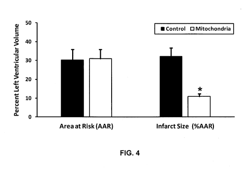
FIG. 4 is a schematic diagram showing quantitation of the area-at-risk (using
Monastryl blue pigment) and infarct size (using triphenyltetrazolium chloride
(TTC) staining)
in control regionally ischemic hearts (n = 3) and in those perfused with 1 x
108 autologously-
derived liver mitochondria (n = 3).
FIG. 5 is a schematic diagram showing regional myocardial function in the
ischemic
area assessed by segmental systolic shortening using three piezoelectric
ultrasonic
transducers.
FIG. 6A is a super-resolution structured illumination microscopy (SR-SIM) red
channel image showing mitochondrial internalization and fusion in human
cardiomyocytes.
FIG. 6B is a SR-SIM green channel image showing mitochondrial internalization
and
fusion in human cardiomyocytes.
FIG. 6C is a SR-SIM blue channel image showing mitochondrial internalization
and
fusion in human cardiomyocytes.
FIG. 6D is a SR-SIM merged image showing mitochondrial internalization and
fusion
in human cardiomyocytes.
FIG. 7A is a graph showing the results of flow cytometry for mitochondria in
the
control group.
FIG. 7B is a graph showing the results of flow cytometry for green fluorescent
protein (GFP)-labeled mitochondria.
FIG. 7C is a graph showing the results of flow cytometry for red fluorescent
protein
(RFP)-labeled mitochondria.
FIG. 7D is a graph showing the results of flow cytometry for mitochondria
isolated
from iCe110 cardiomyocytes treated with GFP-labeled mitochondria.
FIG. 8 is a schematic diagram showing a proposed model of the endosomal
pathways
for mitochondria internalization.
FIG. 9 is a diagram showing an experimental protocol for demonstrating that
injected
mitochondria do not obstruct coronary blood flow.
12
CA 03011472 2018-07-13
WO 2017/124037
PCT/US2017/013564
FIG. 10A is an electrocardiogram (ECG) tracing graph showing the baseline ECG,
and the ECG after the swine model is treated with adenosine, vasopressin, and
epinephrine.
FIG. 10B is an ECG tracing graph showing the ECG after the swine model is
treated
with vehicles and mitochondria.
FIG. 10C is an ECG tracing graph showing the ECG after the swine model is
treated
with 3 um, 10 um and 150 um polystyrene beads.
FIG. 11A is a bar graph showing the baseline QRS, and the QRS after the swine
model is treated with adenosine, vasopressin, epinephrine, mitochondria, and
vehicles.
FIG. 11B is a bar graph showing the baseline corrected QT (cQT) interval, and
the
cQT interval after the swine model is treated with adenosine, vasopressin,
epinephrine,
mitochondria, and vehicles.
FIG. 11C is a bar graph showing the baseline QRS, and the QRS after the swine
model is treated with 3 um, 10 um and 150 um polystyrene beads and
mitochondria.
FIG. 11D is a bar graph showing the baseline cQT interval, and the cQT
interval after
the swine model is treated with 3 um, 10 um and 150 um polystyrene beads and
mitochondria.
FIG. 12A is a bar graph showing percentage of systolic shortening after
coronary
infusion of vehicles, adenosine, epinephrine, vasopressin, and mitochondria.
FIG. 12B is a bar graph showing percentage of systolic shortening after
coronary
infusion of 3 um, 10 um and 150 um polystyrene beads and mitochondria.
FIG. 13A is a bar graph showing coronary blood flow after coronary infusion of
vehicles, adenosine, vasopressin, and mitochondria.
FIG. 13B is a bar graph showing coronary blood flow after coronary infusion of
mitochondria, devitalized mitochondria, and 3 um, 10 um and 150 um polystyrene
beads.
FIG. 14 is a graph showing coronary blood flow at different time points after
coronary infusion of adenosine, vasopressin, mitochondria, and devitalized
mitochondria
(mitochondria 1x10⁹ organelle/ml; adenosine 60 ug; vasopressin 1U; devitalized
mitochondria 1x10⁹ organelle/ml; baseline = 20 ml/min; values are mean SE).
FIG. 15A is a bar graph showing the coronary blood flow in response to
different
doses of mitochondria.
FIG. 15B is a graph showing the coronary blood flow at different time points
in
response to different doses of mitochondria.
13
CA 03011472 2018-07-13
WO 2017/124037
PCT/US2017/013564
FIG. 16 is a diagram showing the methods to demonstrate cardioprotection
afforded
by vascular infusion of mitochondria in a large animal model (swine).
FIG. 17A is a set of graphs showing left ventricular end-diastolic pressure
(LVEDP)
and dP/dt of the left ventricle pressure with vascular infusion of
mitochondria.
FIG. 17B is a graph showing percentage of systolic shortening with vascular
infusion
of mitochondria.
FIG. 18A is a graph showing percentage of area at risk in left ventricle with
vascular
infusion of vehicles and with vascular infusion of mitochondria.
FIG. 18B is a graph showing percentage of infarct size in area at risk with
vascular
infusion of vehicles and with vascular infusion of mitochondria.
FIG. 19A is a bar graph showing number of spots in mice receiving single or
multiple
injections of autogeneic mitochondria, and single and multiple injections of
splenocytes.
FIG. 19B is a bar graph showing number of spots in mice receiving single or
multiple
injections of allogenic mitochondria, and single and multiple injections of
splenocytes.
FIG. 20 is a graph showing percentage of alloantibodies in response to single
and
multiple injections of autogenic and allogeneic mitochondria, and single and
multiple
injections of splenocytes.
FIG. 21 is a diagram showing protocols of determining the optimal
concentration of
Rhodamine 6G, incubation time, and temperatures for mitochondria to uptake
Rhodamine
6G.
FIG. 22A is a bar graph showing Rhodamine 6G concentration in the unbound
fraction under different incubating conditions (4 C).
FIG. 22B is a bar graph showing Rhodamine 6G concentration in the unbound
fraction under different incubating conditions (26 C).
FIG. 23A is a bar graph showing Rhodamine 6G concentration in the bound
fraction
under different incubating conditions (4 C).
FIG. 23B is a bar graph showing Rhodamine 6G concentration in the bound
fraction
under different incubating conditions (26 C).
FIG. 24A is a bar graph showing Rhodamine 6G concentration in the bound
fraction
after incubating mitochondria with 2.5uM Rhodamine 6G under different
incubating
conditions.
14
CA 03011472 2018-07-13
WO 2017/124037
PCT/US2017/013564
FIG. 24B is a bar graph showing Rhodamine 6G concentration in the bound
fraction
after incubating mitochondria with 2.5uM Rhodamine 6G under different
incubating
conditions.
FIG. 25A is a bar graph showing Rhodamine 6G concentration in the bound
fraction
after incubating mitochondria with 1.25uM Rhodamine 6G under different
incubating
conditions.
FIG. 25B is a bar graph showing Rhodamine 6G concentration in the bound
fraction
after incubating mitochondria with 1.25uM Rhodamine 6G under different
incubating
conditions.
FIG. 26 is a schematic diagram showing transferring adoptive mitochondria into
human endothelial colony-forming cells (ECFCs).
FIG. 27A is a schematic diagram showing in vivo vasculogenesis assay.
FIG. 27B is a set of macroscopic images showing explants harvested 7 days
after
transplantation.
FIG. 28A is two Hematoxylin and Eosin (H&E) stain images showing erythrocyte-
filled blood vessels were abundant in implants that contained ECFC-
Mitochondria (ECFC-
Mito), but not in implants that contained ECFC-Control.
FIG. 28B is a graph showing quantification of microvessel density revealing a
higher
vascular density in implants that contained ECFC-Mito than in implants that
contained
ECFC-Control.
FIG. 29A is an image showing binding of Rhodamine-conjugated UEA-1 lectin in
the
lumens of the newly-formed perfused vessels.
FIG. 29B is a human specific CD31 (h-CD31) immunostaining image of the lumens
of the newly-formed perfused vessels.
FIG. 30A is a PET/CT image showing mitochondria distribution in both the left
and
right lungs after "F-rhodamine 6G labeled mitochondria were injected into the
main
pulmonary artery.
FIG. 30B is a PET/CT image showing mitochondria distribution in both the left
and
right lungs after "F-rhodamine 6G labeled mitochondria were injected into the
main
pulmonary artery.
FIG. 31A is a photo showing lung ischemia/reperfusion injury without
mitochondria
treatment.
CA 03011472 2018-07-13
WO 2017/124037
PCT/US2017/013564
FIG. 31B is a photo showing lung ischemia/reperfusion injury with mitochondria
treatment.
FIG. 32 is a PET/CT image showing mitochondria located at the optic nerve
after "F-
rhodamine 6G labeled mitochondria were injected into the common carotid artery
of the
mouse.
FIG. 33 is a schematic diagram showing cardiac segmentations.
DETAILED DESCRIPTION
The present invention is based, at least in part, on the discovery that
isolated
mitochondria, and isolated mitochondria linked to a therapeutic agent,
diagnostic agent
and/or imaging agent, can be delivered to a patient's tissue by injecting them
into the
patient's blood vessels. Skilled practitioners can locally and/or generally
distribute
mitochondria to tissues and/or cells of a patient for a variety of purposes,
using relatively
simple medical procedures. Further, mitochondria can be used as carrier
agents, e.g., to
deliver therapeutic, diagnostic, and/or imaging agents, to a patient's
tissues. Compared to
some traditional therapeutic regimens that involve nanoparticles, it is
further noted that
mitochondria are not toxic and do not cause any substantial adverse immune or
auto-immune
response.
While not intending to be bound by any theory, it is believed that infused
mitochondria extravasate through the capillary wall by first adhering to the
endothelium.
After they are injected or infused into an artery, mitochondria can cross the
endothelium of
the blood vessels and be taken up by tissue cells through an endosomal actin-
dependent
internalization process.
Combined Mitochondrial Agents
Combined mitochondrial agents include mitochondria that are physically
associated
with an agent, such as a therapeutic agent, a diagnostic agent, and/or an
imaging agent.
A therapeutic agent can be any agent that has a therapeutic or prophylactic
use.
Exemplary therapeutic agents include, e.g., therapeutic agents for ischemia-
related disorders,
cytotoxic agents for treating cancer, among many others. In some instances,
mitochondria
can deliver therapeutic agents to specific cells, for example, tumor cells.
The therapeutic
agent may be, e.g., an intracellular inhibitor, deactivator, toxin, arresting
substance and/or
cytostatic/cytotoxic substance that, once inside a cell, inhibits, destroys,
arrests, modifies
16
CA 03011472 2018-07-13
WO 2017/124037
PCT/US2017/013564
and/or alters the cell such that it can no longer function normally and/or
survive. The
therapeutic agent can be an agent to restore a cell's proper function, for
example, a DNA
vector for gene therapy. A therapeutic agent can be, e.g., an inorganic or
organic compound;
a small molecule (less than 500 daltons) or a large molecule; a proteinaceous
molecule, such
as a peptide, polypeptide, protein, post-translationally modified protein, or
antibody; or a
nucleic acid molecule, such as a double-stranded DNA, single-stranded DNA,
double-
stranded RNA, single-stranded RNA, or a triple helix nucleic acid molecule. In
some
embodiments, a therapeutic agent can be a natural product derived from any
known organism
(e.g., from an animal, plant, bacterium, fungus, protist, or virus) or from a
library of synthetic
molecules. In some embodiments, a therapeutic agent can be a monomeric or a
polymeric
compound. Some exemplary therapeutic agents include cytotoxic agents, DNA
vectors,
small interfering RNAs (siRNA), micro RNAs (miRNA), reactive peptides,
nanoparticles,
microspheres, and fluorescent molecules.
A diagnostic agent is an agent that has diagnostic use. As mitochondria carry
a
diagnostic agent into a cell, in some embodiments, the diagnostic agent can be
designed to
determine the condition within a cell, for example pH and oxidative stress
within a cell.
An imaging agent is an agent that is employed for use in imaging techniques.
The
techniques or modalities include, but are not limited to, X-rays, computed
tomography (CT),
magnetic resonance imaging (MRD, scintigraphy, fluorescence, ultrasound, etc.
The imaging
agent can be florescent and/or radioactive. In some embodiments, an imaging
agent can also
be a diagnostic agent. Exemplary imaging agents include, but are not limited
to, MitoTracker
fluorophores (Thermo Fisher Scientific Inc.), CellLight RFP, BacMam 2.0
(Thermo Fisher
Scientific Inc.), pH-sensitive pHrodo fluorescent dyes (Thermo Fisher
Scientific Inc.), 18F_
Rhodamine 6G, "F-labeled rhodamine B, magnetic iron oxide nanoparticles, and
gold- and
platinum-based nanoparticles.
As discussed above, a combined mitochondrial agent comprises a mitochondria
and
an agent that are in direct and/or indirect physical contact with each other.
For example, an
agent can be linked to mitochondria, attached to mitochondria, embedded in the
mitochondrial membrane, or completely or partially enclosed in mitochondria.
In some
instances, a pharmaceutical agent can be linked to mitochondria covalently. In
some
instances, the agent is linked to constituents of mitochondrial membrane
directly through a
covalent bond (e.g., a carboxamide bond and a disulfide bond), or indirectly
through a linker
(e.g., a peptide linker) or another covalently bonded agent. In other
instances, an agent can be
17
CA 03011472 2018-07-13
WO 2017/124037
PCT/US2017/013564
linked to mitochondria non-covalently, for example, through hydrophobic
interaction, Van
der Waals interaction, and/or electrostatic interaction, etc.
In some embodiments, a combined mitochondrial agent can comprise two or more
different types of agents, for example, two different kinds of therapeutic
agents, three
different kinds of imaging agents, one therapeutic agent and one imaging
agent, a therapeutic
agent and a diagnostic agent, etc. Skilled practitioner will appreciate that
any variation is
possible.
One particularly useful linker to link mitochondria and an agent provides a
sustained
release of the agent upon injection. This can be accomplished, for example,
using a
hydrazone functional group. For example, a hydrazone is formed to covalently
bind an agent
to constituents on the mitochondrial membrane. Once this combined
mitochondrial agent is
taken up by cells, the change in pH will result in hydrolysis of the
hydrazone, releasing the
bound agent inside the cell.
In some embodiments, a therapeutic agent, a diagnostic agent, and/or an
imaging
.. agent can be linked to the outer mitochondrial membrane using
functionalized surface
chemistry. In some cases, heterobifunctional chemistries can link a
therapeutic agent, a
diagnostic agent, and/or an imaging agent to the mitochondrial surface, and
once they are
internalized, these agents can be released through interactions with
intercellular esterases
(e.g. via interaction with an acetoxymethyl ester) or through a UV-light
activation or Near-
Infrared light activation strategy. The UV-light activation and Near-Infrared
light activation
strategies are described, e.g., in Zhou, Fang, Hanjie Wang, and Jin Chang,
"Progress in the
Field of Constructing Near-Infrared Light-Responsive Drug Delivery Platforms,"
Journal of
Nanoscience and Nanotechnology 16.3 (2016): 2111-2125; Bansal, Akshaya, and
Yong
Zhang, "Photocontrolled nanoparticle delivery systems for biomedical
applications,"
Accounts of chemical research 47.10 (2014): 3052-3060; Barhoumi, Aoune, Qian
Liu, and
Daniel S. Kohane, "Ultraviolet light-mediated drug delivery: Principles,
applications, and
challenges," Journal of Controlled Release 219 (2015): 31-42. Each of them is
incorporated
by reference in its entirety.
Pharmaceutical and Other Compositions
The disclosure provides compositions that comprise isolated mitochondria,
compositions that comprise combined mitochondrial agents, compositions that
comprise both
18
CA 03011472 2018-07-13
WO 2017/124037
PCT/US2017/013564
isolated mitochondria and combined mitochondrial agents, and methods of using
such
compositions.
A pharmaceutical composition described herein may include mitochondria and/or
combined mitochondria agents and a pharmaceutically acceptable carrier. As
used herein, the
language "pharmaceutically acceptable carrier" includes saline, solvents,
dispersion media,
coatings, antibacterial and antifungal agents, isotonic and absorption
delaying agents, and the
like, compatible with pharmaceutical administration. In some embodiments, the
pharmaceutically acceptable carrier is phosphate buffered saline, saline,
Krebs buffer,
Tyrode's solution, contrast media, or omnipaque, or a mixture thereof In some
embodiments, the pharmaceutically acceptable carrier is sterile mitochondria
buffer (300 mM
sucrose; 10 mM K+-HEPES (potassium buffered (4-(2-hydroxyethyl)-1-
piperazineethanesulfonic acid, pH 7.2); 1 mM K+-EGTA, (potassium buffered
ethylene
glycol tetraacetic acid, pH 8.0)). In some embodiments, the pharmaceutically
acceptable
carrier is respiration buffer (250 mM sucrose, 2 mM KH2PO4, 10 mM MgCl2, 20 mM
K-
HEPES Buffer (pH 7.2), and 0.5 mM K-EGTA (pH 8.0)).
Pharmaceutical compositions are typically formulated to be compatible with its
intended route of administration. Examples of routes of administration include
parenteral,
e.g., intravenous, intradermal, subcutaneous, oral (e.g., inhalation),
sublingual, transdermal
(e.g., topical), transmucosal, and rectal administration.
A pharmaceutical composition can be formulated for various clinical uses,
e.g.,
imaging, treating wounds, treating injuries, preserving organs, improving
mitochondrial
functions in organs or tissues, and skin care. In some cases, the
pharmaceutically acceptable
carrier is a contrast agent for imaging purpose. In some embodiments, the
pharmaceutical
composition may include antiseptic agents, antibacterial agents (e.g.,
antibiotics), antifungal
agents, disinfectants, analgesic agents, anesthetic agents, steroids,
nutritional supplements,
ethereal oils, etc. An anesthetic agent is a drug that can prevent pain during
surgery or
treatment. Exemplary analgesic agents include, without limitation,
paracetamol, nonsteroid
anti-inflammatory drugs, salicylates, ibuprofen and lidocaine. Exemplary
antibacterial agents
include, without limitation, dichlorobenzyl alcohol, amylmetacresol and
antibiotics.
Exemplary antibiotics include penicillins carbapenems, cephalosporins
aminoglycosides,
bacitracin, gramicidin, mupirocin, chloramphenicol, thiamphenicol, lincomycin,
clindamycin,
macrolides, novobiocin, polymyxins, rifamycins, spectinomycin, tetracyclines,
vancomycin,
teicoplanin, streptogramins, anti- folate agents, sulfonamides, trimethoprim,
pyrimethamine,
19
CA 03011472 2018-07-13
WO 2017/124037
PCT/US2017/013564
nitrofurans, methenamine mandelate, methenamine hippurate, nitroimidazoles,
quinolones,
fluoroquinolones, isoniazid, ethambutol, pyrazinamide, para-aminosalicylic
acid, cycloserine,
capreomycin, ethionamide, prothionamide, thiacetazone and viomycin. Antiseptic
agents are
antimicrobial substances that can be applied to living tissue/skin to reduce
the possibility of
infection, sepsis, or putrefaction. Exemplary antiseptics include, without
limitation,
chlorhexidine and salts thereof, benzalkonium and salts thereof, triclosan and
cetylpyridium
chloride. Exemplary antifungal agents include, without limitation, tolnaftate,
miconazole,
fluconazole, clotrimazole, econazole, ketoconazole, itraconazole, terbinafine,
amphotericin,
nystatin and natamycin. Exemplary steroids include, without limitation,
prednisone acetate,
.. prednisone valerate, prednisolone, alclometasone dipropionate, fluocinolone
acetonide,
dexamethasone, methylprednisolone, desonide, pivolate, clocortolone pivolate,
triamcinolone
acetonide, prednicarbate, fluticasone propionate, flurandrenolide, mometasone
furoate,
desoximetasone, betamethasone, betamethasone dipropionate, betamethasone
valerate,
betamethasone propionate, betamethasone benzoate, diflorasone diacetate,
fluocinonide,
.. halcinonide, amcinonide, halobetasol propionate, and clobetasol propionate.
Exemplary
nutritional supplements include, without limitation, vitamins, minerals,
herbal products and
amino acids. Vitamins include without limitation, vitamin A, those in the
vitamin B family,
vitamin C, those in the vitamin D family, vitamin E and vitamin K. Ethereal
oils include
without limitation, those derived from mint, sage, fir, lavender, basil,
lemon, juniper,
rosemary, eucalyptus, marigold, chamomile, orange and the like. Many of these
agents are
described, e.g., in WO 2008152626, which is incorporated by reference in its
entirety.
Compositions comprising mitochondria and/or combined mitochondrial agents can
be
formulated in any form, e.g., liquids, semi-solids, or solids. Exemplary
compositions include
liquids, creams, ointments, salves, oils, emulsions, liposome formulations,
among others.
Compositions for Transplantation
Isolated mitochondria or combined mitochondrial agents can be included in
compositions that are designed for use in organ, tissue, or cell
transplantation. The
composition may include isolated mitochondria and/or combined mitochondrial
agents and a
liquid that is suitable for administration to patients and/or organs in situ
or ex vivo, e.g., for
maintaining organs, tissues or cells ex vivo. In general, the liquid will be
an aqueous solution.
Examples of solutions include Phosphate Buffered Saline (PBS), CelsiorTM
solution,
PerfadexTM solution, Collins solution, citrate solution, tissue culture media
(e.g., Dulbecco's
CA 03011472 2018-07-13
WO 2017/124037
PCT/US2017/013564
Modified Eagle's Medium (DMEM)), the Histidine-tryptophan-ketoglutarate (HTK)
solution,
and the University of Wisconsin (UW) solution (Oxford Textbook of Surgery,
Morris and
Malt, Eds., Oxford University Press, 1994).
The University of Wisconsin cold storage solution is considered a standard
solution
for organ transplantation. It includes the following: 100 mM potassium
lactobionate, 25 mM
KH2PO4, 5 mM MgSO4, 30 mM raffinose, 5 mM adenosine, 3 mM glutathione, 1 mM
allopurinol, and 50 g/L hydroxyethyl starch. Isolated mitochondria or combined
mitochondrial agents can be added to these liquids for organ, tissue and cell
preservation.
Blood Products
Mitochondria and/or combined mitochondrial agents can be included in
compositions
that include blood and/or or products derived from blood. In some embodiments,
the
composition can include mitochondria and/or mitochondrial agents and blood,
e.g., whole
blood, serum, one or more individual blood components, and/or an artificial
blood substitute.
.. In some cases, these blood products can be administered to a subject, and
the mitochondria in
the blood products can improve the mitochondrial function in the subject. For
example, such
blood products can be administered to a patient as a part of a blood
transfusion procedure. As
is art-known, blood or blood products can be stored in any number of vessels,
e.g., in blood
bags, ampules, and/or vials.
Skin and Cosmetic Compositions
Isolated mitochondria and/or combined mitochondrial agents can be included in
compositions that can be applied (e.g., topically and/or by injection) to the
skin and/or to
wounds (e.g., burns, small cuts, larger lacerations, necrotic regions, regions
damaged by
infection with bacteria, fungi, or viruses, or areas with damage caused by
inflammation, e.g.,
rashes), wrinkles, or scars, in the skin. The composition can also include any
known agents
that can be used in skin or cosmetic products, e.g., abrasive agents,
antiseptic agents,
antibacterial agents (e.g., antibiotics), antifungal agents, disinfectants,
analgesic agents,
anesthetic agents, steroids, nutritional supplements, and/or ethereal oils.
Skilled practitioners will appreciate that for a topical composition, e.g., a
composition
such as a liquid, cream, lotion, ointment, or oil, an abrasive agent can be
added to the
composition to aid in delivery of mitochondria and/or combined mitochondrial
agents to
underlying layers of skin cells upon application (e.g., as the composition is
rubbed into and/or
21
CA 03011472 2018-07-13
WO 2017/124037
PCT/US2017/013564
smeared onto the skin). An abrasive agent is a material that is used to wear
away part of the
tissue (e.g., damaged or dead skin cells) by friction. Compositions that
include an abrasive
agent and isolated mitochondria or combined mitochondrial agents can be used
for various
purposes, e.g., cosmetic use, treating wounds, etc. Some abrasive agents are
described, e.g.,
in U.S. Pat. No. 5830445, U.S. Pat. No. 2561043, U.S. Pat. No. 4279890, each
of which is
incorporated by reference in its entirety. Skilled practitioners will also
appreciate that any
art-known agent or composition that aids in transportation of a compound into
underlying
skin layers and/or pores of the skin may be useful in such embodiments and may
be included
in, or applied to a patient separately but in conjunction with, a composition
comprising
mitochondria and/or mitochondrial agents.
Methods of Making Compositions Comprising Mitochondria and/or Combined
Mitochondrial Agents
Isolating mitochondria
Mitochondria for use in the presently described methods can be isolated or
provided
from any source, e.g., isolated from cultured cells or tissues. Exemplary
cells include, but are
not limited to, muscle tissue cells, cardiac fibroblasts, cultured cells, HeLa
cells, prostate
cancer cells, yeast, among others, and any mixture thereof Exemplary tissues
include, but
are not limited to, liver tissue, skeletal muscle, heart, brain, and adipose
tissue. Mitochondria
can be isolated from cells of an autogenous source, an allogeneic source,
and/or a xenogeneic
source. In some instances, mitochondria are isolated from cells with a genetic
modification,
e.g., cells with modified mtDNA or modified nuclear DNA.
Mitochondria can be isolated from cells or tissues by any means known to those
of
skill in the art. In one example, tissue samples or cell samples are collected
and then
homogenized. Following homogenization, mitochondria are isolated by repetitive
centrifugation. Alternatively, the cell homogenate can be filtered through
nylon mesh filters.
Typical methods of isolating mitochondria are described, for example, in
McCully JD,
Cowan DB, Pacak CA, Toumpoulis IK, Dayalan H and Levitsky S, Injection of
isolated
mitochondria during early reperfusion for cardioprotection, Am J Physiol 296,
H94-H105.
PMC2637784 (2009); Frezza, C., Cipolat, S., & Scorrano, L, Organelle
isolation: functional
mitochondria from mouse liver, muscle and cultured filroblasts. Nature
protocols, 2(2), 287-
295 (2007); and a PCT application entitled "Products and Methods to Isolate
Mitochondria"
(PCT/U52015/035584; WO 2015192020); each of which is incorporated by
reference.
22
CA 03011472 2018-07-13
WO 2017/124037
PCT/US2017/013564
Methods of Making Combined Mitochondrial Agents
Skilled practitioners will appreciate that an agent can be linked to
mitochondria in any
number of ways, e.g., by attaching to mitochondria, embedding partially or
completely in the
mitochondrial membrane, enclosing in mitochondria, or encapsulating within the
mitochondria.
While not intending to be bound by any theory or any particular approach, it
is
believed that the outer membrane of mitochondria is adherent and thus
particularly amenable
to combination with various agents. In some embodiments, pharmaceutical agents
can be
attached to the outer membrane of mitochondria simply by incubation. For
example, an
effective amount of pharmaceutic agents can be fully mixed with isolated
mitochondria in a
buffer, e.g., respiration buffer, at a temperature favorable to isolated
mitochondria, e.g., from
0 C to 26 C, from 0 C to 4 C, or about 0 C, 4 C, 26 C. This procedure is
useful to attach
an effective amount of pharmaceutic agents (e.g., nanoparticles, DNA vectors,
RNA vectors)
to mitochondria.
In some embodiments, organic cations (e.g., rhodamine and tetramethylrosamine)
are
readily sequestered by functioning mitochondria because of the electric
potential on
mitochondrial membrane. Healthy mitochondrial membranes maintain a difference
in
electric potential between the interior and exterior of the organelle,
referred to as the
membrane potential. This membrane potential is a direct result of
mitochondrial functional
processes, and can be lost if the mitochondria are not working properly. Lipid-
soluble
cations are sequestered by mitochondria as a consequence of their positive
charge and of their
solubility in both the inner membrane lipids and the matrix aqueous space.
Similarly, in
some other embodiments, anions can be attached to the outer membrane of
mitochondria
because of its negative charge. To link mitochondria with these pharmaceutical
agents, an
effective amount of pharmaceutic agents should be fully mixed with isolated
mitochondria in
a buffer, e.g., respiration buffer, at a temperature favorable to isolated
mitochondria, e.g.,
about 0 C or 4 C.
The therapeutic, diagnostic, and/or imaging agent can be linked to
phospholipids,
peptides, or proteins on the mitochondrial membrane through a chemical bond.
For example,
molecules including fluorophores (pHrodo Red (Thermo Fisher Scientific, Inc.))
and metallic
particles (e.g., 30 nm magnetic iron oxide nanoparticles (Sigma)) can be
covalently linked to
exposed amine groups on proteins and peptides exposed on the outside membrane
of intact
23
CA 03011472 2018-07-13
WO 2017/124037
PCT/US2017/013564
mitochondria using succinimidyl ester conjugates. These reactive reagents
react with non-
protonated aliphatic amine groups, including the amine terminus of proteins
and the c-amino
group of lysine residues, which creates a stable carboxamide bond. In another
example,
when the pharmaceutic agent, e.g., MitoTracker Orange CMTMRos (Invitrogen,
Carlsbad,
CA, now Thermo-Fisher Scientific, Cambridge, MA), are mixed with functional
mitochondria, they are oxidized and then react with thiols on proteins and
peptides on
mitochondria to form conjugates.
There are numerous reactive chemical moieties available for attaching
therapeutic,
diagnostic, and/or imaging agents to the surface of mitochondria (e.g.
carboxylic acid, amine
functionalized, etc.).
Agents can be attached via protein bonding, amine bonding or other attachment
methods either to the outer or inner mitochondrial membrane. Alternatively, or
in addition,
an agent can be attached to the mitochondria membrane through hydrophobic
interaction,
Van der Waals interaction, and/or electrostatic interaction.
In many instances, therapeutic agents, diagnostic agents and imaging agents
may
simply be mixed with isolated mitochondria, and incubated in a buffer (e.g.,
respiration
buffer) for a sufficient period of time (e.g., a few minutes, 5 minutes, 10
minutes, or 1 hour)
at favorable conditions (e.g., from 0 C to 26 C, from 0 C to 4 C, or about
0 C, 4 C, 26 C,
pH 7.2-8.0).
Exemplary methods of preparing combined mitochondrial agents are described in
McCully et al, Injection of isolated mitochondria during early reperfusion for
cardioprotection, Am J Physiol 296, H94-H105. PMC2637784 (2009); and Masuzawa
et al,
Transplantation of autologously derived mitochondria protects the heart from
ischemia-
reperfusion injury, Am J Physiol 304, H966-982. PMC3625892 (2013). Each of the
foregoing are incorporated by reference in its entirety.
Methods of Preparing Compositions Comprising Mitochondria and/or Combined
Mitochondrial Agents
Isolated mitochondria and combined mitochondrial agents can be mixed with a
pharmaceutically acceptable carrier to make a pharmaceutic composition. A
pharmaceutically acceptable carrier includes any compound or composition
useful in
facilitating storage, stability, administration, cell targeting and/or
delivery of the
mitochondria and/or combined mitochondrial agent, including, without
limitation, suitable
24
CA 03011472 2018-07-13
WO 2017/124037
PCT/US2017/013564
vehicles, diluents, solvents, excipients, pH modifiers, salts, colorants,
rheology modifiers,
lubricants, coatings, fillers, antifoaming agents, polymers, hydrogels,
surfactants, emulsifiers,
adjuvants, preservatives, phospholipids, fatty acids, mono-, di- and tri-
glycerides and
derivatives thereof, waxes, oils and water. In some embodiments, isolated
mitochondria
and/or the combined mitochondrial agents are suspended in water, saline,
buffer, respiration
buffer, or sterile mitochondria buffer for delivery in vivo. Pharmaceutically
acceptable salts,
buffers or buffer systems, including, without limitation, saline, phosphate
buffer, phosphate
buffered saline (PBS) or respiration buffer can be included in a composition
described herein.
Vehicles having the ability to facilitate delivery to a cell in vivo, such as
liposomes, may be
utilized to facilitate delivery of the combined mitochondrial agents to the
target cells.
Methods of making compositions, e.g., liquid, semi-solid, and solid
compositions
(e.g., liquids, creams, lotions, ointments, oils, among others), are well-
known in the art.
Skilled practitioners will appreciate that such known methods can be modified
to add one or
more steps to add mitochondria and/or combined mitochondrial agents and form a
composition described herein. Skilled practitioners will appreciate that in
some instances a
composition described herein may include more than one type of combined
mitochondrial
agent. For example, included are compositions comprising mitochondria wherein
essentially
each mitochondrion is associated with multiple types of agents. Also included
are
compositions comprising mitochondria wherein each mitochondrion is paired with
only one
type of agent but wherein the composition comprises a mixture of
mitochondria/agent
pairings.
Methods of Use
Administration
Isolated mitochondria and combined mitochondrial agents can be administered to
a
patient by injection intravenously, intra-arterially, intraperitoneally, intra-
muscularly, and/or
through intraosseous infusion. In some embodiments, isolated mitochondria and
combined
mitochondrial agents, can be delivered by direct injection or by vascular
infusion.
Once mitochondria are injected into a tissue, mitochondria will be taken up by
cells
around the site of injection. Therefore, in some embodiments, the site of
injection is the target
site. In some other embodiments, mitochondria are injected to a blood vessel
which carries
the blood to the target site, for example, an organ, a tissue, or an injured
site. While not
intending to be bound by any theory, evidence suggests that mitochondria
delivered by direct
CA 03011472 2018-07-13
WO 2017/124037
PCT/US2017/013564
injection are internalized by cells through actin-dependent endocytosis.
However,
mitochondrial uptake by vascular delivery appears to be more complicated. The
rapid and
widespread uptake of mitochondria when delivered by vascular infusion would
suggest that
mechanisms allowing for the rapid passage of mitochondria through the vascular
wall are
involved. Some studies support the concept that cells can routinely escape
from the
circulation. It has been shown that certain cardiac and mesenchymal stem cells
appear to be
actively expelled from the vasculature in a process different from diapedesis
(Cheng, K.,
Shen, D., Xie, Y., Cingolani, E., Malliaras, K., Marban, E., 2012, Brief
report: Mechanism
of extravasation of infused stem cells. Stem Cells. 30, 2835-2842.; Allen,
T.A., Gracieux, D.,
Talib, M., Tokarz, D.A., Hensley, M.T., Cores, J., Vandergriff, A., Tang, J.,
de Andrade, J.B.,
Dinh, P.U., Yoder, J.A., Cheng, K., 2017. Angiopellosis as an Alternative
Mechanism of
Cell Extravasation. Stem Cells. 35,170-180). Transmigration of stem cells
through the
vascular wall requires extensive remodeling of the endothelium. Mitochondria
may use a
similar remodeling mechanism to pass through the vascular wall. Another
possible
mechanism for mitochondrial uptake may be diapedesis- like. Some cells
routinely escape
from the circulation. For example, leukocyte extravasation (i.e. diapedesis)
between venous
endothelial cells is a well-understood process that involves cell adhesion
proteins. Further, it
is also possible that infused mitochondria extravasate through the capillary
wall through the
space between the endothelium cells. After mitochondria cross the endothelium
of the blood
vessels, mitochondria are taken up by tissue cells through an endosomal actin-
dependent
internalization process.
Mitochondria or combined mitochondrial agents can be administered to a subject
as a
singular, one-time treatment, or alternatively, multiple treatments, e.g., a
treatment course
that continues intermittently or continuously for about 1, 2, 5, 8, 10, 20,
30, 50, or 60 days,
one year, indefinitely, or until a physician determines that administration of
the mitochondria
or combined mitochondrial agent is no longer necessary.
In one method of administration, mitochondria or combined mitochondrial agents
are
injected into organ tissue directly. The injection is repeated several times
at different sites of
the organ. In such a method, a sterile 1-ml insulin syringe with a small
needle (e.g., 28-
gauge) can be used for the injection and each injection site can receive,
e.g., about 1.2 x 10⁶
of mitochondria.
Skilled practitioners will appreciate that the amount of mitochondria and/or
combined
mitochondrial agents, e.g., compositions comprising mitochondria and/or
combined
26
CA 03011472 2018-07-13
WO 2017/124037
PCT/US2017/013564
mitochondrial agents, that should be administered to a patient will vary
depending upon, e.g.,
the type of disorder being treated, the route of administration, the duration
of the treatment,
the size of an area to be treated, and/or the location of the treatment site
in the patent, among
others. Skilled practitioners will be able to determine dosages to be
administered depending
on these and other variables. For example, a total of about 1 x 10⁷ of
mitochondria can be
administered into a blood vessel of a subject, e.g., to treat localized
ischemia in the
myocardium. As another example, in the case of larger organs or affected
areas, greater
numbers of mitochondria, e.g.,
1 x 10 10 to 1 x 10 14 mitochondria, can be injected into the blood vessel.
Conversely, in the
case of small focal lesions, 1 x 10 3 to 1 x 10 6 mitochondria can be infused
into the patient.
Therefore, an effective amount of mitochondria or combined mitochondrial
agents (or
compositions comprising same) is the total amount of mitochondria or combined
mitochondrial agents sufficient to bring about a desired therapeutic effect.
An effective
amount can be, e.g., at least or about 1 x 10² mitochondria or combined
mitochondrial agents
e.g., from about 1 x 10³ to about 1 x 10¹⁴, about 1 x 10 to about 1 x 10¹³,
about 1 x 10 to
about 1 x 10¹², about 1 x 10⁶ to about 1 x 10¹¹, about 1 x 10 to about 1 x
1010, about 1 x 10³
to about 1 x 10⁷, about 1 x 10 to about 1 x 10⁶, about 1 x 10 to about 1 x
1014, or about 1 x
108 to about 1 x 10¹³, about 1 x 10 to about 1 x 10¹², about 1 x 10 to about 1
x 108 or at least
or about 1 x 10³, 1 x 10 4, 1 X 10 5, 1 X 10 6, 1 X 10 7, 1 X 10 8, 1 X 10 9,
1 X 10 19, 1 X 10 11, 1 X
1012, 1 x 10¹³, or at least or about
1 x 10¹⁴, or e.g., an amount more than 1 x 10 14. As used herein, the term
"total amount" in
the context of administration to a patient can refer to the total amount of
mitochondria or
combined mitochondrial agents in a single administration (e.g., one injection,
one dose
administered in an infusion) or in multiple administrations (e.g., multiple
injections),
depending on the dosing regimen being performed.
Isolated mitochondria and/or combined mitochondrial agents can be administered
to a
subject every 12-24 hours by various routes, e.g., direct injection, vascular
delivery. In some
embodiments, isolated mitochondria or combined mitochondrial agents can be
administered
to a subject every 5-10 minutes (e.g., every 5 minutes, every 10 minutes) by
various routes,
e.g., direct injection, vascular infusion.
In some embodiments, isolated mitochondria or combined mitochondrial agents
can
be directly injected into tissues or organs by Gauge 24, 25, 26, 27, 28, 29,
30, 31, 32, 33, and
27
CA 03011472 2018-07-13
WO 2017/124037
PCT/US2017/013564
34 needles. In some other cases, isolated mitochondria, or combined
mitochondrial agents
can be delivered to a target site by a catheter.
It is noted that in some cases, the effects of mitochondria depend on the
length of the
time period between the time of isolation and the time of use. Thus, in some
instances, the
mitochondria are freshly isolated and viable. The mitochondria or combined
mitochondrial
agents can be administered to a subject within about 5 minutes, about 10
minutes, about 20
minutes, about 30 minutes, about 40 minutes, about 50 minutes, about 60
minutes, about 70
minutes, about 80 minutes, about 90 minutes, about 100 minutes, about 110
minutes, about
120 minutes after the mitochondria are isolated. In some instances, the
mitochondria or
combined mitochondrial agents are administered to a subject within about 5
minutes, about
10 minutes, about 20 minutes, about 30 minutes, about 40 minutes, about 50
minutes, about
60 minutes, about 70 minutes, about 80 minutes, about 90 minutes, about 100
minutes, about
110 minutes, about 120 minutes after starting the mitochondria isolating
process.
Mitochondria and/or combined mitochondrial agents may in some instances be
stored for a
short period of time (e.g., about 10 minutes, about 20 minutes, about 30
minutes, about 40
minutes, about 50 minutes, or about 60 minutes) before use.
It is also noted that, in some cases, frozen-thawed mitochondria are not
viable and not
effective for certain treatments described herein, e.g., treatment of
ischemia/reperfusion
injuries. Thus, in some cases, the mitochondria are not frozen and thawed
after isolation
from tissues and/or cells.
The mitochondria for the treatment can be isolated from cells or tissues of an
autogenous source, an allogeneic source, and a xenogeneic source. In some
instances,
mitochondria are collected from cultured cells or tissues of a subject, and
these mitochondria
are administered back to the same subject. In some other cases, mitochondria
are collected
from cultured cells or tissues of a second subject, and these mitochondria are
administered to
a first subject. In some cases, mitochondria are collected from cultured cells
or tissues from a
different species (e.g., mice, swine, yeast).
Treating ischemic heart and other ischemia-related diseases
The heart is a highly energetic organ that requires a continuous supply of
oxygen to
maintain normal function. Under aerobic conditions, the heart derives its
energy primarily
from the mitochondria, which constitute 30% of the total myocardial cell
volume. Following
28
CA 03011472 2018-07-13
WO 2017/124037
PCT/US2017/013564
the onset of ischemia, there is a rapid decline in high-energy phosphate
levels with alterations
in mitochondrial structure, volume, oxygen consumption, and ATP synthesis.
Attempts to lessen myocardial tissue necrosis and improve post-ischemic
function
using pharmacological and/or exogenous substrate interventions, either alone
or in
combination with procedural techniques, have provided only limited
cardioprotection.
Despite these interventions, mitochondrial damage and dysfunction continue to
represent
major problems following myocardial ischemia and remain significant causes of
morbidity
and mortality.
Mitochondrial damage occurs mainly during ischemia rather than during
reperfusion,
and that preservation of mitochondrial respiratory function enhances
contractile recovery and
decreases myocardial infarct size.
Methods described herein can be used to treat ischemic heart. For example, an
effective amount of isolated mitochondria can be injected into the blood
vessel of a subject,
for example, the coronary vasculature of the subject. For example, about 1 x
107 of
mitochondria can be administered into the coronary vasculature of the subject.
The injected
mitochondria are internalized by cardiomyocytes after transplantation and
provide enhanced
oxygen consumption, upregulate chemokines that enhance post-infarct cardiac
function, and
upregulate the expression of protein pathways that are important in preserving
myocardial
energetics. In another example, an effective amount of mitochondria can be
directly injected
to the area at risk (regional ischemic area). The injection can be repeated
several times at
different sites of the heart.
Reperfusion injury is the tissue damage by blood supply when blood returns to
the
tissue after a period of ischemia or lack of oxygen. The absence of oxygen and
nutrients
during the ischemic period results in inflammation and oxidative damage when
blood flow is
restored. The inflammatory response further leads to the reperfusion injury in
the tissue.
Therefore, in some instances, a treatment also involves administering immune
suppressors to
the patient. The immune suppressors can be, e.g., administrated separately,
but as a
concurrent treatment with the mitochondrial agent. Alternatively, or in
addition, the immune
suppressors can be linked to mitochondria to form a combined mitochondrial
agent, which
can be used for the treatment. Particularly useful immune suppressors are
bisphosphonates.
The ischemia/reperfusion injury in some other organs is often associated with
mitochondrial damage and dysfunction as well. These organs include, but are
not limited to,
lung, kidney, liver, skeletal muscle, brain, etc. These injuries or diseases
include, but are not
29
CA 03011472 2018-07-13
WO 2017/124037
PCT/US2017/013564
limited to, ischemic colitis, mesenteric ischemia, brain ischemia, stroke,
acute limb ischemia,
cyanosis and gangrene. The described method can be also employed to treat
ischemia injury
in these organs/tissues. For these treatments, the isolated mitochondria
and/or combined
mitochondrial agent can be directly injected to the organ tissue or injected
into the blood
vessel which carries the blood to the target organ/tissue or the injured site
of the subject.
Vasodilation and blood flow
It has been demonstrated that mitochondrial delivery by vascular infusion
significantly increases coronary blood flow without altering mean blood
pressure or heart
rate. The ability to increase blood flow with no increase in heart rate allows
for clinical usage
in angina type injury and in ischemia/reperfusion related injury and in tissue
damage areas
where increased blood flow and oxygen delivery would be needed. Thus, the
methods
described herein can be used in coronary artery interventions to remove clots
or obstructions
in blood vessels.
Methods described herein can also be used to increase blood flow and/or oxygen
delivery for various organs or tissues (e.g., heart, lung, kidney, brain,
skeletal muscle). In
some instances, methods described herein can be used to treat peripheral
vascular disease.
Peripheral vascular disease (PVD) is a blood circulation disorder that causes
the blood
vessels outside of the heart and brain to narrow, block, or spasm. This can
happen in the
arteries or veins. PVD typically causes pain and fatigue, often in the legs,
and especially
during exercise. Isolated mitochondria and/or combined mitochondrial agents
can be injected
to a blood vessel. Blood flow may carry isolated mitochondria or combined
mitochondrial
agents to the target site. In some instances, methods described herein can
also be used to
enhance smooth muscle function.
Methods described herein can also be used for vascular dilatation in various
organs.
In some instances, the isolated mitochondria or combined mitochondrial agents
can be used
to decrease vascular resistance in an organ (e.g., heart, kidney, liver, or
lung). Isolated
mitochondria or combined mitochondrial agents can be used to increased blood
flow for
angiography. The isolated mitochondria and/or combined mitochondrial agents
can be added
.. to a contrast agent, and can be used in the identification and removal of
blockages.
Methods described herein can be used to treating blocked blood vessel. The
methods
involve, e.g., the steps of localizing blood clots, positioning a first
catheter with cage distal to
clot, positioning a second catheter proximal to clot, injecting mitochondria
and/or combined
CA 03011472 2018-07-13
WO 2017/124037
PCT/US2017/013564
mitochondrial agents via the proximal catheter to cause vasodilatation,
collecting the clot in a
basket, and removing the clot.
It is noted that the effects of vascular infusion of mitochondria are
dependent on time
from isolation to time of use. The vasodilatory effects decreases as time from
isolation is
extended. While not intending to be bound by any theory, it is hypothesized
that freshly
isolated mitochondria have certain chemicals, which can increase blood flow.
Therefore, in
some methods, the mitochondria are freshly isolated and viable. For example,
the
mitochondria or combined mitochondrial agents are administered to a subject
within about 5
minutes, about 10 minutes, about 20 minutes, about 30 minutes, about 40
minutes, about 50
.. minutes, about 60 minutes, about 70 minutes, about 80 minutes, about 90
minutes, about 100
minutes, about 110 minutes, about 120 minutes after the time point when the
mitochondria
isolation process starts or after the mitochondria are isolated. In some
cases, the mitochondria
or combined mitochondrial agents are administered to a subject within about 20
minutes to
about 60 minutes (e.g., about 20 minutes, about 30 minutes, about 40 minutes,
about 50
minutes, about 60 minutes) after the time point when the mitochondria
isolation process starts
or after the mitochondria are isolated.
In some cases, increasing blood flow is not desirable (e.g., treating
ischemia/reperfusion in lungs). In these cases, mitochondria or combined
mitochondrial
agents can be stored for a short period of time (e.g., from about 30 to about
60 minutes)
before usage. This method can be used to increase tissue viability (e.g.,
treating
ischemia/reperfusion injury) without causing an increase in blood flow. In
these cases, the
mitochondria or combined mitochondrial agents are administered to a subject at
least 60
about minutes (e.g., about 65 minutes, about 70 minutes, about 80 minutes,
about 90 minutes,
about 100 minutes, about 110 minutes, about 120 minutes) after the time point
when the
mitochondria isolation process starts or after the mitochondria are isolated.
Heart surgery
The isolated mitochondria and/or combined mitochondrial agents can be
delivered to
the heart to decrease stunning and allow for weaning of the heart from a
surgical procedure
.. (e.g., cardioplegia), and recovery of the heart without increasing heart
rate or oxygen
demands in the heart. In some embodiments, the methods involve direct
injection of isolated
mitochondria and/or combined mitochondrial agents to the heart. In some
methods, isolated
mitochondria and/or combined mitochondrial agents are injected into a coronary
artery.
31
CA 03011472 2018-07-13
WO 2017/124037
PCT/US2017/013564
Imaging
Imaging agents can be attached to mitochondria, often by co¨incubation of the
mitochondria with the imaging agents. Such imaging agents include, but are not
limited to,
MitoTracker and pHrodo fluorophores from Thermo Fisher Scientific Inc., "F-
Rhodamine
6G, and iron oxide nanoparticles.
Combined mitochondrial agents that include an imaging agent can be injected
into the
tissue or perfused through the blood vessels. Tissues containing the labeled
mitochondria can
be examined using imaging techniques, such as positron emission tomopgrahy
(PET),
microcomputed tomography (pCT), and magnetic resonance imaging (MRI),
brightfield
microscope, and 3-D super-resolution microscopy, etc. Skilled practitioners
will appreciate
that other imaging techniques or modalities may be used. They include, but are
not limited
to, x-rays, scintigraphy, fluorescence and ultrasound.
Positron emission tomography is an imaging technique that produces a three-
dimensional image in the body, and can be used in methods described herein.
The system
detects pairs of gamma rays emitted indirectly by a positron-emitting
radioisotope (tracer).
Three-dimensional images of tracer concentration within the body are then
constructed by
computer analysis. Useful reporter groups include radioactive isotopes, such
as nc, 13N, 15
0, 18F, 64cu, 68Ga, 81m1Kr, 82Rb, 86y, 89zr, "In, 1231, 1241, 133)(e, 201Ti,
1251, 35s 14,,,
3H. In
some methods, mitochondria can be labeled by radioactive isotopes, e.g., I-8F,
or by molecules
that incorporate radioactive isotopes, e.g., 18F-Rhodamine 6G, 18F-labeled
rhodamine B.
After the mitochondria are internalized by target cells, the PET imaging
technique, or similar
technique, can be employed to view the target cells.
Magnetic resonance imaging is a medical imaging technique to image the anatomy
and the physiological processes of the body and can be used in methods
described herein. In
some instances, it can be used in conjugation with some other imaging
techniques, for
example, PET. Images acquired from both devices can be taken sequentially, in
the same
session, and combined into a single superposed (co-registered) image. PET/MRI
scans can
be used to diagnose a health condition in humans and animals, e.g., for
research, medical, and
agricultural purposes.
Micro¨computed tomography uses x-rays to create cross-sections of a physical
object
that can be used to recreate a virtual model without destroying the original
object, and can be
used in methods described herein. In some instances, it is used in conjugation
with some
32
CA 03011472 2018-07-13
WO 2017/124037
PCT/US2017/013564
other imaging techniques, for example, PET. Images acquired from both devices
can be
taken sequentially, in the same session, and combined into a single superposed
(co-registered)
image. Thus, functional imaging obtained by PET, which depicts the spatial
distribution of
metabolic or biochemical activity in the body can be more precisely aligned or
correlated
with anatomic imaging obtained by CT scanning. Two- and three-dimensional
image
reconstruction may be rendered as a function of a common software and control
system.
3D-structured illumination microscopy, 3D-SIM, or 3-D super-resolution
microscopy,
allows complete 3D visualization of structures inside cells and can be used in
the methods
described herein. Structured illumination microscopy is an imaging method
capable of
doubling the spatial resolution of conventional widefield fluorescence
microscopy by using
spatially structured illumination light. It enhances spatial resolution by
collecting
information from frequency space outside the observable region.
The described methods, i.e., methods that include administering mitochondria
and/or
combined mitochondrial agents, are useful for diagnosing a variety of
diseases, such as
cancers, (e.g., lung, brain, pancreatic, melanoma, prostate, colon cancers),
cardiovascular
disease (e.g., myocardial infarction, atherosclerosis), autoimmune diseases
(e.g., multiple
sclerosis, diabetes, irritable bowel syndrome, Celiac disease, Crohn's
disease), and
inflammatory disease.
Methods using agents for imaging purpose are well-known in the art and
described in,
for example, Bartholoma MD, He H, Pacak CA, Dunning P, Fahey FH, McGowan FX,
Cowan DB, Treves ST and Packard AB, Biological characterization of F18-labeled
Rhodamine B, a potential positron emission tomography perfusion tracer, Nucl
Med Biol 40,
1043-1048, PMC3820364 (2013); Bartholoma MD, Zhang S, Akurathi V, Pacak CA,
Dunning P, Fahey FH, Cowan DB, Treves ST and Packard AB, "F-labeled rhodamines
as
potential myocardial perfusion agents: comparison of pharmacokinetic
properties of several
rhodamines, Nucl Med Biol 42, 796-803, PMC4567415 (2015); and Pacak CA, Hammer
PE,
MacKay AA, Dowd RP, Wang KR, Masuzawa A, Sill B, McCully JD and Cowan DB,
Superparamagnetic iron oxide nanoparticles function as a long-term, multi-
modal imaging
label for non-invasive tracking of implanted progenitor cells, PLoS ONE 9,
e108695,
PMC4177390 (2014). Each of the foregoing can be useful in methods described
herein and is
incorporated herein by reference its entirety.
Drug delivery
33
CA 03011472 2018-07-13
WO 2017/124037
PCT/US2017/013564
The present specification provides methods to deliver pharmaceutic agents,
e.g., to
cells and/or tissues of a patient. Mitochondria are taken up by tissue cells
through an actin-
dependent internalization process, thereby providing a way to deliver
pharmaceutic agents
directly into the cells. Moreover, because combined mitochondrial agents are
more likely to
cross the endothelium of the blood vessels near the injection site, in some
instances,
combined mitochondrial agents can be injected into a blood vessel that carries
blood to the
target site. In some instances, combined mitochondrial agents enter into
tissue through the
endothelium of capillaries.
An antibody or an antigen-binding fragment can be linked or attached to
mitochondria. Skilled practitioners will appreciate that linking the antibody
or antigen
binding fragment to mitochondria or combined mitochondrial agent can allow the
mitochondria or combined mitochondrial agent to be targeted to specific sites,
e.g., to target
cells and/or tissues. In some instances, the antibody or the antigen-binding
fragment are
designed to target specific cell types, for example, smooth muscle cells in
lung, immune cells,
macrophages, etc.
Gene therapy
Gene therapy is the therapeutic delivery of nucleic acid polymers into a
patient's cells
as a drug to treat disease. Isolated mitochondria can be used as a carrier to
deliver nucleic
acid polymers into a cell. In some instances, combined mitochondrial agents
that include
nucleic acid polymers can be administered to a subject to replace a mutated
gene in the
subject that causes disease, to inactivate, or "knock out," a mutated gene, or
to introduce a
new gene into the subject. Exemplary nucleic acid polymers include, but are
not limited to,
double-stranded DNA, single-stranded DNA, double-stranded RNA, single-stranded
RNA, or
triple helix nucleic acid molecules. In certain instances, the nucleic acid
polymers are DNA,
interfering RNAs (siRNA), and micro RNAs. In the case of mitochondrial
myopathies
related to mitochondrial DNA dysfunction, gene therapy can be performed by
direct infusion
of the mitochondria to a skeletal muscle or muscles. In the case of nuclear
DNA related
mitochondrial myopathies, multiple infusions over time might be beneficial or
required.
Minimizing cardiotoxicity
Chemotherapy is a common treatment for various cancers, however, it also
causes
several serious complications. Chemotherapy-induced cardiotoxicity is one
complication that
34
CA 03011472 2018-07-13
WO 2017/124037
PCT/US2017/013564
limits the clinical use of chemotherapeutic agents. Certain chemotherapeutic
agents, such as
anthracyclines, are highly effective against acute lymphoblastic and
myeloblastic leukemias,
but are particularly harmful to the heart due to its effects on mitochondria.
The damage to
mitochondria further leads to chemotherapy-induced cardiotoxicity.
Angsutararux P,
Luanpitpong S, Issaragrisil S. Chemotherapy-Induced Cardiotoxicity: Overview
of the Roles
of Oxidative Stress. Oxid Med Cell Longev. 2015;2015:795602. doi:
10.1155/2015/795602
(2015); Guo S, Wong S. Cardiovascular toxicities from systemic breast cancer
therapy, Front
Oncol. 4:346. doi: 10.3389/fonc.2014.00346. eCollection (2014).
One useful method to minimize chemotherapy-induced cardiotoxicity is to
administer
an effective amount of isolated mitochondria and/or a combined mitochondrial
agent to a
patient who is currently under a chemotherapy treatment regimen. If the
patient needs to be
treated with chemotherapy (e.g., because prescribed by a physician or
veterinarian), the
patient can be treated with mitochondria and/or combined mitochondrial agent,
before,
during, and/or after administration of the chemotherapy. For example, patients
can be treated
with mitochondria and/or combined mitochondrial agent starting immediately
after
administration, as a singular treatment or continuing intermittently or
continuously for about
1, 2, 5, 8, 10, 20, 30, 50, or 60 days, one year, indefinitely, or until a
physician determines
that administration of the mitochondria and/or combined mitochondrial agent is
no longer
necessary.
Organ/tissue transplantation
The present disclosure also features methods of transplanting an organ(s),
tissues,
masses of cells and/or isolated cells. The methods can include a step of
exposing the organ(s),
tissues, mass of cells and/or isolated cells to mitochondria or combined
mitochondria' agents
prior to transplantation. Such exposures can occur in situ and/or ex vivo. The
organ(s), tissues
and/or isolated cells may be exposed to a composition comprising mitochondria
or combined
mitochondria' agents.
Exposure of an organ or tissue to compositions comprising mitochondria or
combined
mitochondria" agents can be performed ex vivo and/or in situ by any method
known in the art.
For example, the exposure may be performed ex vivo in any chamber or space
having
sufficient volume for submerging the organ or tissue, completely or partially,
in the
composition. As another example, the organ may be exposed to compositions
comprising
mitochondria or combined mitochondria' agents by placing the organ in any
suitable
CA 03011472 2018-07-13
WO 2017/124037
PCT/US2017/013564
container, and causing the compositions comprising mitochondria or combined
mitochondria'
agents to "wash over" the organ, such that the organ is exposed to a
continuous flow of the
composition.
Alternatively, the organ may be perfused with a composition comprising
mitochondria or combined mitochondria' agents. The term "perfusion" is an art
recognized
term, and relates to the passage of a liquid, e.g., a composition comprising
mitochondria or
combined mitochondria' agents, through the blood vessels of an organ or
tissue. Methods for
perfusing organs ex vivo and in situ are well known in the art. An organ can
be perfused with
a composition ex vivo, for example, by continuous hypothermic machine
perfusion
(see Oxford Textbook of Surgery, Morris and Malt, Eds., Oxford University
Press, 1994).
Optionally, in in situ or ex vivo perfusions, the organ can be perfused with a
wash solution,
e.g., UW solution, prior to perfusion with a composition comprising
mitochondria or
combined mitochondria' agents, to remove the donor's blood from the organ. As
another
option, the UW solution can include mitochondria or combined rnitochondrial
agents.
The organ or tissue may be placed, e.g., submerged, in a medium or solution
that
includes mitochondria or combined mitochondria' agents. Alternatively, or in
addition,
mitochondria or combined mitochondria' agents can be added into the medium or
solution. In
situ exposures can be performed by any method known in the art, e.g., by in
situ flushing or
perfusion of the organ with a composition comprising mitochondria or combined
mitochondria' agents (see Oxford Textbook of Surgery. Morris and Malt, Eds.,
Oxford
University Press, 1994).
The present disclosure contemplates that any or all of the above methods for
exposing
an organ or tissue to a composition comprising mitochondria or combined
mitochondrial
agents, e.g., washing, submerging, or perfusing, can be used in a given
transplantation
procedure.
The present disclosure further contemplates that a solid or semi-solid
composition can
be created. For example, a liquid that is a composition comprising
mitochondria or combined
mitochondria' agents, as described above, can be made into a solid or semi-
solid composition,
in which an organ or tissue may be overlaid or embedded. Alternatively, a semi-
solid
composition can be infused into the organ. Solid or semi-solid compositions
can be made, for
example, by adding a solidifying agent such as a gelling agent (e.g., collagen
or alginate) to
the liquid.
36
CA 03011472 2018-07-13
WO 2017/124037
PCT/US2017/013564
Methods described herein can be used to control ischemia/reperfusion damage
for
transplanted organs. Ischemia-reperfusion injury is a very important problem
during organ
transplantation. Much damage in organ transplantation appears to be induced by
reperfusion
injury. Organs used for transplantation often undergoes lengthy periods of
cold ischemic
storage after devascularization and cold perfusion, resulting in an increased
susceptibility to
damage upon reperfusion. Evidence shows that ischemia/reperfusion injury often
leads to
mitochondrial oxidative damage, which may cause delayed graft function. Dare
AJ, Logan A,
Prime TA, Rogatti S, Goddard M, Bolton EM, Bradley JA, Pettigrew GJ, Murphy
MP, Saeb-
Parsy K. The mitochondria-targeted anti-oxidant MitoQ decreases ischemia-
reperfusion
injury in a murine syngeneic heart transplant model, J Heart Lung Transplant,
34(11):1471-
80. doi: 10.1016/j.healun.2015.05.007 (2015); Liu Q, Krishnasamy Y, Rehman H,
Lemasters
JJ, Schnellmann RG, Zhong Z. Disrupted Renal Mitochondrial Homeostasis after
Liver
Transplantation in Rats. PLoS One 10(10):e0140906. doi:
10.1371/journal.pone.0140906
(2015). In some cases, the transplanted organ can be, e.g., a heart, a lung, a
kidney, or a liver.
In one embodiment, an effective amount (e.g., 1 x 10 7, 1 x 10 8) of
mitochondria or
combined mitochondria agents are injected into the blood vessels (e.g.,
arteries) of the
transplanted organ. In another embodiment, an effective amount (e.g., 1 x 10
7, 1 X 10 8) of
mitochondria or combined mitochondria agents are directly injected into the
organ tissue.
An effective amount of mitochondria or combined mitochondria agents is an
amount
that is effective for enhancing survival and/or improving function of organs,
or cells in vivo
and/or in vitro. Within the context of transplantation of individual cells or
masses of cells,
e.g., transplant donors and/or recipients, an effective amount of mitochondria
or combined
mitochondria agents is an amount that is administered to the transplant donor
and/or recipient
sufficient to enhance survival of the cell or mass of cells, e.g. to reduce
loss of the cell, or
mass of cells, and/or to improve functional performance of a transplanted cell
or a mass of
cells. Within the context of treating cells outside a body, e.g., islet cells
to be cultured and/or
used for transplantation, an effective amount is an amount with which the
cells are incubated
or stored in order to enhance preservation of the cells and/or to reduce cell
loss, e.g., loss via
apoptosis, and/or to enhance function. Within the context of transplantation
of organs and
tissues, e.g., transplant donors and/or recipients, an effective amount of
mitochondria or
combined mitochondria agents is an amount that is administered to the
transplant donor
and/or recipient sufficient to enhance survival of the organ, tissue or cells
of interest, e.g., to
37
CA 03011472 2018-07-13
WO 2017/124037
PCT/US2017/013564
reduce loss of cells from which the organ or tissue is composed, and/or to
improve functional
performance of an organ.
In some instances, the injection is performed before the organ is retrieved
from the
donor. In some instances, the injection is performed at some time point after
organ is
retrieved, but before it is transplanted. In some instances, the injection is
performed after the
organ is transplanted into the recipient. In some instances, injections are
performed before
organ retrieval, after harvesting of the organ, and then again after
implantation into the
recipient. In some instances, the injection is performed during the
transplantation surgery. In
some embodiments, the transplanted organ is preserved in a solution containing
an effective
amount of isolated mitochondria or combined mitochondrial agents. In some
cases, the
solution is University of Wisconsin cold storage solution.
A major limitation for organ transplantation is the availability of donor
organs. In
order to expand the number of donor organs, centers may use organs from donors
with
extended criteria or donors from cardiac death. In these cases, the described
methods can
improve the quality of the organs, thus increasing the availability of donor
organs.
The disclosure also provides methods of improving transplanted tissue and/or
cell
integration. In some embodiments, the tissue is skin tissue or bone marrow. In
some
embodiments, the cells are stem cells. In these cases, mitochondria or
combined
mitochondrial agents can improve the integration of the transplanted tissue
and cells in the
recipient's body.
Treating Mitochondrial Dysfunction Disorder
Due to mitochondria's primary function in cell metabolism, damage and
dysfunction
in mitochondria can cause a range of human diseases. Diseases caused by
mutation in the
mtDNA include Kearns-Sayre syndrome, MELAS syndrome and Leber's hereditary
optic
neuropathy, Pearson syndrome, and progressive external ophthalmoplegia. Other
diseases
that involve mitochondrial dysfunction include, but are not limited to,
mitochondrial
myopathy, diabetes mellitus and deafness (DAD), Leigh syndrome, "Neuropathy,
ataxia,
retinitis pigmentosa, and ptosis" (NARP), myoneurogenic gastrointestinal
encephalopathy
(MNGIE), Myoclonic Epilepsy with Ragged Red Fibers (MERRF syndrome),
encephalomyopathy, lactic acidosis, Parkinson's disease, and stroke-like
symptoms (MELAS
syndrome), etc.
38
CA 03011472 2018-07-13
WO 2017/124037
PCT/US2017/013564
Furthermore, damage and dysfunction in mitochondria can also be caused by
injury,
toxicity, chemotherapy, and age-related changes. The mitochondrial dysfunction
may further
interfere with the proper function of the tissue or the organ of a subject.
The disclosure indicates that mitochondrial transplantation has potential to
rescue cell
function and replace damaged or dysfunctional mitochondria. As mitochondria
can be
effectively delivered to tissues through blood vessel infusion, methods
described here relate
to a novel method to treat mitochondrial dysfunction disorder.
The mitochondria for the treatment can be isolated from cells of an autogenous
source, an allogeneic source, and a xenogeneic source. The goal is to
administer enough
functional mitochondria to the subject to obtain the desired therapeutic
effect. In one
embodiment, isolated mitochondria or combined mitochondrial agents are
administered to a
patient in an amount sufficient to treat the mitochondrial dysfunction
disorder. Because the
symptoms for mitochondrial dysfunction disorder is more likely to manifest at
an organ that
requires a continuous supply of energy, the administration can specifically
target these
affected organs, such as the heart, the brain and the liver. In one
embodiment, the injection
site is the blood vessel which carries the blood to the target organ. In
another embodiment,
the treatment involves systemic administration.
The methods described herein provide a way to treat diabetes mellitus. Some
forms of
diabetes are caused by mitochondrial dysfunction in beta cells. At the islet
13-cell level, acute
insulin release is regulated by mitochondrial ATP production and mitochondrial
ROS may
contribute to the long-term deterioration of insulin secretory capacity seen
in type 2 diabetes.
Mitochondrial function also appears a critical determinant of insulin
sensitivity within
muscle, liver, and adipose tissue. Sivitz, William I., and Mark A. Yorek.
"Mitochondrial
dysfunction in diabetes: from molecular mechanisms to functional significance
and
.. therapeutic opportunities." Antioxidants & redox signaling 12.4 (2010): 537-
577. Treating
these patients with isolated mitochondria or combined mitochondrial agents can
restore the
normal function of beta cells, thereby improving insulin production. In some
embodiments,
the methods involve administering an effective amount of a composition
comprising isolated
mitochondria or combined mitochondrial agents to patients. The composition can
be
administered to the patient by various routes, e.g., the composition can be
directly injected
into the pancreases tissue, alternatively, the composition can be injected
into a blood vessel
that carries the blood the pancreas. In some cases, the blood vessel is a
pancreatic artery,
e.g., greater pancreatic artery. In some embodiments, islet 13-cells are
treated with isolated
39
CA 03011472 2018-07-13
WO 2017/124037
PCT/US2017/013564
mitochondria or combined mitochondrial agents, and then are transferred to a
subject. These
islet 13-cells can come from the same subject, or from a different subject.
In addition, the methods described herein provide a way to treat Parkinson's
disease.
Parkinson's disease results from the dysfunction or the death of dopamine-
generating cells in
the substantia nigra. The causes of the cell dysfunction or the cell death are
poorly
understood. Evidence suggests that reduced mitochondrial activity or
mitochondrial
dysfunction may be part of the causes. Therefore, administering an effective
amount of
isolated mitochondria or combined mitochondrial agents to patients with
Parkinson's disease
can restore the normal function of dopamine-generating cells in these
patients, thereby
improving dopamine production.
Furthermore, mitochondrial dysfunctions are increasingly recognized as key
components in stress-related mental disorders (e.g., post-traumatic stress
disorder (PTSD)).
The relationship between stress-related mental disorders and mitochondrial
dysfunctions is
described, e.g., in Flaquer, A., et al. "Mitochondrial genetic variants
identified to be
associated with posttraumatic stress disorder." Translational psychiatry 5.3
(2015): e524.
Thus, in some cases, a stress-related mental disorder is also a mitochondrial
dysfunction
disorder. Thus, the methods described herein can also be used to treat a
stress-related mental
disorder, e.g., PTSD.
.. Treating injuries
Injuries are often associated with mitochondrial damage and dysfunction. In
some
embodiments, the methods described herein can be used to treat various
injuries, e.g.,
traumatic brain injury, concussion, amputation injury, etc.
Isolated mitochondria or combined mitochondrial agents can be used to treat
wounds
(e.g., open wounds, burns, and rashes). An open wound is an injury involving
an external or
internal break in body tissue (e.g., skin, muscle tissue, bones). In some
cases, isolated
mitochondria or combined mitochondrial agents can be directly injected into
tissue located
around the wounds. Alternatively, isolated mitochondria or combined
mitochondrial agents
can be applied topically at the site of the wound. In some embodiments,
isolated
mitochondria or combined mitochondrial agents can be administered to a subject
by
continuous infusion or by direct application to the injury site periodically,
e.g., every two
hours, until the wound heals
CA 03011472 2018-07-13
WO 2017/124037
PCT/US2017/013564
Treatment of Cancer
Methods described herein also provide treatment of cancers. Cancer cells and
tumor
cells need a dedicated blood supply to provide the oxygen and other essential
nutrients in
order to grow beyond a certain size. They often induce blood vessel growth
(angiogenesis)
by secreting various growth factors (e.g., VEGF). Unlike normal blood vessels,
tumor blood
vessels are dilated with an irregular shape and have more delicate
vasculatures. As
mitochondria with therapeutic agents crosses the endothelium of the blood
vessels, the
extensive structure in tumor blood vessels provides a natural target site for
drug delivery.
After combined mitochondria agents are injected into a blood vessel, they are
more likely to
be delivered to tumor tissues than normal tissues. In one embodiment, a
cytostatic agent or
cytotoxic agent can be delivered to the tumor to kill cancer cells. In one
embodiment, the
therapeutic agent is a chemotherapeutic agent, for example, anthracycline. In
one particularly
useful embodiment, the described methods are used to treat pediatric
neuroblastoma and
prostate cancer.
The term "cancer" refers to cells having the capacity for autonomous growth.
Examples of such cells include cells having an abnormal state or condition
characterized by
rapidly proliferating cell growth. The term is meant to include cancerous
growths, e.g.,
tumors; oncogenic processes, metastatic tissues, and malignantly transformed
cells, tissues, or
organs, irrespective of histopathologic type or stage of invasiveness. Also
included are
malignancies of the various organ systems, such as respiratory,
cardiovascular, renal,
reproductive, hematological, neurological, hepatic, gastrointestinal, and
endocrine systems;
as well as adenocarcinomas which include malignancies such as most colon
cancers, renal-
cell carcinoma, prostate cancer and/or testicular tumors, non-small cell
carcinoma of the lung,
cancer of the small intestine, and cancer of the esophagus. Cancer that is
"naturally arising"
includes any cancer that is not experimentally induced by implantation of
cancer cells into a
subject, and includes, for example, spontaneously arising cancer, cancer
caused by exposure
of a patient to a carcinogen(s), cancer resulting from insertion of a
transgenic oncogene or
knockout of a tumor suppressor gene, and cancer caused by infections, e.g.,
viral infections.
The term "carcinoma" is art recognized and refers to malignancies of
epithelial or endocrine
tissues. The term also includes carcinosarcomas, which include malignant
tumors composed
of carcinomatous and sarcomatous tissues. An "adenocarcinoma" refers to a
carcinoma
derived from glandular tissue or in which the tumor cells form recognizable
glandular
structures. The term "sarcoma" is art recognized and refers to malignant
tumors of
41
CA 03011472 2018-07-13
WO 2017/124037
PCT/US2017/013564
mesenchymal derivation. The term "hematopoietic neoplastic disorders" includes
diseases
involving hyperplastic/neoplastic cells of hematopoietic origin. A
hematopoietic neoplastic
disorder can arise from myeloid, lymphoid or erythroid lineages, or precursor
cells thereof
Cancers that may be treated using the methods and compositions of the present
disclosure include, for example, cancers of the stomach, colon, rectum,
mouth/pharynx,
esophagus, larynx, liver, pancreas, lung, breast, cervix uteri, corpus uteri,
ovary, prostate,
testis, bladder, skin, bone, kidney, head, neck, and throat, Hodgkins disease,
non-Hodgkins
leukemia, sarcomas, choriocarcinoma, lymphoma, brain/central nervous system,
and
neuroblastoma (e.g., pediatric neuroblastoma), among others.
Further, in some embodiments, an antibody or an antigen-binding fragment can
be
linked or attached to mitochondria. Skilled practitioners will appreciate that
linking the
antibody or antigen binding fragment to mitochondria or combined mitochondrial
agents can
allow the mitochondria or combined mitochondrial agents to target specific
sites, e.g., to
target cells and/or tissues. In some instances, the antibody or the antigen-
binding fragment
are designed to target specific cell types, for example, cancer cells.
Treating metabolic disorders
White adipose tissue or white fat is one of the two types of adipose tissue
found in
mammals. It is often used by the body as a store of energy, and includes many
white
adipocytes. The other kind of adipose tissue is brown adipose tissue. The
function of brown
adipose tissue is to transfer energy from food into heat.
White adipocytes often contain a single lipid droplet. In contrast, brown
adipocytes
contain numerous smaller droplets and a much higher number of mitochondria.
With the
recognition that adult humans have in brown adipose tissue an organ with
substantial capacity
.. to dissipate energy, targeting brown adipose tissue thermogenesis is now
viewed as a way to
treat or prevent metabolic disorders, such as obesity and its associated
metabolic diseases
(e.g., type II diabetes). The use of brown adipose tissue to treat obesity and
diabetes is
described, e.g., in Cypess, Aaron M., and C. Ronald Kahn. "Brown fat as a
therapy for
obesity and diabetes." Current opinion in endocrinology, diabetes, and obesity
17.2 (2010):
143, which is incorporated by reference in its entirety.
As one major difference between brown adipocytes and white adipocytes is the
number of mitochondria in the cell, the present disclosure provides methods of
treating and
preventing metabolic disorders. These metabolic disorders include, but are not
limited to,
42
CA 03011472 2018-07-13
WO 2017/124037
PCT/US2017/013564
obesity and its associated metabolic diseases (e.g., type II diabetes). In
some embodiments,
isolated mitochondria and/or combined mitochondrial agents can be directed
injected into
white adipose tissue in the subject. In some embodiments, the methods
involving identifying
a subject having or being at risk of a metabolic disorder, and delivering
mitochondria or
.. combined mitochondrial agents to the white adipose tissue by various routes
(e.g., direct
injection, or inject mitochondria or combined mitochondrial agents into a
blood vessel, which
carries blood to the white adipose tissue). In some embodiments, the methods
described
herein can convert white adipocytes to brown adipocytes, thus converting white
adipose
tissue to brown adipose tissue.
Isolated mitochondria and/or combined mitochondrial agents can be administered
to a
subject by focal delivery. In some embodiments, the methods involve locating
the target site
(e.g., fat tissue under the chin, and abdomen fat tissue), and injecting a
composition
comprising isolated mitochondria and/or combined mitochondrial agents to the
target site. In
some cases, a small amount of the composition is delivered in each injection,
but the injection
is repeated several times until the amount is sufficient to bring a desired
effect.
Cosmetic use
Aged or damaged skins and muscles (e.g., facial muscle) are associated with
mitochondrial damage and dysfunction. Isolated mitochondria or combined
mitochondrial
agents can be used to improve mitochondrial function in these damaged or aged
tissue,
thereby removing skin wrinkles, scars, or treating loose skin, burns, wounds,
lipoma, etc.
In some embodiments, the methods involve administering isolated mitochondria
or
combined mitochondrial agents to the aged or damaged tissues (e.g., skin
tissues, or facial
muscle). In some cases, the administration is performed by Gauge 24, 25, 26,
27, 28, 29, 30,
31, 32, 33, and 34 hypodermic needles. In some other cases, the isolated
mitochondria or
combined mitochondrial agents can be administered to the aged or damaged
tissues by topical
administration. Alternatively, the part of the body (e.g., hands, feet) with
the aged or
damaged tissues may be placed, e.g., submerged, in a container (e.g., a
bathtub) filled with a
medium or solution that includes mitochondria or combined mitochondrial
agents.
Furthermore, the mitochondria and/or combined mitochondrial agents can be
administered in
a continuous flow of the composition, e.g., by pouring the composition over
the aged or
damaged tissues.
43
CA 03011472 2018-07-13
WO 2017/124037
PCT/US2017/013564
The composition may also include an abrasive agent. The abrasive agent can
effectively remove the aged or damaged cells/tissues (e.g., aged skin tissues,
dead cells on a
wound) and expose relatively healthy cells or tissues underneath. The isolated
mitochondria
and/or combined mitochondrial agents in the composition can then be taken up
by these
.. relatively healthy cells or tissues, thereby improving the mitochondrial
function in these cells
or tissues.
The present disclosure also contemplates that a liquid, paste, cream, gel,
solid, semi-
solid composition can be created. These compositions comprise mitochondrial or
combined
mitochondrial agents, and are suitable for external application. For example,
they can be used
.. in a topical treatment. Alternatively, they can be sprayed onto the skin or
wounds.
In vitro fertilization
The mitochondrial genes are not inherited by the same mechanism as nuclear
genes.
They are typically inherited from one parent only. In humans, the mitochondria
come from
.. the egg, thus the mother. Mitochondrial donation is a specialized form of
in vitro fertilization
to prevent the mother's mutated mitochondrial genes from being passed to the
baby. Usually,
the future baby's mitochondrial DNA comes from an egg of a third party. One
prominent
problem of such procedure is that it results in a human offspring with three
genetic parents. It
leads to considerable controversy in the field of bioethics.
The described method provides a method to solve this issue. In one embodiment,
the
future father's cells are collected and cultured. Mitochondria are then
isolated from the
cultured cells. These mitochondria are then co-incubated with a mitochondria-
depleted egg,
which is prepared for in vitro fertilization. In another embodiment, the
father's mitochondria
are co-incubated with the egg, and in some instances, the embryo. In these
cases, even though
.. the mother's mutated mitochondria have not been removed, as long as there
is a sufficient
amount of functional and viable mitochondria in the egg or in the embryo, the
baby may be
treated for mitochondrial disease.
Cell Culture
The present disclosure provides methods of maintaining or culturing an animal
cell in
vitro. The animal cell can be cultured or simply maintained in the presence of
mitochondria'
or combined mitochondria! agents.
44
CA 03011472 2018-07-13
WO 2017/124037
PCT/US2017/013564
The skilled practitioner will appreciate that culture conditions, e.g.,
temperature, can
be selected and/or varied depending upon the type of cell to be cultured (see,
for
example, Cells: A Laboratory Manual, Spector and Leinwand, Eds., Cold Spring
Harbor
Laboratory Press, Cold Spring Harbor, N.Y., 1997). For example, the murine ins
ulinoma cell
line 1iTC3 (DSMZ, Braunschweig, Germany) can be incubated in humidified 5%
CO2/95%
air at 37' C.
The animal cell may be disposed, e.g., suspended or bathed in, a liquid
medium. The
medium. can be any medium known to those of skill in the art to be suitable
for culturing,
preserving, or washing the cells of interest (see, for example, Cells: .A
Laboratory
Manual, Spector and Leinwand, Eds., Cold Spring Harbor Laboratory Press, Cold
Spring
Harbor, N.Y., 1997). Such types of media include, but are not limited to,
various buffers,
Eagle's minimal essential medium (MEM), DulbeccoNogt modified Eagle's minimal
essential medium (DMEM), or R.oswell Park Memorial Institute (RPMI) Medium.
Such
media may also comprise appropriate supplements, e.g., fetal bovine serum
(FBS), individual
amino acids, antibiotics, and/or vitamins. For example, the medium can be RPMI
medium
1640 (Life Technologies, Grand Island, N.Y.) supplemented with 2triM L-
glutamine, 100
U/rril penicillin G, 1001U/mi streptomycin and 10% Fetal Calf Serum (FCS)
(Life
Technologies). In those embodiments, wherein the cells are in a liquid medium,
the cells can
be exposed to a composition comprising mitochondria, and/or combined
mitochondria'
agents.
The present disclosure also contemplates a composition comprising cells,
wherein the
cells comprises combined mitochondrial agents, all ogeneic mitochondria,
xenogeneic
mitochondria, or autogenous mitochondria with appropriate genetic
modification, These cells
can be any cells known in the art, e.g., stem cells, or replacement cells for
various clinic use.
EXAMPLES
The invention is further described in the following examples, which do not
limit the
scope of the invention described in the claims.
Example 1: Isolating Mitochondria from Tissue Samples or Cultured Cells
Experiments were performed to isolate mitochondria from tissue samples or
cultured
cells.
CA 03011472 2018-07-13
WO 2017/124037
PCT/US2017/013564
Preparation
The following solutions were prepared to isolate intact, viable, respiration-
competent
mitochondria. To successfully isolate mitochondria using the present methods,
solutions and
tissue samples should be kept on ice to preserve mitochondrial viability. Even
when
maintained on ice, isolated mitochondria will exhibit a decrease in functional
activity over
time (Olson etal., J Biol Chem 242:325-332, 1967). These solutions should be
pre-prepared
if possible.
1 M K-HEPES Stock Solution (adjust pH to 7.2 with KOH).
0.5 M K-EGTA Stock Solution (adjust pH to 8.0 with KOH).
1 M KH2PO4 Stock Solution.
1 M MgCl2 Stock Solution.
Homogenizing Buffer (pH 7.2): 300 mM sucrose, 10 mM K-HEPES, and 1 mM K-
EGTA. Stored at 4 C.
Respiration Buffer: 250 mM sucrose, 2 mM KH2PO4, 10 mM MgCl2, 20 mM K-
HEPES Buffer (pH 7.2), and 0.5 mM K-EGTA (pH 8.0). Stored at 4 C.
10X PBS Stock Solution: 80 g of NaCl, 2 g of KC1, 14.4 g of Na2HPO4, and 2.4 g
of
KH2PO4 were dissolved in 1 L double distilled H20 (pH 7.4).
1X PBS was prepared by pipetting 100 mL 10X PBS into 1 L double distilled H20.
Subtilisin A Stock was prepared by weighing out 4 mg of Subtilisin A into a
1.5 mL
microfuge tube. Stored at -20 C until use.
BSA Stock was prepared by weighing out 20 mg of BSA into a 1.5 mL microfuge
tube. Stored at -20 C until use.
Isolate mitochondria from tissue
A figure outlining the procedural steps in the isolation of mitochondria using
tissue
dissociation and differential filtration is shown in FIG. 1. Two, 6 mm biopsy
sample punches
were transferred to 5 mL of Homogenizing Buffer in a dissociation C tube and
the samples
were homogenized using the tissue dissociator's 1-minute homogenization
program (A).
Subtilisin A stock solution (250 L) was added to the homogenate in the
dissociation C tube
and incubated on ice for 10 minutes (B). The homogenated was centrifuged at
750 x G for 4
minutes (as an optional step). The homogenate was filtered through a pre-
wetted 40 p.m mesh
filter in a 50 mL conical centrifuge tube on ice and then 250 [IL of BSA stock
solution was
46
CA 03011472 2018-07-13
WO 2017/124037
PCT/US2017/013564
added to the filtrate (C). The filtrate was re-filtered through a new pre-
wetted 40 p.m mesh
filter in a 50 mL conical centrifuge on ice (D). The filtrate was re-filtered
through a new
pre wetted 10 p.m mesh filter in a 50 mL conical centrifuge tube on ice (E).
The filtrate was
re-filtered through a new pre wetted 6 p.m mesh filter in a 50 mL conical
centrifuge tube on
ice. The resulting filtrate can be used immediately or can be concentrated by
centrifugation.
In the case of concentration, the filtrate was transferred to 1.5 mL microfuge
tubes and
centrifuged at 9000 x g for 10 minutes at 4 C (F). The supernatant was
removed, and pellets
containing mitochondria were re-suspended, and combined in 1 mL of Respiration
Buffer
(G).
Immediately prior to isolation, Subtilisin A was dissolved in 1 mL of
Homogenizing
Buffer. Immediately prior to isolation, BSA was dissolved in 1 mL of
Homogenizing Buffer.
Two fresh tissue samples were collected using a 6 mm biopsy sample punch and
stored in 1X
PBS in a 50 mL conical centrifuge tube on ice. The two 6 mm punches of tissue
were
transferred to a dissociation C tube containing 5 mL of ice cold Homogenizing
Buffer. The
tissue was homogenized by fitting the dissociation C tube on the tissue
dissociator and
selecting the pre-set mitochondrial isolation cycle (60 second
homogenization).
The dissociation C tube was removed to an ice-bucket. Subtilisin A Stock
Solution
(250 L) was added to the homogenate, mixed by inversion, and the homogenate
was
incubated on ice for ten minutes. A 40 p.m mesh filter was placed onto a 50 mL
conical
centrifuge tube on ice and the filter was pre-wet with Homogenizing Buffer,
and the
homogenate was filtered into the 50 mL conical centrifuge tube on ice.
Freshly prepared BSA Stock Solution (250 L) was added to the filtrate and
mixed by
inversion. (This step was omitted if mitochondrial protein determination was
required.) A 40
p.m mesh filter was placed onto a 50 mL conical centrifuge tube on ice and the
filter was pre-
wet with Homogenizing Buffer, and the homogenate was filtered into the 50 mL
conical
centrifuge tube on ice. A 10 p.m filter was placed onto the 50 mL conical
centrifuge tube on
ice, and the filter was pre-wetted with Homogenizing Buffer, and the
homogenate was
filtered into the 50 mL conical centrifuge tube on ice. The filtrate was
transferred to two pre-
chilled 1.5 mL microfuge tubes and centrifuge at 9000 x g for 10 minutes at 4
C. The
supernatant was removed, and the pellets were re-suspended and combined in 1
mL of ice-
cold Respiration Buffer.
Mitochondria that were isolated from tissues should be immediately used for
injection
or to prepare combined mitochondrial agents.
47
CA 03011472 2018-07-13
WO 2017/124037
PCT/US2017/013564
Isolate mitochondria from cultured cells
Mitochondria were also isolated from cultured cells. The procedure was
essentially
the same as the procedure for isolating mitochondria from tissue samples,
except that human
fibroblasts were used rather than biopsy samples.
Mitochondrial number
Viable mitochondrial number was determined by labeling an aliquot (10p1) of
isolated mitochondria with MitoTracker Orange CMTMRos (5 p.mo1/1; Invitrogen,
Carlsbad,
CA, now Thermo-Fisher Scientific, Cambridge, MA). Aliquots of labeled
mitochondria were
spotted onto slides and counted using a spinning disk confocal microscope with
a 63x C-
apochromat objective (1.2W Korr/0.17 NA, Zeiss). Mitochondria were
counterstained with
the mitochondria-specific dye MitoFluor Green (Invitrogen, Carlsbad, CA, now
Thermo-
Fisher Scientific, Cambridge, MA). Appropriate wavelengths were chosen for
measurement
of autofluorescence and background fluorescence with use of unstained cells
and tissue.
Briefly, 1 pl of labeled mitochondria was placed on a microscope slide and
covered.
Mitochondrial number was determined at low (x10) magnification covering the
full specimen
area using MetaMorph Imaging Analysis software.
Example 2: Preparing Combined Mitochondrial Agents
Experiments were performed to combine mitochondria with 18F-Rhodamine 6G, iron
oxide nanoparticles, MitoTracker Orange CMTMRos (Invitrogen, Carlsbad, CA, now
Thermo-Fisher Scientific, Cambridge, MA).
Combine mitochondria with -18F-Rhodamine 6G by electric potential
18F-Rhodamine 6G (40-100 pCi in a volume of 20 p.1) was diluted with
mitochondrial
isolation solution A (Homogenizing Buffer: 300 mM sucrose, 10 mM K-HEPES, and
1 mM
K-EGTA, pH 7.2) at 4 C to a volume of 1.0 mL and then fully mixed with
isolated
mitochondria (0.5 ml. containing 1x10⁷-1x 10⁸) in mitochondrial isolation
solution A. In the
mixture, 18F-Rhodamine 6G distributed electrophoretically into the
mitochondrial matrix in
response to the electric potential across the inner mitochondrial membrane,
and therefore was
sequestered by functioning mitochondria. The mixture was incubated on ice for
10-30
minutes. The mixture was washed 3 times by centrifugation at 9,000 rpm
(10,000g) for 10
48
CA 03011472 2018-07-13
WO 2017/124037
PCT/US2017/013564
minutes and the pellet resuspended each time in mitochondrial isolation
solution A.
Following the final wash, the pellet was resuspended in Respiration Buffer.
Combine mitochondria with iron oxide nanoparticles by mitochondrial outer
membrane
Iron oxide nanoparticles containing a succinimidyl ester (10 mg) were
suspended in
respiration buffer at 4 C and then fully mixed with isolated mitochondria (1.0
ml containing
1x10⁷-1x 10⁸). Iron oxide was bound to the mitochondrial amine groups on the
mitochondrial
outer membrane by a succinimidyl ester amine reaction. The mixture was
incubated on ice
for 10-30 minutes. The mixture was washed 3 times by centrifugation at 9,000
rpm (10,000g)
for 10 minutes and the pellet resuspended each time in mitochondrial isolation
solution A.
Following the final wash, the pellet was resuspended in Respiration Buffer.
Combine mitochondria with two pharmaceutical agents
18F-Rhodamine 6G (40-100 p,Ci in a volume of 20 pl) and iron oxide
nanoparticles
containing a succinimidyl ester (10 mg) were combined and diluted with
mitochondrial
isolation solution A at 4 C to a volume of 1.0 mL and then fully mixed with
isolated
mitochondria (0.5 ml. containing 1x10⁷-1x 10⁸) in mitochondrial isolation
solution The
mixture was incubated on ice for 10-30 minutes. The mixture was washed 3 times
by
centrifugation at 9,000 rpm (10,000g) for 10 minutes and the pellet
resuspended each time in
mitochondrial isolation solution A. Following the final wash, the pellet was
resuspended in
Respiration Buffer.
Combine mitochondria through thiols
MitoTracker0 fluorophore (5 p,mo1/1; Invitrogen, Carlsbad, CA, now Thermo-
Fisher
Scientific, Cambridge, MA) was mixed with isolated mitochondria (1.0 mL) in
respiration
buffer. When the probes are mixed with functional mitochondria, they are
oxidized and then
react with thiols on proteins and peptides on mitochondria to form conjugates.
The mixture
was incubated on ice for 10 minutes at 4 C in the dark. The mixture was
washed 3 times by
centrifugation at 9,000 rpm (10,000g) for 10 minutes and the pellet
resuspended each time in
mitochondrial isolation solution A. Following the final wash, the pellet was
resuspended in
Respiration Buffer.
Example 3: Imaging
49
CA 03011472 2018-07-13
WO 2017/124037
PCT/US2017/013564
Experiments were performed to show the imaging use of combined mitochondrial
agents.
Animal model
New Zealand White rabbits (Millbrook Farm, Amherst, MA) were used for the
experiments. Experiments were approved by the Institutional Animal Care and
Use
Committee at Harvard Medical School and conformed to the National Institutes
of Health
(NIH) guidelines regulating the care and use of laboratory animals (NIH
Publication No.
5377-3, 1996). All research was performed in accordance with the American
Physiological
Society's Guiding Principles in the Care and Use of Animals.
The rabbits were sedated with intramuscular administration of acepromazine
(0.5
mg/kg im). A 22-gauge intravenous (iv) catheter was inserted into the marginal
ear vein and
secured with tape, and the rabbits were given an injection of 35 mg/kg
ketamine and 2.5
mg/kg iv xylazine. This intravenous line was also used intraoperatively to
administer heparin
and Lactated Ringer's solution at a rate of 10 ml=kg-1.11-1. Heparin was
injected (3 mg/kg iv
via the intravenous line).
The thoracic cavity was then opened by median stemotomy. The pericardial sac
was
exposed and opened to form a pericardial cradle, and the animals were
euthanized under deep
anesthesia by exsanguination following removal of the heart.
The extracted heart was placed in a 4 C bath of Krebs-Ringer solution (100
mmo1/1
NaCl, 4.7 mmo1/1KC1, 1.1 mmo1/1KH2PO4, 1.2 mmo1/1MgSO4, 25 mmol/lNaHCO3, 1.7
mmo1/1 CaCl2, 11.5 mmo1/1 glucose, 4.9 mmo1/1 pyruvic acid, and 5.4
mmol/lfumaric acid).
The hearts were then subjected to Langendorff retrograde perfusion for 10 min
to wash out
blood.
Ischemia and reperfusion
The left anterior descending artery (LAD) was located, and a Prolene thread (3-
0)
(Ethicon, Somerville, NJ) was passed around the artery with a taper needle,
and both ends of
the Prolene tie were threaded through a small vinyl tube to form a snare. The
coronary artery
was occluded by pulling the snare, which was then fixed by clamping the tube
with a
mosquito clamp. Regional ischemia was confirmed visually by regional cyanosis
of the
myocardial surface. Reperfusion was achieved by releasing the snare.
CA 03011472 2018-07-13
WO 2017/124037
PCT/US2017/013564
In contrast, global ischemia was achieved by cross-clamping the perfusion line
on the
Langendorff retrograde perfusion apparatus. Reperfusion was achieved by
release of the
cross-clamp.
For imaging, ischemia was induced for 20 min. For research on function and
infarct
size, regional ischemia was induced for 30 min.
Administration
Langendorff rabbit hearts were either injected at the area of risk or perfused
into the
coronary artery with 1 x 10⁸ dual-labeled mitochondria.
At the onset of reperfusion, Langendorff rabbit hearts received either several
injections of sterile respiration buffer (Control Group) into the area at
risk, or several
injections of sterile respiration buffer containing 1 x 10 /ml mitochondria
(Injected Group).
A total of 1 x 10⁸ mitochondrial were injected into the area at risk.
Injections were made
using a sterile 1-ml insulin syringe with a 28-gauge needle. For control
groups, respiration
buffer without mitochondria was injected into the area at risk.
In the third group, a total of 1 x 10⁸ mitochondria were perfused into the
coronary
artery (Perfused Group) at the onset of reperfusion.
PET
Imaging was performed using a Siemens Focus 120 MicroPET scanner. Data were
acquired for 60 min and reconstructed into a single image. Reconstruction was
performed
using unweighted OSEM2D generating an image. Image analysis was performed
using the
ASIPro software package (Siemens Medical Solutions).
MRI
To acquire images and T2* relaxation times, the hearts were placed in a
BioSpec
70/30 USR 7T MRI System (Bruker) running ParaVision Version 5.1 software or a
BioSpec
4.7T MRI system (Bruker) running ParaVision Version 4Ø After an initial
positioning scan,
multiple-slice, FLASH cine images were acquired. Images were reconstructed and
intensity
data was analyzed using ImageJ software.
Micro-CT
51
CA 03011472 2018-07-13
WO 2017/124037
PCT/US2017/013564
Micro-CT was performed using an Albira Preclinical Imaging System (Bruker)
running Albira Software Suite version 1.530. Excised hearts were scanned at an
X-ray tube
voltage and current of 45 kV and 400 A, respectively, using 600 projections
per scan. The
reconstructed images were 512x512 x512 voxels with an isotropic voxel size of
125 um.
SPIO gradient images were analyzed using the Amide software package
(http://amide.sourceforge.net). Cross sectional and volume rendered images
were created
using VolView, version 3.4 (Kitware).
Fluorescent staining of heart tissue
Tissue samples for histochemical and microscopy studies were collected about
30
minutes after the onset of ischemia.
Transmyocardial samples were dissected from the area at risk in the left
ventricular
free wall and after embedding, and tissue samples were sectioned completely (5-
to 7-um
thickness) and then mounted on glass slides. The slides were baked overnight
at 65 C,
deparaffinized in xylenes, rehydrated through a graded ethanol series, and
subjected to
antigen retrieval by heating three times for 5 min in 1 mmo1/1
ethylenediaminetetraacetic acid
(pH 8.0) using a 700-W microwave oven set to high. Slides were stained
immunohistochemically with the following antibodies.
Injected heart sections were fluorescently immuno-stained for desmin (green)
and the
human-specific mitochondrial marker MTCO2 (red) (Anti-Mitochondria mouse
monoclonal
antibody [MTCO21 (human specific), Abcam, Cambridge, MA), wheat germ
agglutinin (red),
the 113-1 human mitochondrial marker (green). Nuclei are identified using the
DNA-binding
dye 4',6-diamidino-2-phenylindole (DAPI) (blue) (Invitrogen, Carlsbad, CA, now
Thermo-
Fisher Scientific, Cambridge, MA). Perfused hearts were immuno-stained with a-
actinin (red)
and MTCO2 (green) (Anti-Mitochondria mouse monoclonal antibody [MTCO21 (human
specific), Abcam, Cambridge, MA), or lectin (green) and 113-1 (red) staining
or Prussian
blue staining for iron (blue) and a pararosaniline counter-stain (pink). Some
hearts were
perfused with lectin prior to fixation to reveal luminal vascular surfaces.
MTCO2 and nuclear
staining were shown with phase contrast illumination.
Other antibodies used were human specific anti-MTCO2 rabbit polyclonal
antibody
(ab91317, Abcam, Cambridge, MA), human specific anti-mitochondria mouse
monoclonal
antibody [113-1] (ab92824, Abcam, Cambridge, MA), and human specific anti-
MTCO2
antibody rabbit monoclonal [EPR33141 (ab79393, Abcam, Cambridge, MA).
52
CA 03011472 2018-07-13
WO 2017/124037
PCT/US2017/013564
Results
Regionally ischemic rabbit hearts were injected (FIG. 2A) or perfused (FIG.
2B) with
1 x 10⁸ dual-labeled mitochondria at the onset of reperfusion, respectively.
The top row in
FIG. 2A and FIG. 2B display volumetric renderings of each heart and the micro-
computed
tomography ([1.CT), positron emission tomography (PET), and merged renderings
are shown
from left to right. A metal suture (bright signal on [tCT) indicates the site
of left anterior
descending (LAD) coronary artery ligation. The middle rows in FIG. 2A and FIG.
2B are
single coronary slices of the hearts and magnetic resonance imaging (MRI),
PET, and merged
images are depicted from left to right. The bottom rows of images in FIG. 2A
and FIG. 2B
are single transverse slices of injected or perfused hearts and MRI, PET, and
merged images
are shown from left to right. The regions of hypointense T2* MRI signals from
iron correlate
with PET signals from 18F-Rhodamine 6G. These figures also show that coronary
artery
perfusion of exogenous mitochondria resulted in wide-spread distribution of
these organelles
throughout the heart.
Rabbit hearts subjected to global ischemia exhibited a similar distribution of
18F-
Rhodamine 6G -labeled mitochondria.
FIG. 3A and FIG. 3B show histological staining of ischemic hearts injected and
perfused with human mitochondria, respectively. Injected heart sections (FIG.
3A) were
fluorescently immuno-stained for desmin (green) and the human-specific
mitochondrial
marker MTCO2 (red) (top row). The left middle row shows staining with wheat
germ
agglutinin (red) and the 113-1 human mitochondrial marker (green). Nuclei are
identified
using the DNA-binding dye 4,6-diamidino-2-phenylindole (DAPI) (blue). MTCO2
and
nuclear staining is shown with phase contrast illumination (bottom row).
Transplanted
mitochondria associated with cardiac myocyte membranes are indicated with
arrows (left);
though, the majority of injected organelles remained in the interstitial
spaces. Perfused hearts
(Fig. 3B) were immuno-stained with a-actinin (red) and MTCO2 (green) (top
row).
Transplanted mitochondria are indicated by arrows. Some hearts were perfused
with lectin
prior to fixation to reveal luminal vascular surfaces. The right middle row
shows lectin
(green) and 113-1 (red) staining; whereas, the bottom row shows Prussian blue
staining for
iron (blue) and a pararosaniline counter-stain (pink). These figures show that
mitochondria
were typically found in interstitial spaces; however, some perfused
mitochondria were
associated with the vasculature or internalized in cardiomyocytes.
53
CA 03011472 2018-07-13
WO 2017/124037
PCT/US2017/013564
Example 4: Therapeutic Use of Combined Mitochondrial Agents
Further experiments were performed on the animal models described in Example 3
using unlabeled, autologously-derived liver mitochondria to determine the
cardio protection
effect of delivering combined mitochondrial agents to ischemic hearts.
Tetrazolium test (TTC) and measurement of infarct size
Triphenyl tetrazolium chloride was used to differentiate between metabolically
active
and inactive tissues. In a typical tetrazolium test, the white compound is
enzymatically
reduced to red TPF (1,3,5-triphenylformazan) in living tissues due to the
activity of various
dehydrogenases (enzymes important in oxidation of organic compounds and thus
cellular
metabolism), while it remains as white TTC in areas of necrosis since these
enzymes have
been either denatured or degraded. For this reason, TTC has been employed in
autopsy
pathology to assist post-mortem identification of myocardial infarctions.
Healthy viable heart
muscle will stain deep red from the cardiac lactate dehydrogenase; while areas
of potential
infarctions will be more pale.
Tissue samples for histochemical and microscopy studies were collected about
150
minutes after the onset of ischemia. Area at risk (AAR) was delineated by
injection of
monastryl blue pigment into the aorta. The heart was rapidly removed and
sliced across the
long axis of the LV, from apex to base, into 1 cm thick transverse sections
and traced onto a
clear acetate sheet over a glass plate, under room light. The sliced hearts
were incubated in
1% triphenyl tetrazolium chloride (Sigma Chemical Co., St. Louis, MO) in
phosphate buffer
(pH 7.4) at 38 C for 20 min. A copy of the stained heart slices was traced
onto a clear acetate
sheet over a glass plate under room light. AAR in the LV and the area of
infarct size (IS)
were measured by planimetry. The volumes of the infarcted zone and the AAR
were
calculated by multiplying the planimetered areas by the slice thickness. The
ratio of ARR to
LV weight was calculated. Infarct size was expressed as a percentage of AAR
for each heart
(IS/AAR). A detailed method is described in Wakiyama H, Cowan DB, Toyoda Y,
Federman
M, Levitsky S, McCully JD. Selective opening of mitochondrial ATP-sensitive
potassium
channels during surgically induced myocardial ischemia decreases necrosis and
apoptosis,
Eur J Cardiothorac Surg.21:424-433. doi: 10.1016/S1010-7940(01)01156-3 (2002).
It is
herein incorporated by reference.
54
CA 03011472 2018-07-13
WO 2017/124037
PCT/US2017/013564
Result
Mitochondrial transplantation through blood vessel infusion prior to
reperfusion
significantly decreased myocyte necrosis and significantly enhanced
postischemic function
(FIG. 4). To quantify the extent of myocardial injury, infarct size was
measured
biochemically with TTC staining. Absolute measurement of infarct size by TTC
staining
revealed that, there was no significant difference in the size of the area at
risk (i.e., the region
subjected to ischemia by LAD occlusion) between the control group and the
perfused group
(FIG. 4), myocardial infarct size expressed as a percentage of the area at
risk was
significantly decreased (P <0.05) in hearts treated with mitochondria compared
to controls
(FIG. 4). FIG. 5 shows regional myocardial function in the ischemic area as
assessed by
segmental systolic shortening using three piezoelectric ultrasonic
transducers. It confirms that
mitochondria can provide cardio protection against ischemia and reperfusion
injury.
Example 5: Rescue of Mitochondrial Function and Replacement of mtDNA
Experiments were performed to determine whether administering mitochondria to
cells can rescue mitochondrial function and replace damaged mitochondrial DNA.
Method
HeLap cells are capable of energy generation through fermentation but lack
oxygen
consumption capacity due to depletion of electron transport chain proteins
encoded by
mtDNA. The experiment is designed to restore mitochondrial function in HeLap
cells.
Mitochondria were isolated from HeLa cells containing intact mtDNA, and then
labeled with 18F-Rhodamine 6G and 30 nm iron oxide nanoparticles. HeLap cells
were then
co-incubated with these mitochondria.
The detailed method is described in Pacak CA, Preble JM, Kondo H, Seibel P,
Levitsky S, del Nido PJ, Cowan DB and McCully JD, Actin-dependent
mitochondrial
internalization in cardiomyocytes: evidence for rescue of mitochondrial
function. Biol Open
4, 622-626. PMC4434813 (2015). Herein, it is incorporated by reference in its
entirety.
Result
ATP content was significantly increased in HeLap cells following co-
incubation
with mitochondria at 24, 48, 72 hours and 1 and 2 weeks. The enhanced
intracellular ATP
CA 03011472 2018-07-13
WO 2017/124037
PCT/US2017/013564
content corresponded to significant increases in oxygen consumption rates of
HeLap cells
after mitochondrial internalization.
PCR analysis demonstrated replacement of mtDNA in HeLa p0 cells following
mitochondrial transplantation. While the results show that the absolute
quantity of mtDNA in
HeLap cells co-incubated with Hela cell mitochondria (containing intact
mtDNA) is
significantly less than that observed in HeLa cells, the mtDNA present is
sufficient to
significantly enhance intracellular ATP content and oxygen consumption rate as
compared to
untreated HeLap cells.
In summary, the result suggests that mitochondrial transplantation has
potential to
rescue cell function and replace damaged mitochondrial DNA.
Example 6: Human Patient Treatments
An effective amount of isolated mitochondria was administered to two patients
in
critical conditions on extracorporeal membrane oxygenation (ECMO) or
extracorporeal life
support (ECLS) to assess the therapeutic effects of such treatment. In each
case, about 8-10
separate injections of approximately 100 microliters (containing 1 x 10⁷
mitochondria in
respiration buffer) were delivered to the area at risk (anterior and
posterior) of the left
ventricle. All of these procedures have been reviewed and approved by the
Institutional
Review Board. In each case, it has been determined that the risk associated
with these
procedures can be justified by the anticipated benefit.
Case Report 1
A 9-day old male patient with significant co-morbidities was placed on
Extracorporeal Membrane Oxygenation (ECMO) due to surgical complications of
coronary
insufficiency. On ECMO day 9, the patient received surgical treatment for
coronary artery
repair. Following surgical repair, the patient returned to the Cardiac
Intensive Care Unit
(CICU) on ECMO support.
The patient continued on ECMO support but was unable to be weaned from ECMO
On Day of Life (DOL) 19, rectus muscle was harvested for autologous
mitochondrial
isolation. The mitochondria were injected into the myocardium along the
anterior and lateral
aspects of the left ventricle (LV) and into the area of hypokinesis. The
patient tolerated both
the procedure and pacing wires being placed. The chest was left open and
dressings were
applied. The
patient remained stable overnight on ECMO. The patient was unable to be
56
CA 03011472 2018-07-13
WO 2017/124037
PCT/US2017/013564
decannulated and a second mitochondria transplantation was completed on DOL
21. Rectus
muscle was again biopsied, and processed for mitochondria. The mitochondria
were then
injected in 10 separate injections of approximately 100 microliters. Each
injection (0.1 mL
containing 1 x 10⁷mitochondria in respiration buffer) was delivered to the
area at risk of the
left ventricle.
On the following day, the patient was able to be tolerate an ECMO wean trial
with an
epicardial echocardiogram displaying vigorous RV function and good LV function
with low
flows of 50 mL/kg/min. However, the patient was unable to tolerate a full
clamping of the
circuit, and there continued to be ongoing concern for lung, renal, hepatic
organ failure.
On DOL 23, after discussion with the family the decision was made to redirect
care.
Case Report 2
A second patient was a two-year-old female with history of VACTERL syndrome
(vertebral defects, anal atresia, cardiac defects, tracheo-esophageal fistula,
renal anomalies,
and limb abnormalities), tricuspid atresia 1B with pulmonary stenosis (PS) and
ventricular
septal defect (VSD) with complex cardiac surgical history presented for staged
palliation of
her single ventricle circulation. The intra-operative course was complicated
by numerous runs
of cardiopulmonary bypass (CPB) for poor oxygenation and ventilation and
diminished right
pulmonary artery (RPA) blood flow Post-operative transesophageal
echocardiogram (TEE)
revealed ventricular function varying between normal and moderately depressed.
She was
stabilized and transferred to the cardiac intensive care unit (CICU) with an
open chest and
mechanical ventilation.
On post-operative day (POD) 1 myocardial function deteriorated and she was
cannulated to extracorporeal membrane oxygenator (ECMO). Angiography revealed
that the
left anterior descending coronary artery (LAD) was nearly completely occluded
with
significant narrowing Surgical intervention restored coronary flow. The
patient returned to
CICU on full flow ECMO support and remained critical yet stable. On POD 3, the
patient
returned to catheterization lab for repeat assessment of LCA. There again was
no significant
flow noted through the left main coronary (LMA). The LMA was stented and
expanded and
repeat angiography revealed restoration of flow to the LMA and LAD. On POD 4,
an
echocardiogram revealed mild MR, mild aortic regurgitation (AR), and severe
global LV
dysfunction. Patient remained critical but stable on ECMO support.
57
CA 03011472 2018-07-13
WO 2017/124037
PCT/US2017/013564
On POD 5, the patient still exhibited severe LV dysfunction on echocardiogram.
The
decision was made to move forward with autologous mitochondria transplantation
concurrent
with chest washout, hematoma evacuation, and new atrial lead placement.
Following all
clinical procedures, a TEE was completed to confirm LV dysfunction, mostly
along the
posterior free wall. A rectus muscle biopsy was performed and mitochondria
were harvested
from tissue. The mitochondria were aliquoted into 10 separate injections of
100 microliters
each with 1 x 10⁷mitochondria per injection. Injections into the dysfunctional
myocardium
were made in several positions: five injections anteriorly and five injections
along the
posterior left ventricle. The patient tolerated the injections well with no
evidence of electrical
disturbances as determined by electrocardiogram (ECG) or of intramural
hematoma or
evidence of arrhythmia as determined by echocardiogram.
On POD 7, a repeat catheterization was done and showed at least moderate LV
dysfunction; of note, the LCA stent was stable and there were good flows
through both
coronaries. The patient was decannulated from ECMO the following day with
stable
moderate dysfunction. On POD 14, improvement of function to mild dysfunction
was noted
with no mitral regurgitation (MR) or AR, with continued good flows through
stent. Cardiac
function remained stable at mild dysfunction until the patient's discharge on
POD 38.
Example 7: High Resolution Microscopy
Experiments were performed to image mitochondrial fusion in cardiac cells and
to
demonstrate fusion mechanisms. 3-D super-resolution microscopy was used to
study
internalization of mitochondria in human iPS-derived cardiomyocytes and
primary human
cardiac fibroblasts.
Mitochondria in human fibroblasts were labeled using a mitochondrial specific
baculovirus vector for green fluorescent protein (GFP). Mitochondria in human
iPS cardiac
cells were labeled using a mitochondrial specific baculovirus vector for red
fluorescent
protein (RFP).
GFP-labeled mitochondria were then isolated from BacMam 2.0 infected human
fibroblasts and these organelles were incubated with cardiac fibroblasts
containing RFP-
labeled mitochondria. Isolated mitochondria retained their membrane potential
as determined
by incubation with MitoTracker Red CMXRos and were reactive with an anti-human
mitochondrial antibody. Experiments confirmed that isolated mitochondria
produce ATP and
are respiration-competent, which is a requirement for their internalization
and function. After
58
CA 03011472 2018-07-13
WO 2017/124037
PCT/US2017/013564
2 or 4 hours, the cells were fixed and mounted to assess if super-resolution
structured
illumination microscopy (SR-SIM) could resolve the intracellular position of
internalized
mitochondria. Experiments showed that exogenous mitochondria were rapidly
internalized in
human cardiac fibroblasts (HCF) and co-localized with the mitochondrial
network of
recipient fibroblasts. To establish if endocytosed mitochondria fused with the
mitochondrial
network in recipient cardiac fibroblasts, comparable experiments were
performed on human
cardiac fibroblasts using 3 of the 4 available laser lines.
Rotation of the volumetric rendering of the SR-SIM images revealed what
appeared to
be exogenous mitochondria fusing with the endogenous mitochondrial network of
recipient
fibroblast cells. More extensive analyses of similar experiments using iCe110
cardiomyocytes
(iCell-CM) (FIGS. 6A-6D) confirmed fusion of exogenous, fibroblast-derived
mitochondria
containing a targeted GFP with the endogenous organelle network by 4 hours.
FIGS. 6A-6D
show the 3-D volumetric renderings of a cardiomyocyte expressing mitochondrial-
targeted
RFP that was treated with isolated GFP-mitochondria for 0.5, 1, 2, and 4
hours. Fusion of the
exogenous mitochondria with the endogenous mitochondrial network is readily
apparent
(FIG. 6A shows red channel, FIG. 6B shows green channel, FIG. 6C shows blue
channels,
and FIG. 6D shows the merged image).
These results provided compelling evidence for the potential of using SR-SIM
to
clearly determine the location of mitochondria internalized in cells and
confirmed the fusion
of exogenous mitochondria with endogenous mitochondria.
To confirm and quantitate the observations, fluorescence-activated cell
sorting
(FACS) was used. After verifying that isolated mitochondria using flow
cytometry could be
analyzed, GFP-labeled mitochondria from infected HCFs were added to iCell-CMs
containing RFP-labeled mitochondria for 4 hours. By washing these cells and
then isolating
the entire population of mitochondria, mitochondria that fluoresced both red
and green was
observed (FIGS. 7A-7D). In FIGS. 7A-7D, green fluorescence is shown in the X
axis and
red fluorescence is shown in the Y axe (logarithmic), respectively. The
control group
represents unlabeled mitochondria and the GFP and RFP-Mitochondria groups were
isolated
from infected HCFs. Total mitochondria were isolated from iCell-CMs expressing
RFP-
Mitochondria (endogenous) treated for 4 hours with isolated HCF GFP-labeled
mitochondria
(exogenous). Mitochondrial fusion was evident in organelles that were
fluorescent in both the
green and red channels. These experiments showed that 18.1% of exogenous
mitochondria
were fusing with endogenous mitochondria by 4 hours. Obviously, the endogenous
59
CA 03011472 2018-07-13
WO 2017/124037
PCT/US2017/013564
mitochondria (both labeled and unlabeled) greatly outnumber the exogenous
mitochondria;
however, the experiments have proven that few organelles are required to
elicit an
improvement in cardiac function.
Because the results indicated that a significant number of the exogenous
mitochondria
fused with the endogenous mitochondrial network, other cell compartments were
examined to
understand the point at which these organelles escape from the endosomal-
lysosomal system.
To maximize the information obtained through SR-SIM, all 4 available laser
lines were used.
To test the ability to interpret red, green, blue, and far red channels in a
single acquisition,
iCell-CMs were infected with BacMam 2.0 CellLightTM RFP-Mitochondria and
treated these
cells with 1 x 10⁷ GFP-labeled HCF mitochondria for 4 hours. Following
treatment, the
cardiomyocytes were washed and stained with 4',6-diamidino-2-phenylindole
(DAPI) and an
antibody to a-actinin (ACTN), which was detected with an Alexa 633-conjugated
secondary
antibody. This study showed that using a combination of fluorescent protein
expression and
antibody detection, a lot of information can be extracted from each
acquisition and the
identity of various cell compartments and structures can be determined.
This strategy was used to investigate endosomal escape by treating
cardiomyocytes
with isolated mitochondria for 1 hour. iCell-CMs were first infected with RFP-
Early
Endosome or RFP-Late Endosome CellLightTM reagents and then treated with GFP-
labeled
mitochondria isolated from HCFs. Internalized exogenous mitochondria were
endocytosed
through an actin-dependent mechanism into an acidic cellular compartment. In
addition to
employing the CellLightTM reagents, an anti-human mitochondrial antibody
(MTCO2) was
used to ensure the green fluorescent spheres are internalized mitochondria.
The results showed that by 1 hour, internalized mitochondria had passed
through early
endosomes and were contained within late endosomes. Four channel super-
resolution
microscopy revealed the escape of GFP-labeled mitochondria from the late
endosomes and
all of these mitochondria reacted well with the MTCO2 antibody.
By using SR-SIM and flow cytometry, a proposed model of the endosomal pathways
involved in mitochondrial internalization has been created. FIG. 8 is a
schematic
representation of the intracellular fates of exogenous mitochondria. Isolated
mitochondria
enter cardiac cells (HCFs and iCell-CMs) through actin-dependent endocytosis.
Internalized
organelles rapidly proceed from early endosomes (<0.5 hours) into late
endosomes,
endolysosomes, and lysosomes (0.5 to 4 hours). Each of these compartments can
be identified
using established protein expression profiles (top of figure). Mitochondrial
escape appears to
CA 03011472 2018-07-13
WO 2017/124037
PCT/US2017/013564
occur principally from the late endosomal compartment. Escaped exogenous
mitochondria
then fuse with endogenous mitochondria (indicated by arrowheads). Other
internalized
exogenous mitochondria are directed toward degradation through the
phagolysosomal
pathway.
In summary, the results demonstrated that mitochondria enter the cell by
endocytosis.
These organelles either escape from early endosomes, late endosomes, and
endolysosomes or
are degraded through the lysosomal and phagolysosomal pathway. Exogenous
mitochondria
that escape from the endosomal compartments go on to fuse with the endogenous
cardiomyocyte mitochondrial network. The time frame for uptake occurs at >30
minutes. The
results further demonstrated that 3-D super-resolution structured illumination
microscopy can
be used in connection with labeled mitochondrial for various imaging purposes.
Example 8: Coronary Vascular Infusion and Blood Flow
Examples in this disclosure have demonstrated that mitochondria can be
delivered
through the vasculature to the heart. The vascular delivery of mitochondria
through the
coronary arteries results in the delivery and uptake of mitochondria
throughout the entire
heart in 10-30 minutes after injection. This is in contrast to the direct
injection of
mitochondria into the heart itself where the mitochondria remain in the area
in which they
were injected. Thus, vascular delivery of mitochondria provides a rapid method
to allow for
distribution of mitochondria in the entire heart and to provide energy and
recovery to the
entire heart, but there are some concerns. When the heart is injured, the
vasculature will
change. These changes may affect the delivery or uptake of the injected
mitochondria. It is
possible that the mitochondria rather than being passed through to the heart
cells, may get
stuck in the vasculature due to its altered state and clog the vasculature.
This would damage
the heart by stopping blood flow to the heart and could cause death. To
address these
concerns, experiments were performed to demonstrate that the mitochondria do
not alter
blood flow in the heart.
In this example, all experiments were performed in the clinically relevant
swine
model. Blood flow in the heart was measured, and then mitochondria or other
agents were
delivered by coronary vascular infusion. Blood flow was then measured again to
determine if
there are any changes in blood flow. As shown in FIG. 9, the coronary arteries
were
constricted with vasopressin and then with epinephrine (Epi) to induce
increased heart rate.
Adenosine was used to demonstrate reactivity of the blood vessels. These
experiments were
61
CA 03011472 2018-07-13
WO 2017/124037
PCT/US2017/013564
performed both in the normal heart and the damaged heart. Polystyrene beads
were also used
for positive control. 3 um, 10 um and 150 um polystyrene beads were used to
block blood
flow. No immune or auto-immune reactions were observed in the entire
experiments.
The results show that mitochondrial delivery by coronary vascular infusion
does not
.. alter vascular flow or myocardial perfusion in the normal or
vasoconstricted heart. FIGS.
10A-10C and FIGS. 11A-11D show that there is no change in heart rate or
conductance with
vascular delivery of mitochondria. These results demonstrated that there are
no coronary
blocks following coronary infusion of mitochondria. These results confirm that
coronary
infusion of mitochondria can be readily used in cardiac surgery.
In addition, mitochondrial delivery by vascular infusion significantly
increased
coronary blood flow without altering mean blood pressure or heart rate. FIGS.
12A-12B are a
set of graphs showing percentage of systolic shortening after coronary
infusion of various
agents. FIGS. 13A-13B are a set of graphs showing coronary blood flow after
coronary
infusion of various agents. FIGS. 12A-12B and FIGS. 13A-13B show that coronary
infusion
of mitochondria increases coronary blood flow. This response was greater than
the response
to the drug that can increase blood flow, such as adenosine, and overcame the
vasoconstriction induced by vasopressin. The ability to increase blood flow
with no increase
in heart rate allows for clinical usage in angina type injury and in
ischemia/reperfusion
related injury and in tissue damage areas, wherein increased blood flow and
oxygen delivery
would be needed.
The increase in coronary flow was also concentration dependent and lasted for
approximately 5 min. FIG. 14 shows that mitochondria extended the vasodilation
beyond that
of adenosine, and the vasodilatory effects of mitochondria infusion were
immediate. The
effects were also dependent on the length of the time period between the time
of
mitochondria isolation and the time of use. The vasodilatory effects decreased
as time from
isolation was extended (e.g., being stored for 30-60 minutes). Thus,
mitochondria must be
freshly isolated and viable. Dead and devitalized mitochondria are not
effective in increasing
coronary blood flow (FIGS. 13A-13B and FIG. 14).
FIGS. 15A-15B further show the coronary blood flow in response to different
doses
.. of mitochondria. Optimal coronary flow was achieved using 1 x 10 9
mitochondria.
Example 9: Coronary vascular infusion for treating myocardial damages
62
CA 03011472 2018-07-13
WO 2017/124037
PCT/US2017/013564
Experiments were also performed to demonstrate the efficacy of mitochondrial
delivery by vascular infusion to limit myocardial damage and to enhance
myocardial function
following experimentally induced reversible myocardial ischemia in the
clinically relevant
swine model.
Pigs received either 15 minutes (stunning) or 30 minutes (ischemia/reperfusion
injury)
of
regional ischemia, and 120 minutes reperfusion (FIG. 16). Comparison between
these groups
provided a means to determine the effects of mitochondria infusion in models
of live tissue
(stunning) as compared to a heterogeneous mixed cell (live and dead)
population (ischemia
/reperfusion injury).
Three groups each in stunning and ischemia/reperfusion were investigated: two
regional ischemia groups with LAD regional ischemia, and a sham-control group
where the
snare in the animals was not tightened or fixed and no regional ischemia was
present.
Following 15 or 30 minutes regional ischemia, the snare was released and
hearts would
receive either a single 10
mL injection of sterile respiration buffer (RI-vehicle and Sham Control) or a
single injection
of
sterile respiration buffer (10 mL) containing mitochondria (1.7x10⁷, RI-
Mitochondria)
administered antegrade through the angiography catheter into the left coronary
ostium.
The catheter was flushed with 10 mL saline. The animals remained under
anesthesia for two
hours to allow for reperfusion of the area at risk. These experiments
confirmed that vascular
infusion of mitochondria reduced infarct size and enhanced post-ischemic
functional recovery
in the clinically relevant swine model (FIGS. 17A-17B, FIGS. 18A-18B).
Example 10: Immune Response
Experiments were performed to demonstrate that no B-cell or T-cell immune
response
to single or multiple auto-or allogeneic mitochondria injections at any
concentration.
To demonstrate the immunogenicity of auto- and allogeneic mitochondria, 5 ¨ 8
weeks old female mice, BALB/cJ (recipient and donor) and C57BL/6J (donor) were
used.
The first experiment was designed to determine the immune response to single
and
multiple injections of mitochondria. Three groups were investigated. The mice
received a
single intraperitoneal (ip) injection (0.5-1.0 mL) containing either 1 x 10 5
(n=10); 1 x 10 6
(n=10) or 1 x 10 7 (n=10) of mitochondria. Autogeneic mitochondria were
isolated form
63
CA 03011472 2018-07-13
WO 2017/124037
PCT/US2017/013564
BALB/cJ mice. Allogeneic mitochondria were isolated form C57BL/6J mice. To
provide a
positive control, mice received a single injection of allogeneic splenocytes
isolated from
C57BL/6J mice. A separate group of mice (BALB/cJ, n=10) that received a single
intraperitoneal injection (0.5-1.0 mL) of sterile respiration media was used
as a control.
For single injection, the mice received injections on Day 0 and then were
allowed to
recover for 10 days. On Day 10, immune response was evaluated. For multiple
injections,
BALB/cJ mice received injections on Day -6, Day -3, and Day 0 and then were
allowed to
recover for 10 days. On Day 10, immune response was evaluated. FIGS. 19A-19B
show T-
cell response to single and multiple doses of mitochondria. The results show
that there was no
T-cell response to single or multiple doses of mitochondria at any
concentration. FIG. 20
shows B-cell response to single and multiple doses of mitochondria (* P<0.01
splenocytes vs.
autogeneic mitochondria). The concentration for mitochondria was lx107
organelle/ml, and
the concentration for splenocytes was 2 x 10⁷ cell/ml. The control was a
buffer solution.
These results demonstrated there is no B-cell response to single or multiple
doses of
mitochondria.
The second experiment was performed to determine rejection and allo-response.
5 ¨ 8
weeks old female mice, BALB/cJ (recipient and donor) and C57BL/6J (donor),
received
single and multiple inter peritoneal injections of mitochondria as described
in the first
experiment. On Day 10, the mice received a skin graft from C57BL/6J mice.
Following skin
grafting, the mice were followed for 20 days to determine skin graft rejection
and immune
response. If the mitochondria could cause immune response, the recipient mouse
would reject
the grafted skin sooner than the mouse receiving vehicles (control). The
results showed that
the mouse receiving single or multiple doses of mitochondria did not reject
the grafted skin
sooner than the mice in the control group. Thus, single or multiple doses of
mitochondria do
not cause rejection response.
Example 11: Intracellular delivery of drugs
Experiments were performed to determine the optimal concentration of Rhodamine
6G, incubation time, and temperatures for isolated mitochondria to uptake
Rhodamine 6G.
In this example, 2 x 10⁷ mitochondria were incubated with 0.325 uM, 0.65 uM,
1.25
um and 2.5 uM Rhodamine 6G. Bound and unbound fractions of Rhodamine 6G were
determined following 15, 30, 45 and 60 min incubation at either 4 C or 26 C
(FIG. 21).
64
CA 03011472 2018-07-13
WO 2017/124037
PCT/US2017/013564
FIGS. 22A-22B, 23A-23B, 24A-24B, and 25A-25B show that when incubating
mitochondria with 2.5 uM Rhodamine 6G at 26 C, 60 minutes is significantly
worse than
15minutes. When incubating mitochondria with 1.25 uM Rhodamine 6G at 5 C, 30
minutes,
45 minutes, and 60 minutes are all significantly better than 15minutes.
Furthermore, for
incubating mitochondria with either 2.5 uM or 1.25 uM Rhodamine 6G for 30 or
60 minutes,
the difference between incubation temperature 4 C and 26 C is significant. The
bound
fraction of Rhodamine 6G at 4 C is 9-13%, and the bound fraction of Rhodamine
6G at 26
C is 3-8 %. In summary, at optimal conditions, for 1.25 uM Rhodamine 6G, about
0.15 uM
Rhodamine 6G are bound in 2 x 10⁷ mitochondria, and for 2.5 uM Rhodamine 6G,
about 0.3
uM Rhodamine 6G are bound in 2 x 10 mitochondria.
Furthermore, for 2 x 10⁷ mitochondria, the optimal condition is 2.5 uM
Rhodamine
6G, incubated at 4 C for 30 min.
This example demonstrated the ability of mitochondria to be used as a carrier
for
intracellular delivery of drugs.
Example 12: Adoptive mitochondria transfer enhances endothelial cell
engraftment
Stem cell integration and vascularization is a major hurdle for potential
therapeutic of
stem cells. Experiments were performed to show adoptive mitochondria transfer
enhances
endothelial cell engraftment.
Human Endothelial colony-forming cells (ECFCs) isolation and culture
Human ECFCs were isolated from umbilical cord blood samples in accordance with
an Institutional Review Board-approved protocol as described in Melero-Martin,
J. M.,
Melero-Martin, J. M., Khan, Z. A., Khan, Z. A., Picard, A., Picard, A., et al.
(2007). In vivo
vasculogenic potential of human blood-derived endothelial progenitor cells.
Blood, 109(11),
4761-4768. ECFCs were cultured on 1% gelatin-coated plates using ECFC-medium:
EGM-2
(except for hydrocortisone; Lonza) supplemented with 20% FBS, lx GPS. All
experiments
were carried out with ECFCs between passages 5-8.
Isolation and culture of human mesenchymal stem cells (MSCs)
Human MSCs were isolated from normal discarded subcutaneous white adipose
tissue
obtained during clinically indicated procedures in accordance with an
Institutional Review
Board-approved protocol, as described in Lin, R.-Z., Moreno-Luna, R., Moreno-
Luna, R.,
CA 03011472 2018-07-13
WO 2017/124037
PCT/US2017/013564
Mutioz-Hernandez, R., Mutioz-Hernandez, R., Li, D., et al. (2013). Human white
adipose
tissue vasculature contains endothelial colony-forming cells with robust in
vivo vasculogenic
potential. Angiogenesis, 16(4), 735-744. Human MSCs were cultured on uncoated
plates
using MSC-medium: MSCGM (Lonza) supplemented with 10% MSC-qualified FBS
(Hyclone), lx glutamine-penicillin-streptomycin (GPS; Invitrogen). All
experiments were
carried out with MSCs between passages 4-6.
Isolation of mitochondria from ECFCs
ECFCs (9x10⁶ cells) were harvested from culture and resuspended in 800 1_, of
Reagent A from Mitochondria Isolation Kit for Cultured Cells (Thermo
Scientific). Cells
were lysed on ice using Dounce Homogenization (VWR) for 30 sec. Cell lysates
were mixed
with 800 1_, of Reagent C and then centrifuged at 700g for 10 minutes at 4 C.
The
supernatant was transferred to a new tube and centrifuged at 12,000 g for 15
minutes at 4 C.
After the centrifugation, the pellet was resuspended in 500 1_, of Reagent C
and centrifuged
again at 12,000 g for 15 minutes at 4 C. The final pellet contains isolated
mitochondria ready
for transfer.
Mitochondria transfer into ECFCs
Isolated mitochondria were resuspended in 1 mL of ECFC-medium and added
directly to ECFCs in culture. Mitochondria isolated from 9x10⁶ ECFCs were used
to transfer
to 3x10⁶ cells (3:1 donor-to-recipient ratio). Five hours after mitochondria
transfer, media
were refreshed with new ECFC-medium and recipient ECFCs were then immediately
used
for in vivo transplantation experiments. Recipient ECFCs are referred to as
ECFC-Mito and
control ECFCs that did not receive exogenous mitochondria are referred to as
ECFC-Control.
In vivo transplantation of ECFCs into immunodeficient nude mice
Animal experiments were conducted under a protocol approved by the
Institutional
Animal Care and Use Committee at Children's Hospital Boston in an AAALAC-
approved
facility. Human ECFCs (2x10⁵ cells; with or without mitochondria transfer) and
MSCs
(3x10⁵ cells), resuspended in 200 pL of collagen-fibrin-laminin gel, were
subcutaneously
injected on the back of 6-week-old male athymic nu/nu mice (Massachusetts
General
Hospital, Boston, MA). Mice were euthanized and implants were harvested after
7 days.
66
CA 03011472 2018-07-13
WO 2017/124037
PCT/US2017/013564
Histology, immunohistochemis try, and immunolluorescence
Implants were harvested after 7 days. Explanted grafts were fixed overnight in
10%
buffered formalin, embedded in paraffin and sectioned (7 pm-thick).
Hematoxylin and eosin
(H&E)-stained sections were examined for the presence of vascular structures
using ImageJ
1.47v software (National Institutes of Health). Hematoxylin and eosin (H&E)
stained sections
were examined for the presence of blood vessels containing red blood cells.
For
immunostaining, sections were deparaffinized and antigen retrieval was carried
out with tris¨
EDTA buffer (10 mM Tris-Base, 2 mM EDTA, 0.05 % Tween-20, pH 9.0). Sections
were
then blocked for 30 min in 5-10 % blocking serum and incubated with a mouse
anti-human
CD31 primary antibody (1:50; abcam) for 1 h at room temperature. Horseradish
peroxidase-
conjugated mouse secondary antibody (1:200; Vector Laboratories) and 3,3'-
diaminobenzidine (DAB) were used for detection of hCD31, followed by
hematoxylin
counterstaining and Permount mounting. Fluorescent staining was performed
using
rhodamine-conjugated UEA-1 (1:200) followed by DAPI counterstaining (Vector
Laboratories).
Microvessel density
Microvessel density was reported as the average number of erythrocyte-filled
vessels
(vessels/mm2) in H&E stained sections from the middle of the implants. The
entire area of
each section was analyzed. Values reported for each experimental condition
correspond to the
mean standard error of the mean (SEM), obtained from four individual
implants.
Microscopy
Images were taken using an Axio Observer Z1 inverted microscope (Carl Zeiss)
and
AxioVision Rel. 4.8 software. Fluorescent images were taken with an ApoTome.2
Optical
sectioning system (Carl Zeiss) and 40x/1.4 oil objective lens. Non-fluorescent
images were
taken with an AxioCam MRc5 camera using a 40x/1.4 objective oil lens.
Statistical analyses
Data were expressed as mean standard error of the mean (SEM). Means were
compared using unpaired Student's t tests. All statistical analyses were
performed using
GraphPad Prism v.5 software (GraphPad Software Inc). P<0.05 was considered
statistically
significant.
67
CA 03011472 2018-07-13
WO 2017/124037
PCT/US2017/013564
Results
Mitochondria were isolated from donor ECFCs and transferred to recipient ECFCs
with 3:1 donor-to-recipient ratio. Isolated mitochondria were allowed to be
absorbed by
recipient ECFCs for 5 hours. Recipient ECFCs receiving exogenous mitochondria
(ECFC-
Mito) were then transplanted into mice to evaluate their vasculogenic
capability (FIG. 26).
ECFCs and MSCs were resuspended in a collagen hydrogel and then subcutaneously
injected on the back of immunodeficient nude mice (FIG. 27A). FIG. 27A shows
the in vivo
vasculogenesis assay. FIG. 27B shows explants harvested 7 days after
transplantation. Top
.. images correspond to implants that contained ECFC-Mito (2x10⁵ cells) and
MSC (3x10⁵
cells). Implants containing ECFC-Control (2x10⁵ cells) and MSC (3x10⁵ cells)
served as
control.
H&E images show erythrocyte-filled blood vessels were abundant in implants
that
contained ECFC-Mito, but not in implants that contained ECFC-Control (FIG.
28A).
Microvessel density revealed that a higher vascular density in implants that
contained ECFC-
Mito than in implants that contained ECFC-Control (FIG. 28B). In FIG. 28B,
bars represent
mean SEM (n=4), and the difference was statistically significant (*P<0.05).
Recipient ECFCs receiving the exogenous mitochondria (ECFC-Mito) lined the
lumens of the newly-formed perfused human vessels. Binding of rhodamine-
conjugated
UEA-1 lectin demonstrated the formation of human specific vascular lumens in
implants that
contained ECFC-Mito (FIG. 29A). Of note, UEA-1 lectin binds human endothelium
with
high affinity, but does not bind murine endothelium. Human specific CD31 (h-
CD31)
immunostaining confirmed that the lumens of blood vessels were assembled by
the
transplanted human ECFC-Mito (FIG. 29B).
These experiments show that treating cells with mitochondria enhances cell
engraftment. It enhances blood vessel formation in stem cells and enhances
cell survival in
vivo.
Example 13: Lung ischemia/reperfusion injury
Experiments were preformed to show that mitochondria delivered by vascular
infusion through the pulmonary artery are localized within the lung.
A mouse was anesthetized, and ventilated. "F-rhodamine 6G labeled mouse
mitochondria were injected into the main pulmonary artery (visualized through
a
68
CA 03011472 2018-07-13
WO 2017/124037
PCT/US2017/013564
sternotomy). Images were obtained 20 minutes later by PPET and pCT using an
Alvira
PET/SPECT/CT Imaging System (Bruker, Billerica, MA). The images show that
delivery of
mitochondria into the pulmonary artery of a mouse results in distribution
throughout the
lungs (FIGS. 30A-30B).
In a separate experiment, the middle and inferior lobes of the right lung in
mice were
clamped for 1 h (middle) and 2 h (inferior) of ischemia. After unclamping
(reperfusion), mice
were treated with buffer or with isolated mitochondria. The mice were
evaluated 48 h after
reperfusion. The result shows that the delivery of intact viable mitochondria
reduced lung
ischemia reperfusion injury in the mouse model, and the mitochondria preserved
lung
structure and function following ischemia / reperfusion.
In another experiment, mice were also anesthetized and ventilated. The left
hilar
structure was clamped for 2 h. Buffer or 3cc (cubic centimeter) of
mitochondria solution
were injected to the left pulmonary artery, and the mice were sacrificed the
next day. The
result shows that the delivery of intact viable mitochondria reduces ischemia
reperfusion
.. injury in the left lung (FIGS. 31A-31B).
These results demonstrate that mitochondria can be used to aid in the rescue
of lungs
damaged by ischemia, reperfusion, smoke or toxins, can also be used in lung
preservation for
use in lung transplantation.
Example 14: Mitochondrial myopathies
Experiments were performed to show that mitochondria can be delivered to
optical
nerve by injecting mitochondria into common carotid artery of the mouse. The
PET scan
image shows that the injected mitochondria are located in the optic nerve
(FIG. 32).
These results demonstrate that mitochondrial infusion can be used for the
treatment of
Leber hereditary optic neuropathy (LHON) and other mitochondrial myopathies.
Example 15: Autologous Mitochondrial Transplantation for Dysfunction following
Ischemia Reperfusion Injury
Current treatment for pediatric patients who have suffered myocardial ischemia-
reperfusion injury include inotropic and mechanical circulatory support.
Recovery of
myocardial function following extracorporeal membrane oxygenation (ECMO)
support is
inconsistent, as reflected by 40% failure to separate from ECMO. Mitochondrial
damage and
dysfunction contribute significantly to the myocardial dysfunction in such
patients with
69
CA 03011472 2018-07-13
WO 2017/124037
PCT/US2017/013564
ischemia-reperfusion injury. A novel strategy to repair and replenish damaged
mitochondria,
termed "mitochondrial auto transplantation", has been developed in which
healthy autologous
mitochondria harvested from non-ischemic skeletal muscle are transplanted into
injured
myocardium. Examples in this disclosure have demonstrated that transplanted
mitochondria
restore mitochondrial function and viability, and improve post-ischemic
myocardial function
by internal and extracellular mechanisms that include high-energy synthesis,
transcriptomic
and proteomic alteration, and DNA repair.
Patients and Methods
Pediatric patients who required central ECMO support due to ischemia-
reperfusion
associated myocardial dysfunction following cardiac surgical procedure were
eligible for
mitochondrial auto-transplantation. Patients were included if they experienced
a myocardial
ischemic event following cardiac surgery that was not ameliorated by surgical
intervention
and ECMO support. Patients were excluded if they underwent ECMO cannulation
through
peripheral vessels (cervical or femoral) as access for myocardial injections
is not possible to
this approach.
Mitochondrial harvest and isolation can be performed within 20-30 minutes
during
the same procedure and involves minimal manipulation of muscle tissue. Review
of the
proposed therapy was provided by two independent physicians who were not
involved with
the patient's care, and families were extensively counseled regarding the
potential risks of the
procedure. The treatment was provided under an Innovative Therapies protocol
developed by
the Boston Children's Hospital Institutional Review Board.
In all patients, the mediastinum was accessed and epicardial echocardiogram
was
performed to identify regions of myocardial akinesis or hypokinesis. A 6 mm x
6 mm piece
of healthy rectus abdominis muscle was harvested from the inferior aspect of
the field using
sharp dissection. Autologous mitochondria (1x10⁸ 1x10⁵) were isolated under
sterile
conditions and suspended in 1 mL Respiration Buffer. Ten 100 uL injections
containing
1x10⁷ 1x10⁴ mitochondria each were delivered by direct injection with a 1 mL
tuberculin
syringe (28-gauge needle) to the myocardium affected by ischemia-reperfusion,
as identified
by epicardial echocardiogram. Epicardial echocardiogram was performed at the
conclusion of
the procedure to assess presence of myocardial hematoma related to injections.
Echocardiograms were read by a blinded reviewer for both global and regional
dysfunction segments over the time reported.
CA 03011472 2018-07-13
WO 2017/124037
PCT/US2017/013564
Results
The characteristics and outcomes (mortality and global cardiac function and
regional
hypokinesis segments) of the patients who underwent mitochondrial auto-
transplantation are
described in Table 1. The cardiac segmentation schema for Table 1 is shown in
FIG. 33.
None of the patients experienced arrhythmias or bleeding related to epicardial
injections.
Four out of five subjects demonstrated improvement in ventricular function and
were
successfully separated from ECMO support.
This example describes the use of mitochondrial auto-transplantation for
myocardial
recovery in pediatric patients who require ECMO support due to ischemia-
reperfusion injury.
Patients did not experience adverse short-term complications related to
mitochondrial
injection (arrhythmia, intramyocardial hematoma, or scarring), and all
demonstrated
improvement in ventricular function within several days after treatment.
Mitochondrial
therapy is most advantageous if delivered as soon after ischemic injury as
possible, as
evidenced by studies in animal models. The patients in this series were
selected because they
showed no recovery of myocardial function despite 1-2 days of ECMO support and
spontaneous recovery of ventricular function did not seem likely.
The dose of mitochondria and method of delivery were based on animal
experiments
and extrapolated to human patient cardiac mass. Although epicardial injection
was utilized in
this study, alternative delivery methods including transcoronary delivery is
also possible.
Further, there was no detectable difference in pre- and post-injection markers
of
systemic inflammatory response syndrome (as evidenced by stable respiratory
and renal
status), in agreement with animal study data. Autopsy on Patient 1 revealed no
signs of
inflammation or rejection at sites of injection and white blood cell counts
had no clinically
relevant change.
This example demonstrates the first clinical application of a novel technique
of
mitochondrial auto-transplantation that may be useful for patients with
ischemia-reperfusion
injury.
OTHER EMBODIMENTS
It is to be understood that while the invention has been described in
conjunction with the
detailed description thereof, the foregoing description is intended to
illustrate and not limit the
71
CA 03011472 2018-07-13
WO 2017/124037
PCT/US2017/013564
scope of the invention, which is defined by the scope of the appended claims.
Other aspects,
advantages, and modifications are within the scope of the following claims.
72
CA 03011472 2018-07-13
WO 2017/124037 PCT/US2017/013564
Table 1
Case I Case 2 Case 3 Case 4 Case 5
Sex Male Female Female Female Male
Age 4 days -..$ ,õ,..,õõ,
.2, ,..._:,..,::; 6 days 6 nionths 25 daY3
Tricuspid
Diagnosis D-TGA HL-FIS LVOTO D-TGA
atiesai 1B
Stage 1
Ross
Surdcal repair ASO Fontan Norwood and ASO
Rocedure
RmBTS
External
IX
Occlusion of occlusion of .ccampression
Small and
distention'
Cause of the LCA sit of DRS and
tortuous
WA sub-
Ischenne injury reimplanted E=altkue at LA RCA by
endoctuoi,
LC .A. appendage Imuostahc
ischenna
agent
Reinoval of
Suture Removal of
Revision .cif hemostatic
removal with hemostatic
ischemic Injury aorto- a:ffsent and
s'accessfitl agent and LA vent
intervention cororamy mediastioal
restoration of WA
anastomosis corapressing
flow - mthilization
thrombus
Duration
between ECNIO
15 days 4 days 2 days 3 days 4 days
can and
treatment
Time from
treatirient to iva 3 days 5 days 3 clays 4 <taw)
del:annotation
Segments Serient Segments :Segments
Segments
Injection Site
11., 2 3 4, 5õ 6 2_3 1, 2, 3
Venttioidar Glokii: Gioi-Joi.. Glethak L.voLg7.;, Gioht,th
Illartion prior Moderate 'T V Sevefe IV Severe RV Moderate -
Sevel-e- LV
to treatment, systolic systolic s7,:stolic severe IX
systolic
via dysifiluction dysfunction dyseonction systolic d)sfunction
dt,,Nfitriction
ethocardiograin
73
CA 03011472 2018-07-13
WO 2017/124037 PCT/US2017/013564
Regional Regional Regienol Regiona) Regional
h5pokinesia: Hm,,,okinesio: Hypoldgesio: Hype,,,,kinesia: Hypokinesio:
SegnmIts Segments Segments Senents. Segments
1, 2, 3 3.4 4, 5, 6 2, 3, 4 1, ':,. 3
GicAck G1cii7s..al: Global: Global: Mild
Mild LV Moderate LV Severe LV LV systolic
Ventricular
systolk systolic systolic dysfunction
function 24
dysfuncfion dr'315-17-1ction dysfunction
hours followinff..
ala
treatment, Regional ReFiLuk71.
R4?girSKa T Re,,,,7ional _
via Hypokinesio: Hypokinesia: Hypeidnesia:
Hypokinesia;
echocardiogram
Segments Segments Segments Segment
1, '), 3 3.4 2, 3, 4 .'
Global; Global.; Global: Mild
Mild LV Mild- LV s3-"Sito4ic
Ventricular systolic moderate .LV dysfunction
function 48 ion systolic
dysfunct
hours foll ia
owin,.3. dpfunction
il rila
treatment,
Regional Regional Regional
via
Hypolcinesia,- ii5ipakinesia: Hyp=OktfteSth :
echocardiogram =
Segment Segments Seginerits
,
, 2.3 2.3
Gieharl: Global,' Mild Global: GIabol,- Mild Global:'
Mild LV IN systolic Normal RV to
modera. te Borderline -
Ventricular s3,7Aolic dysfunction systolic
LI.; systelis mild .D.:'
fUnction 4-6 dysfunction ftinction dysfimartian
s.ystolic
days- following
diforietion
treatment,
Rev:i..mal Rei.i.cmas7. Rn - ., 7.
eghano, Re.,,-,,,iorwI
Regional
via
Hvookinesio- 133 S5. 1-
1...pakinesia; Hypoldriesin:- LTypokinesia:
echocardiogram -- = = = =
Segment ricine none Segments none
1
Global; Mild Global: Global,-
Ventricular DI systolic Normal IN Normal LV
function :10 days dysfunction systolic..
systolic
followirto. function frinction
&a rila
treatment,
ReFionol Reg&mal Regional
via
Hm_7okiisasic.7: 1-1.),,poknesia:- Hypokinosio:.
echocardiogram
none none none
Nlortality Deceased Alive Deceased Alive Alive
74
CA 03011472 2018-07-13
WO 2017/124037
PCT/US2017/013564
De:spite Patient On POT) 340 Patient Patient
recovery of dischiuged patient had discharged
dischargõ-d
myocardial on POD 38. mild UV .exi.
POD 52, an POD 30
fctim thf 7-chocardio_ dys:fraictIon. &hcm-dia_ 17,chocardic,
^latin! e 'V 1. , s ,... ,-.-aiii 40 The patient
gram 119 riatia 34 days
not tolerate dayg Winn- ately days
followira
- -.-,
Current Status &cam- followillo- expired from followiAci
thei-apy
ulation due therapy regpnatory therapy showed
tQ persistent showed insufficimcy ghowed
global mild
piilmonary, global :following global
dysnuicnon,
renal', and moderate BEIG at 4 bank:dine
hepatic dysflinction. monthg of
aysifunction.
..
insufficiency ae
Abbreviations:
D-TGA: dextro-transposition of the great arteries
HLHS: hypoplastic left heart syndrome
LVOTO: left ventricular outflow tract obstruction
ASO: arterial switch operation
RmBTS: right modified Blalock-Taussig Shunt
LCA: left coronary artery
POD: post-operative day
DKS: Damus-Kaye-Stansel procedure
ECMO: extracorporeal membrane oxygenation
LV: left ventricle
RV: right ventricle
BDG: bidirectional Glenn
75