Note: Descriptions are shown in the official language in which they were submitted.
CA 03011746 2018-07-17
WO 2017/136820
PCT/US2017/016691
FABS-IN-TANDEM IMMUNOGLOBULIN AND USES THEREOF
CROSS-REFERENCE TO RELATED APPLICATIONS
[0001] This application claims priority to, and the benefit of International
Patent Application
Serial No. PCT/CN2016/073722, filed February 6, 2016, which is herein
incorporated by
reference in its entirety for all purposes.
FIELD OF INVENTION
[0002] The present invention relates to multivalent and multispecific binding
proteins, and to
methods of making and using multivalent and multispecific binding proteins.
DESCRIPTION OF THE TEXT FILE SUBMITTED ELECTRONICALLY
[0003] The contents of the text file submitted electronically herewith are
incorporated herein
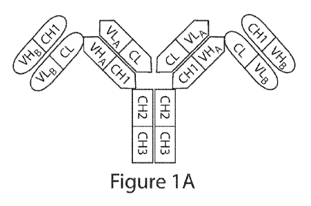
by reference in their entirety: A computer readable format copy of the
Sequence Listing
(filename: EPBI 002 01W0 SeqList ST25.txt, date recorded: February 3, 2017,
file size
510KB).
BACKGROUND OF THE INVENTION
[0004] Bispecific or multispecific antibodies have been generated in attempts
to prepare
molecules useful for the treatment of various inflammatory diseases, cancers,
and other
disorders.
[0005] Bispecific antibodies have been produced using the quadroma technology
(see
Milstein, C. and A.C. Cuello, Nature, 1983. 305(5934): p. 537-40) based on the
somatic
fusion of two different hybridoma cell lines expressing murine monoclonal
antibodies with
the desired specificities of the bispecific antibody. Bispecific antibodies
can also be
produced by chemical conjugation of two different mAbs (see Staerz, U.D., et
al., Nature,
1985. 314(6012): p. 628-31). Other approaches have used chemical conjugation
of two
different monoclonal antibodies or smaller antibody fragments (see Brennan,
M., et al.,
Science, 1985. 229(4708): p. 81-3).
[0006] Another method is the coupling of two parental antibodies with a hetero-
bifunctional
crosslinker. In particular, two different Fab fragments have been chemically
crosslinked at
1
CA 03011746 2018-07-17
WO 2017/136820
PCT/US2017/016691
their hinge cysteine residues in a site-directed manner (see Glennie, M.J., et
al., J Immunol,
1987. 139(7): p. 2367-75).
[0007] Other recombinant bispecific antibody formats have been developed in
the recent past
(see Kriangkum, J., et al., Biomol Eng, 2001. 18(2): p. 31-40). Amongst them
tandem single-
chain Fv molecules and diabodies, and various derivatives thereof, have been
used for the
construction of recombinant bispecific antibodies. Normally, construction of
these molecules
starts from two single-chain Fv (scFv) fragments that recognize different
antigens (see
Economides, A.N., et al., Nat Med, 2003. 9(1): p. 47-52). Tandem scFv
molecules (taFv)
represent a straightforward format simply connecting the two scFv molecules
with an
additional peptide linker. The two scFv fragments present in these tandem scFv
molecules
form separate folding entities. Various linkers can be used to connect the two
scFv fragments
and linkers with a length of up to 63 residues (see Nakanishi, K., et al..
Annu Rev Immunol,
2001. 19: p. 423-74).
[0008] In a recent study, in vivo expression by transgenic rabbits and cattle
of a tandem scFv
directed against CD28 and a melanoma-associated proteoglycan was reported (see
Gracie,
J.A., et al., J Clin Invest, 1999. 104(10): p. 1393-401). In this construct
the two scFv
molecules were connected by a CH1 linker and serum concentrations of up to 100
mg/L of
the bispecific antibody were found. A few studies have now reported expression
of soluble
tandem scFv molecules in bacteria (see Leung, B.P., et al., J Immunol, 2000.
164(12): p.
6495-502; Ito, A., et al., J Immunol, 2003. 170(9): p. 4802-9; Karni, A., et
al., J
Neuroimmunol, 2002. 125(1-2): p. 134-40) using either a very short Ala3 linker
or long
glycine/serine-rich linkers.
[0009] In a recent study, phage display of a tandem scFv repertoire containing
randomized
middle linkers with a length of 3 or 6 residues enriched those molecules which
are produced
in soluble and active form in bacteria. This approach resulted in the
isolation of a preferred
tandem scFv molecule with a 6 amino acid residue linker (see Arndt, M. and J.
Krauss,
Methods Mol Biol, 2003. 207: p. 305-21).
[0010] Bispecific diabodies (Db) utilize the diabody format for expression.
Diabodies are
produced from scFv fragments by reducing the length of the linker connecting
the VH and
VL domain to approximately 5 residues (see Peipp, M. and T. Valerius, Biochem
Soc Trans,
2002. 30(4): p. 507-11). This reduction of linker size facilitates
dimerization of two
polypeptide chains by crossover pairing of the VH and VL domains. Bispecific
diabodies are
produced by expressing two polypeptide chains with either the structure VHA-
VLB and
2
CA 03011746 2018-07-17
WO 2017/136820
PCT/US2017/016691
VHB-VLA (VH-VL configuration) or VLA-VHB and VLB-VHA (VL-VH configuration)
within the same cell. A recent comparative study demonstrates that the
orientation of the
variable domains can influence expression and formation of active binding
sites (see Mack,
M., G. Riethmuller, and P. Kufer, Proc Natl Acad Sci U S A, 1995. 92(15): p.
7021-5).
[0011] One approach to force the generation of bispecific diabodies is the
production of
knob-into-hole diabodies (see Holliger, P., T. Prospero, and G. Winter, Proc
Natl Acad Sci U
S A, 1993. 90(14): p. 6444-8.18). This was demonstrated for a bispecific
diabody directed
against HER2 and CD3. A large knob was introduced in the VH domain by
exchanging
Va137 with Phe and Leu45 with Trp and a complementary hole was produced in the
VL
domain by mutating Phe98 to Met and Tyr87 to Ala, either in the anti- HER2 or
the anti-CD3
variable domains. By using this approach the production of bispecific
diabodies could be
increased from 72% by the parental diabody to over 90% by the knob-into-hole
diabody.
[0012] Single-chain diabodies (scDb) represent an alternative strategy to
improve the
formation of bispecific diabody-like molecules (see Holliger, P. and G.
Winter, Cancer
Immunol Immunother, 1997. 45(3-4): p. 128-30; Wu, A.M., et al.,
Immunotechnology, 1996.
2(1): p. 21-36). Bispecific single-chain diabodies are produced by connecting
the two
diabody-forming polypeptide chains with an additional middle linker with a
length of
approximately 15 amino acid residues. Consequently, all molecules with a
molecular weight
corresponding to monomeric single-chain diabodies (50-60 kDa) are bispecific.
Several
studies have demonstrated that bispecific single chain diabodies are expressed
in bacteria in
soluble and active form with the majority of purified molecules present as
monomers (see
Holliger, P. and G. Winter, Cancer Immunol Immunother, 1997. 45(3-4): p. 128-
30; Wu,
A.M., et al., Immunotechnology, 1996. 2(1): p. 21-36; Pluckthun, A. and P.
Pack,
Immunotechnology, 1997. 3(2): p. 83-105; Ridgway, J.B., et al., Protein Eng,
1996. 9(7): p.
617-21).
[0013] Diabody have been fused to Fc to generate more Ig-like molecules, named
di-diabody
(see Lu, D., et al., J Biol Chem, 2004. 279(4): p. 2856-65). In addition,
multivalent antibody
construct comprising two Fab repeats in the heavy chain of an IgG and capable
of binding
four antigen molecules has been described (see US patent 8,722,859 B2, and
Miller, K., et al.,
J Immunol, 2003. 170(9): p. 4854-61).
[0014] The most recent examples are tetravalent IgG¨single-chain variable
fragment (scFv)
fusions (Dong J, et al. 2011 MAbs 3:273-288; Coloma MJ, Morrison SL 1997 Nat
Biotechnol 15:159-163; Lu D, et al. 2002 J Immunol Methods 267:213-226),
catumaxomab,
3
CA 03011746 2018-07-17
WO 2017/136820
PCT/US2017/016691
a trifunctional rat/mouse hybrid bispecific epithelial cell adhesion molecule-
CD3 antibody
(Lindhofer H, et al 1995 J Immunol 155:219-225), the bispecific CD19-CD3 scFv
antibody
blinatumomab (Bargou R, et al. 2008 Science 321:974-977), "dual- acting Fab"
(DAF)
antibodies (BostromJ, et al. 2009 Science 323:1610-1614), covalently linked
pharmacophore
peptides to catalytic anti- bodies (Doppalapudi VR, et al. 2010 Proc Natl Acad
Sci USA
107:22611-22616), use of the dynamic exchange between half IgG4 molecules to
generate
bispecific antibodies (van der Neut Kolfschoten M, et al. 2007 Science
317:1554-1557;
Stubenrauch K, et al. 2010 Drug Metab Dispos 38:84-91), or by exchange of
heavy-chain
and light-chain domains within the antigen binding fragment (Fab) of one half
of the
bispecific antibody (CrossMab format) (Schaefer Wet al 2011Proc Natl Acad Sci
108:11187-
92).
[0015] There is a need in the art for single molecular entities with dual
antigen binding
function, and for methods of generating such multivalent and multispecific
binding proteins.
The present invention addresses these and other needs.
SUMMARY OF THE INVENTION
[0016] The present invention provides multivalent and multispecific binding
proteins, and
methods of making and using such binding proteins. In one embodiment, the
multivalent and
multispecific binding proteins provided herein are Fabs-in-tandem
immunoglobulins (FIT-
Ig), and are capable of binding two or more antigens, or two or more epitopes
of the same
antigen, or two or more copies of the same epitope. The multivalent and
multispecific binding
proteins provided herein are useful for treatment and/or prevention of acute
and chronic
inflammatory diseases and disorders, autoimmune diseases, cancers, spinal cord
injuries,
sepsis, and other diseases, disorders, and conditions. Pharmaceutical
compositions
comprising the multivalent and multispecific binding proteins are provided
herein. In
addition, nucleic acids, recombinant expression vectors, and host cells for
making such FIT-
Igs are provided herein. Methods of using the FIT-Igs of the invention to
detect specific
antigens, in vivo or in vitro, are also encompassed by the invention.
[0017] The present invention provides a family of binding proteins that are
capable of
binding two or more antigens, e.g., with high affinity. In one aspect, the
present invention
provides an approach to construct a bispecific binding protein using two
parental monoclonal
antibodies: mAb A, which binds to antigen A, and mAb B, which binds to antigen
B. The
4
CA 03011746 2018-07-17
WO 2017/136820
PCT/US2017/016691
binding proteins disclosed herein, in one embodiment, are capable of binding
antigens,
cytokines, chemokines, cytokine receptors, chemokine receptors, cytokine- or
chemokine-
related molecules, or cell surface proteins.
[0018] Thus, in one aspect, binding proteins capable of binding two or more
antigens are
provided. In one embodiment, the present invention provides a binding protein
comprising at
least two polypeptide chains, wherein the polypeptide chains pair to form IgG-
like molecules
capable of binding two or more antigens. In one embodiment, the binding
protein comprises
two, three, four, five, or more polypeptide chains. In one embodiment, the
binding protein
comprises at least one VLA, at least one VLB, at least one VHA, at least one
VHB, at least one
CL, and at least one CH1, wherein VL is a light chain variable domain, VH is a
heavy chain
variable domain, CL is a light chain constant domain, CH1 is the first
constant domain of the
heavy chain, A is a first antigen, and B is a second antigen. In a further
embodiment, the first
polypeptide chain comprises a VLA, a CL, a VHB, and a CH1. In a further
embodiment, the
binding protein further comprises an Fc. In another embodiment, the Fc region
is a variant Fc
region. In a further embodiment, the variant Fc region exhibits modified
effector function,
such as ADCC or CDC. In another embodiment, the variant Fc region exhibits
modified
affinity or avidity for one or more FcyR.
[0019] In one embodiment, the binding protein comprises three polypeptide
chains, wherein
the first polypeptide chain comprises a VLA, a CL, a VHB, and a CH1, the
second polypeptide
chain comprises VHA and CH1, and the third polypeptide chain comprises VLB and
CL. In a
further embodiment, the first polypeptide chain of the binding protein further
comprises an
Fc. In another embodiment, the binding protein comprises two polypeptide
chains, wherein
the first polypeptide chain comprises a VLA, a CL, a VHB, and a CH1, the
second polypeptide
chain comprises VHA, CH1, VLB, and CL. In a further embodiment, the first
polypeptide
chain further comprises an Fc.
[0020] In one embodiment, the binding protein comprises three polypeptide
chains, and their
corresponding cDNA during co-transfection are present at a molar ratio of
first:second:third
of 1:1:1, 1:1.5:1, 1:3:1, 1:1:1.5, 1:1:3, 1:1.5:1.5, 1:3:1.5, 1:1.5:3, or
1:3:3. In another
embodiment, the binding protein comprises two polypeptide chains, and their
corresponding
cDNA during co-transfection are present at a molar ratio of first:second of
1:1, 1:1.5, or 1:3,
or any other ratios, through optimization, in an effort to maximize the
monomeric FIT-Ig
fraction in any given transfection.
CA 03011746 2018-07-17
WO 2017/136820
PCT/US2017/016691
[0021] In one embodiment, the binding protein of the present invention does
not comprise a
peptide linker. In one embodiment, the binding protein of the present
invention comprises at
least one amino acid or polypeptide linker. In a further embodiment, the
linker is selected
from the group consisting of G, GS, SG, GGS, GSG, SGG, GGG, GGGS (SEQ ID NO:
489),
SGGG (SEQ ID NO: 490), GGGGS (SEQ ID NO: 491), GGGGSGS (SEQ ID NO: 492)õ
GGGGSGGS (SEQ ID NO: 493), GGGGSGGGGS (SEQ ID NO: 494),
GGGGSGGGGSGGGGS (SEQ ID NO: 495), AKTTPKLEEGEFSEAR (SEQ ID NO: 496),
AKTTPKLEEGEFSEARV (SEQ ID NO: 497), AKTTPKLGG (SEQ ID NO: 498),
SAKTTPKLGG (SEQ ID NO: 499), SAKTTP (SEQ ID NO: 500), RADAAP (SEQ ID NO:
501), RADAAPTVS (SEQ ID NO: 502), RADAAAAGGPGS (SEQ ID NO: 503),
RADAAAA(G45)4 (SEQ ID NO: 504), SAKTTPKLEEGEFSEARV (SEQ ID NO: 505),
ADAAP (SEQ ID NO: 506), ADAAPTVSIFPP (SEQ ID NO: 507), TVAAP (SEQ ID NO:
508), TVAAPSVFIFPP (SEQ ID NO: 509), QPKAAP (SEQ ID NO: 510),
QPKAAPSVTLFPP (SEQ ID NO: 511), AKTTPP (SEQ ID NO: 512), AKTTPPSVTPLAP
(SEQ ID NO: 513), AKTTAPSVYPLAP (SEQ ID NO: 514), ASTKGP (SEQ ID NO: 515),
ASTKGPSVFPLAP (SEQ ID NO: 516), GENKVEYAPALMALS (SEQ ID NO: 517),
GPAKELTPLKEAKVS (SEQ ID NO: 518), GHEAAAVMQVQYPAS (SEQ ID NO: 519),
and AKTTAP (SEQ ID NO: 80). The linkers can also be in vivo cleavable peptide
linkers,
protease (such as MMPs) sensitive linkers, disulfide bond-based linkers that
can be cleaved
by reduction, etc., as previously described (Fusion Protein Technologies for
Biopharmaceuticals: Applications and Challenges, edited by Stefan R. Schmidt),
or any
cleavable linkers known in the art. Such cleavable linkers can be used to
release the top Fab
in vivo for various purposes, in order to improve tissue/cell penetration and
distribution, to
enhance binding to targets, to reduce potential side effect, as well as to
modulate in vivo
functional and physical half-life of the 2 different Fab regions.
[0022] In one embodiment, the binding protein comprises a first polypeptide
comprising,
from amino to carboxyl terminus, VLA-CL-VHB-CH1-Fc, a second polypeptide chain
comprising, from amino to carboxyl terminus, VHA-CH1, and a third polypeptide
chain
comprising, from amino to carboxyl terminus, VLB-CL; wherein VL is a light
chain variable
domain, CL is a light chain constant domain, VH is a heavy chain variable
domain, CH1 is
the first constant domain of the heavy chain, A is a first epitope or antigen,
and B is a second
epitope or antigen. In one embodiment, the Fc region is human IgGl. In another
embodiment,
the Fc region is a variant Fc region. In a further embodiment, the amino acid
sequence of the
6
CA 03011746 2018-07-17
WO 2017/136820
PCT/US2017/016691
Fc region is at least 65%, at least 70%, at least 75%, at least 80%, at least
85%, at least 90%,
at least 95%, at least 99%, or 100% identical to SEQ ID NO: 20. In a further
embodiment, the
CL of the first polypeptide chain is fused directly to VHB. In another
embodiment, the CL of
the first polypeptide chain is linked to VHB via an amino acid or an
oligopeptide linker. In a
further embodiment, the linker is GSG (SEQ ID NO: 26) or GGGGSGS (SEQ ID NO:
28).
[0023] In another embodiment, the binding protein comprises a first
polypeptide comprising,
from amino to carboxyl terminus, VHB-CH1-VLA-CL-Fc, a second polypeptide chain
comprising, from amino to carboxyl terminus, VHA-CH1, and a third polypeptide
chain
comprising, from amino to carboxyl terminus, VLB-CL; wherein VL is a light
chain variable
domain, CL is a light chain constant domain, VH is a heavy chain variable
domain, CH1 is
the first constant domain of the heavy chain, A is a first epitope or antigen,
and B is a second
epitope or antigen. In one embodiment, the Fc region is human IgGl. In another
embodiment,
the Fc region is a variant Fc region. In a further embodiment, the amino acid
sequence of the
Fc region is at least 65%, at least 70%, at least 75%, at least 80%, at least
85%, at least 90%,
at least 95%, at least 99%, or 100% identical to SEQ ID NO: 20. In one
embodiment, the
CH1 of the first polypeptide chain is fused directly to VLA. In another
embodiment, the CH1
of the first polypeptide chain is linked to VLA via an amino acid or an
oligopeptide linker. In
a further embodiment, the linker is GSG (SEQ ID NO: 26) or GGGGSGS (SEQ ID NO:
28).
[0024] In another embodiment, the binding protein comprises a first
polypeptide comprising,
from amino to carboxyl terminus, VLA-CL-VHB-CH1-Fc, and a second polypeptide
chain
comprising, from amino to carboxyl terminus, VHA-CH1-VLB-CL; wherein VL is a
light
chain variable domain, CL is a light chain constant domain, VH is a heavy
chain variable
domain, CH1 is the first constant domain of the heavy chain, A is a first
epitope or antigen,
and B is a second epitope or antigen. In one embodiment, the Fc region is
human IgGl. In
another embodiment, the Fc region is a variant Fc region. In a further
embodiment, the amino
acid sequence of the Fc region is at least 65%, at least 70%, at least 75%, at
least 80%, at
least 85%, at least 90%, at least 95%, at least 99%, or 100% identical to SEQ
ID NO: 20. In a
further embodiment, the CL of the first polypeptide chain is fused directly to
VHB. In another
embodiment, the CL of the first polypeptide chain is linked to VHB via an
amino acid or an
oligopeptide linker. In a further embodiment, the linker is GSG (SEQ ID NO:
26) or
GGGGSGS (SEQ ID NO: 28).
[0025] In another embodiment, binding protein comprises a first polypeptide
comprising,
from amino to carboxyl terminus, VHB-CH1-VLA-CL-Fc, and a second polypeptide
chain
7
CA 03011746 2018-07-17
WO 2017/136820
PCT/US2017/016691
comprising, from amino to carboxyl terminus, VLB-CL-VHA-CH1; wherein VL is a
light
chain variable domain, CL is a light chain constant domain, VH is a heavy
chain variable
domain, CH1 is the first constant domain of the heavy chain, A is a first
epitope or antigen,
and B is a second epitope or antigen. In one embodiment, the Fc region is
human IgGl. In
another embodiment, the Fc region is a variant Fc region. In a further
embodiment, the amino
acid sequence of the Fc region is at least 65%, at least 70%, at least 75%, at
least 80%, at
least 85%, at least 90%, at least 95%, at least 99%, or 100% identical to SEQ
ID NO: 20. In
one embodiment, the CH1 of the first polypeptide chain is fused directly to
VLA. In another
embodiment, the CH1 of the first polypeptide chain is linked to VLA via an
amino acid or an
oligopeptide linker. In a further embodiment, the linker is GSG (SEQ ID NO:
26) or
GGGGSGS (SEQ ID NO: 28).
[0026] The binding proteins of the present invention are capable of binding
pairs of
cytokines. For example, the binding proteins of the present invention are
capable of binding
pairs of cytokines selected from the group consisting of IL-la and IL-1[3; IL-
12 and IL-18,
TNFa and IL-23, TNFa and IL-13; TNF and IL-18; TNF and IL-12; TNF and IL-
lbeta; TNF
and MIF; TNF and IL-6, TNF and IL-6 Receptor, TNF and IL-17; IL-17 and IL-20;
IL-17
and IL-23; TNF and IL-15; TNF and VEGF; VEGFR and EGFR; PDGFR and VEGF, IL-13
and IL-9; IL-13 and IL-4; IL-13 and IL-5; IL-13 and IL-25; IL-13 and TARC; IL-
13 and
MDC; IL-13 and MIF; IL-13 and TGF-P; IL-13 and LHR agonist; IL-13 and CL25; IL-
13
and SPRR2a; IL-13 and SPRR2b; IL-13 and ADAM8; and TNFa and PGE4, IL-13 and
PED2, TNF and PEG2. In one embodiment, the binding proteins of the present
invention are
capable of binding IL-17 and IL-20. The binding proteins of the present
invention, in one
embodiment, are capable of binding IL-17 and IL-20 and comprise variable heavy
and light
chains derived from the anti-IL-17 antibody LY and the anti-IL-20 antibody
15D2. In one
embodiment, the binding proteins of the present invention are capable of
binding IL-17 and
TNF. The binding proteins of the present invention, in one embodiment, are
capable of
binding IL-17 and TNF and comprise variable heavy and light chains derived
from the anti-
IL-17 antibody LY and the TNF antibody golimumab.
[0027] In one embodiment, the binding proteins of the present invention bind
IL-17 and IL-
20 and comprise a first polypeptide comprising, consisting essentially of, or
consisting of an
amino acid sequence selected from the group consisting of SEQ ID NOs: 15, 25,
and 27; a
second polypeptide chain comprising, consisting essentially of, or consisting
of an amino
8
CA 03011746 2018-07-17
WO 2017/136820
PCT/US2017/016691
acid sequence according to SEQ ID NO: 21; and a third polypeptide chain
comprising,
consisting essentially of, or consisting of a sequence according to SEQ ID NO:
23. In another
embodiment, the binding proteins of the present invention bind IL-27 and IL-20
and comprise
a first polypeptide chain comprising, consisting essentially of, or consisting
of an amino acid
sequence selected from the group consisting of SEQ ID NOs: 15, 25, and 27, and
a second
polypeptide chain comprising, consisting essentially of, or consisting of an
amino acid
sequence selected from the group consisting of SEQ ID NOs: 29, 30, and 31.
[0028] In one embodiment, the binding proteins of the present invention bind
TNF and IL-17
and comprise a first polypeptide comprising, consisting essentially of, or
consisting of an
amino acid sequence according to SEQ ID NOs: 87; a second polypeptide chain
comprising,
consisting essentially of, or consisting of an amino acid sequence according
to SEQ ID NO:
89; and a third polypeptide chain comprising, consisting essentially of, or
consisting of a
sequence according to SEQ ID NO: 91. In another embodiment, the binding
protein is
capable of binding pairs of targets selected from the group consisting of
CD137 and CD20,
CD137 and EGFR, CD137 and Her-2, CD137 and PD-1, CD137 and PDL-1, VEGF and PD-
L1, Lag-3 and TIM-3, 0X40 and PD-1, TIM-3 and PD-1, TIM-3 and PDL-1, EGFR and
DLL-4, CD138 and CD20; CD138 and CD40; CD19 and CD20; CD20 and CD3; CD3 and
CD33; CD3 and CD133; CD47 and CD20, CD38 and CD138; CD38 and CD20; CD20 and
CD22; CD38 and CD40; CD40 and CD20; CD-8 and IL-6; CSPGs and RGM A; CTLA-4
and BTN02; IGF1 and IGF2; IGF1/2 and Erb2B; IGF-1R and EGFR; EGFR and CD13;
IGF-
1R and ErbB3; EGFR-2 and IGFR; VEGFR-2 and Met; VEGF-A and Angiopoietin-2 (Ang-
2); IL-12 and TWEAK; IL-13 and IL-lbeta; PDGFR and VEGFõ EpCAM and CD3, Her2
and CD3, CD19 and CD3, EGFR and Her3, CD16a and CD30, CD30 and PSMA, EGFR and
CD3, CEA and CD3, TROP-2 and HSG, TROP-2 and CD3, MAG and RGM A; NgR and
RGM A; NogoA and RGM A; OMGp and RGM A; PDL-1 and CTLA-4; CTLA-4 and PD-1;
PD-1 and TIM-3; RGM A and RGM B; Te38 and TNFa; TNFa and Blys; TNFa and CD-22;
TNFa and CTLA-4 domain; TNFa and GP130; TNFa and IL-12p40; and TNFa and
RANK ligand, Factor IXa and Factor X; EGFR and PD-Li; EGFR and cMet; Her3 and
IGF-
IR; DLL-4 and VEGF; PD-1 and PD-Li; and Her3 and PD-1.
[0029] In one embodiment, the binding proteins of the present invention are
capable of
binding CD3 and CD20. The binding proteins of the present invention, in one
embodiment,
are capable of binding CD3 and CD20 and comprise variable heavy and light
chains derived
from the anti-CD3 antibody OKT3 or the anti-CD3 antibody disclosed in U.S.
2009/0252683,
9
CA 03011746 2018-07-17
WO 2017/136820
PCT/US2017/016691
which is incorporated herein by reference in it entirety; and the anti-CD20
antibody
ofatumumab. In some embodiments, the polypeptide derived from CD3 antibody is
in the
upper domain and the polypeptide derived from CD20 antibody is in the lower
domain. As
used herein, the upper domain is the N-terminal or "amino proximal" domain,
and the lower
domain is the C-terminal domain or the domain closer to the Fc, if present.
For example, in
some embodiments, the binding proteins comprise a first polypeptide of VLA-CL-
VHB-CH1-
Fc, a second polypeptide of VHA-CH1, and a third polypeptide of VLB-CL,
wherein antigen
A is CD3, and antigen B is CD20. For another example, in some embodiments, the
binding
proteins comprise a first polypeptide of VHB-CH1-VLA-CL-Fc, a second
polypeptide of VLB-
CL, and a third polypeptide of VHA-CH1, wherein antigen B is CD3, and antigen
A is CD20.
In some embodiments, polypeptide derived from CD3 antibody is in the lower
domain and
polypeptide derived from CD20 antibody is in the upper domain. For example, in
some
embodiments, the binding proteins comprise a first polypeptide of VLA-CL-VHB-
CH1-Fc, a
second polypeptide of VHA-CH1, and a third polypeptide of VLB-CL, wherein
antigen A is
CD20, and antigen B is CD-3. For another example, in some embodiments, the
binding
proteins comprise a first polypeptide of VHB-CH1-VLA-CL-Fc, a second
polypeptide of VLB-
CL, and a third polypeptide of VHA-CH1, wherein antigen B is CD20, and antigen
A is CD3.
[0030] In one embodiment, the binding proteins of the present invention bind
CD3 and CD20
and comprise a first polypeptide chain comprising, consisting essentially of,
or consisting of
an amino acid sequence selected from the group consisting of SEQ ID NOs: 41
and 48; a
second polypeptide chain comprising, consisting essentially of, or consisting
of an amino acid
sequence according to SEQ ID NO: 44; and a third polypeptide chain comprising,
consisting
essentially of, or consisting of an amino acid sequence according to SEQ ID
NO: 46. In
another embodiment, the binding proteins of the present invention bind CD20
and CD3 and
comprise a first polypeptide chain comprising, consisting essentially of, or
consisting of an
amino acid sequence according to SEQ ID NO: 114; a second polypeptide chain
comprising,
consisting essentially of, or consisting of an amino acid sequence according
to SEQ ID NO:
115; and a third polypeptide chain comprising, consisting essentially of, or
consisting of an
amino acid sequence according to SEQ ID NO: 116.
[0031] In one embodiment, the binding protein of the present invention is
capable of binding
the same epitope of CD20 and the same epitope of CD3 as that of bispecific
binding protein
FIT018a, wherein the bispecific binding protein FIT018a comprises a first
polypeptide chain
comprising an amino acid sequence of SEQ ID NO: 316; a second polypeptide
chain
CA 03011746 2018-07-17
WO 2017/136820
PCT/US2017/016691
comprising an amino acid sequence of SEQ ID NO: 325; and a third polypeptide
chain
comprising an amino acid sequence of SEQ ID NO: 330.
[0032] In one embodiment, the binding proteins of the present invention bind
CD3 and CD20
and comprise a VLA on the first polypeptide, wherein the VLA of the first
polypeptide
comprises a VLA CDR1 of SEQ ID NO: 318, a VLA CDR2 of SEQ ID NO: 319, and a
VLA
CDR3 of SEQ ID NO: 320.
[0033] In one embodiment, the binding proteins of the present invention bind
CD3 and CD20
and comprise a VHB on the first polypeptide, wherein the VHB of the first
polypeptide
comprises a VHB CDR1 of SEQ ID NO: 322, a VHB CDR2 of SEQ ID NO: 323, and a
VHB
CDR3 of SEQ ID NO: 324.
[0034] In one embodiment, the binding proteins of the present invention bind
CD3 and CD20
and comprise a VHA on the second polypeptide, wherein the VHA of the second
polypeptide
comprises a VHA CDR1 of SEQ ID NO: 327, a VHA CDR2 of SEQ ID NO: 328, and a
VHA
CDR3 of SEQ ID NO: 329.
[0035] In one embodiment, the binding proteins of the present invention bind
CD3 and CD20
and comprise a VLB on the third polypeptide, wherein the VLB of the third
polypeptide
comprises a VLB CDR1 of SEQ ID NO: 332, a VLB CDR2 of SEQ ID NO: 333, and a
VLB
CDR3 of SEQ ID NO: 334.
[0036] In one embodiment, the binding proteins of the present invention bind
CD3 and CD20
and comprise a VLA and VHB on the first polypeptide, a VHA on the second
polypeptide, and
a VLB on the third polypeptide, wherein the VLA of the first polypeptide
comprises a VLA
CDR1 of SEQ ID NO: 318, a VLA CDR2 of SEQ ID NO: 319, and a VLA CDR3 of SEQ ID
NO: 320; wherein the VHB of the first polypeptide comprises a VHB CDR1 of SEQ
ID NO:
322, a VHB CDR2 of SEQ ID NO: 323, and a VHB CDR3 of SEQ ID NO: 324, wherein
the
VHA of the second polypeptide comprises a VHA CDR1 of SEQ ID NO: 327, a VHA
CDR2
of SEQ ID NO: 328, and a VHA CDR3 of SEQ ID NO: 329; and wherein the VLB of
the
third polypeptide comprises a VLB CDR1 of SEQ ID NO: 332, a VLB CDR2 of SEQ ID
NO:
333, and a VLB CDR3 of SEQ ID NO: 334.
[0037] In one embodiment, the binding proteins of the present invention bind
CD3 and CD20
and comprise a first polypeptide chain comprising a VLA having the sequence of
SEQ ID
NO: 317, and a VHB having the sequence of SEQ ID NO: 321, wherein the binding
protein
comprises a second polypeptide chain comprising a VHA having the sequence of
SEQ ID
11
CA 03011746 2018-07-17
WO 2017/136820
PCT/US2017/016691
NO: 326, and wherein the binding protein comprises a third polypeptide chain
comprising a
VLB having the sequence of SEQ ID NO: 331.
[0038] In one embodiment, the binding proteins of the present invention bind
CD3 and CD20
and comprising, consisting essentially of, or consisting of a first
polypeptide chain
comprising an amino acid sequence of SEQ ID NO: 316; a second polypeptide
chain
comprising an amino acid sequence of SEQ ID NO: 325; and a third polypeptide
chain
comprising an amino acid sequence of SEQ ID NO: 330.
[0039] In one embodiment, the binding proteins of the present invention bind
CD3 and
CD20, and are derived from binding proteins described herein by replacing 1,
2, 3, 4, 5, 6, 7,
8, 9, 10, 20, 30, 40, 50, 60, 70, 80, 90, 100, 110, 120, 130, 140, 150, 160,
170, 180, 190, 200,
or more (inclusive of all values therebetween) amino acids with conservative
amino acid
substitution, while still maintaining equivalent activity as the corresponding
binding proteins
without the substitution(s).
[0040] In one embodiment, the binding proteins of the present invention are
capable of
binding CTLA-4 and PD-1. The binding proteins of the present invention, in one
embodiment, are capable of binding CTLA-4 and PD-1 and comprise variable heavy
and
light chains derived from the CTLA-4 antibody ipilimumab and the PD-1 antibody
nivolumab.
[0041] In one embodiment, the binding proteins of the present invention bind
CTLA-4 and
PD-1 and comprise a first polypeptide chain comprising, consisting essentially
of, or
consisting of an amino acid sequence according to SEQ ID NO: 92; a second
polypeptide
chain comprising, consisting essentially of, or consisting of an amino acid
sequence
according to SEQ ID NO: 95; and a third polypeptide chain comprising,
consisting essentially
of, or consisting of an amino acid sequence according to SEQ ID NO: 97. In one
embodiment, the binding protein provided herein is capable of binding one or
more epitopes
on CTLA-4. In one embodiment, the binding protein provided herein is capable
of binding
one or more epitopes on PD-1. In some embodiments, polypeptide derived from
CTLA-4
antibody is in the upper domain and polypeptide derived from PD-1 antibody is
in the lower
domain. For example, in some embodiments, the binding proteins comprise a
first
polypeptide of VLA-CL-VHB-CH1-Fc, a second polypeptide of VHA-CH1, and a third
polypeptide of VLB-CL, wherein antigen A is CTLA-4, and antigen B is PD-1. For
another
example, in some embodiments, the binding proteins comprise a first
polypeptide of VHB-
CH1-VLA-CL-Fc, a second polypeptide of VLB-CL, and a third polypeptide of VHA-
CH1,
12
CA 03011746 2018-07-17
WO 2017/136820
PCT/US2017/016691
wherein antigen B is CTLA-4, and antigen A is PD-1. In some embodiments,
polypeptide
derived from CTLA-4 antibody is in the lower domain and polypeptide derived
from PD-1
antibody is in the upper domain. For example, in some embodiments, the binding
proteins
comprise a first polypeptide of VLA-CL-VHB-CH1-Fc, a second polypeptide of VHA-
CH1,
and a third polypeptide of VLB-CL, wherein antigen A is PD-1, and antigen B is
CTLA-4.
For another example, in some embodiments, the binding proteins comprise a
first polypeptide
of VHB-CH1-VLA-CL-Fc, a second polypeptide of VLB-CL, and a third polypeptide
of VHA-
CH1, wherein antigen B is PD-1, and antigen A is CTLA-4.
[0042] In one embodiment, the binding protein of the present invention is
capable of binding
the same epitope of CTLA-4 and the same epitope of PD-1 as that of bispecific
binding
proteins NBS3, NBS3R, NBS3-C, or NBS3R-C, as described herein.
[0043] The bispecific binding protein NBS3 comprises a first polypeptide chain
comprising
an amino acid sequence of SEQ ID NO: 126; a second polypeptide chain
comprising an
amino acid sequence of SEQ ID NO: 135; and a third polypeptide chain
comprising an amino
acid sequence of SEQ ID NO: 140.
[0044] The bispecific binding protein NBS3R comprises a first polypeptide
chain comprising
an amino acid sequence of SEQ ID NO: 145; a second polypeptide chain
comprising an
amino acid sequence of SEQ ID NO: 154; and a third polypeptide chain
comprising an amino
acid sequence of SEQ ID NO: 159.
[0045] The bispecific binding protein NBS3-C comprises a first polypeptide
chain
comprising an amino acid sequence of SEQ ID NO: 164; a second polypeptide
chain
comprising an amino acid sequence of SEQ ID NO: 173; and a third polypeptide
chain
comprising an amino acid sequence of SEQ ID NO: 178.
[0046] The bispecific binding protein NBS3R-C comprises a first polypeptide
chain
comprising an amino acid sequence of SEQ ID NO: 183; a second polypeptide
chain
comprising an amino acid sequence of SEQ ID NO: 192; and a third polypeptide
chain
comprising an amino acid sequence of SEQ ID NO: 197.
[0047] In one embodiment, the binding proteins of the present invention bind
CTLA4 and
PD-1 and comprise a VLA on the first polypeptide, wherein the VLA of the first
polypeptide
comprises a VLA CDR1 of SEQ ID NO: 128, a VLA CDR2 of SEQ ID NO: 129, and a
VLA
CDR3 of SEQ ID NO: 130 (e.g., those on NBS3). In one embodiment, the binding
proteins
of the present invention bind CTLA4 and PD-1 and comprises a VLA on the first
polypeptide,
wherein the VLA of the first polypeptide comprises a VLA CDR1 of SEQ ID NO:
147, a VLA
13
CA 03011746 2018-07-17
WO 2017/136820
PCT/US2017/016691
CDR2 of SEQ ID NO: 148, and a VLA CDR3 of SEQ ID NO: 149 (e.g., those on
NBS3R).
In one embodiment, the binding proteins of the present invention bind CTLA4
and PD-1 and
comprises a VLA on the first polypeptide, wherein the VLA of the first
polypeptide comprises
a VLA CDR1 of SEQ ID NO: 166, a VLA CDR2 of SEQ ID NO: 167, and a VLA CDR3 of
SEQ ID NO: 168 (e.g., those on NBS3-C). In one embodiment, the binding
proteins of the
present invention bind CTLA4 and PD-1 and comprises a VLA on the first
polypeptide,
wherein the VLA of the first polypeptide comprises a VLA CDR1 of SEQ ID NO:
185, a VLA
CDR2 of SEQ ID NO: 186, and a VLA CDR3 of SEQ ID NO: 187 (e.g., those on NBS3R-
C).
[0048] In one embodiment, the binding proteins of the present invention bind
CTLA4 and
PD-1 and comprise a VHB on the first polypeptide, wherein the VHB of the first
polypeptide
comprises a VHB CDR1 of SEQ ID NO: 132, a VHB CDR2 of SEQ ID NO: 133, and a
VHB
CDR3 of SEQ ID NO: 134 (e.g., those on NBS3). In one embodiment, the binding
proteins
of the present invention bind CTLA4 and PD-1 and comprise a VHB on the first
polypeptide,
wherein the VHB of the first polypeptide comprises a VHB CDR1 of SEQ ID NO:
151, a VHB
CDR2 of SEQ ID NO: 152, and a VHB CDR3 of SEQ ID NO: 153 (e.g., those on
NBS3R). In
one embodiment, the binding proteins of the present invention bind CTLA4 and
PD-1 and
comprise a VHB on the first polypeptide, wherein the VHB of the first
polypeptide comprises
a VHB CDR1 of SEQ ID NO: 166, a VHB CDR2 of SEQ ID NO: 167, and a VHB CDR3 of
SEQ ID NO: 168 (e.g., those on NBS3-C). In one embodiment, the binding
proteins of the
present invention bind CTLA4 and PD-1 and comprise a VHB on the first
polypeptide,
wherein the VHB of the first polypeptide comprise a VHB CDR1 of SEQ ID NO:
185, a VHB
CDR2 of SEQ ID NO: 186, and a VHB CDR3 of SEQ ID NO: 187 (e.g., those on NBS3R-
C).
[0049] In one embodiment, the binding proteins of the present invention bind
CTLA4 and
PD-1 and comprise a VHA on the second polypeptide, wherein the VHA of the
second
polypeptide comprises a VHA CDR1 of SEQ ID NO: 137, a VHA CDR2 of SEQ ID NO:
138,
and a VHA CDR3 of SEQ ID NO: 139 (e.g., those on NBS3). In one embodiment, the
binding proteins of the present invention bind CTLA4 and PD-1 and comprises a
VHA on the
second polypeptide, wherein the VHA of the second polypeptide comprises a VHA
CDR1 of
SEQ ID NO: 156, a VHA CDR2 of SEQ ID NO: 157, and a VHA CDR3 of SEQ ID NO: 158
(e.g., those on NBS3R). In one embodiment, the binding proteins of the present
invention
bind CTLA4 and PD-1 and comprises a VHA on the second polypeptide, wherein the
VHA of
the second polypeptide comprises a VHA CDR1 of SEQ ID NO: 175, a VHA CDR2 of
SEQ
ID NO: 176, and a VHA CDR3 of SEQ ID NO: 177 (e.g., those on NBS-C). In one
14
CA 03011746 2018-07-17
WO 2017/136820
PCT/US2017/016691
embodiment, the binding proteins of the present invention bind CTLA4 and PD-1
and
comprises a VHA on the second polypeptide, wherein the VHA of the second
polypeptide
comprises a VHA CDR1 of SEQ ID NO: 194, a VHA CDR2 of SEQ ID NO: 195, and a
VHA
CDR3 of SEQ ID NO: 196 (e.g., those on NBS3R-C).
[0050] In one embodiment, the binding proteins of the present invention bind
CTLA4 and
PD-1 and comprise a VLB on the third polypeptide, wherein the VLB of the third
polypeptide
comprises a VLB CDR1 of SEQ ID NO: 142, a VLB CDR2 of SEQ ID NO: 143, and a
VLB
CDR3 of SEQ ID NO: 144 (e.g., those on NB S3). In one embodiment, the binding
proteins of
the present invention bind CTLA4 and PD-1 and comprises a VLB on the third
polypeptide,
wherein the VLB of the third polypeptide comprises a VLB CDR1 of SEQ ID NO:
161, a VLB
CDR2 of SEQ ID NO: 162, and a VLB CDR3 of SEQ ID NO: 163 (e.g., those on
NBS3R).
In one embodiment, the binding proteins of the present invention bind CTLA4
and PD-1 and
comprises a VLB on the third polypeptide, wherein the VLB of the third
polypeptide
comprises a VLB CDR1 of SEQ ID NO: 180, a VLB CDR2 of SEQ ID NO: 181, and a
VLB
CDR3 of SEQ ID NO: 182 (e.g., those on NBS3-C). In one embodiment, the binding
proteins
of the present invention bind CTLA4 and PD-1 and comprises a VLB on the third
polypeptide, wherein the VLB of the third polypeptide comprises a VLB CDR1 of
SEQ ID
NO: 199, a VLB CDR2 of SEQ ID NO: 200, and a VLB CDR3 of SEQ ID NO: 201 (e.g.,
those on NBS3R-C).
[0051] In one embodiment, the binding proteins of the present invention bind
CTLA4 and
PD-1 and comprise a VLA and VHB on the first polypeptide, a VHA on the second
polypeptide, and a VLB on the third polypeptide, wherein the VLA of the first
polypeptide
comprises a VLA CDR1 of SEQ ID NO: 128, a VLA CDR2 of SEQ ID NO: 129, and a
VLA
CDR3 of SEQ ID NO: 130; wherein the VHB of the first polypeptide comprises a
VHB
CDR1 of SEQ ID NO: 132, a VHB CDR2 of SEQ ID NO: 133, and a VHB CDR3 of SEQ ID
NO: 134; wherein the VHA of the second polypeptide comprises a VHA CDR1 of SEQ
ID
NO: 137, a VHA CDR2 of SEQ ID NO: 138, and a VHA CDR3 of SEQ ID NO: 139; and
wherein the VLB of the third polypeptide comprises a VLB CDR1 of SEQ ID NO:
142, a VLB
CDR2 of SEQ ID NO: 143, and a VLB CDR3 of SEQ ID NO: 144 (e.g., those on
NBS3).
[0052] In one embodiment, the binding proteins of the present invention bind
CTLA4 and
PD-1 and comprise a VLA and VHB on the first polypeptide, a VHA on the second
polypeptide, and a VLB on the third polypeptide, wherein the VLA of the first
polypeptide
comprises a VLA CDR1 of SEQ ID NO: 147, a VLA CDR2 of SEQ ID NO: 148, and a
VLA
CA 03011746 2018-07-17
WO 2017/136820
PCT/US2017/016691
CDR3 of SEQ ID NO: 149; wherein the VHB of the first polypeptide comprises a
VHB CDR1
of SEQ ID NO: 151, a VHB CDR2 of SEQ ID NO: 152, and a VHB CDR3 of SEQ ID NO:
153; wherein the VHA of the second polypeptide comprises a VHA CDR1 of SEQ ID
NO:
156, a VHA CDR2 of SEQ ID NO: 157, and a VHA CDR3 of SEQ ID NO: 158; and
wherein
the VLB of the third polypeptide comprises a VLB CDR1 of SEQ ID NO: 161, a VLB
CDR2
of SEQ ID NO: 162, and a VLB CDR3 of SEQ ID NO: 163 (e.g., those on NBS3R).
[0053] In one embodiment, the binding proteins of the present invention bind
CTLA4 and
PD-1 and comprise a VLA and VHB on the first polypeptide, a VHA on the second
polypeptide, and a VLB on the third polypeptide, wherein the VLA of the first
polypeptide
comprises a VLA CDR1 of SEQ ID NO: 166, a VLA CDR2 of SEQ ID NO: 167, and a
VLA
CDR3 of SEQ ID NO: 168; wherein the VHB of the first polypeptide comprises a
VHB CDR1
of SEQ ID NO: 170, a VHB CDR2 of SEQ ID NO: 171, and a VHB CDR3 of SEQ ID NO:
172; wherein the VHA of the second polypeptide comprises a VHA CDR1 of SEQ ID
NO:
175, a VHA CDR2 of SEQ ID NO: 176, and a VHA CDR3 of SEQ ID NO: 177; and
wherein
the VLB of the third polypeptide comprises a VLB CDR1 of SEQ ID NO: 180, a VLB
CDR2
of SEQ ID NO: 181, and a VLB CDR3 of SEQ ID NO: 182 (e.g., those on NBS3-C).
[0054] In one embodiment, the binding proteins of the present invention bind
CTLA4 and
PD-1 and comprise a VLA and VHB on the first polypeptide, a VHA on the second
polypeptide, and a VLB on the third polypeptide, wherein the VLA of the first
polypeptide
comprises a VLA CDR1 of SEQ ID NO: 166, a VLA CDR2 of SEQ ID NO: 167, and a
VLA
CDR3 of SEQ ID NO: 168; wherein the VHB of the first polypeptide comprises a
VHB CDR1
of SEQ ID NO: 170, a VHB CDR2 of SEQ ID NO: 171, and a VHB CDR3 of SEQ ID NO:
172; wherein the VHA of the second polypeptide comprises a VHA CDR1 of SEQ ID
NO:
175, a VHA CDR2 of SEQ ID NO: 176, and a VHA CDR3 of SEQ ID NO: 177; and
wherein
the VLB of the third polypeptide comprises a VLB CDR1 of SEQ ID NO: 180, a VLB
CDR2
of SEQ ID NO: 181, and a VLB CDR3 of SEQ ID NO: 182 (e.g., those on NBS3R-C).
[0055] In one embodiment, the binding proteins of the present invention bind
CTLA4 and
PD-1 and comprise a first polypeptide chain comprising a VLA having the
sequence of SEQ
ID NO: 127, and a VHB having the sequence of SEQ ID NO: 131, wherein the
binding
protein comprises a second polypeptide chain comprising a VHA having the
sequence of SEQ
ID NO: 136, and wherein the binding protein comprises a third polypeptide
chain comprising
a VLB having the sequence of SEQ ID NO: 141 (e.g., those on NBS3).
16
CA 03011746 2018-07-17
WO 2017/136820
PCT/US2017/016691
[0056] In one embodiment, the binding proteins of the present invention bind
CTLA4 and
PD-1 and comprise a first polypeptide chain comprising a VLA having the
sequence of SEQ
ID NO: 146, and a VHB having the sequence of SEQ ID NO: 150, wherein the
binding
protein comprises a second polypeptide chain comprising a VHA having the
sequence of SEQ
ID NO: 155, and wherein the binding protein comprises a third polypeptide
chain comprising
a VLB having the sequence of SEQ ID NO: 160 (e.g., those on NBS3R).
[0057] In one embodiment, the binding proteins of the present invention bind
CTLA4 and
PD-1 and comprise a first polypeptide chain comprising a VLA having the
sequence of SEQ
ID NO: 165, and a VHB having the sequence of SEQ ID NO: 169, wherein the
binding
protein comprises a second polypeptide chain comprising a VHA having the
sequence of SEQ
ID NO: 174, and wherein the binding protein comprises a third polypeptide
chain comprising
a VLB having the sequence of SEQ ID NO: 179 (e.g., those on NBS3-C).
[0058] In one embodiment, the binding proteins of the present invention bind
CTLA4 and
PD-1 and comprise a first polypeptide chain comprising a VLA having the
sequence of SEQ
ID NO: 184, and a VHB having the sequence of SEQ ID NO: 188, wherein the
binding
protein comprises a second polypeptide chain comprising a VHA having the
sequence of SEQ
ID NO: 193, and wherein the binding protein comprises a third polypeptide
chain comprising
a VLB having the sequence of SEQ ID NO: 198 (e.g., those on NBS3R-C).
[0059] In one embodiment, the binding proteins of the present invention bind
CTLA4 and
PD-1 and comprising, consisting essentially of, or consisting of a first
polypeptide chain
comprising an amino acid sequence of SEQ ID NO: 126; a second polypeptide
chain
comprising an amino acid sequence of SEQ ID NO: 135; and a third polypeptide
chain
comprising an amino acid sequence of SEQ ID NO: 140 (e.g., those on NBS3).
[0060] In one embodiment, the binding proteins of the present invention bind
CTLA4 and
PD-1 and comprising, consisting essentially of, or consisting of a first
polypeptide chain
comprising an amino acid sequence of SEQ ID NO: 145; a second polypeptide
chain
comprising an amino acid sequence of SEQ ID NO: 154; and a third polypeptide
chain
comprising an amino acid sequence of SEQ ID NO: 159 (e.g., those on NBS3R).
[0061] In one embodiment, the binding proteins of the present invention bind
CTLA4 and
PD-1 and comprising, consisting essentially of, or consisting of a first
polypeptide chain
comprising an amino acid sequence of SEQ ID NO: 164; a second polypeptide
chain
comprising an amino acid sequence of SEQ ID NO: 173; and a third polypeptide
chain
comprising an amino acid sequence of SEQ ID NO: 178 (e.g., those on NBS3-C).
17
CA 03011746 2018-07-17
WO 2017/136820
PCT/US2017/016691
[0062] In one embodiment, the binding proteins of the present invention bind
CTLA4 and
PD-1 and comprising, consisting essentially of, or consisting of a first
polypeptide chain
comprising an amino acid sequence of SEQ ID NO: 183; a second polypeptide
chain
comprising an amino acid sequence of SEQ ID NO: 192; and a third polypeptide
chain
comprising an amino acid sequence of SEQ ID NO: 197 (e.g., those on NBS3R-C).
[0063] In one embodiment, the binding proteins of the present invention bind
CTLA4 and
PD-1 , and are derived from binding proteins described herein by replacing 1,
2, 3, 4, 5, 6, 7,
8, 9, 10, 20, 30, 40, 50, 60, 70, 80, 90, 100, 110, 120, 130, 140, 150, 160,
170, 180, 190, 200,
or more (inclusive of all values therebetween) amino acids with conservative
amino acid
substitution, while still maintaining equivalent activity as the corresponding
binding proteins
without the substitution.
[0064] In one embodiment, the binding proteins of the present invention are
capable of
binding EGFR and PD-Li. The binding proteins of the present invention, in one
embodiment,
are capable of binding EGFR and PD-Li and comprise variable heavy and light
chains
derived from the EGFR antibody panitumumab and the PD-Li antibody 1B12. In
some
embodiments, polypeptide derived from EGFR antibody is in the upper domain and
polypeptide derived from PD-Li antibody is in the lower domain. For example,
in some
embodiments, the binding proteins comprise a first polypeptide of VLA-CL-VHB-
CH1-Fc, a
second polypeptide of VHA-CH1, and a third polypeptide of VLB-CL, wherein
antigen A is
EGFR, and antigen B is PD-Li. For another example, in some embodiments, the
binding
proteins comprise a first polypeptide of VHB-CH1-VLA-CL-Fc, a second
polypeptide of VLB-
CL, and a third polypeptide of VHA-CH1, wherein antigen B is EGFR, and antigen
A is PD-
Ll. In some embodiments, polypeptide derived from EGFR antibody is in the
lower domain
and polypeptide derived from PD-Li antibody is in the upper domain. For
example, in some
embodiments, the binding proteins comprise a first polypeptide of VLA-CL-VHB-
CH1-Fc, a
second polypeptide of VHA-CH1, and a third polypeptide of VLB-CL, wherein
antigen A is
PD-L1, and antigen B is EGFR. For another example, in some embodiments, the
binding
proteins comprise a first polypeptide of VHB-CH1-VLA-CL-Fc, a second
polypeptide of VLB-
CL, and a third polypeptide of VHA-CH1, wherein antigen B is PD-L1, and
antigen A is
EGFR.
[0065] In one embodiment, the binding proteins of the present invention bind
EGFR and PD-
Li and comprise a first polypeptide chain comprising, consisting essentially
of, or consisting
of an amino acid sequence according to SEQ ID NO: 99; a second polypeptide
chain
18
CA 03011746 2018-07-17
WO 2017/136820
PCT/US2017/016691
comprising, consisting essentially of, or consisting of an amino acid sequence
according to
SEQ ID NO: 100; and a third polypeptide chain comprising, consisting
essentially of, or
consisting of an amino acid sequence according to SEQ ID NO: 101. In one
embodiment, the
binding proteins of the present invention are capable of binding the same
epitope of EGFR
and the same epitope of PD-Li as that of bispecific binding protein FIT012a,
wherein the
bispecific binding protein FIT012a comprises a first polypeptide chain
comprising an amino
acid sequence of SEQ ID NO: 99; a second polypeptide chain comprising an amino
acid
sequence of SEQ ID NO: 100; and a third polypeptide chain comprising an amino
acid
sequence of SEQ ID NO: 101 (e.g., those on FIT012a).
[0066] In one embodiment, the binding proteins of the present invention are
capable of
binding the same epitope of EGFR and the same epitope of PD-Li as that of
bispecific
binding protein FIT012b, wherein the bispecific binding protein FIT012b
comprises a first
polypeptide chain comprising an amino acid sequence of SEQ ID NO: 202; a
second
polypeptide chain comprising an amino acid sequence of SEQ ID NO: 211; and a
third
polypeptide chain comprising an amino acid sequence of SEQ ID NO: 216 (e.g.,
those on
FIT012b).
[0067] In one embodiment, the binding proteins of the present invention are
capable of
binding the same epitope of EGFR and the same epitope of PD-Li as that of
bispecific
binding protein FIT012d, wherein the bispecific binding protein FIT012d
comprises a first
polypeptide chain comprising an amino acid sequence of SEQ ID NO: 221; a
second
polypeptide chain comprising an amino acid sequence of SEQ ID NO: 230; and a
third
polypeptide chain comprising an amino acid sequence of SEQ ID NO: 235 (e.g.,
those on
FIT012d).
[0068] In one embodiment, the binding proteins of the present invention bind
EGFR and PD-
Li and comprise a VLA on the first polypeptide, wherein the VLA of the first
polypeptide
comprises a VLA CDR1 of SEQ ID NO: 204, a VLA CDR2 of SEQ ID NO: 205, and a
VLA
CDR3 of SEQ ID NO: 206 (e.g., those on FIT012b). In one embodiment, the
binding
proteins of the present invention bind EGFR and PD-Li and comprise a VLA on
the first
polypeptide, wherein the VLA of the first polypeptide comprises a VLA CDR1 of
SEQ ID
NO: 223, a VLA CDR2 of SEQ ID NO: 224, and a VLA CDR3 of SEQ ID NO: 225 (e.g.,
those on FIT012d).
[0069] In one embodiment, the binding proteins of the present invention bind
EGFR and PD-
Li and comprise a VHB on the first polypeptide, wherein the VHB of the first
polypeptide
19
CA 03011746 2018-07-17
WO 2017/136820
PCT/US2017/016691
comprises a VHB CDR1 of SEQ ID NO: 208, a VHB CDR2 of SEQ ID NO: 209, and a
VHB
CDR3 of SEQ ID NO: 210 (e.g., those on FIT012b). In one embodiment, the
binding proteins
of the present invention bind EGFR and PD-Li and comprise a VHB on the first
polypeptide,
wherein the VHB of the first polypeptide comprises a VHB CDR1 of SEQ ID NO:
227, a VHB
CDR2 of SEQ ID NO: 228, and a VHB CDR3 of SEQ ID NO: 229 (e.g., those on
FIT012d).
[0070] In one embodiment, the binding proteins of the present invention bind
EGFR and PD-
Li and comprise a VHA on the second polypeptide, wherein the VHA of the second
polypeptide comprises a VHA CDR1 of SEQ ID NO: 213, a VHA CDR2 of SEQ ID NO:
214,
and a VHA CDR3 of SEQ ID NO: 215 (e.g., those on FIT012b). In one embodiment,
the
binding proteins of the present invention bind EGFR and PD-Li and comprise a
VHA on the
second polypeptide, wherein the VHA of the second polypeptide comprises a VHA
CDR1 of
SEQ ID NO: 232, a VHA CDR2 of SEQ ID NO: 233, and a VHA CDR3 of SEQ ID NO: 234
(e.g., those on FIT012d).
[0071] In one embodiment, the binding proteins of the present invention bind
EGFR and PD-
Li and comprise a VLB on the third polypeptide, wherein the VLB of the third
polypeptide
comprises a VLB CDR1 of SEQ ID NO: 218, a VLB CDR2 of SEQ ID NO: 219, and a
VLB
CDR3 of SEQ ID NO: 220 (e.g., those on FIT012b). In one embodiment, the
binding proteins
of the present invention bind EGFR and PD-Li and comprise a VLB on the third
polypeptide, wherein the VLB of the third polypeptide comprises a VLB CDR1 of
SEQ ID
NO: 237, a VLB CDR2 of SEQ ID NO: 238, and a VLB CDR3 of SEQ ID NO: 239 (e.g.,
those on FIT012d).
[0072] In one embodiment, the binding proteins of the present invention bind
EGFR and PD-
Li and comprise a VLA and VHB on the first polypeptide, a VHA on the second
polypeptide,
and a VLB on the third polypeptide, wherein the VLA comprises a VLA CDR1 of
SEQ ID
NO: 204, a VLA CDR2 of SEQ ID NO: 205, and a VLA CDR3 of SEQ ID NO: 206, the
VHB
comprises a VHB CDR1 of SEQ ID NO: 208, a VHB CDR2 of SEQ ID NO: 209, and a
VHB
CDR3 of SEQ ID NO: 210, the VHA comprises a VHA CDR1 of SEQ ID NO: 213, a VHA
CDR2 of SEQ ID NO: 214, and a VHA CDR3 of SEQ ID NO: 215, and the VLB
comprises a
VLB CDR1 of SEQ ID NO: 218, a VLB CDR2 of SEQ ID NO: 219, and a VLB CDR3 of
SEQ
ID NO: 220 (e.g., those on FIT012b).
[0073] In one embodiment, the binding proteins of the present invention bind
EGFR and PD-
Li and comprise a VLA and VHB on the first polypeptide, a VHA on the second
polypeptide,
and a VLB on the third polypeptide, wherein the VLA of the first polypeptide
comprises a
CA 03011746 2018-07-17
WO 2017/136820
PCT/US2017/016691
VLA CDR1 of SEQ ID NO: 223, a VLA CDR2 of SEQ ID NO: 224, and a VLA CDR3 of
SEQ ID NO: 225; wherein the VHB of the first polypeptide comprises a VHB CDR1
of SEQ
ID NO: 227, a VHB CDR2 of SEQ ID NO: 228, and a VHB CDR3 of SEQ ID NO: 229;
wherein the VHA of the second polypeptide comprises a VHA CDR1 of SEQ ID NO:
232, a
VHA CDR2 of SEQ ID NO: 233, and a VHA CDR3 of SEQ ID NO: 234; and wherein the
VLB of the third polypeptide comprises a VLB CDR1 of SEQ ID NO: 237, a VLB
CDR2 of
SEQ ID NO: 239, and a VLB CDR3 of SEQ ID NO: 239 (e.g., those on FIT012d).
[0074] In one embodiment, the binding proteins of the present invention bind
EGFR and PD-
Li and comprise a first polypeptide chain comprising a VLA having the sequence
of SEQ ID
NO: 203, and a VHB having the sequence of SEQ ID NO: 207, wherein the binding
protein
comprises a second polypeptide chain comprising a VHA having the sequence of
SEQ ID
NO: 212, and wherein the binding protein comprises a third polypeptide chain
comprising a
VLB having the sequence of SEQ ID NO: 217 (e.g., those on FIT012b).
[0075] In one embodiment, the binding proteins of the present invention bind
EGFR and PD-
Li and comprise a first polypeptide chain comprising a VLA having the sequence
of SEQ ID
NO: 222, and a VHB having the sequence of SEQ ID NO: 226, wherein the binding
protein
comprises a second polypeptide chain comprising a VHA having the sequence of
SEQ ID
NO: 231, and wherein the binding protein comprises a third polypeptide chain
comprising a
VLB having the sequence of SEQ ID NO: 236 (e.g., those on FIT012d).
[0076] In one embodiment, the binding proteins of the present invention bind
EGFR and PD-
Li and comprising, consisting essentially of, or consisting of a first
polypeptide chain
comprising an amino acid sequence of SEQ ID NO: 202; a second polypeptide
chain
comprising an amino acid sequence of SEQ ID NO: 211; and a third polypeptide
chain
comprising an amino acid sequence of SEQ ID NO: 216 (e.g., those on FIT012b).
[0077] In one embodiment, the binding proteins of the present invention bind
EGFR and PD-
Li and comprising, consisting essentially of, or consisting of a first
polypeptide chain
comprising an amino acid sequence of SEQ ID NO: 221; a second polypeptide
chain
comprising an amino acid sequence of SEQ ID NO: 230; and a third polypeptide
chain
comprising an amino acid sequence of SEQ ID NO: 235 (e.g., those on FIT012d).
[0078] In one embodiment, the binding proteins of the present invention bind
EGFR and PD-
L1, and are derived from binding proteins described herein by replacing 1, 2,
3, 4, 5, 6, 7, 8,
9, 10, 20, 30, 40, 50, 60, 70, 80, 90, 100, 110, 120, 130, 140, 150, 160, 170,
180, 190, 200, or
more (inclusive of all values therebetween) amino acids with conservative
amino acid
21
CA 03011746 2018-07-17
WO 2017/136820
PCT/US2017/016691
substitution, while still maintaining equivalent activity as the corresponding
binding proteins
without the substitution(s).
[0079] In one embodiment, the binding proteins of the present invention are
capable of
binding cMet and EGFR. The binding proteins of the present invention, in one
embodiment,
are capable of binding cMet and EGFR and comprise variable heavy and light
chains derived
from the cMet antibody (h1332 (13.3.2L-A91T,H-42K,S97T)) and the EGFR antibody
panitumumab. In some embodiments, polypeptide derived from cMet antibody is in
the
upper domain and polypeptide derived from EGFR antibody is in the lower
domain. For
example, in some embodiments, the binding proteins comprise a first
polypeptide of VLA-
CL-VHB-CH1-Fc, a second polypeptide of VHA-CH1, and a third polypeptide of VLB-
CL,
wherein antigen A is cMet, and antigen B is EGFR. For another example, in some
embodiments, the binding proteins comprise a first polypeptide of VHB-CH1-VLA-
CL-Fc, a
second polypeptide of VLB-CL, and a third polypeptide of VHA-CH1, wherein
antigen B is
cMet, and antigen A is EGFR. In some embodiments, polypeptide derived from
cMet
antibody is in the lower domain and polypeptide derived from EGFR antibody is
in the upper
domain. For example, in some embodiments, the binding proteins comprise a
first
polypeptide of VLA-CL-VHB-CH1-Fc, a second polypeptide of VHA-CH1, and a third
polypeptide of VLB-CL, wherein antigen A is EGFR, and antigen B is cMet. For
another
example, in some embodiments, the binding proteins comprise a first
polypeptide of VHB-
CH1-VLA-CL-Fc, a second polypeptide of VLB-CL, and a third polypeptide of VHA-
CH1,
wherein antigen B is EGFR, and antigen A is cMet.
[0080] In one embodiment, the binding proteins of the present invention bind
cMet and
EGFR and comprise a first polypeptide chain comprising, consisting essentially
of, or
consisting of an amino acid sequence according to SEQ ID NO: 102; a second
polypeptide
chain comprising, consisting essentially of, or consisting of an amino acid
sequence
according to SEQ ID NO: 103; and a third polypeptide chain comprising,
consisting
essentially of, or consisting of an amino acid sequence according to SEQ ID
NO: 104.
[0081] In one embodiment, the binding protein of the present invention is
capable of binding
the same epitope of cMet and the same epitope of EGFR as that of bispecific
binding protein
FIT013a, wherein the bispecific binding protein FIT013a comprises a first
polypeptide chain
comprising an amino acid sequence of SEQ ID NO: 240; a second polypeptide
chain
comprising an amino acid sequence of SEQ ID NO: 249; and a third polypeptide
chain
comprising an amino acid sequence of SEQ ID NO: 254.
22
CA 03011746 2018-07-17
WO 2017/136820
PCT/US2017/016691
[0082] In one embodiment, the binding proteins of the present invention bind
cMet and
EGFR and comprise a VLA on the first polypeptide, wherein the VLA of the first
polypeptide
comprises a VLA CDR1 of SEQ ID NO: 242, a VLA CDR2 of SEQ ID NO: 243, and a
VLA
CDR3 of SEQ ID NO: 244.
[0083] In one embodiment, the binding proteins of the present invention bind
cMet and
EGFR and comprise a VHB on the first polypeptide, wherein the VHB of the first
polypeptide
comprises a VHB CDR1 of SEQ ID NO: 246, a VHB CDR2 of SEQ ID NO: 247, and a
VHB
CDR3 of SEQ ID NO: 248.
[0084] In one embodiment, the binding proteins of the present invention bind
cMet and
EGFR and comprise a VHA on the second polypeptide, wherein the VHA of the
second
polypeptide comprises a VHA CDR1 of SEQ ID NO: 251, a VHA CDR2 of SEQ ID NO:
252,
and a VHA CDR3 of SEQ ID NO: 253.
[0085] In one embodiment, the binding proteins of the present invention bind
cMet and
EGFR and comprise a VLB on the third polypeptide, wherein the VLB of the third
polypeptide
comprises a VLB CDR1 of SEQ ID NO: 256, a VLB CDR2 of SEQ ID NO: 257, and a
VLB
CDR3 of SEQ ID NO: 258.
[0086] In one embodiment, the binding proteins of the present invention bind
cMet and
EGFR and comprise a VLA and VHB on the first polypeptide, a VHA on the second
polypeptide, and a VLB on the third polypeptide, wherein the VLA of the first
polypeptide
comprises a VLA CDR1 of SEQ ID NO: 242, a VLA CDR2 of SEQ ID NO: 243, and a
VLA
CDR3 of SEQ ID NO: 244; wherein the VHB of the first polypeptide comprises a
VHB CDR1
of SEQ ID NO: 246, a VHB CDR2 of SEQ ID NO: 247, and a VHB CDR3 of SEQ ID NO:
248; wherein the VHA of the second polypeptide comprises a VHA CDR1 of SEQ ID
NO:
251, a VHA CDR2 of SEQ ID NO: 252, and a VHA CDR3 of SEQ ID NO: 253; wherein
the
VLB of the third polypeptide comprises a VLB CDR1 of SEQ ID NO: 256, a VLB
CDR2 of
SEQ ID NO: 257, and a VLB CDR3 of SEQ ID NO: 258.
[0087] In one embodiment, the binding proteins of the present invention bind
cMet and
EGFR and comprise a first polypeptide chain comprising a VLA having the
sequence of SEQ
ID NO: 241, and a VHB having the sequence of SEQ ID NO: 245, wherein the
binding
protein comprises a second polypeptide chain comprising a VHA having the
sequence of SEQ
ID NO: 250, and wherein the binding protein comprises a third polypeptide
chain comprising
a VLB having the sequence of SEQ ID NO: 255.
23
CA 03011746 2018-07-17
WO 2017/136820
PCT/US2017/016691
[0088] In one embodiment, the binding proteins of the present invention bind
cMet and
EGFR and comprising, consisting essentially of, or consisting of a first
polypeptide chain
comprising an amino acid sequence of SEQ ID NO: 240; a second polypeptide
chain
comprising an amino acid sequence of SEQ ID NO: 249; and a third polypeptide
chain
comprising an amino acid sequence of SEQ ID NO: 254.
[0089] In one embodiment, the binding proteins of the present invention bind
cMet and
EGFR, and are derived from binding proteins described herein by replacing 1,
2, 3, 4, 5, 6, 7,
8, 9, 10, 20, 30, 40, 50, 60, 70, 80, 90, 100, 110, 120, 130, 140, 150, 160,
170, 180, 190, 200,
or more (inclusive of all values therebetween) amino acids with conservative
amino acid
substitution, while still maintaining equivalent activity as the corresponding
binding proteins
without the substitution(s).
[0090] In one embodiment, the binding proteins of the present invention are
capable of
binding Factor IXa and Factor X. The binding proteins of the present
invention, in one
embodiment, are capable of binding Factor IXa and Factor X and comprise
variable heavy
and light chains derived from an anti-Factor IXa antibody and variable light
and heavy chains
derived from an anti-Factor X antibody. In some embodiments, polypeptide
derived from
Factor IXa antibody is in the upper domain and polypeptide derived from Factor
X antibody
is in the lower domain. For example, in some embodiments, the binding proteins
comprise a
first polypeptide of VLA-CL-VHB-CH1-Fc, a second polypeptide of VHA-CH1, and a
third
polypeptide of VLB-CL, wherein antigen A is Factor IXa, and antigen B is
Factor X. For
another example, in some embodiments, the binding proteins comprise a first
polypeptide of
VHB-CH1-VLA-CL-Fc, a second polypeptide of VLB-CL, and a third polypeptide of
VHA-
CH1, wherein antigen B is Factor IXa, and antigen A is Factor X. In some
embodiments,
polypeptide derived from Factor IXa antibody is in the lower domain and
polypeptide derived
from Factor X antibody is in the upper domain. For example, in some
embodiments, the
binding proteins comprise a first polypeptide of VLA-CL-VHB-CH1-Fc, a second
polypeptide
of VHA-CH1, and a third polypeptide of VLB-CL, wherein antigen A is Factor X,
and antigen
B is Factor IXa. For another example, in some embodiments, the binding
proteins comprise a
first polypeptide of VHB-CH1-VLA-CL-Fc, a second polypeptide of VLB-CL, and a
third
polypeptide of VHA-CH1, wherein antigen B is Factor X, and antigen A is Factor
IXa.
[0091] In one embodiment, the binding proteins of the present invention bind
Factor IXa and
Factor X and comprise a first polypeptide chain comprising, consisting
essentially of, or
consisting of an amino acid sequence according to SEQ ID NO: 105; a second
polypeptide
24
CA 03011746 2018-07-17
WO 2017/136820
PCT/US2017/016691
chain comprising, consisting essentially of, or consisting of an amino acid
sequence
according to SEQ ID NO: 106; and a third polypeptide chain comprising,
consisting
essentially of, or consisting of an amino acid sequence according to SEQ ID
NO: 107.
[0092] In one embodiment, the binding protein of the present invention is
capable of binding
the same epitope of Factor IXa and the same epitope of Factor X as that of
bispecific binding
protein FIT014a, wherein the bispecific binding protein FIT014a comprises a
first
polypeptide chain comprising an amino acid sequence of SEQ ID NO: 259; a
second
polypeptide chain comprising an amino acid sequence of SEQ ID NO: 268; and a
third
polypeptide chain comprising an amino acid sequence of SEQ ID NO: 273.
[0093] In one embodiment, the binding proteins of the present invention bind
Factor IXa and
Factor X and comprise a VLA on the first polypeptide, wherein the VLA of the
first
polypeptide comprises a VLA CDR1 of SEQ ID NO: 261, a VLA CDR2 of SEQ ID NO:
262,
and a VLA CDR3 of SEQ ID NO: 263.
[0094] In one embodiment, the binding proteins of the present invention bind
Factor IXa and
Factor X and comprise a VHB on the first polypeptide, wherein the VHB of the
first
polypeptide comprises a VHB CDR1 of SEQ ID NO: 265, a VHB CDR2 of SEQ ID NO:
266,
and a VHB CDR3 of SEQ ID NO: 267.
[0095] In one embodiment, the binding proteins of the present invention bind
Factor IXa and
Factor X and comprise a VHA on the second polypeptide, wherein the VHA of the
second
polypeptide comprises a VHA CDR1 of SEQ ID NO: 270, a VHA CDR2 of SEQ ID NO:
271,
and a VHA CDR3 of SEQ ID NO: 272.
[0096] In one embodiment, the binding proteins of the present invention bind
Factor IXa and
Factor X and comprise a VLB on the third polypeptide, wherein the VLB of the
third
polypeptide comprises a VLB CDR1 of SEQ ID NO: 275, a VLB CDR2 of SEQ ID NO:
276,
and a VLB CDR3 of SEQ ID NO: 277.
[0097] In one embodiment, the binding proteins of the present invention bind
Factor IXa and
Factor X and comprise a VLA and VHB on the first polypeptide, a VHA on the
second
polypeptide, and a VLB on the third polypeptide, wherein the VLA of the first
polypeptide
comprises a VLA CDR1 of SEQ ID NO: 261, a VLA CDR2 of SEQ ID NO: 262, and a
VLA
CDR3 of SEQ ID NO: 263; wherein the VHB of the first polypeptide comprises a
VHB CDR1
of SEQ ID NO: 265, a VHB CDR2 of SEQ ID NO: 266, and a VHB CDR3 of SEQ ID NO:
267; wherein the VHA of the second polypeptide comprises a VHA CDR1 of SEQ ID
NO:
270, a VHA CDR2 of SEQ ID NO: 271, and a VHA CDR3 of SEQ ID NO: 272; and
wherein
CA 03011746 2018-07-17
WO 2017/136820
PCT/US2017/016691
the VLB of the third polypeptide comprises a VLB CDR1 of SEQ ID NO: 275, a VLB
CDR2
of SEQ ID NO: 276, and a VLB CDR3 of SEQ ID NO: 277.
[0098] In one embodiment, the binding proteins of the present invention bind
Factor IXa and
Factor X and comprise a first polypeptide chain comprising a VLA having the
sequence of
SEQ ID NO: 260, and a VHB having the sequence of SEQ ID NO: 264, wherein the
binding
protein comprises a second polypeptide chain comprising a VHA having the
sequence of SEQ
ID NO: 269, and wherein the binding protein comprises a third polypeptide
chain comprising
a VLB having the sequence of SEQ ID NO: 274.
[0099] In one embodiment, the binding proteins of the present invention bind
Factor IXa and
Factor X and comprising, consisting essentially of, or consisting of a first
polypeptide chain
comprising an amino acid sequence of SEQ ID NO: 259; a second polypeptide
chain
comprising an amino acid sequence of SEQ ID NO: 268; and a third polypeptide
chain
comprising an amino acid sequence of SEQ ID NO: 273.
[00100] In one
embodiment, the binding proteins of the present invention bind Factor
IXa and Factor X, and are derived from binding proteins described herein by
replacing 1, 2,
3, 4, 5, 6, 7, 8, 9, 10, 20, 30, 40, 50, 60, 70, 80, 90, 100, 110, 120, 130,
140, 150, 160, 170,
180, 190, 200, or more (inclusive of all values therebetween) amino acids with
conservative
amino acid substitution, while still maintaining equivalent activity as the
corresponding
binding proteins without the substitution(s).
[00101] In one
embodiment, the binding proteins of the present invention are capable
of binding Her3 and IGF-1R. The binding proteins of the present invention, in
one
embodiment, are capable of binding Her3 and IGF-1R and comprise variable heavy
and light
chains derived from the Her3 antibody patritumab and the IGF-1R antibody
figitumumab. In
some embodiments, polypeptide derived from Her3 antibody is in the upper
domain and
polypeptide derived from IGF-1R antibody is in the lower domain. For example,
in some
embodiments, the binding proteins comprise a first polypeptide of VLA-CL-VHB-
CH1-Fc, a
second polypeptide of VHA-CH1, and a third polypeptide of VLB-CL, wherein
antigen A is
Her3, and antigen B is IGF-1R. For another example, in some embodiments, the
binding
proteins comprise a first polypeptide of VHB-CH1-VLA-CL-Fc, a second
polypeptide of VLB-
CL, and a third polypeptide of VHA-CH1, wherein antigen B is Her3, and antigen
A is IGF-
1R. In some embodiments, polypeptide derived from Her3 antibody is in the
lower domain
and polypeptide derived from IGF-1R antibody is in the upper domain. For
example, in some
embodiments, the binding proteins comprise a first polypeptide of VLA-CL-VHB-
CH1-Fc, a
26
CA 03011746 2018-07-17
WO 2017/136820
PCT/US2017/016691
second polypeptide of VHA-CH1, and a third polypeptide of VLB-CL, wherein
antigen A is
IGF-1R, and antigen B is Her3. For another example, in some embodiments, the
binding
proteins comprise a first polypeptide of VHB-CH1-VLA-CL-Fc, a second
polypeptide of VLB-
CL, and a third polypeptide of VHA-CH1, wherein antigen B is IGF-1R, and
antigen A is
Her3.
[00102] In one
embodiment, the binding proteins of the present invention bind Her3
and IGF-1R and comprise a first polypeptide chain comprising, consisting
essentially of, or
consisting of an amino acid sequence according to SEQ ID NO: 108; a second
polypeptide
chain comprising, consisting essentially of, or consisting of an amino acid
sequence
according to SEQ ID NO: 109; and a third polypeptide chain comprising,
consisting
essentially of, or consisting of an amino acid sequence according to SEQ ID
NO: 110.
[00103] In one
embodiment, the binding protein of the present invention is capable of
binding the same epitope of Her3 and the same epitope of IGF-1R as that of
bispecific
binding protein FIT016a, wherein the bispecific binding protein FIT016a
comprises a first
polypeptide chain comprising an amino acid sequence of SEQ ID NO: 278; a
second
polypeptide chain comprising an amino acid sequence of SEQ ID NO: 287; and a
third
polypeptide chain comprising an amino acid sequence of SEQ ID NO: 292.
[00104] In one
embodiment, the binding proteins of the present invention bind Her3
and IGF-1R and comprise a VLA on the first polypeptide, wherein the VLA of the
first
polypeptide comprises a VLA CDR1 of SEQ ID NO: 280, a VLA CDR2 of SEQ ID NO:
281,
and a VLA CDR3 of SEQ ID NO: 282.
[00105] In one
embodiment, the binding proteins of the present invention bind Her3
and IGF-1R and comprise a VHB on the first polypeptide, wherein the VHB of the
first
polypeptide comprises a VHB CDR1 of SEQ ID NO: 284, a VHB CDR2 of SEQ ID NO:
285,
and a VHB CDR3 of SEQ ID NO: 286.
[00106] In one
embodiment, the binding proteins of the present invention bind Her3
and IGF-1R and comprise a VHA on the second polypeptide, wherein the VHA of
the second
polypeptide comprises a VHA CDR1 of SEQ ID NO: 289, a VHA CDR2 of SEQ ID NO:
290,
and a VHA CDR3 of SEQ ID NO: 291.
[00107] In one
embodiment, the binding proteins of the present invention bind Her3
and IGF-1R and comprise a VLB on the third polypeptide, wherein the VLB of the
third
polypeptide comprises a VLB CDR1 of SEQ ID NO: 294, a VLB CDR2 of SEQ ID NO:
295,
and a VLB CDR3 of SEQ ID NO: 296.
27
CA 03011746 2018-07-17
WO 2017/136820
PCT/US2017/016691
[00108] In one
embodiment, the binding proteins of the present invention bind Her3
and IGF-1R and comprise a VLA and VHB on the first polypeptide, a VHA on the
second
polypeptide, and a VLB on the third polypeptide, wherein the VLA of the first
polypeptide
comprises a VLA CDR1 of SEQ ID NO: 280, a VLA CDR2 of SEQ ID NO: 281, and a
VLA
CDR3 of SEQ ID NO: 282; wherein the VHB of the first polypeptide comprises a
VHB CDR1
of SEQ ID NO: 284, a VHB CDR2 of SEQ ID NO: 285, and a VHB CDR3 of SEQ ID NO:
286; wherein the VHA of the second polypeptide comprises a VHA CDR1 of SEQ ID
NO:
289, a VHA CDR2 of SEQ ID NO: 290, and a VHA CDR3 of SEQ ID NO: 291; wherein
the
VLB of the third polypeptide comprises a VLB CDR1 of SEQ ID NO: 294, a VLB
CDR2 of
SEQ ID NO: 295, and a VLB CDR3 of SEQ ID NO: 296.
[00109] In one
embodiment, the binding proteins of the present invention bind Her3
and IGF-1R and comprise a first polypeptide chain comprising a VLA having the
sequence of
SEQ ID NO: 279, and a VHB having the sequence of SEQ ID NO: 283, wherein the
binding
protein comprises a second polypeptide chain comprising a VHA having the
sequence of SEQ
ID NO: 288, and wherein the binding protein comprises a third polypeptide
chain comprising
a VLB having the sequence of SEQ ID NO: 293.
[00110] In one
embodiment, the binding proteins of the present invention bind Her3
and IGF-1R and comprising, consisting essentially of, or consisting of a first
polypeptide
chain comprising an amino acid sequence of SEQ ID NO: 278; a second
polypeptide chain
comprising an amino acid sequence of SEQ ID NO: 287; and a third polypeptide
chain
comprising an amino acid sequence of SEQ ID NO: 292.
[00111] In one
embodiment, the binding proteins of the present invention bind Her3
and IGF-1R, and are derived from binding proteins described herein by
replacing 1, 2, 3, 4, 5,
6, 7, 8, 9, 10, 20, 30, 40, 50, 60, 70, 80, 90, 100, 110, 120, 130, 140, 150,
160, 170, 180, 190,
200, or more (inclusive of all values therebetween) amino acids with
conservative amino acid
substitution, while still maintaining equivalent activity as the corresponding
binding proteins
without the substitution(s).
[00112] In one
embodiment, the binding proteins of the present invention are capable
of binding DLL-4 and VEGF. The binding proteins of the present invention, in
one
embodiment, are capable of binding DLL-4 and VEGF and comprise variable heavy
and light
chains derived from the DLL-4 antibody demcizumab and the VEGF antibody
bevicizumab.
In some embodiments, polypeptide derived from DLL-4 antibody is in the upper
domain and
polypeptide derived from VEGF antibody is in the lower domain. For example, in
some
28
CA 03011746 2018-07-17
WO 2017/136820
PCT/US2017/016691
embodiments, the binding proteins comprise a first polypeptide of VLA-CL-VHB-
CH1-Fc, a
second polypeptide of VHA-CH1, and a third polypeptide of VLB-CL, wherein
antigen A is
DLL-4, and antigen B is VEGF. For another example, in some embodiments, the
binding
proteins comprise a first polypeptide of VHB-CH1-VLA-CL-Fc, a second
polypeptide of VLB-
CL, and a third polypeptide of VHA-CH1, wherein antigen B is DLL-4, and
antigen A is
VEGF. In some embodiments, polypeptide derived from DLL-4 antibody is in the
lower
domain and polypeptide derived from VEGF antibody is in the upper domain. For
example,
in some embodiments, the binding proteins comprise a first polypeptide of VLA-
CL-VHB-
CH1-Fc, a second polypeptide of VHA-CH1, and a third polypeptide of VLB-CL,
wherein
antigen A is VEGF, and antigen B is DLL-4. For another example, in some
embodiments, the
binding proteins comprise a first polypeptide of VHB-CH1-VLA-CL-Fc, a second
polypeptide
of VLB-CL, and a third polypeptide of VHA-CH1, wherein antigen B is VEGF, and
antigen A
is DLL-4.
[00113] In one
embodiment, the binding proteins of the present invention bind DLL-4
and VEGF and comprise a first polypeptide chain comprising, consisting
essentially of, or
consisting of an amino acid sequence according to SEQ ID NO: 111; a second
polypeptide
chain comprising, consisting essentially of, or consisting of an amino acid
sequence
according to SEQ ID NO: 112; and a third polypeptide chain comprising,
consisting
essentially of, or consisting of an amino acid sequence according to SEQ ID
NO: 113.
[00114] In one
embodiment, the binding protein of the present invention is capable of
binding the same epitope of DLL-4 and the same epitope of VEGF as that of
bispecific
binding protein FIT017a, wherein the bispecific binding protein FIT017a
comprises a first
polypeptide chain comprising an amino acid sequence of SEQ ID NO: 297; a
second
polypeptide chain comprising an amino acid sequence of SEQ ID NO: 306; and a
third
polypeptide chain comprising an amino acid sequence of SEQ ID NO: 311.
[00115] In one
embodiment, the binding proteins of the present invention bind DLL-4
and VEGF and comprise a VLA on the first polypeptide, wherein the VLA of the
first
polypeptide comprises a VLA CDR1 of SEQ ID NO: 299, a VLA CDR2 of SEQ ID NO:
300,
and a VLA CDR3 of SEQ ID NO: 301.
[00116] In one
embodiment, the binding proteins of the present invention bind DLL-4
and VEGF and comprise a VHB on the first polypeptide, wherein the VHB of the
first
polypeptide comprises a VHB CDR1 of SEQ ID NO: 303, a VHB CDR2 of SEQ ID NO:
304,
and a VHB CDR3 of SEQ ID NO: 305.
29
CA 03011746 2018-07-17
WO 2017/136820
PCT/US2017/016691
[00117] In one
embodiment, the binding proteins of the present invention bind DLL-4
and VEGF and comprise a VHA on the second polypeptide, wherein the VHA of the
second
polypeptide comprises a VHA CDR1 of SEQ ID NO: 308, a VHA CDR2 of SEQ ID NO:
309,
and a VHA CDR3 of SEQ ID NO: 310.
[00118] In one
embodiment, the binding proteins of the present invention bind DLL-4
and VEGF and comprise a VLB on the third polypeptide, wherein the VLB of the
third
polypeptide comprises a VLB CDR1 of SEQ ID NO: 313, a VLB CDR2 of SEQ ID NO:
314,
and a VLB CDR3 of SEQ ID NO: 315.
[00119] In one
embodiment, the binding proteins of the present invention bind DLL-4
and VEGF and comprise a VLA and VHB on the first polypeptide, a VHA on the
second
polypeptide, and a VLB on the third polypeptide, wherein the VLA of the first
polypeptide
comprises a VLA CDR1 of SEQ ID NO: 299, a VLA CDR2 of SEQ ID NO: 300, and a
VLA
CDR3 of SEQ ID NO: 301; wherein the VHB of the first polypeptide comprises a
VHB CDR1
of SEQ ID NO: 303, a VHB CDR2 of SEQ ID NO: 304, and a VHB CDR3 of SEQ ID NO:
305; wherein the VHA of the second polypeptide comprises a VHA CDR1 of SEQ ID
NO:
308, a VHA CDR2 of SEQ ID NO: 309, and a VHA CDR3 of SEQ ID NO: 310; and
wherein
the VLB of the third polypeptide comprises a VLB CDR1 of SEQ ID NO: 313, a VLB
CDR2
of SEQ ID NO: 314, and a VLB CDR3 of SEQ ID NO: 315.
[00120] In one
embodiment, the binding proteins of the present invention bind DLL-4
and VEGF and comprise a first polypeptide chain comprising a VLA having the
sequence of
SEQ ID NO: 298, and a VHB having the sequence of SEQ ID NO: 302, wherein the
binding
protein comprises a second polypeptide chain comprising a VHA having the
sequence of SEQ
ID NO: 307, and wherein the binding protein comprises a third polypeptide
chain comprising
a VLB having the sequence of SEQ ID NO: 312.
[00121] In one
embodiment, the binding proteins of the present invention bind DLL-4
and VEGF and comprising, consisting essentially of, or consisting of a first
polypeptide chain
comprising an amino acid sequence of SEQ ID NO: 297; a second polypeptide
chain
comprising an amino acid sequence of SEQ ID NO: 306; and a third polypeptide
chain
comprising an amino acid sequence of SEQ ID NO: 311.
[00122] In one
embodiment, the binding proteins of the present invention bind DLL-4
and VEGF, and are derived from binding proteins described herein by replacing
1, 2, 3, 4, 5,
6, 7, 8, 9, 10, 20, 30, 40, 50, 60, 70, 80, 90, 100, 110, 120, 130, 140, 150,
160, 170, 180, 190,
200, or more (inclusive of all values therebetween) amino acids with
conservative amino acid
CA 03011746 2018-07-17
WO 2017/136820
PCT/US2017/016691
substitution, while still maintaining equivalent activity as the corresponding
binding proteins
without the substitution(s).
[00123] In one
embodiment, the binding proteins of the present invention are capable
of binding Her3 and EGFR. The binding proteins of the present invention, in
one
embodiment, are capable of binding Her3 and EGFR and comprise variable heavy
and light
chains derived from the Her3 antibody patritumab and the EGFR antibody
panitumumab. In
some embodiments, polypeptide derived from Her3 antibody is in the upper
domain and
polypeptide derived from EGFR antibody is in the lower domain. For example, in
some
embodiments, the binding proteins comprise a first polypeptide of VLA-CL-VHB-
CH1-Fc, a
second polypeptide of VHA-CH1, and a third polypeptide of VLB-CL, wherein
antigen A is
Her3, and antigen B is EGFR. For another example, in some embodiments, the
binding
proteins comprise a first polypeptide of VHB-CH1-VLA-CL-Fc, a second
polypeptide of VLB-
CL, and a third polypeptide of VHA-CH1, wherein antigen B is Her3, and antigen
A is EGFR.
In some embodiments, polypeptide derived from Her3 antibody is in the lower
domain and
polypeptide derived from EGFR antibody is in the upper domain. For example, in
some
embodiments, the binding proteins comprise a first polypeptide of VLA-CL-VHB-
CH1-Fc, a
second polypeptide of VHA-CH1, and a third polypeptide of VLB-CL, wherein
antigen A is
EGFR, and antigen B is Her3. For another example, in some embodiments, the
binding
proteins comprise a first polypeptide of VHB-CH1-VLA-CL-Fc, a second
polypeptide of VLB-
CL, and a third polypeptide of VHA-CH1, wherein antigen B is EGFR, and antigen
A is Her3.
[00124] In one
embodiment, the binding proteins of the present invention bind Her3
and EGFR and comprise a first polypeptide chain comprising, consisting
essentially of, or
consisting of an amino acid sequence according to SEQ ID NO: 117; a second
polypeptide
chain comprising, consisting essentially of, or consisting of an amino acid
sequence
according to SEQ ID NO: 118; and a third polypeptide chain comprising,
consisting
essentially of, or consisting of an amino acid sequence according to SEQ ID
NO: 119.
[00125] In one
embodiment, the binding protein of the present invention is capable of
binding the same epitope of Her3 and the same epitope of EGFR as that of
bispecific binding
protein FIT019a, wherein the bispecific binding protein FIT019a comprises a
first
polypeptide chain comprising an amino acid sequence of SEQ ID NO: 335; a
second
polypeptide chain comprising an amino acid sequence of SEQ ID NO: 344; and a
third
polypeptide chain comprising an amino acid sequence of SEQ ID NO: 349. In one
embodiment, the binding protein of the present invention is capable of binding
the same
31
CA 03011746 2018-07-17
WO 2017/136820
PCT/US2017/016691
epitope of Her3 and the same epitope of EGFR as that of bispecific binding
protein FIT019b,
wherein the bispecific binding protein FIT019b comprises a first polypeptide
chain
comprising an amino acid sequence of SEQ ID NO: 354; a second polypeptide
chain
comprising an amino acid sequence of SEQ ID NO: 363; and a third polypeptide
chain
comprising an amino acid sequence of SEQ ID NO: 368.
[00126] In one
embodiment, the binding proteins of the present invention bind Her3
and EGFR and comprise a VLA on the first polypeptide, wherein the VLA of the
first
polypeptide comprises a VLA CDR1 of SEQ ID NO: 337, a VLA CDR2 of SEQ ID NO:
338,
and a VLA CDR3 of SEQ ID NO: 339 (e.g., those on FIT019a). In one embodiment,
the
binding proteins of the present invention bind Her3 and EGFR and comprise a
VLA on the
first polypeptide, wherein the VLA of the first polypeptide comprises a VLA
CDR1 of SEQ
ID NO: 356, a VLA CDR2 of SEQ ID NO: 357, and a VLA CDR3 of SEQ ID NO: 358
(e.g.,
those on FIT019b).
[00127] In one
embodiment, the binding proteins of the present invention bind Her3
and EGFR and comprise a VHB on the first polypeptide, wherein the VHB of the
first
polypeptide comprises a VHB CDR1 of SEQ ID NO: 341, a VHB CDR2 of SEQ ID NO:
342,
and a VHB CDR3 of SEQ ID NO: 343 (e.g., those on FIT019a). In one embodiment,
the
binding proteins of the present invention bind Her3 and EGFR and comprise a
VHB on the
first polypeptide, wherein the VHB of the first polypeptide comprises a VHB
CDR1 of SEQ
ID NO: 360, a VHB CDR2 of SEQ ID NO: 361, and a VHB CDR3 of SEQ ID NO: 362
(e.g.,
those on FIT019b).
[00128] In one
embodiment, the binding proteins of the present invention bind Her3
and EGFR and comprise a VHA on the second polypeptide, wherein the VHA of the
second
polypeptide comprises a VHA CDR1 of SEQ ID NO: 346, a VHA CDR2 of SEQ ID NO:
347,
and a VHA CDR3 of SEQ ID NO: 348 (e.g., those on FIT019a). In one embodiment,
the
binding proteins of the present invention bind Her3 and EGFR and comprise a
VHA on the
second polypeptide, wherein the VHA of the second polypeptide comprises a VHA
CDR1 of
SEQ ID NO: 365, a VHA CDR2 of SEQ ID NO: 366, and a VHA CDR3 of SEQ ID NO: 367
(e.g., those on FIT019b).
[00129] In one
embodiment, the binding proteins of the present invention bind Her3
and EGFR and comprise a VLB on the third polypeptide, wherein the VLB of the
third
polypeptide comprises a VLB CDR1 of SEQ ID NO: 351, a VLB CDR2 of SEQ ID NO:
352,
and a VLB CDR3 of SEQ ID NO: 353 (e.g., those on FIT019a). In one embodiment,
the
32
CA 03011746 2018-07-17
WO 2017/136820
PCT/US2017/016691
binding proteins of the present invention bind Her3 and EGFR and comprise a
VLB on the
third polypeptide, wherein the VLB of the third polypeptide comprises a VLB
CDR1 of SEQ
ID NO: 370, a VLB CDR2 of SEQ ID NO: 371, and a VLB CDR3 of SEQ ID NO: 372
(e.g.,
those on FIT019b).
[00130] In one
embodiment, the binding proteins of the present invention bind Her3
and EGFR and comprise a VLA and VHB on the first polypeptide, a VHA on the
second
polypeptide, and a VLB on the third polypeptide, wherein the VLA of the first
polypeptide
comprises a VLA CDR1 of SEQ ID NO: 337, a VLA CDR2 of SEQ ID NO: 338, and a
VLA
CDR3 of SEQ ID NO: 339; wherein the VHB of the first polypeptide comprises a
VHB CDR1
of SEQ ID NO: 341, a VHB CDR2 of SEQ ID NO: 342, and a VHB CDR3 of SEQ ID NO:
343; wherein the VHA of the second polypeptide comprises a VHA CDR1 of SEQ ID
NO:
346, a VHA CDR2 of SEQ ID NO: 347, and a VHA CDR3 of SEQ ID NO: 348; and
wherein
the VLB of the third polypeptide comprises a VLB CDR1 of SEQ ID NO: 351, a VLB
CDR2
of SEQ ID NO: 352, and a VLB CDR3 of SEQ ID NO: 353 (e.g., those on FIT019a).
[00131] In one
embodiment, the binding proteins of the present invention bind Her3
and EGFR and comprise a VLA and VHB on the first polypeptide, a VHA on the
second
polypeptide, and a VLB on the third polypeptide, wherein the VLA of the first
polypeptide
comprises a VLA CDR1 of SEQ ID NO: 356, a VLA CDR2 of SEQ ID NO: 357, and a
VLA
CDR3 of SEQ ID NO: 358; wherein the VHB of the first polypeptide comprises a
VHB CDR1
of SEQ ID NO: 360, a VHB CDR2 of SEQ ID NO: 361, and a VHB CDR3 of SEQ ID NO:
362; wherein the VHA of the second polypeptide comprises a VHA CDR1 of SEQ ID
NO:
365, a VHA CDR2 of SEQ ID NO: 366, and a VHA CDR3 of SEQ ID NO: 367; and
wherein
the VLB of the third polypeptide comprises a VLB CDR1 of SEQ ID NO: 370, a VLB
CDR2
of SEQ ID NO: 371, and a VLB CDR3 of SEQ ID NO: 372 (e.g., those on FIT019b).
[00132] In one
embodiment, the binding proteins of the present invention bind Her3
and EGFR and comprises a first polypeptide chain comprising a VLA having the
sequence of
SEQ ID NO: 336, and a VHB having the sequence of SEQ ID NO: 340, wherein the
binding
protein comprises a second polypeptide chain comprising a VHA having the
sequence of SEQ
ID NO: 345, and wherein the binding protein comprises a third polypeptide
chain comprising
a VLB having the sequence of SEQ ID NO: 350 (e.g., those on FIT019a). In one
embodiment, the binding proteins of the present invention bind Her3 and EGFR
and
comprises a first polypeptide chain comprising a VLA having the sequence of
SEQ ID NO:
355, and a VHB having the sequence of SEQ ID NO: 359, wherein the binding
protein
33
CA 03011746 2018-07-17
WO 2017/136820
PCT/US2017/016691
comprises a second polypeptide chain comprising a VHA having the sequence of
SEQ ID
NO: 364, and wherein the binding protein comprises a third polypeptide chain
comprising a
VLB having the sequence of SEQ ID NO: 369 (e.g., those on FIT019b).
[00133] In one
embodiment, the binding proteins of the present invention bind Her3
and EGFR and comprising, consisting essentially of, or consisting of a first
polypeptide chain
comprising an amino acid sequence of SEQ ID NO: 335; a second polypeptide
chain
comprising an amino acid sequence of SEQ ID NO: 344; and a third polypeptide
chain
comprising an amino acid sequence of SEQ ID NO: 349 (e.g., those on FIT019a).
In one
embodiment, the binding proteins of the present invention bind Her3 and EGFR
and
comprising, consisting essentially of, or consisting of a first polypeptide
chain comprising an
amino acid sequence of SEQ ID NO: 354; a second polypeptide chain comprising
an amino
acid sequence of SEQ ID NO: 363; and a third polypeptide chain comprising an
amino acid
sequence of SEQ ID NO: 368 (e.g., those on FIT019b).
[00134] In one
embodiment, the binding proteins of the present invention bind Her3
and EGFR, and are derived from binding proteins described herein by replacing
1, 2, 3, 4, 5,
6, 7, 8, 9, 10, 20, 30, 40, 50, 60, 70, 80, 90, 100, 110, 120, 130, 140, 150,
160, 170, 180, 190,
200, or more (inclusive of all values therebetween) amino acids with
conservative amino acid
substitution, while still maintaining equivalent activity as the corresponding
binding proteins
without the substitution(s).
[00135] In one
embodiment, the binding proteins of the present invention are capable
of binding PD-1 and PD-Li. The binding proteins of the present invention, in
one
embodiment, are capable of binding PD-1 and PD-Li and comprise variable heavy
and light
chains derived from the PD-1 antibody nivolumab and the PD-Li antibody 1B12.
In some
embodiments, polypeptide derived from PD-1 antibody is in the upper domain and
polypeptide derived from PD-Li antibody is in the lower domain. For example,
in some
embodiments, the binding proteins comprise a first polypeptide of VLA-CL-VHB-
CH1-Fc, a
second polypeptide of VHA-CH1, and a third polypeptide of VLB-CL, wherein
antigen A is
PD-1, and antigen B is PD-Li. For another example, in some embodiments, the
binding
proteins comprise a first polypeptide of VHB-CH1-VLA-CL-Fc, a second
polypeptide of VLB-
CL, and a third polypeptide of VHA-CH1, wherein antigen B is PD-1, and antigen
A is PD-
Ll. In some embodiments, polypeptide derived from PD-1 antibody is in the
lower domain
and polypeptide derived from PD-Li antibody is in the upper domain. For
example, in some
embodiments, the binding proteins comprise a first polypeptide of VLA-CL-VHB-
CH1-Fc, a
34
CA 03011746 2018-07-17
WO 2017/136820
PCT/US2017/016691
second polypeptide of VHA-CH1, and a third polypeptide of VLB-CL, wherein
antigen A is
PD-L1, and antigen B is PD-1. For another example, in some embodiments, the
binding
proteins comprise a first polypeptide of VHB-CH1-VLA-CL-Fc, a second
polypeptide of VLB-
CL, and a third polypeptide of VHA-CH1, wherein antigen B is PD-L1, and
antigen A is PD-
1.
[00136] In one
embodiment, the binding proteins of the present invention bind PD-1
and PD-Li and comprise a first polypeptide chain comprising, consisting
essentially of, or
consisting of an amino acid sequence according to SEQ ID NO: 120; a second
polypeptide
chain comprising, consisting essentially of, or consisting of an amino acid
sequence
according to SEQ ID NO: 121; and a third polypeptide chain comprising,
consisting
essentially of, or consisting of an amino acid sequence according to SEQ ID
NO: 122.
[00137] In one
embodiment, the binding protein of the present invention is capable of
binding the same epitope of PD-1 and the same epitope of PD-Li as that of
bispecific binding
protein FIT020a, wherein the bispecific binding protein FIT020a comprises a
first
polypeptide chain comprising an amino acid sequence of SEQ ID NO: 120; a
second
polypeptide chain comprising an amino acid sequence of SEQ ID NO: 121; and a
third
polypeptide chain comprising an amino acid sequence of SEQ ID NO: 122.
[00138] In one
embodiment, the binding protein of the present invention is capable of
binding the same epitope of PD-1 and the same epitope of PD-Li as that of
bispecific binding
protein FIT020b, wherein the bispecific binding protein FIT020b comprises a
first
polypeptide chain comprising an amino acid sequence of SEQ ID NO: 297; a
second
polypeptide chain comprising an amino acid sequence of SEQ ID NO: 306; and a
third
polypeptide chain comprising an amino acid sequence of SEQ ID NO: 311.
[00139] In one
embodiment, the binding proteins of the present invention bind PD-1
and PD-Li and comprise a VLA on the first polypeptide, wherein the VLA of the
first
polypeptide comprises a VLA CDR1 of SEQ ID NO: 389, a VLA CDR2 of SEQ ID NO:
390,
and a VLA CDR3 of SEQ ID NO: 391 (e.g., those on FIT020a).
[00140] In one
embodiment, the binding proteins of the present invention bind PD-1
and PD-Li and comprise a VLA CDR1 of SEQ ID NO: 375, a VLA CDR2 of SEQ ID NO:
376, and a VLA CDR3 of SEQ ID NO: 377 (e.g., those on FIT020b).
[00141] In one
embodiment, the binding proteins of the present invention bind PD-1
and PD-Li and comprise a VHB on the first polypeptide, wherein the VHB of the
first
CA 03011746 2018-07-17
WO 2017/136820
PCT/US2017/016691
polypeptide comprises a VHB CDR1 of SEQ ID NO: 384, a VHB CDR2 of SEQ ID NO:
385,
and a VHB CDR3 of SEQ ID NO: 386 (e.g., those on FIT020a).
[00142] In one
embodiment, the binding proteins of the present invention bind PD-1
and PD-Li and comprise a VHB CDR1 of SEQ ID NO: 379, a VHB CDR2 of SEQ ID NO:
380, and a VHB CDR3 of SEQ ID NO: 381 (e.g., those on FIT020b).
[00143] In one
embodiment, the binding proteins of the present invention bind PD-1
and PD-Li and comprise a VHA on the second polypeptide, wherein the VHA of the
second
polypeptide comprises a VHA CDR1 of SEQ ID NO: 379, a VHA CDR2 of SEQ ID NO:
380,
and a VHA CDR3 of SEQ ID NO: 381 (e.g., those on FIT020a).
[00144] In one
embodiment, the binding proteins of the present invention bind PD-1
and PD-Li and comprise a VHA on the second polypeptide, wherein the VHA of the
second
polypeptide comprises a VHA CDR1 of SEQ ID NO: 384, a VHA CDR2 of SEQ ID NO:
385,
and a VHA CDR3 of SEQ ID NO: 386 (e.g., those on FIT020b).
[00145] In one
embodiment, the binding proteins of the present invention bind PD-1
and PD-Li and comprise a VLB on the third polypeptide, wherein the VLB of the
third
polypeptide comprises a VLB CDR1 of SEQ ID NO: 375, a VLB CDR2 of SEQ ID NO:
376,
and a VLB CDR3 of SEQ ID NO: 377 (e.g., those on FIT020a).
[00146] In one
embodiment, the binding proteins of the present invention bind PD-1
and PD-Li and comprise a VLB on the third polypeptide, wherein the VLB of the
third
polypeptide comprises a VLB CDR1 of SEQ ID NO: 389, a VLB CDR2 of SEQ ID NO:
390,
and a VLB CDR3 of SEQ ID NO: 391 (e.g., those on FIT020b).
[00147] In one
embodiment, the binding proteins of the present invention bind PD-1
and PD-Li and comprise a VLA and VHB on the first polypeptide, a VHA on the
second
polypeptide, and a VLB on the third polypeptide, wherein the VLA of the first
polypeptide
comprises a VLA CDR1 of SEQ ID NO: 389, a VLA CDR2 of SEQ ID NO: 390, and a
VLA
CDR3 of SEQ ID NO: 391; wherein the VHB of the first polypeptide comprises a
VHB CDR1
of SEQ ID NO: 384, a VHB CDR2 of SEQ ID NO: 385, and a VHB CDR3 of SEQ ID NO:
386; wherein the VHA of the second polypeptide comprises a VHA CDR1 of SEQ ID
NO:
379, a VHA CDR2 of SEQ ID NO: 380, and a VHA CDR3 of SEQ ID NO: 381; and
wherein
the VLB of the third polypeptide comprises a VLB CDR1 of SEQ ID NO: 375, a VLB
CDR2
of SEQ ID NO: 376, and a VLB CDR3 of SEQ ID NO: 377 (e.g., those on FIT020a).
[00148] In one
embodiment, the binding proteins of the present invention bind PD-1
and PD-Li and comprise a VLA and VHB on the first polypeptide, a VHA on the
second
36
CA 03011746 2018-07-17
WO 2017/136820
PCT/US2017/016691
polypeptide, and a VLB on the third polypeptide, wherein the VLA of the first
polypeptide
comprises a VLA CDR1 of SEQ ID NO: 375, a VLA CDR2 of SEQ ID NO: 376, and a
VLA
CDR3 of SEQ ID NO: 377; wherein the VHB of the first polypeptide comprises a
VHB CDR1
of SEQ ID NO: 379, a VHB CDR2 of SEQ ID NO: 380, and a VHB CDR3 of SEQ ID NO:
381; wherein the VHA of the second polypeptide comprises a VHA CDR1 of SEQ ID
NO:
384, a VHA CDR2 of SEQ ID NO: 385, and a VHA CDR3 of SEQ ID NO: 386; and
wherein
the VLB of the third polypeptide comprises a VLB CDR1 of SEQ ID NO: 389, a VLB
CDR2
of SEQ ID NO: 390, and a VLB CDR3 of SEQ ID NO: 391 (e.g., those on FIT020b).
[00149] In one
embodiment, the binding proteins of the present invention bind PD-1
and PD-Li and comprise a first polypeptide chain comprising a VLA having the
sequence of
SEQ ID NO: 388, and a VHB having the sequence of SEQ ID NO: 383, wherein the
binding
protein comprises a second polypeptide chain comprising a VHA having the
sequence of SEQ
ID NO: 378, and wherein the binding protein comprises a third polypeptide
chain comprising
a VLB having the sequence of SEQ ID NO: 374 (e.g., those on FIT020a).
[00150] In one
embodiment, the binding proteins of the present invention bind PD-1
and PD-Li and comprise a first polypeptide chain comprising a VLA having the
sequence of
SEQ ID NO: 374, and a VHB having the sequence of SEQ ID NO: 378, wherein the
binding
protein comprises a second polypeptide chain comprising a VHA having the
sequence of SEQ
ID NO: 383, and wherein the binding protein comprises a third polypeptide
chain comprising
a VLB having the sequence of SEQ ID NO: 388 (e.g., those on FIT020b).
[00151] In one
embodiment, the binding proteins of the present invention bind PD-1
and PD-Li and comprising, consisting essentially of, or consisting of a first
polypeptide
chain comprising an amino acid sequence of SEQ ID NO: 120; a second
polypeptide chain
comprising an amino acid sequence of SEQ ID NO: 121; and a third polypeptide
chain
comprising an amino acid sequence of SEQ ID NO: 122 (e.g., those on FIT020a).
[00152] In one
embodiment, the binding proteins of the present invention bind PD-1
and PD-Li and comprising, consisting essentially of, or consisting of a first
polypeptide
chain comprising an amino acid sequence of SEQ ID NO: 373; a second
polypeptide chain
comprising an amino acid sequence of SEQ ID NO: 382; and a third polypeptide
chain
comprising an amino acid sequence of SEQ ID NO: 387 (e.g., those on FIT020b).
[00153] In one
embodiment, the binding proteins of the present invention bind PD-1
and PD-L1, and are derived from binding proteins described herein by replacing
1, 2, 3, 4, 5,
6, 7, 8, 9, 10, 20, 30, 40, 50, 60, 70, 80, 90, 100, 110, 120, 130, 140, 150,
160, 170, 180, 190,
37
CA 03011746 2018-07-17
WO 2017/136820
PCT/US2017/016691
200, or more (inclusive of all values therebetween) amino acids with
conservative amino acid
substitution, while still maintaining equivalent activity as the corresponding
binding proteins
without the substitution(s).
[00154] In one
embodiment, the binding proteins of the present invention are capable
of binding Her3 and PD-1. The binding proteins of the present invention, in
one embodiment,
are capable of binding Her3 and PD-1 and comprise variable heavy and light
chains derived
from the Her3 antibody patritumab and the EGFR antibody nivolumab. In some
embodiments, polypeptide derived from Her3 antibody is in the upper domain and
polypeptide derived from PD-1 antibody is in the lower domain. For example, in
some
embodiments, the binding proteins comprise a first polypeptide of VLA-CL-VHB-
CH1-Fc, a
second polypeptide of VHA-CH1, and a third polypeptide of VLB-CL, wherein
antigen A is
Her3, and antigen B is PD-1. For another example, in some embodiments, the
binding
proteins comprise a first polypeptide of VHB-CH1-VLA-CL-Fc, a second
polypeptide of VLB-
CL, and a third polypeptide of VHA-CH1, wherein antigen B is Her3, and antigen
A is PD-1.
In some embodiments, polypeptide derived from Her3 antibody is in the lower
domain and
polypeptide derived from PD-1 antibody is in the upper domain. For example, in
some
embodiments, the binding proteins comprise a first polypeptide of VLA-CL-VHB-
CH1-Fc, a
second polypeptide of VHA-CH1, and a third polypeptide of VLB-CL, wherein
antigen A is
PD-1, and antigen B is Her3. For another example, in some embodiments, the
binding
proteins comprise a first polypeptide of VHB-CH1-VLA-CL-Fc, a second
polypeptide of VLB-
CL, and a third polypeptide of VHA-CH1, wherein antigen B is PD-1, and antigen
A is Her3.
[00155] In one
embodiment, the binding proteins of the present invention bind Her3
and PD-1 and comprise a first polypeptide chain comprising, consisting
essentially of, or
consisting of an amino acid sequence according to SEQ ID NO: 123; a second
polypeptide
chain comprising, consisting essentially of, or consisting of an amino acid
sequence
according to SEQ ID NO: 124; and a third polypeptide chain comprising,
consisting
essentially of, or consisting of an amino acid sequence according to SEQ ID
NO: 125.
[00156] In one
embodiment, the binding protein of the present invention is capable of
binding the same epitope of Her3 and the same epitope of PD-1 as that of
bispecific binding
protein FIT022a, wherein the bispecific binding protein FIT022a comprises a
first
polypeptide chain comprising an amino acid sequence of SEQ ID NO: 411; a
second
polypeptide chain comprising an amino acid sequence of SEQ ID NO: 420; and a
third
polypeptide chain comprising an amino acid sequence of SEQ ID NO: 425.
38
CA 03011746 2018-07-17
WO 2017/136820
PCT/US2017/016691
[00157] In one
embodiment, the binding proteins of the present invention bind Her3
and PD-1 and comprise a VLA on the first polypeptide, wherein the VLA of the
first
polypeptide comprises a VLA CDR1 of SEQ ID NO: 413, a VLA CDR2 of SEQ ID NO:
414,
and a VLA CDR3 of SEQ ID NO: 415.
[00158] In one
embodiment, the binding proteins of the present invention bind Her3
and PD-1 and comprise a VHB on the first polypeptide, wherein the VHB of the
first
polypeptide comprises a VHB CDR1 of SEQ ID NO: 417, a VHB CDR2 of SEQ ID NO:
418,
and a VHB CDR3 of SEQ ID NO: 419.
[00159] In one
embodiment, the binding proteins of the present invention bind Her3
and PD-1 and comprise a VHA on the second polypeptide, wherein the VHA of the
second
polypeptide comprises a VHA CDR1 of SEQ ID NO: 422, a VHA CDR2 of SEQ ID NO:
423,
and a VHA CDR3 of SEQ ID NO: 424.
[00160] In one
embodiment, the binding proteins of the present invention bind Her3
and PD-1 and comprise a VLB on the third polypeptide, wherein the VLB of the
third
polypeptide comprises a VLB CDR1 of SEQ ID NO: 427, a VLB CDR2 of SEQ ID NO:
428,
and a VLB CDR3 of SEQ ID NO: 429.
[00161] In one
embodiment, the binding proteins of the present invention bind Her3
and PD-1 and comprise a VLA and VHB on the first polypeptide, a VHA on the
second
polypeptide, and a VLB on the third polypeptide, wherein the VLA of the first
polypeptide
comprises a VLA CDR1 of SEQ ID NO: 413, a VLA CDR2 of SEQ ID NO: 414, and a
VLA
CDR3 of SEQ ID NO: 415; wherein the VHB of the first polypeptide comprises a
VHB CDR1
of SEQ ID NO: 417, a VHB CDR2 of SEQ ID NO: 418, and a VHB CDR3 of SEQ ID NO:
419; wherein the VHA of the second polypeptide comprises a VHA CDR1 of SEQ ID
NO:
422, a VHA CDR2 of SEQ ID NO: 423, and a VHA CDR3 of SEQ ID NO: 424; and
wherein
the VLB of the third polypeptide comprises a VLB CDR1 of SEQ ID NO: 427, a VLB
CDR2
of SEQ ID NO: 428, and a VLB CDR3 of SEQ ID NO: 429.
[00162] In one
embodiment, the binding proteins of the present invention bind Her3
and PD-1 and comprise a first polypeptide chain comprising a VLA having the
sequence of
SEQ ID NO: 412, and a VHB having the sequence of SEQ ID NO: 416, wherein the
binding
protein comprises a second polypeptide chain comprising a VHA having the
sequence of SEQ
ID NO: 421, and wherein the binding protein comprises a third polypeptide
chain comprising
a VLB having the sequence of SEQ ID NO: 426.
39
CA 03011746 2018-07-17
WO 2017/136820
PCT/US2017/016691
[00163] In one
embodiment, the binding proteins of the present invention bind Her3
and PD-1 and comprising, consisting essentially of, or consisting of a first
polypeptide chain
comprising an amino acid sequence of SEQ ID NO: 411; a second polypeptide
chain
comprising an amino acid sequence of SEQ ID NO: 420; and a third polypeptide
chain
comprising an amino acid sequence of SEQ ID NO: 425.
[00164] In one
embodiment, the binding proteins of the present invention bind Her3
and PD-1, and are derived from binding proteins described herein by replacing
1, 2, 3, 4, 5, 6,
7, 8, 9, 10, 20, 30, 40, 50, 60, 70, 80, 90, 100, 110, 120, 130, 140, 150,
160, 170, 180, 190,
200, or more (inclusive of all values therebetween) amino acids with
conservative amino acid
substitution, while still maintaining equivalent activity as the corresponding
binding proteins
without the substitution(s).
[00165] In one
embodiment, the binding proteins of the present invention are capable
of binding cMet and PD-Li. The binding proteins of the present invention, in
one
embodiment, are capable of binding cMet and PD-Li and comprise variable heavy
and light
chains derived from the cMet antibody h1332 and the PD-Li antibody 1B12. In
some
embodiments, polypeptide derived from cMet antibody is in the upper domain and
polypeptide derived from PD-Li antibody is in the lower domain. For example,
in some
embodiments, the binding proteins comprise a first polypeptide of VLA-CL-VHB-
CH1-Fc, a
second polypeptide of VHA-CH1, and a third polypeptide of VLB-CL, wherein
antigen A is
cMet, and antigen B is PD-Li. For another example, in some embodiments, the
binding
proteins comprise a first polypeptide of VHB-CH1-VLA-CL-Fc, a second
polypeptide of VLB-
CL, and a third polypeptide of VHA-CH1, wherein antigen B is cMet, and antigen
A is PD-
Ll. In some embodiments, polypeptide derived from cMet antibody is in the
lower domain
and polypeptide derived from PD-Li antibody is in the upper domain. For
example, in some
embodiments, the binding proteins comprise a first polypeptide of VLA-CL-VHB-
CH1-Fc, a
second polypeptide of VHA-CH1, and a third polypeptide of VLB-CL, wherein
antigen A is
PD-L1, and antigen B is cMet. For another example, in some embodiments, the
binding
proteins comprise a first polypeptide of VHB-CH1-VLA-CL-Fc, a second
polypeptide of VLB-
CL, and a third polypeptide of VHA-CH1, wherein antigen B is PD-L1, and
antigen A is
cMet.
[00166] In one
embodiment, the binding protein of the present invention is capable of
binding the same epitope of cMet and the same epitope of PD-Li as that of
bispecific binding
protein FIT023a, wherein the bispecific binding protein FIT023a comprises a
first
CA 03011746 2018-07-17
WO 2017/136820
PCT/US2017/016691
polypeptide chain comprising an amino acid sequence of SEQ ID NO: 430; a
second
polypeptide chain comprising an amino acid sequence of SEQ ID NO: 439; and a
third
polypeptide chain comprising an amino acid sequence of SEQ ID NO: 444.
[00167] In one
embodiment, the binding proteins of the present invention bind cMet
and PD-Li and comprise a VLA on the first polypeptide, wherein the VLA of the
first
polypeptide comprises a VLA CDR1 of SEQ ID NO: 432, a VLA CDR2 of SEQ ID NO:
433,
and a VLA CDR3 of SEQ ID NO: 434.
[00168] In one
embodiment, the binding proteins of the present invention bind cMet
and PD-Li and comprise a VHB on the first polypeptide, wherein the VHB of the
first
polypeptide comprises a VHB CDR1 of SEQ ID NO: 436, a VHB CDR2 of SEQ ID NO:
437,
and a VHB CDR3 of SEQ ID NO: 438.
[00169] In one
embodiment, the binding proteins of the present invention bind cMet
and PD-Li and comprise a VHA on the second polypeptide, wherein the VHA of the
second
polypeptide comprises a VHA CDR1 of SEQ ID NO: 441, a VHA CDR2 of SEQ ID NO:
442,
and a VHA CDR3 of SEQ ID NO: 443.
[00170] In one
embodiment, the binding proteins of the present invention bind cMet
and PD-Li and comprise a VLB on the third polypeptide, wherein the VLB of the
third
polypeptide comprises a VLB CDR1 of SEQ ID NO: 446, a VLB CDR2 of SEQ ID NO:
447,
and a VLB CDR3 of SEQ ID NO: 448.
[00171] In one
embodiment, the binding proteins of the present invention bind cMet
and PD-Li and comprise a VLA and VHB on the first polypeptide, a VHA on the
second
polypeptide, and a VLB on the third polypeptide, wherein the VLA of the first
polypeptide
comprises a VLA CDR1 of SEQ ID NO: 432, a VLA CDR2 of SEQ ID NO: 433, and a
VLA
CDR3 of SEQ ID NO: 434; wherein the VHB of the first polypeptide comprises a
VHB CDR1
of SEQ ID NO: 436, a VHB CDR2 of SEQ ID NO: 437, and a VHB CDR3 of SEQ ID NO:
438; wherein the VHA of the second polypeptide comprises a VHA CDR1 of SEQ ID
NO:
441, a VHA CDR2 of SEQ ID NO: 442, and a VHA CDR3 of SEQ ID NO: 443; and
wherein
the VLB of the third polypeptide comprises a VLB CDR1 of SEQ ID NO: 446, a VLB
CDR2
of SEQ ID NO: 447, and a VLB CDR3 of SEQ ID NO: 448.
[00172] In one
embodiment, the binding proteins of the present invention bind cMet
and PD-Li and comprise a first polypeptide chain comprising a VLA having the
sequence of
SEQ ID NO: 431, and a VHB having the sequence of SEQ ID NO: 435, wherein the
binding
protein comprises a second polypeptide chain comprising a VHA having the
sequence of SEQ
41
CA 03011746 2018-07-17
WO 2017/136820
PCT/US2017/016691
ID NO: 440, and wherein the binding protein comprises a third polypeptide
chain comprising
a VLB having the sequence of SEQ ID NO: 445.
[00173] In one
embodiment, the binding proteins of the present invention bind cMet
and PD-Li and comprising, consisting essentially of, or consisting of a first
polypeptide
chain comprising an amino acid sequence of SEQ ID NO: 430; a second
polypeptide chain
comprising an amino acid sequence of SEQ ID NO: 439; and a third polypeptide
chain
comprising an amino acid sequence of SEQ ID NO: 444.
[00174] In one
embodiment, the binding proteins of the present invention bind cMet
and PD-L1, and are derived from binding proteins described herein by replacing
1, 2, 3, 4, 5,
6, 7, 8, 9, 10, 20, 30, 40, 50, 60, 70, 80, 90, 100, 110, 120, 130, 140, 150,
160, 170, 180, 190,
200, or more (inclusive of all values therebetween) amino acids with
conservative amino acid
substitution, while still maintaining equivalent activity as the corresponding
binding proteins
without the substitution(s).
[00175] In one
embodiment, the binding proteins of the present invention are capable
of binding BTLA and PD-1. The binding proteins of the present invention, in
one
embodiment, are capable of binding BTLA and PD-1 and comprise variable heavy
and light
chains derived from the BTLA antibody 6A5 and the PD-1 antibody Nivolumab. In
some
embodiments, polypeptide derived from BTLA antibody is in the upper domain and
polypeptide derived from PD-1 antibody is in the lower domain. For example, in
some
embodiments, the binding proteins comprise a first polypeptide of VLA-CL-VHB-
CH1-Fc, a
second polypeptide of VHA-CH1, and a third polypeptide of VLB-CL, wherein
antigen A is
BTLA, and antigen B is PD-1. For another example, in some embodiments, the
binding
proteins comprise a first polypeptide of VHB-CH1-VLA-CL-Fc, a second
polypeptide of VLB-
CL, and a third polypeptide of VHA-CH1, wherein antigen B is BTLA, and antigen
A is PD-
1. In some embodiments, polypeptide derived from BTLA antibody is in the lower
domain
and polypeptide derived from PD-1 antibody is in the upper domain. For
example, in some
embodiments, the binding proteins comprise a first polypeptide of VLA-CL-VHB-
CH1-Fc, a
second polypeptide of VHA-CH1, and a third polypeptide of VLB-CL, wherein
antigen A is
PD-1, and antigen B is BTLA. For another example, in some embodiments, the
binding
proteins comprise a first polypeptide of VHB-CH1-VLA-CL-Fc, a second
polypeptide of VLB-
CL, and a third polypeptide of VHA-CH1, wherein antigen B is PD-1, and antigen
A is
BTLA.
42
CA 03011746 2018-07-17
WO 2017/136820
PCT/US2017/016691
[00176] In one
embodiment, the binding protein of the present invention is capable of
binding the same epitope of BTLA and the same epitope of PD-1 as that of
bispecific binding
protein FIT024a, wherein the bispecific binding protein FIT024a comprises a
first
polypeptide chain comprising an amino acid sequence of SEQ ID NO: 449; a
second
polypeptide chain comprising an amino acid sequence of SEQ ID NO: 458; and a
third
polypeptide chain comprising an amino acid sequence of SEQ ID NO: 463.
[00177] In one
embodiment, the binding protein of the present invention is capable of
binding the same epitope of BTLA and the same epitope of PD-1 as that of
bispecific binding
protein FIT024b, wherein the bispecific binding protein FIT024b comprises a
first
polypeptide chain comprising an amino acid sequence of SEQ ID NO: 468; a
second
polypeptide chain comprising an amino acid sequence of SEQ ID NO: 477; and a
third
polypeptide chain comprising an amino acid sequence of SEQ ID NO: 482.
[00178] In one
embodiment, the binding proteins of the present invention bind BTLA
and PD-1 and comprise a VLA on the first polypeptide, wherein the VLA of the
first
polypeptide comprises a VLA CDR1 of SEQ ID NO: 451, a VLA CDR2 of SEQ ID NO:
452,
and a VLA CDR3 of SEQ ID NO: 453 (e.g., those of FIT024a).
[00179] In one
embodiment, the binding proteins of the present invention bind BTLA
and PD-1 and comprise a VLA on the first polypeptide, wherein the VLA of the
first
polypeptide comprises a VLA CDR1 of SEQ ID NO: 470, a VLA CDR2 of SEQ ID NO:
471,
and a VLA CDR3 of SEQ ID NO: 472 (e.g., those of FIT024b).
[00180] In one
embodiment, the binding proteins of the present invention bind BTLA
and PD-1 and comprise a VHB on the first polypeptide, wherein the VHB of the
first
polypeptide comprises a VHB CDR1 of SEQ ID NO: 455, a VHB CDR2 of SEQ ID NO:
456,
and a Vtin CDR3 of SEQ ID NO: 457 (e.g., those of FIT024a).
[00181] In one
embodiment, the binding proteins of the present invention bind BTLA
and PD-1 and comprise a VHB on the first polypeptide, wherein the VHB of the
first
polypeptide comprises a VHB CDR1 of SEQ ID NO: 474, a VHB CDR2 of SEQ ID NO:
475,
and a VHB CDR3 of SEQ ID NO: 476 (e.g., those of FIT024b).
[00182] In one
embodiment, the binding proteins of the present invention bind BTLA
and PD-1 and comprise a VHA on the second polypeptide, wherein the VHA of the
second
polypeptide comprises a VHA CDR1 of SEQ ID NO: 460, a VHA CDR2 of SEQ ID NO:
461,
and a VHA CDR3 of SEQ ID NO: 462 (e.g., those of FIT024a).
43
CA 03011746 2018-07-17
WO 2017/136820
PCT/US2017/016691
[00183] In one
embodiment, the binding proteins of the present invention bind BTLA
and PD-1 and comprise a VHA on the second polypeptide, wherein the VHA of the
second
polypeptide comprises a VHA CDR1 of SEQ ID NO: 479, a VHA CDR2 of SEQ ID NO:
480,
and a VHA CDR3 of SEQ ID NO: 481 (e.g., those of FIT024b).
[00184] In one
embodiment, the binding proteins of the present invention bind BTLA
and PD-1 and comprise a VLB on the third polypeptide, wherein the VLB of the
third
polypeptide comprises a VLB CDR1 of SEQ ID NO: 465, a VLB CDR2 of SEQ ID NO:
466,
and a VLB CDR3 of SEQ ID NO: 467 (e.g., those of FIT024a).
[00185] In one
embodiment, the binding proteins of the present invention bind BTLA
and PD-1 and comprise a VLB on the third polypeptide, wherein the VLB of the
third
polypeptide comprises a VLB CDR1 of SEQ ID NO: 484, a VLB CDR2 of SEQ ID NO:
485,
and a VLB CDR3 of SEQ ID NO: 486 (e.g., those of FIT024b).
[00186] In one
embodiment, the binding proteins of the present invention bind BTLA
and PD-1 and comprise a VLA and VHB on the first polypeptide, a VHA on the
second
polypeptide, and a VLB on the third polypeptide, wherein the VLA of the first
polypeptide
comprises a VLA CDR1 of SEQ ID NO: 451, a VLA CDR2 of SEQ ID NO: 452, and a
VLA
CDR3 of SEQ ID NO: 453; wherein the VHB of the first polypeptide comprises a
VHB CDR1
of SEQ ID NO: 455, a VHB CDR2 of SEQ ID NO: 456, and a VHB CDR3 of SEQ ID NO:
457; wherein the VHA of the second polypeptide comprises a VHA CDR1 of SEQ ID
NO:
460, a VHA CDR2 of SEQ ID NO: 461, and a VHA CDR3 of SEQ ID NO: 462; and
wherein
the VLB of the third polypeptide comprises a VLB CDR1 of SEQ ID NO: 465, a VLB
CDR2
of SEQ ID NO: 466, and a VLB CDR3 of SEQ ID NO: 467 (e.g., those of FIT024a).
[00187] In one
embodiment, the binding proteins of the present invention bind BTLA
and PD-1 and comprise a VLA and VHB on the first polypeptide, a VHA on the
second
polypeptide, and a VLB on the third polypeptide, wherein the VLA of the first
polypeptide
comprises a VLA CDR1 of SEQ ID NO: 470, a VLA CDR2 of SEQ ID NO: 471, and a
VLA
CDR3 of SEQ ID NO: 472; wherein the VHB of the first polypeptide comprises a
VHB
CDR1 of SEQ ID NO: 474, a VHB CDR2 of SEQ ID NO: 475, and a VHB CDR3 of SEQ ID
NO: 476; wherein the VHA of the second polypeptide comprises a VHA CDR1 of SEQ
ID
NO: 479, a VHA CDR2 of SEQ ID NO: 480, and a VHA CDR3 of SEQ ID NO: 481; and
wherein the VLB of the third polypeptide comprises a VLB CDR1 of SEQ ID NO:
484, a VLB
CDR2 of SEQ ID NO: 485, and a VLB CDR3 of SEQ ID NO: 486 (e.g., those of
FIT024b).
44
CA 03011746 2018-07-17
WO 2017/136820
PCT/US2017/016691
[00188] In one
embodiment, the binding proteins of the present invention bind BTLA
and PD-1 and comprise a first polypeptide chain comprising a VLA having the
sequence of
SEQ ID NO: 450, and a VHB having the sequence of SEQ ID NO: 454, wherein the
binding
protein comprises a second polypeptide chain comprising a VHA having the
sequence of SEQ
ID NO: 459, and wherein the binding protein comprises a third polypeptide
chain comprising
a VLB having the sequence of SEQ ID NO: 464 (e.g., those of FIT024a).
[00189] In one
embodiment, the binding proteins of the present invention bind BTLA
and PD-1 and comprise a first polypeptide chain comprising a VLA having the
sequence of
SEQ ID NO: 469, and a VHB having the sequence of SEQ ID NO: 473, wherein the
binding
protein comprises a second polypeptide chain comprising a VHA having the
sequence of SEQ
ID NO: 478, and wherein the binding protein comprises a third polypeptide
chain comprising
a VLB having the sequence of SEQ ID NO: 483 (e.g., those of FIT024b).
[00190] In one
embodiment, the binding proteins of the present invention bind BTLA
and PD-1 and comprising, consisting essentially of, or consisting of a first
polypeptide chain
comprising an amino acid sequence of SEQ ID NO: 449; a second polypeptide
chain
comprising an amino acid sequence of SEQ ID NO: 458; and a third polypeptide
chain
comprising an amino acid sequence of SEQ ID NO: 463 (those of FIT024a).
[00191] In one
embodiment, the binding proteins of the present invention bind BTLA
and PD-1 and comprising, consisting essentially of, or consisting of a first
polypeptide chain
comprising an amino acid sequence of SEQ ID NO: 468; a second polypeptide
chain
comprising an amino acid sequence of SEQ ID NO: 477; and a third polypeptide
chain
comprising an amino acid sequence of SEQ ID NO: 482 (those of FIT024b).
[00192] In one
embodiment, the binding proteins of the present invention bind BTLA
and PD-1, and are derived from binding proteins described herein by replacing
1, 2, 3, 4, 5, 6,
7, 8, 9, 10, 20, 30, 40, 50, 60, 70, 80, 90, 100, 110, 120, 130, 140, 150,
160, 170, 180, 190,
200, or more (inclusive of all values therebetween) amino acids with
conservative amino acid
substitution, while still maintaining equivalent activity as the corresponding
binding proteins
without the substitution(s).
[00193] In one
embodiment, the binding proteins of the present invention are capable
of binding CD20 and CD22. The binding proteins of the present invention, in
one
embodiment, are capable of binding CD20 and CD22 and comprise variable heavy
and light
chains derived from the CD20 antibody Ofatumumab and the CD22 antibody
Epratuzumab.
In some embodiments, polypeptide derived from CD20 antibody is in the upper
domain and
CA 03011746 2018-07-17
WO 2017/136820
PCT/US2017/016691
polypeptide derived from CD22 antibody is in the lower domain. For example, in
some
embodiments, the binding proteins comprise a first polypeptide of VLA-CL-VHB-
CH1-Fc, a
second polypeptide of VHA-CH1, and a third polypeptide of VLB-CL, wherein
antigen A is
CD20, and antigen B is CD22. For another example, in some embodiments, the
binding
proteins comprise a first polypeptide of VHB-CH1-VLA-CL-Fc, a second
polypeptide of VLB-
CL, and a third polypeptide of VHA-CH1, wherein antigen B is CD20, and antigen
A is
CD22. In some embodiments, polypeptide derived from CD20 antibody is in the
lower
domain and polypeptide derived from CD22 antibody is in the upper domain. For
example, in
some embodiments, the binding proteins comprise a first polypeptide of VLA-CL-
VHB-CH1-
Fc, a second polypeptide of VHA-CH1, and a third polypeptide of VLB-CL,
wherein antigen
A is CD22, and antigen B is CD20. For another example, in some embodiments,
the binding
proteins comprise a first polypeptide of VHB-CH1-VLA-CL-Fc, a second
polypeptide of VLB-
CL, and a third polypeptide of VHA-CH1, wherein antigen B is CD22, and antigen
A is
CD20.
[00194] In one
embodiment, the binding protein of the present invention is capable of
binding the same epitope of CD20 and the same epitope of CD22 as that of
bispecific binding
protein FIT021b, wherein the bispecific binding protein FIT021b comprises a
first
polypeptide chain comprising an amino acid sequence of SEQ ID NO: 392; a
second
polypeptide chain comprising an amino acid sequence of SEQ ID NO: 401; and a
third
polypeptide chain comprising an amino acid sequence of SEQ ID NO: 406.
[00195] In one
embodiment, the binding proteins of the present invention bind CD20
and CD22 and comprise a VLA on the first polypeptide, wherein the VLA of the
first
polypeptide comprises a VLA CDR1 of SEQ ID NO: 394, a VLA CDR2 of SEQ ID NO:
395,
and a VLA CDR3 of SEQ ID NO: 396.
[00196] In one
embodiment, the binding proteins of the present invention bind CD20
and CD22 and comprise a VHB on the first polypeptide, wherein the VHB of the
first
polypeptide comprises a VHB CDR1 of SEQ ID NO: 398, a VHB CDR2 of SEQ ID NO:
399,
and a VHB CDR3 of SEQ ID NO: 400.
[00197] In one
embodiment, the binding proteins of the present invention bind CD20
and CD22 and comprise a VHA on the second polypeptide, wherein the VHA of the
second
polypeptide comprises a VHA CDR1 of SEQ ID NO: 403, a VHA CDR2 of SEQ ID NO:
404,
and a VHA CDR3 of SEQ ID NO: 405.
46
CA 03011746 2018-07-17
WO 2017/136820
PCT/US2017/016691
[00198] In one
embodiment, the binding proteins of the present invention bind CD20
and CD22 and comprise a VLB on the third polypeptide, wherein the VLB of the
third
polypeptide comprises a VLB CDR1 of SEQ ID NO: 408, a VLB CDR2 of SEQ ID NO:
409,
and a VLB CDR3 of SEQ ID NO: 410.
[00199] In one
embodiment, the binding proteins of the present invention bind CD20
and CD22 and comprise a VLA and VHB on the first polypeptide, a VHA on the
second
polypeptide, and a VLB on the third polypeptide, wherein the VLA of the first
polypeptide
comprises a VLA CDR1 of SEQ ID NO: 394, a VLA CDR2 of SEQ ID NO: 395, and a
VLA
CDR3 of SEQ ID NO: 396; wherein the VHB of the first polypeptide comprises a
VHB CDR1
of SEQ ID NO: 398, a VHB CDR2 of SEQ ID NO: 399, and a VHB CDR3 of SEQ ID NO:
400; wherein the VHA of the second polypeptide comprises a VHA CDR1 of SEQ ID
NO:
403, a VHA CDR2 of SEQ ID NO: 404, and a VHA CDR3 of SEQ ID NO: 405; and
wherein
the VLB of the third polypeptide comprises a VLB CDR1 of SEQ ID NO: 408, a VLB
CDR2
of SEQ ID NO: 409, and a VLB CDR3 of SEQ ID NO: 410.
[00200] In one
embodiment, the binding proteins of the present invention bind CD20
and CD22 and comprise a first polypeptide chain comprising a VLA having the
sequence of
SEQ ID NO: 393, and a VHB having the sequence of SEQ ID NO: 397, wherein the
binding
protein comprises a second polypeptide chain comprising a VHA having the
sequence of SEQ
ID NO: 402, and wherein the binding protein comprises a third polypeptide
chain comprising
a VLB having the sequence of SEQ ID NO: 407.
[00201] In one
embodiment, the binding proteins of the present invention bind CD20
and CD22 and comprising, consisting essentially of, or consisting of a first
polypeptide chain
comprising an amino acid sequence of SEQ ID NO: 392; a second polypeptide
chain
comprising an amino acid sequence of SEQ ID NO: 401; and a third polypeptide
chain
comprising an amino acid sequence of SEQ ID NO: 406.
[00202] In one
embodiment, the binding proteins of the present invention bind CD20
and CD22, and are derived from binding proteins described herein by replacing
1, 2, 3, 4, 5,
6, 7, 8, 9, 10, 20, 30, 40, 50, 60, 70, 80, 90, 100, 110, 120, 130, 140, 150,
160, 170, 180, 190,
200, or more (inclusive of all values therebetween) amino acids with
conservative amino acid
substitution, while still maintaining equivalent activity as the corresponding
binding proteins
without the substitution(s).
[00203] In one
embodiment, the binding proteins of the present invention are capable
of binding PD-Li and TIM3. In some embodiments, polypeptide derived from PD-Li
47
CA 03011746 2018-07-17
WO 2017/136820
PCT/US2017/016691
antibody is in the upper domain and polypeptide derived from TIM3 antibody is
in the lower
domain. For example, in some embodiments, the binding proteins comprise a
first
polypeptide of VLA-CL-VHB-CH1-Fc, a second polypeptide of VHA-CH1, and a third
polypeptide of VLB-CL, wherein antigen A is PD-L1, and antigen B is TIM3. For
another
example, in some embodiments, the binding proteins comprise a first
polypeptide of VHB-
CH1-VLA-CL-Fc, a second polypeptide of VLB-CL, and a third polypeptide of VHA-
CH1,
wherein antigen B is PD-L1, and antigen A is TIM3. In some embodiments,
polypeptide
derived from PD-L1 antibody is in the lower domain and polypeptide derived
from TIM3
antibody is in the upper domain. For example, in some embodiments, the binding
proteins
comprise a first polypeptide of VLA-CL-VHB-CH1-Fc, a second polypeptide of VHA-
CH1,
and a third polypeptide of VLB-CL, wherein antigen A is TIM3, and antigen B is
PD-Li. For
another example, in some embodiments, the binding proteins comprise a first
polypeptide of
VHB-CH1-VLA-CL-Fc, a second polypeptide of VLB-CL, and a third polypeptide of
VHA-
CH1, wherein antigen B is TIM3, and antigen A is PD-Li.
[00204] In one
embodiment, the binding protein is capable of binding one or more
epitopes on one or more immune checkpoint protein on T cells such as, for
example, TIM-3,
Lag3, ICOS, BTLA, CD160, 2B4, KIR, CD137, CD27, 0X40, CD4OL, and A2aR. In
another
embodiment, the binding protein is capable of binding one or more epitopes on
one or more
tumor cell surface protein that is involved with immune checkpoint pathways,
such as, for
example, PD-L1, PD-L2, Galectin9, HVEM, CD48, B7-1, B7-2, ICOSL, B7-H3, B7-H4,
CD137L, OX4OL, CD70, and CD40.
[00205] In one
aspect, the present invention provides pharmaceutical compositions
comprising the binding proteins described herein. In one embodiment, provided
herein are
pharmaceutical compositions comprising the binding protein of any one of the
preceding
claims and one or more pharmaceutically acceptable carrier.
[00206] In
another aspect, the present invention provides methods of treating or
preventing an inflammatory disease, autoimmune disease, neurodegenerative
disease, cancer,
sepsis, or spinal cord injury in a subject in need thereof In one embodiment,
the method
comprises administering to a subject an effective amount of one or more of the
binding
proteins provided herein, or one or more pharmaceutical compositions
comprising the
binding proteins provided herein and a pharmaceutically acceptable carrier.
Uses of the
binding proteins described herein in the manufacture of a medicament for
treatment or
prevention of an inflammatory disease, autoimmune disease, neurodegenerative
disease,
48
CA 03011746 2018-07-17
WO 2017/136820
PCT/US2017/016691
cancer, spinal cord injury, or other conditions are also provided herein. In
one embodiment,
the inflammatory disease, autoimmune disease, cancer, neurodegenerative
disease, and other
conditions include, but are not limited to, asthma, rheumatoid arthritis,
systemic lupus
erythematosus, multiple sclerosis, Alzheimer's disease, Parkinson's disease,
infectious
diseases and disorders, such as psoriasis, psoriatic arthritis, dermatitis,
systemic sclerosis,
inflammatory bowel disease (IBD), Crohn's disease, ulcerative colitis,
respiratory distress
syndrome, meningitis, encephalitis, uveitis, glomerulonephritis, eczema,
asthma,
atherosclerosis, leukocyte adhesion deficiency, Raynaud's syndrome, Sjogren's
syndrome,
juvenile onset diabetes, Reiter's disease, Behcet's disease, immune complex
nephritis, IgA
nephropathy, IgM polyneuropathies, immune-mediated thrombocytopenias, such as
acute
idiopathic thrombocytopenic purpura and chronic idiopathic thrombocytopenic
purpura,
hemolytic anemia, myasthenia gravis, lupus nephritis, atopic dermatitis,
pemphigus, Graves'
disease, severe acute respiratory distress syndrome, choreoretinitis,
Hashimoto's thyroiditis,
Wegener's granulomatosis, Omenn's syndrome, chronic renal failure, acute
infectious
mononucleosis, HIV, herpes virus associated diseases, type 1 diabetes, graft
versus host
disease (GVHD); immune disorders associated with graft transplantation
rejection; T cell
lymphoma, T cell acute lymphoblastic leukemia, testicular angiocentric T cell
lymphoma,
benign lymphocytic angiitis, primary myxedema, pernicious anemia, autoimmune
atrophic
gastritis, Addison's disease, insulin dependent diabetes mellitis, good
pasture's syndrome,
sympathetic ophthalmia, idiopathic thrombocytopenia, primary biliary
cirrhosis, chronic
action hepatitis, ulceratis colitis, Sjogren's syndrome, rheumatoid arthritis,
polymyositis,
scleroderma, mixed connective tissue disease, pemphigus vulgaris, pemphigoid,
ankylosing
spondylitis, aplastic anemia, autoimmune hepatitis, coeliac disease,
dermatomyositis,
Goodpasture's syndrome, Guillain-Barre syndrome, idiopathic leucopenia,
idiopathic
thrombocytopenic purpura, male infertility, phacogenic uveitis, primary
myxoedema, Reiter's
syndrome, stiff man syndrome, thyrotoxicosis, ulceritive colitis, breast
cancer, ovarian
cancer, lung cancer, colorectal cancer, anal cancer, prostate cancer, kidney
cancer, bladder
cancer, head and neck cancer, pancreatic cancer, skin cancer, oral cancer,
esophageal cancer,
vaginal cancer, cervical cancer, cancer of the spleen, testicular cancer,
cancer of the thymus,
squamous cell carcinoma, small cell lung cancer, non-small cell lung cancer,
blastoma,
sarcom, adenocarcinoma of the lung, squamous cell carcinoma of the lung,
peritoneal
carcinoma, dermal cancer, dermal or intraocular melanoma, rectal cancer,
perianal cancer,
esophageal cancer, small intestine cancer, endocrine gland cancer, parathyroid
cancer,
49
CA 03011746 2018-07-17
WO 2017/136820
PCT/US2017/016691
adrenal gland cancer, soft tissue sarcoma, urethral cancer, male/female
genital tract cancer,
nerve cancer, chronic or acute leukemia, lymphocyte lymphoma, hepatoma,
stomach cancer,
glioblastoma, ovarian cancer, liver cancer, hepatic tumor, colon cancer, large
intestine cancer,
endometrial cancer, uterine cancer, salivary gland cancer, renal cancer,
vulvar cancer, thyroid
cancer, gestational diabetes, chronic thromboembolic diseases or disorders
associated with
fibrin formation including vascular disorders such as deep venous thrombosis,
arterial
thrombosis, stroke, tumor metastasis, thrombolysis, arteriosclerosis and
restenosis following
angioplasty, septic shock, septicemia, hypotension, adult respiratory distress
syndrome
(ARDS), disseminated intravascular coagulopathy (DIC), sarcoidosis, arterial
arteriosclerosis,
peptic ulcers, burns, pancreatitis, polycystic ovarian disease (POD),
endometriosis, uterine
fibroid, benign prostate hypertrophy, T-cell acute lymphoblastic leukemia (T-
ALL), cerebral
autosomal dominant arteriopathy with subcortical infarcts and
leukoencephalopathy
(CADASIL), tetralogy of Fallot (TOF), Alagille syndrome (AS), macular
degeneration and
age-related macular degeneration diseases, inflammatory fibrosis (e.g.,
scleroderma, lung
fibrosis, and cirrhosis), osteoarthritis, osteoporosis, asthma (including
allergic asthma),
allergies, chronic obstructive pulmonary disease (COPD), juvenile early-onset
Type I
diabetes, transplant rejection, and SLE.
[00207] In one
embodiment, the present disclosure provides methods for treating or
preventing rheumatoid arthritis, psoriasis, osteoporosis, stroke, liver
disease, or oral cancer to
a subject in need thereof, the method comprising administering to the subject
a FIT-Ig
binding protein described herein, wherein the binding protein is capable of
binding IL-17 and
IL-20. In a further embodiment, the FIT-Ig binding protein comprises an amino
acid
sequence selected from SEQ ID NOs: 15, 25, and 27; and amino acid sequence
according to
SEQ ID NO: 21; and an amino acid sequence according to SEQ ID NO: 23. In
another
embodiment, the FIT-Ig binding protein comprises an amino acid sequence
selected from
SEQ ID NOs: 15, 25, and 27; and an amino acid sequence selected from SEQ ID
NOs: 29, 30
and 31.
[00208] In one
embodiment, the present disclosure provides methods for treating or
preventing a B cell cancer in a subject in need thereof, the method comprising
administering
to the subject a FIT-Ig binding protein, wherein the FIT-Ig binding protein is
capable of
binding one or more B cell antigen. In a further embodiment, the FIT-Ig
binding protein is
capable of binding CD20. In a further embodiment, the FIT-Ig binding protein
is capable of
binding CD20 and another antigen. In a further embodiment, the binding protein
is capable of
CA 03011746 2018-07-17
WO 2017/136820
PCT/US2017/016691
binding CD3 and CD20. In a further embodiment, the cancer is a B cell cancer.
In a still
further embodiment, the B cell cancer is selected from the group consisting of
Hodgkin's
lymphoma, non-Hodgkin's lymphoma [NHL], precursor B cell lymphoblastic
leukemia/lymphoma, mature B cell neoplasms, B cell chronic lymphocytic
leukemia/small
lymphocytic lymphoma, B cell prolymphocytic leukemia, lymphoplasmacytic
lymphoma, mantle
cell lymphoma, follicular lymphoma, cutaneous follicle center lymphoma,
marginal zone B cell
lymphoma, hairy cell leukemia, diffuse large B cell lymphoma, Burkitt's
lymphoma,
plasmacytoma, plasma cell myeloma, post-transplant lymphoproliferative
disorder,
Waldenstrom's macroglobulinemia, and anaplastic large-cell lymphoma. In one
embodiment, the
present disclosure provides methods for treating or preventing a B cell cancer
in a subject in
need thereof, the method comprising administering to the subject a FIT-Ig
binding protein,
wherein the FIT-Ig binding protein comprises an amino acid sequence according
to SEQ ID
NOs: 41 or 48; and amino acid sequence according to SEQ ID NO: 44, and an
amino acid
sequence according to SEQ ID NO: 46.
[00209] In one
embodiment, the present disclosure provides methods for treating or
preventing an autoimmune disease, inflammatory disease, or infection in a
subject in need
thereof, the method comprising administering to the subject a FIT-Ig binding
protein
described herein, wherein the binding protein is capable of binding TNF and IL-
17. In a
further embodiment, the FIT-Ig binding protein comprises sequences according
to SEQ ID
NOs: 87, 89, and 91. In another embodiment, the present disclosure provides
methods for
treating or preventing an autoimmune or inflammatory disease, the method
comprising
administering to the subject a FIT-Ig binding protein, wherein the binding
protein is capable of
binding TNF and IL-17, and wherein the autoimmune or inflammatory disease is
selected from
the group consisting of Crohn's disease, psoriasis (including plaque
psoriasis), arthritis (including
rheumatoid arthritis, psoratic arthritis, osteoarthritis, or juvenile
idiopathic arthritis), multiple
sclerosis, ankylosing spondylitis, spondylosing arthropathy, systemic lupus
erythematosus,
uveitis, sepsis, neurodegenerative diseases, neuronal regeneration, spinal
cord injury, primary and
metastatic cancers, a respiratory disorder; asthma; allergic and nonallergic
asthma; asthma due to
infection; asthma due to infection with respiratory syncytial virus (RSV);
chronic obstructive
pulmonary disease (COPD); a condition involving airway inflammation;
eosinophilia; fibrosis
and excess mucus production; cystic fibrosis; pulmonary fibrosis; an atopic
disorder; atopic
dermatitis; urticaria; eczema; allergic rhinitis; allergic enterogastritis; an
inflammatory and/or
autoimmune condition of the skin; an inflammatory and/or autoimmune condition
of
gastrointestinal organs; inflammatory bowel diseases (IBD); ulcerative
colitis; an inflammatory
51
CA 03011746 2018-07-17
WO 2017/136820
PCT/US2017/016691
and/or autoimmune condition of the liver; liver cirrhosis; liver fibrosis; and
liver fibrosis caused
by hepatitis B and/or C virus; scleroderma. In another embodiment, In another
embodiment, the
present disclosure provides methods for treating or preventing cancer in a
subject in need
thereof, the method comprising administering to the subject a FIT-Ig binding
protein
described herein, wherein the binding protein is capable of binding TNF and IL-
17. In a
further embodiment, the cancer is hepatocellular carcinoma; glioblastoma;
lymphoma; or
Hodgkin's lymphoma. In another embodiment, the present disclosure provides
methods for
treating or preventing and infection in a subject in need thereof, the method
comprising
administering to the subject a FIT-Ig binding protein described herein,
wherein the infection is a
viral infection, a bacterial infection, a parasitic infection, HTLV-1
infection. In one embodiment,
the present disclosure provides methods for suppression of expression of
protective type 1
immune responses, and suppression of expression of a protective type 1 immune
response during
vaccination.
[00210] In one
embodiment, the present disclosure provides methods for treating
rheumatoid arthritis in a subject in need thereof, the method comprising
administering to the
subject a FIT-Ig binding protein, wherein the binding protein comprises
sequences according
to SEQ ID NOs: 87, 89, and 91.
[00211] In one
embodiment, the present disclosure provides methods for treating or
preventing cancer in a subject in need thereof, the method comprising
administering to the
subject a FIT-Ig binding protein described herein, wherein the binding protein
is capable of
binding CTLA-4 and PD-1. In a further embodiment, the FIT-Ig binding protein
comprises an
amino acid sequence comprising SEQ ID NOs: 92, 95, and 97. In another
embodiment, the
present disclosure provides methods for treating or preventing cancer in a
subject in need
thereof, wherein the binding protein is capable of binding CTLA-4 and PD-1,
and wherein the
cancer is a cancer typically responsive to immunotherapy. In another
embodiment, the cancer is a
cancer that has not been associated with immunotherapy. In another embodiment,
the cancer is a
cancer that is a refractory or recurring malignancy. In another embodiment,
the binding protein
inhibits the growth or survival of tumor cells. In another embodiment, the
cancer is selected from
the group consisting of melanoma (e.g., metastatic malignant melanoma), renal
cancer (e.g. clear
cell carcinoma), prostate cancer (e.g. hormone refractory prostate
adenocarcinoma), pancreatic
adenocarcinoma, breast cancer, colon cancer, lung cancer (e.g. non-small cell
lung cancer),
esophageal cancer, squamous cell carcinoma of the head and neck, liver cancer,
ovarian cancer,
cervical cancer, thyroid cancer, glioblastoma, glioma, leukemia, lymphoma, and
other neoplastic
malignancies.
52
CA 03011746 2018-07-17
WO 2017/136820
PCT/US2017/016691
[00212] In one
embodiment, the present disclosure provides methods for treating or
preventing melanoma in a subject in need thereof, the method comprising
administering to
the subject a FIT-Ig binding protein described herein, wherein the binding
protein is capable
of binding CTLA-4 and PD-1. In a further embodiment, the present disclosure
provides methods
for treating or preventing melanoma in a subject in need thereof, wherein the
method comprises
administering to the subject a FIT-Ig binding protein comprising amino acid
sequences according
to SEQ ID NOs: 92, 95, and 97.
[00213] In
another embodiment, the present disclosure provides methods for treating or
preventing infections or infectious disease in a subject in need thereof, the
method comprising
administering to the subject a FIT-Ig binding protein described herein,
wherein the binding
protein is capable of binding CTLA-4 and PD-1. In one embodiment, the FIT-Ig
binding protein
is administered alone, or in combination with vaccines, to stimulate the
immune response to
pathogens, toxins, and self-antigens. Therefore, in one embodiment, the
binding proteins
provided herein can be used to stimulate immune response to viruses infectious
to humans, such
as, but not limited to, human immunodeficiency viruses, hepatitis viruses
class A, B and C,
Epstein Barr virus, human cytomegalovirus, human papilloma viruses, herpes
viruses, bacteria,
fungal parasites, or other pathogens.
BRIEF DESCRIPTION OF THE DRAWINGS
[00214] Figure
1A shows the structure of FIT-Igs that are made up of three constructs,
such as FIT1-Ig, FIT2-Ig, and FIT3-Ig. Figure 1B shows the three constructs
used to prepare
such FIT1-Igs.
[00215] Figure
2A shows the basic structure of FIT-Igs that are made up of two
constructs. Figure 2B shows the two constructs used to prepare such FIT-Igs.
[00216] Figure 3
provides the dual-specific antigen binding of FIT1-Ig as measured by
Biacore. The top panel of Figure 3 shows the results of the Biacore binding
assay in which
FIT1-Ig was first saturated by IL-17, followed by IL-20. The bottom panel of
Figure 3 shows
the results of the Biacore assay in which FIT1-Ig was first saturated by IL-
20, followed by
IL-17.
[00217] Figure
4. Shows the solubility at a range of pH of anti-IL-17/IL-20 FIT Ig or
monoclonal antibody rituximab, as measured by PEG-induced precipitation.
[00218] Figure 5
shows the binding to CTLA-4 (Figure 5A) or PD-1 (Figure 5B) by
FIT10-Ig or the parental antibodies Ipilimumab and Nivolumab, as assessed by
ELISA.
53
CA 03011746 2018-07-17
WO 2017/136820
PCT/US2017/016691
[00219] Figure 6
shows a multiple binding study of FIT10-Ig against both CTLA-4
and PD-1. Binding to CTLA-4 followed by PD-1; and binding by PD-1 followed by
CTLA-4
are both shown as indicated in Figure 6.
[00220] Figures
7a-7i show the SEC profiles for FIT12a-Ig (EGFR/PD-Li; Figure
7a), FIT13a-Ig (cMet/EGFR; Figure 7b), FIT14-Ig (Factor IXa/Factor X; Figure
7c),
FIT16a-Ig (Her3/IGF-1R; Figure 7d), FIT17a-Ig (DLL-4NEGF; Figure 7e), FIT18a-
Ig
(CD20/CD3; Figure 71), FIT19a-Ig (Her3/EGFR; Figure 7g), FIT20a-Ig (PD-1/PD-
Li;
Figure 7h), and FIT22a-Ig (Her3/PD-1; Figure 7i).
[00221] Figure 8
shows a multiple binding study of FIT13a-Ig against both cMet and
EGFR.
[00222] Figure
9A and Figure 9B show the binding of NBSR or the parental antibody
Nivolumab to PD-1 (Figure 9A) or CTLA-4 (Figure 9B), as assessed by ELISA.
Human
IgG1 was included as a control.
[00223] Figure
10 shows a multiple binding study of NBS3 against both CTLA-4 and
PD-1. Binding to CTLA-4 followed by PD-1; and binding by PD-1 followed by CTLA-
4 are
both shown as indicated.
[00224] Figure
11A shows mean serum concentration-time profiles of 5mg/kg NBS3
administered intravenously (IV) or subcutaneously (SC). Figure 11B provides
the detail PK
parameters of this study.
[00225] Figure
12 shows cell-based receptor blocking assay of NBS3, NBS3-C,
NBS3R-C compared to the parental antibody Nivolumab. Human IgG4 was included
as a
control.
[00226] Figure
13A shows functional activity of NBS3, NBS3-C, and NBS3R-C in
MLR assays, when compared to the parental antibody Nivolumab at a
concentration of 0,
0.01, 0.1, 1, or 10 g/ml. The induction of IFN-y was measured for each
antibody. Human
IgG was included as a control. Figure 13B shows the induction of IL-2 by these
antibodies.
[00227] Figure
14 shows functional activity of NBS3, NBS3-C, and NBS3R-C in
PBMC SEB-stimulation assays when compared to the parental antibody 1pilimumab,
at a
concentration of 0.016. 0.08, 0.4, 2, or 10 g/ml. The IL-2 cytokine
production in the
supernatant was detected by ELISA.
[00228] Figure
15 shows a multiple binding study of FIT012b against both EGFR and
PD-Li. Binding to human EGFR followed by human PD-Li; and binding by human PD-
Li
followed by human EGFR are both shown as indicated.
54
CA 03011746 2018-07-17
WO 2017/136820
PCT/US2017/016691
[00229] Figure
16A and Figure 16B show a multiple binding study of FIT012d
against both human EGFR and human PD-Li. Binding to human EGFR followed by
human
PD-Li (Figure 16A); and binding by human PD-Li followed by human EGFR (Figure
16B)
are both shown as indicated.
[00230] Figure
17 shows a multiple binding study of FIT013a against both cMet and
EGFR. Binding to cMet followed by EGFR; and binding by EGFR followed by cMet
are
both shown as indicated.
[00231] Figure
18A to Figure 18C show FACS assays in which FIT013a's binding
activity to membrane c-Met and EGFR was tested. Figure 18A (right panel) shows
dual
binding to MKN-45 cell as measured by a BD FACSVerse flow cytometer. Figure
18A (left
panel) indicates that in MKN-45 cell, membrane expression level c-Met is much
higher than
EGFR, so c-Met binding cite of FIT013a and FIT013a-Fab can be occupied by
membrane c-
Met, the free EGFR binding cite of FIT013a and FIT013a-Fab can be detected by
biotinylated EGFR. Figure 18B (right panel) shows dual binding to SGC-7901
cell as
measured by a BD FACSVerse flow cytometer. Figure 18B (left panel) indicates
that in
SGC-7901 cell, membrane expression level EGFR is much higher than c-Met, so
EGFR
binding cite of FIT013a and FIT013a-Fab can be occupied by membrane EGFR, the
free c-
Met binding cite of FIT013a and FIT013a-Fab can be detected by biotinylated c-
Met. Figure
18C (right panel) shows dual binding to NCI-H1975 cell as measured by a BD
FACSVerse
flow cytometer. Figure 18C (left panel) indicates that in NCI-H1975 cell,
membrane
expression level c-Met is equal to EGFR, so c-Met and EGFR binding cites of
FIT013a and
FIT013a-Fab are occupied simultaneously, and no free EGFR or c-Met binding
cite of
FIT013a and FIT013a-Fab can be detected.
[00232] Figure
19 shows FIT013a's ability of inhibiting HGF induced AKT
phosphorylation in NCI-H292 cells. FIT013a, h1332, panitumumab, a combination
of
h1332/panitumumab, and human IgG1 were added to cells and incubated for 30m1ns
and then
40ng/m1 HGF was added to the assay plate for 5mins. The cells were lysed and
AKT
phosphorylation was detected by ERK phospho-T202/Y204 kit.
[00233] Figure
20 shows FIT013a's agonist effect in the absence of HGF as measured
by AKT phosphorylation. Serially diluted FIT013a or other Abs (h1332, a
combination of
h1332/panitumumab, emibetuzumab, 9.1.2, and human IgG1) were added to cells
and
incubate for 30 mins. The cells were lysed and AKT phosphorylation was
detected by ERK
phospho-T202/Y204 kit.
CA 03011746 2018-07-17
WO 2017/136820
PCT/US2017/016691
[00234] Figure
21A shows individual serum concentration-time profiles of FIT013a
(c-met/EGFR) after an IV dose of 5 mg/kg in male SD rats(N=4/time point, c-met
plate).
Figure 21B shows individual serum concentration-time profiles of FIT013a (c-
met/EGFR)
after a SC dose of 5 mg/kg in male SD rats(N=4/time point, c-met plate).
Figure 21C shows
individual serum concentration-time profiles of FIT013a (c-Met/EGFR) after an
IV dose of 5
mg/kg in male SD rats (N=4/time point, EGFR plate). Figure 21D shows
individual serum
concentration-time profiles of FIT013a (c-met/EGFR) after a SC dose of 5 mg/kg
in male SD
rats (N=4/time point, EGFR plate).
[00235] Figure
22 shows distributions of FIT013a, panitumumab, and H1332 in the
serum and tumor in nude BALB/c mice (N=3 animal/group) inoculated with NCI-
H1975-
HGF tumor cells.
[00236] Figure
23 shows the efficacy of FIT013a, panitumumab, H1332, vehicle in
inhibiting tumor size in nude BALB/c mice (N=8 animal/group) inoculated with
NCI-H1975-
HGF tumor cells. The antibodies were dosed two times/week i.p. for three
weeks. The dosing
for FIT013a was 16mg/kg, for H1332 or Panitumumab was 10mg/kg.
[00237] Figure
24 shows enzyme assay for Factor VIIIa-like activity using BIOPHEN
FVIII:C kit(Hyphen-Biomed). Samples containing mAb FIX, mAb FX, a combination
of
mAb FIX and mAb FX, Emicizumab, FIT014a, and purified FVIIIa were analyzed.
[00238] Figure
25 shows a multiple binding study of FIT014a against both FIX and
FX. Binding to FIX followed by FX; and binding by FX followed by FIX are both
shown as
indicated.
[00239] Figure
26 shows mean serum concentration-time profiles of 5mg/kg FIT014a
administered intravenously (IV) or subcutaneously (SC) on either hIgG plate or
Factor X
plate. Antibody concentrations in rat serum samples were detected by ELISA
with LLOQ of
62.5 ng/mL. On hIgG plate, the coating protein is anti-hIgG Fc, and the
detection antibody is
anti-hIgG Fab. On Factor X plate, the coating protein is Factor X, while the
detection
antibody is anti-human-IgG Fc.
[00240] Figure
27 shows a multiple binding study of FIT016a against both Her3 and
IGF1R Binding to Her3 followed by IGF1R; and binding by IGF1R followed by Her3
are
both shown as indicated.
[00241] Figure
28 shows a multiple binding study of FIT017a against both DLL4 and
VEGF Binding to DLL4 followed by VEGF; and binding by VEGF followed by DLL4
are
both shown as indicated.
56
CA 03011746 2018-07-17
WO 2017/136820
PCT/US2017/016691
[00242] Figure
29A and Figure 29B show cell based FACS binding assays of
FIT018a compared to its related parental antibodies (CD3 mAb, Ofatumumab, and
a
combination of CD3 mAb and Ofatumumab), and human IgG1 for their ability of
binding to
CD20 and CD3 on human B cells and human T cells. In the assay of Figure 29A,
human B
cell line Raji was used. In the assay of Figure 29B, human T cell line Jurkat
was used.
[00243] Figure
30A shows cell based FACS binding assays of FIT018a compared to
ofatumumab (CD20) and anti-RAC-human IgG1 for their ability of binding to
cynomologus
CD20 cells. Figure 30B shows cell based FACS binding assays of FIT018a
compared to
ofatumumab (CD20), CD3 mAb for their ability of binding to cynomolgus T cells.
[00244] Figure
31A and Figure 31B show the ability of FIT018a, Ofatumumab, CD3
mAb, and a combination ofatumumab and CD3 mAb (1:1) in inducing apoptosis of
human B
cell (Raji) at day 2 (Figure 31A) and day 3 (Figure 31B), as measured in B-
cell depletion
assays.
[00245] Figure
32 shows a multiple binding study of FIT019a against both Her3 and
EGFR. Binding to Her3 followed by EGFR, and binding by EGFR followed by Her3
are
both shown as indicated.
[00246] Figure
33A and Figure 33B show a multiple binding study of FIT019b
against both Her3 and hEGFR. Binding to EGFR followed by Her3 (Figure 33A);
and
binding by Her3 followed by EGFR (Figure 33B) are both shown as indicated.
[00247] Figure
34A shows functional activity of FIT020b in MLR assays, when
compared to the parental antibody Nivolumab, 1B12 and a combination of
Nivolumab and
1B12 (1:1) at a concentration of 0, 0.01, 0.1, 1, 10, or 100 nM, as measured
by the level of
induced IL-2. Figure 34B shows the induction of IL-2 at a concentration of 0,
0.01, 0.03, 0.1,
0.3, 1, 3, 10, 30, and 100 nM by these antibodies.
[00248] Figure
35 shows a multiple binding study of FIT020b against both PD-Li and
PD-1. Binding to PD-Li followed by PD-1; and binding by PD-1 followed by PD-Li
are
both shown as indicated.
[00249]
[00250] Figure
36A and Figure 36B show cell based FACS binding assays of
FIT021b compared to its related parental antibodies (Ofatumumab, Epratuzumab,
and a
combination ofatumumab and Epratuzumab (1:1)), and human IgG1 for its ability
of binding
to human B cells. In the assay of Figure 36A, human B cell line Raji was used.
In the assay
of Figure 36B, human B cell line Daudi was used.
57
CA 03011746 2018-07-17
WO 2017/136820
PCT/US2017/016691
[00251] Figure
37 shows a multiple binding study of FIT022a against both Her3 and
human PD-1. Binding to Her3 followed by human PD-1; and binding by human PD-1
followed by Her3 are both shown as indicated
[00252] Figure
38A shows functional activity of FIT022a in MLR assays, when
compared to the parental antibody Nivolumab and Patritumab at a concentration
of 0, 0.01,
0.1, 1, or 10 ug/ml. The induction of LI-2 was measured for each antibody.
Human IgG1 and
human IgG4 were included as controls. Figure 38B shows the induction of IFN-y
by these
antibodies.
[00253] Figure
39A and Figure 39B show a multiple binding study of FIT023a
against both cMet-his and PD-Li-his. Binding to cMet-his followed by PD-Li-his
(Figure
39A); and binding by PD-Li-his followed by cMet-his (Figure 39B) are both
shown as
indicated.
[00254] Figure
40A and Figure 40B show a multiple binding study of FIT024a
against both BTLA-his and PD1-his. Binding to BTLA-his followed by PD1-his
(Figure
40A); and binding by PD1-his followed by BTLA-his (Figure 40B) are both shown
as
indicated.
[00255] Figure
41A and Figure 41B show a multiple binding study of FIT024b
against both BTLA-his and PD1-his. Binding to BT:A-his followed by PD1-his
(Figure 41A);
and binding by PD 1-his followed by BTLA-his (Figure 41B) are both shown as
indicated.
[00256] Figure
42A shows functional activity of FIT024a and FIT024b in MLR assays,
when compared to the parental antibody Nivolumab at a concentration of 0,
0.01, 0.03, 0.1,
0.3, 1, 3, 10, 30, or 100 nM, as measured by the level of induced IL-2. Human
IgG1 was
included as a control. Figure 42B shows the induction of IL-2 at a
concentration of 0, 0.01,
0.1, 1, 10, or 100 by these antibodies.
DETAILED DESCRIPTION
[00257] The
present invention relates to multivalent and multispecific binding proteins,
methods of making the binding proteins, and to their uses in the prevention
and/or treatment
of acute and chronic inflammatory diseases and disorders, cancers, and other
diseases. This
invention pertains to multivalent and/or multispecific binding proteins
capable of binding two
or more antigens. Specifically, the invention relates to Fabs-in-tandem
immunoglobulins
(FIT-Ig), and pharmaceutical compositions thereof, as well as nucleic acids,
recombinant
expression vectors and host cells for making such FIT-Igs. Methods of using
the FIT-Igs of
58
CA 03011746 2018-07-17
WO 2017/136820
PCT/US2017/016691
the invention to detect specific antigens, either in vitro or in vivo are also
encompassed by the
invention.
[00258] The
novel family of binding proteins provided herein are capable of binding
two or more antigens, e.g., with high affinity. Specifically, the present
invention provides an
approach to construct a bispecific binding protein using 2 parental monoclonal
antibodies:
mAb A, which binds to antigen a; and mAb B, which binds to antigen b.
[00259] In one
aspect, the present invention provides a binding protein comprising a
variable light chain specific for a first antigen or epitope, a first light
chain constant domain, a
variable heavy chain specific for a second antigen or epitope, a first heavy
chain CH1, a
variable heavy chain specific for the first antigen or epitope, a second heavy
chain CH1, a
variable heavy chain specific for the second antigen or epitope, and a second
light chain
constant domain. In one embodiment, the binding protein further comprises an
Fc region. The
binding protein may further comprise one or more amino acid or polypeptide
linker linking
two or more of the components of the binding protein. For example, the binding
protein may
comprise a polypeptide linker linking the light chain variable region to the
light chain
constant region.
[00260] In one
embodiment, the present disclosure provides a binding protein
comprising a polypeptide chain comprising VLA-CL-(X1)n-VHB-CH1-(X2)n, wherein
VLA
is the light chain variable domain of mAb A, CL is a light chain constant
domain, X1
represents an amino acid or an oligopeptide linker, VHB is the heavy chain
variable domain
of mAb B, CH1 is the first constant domain of the heavy chain, X2 represents
an Fc region
or a different dimerization domain, and n is 0 or 1.
[00261] In one
embodiment, the invention provides a binding protein comprising three
different polypeptide chains (Figure 1), wherein the first polypeptide chain
(construct #1)
comprises VLA-CL-(X1)n-VHB-CH1-(X2)n, wherein VLA is the light chain variable
domain
of mAb A, CL is a light chain constant domain, X1 represents an amino acid or
an
oligopeptide linker, VHB is the heavy chain variable domain of mAb B, CH1 is
the first
constant domain of the heavy chain, X2 represents an Fc region or a different
dimerization
domain, and n is 0 or 1. The second polypeptide chain (construct #2) comprises
VHA-CH1,
wherein VHA is the heavy chain variable domain of mAb A, and CH1 is the first
constant
domain of the heavy chain. The third polypeptide chain (construct #3)
comprises VLB-CL,
wherein VLB is the light chain variable domain of mAb B, and CL is the
constant domain of
the light chain.
59
CA 03011746 2018-07-17
WO 2017/136820
PCT/US2017/016691
[00262] In
another embodiment, the invention provides a binding protein comprising
three different polypeptide chains with the overall molecular design similar
to the previous
embodiment except the order of the variable domains are reversed. In the
embodiment the
first polypeptide chain comprises VHB-CH1-(X1)n-VLA-CL-(X2)n, wherein VLA is a
light
chain variable domain of mAb A, CL is a light chain constant domain, X1
represents an
amino acid or an oligopeptide linker, VHB is the heavy chain variable domain
of mAb B,
CH1 is the first constant domain of the heavy chain, X2 represents an Fc
region or a different
dimerization domain, and n is 0 or 1. The second polypeptide chain comprises
VHA-CH1,
wherein VHA is the heavy chain variable domain of mAb A and CH1 is the first
constant
domain of the heavy chain. The third polypeptide chain comprises VLB-CL,
wherein VLB is
the light chain variable domain of mAb B and CL is the constant domain of the
light chain.
[00263] In
another embodiment the invention provides a binding protein comprising
two different polypeptide chains (Figure 2), wherein the first polypeptide
chain (construct
#1) comprises VLA-CL-(X1)n-VHB-CH1-(X2)n, wherein VLA is a light chain
variable
domain of mAb A, CL is a light chain constant domain, X1 represents an amino
acid or an
oligopeptide linker, VHB is the heavy chain variable domain of mAb B, CH1 is
the first
constant domain of the heavy chain, X2 represents an Fc region or a different
dimerization
domain, and n is 0 or 1. The second polypeptide chain (construct #4) comprises
VHA-CH1-
(X3)n-VLB-CL, wherein VHA is the heavy chain variable domain of mAb A, CH1 is
the first
constant domain of the heavy chain, X3 represents an amino acid or polypeptide
that is not a
constant domain, n is 0 or 1, VLB is the light chain variable domain of mAb B,
and CL is the
constant domain of the light chain.
[00264] In
another embodiment the invention provides a binding protein comprising
two polypeptide chains with the overall molecular design similar to the
previous embodiment
except the order of the variable domains are reversed. In this embodiment the
first
polypeptide chain comprises VHB-CH1-(X1)n-VLA-CL-(X2)n, wherein VLA is a light
chain
variable domain of mAb A, CL is a light chain constant domain, X1 represents
an amino acid
or an oligopeptide linker, VHB is the heavy chain variable domain of mAb B,
CH1 is the first
constant domain of the heavy chain, X2 represents an Fc region or a different
dimerization
domain, and n is 0 or 1. The second polypeptide chain comprises VLB-CL-(X3)n-
VHA-CH1,
wherein VHA is the heavy chain variable domain of mAb A, CH1 is the first
constant domain
of the heavy chain, X3 represents an amino acid or an oligopeptide linker, n
is 0 or 1, VLB is
the light chain variable domain of mAb B, and CL is the constant domain of the
light chain.
CA 03011746 2018-07-17
WO 2017/136820
PCT/US2017/016691
[00265] In one
embodiment, the VH and VL domains in the binding protein are
selected from the group consisting of murine heavy/light chain variable
domains, fully human
heavy/light chain variable domains, CDR grafted heavy/light chain variable
domains,
humanized heavy/light chain variable domains, and mixtures thereof In a
preferred
embodiment VHANLA and VHBNLB are capable of binding the same antigen. In
another
embodiment VHA/VLA and VHBNLB are capable of binding different antigens.
[00266] In one
embodiment, the first polypeptide chain comprises VLA-CL-VHB-CH1-
Fc, and the CL and VHB of the first polypeptide chain are directly fused
together. In another
embodiment, the CL and VHB are linked by an amino acid or an oligopeptide
linker. In
another embodiment, the first polypeptide chain comprises VHB-CH1-VLA-CL-Fc,
and the
CH1 and VLA are directly fused together. In another embodiment, the CH1 and
VLA are
linked by an amino acid or an oligopeptide linker. In a further embodiment,
the oligo- or
poly-peptide linker comprises 1 or more amino acids of any reasonable sequence
that
provides flexibility. Preferably the linker is selected from the group
consisting of G, GS, SG,
GGS, GSG, SGG, GGG, GGGS (SEQ ID NO: 489), SGGG (SEQ ID NO: 490), GGGGS
(SEQ ID NO: 491), GGGGSGS (SEQ ID NO: 492)õ GGGGSGGS (SEQ ID NO: 493),
GGGGSGGGGS (SEQ ID NO: 494), GGGGSGGGGSGGGGS (SEQ ID NO: 495),
AKTTPKLEEGEFSEAR (SEQ ID NO: 496), AKTTPKLEEGEFSEARV (SEQ ID NO: 497),
AKTTPKLGG (SEQ ID NO: 498), SAKTTPKLGG (SEQ ID NO: 499), SAKTTP (SEQ ID
NO: 500), RADAAP (SEQ ID NO: 501), RADAAPTVS (SEQ ID NO: 502),
RADAAAAGGPGS (SEQ ID NO: 503), RADAAAA(G45)4 (SEQ ID NO: 504),
SAKTTPKLEEGEFSEARV (SEQ ID NO: 505), ADAAP (SEQ ID NO: 506),
ADAAPTVSIFPP (SEQ ID NO: 507), TVAAP (SEQ ID NO: 508), TVAAPSVFIFPP (SEQ
ID NO: 509), QPKAAP (SEQ ID NO: 510), QPKAAPSVTLFPP (SEQ ID NO: 511),
AKTTPP (SEQ ID NO: 512), AKTTPPSVTPLAP (SEQ ID NO: 513), AKTTAPSVYPLAP
(SEQ ID NO: 514), ASTKGP (SEQ ID NO: 515), ASTKGPSVFPLAP (SEQ ID NO: 516),
GENKVEYAPALMALS (SEQ ID NO: 517), GPAKELTPLKEAKVS (SEQ ID NO: 518),
GHEAAAVMQVQYPAS (SEQ ID NO: 519), and AKTTAP (SEQ ID NO: 80). In one
embodiment, the amino acid sequence of the linker may be selected from the
group
consisting of SEQ ID NOs. 26, 28, and 49-86. In one embodiment, the linker is
GSG (SEQ
ID NO: 26) or GGGGSGS (SEQ ID NO: 28). The linkers can also be in vivo
cleavable
peptide linkers, protease (such as MMPs) sensitive linkers, disulfide bond-
based linkers that
can be cleaved by reduction, etc., as previously described (Fusion Protein
Technologies for
61
CA 03011746 2018-07-17
WO 2017/136820
PCT/US2017/016691
Biopharmaceuticals: Applications and Challenges, edited by Stefan R. Schmidt),
or any
cleavable linkers known in the art. Such cleavable linkers can be used to
release the top Fab
in vivo for various purposes, in order to improve tissue/cell penetration and
distribution, to
enhance binding to targets, to reduce potential side effect, as well as to
modulate in vivo
functional and physical half-life of the 2 different Fab regions. In one
embodiment, the
binding protein comprises an Fc region. As used herein, the term "Fc region"
refers to the C-
terminal region of an IgG heavy chain. An example of the amino acid sequence
containing
the human IgG1 Fc region is SEQ ID NO: 20. The Fc region of an IgG comprises
two
constant domains, CH2 and CH3.
[00267] In one
embodiment, the Fc region is a variant Fc region. In one embodiment,
the variant Fc region has one or more amino acid modifications, such as
substitutions,
deletions, or insertions, relative to the parent Fc region. In a further
embodiment, amino acid
modifications of the Fc region alter the effector function activity relative
to the parent Fc
region activity. For example, in one embodiment, the variant Fc region may
have altered (i.e.,
increased or decreased) antibody-dependent cytotoxicity (ADCC), complement-
mediated
cytotoxicity (CDC), phagocytosis, opsonization, or cell binding. In another
embodiment,
amino acid modifications of the Fc region may alter (i.e., increase or
decrease) the affinity of
the variant Fc region for an FcyR relative to the parent Fc region. For
example, the variant Fc
region may alter the affinity for FcyRI, FcyRII, FcyRIII.
[00268] In one
preferred embodiment, the binding proteins provided herein are capable
of binding one or more targets. In one embodiment, the target is selected from
the group
consisting of cytokines, cell surface proteins, enzymes and receptors.
Preferably the binding
protein is capable of modulating a biological function of one or more targets.
More
preferably the binding protein is capable of neutralizing one or more targets.
[00269] In one
embodiment, the binding protein of the invention is capable of binding
cytokines selected from the group consisting of lymphokines, monokines, and
polypeptide
hormones. In a further embodiment, the binding protein is capable of binding
pairs of
cytokines selected from the group consisting of IL-la and IL-1[3; IL-12 and IL-
18, TNFa and
IL-23, TNFa and IL-13; TNF and IL-18; TNF and IL-12; TNF and IL-lbeta; TNF and
MIF;
TNF and IL-6, TNF and IL-6 Receptor, TNF and IL-17; IL-17 and IL-20; IL-17 and
IL-23;
TNF and IL-15; TNF and VEGF; VEGFR and EGFR; IL-13 and IL-9; IL-13 and IL-4;
IL-13
and IL-5; IL-13 and IL-25; IL-13 and TARC; IL-13 and MDC; IL-13 and MIF; IL-13
and
62
CA 03011746 2018-07-17
WO 2017/136820
PCT/US2017/016691
TGF-13; IL-13 and LHR agonist; IL-13 and CL25; IL-13 and SPRR2a; IL-13 and
SPRR2b;
IL-13 and ADAM8; and TNFa and PGE4, IL-13 and PED2, TNF and PEG2.
[00270] In
another embodiment, the binding protein of the invention is capable of
binding pairs of targets selected from the group consisting of CD137 and CD20,
CD137 and
EGFR, CD137 and Her-2, CD137 and PD-1, CD137 and PDL-1, VEGF and PD-L1, Lag-3
and TIM-3, 0X40 and PD-1, TIM-3 and PD-1, TIM-3 and PDL-1, EGFR and DLL-4,
VEGF
and EGFR, HGF and VEGF, VEGF and VEGF (same or a different epitope), VEGF and
Ang2, EGFR and cMet, PDGF and VEGF, VEGF and DLL-4, 0X40 and PD-L1, ICOS and
PD-1, ICOS and PD-L1, Lag-3 and PD-1, Lag-3 and PD-L1, Lag-3 and CTLA-4, ICOS
and
CTLA-4, CD138 and CD20; CD138 and CD40; CD19 and CD20; CD20 and CD3; CD3 and
CD33; CD3 and CD133; CD38 & CD138; CD38 and CD20; CD20 and CD22; CD38 and
CD40; CD40 and CD20; CD47 and CD20, CD-8 and IL-6; CSPGs and RGM A; CTLA-4 and
BTN02; CTLA-4 and PD-1; IGF1 and IGF2; IGF1/2 and Erb2B; IGF-1R and EGFR; EGFR
and CD13; IGF-1R and ErbB3; EGFR-2 and IGFR; Her2 and Her2 (same or a
different
epitope); Factor IXa and Factor X ,VEGFR-2 and Met; VEGF-A and Angiopoietin-2
(Ang-2);
IL-12 and TWEAK; IL-13 and IL-lbeta ; MAG and RGM A; NgR and RGM A; NogoA and
RGM A; OMGp and RGM A; PDL-1 and CTLA-4; PD-1 and CTLA-4, PD-1 and TIM-3;
RGM A and RGM B; Te38 and TNFa; TNFa and Blys; TNFa and CD-22; TNFa and
CTLA-4 domain; TNFa and GP130; TNFa and IL-12p40; TNFa and RANK ligand; EGFR
and PD-Li; EGFR and cMet; Her3 and IGF-IR; DLL-4 and VEGF; PD-1 and PD-Li;
Her3
and PD-1, Her3 and EGFR, cMet and PD-L1, and BTLA and PD-1.
[00271] In some
embodiments, the binding proteins contain variable regions or CDRs
derived from CD20 antibodies including, but not limited to, ofatumumab,
rituximab, iodine
i 131 tositumomab, obinutuzumab, ibritumomab, and those described in U.S.
Patent Nos.
9228008, 8206711, 7682612, 8562992, 7799900, 7422739, 7850962, 8097713,
8057793,
8592156, 6652852, 6893625, 6120767, 8084582, 8778339, 9184781, 7381560,
8101179,
9382327, 7151164, 7435803, 8529902, 9416187, 7812116, 8329181, 8034902,
9289479,
9234045, 4987084, 9173961, 9175086, 6410319, each of which is incorporated by
reference
in its entirety.
[00272] In some
embodiments, the binding proteins contain variable regions or CDRs
derived from CD3 antibodies including, but not limited to, muromonab-CD3,
otelixizumab,
teplizumab and visilizumab, and those described in U.S. Patent Nos. 8569450,
63
CA 03011746 2018-07-17
WO 2017/136820
PCT/US2017/016691
7635472,5585097, 6706265, 5834597, 9056906, 9486475, 7728114, 8551478,
9226962,
9192665, 9505849, 8394926, 6306575, 5795727, 8840888, 5627040, 9249217,
8663634,
6491917, 5877299, each of which is incorporated by reference in its entirety.
[00273] In some
embodiments, the binding proteins contain variable regions or CDRs
derived from CTLA-4 antibodies including, but not limited to, ipilimumab and
those
described in U.S. Patent Nos. 5434131, 5968510, 5844095, 7572772, 6090914,
7311910,
5885796, 5885579, 5770197, 5851795, 5977318, 7161058, 6875904, 7504554,
7034121,
6719972, 7592007, each of which is incorporated by reference in its entirety.
[00274] In some
embodiments, the binding proteins contain variable regions or CDRs
derived from PD-1 antibodies including, but are not limited to, pembrolizumab,
nivolumab,
atezolizumab, and those described in U.S. Patent Nos. 8741295, 7029674,
7722868,
9243052, 8927697, 9181342, 8552154, 9102727, 9220776, 9084776, 8008449,
9387247,
9492540, 8779105, 9358289, 9492539, 9205148, 8900587, 8952136, 8460886,
7414171,
8287856, 8580247, 7488802, 7521051, 8088905, 7709214, 8617546, 9381244,
8993731,
8574872, 7432059, 8216996, 9499603, 9102728, 9212224, each of which is
incorporated by
reference in its entirety.
[00275] In some
embodiments, the binding proteins contain variable regions or CDRs
derived from PD-Li antibodies including, but not limited to, durvalumab,
avelumab, and
those described in U.S. Patent Nos. 8741295, 9102725, 8168179, 8952136,
8552154,
8617546, 9212224, 8217149, 8383796, each of which is incorporated by reference
in its
entirety.
[00276] In some
embodiments, the binding proteins contain variable regions or CDRs
derived from EGFR antibodies including, but not limited to, gefitinib,
erlotinib, lapatinib,
cettiximab, panitumumab, vandetanib, necitumumab, osimertinib, and those
described in U.S.
Patent Nos. 7723484, 9044460, 9226964, 8658175, 7618631, 8748175, 9499622,
9527913,
9493568, 8580263, 7514240, 9314536, 9051370, 9233171, 9029513, 8592152,
8597652,
9327035, 8628773, 9023356, 9132192, 8637026, 9283276, 9540440, 9545442,
8758756,
9120853, 7981605, 8546107, 7598350, 5212290, 8017321, 7589180, 9260524,
8790649,
9125896, 9238690, 8071093, each of which is incorporated by reference in its
entirety.
[00277] In some
embodiments, the binding proteins contain variable regions or CDRs
derived from TIM3 (CD366) antibodies including, but not limited to, 4C4G3,
7D3, B8.2C12,
64
CA 03011746 2018-07-17
WO 2017/136820
PCT/US2017/016691
F38-2E2, and those described in U.S. Patent Nos. 8841418, 8552156, 9556270,
each of
which is incorporated by reference in its entirety.
[00278] In some
embodiments, the binding proteins contain variable regions or CDRs
derived from cMet antibodies including, but not limited to, h1332, 71-8000,
ab74217, and
those described in U.S. Patent Nos. 8673302, 9120852, 7476724, 7892550,
9249221,
9535055, 9487589, 8329173, 9101610, 8101727, 9068011, 9260531, 9296817,
8481689,
8546544, 8563696, 8871909, 8889832, 8871910, 9107907, 8747850, 9469691,
8765128,
8729249, 8741290, 8637027, 8900582, 9192666, 9201074, 9505843, 8821869,
8163280,
7498420, 8562985, 8545839, 9213031, 9213032, 8217148, 8398974, 9394367,
9364556,
8623359, 9011865, 9375425, 9233155, 9169329, 9150655, each of which is
incorporated by
reference in its entirety.
[00279] In some
embodiments, the binding proteins contain variable regions or CDRs
derived from Factor IXa antibodies including, but not limited to, ab97619,
ab128048,
ab128038, and those described in U.S. Patent Nos. 7279161, 7033590, 4786726,
6624295,
7049411, 7297336, each of which is incorporated by reference in its entirety.
[00280] In some
embodiments, the binding proteins contain variable regions or CDRs
derived from Factor X antibodies including, but not limited to, PAS-22059,
ab97632,
B122M.
[00281] In some
embodiments, the binding proteins contain variable regions or CDRs
derived from Her3 (ErbB3) antibodies including, but not limited to,
duligotumab,
elgemtumab, lumretuzumab, patritumab, seribantumab, and those described in
U.S. Patent
Nos. 9346883, 9321839, 8859737, 8362215, 8828388, 9220775, 9217039, 9527913,
9085622, 9192663, 8735551, 9011851, 7846440, 9284380, 8791244, 8691225,
9487588,
8961966, 9034328, 5968511, 9346889, 9217039, 9346890, each of which is
incorporated by
reference in its entirety.
[00282] In some
embodiments, the binding proteins contain variable regions or CDRs
derived from IGF-1R (CD221) antibodies including, but not limited to
cixutumumab,
dalotuzumab, figitumumab, ganitumab, robatumumab, teprotumumab, and those
described in
U.S. Patent Nos. 7572897, 7579157, 7968093, 7638605, 7329745, 7037498,
7982024,
8642037, 7700742, 9234041, 7815907, 8945871, 8361461, 9056907, 8168410,
7241444,
7914784, 9150644, 7985842, 7538195, 8268617, 8034904, 8344112, 7553485,
8101180,
8105598, 7824681, 8124079, 8420085, 7854930, each of which is incorporated by
reference
in its entirety.
CA 03011746 2018-07-17
WO 2017/136820
PCT/US2017/016691
[00283] In some
embodiments, the binding proteins contain variable regions or CDRs
derived from DLL4 antibodies including, but not limited to demcizumab,
enoticumab,
navicixizumab, and those described in U.S. Patent Nos. 9469689, 9115195,
8623358,
9132190, 9469688, 9403904, 8663636, 8192738, 750124, each of which is
incorporated by
reference in its entirety.
[00284] In some
embodiments, the binding proteins contain variable regions or CDRs
derived from VEGF antibodies including, but not limited to Bevacizumab,
Brolucizumab,
Ranibizumab, and those described in U.S. Patent Nos. 8921537, 7910098,
7365166,
7060269, 7169901, 6884879, 7297334, 7375193, 9388239, 8834883, 8287873,
7998931,
8007799, 7785803, 9102720, 8486397, 6730489, 6383484, 9441034, 7097986,
9079953,
8945552, 8236312, 7740844, 6403088, 9018357, 8975381, 7691977, 7758859,
8512699,
8492527, 9353177, 8092797, 7811785, 8101177, 8592563, 9090684, 8349322, each
of which
is incorporated by reference in its entirety.
[00285] In some
embodiments, the binding proteins contain variable regions or CDRs
derived from CD22 antibodies including, but not limited to bectumomab,
epratuzumab,
inotuzumab, moxetumomab, pinatuzumab, and those described in U.S. Patent Nos.
9181343,
5484892, 9279019, 8591889, 9499632, 8481683, 7355012, 7777019, 8809502,
8389688,
7829086, each of which is incorporated by reference in its entirety.
[00286] In some
embodiments, the binding proteins contain variable regions or CDRs
derived from BTLA(CD272) antibodies including, but not limited to MIH26,
AAP44003,
MA5-16843, 6A5, and those described in U.S. Patent Nos. 8563694, 9346882,
8580259,
8247537, each of which is incorporated by reference in its entirety.
[00287] In some
embodiments, the binding proteins contain antibodies as described in
W02015103072, which is herein incorporated by reference in its entirety.
[00288] In one
embodiment, the binding protein is capable of binding human IL-17
and human IL-20. In a further embodiment, the binding protein is capable of
binding human
IL-17 and human IL-20 and comprises a FIT-Ig polypeptide chain #1 sequence
that is about
65%, about 70%, about 75%, about 80%, about 85%, about 90%, about 95%, about
99%, or
100% identical to a sequence selected from the group consisting of SEQ ID NO.
15, 25, and
27; a polypeptide chain #2 sequence that is about 65%, about 70%, about 75%,
about 80%,
about 85%, about 90%, about 95%, about 99%, or 100% identical to SEQ ID NO.
21; and a
polypeptide chain #3 sequence that is about 65%, about 70%, about 75%, about
80%, about
85%, about 90%, about 95%, about 99%, or 100% identical to SEQ ID NO. 23. In
another
66
CA 03011746 2018-07-17
WO 2017/136820
PCT/US2017/016691
embodiment, the binding protein is capable of binding human IL-17 and human IL-
20 and
comprises FIT-Ig polypeptide chain #1 sequence that is about 65%, about 70%,
about 75%,
about 80%, about 85%, about 90%, about 95%, about 99%, or 100% identical to a
sequence
selected from the group consisting of SEQ ID NO. 15, 25, and 27; and a
polypeptide chain #4
that is about 65%, about 70%, about 75%, about 80%, about 85%, about 90%,
about 95%,
about 99%, or 100% identical to a sequence selected from the group consisting
of SEQ ID
NO. 29, 30, and 31.
[00289] In one
embodiment, the binding protein is capable of binding human CD3 and
human CD20. In a further embodiment, the binding protein comprises a FIT-Ig
polypeptide
chain #1 sequence that is about 65%, about 70%, about 75%, about 80%, about
85%, about
90%, about 95%, about 99%, or 100% identical to a sequence selected from the
group
consisting of SEQ ID NO. 41, 48 and 316; a polypeptide chain #2 sequence that
is about
65%, about 70%, about 75%, about 80%, about 85%, about 90%, about 95%, about
99%, or
100% identical to SEQ ID NO.44 or SEQ ID NO. 325; and a polypeptide chain #3
sequence
that is about 65%, about 70%, about 75%, about 80%, about 85%, about 90%,
about 95%,
about 99%, or 100% identical to SEQ ID NO. 46 or SEQ ID NO. 330.
[00290] In one
embodiment, the binding protein is capable of binding human IL-17
and human TNF. In a further embodiment, the binding protein is capable of
binding human
IL-17 and human TNF and comprises a FIT-Ig polypeptide chain #1 sequence that
is about
65%, about 70%, about 75%, about 80%, about 85%, about 90%, about 95%, about
99%, or
100% identical to SEQ ID NO. 87; a polypeptide chain #2 sequence that is about
65%, about
70%, about 75%, about 80%, about 85%, about 90%, about 95%, about 99%, or 100%
identical to SEQ ID NO. 89; and a polypeptide chain #3 sequence that is about
65%, about
70%, about 75%, about 80%, about 85%, about 90%, about 95%, about 99%, or 100%
identical to SEQ ID NO. 91.
[00291] In one
embodiment, the binding protein is capable of binding human CTLA-4
and human PD-1. In a further embodiment, the binding protein is capable of
binding human
CTLA-4 and human PD-1 and comprises a FIT-Ig polypeptide chain #1 sequence
that is
about 65%, about 70%, about 75%, about 80%, about 85%, about 90%, about 95%,
about
99%, or 100% identical to SEQ ID NO. 92, SEQ ID NO. 126, SEQ ID NO. 145, SEQ
ID NO.
164, or SEQ ID NO. 183, a polypeptide chain #2 sequence that is about 65%,
about 70%,
about 75%, about 80%, about 85%, about 90%, about 95%, about 99%, or 100%
identical to
67
CA 03011746 2018-07-17
WO 2017/136820
PCT/US2017/016691
SEQ ID NO. 95, SEQ ID NO. 135, SEQ ID NO. 154, SEQ ID NO. 173, or SEQ ID NO.
192;
and a polypeptide chain #3 sequence that is about 65%, about 70%, about 75%,
about 80%,
about 85%, about 90%, about 95%, about 99%, or 100% identical to SEQ ID NO.
97, SEQ
ID NO. 140, SEQ ID NO. 159, SEQ ID NO. 178, or SEQ ID NO. 197.
[00292] In one
embodiment, the binding protein is capable of binding EGFR and PD-
Ll. In a further embodiment, the binding protein is capable of binding EGFR
and PD-Li and
comprises a FIT-Ig polypeptide chain #1 sequence that that is about 65%, about
70%, about
75%, about 80%, about 85%, about 90%, about 95%, about 99%, or 100% identical
to SEQ
ID NO. 99, SEQ ID NO. 202 or SEQ ID NO. 221; a polypeptide chain #2 sequence
that is
about 65%, about 70%, about 75%, about 80%, about 85%, about 90%, about 95%,
about
99%, or 100% identical to SEQ ID NO. 100, SEQ ID NO. 211, or SEQ ID NO. 230;
and a
polypeptide chain #3 sequence that is about 65%, about 70%, about 75%, about
80%, about
85%, about 90%, about 95%, about 99%, or 100% identical to SEQ ID NO. 101, SEQ
ID
NO. 216, or SEQ ID NO. 235.
[00293] In one
embodiment, the binding protein is capable of binding cMet and EGFR.
In a further embodiment, the binding protein is capable of binding cMET and
EGFR and
comprises a FIT-Ig polypeptide chain #1 sequence that that is about 65%, about
70%, about
75%, about 80%, about 85%, about 90%, about 95%, about 99%, or 100% identical
to SEQ
ID NO. 102 or SEQ ID NO. 240; a polypeptide chain #2 sequence that is about
65%, about
70%, about 75%, about 80%, about 85%, about 90%, about 95%, about 99%, or 100%
identical to SEQ ID NO. 103 or SEQ ID NO. 249; and a polypeptide chain #3
sequence that
is about 65%, about 70%, about 75%, about 80%, about 85%, about 90%, about
95%, about
99%, or 100% identical to SEQ ID NO. 104 or SEQ ID NO. 254.
[00294] In one
embodiment, the binding protein is capable of binding Factor IXa and
Factor X. In a further embodiment, the binding protein is capable of binding
Factor IXa and
Factor X and comprises a FIT-Ig polypeptide chain #1 sequence that that is
about 65%, about
70%, about 75%, about 80%, about 85%, about 90%, about 95%, about 99%, or 100%
identical to SEQ ID NO. 105, or SEQ ID NO. 259; a polypeptide chain #2
sequence that is
about 65%, about 70%, about 75%, about 80%, about 85%, about 90%, about 95%,
about
99%, or 100% identical to SEQ ID NO. 106 or SEQ ID NO. 268; and a polypeptide
chain #3
sequence that is about 65%, about 70%, about 75%, about 80%, about 85%, about
90%,
about 95%, about 99%, or 100% identical to SEQ ID NO. 107 or SEQ ID NO. 273.
68
CA 03011746 2018-07-17
WO 2017/136820
PCT/US2017/016691
[00295] In one
embodiment, the binding protein is capable of binding Her3 and IGF-
1R. In a further embodiment, the binding protein is capable of binding Her3
and IGF-1R and
comprises a FIT-Ig polypeptide chain #1 sequence that that is about 65%, about
70%, about
75%, about 80%, about 85%, about 90%, about 95%, about 99%, or 100% identical
to SEQ
ID NO. 108, SEQ ID NO. 278; a polypeptide chain #2 sequence that is about 65%,
about
70%, about 75%, about 80%, about 85%, about 90%, about 95%, about 99%, or 100%
identical to SEQ ID NO. 109 or SEQ ID NO. 287; and a polypeptide chain #3
sequence that
is about 65%, about 70%, about 75%, about 80%, about 85%, about 90%, about
95%, about
99%, or 100% identical to SEQ ID NO. 110 or SEQ ID NO. 292.
[00296] In one
embodiment, the binding protein is capable of binding DLL-4 and
VEGF. In a further embodiment, the binding protein is capable of binding DLL-4
and VEGF
and comprises a FIT-Ig polypeptide chain #1 sequence that that is about 65%,
about 70%,
about 75%, about 80%, about 85%, about 90%, about 95%, about 99%, or 100%
identical to
SEQ ID NO. 111, or SEQ ID NO. 297; a polypeptide chain #2 sequence that is
about 65%,
about 70%, about 75%, about 80%, about 85%, about 90%, about 95%, about 99%,
or 100%
identical to SEQ ID NO. 112, or SEQ ID NO. 306; and a polypeptide chain #3
sequence that
is about 65%, about 70%, about 75%, about 80%, about 85%, about 90%, about
95%, about
99%, or 100% identical to SEQ ID NO. 113 or SEQ ID NO. 311.
[00297] In one
embodiment, the binding protein is capable of binding CD20 and CD3.
In a further embodiment, the binding protein is capable of binding CD20 and
CD3 and
comprises a FIT-Ig polypeptide chain #1 sequence that that is about 65%, about
70%, about
75%, about 80%, about 85%, about 90%, about 95%, about 99%, or 100% identical
to SEQ
ID NO. 114 or SEQ ID NO. 316; a polypeptide chain #2 sequence that is about
65%, about
70%, about 75%, about 80%, about 85%, about 90%, about 95%, about 99%, or 100%
identical to SEQ ID NO. 115 or SEQ ID NO. 325; and a polypeptide chain #3
sequence that
is about 65%, about 70%, about 75%, about 80%, about 85%, about 90%, about
95%, about
99%, or 100% identical to SEQ ID NO. 116 or SEQ ID NO. 330.
[00298] In one
embodiment, the binding protein is capable of binding Her3 and EGFR.
In a further embodiment, the binding protein is capable of binding Her3 and
EGFR and
comprises a FIT-Ig polypeptide chain #1 sequence that that is about 65%, about
70%, about
75%, about 80%, about 85%, about 90%, about 95%, about 99%, or 100% identical
to SEQ
ID NO. 117, SEQ ID NO. 335, or SEQ ID NO. 354; a polypeptide chain #2 sequence
that is
about 65%, about 70%, about 75%, about 80%, about 85%, about 90%, about 95%,
about
69
CA 03011746 2018-07-17
WO 2017/136820
PCT/US2017/016691
99%, or 100% identical to SEQ ID NO. 118, SEQ ID NO. 344, or SEQ ID NO. 363;
and a
polypeptide chain #3 sequence that is about 65%, about 70%, about 75%, about
80%, about
85%, about 90%, about 95%, about 99%, or 100% identical to SEQ ID NO. 119, SEQ
ID
NO. 349, or SEQ ID NO. 368.
1002991 In one
embodiment, the binding protein is capable of binding PD-1 and PD-
Ll. In a further embodiment, the binding protein is capable of binding PD-1
and PD-Li and
comprises a FIT-Ig polypeptide chain #1 sequence that that is about 65%, about
70%, about
75%, about 80%, about 85%, about 90%, about 95%, about 99%, or 100% identical
to SEQ
ID NO. 120 or SEQ ID NO. 373; a polypeptide chain #2 sequence that is about
65%, about
70%, about 75%, about 80%, about 85%, about 90%, about 95%, about 99%, or 100%
identical to SEQ ID NO. 121 or SEQ ID NO. 382; and a polypeptide chain #3
sequence that
is about 65%, about 70%, about 75%, about 80%, about 85%, about 90%, about
95%, about
99%, or 100% identical to SEQ ID NO. 122 or SEQ ID NO. 387.
[00300] In one
embodiment, the binding protein is capable of binding Her3 and PD-1.
In a further embodiment, the binding protein is capable of binding Her3 and PD-
1 and
comprises a FIT-Ig polypeptide chain #1 sequence that that is about 65%, about
70%, about
75%, about 80%, about 85%, about 90%, about 95%, about 99%, or 100% identical
to SEQ
ID NO. 123 or SEQ ID NO. 411; a polypeptide chain #2 sequence that is about
65%, about
70%, about 75%, about 80%, about 85%, about 90%, about 95%, about 99%, or 100%
identical to SEQ ID NO. 124 or SEQ ID NO. 420; and a polypeptide chain #3
sequence that
is about 65%, about 70%, about 75%, about 80%, about 85%, about 90%, about
95%, about
99%, or 100% identical to SEQ ID NO. 125 or SEQ ID NO. 425.
[00301] In one
embodiment, the binding protein is capable of binding cMet and PD-
Ll. In a further embodiment, the binding protein is capable of binding cMet
and PD-Li and
comprises a FIT-Ig polypeptide chain #1 sequence that that is about 65%, about
70%, about
75%, about 80%, about 85%, about 90%, about 95%, about 99%, or 100% identical
to SEQ
ID NO. 430; a polypeptide chain #2 sequence that is about 65%, about 70%,
about 75%,
about 80%, about 85%, about 90%, about 95%, about 99%, or 100% identical to
SEQ ID NO.
439; and a polypeptide chain #3 sequence that is about 65%, about 70%, about
75%, about
80%, about 85%, about 90%, about 95%, about 99%, or 100% identical to SEQ ID
NO. 444.
[00302] In one
embodiment, the binding protein is capable of binding BTLA and PD-
1. In a further embodiment, the binding protein is capable of binding BTLA and
PD-1 and
comprises a FIT-Ig polypeptide chain #1 sequence that that is about 65%, about
70%, about
CA 03011746 2018-07-17
WO 2017/136820
PCT/US2017/016691
75%, about 80%, about 85%, about 90%, about 95%, about 99%, or 100% identical
to SEQ
ID NO. 449 or SEQ ID NO. 468; a polypeptide chain #2 sequence that is about
65%, about
70%, about 75%, about 80%, about 85%, about 90%, about 95%, about 99%, or 100%
identical to SEQ ID NO. 458 or SEQ ID NO. 477; and a polypeptide chain #3
sequence that
is about 65%, about 70%, about 75%, about 80%, about 85%, about 90%, about
95%, about
99%, or 100% identical to SEQ ID NO. 463 or SEQ ID NO. 482.
[00303] In one
embodiment, the binding protein is capable of binding CD20 and
CD22. In a further embodiment, the binding protein is capable of binding CD20
and CD22
and comprises a FIT-Ig polypeptide chain #1 sequence that that is about 65%,
about 70%,
about 75%, about 80%, about 85%, about 90%, about 95%, about 99%, or 100%
identical to
SEQ ID NO. 392; a polypeptide chain #2 sequence that is about 65%, about 70%,
about 75%,
about 80%, about 85%, about 90%, about 95%, about 99%, or 100% identical to
SEQ ID NO.
401; and a polypeptide chain #3 sequence that is about 65%, about 70%, about
75%, about
80%, about 85%, about 90%, about 95%, about 99%, or 100% identical to SEQ ID
NO. 406.
[00304]
Biologically active variants or functional variants of the exemplary binding
proteins described herein are also a part of the present invention. As used
herein, the phrase
"a biologically active variant" or "functional variant" with respect to a
protein refers to an
amino acid sequence that is altered by one or more amino acids with respect to
a reference
sequence, while still maintains substantial biological activity of the
reference sequence. The
variant can have "conservative" changes, wherein a substituted amino acid has
similar
structural or chemical properties, e.g., replacement of leucine with
isoleucine. The following
table shows exemplary conservative amino acid substitutions. In some
embodiments, the
variant has one or more amino acid substitutions, wherein one or more or all
substitutions are
acidic amino acid, such as Aspartic acid, Asparagine, Glutamc acid, or
Glutamine.
Original Very Highly - Highly Conserved Conserved
Substitutions
Residue Conserved Substitutions (from the (from the Blosum65 Matrix)
Substitutions Blosum90 Matrix)
Ala Ser Gly, Ser, Thr Cys, Gly, Ser, Thr, Val
Arg Lys Gln, His, Lys Asn, Gln, Glu, His, Lys
Asn Gln; His Asp, Gln, His, Lys, Ser, Thr Arg, Asp, Gln, Glu, His,
Lys, Ser, Thr
Asp Glu Asn, Glu Asn, Gln, Glu, Ser
Cys Ser None Ala
71
CA 03011746 2018-07-17
WO 2017/136820
PCT/US2017/016691
Gin Asn Arg, Asn, Gin, His, Lys, Met Arg, Asn, Asp, Gin, His,
Lys, Met, Ser
Glu Asp Asp, Gin, Lys Arg, Asn, Asp, Gin, His, Lys, Ser
Gly Pro Ala Ala, Ser
His Asn; Gin Arg, Asn, Gin, Tyr Arg, Asn, Gin, Glu, Tyr
Ile Leu; Val Leu, Met, Val Leu, Met, Phe, Val
Leu Ile; Val Ile, Met, Phe, Val Ile, Met, Phe, Val
Lys Arg; Gin; Glu Arg, Asn, Gin, Glu Arg, Asn, Gin, Glu, Ser,
Met Leu; Ile Gin, Ile, Leu, Val Gin, Ile, Leu, Phe, Val
Phe Met; Leu; Tyr Leu, Trp, Tyr Ile, Leu, Met, Trp, Tyr
Ser Thr Ala, Asn, Thr Ala, Asn, Asp, Gin, Glu, Gly, Lys,
Thr
Thr Ser Ala, Asn, Ser Ala, Asn, Ser, Val
Trp Tyr Phe, Tyr Phe, Tyr
Tyr Trp; Phe His, Phe, Trp His, Phe, Trp
Val Ile; Leu Ile, Leu, Met Ala, Ile, Leu, Met, Thr
Alternatively, a variant can have "nonconservative" changes, e.g., replacement
of a glycine
with a tryptophan. Analogous minor variations can also include amino acid
deletion or
insertion, or both. Guidance in determining which amino acid residues can be
substituted,
inserted, or deleted without eliminating biological or immunological activity
can be found
using computer programs well known in the art, for example, DNASTAR software.
[00305] Binding
proteins that are capable of binding the same epitopes on a given
group of targets as that of an exemplary bispecific binding protein described
herein are also a
part of the present invention. The epitopes can be linear epitopes,
conformational epitopes,
or a mixture thereof In some embodiments, such same epitopes can be identified
by a
suitable epitope mapping technique, including but not limited to, X-ray co-
crystallography,
array-based oligo-peptide scanning, site-directed mutagenesis, high throughput
mutagenesis
mapping, bacteriophage surface display, and hydrogen-deuterium exchange.
Additional
methods are described in U.S. Patent Nos. 5955264, 65796676, 6984488, and
8802375, each
of which is incorporated by reference in its entirety for all purposes.
[00306] In
another embodiment, the binding protein of the invention is capable of
binding one or two cytokines, cytokine-related proteins, and cytokine
receptors selected from
the group consisting of BMP1, BMP2, BMP3B (GDF10), BMP4, BMP6, BMP8, CSF1 (M-
CSF), CSF2 (GM-CSF), CSF3 (G-CSF), EPO, FGF1 (aFGF), FGF2 (bFGF), FGF3 (int-
2),
FGF4 (HST), FGF5, FGF6 (HST-2), FGF7 (KGF), FGF9, FGF10, FGF11, FGF12, FGF12B,
FGF14, FGF16, FGF17, FGF19, FGF20, FGF21, FGF23, IGF1, IGF2, IFNA1, IFNA2,
72
CA 03011746 2018-07-17
WO 2017/136820
PCT/US2017/016691
IFNA4, IFNA5, IFNA6, IFNA7, IFNB1, IFNG, IFNW1, FILL FIL1 (EPSILON), FIL1
(ZETA), ILIA, IL1B, IL2, IL3, IL4, IL5, IL6, IL7, IL8, IL9, IL10, IL11, IL12A,
IL12B,
IL13, IL14, IL15, IL16, IL17, IL17B, IL18, IL19, IL20, IL22, IL23, IL24, IL25,
IL26, IL27,
IL28A, IL28B, IL29, IL30, PDGFA, FGER1, FGFR2, FGFR3, EGFR, ROR1, 2B4, KIR,
CD137, CD27, 0X40, CD4OL, A2aR, CD48, B7-1, B7-2, ICOSL, B7-H3, B7-H4, CD137L,
OX4OL, CD70, CD40, PDGFB, TGFA, TGFB1, TGFB2, TGFB3, LTA (TNF-b), LTB, TNF
(TNF-a ), TNFSF4 (0X40 ligand), TNFSF5 (CD40 ligand), TNFSF6 (FasL), TNFSF7
(CD27 ligand), TNFSF8 (CD30 ligand), TNFSF9 (4-1BB ligand), TNFSF10 (TRAIL),
TNFSF11 (TRANCE), TNFSF12 (APO3L), TNFSF13 (April), TNFSF13B, TNFSF14
(HVEM-L), TNFSF15 (VEGD, TNFSF18, FIGF (VEGFD), VEGF, VEGFB, VEGFC,
IL1R1, IL1R2, IL1RL1, IL1RL2, IL2RA, IL2RB, IL2RG, IL3RA, IL4R, IL5RA, IL6R,
IL7R, IL8RA, IL8RB, IL9R, ILlORA, ILlORB, IL11RA, IL12RB1, IL12RB2, IL13RA1,
IL13RA2, IL15RA, IL17R, IL18R1, IL20RA, IL21R, IL22R, IL1HY1, IL1RAP,
IL1RAPL1,
IL1RAPL2, IL1RN, IL6ST, IL18BP, IL18RAP, IL22RA2, AIF1, HGF, LEP (leptin),
PTN,
and THPO.
[00307] The
binding protein of the invention is capable of binding one or more
chemokines, chemokine receptors, and chemokine-related proteins selected from
the group
consisting of CCL1 (1-309), CCL2 (MCP -1 / MCAF), CCL3 (MIP-1a), CCL4 (MIP-
1b),
CCL5 (RANTES), CCL7 (MCP-3), CCL8 (mcp-2), CCL11 (eotaxin), CCL13 (MCP-4),
CCL15 (MIP-1d), CCL16 (HCC-4), CCL17 (TARC), CCL18 (PARC), CCL19 (MIP-3b),
CCL20 (MIP-3a), CCL21 (SLC / exodus-2), CCL22 (MDC / STC-1), CCL23 (MPIF-1),
CCL24 (MPIF-2 / eotaxin-2), CCL25 (TECK), CCL26 (eotaxin-3), CCL27 (CTACK /
ILC),
CCL28, CXCL1 (GRO1), CXCL2 (GRO2), CXCL3 (GRO3), CXCL5 (ENA-78), CXCL6
(GCP-2), CXCL9 (MIG), CXCL10 (IP 10), CXCL11 (I-TAC), CXCL12 (SDF1), CXCL13,
CXCL14, CXCL16, PF4 (CXCL4), PPBP (CXCL7), CX3CL1 (SCYD1), SCYE1, XCL1
(lymphotactin), XCL2 (SCM-1b), BLR1 (MDR15), CCBP2 (D6 / JAB61), CCR1 (CKR1 /
HM145), CCR2 (mcp-1RB / RA), CCR3 (CKR3 / CMKBR3), CCR4, CCR5 (CMKBR5 /
ChemR13), CCR6 (CMKBR6 / CKR-L3 / STRL22 / DRY6), CCR7 (CKR7 / EBI1), CCR8
(CMKBR8 / TER1 / CKR-L1), CCR9 (GPR-9-6), CCRL1 (VSHK1), CCRL2 (L-CCR),
XCR1 (GPR5 / CCXCR1), CMKLR1, CMKOR1 (RDC1), CX3CR1 (V28), CXCR4, GPR2
(CCR10), GPR31, GPR81 (FKSG80), CXCR3 (GPR9/CKR-L2), CXCR6 (TYMSTR
/STRL33 / Bonzo), HM74, IL8RA (IL8Ra), IL8RB (IL8Rb), LTB4R (GPR16), TCP10,
CKLFSF2, CKLFSF3, CKLFSF4, CKLFSF5, CKLFSF6, CKLFSF7, CKLFSF8, BDNF,
73
CA 03011746 2018-07-17
WO 2017/136820
PCT/US2017/016691
C5R1, CSF3, GRCC10 (C10), EPO, FY (DARC), GDF5, HIF1A, IL8, PRL, RGS3, RGS13,
SDF2, SLIT2, TLR2, TLR4, TREM1, TREM2, and VHL.
[00308] In
another embodiment, a binding protein of the invention is capable of
binding cell surface protein such as, for example, integrins. In another
embodiment, the
binding protein of the invention is capable of binding enzymes selected from
the group
consisting of kinases and proteases. In yet another embodiment, the binding
protein of the
invention is capable of binding receptors selected from the group consisting
of lymphokine
receptors, monokine receptors, and polypeptide hormone receptors.
[00309] In one embodiment, the binding protein is multivalent. In
another
embodiment, the binding protein is multispecific. The multivalent and or
multispecific
binding proteins described above have desirable properties particularly from a
therapeutic
standpoint. For instance, the multivalent and or multispecific binding protein
may (1) be
internalized (and/or catabolized) faster than a bivalent antibody by a cell
expressing an
antigen to which the antibodies bind; (2) be an agonist antibody; and/or (3)
induce cell death
and/or apoptosis of a cell expressing an antigen which the multivalent
antibody is capable of
binding to. The "parent antibody" which provides at least one antigen binding
specificity of
the multivalent and or multispecific binding proteins may be one which is
internalized
(and/or catabolized) by a cell expressing an antigen to which the antibody
binds; and/or may
be an agonist, cell death-inducing, and/or apoptosis-inducing antibody, and
the multivalent
and or multispecific binding protein as described herein may display
improvement(s) in one
or more of these properties. Moreover, the parent antibody may lack any one or
more of these
properties, but may be endowed with them when constructed as a multivalent
binding protein
as herein described.
[00310] In
another embodiment a binding protein of the invention has an on rate
constant (Kon) to one or more targets selected from the group consisting of:
at least about
102M-1s-1; at least about 103M-1s-1; at least about 104M-1s-1; at least about
105M-1s-1; and at
least about 106M-1s-1 (inclusive of all values therebetween), as measured by
surface plasmon
resonance. Preferably, the binding protein of the invention has an on rate
constant (Kon) to
one or more targets between 102M-1s-1 to 103M-1s-1; between 103M-1s-1 to 104M-
1s-1; between
104M-1s-1 to 105M-1s-1; or between 105M-1s-1 to 106M-1s-1 (inclusive of all
values
therebetween), as measured by surface plasmon resonance.
[00311] In
another embodiment a binding protein has an off rate constant (Koff) for
one or more targets selected from the group consisting of: at most about 10-3s-
1; at most about
74
CA 03011746 2018-07-17
WO 2017/136820
PCT/US2017/016691
10-4s-1; at most about 1055-1; and at most about 1065-1, as measured by
surface plasmon
resonance (inclusive of all values therebetween). Preferably, the binding
protein of the
invention has an off rate constant (Koff) to one or more targets of 10-35-1 to
10-45-1; of 10-45-1
to 10-5s-1; or of 105s1 to 106s-1, as measured by surface plasmon resonance
(inclusive of all
values therebetween).
[00312] In
another embodiment a binding protein has a dissociation constant (KD) to
one or more targets selected from the group consisting of: at most about 10-7
M; at most
about 10-8 M; at most about 10-9 M; at most about 10-10 M; at most about 10-11
M; at most
about 10-12 M; and at most 10-13 M (inclusive of all values therebetween).
Preferably, the
binding protein of the invention has a dissociation constant (KD) to IL-12 or
IL-23 of 10-7 M
to 10-8 M; of 10-8 M to 10-9 M; of 10-9 M to 10-10 M; of 10-10 to 10-11 M; of
10-11 M to 10-12
M; or of 10-12 M to 10-13M (inclusive of all values therebetween).
[00313] In
another embodiment a binding protein of the invention has a monomer % of
at least about 75%, about 76%, about 77%, about 78%, about 79%, about 80%,
about 80%,
about 82%, about 83%, about 84%, about 85%, about 86%, about 87%, about 88%,
about
89%, about 90%, about 91%, about 92%, about 93%, about 94%, about 95%, about
96%,
about 97%, about 98%, about 99%, about 99.1%, about 99.2%, about 99.3%, about
99.4%,
about 99.5%, about 99.6%, about 99.7%, about 99.8%, about 99.9%, or more, or
100%
(inclusive of all values therebetween) in a one-step Protein A purification
using Size-
exclusion chromatography (SEC)- HPLC.
[00314] In
another embodiment a binding protein of the invention has an expression
level at least about 0.01 mg/L, 0.05 mg/L, 0.1 mg/L, about 0.2 mg/L, about 0.3
mg/L, about
0.4 mg/L, about 0.5 mg/L, about 0.6 mg/L, about 0.7 mg/L, about 0.8 mg/L,
about 0.9
mg/L, about 1.0 mg/L, about 2 mg/L, about 3 mg/L, about 4 mg/L, about 5 mg/L,
about 6
mg/L, about 7 mg/L, about 8 mg/L, about 9 mg/L, about 10 mg/L, about 11 mg/L,
about
12 mg/L, about 13 mg/L, about 14 mg/L, about 15 mg/L, about 16 mg/L, about 17
mg/L,
about 18 mg/L, about 19 mg/L, about 20 mg/L, about 21 mg/L, about 22 mg/L,
about 23
mg/L, about 24 mg/L, about 25 mg/L, about 26 mg/L, about 27 mg/L, about 28
mg/L, about
29 mg/L, about 30 mg/L, about 40 mg/L, about 50 mg/L, about 60 mg/L, about 70
mg/L,
about 80 mg/L, about 90 mg/L, about 100 mg/L, about 200 mg/L, about 300 mg/L,
about
400 mg/L, about 500 mg/L, about 600 mg/L, about 700 mg/L, about 800 mg/L,
about 900
mg/L, about 1000 mg/L (inclusive of all values therebetween) or more, under
optimized
CA 03011746 2018-07-17
WO 2017/136820
PCT/US2017/016691
conditions. In some embodiments, the binding protein of the invention is
expressed in 293E
cells or in any other cells suitable for the purpose.
[00315] In
another embodiment a binding protein of the invention has an Tml
transition temperature as measured by Differential scanning calorimetry (DSC)
which is at
least about 50 C, about 51 C, about 52 C, about 53 C, about 54 C, about 55 C,
about 56 C,
about 57 C, about 58 C, about 59 C, about 60 C, about 61 C, about 62 C, about
63 C, about
64 C, about 65 C, about 66 C, about 67 C, about 68 C, about 69 C, about 70 C,
about 71 C,
about 72 C, about 73 C, about 74 C, about 75 C, about 76 C, about 77 C, about
78 C, about
79 C, about 80 C, about 81 C, about 82 C, about 83 C, about 84 C, about 85 C,
about 86 C,
about 87 C, about 88 C, about 89 C, about 90 C, about 95 C, about 99 C
(inclusive of all
values therebetween) or more.
[00316] A
binding protein of the invention has great stability in freeze-thaw test. In
some embodiments, when a binding protein sample of the invention is thawed and
incubated
at 4 C, 25 C and 40 C for 1 day, 3 days or 7 days, reduction of intact
binding protein due to
aggregates is less than about 5%, about 4%, about 3%, about 2%, about 1%,
about 0.5%,
about 0.1%, about 0.05%, about 0.01% (inclusive of all values therebetween),
or less, as
measured by SEC-HPLC.
[00317] In
another embodiment, when a binding protein of the invention is
administered intravenously, it has one or more of the following PK parameters
(1) an
Apparent total body clearance of the drug from plasma (CL, mL/day/kg) of about
0.1, 0.5, 1,
2, 3,4, 5, 6, 7, 8, 9, 10, 15, 20, 25, 30, 35, 40, 45, 50 (inclusive of all
values therebetween) or
more; (2) an Apparent volume of distribution at steady state (Vss, mL/kg) of
about 1, 5, 10,
15, 20, 25, 30, 35, 40, 45, 50, 55, 60, 65, 70, 75, 80, 85, 90, 95, 100, 110,
120, 130, 140, 150,
160, 170, 180, 190, 200 (inclusive of all values therebetween), or more; (3)
an Apparent
volume of the central or plasma compartment in a two-compartment model (V1,
mL/kg) of
about 1, 5, 10, 15, 20, 25, 30, 35, 40, 45, 50, 55, 60, 65, 70, 75, 80, 85,
90, 95, 100, 110, 120,
130, 140, 150, 160, 170, 180, 190, 200 (inclusive of all values therebetween),
or more; (4) an
initial or disposition half-life (Alpha t1/2, day) of about 0.01, 0.05, 0.1,
0.2 0.3, 0.4, 0.5, 0.6,
0.7, 0.8, 0.9, 1, 2, 3, 4, 5, 6, 7, 8, 9, 10, 15, 20, 25, 30 (inclusive of all
values therebetween) or
more; (5) a Terminal elimination half-life (Beta t1/2, day) of about 0.01,
0.05, 0.1, 0.2 0.3,
0.4, 0.5, 0.6, 0.7, 0.8, 0.9, 1, 2, 3, 4, 5, 6, 7, 8, 9, 10, 15, 20, 25, 30
(inclusive of all values
therebetween) or more; (6) an area under the plasma concentration-time curve
(AUC, day x
pg/mL) of about 10, 20, 30, 40, 50, 60, 70, 80, 90, 100, 200, 300, 400, 500,
600, 700, 800,
76
CA 03011746 2018-07-17
WO 2017/136820
PCT/US2017/016691
900, 1000, 1500, 2000, 2500, 3000 (inclusive of all values therebetween) or
more; and (6) a
Mean residence time (MRT, day) of about 0.01, 0.05, 0.1, 0.2 0.3, 0.4, 0.5,
0.6, 0.7, 0.8, 0.9,
1, 2, 3, 4, 5, 6, 7, 8, 9, 10, 15, 20, 25, 30 (inclusive of all values
therebetween) or more. In
some embodiments, the above parameters are associated with a dosage of about 1
mg/kg, 5
mg/kg, 10 mg/kg, 20 mg/kg, 50 mg/kg, 100 mg/kg, 200 mg/kg (inclusive of all
values
therebetween) or more.
[00318] In
another embodiment, when a binding protein of the invention is
administered subcutaneously, it has one or more of the following PK parameters
(1) a Time
to reach maximum (peak) plasma concentration following drug administration
(Tmax, day) of
about 0.05, 0.1, 0.2 0.3, 0.4, 0.5, 0.6, 0.7, 0.8, 0.9, 1, 2, 3, 4, 5, 6, 7,
8, 9, 10, 15, 20, 25, 30
(inclusive of all values therebetween) or more; (2) a Maximum (peak) plasma
drug
concentration (Cmax, [tg/m1) of about 0.1, 0.5, 1, 2, 3, 4, 5, 6, 7, 8, 9, 10,
15, 20, 25, 30, 35,
40, 45, 50,55, 60, 65, 70, 75, 80, 85, 90, 95, 100, 150, 200, 250, 300, 350,
400, 450, 500
(inclusive of all values therebetween) or more; (3) an Elimination half-life
(Terminal t112, day)
of about 0.01, 0.05, 0.1, 0.2 0.3, 0.4, 0.5, 0.6, 0.7, 0.8, 0.9, 1, 2, 3, 4,
5, 6, 7, 8, 9, 10, 15, 20,
25, 30 (inclusive of all values therebetween) or more; (4) an Area under the
plasma
concentration-time curve from time zero to time of last measurable
concentration (AUClast,
day x [tg/m1) of about 10, 20, 30, 40, 50, 60, 70, 80, 90, 100, 200, 300, 400,
500, 600, 700,
800, 900, 1000, 1500, 2000, 2500, 3000 (inclusive of all values therebetween)
or more; (5) an
Area under the plasma concentration-time curve from time zero to infinity
(AUCinf, day x
[tg/m1) of about 10, 20, 30, 40, 50, 60, 70, 80, 90, 100, 200, 300, 400, 500,
600, 700, 800,
900, 1000, 1500, 2000, 2500, 3000 (inclusive of all values therebetween) or
more; (6) a
Formation clearance of drug to metabolite (CL/F, mL/day/kg) of about 0.1, 0.5,
1, 2, 3, 4, 5, 6,
7, 8, 9, 10, 15, 20, 25, 30, 35, 40, 45, 50 (inclusive of all values
therebetween) or more; (7) a
Bioavailability (F, %) of about 1, 5, 10, 15, 20, 25, 30, 35, 40, 45, 50, 55,
60, 65, 70, 75, 80,
85, 90, 95, 100, 110, 120, 130, 140, 150, 160, 170, 180, 190, 200 (inclusive
of all values
therebetween), or more. In some embodiments, the above parameters are
associated with a
dosage of about 1 mg/kg, 5 mg/kg, 10 mg/kg, 20 mg/kg, 50 mg/kg, 100 mg/kg, 200
mg/kg
(inclusive of all values therebetween) or more.
[00319] In
another embodiment, the binding protein described above is a conjugate
further comprising an agent selected from the group consisting of an
immunoadhesion
molecule, an imaging agent, a therapeutic agent, and a cytotoxic agent. In one
embodiment,
the imaging agent is selected from the group consisting of a radiolabel, an
enzyme, a
77
CA 03011746 2018-07-17
WO 2017/136820
PCT/US2017/016691
fluorescent label, a luminescent label, a bioluminescent label, a magnetic
label, and biotin. In
a further embodiment, the imaging agent is a radiolabel selected from the
group consisting of:
3H, 14C, 35s, 90y, 99Tc, 1%, 1251, 1311, 177Lu, 166H0, and 153
Sm. In one embodiment, the
therapeutic or cytotoxic agent is selected from the group consisting of an
immunosuppressive
agent, an immuno-stimulatory agent, an anti-metabolite, an alkylating agent,
an antibiotic, a
growth factor, a cytokine, an anti-angiogenic agent, an anti-mitotic agent, an
anthracycline, a
toxin, and an apoptotic agent. In one embodiment, the binding protein is
conjugated directly
to the agent. In another embodiment, the binding protein is conjugated to the
agent via a
linker. Suitable linkers include, but are not limited to, amino acid and
polypeptide linkers
disclosed herein. Linkers may be cleavable or non-cleavable.
[00320] In
another embodiment the binding protein described above is a crystallized
binding protein and exists as a crystal. Preferably the crystal is a carrier-
free pharmaceutical
controlled release crystal. More preferably the crystallized binding protein
has a greater half
life in vivo than the soluble counterpart of said binding protein. Most
preferably the
crystallized binding protein retains biological activity.
[00321] In
another embodiment the binding protein described above is glycosylated.
Preferably, the glycosylation is a human glycosylation pattern.
[00322] One
aspect of the invention pertains to an isolated nucleic acid encoding any
one of the binding protein disclosed above. A further embodiment provides a
vector
comprising the isolated nucleic acid disclosed above wherein said vector is
selected from the
group consisting of pcDNA; pTT (Durocher et al., Nucleic Acids Research 2002,
Vol 30,
No.2); pTT3 (pTT with additional multiple cloning site; pEFBOS (Mizushima, S.
and
Nagata, S., (1990) Nucleic acids Research Vol 18, No. 17); pBV; p1V; pcDNA3.1
TOPO,
pEF6 TOPO and pBJ. The multi-specific binding proteins and methods of making
the same
are provided. The binding protein can be generated using various techniques.
Expression
vectors, host cells and methods of generating the binding proteins are
provided in this
disclosure.
[00323] The
antigen-binding variable domains of the binding proteins of this
disclosure can be obtained from parent binding proteins, including polyclonal
Abs,
monoclonal Abs, and or receptors capable of binding antigens of interest.
These parent
binding proteins may be naturally occurring or may be generated by recombinant
technology.
The person of ordinary skill in the art is well familiar with many methods for
producing
antibodies and/or isolated receptors, including, but not limited to using
hybridoma
78
CA 03011746 2018-07-17
WO 2017/136820
PCT/US2017/016691
techniques, selected lymphocyte antibody method (SLAM), use of a phage, yeast,
or RNA-
protein fusion display or other library, immunizing a non-human animal
comprising at least
some of the human immunoglobulin locus, and preparation of chimeric, CDR-
grafted, and
humanized antibodies. See, e.g., US Patent Publication No. 20090311253 Al.
Variable
domains may also be prepared using affinity maturation techniques. The binding
variable
domains of the binding proteins can also be obtained from isolated receptor
molecules
obtained by extraction procedures known in the art (e.g., using solvents,
detergents, and/or
affinity purifications), or determined by biophysical methods known in the art
(e.g., X-ray
crystallography, NMR, interferometry, and/or computer modeling).
[00324] An
embodiment is provided comprising selecting parent binding proteins with
at least one or more properties desired in the binding protein molecule. In an
embodiment, the
desired property is one or more of those used to characterize antibody
parameters, such as,
for example, antigen specificity, affinity to antigen, potency, biological
function, epitope
recognition, stability, solubility, production efficiency, immunogenicity,
pharmacokinetics,
bioavailability, tissue cross reactivity, or orthologous antigen binding. See,
e.g., US Patent
Publication No. 20090311253.
[00325] The
multi-specific antibodies may also be designed such that one or more of
the antigen binding domain are rendered non-functional. The variable domains
may be
obtained using recombinant DNA techniques from parent binding proteins
generated by any
one of the methods described herein. In an embodiment, a variable domain is a
murine heavy
or light chain variable domain. In another embodiment, a variable domain is a
CDR grafted
or a humanized variable heavy or light chain domain. In an embodiment, a
variable domain is
a human heavy or light chain variable domain.
[00326] In an
embodiment, one or more constant domains are linked to the variable
domains using recombinant DNA techniques. In an embodiment, a sequence
comprising one
or more heavy chain variable domains is linked to a heavy chain constant
domain and a
sequence comprising one or more light chain variable domains is linked to a
light chain
constant domain. In an embodiment, the constant domains are human heavy chain
constant
domains and human light chain constant domains, respectively. In an
embodiment, the heavy
chain is further linked to an Fc region. The Fc region may be a native
sequence Fc region or a
variant Fc region. In another embodiment, the Fc region is a human Fc region.
In another
embodiment, the Fc region includes Fc region from IgGl, IgG2, IgG3, IgG4, IgA,
IgM, IgE,
or IgD.
79
CA 03011746 2018-07-17
WO 2017/136820
PCT/US2017/016691
[00327]
Additionally, the binding proteins provided herein can be employed for tissue-
specific delivery (target a tissue marker and a disease mediator for enhanced
local PK thus
higher efficacy and/or lower toxicity), including intracellular delivery
(targeting an
internalizing receptor and an intracellular molecule), delivering to inside
brain (targeting
transferrin receptor and a CNS disease mediator for crossing the blood-brain
barrier). The
binding proteins can also serve as a carrier protein to deliver an antigen to
a specific location
via binding to a non-neutralizing epitope of that antigen and also to increase
the half-life of
the antigen. Furthermore, the binding proteins can be designed to either be
physically linked
to medical devices implanted into patients or target these medical devices
(see Burke et al.
(2006) Advanced Drug Deliv. Rev. 58(3): 437-446; Hildebrand et al. (2006)
Surface and
Coatings Technol. 200(22-23): 6318-6324; Drug/device combinations for local
drug therapies
and infection prophylaxis, Wu (2006) Biomaterials 27(11):2450-2467; Mediation
of the
cytokine network in the implantation of orthopedic devices, Marques (2005)
Biodegradable
Systems in Tissue Engineer. Regen. Med. 377-397). Directing appropriate types
of cell to the
site of medical implant may promote healing and restoring normal tissue
function.
Alternatively, inhibition of mediators (including but not limited to
cytokines), released upon
device implantation by a receptor antibody fusion protein coupled to or target
to a device is
also provided.
[00328] In one
aspect, a host cell is transformed with the vector disclosed above. In
one embodiment, the host cell is a prokaryotic cell. In a further embodiment,
the host cell is
Escherecia coil. In another embodiment, the host cell is an eukaryotic cell.
In a further
embodiment, the eukaryotic cell is selected from the group consisting of
protist cell, animal
cell, plant cell and fungal cell. In one embodiment, the host cell is a
mammalian cell
including, but not limited to, 293, COS, NSO, and CHO and; or a fungal cell
such as
Saccharomyces cerevisiae; or an insect cell such as Sf9.
[00329] Another
aspect of the invention provides a method of producing a binding
protein disclosed above comprising culturing any one of the host cells also
disclosed above in
a culture medium under conditions sufficient to produce the binding protein.
Preferably
50%-75% of the binding protein produced by this method is a dual specific
tetravalent
binding protein. More preferably 75%-90% of the binding protein produced by
this method is
a dual specific tetravalent binding protein. Most preferably 90%-95% of the
binding protein
produced is a dual specific tetravalent binding protein.
CA 03011746 2018-07-17
WO 2017/136820
PCT/US2017/016691
[00330] Another
embodiment provides a binding protein produced according to the
method disclosed above.
[00331] One
embodiment provides a composition for the release of a binding protein
wherein the composition comprises a formulation which in turn comprises a
crystallized
binding protein, as disclosed above and an ingredient; and at least one
polymeric carrier.
Preferably the polymeric carrier is a polymer selected from one or more of the
group
consisting of: poly (acrylic acid), poly (cyanoacrylates), poly (amino acids),
poly
(anhydrides), poly (depsipeptide), poly (esters), poly (lactic acid), poly
(lactic-co-glycolic
acid) or PLGA, poly (b-hydroxybutryate), poly (caprolactone), poly
(dioxanone); poly
(ethylene glycol), poly ((hydroxypropyl) methacrylamide, poly
Rorgano)phosphazene], poly
(ortho esters), poly (vinyl alcohol), poly (vinylpyrrolidone), maleic
anhydride- alkyl vinyl
ether copolymers, pluronic polyols, albumin, alginate, cellulose and cellulose
derivatives,
collagen, fibrin, gelatin, hyaluronic acid, oligosaccharides,
glycaminoglycans, sulfated
polyeaccharides, blends and copolymers thereof Preferably the ingredient is
selected from
the group consisting of albumin, sucrose, trehalose, lactitol, gelatin,
hydroxypropyl-P-
cyclodextrin, methoxypolyethylene glycol and polyethylene glycol. Another
embodiment
provides a method for treating a mammal comprising the step of administering
to the
mammal an effective amount of the composition disclosed above.
[00332] The
invention also provides a pharmaceutical composition comprising a
binding protein, as disclosed above and a pharmaceutically acceptable carrier.
Pharmaceutically acceptable carriers include, but are not limited to,
phosphate buffer or
saline. Other common parenteral vehicles include sodium phosphate solutions,
Ringer's
dextrose, dextrose and sodium chloride, lactated Ringer's, or fixed oils.
Intravenous vehicles
include fluid and nutrient replenishers, electrolyte replenishers, such as
those based on
Ringer's dextrose, and the like. Preservatives and other additives may also be
present such as
for example, antimicrobials, antioxidants, chelating agents, and inert gases
and the like. More
particularly, pharmaceutical compositions suitable for injectable use include
sterile aqueous
solutions (where water soluble) or dispersions and sterile powders for the
extemporaneous
preparation of sterile injectable solutions or dispersions. The carrier can be
a solvent or
dispersion medium containing, for example, water, ethanol, polyol (e.g.,
glycerol, propylene
glycol, and liquid polyethylene glycol, and the like), and suitable mixtures
thereof In some
cases, it will be preferable to include isotonic agents, for example, sugars,
polyalcohols, such
as mannitol, sorbitol, or sodium chloride in the composition. Prolonged
absorption of the
81
CA 03011746 2018-07-17
WO 2017/136820
PCT/US2017/016691
injectable compositions can be brought about by including in the composition
an agent which
delays absorption, for example, aluminum monostearate and gelatin.
[00333] In a
further embodiment the pharmaceutical composition comprises at least
one additional therapeutic agent for treating a disorder. In one embodiment,
the additional
agent is selected from the group consisting of: therapeutic agents, imaging
agents, cytotoxic
agent, angiogenesis inhibitors (including but not limited to anti-VEGF
antibodies or VEGF-
trap); kinase inhibitors (including but not limited to KDR and TIE-2
inhibitors); co-
stimulation molecule blockers (including but not limited to anti-B7.1, anti-
B7.2, CTLA4-Ig,
anti-PD-1, anti-CD20); adhesion molecule blockers (including but not limited
to anti-LFA-1
Abs, anti-E/L selectin Abs, small molecule inhibitors); anti-cytokine antibody
or functional
fragment thereof (including but not limited to anti-IL-18, anti-TNF, anti-IL-
6/cytokine
receptor antibodies); methotrexate; cyclosporin; rapamycin; FK506; detectable
label or
reporter; a TNF antagonist; an antirheumatic; a muscle relaxant, a narcotic, a
non-steroid
anti-inflammatory drug (NSAID), an analgesic, an anesthetic, a sedative, a
local anesthetic, a
neuromuscular blocker, an antimicrobial, an antipsoriatic, a corticosteriod,
an anabolic
steroid, an erythropoietin, an immunization, an immunoglobulin, an
immunosuppressive, a
growth hormone, a hormone replacement drug, a radiopharmaceutical, an
antidepressant, an
antipsychotic, a stimulant, an asthma medication, a beta agonist, an inhaled
steroid, an
epinephrine or analog, a cytokine, and a cytokine antagonist.
[00334] In
another aspect, the invention provides a method for treating a human
subject suffering from a disorder in which the target, or targets, capable of
being bound by
the binding protein disclosed above is detrimental, comprising administering
to the human
subject a binding protein disclosed above such that the activity of the target
or targets in the
human subject is inhibited and treatment or preventions of the disorder is
achieved. In one
embodiment, the disease or disorder is an inflammatory condition, autoimmune
disease, or
cancer. In one embodiment, the disease or disorder is selected from the group
comprising
arthritis, osteoarthritis, juvenile chronic arthritis, septic arthritis, Lyme
arthritis, psoriatic
arthritis, reactive arthritis, spondyloarthropathy, systemic lupus
erythematosus, Crohn's
disease, ulcerative colitis, inflammatory bowel disease, insulin dependent
diabetes mellitus,
thyroiditis, asthma, allergic diseases, psoriasis, dermatitis scleroderma,
graft versus host
disease, organ transplant rejection, acute or chronic immune disease
associated with organ
transplantation, sarcoidosis, atherosclerosis, disseminated intravascular
coagulation,
Kawasaki's disease, Grave's disease, nephrotic syndrome, chronic fatigue
syndrome,
82
CA 03011746 2018-07-17
WO 2017/136820
PCT/US2017/016691
Wegener's granulomatosis, Henoch-Schoenlein purpurea, microscopic vasculitis
of the
kidneys, chronic active hepatitis, uveitis, septic shock, toxic shock
syndrome, sepsis
syndrome, cachexia, infectious diseases, parasitic diseases, acquired
immunodeficiency
syndrome, acute transverse myelitis, Huntington's chorea, Parkinson's disease,
Alzheimer's
disease, stroke, primary biliary cirrhosis, hemolytic anemia, malignancies,
heart failure,
myocardial infarction, Addison's disease, sporadic, polyglandular deficiency
type I and
polyglandular deficiency type II, Schmidt's syndrome, adult (acute)
respiratory distress
syndrome, alopecia, alopecia areata, seronegative arthopathy, arthropathy,
Reiter's disease,
psoriatic arthropathy, ulcerative colitic arthropathy, enteropathic synovitis,
chlamydia,
yersinia and salmonella associated arthropathy, spondyloarthopathy,
atheromatous
disease/arteriosclerosis, atopic allergy, autoimmune bullous disease,
pemphigus vulgaris,
pemphigus foliaceus, pemphigoid, linear IgA disease, autoimmune haemolytic
anaemia,
Coombs positive haemolytic anaemia, acquired pernicious anaemia, juvenile
pernicious
anaemia, myalgic encephalitis/Royal Free Disease, chronic mucocutaneous
candidiasis, giant
cell arteritis, primary sclerosing hepatitis, cryptogenic autoimmune
hepatitis, Acquired
Immunodeficiency Disease Syndrome, Acquired Immunodeficiency Related Diseases,
Hepatitis B, Hepatitis C, common varied immunodeficiency (common variable
hypogammaglobulinaemia), dilated cardiomyopathy, female infertility, ovarian
failure,
premature ovarian failure, fibrotic lung disease, cryptogenic fibrosing
alveolitis, post-
inflammatory interstitial lung disease, interstitial pneumonitis, connective
tissue disease
associated interstitial lung disease, mixed connective tissue disease
associated lung disease,
systemic sclerosis associated interstitial lung disease, rheumatoid arthritis
associated
interstitial lung disease, systemic lupus erythematosus associated lung
disease,
dermatomyositis/polymyositis associated lung disease, Sjogren's disease
associated lung
disease, ankylosing spondylitis associated lung disease, vasculitic diffuse
lung disease,
haemosiderosis associated lung disease, drug-induced interstitial lung
disease, fibrosis,
radiation fibrosis, bronchiolitis obliterans, chronic eosinophilic pneumonia,
lymphocytic
infiltrative lung disease, postinfectious interstitial lung disease, gouty
arthritis, autoimmune
hepatitis, type-1 autoimmune hepatitis (classical autoimmune or lupoid
hepatitis), type-2
autoimmune hepatitis (anti-LKM antibody hepatitis), autoimmune mediated
hypoglycaemia,
type B insulin resistance with acanthosis nigricans, hypoparathyroidism, acute
immune
disease associated with organ transplantation, chronic immune disease
associated with organ
transplantation, osteoarthrosis, primary sclerosing cholangitis, psoriasis
type 1, psoriasis type
83
CA 03011746 2018-07-17
WO 2017/136820
PCT/US2017/016691
2, idiopathic leucopaenia, autoimmune neutropaenia, renal disease NOS,
glomerulonephritides, microscopic vasulitis of the kidneys, lyme disease,
discoid lupus
erythematosus, male infertility idiopathic or NOS, sperm autoimmunity,
multiple sclerosis
(all subtypes), sympathetic ophthalmia, pulmonary hypertension secondary to
connective
tissue disease, Goodpasture's syndrome, pulmonary manifestation of
polyarteritis nodosa,
acute rheumatic fever, rheumatoid spondylitis, Still's disease, systemic
sclerosis, Sjorgren's
syndrome, Takayasu's disease/arteritis, autoimmune thrombocytopaenia,
idiopathic
thrombocytopaenia, autoimmune thyroid disease, hyperthyroidism, goitrous
autoimmune
hypothyroidism (Hashimoto's disease), atrophic autoimmune hypothyroidism,
primary
myxoedema, phacogenic uveitis, primary vasculitis, vitiligo acute liver
disease, chronic liver
diseases, alcoholic cirrhosis, alcohol-induced liver injury, choleosatatis,
idiosyncratic liver
disease, Drug-Induced hepatitis, Non-alcoholic Steatohepatitis, allergy and
asthma, group B
streptococci (GB S) infection, mental disorders (e.g., depression and
schizophrenia), Th2
Type and Thl Type mediated diseases, acute and chronic pain (different forms
of pain), and
cancers such as lung, breast, stomach, bladder, colon, pancreas, ovarian,
prostate and rectal
cancer and hematopoietic malignancies (leukemia and lymphoma),
Abetalipoprotemia,
Acrocyanosis, acute and chronic parasitic or infectious processes, acute
leukemia, acute
lymphoblastic leukemia (ALL), acute myeloid leukemia (AML), acute or chronic
bacterial
infection, acute pancreatitis, acute renal failure, adenocarcinomas, aerial
ectopic beats, AIDS
dementia complex, alcohol-induced hepatitis, allergic conjunctivitis, allergic
contact
dermatitis, allergic rhinitis, allograft rejection, alpha-l- antitrypsin
deficiency, amyotrophic
lateral sclerosis, anemia, angina pectoris, anterior horn cell degeneration,
anti cd3 therapy,
antiphospholipid syndrome, anti-receptor hypersensitivity reactions, aordic
and peripheral
aneuryisms, aortic dissection, arterial hypertension, arteriosclerosis,
arteriovenous fistula,
ataxia, atrial fibrillation (sustained or paroxysmal), atrial flutter,
atrioventricular block, B cell
lymphoma, bone graft rejection, bone marrow transplant (BMT) rejection, bundle
branch
block, Burkitt's lymphoma, Burns, cardiac arrhythmias, cardiac stun syndrome,
cardiac
tumors, cardiomyopathy, cardiopulmonary bypass inflammation response,
cartilage transplant
rejection, cerebellar cortical degenerations, cerebellar disorders, chaotic or
multifocal atrial
tachycardia, chemotherapy associated disorders, chronic myelocytic leukemia
(CML),
chronic alcoholism, chronic inflammatory pathologies, chronic lymphocytic
leukemia (CLL),
chronic obstructive pulmonary disease (COPD), chronic salicylate intoxication,
colorectal
carcinoma, congestive heart failure, conjunctivitis, contact dermatitis, cor
pulmonale,
84
CA 03011746 2018-07-17
WO 2017/136820
PCT/US2017/016691
coronary artery disease, Creutzfeldt-Jakob disease, culture negative sepsis,
cystic fibrosis,
cytokine therapy associated disorders, Dementia pugilistica, demyelinating
diseases, dengue
hemorrhagic fever, dermatitis, dermatologic conditions, diabetes, diabetes
mellitus, diabetic
ateriosclerotic disease, Diffuse Lewy body disease, dilated congestive
cardiomyopathy,
disorders of the basal ganglia, Down's Syndrome in middle age, drug- induced
movement
disorders induced by drugs which block CNS dopamine receptors, drug
sensitivity, eczema,
encephalomyelitis, endocarditis, endocrinopathy, epiglottitis, epstein-barr
virus infection,
erythromelalgia, extrapyramidal and cerebellar disorders, familial
hematophagocytic
lymphohistiocytosis, fetal thymus implant rejection, Friedreich's ataxia,
functional peripheral
arterial disorders, fungal sepsis, gas gangrene, gastric ulcer, glomerular
nephritis, graft
rejection of any organ or tissue, gram negative sepsis, gram positive sepsis,
granulomas due
to intracellular organisms, hairy cell leukemia, Hallerrorden-Spatz disease,
hashimoto's
thyroiditis, hay fever, heart transplant rejection, hemachromatosis,
hemodialysis, hemolytic
uremic syndrome/thrombolytic thrombocytopenic purpura, hemorrhage, hepatitis
(A), His
bundle arrythmias, HIV infection/HIV neuropathy, Hodgkin's disease,
hyperkinetic
movement disorders, hypersensitity reactions, hypersensitivity pneumonitis,
hypertension,
hypokinetic movement disorders, hypothalamic-pituitary-adrenal axis
evaluation, idiopathic
Addison's disease, idiopathic pulmonary fibrosis, antibody mediated
cytotoxicity, Asthenia,
infantile spinal muscular atrophy, inflammation of the aorta, influenza a,
ionizing radiation
exposure, iridocyclitis/uveitis/optic neuritis, ischemia- reperfusion injury,
ischemic stroke,
juvenile rheumatoid arthritis, juvenile spinal muscular atrophy, Kaposi's
sarcoma, kidney
transplant rejection, legionella, leishmaniasis, leprosy, lesions of the
corticospinal system,
lipedema, liver transplant rejection, lymphederma, malaria, malignamt
Lymphoma, malignant
histiocytosis, malignant melanoma, meningitis, meningococcemia,
metabolic/idiopathic,
migraine headache, mitochondrial multi-system disorder, mixed connective
tissue disease,
monoclonal gammopathy, multiple myeloma, multiple systems degenerations
(Mencel
Dejerine- Thomas Shi-Drager and Machado-Joseph), myasthenia gravis,
mycobacterium
avium intracellulare, mycobacterium tuberculosis, myelodyplastic syndrome,
myocardial
infarction, myocardial ischemic disorders, nasopharyngeal carcinoma, neonatal
chronic lung
disease, nephritis, nephrosis, neurodegenerative diseases, neurogenic I
muscular atrophies,
neutropenic fever, non- Hodgkin's lymphoma, occlusion of the abdominal aorta
and its
branches, occlusive arterial disorders, okt3 therapy, orchitis/epidydimitis,
orchitis/vasectomy
reversal procedures, organomegaly, osteoporosis, pancreas transplant
rejection, pancreatic
CA 03011746 2018-07-17
WO 2017/136820
PCT/US2017/016691
carcinoma, paraneoplastic syndrome/hypercalcemia of malignancy, parathyroid
transplant
rejection, pelvic inflammatory disease, perennial rhinitis, pericardial
disease, peripheral
atherlosclerotic disease, peripheral vascular disorders, peritonitis,
pernicious anemia,
pneumocystis carinii pneumonia, pneumonia, POEMS syndrome (polyneuropathy,
organomegaly, endocrinopathy, monoclonal gammopathy, and skin changes
syndrome), post
perfusion syndrome, post pump syndrome, post-MI cardiotomy syndrome,
preeclampsia,
Progressive supranucleo Palsy, primary pulmonary hypertension, radiation
therapy,
Raynaud's phenomenon and disease, Raynoud's disease, Refsum's disease, regular
narrow
QRS tachycardia, renovascular hypertension, reperfusion injury, restrictive
cardiomyopathy,
sarcomas, scleroderma, senile chorea, Senile Dementia of Lewy body type,
seronegative
arthropathies, shock, sickle cell anemia, skin allograft rejection, skin
changes syndrome,
small bowel transplant rejection, solid tumors, specific arrythmias, spinal
ataxia,
spinocerebellar degenerations, streptococcal myositis, structural lesions of
the cerebellum,
Subacute sclerosing panencephalitis, Syncope, syphilis of the cardiovascular
system,
systemic anaphalaxis, systemic inflammatory response syndrome, systemic onset
juvenile
rheumatoid arthritis, T-cell or FAB ALL, Telangiectasia, thromboangitis
obliterans,
thrombocytopenia, toxicity, transplants, trauma/hemorrhage, type III
hypersensitivity
reactions, type IV hypersensitivity, unstable angina, uremia, urosepsis,
urticaria, valvular
heart diseases, varicose veins, vasculitis, venous diseases, venous
thrombosis, ventricular
fibrillation, viral and fungal infections, vital encephalitis/aseptic
meningitis, vital-associated
hemaphagocytic syndrome, Wernicke- Korsakoff syndrome, Wilson's disease,
xenograft
rejection of any organ or tissue.
[00335] In
another aspect the invention provides a method of treating a patient
suffering from a disorder comprising the step of administering any one of the
binding
proteins disclosed above before, concurrent, or after the administration of a
second agent, as
discussed above. In a preferred embodiment the second agent is selected from
the group
consisting of budenoside, epidermal growth factor, corticosteroids,
cyclosporin, sulfasalazine,
aminosalicylates, 6-mercaptopurine, azathioprine, metronidazole, lipoxygenase
inhibitors,
mesalamine, olsalazine, balsalazide, antioxidants, thromboxane inhibitors, IL-
1 receptor
antagonists, anti-IL-1P monoclonal antibodies, anti-IL-6 monoclonal
antibodies, growth
factors, elastase inhibitors, pyridinyl-imidazole compounds, antibodies or
agonists of TNF,
LT, IL-1, IL-2, IL-6, IL-7, IL-8, IL-12, IL-13, IL-15, IL-16, IL-18, IL-23,
EMAP-II, GM-
CSF, FGF, and PDGF, antibodies of CD2, CD3, CD4, CD8, CD-19, CD25, CD28, CD30,
86
CA 03011746 2018-07-17
WO 2017/136820
PCT/US2017/016691
CD40, CD45, CD69, CD90 or their ligands, methotrexate, cyclosporin, FK506,
rapamycin,
mycophenolate mofetil, leflunomide, NSAIDs, ibuprofen, corticosteroids,
prednisolone,
phosphodiesterase inhibitors, adensosine agonists, antithrombotic agents,
complement
inhibitors, adrenergic agents, IRAK, NIK, IKK, p38, MAP kinase inhibitors, IL-
1(3
converting enzyme inhibitors, TNFa converting enzyme inhibitors, T-cell
signalling
inhibitors, metalloproteinase inhibitors, sulfasalazine, azathioprine, 6-
mercaptopurines,
angiotensin converting enzyme inhibitors, soluble cytokine receptors, soluble
p55 TNF
receptor, soluble p75 TNF receptor, sIL-1RI, sIL-1RII, sIL-6R,
antiinflammatory cytokines,
IL-4, IL-10, IL-11, IL-13 and TGFP.
[00336] In one
embodiment, the pharmaceutical compositions disclosed above are
administered to the subject by at least one mode selected from parenteral,
subcutaneous,
intramuscular, intravenous, intrarticular, intrabronchial, intraabdominal,
intracapsular,
intracartilaginous, intracavitary, intracelial, intracerebellar,
intracerebroventricular, intracolic,
intracervical, intragastric, intrahepatic, intramyocardial, intraosteal,
intrapelvic,
intrapericardiac, intraperitoneal, intrapleural, intraprostatic,
intrapulmonary, intrarectal,
intrarenal, intraretinal, intraspinal, intrasynovial, intrathoracic,
intrauterine, intravesical,
bolus, vaginal, rectal, buccal, sublingual, intranasal, and transdermal.
[00337] One
aspect of the invention provides at least one anti-idiotype antibody to at
least one binding protein of the present invention. The anti-idiotype antibody
includes any
protein or peptide containing molecule that comprises at least a portion of an
immunoglobulin molecule such as, but not limited to, at least one
complementarily
determining region (CDR) of a heavy or light chain or a ligand binding portion
thereof, a
heavy chain or light chain variable region, a heavy chain or light chain
constant region, a
framework region, or; any portion thereof, that can be incorporated into a
binding protein of
the present invention.
[00338] In
another embodiment the binding proteins of the invention are capable of
binding one or more targets selected from the group consisting of ABCF1;
ACVR1;
ACVR1B; ACVR2; ACVR2B; ACVRL1; ADORA2A; Aggrecan; AGR2; AICDA; AIF1;
AIG1; AKAP1; AKAP2; AMH; AMHR2; ANGPT1; ANGPT2; ANGPTL3; ANGPTL4;
ANPEP; APC; APOC1; AR; AZGP1 (zinc-a-glycoprotein); B7.1; B7.2; BAD; BAFF;
BAG1; BAIl; BCL2; BCL6; BDNF; BLNK; BLR1 (MDR15); BlyS; BMPl; BMP2; BMP3B
(GDF10); BMP4; BMP6; BMP8; BMPR1A; BMPR1B; BMPR2; BPAG1 (plectin); BRCAl;
87
CA 03011746 2018-07-17
WO 2017/136820
PCT/US2017/016691
C19orf10 (IL27w); C3; C4A; C5; C5R1; CANT1; CASP1; CASP4; CAV1; CCBP2 (D6 /
JAB61); CCL1 (1-309); CCL11 (eotaxin); CCL13 (MCP-4); CCL15 (MIP-1d); CCL16
(HCC-4); CCL17 (TARC); CCL18 (PARC); CCL19 (MIP-3b); CCL2 (MCP -1); MCAF;
CCL20 (MIP-3a); CCL21 (MIP-2); SLC; exodus-2; CCL22 (MDC / STC-1); CCL23 (MPIF-
1); CCL24 (MPIF-2 / eotaxin-2); CCL25 (TECK); CCL26 (eotaxin-3); CCL27 (CTACK
/
ILC); CCL28; CCL3 (MIP-1a); CCL4 (MIP-1b); CCL5 (RANTES); CCL7 (MCP-3); CCL8
(mcp-2); CCNAL CCNA2; CCND1; CCNE1; CCNE2; CCR1 (CKR1 / HM145); CCR2
(mcp-1RB / RA);CCR3 (CKR3 / CMKBR3); CCR4; CCR5 (CMKBR5 / ChemR13); CCR6
(CMKBR6 / CKR-L3 / STRL22 / DRY6); CCR7 (CKR7 / EBI1); CCR8 (CMKBR8 / TER1 /
CKR-L1); CCR9 (GPR-9-6); CCRL1 (VSHK1); CCRL2 (L-CCR); CD164; CD19; CD1C;
CD20; CD200; CD-22; CD24; CD28; CD3; CD37; CD38; CD3E; CD3G; CD3Z; CD4;
CD40; CD4OL; CD44; CD45RB; CD47, CD48, CD52; CD69; CD70, CD72; CD74; CD79A;
CD79B; CD8; CD80; CD81; CD83; CD86; CD137, CD138, B7-1, B7-2, ICOSL, B7-H3, B7-
H4, CD137L, OX4OL, CDH1 (E-cadherin); CDH10; CDH12; CDH13; CDH18; CDH19;
CDH20; CDH5; CDH7; CDH8; CDH9; CDK2; CDK3; CDK4; CDK5; CDK6; CDK7;
CDK9; CDKN1A (p21Wapl/Cipl); CDKN1B (p27Kipl); CDKN1C; CDKN2A
(p16INK4a); CDKN2B; CDKN2C; CDKN3; CEBPB; CER1; CHGA; CHGB; Chitinase;
CHST10; CKLFSF2; CKLFSF3; CKLFSF4; CKLFSF5; CKLFSF6; CKLFSF7; CKLFSF8;
CLDN3; CLDN7 (claudin-7); CLN3; CLU (clusterin); cMet; CMKLR1; CMKOR1 (RDC1);
CNR1; COL18A1; COL1A1; COL4A3; COL6A1; CR2; CRP; CSF1 (M-CSF); CSF2 (GM-
CSF); CSF3 (GCSF); CTLA-4; CTNNB1 (b-catenin); CTSB (cathepsin B); CX3CL1
(SCYD1); CX3CR1 (V28); CXCL1 (GRO1); CXCL10 (IP-10); CXCL11 (I-TAC / IP-9);
CXCL12 (SDF1); CXCL13; CXCL14; CXCL16; CXCL2 (GRO2); CXCL3 (GRO3);
CXCL5 (ENA-78 / LIX); CXCL6 (GCP-2); CXCL9 (MIG); CXCR3 (GPR9/CKR-L2);
CXCR4; CXCR6 (TYMSTR /STRL33 / Bonzo); CYB5; CYCl; CYSLTR1; DAB2IP; DES;
DKFZp451J0118; DLL-4; DNCL1; DPP4; E2F1; ECGF1; EDG1; EFNAl; EFNA3; EFNB2;
EGF; EGFR; ELAC2; ENG; EN01; EN02; EN03; EPHB4; EPO; ERBB2 (Her-2); EREG;
ERK8; ESR1; ESR2; F3 (TF); FADD; FasL; FASN; FCER1A; FCER2; FCGR3A; FGF;
FGF1 (aFGF); FGF10; FGF11; FGF12; FGF12B; FGF13; FGF14; FGF16; FGF17; FGF18;
FGF19; FGF2 (bFGF); FGF20; FGF21; FGF22; FGF23; FGF3 (int-2); FGF4 (HST);
FGF5;
FGF6 (HST-2); FGF7 (KGF); FGF8; FGF9; FGFR3; FIGF (VEGFD); FIL1 (EPSILON);
FIL1 (ZETA); FLJ12584; FLJ25530; FLRT1 (fibronectin); FLT1; FOS; FOSL1 (FRA-
1); FY
(DARC); GABRP (GABAa); GAGEB1; GAGEC1; GALNAC4S-65T; GATA3; GDF5;
88
CA 03011746 2018-07-17
WO 2017/136820
PCT/US2017/016691
GFIl; GGT1; GM-CSF; GNAS1; GNRH1; GPR2 (CCR10); GPR31; GPR44; GPR81
(FKSG80); GRCC10 (C10); GRP; GSN (Gelsolin); GSTP1; HAVCR2; HDAC4; HDAC5;
HDAC7A; HDAC9; HGF; HIF1A; HIP1; histamine and histamine receptors; Her3; HLA-
A;
HLA-DRA; HM74; HMOX1; HUMCYT2A; ICEBERG; ICOSL; ID2; IFN-a; IFNAl;
IFNA2; IFNA4; IFNA5; IFNA6; IFNA7; IFNB1; IFNgamma; IFNW1; IGBP1; IGF1;
IGF1R; IGF2; IGFBP2; IGFBP3; IGFBP6; IL-1; IL10; ILlORA; ILlORB; IL11; IL11RA;
IL-
12; IL12A; IL12B; IL12RB1; IL12RB2; IL13; IL13RAl; IL13RA2; IL14; IL15;
IL15RA;
IL16; IL17; IL17B; IL17C; IL17R; IL18; IL18BP; IL18R1; IL18RAP; IL19; ILIA;
IL1B;
IL1F10; IL1F5; IL1F6; IL1F7; IL1F8; IL1F9; IL1HY1; IL1R1; IL1R2; IL1RAP;
IL1RAPL1;
IL1RAPL2;IL1RL1;IL1RL2 IL1RN; IL2; IL20; IL2ORA; IL21R; IL22; IL22R; IL22RA2;
IL23; IL24; IL25; IL26; IL27; IL28A; IL28B; IL29; IL2RA; IL2RB; IL2RG; IL3;
IL30;
IL3RA; IL4; IL4R; IL5; IL5RA; IL6; IL6R; IL6ST (glycoprotein 130); IL7; IL7R;
IL8;
IL8RA; IL8RB; IL8RB; IL9; IL9R; ILK; INHA; INHBA; INSL3; INSL4; IRAK1; IRAK2;
ITGAl; ITGA2; ITGA3; ITGA6 (a6 integrin); ITGAV; ITGB3; ITGB4 (b 4 integrin);
JAG1;
JAK1; JAK3; JUN; K6HF; KAIl; KDR; KITLG; KLF5 (GC Box BP); KLF6; KLK10;
KLK12; KLK13; KLK14; KLK15; KLK3; KLK4; KLK5; KLK6; KLK9; KRT1; KRT19
(Keratin 19); KRT2A; KRTHB6 (hair-specific type II keratin); LAMAS; LEP
(leptin);
Lingo-p75; Lingo-Troy; LPS; LTA (TNF-b); LTB; LTB4R (GPR16); LTB4R2; LTBR;
MACMARCKS; MAG or Omgp ; MAP2K7 (c-Jun); MDK; MIB1; midkine; MIF; MIP-2;
MKI67 (Ki-67); MMP2; MMP9; MS4A1; MSMB; MT3 (metallothionectin-III); MTSS1;
MUC1 (mucin); MYC; MYD88; NCK2; neurocan; NFKB1; NFKB2; NGFB (NGF); NGFR;
NgR-Lingo; NgR-Nogo66 (Nogo); NgR-p75; NgR-Troy; NME1 (NM23A); NOX5; NPPB;
NROB1; NROB2; NR1D1; NR1D2; NR1H2; NR1H3; NR1H4; NR1I2; NR1I3; NR2C1;
NR2C2; NR2E1; NR2E3; NR2F1; NR2F2; NR2F6; NR3C1; NR3C2; NR4A1; NR4A2;
NR4A3; NR5A1; NR5A2; NR6A1; NRP1; NRP2; NT5E; NTN4; ODZ1; OPRD1; PCSK9;
P2RX7; PAP; PART1; PATE; PAWR; PCA3; PCNA; PD-1; PD-Li; alpha4beta7, 0X40,
GITR, TIM-3, Lag-3, B7-H3, B7-H4, GDF8, CGRP, Lingo-1, Factor IXa, Factor X,
ICOS,
GARP, BTLA, CD160, ROR1, 2B4, KIR, CD27, 0X40, CD4OL, A2aR, PDGFA; PDGFB;
PECAM1; PF4 (CXCL4); PGF; PGR; phosphacan; PIAS2; PIK3CG; PLAU (uPA); PLG;
PLXDC1; PPBP (CXCL7); PPID; PR1; PRKCQ; PRKD1; PRL; PROC; PROK2; PSAP;
PSCA; PTAFR; PTEN; PTGS2 (COX-2); PTN; RAC2 (p21Rac2); RARB; RGS1; RGS13;
RGS3; RNF110 (ZNF144); ROB02; S100A2; SCGB1D2 (lipophilin B); SCGB2A1
(mammaglobin 2); SCGB2A2 (mammaglobin 1); SCYE1 (endothelial Monocyte-
activating
89
CA 03011746 2018-07-17
WO 2017/136820
PCT/US2017/016691
cytokine); SDF2; SERPINAl; SERPINA3; SERPINB5 (maspin); SERPINE1 (PAT-1);
SERPINF1; SHBG; SLA2; SLC2A2; SLC33A1; SLC43A1; SLIT2; SPP1; SPRR1B (Sprl);
ST6GAL1; STABl; STAT6; STEAP; STEAP2; TB4R2; TBX21; TCP10; TDGF1; TEK;
TGFA; TGFB1; TGFB1I1; TGFB2; TGFB3; TGFBI; TGFBR1; TGFBR2; TGFBR3; TH1L;
THBS1 (thrombospondin-1); THBS2; THBS4; THPO; TIE (Tie-1); TIMP3; tissue
factor;
TLR10; TLR2; TLR3; TLR4; TLR5; TLR6; TLR7; TLR8; TLR9; TNF; TNF-a; TNFAIP2
(B94); TNFAIP3; TNFRSF11A; TNFRSF1A; TNFRSF1B; TNFRSF21; TNFRSF5;
TNFRSF6 (Fas); TNFRSF7; TNFRSF8; TNFRSF9; TNFSF10 (TRAIL); TNFSF11
(TRANCE); TNFSF12 (APO3L); TNFSF13 (April); TNFSF13B; TNFSF14 (HVEM-L);
TNFSF15 (VEGI); TNFSF18; TNFSF4 (0X40 ligand); TNFSF5 (CD40 ligand); TNFSF6
(FasL); TNFSF7 (CD27 ligand); TNFSF8 (CD30 ligand); TNFSF9 (4-1BB ligand);
TOLLIP;
Toll-like receptors; TOP2A (topoisomerase ha); TP53; TPM1; TPM2; TRADD; TRAF1;
TRAF2; TRAF3; TRAF4; TRAF5; TRAF6; TREM1; TREM2; TRPC6; TSLP; TWEAK;
VEGF; VEGFB; VEGFC; versican; VHL C5; VLA-4; XCL1 (lymphotactin); XCL2 (SCM-
lb); XCR1 (GPR5 / CCXCR1); YY1; and ZFPM2.
[00339] Given
their ability to bind to two or more antigens, the binding proteins of the
present invention can be used to detect the antigens (e.g., in a biological
sample, such as
serum or plasma), using a conventional immunoassay, such as an enzyme linked
immunosorbent assays (ELISA), an radioimmunoassay (RIA) or tissue
immunohistochemistry. The FIT-hg is directly or indirectly labeled with a
detectable
substance to facilitate detection of the bound or unbound antibody. Suitable
detectable
substances include various enzymes, prosthetic groups, fluorescent materials,
luminescent
materials and radioactive materials. Examples of suitable enzymes include
horseradish
peroxidase, alkaline phosphatase, beta-galactosidase, or acetylcholinesterase;
examples of
suitable prosthetic group complexes include streptavidin/biotin and
avidin/biotin; examples
of suitable fluorescent materials include umbelliferone, fluorescein,
fluorescein
isothiocyanate, rhodamine, dichlorotriazinylamine fluorescein, dansyl chloride
or
phycoerythrin; an example of a luminescent material includes luminol; and
examples of
suitable radioactive material include 3H, 14C, 35S, 90Y, 99Tc, 111In, 1251,
1311, 177Lu,
166Ho, or 1535m.
[00340] The
binding proteins of the invention, in one embodiment, are capable of
neutralizing the activity of the antigens both in vitro and in vivo.
Accordingly, such FIT-Igs
can be used to inhibit antigen activity, e.g., in a cell culture containing
the antigens, in human
CA 03011746 2018-07-17
WO 2017/136820
PCT/US2017/016691
subjects or in other mammalian subjects having the antigens with which a
binding protein of
the invention cross-reacts. In another embodiment, the invention provides a
method for
reducing antigen activity in a subject suffering from a disease or disorder in
which the
antigen activity is detrimental. A binding protein of the invention can be
administered to a
human subject for therapeutic purposes.
[00341] As used
herein, the term "a disorder in which antigen activity is detrimental" is
intended to include diseases and other disorders in which the presence of the
antigen in a
subject suffering from the disorder has been shown to be or is suspected of
being either
responsible for the pathophysiology of the disorder or a factor that
contributes to a worsening
of the disorder. Accordingly, a disorder in which antigen activity is
detrimental is a disorder
in which reduction of antigen activity is expected to alleviate the symptoms
and/or
progression of the disorder. Such disorders may be evidenced, for example, by
an increase in
the concentration of the antigen in a biological fluid of a subject suffering
from the disorder
(e.g., an increase in the concentration of antigen in serum, plasma, synovial
fluid, etc. of the
subject). Non-limiting examples of disorders that can be treated with the
binding proteins of
the invention include those disorders discussed below and in the section
pertaining to
pharmaceutical compositions of the antibodies of the invention.
[00342] The FIT-
Igs of the invention may bind one antigen or multiple antigens. Such
antigens include, but are not limited to, the targets listed in the following
databases, which
databases are incorporated herein by reference. These target databases
include, but are not
limited to, the following listings:
= Therapeutic targets (http : //xin. cz3.nus. edu. sg/group/cjttd/ttd.
asp);
= Cytokines and cytokine receptors (http://www.cytokinewebfacts.com/,
http://www. cop ewithcytokines . de/cope. cgi, and
= http : //cmbi bj mu. edu. cn/cmbidata/cgf/CGF Datab as e/cytokine. medic.
kumamoto-
u. ac j p/CFC/indexR.html);
= Chemokines (http: //cytokine.medic. kumamoto-u. ac. j p/C F
C/CK/Chemokine. html);
= Chemokine receptors and
GPCRs (http: //csp. medic. kumamoto-
u. ac p/C S P/Receptor. html , http : //www. gp cr. org/7tm/);
= Olfactory Receptors (http : //s ens el ab. med. y ale. edu/s ens el
ab/ORDB/default. asp);
= Receptors (http://www.iuphar-db.org/iuphar-rd/list/index.htm);
= Cancer targets (http : //cged. hgc. j p/cgi-bin/input. cgi);
91
CA 03011746 2018-07-17
WO 2017/136820
PCT/US2017/016691
= Secreted proteins as potential antibody targets
(http://spd.cbi.pku.edu.cn/);
= Protein kinas es (http://spd.cbi. pku. edu. cn/), and
= Human CD ..
markers
(http://content.labvelocity.com/tools/6/1226/CD table final locked.pdf) and
(Zola H, 2005
CD molecules 2005: human cell differentiation molecules Blood, 106:3123-6).
[00343] FIT-Igs
are useful as therapeutic agents to simultaneously block two different
targets to enhance efficacy/safety and/or increase patient coverage. Such
targets may include
soluble targets (e.g., IL-13 and TNF) and cell surface receptor targets (e.g.,
VEGFR and
EGFR). It can also be used to induce redirected cytotoxicity between tumor
cells and T cells
(Her2 and CD3) for cancer therapy, or between autoreactive cell and effector
cells for
autoimmune/transplantation, or between any target cell and effector cell to
eliminate disease-
causing cells in any given disease.
[00344] In
addition, FIT-Ig can be used to trigger receptor clustering and activation
when it is designed to target two different epitopes on the same receptor.
This may have
benefit in making agonistic and antagonistic anti-GPCR therapeutics. In this
case, FIT-Ig can
be used to target two different epitopes on one cell for clustering/signaling
(two cell surface
molecules) or signaling (on one molecule). Similarly, a FIT-Ig molecule can be
designed to
trigger CTLA-4 ligation, and a negative signal by targeting two different
epitopes (or 2
copies of the same epitope) of CTLA-4 extracellular domain, leading to down
regulation of
the immune response. CTLA-4 is a clinically validated target for therapeutic
treatment of a
number of immunological disorders. CTLA-4/B7 interactions negatively regulate
T cell
activation by attenuating cell cycle progression, IL-2 production, and
proliferation of T cells
following activation, and CTLA-4 (CD152) engagement can down-regulate T cell
activation
and promote the induction of immune tolerance. However, the strategy of
attenuating T cell
activation by agonistic antibody engagement of CTLA-4 has been unsuccessful
since CTLA-
4 activation requires ligation. The molecular interaction of CTLA-4/B7 is in
"skewed zipper"
arrays, as demonstrated by crystal structural analysis (Stamper 2001
Nature410:608).
However none of the currently available CTLA-4 binding reagents have ligation
properties,
including anti-CTLA-4 monoclonal antibodies. There have been several attempts
to address
this issue. In one case, a cell member-bound single chain antibody was
generated, and
significantly inhibited allogeneic rejection in mice (Hwang 2002 JI 169:633).
In a separate
case, artificial APC surface-linked single-chain antibody to CTLA-4 was
generated and
demonstrated to attenuate T cell responses (Griffin 2000 JI 164:4433). In both
cases, CTLA-
92
CA 03011746 2018-07-17
WO 2017/136820
PCT/US2017/016691
4 ligation was achieved by closely localized member-bound antibodies in
artificial systems.
While these experiments provide proof-of-concept for immune down-regulation by
triggering
CTLA-4 negative signaling, the reagents used in these reports are not suitable
for therapeutic
use. To this end, CTLA-4 ligation may be achieved by using a FIT-Ig molecule,
which target
two different epitopes (or 2 copies of the same epitope) of CTLA-4
extracellular domain.
The rationale is that the distance spanning two binding sites of an IgG,
approximately 150-
170A, is too large for active ligation of CTLA-4 (30-50 A between 2 CTLA-4
homodimer).
However the distance between the two binding sites on FIT-Ig (one arm) is much
shorter,
also in the range of 30-50 A, allowing proper ligation of CTLA-4.
[00345]
Similarly, FIT-Ig can target two different members of a cell surface receptor
complex (e.g. IL-12R alpha and beta). Furthermore, FIT-Ig can target CR1 and a
soluble
protein/pathogen to drive rapid clearance of the target soluble
protein/pathogen.
[00346]
Additionally, FIT-Igs of the invention can be employed for tissue-specific
delivery (target a tissue marker and a disease mediator for enhanced local PK
thus higher
efficacy and/or lower toxicity), including intracellular delivery (targeting
an internalizing
receptor and a intracellular molecule), delivering to inside brain (targeting
transferrin receptor
and a CNS disease mediator for crossing the blood-brain barrier). FIT-Ig can
also serve as a
carrier protein to deliver an antigen to a specific location via biding to a
non-neutralizing
epitope of that antigen and also to increase the half-life of the antigen.
Furthermore, FIT-Ig
can be designed to either be physically linked to medical devices implanted
into patients or
target these medical devices (Burke, Sandra E.; Kuntz, Richard E.; Schwartz,
Lewis B.
Zotarolimus (ABT-578) eluting stents. Advanced Drug Delivery Reviews (2006),
58(3),
437-446. ; Surface coatings for biological activation and functionalization of
medical
devices.
Hildebrand, H. F.; Blanchemain, N.; Mayer, G.; Chai, F.; Lefebvre, M.;
Boschin,
F. Surface
and Coatings Technology (2006), 200(22-23), 6318-6324. ; Drug/ device
combinations for local drug therapies and infection prophylaxis. Wu,
Peng; Grainger,
David W. Biomaterials (2006), 27(11), 2450-2467. ; Mediation of the cytokine
network in
the implantation of orthopedic devices. Marques,
A. P.; Hunt, J. A.; Reis, Rui L.
Biodegradable Systems in Tissue Engineering and Regenerative Medicine (2005),
377-
397; Page: 52
[00347]
Mediation of the cytokine network in the implantation of orthopedic devices.
Marques, A. P.; Hunt, J. A.; Reis, Rui L. Biodegradable Systems in Tissue
Engineering and
Regenerative Medicine (2005), 377-
397.) Briefly, directing appropriate types of cell to
93
CA 03011746 2018-07-17
WO 2017/136820
PCT/US2017/016691
the site of medical implant may promote healing and restoring normal tissue
function.
Alternatively, inhibition of mediators (including but not limited to
cytokines), released upon
device implantation by a FIT-Ig coupled to or target to a device is also
provided. For
example, Stents have been used for years in interventional cardiology to clear
blocked
arteries and to improve the flow of blood to the heart muscle. However,
traditional bare metal
stents have been known to cause restenosis (re-narrowing of the artery in a
treated area) in
some patients and can lead to blood clots. Recently, an anti-CD34 antibody
coated stent has
been described which reduced restenosis and prevents blood clots from
occurring by
capturing endothelial progenitor cells (EPC) circulating throughout the blood.
Endothelial
cells are cells that line blood vessels, allowing blood to flow smoothly. The
EPCs adhere to
the hard surface of the stent forming a smooth layer that not only promotes
healing but
prevents restenosis and blood clots, complications previously associated with
the use of stents
(Aoji et al. 2005 J Am Coll Cardiol. 45(10):1574-9). In addition to improving
outcomes for
patients requiring stents, there are also implications for patients requiring
cardiovascular
bypass surgery. For example, a prosthetic vascular conduit (artificial artery)
coated with anti-
EPC antibodies would eliminate the need to use arteries from patients legs or
arms for bypass
surgery grafts. This would reduce surgery and anesthesia times, which in turn
will reduce
coronary surgery deaths. FIT-Ig are designed in such a way that it binds to a
cell surface
marker (such as CD34) as well as a protein (or an epitope of any kind,
including but not
limited to lipids and polysaccharides) that has been coated on the implanted
device to
facilitate the cell recruitment. Such approaches can also be applied to other
medical implants
in general. Alternatively, FIT-Igs can be coated on medical devices and upon
implantation
and releasing all FITs from the device (or any other need which may require
additional fresh
FIT-Ig, including aging and denaturation of the already loaded FIT-Ig) the
device could be
reloaded by systemic administration of fresh FIT-Ig to the patient, where the
FIT-Ig is
designed to binds to a target of interest (a cytokine, a cell surface marker
(such as CD34) etc.)
with one set of binding sites and to a target coated on the device (including
a protein, an
epitope of any kind, including but not limited to lipids, polysaccharides and
polymers ) with
the other. This technology has the advantage of extending the usefulness of
coated implants.
[00348] FIT-Ig
molecules of the invention are also useful as therapeutic molecules to
treat various diseases. Such FIT-Ig molecules may bind one or more targets
involved in a
specific disease. Examples of such targets in various diseases are described
below.
94
CA 03011746 2018-07-17
WO 2017/136820
PCT/US2017/016691
[00349] Many
proteins have been implicated in general autoimmune and inflammatory
responses, including C5, CCL1 (1-309), CCL11 (eotaxin), CCL13 (mcp-4), CCL15
(MIP-1d),
CCL16 (HCC-4), CCL17 (TARC), CCL18 (PARC), CCL19, CCL2 (mcp-1), CCL20 (MIP-
3a), CCL21 (MIP-2), CCL23 (MPIF-1), CCL24 (MPIF-2 / eotaxin-2), CCL25 (TECK),
CCL26, CCL3 (MIP-1a), CCL4 (MIP-1b), CCL5 (RANTES), CCL7 (mcp-3), CCL8 (mcp-
2), CXCL1, CXCL10 (IP-10), CXCL11 (I-TAC / IP-9), CXCL12 (SDF1), CXCL13,
CXCL14, CXCL2, CXCL3, CXCL5 (ENA-78 / LIX), CXCL6 (GCP-2), CXCL9, IL13, IL8,
CCL13 (mcp-4), CCR1, CCR2, CCR3, CCR4, CCR5, CCR6, CCR7, CCR8, CCR9,
CX3CR1, IL8RA, XCR1 (CCXCR1), IFNA2, IL10, IL13, IL17C, ILIA, IL1B, IL1F10,
IL1F5, IL1F6, IL1F7, IL1F8, IL1F9, IL22, IL5, IL8, IL9, LTA, LTB, MIF, SCYE1
(endothelial Monocyte-activating cytokine), SPP1, TNF, TNFSF5, IFNA2, ILlORA,
ILlORB, IL13, IL13RA1, IL5RA, IL9, IL9R, ABCF1, BCL6, C3, C4A, CEBPB, CRP,
ICEBERG, IL1R1, IL1RN, IL8RB, LTB4R, TOLLIP, FADD, IRAK1, IRAK2, MYD88,
NCK2, TNFAIP3, TRADD, TRAF1, TRAF2, TRAF3, TRAF4, TRAF5, TRAF6, ACVR1,
ACVR1B, ACVR2, ACVR2B, ACVRL1, CD28, CD3E, CD3G, CD3Z, CD69, CD80, CD86,
CNR1, CTLA-4, CYSLTR1, FCER1A, FCER2, FCGR3A, GPR44, HAVCR2, OPRD1,
P2RX7, TLR2, TLR3, TLR4, TLR5, TLR6, TLR7, TLR8, TLR9, TLR10, BLR1, CCL1,
CCL2, CCL3, CCL4, CCL5, CCL7, CCL8, CCL11, CCL13, CCL15, CCL16, CCL17,
CCL18, CCL19, CCL20, CCL21, CCL22, CCL23, CCL24, CCL25, CCR1, CCR2, CCR3,
CCR4, CCR5, CCR6, CCR7, CCR8, CCR9, CX3CL1, CX3CR1, CXCL1, CXCL2, CXCL3,
CXCL5, CXCL6, CXCL10, CXCL11, CXCL12, CXCL13, CXCR4, GPR2, SCYE1, SDF2,
XCL1, XCL2, XCR1, AMH, AMHR2, BMPR1A, BMPR1B, BMPR2, Cl9orf10 (IL27w),
CER1, CSF1, CSF2, CSF3, DKFZp451J0118, FGF2, GFIl, IFNA1, IFNB1, IFNG, IGF1,
ILIA, IL1B, IL1R1, IL1R2, IL2, IL2RA, IL2RB, IL2RG, IL3, IL4, IL4R, IL5,
IL5RA, IL6,
IL6R, IL6ST, IL7, IL8, IL8RA, IL8RB, IL9, IL9R, IL10, ILlORA, ILlORB, IL11,
IL11RA,
IL12A, IL12B, IL12RB1, IL12RB2, IL13, IL13RA1, IL13RA2, IL15, IL15RA, IL16,
IL17,
IL17R, IL18, IL18R1, IL19, IL20, KITLG, LEP, LTA, LTB, LTB4R, LTB4R2, LTBR,
MIF,
NPPB, PDGFB, TBX21, TDGF1, TGFA, TGFB1, TGFB1I1, TGFB2, TGFB3, TGFBI,
TGFBR1, TGFBR2, TGFBR3, TH1L, TNF, TNFRSF1A, TNFRSF1B, TNFRSF7,
TNFRSF8, TNFRSF9, TNFRSF11A, TNFRSF21, TNFSF4, TNFSF5, TNFSF6, TNFSF11,
VEGF, ZFPM2, and RNF110 (ZNF144). FIT-Igs capable of binding one or more of
the
targets listed above are also contemplated.
CA 03011746 2018-07-17
WO 2017/136820
PCT/US2017/016691
[00350] Allergic
asthma is characterized by the presence of eosinophilia, goblet cell
metaplasia, epithelial cell alterations, airway hyperreactivity (AHR), and Th2
and Thl
cytokine expression, as well as elevated serum IgE levels. It is now widely
accepted that
airway inflammation is the key factor underlying the pathogenesis of asthma,
involving a
complex interplay of inflammatory cells such as T cells, B cells, eosinophils,
mast cells and
macrophages, and of their secreted mediators including cytokines and
chemokines.
Corticosteroids are the most important anti-inflammatory treatment for asthma
today,
however their mechanism of action is non-specific and safety concerns exist,
especially in the
juvenile patient population. The development of more specific and targeted
therapies is
therefore warranted. There is increasing evidence that IL-13 in mice mimics
many of the
features of asthma, including AHR, mucus hypersecretion and airway fibrosis,
independently
of eosinophilic inflammation (Finotto et al., International Immunology (2005),
17(8), 993-
1007; Padilla et al., Journal of Immunology (2005), 174(12), 8097-8105).
[00351] IL-13
has been implicated as having a pivotal role in causing pathological
responses associated with asthma. The development of anti-IL-13 monoclonal
antibody
therapy to reduce the effects of IL-13 in the lung is an exciting new approach
that offers
considerable promise as a novel treatment for asthma. However other mediators
of
differential immunological pathways are also involved in asthma pathogenesis,
and blocking
these mediators, in addition to IL-13, may offer additional therapeutic
benefit. Such target
pairs include, but are not limited to, IL-13 and a pro-inflammatory cytokine,
such as tumor
necrosis factor-a (TNF-a). TNF-a may amplify the inflammatory response in
asthma and
may be linked to disease severity (McDonnell, et al., Progress in Respiratory
Research
(2001), 31(New Drugs for Asthma, Allergy and COPD), 247-250.). This suggests
that
blocking both IL-13 and TNF-a may have beneficial effects, particularly in
severe airway
disease. In a preferred embodiment the FIT-Ig of the invention binds the
targets IL-13 and
TNFa and is used for treating asthma.
[00352] Animal
models such as OVA-induced asthma mouse model, where both
inflammation and AHR can be assessed, are known in the art and may be used to
determine
the ability of various FIT-Ig molecules to treat asthma. Animal models for
studying asthma
are disclosed in Coffman, et al., Journal of Experimental Medicine (2005),
201(12), 1875-
1879; Lloyd, et al., Advances in Immunology (2001), 77, 263-295; Boyce et al.,
Journal of
Experimental Medicine (2005), 201(12), 1869-1873; and Snibson, et al., Journal
of the
British Society for Allergy and Clinical Immunology (2005), 35(2), 146-52. In
addition to
96
CA 03011746 2018-07-17
WO 2017/136820
PCT/US2017/016691
routine safety assessments of these target pairs specific tests for the degree
of
immunosuppression may be warranted and helpful in selecting the best target
pairs (see
Luster et al., Toxicology (1994), 92(1-3), 229-43; Descotes, et al.,
Developments in
biological standardization (1992), 77 99-102; Hart et al., Journal of Allergy
and Clinical
Immunology (2001), 108(2), 250-257).
[00353] Based on
the rationale disclosed above and using the same evaluation model
for efficacy and safety other pairs of targets that FIT-Ig molecules can bind
and be useful to
treat asthma may be determined. Preferably such targets include, but are not
limited to, IL-13
and IL-lbeta, since IL-lbeta is also implicated in inflammatory response in
asthma; IL-13
and cytokines and chemokines that are involved in inflammation, such as IL-13
and IL-9; IL-
13 and IL-4; IL-13 and IL-5; IL-13 and IL-25; IL-13 and TARC; IL-13 and MDC;
IL-13 and
MIF; IL-13 and TGF-r3; IL-13 and LHR agonist; IL-13 and CL25; IL-13 and
SPRR2a; IL-13
and SPRR2b; and IL-13 and ADAM8. The present invention also contemplates FIT-
Igs
capable of binding one or more targets involved in asthma selected from the
group consisting
of CSF1 (MCSF), CSF2 (GM-CSF), CSF3 (GCSF), FGF2, IFNA1, IFNB1, IFNG,
histamine
and histamine receptors, IL1A, IL1B, IL2, IL3, IL4, IL5, IL6, IL7, IL8, IL9,
IL10, IL11,
IL12A, IL12B, IL13, IL14, IL15, IL16, IL17, IL18, IL19, KITLG, PDGFB, IL2RA,
IL4R,
IL5RA, IL8RA, IL8RB, IL12RB1, IL12RB2, IL13RA1, IL13RA2, IL18R1, TSLP, CCL1,
CCL2, CCL3, CCL4, CCL5, CCL7, CCL8, CCL13, CCL17, CCL18, CCL19, CCL20,
CCL22, CCL24,CX3CL1, CXCL1, CXCL2, CXCL3, XCL1, CCR2, CCR3, CCR4, CCR5,
CCR6, CCR7, CCR8, CX3CR1, GPR2, XCR1, FOS, GATA3, JAK1, JAK3, STAT6,
TBX21, TGFB1, TNFSF6, YY1, CYSLTR1, FCER1A, FCER2, LTB4R, TB4R2, LTBR, and
Chitinase.
[00354]
Rheumatoid arthritis (RA), a systemic disease, is characterized by a chronic
inflammatory reaction in the synovium of joints and is associated with
degeneration of
cartilage and erosion of juxta-articular bone. Many pro-inflammatory cytokines
including
TNF, chemokines, and growth factors are expressed in diseased joints. Systemic
administration of anti-TNF antibody or sTNFR fusion protein to mouse models of
RA was
shown to be anti-inflammatory and joint protective. Clinical investigations in
which the
activity of TNF in RA patients was blocked with intravenously administered
infliximab
(Harriman G, Harper LK, Schaible TF. 1999 Summary of clinical trials in
rheumatoid
arthritis using infliximab, an anti-TNFalpha treatment. Ann Rheum Dis 58 Suppl
1:161-4. ), a
chimeric anti-TNF monoclonal antibody (mAB), has provided evidence that TNF
regulates
97
CA 03011746 2018-07-17
WO 2017/136820
PCT/US2017/016691
IL-6, IL-8, MCP-1, and VEGF production, recruitment of immune and inflammatory
cells
into joints, angiogenesis, and reduction of blood levels of matrix
metalloproteinases-1 and -3.
A better understanding of the inflammatory pathway in rheumatoid arthritis has
led to
identification of other therapeutic targets involved in rheumatoid arthritis.
Promising
treatments such as interleukin-6 antagonists (MRA), CTLA4Ig (abatacept,
Genovese Mc et al
2005 Abatacept for rheumatoid arthritis refractory to tumor necrosis factor
alpha inhibition.
N Engl J Med. 353:1114-23.), and anti-B cell therapy (ritthximab, Okamoto H,
Kamatani N.
2004 Ritthximab for rheumatoid arthritis. N Engl J Med. 351:1909) have already
been tested
in randomized controlled trials over the past year. Other cytokines have been
identified and
have been shown to be of benefit in animal models, including interleukin-15,
interleukin-17,
and interleukin-18, and clinical trials of these agents are currently under
way. Dual-specific
antibody therapy, combining anti-TNF and another mediator, has great potential
in enhancing
clinical efficacy and/or patient coverage. For example, blocking both TNF and
VEGF can
potentially eradicate inflammation and angiogenesis, both of which are
involved in
pathophysiology of RA. Blocking other pairs of targets involved in RA
including, but not
limited to, TNF and IL-18; TNF and IL-12; TNF and IL-23; TNF and IL-lbeta; TNF
and
MIF; TNF and IL-17; and TNF and IL-15 with specific FIT-Ig Igs is also
contemplated. In
addition to routine safety assessments of these target pairs, specific tests
for the degree of
immunosuppression may be warranted and helpful in selecting the best target
pairs (see
Luster et al., Toxicology (1994), 92(1-3), 229-43; Descotes, et al.,
Developments in
biological standardization (1992), 77 99-102; Hart et al., Journal of Allergy
and Clinical
Immunology (2001), 108(2), 250-257). Whether a FIT-Ig Ig molecule will be
useful for the
treatment of rheumatoid arthritis can be assessed using pre-clinical animal RA
models such
as the collagen-induced arthritis mouse model. Other useful models are also
well known in
the art (see Brand DD., Comp Med. (2005) 55(2):114-22).
[00355] The
immunopathogenic hallmark of systemic lupus erythematosus (SLE) is
the polyclonal B cell activation, which leads to hyperglobulinemia,
autoantibody production
and immune complex formation. The fundamental abnormality appears to be the
failure of T
cells to suppress the forbidden B cell clones due to generalized T cell
dysregulation. In
addition, B and T-cell interaction is facilitated by several cytokines such as
IL-10 as well as
co-stimulatory molecules such as CD40 and CD4OL, B7 and CD28 and CTLA-4, which
initiate the second signal. These interactions together with impaired
phagocytic clearance of
immune complexes and apoptotic material, perpetuate the immune response with
resultant
98
CA 03011746 2018-07-17
WO 2017/136820
PCT/US2017/016691
tissue injury. The following targets may be involved in SLE and can
potentially be used for
FIT-Ig approach for therapeutic intervention: B cell targeted therapies: CD-
20, CD-22, CD-
19, CD28, CD4, CD80, HLA-DRA, IL10, IL2, IL4, TNFRSF5, TNFRSF6, TNFSF5,
TNFSF6, BLR1, HDAC4, HDAC5, HDAC7A, HDAC9, ICOSL, IGBP1, MS4A1, RGS1,
SLA2, CD81, IFNB1, IL10, TNFRSF5, TNFRSF7, TNFSF5, AICDA, BLNK, GALNAC4S-
6ST, HDAC4, HDAC5, HDAC7A, HDAC9, IL10, IL11, IL4, INHA, INHBA, KLF6,
TNFRSF7, CD28, CD38, CD69, CD80, CD83, CD86, DPP4, FCER2, IL2RA, TNFRSF8,
TNFSF7, CD24, CD37, CD40, CD72, CD74, CD79A, CD79B, CR2, IL1R2, ITGA2, ITGA3,
MS4A1, ST6GAL1, CD1C, CHST10, HLA-A, HLA-DRA, and NT5E.; co-stimulatory
signals: CTLA-4 or B7.1/B7.2; inhibition of B cell survival: BlyS, BAFF;
Complement
inactivation: C5; Cytokine modulation: the key principle is that the net
biologic response in
any tissue is the result of a balance between local levels of proinflammatory
or anti-
inflammatory cytokines (see Sfikakis PP et al 2005 Curr Opin Rheumatol 17:550-
7). SLE is
considered to be a Th-2 driven disease with documented elevations in serum IL-
4, IL-6, IL-
10. FIT-Ig Igs capable of binding one or more targets selected from the group
consisting of
IL-4, IL-6, IL-10, IFN-a, and TNF-a are also contemplated. Combination of
targets discussed
above will enhance therapeutic efficacy for SLE which can be tested in a
number of lupus
preclinical models (see Peng SL (2004) Methods Mol Med.;102:227-72).
[00356] Multiple
sclerosis (MS) is a complex human autoimmune-type disease with a
predominantly unknown etiology. Immunologic destruction of myelin basic
protein (MBP)
throughout the nervous system is the major pathology of multiple sclerosis. MS
is a disease
of complex pathologies, which involves infiltration by CD4+ and CD8+ T cells
and of
response within the central nervous system. Expression in the CNS of
cytokines, reactive
nitrogen species and costimulator molecules have all been described in MS. Of
major
consideration are immunological mechanisms that contribute to the development
of
autoimmunity. In particular, antigen expression, cytokine and leukocyte
interactions, and
regulatory T-cells, which help balance/modulate other T-cells such as Thl and
Th2 cells, are
important areas for therapeutic target identification.
[00357] IL-12 is
a proinflammatory cytokine that is produced by APC and promotes
differentiation of Thl effector cells. IL-12 is produced in the developing
lesions of patients
with MS as well as in EAE-affected animals. Previously it was shown that
interference in IL-
12 pathways effectively prevents EAE in rodents, and that in vivo
neutralization of IL-12p40
99
CA 03011746 2018-07-17
WO 2017/136820
PCT/US2017/016691
using a anti-IL-12 mAb has beneficial effects in the myelin-induced EAE model
in common
marmosets.
[00358] TWEAK is
a member of the TNF family, constitutively expressed in the
central nervous system (CNS), with pro-inflammatory, proliferative or
apoptotic effects
depending upon cell types. Its receptor, Fn14, is expressed in CNS by
endothelial cells,
reactive astrocytes and neurons. TWEAK and Fn14 mRNA expression increased in
spinal
cord during experimental autoimmune encephalomyelitis (EAE). Anti-TWEAK
antibody
treatment in myelin oligodendrocyte glycoprotein (MOG) induced EAE in C57BL/6
mice
resulted in a reduction of disease severity and leukocyte infiltration when
mice were treated
after the priming phase.
[00359] One
aspect of the invention pertains to FIT-Ig Ig molecules capable of binding
one or more, preferably two, targets selected from the group consisting of IL-
12, TWEAK,
IL-23, CXCL13, CD40, CD4OL, IL-18, VEGF, VLA-4, TNF, CD45RB, CD200, IFNgamma,
GM-CSF, FGF, C5, CD52, and CCR2. A preferred embodiment includes a dual-
specific anti-
IL-12/TWEAK FIT-Ig Ig as a therapeutic agent beneficial for the treatment of
MS. Several
animal models for assessing the usefulness of the FIT-Ig molecules to treat MS
are known in
the art (see Steinman L, et al., (2005) Trends Immunol. 26(11):565-71; Lublin
FD., et al.,
(1985) Springer Semin Immunopathol.8(3):197-208; Genain CP, et al., (1997) J
Mol Med.
75(3):187-97; Tuohy VK, et al., (1999) J Exp Med. 189(7):1033-42; Owens T, et
al., (1995)
Neurol Clin.13(1):51-73; and 't Hart BA, et al., (2005) J Immunol 175(7):4761-
8. In addition
to routine safety assessments of these target pairs specific tests for the
degree of
immunosuppression may be warranted and helpful in selecting the best target
pairs (see
Luster et al., Toxicology (1994), 92(1-3), 229-43; Descotes, et al.,
Developments in
biological standardization (1992), 77 99-102; Jones R. 2000 Rovelizumab (ICOS
Corp).
IDrugs. 3(4):442-6).
[00360] The
pathophysiology of sepsis is initiated by the outer membrane components
of both gram-negative organisms (lipopolysaccharide [LPS], lipid A, endotoxin)
and gram-
positive organisms (lipoteichoic acid, peptidoglycan). These outer membrane
components are
able to bind to the CD14 receptor on the surface of monocytes. By virtue of
the recently
described toll-like receptors, a signal is then transmitted to the cell,
leading to the eventual
production of the proinflammatory cytokines tumor necrosis factor-alpha (TNF-
alpha) and
interleukin-1 (IL-0. Overwhelming inflammatory and immune responses are
essential
features of septic shock and play a central part in the pathogenesis of tissue
damage, multiple
100
CA 03011746 2018-07-17
WO 2017/136820
PCT/US2017/016691
organ failure, and death induced by sepsis. Cytokines, especially tumor
necrosis factor (TNF)
and interleukin (IL)-1, have been shown to be critical mediators of septic
shock. These
cytokines have a direct toxic effect on tissues; they also activate
phospholipase A2. These
and other effects lead to increased concentrations of platelet-activating
factor, promotion of
nitric oxide synthase activity, promotion of tissue infiltration by
neutrophils, and promotion
of neutrophil activity.
[00361] The
treatment of sepsis and septic shock remains a clinical conundrum, and
recent prospective trials with biological response modifiers (i.e. anti-TNF,
anti-MIF) aimed at
the inflammatory response have shown only modest clinical benefit. Recently,
interest has
shifted toward therapies aimed at reversing the accompanying periods of immune
suppression. Studies in experimental animals and critically ill patients have
demonstrated that
increased apoptosis of lymphoid organs and some parenchymal tissues contribute
to this
immune suppression, anergy, and organ system dysfunction. During sepsis
syndromes,
lymphocyte apoptosis can be triggered by the absence of IL-2 or by the release
of
glucocorticoids, granzymes, or the so-called 'death' cytokines: tumor necrosis
factor alpha or
Fos ligand. Apoptosis proceeds via auto-activation of cytosolic and/or
mitochondrial
caspases, which can be influenced by the pro- and anti-apoptotic members of
the Bc1-2
family. In experimental animals, not only can treatment with inhibitors of
apoptosis prevent
lymphoid cell apoptosis; it may also improve outcome. Although clinical trials
with anti-
apoptotic agents remain distant due in large part to technical difficulties
associated with their
administration and tissue targeting, inhibition of lymphocyte apoptosis
represents an
attractive therapeutic target for the septic patient. Likewise, a dual-
specific agent targeting
both inflammatory mediator and a apoptotic mediator, may have added benefit.
One aspect
of the invention pertains to FIT-Ig Igs capable of binding one or more targets
involved in
sepsis, preferably two targets, selected from the group consisting TNF, IL-1,
MIF, IL-6, IL-8,
IL-18, IL-12, IL-23, FasL, LPS, Toll-like receptors, TLR-4, tissue factor, MIP-
2,
ADORA2A, CASP1, CASP4, IL10, IL1B, NFKB1, PROC, TNFRSF1A, CSF3, IL10, IL1B,
IL6, ADORA2A, CCR3, IL10, IL1B, IL1RN, MIF, NFKB1, PTAFR, TLR2, TLR4, GPR44,
HMOX1, midkine, IRAK1, NFKB2, SERPINA1, SERPINE1, and TREM1. The efficacy of
such FIT-Ig Igs for sepsis can be assessed in preclinical animal models known
in the art (see
Buras JA, et al.,(2005) Nat Rev Drug Discov. 4(10):854-65 and Calandra T, et
al., (2000) Nat
Med. 6(2):164-70).
101
CA 03011746 2018-07-17
WO 2017/136820
PCT/US2017/016691
1003621 Chronic
neurodegenerative diseases are usually age-dependent diseases
characterized by progressive loss of neuronal functions (neuronal cell death,
demyelination),
loss of mobility and loss of memory. Emerging knowledge of the mechanisms
underlying
chronic neurodegenerative diseases (e.g. Alzheimer's disease) show a complex
etiology and a
variety of factors have been recognized to contribute to their development and
progression
e.g. age, glycemic status, amyloid production and multimerization,
accumulation of advanced
glycation-end products (AGE) which bind to their receptor RAGE (receptor for
AGE),
increased brain oxidative stress, decreased cerebral blood flow,
neuroinflammation including
release of inflammatory cytokines and chemokines, neuronal dysfunction and
microglial
activation. Thus these chronic neurodegenerative diseases represent a complex
interaction
between multiple cell types and mediators. Treatment strategies for such
diseases are limited
and mostly constitute either blocking inflammatory processes with non-specific
anti-
inflammatory agents (e.g., corticosteroids, COX inhibitors) or agents to
prevent neuron loss
and/or synaptic functions. These treatments fail to stop disease progression.
Recent studies
suggest that more targeted therapies such as antibodies to soluble A-(3
peptide (including the
A-b oligomeric forms) can not only help stop disease progression but may help
maintain
memory as well. These preliminary observations suggest that specific therapies
targeting
more than one disease mediator (e.g. A-(3 and a pro-inflammatory cytokine such
as TNF) may
provide even better therapeutic efficacy for chronic neurodegenerative
diseases than observed
with targeting a single disease mechanism (e.g. soluble A-Palone) (see C.E.
Shepherd, et al,
Neurobiol Aging. 2005 Oct 24; Nelson RB., Curr Pharm Des. 2005;11:3335;
William L.
Klein.; Neurochem Int. 2002 ;41:345; Michelle C Janelsins, et al., J
Neuroinflammation.
2005 ;2:23; Soloman B., Curr Alzheimer Res. 2004;1:149; Igor Klyubin, et al.,
Nat Med.
2005;11:556-61; Arancio 0, et al., EMBO Journal (2004) 1-10; Bornemann KD, et
al., Am J
Pathol. 2001;158:63; Deane R, et al., Nat Med. 2003;9:907-13; and Eliezer
Masliah, et al.,
Neuron. 2005;46:857).
1003631 The FIT-
Ig molecules of the invention can bind one or more targets involved
in Chronic neurodegenerative diseases such as Alzheimer's. Such targets
include, but are not
limited to, any mediator, soluble or cell surface, implicated in AD
pathogenesis e.g. AGE
(S100 A, amphoterin), pro-inflammatory cytokines (e.g. IL-1), chemokines (e.g.
MCP 1),
molecules that inhibit nerve regeneration (e.g. Nogo, RGM A), molecules that
enhance
neurite growth (neurotrophins). The efficacy of FIT-Ig molecules can be
validated in pre-
102
CA 03011746 2018-07-17
WO 2017/136820
PCT/US2017/016691
clinical animal models such as the transgenic mice that over-express amyloid
precursor
protein or RAGE and develop Alzheimer's disease-like symptoms. In addition,
FIT-Ig
molecules can be constructed and tested for efficacy in the animal models and
the best
therapeutic FIT-Ig can be selected for testing in human patients. FIT-Ig
molecules can also be
employed for treatment of other neurodegenerative diseases such as Parkinson's
disease.
Alpha-Synuclein is involved in Parkinson's pathology. A FIT-Ig capable of
targeting alpha-
synuclein and inflammatory mediators such as TNF, IL-1, MCP-1 can prove
effective therapy
for Parkinson's disease and are contemplated in the invention.
[00364] Despite
an increase in knowledge of the pathologic mechanisms, spinal cord
injury (SCI) is still a devastating condition and represents a medical
indication characterized
by a high medical need. Most spinal cord injuries are contusion or compression
injuries and
the primary injury is usually followed by secondary injury mechanisms
(inflammatory
mediators e.g. cytokines and chemokines) that worsen the initial injury and
result in
significant enlargement of the lesion area, sometimes more than 10-fold. These
primary and
secondary mechanisms in SCI are very similar to those in brain injury caused
by other means
e.g. stroke. No
satisfying treatment exists and high dose bolus injection of
methylprednisolone (MP) is the only used therapy within a narrow time window
of 8 h post
injury. This treatment, however, is only intended to prevent secondary injury
without causing
any significant functional recovery. It is heavily criticized for the lack of
unequivocal
efficacy and severe adverse effects, like immunosuppression with subsequent
infections and
severe histopathological muscle alterations. No other drugs, biologics or
small molecules,
stimulating the endogenous regenerative potential are approved, but promising
treatment
principles and drug candidates have shown efficacy in animal models of SCI in
recent years.
To a large extent the lack of functional recovery in human SCI is caused by
factors inhibiting
neurite growth, at lesion sites, in scar tissue, in myelin as well as on
injury-associated cells.
Such factors are the myelin-associated proteins NogoA, 0Mgp and MAG, RGM A,
the scar-
associated CSPG (Chondroitin Sulfate Proteoglycans) and inhibitory factors on
reactive
astrocytes (some semaphorins and ephrins). However, at the lesion site not
only growth
inhibitory molecules are found but also neurite growth stimulating factors
like neurotrophins,
laminin, Li and others. This ensemble of neurite growth inhibitory and growth
promoting
molecules may explain that blocking single factors, like NogoA or RGM A,
resulted in
significant functional recovery in rodent SCI models, because a reduction of
the inhibitory
influences could shift the balance from growth inhibition to growth promotion.
However,
103
CA 03011746 2018-07-17
WO 2017/136820
PCT/US2017/016691
recoveries observed with blocking a single neurite outgrowth inhibitory
molecule were not
complete. To achieve faster and more pronounced recoveries either blocking two
neurite
outgrowth inhibitory molecules e.g. Nogo and RGM A, or blocking an neurite
outgrowth
inhibitory molecule and enhancing functions of a neurite outgrowth enhancing
molecule e.g.
Nogo and neurotrophins, or blocking a neurite outgrowth inhibitory molecule
e.g. Nogo and a
pro-inflammatory molecule e.g. TNF, may be desirable (see McGee AW, et al.,
Trends
Neurosci. 2003;26:193; Marco Domeniconi, et al., J Neurol Sci. 2005;233:43;
Milan
Makwanal, et al., FEBS J. 2005;272:2628; Barry J. Dickson, Science.
2002;298:1959;
Felicia Yu Hsuan Teng, et al., J Neurosci Res. 2005;79:273; Tara Karnezis, et
al., Nature
Neuroscience 2004; 7, 736; Gang Xu, et al., J. Neurochem.2004; 91; 1018).
[00365] Other
FIT-Igs contemplated are those capable of binding target pairs such as
NgR and RGM A; NogoA and RGM A; MAG and RGM A; OMGp and RGM A; RGM A
and RGM B; CSPGs and RGM A; aggrecan, midkine, neurocan, versican, phosphacan,
Te38
and TNF-a; AB globulomer-specific antibodies combined with antibodies
promoting dendrite
& axon sprouting. Dendrite pathology is a very early sign of AD and it is
known that NOGO
A restricts dendrite growth. One can combine such type of ab with any of the
SCI-candidate
(myelin-proteins) Ab. Other FIT-Ig targets may include any combination of NgR-
p75, NgR-
Troy, NgR-Nogo66 (Nogo), NgR-Lingo, Lingo-Troy, Lingo-p75, MAG or Omgp.
Additionally, targets may also include any mediator, soluble or cell surface,
implicated in
inhibition of neurite e.g Nogo, Ompg, MAG, RGM A, semaphorins, ephrins,
soluble A-b,
pro-inflammatory cytokines (e.g. IL-1), chemokines (e.g. MIP la), molecules
that inhibit
nerve regeneration. The efficacy of anti-nogo / anti-RGM A or similar FIT-Ig
molecules can
be validated in pre-clinical animal models of spinal cord injury. In addition,
these FIT-Ig
molecules can be constructed and tested for efficacy in the animal models and
the best
therapeutic FIT-Ig can be selected for testing in human patients. In addition,
FIT-Ig
molecules can be constructed that target two distinct ligand binding sites on
a single receptor
e.g. Nogo receptor which binds three ligand Nogo, Ompg, and MAG and RAGE that
binds
A-b and S100 A. Furthermore, neurite outgrowth inhibitors e.g. nogo and nogo
receptor, also
play a role in preventing nerve regeneration in immunological diseases like
multiple
sclerosis. Inhibition of nogo-nogo receptor interaction has been shown to
enhance recovery
in animal models of multiple sclerosis. Therefore, FIT-Ig molecules that can
block the
function of one immune mediator eg a cytokine like IL-12 and a neurite
outgrowth inhibitor
104
CA 03011746 2018-07-17
WO 2017/136820
PCT/US2017/016691
molecule eg nogo or RGM may offer faster and greater efficacy than blocking
either an
immune or an neurite outgrowth inhibitor molecule alone.
[00366]
Monoclonal antibody therapy has emerged as an important therapeutic
modality for cancer (von Mehren M, et al 2003 Monoclonal antibody therapy for
cancer.
Annu Rev Med.;54:343-69). Antibodies may exert antitumor effects by inducing
apoptosis,
redirected cytotoxicity, interfering with ligand-receptor interactions, or
preventing the
expression of proteins that are critical to the neoplastic phenotype. In
addition, antibodies can
target components of the tumor microenvironment, perturbing vital structures
such as the
formation of tumor-associated vasculature. Antibodies can also target
receptors whose
ligands are growth factors, such as the epidermal growth factor receptor. The
antibody thus
inhibits natural ligands that stimulate cell growth from binding to targeted
tumor cells.
Alternatively, antibodies may induce an anti-idiotype network, complement-
mediated
cytotoxicity, or antibody-dependent cellular cytotoxicity (ADCC). The use of
dual-specific
antibody that targets two separate tumor mediators will likely give additional
benefit
compared to a mono-specific therapy. FIT-Ig Igs capable of binding the
following pairs of
targets to treat oncological disease are also contemplated: IGF1 and IGF2;
IGF1/2 and
Erb2B; VEGFR and EGFR; CD20 and CD3, CD138 and CD20, CD38 and CD20, CD38 &
CD138, CD40 and CD20, CD138 and CD40, CD38 and CD40. Other target combinations
include one or more members of the EGF/erb-2/erb-3 family. Other targets (one
or more)
involved in oncological diseases that FIT-Ig Igs may bind include, but are not
limited to those
selected from the group consisting of: CD52, CD20, CD19, CD3, CD4, CD8, BMP6,
IL12A,
IL1A, IL1B, IL2, IL24, INHA, TNF, TNFSF10, BMP6, EGF, FGF1, FGF10, FGF11,
FGF12, FGF13, FGF14, FGF16, FGF17, FGF18, FGF19, FGF2, FGF20, FGF21, FGF22,
FGF23, FGF3, FGF4, FGF5, FGF6, FGF7, FGF8, FGF9, GRP, IGF1, IGF2, IL12A, IL1A,
IL1B, IL2, INHA, TGFA, TGFB1, TGFB2, TGFB3, VEGF, CDK2, EGF, FGF10, FGF18,
FGF2, FGF4, FGF7, IGF1, IGF1R, IL2, VEGF, BCL2, CD164, CDKN1A, CDKN1B,
CDKN1C, CDKN2A, CDKN2B, CDKN2C, CDKN3, GNRH1, IGFBP6, IL1A, IL1B,
ODZ1, PAWR, PLG, TGFB1I1, AR, BRCA1, CDK3, CDK4, CDK5, CDK6, CDK7, CDK9,
E2F1, EGFR, EN01, ERBB2, ESR1, ESR2, IGFBP3, IGFBP6, IL2, INSL4, MYC, NOX5,
NR6A1, PAP, PCNA, PRKCQ, PRKD1, PRL, TP53, FGF22, FGF23, FGF9, IGFBP3, IL2,
INHA, KLK6, TP53, CHGB, GNRH1, IGF1, IGF2, INHA, INSL3, INSL4, PRL, KLK6,
SHBG, NR1D1, NR1H3, NR1I3, NR2F6, NR4A3, ESR1, ESR2, NR0B1, NROB2, NR1D2,
NR1H2, NR1H4, NR1I2, NR2C1, NR2C2, NR2E1, NR2E3, NR2F1, NR2F2, NR3C1,
105
CA 03011746 2018-07-17
WO 2017/136820
PCT/US2017/016691
NR3C2, NR4A1, NR4A2, NR5A1, NR5A2, NR6A1, PGR, RARB, FGF1, FGF2, FGF6,
KLK3, KRT1, APOC1, BRCA1, CHGA, CHGB, CLU, COL1A1, C0L6A1, EGF, ERBB2,
ERK8, FGF1, FGF10, FGF11, FGF13, FGF14, FGF16, FGF17, FGF18, FGF2, FGF20,
FGF21, FGF22, FGF23, FGF3, FGF4, FGF5, FGF6, FGF7, FGF8, FGF9, GNRH1, IGF1,
IGF2, IGFBP3, IGFBP6, IL12A, ILIA, IL1B, IL2, IL24, INHA, INSL3, INSL4, KLK10,
KLK12, KLK13, KLK14, KLK15, KLK3, KLK4, KLK5, KLK6, KLK9, MMP2, MMP9,
MSMB, NTN4, ODZ1, PAP, PLAU, PRL, PSAP, SERPINA3, SHBG, TGFA, TIMP3,
CD44, CDH1, CDH10, CDH19, CDH20, CDH7, CDH9, CDH1, CDH10, CDH13, CDH18,
CDH19, CDH20, CDH7, CDH8, CDH9, ROB02, CD44, ILK, ITGA1, APC, CD164,
C0L6A1, MTSS1, PAP, TGFB1I1, AGR2, AIG1, AKAP1, AKAP2, CANT1, CAV1,
CDH12, CLDN3, CLN3, CYB5, CYCL DAB2IP, DES, DNCL1, ELAC2, EN02, EN03,
FASN, FLJ12584, F1125530, GAGEB1, GAGEC1, GGT1, GSTP1, HIFI, HUMCYT2A,
IL29, K6HF, KAIL KRT2A, MIB1, PART1, PATE, PCA3, PIAS2, PIK3CG, PPID, PR',
PSCA, SLC2A2, 5LC33A1, 5LC43A1, STEAP, STEAP2, TPM1, TPM2, TRPC6, ANGPT1,
ANGPT2, ANPEP, ECGF1, EREG, FGF1, FGF2, FIGF, FLT1, JAG1, KDR, LAMAS,
NRP1, NRP2, PGF, PLXDC1, STAB1, VEGF, VEGFC, ANGPTL3, BAIL COL4A3, IL8,
LAMAS, NRP1, NRP2, STAB1, ANGPTL4, PECAM1, PF4, PROK2, SERPINF1,
TNFAIP2, CCL11, CCL2, CXCL1, CXCL10, CXCL3, CXCL5, CXCL6, CXCL9, IFNA1,
IFNB1, IFNG, IL1B, IL6, MDK, EDG1, EFNA1, EFNA3, EFNB2, EGF, EPHB4, FGFR3,
HGF, IGF1, ITGB3, PDGFA, TEK, TGFA, TGFB1, TGFB2, TGFBR1, CCL2, CDH5,
C0L18A1, EDG1, ENG, ITGAV, ITGB3, THBS1, THBS2, BAD, BAG1, BCL2, CCNA1,
CCNA2, CCND1, CCNE1, CCNE2, CDH1 (E-cadherin), CDKN1B (p27Kipl), CDKN2A
(p16INK4a), C0L6A1, CTNNB1 (b-catenin), CTSB (cathepsin B), ERBB2 (Her-2),
ESR1,
ESR2, F3 (TF), FOSL1 (FRA-1), GATA3, GSN (Gelsolin), IGFBP2, IL2RA, IL6, IL6R,
IL6ST (glycoprotein 130), ITGA6 (a6 integrin), JUN, KLK5, KRT19, MAP2K7 (c-
Jun),
MKI67 (Ki-67), NGFB (NGF), NGFR, NME1 (NM23A), PGR, PLAU (uPA), PTEN, CTLA-
4, 0X40, GITR, TIM-3, Lag-3, B7-H3, B7-H4, GDF8, CGRP, Lingo-1, ICOS, GARP,
BTLA, CD160, ROR1, SERPINB5 (maspin), SERPINE1 (PAI-1), TGFA, THBS1
(thrombospondin-1), TIE (Tie-1), TNFRSF6 (Fas), TNFSF6 (FasL), TOP2A
(topoisomerase
lia), TP53, AZGP1 (zinc-a-glycoprotein), BPAG1 (plectin), CDKN1A
(p21Wapl/Cipl),
CLDN7 (claudin-7), CLU (clusterin), ERBB2 (Her-2), FGF1, FLRT1 (fibronectin),
GABRP
(GABAa), GNAS1, ID2, ITGA6 (a6 integrin), ITGB4 (b 4 integrin), KLF5 (GC Box
BP),
KRT19 (Keratin 19), KRTHB6 (hair-specific type II keratin), MACMARCKS, MT3
106
CA 03011746 2018-07-17
WO 2017/136820
PCT/US2017/016691
(metallothionectin-III), MUC1 (mucin), PTGS2 (COX-2), RAC2 (p21Rac2), S100A2,
SCGB1D2 (lipophilin B), SCGB2A1 (mammaglobin 2), SCGB2A2 (mammaglobin 1),
SPRR1B (Spa), THBS1, THBS2, THBS4, and TNFAIP2 (B94).
[00367] In an
embodiment, diseases that can be treated or diagnosed with the
compositions and methods provided herein include, but are not limited to,
primary and
metastatic cancers, including carcinomas of breast, colon, rectum, lung,
oropharynx,
hypopharynx, esophagus, stomach, pancreas, liver, gallbladder and bile ducts,
small intestine,
urinary tract (including kidney, bladder and urothelium), female genital tract
(including
cervix, uterus, and ovaries as well as choriocarcinoma and gestational
trophoblastic disease),
male genital tract (including prostate, seminal vesicles, testes and germ cell
tumors),
endocrine glands (including the thyroid, adrenal, and pituitary glands), and
skin, as well as
hemangiomas, melanomas, sarcomas (including those arising from bone and soft
tissues as
well as Kaposi's sarcoma), tumors of the brain, nerves, eyes, and meninges
(including
astrocytomas, gliomas, glioblastomas, retinoblastomas, neuromas,
neuroblastomas,
Schwannomas, and meningiomas), solid tumors arising from hematopoietic
malignancies
such as leukemias, and lymphomas (both Hodgkin's and non-Hodgkin's lymphomas).
[00368] In an
embodiment, the antibodies provided herein or antigen-binding portions
thereof, are used to treat cancer or in the prevention of metastases from the
tumors described
herein either when used alone or in combination with radiotherapy and/or other
chemotherapeutic agents.
[00369]
According to another embodiment of the invention, the human immune
effector cell is a member of the human lymphoid cell lineage. In this
embodiment, the
effector cell may advantageously be a human T cell, a human B cell or a human
natural killer
(NK) cell. Advantageously, such cells will have either a cytotoxic or an
apoptotic effect on
the target cell. Especially advantageously, the human lymphoid cell is a
cytotoxic T cell
which, when activated, exerts a cytotoxic effect on the target cell. According
to this
embodiment, then, the recruited activity of the human effector cell is this
cell's cytotoxic
activity.
[00370]
According to a preferred embodiment, activation of the cytotoxic T cell may
occur via binding of the CD3 antigen as effector antigen on the surface of the
cytotoxic T cell
by a bispecific antibody of this embodiment of the invention. The human CD3
antigen is
present on both helper T cells and cytotoxic T cells. Human CD3 denotes an
antigen which is
107
CA 03011746 2018-07-17
WO 2017/136820
PCT/US2017/016691
expressed on T cells as part of the multimolecular T cell complex and which
comprises three
different chains: CD3-epsilon, CD3-delta and CD3-gamma.
[00371] The
activation of the cytotoxic potential of T cells is a complex phenomenon
which requires the interplay of multiple proteins. The T cell receptor ("TCR")
protein is a
membrane bound disulfide-linked heterodimer consisting of two different
glycoprotein
subunits. The TCR recognizes and binds foreign peptidic antigen which itself
has been bound
by a member of the highly diverse class of major histocompatibility complex
("MHC")
proteins and has been presented, bound to the MHC, on the surface of antigen
presenting
cells ("APCs").
[00372] Although
the variable TCR binds foreign antigen as outlined above, signaling
to the T cell that this binding has taken place depends on the presence of
other, invariant,
signaling proteins associated with the TCR. These signaling proteins in
associated form are
collectively referred to as the CD3 complex, here collectively referred to as
the CD3 antigen.
[00373] The
activation of T cell cytotoxicity, then, normally depends first on the
binding of the TCR with an MHC protein, itself bound to foreign antigen,
located on a
separate cell. Only when this initial TCR-MHC binding has taken place can the
CD3-
dependent signaling cascade responsible for T cell clonal expansion and,
ultimately, T cell
cytotoxicity ensue.
[00374] However,
binding of the human CD3 antigen by the first or second portion of
a bispecific antibody of the invention activates T cells to exert a cytotoxic
effect on other
cells in the absence of independent TCR-MHC binding. This means that T cells
may be
cytotoxically activated in a clonally independent fashion, i.e., in a manner
which is
independent of the specific TCR clone carried by the T cell. This allows an
activation of the
entire T cell compartment rather than only specific T cells of a certain
clonal identity.
[00375] In light
of the foregoing discussion, then, an especially preferred embodiment
of the invention provides a bispecific antibody in which the effector antigen
is the human
CD3 antigen. The bispecific antibody according to this embodiment of the
invention may
have a total of either two or three antibody variable domains.
[00376]
According to further embodiments of the invention, other lymphoid cell-
associated effector antigens bound by a bispecific antibody of the invention
may be the
human CD16 antigen, the human NKG2D antigen, the human NKp46 antigen, the
human
CD2 antigen, the human CD28 antigen or the human CD25 antigen.
108
CA 03011746 2018-07-17
WO 2017/136820
PCT/US2017/016691
[00377]
According to another embodiment of the invention, the human effector cell is
a member of the human myeloid lineage. Advantageously, the effector cell may
be a human
monocyte, a human neutrophilic granulocyte or a human dendritic cell.
Advantageously, such
cells will have either a cytotoxic or an apoptotic effect on the target cell.
Advantageous
antigens within this embodiment which may be bound by a bispecific antibody of
the
invention may be the human CD64 antigen or the human CD89 antigen.
[00378]
According to another embodiment of the invention, the target antigen is an
antigen which is uniquely expressed on a target cell or effector cell in a
disease condition, but
which remains either non-expressed, expressed at a low level or non-accessible
in a healthy
condition. Examples of such target antigens which might be specifically bound
by a
bispecific antibody of the invention may advantageously be selected from
EpCAM, CCR5,
CD19, HER-2 neu, HER-3, HER-4, EGFR, PSMA, CEA, MUC-1 (mucin), MUC2, MUC3,
MUC4, MUC5AC, MUC5B, MUC7, r3hCG, Lewis-Y, CD20, CD33, CD30, ganglioside
GD3, 9-0-Acetyl-GD3, GM2, Globo H, fucosyl GM1, Poly SA, GD2, Carboanhydrase
IX
(MN/CA IX), CD44v6, Sonic Hedgehog (Shh), Wue-1, Plasma Cell Antigen,
(membrane-
bound) IgE, Melanoma Chondroitin Sulfate Proteoglycan (MCSP), CCR8, TNF-alpha
precursor, STEAP, mesothelin, A33 Antigen, Prostate Stem Cell Antigen (PSCA),
Ly-6;
desmoglein 4, E-cadherin neoepitope, Fetal Acetylcholine Receptor, CD25, CA19-
9 marker,
CA-125 marker and Muellerian Inhibitory Substance (MIS) Receptor type II, sTn
(sialylated
Tn antigen; TAG-72), FAP (fibroblast activation antigen), endosialin,
EGFRvIII, LG, SAS
and CD63.
[00379]
According to a specific embodiment, the target antigen specifically bound by a
bispecific antibody may be a cancer-related antigen, e.g., an antigen related
to a malignant
condition. Such an antigen is either expressed or accessible on a malignant
cell, whereas the
antigen is either not present, not significantly present, or is not accessible
on a non-malignant
cell. As such, a bispecific antibody according to this embodiment of the
invention is a
bispecific antibody which recruits the activity of a human immune effector
cell against the
malignant target cell bearing the target antigen, or rendering the target
antigen accessible.
[00380] Gene
Therapy: In a specific embodiment, nucleic acid sequences encoding a
binding protein provided herein or another prophylactic or therapeutic agent
provided herein
are administered to treat, prevent, manage, or ameliorate a disorder or one or
more symptoms
thereof by way of gene therapy. Gene therapy refers to therapy performed by
the
administration to a subject of an expressed or expressible nucleic acid. In
this embodiment,
109
CA 03011746 2018-07-17
WO 2017/136820
PCT/US2017/016691
the nucleic acids produce their encoded antibody or prophylactic or
therapeutic agent
provided herein that mediates a prophylactic or therapeutic effect.
[00381] Any of
the methods for gene therapy available in the art can be used in the
methods provided herein. For general reviews of the methods of gene therapy,
see Goldspiel
et al. (1993) Clin. Pharmacy 12:488-505; Wu and Wu (1991) Biotherapy 3:87-95;
Tolstoshev
(1993) Ann Rev. Pharmacol. Toxicol. 32:573-596; Mulligan (1993) Science
260:926-932;
Morgan and Anderson (1993) Ann Rev. Biochem. 62:191-217; and May (1993)
TIBTECH
11(5):155-215. Methods commonly known in the art of recombinant DNA technology
which
can be used are described in Ausubel et al. (eds.), Current Protocols in
Molecular Biology,
John Wiley &Sons, NY (1993); and Kriegler, Gene Transfer and Expression, A
Laboratory
Manual, Stockton Press, NY (1990). Detailed description of various methods of
gene therapy
is disclosed in US Patent Publication No. U520050042664.
[00382]
Diagnostics: The disclosure herein also provides diagnostic applications
including, but not limited to, diagnostic assay methods, diagnostic kits
containing one or
more binding proteins, and adaptation of the methods and kits for use in
automated and/or
semi-automated systems. The methods, kits, and adaptations provided may be
employed in
the detection, monitoring, and/or treatment of a disease or disorder in an
individual. This is
further elucidated below.
[00383] A.
Method of Assay: The present disclosure also provides a method for
determining the presence, amount or concentration of an analyte, or fragment
thereof, in a
test sample using at least one binding protein as described herein. Any
suitable assay as is
known in the art can be used in the method. Examples include, but are not
limited to,
immunoassays and/or methods employing mass spectrometry. Immunoassays provided
by the
present disclosure may include sandwich immunoassays, radioimmunoassay (RIA),
enzyme
immunoassay (ETA), enzyme-linked immunosorbent assay (ELISA), competitive-
inhibition
immunoassays, fluorescence polarization immunoassay (FPIA), enzyme multiplied
immunoassay technique (EMIT), bioluminescence resonance energy transfer
(BRET), and
homogenous chemiluminescent assays, among others. A chemiluminescent
microparticle
immunoassay, in particular one employing the ARCHITECT automated analyzer
(Abbott
Laboratories, Abbott Park, Ill.), is an example of an immunoassay. Methods
employing mass
spectrometry are provided by the present disclosure and include, but are not
limited to
110
CA 03011746 2018-07-17
WO 2017/136820
PCT/US2017/016691
MALDI (matrix-assisted laser desorption/ionization) or by SELDI (surface-
enhanced laser
desorption/ionization).
[00384] Methods
for collecting, handling, processing, and analyzing biological test
samples using immunoassays and mass spectrometry would be well-known to one
skilled in
the art, are provided for in the practice of the present disclosure (US 2009-
0311253 Al).
[00385] B. Kit:
A kit for assaying a test sample for the presence, amount or
concentration of an analyte, or fragment thereof, in a test sample is also
provided. The kit
comprises at least one component for assaying the test sample for the analyte,
or fragment
thereof, and instructions for assaying the test sample for the analyte, or
fragment thereof The
at least one component for assaying the test sample for the analyte, or
fragment thereof, can
include a composition comprising a binding protein, as disclosed herein,
and/or an anti-
analyte binding protein (or a fragment, a variant, or a fragment of a variant
thereof), which is
optionally immobilized on a solid phase. Optionally, the kit may comprise a
calibrator or
control, which may comprise isolated or purified analyte. The kit can comprise
at least one
component for assaying the test sample for an analyte by immunoassay and/or
mass
spectrometry. The kit components, including the analyte, binding protein,
and/or anti-analyte
binding protein, or fragments thereof, may be optionally labeled using any art-
known
detectable label. The materials and methods for the creation provided for in
the practice of the
present disclosure would be known to one skilled in the art (US 2009-0311253
Al).
[00386] C.
Adaptation of Kit and Method: The kit (or components thereof), as well as
the method of determining the presence, amount or concentration of an analyte
in a test
sample by an assay, such as an immunoassay as described herein, can be adapted
for use in a
variety of automated and semi-automated systems (including those wherein the
solid phase
comprises a microparticle), as described, for example, in U.S. Pat. Nos.
5,089,424 and
5,006,309, and as commercially marketed, for example, by Abbott Laboratories
(Abbott Park,
Ill.) as ARCHITECT . Other platforms available from Abbott Laboratories
include, but are
not limited to, AxSYMO, IMx0 (see, for example, U.S. Pat. No. 5,294,404, PRISM
, ETA
(bead), and QuantumTM II, as well as other platforms. Additionally, the
assays, kits and kit
components can be employed in other formats, for example, on electrochemical
or other
hand-held or point-of-care assay systems. The present disclosure is, for
example, applicable
to the commercial Abbott Point of Care (i-STATO, Abbott Laboratories)
electrochemical
immunoassay system that performs sandwich immunoassays. Immunosensors and
their
111
CA 03011746 2018-07-17
WO 2017/136820
PCT/US2017/016691
methods of manufacture and operation in single-use test devices are described,
for example
in, U.S. Pat. Nos. 5,063,081, 7,419,821, and 7,682,833; and US Publication
Nos.
20040018577, 20060160164 and US 20090311253. It will be readily apparent to
those skilled
in the art that other suitable modifications and adaptations of the methods
described herein
are obvious and may be made using suitable equivalents without departing from
the scope of
the embodiments disclosed herein. Having now described certain embodiments in
detail, the
same will be more clearly understood by reference to the following examples,
which are
included for purposes of illustration only and are not intended to be
limiting.
[00387] Although
the foregoing invention has been described in some detail by way of
illustration and example for purposes of clarity of understanding, it will be
readily apparent to
one of ordinary skill in the art in light of the teachings of this invention
that certain changes
and modifications may be made thereto without departing from the spirit or
scope of the
appended claims. The following examples are provided by way of illustration
only and not by
way of limitation. Those of skill in the art will readily recognize a variety
of non-critical
parameters that could be changed or modified to yield essentially similar
results.
EXAMPLES
Example 1. Construction, expression, purification, and analysis of anti-
IL17/IL-20
Fabs-in-tandem immunoglobulin (FIT-I2)
[00388] To
demonstrate the FIT-Ig technology, we have generated a group of anti-IL-
17/IL-20 FIT-Ig molecules: FIT1-Ig, FIT2-Ig, and FIT3-Ig, all of which
contains 3 different
polypeptides, as shown in Figure 1, where antigen A is IL-17 and antigen B is
IL-20. The
DNA construct used to generate FIT-Ig capable of binding IL-17 and IL-20 is
illustrated in
Figure 1B. Briefly, parental mAbs included two high affinity antibodies, anti-
IL-17 (clone
LY) (U.S. Patent No. 7,838,638) and anti-hIL-20 (clone 15D2) (U.S. Patent
Application
Publication No. U52014/0194599). To generate FIT-Ig construct #1, the VL-CL of
LY was
directly (FIT1-Ig), or through a linker of 3 amino acids (FIT2-Ig) or 7 amino
acids (FIT3-Ig)
fused to the N-terminus of the 15D2 heavy chain (as shown in Table 1). The
construct #2 is
VH-CH1 of LY, and the 3rd construct is VL-CL of 15D2. The 3 constructs for
each FIT-Ig
were co-transfected in 293 cells, resulting in the expression and secretion of
FIT-Ig protein.
112
CA 03011746 2018-07-17
WO 2017/136820
PCT/US2017/016691
[00389] We also
generated a group of anti-IL-17/IL-20 FIT-Ig molecules: FIT4-Ig,
FIT5-Ig, and FIT6-Ig, each of which contains 2 different polypeptides, as
shown in Figure 2.
The DNA constructs used to generate FIT-Ig capable of binding IL-17 and IL-20
are
illustrated in Figure 2B, where antigen A is IL-17 and antigen B is IL-20.
Briefly, parental
mAbs included two high affinity antibodies, anti-IL-17 (clone LY) and anti-hIL-
20 (clone
15D2). To generate FIT-Ig construct #1, the VL-CL of LY was directly (FIT4-
Ig), or through
a linker of 3 amino acids (FIT5-Ig) or 7 amino acids (FIT6-Ig) fused to the N-
terminus of the
15D2 heavy chain (as shown in Table 1). To generate FIT-Ig construct #4, the
VH-CH1 of
LY was directly (FIT4-Ig), or through a linker of 3 amino acids (FIT5-Ig) or 7
amino acids
(FIT6-Ig) fused to the N-terminus of the 15D2 light chain. The 2 DNA
constructs (construct
#1 and #4) for each FIT-Ig were co-transfected in 293 cells, resulting in the
expression and
secretion of FIT-Ig protein. The detailed procedures of the PCR cloning are
described below.
Example 1.1: Molecular cloning of anti-IL-17/IL-20 FIT-Ig molecules:
[00390] For construct
#1 cloning, LY light chain was amplified by PCR using forward
primers annealing on light chain signal sequence and reverse primers annealing
on C-
terminus of the light chain. 15D2 heavy chain was amplified by PCR using
forward primers
annealing on N-terminus of 15D2 VH and reverse primers annealing on C-terminus
of CH.
These 2 PCR fragments were gel purified and combined by overlapping PCR using
signal
peptide and CH primer pair. The combined PCR product was cloned into a 293
expression
vector, which already contained the human Fc sequence.
Table 1. Anti-IL-17/IL-20 FIT-Ig molecules and DNA constructs.
FIT-Ig Construct Linker Construct Construct Construct
molecule #1 #2 #3 #4
FIT 1 -Ig VL F-CL -VH20-CH 1 -Fc No linker VH17-CH 1
VL20-CL
FIT2-Ig VL 17-CL-linker-VH20- GS G VH17-CH 1 VL20-CL
CH 1 -Fc
FIT3 -Ig VL 17-CL-linker-VH20- GGGGS GS VHF-CH 1 VL20-CL
CH 1 -Fc
FIT4-Ig VL F-CL -VH20-CH 1 -Fc No
linker VH17-CH1-VL20-CL
FIT5-Ig VL 17-CL-linker-VH20- GS G VH17-CH 1-linker-
VL20-
CH 1 -Fc CL
FIT6-Ig VL 17-CL-linker-VH20- GGGGS GS VH17-CH 1-
linker-VL20-
CH 1 -Fc CL
113
CA 03011746 2018-07-17
WO 2017/136820
PCT/US2017/016691
[00391] For
construct #2 cloning, LY VH-CH1 was amplified by PCR using forward
primers annealing on heavy chain signal peptide and reverse primer annealing
on C-terminal
of CH1. The PCR product was gel purified before cloning into 293 expression
vector.
[00392] For
construct #3, 15D2 light chain was amplified by PCR using forward
primer annealing on N-terminal of light chain signal peptide and reverse
primer annealing on
the end of CL. The PCR product was gel purified before cloning into 293
expression vector.
[00393] For
construct #4 cloning, LY VH-CH1 was amplified by PCR using forward
primer annealing on N-terminus of heavy chain signal peptide and reverse
primer annealing
on the end of CH1. 15D2 VL was amplified using primers annealing on the end of
15D2 VL.
Both PCR products were gel purified and combined by overlap PCR. The combined
PCR
product was gel purified and cloned in 293 expression vector. Table 2 shows
sequences of
PCR primers used for above molecular cloning.
Table 2. PCR primers used for molecular construction of anti-IL-17/anti-CD20
FIT-Igs
P1: 5' CAGGTGCAGCTGGTGCAGAGCGGCGCCGAAG 3' SEQ ID NO. 1
P2: 5'GCTGGACCTGAGAGCCTGAACCGCCACCACCACACTCTCCCCTGTTGAAGC SEQ ID NO. 2
3'
P3: 5' SEQ ID NO. 3
GGTGGTGGCGGTTCAGGCTCTCAGGTCCAGCTTGTGCAATCTGGCGCCGAGG3'
P4: 5' GTCTGCGGCCGCTCATTTACCCGGAGACAGGGAGAG 3' SEQ ID NO. 4
P5: 5' TAAGCGTACGGTGGCTGCACCATCTGTCTTC 3' SEQ ID NO. 5
P6: 5' SEQ ID NO. 6
CGGCGCCAGATTGCACAAGCTGGACCTGGCCTGAACCACACTCTCCCCTGTTGAAGCTC
3'
P7: 5' SEQ ID NO. 7
GCTGGACCTGAGAGCCTGAACCGCCACCACCACACTCTCCCCTGTTGAAGC3'
P8: 5' SEQ ID NO. 8
GGTGGTGGCGGTTCAGGCTCTCAGGTCCAGCTTGTGCAATCTGGCGCCGAGG3'
P9: 5' SEQ ID NO. 9
TACCTCGGCGCCAGATTGCACAAGCTGGACCTGACACTCTCCCCTGTTGAAGCTCTTTG
3'
P10: 5' SEQ ID NO.
CATGACACCTTAACAGAGGCCCCAGGTCGTTTTACCTCGGCGCCAGATTGCACAAG3' 10
P11: 5' CAATAAGCTTTACATGACACCTTAACAGAGGCCCCAG3' SEQ ID NO.
11
P12: 5' TCGAGCGGCCGCTCAACAAGATTTGGGCTCAACTTTCTTG3' SEQ ID NO.
12
P13: SEQ ID NO.
5'GCTGCTGCTGTGGTTCCCCGGCTCGCGATGCGCTATACAGTTGACACAGTC3' 13
P14: 5' SEQ ID NO.
GAAGATGAAGACAGATGGTGCAGCCACCGTACGCTTGATCTCTACCTTTGTTC 14
3'
[00394] The
final sequences of hIL-17/hIL-20 FIT1-Ig, FIT2-Ig, FIT3-Ig, FIT4-Ig,
FIT5-Ig, and FIT6-Ig are listed in Table 3.
114
CA 03011746 2018-07-17
WO 2017/136820
PCT/US2017/016691
Table 3. Amino acid sequences of anti-IL-17/IL-20 FIT-Ig molecules
Protein Sequence
___________________ Sequence
Protein region Identifier 12345678901234567890
D I VMTQTPLSLSVT PGQPAS I SCRSSRSLVHSRGNTYLHWY
Anti-IL-17/IL-20 LQKPGQ
SPQLL I YKVSNRF I GVPDRF SGSGSGTDFTLKI SR
FIT1-Ig
VEAEDVGVYYCSQS THLP FT FGQGTKLE I KRTVAAPSVF I F
POLYPEPTIDE #1 SEQ ID
PPSDEQLKSGTASVVCLLNNFYPREAKVQWKVDNALQSGNS
NO.:15 QE SVTEQDSKDS TYSL SS TLTL SKADYEKHKVYACEVTHQG
LS S PVT KS FNRGE CQVQLVQ SGAEVKRPGASVKVS CKASGY
T F TND I I HWVRQAPGQ RL EWMGW I NAGYGNT QY S QNF QDRV
S I TRDT SASTAYMEL I SLRSEDTAVYYCARE PLWFGE SS PH
DYYGMDVWGQGTTVTVS SAS TKGP SVF PLAP S S KS TSGGTA
ALGCLVKDYF PE PVTVSWNSGALT SGVHT FPAVLQ SSGLYS
L S SVVTVP SS SLGTQTYI CNVNHKPSNTKVDKKVEPKSCDK
THTCPP CPAPELLGGP SVFL FP PKPKDTLMI SRTPEVTCVV
VDVSHEDPEVKFNWYVDGVEVHNAKTKPREEQYNSTYRVVS
VLTVLHQDWLNGKEYKCKVSNKAL PAP I E KT I SKAKGQPRE
PQVYTL PP SREEMTKNQVSLTCLVKGFYP SD IAVEWE SNGQ
PENNYKTT PPVLDSDGS F FLYS KLTVDKS RWQQGNVF SCSV
MHEALHNHYTQKSLSLSPGK
LY VL SEQ ID DI VMTQTPLSLSVT
PGQPAS I SCRSSRSLVHSRGNTYLHWY
NO.:16 LQKPGQ SPQLL I YKVSNRF I GVPDRF SGSGSGTDFTLKI SR
VEAEDVGVYYCSQS THLP FT FGQGTKLE I K
CL SEQ ID RTVAAP SVF I FP
PSDEQLKSGTASVVCLLNNFYPREAKVQW
NO.:17 KVDNALQSGNSQESVTEQDSKDSTYSL SS TLTL SKADYEKH
KVYACEVTHQGL SS PVTKSFNRGE C
Linker None
15D2 VII SEQ ID QVQLVQ
SGAEVKRPGASVKVS CKASGYT F TND I I HWVRQAP
NO.:18 GQRLEWMGWI NAGYGNTQYSQNFQDRVS I TRDTSASTAYME
L I SLRSEDTAVYYCAREPLWFGESSPHDYYGMDVWGQGTTV
TVSS
CH1 SEQ ID AS TKGP SVFPLAPS
SKST SGGTAALGCLVKDYF PE PVTVSW
NO.:19 NSGALT SGVHTF PAVLQS SGLYSL SSVVTVP SS SLGTQTYI
CNVNHKP SNT KVDKKVE P KS C
Fc SEQ ID DKTHTCPP CPAPELLGGP
SVFL FP PKPKDTLMI SRTPEVTC
NO.:20 VVVDVSHEDPEVKFNWYVDGVEVHNAKTKPREEQYNSTYRV
VSVLTVLHQDWLNGKEYKCKVSNKAL PAP I E KT I SKAKGQP
RE PQVYTL PP SREEMTKNQVSLTCLVKGFYP SD IAVEWE SN
GQ PENNYKTT PPVLDSDGS F FLYS KLTVDKS RWQQGNVF SC
SVMHEALHNHYTQKSLSLSPGK
QVQLVQ SGAEVKKPGS SVKVS CKASGY S F TDYH I HWVRQAP
GQGLEWMGVI NPMYGTTDYNQRFKGRVT I TADE ST STAYME
Anti-IL-17/IL-20 LS SLRS
EDTAVYYCARYDYFTGTGVYWGQGTLVTVS SAS TK
FIT1-Ig SEQ ID GP
SVFPLAPS SKST SGGTAALGCLVKDYF PE PVTVSWNSGA
POLYPEPTIDE #2 NO.:21 LT
SGVHTF PAVLQS SGLYSL SSVVTVP SS SLGTQTYI CNVN
HKPSNTKVDKKVEPKSC
LY VII SEQ ID QVQLVQ SGAEVKKPGS
SVKVS CKASGY S F TDYH I HWVRQAP
NO.:22 GQGLEWMGVI NPMYGTTDYNQRFKGRVT I TADE ST STAYME
LS SLRS ED TAVYYCARYDYF TGTGVYWGQGT LVTVS S
CH1 SEQ ID AS TKGP SVFPLAPS
SKST SGGTAALGCLVKDYF PE PVTVSW
NO.:19 NSGALT SGVHTF PAVLQS SGLYSL SSVVTVP SS SLGTQTYI
CNVNHKP SNT KVDKKVE P KS C
SEQ ID AI QLTQ SP SSLSASVGDRVT I T CRASQGI SSALAWYQQKPG
NO.:23 KAPKLL
IYDASSLESGVPSRFSGSGSGTDFTLT I SSLQPED
Anti-IL-17/IL-20 FATYYCQQ
FNSYPLTFGGGTKVE I KRTVAAP SVF I FP PSDE
FIT1-Ig
QLKSGTASVVCLLNNFYPREAKVQWKVDNALQSGNSQESVT
115
CA 03011746 2018-07-17
WO 2017/136820
PCT/US2017/016691
Protein Sequence
Sequence
Protein region Identifier 12345678901234567890
POLYPEPTIDE #3
EQDSKDSTYSLS STLTLSKADYEKHKVYACEVTHQGL SS PV
TKSFNRGEC
15D2 VL SEQ ID AI QLTQSP
SSLSASVGDRVT I T CRASQGI SSALAWYQQKPG
NO.:24 KAPKLL IYDASSLE SGVP SRFSGSGSGTDFTLT I SSLQPED
FATYYCQQFNSYPLTFGGGTKVE I K
CL SEQ ID RTVAAPSVFI FP
PSDEQLKSGTASVVCLLNNFYPREAKVQW
NO.:17 KVDNALQSGNSQESVTEQDSKDSTYSL SS TLTL SKADYEKH
KVYACEVTHQGL SS PVTKSFNRGE C
Anti-IL-17/IL-20 SEQ ID
DIVMTQTPLSLSVTPGQPAS I SCRSSRSLVHSRGNTYLHWY
FIT2-Ig NO.:25
LQKPGQSPQLL I YKVSNRFI GVPDRF SGSGSGTDFTLKI SR
POLYPEPTIDE #1 VEAEDVGVYYCSQS THLP FT FGQGTKLE I KRTVAAPSVF I F
PPSDEQLKSGTASVVCLLNNFYPREAKVQWKVDNALQSGNS
QE SVTEQDSKDS TYSL SS TLTL SKADYEKHKVYACEVTHQG
LSSPVTKSFNRGECGSGQVQLVQSGAEVKRPGASVKVSCKA
SGYT FTND I I HWVRQAPGQRLEWMGW I NAGYGNTQYSQNFQ
DRVS I TRDTSAS TAYMEL I SLRSEDTAVYYCAREPLWFGES
S PHDYYGMDVWGQGTTVTVS SASTKGP SVFPLAPS SKST SG
GTAALGCLVKDYFPE PVTVSWNSGALT SGVHTF PAVLQS SG
LYSLSSVVTVPSSSLGTQTYI CNVNHKPSNTKVDKKVEPKS
CDKTHT CP PCPAPELLGGPSVFLF PPKPKDTLMI SRTPEVT
CVVVDVSHED PEVKFNWYVDGVEVHNAKTKP RE EQYNS TYR
VVSVLTVLHQDWLNGKEYKCKVSNKAL PAP I EKT I SKAKGQ
PREPQVYTLPPSREEMTKNQVSLTCLVKGFYPSDIAVEWES
NGQPENNYKTTPPVLDSDGSFFLYSKLTVDKSRWQQGNVFS
CSVMHEALHNHYTQKSLSLSPGK
LY VL SEQ ID DIVMTQTPLSLSVTPGQPAS
I SCRSSRSLVHSRGNTYLHWY
NO.:16 LQKPGQSPQLL I YKVSNRFI GVPDRF SGSGSGTDFTLKI SR
VEAEDVGVYYCSQS THLP FT FGQGTKLE I K
CL SEQ ID RTVAAPSVFI FP
PSDEQLKSGTASVVCLLNNFYPREAKVQW
NO.:17 KVDNALQSGNSQESVTEQDSKDSTYSL SS TLTL SKADYEKH
KVYACEVTHQGL SS PVTKSFNRGE C
Linker SEQ ID GSG
NO.:26
15D2 VII SEQ ID QVQLVQ
SGAEVKRPGASVKVS CKASGYT F TND I I HWVRQAP
NO.:18 GQRLEWMGWI NAGYGNTQYSQNFQDRVS I TRDTSASTAYME
L I SLRSEDTAVYYCAREPLWFGESSPHDYYGMDVWGQGTTV
TVSS
CH1 SEQ ID AS TKGP SVFPLAPS
SKST SGGTAALGCLVKDYF PE PVTVSW
NO.:19 NSGALT SGVHTF PAVLQS SGLYSL SSVVTVP SS SLGTQTYI
CNVNHKPSNTKVDKKVEPKSC
Fc SEQ ID DKTHTCPP CPAPELLGGP
SVFL FP PKPKDTLMI SRTPEVTC
NO.:20 VVVDVSHEDPEVKFNWYVDGVEVHNAKTKPREEQYNSTYRV
VSVLTVLHQDWLNGKEYKCKVSNKAL PAP I E KT I SKAKGQP
RE PQVYTL PP SREEMTKNQVSLTCLVKGFYP SD IAVEWE SN
GQ PENNYKTT PPVLDSDGS F FLYS KLTVDKS RWQQGNVF SC
SVMHEALHNHYTQKSLSLSPGK
Anti-IL-17/IL-20 QVQLVQ
SGAEVKKPGS SVKVS CKASGYS F TDYH I HWVRQAP
FIT2-Ig
GQGLEWMGVINPMYGTTDYNQRFKGRVTI TADE ST STAYME
POLYPEPTIDE #2 LS SLRS
EDTAVYYCARYDYFTGTGVYWGQGTLVTVS SAS TK
SEQ ID GP SVFPLAPS SKST SGGTAALGCLVKDYF PE PVTVSWNSGA
NO.:21 LT SGVHTF PAVLQS SGLYSL SSVVTVP SS SLGTQTYI CNVN
HKPSNTKVDKKVEPKSC
LY VII SEQ ID QVQLVQ SGAEVKKPGS
SVKVS CKASGYS F TDYH I HWVRQAP
NO.:22 GQGLEWMGVINPMYGTTDYNQRFKGRVTI TADE ST STAYME
LS SLRS ED TAVYYCARYDYF TGTGVYWGQGT LVTVS S
116
CA 03011746 2018-07-17
WO 2017/136820
PCT/US2017/016691
Protein Sequence
Sequence
Protein region Identifier 12345678901234567890
CH1 SEQ ID AS TKGP SVFPLAPS
SKST SGGTAALGCLVKDYF PE PVTVSW
NO.:19 NSGALT SGVHTF PAVLQS SGLYSL SSVVTVP SS SLGTQTYI
CNVNHKP SNT KVDKKVE P KS C
Anti-IL-17/IL-20 SEQ ID AI
QLTQ SP SSLSASVGDRVT I T CRASQGI SSALAWYQQKPG
FIT2-Ig NO.:23 KAPKLL
IYDASSLESGVPSRFSGSGSGTDFTLT I SSLQPED
POLYPEPTIDE #3 FATYYCQQFNSYPLTFGGGTKVE I KRTVAAP SVF I FP PSDE
QLKSGTASVVCLLNNFYPREAKVQWKVDNALQSGNSQESVT
EQDSKDSTYSLS STLTLSKADYEKHKVYACEVTHQGL SS PV
TKSFNRGEC
15D2 VL SEQ ID AI QLTQ SP
SSLSASVGDRVT I T CRASQGI SSALAWYQQKPG
NO.:24 KAPKLL IYDASSLESGVPSRFSGSGSGTDFTLT I SSLQPED
FATYYCQQFNSYPLTFGGGTKVE I K
CL SEQ ID RTVAAP SVF I FP
PSDEQLKSGTASVVCLLNNFYPREAKVQW
NO.: !7 KVDNALQSGNSQESVTEQDSKDSTYSL SS TLTL SKADYEKH
KVYACEVTHQGL SS PVTKSFNRGE C
Anti-IL-17/IL-20 SEQ ID D
I VMTQTPLSLSVT PGQPAS I SCRSSRSLVHSRGNTYLHWY
FIT3-Ig NO.:27 LQKPGQ
SPQLL I YKVSNRF I GVPDRF SGSGSGTDFTLKI SR
POLYPEPTIDE #1 VEAEDVGVYYCSQS THLP FT FGQGTKLE I KRTVAAPSVF I F
PPSDEQLKSGTASVVCLLNNFYPREAKVQWKVDNALQSGNS
QE SVTEQDSKDS TYSL SS TLTL SKADYEKHKVYACEVTHQG
LSSPVTKSFNRGECGGGGSGSQVQLVQSGAEVKRPGASVKV
S CKASGYT FTND I I HWVRQAPGQRLEWMGWI NAGYGNTQYS
QNFQDRVS I TRDTSAS TAYMEL I SLRSEDTAVYYCAREPLW
FGES SPHDYYGMDVWGQGTTVTVS SAS TKGP SVFPLAPS SK
STSGGTAALGCLVKDYFPEPVTVSWNSGALTSGVHTFPAVL
QSSGLYSLSSVVTVPSSSLGTQTYI CNVNHKPSNTKVDKKV
E PKS CDKTHT CP PCPAPELLGGPSVFL FP PKPKDTLMI SRT
P EVT CVVVDVSHED PEVKFNWYVDGVEVHNAKT KP RE EQYN
S TYRVVSVLTVLHQDWLNGKEYKCKVSNKAL PAP I EKT I SK
AKGQPREPQVYTLPPSREEMTKNQVSLTCLVKGFYPSDIAV
EWESNGQPENNYKTTPPVLDSDGSFFLYSKLTVDKSRWQQG
NVFSCSVMHEALHNHYTQKSLSLSPGK
LY VL SEQ ID D I VMTQTPLSLSVT
PGQPAS I SCRSSRSLVHSRGNTYLHWY
NO.:16 LQKPGQ SPQLL I YKVSNRF I GVPDRF SGSGSGTDFTLKI SR
VEAEDVGVYYCSQS THLP FT FGQGTKLE I K
CL SEQ ID RTVAAP SVF I FP
PSDEQLKSGTASVVCLLNNFYPREAKVQW
NO.: !7 KVDNALQSGNSQESVTEQDSKDSTYSL SS TLTL SKADYEKH
KVYACEVTHQGL SS PVTKSFNRGE C
Linker SEQ ID GGGGSGS
NO.:28
15D2 VII SEQ ID QVQLVQ
SGAEVKRPGASVKVS CKASGYT F TND I I HWVRQAP
NO.: !8 GQRLEWMGWI NAGYGNTQYSQNFQDRVS I TRDTSASTAYME
L I SLRSEDTAVYYCAREPLWFGESSPHDYYGMDVWGQGTTV
TVSS
CH1 SEQ ID AS TKGP SVFPLAPS
SKST SGGTAALGCLVKDYF PE PVTVSW
NO.:19 NSGALT SGVHTF PAVLQS SGLYSL SSVVTVP SS SLGTQTYI
CNVNHKP SNT KVDKKVE P KS C
Fc SEQ ID DKTHTCPP CPAPELLGGP
SVFL FP PKPKDTLMI SRTPEVTC
NO.:20 VVVDVSHEDPEVKFNWYVDGVEVHNAKTKPREEQYNSTYRV
VSVLTVLHQDWLNGKEYKCKVSNKAL PAP I E KT I SKAKGQP
RE PQVYTL PP SREEMTKNQVSLTCLVKGFYP SD IAVEWE SN
GQ PENNYKTT PPVLDSDGS F FLYS KLTVDKS RWQQGNVF SC
SVMHEALHNHYTQKSLSLSPGK
Anti-IL-17/IL- SEQ ID
QVQLVQ SGAEVKKPGS SVKVS CKASGY S F TDYH I HWVRQAP
20 FIT3-Ig NO.:21
GQGLEWMGVI NPMYGTTDYNQRFKGRVT I TADE ST STAYME
LS SLRS EDTAVYYCARYDYFTGTGVYWGQGTLVTVS SAS TK
117
CA 03011746 2018-07-17
WO 2017/136820
PCT/US2017/016691
Protein Sequence
Sequence
Protein region Identifier 12345678901234567890
POLYPEPTIDE GP
SVFPLAPS SKST SGGTAALGCLVKDYF PE PVTVSWNSGA
#2 LT SGVHTF
PAVLQS SGLYSL SSVVTVP SS SLGTQTYI CNVN
HKPSNTKVDKKVEPKSC
LY VII SEQ ID QVQLVQ SGAEVKKPGS
SVKVS CKASGY S F TDYH I HWVRQAP
NO.:22 GQGLEWMGVI NPMYGTTDYNQRFKGRVT I TADE ST STAYME
LS SLRS ED TAVYYCARYDYF TGTGVYWGQGT LVTVS S
CH1 SEQ ID AS TKGP SVFPLAPS
SKST SGGTAALGCLVKDYF PE PVTVSW
NO.:19 NSGALT SGVHTF PAVLQS SGLYSL SSVVTVP SS SLGTQTYI
CNVNHKP SNT KVDKKVE P KS C
Anti-IL-17/IL- SEQ ID AI
QLTQ SP SSLSASVGDRVT I T CRASQGI SSALAWYQQKPG
20 FIT3-Ig NO.:23 KAPKLL
IYDASSLESGVPSRFSGSGSGTDFTLT I SSLQPED
POLYPEPTIDE
FATYYCQQFNSYPLTFGGGTKVE I KRTVAAP SVF I FP PSDE
#3
QLKSGTASVVCLLNNFYPREAKVQWKVDNALQSGNSQESVT
EQDSKDSTYSLS STLTLSKADYEKHKVYACEVTHQGL SS PV
TKSFNRGEC
15D2 VL SEQ ID AI QLTQ SP
SSLSASVGDRVT I T CRASQGI SSALAWYQQKPG
NO.:24 KAPKLL IYDASSLESGVPSRFSGSGSGTDFTLT I SSLQPED
FATYYCQQFNSYPLTFGGGTKVE I K
CL SEQ ID RTVAAP SVF I FP
PSDEQLKSGTASVVCLLNNFYPREAKVQW
NO.: !7 KVDNALQSGNSQESVTEQDSKDSTYSL SS TLTL SKADYEKH
KVYACEVTHQGL SS PVTKSFNRGE C
Anti-IL-17/IL-20 FIT SEQ ID D I VMTQTPLSLSVT
PGQPAS I SCRSSRSLVHSRGNTYLHWY
4-Ig POLYPEPTIDE NO.: 15 LQKPGQ SPQLL I
YKVSNRF I GVPDRF SGSGSGTDFTLKI SR
#1 VEAEDVGVYYCSQS THLP FT
FGQGTKLE I KRTVAAPSVF I F
PPSDEQLKSGTASVVCLLNNFYPREAKVQWKVDNALQSGNS
QE SVTEQDSKDS TYSL SS TLTL SKADYEKHKVYACEVTHQG
LS S PVT KS FNRGE CQVQLVQ SGAEVKRPGASVKVS CKASGY
T F TND I I HWVRQAPGQ RL EWMGW I NAGYGNT QY SQNF QDRV
S I TRDT SASTAYMEL I SLRSEDTAVYYCARE PLWFGE SS PH
DYYGMDVWGQGTTVTVS SAS TKGP SVF PLAP S S KS TSGGTA
ALGCLVKDYF PE PVTVSWNSGALT SGVHT FPAVLQ SSGLYS
L S SVVTVP SS SLGTQTYI CNVNHKPSNTKVDKKVEPKSCDK
THTCPP CPAPELLGGP SVFL FP PKPKDTLMI SRTPEVTCVV
VDVSHEDPEVKFNWYVDGVEVHNAKTKPREEQYNSTYRVVS
VLTVLHQDWLNGKEYKCKVSNKAL PAP I E KT I SKAKGQPRE
PQVYTL PP SREEMTKNQVSLTCLVKGFYP SD IAVEWE SNGQ
PENNYKTT PPVLDSDGS F FLYS KLTVDKS RWQQGNVF SCSV
MHEALHNHYTQKSLSLSPGK
LY VL SEQ ID D I VMTQTPLSLSVT PGQPAS I
SCRSSRSLVHSRGNTYLHWY
NO.:16 LQKPGQ SPQLL I YKVSNRF I GVPDRF
SGSGSGTDFTLKI SR
VEAEDVGVYYCSQS THLP FT FGQGTKLE I K
CL SEQ ID RTVAAP SVF I FP
PSDEQLKSGTASVVCLLNNFYPREAKVQW
NO.: !7 KVDNALQSGNSQESVTEQDSKDSTYSL SS TLTL
SKADYEKH
KVYACEVTHQGL SS PVTKSFNRGE C
Linker None
15D2 VII SEQ ID QVQLVQ
SGAEVKRPGASVKVS CKASGYTFTND I I HWVRQAP
NO.: !8 GQRLEWMGWI NAGYGNTQYSQNFQDRVS I TRDTSASTAYME
L I SLRSEDTAVYYCAREPLWFGESSPHDYYGMDVWGQGTTV
TVSS
CH1 SEQ ID AS TKGP SVFPLAPS
SKST SGGTAALGCLVKDYF PE PVTVSW
NO.:19 NSGALT SGVHTF PAVLQS SGLYSL SSVVTVP SS SLGTQTYI
CNVNHKP SNT KVDKKVE P KS C
Fc SEQ ID DKTHTCPP CPAPELLGGP
SVFL FP PKPKDTLMI SRTPEVTC
NO.:20 VVVDVSHEDPEVKFNWYVDGVEVHNAKTKPREEQYNSTYRV
VSVLTVLHQDWLNGKEYKCKVSNKAL PAP I E KT I SKAKGQP
118
CA 03011746 2018-07-17
WO 2017/136820
PCT/US2017/016691
Protein Sequence
Sequence
Protein region Identifier 12345678901234567890
RE PQVYTL PP SREEMTKNQVSLTCLVKGFYP SD IAVEWE SN
GQ PENNYKTT PPVLDSDGS F FLYS KLTVDKS RWQQGNVF SC
SVMHEALHNHYTQKSLSLSPGK
Anti-IL-17/IL-20 SEQ ID QVQLVQ SGAEVKKPGS
SVKVS CKASGY S F TDYH I HWVRQAP
FIT4-Ig NO.:29 GQGLEWMGVI
NPMYGTTDYNQRFKGRVT I TADE ST STAYME
POLYPEPTIDE #4 LS SLRS
EDTAVYYCARYDYFTGTGVYWGQGTLVTVS SAS TK
GP SVFPLAPS SKST SGGTAALGCLVKDYF PE PVTVSWNSGA
LT SGVHTF PAVLQS SGLYSL SSVVTVP SS SLGTQTYI CNVN
HKPSNTKVDKKVEPKS CAI QLTQS PS SLSASVGDRVT I T CR
ASQGI S SALAWYQQKPGKAPKLL I YDASSLE SGVP SRFSGS
GSGTDFTLT I SSLQ PEDFATYYCQQFNSYPLTEGGGTKVE I
KRTVAAPSVF I F PP SDEQLKSGTASVVCLLNNFYPREAKVQ
WKVDNALQSGNSQESVTEQDSKDSTYSLSSTLTLSKADYEK
HKVYACEVTHQGL S S PVT KS FNRGE C
LY VII SEQ ID QVQLVQ SGAEVKKPGS
SVKVS CKASGY S F TDYH I HWVRQAP
NO.:22 GQGLEWMGVI NPMYGTTDYNQRFKGRVT I TADE ST STAYME
LS SLRS ED TAVYYCARYDYF TGTGVYWGQGT LVTVS S
CH1 SEQ ID AS TKGP SVFPLAPS
SKST SGGTAALGCLVKDYF PE PVTVSW
NO.:19 NSGALT SGVHTF PAVLQS SGLYSL SSVVTVP SS SLGTQTYI
CNVNHKP SNT KVDKKVE P KS C
Linker none
15D2 VL SEQ ID AI QLTQ SP
SSLSASVGDRVT I T CRASQGI SSALAWYQQKPG
NO.:24 KAPKLL IYDASSLESGVPSRFSGSGSGTDFTLT I SSLQPED
FATYYCQQ ENSYPLTEGGGTKVE I K
CL SEQ ID RTVAAP SVF I FP
PSDEQLKSGTASVVCLLNNFYPREAKVQW
NO.: !7 KVDNALQSGNSQESVTEQDSKDSTYSL SS TLTL SKADYEKH
KVYACEVTHQGL SS PVTKSFNRGE C
Anti-IL-17/IL-20 FIT SEQ ID D I VMTQTPLSLSVT
PGQPAS I SCRSSRSLVHSRGNTYLHWY
5-Ig POLYPEPTIDE NO.:25 LQKPGQ SPQLL I
YKVSNRF I GVPDRF SGSGSGTDFTLKI SR
#1 VEAEDVGVYYCSQS THLP FT
FGQGTKLE I KRTVAAPSVF I F
PPSDEQLKSGTASVVCLLNNFYPREAKVQWKVDNALQSGNS
QE SVTEQDSKDS TYSL SS TLTL SKADYEKHKVYACEVTHQG
LS SPVTKS FNRGECGSGQVQLVQSGAEVKRPGASVKVSCKA
SGYT FTND I I HWVRQAPGQRLEWMGW I NAGYGNTQYSQNFQ
DRVS I TRDTSAS TAYMEL I SLRSEDTAVYYCAREPLWFGES
S PHDYYGMDVWGQGTTVTVS SASTKGP SVFPLAPS SKST SG
GTAALGCLVKDYFPE PVTVSWNSGALT SGVHTF PAVLQS SG
LYSLSSVVTVPSSSLGTQTYI CNVNHKPSNTKVDKKVEPKS
CDKTHT CP PCPAPELLGGPSVFLEPPKPKDTLMI SRTPEVT
CVVVDVSHED PEVKFNWYVDGVEVHNAKT KP RE EQYNS TYR
VVSVLTVLHQDWLNGKEYKCKVSNKAL PAP I EKT I SKAKGQ
PREPQVYTLPPSREEMTKNQVSLTCLVKGFYPSDIAVEWES
NGQPENNYKTTPPVLDSDGSFELYSKLTVDKSRWQQGNVES
CSVMHEALHNHYTQKSLSLSPGK
LY VL SEQ ID D I VMTQTPLSLSVT PGQPAS I
SCRSSRSLVHSRGNTYLHWY
NO.:16 LQKPGQ SPQLL I YKVSNRF I GVPDRF
SGSGSGTDFTLKI SR
VEAEDVGVYYCSQS THLP FT FGQGTKLE I K
CL SEQ ID RTVAAP SVF I FP
PSDEQLKSGTASVVCLLNNFYPREAKVQW
NO.: !7 KVDNALQSGNSQESVTEQDSKDSTYSL SS TLTL
SKADYEKH
KVYACEVTHQGL SS PVTKSFNRGE C
Linker SEQ ID GSG
NO.:26
15D2 VII SEQ ID QVQLVQ
SGAEVKRPGASVKVS CKASGYT F TND I I HWVRQAP
NO.: !8 GQRLEWMGWI NAGYGNTQYSQNFQDRVS I TRDTSASTAYME
119
CA 03011746 2018-07-17
WO 2017/136820
PCT/US2017/016691
Protein Sequence
Sequence
Protein region Identifier 12345678901234567890
LI SLRSEDTAVYYCAREPLWFGESSPHDYYGMDVWGQGTTV
TVSS
CH1 SEQ ID AS TKGP SVFPLAPS
SKST SGGTAALGCLVKDYF PE PVTVSW
NO.:19 NSGALT SGVHTF PAVLQS SGLYSL SSVVTVP SS SLGTQTYI
CNVNHKP SNT KVDKKVE P KS C
Fc SEQ ID DKTHTCPP CPAPELLGGP
SVFL FP PKPKDTLMI SRTPEVTC
NO.:20 VVVDVSHEDPEVKFNWYVDGVEVHNAKTKPREEQYNSTYRV
VSVLTVLHQDWLNGKEYKCKVSNKAL PAP I E KT I SKAKGQP
RE PQVYTL PP SREEMTKNQVSLTCLVKGFYP SD IAVEWE SN
GQ PENNYKTT PPVLDSDGS F FLYS KLTVDKS RWQQGNVF SC
SVMHEALHNHYTQKSLSLSPGK
Anti-IL-17/IL-20 SEQ ID QVQLVQ SGAEVKKPGS
SVKVS CKASGY S F TDYH I HWVRQAP
FIT5-Ig NO.:30 GQGLEWMGVI
NPMYGTTDYNQRFKGRVT I TADE ST STAYME
POLYPEPTIDE #4 LS SLRS
EDTAVYYCARYDYFTGTGVYWGQGTLVTVS SAS TK
GP SVFPLAPS SKST SGGTAALGCLVKDYF PE PVTVSWNSGA
LT SGVHTF PAVLQS SGLYSL SSVVTVP SS SLGTQTYI CNVN
HKPSNTKVDKKVEPKSCGSGAI QLTQ S PS SL SASVGDRVT I
TCRASQGI SSALAWYQQKPGKAPKLL I YDAS SLESGVPSRF
SGSGSGTDFTLT I SSLQPEDFATYYCQQFNSYPLTFGGGTK
VE I KRTVAAP SVF I FP PSDEQLKSGTASVVCLLNNFYPREA
KVQWKVDNALQSGNSQESVTEQDSKDS TYSL SS TLTL SKAD
YEKHKVYACEVTHQGL SS PVTKSFNRGEC
LY VII SEQ ID QVQLVQ SGAEVKKPGS
SVKVS CKASGY S F TDYH I HWVRQAP
NO.:22 GQGLEWMGVI NPMYGTTDYNQRFKGRVT I TADE ST STAYME
LS SLRS ED TAVYYCARYDYF TGTGVYWGQGT LVTVS S
CH1 SEQ ID AS TKGP SVFPLAPS
SKST SGGTAALGCLVKDYF PE PVTVSW
NO.:19 NSGALT SGVHTF PAVLQS SGLYSL SSVVTVP SS SLGTQTYI
CNVNHKP SNT KVDKKVE P KS C
Linker SEQ ID GSG
NO.:26
15D2 VL SEQ ID AI QLTQ SP
SSLSASVGDRVT I T CRASQGI SSALAWYQQKPG
NO.:24 KAPKLL IYDASSLESGVPSRFSGSGSGTDFTLT I SSLQPED
FATYYCQQ FNSYPLTFGGGTKVE I K
CL SEQ ID RTVAAP SVF I FP
PSDEQLKSGTASVVCLLNNFYPREAKVQW
NO.: !7 KVDNALQSGNSQESVTEQDSKDSTYSL SS TLTL SKADYEKH
KVYACEVTHQGL SS PVTKSFNRGE C
Anti-IL-17/IL-20 FIT SEQ ID D I VMTQTPLSLSVT
PGQPAS I SCRSSRSLVHSRGNTYLHWY
6-Ig POLYPEPTIDE NO.:27 LQKPGQ SPQLL I
YKVSNRF I GVPDRF SGSGSGTDFTLKI SR
#1 VEAEDVGVYYCSQS THLP FT
FGQGTKLE I KRTVAAPSVF I F
PPSDEQLKSGTASVVCLLNNFYPREAKVQWKVDNALQSGNS
QE SVTEQDSKDS TYSL SS TLTL SKADYEKHKVYACEVTHQG
LSSPVTKSFNRGECGGGGSGSQVQLVQSGAEVKRPGASVKV
S CKASGYT FTND I I HWVRQAPGQRLEWMGWI NAGYGNTQYS
QNFQDRVS I TRDTSAS TAYMEL I SLRSEDTAVYYCAREPLW
FGES SPHDYYGMDVWGQGTTVTVS SAS TKGP SVFPLAPS SK
STSGGTAALGCLVKDYFPEPVTVSWNSGALTSGVHTFPAVL
QSSGLYSLSSVVTVPSSSLGTQTYI CNVNHKPSNTKVDKKV
E PKS CDKTHT CP PCPAPELLGGPSVFL FP PKPKDTLMI SRT
P EVT CVVVDVSHED PEVKFNWYVDGVEVHNAKT KP RE EQYN
S TYRVVSVLTVLHQDWLNGKEYKCKVSNKAL PAP I EKT I SK
AKGQPREPQVYTLPPSREEMTKNQVSLTCLVKGFYPSDIAV
EWESNGQPENNYKTTPPVLDSDGSFFLYSKLTVDKSRWQQG
NVFSCSVMHEALHNHYTQKSLSLSPGK
LY VL SEQ ID D I VMTQTPLSLSVT
PGQPAS I SCRSSRSLVHSRGNTYLHWY
LQKPGQ SPQLL I YKVSNRF I GVPDRF SGSGSGTDFTLKI SR
120
CA 03011746 2018-07-17
WO 2017/136820
PCT/US2017/016691
Protein Sequence
Sequence
Protein region Identifier 12345678901234567890
NO.:16 VEAEDVGVYYCS QS THLP FT FGQGTKLE I K
CL SEQ ID RTVAAP SVF I FP
PSDEQLKSGTASVVCLLNNFYPREAKVQW
NO.:17 KVDNALQSGNSQESVTEQDSKDSTYSL SS TLTL SKADYEKH
KVYACEVTHQGL SS PVTKSFNRGE C
Linker GGGGSGS
15D2 VII SEQ ID QVQLVQ
SGAEVKRPGASVKVS CKASGYT F TND I I HWVRQAP
NO.:18 GQRLEWMGWI NAGYGNTQYS QNFQDRVS I TRDTSASTAYME
L I SLRSEDTAVYYCAREPLWFGESSPHDYYGMDVWGQGTTV
TVSS
CH1 SEQ ID AS TKGP SVFPLAPS
SKST SGGTAALGCLVKDYF PE PVTVSW
NO.:19 NSGALT SGVHTF PAVLQS SGLYSL SSVVTVP SS SLGTQTYI
CNVNHKP SNT KVDKKVE P KS C
Fc SEQ ID DKTHTCPP CPAPELLGGP
SVFL FP PKPKDTLMI SRTPEVTC
NO.:20 VVVDVSHEDPEVKFNWYVDGVEVHNAKTKPREEQYNSTYRV
VSVLTVLHQDWLNGKEYKCKVSNKAL PAP I E KT I SKAKGQP
RE PQVYTL PP SREEMTKNQVSLTCLVKGFYP SD IAVEWE SN
GQ PENNYKTT PPVLDSDGS F FLYS KLTVDKS RWQQGNVF SC
SVMHEALHNHYTQKSLSLSPGK
Anti-IL-17/IL-20 SEQ ID QVQLVQ SGAEVKKPGS
SVKVS CKASGYS FTDYH I HWVRQAP
FIT6-Ig NO.:31 GQGLEWMGVI
NPMYGTTDYNQRFKGRVT I TADE ST STAYME
POLYPEPTIDE #4 LS SLRS
EDTAVYYCARYDYFTGTGVYWGQGTLVTVS SAS TK
GP SVFPLAPS SKST SGGTAALGCLVKDYF PE PVTVSWNSGA
LT SGVHTF PAVLQS SGLYSL SSVVTVP SS SLGTQTYI CNVN
HKPSNTKVDKKVEPKSCGGGGSGSAI QLTQS PS SL SASVGD
RVT I TCRASQGI SSALAWYQQKPGKAPKLL I YDAS SLESGV
PSRFSGSGSGTDFTLT I SSLQPEDFATYYCQQFNSYPLTFG
GGTKVE I KRTVAAP SVF I FP PSDEQLKSGTASVVCLLNNFY
PREAKVQWKVDNALQSGNSQESVTEQDSKDS TYSL SS TLTL
SKADYEKHKVYACEVTHQGL SS PVTKS FNRGEC
LY VII SEQ ID QVQLVQ SGAEVKKPGS
SVKVS CKASGYS FTDYH I HWVRQAP
NO.:22 GQGLEWMGVI NPMYGTTDYNQRFKGRVT I TADE ST STAYME
LS SLRS ED TAVYYCARYDYF TGTGVYWGQGT LVTVS S
CH1 SEQ ID AS TKGP SVFPLAPS
SKST SGGTAALGCLVKDYF PE PVTVSW
NO.:19 NSGALT SGVHTF PAVLQS SGLYSL SSVVTVP SS SLGTQTYI
CNVNHKP SNT KVDKKVE P KS C
Linker SEQ ID GGGGSGS
NO.:28
15D2 VL SEQ ID AI QLTQ SP
SSLSASVGDRVT I T CRAS QGI SSALAWYQQKPG
NO.:24 KAPKLL IYDASSLESGVPSRFSGSGSGTDFTLT I SSLQPED
FATYYCQQ FNSYPLTFGGGTKVE I K
CL SEQ ID RTVAAP SVF I FP
PSDEQLKSGTASVVCLLNNFYPREAKVQW
NO.:17 KVDNALQSGNSQESVTEQDSKDSTYSL SS TLTL SKADYEKH
KVYACEVTHQGL SS PVTKSFNRGE C
Example 1.2: Expression, purification, and analysis of anti-IL-17/IL-20 FIT-Ig
proteins:
[00395] All DNA constructs
of each FIT-Ig were subcloned into pBOS based vectors,
and sequenced to ensure accuracy. Construct #1, #2, and #3 of each FIT-Ig (1,
2, and 3), or
construct #1 and #4 of each FIT-Ig (4, 5, and 6) were transiently co-expressed
using
121
CA 03011746 2018-07-17
WO 2017/136820 PCT/US2017/016691
Polyethyleneimine (PEI) in 293E cells. Briefly, DNA in FreeStyleTM 293
Expression
Medium was mixed with the PEI with the final concentration of DNA to PEI ratio
of 1:2,
incubated for 15min (no more than 20min) at room temperature, and then added
to the 293E
cells (1.0-1.2 x 10⁶/ml, cell viability > 95%) at 60n.g DNA/120m1 culture.
After 6-24 hours
culture in shaker, peptone was added to the transfected cells at a final
concentration of 5%,
with shaking at 125 rpm/min., at 37 C, 8% CO2. On the 6th - 7th day,
supernatant was
harvested by centrifugation and filtration, and FIT-Ig protein was purified
using protein A
chromatography (Pierce, Rockford, IL) according to the manufacturer's
instructions. The
proteins were analyzed by SDS-PAGE and their concentrations determined by A280
and
BCA (Pierce, Rockford, IL).
[00396] For the
expression of FIT1-Ig, FIT2-Ig, and FIT3-Ig, different DNA molar
ratios of the 3 constructs were used, including construct #1:#2:#3 = 1:1:1,
construct #1:#2:#3
= 1:1.5:1.5, and construct #1:#2:#3 = 1:3:3 (Table 4). FIT-Ig
proteins were purified by
protein A chromatography. The purification yield (7-16 mg/L) was consistent
with hIgG
quantification of the expression medium for each protein. The composition and
purity of the
purified FIT-Igs were analyzed by SDS-PAGE in both reduced and non-reduced
conditions.
In non-reduced conditions, FIT-Ig migrated as a single band of approximately
250 KDa. In
reducing conditions, each of the FIT-Ig proteins yielded two bands, one higher
MW band is
construct #1 of approximately 75 KDa, and one lower MW band corresponds to
both
construct#2 and #3 overlapped at approximately 25 KDa. The SDS-PAGE showed
that each
FIT-Ig is expressed as a single species, and the 3 polypeptide chains are
efficiently paired to
form an IgG-like molecule. The sizes of the chains as well as the full-length
protein of FIT-
Ig molecules are consistent with their calculated molecular mass based on
amino acid
sequences.
Table 4. Expression and SEC analysis of hIL-17/IL-20 FIT-Ig proteins
FIT-Ig protein DNA ratio: Expression level % Peak monomeric fraction
Construct 1:2:3 (mg/L) by SEC
FIT1-Ig 1:1:1 15.16 92.07
1:1.5:1.5 14.73 95.49
1:3:3 9.87 97.92
FIT2-Ig 1:1:1 15.59 90.92
1:1.5:1.5 12.61 94.73
1:3:3 7.03 97.29
FIT3-Ig 1:1:1 15.59 91.47
1:1.5:1.5 15.16 94.08
1:3:3 7.75 97.57
122
CA 03011746 2018-07-17
WO 2017/136820
PCT/US2017/016691
[00397] To
further study the physical properties of FIT-Ig in solution, size exclusion
chromatography (SEC) was used to analyze each protein. For SEC analysis of the
FIT-Ig,
purified FIT-Ig, in PBS, was applied on a TSKgel SuperSW3000, 300 x 4.6 mm
column
(TOSOH). An HPLC instrument, Model U3000 (DIONEX) was used for SEC. All
proteins
were determined using UV detection at 280 nm and 214 nm. The elution was
isocratic at a
flow rate of 0.25 mL/min. All 3 FIT-Ig proteins exhibited a single major peak,
demonstrating
physical homogeneity as monomeric proteins (Table 4). The ratio of construct
#1:#2:#3 =
1:3:3 showed a better monomeric profile by SEC for all 3 FIT-Ig proteins
(Table 4).
[00398] Table 4
also shows that the expression levels of all the FIT-Ig proteins are
comparable to that of the regular mAbs, indicating that the FIT-Ig can be
expressed
efficiently in mammalian cells. For the expression of FIT4-Ig, FITS-Ig, and
FIT6-Ig, the
DNA ration of construct #1:#4 = 1:1, and the expression level were in the
range of 1-10 mg/L,
and the % Peak monomeric fraction as determined by SEC was in the range of 58-
76%.
Based on this particular mAb combination (LY and 15D2), the 3-polypepide FIT-
Ig
constructs (FIT1-Ig, FIT2-Ig, and FIT3-Ig) showed better expression profile
than that of the
2-polypeptide FIT-Ig constructs (FIT4-Ig, FITS-Ig, and FIT6-Ig), therefore
FIT1-Ig, FIT2-Ig,
and FIT3-Ig were further analyzed for functional properties
Example 1.3 Determination of antigen binding affinity of anti-IL-17/IL-20 FIT-
Igs
[00399] The
kinetics of FIT-Ig binding to rhIL-17 and rhIL-20 was determined by
surface plasmon resonance (Table 5) with a Biacore X100 instrument (Biacore
AB, Uppsala,
Sweden) using HBS-EP (10 mM HEPES, pH 7.4, 150 mM NaCl, 3 mM EDTA, and 0.005%
surfactant P20) at 25 C. Briefly, goat anti-human IgG Fcy fragment specific
polyclonal
antibody (Pierce Biotechnology Inc, Rockford, IL) was directly immobilized
across a CMS
research grade biosensor chip using a standard amine coupling kit according to
manufacturer's instructions. Purified FIT-Ig samples were diluted in HEPES-
buffered saline
for capture across goat anti-human IgG Fc specific reaction surfaces and
injected over
reaction matrices at a flow rate of 5 1/min. The association and dissociation
rate constants,
kon (M-ls-1) and koff (s-1) were determined under a continuous flow rate of 30
4/min.
Rate constants were derived by making kinetic binding measurements at ten
different antigen
concentrations ranging from 1.25 to 1000 nM. The equilibrium dissociation
constant (M) of
the reaction between FIT-Ig and the target proteins was then calculated from
the kinetic rate
123
CA 03011746 2018-07-17
WO 2017/136820
PCT/US2017/016691
constants by the following formula: KD = koff/kon. Aliquots of antigen samples
were also
simultaneously injected over a blank reference and reaction CM surface to
record and
subtract any nonspecific binding background to eliminate the majority of the
refractive index
change and injection noise. Surfaces were regenerated with two subsequent 25
ml injections
of 10 mM Glycine (pH 1.5) at a flow rate of 10 4/min. The anti-Fc antibody
immobilized
surfaces were completely regenerated and retained their full capture capacity
over twelve
cycles.
Table 5. Functional characterizations of anti-IL-17/IL-20 FIT-Ig molecules
Binding Kinetics by Biacore
Neutralization
mAb or
FIT-I Antigen Potency
g
kon koff Kd IC50 (PM)
(M-1 s-1) (s-1) (VD
LY hIL-17 8.24E+5 1.80E-5 2.18E-11 101
FIT1-Ig hIL-17 1.07E+7 3.88E-5 3.64E-12 102
FIT2-Ig hIL-17 9.24E+6 1.53E-5 1.65E-12 137
FIT3-Ig hIL-17 8.71E+6 9.58E-6 1.10E-12 146
15D2 hIL-20 1.70E+6 8.30E-5 5.00E-11 50
FIT1-Ig hIL-20 1.40E+6 3.82E-5 2.73E-11 54
FIT2-Ig hIL-20 1.80E+6 3.50E-5 1.95E-11 50
FIT3-Ig hIL-20 1.40E+6 3.82E-5 2.73E-11 72
[00400] The
Biacore analysis indicated the overall binding parameters of the three
FIT-Igs to hIL-17 and hIL-20 were similar, with the affinities of the FIT-Igs
being very close
to that of the parental mAb LY and 15D2, and there was no lose of binding
affinities for
either antigen binding domains (Table 5).
[00401] In
addition, tetravalent dual-specific antigen binding of FIT-Ig was also
analyzed by Biacore. FIT1-Ig was first captured via a goat anti-human Fc
antibody on the
Biacore sensor chip, and the first antigen was injected and a binding signal
observed. As the
FIT1-Ig was saturated by the first antigen, the second antigen was then
injected and the
second signal observed. This was done either by first injecting IL-17 then IL-
20 or by first
injecting IL-20 followed by IL-17 for FIT2-Ig (Figure 3). In either sequence,
a dual-binding
activity was detected, and both antigen binding was saturated at 25-30 RU.
Similar results
were obtained for FIT2-Ig and FIT3-Ig. Thus each FIT-Ig was able to bind both
antigens
simultaneously as a dual-specific tetravalent molecule.
124
CA 03011746 2018-07-17
WO 2017/136820
PCT/US2017/016691
[00402] The
expression profile and dual-binding properties of FIT-Ig clearly
demonstrated that, within the FIT-Ig molecule, both VL-CL paired correctly
with their
corresponding VH-CH1 to form 2 functional binding domains, and expressed as a
single
monomeric, tetravalent, and bispecific full length FIT-Ig protein. This is in
contrast to the
multivalent antibody type of molecules (Miller and Presta, US Patent 8722859),
which
displayed tetravalent but mono-specific binding activities to one target
antigen.
Example 1.4 Determination of biological activity of anti-IL-17/IL-20 FIT-Ig
[00403] The
biological activity of FIT-Ig to neutralize IL-17 function was measured
using GROa bioassay. Briefly, Hs27 cells were seeded at 10000 cells/504/well
into 96 well
plates. FIT-Ig or anti-IL-17 control antibody (25 4) were added in duplicate
wells, with
starting concentration at 2.5 nM followed by 1:2 serial dilutions until 5 pM.
IL-17A (25 4)
was then added to each well. The final concentration of IL-17A was 0.3 nM.
Cells were
incubated at 37 C for 17h before cell culture supernatant were collected.
Concentrations of
GRO-a in cell culture supernatants were measured by human CACL1/GRO alpha
Quantikine
kit according to the manufacturer's protocol (R&D systems).
[00404] The
biological activity of FIT-Ig to neutralize IL-20 function was measured
using IL-20R+ BAF3 cell proliferation assay. Briefly, 254 of recombinant human
IL-20 at
0.8 nM was added to each well of 96-well plates (the final concentration of IL-
20 is 0.2 nM).
Anti-IL20 antibody or FIT-Ig or other control antibody were diluted to 400 nM
(working
concentration was 100 nM) followed by 5-fold serial dilutions and were added
to 96-well
assay plates (254 per well). BaF3 cells stably transfected with IL-20 receptor
were then
added to each well at concentration of 10000 cell/well in volume of 50 4 RPMI
1640 plus
10% FBS, Hygromycin B at the concentration of 800 g/ml, G418 at the
concentration of
800 g/ml. After 48-hr incubation, 1004 CellTiter-Glo Luminescent buffer were
added to
each well. Contents were mixed for 2 minutes on an orbital shaker to induce
cell lysis and
plates were incubated at room temperature for 10 minutes to stabilize
luminescent signal.
Luminescence was recorded by SpectraMax M5.
[00405] As shown
in Table 5, all FIT-Igs were able to neutralize both hIL-20 and hIL-
17, with affinities similar to that of the paternal antibodies. Based on
functional analysis
using both Biacore and cell-based neutralization assays, it appears that all 3
FIT-Igs fully
retain the activities of the parental mAbs. There was no significant
functional difference
125
CA 03011746 2018-07-17
WO 2017/136820
PCT/US2017/016691
among the three FIT-Igs, indicating that the linker was optional, and that FIT-
Ig construct
provided sufficient flexibility and special dimension to allow dual binding in
the absence of a
peptide spacer between the 2 Fab binding regions. This is in contrast to DVD-
Ig type of
molecules, where a linker between the 2 variable domains on each of the 2
polypeptide chain
is required for retaining activities of the lower (211d) variable domain.
Example 1.5 Stability study of anti-IL-17/IL-20 FIT-Ig
[00406] FIT1-Ig
protein samples in citrate buffer (pH=6.0) were individually incubated
at constant 4 C, 25 C and 40 C for 1 day, 3 days or 7 days; Similarly, FIT1-
Ig protein
samples were freeze-thawed once, twice or three times. The fractions of intact
full
monomeric protein of all samples was detected by SEC-HPLC, with 10 ug of each
protein
sample injected into Utimate 3000 HPLC equipping Superdex200 5/150 GL at flow
rate 0.3
mL/min for 15 min, and data was recorded and analyzed using Chromeleon
software supplied
by the manufacturer. Table 6 shows that FIT1-Ig and FIT3-Ig remained full
intact
monomeric molecule under these thermo-challenged conditions.
Table 6. Stability analysis of FIT-Ig by measuring % full monomeric fractions
by SEC
Temp. ( C) Time (day) FIT1-Ig FIT3-Ig
0 (Starting) 98.74 98.60
1 98.09 97.78
4
3 97.81 97.45
7 97.63 97.65
1 99.00 98.26
25 3 99.00 98.01
7 98.86 98.53
1 98.95 98.50
40 3 98.94 98.35
7 98.82 98.37
lx freeze-thaw 98.89 98.21
2X freeze-thaw 95.37 98.21
3X freeze-thaw 95.24 98.35
Example 1.6 Solubility study of anti-IL-17/IL-20 FIT-Ig
126
CA 03011746 2018-07-17
WO 2017/136820
PCT/US2017/016691
[00407] The
solubility of FIT1-Ig was analyzed by measuring sign of precipitation in
the presence of increasing concentration of PEG6000 (PEG6000 was purchased
from
Shanghai lingfeng chemical reagent co. Ltd). Briefly, solubility of protein in
the presence of
PEG6000 was obtained as a function of PEG6000 concentration (0, 5%, 10%, 15%,
20%,
25% and 30%). The solubility studies were conducted at a temperature of 25 C
at a solution
pH of 6Ø Briefly, protein was precipitated by mixing appropriate quantities
of buffered stock
solutions of the protein, PEG and the buffer to get the desired concentration
of the
components. The final volume was made up to 200 ul and the concentration of
protein was
set at 1.0 mg/mL. The final solutions were mixed well and equilibrated for 16
h. After
equilibration, the solutions were centrifuged at 13000 rpm for 10 min to
separate the protein
precipitate. Protein solubility was measured at 280 nm using Spectra Max
Plus384
(Molecular Device) and obtained from the absorbance of the supernatant, and
calculating the
concentration based on standard curve of protein concentration (Figure 4A). We
also
analyzed a commercial antibody Rituxan using the same experimental method
under 3
different pH conditions (Figure 4B). It appears that the protein solubility is
dependent on the
pH conditions, and that the predicted solubility of FIT-Ig would be in the
range of
monoclonal antibodies.
Example 1.7 Pharmacokinetic study of anti-IL-17/IL-20 FIT-Ig
[00408]
Pharmacokinetic properties of FIT1-Ig were assessed in male Sprague-Dawley
(SD) rats. FIT-Ig proteins were administered to male SD rats at a single
intravenous dose of 5
mg/kg via a jugular cannula or subcutaneously under the dorsal skin. Serum
samples were
collected at different time points over a period of 28 days with sampling at
0, 5, 15, and
30min; 1, 2, 4, 8, and 24hr; and 2, 4, 7, 10, 14, 21, and 28 day serial
bleeding via tail vein,
and analyzed by human IL-17 capture and/or human IL-20 capture ELISAs.
Briefly, ELISA
plates were coated with goat anti-biotin antibody (5 ug/ml, 4 C., overnight),
blocked with
Superblock (Pierce), and incubated with biotinylated human IL-17 (IL-17
capture ELISA) or
IL-20 (IL-20 capture ELISA) at 50 ng/ml in 10% Superblock TTBS at room
temperature for
2 h. Serum samples were serially diluted (0.5% serum, 10% Superblock in TTBS)
and
incubated on the plate for 30 min at room temperature. Detection was carried
out with HRP-
labeled goat anti human antibody and concentrations were determined with the
help of
standard curves using the four parameter logistic fit. Several animals,
especially in the
subcutaneous group, showed a sudden drop in FIT-Ig concentrations following
day 10,
127
CA 03011746 2018-07-17
WO 2017/136820
PCT/US2017/016691
probably due to developing an anti-human response. These animals were
eliminated from the
final calculations. Values for the pharmacokinetic parameters were determined
by non-
compartmental model using WinNonlin software (Pharsight Corporation, Mountain
View,
Calif).
[00409] The rat
PK study, FIT1-Ig serum concentrations were very similar when
determined by the two different ELISA methods, indicating that the molecule
was intact, and
capable of binding both antigens in vivo. Upon IV dosing, FIT1-Ig exhibited a
bi-phasic
pharmacokinetic profile, consisting of a distribution phase followed by an
elimination phase,
similar to the PK profile of conventional IgG molecules. The pharmacokinetic
parameters
calculated based on the two different analytical methods were very similar and
are shown in
Table 7. Clearance of FIT-Ig was low (12 mL/day/kg), with low volumes of
distribution
(Vss-130 mL/kg) resulting in a long half-life (T1/2>10 days). Following
subcutaneous
administration, FIT-Ig absorbed slowly, with maximum serum concentrations of
approximately 26.9 pg/ml reached at 4 days post-dose. The terminal half-life
was about 11
days and the subcutaneous bioavailability was close to 100%. As demonstrated
by these
results, the properties of FIT1-Ig are very similar to a conventional IgG
molecule in vivo,
indicating a potential for therapeutic applications using comparable dosing
regimens.
[00410] The
pharmacokinetics study of FIT-Ig has demonstrated a surprising
breakthrough in the field of multi-specific Ig-like biologics development. The
rat
pharmacokinetic system is commonly used in the pharmaceutical industry for
preclinical
evaluation of therapeutic mAbs, and it well predicts the pharmacokinetic
profile of mAbs in
humans. The long half-life and low clearance of FIT-Ig will enable its
therapeutic utility for
chronic indications with less frequent dosing, similar to a therapeutic mAb.
In addition, FIT-
Ig, being 100-kDa larger than an IgG, seemed to penetrate efficiently into the
tissues based
on its IgG-like volume of distribution parameter from the PK study.
Table 7. Pharmacokinetics analysis of FIT1-Ig in SD Rats
IV
PK parameters CL Vss Beta tin AUC MRT
Unit mUclay/kg mL/kg Day Day x ttg/mL Day
IL-17 ELISA 12.2 131 10.8 411 10.7
IL-20 ELISA 11.9 128 10.8 421 10.7
128
CA 03011746 2018-07-17
WO 2017/136820
PCT/US2017/016691
Sc
PK parameters T. Cmax t% AUC1NF CL/F
Unit Day ug/mL Day Day x ug/mL mL/day/kg
IL-17 ELISA 4.00 26.9 11.0 406 12.4 103.5
IL-20 ELISA 4.00 23.1 10.4 350 14.3 86.4
Example 1.8 Stable CHO cell line development studies of FIT-Ig
[00411] It has
been observed that FIT-Ig was efficiently expressed in transiently-
transfected 293E cells. In order to further determine the manufacturing
feasibility of FIT-Ig,
stable transfections were carried out in both CHO-DG44 and CHO-S cell lines,
and
subsequent clone selections as well as productivity analysis were performed.
Briefly, CHO
cells were transfected by electroporation with 8x10⁶ cells in 400 pl
transfection solution plus
20ug DNA (for CHO DG44 cells) or 25 pg DNA (for CHO-S cells) subcloned in
Freedom
pCHO vector (Life Technologies). The stable cell line selection was done using
routine
procedures. Briefly, for CHO-DG44 selection, upon transfection, stable pool
was selected (-
HT/2P/400G, where P is pg/mL Puromycin, G is pg/mL G418), and protein
production was
analyzed by IgG ELISA. Top pools were selected and proceed to amplification
for several
rounds with increasing concentration of MTX (50, 100, 200 and 500 nM),
followed by
analysis of protein production by IgG ELISA. The top pools were then selected
for
subcloning. For CHO-S cell selection, the first phase selection was performed
in medium
containing 10P/400G/100M (M is nM MTX), followed by analysis of protein
production.
Then the top pools were selected and proceed to 2nd phase selection in either
30P/400G/500M
or 50P/400G/1000M, followed by protein production measurement by ELISA. The
top pools
were then selected for subcloning. For protein productivity analysis, fully
recovered cell
pools (viability >90%) were seeded at 5 x 10⁵ viable cells/mL (CHO DG44) or 3
x 105 viable
cells/mL (CHO-S) using 30 mL fresh medium (CD FortiCHOTM medium supplemented
with
6 mM L-glutamine) in 125-mL shake flasks. The cells were incubated on a
shaking platform
at 37 C, 80% relative humidity, 8% CO2, and 130 rpm. Sample cultures daily or
at regular
intervals (e.g., on day 0, 3, 5, 7, 10, 12, and 14) to determine the cell
density, viability, and
productivity until culture viability drops below 50% or day 14 of culture is
reached. After
sampling, feed the cultures with glucose as needed.
[00412] The
overall process of FIT1-Ig CHO stable cell line development showed
features similar to that of a monoclonal antibody development in CHO cells.
For example,
129
CA 03011746 2018-07-17
WO 2017/136820
PCT/US2017/016691
during DG44 pool analysis under 2P/400G, the VCD continued to increase until
day 10-12 up
to about 1.3E7, whereas cell viability remained above 80% up to day 13-14, and
the
productivity reached almost 40 mg/mL on day 14. Upon amplification at
5P/400G/50M,
productivity reached above 50 mg/mL on day 14. For CHO-S cell selection, the
titer reached
above 200 mg/mL during the phase 1 selection, and above 370 mg/mL at the phase
2
selection. These levels of productivity are similar to what have been
previously observed for
regular human mAb development is our laboratory, suggesting that FIT-Ig
display mAb-like
manufacturing feasibility for commercial applications.
Example 2: Construction, expression, and purification of anti-CD3/CD20 Fabs-
in-
tandem immunoglobulin (FIT-I2)
[00413] To demonstrate
if a FIT-Ig can bind to cell surface antigens, we have
generated an anti-CD3/CD20 FIT-Ig molecule FIT7-Ig and FIT8-Ig, which is the 3-
polypeptide construct, as shown in Figure 1. The construct used to generate
FIT-Ig capable
of binding cell surface CD3 and CD20 is illustrated in FigurelB. Briefly,
parental mAbs
include two high affinity antibodies, anti-CD3 (OKT3) and anti-CD20
(Ofatumumab). To
generate FIT7-Ig construct #1, the VL-CL of OKT3 was fused directly (FIT7-Ig)
or through a
linker of 7 amino acids linker (FIT8-Ig) to the N-terminus of the Ofatumumab
heavy chain
(as shown in Table 8). The construct #2 is VH-CH1 of OKT3 and the 3rd
construct is VL-CL
of Ofatumumab. The 3 constructs for FIT-Ig were co-transfected in 293 cells,
resulting in the
expression and secretion of FIT-Ig proteins. The detailed procedures of the
PCR cloning are
described below:
Example 2.1 Molecular cloning of anti-CD3/CD20 FIT-Ig:
[00414] The molecular
cloning method is similar as that for anti-hIL-17/hIL-20 FIT-Ig.
Table 8. Anti-CD3/CD20 FIT-Ig molecules and constructs.
FIT-Ig Construct #1 Linker Construct #2
Construct #3
molecule
FIT7-Ig VL -CL-VHco2o-CH 1 -Fc No linker VI-co20-
CL
FIT8-Ig VL -CL-linker-VHco2o-CH 1 -Fc GGGGSGS VI-
co20-CL
[00415] Table 9 shows
sequences of PCR primers used for molecular construction
above.
130
CA 03011746 2018-07-17
WO 2017/136820
PCT/US2017/016691
Table 9. PCR primers used for molecular construction of anti-IL-17/IL-20 FIT-
Igs
SEQ ID NO.
P4: GTCTGCGGCCGCTCATTTACCCGGAGACAGGGAGAG 32
P12: TCGAGCGGCCGCTCAACAAGATTTGGGCTCAACTTTCTTG 33
P20: CAGGTCCAGCTGCAGCAGTCTG 34
P22:GCCTGCGAAGTCACCCATCAGGGCCTGAGCTCGCCCGTCACAAAGAGCTTCAACAG 35
GGG
P23: TACGAGAAACACAAAGTCTACGCCTGCGAAGTCACCCATCAGGGCCTGAG 36
P24: TGACGCTGAGCAAAGCAGACTACGAGAAACACAAAGTCTACGCCTGCGAA 37
P25:CTCGCCCGTCACAAAGAGCTTCAACAGGGGAGAGTGTGAAGTGCAGCTGGTGGAGT 38
CTG
P28: GCTGCTGCTGTGGTTCCCCGGCTCGCGATGCGAAATTGTGTTGACACAGTC 39
P29: AAGATGAAGACAGATGGTGCAGCCACCGTACGTTTAATCTCCAGTCGTGTCC 40
[00416] The final sequences of anti-CD3/CD20 FIT-Ig are described in Table
10.
Table 10. Amino acid sequences of anti-CD3/CD20 FIT-Ig
Protein Sequence
Sequence
Protein region Identifier 12345678901234567890
OKT3/0fatumumab SEQID QIVLTQSPAIMSASPGEKVTMTCSASSSVSYMNWYQQKS
FIT74gPOLYPEPTIDE I\11:41 GTSPKRWIYDTSKLASGVPAHFRGSGSGTSYSLTISGME
#1 AEDAATYYCQQWSSNPFTFGSGTKLEINRTVAAPSVFIF
PPSDEQLKSGTASVVCLLNNFYPREAKVQWKVDNALQSG
NSQESVTEQDSKDSTYSLSSTLTLSKADYEKHKVYACEV
THQGLSSPVTKSFNRGECEVQLVESGGGLVQPGRSLRLS
CAASGFTFNDYAMHWVRQAPGKGLEWVSTISWNSGSIGY
ADSVKGRFTISRDNAKKSLYLQMNSLRAEDTALYYCAKD
IQYGNYYYGMDVWGQGTTVTVSSASTKGPSVFPLAPSSK
STSGGTAALGCLVKDYFPEPVTVSWNSGALTSGVHTFPA
VLQSSGLYSLSSVVTVPSSSLGTQTYICNVNHKPSNTKV
DKKVEPKSCDKTHTCPPCPAPELLGGPSVFLFPPKPKDT
LMISRTPEVTCVVVDVSHEDPEVKFNWYVDGVEVHNAKT
KPREEQYNSTYRVVSVLTVLHQDWLNGKEYKCKVSNKAL
PAPIEKTISKAKGQPREPQVYTLPPSREEMTKNQVSLTC
LVKGFYPSDIAVEWESNGQPENNYKTTPPVLDSDGSFFL
YSKLTVDKSRWQQGNVFSCSVMHEALHNHYTQKSLSLSP
GK
OKT3VL SEQID QIVLTQSPAIMSASPGEKVTMTCSASSSVSYMNWYQQKS
NCY:42 GTSPKRWIYDTSKLASGVPAHFRGSGSGTSYSLTISGME
AEDAATYYCQQWSSNPFTFGSGTKLEIN
CL SEQID RTVAAPSVFIFPPSDEQLKSGTASVVCLLNNFYPREAKV
NTO.:17 QWKVDNALQSGNSQESVTEQDSKDSTYSLSSTLTLSKAD
YEKHKVYACEVTHQGLSSPVTKSFNRGEC
Linker none
WatumumabNili SEQID EVQLVESGGGLVQPGRSLRLSCAASGFTFNDYAMHWVRQ
I\11:43 APGKGLEWVSTISWNSGSIGYADSVKGRFTISRDNAKKS
LYLQMNSLRAEDTALYYCAKDIQYGNYYYGMDVWGQGTT
VTVSS
131
CA 03011746 2018-07-17
WO 2017/136820
PCT/US2017/016691
Protein Sequence
Sequence
Protein region Identifier 12345678901234567890
CH1 SEQ ID ASTKGPSVFPLAPSSKSTSGGTAALGCLVKDYFPEPVTV
NO.:19 SWNSGALTSGVHTFPAVLQSSGLYSLSSVVTVPSSSLGT
QTYI CNVNHKPSNTKVDKKVEPKSC
Fc SEQ ID DKTHTCPP CPAPELLGGP SVFL FP PKPKDTLMI SRTPEV
NO. :20 T CVVVDVSHEDP EVKFNWYVDGVEVHNAKTKP RE E QYNS
TYRVVSVL TVLHQDWLNGKE YKCKVSNKAL PAP I E KT I S
KAKGQPRE PQVYTL PP SREEMTKNQVSLTCLVKGFYP SD
IAVEWESNGQPENNYKTTPPVLDSDGSFFLYSKLTVDKS
RWQQGNVFSCSVMHEALHNHYTQKSLSLSPGK
OKT3/0fatumumab SEQ ID QVQ LQQ SGAE LARPGASVKMS CKASGYT FT RYTMHWVKQ
FIT7-Ig POLYPEPTIDE NO.:44 RPGQGLEWI GYI NP SRGYTNYNQKFKDKATLTTDKSS ST
#2 AYMQLSSLTSEDSAVYYCARYYDDHYCLDYWGQGTTLTV
S SASTKGPSVFPLAPS SKST SGGTAALGCLVKDYF PE PV
TVSWNSGALT SGVHTF PAVLQS SGLYSL SSVVTVP SS SL
GTQTYI CNVNHKPSNTKVDKKVEPKSC
OKT3 VII SEQ ID QVQL QQ SGAE LARPGASVKMS CKASGYT FT RYTMHWVKQ
NO.:45 RPGQGLEW IGYI NP SRGYTNYNQKFKDKATLTTDKSS ST
AYMQLS SLTS ED SAVYYCARYYDDHYCLDYWGQGTTLTV
SS
CH1 SEQ ID ASTKGPSVFPLAPSSKSTSGGTAALGCLVKDYFPEPVTV
NO.:19 SWNSGALTSGVHTFPAVLQSSGLYSLSSVVTVPSSSLGT
QTYI CNVNHKPSNTKVDKKVEPKSC
OKT3/0fatumumab SEQ ID E IVLTQSPATLSLSPGERATLSCRASQSVSSYLAWYQQK
FIT7-Ig POLYPEPTIDE NO.:46 PGQAPRLL I YDASNRATGI PARFSGSGSGTDFTLT I SSL
#3 E PEDFAVYYCQQRSNWP I TFGQGTRLE I KRTVAAP SVF I
F PP SDEQLKSGTASVVCLLNNFYPREAKVQWKVDNALQS
GNSQESVTEQDSKDSTYSLSSTLTLSKADYEKHKVYACE
VTHQGLSSPVTKSFNRGEC
Ofatumumab VL SEQ ID E IVLTQSPATLSLSPGERATLSCRASQSVSSYLAWYQQK
NO.:47 PGQAPRLL I YDASNRATGI PARFSGSGSGTDFTLT I SSL
E PEDFAVYYCQQRSNWP I TFGQGTRLE I K
CL SEQ ID RTVAAPSVF I FP PSDEQLKSGTASVVCLLNNFYPREAKV
NO.:17 QWKVDNALQSGNSQESVTEQDSKDSTYSLSSTLTLSKAD
YEKHKVYACEVTHQGLSSPVTKSFNRGEC
OKT3/0fatumumab SEQ ID QIVLTQSPAIMSASPGEKVTMTCSASSSVSYMNWYQQKS
FIT8-Ig POLYPEPTIDE NO.:48 GT SPKRWI YDTSKLASGVPAHFRGSGSGTSYSLT I SGME
#1 AEDAATYYCQQWSSNP FT FGSGTKLE INRTVAAP SVF I F
PP SDEQLKSGTASVVCLLNNFYPREAKVQWKVDNALQ SG
NSQE SVTEQDSKDS TYSL SS TLTL SKADYEKHKVYACEV
THQGLSSPVTKSFNRGECGGGGSGSEVQLVESGGGLVQP
GRSLRL SCAASGFT FNDYAMHWVRQAPGKGLEWVS T I SW
NSGS IGYADSVKGRFT I SRDNAKKSLYLQMNSLRAEDTA
LYYCAKD I QYGNYYYGMDVWGQGT TVTVS SAS TKGP SVF
PLAP SSKS TSGGTAALGCLVKDYF PE PVTVSWNSGALTS
GVHT FPAVLQ SSGLYSLS SVVTVP SS SLGTQTYI CNVNH
KPSNTKVDKKVEPKSCDKTHTCPPCPAPELLGGPSVFLF
PPKPKDTLMI SRTPEVTCVVVDVSHEDPEVKFNWYVDGV
EVHNAKTKPREEQYNSTYRVVSVLTVLHQDWLNGKEYKC
KVSNKALPAP I EKT I SKAKGQPRE PQVYTL PP SREEMTK
NQVSLT CLVKGFYP SD IAVE WE SNGQPENNYKTT P PVLD
SDGSFFLYSKLTVDKSRWQQGNVFSCSVMHEALHNHYTQ
KSLSLSPGK
OKT3 VL SEQ ID QIVLTQSPAIMSASPGEKVTMTCSASSSVSYMNWYQQKS
NO.:42 GTS PKRW IYDTSKLASGVPAHFRGSGSGTSYSLT I SGME
AEDAATYYCQQWSSNP FT FGSGTKLE IN
CL SEQ ID RTVAAPSVF I FP PSDEQLKSGTASVVCLLNNFYPREAKV
132
CA 03011746 2018-07-17
WO 2017/136820
PCT/US2017/016691
Protein Sequence
Sequence
Protein region Identifier 12345678901234567890
NO.: 17 QWKVDNALQSGNSQESVTEQDSKDSTYSLSSTLTLSKAD
YEKHKVYACEVTHQGLSSPVTKSFNRGEC
Linker SEQ ID GGGGSGS
NO.:28
Ofatumumab VII EVQLVESGGGLVQPGRSLRLSCAASGFTFNDYAMHWVRQ
APGKGLEWVSTISWNSGSIGYADSVKGRFTISRDNAKKS
LYLQMNSLRAEDTALYYCAKDIQYGNYYYGMDVWGQGTT
VTVSS
CH1 SEQ ID ASTKGPSVFPLAPSSKSTSGGTAALGCLVKDYFPEPVTV
NO.:19 SWNSGALTSGVHTFPAVLQSSGLYSLSSVVTVPSSSLGT
QTYICNVNHKPSNTKVDKKVEPKSC
Fc SEQ ID DKTHTCPPCPAPELLGGPSVFLFPPKPKDTLMISRTPEV
NO. :20 TCVVVDVSHEDPEVKFNWYVDGVEVHNAKTKPREEQYNS
TYRVVSVLTVLHQDWLNGKEYKCKVSNKALPAPIEKTIS
KAKGQPREPQVYTLPPSREEMTKNQVSLTCLVKGFYPSD
IAVEWESNGQPENNYKTTPPVLDSDGSFFLYSKLTVDKS
RWQQGNVFSCSVMHEALHNHYTQKSLSLSPGK
OKT3/0fatumumab SEQ ID QVQLQQSGAELARPGASVKMSCKASGYTFTRYTMHWVKQ
FIT8-Ig POLYPEPTIDE NO.:44 RPGQGLEWIGYINPSRGYTNYNQKFKDKATLTTDKSSST
#2 AYMQLSSLTSEDSAVYYCARYYDDHYCLDYWGQGTTLTV
SSASTKGPSVFPLAPSSKSTSGGTAALGCLVKDYFPEPV
TVSWNSGALTSGVHTFPAVLQSSGLYSLSSVVTVPSSSL
GTQTYICNVNHKPSNTKVDKKVEPKSC
OKT3 VII SEQ ID QVQLQQSGAELARPGASVKMSCKASGYTFTRYTMHWVKQ
NO.:45 RPGQGLEWIGYINPSRGYTNYNQKFKDKATLTTDKSSST
AYMQLSSLTSEDSAVYYCARYYDDHYCLDYWGQGTTLTV
SS
CH1 SEQ ID ASTKGPSVFPLAPSSKSTSGGTAALGCLVKDYFPEPVTV
NO.:19 SWNSGALTSGVHTFPAVLQSSGLYSLSSVVTVPSSSLGT
QTYICNVNHKPSNTKVDKKVEPKSC
OKT3/0fatumumab SEQ ID EIVLTQSPATLSLSPGERATLSCRASQSVSSYLAWYQQK
FIT8-Ig POLYPEPTIDE NO.:46 PGQAPRLLIYDASNRATGIPARFSGSGSGTDFTLTISSL
#3 EPEDFAVYYCQQRSNWPITFGQGTRLEIKRTVAAPSVFI
FPPSDEQLKSGTASVVCLLNNFYPREAKVQWKVDNALQS
GNSQESVTEQDSKDSTYSLSSTLTLSKADYEKHKVYACE
VTHQGLSSPVTKSFNRGEC
Ofatumumab VL SEQ ID EIVLTQSPATLSLSPGERATLSCRASQSVSSYLAWYQQK
NO.:47 PGQAPRLLIYDASNRATGIPARFSGSGSGTDFTLTISSL
EPEDFAVYYCQQRSNWPITFGQGTRLEIK
CL SEQ ID RTVAAPSVFIFPPSDEQLKSGTASVVCLLNNFYPREAKV
NO.:17 QWKVDNALQSGNSQESVTEQDSKDSTYSLSSTLTLSKAD
YEKHKVYACEVTHQGLSSPVTKSFNRGEC
Example 2.2 Expression and purification of anti-CD3/CD20 FIT-Ig:
[00417] All DNA
constructs of each FIT-Ig were subcloned into pBOS based vectors,
and sequenced to ensure accuracy. Construct #1, #2, and #3 of each FIT-Ig were
transiently
co-expressed using Polyethyleneimine (PEI) in 293E cells. Briefly, DNA in
FreeStyleTM 293
Expression Medium was mixed with the PEI with the final concentration of DNA
to PEI ratio
of 1:2, incubated for 15min (no more than 20min) at room temperature, and then
added to the
133
CA 03011746 2018-07-17
WO 2017/136820
PCT/US2017/016691
293E cells (1.0-1.2 x 10⁶/ml, cell viability > 95%) at 60 g DNA/120m1 culture.
After 6-24
hours culture in shaker, add peptone to the transfected cells at a final
concentration of 5%,
with shaking at 125 rpm/min., at 37 C, 8% CO2. On the 6th - 7th day,
supernatant was
harvested by centrifugation and filtration, and FIT-Ig protein purified using
protein A
chromatography (Pierce, Rockford, IL) according to manufacturer's
instructions. The
proteins were analyzed by SDS-PAGE and their concentration determined by A280
and BCA
(Pierce, Rockford, IL) (Table 11).
Table 11. Expression and SEC analysis of anti-CD3/CD20 FIT-Ig proteins
FIT-Ig DNA ratio: Expression level % Peak monomeric
Construct 1:2:3 (mg/L) fraction by SEC
protein
FIT7-Ig 1:3:3 21.3 99.53
FIT8-Ig 1:3:3 25.6 99.16
Example 2.3 Binding activities of anti-CD3/CD20 FIT-Ig molecules:
[00418] Binding
of anti-CD3/CD20 FIT-Igs to both targets were analyzed by FACS,
using Jurkat cells that express CD3 on the cell surface, as well as Raji cells
that express
CD20 on the cell surface. Briefly, 5x10⁵ cells were washed in ice-cold PBS and
blocked with
2% FBS on ice for 1 hr. Cells were incubated with antibody, FIT-Ig (100 nM),
or isotype
control on ice for lhr and washed 3 times with PBS. Secondary antibody (goat
anti-human
IgG labeled with Alexa Fluor 488, Invitrogen) were added and incubated with
cells on ice for
1 hr in dark followed by three times wash with PBS. Samples were analyzed in
FACs calibur.
The cell surface binding shows that both FIT7-Ig and FIT8-Ig were able to
binding to both
cell surface antigens CD3 and CD20 in a concentration dependent manner.
Compared to the
binding activities of the parental mAbs, FIT-Ig showed a reduced binding
intensity to CD3 on
Jurkat cells, but an enhanced binding intensity to CD20 on Raji cells. In all
binding studies,
FIT7-Ig and FIT8-Ig showed similar binding activities to both antigens,
indicating the linker
did not make a significant impact on its binding ability for FIT8-Ig (Table
12).
Table 12. Cell surface antigen binding studies of anti-CD3/CD20 FIT-Ig
proteins
FIT-Ig protein Antigen (cell line) Binding Intensity
by
FACS (MFI)
OKT3 399
FIT7-Ig 159
134
CA 03011746 2018-07-17
WO 2017/136820
PCT/US2017/016691
FIT8-Ig CD3 (Jurkat) 211
Ofatumumab 181
FIT7-Ig 291
CD20 (Raji)
FIT8-Ig 274
Example 3: Construction, expression, and purification of anti-TNF/IL-17 Fabs-
in-
tandem immunoglobulin (FIT-I2)
[00419] Another
FIT-Ig that can bind to human IL-17 and human TNFa (FIT9-Ig) was
also generated using anti-IL-17 mAb clone LY, and anti-TNF mAb Golimumab, in
the 3-
polypeptide construct, as shown in Figure 1. To generate FIT9-Ig construct #1,
the VL-CL
of Golimumab was fused directly to the N-terminus of LY heavy chain (as shown
in Table
13). The construct #2 is VH-CH1 of Golimumab and the 3rd construct is VL-CL of
LY. The
3 constructs for FIT9-Ig were co-transfected in 293 cells, resulting in the
expression and
secretion of FIT9-Ig proteins. The final sequences of anti-TNF/IL-17 FIT-Ig
are described in
Table 14.
Example 3.1 Molecular cloning of anti-TNF/IL-17 FIT-Ig:
[00420] The
molecular cloning method is similar as that for anti-hIL-17/hIL-20 FIT-Ig.
Table 13. Anti-TNF/IL-17 FIT-Ig molecule and constructs.
FIT-Ig Construct #1 Linker Construct #2 Construct #3
molecule
FIT9-Ig No linker VHINF-CH1 VI4L_F-CL
Table 14: Amino acid sequences of anti-TNF/IL-17 FIT-Ig molecules
Protein Sequence
Protein region SEQ ID 12345678901234567890
NO:
E I VLTQ SPATLSLS PGERATLS CRAS QSVYSYLAWYQQK
PGQAPRLL I YDASNRATGI PARFSGSGSGTDFTLT I SSL
Anti-IL-TNF/IL-17 EPEDFAVYYCQQRSNWPP FT FGPGTKVD I KRTVAAPSVF
FIT9-Ig SEQ ID I F PP SDEQLKSGTASVVCLLNNFYPREAKVQWKVDNALQ
POLYPEPTIDE #1 NO.:87 SGNS QE SVTE QDSKDS TYSL SS TLTL
SKADYEKHKVYAC
EVTHQGLSSPVTKSFNRGECQVQLVQSGAEVKKPGSSVK
VS CKASGY S F TDYH I HWVRQAPGQGL EWMGVI NPMYGTT
DYNQRFKGRVT I TADE ST STAYMELS SLRSEDTAVYYCA
RYDYFTGTGVYWGQGTLVTVS SAS TKGP SVF PLAP S S KS
TSGGTAALGCLVKDYF PE PVTVSWNSGALT SGVHT FPAV
LQ SSGLYSLS SVVTVP SS SLGTQTYI CNVNHKPSNTKVD
135
CA 03011746 2018-07-17
WO 2017/136820
PCT/US2017/016691
Table 14: Amino acid sequences of anti-TNF/IL-17 FIT-Ig molecules
Protein Sequence
Protein region SEQ ID 12345678901234567890
NO:
KKVE PKSCDKTHTCPP CPAPELLGGP SVFL FPPKPKDTL
MI SRTPEVTCVVVDVSHEDPEVKFNWYVDGVEVHNAKTK
PREEQYNSTYRVVSVLTVLHQDWLNGKEYKCKVSNKALP
AP I EKT I SKAKGQPRE PQVYTL PP SREEMTKNQVSLT CL
VKGFYP SD IAVE WE SNGQ PENNYKTT PPVLDSDGS FFLY
SKLTVDKS RWQQGNVF SCSVMHEALHNHYTQKSLSLS PG
GOLIMUMAB VL SEQ ID E IVLTQSPATLSLSPGERATLSCRASQSVYSYLAWYQQK
PGQAPRLL IYDASNRATGI PARFSGSGSGTDFTLT I SSL
NO.:88
E PEDFAVYYCQQRSNWPP FT FGPGTKVD I K
CL SEQ ID RTVAAP SVF I FP PSDEQLKSGTASVVCLLNNFYPREAKV
QWKVDNALQSGNSQESVTEQDSKDSTYSLSSTLTLSKAD
NO.:17
YEKHKVYACEVTHQGL SS PVTKSFNRGE C
Linker None
LY VII SEQ ID QVQLVQ SGAEVKKPGS SVKVS CKASGYS FTDYH I HWVRQ
NO.:22 APGQGLEWMGVINPMYGTTDYNQRFKGRVT I TADE ST ST
AYMEL S SLRS ED TAVYYCARYDYF TGTGVYWGQGT LVTV
SS
CH1 SEQ ID ASTKGP SVFPLAPS SKST SGGTAALGCLVKDYF PE PVTV
NO.:19 SWNSGALT SGVHTF PAVLQS SGLYSL SSVVTVP SS SLGT
QTY I CNVNHKPSNTKVDKKVEPKSC
Fc SEQ ID DKTHT CPP CPAPELLGGP SVFL FP PKPKDTLMI SRTPEV
NO.:20 TCVVVDVSHEDPEVKFNWYVDGVEVHNAKTKPREEQYNS
TYRVVSVL TVLHQDWLNGKE YKCKVSNKAL PAP I E KT IS
KAKGQ PRE PQVYTL PP SREEMTKNQVSLTCLVKGFYP SD
IAVEWE SNGQ PENNYKTT PPVLDSDGSF FLYSKLTVDKS
RWQ QGNVF SCSVMHEALHNHYT QKSL SL SPGK
QVQLVESGGGVVQPGRSLRLSCAASGF I FSSYAMHWVRQ
APGNGL EWVAFMSYDGSNKKYAD SVKGRF T I SRDNSKNT
Anti-TNF/IL-17 LYLQMNSLRAEDTAVYYCARDRGIAAGGNYYYYGMDVWG
FIT9-Ig SEQ ID QGTTVTVSSASTKGPSVFPLAPSSKSTSGGTAALGCLVK
POLYPEPTIDE #2 NO.:89 DYFPEPVTVSWNSGALTSGVHTFPAVLQSSGLYSLSSVV
TVPSSSLGTQTYI CNVNHKPSNTKVDKKVEPKSC
GOLIMUMAB VII SEQ ID QVQ LVE SGGGVVQ PGRSLRL SCAASGF I FS SYAMHWVRQ
NO.:90 APGNGLEWVAFMSYDGSNKKYADSVKGRFT I SRDNSKNT
LYLQMNSLRAEDTAVYYCARDRGIAAGGNYYYYGMDVWG
QGTTVTVSS
CH1 SEQ ID ASTKGP SVFPLAPS SKST SGGTAALGCLVKDYF PE PVTV
NO.:19 SWNSGALT SGVHTF PAVLQS SGLYSL SSVVTVP SS SLGT
QTY I CNVNHKPSNTKVDKKVEPKSC
SEQ ID DI VMTQTPLSLSVT PGQPAS I SCRSSRSLVHSRGNTYLH
NO.:91 WYLQKPGQ SPQLL I YKVSNRF I GVPDRF SGSGSGTDFTL
Anti-IL-TNF/IL-17 KI SRVEAEDVGVYYCSQS THLP FT FGQGTKLE I KRTVAA
FIT9-Ig PSVF I F PP SDEQLKSGTASVVCLLNNFYPREAKVQWKVD
POLYPEPTIDE #3 NALQ SGNSQE SVTEQDSKDS TYSL SS TLTL SKADYEKHK
VYACEVTHQGL S S PVT KS FNRGE C
LY VL SEQ ID DIVMTQTPLSLSVTPGQPAS I SCRSSRSLVHSRGNTYLH
NO.:16 WYLQKPGQ SPQLL I YKVSNRF I GVPDRF SGSGSGTDFTL
KI SRVEAEDVGVYYCSQS THLP FT FGQGTKLE I K
CL SEQ ID RTVAAP SVF I FP PSDEQLKSGTASVVCLLNNFYPREAKV
NO.:17 QWKVDNALQSGNSQESVTEQDSKDSTYSLSSTLTLSKAD
YEKHKVYACEVTHQGL SS PVTKSFNRGE C
136
CA 03011746 2018-07-17
WO 2017/136820
PCT/US2017/016691
Example 3.2 Expression, purification, and analysis of anti-TNF/IL-17 FIT-Ig
proteins:
[00421] All DNA
constructs of each FIT-Ig were subcloned into pBOS based vectors,
and sequenced to ensure accuracy. Construct #1, #2, and #3 of FIT9-Ig were
transiently co-
expressed using Polyethyleneimine (PEI) in 293E cells as described previously
and FIT9-Ig
proteins were purified by protein A chromatography. The expression level was
10-23 mg/L.
The purified protein was subjected to functional analysis using cell-based
assays for IL-17
(production of GROa by Hs27 cells) and TNF (production of IL-8 by L929 cells).
The
neutralization potency of FIT9-Ig against human TNF was 11.6 pM (compared to
15.9 pM by
Golimumab in the same experiment), as against human IL-17 was 122 pM (compared
to 51.5
pM by LY in the same experiment). Overall FIT9-Ig maintained the biological
activities of
the parental mAbs.
Example 4: Construction, expression, and purification of anti-CTLA-4/PD-1 Fabs-
in-
tandem immunoglobulin (FIT-I2)
[00422] Another
FIT-Ig that can bind to human CTLA-4 and human PD-1 (FIT10-Ig)
was generated using anti-CTLA-4 mAb Ipilimumab, and anti-PD-1 mAb Nivolumab,
in the
3-polypeptide construct, as shown in Figure 1. To generate FIT10-Ig construct
#1, the VL-
CL of Ipilimumab was fused directly to the N-terminus of Nivolumab heavy chain
(as shown
in Table 15). The construct #2 is VH-CH1 of Ipilimumab and the 3rd construct
is VL-CL of
Nivolumab. The 3 constructs for FIT10-Ig were co-transfected in 293 cells,
resulting in the
expression and secretion of FIT10-Ig proteins.
Example 4.1 Molecular cloning of anti-CTLA-4/PD-1 FIT-Ig:
[00423] The
molecular cloning method is similar as that for anti-hIL-17/hIL-20 FIT-Ig.
The final sequences of anti- CTLA-4/PD-1 FIT-Ig are described in Table 16.
Table 15. Anti-CTLA-4/PD-1 FIT-Ig molecule and constructs.
FIT-Ig Construct #1 Linker Construct #2 -- Construct #3
molecule
FIT10-Ig VLcrLA-4-CL -VHpo_ -CH 1 -Fc No linker VHcrLA-4-CH 1 VLpD_I-
CL
Table 16. Amino acid sequences of anti-CTLA-4/PD-1 FIT-Ig molecules
137
CA 03011746 2018-07-17
WO 2017/136820
PCT/US2017/016691
Protein Sequence
Protein region SEQ ID 12345678901234567890
NO:
E IVLTQSPGTLSLSPGERATLSCRASQSVGSSYLAWYQQKPGQ
APRLL IYGAFSRATGI PDRFSGSGSGTDFTLT I SRLEPEDFAV
Anti-CTLA-4/PD-1 YYCQQYGSSPWTFGQGTKVE I KRTVAAP SVF I FP PSDEQLKSG
FIT10-Ig SEQ ID TASVVCLLNNFYPREAKVQWKVDNALQSGNSQESVTEQDSKDS
POLYPEPTIDE #1 NO.:92 TYSLS STLTLSKADYEKHKVYACEVTHQGL SS PVTKSFNRGEC
QVQLVESGGGVVQPGRSLRLDCKASGI T FSNSGMHWVRQAPGK
GLEWVAVIWYDGSKRYYADSVKGRFT I SRDNSKNTLFLQMNSL
RAEDTAVYYCATNDDYWGQGTLVTVS SAS TKGP SVF PLAP S SK
STSGGTAALGCLVKDYFPEPVTVSWNSGALTSGVHTFPAVLQS
SGLYSLS SVVTVP SS SLGTQTYI CNVNHKPSNTKVDKKVEPKS
CDKTHTCPPCPAPELLGGPSVFLEPPKPKDTLMI SRTPEVT CV
VVDVSHEDPEVKFNWYVDGVEVHNAKTKPREEQYNSTYRVVSV
LTVLHQDWLNGKEYKCKVSNKALPAP I EKT I SKAKGQPREPQV
YTL PP SREEMTKNQVSLT CLVKGFYP SD IAVE WE SNGQPENNY
KTT PPVLDSDGS F FLYSKLTVDKS RWQQGNVF SCSVMHEALHN
HYTQKSLSLSPGK
IPILIMUMAB VL SEQ ID E IVLTQSPGTLSLSPGERATLSCRASQSVGSSYLAWYQQKPGQ
APRLL IYGAFSRATGI PDRFSGSGSGTDFTLT I SRLEPEDFAV
NO.:93
YYCQQYGSSPWTFGQGTKVE I K
CL SEQ ID RTVAAPSVF I F PP SDEQLKSGTASVVCLLNNFYPREAKVQWKV
DNALQSGNSQESVTEQDSKDSTYSLSSTLTLSKADYEKHKVYA
NO.:17
CEVTHQGLSSPVTKSFNRGEC
Linker None
NIVOLUMAB VII SEQ ID QVQLVESGGGVVQPGRSLRLDCKASGI T FSNSGMHWVRQAPGK
NO.:94 GLEWVAVIWYDGSKRYYADSVKGRFT I SRDNSKNTLFLQMNSL
RAEDTAVYYCATNDDYWGQGTLVTVSS
CH1 SEQ ID ASTKGPSVFPLAPSSKSTSGGTAALGCLVKDYFPEPVTVSWNS
NO.:19 GALTSGVHTFPAVLQSSGLYSLSSVVTVPSSSLGTQTYI CNVN
HKPSNTKVDKKVEPKSC
Fc SEQ ID DKTHT CP PCPAPELLGGP SVFL FP PKPKDTLMI SRTPEVTCVV
NO.:20 VDVSHED PEVKFNWYVDGVEVHNAKTKP RE EQYNS TYRVVSVL
TVLHQDWLNGKEYKCKVSNKAL PAP I EKT I SKAKGQ PRE P QVY
TLPPSREEMTKNQVSLTCLVKGFYPSDIAVEWESNGQPENNYK
TTP PVLDSDGS FFLYSKLTVDKSRWQQGNVES CSVMHEALHNH
YTQKSLSLSPGK
QVQLVESGGGVVQPGRSLRL SCAASGFT FS SYTMHWVRQAPGK
GLEWVTF I SYDGNNKYYADSVKGRFT I SRDNSKNTLYLQMNSL
Anti-CTLA-4/PD-1 RAEDTAIYYCARTGWLGPFDYWGQGTLVTVSSASTKGPSVFPL
FIT10-Ig SEQ ID APS SKST SGGTAALGCLVKDYF PE PVTVSWNSGALT SGVHT FP
POLYPEPTIDE #2 NO.:95 AVLQS SGLYSL SSVVTVP SS SLGTQTYI CNVNHKPSNTKVDKK
VEPKSC
IPILIMUMAB VII SEQ ID QVQLVESGGGVVQPGRSLRL SCAASGFT FS SYTMHWVRQAPGK
NO.:96 GLEWVTF I SYDGNNKYYADSVKGRFT I SRDNSKNTLYLQMNSL
RAEDTAIYYCARTGWLGPFDYWGQGTLVTVSS
CH1 SEQ ID ASTKGPSVFPLAPSSKSTSGGTAALGCLVKDYFPEPVTVSWNS
NO.:19 GALTSGVHTFPAVLQSSGLYSLSSVVTVPSSSLGTQTYI CNVN
HKPSNTKVDKKVEPKSC
SEQ ID E IVLTQSPATLSLSPGERATLSCRASQSVSSYLAWYQQKPGQA
NO.:97 PRLL I YDASNRATGI PARFSGSGSGTDFTLT I SSLEPEDFAVY
Anti-CTLA-4/PD-1 YCQQS SNWPRT FGQGTKVE I KRTVAAPSVF I F PP SDEQLKSGT
FIT10-Ig ASVVCLLNNFYPREAKVQWKVDNALQSGNSQESVTEQDSKDST
POLYPEPTIDE #3 YSL SS TLTL SKADYEKHKVYACEVTHQGLS SPVTKS PURGE C
Nivolumab VL SEQ ID E IVLTQSPATLSLSPGERATLSCRASQSVSSYLAWYQQKPGQA
NO.:98 PRLL I YDASNRATGI PARFSGSGSGTDFTLT I SSLEPEDFAVY
138
CA 03011746 2018-07-17
WO 2017/136820
PCT/US2017/016691
Table 16. Amino acid sequences of anti-CTLA-4/PD-1 FIT-Ig molecules
Protein Sequence
Protein region SEQ ID 12345678901234567890
NO:
YCQQSSNWPRTFGQGTKVEIK
CL SEQ ID RTVAAPSVFIFPPSDEQLKSGTASVVCLLNNFYPREAKVQWKV
NO.:17 DNALQSGNSQESVTEQDSKDSTYSLSSTLTLSKADYEKHKVYA
CEVTHQGLSSPVTKSFNRGEC
Example 4.2 Expression, purification, and functional analysis of anti-CTLA-
4/PD-1
FIT-Ig proteins:
[00424] All DNA
constructs of each FIT-Ig were subcloned into pBOS based vectors,
and sequenced to ensure accuracy. Construct #1, #2, and #3 of FIT10-Ig were
transiently co-
expressed using Polyethyleneimine (PEI) in 293E cells as described previously
and FIT9-Ig
proteins were purified by protein A chromatography to 98% monomeric full
protein. The
expression levels were up to 43 mg/L. The purified protein was subjected to
binding analysis
using ELISA against recombinant CTLA-4Ig and PD-1. Briefly, for binding to
CTLA-4,
human CTLA-4Ig (R&D Systems) was immobilized on 96-well plates, followed by
routine
wash and blocking procedures. Then FIT-10-Ig or Ipilimumab at various
concentrations were
added to the plate, followed by incubation and multiple wash steps, and
detected with anti-
human Fab ¨HRP. For binding to PD-1, human PD-1 (with a his tag) (R&D Systems)
was
immobilized on 96-well plates, followed by routine wash and blocking
procedures. Then FIT-
10-Ig or Nivolumab at various concentrations were added to the plate, followed
by incubation
and multiple wash steps, and detected with anti-human Fc ¨HRP (Figure 5). It
appears that
FIT10-Ig was able to bind both CTLA-4 (A) and PD-1 (B) with similar activities
as the
parental mAbs Ipilimumab and Nivolumab, respectively.
[00425] In
addition, multiple-antigen binding study was done using OctetRed to
determine if FIT10-Ig was able to bind recombinant CTLA-4 and PD-1
simultaneously.
Briefly, FIT10-Ig was immobilize on AR2G sensor at concentration of 10 pg/ml,
followed by
binding of CTLA-41g and then PD-1 (or PD-1 first, then CTLA-41g) in assay
buffer (PBS pH
7.4, 0.1% BSA, 0.02% Tween), with concentration at 80 nM. At the end of the
experiment,
the surface was regenerated with 10 mM glycine at pH1.5 five times (Figure 6).
This
experiment shows that FIT10-Ig was able to bind PD-1 when it had already bound
to CTLA-
4, and vice versa, indicating that FIT10-Ig was able to bind both CTLA-41g and
PD-1
simultaneously.
139
CA 03011746 2018-07-17
WO 2017/136820
PCT/US2017/016691
Example 5: Construction, expression, and purification of additional Fabs-in-
tandem
immunoglobulin (FIT-I2)
[00426] FIT-Ig
having specificity for EGFR and PD-Li; cMet and EGFR; Factor IXa
and Factor X; Her3 and IGF-1R; DLL-4 and VEGF; CD20 and CD3; Her3 and EGFR; PD-
1
and PD-Li; and Her3 and PD-1 were constructed as in the foregoing Examples.
These
exemplary FIT-Ig and their corresponding sequences are provided below in Table
17. Table
18 provides the expression level in 293E cells and the SEC profile for each of
the FIT-Ig.
Table 17. Amino acid sequences of additional exemplary FIT-Ig
Name Protein SEQ Sequence
Target region ID
(mAb) NO
FIT12a-Ig Pain VL-hCk- 99 MDMRVPAQLLGLLLLWFPGSRCD I QMTQS PS SL
SASVGDRVT I
1B 12VH-hCg 1 TCQASQDI SNYLNWYQQKPGKAPKLL I YDASNLETGVP SRFSG
EGFR SGSGTDFTFTI SSLQPEDIATYFCQHFDHLPLAFGGGTKVE I K
(panitumumab)/ RTVAAP SVF I FPP SDEQLKSGTASVVCLLNNFYPREAKVQWKV
PD-Li (1B12) DNALQSGNSQESVTEQDSKDSTYSLSSTLTLSKADYEKHKVYA
CEVTHQGLSSPVTKSFNRGECQVQLVQSGAEVKKPGSSVKVSC
KT SGDTFS SYAI SWVRQAPGQGLEWMGGI I PT FGRAHYAQKFQ
GRVT I TADE ST STAYMELS SLRSEDTAVYFCARKFHFVSGSPF
GMDVWGQGTTVTVSSASTKGPSVFPLAPSSKSTSGGTAALGCL
VKDYFPEPVTVSWNSGALTSGVHTFPAVLQSSGLYSLSSVVTV
PS SSLGTQTYI CNVNHKPSNTKVDKKVEPKS CDKTHTCPP CPA
PELLGGPSVFLFPPKPKDTLMI SRTPEVTCVVVDVSHEDPEVK
FNWYVDGVEVHNAKTKP RE EQYNSTYRVVSVLTVLHQDWLNGK
EYKCKVSNKALPAPI EKTI SKAKGQPREPQVYTL PP SREEMTK
NQVSLTCLVKGFYPSDIAVEWESNGQPENNYKTTPPVLDSDGS
FFLYSKLTVDKSRWQQGNVFSCSVMHEALHNHYTQKSLSLSPG
Patti VH-CH1h 100 MEFGLSWLFLVAI LKGVQCQVQLQESGPGLVKPSETLSLTCTV
SGGSVSSGDYYWTWIRQSPGKGLEWIGHIYYSGNTNYNPSLKS
RLT I S IDTSKTQFSLKLSSVTAADTAIYYCVRDRVTGAFDIWG
QGTMVTVSSASTKGPSVFPLAPSSKSTSGGTAALGCLVKDYFP
E PVTVSWNSGALT SGVHTFPAVLQS SGLYSL SSVVTVP SS SLG
TQTYI CNVNHKPSNTKVDKKVEPKSC
1B12 VL-hCk 101 MDMRVPAQLLGLLLLWFPGSRCE IVLTQSPATLSLSPGERATL
S CRASQSVS SYLAWYQQKPGQAPRLL I YDASNRATGI PARFSG
SGSGTDFTLTI SSLEPEDFAVYYCQQRSNWPTFGQGTKVE I KR
TVAAPSVFI FPPSDEQLKSGTASVVCLLNNFYPREAKVQWKVD
NALQSGNSQESVTEQDSKDSTYSLSSTLTLSKADYEKHKVYAC
EVTHQGL SS PVTKSFNRGE C
FIT13a-Ig h1332 VL-hCk- 102 MDMRVPAQLLGLLLLWFPGSRCDIQMTQSPSSVSASVGDRVTI
PaniVH-hCgl TCRASQGINTWLAWYQQKPGKAPKLL I YAAS SLKSGVP SRFSG
cMet SGSGTDFTLTISSLQPEDFATYYCQQANSFPLTEGGGTKVEIK
(h1332)/EGFR RTVAAPSVFIFPPSDEQLKSGTASVVCLLNNEYPREAKVQWKV
(panitunnunab) DNALQSGNSQESVTEQDSKDSTYSLSSTLTLSKADYEKHKVYA
CEVTHQGLSSPVTKSFNRGECQVQLQESGPGLVKPSETLSLTC
TVSGGSVSSGDYYWTWIRQSPGKGLEWIGHIYYSGNTNYNPSL
KSRLT I S IDTSKTQF SLKL SSVTAADTAI YYCVRDRVTGAFD I
140
CA 03011746 2018-07-17
WO 2017/136820
PCT/US2017/016691
Name Protein SEQ Sequence
Target region ID
(mAb) NO
WGQGTMVTVSSASTKGPSVFPLAPSSKSTSGGTAALGCLVKDY
FPEPVTVS WNSGALT SGVHTF PAVLQS SGLYSL S SVVTVP SS S
LGTQTYI CNVNHKPSNTKVDKKVEPKSCDKTHTCPPCPAPELL
GGPSVFLFPPKPKDTLMI SRTPEVTCVVVDVSHEDPEVKFNWY
VDGVEVHNAKTKP RE EQYNS TYRVVSVLTVLHQDWLNGKE YKC
KVSNKALPAP I EKT I SKAKGQPREPQVYTLPPSREEMTKNQVS
LT CLVKGFYPSDIAVEWESNGQPENNYKTTP PVLDSDGSF FLY
SKLTVDKSRWQQGNVFSCSVMHEALHNHYTQKSLSLSPGK
h1332 VH- 103 MEFGLSWLFLVAI LKGVQCQVQLVQSGAEVKKPGASVKVSCKA
CH 1 h SGYTFTSYGFSWVRQAPGQGLEWMGWI SASNGNTYYAQKLQGR
VTMTTDTSTSTAYMELRSLRSDDTAVYYCARVYADYADYWGQG
TLVTVSSAS TKGP SVFPLAPS SKST SGGTAALGCLVKDYF PE P
VTVSWNSGALT SGVHTF PAVLQS SGLYSL SSVVTVP SS SLGTQ
TYI CNVNHKPSNTKVDKKVEPKSC
Pam VL-hCk 104 MDMRVPAQLLGLLLLWF PGSRCD I QMTQS PS SL
SASVGDRVT I
T CQASQD I SNYLNWYQQKPGKAPKLL I YDASNLETGVP SRFSG
SGSGTDFTFT I SSLQPEDIATYF CQHFDHLPLAFGGGTKVE I K
RTVAAPSVF I F PP SDEQLKSGTASVVCLLNNFYPREAKVQWKV
DNALQ SGNSQE SVTEQDSKDS TYSL SS TLTL SKADYEKHKVYA
CEVTHQGLSSPVTKSFNRGEC
FIT14-Ig FIX VL-hCk- 105 MDMRVPAQLLGLLLLWF PGSRCD I QMTQS PS SL
SASVGDRVT I
FX-VH-hCg4 T CKASRNI ERQLAWYQQKPGQAPELL I YQASRKE SGVPDRFSG
Factor IXa/ SRYGTDFTLT I SSLQPEDIATYYCQQYSDPPLT FGGGTKVE I K
Factor X RTVAAPSVF I F PP SDEQLKSGTASVVCLLNNFYPREAKVQWKV
DNALQ SGNSQE SVTEQDSKDS TYSL SS TLTL SKADYEKHKVYA
CEVTHQGLSSPVTKSFNRGECQVQLVQSGSELKKPGASVKVSC
KASGYTFTDNNMDWVRQAPGQGLEWMGD I NTRSGGS I YNE E FQ
DRVIMTVDKSTDTAYMELSSLRSEDTATYHCARRKSYGYYLDE
WGEGTLVTVSSAS TKGP SVFPLAPCSRST SE STAALGCLVKDY
F PE PVTVSWNSGALT SGVHTF PAVLQS SGLYSL S SVVTVP SS S
LGTQTYTCNVDHKPSNTKVDKRVESKYGPPCPPCPAPEFLGGP
SVFLFPPKPKDTLMI SRTPEVTCVVVDVSQEDPEVQFNWYVDG
VEVHNAKTKPREEQYNSTYRVVSVLTVLHQDWLNGKEYKCKVS
NKGLP SS I EKT I SKAKGQPRE PQVYTL PP SQEEMTKNQVSLT C
LVKGFYP SD IAVE WE SNGQPENNYKTT PPVLDSDGS FFLYSKL
TVDKSRWQEGNVFSCSVMHEALHNHYTQESLSLSP
F-IX VH-CH 1 h 106 MEFGLSWLFLVAI LKGVQCQVQLVE SGGGLVQPGGSLRLS CAA
SGFTF SYYD I QWVRQAPGKGLEWVS S I SP SGQS TYYRREVKGR
FT I SRDNSKNTLYLQMNSLRAEDTAVYYCARRTGREYGGGWYF
DYWGQGTLVTVSSASTKGPSVFPLAPSSKSTSGGTAALGCLVK
DYF PE PVTVSWNSGALT SGVHTF PAVLQS SGLYSLS SVVTVP S
SSLGTQTYI CNVNHKPSNTKVDKKVEPKSC
F-X VL-hCk 107 MDMRVPAQLLGLLLLWF PGSRCD I QMTQS PS SL
SASVGDRVT I
T CKASRNI ERQLAWYQQKPGQAPELL I YQASRKE SGVPDRFSG
SRYGTDFTLT I SSLQPEDIATYYCQQYSDPPLT FGGGTKVE I K
RTVAAPSVF I F PP SDEQLKSGTASVVCLLNNFYPREAKVQWKV
DNALQ SGNSQE SVTEQDSKDS TYSL SS TLTL SKADYEKHKVYA
CEVTHQGLSSPVTKSFNRGEC
FIT16a-Ig Paritu VL-hCk- 108 MDMRVPAQLLGLLLLWF PGSRCD I EMTQS
PDSLAVSLGERAT I
figituVH-hCgl NCRSSQSVLYS SSNRNYLAWYQQNPGQPPKLL I YWASTRE SGV
Her3 PDRFSGSGSGTDFTLT I SSLQAEDVAVYYCQQYYSTPRTFGQG
(paritumumab)/ TKVE I KRTVAAPSVF I F PP SDEQLKSGTASVVCLLNNFYPREA
IGF-1R KVQWKVDNALQ SGNSQE SVTEQDSKDS TYSL SS TLTLSKADYE
KHKVYACEVTHQGL S S PVTKS FNRGE CEVQL LE SGGGLVQ PGG
SLRLSCTASGFTFSSYAMNWVRQAPGKGLEWVSAI SGSGGTTF
141
CA 03011746 2018-07-17
WO 2017/136820
PCT/US2017/016691
Name Protein SEQ Sequence
Target region ID
(mAb) NO
(figitumumab) YADSVKGRFT I SRDNSRTTLYLQMNSLRAEDTAVYYCAKDLGW
SDSYYYYYGMDVWGQGTTVTVS SAS TKGP SVFPLAP S S KS TSG
GTAALGCLVKDYF PE PVTVSWNSGALT SGVHTF PAVLQ SSGLY
SLS SVVTVP SS SLGTQTYI CNVNHKPSNTKVDKKVEPKSCDKT
HTCPP CPAPELLGGP SVFL FP PKPKDTLMI SRTPEVTCVVVDV
SHEDP EVKFNWYVDGVEVHNAKTKP RE EQYNS TYRVVSVL TVL
HQDWLNGKEYKCKVSNKAL PAP I EKT I SKAKGQPREPQVYTLP
PSREEMTKNQVSLTCLVKGFYPSDIAVEWESNGQPENNYKTTP
PVLDSDGSFFLYSKLTVDKSRWQQGNVFSCSVMHEALHNHYTQ
KSLSLSPGK
Patritumab - 109 MEFGLSWLFLVAI LKGVQCQVQLQQWGAGLLKPSETLSLTCAV
CHlh YGGSFSGYYWSWI RQPPGKGLEW IGE I NHSGSTNYNPSLKSRV
TI SVE TS KNQF SLKL S SVTAADTAVYYCARDKWTWYFDLWGRG
TLVTVSSAS TKGP SVFPLAPS SKST SGGTAALGCLVKDYF PE P
VTVSWNSGALT SGVHTF PAVLQS SGLYSL SSVVTVP SS SLGTQ
TYI CNVNHKPSNTKVDKKVEPKSC
Figitu VL-hCk 110 MDMRVPAQLLGLLLLWF PGSRCD I QMTQF PS SL SASVGDRVT
I
T CRASQGI RNDLGWYQQKPGKAPKRL I YAASRLHRGVP SRFSG
SGSGTEFTLT I SSLQPEDFATYYCLQHNSYP CS FGQGTKLE I K
RTVAAPSVF I F PP SDEQLKSGTASVVCLLNNFYPREAKVQWKV
DNALQ SGNSQE SVTEQDSKDS TYSL SS TLTL SKADYEKHKVYA
CEVTHQGLSSPVTKSFNRGEC
FIT17a-Ig Demci VL- 111 MDMRVPAQLLGLLLLWFPGSRCDIVMTQSPDSLAVSLGERAT I
hCk-Bevci VH- SCRASESVDNYGI SFMKWFQQKPGQPPKLL I YAASNQGSGVPD
DLL-4 hCgl RFSGSGSGTDFTLT I SSLQAEDVAVYYCQQSKEVPWTFGGGTK
(demcizumab)/ VE I KRTVAAPSVF I F PP SDEQLKSGTASVVCLLNNFYPREAKV
VEGF QWKVDNALQ SGNSQE SVTEQDSKDS TYSL SS TLTLSKADYEKH
(bevicizumab) KVYACEVTHQGL S S PVTKS FNRGE CEVQLVE SGGGLVQ PGGS
L
RL S CAASGYT F TNYGMNWVRQAPGKGL EWVGW I NTYTGE P TYA
ADFKRRFTFSLDTSKSTAYLQMNSLRAEDTAVYYCAKYPHYYG
S SHWYFDVWGQGTLVTVSSAS TKGP SVFPLAPS SKS TSGGTAA
LGCLVKDYF PE PVTVSWNSGALT SGVHTF PAVLQ SSGLYSLS S
VVTVP SS SLGTQTYI CNVNHKPSNTKVDKKVEPKSCDKTHTCP
P CPAPELLGGP SVFL FP PKPKDTLMI SRTPEVTCVVVDVSHED
P EVKFNWYVDGVEVHNAKTKP RE EQYNS TYRVVSVL TVLHQDW
LNGKEYKCKVSNKAL PAP I EKT I SKAKGQPREPQVYTL PP SRE
EMTKNQVSLTCLVKGFYPSDIAVEWESNGQPENNYKTTPPVLD
SDGSFFLYSKLTVDKSRWQQGNVFSCSVMHEALHNHYTQKSLS
LSPGK
Demci -CH 1h 112 MEFGLSWLFLVAI LKGVQCQVQLVQSGAEVKKPGASVKI SCKA
SGY S F TAYY I HWVKQAPGQGL EW I GYI SSYNGATNYNQKFKGR
VT F TTDT S T S TAYME LRSL RSDD TAVYYCARDYDYDVGMDYWG
QGTLVTVSSASTKGPSVFPLAPSSKSTSGGTAALGCLVKDYFP
E PVTVSWNSGALT SGVHTF PAVLQS SGLYSL SSVVTVP SS SLG
TQTYI CNVNHKPSNTKVDKKVEPKSC
Bevci VL-hCk 113 MDMRVPAQLLGLLLLWF PGSRCD I QMTQS PS SL SASVGDRVT
I
T CSASQD I SNYLNWYQQKPGKAPKVL I YFTS SLHSGVP SRFSG
SGSGTDFTLT I SSLQPEDFATYYCQQYSTVPWT FGQGTKVE I K
RTVAAPSVF I F PP SDEQLKSGTASVVCLLNNFYPREAKVQWKV
DNALQ SGNSQE SVTEQDSKDS TYSL SS TLTL SKADYEKHKVYA
CEVTHQGLSSPVTKSFNRGEC
FIT18a-Ig OfatuVL-hCk- 114 MDMRVPAQLLGLLLLWFPGSRCE IVLTQSPATLSLSPGERATL
CD3mAb VH- S CRASQSVS SYLAWYQQKPGQAPRLL I YDASNRATGI PARFSG
CD20 hCglmut SGSGTDFTLT I SSLEPEDFAVYYCQQRSNWP I T FGQGTRLE I
K
(ofatumumab)/ RTVAAPSVF I F PP SDEQLKSGTASVVCLLNNFYPREAKVQWKV
142
CA 03011746 2018-07-17
WO 2017/136820
PCT/US2017/016691
Name Protein SEQ Sequence
Target region ID
(mAb) NO
CD3 DNALQSGNSQE SVTEQDSKDS TYSL SS TLTL SKADYEKHKVYA
CEVTHQGLS SPVTKS FNRGECEVQLLE SGGGLVQPGGSLKLS C
AASGF T FNTYAMNWVRQAPGKGL EWVARI RS KYNNYATYYAD S
VKDRF T I SRDD SKNTAYLQMNNL KT ED TAVYYCVRHGNFGNS Y
VSWFAYWGQGTLVTVSSAS TKGP SVFPLAPS SKS TSGGTAALG
CLVKDYF PE PVTVSWNSGALT SGVHTF PAVLQS SGLYSLS SVV
TVP SS SLGTQTYI CNVNHKPSNTKVDKKVEPKSCDKTHTCPPC
PAPEAAGGP SVFL FP PKPKDTLMI SRTPEVTCVVVDVSHEDPE
VKFNWYVDGVEVHNAKTKP RE EQYNS TYRVVSVL TVLHQDWLN
GKEYKCKVSNKAL PAP I EKT I SKAKGQPREPQVYTL PP SREEM
TKNQVSLTCLVKGFYPSDIAVEWESNGQPENNYKTTPPVLDSD
GSF FLYSKLTVDKSRWQQGNVFS CSVMHEALHNHYTQKSL SL S
PGK
Ofatu VH-CH1 115 MEFGLSWLFLVAI LKGVQCEVQLVE SGGGLVQPGRSLRLS CAA
SGF T FNDYAMHWVRQAPGKGL EWVS T I SWNSGS I GYAD SVKGR
FT I SRDNAKKSLYLQMNSLRAEDTALYYCAKDI QYGNYYYGMD
VWGQGTTVTVS SASTKGPSVF PLAP S S KS TSGGTAALGCLVKD
YFPEPVTVSWNSGALTSGVHTFPAVLQSSGLYSLSSVVTVPSS
SLGTQTYI CNVNHKPSNTKVDKKVEPKSC
CD3mAb VL- 116 MTWTPLLFLTLLLHCTGSLSELVVTQEPSLTVSPGGTVTLTCR
hCL S STGAVTTSNYANWVQQKPGQAPRGL I GGTNKRAPGTPARFSG
SLLGGKAALTLSGVQPEDEAEYYCALWYSNLWVFGGGTKLTVL
GQPKAAP SVTL FP PS SEELQANKATLVCL I SDFYPGAVTVAWK
ADS SPVKAGVE TTTP SKQSNNKYAASSYL SLTPEQWKSHRSYS
CQVTHEGSTVEKTVAPTECS
FIT19a-Ig patritu VL-hCk- 117 MDMRVPAQLLGLLLLWF PGSRCD I EMTQS
PDSLAVSLGERAT I
PaniVH-hCgl NCRSSQSVLYS SSNRNYLAWYQQNPGQPPKLL I YWASTRE SGV
Her3 PDRFSGSGSGTDFTLT I SSLQAEDVAVYYCQQYYSTPRTFGQG
(patritumab)/ TKVE I KRTVAAPSVF I F PP SDEQLKSGTASVVCLLNNFYPREA
EGFR KVQWKVDNALQ SGNSQE SVTEQDSKDS TYSL SS TLTLSKADYE
(panitumumab) KHKVYACEVTHQGLSSPVTKSFNRGECQVQLQESGPGLVKPSE
TLSLTCTVSGGSVSSGDYYWTWI RQ SPGKGLEW I GHIYYSGNT
NYNPSLKSRLT I S IDTSKTQFSLKLSSVTAADTAIYYCVRDRV
TGAFD IWGQGTMVTVSSAS TKGP SVFPLAPS SKS TSGGTAALG
CLVKDYF PE PVTVSWNSGALT SGVHTF PAVLQS SGLYSLS SVV
TVP SS SLGTQTYI CNVNHKPSNTKVDKKVEPKSCDKTHTCPPC
PAPELLGGP SVFL FP PKPKDTLMI SRTPEVTCVVVDVSHEDPE
VKFNWYVDGVEVHNAKTKP RE EQYNS TYRVVSVL TVLHQDWLN
GKEYKCKVSNKAL PAP I EKT I SKAKGQPREPQVYTL PP SREEM
TKNQVSLTCLVKGFYPSDIAVEWESNGQPENNYKTTPPVLDSD
GSF FLYSKLTVDKSRWQQGNVFS CSVMHEALHNHYTQKSL SL S
PGK
Patritumab - 118 MEFGLSWLFLVAI LKGVQCQVQLQQWGAGLLKPSETLSLTCAV
CH1 YGGSFSGYYWSWI RQPPGKGLEW IGE I NHSGSTNYNPSLKSRV
TI SVE TS KNQF SLKL S SVTAADTAVYYCARDKWTWYFDLWGRG
TLVTVSSAS TKGP SVFPLAPS SKST SGGTAALGCLVKDYF PE P
VTVSWNSGALT SGVHTF PAVLQS SGLYSL SSVVTVP SS SLGTQ
TYI CNVNHKP SNTKVDKKVE P KS C
Pant VL-hCk 119 MDMRVPAQLLGLLLLWF PGSRCD I QMTQS PS SL
SASVGDRVT I
T CQASQD I SNYLNWYQQKPGKAPKLL I YDASNLE TGVP SRFSG
SGSGTDFTFT I SSLQPEDIATYF CQHFDHLPLAFGGGTKVE I K
RTVAAPSVF I F PP SDEQLKSGTASVVCLLNNFYPREAKVQWKV
DNALQ SGNSQE SVTEQDSKDS TYSL SS TLTL SKADYEKHKVYA
CEVTHQGLS SPVTKS FNRGEC
143
CA 03011746 2018-07-17
WO 2017/136820
PCT/US2017/016691
Name Protein SEQ Sequence
Target region ID
(mAb) NO
FIT20a-Ig Nivolu VL- 120 MDMRVPAQLLGLLLLWFPGSRCE IVLTQSPATLSLSPGERATL
hCk-1B12 VH- S CRASQSVS SYLAWYQQKPGQAPRLL I YDASNRATGI PARFSG
PD-1 hCg1Mut SGSGTDFTLTI SSLE PEDFAVYYCQQS SNWPRT FGQGTKVE I K
(nivolumab)/ RTVAAPSVF I F PP SDEQLKSGTASVVCLLNNFYPREAKVQWKV
PD-Li (1B12) DNALQSGNSQE SVTEQDSKDS TYSL SS TLTL SKADYEKHKVYA
CEVTHQGLS S PVTKS FNRGE CQVQLVQ SGAEVKKPGS SVKVS C
KTSGDTFSSYAI SWVRQAPGQGLEWMGGI I PI FGRAHYAQKFQ
GRVTI TADE ST STAYMELS SLRSEDTAVYFCARKFHFVSGSP F
GMDVWGQGTTVTVSSASTKGPSVFPLAPSSKSTSGGTAALGCL
VKDYF PE PVTVSWNSGALT SGVHTF PAVLQS SGLYSLS SVVTV
P SS SLGTQTYI CNVNHKPSNTKVDKKVEPKS CDKTHTCPP CPA
PELLGGP SVFL FP PKPKDTLMI SRTPEVTCVVVDVSHEDPEVK
FNWYVDGVEVHNAKTKP RE EQYNS TYRVVSVLTVLHQDWLNGK
EYKCKVSNKAL PAP I EKTI SKAKGQPREPQVYTL PP SREEMTK
NQVSLTCLVKGFYPSDIAVEWESNGQPENNYKTTPPVLDSDGS
FFLYSKLTVDKSRWQQGNVFSCSVMHEALHNHYTQKSLSLSPG
Nivo VH-CH1 121 MEFGLSWLFLVAI LKGVQCQVQLVESGGGVVQPGRSLRLDCKA
SGI TFSNSGMHWVRQAPGKGLEWVAVIWYDGSKRYYADSVKGR
FTI SRDNSKNTLFLQMNSLRAEDTAVYYCATNDDYWGQGTLVT
VSSAS TKGP SVFPLAPS SKST SGGTAALGCLVKDYF PE PVTVS
WNSGALTSGVHTFPAVLQSSGLYSLSSVVTVPSSSLGTQTYI C
NVNHKP S NT KVDKKVE P KS C
1B12 VL-hCk 122 MDMRVPAQLLGLLLLWFPGSRCE IVLTQSPATLSLSPGERATL
S CRASQSVS SYLAWYQQKPGQAPRLL I YDASNRATGI PARFSG
SGSGTDFTLTI SSLEPEDFAVYYCQQRSNWPTFGQGTKVE I KR
TVAAPSVFI FP PSDEQLKSGTASVVCLLNNFYPREAKVQWKVD
NALQSGNSQESVTEQDSKDSTYSLSSTLTLSKADYEKHKVYAC
EVTHQGL SS PVTKSFNRGE C
FIT22a-Ig patritu VL-hCk- 123 MDMRVPAQLLGLLLLWF PGSRCD I EMTQS
PDSLAVSLGERAT I
Nivolu VI-I- NCRSSQSVLYS SSNRNYLAWYQQNPGQPPKLL I YWASTRE SGV
Her3 hCglmut PDRFSGSGSGTDFTLTI SSLQAEDVAVYYCQQYYSTPRTFGQG
(patritumab)/ TKVE I KRTVAAPSVF I F PP SDEQLKSGTASVVCLLNNFYPREA
PD-1 KVQWKVDNALQSGNSQE SVTEQDSKDS TYSL SS TLTLSKADYE
(nivolumab) KHKVYACEVTHQGL S S PVTKS FNRGE CQVQLVE SGGGVVQ
PGR
SLRLDCKASGI TFSNSGMHWVRQAPGKGLEWVAVIWYDGSKRY
YAD SVKGRFT I SRDNSKNTLFLQMNSLRAEDTAVYYCATNDDY
WGQGTLVTVSSASTKGPSVFPLAPSSKSTSGGTAALGCLVKDY
F PE PVTVSWNSGALT SGVHTF PAVLQS SGLYSL S SVVTVP SS S
LGTQTYI CNVNHKPSNTKVDKKVEPKSCDKTHTCPPCPAPELL
GGP SVFL FP PKPKDTLMI SRTPEVTCVVVDVSHEDPEVKFNWY
VDGVEVHNAKTKP RE EQYNS TYRVVSVLTVLHQDWLNGKE YKC
KVSNKAL PAP I EKTI SKAKGQPREPQVYTLPPSREEMTKNQVS
LTCLVKGFYPSDIAVEWESNGQPENNYKTTP PVLDSDGSF FLY
SKLTVDKSRWQQGNVFSCSVMHEALHNHYTQKSLSLSPGK
Patritumab - 124 MEFGLSWLFLVAI LKGVQCQVQLQQWGAGLLKPSETLSLTCAV
CH1 YGGSFSGYYWSWI RQPPGKGLEW IGE I NHSGSTNYNPSLKSRV
TI SVE TS KNQF SLKL S SVTAADTAVYYCARDKWTWYFDLWGRG
TLVTVSSAS TKGP SVFPLAPS SKST SGGTAALGCLVKDYF PE P
VTVSWNSGALT SGVHTF PAVLQS SGLYSL SSVVTVP SS SLGTQ
TYI CNVNHKPSNTKVDKKVEPKSC
NivoVL-hCK 125 MDMRVPAQLLGLLLLWFPGSRCE IVLTQSPATLSLSPGERATL
S CRASQSVS SYLAWYQQKPGQAPRLL I YDASNRATGI PARFSG
SGSGTDFTLTI SSLE PEDFAVYYCQQS SNWPRT FGQGTKVE I K
RTVAAPSVF I F PP SDEQLKSGTASVVCLLNNFYPREAKVQWKV
DNALQSGNSQE SVTEQDSKDS TYSL SS TLTL SKADYEKHKVYA
144
CA 03011746 2018-07-17
WO 2017/136820
PCT/US2017/016691
Name Protein SEQ Sequence
Target region ID
(mAb) NO
CEVTHQGLSSPVTKSFNRGEC
Table 18. Expression level in 293E cells and SEC profile for FIT-Igs
Name Expression Level in SEC Profile
Target (mAb) 293E cells (% monomeric fraction)
(mg/L)
FIT12a-Ig 0.26 74.72
EGFR (panitumumab)! PD-Li (1B12)
FIT13a-Ig 0.705 97.70
cMet (h1332)/EGFR (panitumumab)
FIT14-Ig 0.135 100.00
Factor IXa/Factor X
FIT16a-Ig 0.15 100.00
Her3 (paritumumab)/ IGF-1R (figitumumab)
FIT17a-Ig 0.11 78.384
DLL-4 (demcizumab)/ VEGF (bevicizumab)
FIT18a-Ig 0.39 100.00
CD20 (ofatumumab)/ CD3
FIT19a-Ig 0.37 86.762
Her3 (patritumab)/ EGFR (panitumumab)
FIT20a-Ig 0.51 73.721
PD-1 (nivolumab)/ PD-Li (1B12)
FIT22a-Ig 0.098 100.00
Her3 (patritumab)/ PD-1 (nivolumab)
[00427] The SEC
profiles for each of the FIT-Ig of Tables 17 and 18 are also provided
in Figure 7a-7i.
[00428]
Functional binding data for FIT13a-Ig are provided below in Table 19. In
addition, a multiple-antigen binding study was performed to determine if
FIT13a-Ig was able
to bind cMet and EGFR. The results of the study are shown in Figure 8, and
show that
FIT13a-Ig was able to bind both cMet and EGFR simultaneously.
Table 19. Functional binding data for FIT13a-Ig
Ig Target Kon Koff KD
mAb-h1332 2.61E+05 6.87E-04 2.63E-09
c-met
FIT-Igl3a 2.94E+05 7.26E-04 2.47E-09
Panitumumab 3.61E+05 5.59E-04 1.55E-09
hEGFR
FIT-Igl3a 2.69E+05 4.07E-04 1.52E-09
[00429]
Functional binding data for the Factor IXa binding activity of FIT14-Ig are
provided below in Table 20.
145
CA 03011746 2018-07-17
WO 2017/136820
PCT/US2017/016691
Table 20. Functional binding data for FIT14-Ig
Ig Target Kon Koff KD
Factor IXa mAb 2.74E+04 3.55E-04 1.30E-08
Factor IXa
FIT-Ig 014 3.35E+04 3.32E-04 9.90E-09
Factor X mAb
FIT-Ig 14 Factor X
[00430] The
results of the study show that additional FIT-Ig can be constructed,
expressed, and purified and will exhibit functional binding to the target
proteins.
Example 6: Construction, expression, and purification of new anti-CTLA-4/PD-1
Fabs-
in-tandem immunoglobulin (FIT-I2)
[00431] New FIT-
Ig having specificity for CTLA4 and PD1 were constructed as in the
foregoing Examples. These exemplary FIT-Ig and their corresponding sequences
are
provided below in Table 21. Table 21 provides the expression level in 293E
cells and the
SEC profile for each of the FIT-Ig.
Table 21. Amino acid sequences of additional exemplary FIT-Ig for CTLA4 and
PD!
Name Protein region SE Q Sequences
Target (mAb) ID
NO
mAbl (upper
domain) / mAb2
(lower domain)
NBS3-Ig Long chain 126
MDMRVPAQLLGLLLLWFPGSRCEIVLTQSPGTLSLSPGER
(IpiliVL-hCk-
ATLSCRASQSVGSSYLAWYQQKPGQAPRLLIYGAFSRATG
CTLA4 NivoluVH-
IPDRFSGSGSGTDFTLTISRLEPEDFAVYYCQQYGSSPWT
(ipilimumab)/ hCglmut)
FGQGTKVE IKRTVAAPSVF I F PP SDEQLKSGTASVVCLLN
PD-1(nivolumab) NFYPREAKVQWKVDNALQSGNSQE SVTE QD SKD STYS LS S
TLTLSKADYEKHKVYACEVTHQGLSSPVTKSFNRGECQVQ
LVESGGGVVQPGRSLRLDCKASGITFSNSGMHWVRQAPGK
GLEWVAVIWYDGSKRYYADSVKGRFTISRDNSKNTLFLQM
NSLRAEDTAVYYCATNDDYWGQGTLVTVSSASTKGPSVFP
LAPS SKSTSGGTAALGCLVKDYFPEPVTVSWNSGALTSGV
HT FPAVLQ S SGLYS LS SVVTVPSS SLGTQTY I CNVNHKPS
NTKVDKKVEPKS CDKTHT CP PC PAPEAAGGP SVFL FP PKP
KDTLMI SRTPEVTCVVVDVSHEDPEVKFNWYVDGVEVHNA
KTKPREEQYNSTYRVVSVLTVLHQDWLNGKEYKCKVSNKA
LPAP I EKT I SKAKGQPRE PQVYTL PP SREEMTKNQVS LT C
LVKGFYPSDIAVEWESNGQPENNYKTTPPVLDSDGSFFLY
SKLTVDKSRWQQGNVFSCSVMHEALHNHYTQKSLSLSPGK
Ipili VL 127 E I
VL TQ S PGTLS LS PGERATLS CRASQSVGS SYLAWYQQK
PGQAPRLL I YGAFS RATG I PDRFSGSGSGTDFTLT I SRLE
PEDFAVYYCQQYGS SPWTFGQGTKVE 1K
146
CA 03011746 2018-07-17
WO 2017/136820
PCT/US2017/016691
Name Protein region SEQ Sequences
Target (mAb) ID
NO
mAbl (upper
domain) / mAb2
(lower domain)
Ipili VL ¨ CDR1 128 RASQSVGS SYLA
Ipili VL ¨ CDR2 129 GAFSRAT
Ipili VL ¨ CDR3 130 QQYGSS PWT
NivoluVH 131 QVQLVE SGGGVVQPGRSLRLDCKASG I TFSNSGMHWVRQA
PGKGLEWVAVIWYDGSKRYYADSVKGRFT I SRDNSKNTLF
LQMNSLRAEDTAVYYCATNDDYWGQGTLVTVSS
Nivolu VH ¨ CDR1 132 NSGMH
Nivolu VH ¨ CDR2 133 VIWYDGSKRYYADSVKG
Nivolu VH ¨ CDR3 134 NDDY
Short Chain #1 (Ipili 135 ME FGLSWLFLVAILKGVQCQVQLVESGGGVVQPGRSLRLS
VH-CH1) CAASGFTESSYTMHWVRQAPGKGLEWVTFISYDGNNKYYA
DSVKGRFTISRDNSKNTLYLQMNSLRAEDTAIYYCARTGW
LGPFDYWGQGTLVTVSSASTKGPSVFPLAPSSKSTSGGTA
ALGCLVKDYF PE PVTVSWNSGALTSGVHTFPAVLQSSGLY
SLSSVVTVPS S S LGTQTY I CNVNHKPSNTKVDKKVEPKS C
Ipili VH 136 QVQLVE SGGGVVQPGRSLRL S CAASGFT FS SYTMHWVRQA
PGKGLEWVTF I SYDGNNKYYAD SVKGRFT I SRDNSKNTLY
LQMNSLRAEDTAIYYCARTGWLGPFDYWGQGTLVTVS S
Ipili VH ¨ CDR1 137 SYTMH
Ipili VH ¨ CDR2 138 Fl SYDGNNKYYADSVKG
Ipili VH ¨ CDR3 139 TGWLGPFDY
Short Chain #2 140 MDMRVPAQLLGLLLLWFPGSRCEIVLTQSPATLSLSPGERA
(Nivolu VL-hCK) TLSCRASQSVSSYLAWYQQKPGQAPRLLIYDASNRATGIPA
RFSGSGSGTDFTLTISSLEPEDFAVYYCQQSSNWPRTFGQG
TKVEIKRTVAAPSVFI FPPSDEQLKSGTASVVCLLNNFYPR
EAKVQWKVDNALQ SGNSQE SVTEQDSKD STYS LS STLTLSK
ADYE KHKVYAC EVTHQGL S S PVTKSFNRGE C
Nivolu VL 141 EIVLTQSPATLSLSPGERATLSCRASQSVSSYLAWYQQKP
GQAPRLL I YDASNRATGI PARF SGSGSGTDFTL T I SSLEP
ED FAVYYCQQ S SNWPRTFGQGTKVE I K
Nivolu VL ¨ CDR1 142 RASQSVSSYLA
Nivolu VL ¨ CDR2 143 DASNRAT
Nivolu VL ¨ CDR3 144 QQSSNWPRT
NBS3R - Ig Long Chain (Nivolu 145 MDMRVPAQLLGLLLLWFPGSRCEIVLTQSPATLSLSPGER
VL-hCk-Ipili VH- ATLSCRASQSVSSYLAWYQQKPGQAPRLLIYDASNRATGI
PD- hCglmut) PARFSGSGSGTDFTLTISSLEPEDFAVYYCQQSSNWPRTF
GQGTKVEIKRTVAAPSVF I F PP SDEQLKSGTASVVCLLNN
1(nivolumab)/
FYPREAKVQWKVDNALQSGNSQESVTEQDSKDSTYSLSST
CTLA4
LT L S KADYE KHKVYAC EVTHQGL S S PVT KS FNRGE CQVQL
(ipilimumab)
VESGGGVVQPGRSLRLSCAASGFTFSSYTMHWVRQAPGKG
LEWVTFISYDGNNKYYADSVKGRFTISRDNSKNTLYLQMN
SLRAEDTAIYYCARTGWLGPFDYWGQGTLVTVSSASTKGP
SVFPLAPS SKSTSGGTAALGCLVKDYFPEPVTVSWNSGAL
TSGVHT FPAVLQ S SGLYS LS SVVTVPSS SLGTQTY I CNVN
HKPSNTKVDKKVEPKS CDKTHT CP PC PAPEAAGGP SVFL F
PPKPKDTLMI SRTPEVTCVVVDVSHEDPEVKFNWYVDGVE
VHNAKTKPREEQYNSTYRVVSVLTVLHQDWLNGKEYKCKV
SNKALPAP I E KT I SKAKGQPRE PQVYTL PP S RE EMTKNQV
SLTCLVKGFYPSDIAVEWESNGQPENNYKTTPPVLDSDGS
FFLYSKLTVDKSRWQQGNVFSCSVMHEALHNHYTQKSLSL
147
CA 03011746 2018-07-17
WO 2017/136820
PCT/US2017/016691
Name Protein region SEQ Sequences
Target (mAb) ID
NO
mAbl (upper
domain) / mAb2
(lower domain)
SPGK
Nivolu VL 146 E I VL TQ S PATLS LS PGERATLS CRASQSvs
SYLAWYQQKP
GQAPRLL I YDASNRATGI PARF SGSGSGTDFTLT I SSLEP
ED FAVYYCQQ S SNWPRTFGQGTKVE I K
Nivolu VL ¨ CDR1 147 RASQSVSSYLA
Nivolu VL ¨ CDR2 148 DASNRAT
Nivolu VL ¨ CDR3 149 QQSSNWPRT
Ipili VH 150 QVQLVE SGGGVVQPGRSLRL S CAASGFT FS SYTMHWVRQA
PGKGLEWVTF I SYDGNNKYYAD SVKGRFT I SRDNSKNTLY
LQMNSLRAEDTAIYYCARTGWLGPFDYWGQGTLVTVSS
Ipili VH ¨ CDR1 151 SYTMH
Ipili VH ¨ CDR2 152 Fl SYDGNNKYYADSVKG
Ipili VH ¨ CDR3 153 TGWLGPFDY
Short Chain #1 154 ME FGLSWLFLVAILKGVQCQVQLVESGGGVVQPGRSLRLD
(Nivolu VH-CH1) CKASGITFSNSGMHWVRQAPGKGLEWVAVIWYDGSKRYYA
DSVKGRFTISRDNSKNTLFLQMNSLRAEDTAVYYCATNDD
YWGQGTLVTVSSASTKGPSVFPLAPSSKSTSGGTAALGCL
VKDYFPEPVTVSWNSGALTSGVHTFPAVLQSSGLYSLSSV
VTVP SS SLGTQTYI CNVNHKPSNTKVDKKVEPKSC
Nivolu VH 155 QVQLVE SGGGVVQPGRSLRLDCKASG I TFSNSGMHWVRQA
PGKGLEWVAVIWYDGSKRYYADSVKGRFT I SRDNSKNTLF
LQMNSLRAEDTAVYYCATNDDYWGQGTLVTVSS
Nivolu VH ¨ CDR1 156 GMH
Nivolu VH ¨ CDR2 157 VI WYDGSKRYYADSVKG
Nivolu VH ¨ CDR3 158 NDDY
Short Chain #2 (Ipili 159 MDMRVPAQLLGLLLLWFPGSRCEIVLTQSPGTLSLSPGER
VL-hCK) ATLSCRASQSVGSSYLAWYQQKPGQAPRLLIYGAFSRATG
IPDRFSGSGSGTDFTLTISRLEPEDFAVYYCQQYGSSPWT
FGQGTKVE IKRTVAAPSVF I F PP SDEQLKSGTASVVCLLN
NFYPREAKVQWKVDNALQSGNSQE SVTE QD SKD STYS LS S
TLTLSKADYEKHKVYACEVTHQGLSSPVTKSFNRGEC
Ipili VL 160 E I VL TQ S PGTLS LS PGERATLS CRASQsvGs
SYLAWYQQK
PGQAPRLL I YGAFS RATG I PDRFSGSGSGTDFTLT I SRLE
PEDFAVYYCQQYGSSPWTFGQGTKVEIK
Ipili VL ¨ CDR1 161 RASQSVGSSYLA
Ipili VL ¨ CDR2 162 GAFSRAT
Ipili VL ¨ CDR3 163 YGSSPWT
NBS3-C - Ig Long Chain 164 MDMRVPAQLLGLLLLWFPGSRCEIVLTQSPGTLSLSPGER
(IpiliVL-hCk- ATLSCRASQSVGSSYLAWYQQKPGQAPRLLIYGAFSRATG
CTLA4 NivoluVH-hCg4): IPDRFSGSGSGTDFTLTISRLEPEDFAVYYCQQYGSSPWT
FGQGTKVE IKRTVAAPSVF I F PP SDEQLKSGTASVVCLLN
(ipilimumab)/
NFYPREAKVQWKVDNALQSGNSQE SVTE QD SKD STYS LS S
PD-1(nivolumab)
TLTLSKADYEKHKVYACEVTHQGLSSPVTKSFNRGECQVQ
LVESGGGVVQPGRSLRLDCKASGITFSNSGMHWVRQAPGK
GLEWVAVIWYDGSKRYYADSVKGRFTISRDNSKNTLFLQM
NSLRAEDTAVYYCATNDDYWGQGTLVTVSSASTKGPSVFP
LAPC SRST SE STAALGCLVKDYFPEPVTVSWNSGALTSGV
HTFPAVLQSSGLYSLSSVVTVPSSSLGTKTYTCNVDHKPS
NTKVDKRVESKYGPPCPPCPAPEFLGGPSVFLFPPKPKDT
148
CA 03011746 2018-07-17
WO 2017/136820
PCT/US2017/016691
Name Protein region SEQ Sequences
Target (mAb) ID
NO
mAbl (upper
domain) / mAb2
(lower domain)
LM I S RT PEVT CVVVDVS Q ED PEVQ FNWYVDGVEVHNAKT K
PREEQFNSTYRVVSVLTVLHQDWLNGKEYKCKVSNKGLPS
SI EKT I SKAKGQPREPQVYTLPPSQEEMTKNQVSLTCLVK
GFYP SD IAVE WE SNGQ PENNYKTT PPVLDSDGS FFLYSRL
TVDKSRWQEGNVFSCSVMHEALHNHYTQKSLSLSLGK
165 EIVLTQSPGTLSLSPGERATLSCRAsQsvGssyLAWYQQK
PGQAPRLL I YGAFS RATG I PDRFSGSGSGTDFTLT I SRLE
PEDFAVYYCQQYGS SPWTFGQGTKVE 1K
¨ CDR1 166 RASQSVGS SYLA
¨ CDR2 167 GAFSRAT
¨ CDR3 168 QQYGSS
NivoluVH 169
QVQLVE SGGGVVQPGRSLRLDCKASG I TFSNSGMHWVRQA
PGKGLEWVAVIWYDGSKRYYADSVKGRFT I SRDNSKNTLF
LQMNSLRAEDTAVYYCATNDDYWGQGTLVTVSS
NivoluVH ¨ CDR1 170 NSGMH
NivoluVH ¨ CDR2 171 VI WYDGSKRYYADSVKG
NivoluVH ¨ CDR3 172 NDDY
Short Chain #1 (Ipili 173 ME FGLSWL FLVAI LKGVQCQVQLVESGGGVVQPGRSLRLS
VH-CH1) CAASGFTESSYTMHWVRQAPGKGLEWVTFISYDGNNKYYA
DSVKGRFTISRDNSKNTLYLQMNSLRAEDTAIYYCARTGW
LGPFDYWGQGTLVTVSSASTKGPSVFPLAPSSKSTSGGTA
ALGCLVKDYF PE PVTVSWNSGALT SGVHTF PAVLQ SSGLY
SLSSVVTVPSSSLGTQTYI CNVNHKPSNTKVDKKVEPKSC
Ipili VH 174
QVQLVE SGGGVVQPGRSLRLSCAASGFTFSSYTMHWVRQA
PGKGLEWVTF I SYDGNNKYYADSVKGRFT I SRDNSKNTLY
LQMNSLRAEDTAIYYCARTGWLGPFDYWGQGTLVTVSS
Ipili VH ¨ CDR1 175 SYTMH
Ipili VH ¨ CDR2 176 Fl SYDGNNKYYADSVKG
Ipili VH ¨ CDR3 177 TGWLGPFDY
Short Chain #2 178
MDMRVPAQLLGLLLLWFPGSRCEIVLTQSPATLSLSPGER
(Nivolu VL-hCK)
ATLSCRASQSVSSYLAWYQQKPGQAPRLLIYDASNRATGI
PARFSGSGSGTDFTLTISSLEPEDFAVYYCQQSSNWPRTF
GQGTKVEIKRTVAAPSVF I F PP SDEQLKSGTASVVCLLNN
FYPREAKVQWKVDNALQSGNSQESVTEQDSKDSTYSLSST
LT L S KADYE KHKVYAC EVTHQGL S S PVT KS FNRGE C
Nivolu VL 179 E I
VL TQ S PATLS LS PGERATLS CRASQSVS SYLAWYQQKP
GQAPRLL I YDASNRATGI PARF SGSGSGTDFTL T I SSLEP
ED FAVYYCQQ S SNWPRTFGQGTKVE I K
Nivolu VL ¨ CDR1 180 RASQSVSSYLA
Nivolu VL ¨ CDR2 181 DASNRAT
Nivolu VL ¨ CDR3 182 QQSSNWPRT
NBS3R-C - Long
Chain (Nivolu 183 MDMRVPAQLLGLLLLWFPGSRCEIVLTQSPATLSLSPGER
to VH-
ATLSCRASQSVSSYLAWYQQKPGQAPRLLIYDASNRATGI
hCglmut) PARFSGSGSGTDFTLTISSLEPEDFAVYYCQQSSNWPRTF
GQGTKVEIKRTVAAPSVF I F PP SDEQLKSGTASVVCLLNN
PD-
FYPREAKVQWKVDNALQSGNSQESVTEQDSKDSTYSLSST
1(nivolumab)/ LT L S
KADYE KHKVYAC EVTHQGL S S PVT KS FNRGE CQVQL
CTLA4
VESGGGVVQPGRSLRLSCAASGFTFSSYTMHWVRQAPGKG
(ipilimumab)
LEWVTFISYDGNNKYYADSVKGRFTISRDNSKNTLYLQMN
SLRAEDTAIYYCARTGWLGPFDYWGQGTLVTVS SAS T KGP
149
CA 03011746 2018-07-17
WO 2017/136820
PCT/US2017/016691
Name Protein region SEQ Sequences
Target (mAb) ID
NO
mAbl (upper
domain) / mAb2
(lower domain)
SVFPLAPSSKSTSGGTAALGCLVKDYFPEPVTVSWNSGAL
TSGVHT FPAVLQ SSGLYSLS SVVTVP SS SLGTQTYI CNVN
HKPSNTKVDKKVEPKS CDKTHT CP PCPAPEAAGGP SVFL F
PPKPKDTLMI SRTPEVTCVVVDVSHEDPEVKFNWYVDGVE
VHNAKT KP RE EQYNS TYRVVSVLTVLHQDWLNGKE YKCKV
SNKALPAP I EKT I SKAKGQPRE PQVYTL PP SREEMTKNQV
SLTCLVKGFYPSDIAVEWESNGQPENNYKTTPPVLDSDGS
FFLYSKLTVDKS RWQQGNVF SCSVMHEALHNHYTQKS LS L
SPGK
Nivolu VL 184 E I VLTQ SPATLSLS PGERATLS CRASQSVS SYLAWYQQKP
GQAPRLL I YDASNRATGI PARF SGSGSGTDFTLT I SSLEP
EDFAVYYCQQ SSNWPRTFGQGTKVE I K
Nivolu VL ¨ CDR1 185 RASQSVSSYLA
Nivolu VL ¨ CDR2 186 DASNRAT
Nivolu VL ¨ CDR3 187 QQSSNWPRT
Ipili VH 188 QVQLVE SGGGVVQPGRSLRL SCAASGFT FS SYTMHWVRQA
PGKGLEWVTF I SYDGNNKYYADSVKGRFT I SRDNSKNTLY
LQMNSLRAEDTAIYYCARTGWLGPFDYWGQGTLVTVSS
Ipili VH ¨ CDR1 189 SYTMH
Ipili VH ¨ CDR2 190 Fl SYDGNNKYYADSVKG
Ipili VH ¨ CDR3 191 TGWLGPFDY
Short Chain #1 192 ME FGLSWL FLVAI LKGVQCQVQLVESGGGVVQPGRSLRLD
(Nivolu VH-IgG4- CKASGITESNSGMEIWVRQAPGKGLEWVAVIWYDGSKRYYA
CH1) DSVKGRFTISRDNSKNTLFLQMNSLRAEDTAVYYCATNDD
YWGQGTLVTVS SAS TKGP SVFPLAPC SRST SES TAALGCL
VKDYFPEPVTVSWNSGALTSGVHTFPAVLQSSGLYSLSSV
VTVPSSSLGTKTYTCNVDHKPSNTKVDKRVES
Nivolu VH 193 QVQLVESGGGVVQPGRSLRLDCKASGITFSNSGMHWVRQA
PGKGLEWVAVIWYDGSKRYYADSVKGRFT I SRDNSKNTLF
LQMNSLRAEDTAVYYCATNDDYWGQGTLVTVSS
Nivolu VH ¨ CDR1 194 NSGMH
Nivolu VH ¨ CDR2 195 VI WYDGSKRYYADSVKG
Nivolu VH ¨ CDR3 196 NDDY
Short Chain #2 (Ipili 197 MDMRVPAQLLGLLLLWFPGSRCEIVLTQSPGTLSLSPGER
VL-hCK) ATLSCRASQSVGSSYLAWYQQKPGQAPRLLIYGAFSRATG
IPDRFSGSGSGTDFTLTISRLEPEDFAVYYCQQYGSSPWT
FGQGTKVEIKRTVAAPSVF I F PP SDEQLKSGTASVVCLLN
NFYPREAKVQWKVDNALQSGNSQESVTEQDSKDSTYSLSS
TLTL SKADYEKHKVYACEVTHQGL SS PVTKS FNRGEC
Ipili VL 198 EIVLTQSPGTLSLSPGERATLSCRASQSVGSSYLAWYQQK
PGQAPRLL IYGAFSRATGI PDRFSGSGSGTDFTLT I SRLE
PEDFAVYYCQQYGSSPWTFGQGTKVE 1K
Ipili VL ¨ CDR1 199 RAS Q SVGS SYLA
Ipili VL ¨ CDR2 200 GAFSRAT
Ipili VL ¨ CDR3 201 QQYGSS PWT
[00432] The long
chain and short chain comprise a leader sequence, which can be
either MDMRVPAQLLGLLLLWFPGSRC (SEQ ID NO: 487), or
MEFGLSWLFLVAILKGVQC (SEQ ID NO: 488).
150
CA 03011746 2018-07-17
WO 2017/136820
PCT/US2017/016691
Table 22. SEC Profile/Expression level in 293E cells:
Expression
FIT-Ig Monomer% in SEC
level(mg/L)
NBS3 >95% 24.6
NBS3R 100% 6.3
NBS3-C 98.28 9.6
NBS3R-C 100% 4.0
[00433]
Expression level was evaluated in small scale production without any
optimization, except NBS3. All SEC samples are under one step Protein A
purification. FIT-
Ig in both IgG1 and IgG4 format can be produced as homogeneous protein from
293 cells.
Functional Studies
[00434]
Functional binding data for these antibodies is provided below in Table 23.
The data suggests that the affinity was not affected by changing IgG constant
sequences, but
can be improved by place certain Fab in upper domain.
Table 23. Functional binding data
Captured Antigen ka (1/Ms) kd (1/s) KD (M)
Antibody
NBS3R-C PD1-his 3.412E+5 0.001407 4.123E-9
NBS3R 3.315E+5 0.001402 4.228E-9
NBS3-C 1.746E+5 0.002457 1.408E-8
NBS3 1.868E+5 0.002605 1.395E-8
NBS3R-C CTLA-4, his 7.79E+04 0.001573 2.02E-08
NBS3R 9.85E+04 0.001166 1.18E-08
NBS3-C 1.93E+05 0.001123 5.82E-09
NBS3 2.05E+05 0.001096 5.34E-09
[00435] ELISA
binding study: ELISA binding of NBS3R was tested. Briefly, for
binding to CTLA-4, human CTLA-41g (R&D Systems) was immobilized on 96-well
plates,
followed by routine wash and blocking procedures. Then FIT-Ig or related
monoclonal
antibodies at various concentrations were added to the plate, followed by
incubation and
multiple wash steps, and detected with anti-human Fab -HRP. For binding to PD-
1, human
PD-1 (with a his tag) (R&D Systems) was immobilized on 96-well plates,
followed by
routine wash and blocking procedures. Then NBS3R or Nivolumab at various
concentrations
was added to the plate, followed by incubation and multiple wash steps, and
detected with
anti-human Fc -HRP. It appears that NBS3R was able to bind both CTLA-4 (A) and
PD-1
(B), respectively. The result is shown in Figure 9A and Figure. 9B.
151
CA 03011746 2018-07-17
WO 2017/136820
PCT/US2017/016691
[00436] Multiple
binding study: A multiple binding study of NBS3 was also carried
out. The result is shown in Figure. 10.
[00437] Thermo
stability study: Thermo stability test was also performed on these
antibodies, the result of which is shown in Table 24. The melting temperature
was measured
by DSC.
Table 24. Thermo stability test
Antibody Tm ( C)
Code
T. 1( C) T.2 ( C) T.3 ( C) T.4 ( C)
NBS3R-c 57.38 70.24 74.42 82.38
NBS3R 69.78 75.54 82.75
NBS3-c 68.4 75.03
NBS3 69.38 75.01 82.52
[00438] Storage
stability study: The storage stability of NBS3 was assessed by SEC-
HPLC method, and result is shown in Table 25. Samples were treated by
freeze/thaw cycle
for one time, two times or three times, no aggregation or degradation was
observed by SEC-
HPLC profile. Samples was treated at 4 C, 25 C or 40 C for 1 day, 3 days or 7
days, no
aggregation or degradation was observed by SEC-HPLC profile.
Table 25. Storage stability of NBS3
Sample Name Rel.Area% Rel.Area% Rel.Area%
3
N BS3_DO O. 99.23 !La.
N BS3_F/T1 1.23 98.7' 11.11.
N BS3_F/T2 1.32 98.68 !La .
N BS3_F/T3 I .39 98.6 I
N BS3_4C-D1 1-15 98.55 !La.
N BS3_25C-D1
N BS3_40C-D1 I .44 98.56 !Li.
N BS3_4C-D3 1.49
N BS3_25C-D3 I50 98.50 11.a.
N BS3_40C-D3 1.62 97.85 0.53
N BS3_4C-D7 1.61 98.39 !Li.
N BS3_25C-D7
NBS3_40C-D7 1.76 97.70 0.55
[00439] NBS3 was
further tested in rat PK study, and the result is shown in Figure
11A and Figure 11B. The purpose of this study was to evaluate the
pharmacokinetic of
152
CA 03011746 2018-07-17
WO 2017/136820
PCT/US2017/016691
NBS3 following single intravenous (IV) or subcutaneous (SC) administrations in
SD rats.
The IV dose was administered via foot dorsal vein injection and SC dose was
administered
via subcutaneous injection. At the designated time-points, the animals were
restrained
manually, and approximately 240 [IL blood/time point was collected via tail
vein puncture or
cardiac puncture into tubes. The blood samples were placed at room temperature
for 0.5 hr.
Then blood samples were centrifuged (10000 g, 5 min under 4 C) to obtain the
serum
samples. The serum samples were immediately stored at -80 C until analysis.
Samples were
analyzed together with dosing solution via ELISA. The measured dosing
concentration was
used for the PK parameter calculation.
[00440] NBS3,
NBS3-C, and NBS3R-C were tested in cell-based receptor blocking
assay. Briefly, PD1-Fc was immobilized on 96-well plates, followed by routine
wash and
blocking procedures. Then diluted FIT-Ig and biotinylated PD-L1-Fc was added
to each well,
followed by incubation and multiple wash steps, and detected with Streptavidin-
HRP. The
result is shown in Figure 12.
[00441] NBS3,
NBS3-C, and NBS3R-C were further tested in MLR (Mixed
Lymphocyte Reaction) assays and PBMC SEB-stimulation assay for their
functional activity.
[00442] In the
MLR assays, mixed lymphocyte reaction was performed using
monocyte-derived dendritic cells from one donor and allogeneic CD4 T cells
from another
donor. The whole blood samples were collected from healthy donors, and PBMC
were
isolated from whole blood using Ficoll-Pague gradient centrifugation. On day
1, PBMC from
one donor was isolated and diluted with serum-free RPMI 1640 at 1X10⁶/ml. The
diluted
PBMC was seeded into 6-well tissue culture plate at 3m1/well and incubated for
3h.
Supernatant was removed and unattached cells were washed off The attached
monocyte were
polarized into dendritic cells with 250 U/ml IL-4 and 500 U/ml GM-CSF in
RPMI1640 with
%FBS. The medium was replaced with fresh IL-4 and GM-CSF at day 4. At day 7,
immature DC was collected and treated with 1 g/m1 LPS in RPMI 1640 with 10%
FBS for
additional 24h for maturation. At Day 8, CD4 T cells were isolated from
another donor
PBMC by negative selection and adjusted to final concentration at 2X10⁶
cells/ml. Mature
DC were treated with mitomycin C at 37 C for 1.5 hr. Then DC were washed with
PBS and
adjusted to final concentration at 1X10⁶ cells/ml. CD4 T cells (Responder
cells) were added
into 96 well plate at 100W/well and pre-treated with test antibody at diluted
concentration for
30 minutes. Then mature DC (Stimulator cells) were added into the well at
100W/well. The
final volume of each well is 2000 The MLR were incubated at 37 degree for 72
hr for IL-2
153
CA 03011746 2018-07-17
WO 2017/136820
PCT/US2017/016691
test and 120 hr for IFN-gamma test respectively using ELISA. The result is
shown in Figure
13A and Figure 13B.
[00443] In the
PBMC SEB-stimulation assays, PBMC were isolated from healthy
donor blood by Ficoll-Pague gradient centrifugation. The isolated PBMC were
seeded into
96-well tissue culture plate at 1X10⁵ cells/well. Then the PBMC were pre-
treated with diluted
test antibodies for 30min. Staphylococcus enterotoxin B (SEB) was added into
cell culture
medium with final concentration at 10Ong/ml. The final assay volume was
2001A/well. The
cells were cultured for 96 hr and the culture supernatant was collected. IL-2
cytokine
production in the supernatant was detected by ELISA. The result is shown in
Figure 14.
Example 7: Construction, expression, and purification of additional anti-
EGFR/PD-
Li Fabs-in-tandem immunoglobulin (FIT-I2)
[00444] New FIT-
Igs having specificity for EGFR and PD-Li were constructed as in
the foregoing Examples. These exemplary FIT-Igs and their corresponding
sequences are
provided below in Table 26. Table 27 provides the expression level in 293E
cells and the
SEC profile for each of the FIT-Ig.
Table 26. Amino acid sequences of additional exemplary FIT-Ig for EGFR and PD-
Li
Name Protein region SEQ Sequences
Target (mAb) ID
NO
mAbl (upper
domain) / mAb2
(lower domain)
Long Chain (Pain 202
MDMRVPAQLLGLLLLWF PGSRCD IQMTQ SP SSLSASVGDR
FIT012b ¨ Ig VL-hCk-1B12VH- VT I
TCQASQDI SNYLNWYQQKPGKAPKLLIYDASNLETGV
hCgl) P
SRFSGSGSGTDFTFT I SSLQPEDIATYFCQHFDHLPLAF
EGFR GGGTKVE
IKRTVAAP SVF I FP PSDEQLKSGTASVVCLLNN
(panitunnunab)/
FYPREAKVQWKVDNALQ SGNSQE SVTEQDSKDSTYSLS ST
PD-Li (1B12) LTL
SKADYEKHKVYACEVTHQGL S SPVTKS FNRGECQVQL
VQSGAEVKKPGSSVKVSCKTSGDTFSSYAI SWVRQAPGQG
LEWMGGI IP IFGRAHYAQKFQGRVT I TADEST STAYMELS
S LRSEDTAVYF CARKFHFVSGSPFGMDVWGQGTTVTVS SA
S TKGP SVFPLAPS SKST SGGTAALGCLVKDYF PE PVTVSW
NSGALTSGVHTFPAVLQSSGLYSLSSVVTVPSSSLGTQTY
I CNVNHKPSNTKVDKKVEPKSCDKTHTCPPCPAPELLGGP
SVFLEPPKPKDTLMI SRTPEVTCVVVDVSHEDPEVKFNWY
VDGVEVHNAKT KP RE EQYNS TYRVVSVL TVLHQDWLNGKE
YKCKVSNKALPAP I EKT I SKAKGQPREPQVYTLPPSREEM
TKNQVSLTCLVKGFYPSDIAVEWESNGQPENNYKTTPPVL
DSDGSFELYSKLTVDKSRWQQGNVESCSVMHEALHNHYTQ
KSLSLSPGK
154
CA 03011746 2018-07-17
WO 2017/136820
PCT/US2017/016691
Name Protein region SEQ Sequences
Target (mAb) ID
NO
mAbl (upper
domain) / mAb2
(lower domain)
Pam VL 203 D I QMTQS PS SL SASVGDRVTI TCQASQD I
SNyLNWYQQKP
GKAPKLL IYDASNLETGVPSRFSGSGSGTDFTFT I S SLQP
EDIATYFCQHFDHL PLAFGGGTKVE I K
Pani VL ¨ CDR1 204 QAS QD I SNYLN
Pani VL ¨ CDR2 205 DASNLET
Pani VL ¨ CDR3 206 QHFDHLPLA
1B 12VH 207 QVQLVQSGAEVKKPGSSVKVSCKTSGDTFSSYAI SWVRQA
PGQGLEWMGGI I PT FGRAHYAQKFQGRVTI TADESTSTAY
MEL SSLRSEDTAVYFCARKFHFVSGS PFGMDVWGQGTTVT
VS S
1B 12VH ¨ CDR1 208 SYAIS
1B 12VH ¨ CDR2 209 GI I PI FGRAHYAQKFQG
1B 12VH ¨ CDR3 210 KFHFVSGSPFGMDV
Short Chain #1 211 MEFGLSWLFLVAILKGVQCQVQLQESGPGLVKPSETLSLT
(Pam VH -CH1 h) CTVSGGSVSSGDYYWTWIRQSPGKGLEWIGHIYYSGNTNY
NPSLKSRLTISIDTSKTQFSLKLSSVTAADTAIYYCVRDR
VTGAFDIWGQGTMVTVSSASTKGPSVFPLAPSSKSTSGGT
AALGCLVKDYF PE PVTVSWNSGALTSGVHT FPAVLQS SGL
YSLSSVVTVPSSSLGTQTYI CNVNHKPSNTKVDKKVEPKS
Pam VH 212 QVQLQESGPGLVKPSETLSLTCTVSGGSVSSGDYYWTWIR
QSPGKGLEWIGHIYYSGNTNYNPSLKSRLT I S IDTSKTQF
S LKL S SVTAADTAI YYCVRDRVTGAFD I WGQGTMVTVS S
Pani VH ¨ CDR1 213 SGDYYWT
Pam VH ¨ CDR2 214 HIYYSGNTNYNPSLKS
Pani VH ¨ CDR3 215 DRVTGAFDI
Short Chain #2 216 MDMRVPAQLLGLLLLWFPGSRCEIVLTQSPATLSLSPGER
(1B12 VL-hCk) ATLSCRASQSVSSYLAWYQQKPGQAPRLLIYDASNRATGI
PARFSGSGSGTDFTLTISSLEPEDFAVYYCQQRSNWPTFG
QGTKVEIKRTVAAPSVFI FPPSDEQLKSGTASVVCLLNNF
YPREAKVQWKVDNALQSGNSQESVTEQDSKDSTYSLSSTL
TLSKADYEKHKVYACEVTHQGLSSPVTKSFNRGEC
1B12 VL 217 E IVLTQS PATL SL SPGERATL SCRASQSVS
SyLAWYQQKP
GQAPRLL IYDASNRATGI PARFSGSGSGTDFTLT I S SLEP
EDFAVYYCQQRSNWPTFGQGTKVE 1K
1B12 VL ¨ CDR1 218 RASQSVSSYLA
1B12 VL ¨ CDR2 219 DASNRAT
1B12 VL ¨ CDR3 220 QQRSNWPT
Long Chain (Pam 221 MDMRVPAQLLGLLLLWFPGSRCDIQMTQSPSSLSASVGDR
VL-hCk-10A5VH- VTITCQASQDISNYLNWYQQKPGKAPKLLIYDASNLETGV
hCgl) PSRFSGSGSGTDFTFTISSLQPEDIATYFCQHFDHLPLAF
GGGTKVEIKRTVAAPSVFI FPPSDEQLKSGTASVVCLLNN
FYPREAKVQWKVDNALQSGNSQE SVTEQDSKDSTYSLS ST
LTLSKADYEKHKVYACEVTHQGLSSPVTKSFNRGECQVQL
VQSGAEVKKPGASVKVSCKASGYTFTSYDVHWVRQAPGQR
LEWMGWLHADTGITKFSQKFQGRVTITRDTSASTAYMELS
SLRSEDTAVYYCARERIQLWFDYWGQGTLVTVSSASTKGP
SVF PLAPSS KS TSGGTAALGCLVKDYFPEPVTVSWNSGAL
TSGVHTFPAVLQSSGLYSLSSVVTVPSSSLGTQTYI CNVN
HKPSNTKVDKKVEPKSCDKTHTCPPCPAPELLGGPSVFLF
155
CA 03011746 2018-07-17
WO 2017/136820
PCT/US2017/016691
Name Protein region SEQ Sequences
Target (mAb) ID
NO
mAbl (upper
domain) / mAb2
(lower domain)
P PKPKDT LM I SRTPEVTCVVVDVSHEDPEVKFNWYVDGVE
VHNAKTKPREEQYNSTYRVVSVLTVLHQDWLNGKEYKCKV
SNKAL PAP I EKT I SKAKGQPREPQVYTL PP SREEMTKNQV
SLT CLVKGFYP SD IAVE WE SNGQPENNYKTTP PVLDSDGS
FFLYSKLTVDKSRWQQGNVFSCSVMHEALHNHYTQKSLSL
SPGK
Pain VL 222 D I QMTQS PS SL SASVGDRVT I TCQASQD I
SNyLNWYQQKP
GKAPKLL IYDASNLE TGVP SRFSGSGSGTDFT FT I SSLQP
EDIATYF CQHFDHL PLAFGGGTKVE I K
Pani VL ¨ CDR1 223 QASQD I SNYLN
Pani VL ¨ CDR2 224 DASNLET
Pani VL ¨ CDR3 225 QHFDHLPLA
10A5VH 226 QVQLVQSGAEVKKPGASVKVS CKASGYT FT SYDVHWVRQA
PGQRLEWMGWLHADTGI TKFSQKFQGRVT I TRDTSASTAY
MEL SSLRSEDTAVYYCARERI QLWFDYWGQGTLVTVSS
10A5VH ¨ CDR1 227 SYDVH
10A5VH ¨ CDR2 228 WLHADTGI TKFSQKFQG
10A5VH ¨ CDR3 229 E RI QLWFDY
Short Chain #1 230 MEFGLSWLFLVAI LKGVQCQVQLQESGPGLVKPSETLSLT
(Pam VH-CH1h) CTVSGGSVSSGDYYNTWIRQSPGKGLEWIGHTYYSGNTNY
NPSLKSRLTISIDTSKTQFSLKLSSVTAADTAIYYCVRDR
VTGAFDIWGQGTMVTVSSASTKGPSVFPLAPSSKSTSGGT
AALGCLVKDYF PE PVTVSWNSGALTSGVHT FPAVLQ SSGL
YSLSSVVTVPSSSLGTQTYI CNVNHKPSNTKVDKKVEPKS
Pam VH 231 QVQLQESGPGLVKPSETLSLTCTVSGGSVSSGDYYWTWIR
QSPGKGLEWIGHIYYSGNTNYNPSLKSRLT I S IDTSKTQF
S LKL S SVTAAD TA I YYCVRDRVTGAFD I WGQGTMVTVS S
Pam VH ¨ CDR1 232 SGDYYWT
Pam VH ¨ CDR2 233 HIYYSGNTNYNPSLKS
Pani VH ¨ CDR3 234 DRVTGAFDI
Short Chain #2 235 MDMRVPAQLLGLLLLWFPGSRCDIQMTQSPSSLSASVGDR
(10A5 VL-hCk) VTITCRASQGISSWLAWYQQKPEKAPKSLIYAASSLQSGV
PSRFSGSGSGTDFTLTISSLQPEDFATYYCQQYNSYPYTF
GQGTKLEIKRTVAAPSVFI FP PSDEQLKSGTASVVCLLNN
FYPREAKVQWKVDNALQ SGNSQE SVTEQDSKDSTYSLS ST
LTLSKADYEKHKVYACEVTHQGLSSPVTKSFNRGEC
10A5 VL 236 D I QMTQS PS SL SASVGDRVT I TCRASQGI S
SWLAWYQQKP
EKAPKSL I YAAS S LQ SGVP SRFSGSGSGTD FTLT I S SLQP
EDFATYYCQQYNSYPYTFGQGTKLEIK
10A5 VL ¨ CDR1 237 RAS QGI S SWLA
10A5 VL ¨ CDR2 238 AASSLQS
10A5 VL ¨ CDR3 239 QQYNSYPYT
Table 27. SEC Profile/Expression level in 293E cells:
Expression
FIT-Ig Monomer% in SEC
level(mg/L)
156
CA 03011746 2018-07-17
WO 2017/136820
PCT/US2017/016691
FIT012b - Ig 74.72% 5.3
FIT012d - Ig >87% 1.05
[00445] The SEC
profile and expression data suggest that FIT012d exhibited better
purity than FIT012b by changing PD-Li antibody sequences.
[00446]
Functional binding data for these two antibodies is provided below in Table
28 and Table 29. The data suggests that the affinity was not affected by
changing IgG
constant sequences, but can be improved by place certain Fab in upper domain.
Table 28. Functional binding data of FIT012b
Ig Target Kon Koff KD IC50
Panitunnunab 1.85E-09
1(EGFR) 2.42E+05 4.48E-04
FIT012b 3.08E+05 7.83E-04 2.54E-09
1B12 2(PD - L1) 2.35E+05 2.14E-03 9.08E-09
FIT012b 6.97E+05 2.71E-03 3.89E-09
Table 29. Functional binding data of FIT012d
Ig Target Kon Koff KD IC50
Panitunnunab 1(EGFR) 1.05E+05 4.91E-05 4.66E-10
FIT012d 1.03E+05 4.92E-05 4.76E-10
10A5 2(PD - L1) 8.91E+05 6.54E-04 7.35E-10
FIT012d 1.03E+06 6.00E-04 5.80E-10
[00447] A
multiple binding study of FIT012b and FIT012d was also carried out. The
result is shown in Figure 15 (FIT012b) and Figure 16A to Figure 16B (FIT012d).
Example 8: Construction, expression, and purification of anti-cMet/EGFR Fabs-
in-
tandem immunoglobulin (FIT-I2)
[00448] New FIT-
Ig having specificity for cMet and EGFR was constructed as in the
foregoing Examples. This exemplary FIT-Ig and its corresponding sequences are
provided
below in Table 30. Table 31 provides the expression level in 293E cells and
the SEC profile
for each of the FIT-Ig.
Table 30. Amino acid sequences of additional exemplary FIT-Ig for cMet and
EGFR
Name Protein region SEQ Sequences
Target (mAb) ID
NO
mAbl (upper
domain) / mAb2
(lower domain)
FIT013a Long Chain (hl 332 240 MDMRVPAQLLGLLLLWFPGSRCDIQMTQSPSSVSASVGDR
VL-hCk-Pani VI-I- VTITCRASQGINTWLAWYQQKPGKAPKLLIYAASSLKSGV
cMet (h1332 hCgl) PSRFSGSGSGTDFTLTISSLQPEDFATYYCQQANSFPLTF
(13.3.2L- GGGTKVEIKRTVAAPSVFIFPPSDEQLKSGTASVVCLLNN
157
CA 03011746 2018-07-17
WO 2017/136820
PCT/US2017/016691
Name Protein region SEQ Sequences
Target (mAb) ID
NO
mAbl (upper
domain) / mAb2
(lower domain)
A91T,H- FYPREAKVQWKVDNALQSGNSQESVTEQDSKDSTYSLSST
42K,S97T)) LTLSKADYEKHKVYACEVTHQGLSSPVTKSFNRGE CQVQL
QESGPGLVKPSETLSLTCTVSGGSVS SGDYYWTWIRQSPG
KGLEWI GHIYYSGNTNYNPSLKSRLT IS IDTSKTQFSLKL
S SVTAADTAI YYCVRD RVTGAFD I WGQGTMVTVS SASTKG
PSVF PLAP S SKS TSGGTAALGCLVKDYF PE PVTVSWNSGA
LT SGVHTF PAVLQS SGLYSL SSVVTVPS SSLGTQTYI CNV
NHKPSNTKVDKKVE PKS CDKTHTC PP CPAPE LLGGPSVFL
FP PKPKDTLMI SRTPEVTCVVVDVSHEDPEVKFNWYVDGV
EVHNAKTKPREEQYNSTYRVVSVLTVLHQDWLNGKEYKCK
VSNKAL PAP I EKT I SKAKGQPREPQVYTLPPSREEMTKNQ
VS LT CLVKGFYP SD IAVE WE SNGQPENNYKTTPPVLDSDG
SF FLYSKL TVDKSRWQQGNVFS CSVMHEALHNHYTQKSLS
LSPGK
h1332 VL 241 DI QMTQ SP S SVSASVGDRVT I TCRASQGINTWLAWYQQKP
GKAPKLL I YAAS SLKSGVPSRF SGSGSGTDFTLT I SSLQP
ED FATYYCQQANSF PL TFGGGTKVE I K
h1332 VL ¨ CDR1 242 RASQGINTWLA
h1332 VL ¨ CDR2 243 AASSLKS
h1332 VL ¨ CDR3 244 QQANSFPLT
Pani VH 245 QVQLQE SGPGLVKP SE TL SL TCTVSGGSVS SGDYYWTWI R
QS PGKGLEWI GHI YYSGNTNYNPSLKSRLT I S I DT SKTQ F
SLKLSSVTAADTAI YY CVRD RVTGAFD I WGQGTMVTVSS
Pani VH ¨ CDR1 246 SGDYYWT
Pani VH ¨ CDR2 247 HI YYSGNTNYNPSLKS
Pani VH ¨ CDR3 248 DRVTGAFDI
Short Chain #1 249 MEFGLSWLFLVAILKGVQCQVQLVQSGAEVKKPGASVKVS
(h1332 VH-CH1) CKASGYTFTSYGFSWVRQAPGQGLEWMGWI SASNGNTYYA
QKLQ GRVTMT TDTS TS TAYMEL RS LR SDDTAVYYCARVYA
DYADYWGQGTLVTVS SAS TKGP SVFPLAPS SKS TSGGTAA
LGCLVKDYFPEPVTVSWNSGALTSGVHTFPAVLQSSGLYS
LS SVVTVP SS SLGTQTYI CNVNHKPSNTKVDKKVEPKSC
h1332 VH 250 QVQLVQSGAEVKKPGASVKVSCKASGYTFTSYGFSWVRQA
PGQGLE WMGW I SAS NGNTYYAQ KL QGRVTMT TDTS TS TAY
MELRSLRSDDTAVYYCARVYADYADYWGQGTLVTVSS
h1332 VH ¨ CDR1 251 SYGFS
h1332 VH ¨ CDR2 252 WI SASNGNTYYAQKLQG
h1332 VH ¨ CDR3 253 VYADYADY
Short Chain #2 254 MDMRVPAQLLGLLLLWFPGSRCDIQMTQSPSSLSASVGDR
(Pain VL-hCk) VT I T CQAS QD I SNYLNWYQQKP GKAPKL L I
YDASNLE TGV
PS RF SGSGSGTDFTFT IS SLQPED IATYFCQHFDHLPLAF
GGGTKVEIKRTVAAPSVF I F PP SDEQLKSGTASVVCLLNN
FYPREAKVQWKVDNALQSGNSQESVTEQDSKDSTYSLSST
LT L S KADYE KHKVYAC EVTHQGL S S PVT KS FNRGE C
Pani VL 255 DI QMTQ SP S S LSASVGDRVT I TCQASQD I
SNYLNWYQQKP
GKAPKLL I YDASNLETGVPSRF SGSGSGTDFTFT I SSLQP
EDIATYFCQHFDHLPLAFGGGTKVEIK
Pani VL ¨ CDR1 256 QASQDISNYLN
Pani VL ¨ CDR2 257 DASNLET
158
CA 03011746 2018-07-17
WO 2017/136820
PCT/US2017/016691
Name Protein region SEQ Sequences
Target (mAb) ID
NO
mAbl (upper
domain) / mAb2
(lower domain)
Pani VL - CDR3 258 QHFDHLPLA
Table 31. SEC Profile/Expression level in 293E cells:
FIT-Ig Monomer% in SEC Expression
level(mg/L)
FIT013a 98.48% 22
Stability data
[00449] The
storage stability of FIT013a was assessed by SEC-HPLC method, and
result is shown in Table 32. Samples were treated by freeze/thaw cycle for one
time, two
times or three times, no aggregation or degradation was observed by SEC-HPLC
profile.
Samples was treated at 4 C, 25 C or 40 C for 1 day, 3 days or 7 days, no
aggregation or
degradation was observed by SEC-HPLC profile.
Table 32. Storage stability of FIT013a
Sample Name Rel.Area% Rel.Area% Rel.Area%
agg mono clip
FIT013a_D0 2.95 97.05 n.a.
FIT013a_F/T1 3.26 96.74 n.a.
FIT013a_F/T2 3.38 96.62 n.a.
FIT013a_F/T3 3.47 96.53 n.a.
FIT013a_4C-D1 3.56 96.44 n.a.
FIT013a_25C-D1 3.66 96.34 n.a.
FIT013a_40C-D1 3.72 96.22 0.06
FIT013a_4C-D3 3.63 96.37 n.a.
FIT013a_25C-D3 3.65 96.35 n.a.
FIT013a_40C-D3 3.76 96.16 0.08
FIT013a_4C-D7 3.64 96.36 n.a.
FIT013a_25C-D7 3.72 96.28 n.a.
FIT013a_40C-D7 3.95 95.92 0.12
[00450]
Functional Study: Protein based binding data for FIT013a is provided below in
Table 33 and Table 34.
Table 33. Functional binding data of FIT013a (human cMet/human EGFR)
159
CA 03011746 2018-07-17
WO 2017/136820
PCT/US2017/016691
Ig Target Kon Koff KD IC50
H1332 4.14E+05 5.27E-04 1.27E-09
1(human cMet)
FIT013a 4.75E+05 5.02E-04 1.06E-09
Panitumumab 2(human 8.43E+04 5.10E-05 6.05E-10
FIT013a EGFR) 1.09E+05 5.84E-05 5.34E-10
Table 34. Functional binding data of FIT013a (Cyno cMEt/Cyno EGFR)
Ig Target Kon Koff KD IC50
H1332 2.21E+05 1.00E-03 4.53E-09
1(Cyno cMet)
FIT013a 2.86E+05 9.74E-04 3.40E-09
Panitumumab 5.87E+05 2.12E-04 3.62E-10
2(Cyno EGFR)
FIT013a 2.66E+05 1.54E-04 5.77E-10
[00451] A
multiple binding study of FIT013a was also carried out. The result is shown
in Figure 17.
[00452] Further,
the binding activity of FIT013a in cancer cell lines was determined by
flow cytometry using BD FACSVerse. Cells grown in culture were detached from
flask with
trypsin-free medium and collected. The collected cells were washed in PBS
buffer containing
2% FBS. Cells were then aliquot and incubated with 1:5 serially diluted
FIT013a on ice for
lhr. The starting working concentration of FTI013a was 20 g/ml. Cells were
washed,
resuspended and incubated with 1:100 diluted Alexa Fluor 488 labeled mouse
anti-human
IgG1 (Invitrogen, Cat.No.A-10631) on ice protected from light for lhr. Cells
were washed
and signal was detected with a BD FACSVerse flow cytometer according to
manufacture's
protocols.
[00453] These
experiments demonstrate that FIT013a can bind to many cancer cell
lines, like NCI-H1993, HCC827, MKN-45, SGC-7901, EBC-1, A549, KP4, NCI-H292,
NCI-
H1975, etc. Geometric mean fluorescence intensity (MFI) and EC50 for each cell
lines are
listed in the Table 35.
Table 35. Cell based binding data for FIT013a
Cell lines MFI EC50 (nM)
NCI-H1993 447 37.14
HCC827 2000 41.33
MKN-45 2050 8.89
SGC-7901 590 2.53
EBC-1 6000 1.5
A549 1000 0.62
KP4 345 0.31
NCI-H292 920 0.58
HCI-H1975 507
160
CA 03011746 2018-07-17
WO 2017/136820
PCT/US2017/016691
[00454] In
addition, FACS assays were conducted to measure FIT013a's dual binding
to membrane c-Met and EGFR to show its multiple binding activity.
[00455] Several
cell lines were used n this assay. MKN-45 (human gastric
adenocarcinoma cells) expressed high level of c-Met and low level of EGFR. SGC-
7901
(human gastric cancer) expressed high level of EGFR and low level of c-Met.
NCI-H1975
(human non-small cell lung cancer) expressed equal level of c-Met and EGFR.
[00456] Cells
grown in culture were detached from flask with trypsin-free medium and
collected. The collected cells were washed in PBS buffer containing 2% FBS.
Cells were
aliquot and incubated with serially diluted FIT013a or FIT013a-Fab on ice for
lhr. Cells were
then washed and incubated with 1 g/m1 biotinylated human c-MET or biotinylated
EGFR on
ice for lhr. Cells were resuspended and incubated with 4 g/m1 Alexa Fluor 488
labeled
Streptavidin (Invitrogen, Cat.No S32354) on ice protected from light for lhr.
Cells were
washed and signal was detected with a BD FACSVerse flow cytometer according to
manufacture's protocols.
[00457] When
cell membrane expression level c-Met is much higher than EGFR (e.g.,
on MKN-45 cell), c-Met binding cite of FIT013a and FIT013a-Fab can be occupied
by
membrane c-Met, the free EGFR binding cite of FIT013a and FIT013a-Fab can be
detected
by biotinylated EGFR. When cell membrane expression level EGFR is much higher
than c-
Met (e.g., on SGC-7901), EGFR binding cite of FIT013a and FIT013a-Fab can be
occupied
by membrane EGFR, the free c-Met binding cite of FIT013a and FIT013a-Fab can
be
detected by biotinylated c-Met. When cell membrane expression level c-Met is
equal to
EGFR (e.g., on NCI-H1975 cell), c-Met and EGFR binding cites of FIT013a and
FIT013a-
Fab are occupied simultaneously, no free EGFR or c-Met binding cite of FIT013a
and
FIT013a-Fab can be detected. As demonstrated in Figure 18A to Figure 18C,
FIT013a and
FIT013a-Fab can simultaneously bind to two receptors c-Met and EGFR on the
cell surface.
[00458]
Signaling assay: Next, FIT013a was used to inhibit HGF induced AKT
phosphorylation in cells. NCI-H292 cells were plated at 2x10⁵ per well in 96-
well plate and
serum starved overnight. Serially diluted FIT013a or other related Abs were
added to plate
and incubate for 30m1ns and then 40ng/m1HGF was added to the assay plate for
5mins. The
cells were lysed and AKT phosphorylation was detected by ERK phospho-T202/Y204
kit
(Cisbio, Cat: 64AKSPEG). The experiment demonstrates that FIT013a shows
superior
activity than mAbs combo in neutralizing HGF induced AKT phosphorylation in
NCI-H292
161
CA 03011746 2018-07-17
WO 2017/136820 PCT/US2017/016691
cell. FIT013a inhibited 80% of AKT phosphorylation while EGFR and c-Met
antibody
combo inhibit 60% of AKT phosphorylation, see Figure 19.
[00459] Agonist assay: Agonist effect of c-Met, EGFR antibody in FIT013a
was
tested by AKT phosphorylation. NCI-H441 cells were plated at 2 X 10⁵ per well
in 96-well
plate and serum starved overnight. Serially diluted FIT013a or other related
Abs were added
to plate and incubate for 30mins. The cells were lysed and AKT phosphorylation
was
detected by ERK phospho-T202/Y204 kit (Cisbio, Cat: 64AKSPEG). As shown in
Figure 20,
the experiment demonstrates that FIT013a showed weak agonist effect in absence
of HGF.
[00460] Receptor depletion study: FIT013a was tested to see if it can
deplete both c-
Met and EGFR on cell membrane. The c-Met antibody, EGFR antibody and FIT013a
were
incubated with H441 cell for more than 16 hrs at 37 C and then EGFR and c-Met
remaining
on the cell surface was detected by FACS. The result indicates that FIT013a
can deplete near
70% of cell membrane c-Met and EGFR, higher than the c-Met antibody or the
EGFR
antibody, see Table 36.
Table 36. Receptor depletion by FIT013a
EGFR c-Met
H441 cell
Depletion (%) Depletion (%)
h1332 0.0 54.0
Panitumumab 56.6 0.0
FIT013a 65.9 64.5
[00461] Rat PK study: The purpose of this study was to evaluate the
pharmacokinetic
of FIT013a (c-met/EGFR) following single intravenous (IV) or subcutaneous (SC)
administrations in SD rats. For IV and SC dosing, the test article FIT013a (c-
met/EGFR) was
dissolved (in 10mM sodium citrate, 50mM NaCl, pH 6.0) at 2.3 mg/mL (lot:
160408001). A
total of 8 male SD rats, approximately 195-208g of body weight, purchased from
SLAC
Laboratory Animal Co. LTD. with Qualification No.: SCXK (SH) 2008001659659,
were
used in this study. The dosing and sampling is designed as Table 37.
Table 37. Dosing and Sampling Design
Route Dose Solution Dose
Treatment No of
Treatment . of Level Conc. Volume Time
points
Group animals .
admin. (mg/kg) (mg/mL) (mL/kg)
FIT013a Sampling at 0,
10min, 1, 4, 8, 24hr,
1 (c- 4 IV 5 2 2.5
2, 4, 7, 10, 14, 21,
met/EGFR)
28d, serial bleeding
162
CA 03011746 2018-07-17
WO 2017/136820
PCT/US2017/016691
via tail vein for
semm only. -240 uL
blood per time point.
Sampling at 0,
30min, 1, 4, 8, 12,
24hr, 2, 4, 7, 10, 14,
FIT013a
21, 28d, serial
2 (c- 4 SC 5 2 2.5
bleeding via tail vein
met/EGFR)
for serum only.
-240 uL blood per
time point.
[00462] The IV
dose was administered via foot dorsal vein injection and SC dose was
administered via subcutaneous injection. At the designated time-points, the
animals were
restrained manually, and approximately 240 [1.1_, blood/time point was
collected via tail vein
puncture or cardiac puncture into tubes. The blood samples were placed at room
temperature
for 0.5 hr. Then blood samples were centrifuged (10000 g, 5 min under 4 C) to
obtain the
serum samples. The serum samples were immediately stored at -80 C until
analysis. The
samples were detected by ELISA using c-Met and EGFR protein respectively. The
concentration-time data of FIT013a (c-met/EGFR) in rat serum for IV and SC
studies are
listed in Table 38 to Table 41, and are illustrated in Figure 21A to Figure
21D.
Table 38. Serum concentration-time data and pharmacokinetic parameters of
FIT013a
(c-met/EGFR) after an IV dose at 5 mg/kg in male SD rats (c-met plate)
Dose Dose Sampling Concentration Mean
(mg/kg) route time (Kg/Inl-) (jigilla)
SD CV (%)
(Day) Rat #1 Rat #2 Rat #3 Rat #4
IV 0 BQL BQL BQL BQL BQL NA NA
c-met plate 0.00694 138 118 82.0 108 112 23.3
20.9
0.0417 114 105 74.3 99.3 98.2 17.0 17.4
0.167 103 97.8 71.7 83.8 89.0 14.0 15.8
0.333 79.1 87.9 62.8 70.9 75.2 10.8
14.4
1 60.8 62.8 56.3 53.6 58.4 4.17
7.14
2 51.0 54.2 45.4 42.6 48.3 5.27
10.9
4 44.0 45.5 37.7 39.0 41.5 3.81
9.17
7 31.9 33.1 29.6 28.6 30.8 2.06
6.68
24.7 22.7 24.4 21.8 23.4 1.40 5.97
14 20.0 19.7 18.5 18.0 19.1 0.946
4.96
21 12.3 11.9 12.1 11.5 12.0 0.319
2.67
28 7.68 8.12 9.53 7.43 8.19 0.938
11.5
PK parameters Unit Rat #1 Rat #2 Rat #3 Rat #4 Mean SD
CV (%)
CL mL/day/kg 6.24 6.14 6.21 6.86 6.36 0.335
5.27
Vss mL/kg 82.5 82.7 105 97.6 91.8 11.0 12.0
V1 mL/kg 38.2 44.4 65.3 47.6 48.9 11.6 23.8
Alpha ty2 day 0.235 0.491 0.879 0.309 0.478 0.288
60.2
Beta t112 day 9.46 9.80 12.3 10.21 10.4 1.26
12.1
AUC Day x g/mL 802 815 805 729 788 39.5 5.02
163
CA 03011746 2018-07-17
WO 2017/136820
PCT/US2017/016691
MRT day 13.2 13.5 16.8 14.2 14.4 1.65
11.4
Table 39. Serum concentration-time data and pharmacokinetic parameters of
FIT013a (c-met/EGFR)
after a SC dose of 5 mg/kg in male SD rats (c-met plate)
Dose Dose Sampling Concentration Mean SD
CV(%)
(mg/kg) route time (lig/m.1-) ( g/mL)
(Day) Rat #5 Rat #6 Rat #7 Rat #8
SC 0 BQL BQL BQL BQL BQL NA NA
c-Met plate 0.00694 0.0780 BQL 0.0500 BQL 0.0640
NA NA
0.0417 0.167 0.150 0.172 0.228 0.179 0.0338
18.9
0.167 0.865 2.38 1.70 3.28 2.06 1.03 49.9
0.333 2.44 4.89 3.66 6.17 4.29 1.60 37.4
0.5 5.69 8.74 7.37 10.8 8.16 2.18 26.7
1 21.3 29.5 23.6 28.3 25.7 3.85 15.0
2 39.4 47.9 40.9 43.7 43.0 3.73 8.69
4 47.4 36.8 42.3 47.2 43.4 5.01 11.5
7 36.8 38.5 34.4 37.7 36.9 1.76 4.78
10 28.8 25.7 27.0 29.9 27.9 1.85 6.64
14 19.8 *0.753 18.5 18.6 19.0 0.748 3.94
21 14.2 BQL 12.8 16.2 14.4 1.67 11.6
28 11.6 BQL 9.52 10.5 10.5 1.03 9.74
PK parameters Unit Rat #5 Rat #6 Rat #7 Rat #8 Mean
SD CV(%)
T.x day 4.00 2.00 4.00 4.00 3.50 1.00
28.6
C.x g/ml 47.4 47.9 42.3 47.2 46.2 2.63
5.70
Terminal t1/2 day 11.6 11.6 11.4 12.0 11.7 0.262
2.24
AUCiast Day x g/ml 656 344 611 680 573 155
27.1
AUCINF Day x g/ml 850 775 767 863 814 49.8
6.12
CL/F mL/day/kg 5.88 6.45 6.52 5.79 6.16
0.376 6.11
F % 108 98.4 97.4 110 103 6.32 6.12
*:The serum concentration of these time point were excluded from mean value
and PK parameters calculation due to the
posibility of anti-drug antibody.
Table 40. Serum concentration-time data and pharmacokinetic parameters of
FIT013a (c-
met/EGFR) after an IV dose at 5 mg/kg in male SD rats (EGFR plate)
Dose Dose Sampling Concentration Mean
(mg/kg) route time ( g/mL) (11g/m1-) SD CV(%)
(Day) Rat #1 Rat #2 Rat #3 Rat #4
5 IV 0 BQL BQL BQL BQL BQL NA NA
EGFR plate 0.00694 130 115 80.0 105 107 21.0
19.6
0.0417 111 103 75.8 97.3 96.9 15.2 15.7
0.167 98.0 95.5 72.3 83.4 87.3 11.9 13.6
0.333 76.2 83.6 62.1 70.8 73.2 9.06 12.4
1 57.0 59.3 56.6 53.1 56.5 2.59 4.58
2 47.6 50.9 45.6 44.6 47.2 2.79 5.92
4 45.6 45.3 39.9 39.8 42.6 3.27 7.67
7 32.7 33.8 31.1 29.5 31.8 1.88 5.90
10 25.1 23.6 25.2 22.3 24.0 1.37 5.69
164
CA 03011746 2018-07-17
WO 2017/136820
PCT/US2017/016691
14 19.2 19.7 19.1 18.4 19.1 0.524
2.75
21 12.8 12.2 12.8 12.1 12.5 0.365
2.92
28 8.38 8.62 10.0 7.71 8.68 0.973
11.2
PK parameters Unit Rat #1 Rat #2 Rat #3 Rat #4 Mean
SD CV(%)
CL mL/day/kg 6.14 6.09 5.98 6.66 6.22 0.302
4.86
Vss mL/kg 86.3 85.2 102 96.0 92.3 7.94 8.61
V1 mL/kg 39.7 44.8 65.3 48.7 49.6 11.1 22.3
Alpha t1/2 day 0.222 0.368 0.795 0.306 0.423 0.255
60.3
Beta ha. day 10.0 10.0 12.3 10.3 10.7 1.09 10.2
AUC Day x ug/mL 815 821 836 751 806 37.6
4.67
MRT day 14.1 14.0 17.0 14.4 14.9 1.44 9.70
Table 41. Serum concentration-time data and pharmacokinetic parameters of
FIT013a (c-
met/EGFR) after a SC dose at 5 mg/kg in male SD rats (EGFRplate)
Dose Dose Sampling Concentration Mean
(mg/kg) route time 1118/m14 (
g/mL) SD CV(%)
(Day) Rat #5 Rat #6 Rat #7 Rat #8
SC 0 BQL BQL BQL BQL BQL NA NA
EGFR plate 0.00694 0.0760 BQL 0.0490 BQL 0.0625 NA
NA
0.0417 0.164 0.144 0.167 0.231 0.177 0.0377
21.4
0.167 0.836 2.32 1.66 3.30 2.03 1.04 51.4
0.333 2.45 4.45 3.74 6.37 4.25 1.64 38.5
0.5 5.85 8.61 7.69 11.6 8.45 2.42 28.6
1 21.6 27.7 24.5 29.8 25.9 3.61 14.0
2 41.5 45.3 40.0 43.8 42.6 2.39 5.60
4 46.2 38.9 41.9 46.2 43.3 3.56 8.22
7 35.9 37.3 34.7 39.6 36.9 2.13 5.77
28.5 25.0 26.8 30.6 27.7 2.40 8.65
14 19.3 *0.768 19.4 19.0 19.2 0.183
0.950
21 14.7 BQL 13.9 16.5 15.0 1.33 8.84
28 11.7 BQL 10.5 11.4 11.2 0.601 5.37
PK parameters Unit Rat #5 Rat #6 Rat #7 Rat #8 Mean
SD CV(%)
TmaX day 4.00 2.00 4.00 4.00 3.50 1.00 28.6
Cmax g/ml 46.2 45.3 41.9 46.2 44.9 2.03 4.51
Terminal tin. day 13.1 9.38 12.4 12.3 11.8 1.66 14.0
AUCiast Day x g/ml 653 339 625 694 578 162 28.0
AUCINF Day x g/ml 875 677 813 896 815 98.8 12.1
CL/F mL/day/kg 5.72 7.39 6.15 5.58 6.21 0.823
13.3
F % 109 84.0 101 111 101 12.3 12.1
*:The serum concentration of these time point were excluded from mean value
and PK parameters calculation due to the
posibility of anti-drug antibody.
NCI-H1975-HGF xenograft model tumor distribution study
[00463] In this
study, serum and tumor concentration of FIT013a was measured 24 hrs
after single IP dose in NCI-H1975-HGF tumor bearing nude BALB/c Mice.
165
CA 03011746 2018-07-17
WO 2017/136820
PCT/US2017/016691
[00464] NCI-
H1975-HGF cells were subcutaneously inoculated to nude BALB/c mice.
When the average tumor volume reached to 200-250 mm3, the mice were randomly
allocated
to four groups, FIT013a, H1332, Panitumumab and vehicle group. FIT013a group
was single
IP dose, 16mg/kg; H1332 or Panitumumab was single IP dose, 10mg/kg. Vehicle
group was
dosed with formulation buffer 10mM sodium citrate, 50mM NaCl, pH 6Ø 24hrs
after dosing,
tumor and serum were collect. Tumors were homogenized and the supernatant was
collected
for the following ELISA study. FIT013a, Panitumumab, and H1332 were quantified
by using
generic hIgG ELISA method for both serum and tumor.
[00465] The
experiment demonstrates that FIT013a showed comparable distribution
activity in serum and tumor with monoclonal antibodies, see Figure 22.
NCI-H1975-HGF xenograft model efficacy study
[00466] In this
study, the efficacy of FIT013a was measured in NCI-H1975-HGF
xenograft model.
[00467] NCI-
H1975-HGF cells were subcutaneously inoculated to nude BALB/c mice.
When the average tumor volume reached to 100-130 mm3, and the largest tumor
volume was
less than 140 mm3. The mice were randomly allocated to four groups, FIT013a,
H1332,
Panitumumab and vehicle group. The antibodies were dosed two times/week i.p.
for three
weeks. The dosing for FIT013a was 16mg/kg, for H1332 or Panitumumab was
10mg/kg.
Vehicle group was dosed with formulation buffer, 10mM sodium citrate, 50mM
NaCl, pH
6Ø The tumor volume and mouse body weight was measured twice/week.
Percentage tumor
growth inhibition (% TGI) was defined as the difference between the control-
treated group
mean tumor volume (MTV) and the test antibody-treated group MTV.
[00468] The
experiment demonstrates that FIT013a showed FIT013a showed better
efficacy than EGFR or c-Met monoclonal Ab, see Figure 23.
Example 9: Study of anti-Factor IXa/Factor X Fabs-in-tandem immunoglobulin
(FIT-
1004691 FIT-Ig
having specificity for Factor IXa and Factor X was constructed as in
the foregoing Examples. This exemplary FIT-Ig and its corresponding sequences
are
166
CA 03011746 2018-07-17
WO 2017/136820
PCT/US2017/016691
provided below in Table 42. Table 43 provides the expression level in 293E
cells and the
SEC profile for each of the FIT-Ig.
Table 42. Amino acid sequences of additional exemplary FIT-Ig for cMEt and
EGFR
Name Protein region SEQ Sequences
Target (mAb) ID
NO
mAbl (upper
domain) / mAb2
(lower domain)
FIT014a Long Chain (FIX 259 MDMRVPAQLLGLLLLWF PGSRCD IQMTQ SP
SSLSASVGDR
VL-hCk-FX-VH- VT I TCKASRNI ERQLAWYQQKPGQAPELL I YQAS RKES
GV
hCg4) PDRFS GS RYGTDFTL TI SS LQPED
IATYYCQQYSDPPLTF
Factor IX GGGTKVE IKRTVAAP SVF I FP PSDEQLKSGTASVVCLLNN
(Factor IX FYPREAKVQWKVDNALQ SGNS QE SVTEQDSKDSTYSLS ST
Ab)/Factor X LTLSKADYEKHKVYACEVTHQGLSSPVTKSFNRGECQVQL
(Factor X Ab) VQ S GS ELKKPGASVKVS CKAS GYT
FTDNNMDWVRQAPGQG
LEWMGDINTRSGGS IYNEEFQDRVIMTVDKSTDTAYMELS
SLRSEDTATYHCARRKSYGYYLDEWGEGTLVTVS SASTKG
P SVFPLAPCSRST SE STAALGCLVKDYF PE PVTVSWNSGA
LTSGVHTFPAVLQSSGLYSLSSVVTVPSSSLGTQTYTCNV
DHKPSNTKVDKRVES KYGP PC PP C PAPE FLGGPSVFLF PP
KPKDTLM I SRTPEVTCVVVDVSQEDPEVQFNWYVDGVEVH
NAKTKPREEQYNSTYRVVSVLTVLHQDWLNGKEYKCKVSN
KGL PS S I EKT I SKAKGQ PREPQVYTL PP SQEEMTKNQVSL
T CLVKGFYP SD IAVE WE SNGQ PENNYKTTP PVLDSDGS FF
LYSKLTVDKSRWQEGNVFS CSVMHEALHNHYTQE SL SL SP
FIX VL 260 D I QMTQS PS SL SASVGDRVT I
TCKASRNIERQLAWYQQKP
GQAPELL IYQASRKESGVPDRFSGSRYGTDFTLT I SSLQP
EDIATYYCQQYSDP PLTFGGGTKVE I K
FIX VL ¨ CDR1 261 KASRNIERQLA
FIX VL ¨ CDR2 262 QASRKES
FIX VL ¨ CDR3 263 QQYSDPPLT
FX-VH 264 QVQ LVQ SGS EL KKPGASVKVS CKASGYT
FTDNNMDWVRQA
PGQGLEWMGDI NTRSGGS I YNEE FQDRVIMTVDKSTDTAY
MEL SSLRSEDTATYHCARRKSYGYYLDEWGEGTLVTVS S
FX-VH ¨ CDR1 265 DNNMD
FX-VH ¨ CDR2 266 DINTRSGGS IYNEEFQD
FX-VH ¨ CDR3 267 RKSYGYYLDE
Short Chain #1 (F- 268 MEFGLSWLFLVAILKGVQCQVQLVESGGGLVQPGGSLRLS
IX VH-CH1h) CAASGFTESYYD IQWVRQAPGKGLEWVS S I SP SGQS
TYYR
REVKGRF T I SRDNSKNTLYLQMNSLRAEDTAVYYCARRTG
REYGGGWYFDYWGQGTLVTVS SAS TKGP SVFPLAPS SKST
SGGTAALGCLVKDYF PE PVTVSWNSGALTSGVHT FPAVLQ
S SGLYSL SSVVTVP SS SLGTQTYI CNVNHKPSNTKVDKKV
EPKSC
F-IX VH 269 QVQLVES GGGLVQPGGS LRLS CAASGFTESYYD I
QWVRQA
PGKGLEWVS S I SP SGQS TYYRREVKGRFT I SRDNSKNTLY
LQMNSLRAEDTAVYYCARRTGREYGGGWYFDYWGQGTLVT
VS S
F-IX VH ¨ CDR1 270 SYYD I Q
F-IX VH ¨ CDR2 271 SISP sGQ sTYYRREVKG
F-IX VH ¨ CDR3 272 RTGREYGGGWYFDY
Short Chain #2 (F-X 273 MDMRVPAQLLGLLLLWF PGSRCD IQMTQ SP SSLSASVGDR
167
CA 03011746 2018-07-17
WO 2017/136820
PCT/US2017/016691
Name Protein region SEQ Sequences
Target (mAb) ID
NO
mAbl (upper
domain) / mAb2
(lower domain)
VL-hCk) VTITCKASRNIERQLAWYQQKPGQAPELLIYQASRKESGV
PDRFSGSRYGTDFTLTISSLQPEDIATYYCQQYSDPPLTF
GGGTKVEIKRTVAAPSVFI FP PSDEQLKSGTASVVCLLNN
FYPREAKVQWKVDNALQ SGNS QE SVTEQDSKDSTYSLS ST
LTLSKADYEKHKVYACEVTHQGLSSPVTKSFNRGEC
F-X VL 274 D I QMTQS PS SL SASVGDRVT I
TCKASRNIERQLAWYQQKP
GQAPELL IYQASRKESGVPDRFSGSRYGTDFTLT I SSLQP
EDIATYYCQQYSDP PLTFGGGTKVE I K
F-X VL - CDR1 275 KASRNIERQLA
F-X VL - CDR2 276 QASRKES
F-X VL - CDR3 277 QQYSDPPLT
Table 43. SEC Profile/Expression level in 293E cells:
Expression
FIT-Ig Monomer% in SEC
level(mg/L)
FIT014a 98.8% 10.5
Functional Study
[00470] Affinity
measurement by surface plasmon resonance: The kinetics of FIT-Ig
binding to hFactor IX and hFactor X (Enzyme Research Laboratory) was
determined by
surface plasmon resonance with a Biacore X100 instrument (Biacore AB, Uppsala,
Sweden)
using HBS-EP (10 mM HEPES, pH 7.4, 150 mM NaCl, 3 mM EDTA, and 0.005%
surfactant
P20) at 25 C. Briefly, goat anti-human IgG Fc fragment specific polyclonal
antibody (Pierce
Biotechnology Inc, Rockford, IL) was directly immobilized across a CMS
research grade
biosensor chip using a standard amine coupling kit according to manufacturer's
instructions.
Purified FIT-Ig samples were diluted in HEPES-buffered saline for capture
across goat anti-
human IgG Fc specific reaction surfaces and injected over reaction matrices at
a flow rate of
gmin. The association and dissociation rate constants, kon (M-ls-1) and koff
(s-1) were
determined under a continuous flow rate of 30 4/min. Rate constants were
derived by
making kinetic binding measurements at 500 nM antigen concentrations. The
equilibrium
168
CA 03011746 2018-07-17
WO 2017/136820
PCT/US2017/016691
dissociation constant (M) of the reaction between FIT-Ig and the target
proteins was then
calculated from the kinetic rate constants by the following formula: KD =
koff/kon. Aliquots
of antigen samples were also simultaneously injected over a blank reference
and reaction CM
surface to record and subtract any nonspecific binding background to eliminate
the majority
of the refractive index change and injection noise. Surfaces were regenerated
with two
subsequent 25 ml injections of 10 mM Glycine (pH 1.5) at a flow rate of 10
L/min. The
anti-Fc antibody immobilized surfaces were completely regenerated and retained
their full
capture capacity over twelve cycles. Protein based binding data for FIT014a is
provided
below in Table 44.
Table 44. Functional binding data of FIT014
Ig Target Kon Koff KD
Factor IX mAb 2.74E+04 3.55E-04 1.30E-08
Factor IX
FIT-Ig 014a 3.35E+04 3.32E-04 9.91E-09
Factor X mAb F actor X 3.15E+04 1.42E-03 4.51E-08
FIT-Ig 014a 7.75E+04 7.76E-04 1.00E-08
[00471] Factor
Villa-like activity assay: FVIIIa-like activity of the FIT-Ig was
evaluated by an enzyme assay according to manufacturer's instructions of
BIOPHEN
FVIII:C kit(Hyphen-Biomed, 221402-RU0). The result indicates that FIT014a has
comparable FVIIIa like activity with Emicizumab and purified FVIIIa, while
monoclonal
antibody of Factor IX and Factor X, as well as a combination of them, has no
activity, see
Figure 24.
[00472] Multiple-
antigen binding study: this study was done using OctetRed to
determine if FIT014a is able to bind hFactor IX and hFactor X simultaneously.
Briefly,
FIT014a was immobilize on AR2G sensor at concentration of 10 pg/ml, followed
by binding
of hFactor IX and then hFactor X(Enzyme Research Laboratory) in assay buffer
(PBS pH 7.4,
0.1% BSA, 0.02% Tween), with concentration at 500 nM. At the end of the
experiment, the
surface was regenerated with 10 mM glycine at pH1.5 five times. This
experiment shows
that FIT014a is able to bind hFactor X when it had already bound to hFactor
IX, indicating
that FIT014a is able to bind both hFactor IX and hFactor X simultaneously, see
Figure 25.
[00473]
Stability study: FIT014a protein samples in citrate buffer (pH=6.0) were
individually incubated at constant 4 C, 25 C and 40 C for 1 day, 3 days or
7 days. Similarly,
FIT014a protein samples were freeze-thawed once, twice or three times. The
fractions of
intact full monomeric protein of all samples was detected by SEC-HPLC, with 10
pg of each
169
CA 03011746 2018-07-17
WO 2017/136820
PCT/US2017/016691
protein sample injected into Utimate 3000 HPLC equipping Superdex200 5/150 GL
at flow
rate 0.3 mL/min for 15 min, and data was recorded and analyzed using
Chromeleon software
supplied by the manufacturer. Table 45 shows that FIT014a remained full intact
monomeric
molecule under these thermo-challenged conditions.
Table 45. Storage stability of FIT014
Sample Name Rel.Area% Rel.Area% Rel.Area%
1 2 3
FIT014_DO 6.28 93.32 0.40
FIT014_F/T1 6.86 92.25 0.89
FIT014-F/T2 6.72 92.36 0.93
FIT014_F/T3 6.80 92.03 1.16
FIT014_4C-D1 6.19 93.51 0.30
F1T014_25C-D1 6.81 92.24 0.95
FIT014_40C-D1 6.60 92.36 1.04
FIT014_4C-D3 6.63 92.42 0.95
F1T014_25C-D3 6.92 92.02 1.07
FIT014_40C-D3 6.60 92.15 1.25
FIT014_4C-D7 6.68 92.45 0.87
F1T014_25C-D7 6.58 92.39 1.03
FIT014_40C-D7 6.94 91.74 1.32
[00474] Rat PK
study: FIT014a was subjected to PK study in rat, and the result is
shown in Figure 26 and Table 46. Antibody concentrations in rat serum samples
were
detected by ELISA with LLOQ of 62.5 ng/mL. On hIgG plate, the coating protein
is anti-
hIgG Fc, and the detection antibody is anti-hIgG Fab. On Factor X plate, the
coating protein
is hFactor X, while the detection antibody is anti-human-IgG Fc.
Table 46. Rat PK data for FIT014a
PK hIgG plate Factor X plate
parameters
IV, SC, IV, SC,
mg/kg 5 mg/kg 5 mg/kg 5 mg/kg
CL, 11.2 NA 14.5 NA
mL/day/kg
Alpha 0/2, 0.329 NA 0.233 NA
Day
Beta 0/2, 8.79 6.90 7.03 5.76
day
170
CA 03011746 2018-07-17
WO 2017/136820
PCT/US2017/016691
V1, 54.0 NA 52.6 NA
mL/kg
T., day 0.00347 3.00 0.00347 2.50
Rg/1111- 104 30.7 105 16.3
F (%) NA 73.8 NA 48.3
Example 10: Study of anti-HER3/IGF-1R Fabs-in-tandem immuno2lobulin (FIT-I2)
[00475] FIT-Ig having specificity for HER3 and IGF-1R was constructed as in
the
foregoing Examples. This exemplary FIT-Ig and its corresponding sequences are
provided
below in Table 47. Table 48 provides the expression level in 293E cells and
the SEC profile
for the FIT-Ig.
Table 47. Amino acid sequences of additional exemplary FIT-Ig for HER3 and IGF-
1R
Name Protein region SE Q Sequences
Target (mAb) ID
NO
mAbl (upper
domain) / mAb2
(lower domain)
FIT016a Long Chain (paritu VL- 278
MDMRVPAQLLGLLLLWFPGSRCDIEMTQSPDSLAVSL
hCk-FigituVH-hCg1) GERAT INCRSSQSVLYSSSNRNYLAWYQQNPGQPPKL
HER3 L IYWASTRESGVPDRFSGSGSGTDFTLT IS SLQAEDV
(Patritumab)/IGF AVYYCQQYYSTPRTFGQGTKVEIKRTVAAP SVF I FP P
1R SDEQLKSGTASVVCLLNNFYPREAKVQWKVDNALQSG
(Figitumumab) NSQESVTEQDSKDSTYSLSSTLTLSKADYEKHKVYAC
EVTHQGL SS PVTKSFNRGE CEVQLLESGGGLVQPGGS
LRLSCTASGFTESSYAMNWVRQAPGKGLEWVSAI SGS
GGTTFYADSVKGRFT I SRDNSRTTLYLQMNSLRAEDT
AVYYCAKDL GWSD SYYYYYGMDVWGQGT TVTVS SAS T
KGP SVFPLAPS SKST SGGTAALGCLVKDYF PE PVTVS
WNSGALT SGVHTF PAVLQS SGLYSL SSVVTVP SS SLG
TQTYI CNVNHKPSNTKVDKKVEPKS CDKTHTCPP CPA
PELLGGP SVFL FP PKPKDTLMI SRTPEVTCVVVDVSH
EDP EVKFNWYVDGVEVHNAKTKP RE EQYNS TYRVVSV
LTVLHQDWLNGKEYKCKVSNKAL PAP I E KT I SKAKGQ
PRE PQVYTL PP SREEMTKNQVSLTCLVKGFYP SD IAV
EWE SNGQPENNYKTT PPVLDSDGSF FLYSKLTVDKSR
WQQGNVFSCSVMHEALHNHYTQKSLSLSPGK
paritu VL 279 D I EMTQS PDSLAVSLGERAT I NCRS SQ SVLYS
SSNRN
YLAWYQQNPGQPPKLL I YWAS TRESGVPDRFSGSGSG
TDFTLT I SSLQAEDVAVYYCQQYYSTPRTFGQGTKVE
1K
paritu VL ¨ CDR1 280 RSSQSVLYSSSNRNYLA
paritu VL ¨ CDR2 281 WASTRES
paritu VL ¨ CDR3 282 QQYYSTPRT
FigituVH 283 EVQLLESGGGLVQPGGSLRLS CTASGFT FS SYAMNWV
RQAPGKGLEWVSAI SGSGGTTFYADSVKGRFT I SRDN
S RT TLYL QMNS LRAEDTAVYY CAKDLGW SD SYYYYYG
MDVWGQGTTVTVSS
171
CA 03011746 2018-07-17
WO 2017/136820
PCT/US2017/016691
Name Protein region SEQ Sequences
Target (mAb) ID
NO
mAbl (upper
domain) / mAb2
(lower domain)
FigituVH ¨ CDR1 284 SYAMN
FigituVH ¨ CDR2 285 AI SGSGGTTFYADSVKG
FigituVH ¨ CDR3 286 DLGWSDSYYYYYGMDV
Short Chain #1 287 MEFGLSWLFLVAI LKGVQCQVQLQQWGAGLLKPSETL
(PatritumabVH -CH 1) SLTCAVYGGSF SGYYWSWI RQPPGKGLEWI GE INHSG
S TNYNP S LK SRVT I SVE T SKNQF SLKL S SVTAAD TAV
YYCARDKWTWYFDLWGRGT LVTVS SAS T KGP SVF PLA
P SSKS TSGGTAALGCLVKDYF PE PVTVSWNSGALTSG
VHT FPAVLQ SSGLYSLS SVVTVP SS SLGTQTYI CNVN
HKPSNTKVDKKVEPKSC
PatritumabVH 288 QVQLQQWGAGLLKPSETLSLT CAVYGGS FSGYYWSW I
RQP PGKGLEWI GE INHSGS TNYNPSLKSRVT I SVETS
KNQFSLKLSSVTAADTAVYYCARDKWTWYFDLWGRGT
LVTVSS
PatritumabVH ¨ CDR1 289 GYYWS
PatritumabVH ¨ CDR2 290 E I NHSGS TNYNP S LKS
PatritumabVH ¨ CDR3 291 DKWTWYFDL
Short Chain #2 (Figitu 292 MDMRVPAQLLGLLLLWFPGSRCDIQMTQFPSSLSASV
VL-hCk) GDRVT I T CRASQG I RNDLGWYQQKP GKAPKRL
IYAAS
RLHRGVP SRFSGSGSGTEFTLT I SSLQPEDFATYYCL
QHNSYPCSFGQGTKLEIKRTVAAP SVF I FP PSDEQLK
SGTASVVCLLNNFYPREAKVQWKVDNALQSGNSQESV
TEQDSKDSTYSLSSTLTLSKADYEKHKVYACEVTHQG
L SS PVTKSFNRGE C
Figitu VL 293 D I QMTQF PS SL SASVGDRVT I
TCRASQGIRNDLGWYQ
QKPGKAPKRL I YAASRLHRGVPSRF SGSGSGTEFTLT
I SSLQPEDFATYYCLQHNSYP CS FGQGTKLE I K
Figitu VL ¨ CDR1 294 RAS QG I RNDLG
Figitu VL ¨ CDR2 295 AASRLHR
Figitu VL ¨ CDR3 296 LQHNSYPCS ______________________
Table 48. SEC Profile/Expression level in 293E cells:
Expression
FIT-Ig Monomer% in SEC
level(mg/L)
FIT016a 99.54% 16
Functional Studies
[00476]
Functional binding study: Functional binding data for FIT016a is provided
below in Table 49.
Table 49. Functional binding data
Ig Target Kon Koff KD IC50
Patritumab 1(HER3) 3.17E+05 2.85E-04 9.00E-10
FIT016a 3.19E+05 3.19E-04 1.00E-09
Figitumumab 2(IGF1R) 1.30E+05 9.48E-05 7.29E-10
172
CA 03011746 2018-07-17
WO 2017/136820
PCT/US2017/016691
FIT016a 3.38E+04 9.19E-05 2.72E-09
[00477] Multiple-
antigen binding study: this study was done using OctetRed to
determine if FIT016a is able to bind Her3 and IGF1R simultaneously. This
experiment
shows that FIT016a is able to bind Her3 when it had already bound to IGF1R,
indicating that
FIT016a is able to bind both Her3 and IGF-1R simultaneously, see Figure 27.
Example 11: Study of anti-DLL4/VEGF Fabs-in-tandem immunoglobulin (FIT-I2)
[00478] FIT-Ig
having specificity for DLL4 and IGF1R was constructed as in the
foregoing Examples. This exemplary FIT-Ig and its corresponding sequences are
provided
below in Table 50. Table 51 provides the expression level in 293E cells and
the SEC profile
for the FIT-Ig.
Table 50. Amino acid sequences of additional exemplary FIT-Ig for HER3 and IGF-
IR
Name Protein region SEQ Sequences
Target (mAb) ID
NO
mAbl (upper
domain) / mAb2
(lower domain)
FIT017a Long Chain (Demci 297 MDMRVPAQLLGLLLLWFPGSRCDIVMTQSPDSLAVSLGE
VL-hCk-Bevci RAT I S CRAS ESVDNYGI SFMKWFQQKPGQPPKLL IYAAS
DLL4(demcizum hCgl) NQGSGVPDRFSGSGSGTDFTLT IS SLQAEDVAVYYCQQS
ab)/VEGF(bevciz KEVPWTEGGGTKVE IKRTVAAP SVF I FP PSDEQLKSGTA
umab) SVVCLLNNFYPREAKVQWKVDNALQSGNSQESVTEQDSK
DSTYSLS STLTLSKADYEKHKVYACEVTHQGL SS PVTKS
FNRGECEVQLVESGGGLVQPGGSLRLSCAASGYTFTNYG
MNWVRQAPGKGL ENVGWI NTYT GE PTYAAD FKRRFT F S L
D T SKS TAYLQMNS LRAEDTAVYYCAKYPHYYG S S HWYFD
VWGQGTLVTVS SAS TKGP SVFPLAPS SKSTSGGTAALGC
LVKDYFPEPVTVSWNSGALTSGVHTFPAVLQSSGLYSLS
SVVTVPSSSLGTQTYI CNVNHKPSNTKVDKKVEPKSCDK
THT CP PCPAPELLGGPSVFLF PPKPKDTLMI SRTPEVTC
VVVDVSHED PEVKFNWYVDGVEVHNAKTKP RE EQYNS TY
RVVSVLTVLHQDWLNGKEYKCKVSNKAL PAP I EKT I SKA
KGQPREPQVYTLPPSREEMTKNQVSLTCLVKGFYPSDIA
VEWESNGQPENNYKTTPPVLDSDGSFFLYSKLTVDKSRW
QQGNVFSCSVMHEALHNHYTQKSLSLSPGK
Demci VL 298 D IVMTQS PDSLAVSLGERAT I SCRASESVDNYGI SFMKW
FQQKPGQPPKLL I YAASNQGSGVPDRFSGSGSGTDFTLT
I SSLQAEDVAVYYCQQSKEVPWT FGGGTKVE I K
Demci VL ¨ CDR1 299 RASESVDNYGI SFMK
Demci VL ¨ CDR2 300 AASNQGS
Demci VL ¨ CDR3 301 QQSKEVPWT
Bevci VH 302 EVQLVESGGGLVQPGGSLRLSCAASGYTFTNYGMNWVRQ
APGKGLEWVGW I NTYTGE PTYAADFKRRFT FSLDTSKS T
AYLQMNSLRAEDTAVYYCAKYPHYYGSSHWYFDVWGQGT
LVTVSS
Bevci VH ¨ CDR1 303 NYGMN
173
CA 03011746 2018-07-17
WO 2017/136820
PCT/US2017/016691
Name Protein region SEQ Sequences
Target (mAb) ID
NO
mAbl (upper
domain) / mAb2
(lower domain)
Bevci VH ¨ CDR2 304 WINTYTGEPTYAADFKR
Bevci VH ¨ CDR3 305 YPHYYGSSHWYFDV
Short Chain #1 306 MEFGLSWLFLVAILKGVQCQVQLVQSGAEVKKPGASVKI
(Demci VH -CH1h) S CKAS GY SF TAYY I HWVKQAPGQGLEWI GY I S
SYNGATN
YNQKFKGRVTFTTDTS TS TAYMELRSLRSDDTAVYYCAR
DYDYDVGMDYWGQGTLVTVSSASTKGPSVFPLAPSSKST
SGGTAALGCLVKDYF PE PVTVSWNSGALTSGVHTFPAVL
QSSGLYSLS SVVTVPSS SLGTQTY I CNVNHKPSNTKVDK
KVE PKSC
Demci VH 307 QVQ LVQ S GAEVKKPGASVK I S CKASGYS FTAYY I
HWVKQ
APGQGLEWIGYI SSYNGATNYNQKFKGRVT FTTDTS TS T
AYMELRSLRSDDTAVYYCARDYDYDVGMDYWGQGTLVTV
SS
Demci VH ¨ CDR1 308 AYYIH
Demci VH ¨ CDR2 309 Y I S SYNGATNYNQ KF KG
Demci VH ¨ CDR3 310 DYDYDVGMDY
Short Chain #2 311 MDMRVPAQLLGLLLLWFPGSRCD I QMTQ SP S S
LSASVGD
(Bevci VL-hCk)) RVT ITCSASQD I SNYLNWYQQKPGICAPKVL IYFTSSLHS
GVP SRFSGSGSGTDFTLT I SSLQPEDFATYYCQQYSTVP
WTFGQGTKVEIKRTVAAP SVF I FPPSDEQLKSGTASVVC
LLNNFYPREAKVQWKVDNALQSGNSQESVTEQDSKDSTY
SLSSTLTLSKADYEKHKVYACEVTHQGLSSPVTKSFNRG
EC
Bevci VL 312 DI QMTQS PS SL SASVGDRVT I TC SAS QD
sNyLNWYQQK
PGKAPKVL I YFT SSLHSGVP SRFSGSGSGTDFTLT I SSL
Q PEDFATYYCQQYS TVPWTFGQGTKVE I K
Bevci VL ¨ CDR1 313 SAS QD I SNYLN
Bevci VL ¨ CDR2 314 F TS SLHS
Bevci VL ¨ CDR3 315 QQYSTVPWT
Table 51. SEC Profile/Expression level in 293E cells:
Expression
FIT-Ig Monomer% in SEC
level(mg/L)
FIT017a > 8296 2.2
Functional Studies
[00479]
Functional binding study: Functional binding data for FIT017a is provided
below in Table 52.
Table 52. Functional binding data
Ig Target Kon Koff KD IC50
demcizumab 7.36E-09
1 (DLL4) 1.74E+05 1.28E-04
FIT017a 1.88E+05 1.28E-04 6.81E-09
bevcizumab 2 (VEGF) 3.46E+05 2.43E-06 7.04E-12
FIT017a 3.50E+05 1.28E-05 3.65E-11
174
CA 03011746 2018-07-17
WO 2017/136820
PCT/US2017/016691
[00480] Multiple-
antigen binding study: this study was done using OctetRed to
determine if FIT017a is able to bind DLL4 and VEGF simultaneously. This
experiment
shows that FIT017a is able to bind DLL4 when it had already bound to VEGF,
indicating that
FIT017a is able to bind both DLL4 and VEGF simultaneously, see Figure 28.
Example 12: Study of anti-CD20/CD3 Fabs-in-tandem immunoglobulin (FIT-I2)
[00481] FIT-Ig
having specificity for CD20 and CD3 was constructed as in the
foregoing Examples. This exemplary FIT-Ig and its corresponding sequences are
provided
below in Table 53. Table 54 provides the expression level in 293E cells and
the SEC profile
for the FIT-Ig.
Table 53. Amino acid sequences of additional exemplary FIT-Ig for CD20 and CD3
Name Protein region SEQ Sequences
Target (mAb) ID
NO
mAbl (upper
domain) / mAb2
(lower domain)
FIT018a Long Chain 316 MDMRVPAQLLGLLLLWFPGSRCEIVLTQSPATLSLSPG
(OfatuVL-hCk- ERATLSCRASQSVS SYLAWYQQKPGQAPRLL I YDASNR
CD20(0fatumum CD3mAb VI-1- ATGI PARE SGSGSGTDFTLT IS SLEPEDFAVYYCQQRS
ab)/CD3 mAb hCglmut) NWP I TFGQGTRLEIKRTVAAPSVF I F PP SDEQLKSGTA
(described in SVVCLLNNEYPREAKVQWKVDNALQSGNSQESVTEQDS
US2009/0252683 KDSTYSLS STLTLSKADYEKHKVYACEVTHQGLS S PVT
, incorporated by KS FNRGECEVQLLESGGGLVQPGGSLKLSCAASGFTFN
reference) TYAMNWVRQAPGKGLEWVAR I R SKYNNYATYYAD SVKD
RF T I S RDD SKNTAYLQMNNLKT ED TAVYYCVREIGNEGN
SYVSWFAYWGQGTLVTVS SASTKGPSVFPLAPSSKSTS
GGTAALGCLVKDYF PE PVTVSWNSGALT SGVHTF PAVL
QS SGLYSL SSVVTVPS SSLGTQTY I CNVNHKPSNTKVD
KKVE PKSCDKTHTCPP CPAPEAAGGP SVFL FP PKPKDT
LM I SRTPEVTCVVVDVSHEDPEVKFNWYVDGVEVHNAK
TKPREEQYNSTYRVVSVLTVLHQDWLNGKEYKCKVSNK
AL PAP I EKT I SKAKGQPREPQVYTLPPSREEMTKNQVS
LT CLVKGFYP SD IAVE WE SNGQ PENNYKTT PPVLDSDG
SF FLYSKLTVDKSRWQQGNVES CSVMHEALHNHYTQKS
LSLSPGK
OfatuVL 317 E IVLTQSPATLSLSPGERATLSCRASQSVSSYLAWYQQ
KPGQAPRLL I YDASNRATGI PARE SGSGSGTDFTLT I S
SLEPEDFAVYYCQQRSNWP I TFGQGTRLE I K
OfatuVL ¨ CDR1 318 RAS QSVS SYLA
OfatuVL¨ CDR2 319 DASNRAT
OfatuVL ¨ CDR3 320 QQRSNWP IT
CD3mAb VH 321 EVQLLESGGGLVQPGGSLKLSCAASGFTFNTYAMNWVR
QAPGKGLE WVAR I RSKYNNYATYYAD SVKDRF T I SRDD
SKNTAYLQMNNL KT ED TAVYYCVRHGNFGNSYVS W FAY
WGQGTLVTVSS
175
CA 03011746 2018-07-17
WO 2017/136820
PCT/US2017/016691
Name Protein region SEQ Sequences
Target (mAb) ID
NO
mAbl (upper
domain) / mAb2
(lower domain)
CD3mAb VH¨ CDR1 322 TYAMN
CD3mAb VH¨ CDR2 323 RIRSKYNNYATYYADSVKD
CD3mAb VH¨ CDR3 324 HGNFGNSYVSWFAY
Short Chain #1 (Ofatu 325 MEFGLSWLFLVAILKGVQCEVQLVESGGGLVQPGRSLR
VH-CH1) LSCAASGFTFNDYAMEIWVRQAPGKGLEWVSTISWNSGS
IGYADSVKGRFTISRDNAKKSLYLQMNSLRAEDTALYY
CAKDIQYGNYYYGMDVWGQGTTVTVSSASTKGPSVFPL
APSSKSTSGGTAALGCLVKDYFPEPVTVSWNSGALTSG
VHTFPAVLQSSGLYSLSSVVTVPSSSLGTQTYICNVNH
KPSNTKVDKKVEPKSC
Ofatu VH 326 EVQLVESGGGLVQPGRSLRL S CAASG FT FNDYAMHWVR
QAPGKGLEWVST I SWNSGS I GYAD SVKGRF T I SRDNAK
KS LYLQMN S L RAED TALYYCAKD I QYGNYYYGMDVWGQ
GTTVTVSS
Ofatu VH ¨ CDR1 327 DYAMH
Ofatu VH ¨ CDR2 328 T I SWNSGS I GYADSVKG
Ofatu VH ¨ CDR3 329 D I QYGNYYYGMDV
Short Chain #2 330 MTWTPLLFLTLLLHCTGSLSELVVTQEPSLTVSPGGTV
(CD3mAb VL-hCL) TLTCRSSTGAVTTSNYANWVQQKPGQAPRGLIGGTNKR
APGTPARFSGSLLGGKAALTLSGVQPEDEAEYYCALWY
SNLWVFGGGTKLTVLGQPKAAPSVTLFPPSSEELQANK
ATLVCL I SDFYPGAVTVAWKADSS PVKAGVET TT P SKQ
SNNKYAAS SYL S LT PE QWKSHRSYS CQVTHEGSTVEKT
VAPTECS
CD3mAb VL 331 ELVVTQE PS L TVS PGGTVTL TCRS
STGAVTTSNYANWV
QQ KPGQAP RGL I GGTNKRAPGT PARF SG S L LGGKAAL T
LSGVQPEDEAEYYCALWYSNLWVFGGGTKLTVL
CD3mAb VL ¨ CDR1 332 RSSTGAVTTSNYAN
CD3mAb VL ¨ CDR2 333 GTNKRAP
CD3mAb VL ¨ CDR3 334 ALWYSNLWV
Table 54. SEC Profile/Expression level in 293E cells:
Expression
FIT-Ig Monomer% in SEC
level(mg/L)
FIT017a 97.03% 7.8
Functional Studies
[00482]
Functional binding study: Functional binding data for FIT018a is provided
below in Table 55.
Table 55. Functional binding data
Ig Target Kon Koff KD IC50
Ofatumumab
1 (CD20)
FITO 18a
CD3 mAb 2(CD3 e) 6.69E+05 8.86E-05 1.32E-10
FIT018a 7.79E+05 1.22E-04 1.57E-10
176
CA 03011746 2018-07-17
WO 2017/136820
PCT/US2017/016691
[00483] Cell
based binding study: The binding activity of FIT018a to human or
cynomolgus B cell and T cell were determined by flow cytometry using BD
FACSVerse.
Raji cell was used for detecting human B cell binding. Jurakt cell was used
for detecting
human T cell binding. Primary cynomolgus T cell was used for detecting
cynomolgus T cell
binding. HEK 293 cell transit transfected with cynoCD20 was used for detecting
cynomolgus
B cell binding. Cells were washed in PBS buffer containing 2% FBS. Cells were
then aliquot
and incubated with 1:5 serially diluted FIT018a on ice for lhr. The starting
working
concentration of FIT018a was 20[1g/ml. Cells were washed, resuspended and
incubated with
1:100 diluted Alexa Fluor 488 labeled mouse anti-human IgG1 (Invitrogen,
Cat.No.A-
10631) on ice protected from light for lhr. Cells were washed and signal was
detected with a
BD FACSVerse flow cytometer according to manufacture's protocols. These
experiments
demonstrate that FIT018a can bind to human B cell and T cell (Raji is human B
cell line and
Jurkat is human T cell line), see Figure 29A and Figure 29B. FIT018a can also
bind to
cynomolgus CD20 and CD3, see Figure 30A and Figure 30B.
[00484] B-cell
depletion assay: The in vitro activity of FIT018a was measured by B-
cell depletion assay. Human PBMCs were isolated by Ficoll Paque Plus (GE
HEALTHCARE,
cat: GE17144002) according to manufacture's instruction. Target cell Raji was
harvested and
seeded to assay plate at 5 x10⁴ per well. Antibodies were serially diluted and
added to assay
plate. 2.5x10⁵ per well PBMCs were added to assay plate and incubate for 2
days or 3 days.
After incubation, B cell was detected by anti-CD19 antibody using FACS
machine. The
experiments demonstrate that FIT018a can induce B cell apoptosis, see Figure
31A and
Figure 31B.
Example 13: Study of anti-HER3/EGFR Fabs-in-tandem immunoglobulin (FIT-I2)
[00485] FIT-Ig
having specificity for HER3 and EGFR was constructed as in the
foregoing Examples. This exemplary FIT-Ig and corresponding sequences are
provided
below in Table 56. Table 57 provides the expression level in 293E cells and
the SEC profile
for the FIT-Ig.
Table 56 Amino acid sequences of additional exemplary FIT-Ig for HER3 and EGFR
177
CA 03011746 2018-07-17
WO 2017/136820
PCT/US2017/016691
Name Protein region SEQ Sequences
Target (mAb) ID
NO
mAbl (upper
domain) / mAb2
(lower domain)
Long Chain (patritu 335 MDMRVPAQLLGLLLLWFPGSRCDIEMTQSPDSLAVSLGE
FIT019a -Ig VL-hCk-PaniVH- RATINCRSSQSVLYSSSNRNYLAWYQQNPGQPPKLLIYW
hCgl) ASTRESGVPDRFSGSGSGTDFTLTISSLQAEDVAVYYCQ
HER3 (patrituma QYYSTPRTFGQGTKVEIKRTVAAPSVFI FP PSDEQLKSG
b)/EGFR(Panitu TASVVCLLNNFYPREAKVQWKVDNALQSGNSQESVTEQD
mumab) SKDSTYSLS STLTLSKADYEKHKVYACEVTHQGL SS PVT
KSFNRGECQVQLQESGPGLVKPSETLSLTCTVSGGSVSS
GDYYWTWIRQSPGKGLEWIGHIYYSGNTNYNPSLKSRLT
ISIDTSKTQFSLKLSSVTAADTAIYYCVRDRVTGAFDIW
GQGTMVTVSSASTKGPSVFPLAPSSKSTSGGTAALGCLV
KDYFPEPVTVSWNSGALTSGVHTFPAVLQSSGLYSLSSV
VTVPSSSLGTQTYI CNVNHKPSNTKVDKKVEPKSCDKTH
T CP PCPAPELLGGP SVFL FP PKPKDTLMI SRTPEVTCVV
VDVSHED PEVKFNWYVDGVEVHNAKTKP RE EQYNS TYRV
VSVLTVLHQDWLNGKEYKCKVSNKAL PAP I EKT I SKAKG
QPREPQVYTLPPSREEMTKNQVSLTCLVKGFYPSDIAVE
WESNGQPENNYKTTP PVLDSDGS F FLYSKLTVDKSRWQQ
GNVFSCSVMHEALHNHYTQKSLSLSPGK
patritu VL 336 D I EMTQS PDSLAVSLGERATI NCRSSQSVLYS SSNRNYL
AWYQQNPGQPPKLL IYWASTRESGVPDRFSGSGSGTDFT
LTI SSLQAEDVAVYYCQQYYS TPRTFGQGTKVE I K
patritu VL ¨ CDR1 337 RSSQSVLYSSSNRNYLA
patritu VL¨ CDR2 338 WASTRES
patritu VL ¨ CDR3 339 QYYSTPRT
PaniVH 340 QVQLQESGPGLVKPSETLSLT CTVSGGSVS SGDYYWTW I
RQSPGKGLEWIGHIYYSGNTNYNPSLKSRLTI S I DT SKT
Q F S LKL S SVTAADTAI YYCVRDRVTGAFD I WGQGTMVTV
SS
Pam VH ¨ CDR1 341 SGDYYWT
Pani VH ¨ CDR2 342 HIYYSGNTNYNPSLKS
Pani VH ¨ CDR3 343 DRVTGAFD I
Short Chain #1 344 MEFGLSWLFLVAILKGVQCQVQLQQWGAGLLKPSETLSL
(Patritumab - TCAVYGGSFSGYYWSWIRQPPGKGLEWIGEINHSGSTNY
CH1) NPSLKSRVTISVETSKNQFSLKLSSVTAADTAVYYCARD
KWTWYFDLWGRGTLVTVSSASTKGPSVFPLAPSSKSTSG
GTAALGCLVKDYF PE PVTVSWNSGALTSGVHT FPAVLQS
SGLYSLS SVVTVP SS SLGTQTYI CNVNHKPSNTKVDKKV
EPKSC
Patritumab 345 QVQLQQWGAGLLKPSETLSLT CAVYGGS F S GYYW SW I
RQ
P PGKGLE W I GE I NHSGSTNYNP SL KS RVT I SVET SKNQ F
SLKLS SVTAADTAVYYCARDKWTWYFDLWGRGTLVTVS S
Patritumab VH ¨ 346 GYYWS
CDR1
Patritumab VH ¨ 347 E INHSGSTNYNPSLKS
CDR2
Patritumab VH ¨ 348 DKWTWYFDL
CDR3
Short Chain #2 349 MDMRVPAQLLGLLLLWFPGSRCDIQMTQSPSSLSASVGD
(Patti VL-hCk) RVTITCQASQDISNYLNWYQQKPGKAPKLLIYDASNLET
GVPSPRFSGSGSGTDFTFTISSLQPEDIATYFCQHFDHL
PLAFGGGTKVE IKRTVAAPSVF I F PP SDEQLKSGTASVV
CLLNNFYPREAKVQWKVDNALQSGNSQESVTEQDSKDST
YSL SS TLTL SKADYEKHKVYACEVTHQGLS SPVTKS FNR
178
CA 03011746 2018-07-17
WO 2017/136820
PCT/US2017/016691
Name Protein region SEQ Sequences
Target (mAb) ID
NO
mAbl (upper
domain) / mAb2
(lower domain)
GEC
Pain VL 350 DI QMTQS PS SL SASVGDRVT I TCQAS QD I
SNyLNWYQQK
PGKAPKLL I YDASNLE TGVP SRFSGSGSGTDFTFT I SSL
Q PED I ATYF CQHFDHL PLAFGGGTKVE I K
Pani VL ¨ CDR1 351 QAS QD I SNYLN
Pani VL ¨ CDR2 352 DASNLET
Pani VL ¨ CDR3 353 QHFDHLPLA
FIT019b - Ig Long Chain (Panitu 354
MDMRVPAQLLGLLLLWFPGSRCDIQMTQSPSSLSASVGD
VL-hCk-Patritu- RVT I T CQASQD I SNYLNWYQQKP GKAPKLL
IYDASNLET
EGFR hCgl) GVP SRFSGSGSGTDFTFT I SSLQPEDIATYFCQHFDHLP
(Panitumumab)/ LAFGGGTKVEIKRTVAAP SVF I FPPSDEQLKSGTASVVC
HER3 LLNNFYPREAKVQWKVDNALQSGNSQESVTEQDSKDSTY
(patritumab) SLSSTLTLSKADYEKHKVYACEVTHQGLSSPVTKSFNRG
ECQVQLQQWGAGLLKPSETLSLTCAVYGGSFSGYYWSWI
RQPPGKGLEWI GE INHSGS TNYNP SLKSRVT I SVETSKN
QFSLKLSSVTAADTAVYYCARDKWTWYFDLWGRGTLVTV
SSASTKGPSVFPLAPSSKSTSGGTAALGCLVKDYFPEPV
TVSWNSGALTSGVHTFPAVLQSSGLYSLSSVVTVPSSSL
GTQTY I CNVNHKPSNTKVDKKVE PKS CDKTHT CP PC PAP
ELLGGPSVFLFPPKPKDTLMI SRTPEVTCVVVDVSHEDP
EVKFNWYVDGVEVHNAKTKPREEQYNSTYRVVSVLTVLH
QDWLNGKEYKCKVSNKAL PAP I EKT I SKAKGQ PRE PQVY
TLPPSREEMTKNQVSLTCLVKGFYPSDIAVEWESNGQPE
NNYKTTPPVLDSDGSFFLYSKLTVDKSRWQQGNVFS CSV
MHEALHNHYTQKSLSLSPGK
Panitu VL 355 DI QMTQS PS SL SASVGDRVT I TCQAS QD I
SNYLNWYQQK
PGKAPKLL I YDASNLE TGVP SRFSGSGSGTDFTFT I SSL
Q PED I ATYF CQHFDHL PLAFGGGTKVE I K
Panitu VL ¨ CDR1 356 QASQDISNYLN
Panitu VL¨ CDR2 357 DASNLET
Panitu VL ¨ CDR3 358 QHFDHLPLA
PatrituVH 359 QVQLQQWGAGLLKPSETLSLTCAVYGGSFSGYYWSWIRQ
P PGKGLEWI GE I NHSGSTNYNP SLKSRVT I SVETSKNQF
SLKLSSVTAADTAVYYCARDKWTWYFDLWGRGTLVTVSS
Patritu VH ¨ CDR1 360 GYYWS
Patritu VH ¨ CDR2 361 EINHSGSTNYNPSLKS
Patritu VH ¨ CDR3 362 DKWTWYFDL
Short Chain #1 363 MEFGLSWLFLVAILKGVQCQVQLQESGPGLVKPSETLSL
(Panitu VH -CH1) TCTVSGGSVSSGDYYWTWIRQSPGKGLEWIGHIYYSGNT
NYNPSLKSRLT I SIDTSKTQFSLKLSSVTAADTAIYYCV
RDRVTGAFDIWGQGTMVTVSSASTKGPSVFPLAPSSKST
SGGTAALGCLVKDYFPEPVTVSWNSGALTSGVHTFPAVL
Q S SGLYS LS SVVTVP S S SLGTQTY I CNVNHKPSNTKVDK
KVEPKSC
Panitu VH 364 QVQLQESGPGLVKPSETLSLTCTVSGGSVSSGDYYWTWI
RQS PGKGLEWI GHIYYSGNTNYNP SLKSRLT I S I DT SKT
Q F S LKL S SVTAAD TA I YYCVRDRVTGAFD I WGQGTMVTV
SS
Panitu VH ¨ CDR1 365 DYYWT
Panitu VH ¨ CDR2 366 HIYYSGNTNYNPSLKS
Panitu VH ¨ CDR3 367 DRVTGAFDI
Short Chain #2 368 MDMRVPAQLLGLLLLWFPGSRCD I EMTQ SPDSLAVSLGE
179
CA 03011746 2018-07-17
WO 2017/136820
PCT/US2017/016691
Name Protein region SEQ Sequences
Target (mAb) ID
NO
mAbl (upper
domain) / mAb2
(lower domain)
(Patritu VL-hCk) RATINCRSSQSVLYSSSNRNYLAWYQQNPGQPPKLLIYW
ASTRESGVPDRFSGSGSGTDFTLTISSLQAEDVAVYYCQ
QYYSTPRTFGQGTKVEIKRTVAAPSVFI FPPSDEQLKSG
TASVVCLLNNFYPREAKVQWKVDNALQSGNSQESVTEQD
SKDSTYSLS STLTLSKADYEKHKVYACEVTHQGLSS PVT
KS FNRGE C
Patritu VL 369 DIEMTQSPDSLAVSLGERATINCRSSQSVLYSSSNRNYL
AWYQQNPGQPPKLLIYWASTRESGVPDRFSGSGSGTDFT
LTI SSLQAEDVAVYYCQQYYSTPRTFGQGTKVE I K
Patritu VL ¨ CDR1 370 RSSQSVLYSSSNRNYLA
Patritu VL ¨ CDR2 371 WAS TRE S
Patritu VL ¨ CDR3 372 QQYYSTPRT
Table 57. SEC Profile/Expression level in 293E cells:
Expression level
FIT-Ig Monomer% in SEC
(mg/L)
FIT019a > 86% 15.6
FIT019b 9 6 . 7% 10.2
Functional Studies
[00486]
Functional binding study: Functional binding data for FIT019a and FIT019b is
provided below in Table 58 and Table 59, respectively.
Table 58. Functional binding data of FIT019a
Ig Target Kon Koff KD IC50
Patritumab 3.02E-11
1(Her3) 1.59E+05 4.80E-06
FIT019a 1.57E+05 5.05E-06 3.21E-11
Panitumumab 2 (EGFR) 4.45E+05 4.92E-04 1.10E-09
FIT019a 2.86E+05 3.91E-04 1.37E-09
Table 59. Functional binding data of FIT019b
Ig Target Kon Koff KD IC50
Panitumumab 1(EGFR) 8.43E+04 5.10E-05 6.05E-10
FIT019b 2.84E+05 9.60E-05 3.38E-10
Patritumab 2(H er3) 3.17E+05 2.85E-04 9.00E-10
FIT019b 1.20E+05 2.37E-04 1.98E-09
[00487] Multiple
binding study: A multiple binding study of FIT019a and FIT019b
was carried out. The result is shown in Figure 32 (FIT019a) and Figure 33A to
Figure 33B
(FIT019b), respectively. The result indicates that both FIT019a and FIT019b
can bind to
HER3 and EGFR simultaneously.
180
CA 03011746 2018-07-17
WO 2017/136820
PCT/US2017/016691
Example 14: Study of anti-PD-Ll/PD-1 Fabs-in-tandem immuno2lobulin (FIT-I2)
[00488] FIT-Ig
having specificity for PD-Li and PD-1 was constructed as in the
foregoing Examples. This exemplary FIT-Ig and its corresponding sequences are
provided
below in Table 60. Table 61 provides the expression level in 293E cells and
the SEC profile
for the FIT-Ig.
Table 60. Amino acid sequences of additional exemplary FIT-Ig for PD-Li and PD-
1
Name Protein region SEQ Sequences
Target (mAb) ID
NO
mAbl (upper
domain) / mAb2
(lower domain)
FIT020b Long Chain (1B12 373
MDMRVPAQLLGLLLLWFPGSRCEIVLTQSPATLSLSPGE
VL-hCk-Nivolu RATLSCRASQSVSSYLAWYQQKPGQAPRLLIYDASNRAT
PD-Li VH-hCg1Mut) GIPARFSGSGSGTDFTLTISSLEPEDFAVYYCQQRSNWP
(1B12)/PD-1 TFGQGTKVEIKRTVAAPSVF I F PP SDEQLKSGTASVVCL
(nivolumab) LNNFYPREAKVQWKVDNALQSGNSQESVTEQDSKDSTYS
L SS TLTL SKADYEKHKVYACEVTHQGLS SPVTKS PURGE
CQVQLVESGGGVVQPGRSLRLDCKASGITFSNSGMHWVR
QAPGKGLEWVAVIWYDGSKRYYADSVKGRFTISRDNSKN
TLFLQMNSLRAEDTAVYYCATNDDYWGQGTLVTVS SAS T
KGP SVFPLAPS SKST SGGTAALGCLVKDYF PE PVTVSWN
SGALT SGVHTF PAVLQS SGLYSL S SVVTVP SS SLGTQTY
I CNVNHKPSNTKVDKKVE PKS CDKTHTC PP CPAPELLGG
P SVFL FP PKPKDTLMI SRTPEVTCVVVDVSHEDPEVKFN
WYVDGVEVHNAKT KP RE EQYNS TYRVVSVL TVLHQDWLN
GKEYKCKVSNKAL PAP I EKT I SKAKGQPRE PQVYTL PP S
REEMTKNQVSLTCLVKGFYPSDIAVEWESNGQPENNYKT
T PPVLDSDGS F FLYS KLTVDKSRWQQGNVF SCSVMHEAL
HNHYTQKSLSLSPGK
IlB12 VL 374 E IVLTQSPATLSLSPGERATLSCRASQSVSSYLAWYQQK
PGQAPRLL I YDASNRATGI PARFSGSGSGTDFTLT I SSL
E PEDFAVYYCQQRSNWPT FGQGTKVE 1K
1B12 VL - CDR1 375 RASQSVSSYLA
1B12 VL - CDR2 376 DASNRAT
1B12 VL - CDR3 377 QQRSNWPT
NivoluVH 378 QVQLVESGGGVVQ PGRSLRLDCKASGI T FSNSGMHWVRQ
APGKGLEWVAVIWYDGSKRYYADSVKGRFT I SRDNSKNT
L FL QMNS LRAEDTAVYY CATNDDYWGQGTLVTVS S
NivoluVH - CDR1 379 NSGMH
NivoluVH - CDR2 380 V I WYDGS KRYYAD SVKG
NivoluVH - CDR3 381 NDDY
Short Chain #1 382 MEFGLSWLFLVAI LKGVQCQVQLVQSGAEVKKPGSSVKV
(1B12 VH-CH1) SCKTSGDTESSYAISWVRQAPGQGLEWMGGIIPIFGRAH
YAQKFQGRVTITADESTSTAYMELSSLRSEDTAVYFCAR
KFHFVSGSPFGMDVWGQGTTVTVSSASTKGPSVFPLAPS
SKS TSGGTAALGCLVKDYF PE PVTVSWNSGALTSGVHT F
PAVLQ SSGLYSLS SVVTVP SS SLGTQTYI CNVNHKPSNT
KVDKKVEPKSC
1B12 VH 383 QVQLVQSGAEVKKPGSSVKVS CKT SGDT FS SYAI SWVRQ
181
CA 03011746 2018-07-17
WO 2017/136820
PCT/US2017/016691
Name Protein region SEQ Sequences
Target (mAb) ID
NO
mAbl (upper
domain) / mAb2
(lower domain)
APGQGLEWMGGI I PT FGRAHYAQKFQGRVT I TADESTST
AYMELSSLRSEDTAVYFCARKFHFVSGSPFGMDVWGQGT
TVTVSS
1B12 VH ¨ CDR1 384 SYAIS
1B12 VH ¨ CDR2 385 GI I PI FGRAHYAQKFQG
1B12 VH ¨ CDR3 386 KFHFVSGSPFGMDV
Short Chain #2 387 MDMRVPAQLLGLLLLWFPGSRCEIVLTQSPATLSLSPGE
(Nivolu VL-hCK) RATLSCRASQSVSSYLAWYQQKPGQAPRLL IYDASNRAT
GIPARFSGSGSGTDFTLTI SSLEPEDFAVYYCQQSSNWP
RTFGQGTKVEIKRTVAAPSVFI FPPSDEQLKSGTASVVC
LLNNFYPREAKVQWKVDNALQSGNSQESVTEQDSKDSTY
SLSSTLTLSKADYEKHKVYACEVTHQGLSSPVTKSFNRG
EC
Nivolu VL 388 EIVLTQSPATLSLSPGERATLSCRASQSVSSYLAWYQQK
PGQAPRLLIYDASNRATGIPARFSGSGSGTDFTLTI SSL
E PEDFAVYYCQQSSNWPRTFGQGTKVE I K
Nivolu VL ¨ CDR1 389 RASQSVSSYLA
Nivolu VL ¨ CDR2 390 DASNRAT
Nivolu VL ¨ CDR3 391 QQSSNWPRT
Table 61. SEC Profile/Expression level in 293E cells:
Expression level
FIT-Ig Monomer% in SEC
(mg/L)
FIT020b 100% 6.2
Functional Studies
[00489] Affinity
measurement by surface plasmon resonance: The kinetics of FIT-Ig
binding to rhPD-L1 and rhPD-1 was determined by surface plasmon resonance with
a
Biacore X100 instrument (Biacore AB, Uppsala, Sweden) using HBS-EP (10 m1\4
HEPES,
pH 7.4, 150 m1\4 NaCl, 3 m1\4 EDTA, and 0.005% surfactant P20) at 25 C.
Briefly, for
rhPD-L1 and rhPD-1 was directly immobilized at 40 RU across a CMS research
grade
biosensor chip using a standard amine coupling kit according to manufacturer's
instructions.
Rate constants were derived by making kinetic binding measurements at seven
different
antigen concentrations ranging from 1 to 40 nM. The equilibrium dissociation
constant (M)
of the reaction between FIT-Ig and the target proteins was then calculated
from the kinetic
rate constants by the following formula: KD = koff/kon. Aliquots of FIT-Ig/Ab
samples were
also simultaneously injected over a blank reference and reaction CM surface to
record and
subtract any nonspecific binding background to eliminate the majority of the
refractive index
182
CA 03011746 2018-07-17
WO 2017/136820
PCT/US2017/016691
change and injection noise. Surfaces were regenerated with two subsequent 25
ml injections
of 10 mM Glycine (pH 1.5) at a flow rate of 10 .it/min. Functional binding
data for FIT020b
is provided below in Table 62.
Table 62. Functional binding data
Ig Target Kon Koff KD IC50
1B12 6.77E+05 2.72E-04 4.02E-10
1 (PD-L 1)
FIT020b 2.92E+05 1.85E-04 6.34E-10
Nivolumab 2(PD 1) 5.00E+05 1.76E-04 3.52E-10
FIT020b 8.89E+04 3.02E-04 3.39E-09
[00490]
Functional activity test by MLR assay: Mixed lymphocyte reaction was
performed using monocyte-derived dendritic cells from one donor and allogeneic
CD4 T cells
from another donor. The whole blood samples were collected from healthy
donors, and
PBMC were isolated from whole blood using Ficoll-Pague gradient
centrifugation. On day 1,
PBMC from one donor was isolated and diluted with serum-free RPMI 1640 at
1X10⁶/ml.
The diluted PBMC was seeded into 6-well tissue culture plate at 3m1/well and
incubated for
3h. Supernatant was removed and unattached cells were washed off The attached
monocyte
were polarized into dendritic cells with 250 U/ml IL-4 and 500 U/ml GM-CSF in
RPMI1640
with 10 %FBS. The medium was replaced with fresh IL-4 and GM-CSF at day 4. At
day 7,
immature DC was collected and treated with 11,1g/m1 LPS in RPMI 1640 with 10%
FBS for
additional 24h for maturation. At Day 8, CD4 T cells were isolated from
another donor
PBMC by negative selection and adjusted to final concentration at 2X10⁶
cells/ml. Mature
DC were treated with mitomycin C at 37 C for 1.5 hr. Then DC were washed with
PBS and
adjusted to final concentration at 1X10⁶ cells/ml. CD4 T cells (Responder
cells) were added
into 96 well plate at 100W/well and pre-treated with test antibody at diluted
concentration for
30 minutes. Then mature DC (Stimulator cells) were added into the well at
100W/well. The
final volume of each well is 2000 The MLR were incubated at 37 degree for 72
hr for IL-2
test. The result is shown in Figure 34A and Figure 34B.
[00491] Multiple
binding study: A multiple binding study of FIT020b was carried out.
The result is shown in Figure 35. The result indicates that FIT020b can bind
to PD-Li and
PD-1 simultaneously.
Example 15: Study of anti-CD20/CD-22 Fabs-in-tandem immunoglobulin (FIT-I2)
183
CA 03011746 2018-07-17
WO 2017/136820
PCT/US2017/016691
[00492] FIT-Ig
having specificity for CD20 and CD22 was constructed as in the
foregoing Examples. This exemplary FIT-Ig and its corresponding sequences are
provided
below in Table 63. Table 64 provides the expression level in 293E cells and
the SEC profile
for the FIT-Ig.
Table 63. Amino acid sequences of additional exemplary FIT-Ig for CD20 and
CD22
Name Protein region SEQ Sequences
Target (mAb) ID
NO
mAbl (upper
domain) / mAb2
(lower domain)
FIT021b Long Chain (Ofatu 392 MDMRVPAQLLGLLLLWFPGSRCEIVLTQSPATLSLSPGE
VL-hCk-Epratu VH- RATLSCRASQSVSSYLAWYQQKPGQAPRLLIYDASNRAT
CD20 hCgl) GIPARFSGSGSGTDFTLTISSLEPEDFAVYYCQQRSNWP
(Ofatuniumab) / ITFGQGTRLEIKRTVAAPSVFIFPPSDEQLKSGTASVVC
CD22 LLNNFYPREAKVQWKVDNALQSGNSQESVTEQDSKDSTY
(Epratuzumab) SLS STLTLSKADYEKHKVYACEVTHQGL SS PVTKSFNRG
ECQVQLVQSGAEVKKPGSSVKVSCKASGYTFTSYWLHWV
RQAPGQGLEWIGYINPRNDYTEYNQNFKDKATITADEST
NTAYMELSSLRSEDTAFYFCARRDITTFYWGQGTTVTVS
SAS TKGP SVFPLAP SSKS TSGGTAALGCLVKDYF PE PVT
VSWNSGALT SGVHT FPAVLQ SSGLYSLS SVVTVP SS SLG
TQTYI CNVNHKPSNTKVDKKVEPKSCDKTHTCPPCPAPE
LLGGP SVFL FP PKPKDTLMI SRTPEVTCVVVDVSHEDPE
VKFNWYVDGVEVHNAKT KP RE E QYNS TYRVVSVL TVLHQ
DWLNGKEYKCKVSNKAL PAP I EKT I SKAKGQPREPQVYT
L PP SREEMTKNQVSLTCLVKGFYP SD IAVEWE SNGQ PEN
NYKTT PPVLDSDGS F FLYS KLTVDKS RWQQGNVF SCSVM
HEALHNHYTQKSLSLSPGK
OfatuVL 393 E IVLTQSPATLSLSPGERATLSCRASQSVSSYLAWYQQK
PGQAPRLL I YDASNRATGI PARFSGSGSGTDFTLT I SSL
E PEDFAVYYCQQRSNWP I TFGQGTRLE I K
Ofatu VL ¨ CDR1 394 RAS QSVS SYLA
Ofatu VL ¨ CDR2 395 DASNRAT
Ofatu VL ¨ CDR3 396 QQRSNWP IT
Epratu VH 397 QVQ LVQ SGAEVKKPGS SVKVS CKASGYT FT
SYWLHWVRQ
APGQGLEWI GY I NPRNDYTEYNQNFKDKAT I TADES TNT
AYMELSSLRSEDTAFYFCARRDI TTFYWGQGTTVTVSS
Epratu VH ¨ CDR1 398 S YwLH
Epratu VH ¨ CDR2 399 YINPRNDYTEYNQNFKD
Epratu VH ¨ CDR3 400 RDI TT FY
Short Chain #1 401 MEFGLSWLFLVAI LKGVQCEVQLVESGGGLVQPGRSLRL
(Ofatu VH-CH1) SCAASGFTENDYAMHWVRQAPGKGLEWVSTISWNSGSIG
YADSVKGRFTISRDNAKKSLYLQMNSLRAEDTALYYCAK
DIQYGNYYYGMDVWGQGTTVTVS SAS TKGP SVFPLAPS S
KSTSGGTAALGCLVKDYFPEPVTVSWNSGALTSGVHTFP
AVLQSSGLYSLSSVVTVPSSSLGTQTYI CNVNHKPSNTK
VDKKVE P KS C
Ofatu VH 402 EVQLVE SGGGLVQPGRSLRLSCAASGFTFNDYAMHWVRQ
APGKGLEWVST I SWNSGS IGYADSVKGRFT I SRDNAKKS
LYL QMNS LRAEDTALYY CAKD I QYGNYYYGMDVWGQGT T
VTVSS
184
CA 03011746 2018-07-17
WO 2017/136820
PCT/US2017/016691
Name Protein region SEQ Sequences
Target (mAb) ID
NO
mAbl (upper
domain) / mAb2
(lower domain)
Ofatu VH ¨ CDR1 403 DYAMH
Ofatu VH ¨ CDR2 404 T I S WNSGS I GYAD SVKG
Ofatu VH ¨ CDR3 405 D I QYGNYYYGMDV
Short Chain #2 406 MDMRVPAQLLGLLLLWF PGSRCD IQLTQ SP SSLSASVGD
(Epratu VL-hCk) RVTMSCKSSQSVLYSANHKNYLAWYQQKPGKAPKLLIYW
ASTRESGVPSRFSGSGSGTDFTFTISSLQPEDIATYYCH
QYLSSWTFGGGTKLEIKRTVAAPSVF I F PP SDEQLKSGT
ASVVCLLNNFYPREAKVQWKVDNALQSGNSQESVTEQDS
KDS TYSL SS TLTL SKADYEKHKVYACEVTHQGLS SPVTK
SFNRGEC
Epratu VL 407 D I QLTQS PS SL SASVGDRVTMSCKSSQSVLYSANHKNYL
AWYQQKPGKAPKLL IYWASTRESGVPSRFSGSGSGTDFT
FT I SSLQPEDIATYYCHQYLSSWTFGGGTKLE I K
Epratu VL ¨ CDR1 408 KSSQSVLYSANHKNYLA
Epratu VL ¨ CDR2 409 WAS TRES
Epratu VL ¨ CDR3 410 HQYLSSWT
Table 64. SEC Profile/Expression level in 293E cells:
Expression level
FIT-Ig Monomer% in SEC
(mg/L)
FIT021b 100% 4.9
Functional Studies
[00493] Cell
based binding study: The binding activity of FIT021b to B lymphoma cell
lines Raji or Daudi were determined by flow cytometry using BD FACSVerse.
Cells were
washed in PBS buffer containing 2% FBS. Cells were then aliquot and incubated
with 1:5
serially diluted FIT021b on ice for thr. The starting working concentration of
FIT021b was
20 g/ml. Cells were washed, resuspended and incubated with 1:100 diluted Alexa
Fluor
488 labeled mouse anti-human IgG1 (Invitrogen, Cat.No.A-10631) on ice
protected from
light for thr. Cells were washed and signal was detected with a BD FACSVerse
flow
cytometer according to manufacture's protocols. These experiments demonstrate
that
FIT021b can bind to B lymphoma cell lines Raji or Daudi, see Figure 36A and
Figure 36B.
Example 16: Study of anti-HER3/PD-1 Fabs-in-tandem immuno2lobulin (FIT-I2)
[00494] FIT-Ig
having specificity for HER3 and PD1 was constructed as in the
foregoing Examples. This exemplary FIT-Ig and its corresponding sequences are
provided
185
CA 03011746 2018-07-17
WO 2017/136820
PCT/US2017/016691
below in Table 65. Table 66 provides the expression level in 293E cells and
the SEC profile
for the FIT-Ig.
Table 65. Amino acid sequences of additional exemplary FIT-Ig for HER3 and PD-
1
Name Protein region SEQ Sequences
Target (mAb) ID
NO
mAbl (upper
domain) / mAb2
(lower domain)
FIT022a Long Chain (patritu 411 MDMRVPAQLLGLLLLWFPGSRCDIEMTQSPDSLAVSLGE
VL-hCk-Nivolu RATINCRSSQSVLYSSSNRNYLAWYQQNPGQPPKLLIYW
HER3 VH-hCglmut) ASTRESGVPDRFSGSGSGTDFTLTISSLQAEDVAVYYCQ
(patritumab) / QYYSTPRTFGQGTKVEIKRTVAAPSVFIFPPSDEQLKSG
PD-1 TASVVCLLNNFYPREAKVQWKVDNALQSGNSQESVTEQD
(nivolumab) SKDSTYSLS STLTLSKADYEKHKVYACEVTHQGL SS PVT
KSFNRGECQVQLVESGGGVVQPGRSLRLDCKASGITFSN
SGMHWVRQAPGKGLEWVAVIWYDGSKRYYADSVKGRFTI
SRDNSKNTLFLQMNSLRAEDTAVYYCATNDDYWGQGTLV
TVS SASTKGPSVFPLAPS SKST SGGTAALGCLVKDYFPE
PVTVSWNSGALTSGVHTFPAVLQSSGLYSLSSVVTVPSS
SLGTQTYI CNVNHKP SNTKVDKKVEPKS CDKTHT CP PCP
APELLGGPSVFLFPPKPKDTLMI SRTPEVTCVVVDVSHE
DPEVKFNWYVDGVEVHNAKTKPREEQYNSTYRVVSVLTV
LHQDWLNGKEYKCKVSNKALPAP I EKT I SKAKGQPREPQ
VYTLPPSREEMTKNQVSLTCLVKGFYPSDIAVEWESNGQ
PENNYKTTPPVLDSDGSFFLYSKLTVDKSRWQQGNVFSC
SVMHEALHNHYTQKSLSLSPGK
patrituVL 412 D I EMTQS PDSLAVSLGERAT I NCRSSQSVLYS
SSNRNYL
AWYQQNPGQPPKLL IYWASTRESGVPDRFSGSGSGTDFT
LT I SSLQAEDVAVYYCQQYYS TPRTFGQGTKVE I K
patritu VL ¨ CDR1 413 RSSQSVLYSSSNRNYLA
patritu VL ¨ CDR2 414 WAS TRE S
patritu VL ¨ CDR3 415 QQYYSTPRT
Nivolu VH 416 QVQLVESGGGVVQPGRSLRLDCKASGITFSNSGMHWVRQ
APGKGLEWVAVI WYDGSKRYYADSVKGRFT I SRDNSKNT
L FL QMNS LRAEDTAVYY CATNDDYWGQGTLVTVS S
Nivolu VH ¨ CDR1 417 NSGMH
Nivolu VH ¨ CDR2 418 VIWYDGSKRYYAD SVKG
Nivolu VH ¨ CDR3 419 NDDY
Short Chain #1 420 MEFGLSWLFLVAI LKGVQCQVQLQQWGAGLLKPSETLSL
(Patritumab -CH1) TCAVYGGSFSGYYWSWIRQPPGKGLEWIGEINHSGSTNY
NPSLKSRVTISVETSKNQFSLKLSSVTAADTAVYYCARD
KWTWYFDLWGRGTLVTVSSASTKGPSVFPLAPSSKSTSG
GTAALGCLVKDYF PE PVTVSWNSGALTSGVHT FPAVLQ S
SGLYSLSSVVTVPSSSLGTQTYI CNVNHKPSNTKVDKKV
EPKSC
Patritumab VH 421 QVQLQQWGAGLLKPSETLSLTCAVYGGSFSGYYWSWIRQ
PPGKGLEWI GE I NHSGSTNYNP SLKS RVT I SVETSKNQF
SLKLSSVTAADTAVYYCARDKWTWYFDLWGRGTLVTVSS
Patritumab VH ¨ 422 GYYWS
CDR1
Patritumab VH ¨ 423 E I NHSGS TNYNP S LKS
CDR2
186
CA 03011746 2018-07-17
WO 2017/136820
PCT/US2017/016691
Name Protein region SEQ Sequences
Target (mAb) ID
NO
mAbl (upper
domain) / mAb2
(lower domain)
Patritumab VH ¨ 424 DKWTWYFDL
CDR3
Short Chain #2 425 MDMRVPAQLLGLLLLWFPGSRCEIVLTQSPATLSLSPGE
(NivoVL-hCK) RATLSCRASQSVSSYLAWYQQKPGQAPRLL IYDASNRAT
GIPARFSGSGSGTDFTLTI SSLEPEDFAVYYCQQSSNWP
RTFGQGTKVEIKRTVAAPSVFI FPPSDEQLKSGTASVVC
LLNNFYPREAKVQWKVDNALQSGNSQESVTEQDSKDSTY
SLS STLTLSKADYEKHKVYACEVTHQGL SS PVTKSFNRG
EC
Nivo VL 426 E IVLTQSPATLSLSPGERATLSCRASQSVSSYL
AWYQQKPGQAPRLL IYDASNRATGI PARFSGSG
SGTDFTLTI SSLEPEDFAVYYCQQSSNWPRTFG
QGTKVE I K
Nivo VL ¨ CDR1 427 RASQSVSSYLA
Nivo VL ¨ CDR2 428 DASNRAT
Nivo VL ¨ CDR3 429 QQSSNWPRT
Table 66. SEC Profile/Expression level in 293E cells:
Expression level
FIT-Ig Monomer% in SEC
(mg/L)
FIT022a 100% 1.6
Functional Studies
[00495] Affinity
measurement by surface plasmon resonance: The kinetics of
FIT022a-Ig binding to Her3-his and hPD-1-Fc was determined by surface plasmon
resonance.
The result is shown in Table 67.
Table 67. Functional binding data for FIT022a
Ig Target Kon Koff KD IC50
Patritumab 2.43E+05 3.12E-04 1.28E-09
Her3 -his
FIT022a 2.68E+05 3.23E-04 1.21E-09
Nivolumab hPD1 - F c 5.00E+05 1.76E-04 3.52E-10
FIT022a 1.29E+05 3.01E-04 2.34E-09
[00496] Multiple
binding study: A multiple binding study of FIT022a was carried out.
The result is shown in Figure 37. The result indicates that FIT022a can bind
to PD-Li and
PD-1 simultaneously.
[00497] MLR
functional assay: Mixed lymphocyte reaction was performed using
monocyte-derived dendritic cells from one donor and allogeneic CD4 T cells
from another
donor. The whole blood samples were collected from healthy donors, and PBMC
were
187
CA 03011746 2018-07-17
WO 2017/136820
PCT/US2017/016691
isolated from whole blood using Ficoll-Pague gradient centrifugation. On day
1, PBMC from
one donor was isolated and diluted with serum-free RPMI 1640 at 1X10⁶/ml. The
diluted
PBMC was seeded into 6-well tissue culture plate at 3m1/well and incubated for
3h.
Supernatant was removed and unattached cells were washed off The attached
monocyte were
polarized into dendritic cells with 250 U/ml IL-4 and 500 U/ml GM-CSF in
RPMI1640 with
%FBS. The medium was replaced with fresh IL-4 and GM-CSF at day 4. At day 7,
immature DC was collected and treated with 11,1g/m1 LPS in RPMI 1640 with 10%
FBS for
additional 24h for maturation. At Day 8, CD4 T cells were isolated from
another donor
PBMC by negative selection and adjusted to final concentration at 2X10e6
cells/ml. Mature
DC were treated with mitomycin C at 37 C for 1.5 hr. Then DC were washed with
PBS and
adjusted to final concentration at 1X10⁶ cells/ml. CD4 T cells (Responder
cells) were added
into 96 well plate at 100 1/well and pre-treated with test antibody at diluted
concentration for
30 minutes. Then mature DC (Stimulator cells) were added into the well at 100
1/well. The
final volume of each well is 2000 The MLR were incubated at 37 degree for 72
hr for IL-2
test and 120 hr for IFN-gamma test respectively using ELISA. The result is
shown in Figure
38A and Figure 38B.
Example 17: Study of anti-cMet/PD-L1 Fabs-in-tandem immunoglobulin (FIT-I2)
[00498] FIT-Ig
having specificity for cMet and PD-Li was constructed as in the
foregoing Examples. This exemplary FIT-Ig and its corresponding sequences are
provided
below in Table 68. Table 69 provides the expression level in 293E cells and
the SEC profile
for the FIT-Ig.
Table 68. Amino acid sequences of additional exemplary FIT-Ig for cMet and PD-
Li
Name Protein region SEQ Sequences
Target (mAb) ID
NO
mAbl (upper
domain) / mAb2
(lower domain)
FIT023a Long Chain (h1332 430 MDMRVPAQLLGLLLLWFPGSRCDIQMTQSPSSVSASVGD
VL-hCk-1B12 VI-I- RVT I T CRASQG INTWLAWYQQKP GKAPKLL IYAAS S
LK S
cMet (h1332)/ hCgl)
GVPSRFSGSGSGTDFTLTISSLQPEDFATYYCQQANSFP
PD-Li (1B12) LTFGGGTKVEIKRTVAAP SVF I FP PSDEQLKSGTASVVC
LLNNFYPREAKVQWKVDNALQSGNSQESVTEQDSKDSTY
SLS STLTLSKADYEKHKVYACEVTHQGL SS PVTKSFNRG
ECQVQLVQSGAEVKKPGSSVKVSCKTSGDTFSSYAI SWV
188
CA 03011746 2018-07-17
WO 2017/136820
PCT/US2017/016691
Name Protein region SEQ Sequences
Target (mAb) ID
NO
mAbl (upper
domain) / mAb2
(lower domain)
RQAPGQGLEWMGGIIPIFGRAHYAQKFQGRVTITADEST
STAYMELSSLRSEDTAVYFCARKFHFVSGSPFGMDVWGQ
GTTVTVSSASTKGPSVFPLAPSSKSTSGGTAALGCLVKD
YFPEPVTVSWNSGALTSGVHTFPAVLQSSGLYSLSSVVT
VPSSSLGTQTYI CNVNHKPSNTKVDKKVEPKSCDKTHTC
PPCPAPELLGGPSVFLFPPKPKDTLMI SRTPEVTCVVVD
VSHED PEVKFNWYVDGVEVHNAKTKP RE EQYNS TYRVVS
VLTVLHQDWLNGKEYKCKVSNKAL PAP I EKT I SKAKGQP
REPQVYTLPPSREEMTKNQVSLTCLVKGFYPSDIAVEWE
SNGQPENNYKTTPPVLDSDGSFFLYSKLTVDKSRWQQGN
VFSCSVMHEALHNHYTQKSLSLSPGK
h1332 VL 431 D I QMTQS PS SVSASVGDRVTI TCRASQGINTWLAWYQQK
PGKAPKLL I YAASSLKSGVP SRFSGSGSGTDFTLTI SSL
QPEDFATYYCQQANSF PLTFGGGTKVE I K
h1332 VL ¨ CDR1 432 RAS QG I NTWLA
h1332 VL ¨ CDR2 433 AASSLKS
h1332 VL ¨ CDR3 434 QQANSFPLT
1B12 VI-I 435 QVQLVQSGAEVKKPGS SVKVS CKT SGDT FS SYAI
SWVRQ
APGQGLEWMGG I IPI FGRAHYAQKFQGRVT I TADE S TS T
AYMELSSLRSEDTAVYFCARKFHFVSGS PFGMDVWGQGT
TVTVS S
1B12 VH ¨ CDR1 436 SYAI S
1B12 VH ¨ CDR2 437 GI I PI FGRAHY
1B12 VH ¨ CDR3 438 KFHFVSGSPFGMDV
Short Chain #1 439 MEFGLSWLFLVAI LKGVQCQVQLVQSGAEVKKPGASVKV
(h1332 VH-CH1) SCKASGYTFTSYGFSWVRQAPGQGLEWMGWISASNGNTY
YAQKLQGRVTMTTDTSTSTAYMELRSLRSDDTAVYYCAR
VYADYADYWGQGTLVTVS SASTKGPSVF PLAP S S KS TSG
GTAALGCLVKDYF PE PVTVSWNSGALTSGVHT FPAVLQS
SGLYSLS SVVTVP SS SLGTQTYI CNVNHKPSNTKVDKKV
EPKSC
h1332 VH 440 QVQLVQSGAEVKKPGASVKVS CKASGYT FT SYGF SWVRQ
APGQGLEWMGWISASNGNTYYAQKLQGRVTMTTDTSTST
AYME L RS LR SDDTAVYY CARVYADYADYWGQGTLVTVS S
h1332 VH ¨ CDR1 441 SYGFS
h1332 VH ¨ CDR2 442 WI SAS NGNTYYAQ KL QG
h1332 VH ¨ CDR3 443 VYADYADY
Short Chain #2 444 MDMRVPAQLLGLLLLWFPGSRCEIVLTQSPATLSLSPGE
(1B12 VL-hCK) RATLSCRASQSVSSYLAWYQQKPGQAPRLLIYDASNRAT
GIPARFSGSGSGTDFTLTISSLEPEDFAVYYCQQRSNWP
TFGQGTKVE IKRTVAAPSVF I F PP SDEQLKSGTASVVCL
LNNFYPREAKVQWKVDNALQSGNSQESVTEQDSKDSTYS
L SS TLTL SKADYEKHKVYACEVTHQGLS SPVTKS PURGE
C
1B12 VL 445 EIVLTQSPATLSLSPGERATLSCRASQSVSSYLAWYQQK
PGQAPRLL I YDASNRATGI PARFSGSGSGTDFTLTI SSL
EPEDFAVYYCQQRSNWPTFGQGTKVE 1K
1B12 VL ¨ CDR1 446 RASQSVSSYLA
1B12 VL ¨ CDR2 447 DASNRAT
189
CA 03011746 2018-07-17
WO 2017/136820
PCT/US2017/016691
Name Protein region SEQ Sequences
Target (mAb) ID
NO
mAbl (upper
domain) / mAb2
(lower domain)
1B12 VL ¨ CDR3 448 QQRSNWPT
Table 69. SEC Profile/Expression level in 293E cells:
Expression level
FIT-Ig Monomer% in SEC
(mg/L)
FIT023a 97.49% 5.94
Functional Studies
[00499] Affinity
measurement by surface plasmon resonance: The kinetics of
FIT023a-Ig binding to cMet and PD-Li was determined by surface plasmon
resonance. The
result is shown in Table 70.
Table 70. Functional binding data for FIT023a
Ig Target Kon Koff KD
H1332 2.47E+05 5.39E-04 2.19E-09
1(cMet)
FIT023a 3.42E+05 5.10E-04 1.49E-09
1B12 2(PD - L1) 3.21E+06 2.28E-03 7.08E-10
FIT023a 2.84E+06 2.08E-03 7.31E-10
[00500] Multiple
binding study: A multiple binding study of FIT023a was carried out.
The result is shown in Figure 39A and Figure 39B. The result indicates that
FIT023a can
bind to cMet and PD-Li simultaneously.
Example 18: Study of anti-BTLA/PD-1 Fabs-in-tandem immunoglobulin (FIT-I2)
[00501] FIT-Ig
having specificity for BTLA and PD-Li were constructed as in the
foregoing Examples. This exemplary FIT-Ig and their corresponding sequences
are provided
below in Table 71. Table 72 provides the expression level in 293E cells and
the SEC profile
for the FIT-Ig. Both FIT024a and FIT024b have good purity after one-step
Protein A
purification.
Table 71. Amino acid sequences of additional exemplary FIT-Ig for BTLA and PD-
1
190
CA 03011746 2018-07-17
WO 2017/136820
PCT/US2017/016691
Name Protein region SEQ Sequences
Target (mAb) ID
NO
mAbl (upper
domain) / mAb2
(lower domain)
FIT024a - Ig Long Chain (6A5 449
MDMRVPAQLLGLLLLWFPGSRCEIVLTQSPGTLSLSPGE
VL-hCk-Nivolu RATLS CRASQSVS STYLAWYQQKPGQAPRLL I YGAS SRA
BTLA (6A5)/ VH-hCglmut) TGI PDRF SGSGSGTDFTLT I SRLEPEDFAVYYCQQYGSS
PD-1 PP I TFGQGTRLE IKRTVAAP SVF I FP
PSDEQLKSGTASV
(Nivolumab) VCLLNNEYPREAKVQWKVDNALQSGNSQESVTEQDSKDS
TYSLS STLTLSKADYEKHKVYACEVTHQGL SS PVTKSFN
RGECQVQLVESGGGVVQPGRSLRLDCKASGITFSNSGMH
WVRQAPGKGLEWVAVIWYDGSKRYYADSVKGRFT I S RDN
SKNTLFLQMNSLRAEDTAVYYCATNDDYWGQGTLVTVSS
ASTKGPSVF PLAP SS KS TSGGTAALGCLVKDYFPEPVTV
SWNSGALTSGVHTFPAVLQSSGLYSLSSVVTVPSSSLGT
QTYI CNVNHKP SNTKVDKKVE PKS CDKTHT CP PCPAPEA
AGGPSVFLEPPKPKDTLMI SRTPEVTCVVVDVSHEDPEV
KENWYVDGVEVHNAKTKPREEQYNSTYRVVSVLTVLHQD
WLNGKEYKCKVSNKALPAP I EKT I SKAKGQPREPQVYTL
PPSREEMTKNQVSLTCLVKGFYPSDIAVEWESNGQPENN
YKTTPPVLDSDGSFELYSKLTVDKSRWQQGNVESCSVMH
EALHNHYTQKSLSLSPGK
6A5 VL 450 E IVLTQSPGTLSLSPGERATLSCRASQSVSSTYLAWYQQ
KPGQAPRLL IYGASSRATGI PDRFSGSGSGTDFTLT I SR
LEPEDFAVYYCQQYGS SP P I TFGQGTRLE I K
6A5 VL ¨ CDR1 451 RASQSVSSTYLA
6A5 VL ¨ CDR2 452 GAS SRAT
6A5 VL ¨ CDR3 453 QQYGS SP P I T
Nivolu VH 454 QVQLVESGGGVVQ PGRSLRLDCKASGI T FSNSGMHWVRQ
APGKGLEWVAVIWYDGSKRYYADSVKGRFT I SRDNSKNT
L FL QMNS LRAEDTAVYY CATNDDYWGQGTLVTVS S
Nivolu VH ¨ CDR1 455 NSGMH
Nivolu VH ¨ CDR2 456 VIWYDGSKRYYADSVKG
Nivolu VH ¨ CDR3 457 NDDY
Short Chain #1 (6A5 458 ME FGL SWLFLVAI LKGVQCQ I TLKES GP
TLVKPTQTLTL
VH-CH 1) TCTFSGESL ST SGVGVGWIRQPPGKALEWLAL IYWDDDK
RYS P S LK SRLT I TKDT SKNQVVLTMANMDPVD TATYYCA
HIRITEVRGVI I SYYGMDVWGQGTTVTVS SAS TKGP SVF
PLAPS SKST SGGTAALGCLVKDYF PE PVTVSWNSGALT S
GVHTF PAVLQS SGLYSL SSVVTVP SS SLGTQTYI CNVNH
KPSNTKVDKKVEPKSC
6A5 VH 459 QITLKESGPTLVKPTQTLTLTCTFSGFSLSTSGVGVGWI
RQPPGKALEWLAL I YWDDDKRYS P SLKS RL T I TKDTSKN
QVVLTMANMD PVD TATYYCAH I R I TEVRGV I I SYYGMDV
WGQGTTVTVSS
6A5 VH ¨ CDR1 460 TSGVGVG
6A5 VH ¨ CDR2 461 L I YWDDDKRYS PSLKS
6A5 VH ¨ CDR3 462 I RI TEVRGVI I SYYGMDV
Short Chain #2 463 MDMRVPAQLLGLLLLWFPGSRCEIVLTQSPATLSLSPGE
(Nivolu VL-hCK) RATLSCRASQSVSSYLAWYQQKPGQAPRLL IYDASNRAT
GIPARFSGSGSGTDFTLT I SSLEPEDFAVYYCQQSSNWP
RTFGQGTKVEIKRTVAAP SVF I FP PSDEQLKSGTASVVC
LLNNEYPREAKVQWKVDNALQSGNSQESVTEQDSKDSTY
SLS STLTLSKADYEKHKVYACEVTHQGL SS PVTKSFNRG
EC
Nivolu VL 464 EIVLTQSPATLSLSPGERATLSCRASQSVSSYLAWYQQK
191
CA 03011746 2018-07-17
WO 2017/136820
PCT/US2017/016691
Name Protein region SEQ Sequences
Target (mAb) ID
NO
mAbl (upper
domain) / mAb2
(lower domain)
PGQAPRLL I YDASNRATGI PARFSGSGSGTDFTLT I SSL
E PEDFAVYYCQQ SSNWPRTFGQGTKVE I K
Nivolu VL ¨ CDR1 465 RASQSVSSYLA
Nivolu VL ¨ CDR2 466 DASNRAT
Nivolu VL ¨ CDR3 467 QQSSNWPRT
FIT024b - Ig Long Chain (Nivolu 468
MDMRVPAQLLGLLLLWFPGSRCEIVLTQSPATLSLSPGE
VL-hCk-6A5 RATLSCRASQSVSSYLAWYQQKPGQAPRLL IYDASNRAT
PD-1 hCglmut) GIPARFSGSGSGTDFTLT I SSLEPEDFAVYYCQQSSNWP
(Nivolumab)/ RTFGQGTKVEIKRTVAAP SVF I FP PSDEQLKSGTASVVC
LLNNFYPREAKVQWKVDNALQSGNSQESVTEQDSKDSTY
BTLA (6A5) SLS STLTLSKADYEKHKVYACEVTHQGL SS PVTKSFNRG
ECQITLKESGPTLVKPTQTLTLTCTFSGFSLSTSGVGVG
WIRQPPGKALEWLAL IYWDDDKRYSP SLKSRLT I TKDT S
KNQVVLTMANMDPVD TATYYCAH I RI TEVRGVI I SYYGM
DVWGQGTTVTVSSASTKGPSVFPLAPSSKSTSGGTAALG
CLVKDYF PE PVTVSWNSGALT SGVHT FPAVLQ SSGLYSL
S SVVTVP SS SLGTQTYI CNVNHKPSNTKVDKKVEPKSCD
KTHTCPP CPAPEAAGGP SVFL FP PKPKDTLMI SRTPEVT
CVVVDVSHEDPEVKFNWYVDGVEVHNAKTKPREEQYNST
YRVVSVLTVLHQDWLNGKEYKCKVSNKALPAP I EKT I SK
AKGQPRE PQVYTL PP SREEMTKNQVSLT CLVKGFYP SD I
AVE WE SNGQPENNYKTT PPVLDSDGS FFLYSKLTVDKSR
WQQGNVFSCSVMHEALHNHYTQKSLSLSPGK
Nivolu VL 469 EIVLTQSPATLSLSPGERATLSCRASQSVSSYLAWYQQK
PGQAPRLL I YDASNRATGI PARFSGSGSGTDFTLT I SSL
E PEDFAVYYCQQ SSNWPRTFGQGTKVE I K
Nivolu VL ¨ CDR1 470 RASQSVSSYLA
Nivolu VL ¨ CDR2 471 DASNRAT
Nivolu VL ¨ CDR3 472 QQSSNWPRT
6A5 VH 473 Q I TLKE SGP TLVKPTQTLTLT CT F SGFS L S
TSGVGVGW I
RQPPGKALEWLAL I YWDDDKRYS P SLKS RL T I TKDTSKN
QVVLTMANMD PVD TATYYCAH I R I TEVRGV I I SYYGMDV
WGQGTTVTVSS
6A5 VH ¨ CDR1 474 TSGVGVG
6A5 VH ¨ CDR2 475 L I YWDDDKRYS PS LKS
6A5 VH ¨ CDR3 476 I RI TEVRGVI I SYYGMDV
Short Chain #1 477 MEFGLSWLFLVAILKGVQCQVQLVESGGGVVQPGRSLRL
(Nivolu VH-CH1) DCKAS GI TF SNS GMHWVRQAPGKGLEWVAVIWYDGSKRY
YAD SVKGRF T I SRDNSKNTLFLQMNSLRAEDTAVYYCAT
NDDYWGQGTLVTVS SASTKGPSVF PLAP S S KS TSGGTAA
LGCLVKDYF PE PVTVSWNSGALT SGVHT FPAVLQ SSGLY
SLS SVVTVP SS SLGTQTYI CNVNHKPSNTKVDKKVEPKS
Nivolu VH 478 QVQLVE SGGGVVQ PGRS LRLD CKASG I
TFSNSGMHWVRQ
APGKGLEWVAVI WYDGSKRYYADSVKGRFT I SRDNSKNT
L FL QMNS LRAE DTAVYY CATNDDYWGQGTLVTVS S
Nivolu VH ¨ CDR1 479 NSGMH
Nivolu VH ¨ CDR2 480 VIWYDGSKRYYADSVKG
Nivolu VH ¨ CDR3 481 NDDY
Short Chain #2 (6A5 482 MDMRVPAQLLGLLLLWFPGSRCEIVLTQSPGTLSLSPGE
VL-hCK) RATLS CRASQSVS STYLAWYQQKPGQAPRLL I YGAS SRA
TGI PDRF SGSGSGTDFTLT I SRLEPEDFAVYYCQQYGSS
192
CA 03011746 2018-07-17
WO 2017/136820
PCT/US2017/016691
Name Protein region SEQ Sequences
Target (mAb) ID
NO
mAbl (upper
domain) / mAb2
(lower domain)
PPITFGQGTRLEIKRTVAAPSVFI FPPSDEQLKSGTASV
VCLLNNFYPREAKVQWKVDNALQSGNSQESVTEQDSKDS
TYSLS STLTLSKADYEKHKVYACEVTHQGL SS PVTKSFN
RGEC
6A5 VL 483 EIVLTQSPGTLSLSPGERATLSCRASQSVSSTYLAWYQQ
KPGQAPRLLIYGASSRATGIPDRFSGSGSGTDFTLTISR
LEPEDFAVYYCQQYGSSPPITFGQGTRLEIK
6A5 VL ¨ CDR1 484 RASQSVSSTYLA
6A5 VL ¨ CDR2 485 GAS SRAT
6A5 VL ¨ CDR3 486 QQYGSSPPI T
Table 72. SEC Profile/Expression level in 293E cells:
Expression level
FIT-Ig Monomer% in SEC
(mg/L)
FIT024a 99.68% 93
FIT024b 98.61% 13.0
Functional Studies
[00502] Affinity
measurement by surface plasmon resonance: The kinetics of
FIT024a-Ig and FIT024b-Ig binding to BTLA4 and PD-1 was determined by surface
plasmon
resonance. The result is shown in Table 73.
Table 73. Functional binding data for FIT024a and FIT024b
Ig Target Kon Koff KD
6A5 6.68E+04 7.34E-04 1.10E-08
FIT024a BTLA-his 6.16E+04 8.05E-04 1.31E-08
FIT024b 4.45E+04 8.14E-04 1.83E-08
Nivolumab 3.76E+05 1.39E-03 3.70E-09
FIT024a hPD1-his 1.76E+05 2.58E-03 1.46E-08
FIT024b 3.53E+05 1.48E-03 4.20E-09
[00503] Multiple
binding study: A multiple binding study of FIT024a and FIT024b
was carried out. The result is shown in Figure 40A to Figure 40B and Figure
41A to
Figure 41B. The result indicates that both FIT024a and FIT024b can bind to
BTLA4 and
PD-1 simultaneously.
[00504] MLR
functional assay: Mixed lymphocyte reaction was performed using
monocyte-derived dendritic cells from one donor and allogeneic CD4 T cells
from another
donor. The whole blood samples were collected from healthy donors, and PBMC
were
isolated from whole blood using Ficoll-Pague gradient centrifugation. On day
1, PBMC from
one donor was isolated and diluted with serum-free RPMI 1640 at 1X10⁶/ml. The
diluted
193
CA 03011746 2018-07-17
WO 2017/136820
PCT/US2017/016691
PBMC was seeded into 6-well tissue culture plate at 3m1/well and incubated for
3h.
Supernatant was removed and unattached cells were washed off The attached
monocyte were
polarized into dendritic cells with 250 U/ml IL-4 and 500 U/ml GM-CSF in
RPMI1640 with
%FBS. The medium was replaced with fresh IL-4 and GM-CSF at day 4. At day 7,
immature DC was collected and treated with 1 g/m1 LPS in RPMI 1640 with 10%
FBS for
additional 24h for maturation. At Day 8, CD4 T cells were isolated from
another donor
PBMC by negative selection and adjusted to final concentration at 2X10e6
cells/ml. Mature
DC were treated with mitomycin C at 37 C for 1.5 hr. Then DC were washed with
PBS and
adjusted to final concentration at 1X10⁶ cells/ml. CD4 T cells (Responder
cells) were added
into 96 well plate at 100 1/well and pre-treated with test antibody at diluted
concentration for
30 minutes. Then mature DC (Stimulator cells) were added into the well at 100
1/well. The
final volume of each well is 200p1. The MLR were incubated at 37 degree for 72
hr for IL-2
test. The result is shown in Figure 42A and Figure 42B. Both FIT024a and
FIT024b have
higher activity to enhance T cell activation in MLR study compared to
Nivolumab.
[00505] All
publications, patent applications, and issued patents cited in this
specification are herein incorporated by reference as if each individual
publication, patent
application, or issued patent were specifically and individually indicated to
be incorporated
by reference in its entirety.
[00506] Unless
defined otherwise, all technical and scientific terms herein have the
same meaning as commonly understood by one of ordinary skill in the art to
which this
invention belongs. Although any methods and materials, similar or equivalent
to those
described herein, can be used in the practice or testing of the present
invention, the preferred
methods and materials are described herein.
[00507] The
publications discussed herein are provided solely for their disclosure prior
to the filing date of the present application. Nothing herein is to be
construed as an admission
that the present invention is not entitled to antedate such publication by
virtue of prior
invention.
[00508] While
the invention has been described in connection with specific
embodiments thereof, it will be understood that it is capable of further
modifications and this
application is intended to cover any variations, uses, or adaptations of the
invention
following, in general, the principles of the invention and including such
departures from the
194
CA 03011746 2018-07-17
WO 2017/136820
PCT/US2017/016691
present disclosure as come within known or customary practice within the art
to which the
invention pertains and as may be applied to the essential features
hereinbefore set forth and as
follows in the scope of the appended claims.
195