Note: Descriptions are shown in the official language in which they were submitted.
CA 03011815 2018-07-18
WO 2017/127664
PCT/US2017/014311
1
Rai AnsitmeN Compositions and ;Ie.:at:cc: il:f,'e.:aods
CROSS-REFERENCE TO RELATED APPLICATIONS
[0001] The subject patent application claims the benefit of priority to US.
Provisional
Patent Application Number 62/280,843 (filed January 20, 2016). The fitil
disclosure of the
priority application is incorporated herein by reference in its entirety and
for aH purposes,
BACKGROUND OF THE INVENTION
[0002] Cancer is one of the leading causes of death, It is a class of
diseases which is
caused by malignant transformation of healthy cells, caused by genetic
alterations, like
chromosomal translocations, mutations in tumor suppressor genes, transcription
factors or
growth-factor receptors, leading to the immortalization of the cells. If the
immortalization is
combined with excessive proliferation the immortalized cells generate tumors
with or
without metastasis (in ease of solid tumors) or leukemias and lymphomas
(cancers of the
blood). Defective apoptosis, or programmed cell death, can further contribute
to malignant
transformation of cells leading to cancer.
[9003] A family of membrane associated receptor tyrosine kinases,
consisting of the
receptor tyrosine kinase orphan receptors-I and -2 (ROR1 and ROR2) have been
described
as being specifically associated with particular cancers (Rebagay et al.
(2012) Front Oncol.
2(34)), while being largely absent in expression on healthy tissue with few
exceptions
(Balakrishnari et al. (2016) Clin Cancer Res. dot: 10.1158/1078-0432). Whether
or not ROR
expression is functionally associated with turnorigenesis remains unclear.
However, due to
the very tumor-selective expression of the ROR family members, they represent
relevant
targets for targeted cancer therapies. Receptor tyrosine kinase orphan
receptors-1 (RORI) is
of particular interest as a cancer target, due to its nearly 100% association
with chronic
lymphocytic leukemia (CIA) (Cui et al. (20.16) Blood 128(25), p. 2931) and the
observation
that is also expressed in certain solid tumors, like that of h.mg and breast
(Balakrishnan et al,
(2016) Clin Cancer Res, doi: 10.1158/1078-0432). Members of the ROR family are
type-I
transmembrane proteins containing three distinct extracellular domains, an Ig,
a Krill& and
a Frizzled domain, followed a transmembrane spanning region, and an
intracellular portion.
CA 03011815 2018-07-18
WO 2017/127664
PCT/US2017/014311
2
Within the intracellular portion, RORI possesses a tyrosine kinase domain, two
serinelthreoninc-rich domains and a proline-rich domain. RORs have been
studied in the
context of embryonic patterning and neuro genesis through a variety of
homologs. These
physiologic functions arc dichotomous based on the requirement of the kinase
domain, A
growing literature has established RORI as a marker for cancer, such as in
chronic
lymphocytie leukemia (CLL) for which ROR1 expression is nearly 100%
correlated, some
acute lymphoblastic leukemias (ALL), mantle cell lymphomas, and some other
blood
malignancies. In addition, RORI is critically involved in progression of a
number of solid
tumors, such as in neuroblastoma, sarcoma, renal cell carcinoma, breast
cancer, lung cancer,
colon cancer, head and neck cancer, melanoma, and other cancers. RORI has been
shown to
inhibit apoptosis, potentiate EGFR signaling, induce epithelial-mesenchyrnal
transition
(EMT), and contribute to caveolae formation. Importantly, RORI is mainly
detectable in
embryonic tissue and generally absent in adult tissue, making the protein an
ideal drug target
for cancer therapy. As such, RORI has previously been recognized as a target
for the
development of ROR I specific antibodies. However, due to the high homology of
ROR I
between different mammalian species, which is 100% conserved on the amino acid
level
between humans and cynomolgus monkeys, 96,7 % homologous between human and
mouse,
and 96.3 % homologous between human and rabbit, it has been difficult to raise
high affinity
antibodies against this target by standard technologies, like animal
immunizations.
[00041 A few murine and rabbit antibodies have been discussed in the
literature. For
example, WO 2007/051077 discussed monoclonal antibodies, including humanized
antibodies, directed against native RORI found on lymphomas including C1.1,,
small
lymphocytie lymphoma, marginal B-cell lymphoma and Burkett's lymphoma, Methods
for
inhibiting growth of a tumor cell using agents, which may be RORI -binding
antibodies that
inhibit RORI kinase activity, are the subject of WO 20071146957, WO
2011/054007
discussed a method of treatment or prophylaxis of cancer in which the
extracellular domain
of RORI is expressed by administration of specific ROR1--targeting antibodies.
[00051 Additionally, WO 2010/124188 discussed anti-human RORI antibodies,
and in
particular to monoclonal murine antibody referred to under the name 2A2, while
WO
2012/075158 refers to monoclonal rabbit antibodies named RI I and RI2.
Particular RORI
-
targeting antibodies are also mentioned in WO 2016/094873. Both WO 2011/079902
and
WO 20121076066 discussed biological inhibitors of RORI capable of inducing
cell death
CA 03011815 2018-07-18
WO 2017/127664
PCT/US2017/014311
3
that bind to selected extracellular ROR1 domain sequences. WO 2014/031174
refers to anti-
ROR1 antibodies having the same binding specificity as an antibody named
99961. Binding
epitopes of anti-RORI antibodies are further referred to in WO 2016/187220. WO
2011/159847 discussed particular scFai antibody fragment conjugates that bind
ROR1. WO
2014/167022, WO 2016/055592 and WO 2016/055593 discussed bispecific ROR1-
targeting
antibodies and their uses, while WO 2015/184203 discussed tri-specific binding
molecules.
Especially newer documents disclosing humanized anti-RORI monoclonal
antibodies are
based on the originally disclosed mouse or rabbit antibodies, like 2A2, RI I,
R12 or D10.
[0006] Due to the low number of available ROR1 specific monoclonal
antibodies, there
is a need in the art for better anti-ROR1 antibodies that have higher affinity
or other
functional properties not possessed by the known antibody clones. There is
also a need for
additional diagnostic tools for detecting ROR1 expressions in RORI-related
disease
conditions by, e.g, Western blotting and/or immunohistochemistry GHQ. The
instant
invention is directed to addressing these and other needs,
SUMMARY OF THE INVENTION
100071 In one aspect, the invention provides novel, high-affinity binding
domains of
rabbit antibodies that specifically bind to the extracellular domain of human
receptor
tyrosine kinase-like orphan receptor I (hROR1) and that have been selected
from highly
diverse phage-display libraries of non-immunized rabbits using human RORI
(hRORI)
extracellular domains expressed in mammalian cells as a bait. The variable
regions of rabbit
antibodies have been selected by screening for the binding against the ECD of
hRORI both
as recombinant proteins and also based on the binding of hRORI over-expressed
on the
surface of mammalian host cells. By this strategy novel antibodies for hROR1
of
unprecedented quality and favorable functional properties have been
identified. Furthermore,
the invention provides chimeric Rill-length antibodies of the rabbit variable
domains fused to
the constant region domains of human IgGi antibodies. Furthermore the
invention provides
novel, high-affinity humanized antibodies that were generated by CDR grafting
of the rabbit
anti-ROR1 antibodies disclosed herein into the framework of variable
irnmunoglohulin
heavy and light chains. Such humanized antibodies can be used for the therapy
of human
diseases due to the high homology of said humanized antibodies to endogenous,
fully human
antibodies. In a second aspect of the invention site-specifically conjugated
antibody drug
CA 03011815 2018-07-18
WO 2017/127664
PCT/US2017/014311
4
conjugates (ADCs) based on the chimeric rabbit-human and humanized anti-human
ROM
(hROR.1) antibodies with an ultra-potent anthracyclinc toxin are provided by
the invention.
The site-specific conjugation is achieved by enzymatic conjugation using
sortase enzyme,
essentially as disclosed in W02014140317, which is incorporated as reference
herein. The
ultra-potent arithracycline toxin resulting in anti-hROR1 ADCs with
unprecedented potency
in various in vitro and in vivo tumor models has been disclosed in
W02016102679, which is
incorporated as reference herein,
[0008] Lastly, the invention provides chimeric antigen receptors (CARs) and
T cells
engineered with these CARs. Le, so-called CAR-I' cells, employing said anti-
hROR1
binding domains showing high efficacy in vitro.
[0009] Therefore the invention relates to anti-hROR1 antibodies, antibody-
based binding
proteins, antibody fragments thereof, antibody drug conjugates (ADCs), or CARs
having the
same binding specificity for hROR1 as that of hROR1 specific antibodies
containing an
immunoglobulin heavy chain variable region sequence and an immunoglobulin
light chain
variable region sequence, respectively, shown in (1) SEQ ID NO:1 and SEQ ID
NO:1.4; (2)
SEQ NO:2 and
SEQ ID NO:15; (3) SEQ ID NO:3 and SEQ ID NO:16; (4) SEQ ID NO:4
and SEQ ID NO:17; (5) SEQ ID NO;5 and SEQ ID NO:18; (6) SEQ ID NO:6 and SEQ ID
NO:19; (7) SEQ ID NO:7 and SEQ ID NO:20; (8) SEQ ID NO:8 and SEQ ID NO:21; (9)
SEQ ID NO:9 and SEQ ID NO:22; (10) SEQ ID NO:10 and SEQ ID NO:23; (11) SEQ ID
NO:1 I and SEQ ID NO:24; (12) SEQ ID NO:12 and SEQ ID NO:25; (13) SEQ ID NO:13
and SEQ ID NO:26; (14) SEQ ID NO:130 and SEQ ID NO:136; (15) SEQ ID NO:131 and
SEQ ID NO:137; (16) SEQ ID NO:132 and SEQ ID NO:138; (17) SEQ ID NO:133 and
SEQ ID NO:139; (18) SEQ ID NO:134 and SEQ ID NO:140; or (19) SEQ ID NO:135 and
SEQ ID NO:141,
NOM The invention further relates to anti-hROR1 antibodies, antibody-
based binding
proteins, antibody fragments thereof, antibody drug conjugates (ADCs), or CARs
comprising immunoglobulin heavy chain CDR sequences and immunoglobulin light
chain
CDR sequences that are at least 90%, or at least 95% or greater than 95%, but
less than
100% identical, respectively, to (1) SEQ ID NOs:27-29 and SEQ ID NOs:66-68,
(2) SEQ ID
NOs:30-32 and SEQ ID NOs:69-71, (3) SEQ ID NOs:33-35 and SEQ ID NOs:72-74, (4)
SEQ ID NOs:36-38 and SEQ ID NOs:75-77, (5) SEQ ID NOs:39-41 and SEQ ID NOs:78-
80, (6) SEQ fD NOs:42-44 and SEQ ID NOs:81-83, (7) SEQ ID NOs:45-47 and SEQ ID
CA 03011815 2018-07-18
WO 2017/127664
PCT/US2017/014311
NOs:84-86, (8) SEQ ID NOs:48-50 and SEQ. ID NOs:87-89, (9) SEQ ID NOs:51-53
and
SEQ ID NOs:90-92, (10) SEQ ID NOs:54-56 and SEQ ID NOs:93-95, (11) SEQ ID
NOs:57-59 and SEQ ID NOs:96-98, (12) SEQ ID NOs:60-62 and SEQ ID NOs:99-10 or
(13) SEQ D NOs:63-65 and SEQ ID NOs:102-104.
100111 The invention further relates to ariti-hROR I antibodies, antibody-
based binding
proteins, antibody fragments thereof, antibody drug conjugates (ADCs), or CARs
comprising immunoglobulin heavy chain CDR sequences and immunoglobulin light
chain
CDR sequences that are identical, respectively, to (1) SEQ ID NOs:27-29 and
SEQ ID
NOs:66-68, (2) SEQ ID NOs:30-32 and SEQ ID NOs:69-71, (3) SEQ H.) NOs;33-35
and
SEQ ID NOs:72-74, (4) SEQ ID NOs:36-38 and SEQ ID NOs:75-77, (5) SEQ ID NOs;39-
41 and SEQ ID NOs:78-80, (6) SEQ ID NOs:42-44 and SEQ ID NOs:81-83, (7) SEQ ID
NOs:45-47 and SEQ ID NOs:84-86, (8) SEQ ID NOs:48-50 and SEQ ID NOs:87-89, (9)
SEQ ID NOs:51-53 and SEQ ID NOs:90-92, (10) SEQ ID NOs:54-56 and SEQ 11)
NOs:93-
95, (11) SEQ ID NOs:57-59 and SEQ ID NOs:96-98, (12) SEQ ID NOs:60-62 and SEQ
ID
NOs:99-101, or (13) SEQ ID NOs:63-65 and SEQ ID NOs:102-104.
[00121 The invention firther relates to anti-hROR I antibodies, antibody-
based binding
proteins, antibody fragments thereof, antibody drug conjugates (ADCs), or CARs
comprising either an immunoglobulin heavy chain variable region sequence or an
immunoglobulin light chain variable region sequence with at least 90%, or at
least 95%, or
greater than 95% but less than 100% identity at amino acid level relative to
an
immunoglobtilin heavy chain variable region sequence and an immunoglobtilin
light chain
variable region sequence, respectively, shown in (1) SEQ ID NO: and SEQ ID
NO:14; (2)
SEQ ID NO2 mid SEQ ID NO:15; (3) SEQ ID NO:3 and SEQ ID NO:16; (4) SEQ ID NO:4
and SEQ ID .NO:17; (5) SEQ ID NO:5 and SEQ ID NO:18; (6) SEQ ID NO:6 and SEQ
ID
NO:19; (7) SEQ ID NO:7 and SEQ ID NO:20; (8) SEQ 1D NO: and SEQ ID NO:21; (9)
SEQ ID NO:9 and SEQ ID NO:22; (10) SEQ ID NO:10 and SEQ ID NO:23; (11) SEQ ID
NO:! 1 and SEQ ID NO:24; (12) SEQ ID NO:12 and SEQ ID NO:25; or (13) SEQ ID
NO:13
and SEQ ID NO:26.
[0013] The invention further relates to anti-hROR1 antibodies, antibody-
based binding
proteins, antibody fragments thereof, antibody drug conjugates (ADCs), or CARs
comprising an immunoglobulin heavy chain CDR sequence selected from the group
consisting of SEQ ID NOs:27-65. The invention further relates to relates to
anti-hROR I
CA 03011815 2018-07-18
WO 2017/127664
PCT/US2017/014311
6
antibodies, antibody fragments thereof, antibody drug conjugates (A)Cs), or
CARs
comprising ar immunoglobulin light chain CDR sequence selected from the group
consisting of SEQ ID NOs:66-104, In some embodiments, the IIRORI-specific.
antibodies,
antibody-based binding proteins, or antibody fragments thereof, antibody drug
conjugates
(ADCs), or CARs contain heavy chain CDR1, CD1U, and CDR3 sequences that are
respectively identical to SEQ ID NOs:27-29, SEQ ID NOs:30-32, SEQ ID NOs:33-
35, SEQ
ID NO:36-38, SEQ ID NOs:39-41, SEQ ID NOs:42-44, SEQ ID NOs:45-47, SEQ ID
NOs:48-50, SEQ ID NOs:51-53, SEQ ID NOs:54-56, SEQ ID NOs:57-59, SEQ ID NOs:60-
62, or SEQ ID NOs:63-65,
100141 The invention further relates to anti-hROR1 antibodies, antibody-
based binding
proteins, antibody fragments thereof, antibody drug conjugates (ADCs), or CARs
comprising either an immunoglobulin heavy chain variable region sequence or an
immunoglobulin light chain variable region sequence identical to (1) SEQ ID
NO:1 and SEQ
ID NO:14; (2) SEQ ID NO:2 and SEQ ID NO:15; (3) SEQ ID NO3 and SEQ ID NO:16;
(4)
SEQ ID NO:4 and SEQ ID NO: 17; (5) SEQ ID NO:5 and SEQ ID NO:18; (6) SEQ ID
NO:6
and SEQ ID N0:19; (7) SEQ ID Na7 and SEQ ID NO:20; (8) SEQ ID NO: and SEQ ID
NO;21; (9) SEQ ID NO:9 and SEQ ID NO:22; (10) SEQ ID NO:10 and SEQ ID NO:23;
(11) SEQ ID NO:11 and SEQ ID NO:24; (12) SEQ ID NO:12 and SEQ ID NO:25; (13)
SEQ
ID NO:13 and SEQ ID NO:26; (14) SEQ ID NO:130 and SEQ ID NO:136; (15) SEQ ID
NO:131 and SEQ ID NO:137; (16) SEQ ID NO:132 and SEQ ID NO:138; (17) SEQ ID
NO:133 and SEQ ID NO:139; (18) SEQ ID NO 34 and SEQ ID NO:140; or (19) SEQ ID
NO:135 and SEQ ID NO:141
11101.51 The invention further relates to anti-hRORI antibodies, antibody-
based binding
proteins, antibody fragments thereof, antibody drug conjugates (ADCs), or CARs
comprising anti-hROR1 specific antibodies, antibody-based-binding proteins or
antibody
-fragments thereof that comprise an immunoglobulin heavy chain variable region
sequence
and an immunoglobulin light chain variable region sequence, respectively,
identical to (1)
SEQ ID NO:! and SEQ ID NO:14; (2) SEQ ID .N0:2 and SEQ ID NO:15; (3) SEQ ID
NO:3
and SEQ ID NO:16; (4) SEQ ID NO:4 and SEQ ID NO:17; (5) SEQ ID NO:5 and SEQ ID
NO:18; (6) SEQ ID NO:6 and SEQ D NO:19; (7) SEQ ID NO:7 and SEQ ID NO:20; (8)
SEQ ID NO and SEQ ID NO:21; (9) SEQ ID NO:9 and SEQ ED NO:22; (ID) SEQ ID
NO:10 and SEQ 'NO:23; (11) SEQ ID NO:11 and SEQ ID NO:24; (12) SEQ ID NO:12
CA 03011815 2018-07-18
WO 2017/127664
PCT/US2017/014311
7
and SEQ ID NO:25; (13) SEQ ID NO:13 and SEQ ID NO:26; (14) SEQ ID NO:130 and
SEQ ID NO:136; (15) SEQ ID NO:1.31 and SEQ ID NO:137; (16) SEQ ID NO:132 and
SEQ ID NO:138; (17) SEQ ID NO:133 and SEQ ID NO:139; (18) SEQ ID NO;134 and
SEQ ID NO:140; or (19) SEQ ID NO:135 and SEQ ID NO:141,
[OI6] In some
embodiments, anti-hROR1 antibodies, antibody-based binding proteins,
antibody fragments thereof, antibody drug conjugates (ADCs), or CARs comprise
an
immunoglobulin light chain CDR sequence selected from the group consisting of
SEQ ID
NOs:66-104õ Some of these molecules further harbor an immunoglobulin heavy
chain CDR
sequence selected from the group consisting of SEQ ID NOs:27-65. Some of these
molecules harbor immunoglobulin light chain CDR Iõ CDR2õ and CDR3 sequences
that are,
respectively, identical to SEQ ID NOs:66-68õ SEQ ID NOs:69-71, SEQ ID NOs:72-
74, SEQ
ID NO:75-77, SEQ ID NOs:78-80, SEQ ID NOs:81-83, SEQ ID NOs:84-86, SEQ ID
NOs:87-89, SEQ ID NOs:90-92, SEQ ID NOs:93-95, SEQ ID NOs:96-98, SEQ ID NOs99,-
101, or SEQ ID NOs:102-104. in some embodiments, the anti-hROR1 antibodies,
antibody-
based binding proteins, antibody fragments thereof, antibody drug conjugates
(ADCs), or
CARs comprise immunoglobulin heavy chain CDR1, CDR.2 and CDR3 sequences and
immunoglobulin light chain CDR1, CDR2 and CDR3 sequences, respectively, shown
in (1)
SEQ ID NOs:27-29 and SEQ ID NOs:66-68, (2) SEQ ID NOs:30-32 and SEQ ID NOs:69-
71, (3) SEQ ID NOs:33-35 and SEQ ID NOs:72-74, (4) SEQ ID NOs:36-38 and SEQ ID
.NOs:75-77, (5) SEQ ID NOs;39-41 and SEQ ID NOs:78-80, (6) SEQ ID NOs:42-44
and
SEQ ID NOs:81-83, (7) SEQ ID NOs:45-47 and SEQ ID NOs:84-86, (8) SEQ ID NOs:48-
50 and SEQ ID NOs:87-89, (9) SEQ ID NOs:51-53 and SEQ ID NOs:90-92, (10) SEQ
ID
NOs:54-56 and SEQ ID NOs:93-95, (11) SEQ ID NOs:57-59 and SEQ DNOs:96-98, (12)
SEQ ID NOs:60-62 and SEQ ID NOs:99-101, or (13) SEQ ID NOs:63-65 and SEQ ID
NOs:102-104,
110017] The
invention further relates to hRORI -specific humanized antibodies, antibody-
based binding proteins, antibody fragments thereof, antibody drug conjugates
(ADCs), or
CARs with at least 90%, or at least 95%, or at least 95% but less than 100%
homology at
amino acid level to immunoglobulin heavy or immunoglobulin light chain
provided of; (14)
SEQ ID NO:130 and SEQ ID NO:136; (15) SEQ ID NO:131 and SEQ ID NO:137; (16)
SEQ ID NO:132 and SEQ ID NO:138; (17) SEQ ID NO:133 and SEQ ID NO:139; (18)
SEQ ID NO:134 and SEQ ID NO:140; or (1.9) SEQ ID NO:135 and SEQ ID NO:1.41.
CA 03011815 2018-07-18
WO 2017/127664
PCT/US2017/014311
8
100181 In yet
additional embodiments, the hROR1-specific antibodies, antibody-based
binding proteins or antibody fragments thereof are either of igA IgA2, IgD,
IgE, IgG, Ig(12,
IgG3 Ig04, or IgM isotypes, or F(ab)2, Fv, seFv, IgGACH2, F(ab)2, seFv2C1-13,
Fab, VI., VH,
SCFV4, SCFV39 scFv2, dsFv, Fv, scFv-Fc, (scFv)2 fragments thereof, or flon-
depleting IgG,
diahodies or bivalent antibodies. Some of the molecules are IgGs selected from
the group
consisting of naturally occurring IgG], IgG2, IgG3, IgG4 isotypes, or
synthetic igGs. Some of
the molecules are Fab, scFv, or dsFy. In some embodiments, the hRORI-specific
antibodies, antibody-based binding proteins or antibody fragments thereof of
the invention
are conjugated to a synthetic molecule. The synthetic molecule can be, e.g., a
label, a
eytotoxic agent, a radioisotope, or a liposome. The cytotoxic agent can be,
e.g., a small
molecule weight toxin, a peptide toxin, or a protein toxin. In some
embodiments, the
hROR1-specific antibodies, antibody-based binding proteins or antibody
fragments thereof
are conjugated to a tmlsmembrane region and an intracellular T-cell receptor
(TCR)
signaling domain to form a chimeric antigen receptor (CAR).
[00191 The
invention further relates to antibody drug conjugates (ADCs) comprising a
hRORI-specific humanized or chimeric antibody, antibody-based binding protein
or
antibody fragment with a toxin payload that effects efficient killing of hRORI
specific cells.
In said ADCs the toxin payload can be conjugated non-site-specifically to the
antibody,
antibody-based binding protein or antibody fragment via lysine or cysteine
amino acid side
chains employing classical chemical linkers with maleimide functionality, or
other chemical
known in the art that can mediate conjugation to lysine or cysteine amino acid
side chains. In
said ADCs the small molecular weight payload can also be conjugated site-
specifically
either by chemical, chemo-enzymatic, or enzymatic conjugations known in the
art, like e.g.
with bifunctional linkers, linkers allowing Pictet-Spengler chemistry on
forrnyl-glycirie
forming enzyme modified antibodies, by glyean-remodeled antibodies, or by
bacterial
transglutarninase or sortase enzymes.
[00201 In some
related aspects, the invention provides pharmaceutical compositions or
kits that contain a therapeutically effective amount of an anti-hRORI
antibody, antibody
based binding protein, antibody fragment thereof, antibody drug conjugate
(ADC) described
herein and a pharmaceutically acceptable carrier. Some kits of the invention
can additionally
contain one or more immunoassay buffers. Also provided in the invention are
polynucleotides encoding the variable region of the immunoglobulin heavy chain
or
CA 03011815 2018-07-18
WO 2017/127664
PCT/US2017/014311
9
immunoglobulin light chain of the anti-hRORI antibodies, antibody-based
binding proteins,
antibody fragments thereof, antibody drug conjugates (ADCs), or CARs disclosed
herein, as
well as expression vectors harboring such a polynticleotide sequence.
[0021i In another aspect, the invention provides methods for killing or
inhibiting the
growth of a cell expressing hRORL The methods involve administering a
therapeutically
effective amount of anti-hRORI antibodies, antibody-based binding protein,
antibody
fragment thereof, antibody drug conjugate (ADC), or CAR of the invention to a
subject in
need thereof, which enables killing or inhibition of the growth of the cell
expressing hRORI
in the subject. Some of these methods are specifically directed to killing or
inhibiting tumor
cells. In another aspect, the invention provides methods of treating a disease
or condition
associated with elevated expression of ItROR1 in a subject. These methods
entail
administering a therapeutically effective amount of anti-hROR1 antibodies,
antibody-based
binding proteins, antibody fragments thereof; ADCs, or CARs of the invention
to a subject
afflicted with a disease or condition associated with elevated expression of
hROR1, which
allows treatment of the disease or condition associated with elevated
expression of hRORI
in the subject. Some of these therapeutic methods are specifically directed to
treating cancer.
For example, the methods can be employed to treat subjects suffering from
various types of
cancer, including, e.g,, CLIIõ ALL, mantle cell lymphoma, neuroblastorna,
sarcoma, renal
cell carcinoma, breast cancer, lung cancer, colon cancer, head and neck
cancer, and
melanoma.
10022.1 In still another aspect, the invention provides methods of
detecting an altered
ROR1 level in a subject, Such methods involve (a) obtaining a biological
sample from the
subject; (b) contacting the sample with anti-hRORI antibodies, antibody-based
binding
proteins or antibody fragments thereof of the invention; (c) determining the
level of ROR I in
the biological sample; and (d) comparing the level of RORI in the biological
sample to a
control level of ROR I to thereby determine whether the RORI level in the
biological sample
is altered relative to the control level of RORI. In some of these methods, an
increased
RORI level in the subject relative to the control level is indicative of a
disease or condition
associated with elevated expression of RORI in the subject. Examples of
specific diseases
or conditions suitable for the methods include, e.g., CLL, ALL, mantle cell
lymphoma,
neuroblastoma, sarcoma, renal cell carcinoma, breast cancer, lung cancer,
colon cancer, head
and neck cancer, or melanoma.
CA 03011815 2018-07-18
WO 2017/127664
PCT/US2017/014311
[0023] In another related aspect, the invention provides methods for
detecting a RORI -
expressing tumor in a subject. The methods involve (a) administering an hRORI
antibody,
antibody-based binding protein or antibody fragment thereof of the invention
to a subject
that has, is suspected to have, or is at risk of developing a RORI-expressing
tumor; and (b)
imaging the subject for a region of altered conjugated label density or
concentration, wherein
the density or concentration is relative to (i) background in proximal tissue
or (ii) the density
or concentration previously detected in the same region of the subject, such
that the
existence of a region of altered conjugated label density or concentration is
an indication of
the presence of an ROR1-expressing tumor in the subject.
[0024) A further understanding of the nature and advantages of the present
invention
may be realized by reference to the remaining portions of the specification
and claims.
DESCRIPTION OF THE DRAWINGS
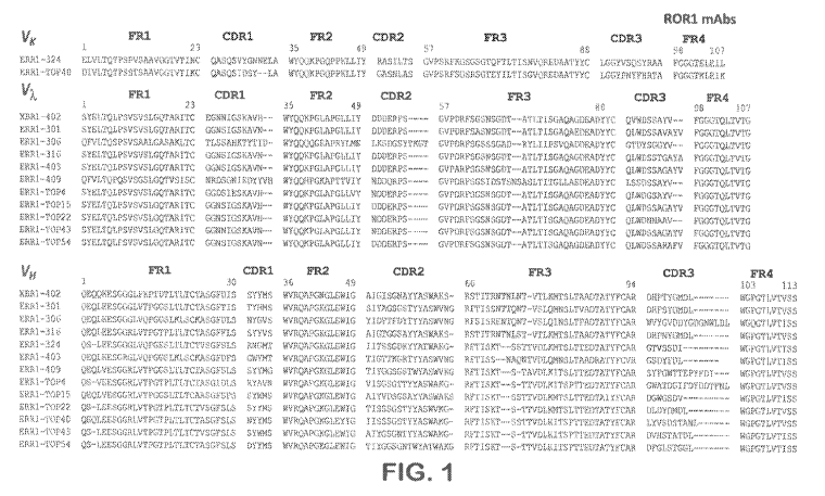
[0025] Figure 1 shows the amino acid sequences of variable immunoglobulin
heavy and
light chains of novel rabbit anti-hROR1 mAbs, as indicated. The amino acid
sequence
alignment of the rabbit variable domains (Võ, Vx, and VH) is shown with
framework regions
(FR) and complementarity determining regions (CDR) using Kabat numbering.
Shown in
the figure are the heavy chain variable domain sequences (SEQ ID NOs:1-13,
respectively)
and the light chain variable domain sequences (SEQ ID NOs:14-26, respectively)
of 13
antibodies designated XBR1-402, ERR1-301, ERR -306. ERR1-316, ERR1-324, ERR1-
403, ERR1-409, ERR1.-TOP4, ERRI-TOP15, ERR.1-TOP22, ERR1-TOF40, ERRI-TOF43,
and ERR1-TOP54. As indicated in the figure clones XBR1-402, ERRI-301, ERR1-
306,
ERR1-316, ERR-4O3, ERR1-409, ERR1-TOF4, ERRI-TOP15, ERR1-TOP22, ERR1-
TOP43, and ERRI-TOP54 are variable domains of immunoglobulin A, light chains,
while
antibodies ERR.1-324 and ERR1-TOF40 are variable domains of immunoglobulin K
light
chains.
j0026] Figure 2 shows the binding activity of chimeric rabbit/human -1:al's
to human
RORI (hROR.1) and mouse R.ORI (m.ROR1) expressed as fusion proteins of the
extracellular domain (ECD) of hRORI and mR0R1 to the human Fc domain of a
human
IgG1 antibody. The binding of each chimeric rabbit/human Fab to hROR1 and
InROR1
fused with human IgG1 Fc (hFc-hROR1 and hFc-rnRORI) was analyzed by ELISA. hFc-
ROR I or hFc-mR0R1 were captured by anti-human IgG1 Fe antibody immobilized on
plate
CA 03011815 2018-07-18
WO 2017/127664
PCT/US2017/014311
11
and then incubated with hROR1 specific Fabs comprising a His-tag via detection
with mouse
anti-His tag. Specificity of the Fabs was confirmed by using fusion proteins
of the
extracellular domain (ECD) of hROR2 with the human Fe domain of a human lgGI
antibody (hFc-hROR2) as control
100271 Figure 3 shows binding activity of chimeric rabbit/human Fabs to
native human
RORI protein expressed on the cell surface of murine preB cell line 63-12 (see
Example 1).
The binding of each chimeric rabbit/human Fab to the ectopically expressed
human RORI
on mouse pre-B cell (63-12) surface was analyzed by flow eytomeny. F,RR2-T0P35
isaõ
rnAb against hROR2 that served as an isotype-matched control,
100281 Figure 4 shows epitope mapping studies for chimeric rabbit/human
Fabs on six
different immobilized IgG1-Fe fusion proteins that comprise different parts of
the
extracellular domain of human RORI: fiFe-hROR1-Ig (comprising the
Immunoglobulin-
domain of hROR1), hFc-hROR1-Fr (comprising the Frizzled domain of hRORI ), hFc-
hRORI-Kr (comprising the Kringle domain of hROR1), hFc-hROR14g-Fr (comprising
the
Immunoglobulin and Frizzled domains of bR.ORI), hFc-hRORI-Fr-Ki (comprising
the
Frizzled and Kringle domains of hROR1) and liFc-hRORI (comprising the entire
extracellular domain (ECD) of hROR1).
[0029] Figure 5 shows epitope binding studies performed by surface plasmon
resonance.
Shown are SPR serisograms obtained for the binding of different Fabs to hFc-
hROR1
captured by anti-human Fey antibody immobilized on a CMS. chip. Fabs were
injected in
different orders to identify independent and overlapping epitopes. Resonance
unit (RU, y
axis) increases that exceeded the values found for previously injected Fabs
indicated
independent epitopes because they allow simultaneous binding. For example, the
increase
found for the binding of Fab RI I exceeded the values found for XBR1-402
alone, indicating
that Fab R11 and XBR1-402 can bind simultaneously to human RORI. By contrast,
the
epitope of Fab XBRI-402 overlaps with the epitopes of ERRI-301, ERR1-403 and
R12 (left
graph); the epitope of Fab ERR1-T0P43 overlaps with the epitope of ERRI-306,
XBRI-402
and ERRI-TOP40. The x axis depicts the time in seconds (s),
100301 Figure 6 shows affinity measurements of anti-hROR I specific Fabs to
hROR1
ECD by surface plasinon resonance (SPR). (A) Shown are ,Biacore X100
sensorgrants
obtained for the binding of each Fab to hFc-hRORI captured by anti-human Foy
antibody
immobilized on CM5 chip after instantaneous background depletion,. Fabs were
injected at
CA 03011815 2018-07-18
WO 2017/127664
PCT/US2017/014311
12
five different concentrations with the highest concentration indicated in
table (B), one of the
five concentrations was tested in duplicates. (B) Monovalent affinities of
each Fab are
shown in the table. The equilibrium dissociation constant (K,4) was calculated
from k0/k,
association rate constant; ki,j; dissociation rate constant).
[00311 Figure 7
shows FACS-hased cell staining of hROR1 on various human cancer
cell lines with anti-human R.OR I antibody 2A2 as described in Example 9. Cell
lines
analyzed lines include 697 (human acute lymphocytic leukemia, ALL), Kasumi-2
(human
prel3 acute lymphocytic leukemia), human triple-negative breast cancer cell
lines MDA-MB-
231, MDA-MB-468 and HS-578T, as well as human breast cancer cell line T47D.
Except for
the T47D human breast cancer cell line, all of the evaluated cells are
positive for hROR I
expression.
(0032] Figure 8
shows the binding activity of selected chimeric rabbit/human WI to
endogenous hRORl expressed on breast cancer cells measured by fluorescence
activated cell
sorting (FACS). Human breast cancer cell line MDA-MB-231 is known to express
hROR1,
human breast cancer cell line T47D is known to be negative for hRORI. In
contrast, T47D is
known to be ROR2 positive, whereas MDA-MB-231 is known to be negative for ROR-
2
expression, ERR1-Top54, ERR1-Top43, ERR 1-324, XBR1-402 were selected anti-
hROR1
specific mAbs, XBR2-401 was a hROR2 specific rriAb used as a specificity
control (A) The
expression of endogenous hRORI and hROR2 on breast cancer cells was detected
by flow
cytornetry using commercially available goat anti-human ROR1 and goat anti-
human ROR2
polyclonal antibodies (R&D Systems), respectively, followed by Alexa Fluor 647-
conjugated AffiniPure IF(ab')2 donkey anti-goat IgG (H+L) polyclonal
antibodies (Jackson
InimunoReseamh Laboratories). Control stainings were done with the Alexa Fluor
647-
conjugated AffiniPure F(ah')2 donkey anti-goat IgG (H+L) polyclonal antibodies
alone, (B)
The binding of chimeric rabbit/human IgG I of selected clones ERR I-Top54, ERR
I -Top43,
ERR1-324, XBRI -402 (all hROR I specific) and XBR2-401 (hROR2 specific) to
ROR1
expressing human breast cancer cell line MDA-MB-23 I and to ROR2 expressing
human
breast cancer cell line T47D was analyzed by flow cytornetry using the
chimeric
rabbit/human IgGi as primary antibodies and APC-labeled goat anti-human Fe-
specific
polyclonal antibodies as secondary antibody.
[0033] Figure 9
shows the binding activity of chimeric rabbit/human IgG1 XBRI-402
and ERRI-TOP43 (both hROR I specific) to denatured hROR1 in Western-blot
experiments.
CA 03011815 2018-07-18
WO 2017/127664
PCT/US2017/014311
13
hROR1 expressed on the cell surface K562 cells or purified protein was
denatured and
detected by Western blotting. The Western blots contained the following
samples as
indicated; Lane 1: K562 cells eetopically expressing full length of hROR1,
Lane 2:
untransfected K62 cells, Lane 3: purified extracellular domain of hROR1. Lane
4; purified
extracellular domain of fiROR2.
[00341 Figure 10 shows the binding analyzed by ELISA of selected hRORI
specific.
rabbit-human-Fe chimeric antibodies of selected clones ERRI -301, XBR1-402,
ERR1-306,
ERR1-324, ERR1-403 and ERR1-Top43 to recombinant, purified hROR1 (panel A) and
to
recombinant, purified hROR2 as a negative control (panel B),
10035] Figure 11 shows schematically how site-specifically conjugated ADCs
disclosed
in this invention have been generated. (A) schematically shows the mechanism
of sortase-
enzyme mediated antibody conjugation (SMAC-technology) as disclosed in
W02014140317. In order to generate site-specifically conjugated ADCs,
recombinant
antibodies need to be expressed with the C-terminal pentapeptide motif LPXTG
(SEQ ID
NO;144), which serve as recognition sites for the sortase enzyme A from
Staphylococcus
aureus (SrtA). When a glycine modified toxin substrate is incubated with
pentapeptide motif
LPXTG containing antibody and sortase A enzyme, the sortase A enzyme catalyzes
a
transpeptidation reaction by which the glycine-modified toxin replaces the C-
terminal
glyeine of the LPXTG motif and is covalently coupled to the threonine of the
remaining
Lpxr (SEQ ID NO:147) sequence. This way C-terminally toxin-conjugated ADCs can
be
generated with high efficiency, (B) shows the structure of the preferred
toxin, a PNU-I59682
derivative comprising an ethylene-diamino (EDA) linker connecting a 5x
glycirie stretch to
the carbonyl group at C13 of the anthracycline structure, as disclosed in
W02016102697.
[0036] Figure 12 shows the efficacy for in vitro cell killing assays
perfbrined on (panel A)
immortalized human breast cancer cell line MDA-MB-468 with known hROR1-
targeting
ADCs (2A2-05-PNLI, R12-G5-PNU) and a novel ADC provided in the invention (ERRI-
Top43-05-PNU), and (panel B) immortalized human breast cancer cell line HS
5781' with
known hROR1-targeting ADCs (2A2-G5-PNU, R12-(15-FNU) and novel ADCs provided
in
the invention (XBRI-402-G5-PNII and ERIU-Top43-G5-PNI1). CD30 targeting ADC
Act 0-05-PNU was used as an isotype-matched control ADC in both panels.
[0037] Figure 13 shows ill vitro potency for cell killing of RORI positive
acute
lymphocytic leukemia cell line 697 with four anti-ROR1-PNIJ ADCs, including
ADCs
CA 03011815 2018-07-18
WO 2017/127664
PCT/US2017/014311
14
based on antibody clones 2A2, RII and RI2, and based on anti-ROR1-antibody
clone
XBR1-402 disclosed herein. All antibodies were expressed as rabbit-human (R11,
R12,
XBR-402) or marine-human (2A2) chimeric IgG, (A) shows the cell killing over a
concentration range of ADCs site-specifically conjugated to the toxin payload
Cilys-EDA-
PNU (abbreviated (5-PNU) with each of the ADCs. (B) shows the numeric ICso
values for
cell killing calculated from the curves in (A) for each of the anti-ROR1 ADCs.
10038) Figure 14 shows in vitro cell killing of ROR1 positive ALL cell
lines Kasumi-2
(A) and 697 (13) with selected anti-hROR1 ADCs site specifically conjugated to
the Glyi¨
EDA-PNU toxin payload (abbreviated G5-PN-U). The ADCs are based on anti-ROR1
antibody clone 2A2 and R12, and anti-ROR1 clone XBR1-402 as indicated. HER-2
specific
trastuzumab, site-specifically conjugated to GlyrEDA-PNU toxin payload was
used as an
isotype matched control ADC. Panels C and D show the expression levels of ROR1
measured on the cell surface of Kasumi-2 and 697 as analyzed by FACS using
antibody 2A2
as a primary antibody versus an isotype-matched control antibody.
10939] Figure 15. (A) shows in vivo efficacy of ADCs in a disseminated
mouse model of
ROR1-positive 697 with EDA-G1y5-PNU ADCs. Mice (groups of 8 animals) have been
transplanted intravenously with 106 697 human ALL cells and treated 7 and 14
days later
with each 1 mg/kg PNU-ADC based on antibody 2A2 and novel antibody XBR1-402,
or as a
negative control, with PNU-ADC based on HER2-specific antibody trastuzumab.
Percent
survival in the groups of mice was plotted over time. (B) shows the plasma
concentration of
the ADCs measured in the mice that received 1 mg/kg after 12 and 19 days
measured by
immuno-based ELISA assay using capturing with an anti-human Fcg reagent and
detection
with either an anti-kappa-light chain detection antibody for the antibody
concentration and
an anti-PNU detection antibody for the ADC concentration.
[00401 Figure 16, Pane/ (A) shows in vitro cell killing of hRORI
transfected mouse
breast cancer cell line EIVIT6-clone14 (abbreviated EMT6-c114) with site-
specifically
conjugated FNU-ADCs based on anti-ROR1 antibody R12 and novel antibody XBR1-
402,
both expressed as chimeric human IgOl antibodies, A Trastuzurnab-G5-P1'4U ADC,
specific
for HER2, was used as an isotype-matched control ADC, (B) As a further control
the same
cell killing experiment with the same ADCs was also performed on the
untransfected (and
RORI-negative) EM'f6 parental cells. Panel (C) shows the relative expression
of hRORI in
CA 03011815 2018-07-18
WO 2017/127664
PCT/US2017/014311
bRORI transfected versus untransfected EMT6 cells as detected by FACS with
ROR1-
specific antibody 2A2,
[0041] Figure 17, In vitro celi killing of itRORI transfeeted mouse breast
cancer cell line
EMT6-elonel4 (abbreviated EMT6-c1.14) with site-specifically conjugated PNU-
ADCs
based on anti-ROR1 antibody- rns961 and novel antibodies XBR1-402 and ERR1-
324, each
expressed as chimeric human IgG1 antibodies. A Trastuzumab-G5-FNU ADC,
specific for
HERZ was used as an isotype-matched control ADC.
[0042) Figure 18 (A) shows results of an in vivo efficacy study with an
orthotopic mouse
breast cancer model using hROIR1 transfected EMT6 mouse breast cancer cell
line that was
implanted into the mammary fat pads of Ball* wild-type mice. The upper panel
shows
survival curves of mice treated twice with control ADC trastuzumab-G5-PNU. The
middle
panel shows survival curves of mice treated with PNU-ADC based on antiRORI
antibody
R12, and the lower panel shows the survival curves of mice treated with FNU-
ADC based
on novel anti-ROR1 antibody XBR1-402, Little triangles below the x-axis
indicate the two
treatments with 1 mg/kg of each respective ADC at day 14 and 21 after
transplantation of the
tumors, (B) shows the Kaplan-Meier Plot of the three experiments displayed in
panel (A).
/00431 Figure 19 shows the in vitro stability of the XBR1-402-G5-PNU ADC in
NOD
SCID mouse serum (panel A) and in human serum (panel B) analyzed by an immune-
based
ELISA assay detecting either the total antibody (solid line) or the intact ADC
(dotted line),
[00441 Figure 20 shows the in vivo plasma stability of novel naked anti-ROR1
antibody
XBR I -402, as well as of XBR I -402-G5-PNU ADC evaluated in female CD4 mice.
Depicted are plasma stability measured by immune-based ELBA assay of total IgG
detected
with a human Fe detection reagent as well as of intact ADC detected with a PNU-
specifie
detection rea2ent,
/00451 Figure 21 shows the analysis of different patient derived tumor
ysates for
hROR I protein expression by Western-Blot analysis, including ysates from two
control c,ell
lines Kastirni-2 (human ALL cell line) and A549 (human lung cancer cell line).
The patient-
derived tuniorlysates are of the following designation and origin: PXF 1118:
pleuramesothelioma, R.,XF 486: hypernephroma, PXF 541, pleuramesothelioma,
SXFS 1407:
neurofibrosarcoma, CXF 533: adenocarcinorna,
100461 Figure 22 shows the efficacy of the site-specifically conjugated PNU-
ADC of
XBR1-402 anti-ROR1 ADC in different patient derived tumor models (PDX models)
CA 03011815 2018-07-18
WO 2017/127664
PCT/US2017/014311
16
established in female NMRI nude mice implanted with 00'533, R.)0' 486, PXIF
1118,
SXFS 1407 and PXF 541 patient-derived tumor material as compared to mice
treated with
vehicle control,
10047] Figure 23 shows VII and VI, amino acid sequences of humanized
antibody
clones derived from novel anti-ROR1 antibody XBR1-402, The amino acid sequence
alignment of the humanized variable domains (Vi, and VH) is shown with
framework regions
(FR) and complernentarity determining regions (CDR) using Kabat numbering.
Shown in
the figure are the heavy chain variable domain sequences (SEQ ID NOs:130-135,
respectively) and the light chain variable domain sequences (SEQ ID NOs:136-
141,
respectively) of 6 antibodies designated HuXBR1-402(3), HuXBR1-402(8), HuXBRI-
402(15), fluXBRI-402(17), FluXBRI -402(19), and HuXBR1-402(26),
(00481 Figure 24 provides data of the affinity measurements with novel
humanized
clones of parental inAb XBRI-402, including kon and KAT data as indicated,
[0049] Figure 25 shows (Panel A) the dose-response curves of in vitro cell
killing assays
performed on human 697 ALL cancer cells with hROR1-targeting parental XBRI-402-
05-
PNU and with ADCs based on humanized antibodies: huXBR1-402-3-G5-FNU, huXBR1-
402-8-G5-PNU, huXBR I -402-15-G5-PNU, huXBR1-402-17-G5-PNU, huXBR1-402-19-
G5-PNU and huXBRI-402-26-G5-PNU, A PNU-ADC based on HER2-targeting antibody
trastuzumab was used as an isotype control ADC (Tras-G5-PNU). Panel B shows
the
quantification of the in vitro cell killing efficacy (IC50),
100501 Figure 26 shows a comparison of the in vitro activities of RORI-
targeting XBRI-
402 CAR-T and R12 CAR-T.
(00511 Figure 27 shows a comparison of the in vitro activities of ROR1-
targeting XBR1-
402 CAR-T with short and long spacer.
[0052] Haire 28 provides an overview of the specificity analysis of chimeric
rabbit/human anti-human RORI IgGI XBRI-402 and, as a control, chimeric
rabbit/human
anti-human ROR2 IgGI XBR2-40I, with the Retrogenix Cell Microarralc,e
Platform.
(0053] Figure 29 shows a specificity analysis of chimeric rabbit/human anti-
human RORI
NG), XBR1-402 and, as a control, chimeric rabbit/human anti-human ROR2. IgGI
XBR2-
401, with the Retrogenix Cell Microarray Platform. Primary binding hits from
the large
screen involving 4,336 human plasma membrane proteins (see Figure 28) were
combined on
a single slide and stained with chimeric rabbit/human anti-human RORI IgGI
XBRI-402
CA 03011815 2018-07-18
WO 2017/127664
PCT/US2017/014311
17
and, as controls, chimeric rabbit/human anti-human ROR2 1031 XBR2-401 and a
rituximab
biosimilar. ZsGreen' signals on the left indicate the expression levels of the
various human
membrane proteins. in addition to their respective antigens (ROR1, ROR2, and
CD20), the
tested antibodies in IgG1 format also bind to Fey receptors FCGR3B (CDI6B),
FCGR1A
(CD64A), and FCGR2A (CD32A) as expected. Staining with the secondary antibody
alone
detects the human IgG3 heavy chain (IG1-163) as expected.
DETAILED DESCRIPTION
I. Overview
100541 The invention is predicated in part on the generation by the present
inventors of a
large naive chimeric rabbit/human Fab library and selection for binders to
human ROR I via
phage display. Receptor tyrosine kinase orphan receptors-1 and -2, ROR1 and
ROR2, are
the only two family members defining a new receptor tyrosine kinase family,
based on the
overall structural design and some functional similarities. Both ROR1 and ROR2
proteins
are type I-single pass trans-membrane receptors with an extracellular domain
(ECD)
consisting of an immunoglobulin domain, a cysteine rich f1i771ed domain and a
Kringle
domain. These three extracellular domains are followed by a trans-membrane
domain
connecting the ECD to an intracellular portion of the protein comprising
kinase domains
(Rabagay et al, (2012) Frontiers Oncol. 2: 1-8). The human ROR I and ROR2
proteins are
58% homologous between each other, but each of the ROR proteins is highly
conserved
between species. The most conserved is actually the RORI protein, a 937 an
long protein,
that is over 98.5% identical between humans and all sequenced non-human
primate species,
and even 96.7 and 96,3 % homologous between human and mouse and rabbit ROR1,
respectively (Boreherding et al, (2014) Protein Cell 5: 496-502). Therefbre,
it has been a
challenge to generate high-quality anti-ROR1 antibodies by mouse or rabbit
immunizations,
and there are only very few known antibodies with acceptable affinity. See,
e.g., WO
2010/124188 (murine monoclonal antibody 2A2), WO 2012/075158 (rabbit
antibodies R11
and R12), WO 2012/097313 (mouse monoclonal antibody 1)10) and W02014/031174
(humanized versions of mouse mAh 99961, which binds the same epitope as that
by mAb
D10),
[00551 In order to not repeat generation of anti-ROR1 antibodies by
conventional
immunization/screening of mice/rabbits that would direct antibodies against
epitopes of
CA 03011815 2018-07-18
WO 2017/127664
PCT/US2017/014311
18
greatest divergence between mouse/rabbit ROW' and human ROR1 (as was the case
in the
identification of mAbs 2A2, R11, R12, DI 0 and 99961), the present Inventors
have
generated a very high-complexity naïve rabbit antibody Fab library displayed
by phage and
screened this library for binding to native mammalian recombinant ECD of RORI
and to
cell-surface expressed human RORI, in order to select most functional and
diverse antibody
clones reactive with native human RORI protein, This strategy was chosen
because the
antibody repertoire to be mined is still derived from natural rabbit B
lymphocytes and thus
selected for immune-system pm-selected antibody heavy and light chains.
However, due to
the applied screening strategy involving native recombinant and cell-expressed
human
RORI, it was the hope that hROR I specific antibodies would be identified with
good
developability and functional qualities and that are particularly useful for
the therapy of
human diseases associated with RORI expression, like in particular ROR I-
positive cancer.
[00561 As a result of the chosen strategy, a number of novel rabbit high-
affinity anti-
human RORI antibodies have been identified with diverse CDR1, 2 and 3
clonotypes
(Figure 1) and with high binding selectivity for human RORI, but not for its
most related
"sister molecule", human ROR2 (Figures 2, 3 and 10). Some of the hROR1-
specific
antibodies showed high affinity (single-digit riM affinities) for the hRORI
target (Figure 6),
As detailed herein, thirteen monoclonal antibodies (tnAbs) in chimeric
rabbit/human Fab
format with different heavy and light chain sequences were obtained. These
mAbs were
tentatively named "XBR I -402", "ERR1-301", "ERR1-306", "ERRI -316", "ERR1-
324",
"ERRI-403", "ERRI-409", "ERR1-TOP4", "ERR1-TOPI5", "ERR1-TOF22", "ERR 1 -
T0P40", "ERRI-T0F43", and "ERR1-T0P54". All thirteen antibodies bind to
purified
human RORI as analyzed by ELISA and to cell surface human RORI. as analyzed by
flow
cytornetry. Neither binds to ROR2, which is the closest relative of RORI and
shares 58%
amino acid sequence identity with RORI. Two mAbs ("ERR1-306" and "ERRI-T0P22")
bind to both human and mouse RORI whereas the remaining eleven mAbs only bind
to
human RORI.
[00571 The affinity of all thirteen mAbs was determined by biolayer
inferometry and
surface plasmon resonance. In addition, several mAbs (4ERR-301", ("ERR-306",
"ERR-
403", "XBR1-402", "ERRI-324", "ERRI-T0P43", and "ERR1-T0P54") were converted
to
the chimeric rabbit/human IgG1 format, expressed in mammalian cells, and
purified by
Protein A affinity chromatography. Particularly, the highest affinity clones
XBR1-402 and
CA 03011815 2018-07-18
WO 2017/127664
PCT/US2017/014311
19
ERRi-TOP43 in further evaluation showed highest staining activities by FACS
with human
ROR1 overexpressing (Figure 3) and naturally hRORi expressing mammalian cells
(Figure
8). The two top-binding clones XBRI402 and ERRI -TOP43 were also able to
detect
denatured ROR1 protein by Western-Blotting (Figure 9), allowing the use of
these clones for
the development of a companion diagnostic for ROR I expressing cancers,
100581 In addition, several mAbs were expressed as chimeric rabbit/human
IgG1 with C-
terminal sortase-recognition tags, allowing site-specific conjugation of
payloads to the
antibody C-termini by sortase-enzyrne mediated antibody conjugation technology
(SMAC-
technologyTm) essentially as described in W02014140317. These anti-hROR1
antibodies
have then been site-specifically conjugated to a highly potent anthracycline-
based PN1j-
159682. toxin derivative, Glys-EDA-PNU (Figure 11B) in order to generate
highly potent
antibody drug conjugates (ADCs), essentially as disclosed in W02016102679
(which is
incorporated by reference herein and the text of which is included as an
Appendix to this
application). These ADCs have functionally been evaluated in various in vitro
and in vivo
tumor models against ADCs gen.erated based on known anti-hROR1 antibodies. It
was
observed that one particular lead clone, called XBR1-402, displayed the
highest potency and
efficacy in comparison to various known antibodies (e.g. 2A2 (from WO
2010/124188),
R11, R12 (both from WO 2012/075158), or rns961 (from WO 2014/031174), which
forms
the basis of humanized anti-hRORi rnAb cirmtuzumab, currently in clinical
trials in CLL as
a naked IgG1 rnAb.
[0059] Based on the best-in-class properties in terms of functionality on
tumor cell
killing as an ADC, the lead clone XBR1-402 has then been humanized, which
generated
several humanized clones with further increased affinity against hR.OR1,
called "huXBRI-
402-3", "htiXBRI -402-8", "huXBR1-402-15", "huXBR1-402-17", "huXBR1-402-19"
and
"huXBRI-402-26". These humanized versions of lead clone XBR1-402 have also
been
evaivated as site-specifically conjugated PNU-ADCs and each of which exhibited
further
improved tumor cell killing in in vitro hROR1 tumor mode:is,
[0060] To further investigate the therapeutic utility of the ROR1 -
targeting mAbs, CAR-
T cells based on XBR1-402 were engineered using methods previously described
for known
ROR14argeting mAbs RI I and R12 (Fludecek, M., Lupo-Stanghellini, M. T.
Kosasih, P. L.,
Sommerrneyer, D., Jensen, M. C., Rader, C. and Riddell, S. R. (2013) Receptor
affinity and
extracellular domain modifications affect tumor recognition by RORI-specific
chimeric
CA 03011815 2018-07-18
WO 2017/127664
PCT/US2017/014311
antigen receptor T cells. Clin, Cancer Res, 19, 3153-3164). In brief, ex vivo
expanded
healthy donor CD8+ CD62L+ T cells were lentivirally transduced with an EFI a
promoter
-
driven expression cassette containing XM1-402 in scFv format, followed by a
short or long
spacer, the transmembrane domain of human CD28, the signaling domain of 4-1BB,
the
signaling domain of CD3'c, and a T2A-separated transmembrane EGFR fragment
with
truncated ligand binding arid tyrosine kinase domains. FACS isolation of EGFR+
transduced
T cells, revealed robust anti-ROR1 recognition in >90% of CART cells. The
activity of the
ROR1-targeting XBR1-402 CAR-I' with a short spacer was tested against breast
cancer cell
lines MDA-IVIB-231 (ROR1+ ROR2¨) and T470 (ROR1¨ ROR2+). In the presence of
ROR1=+ ROR2--- but not ROR1¨ ROR2+ target cells, XBR1-402 CART rapidly
proliferated,
massively secreted IFN-y and 1L-2, and potently killed the target cells in
vitro (Figure 26).
Notably, in direct comparison, the XBRI-402 CART was found to he equally or
more
potent than the clinically investigated R12 CART with the same short spacer
and signaling
domains.
wo611 Moreover, the inventors hypothesized that an optimal distance
between T cell
and target cell can be achieved by equipping CART cells targeting membrane-
distal
epitopes with shorter spacers and vice versa. Based on this hypothesis, XBRI-
402 CAR-T,
which has an overlapping epitope with R12, is predicted to be more active when
equipped
with a short compared to a long spacer. Indeed, it was tbund that, in the
presence of ROR1+
ROR2¨ target ces, XBR I-402 CART with short linker proliferated more rapidly
than
XBR1-402 CART with long spacer (Figure 27) and also secreted significantly
more IFNI
and 1L-2. In vitro cytotoxicity, however, was found to he equally potent
(Figure 27).
[00621 In accordance with these studies, the present invention provides
novel
monoclonal rabbit and humanized antibodies arid related antibody-based binding
proteins
and antibody fragments thereof that specifically recognize RORI, as well as
antibody drug
conjugates and CAR with specific anti-tumor activity in hRORI expressing tumor
models
in vitro and in vivo Also provided in the invention are methods of using these
antibody
agents in therapeutic and diagnostic applications for diseases and conditions
associated with
abnormal or elevated RORI expression, e.g., cancer.
[0063) The antibodies and related compositions of the invention have
demonstrated
other surprisingly advantageous properties. Functional evaluation of the novel
clones as
site-specifically conjugated ADCs with a highly potent PNU-anthracycline
payload,
CA 03011815 2018-07-18
WO 2017/127664
PCT/US2017/014311
21
employinz various in vitro and in vivo models, revealed that particularly
novel clone XBR I-
402 performed better than any of the known antibodies. For instance, RI I-
based ADCs had
limited potency already in in vitro tumor models (Figure 13). In comparison to
antibody
clones 99961 (ins961), 2A2 and R12, novel XBRI-402 described herein also
consistently
performed better in tumor models in vitro (Figures 13, 14, 17), which was even
more evident
in tumor models in vivo (Figures /5 & 18). Combined with the favorable
properties of the
X8RI402 clone, in terms of its highly specific recognition of ROR1when tested
against
4,336 human plasma membrane proteins (Figure 29), arid the favorable stability
of the ADC
in mouse plasma and other sera evaluated (Figures 15B, 19 & 20), XBR1-402-
based ADCs
appear to have the potential for best-in-class anti-ROR1 targeting products
with high
potential for ADC therapy.
[00641 Furthermore, humanized versions of anti-ROR1 mAb XBR1-402
unexpectedly
showed even higher affinity versus the parental rabbit clone XBR1-402 (Figure
24) because
affinities are often being reduced during the process of humanization of non-
human
antibodies (Margreitter et al. (2016) J. Mol. Recognit. 29: 266-275), The
increased affinity
also correlated with improved potency of these humanized niAbs when evaluated
for anti-
tumor activity as ADCs (Figure 25), These data evidence the high potential of
the evaluated
anti-ROR1 ADC, based on humanized XBR1-402 antibodies for the therapy of human
disease. This high potential for an effective tumor therapy of a PNU-ADC based
on XBR1-
402 and/or humanized XBR1-402 is supported by the high efficacy of the XBR1-
402 based
ADC in a variety of hROR1 expressing patient derived xertograft models (Figure
22).
[0065] The highly functional anti-hROR1 antibodies and related compositions
described
herein have displayed exquisite functional properties for use as therapeutic
agents in the
therapy of human cancers associated with RORI expression. These include the
various
hROR1 antibodies, antibody fragments, antibody-based binding proteins, ADCs or
CARs
described herein, which have the same or essentially the same binding
properties as
demonstrated by the specific antibodies exemplified herein (e.g., XBR1-402).
Thus, the
favorable properties and high therapeutic potential demonstrated by the
exemplified
antibodies herein can be extended to homologous antibodies, antibody
fragments, antibody'
based binding proteins, ADCs, CARs that contain some or all of the CDR
sequences of the
variable heavy andIor light chains disclosed in the invention, or essentially
similar CDR
sequences of the variable heavy and/or light chains disclosed in the
invention. The favorable
CA 03011815 2018-07-18
WO 2017/127664
PCT/US2017/014311
22
properties and/or the high therapeutic potential can also be extended to
antibodies, antibody
fragments, antibody-based binding proteins, ADCs, CARs that only contain one
of the two
immunoglobulin chains of the disclosed antibodies (i.e., either heavy or light
chain), or one
of the two immunoglobulin chains (i.e. either heavy or light chain) that are
homologous to
the exemplified antibodies.
11. Definitions
[00661 Unless defined otherwise, all technical and scientific terms used
herein have the
same meaning as commonly understood by those of ordinary skill in the art to
which this
invention pertains. The following references provide one of skill with a
general definition of
many of the terms used in this invention: Academic Press Dictionary of Science
and
Technology, Morris (Ed.), Academic Press (15t ed., 1992); Oxford Dictionary of
Biochemistry and Molecular Biology, Smith et al. (Eds.), Oxford University
Press (revised
ed., 2000); Encyclopaedic Dictionary of Chemistry, Kumar (Ed.), Anmol
Publications Pvt.
Ltd. (2002); Dictionary of Microbiology and Molecular Biology, Singleton et
al. (Eds.), John
Wiley & Sons (3rd ed., 2002); Dictionary of Chemistry, Hunt (Ed.), Routledge
(1st ed., 1999);
Dictionary of Pharmaceutical Medicine, Nab let (Ed.), Springer-Verlag Telos
(1994);
Dictionary of Organic Chemistry, Kumar and Anandand (Eds.), Anmol Publications
Pvt.
Ltd. (2002); and A Dictionary of Biology (Oxford Paperback Reference), Martin
and Hine
(Eds.), Oxford University Press (41h ed., 2000). In addition, the following
definitions are
provided to assist the reader in the practice of the invention.
[0067] The term "antibody" also synonymously called "immunoglobulins" (1g),
or
"antigen-binding fragment" refers to polypeptide chain(s) which exhibit a
strong
monovalent, bivalent or polyvalent binding to a given antigen, epitope or
epitopes. Unless
otherwise noted, antibodies or antigen-binding fragments used in the invention
can have
sequences derived from any vertebrate species. They can be generated using any
suitable
technology, e.g., hybridoma technology, ribosome display, phage display, gene
shuffling
libraries, semi-synthetic or fully synthetic libraries or combinations
thereof. Unless
otherwise noted, the term "antibody" as used in the present invention includes
intact
antibodies, antigen-binding polypeptide fragments and other designer
antibodies that are
described below or well known in the art (see, e.g., Serafmi, J Nucl. Med.
34:533-6, 1993).
CA 03011815 2018-07-18
WO 2017/127664
PCT/US2017/014311
23
[0068] An intact "antibody" typically comprises at least two heavy (H)
chains (about 50-
70 kD) and two light (L) chains (about 25 kD) inter-connected by disulfide
bonds. The
recognized immunoglobulin genes encoding antibody chains include the kappa,
lambda,
alpha, gamma, delta, epsilon, and mu constant region genes, as well as the
myriad
immunoglobulin variable region genes. Light chains are classified as either
kappa or
lambda. Heavy chains are classified as gamma, mu, alpha, delta, or epsilon,
which in turn
define the immunoglobulin classes, IgG, IgM, IgA, IgD and IgE, respectively.
109691 Each heavy chain of an antibody is comprised of a heavy chain
variable region
(VF) and a heavy chain constant region. The heavy chain constant region of
most IgG
isotypes (subclasses) is comprised of three domains, Cm, C H2 and C H3, some
IgG isotym,
like IgM or IgE comprise a fourth constant region domain, CH4 Each light chain
is
comprised of a light chain variable region (VI) and a light chain constant
region. The light
chain constant region is comprised of one domain, CL. The variable regions of
the heavy
and light chains contain a binding domain that interacts with an antigen. The
constant
regions of the antibodies may mediate the binding of the immunoglobulin to
host tissues or
factors, including various cells of the immune system and the first component
(Clq) of the
classical complement system.
[0070] The Vii and VL regions of an antibody can be further subdivided into
regions of
hypervariability, also termed complementarily determining regions (CDRs),
which are
interspersed with the more conserved framework regions (FRs). Each VH and VL
is
composed of three CDRs and four FRs, arranged from amino-terminus to carboxyl-
terminus
in the following order: FRI, CDR I, FR2, CDR2, FR3, CDR:3, FR4. The locations
of CDR
and FR regions and a numbering system have been defined by, e.g., Kabat et
al., Sequences
of Proteins of Immunological Interest, U.S. Department of Health and Human
Services, U.S.
Government Printing Office (1987 and .199l)
[0071] An "antibody-based binding protein", as used herein, may represent
any protein
that contains at least one antibody-derived VH, VI., or CH immunoglobulin
domain in the
context of other non-irrimunoglobulin, or non-antibody derived components.
Such antibody
based proteins include, but are not limited to (I) Fe-fusion proteins of
binding proteins,
including receptors or receptor components with all or parts of the
immunoglobulin CH
domains, (ii) binding proteins, in which VH and or VL domains are coupled to
alternative
molecular scaffolds, or (iii) molecules, in which immunoglobulin VH, and/or
V1,, and/or CH
CA 03011815 2018-07-18
WO 2017/127664
PCT/US2017/014311
24
domains are combined and/or assembled in a fashion not normally found in
naturally
OCCUITinE antibodies or antibody fragments,
[00721 "Binding affinity" is generally expressed in terms of equilibrium
association or
dissociation constants (KA or KD, respectively), which are in turn reciprocal
ratios of
dissociation and association rate constants (kar and k, respectively). Thus,
equivalent
affinities may correspond to different rate constants, so long as the ratio of
the rate constants
remains the same. The binding affinity of an antibody is usually be expressed
as the K0 of a
monovalent fragment (e.g. a Fab fragment) of the antibody, with 1(0 values in
the single-digit
nanomolar range or below (subnanomolar or picomolar) being considered as very
high and
of therapeutic and diagnostic relevance.
[0073.1 As used herein, the term "binding specificity" refers to the
selective affinity of
one molecule for another such as the binding of antibodies to antigens (or an
epitope or
antigenic determinant thereof), receptors to ligands, and enzymes to
substrates. Thus, all
monoclonal antibodies that bind to a particular antigenic determinant of an
entity (e.g., a
specific epitope of RORI or ROR2) are deemed to have the same binding
specificity for that
entity.
(00741 The term "Antibody Drug Conjugate", or "ADC" refers to an antibody
to which a
therapeutically active substance or an active pharmaceutical ingredient (API)
has been
covalently coupled, such that the therapeutically active substance or an
active
pharmaceutical ingredient (API) can be targeted to the binding target of the
antibody to
exhibit its pharmacologic function. The therapeuticaliy active substance or an
active
phamaceutical ingredient can be a cellular toxin that is able to effect
killing of the cells
targeted by the ADCs, preferably malignant or cancer cells. The covalent
attachment of a
therapeutically active substance, an active pharmaceutical ingredient or a
cellular toxin can
be performed in a non-site specific manner using standard chernicai linkers
that couple
payloads to iysine or cysteine residues, or, preferably the conjugation is
performed in a site
-
specific manner, that allows -full control of conjugation site and drug to
antibody ratio (DAR)
of the ADC to be generated,
[00751 The term "conservatively modified variant" applies to both amino
acid and
nucleic acid sequences. With respect to particular nucleic acid sequences,
conservatively
modified variants refers to those nucleic acids which encode identical or
essentially identical
amino add sequences, or where the nucleic acid does not encode an amino acid
sequence, to
CA 03011815 2018-07-18
WO 2017/127664
PCT/US2017/014311
essentially identical sequences. Because of the degeneracy of the genetic
code, a large
number of functionally identical nucleic acids encode any given protein. For
instance, the
colons GCA, GCC, GCG and GCU all encode the amino acid alanine. Thus, at every
position where an alanine is specified by a codon, the codon can be altered to
any of the
corresponding codons described without altering the encoded polypeptide. Such
nucleic
acid variations are "silent variations," which are one species of
conservatively modified
variations. Every nucleic acid sequence herein which encodes a polypeptide
also describes
every possible silent variation of the nucleic acid. One of skill will
recognize that each
codon in a nucleic acid (except AUG, which is ordinarily the only codon for
methionine, and
TGG, which is ordinarily the only codon for tryptophan) can be modified to
yield a
functionally identical molecule. Accordingly, each silent variation of a
nucleic acid that
encodes a polypeptide is implicit in each described sequence.
[0076] For poly-peptide sequences, "conservatively modified variants" refer
to a variant
which has conservative amino acid substitutions, amino acid residues replaced
with other
amino acid residue having a side chain with a similar charge. Families of
amino acid
residues having side chains with similar charges have been defined in the art.
These families
include amino acids with basic side chains (e.g., lysine, arginine,
histidine), acidic side
chains (e.g., aspartic acid, glutamic acid), uncharged polar side chains
(e.g., glycine,
asparagine, glutamine, serine, threonine, tyrosine, cysteine), nonpolar side
chains (e.g.,
alanine, valine, leucine, isoleucine, proline, phenyialanine, methionine,
tryptophan), beta
branched side chains (e.g,, threonirie, valine, isoleucine) and aromatic side
chains (e.g.,
tyrosine, phenylalanine, tryptophan, histidine).
[0077] The term "contacting" has its normal meaning and refers to combining
two or
more agents (e.g., polypeptides or phage), combining agents and cells, or
combining two
populations of different cells. Contacting can occur in vitro, e.g., mixing an
antibody and a
cell or mixing a population of antibodies with a population of cells in a test
tube or growth
medium, Contacting can also occur in a cell or in situ, e.g., contacting two
polypeptides in a
cell by co-expression in the cell of recombinant polynucleotides encoding the
two
polypeptides, or in a cell lysate. Contacting can also occur in vivo inside a
subject, e.g., by
administering an agent to a subject for delivery the agent to a target cell.
roirsj A "humanized antibody" is an antibody or antibody fragment, antigen-
binding
fragment, or antibody-based binding protein comprising antibody VH or V1
domains with
CA 03011815 2018-07-18
WO 2017/127664
PCT/US2017/014311
26
ilomoiogy to human VH or VI, antibody framework sequences having a T20 score
of greater
than 80, as defined by defined by Gar) et al. (2013) BMC Biotechnol. 13, pp.
55.
10079i The terms "identical" or percent "identity," in the context of two
or more nucleic
acids or polypeptide sequences, refer to two or more sequences or subsequences
that are the
same. Two sequences are "substantially identical" if two sequences have a
specified
percentage of amino acid residues or nucleotides that ate the same (i.e, 60%
identity,
optionally 65%, 70%, 75%, 80%, 85%, 90%, 95%, or 99% identity over a specified
region,
or, when not specified, over the entire sequence), when compared and aligned
for maximum
correspondence over a comparison window, or designated region as measured
using one of
the following sequence comparison algorithms or by manual alignment and visual
inspection. Optionally, the identity exists over a region that is at least
about 50 nucleotides
for 10 amino acids) in length, or more preferably over a region that is 100 to
500 or 1000 or
more nucleotides (or 20, 50, 200 or more amino acids) in length.
[0080] Methods of alignment of sequences for comparison are well known in
the art.
Optimal alignment of sequences for comparison can be conducted, e.g,, by the
local
homology algorithm of Smith and Waterman, Adv. App!. Math. 2:482c, 1970; by
the
homology alignment algorithm of Needleman and Wunsch, J. MoL Biol, 48:443,
1970; by
the search for similarity- method of Pearson and Lipman, Proc. Nat'l, Mad.
Sei, USA
852444, 1988; by computerized implementations of these algorithms (GAP,
BESTFIT,
FASTA, and 'TFASTA in the Wisconsin Genetics Software Package, Genetics
Computer
Group, Madison, WI); or by manual alignment and visual inspection (see, e,g.,
Brent et al,,
Current Protocols in Molecular Biology, John 'Wiley & Sons, Inc, (rin2bou ed.,
2003)).
Two examples of algorithms that are suitable for determining percent sequence
identity and
sequence similarity are the BLAST and BLAST 2.0 algorithms, which are
described in
Altschul et al., Nuc, Acids Res. 25;3389-3402, 1977; and Altschul et al., J.
Mal. Biol.
215;403-410, 1990, respectively.
[0081] The term "subject" refers to human and non-human animals (especially
non-
human mammals). The term "subject" is used herein, for example, in connection
with
therapeutic and diagnostic methods, to refer to human or animal subjects.
Animal subjects
include, but are not limited to, animal models, such as, mammalian models of
conditions or
disorders associated with elevated ROR1 expression such as CLL, ALL, mantle
cell
lymphoma, neurobiastornaõ sarcoma, renal cell carcinoma., breast cancer, lung
cancer, colon
CA 03011815 2018-07-18
WO 2017/127664
PCT/US2017/014311
27
cancer, head and neck cancer, melanoma, and other cancers. Other specific
examples of
non-human subjects include, e.g., cows, horses, sheep, pigs, cats, dogs, mice,
rats, rabbits,
guinea pigs, monkeys.
[00821 Artificial T cell receptors (also known as chimeric T cell
receptors, chimeric
immunoreceptors, chimeric antigen receptors (CARs) or T-bodies) are engineered
receptors,
which graft an arbitrary specificity onto an immune effector cell. Typically,
these receptors
are used to grafi the specificity of a monoclonal antibody onto a T cell; with
transfer of their
coding sequence facilitated by retroviral or lentiviral vectors or by
transposons. CAR-
engineered T cells (also abbreviated CAR-T cells) are genetically engineered T
cells armed
with chimeric receptors whose extracellular recognition unit is comprised of
an antibody-
derived recognition domain and whose intracellular region is derived from one
or more
lymphocyte stimulating moieties. The structure of the prototypic CAR is
modular, designed
to accommodate various functional domains and thereby to enable choice of
specificity and
controlled activation of T cells. The preferred antibody-derived recognition
unit is a single
chain variable fragment (scFv) that combines the specificity and binding
residues of both the
heavy and light chain variable regions of a monoclonal antibody. The most
common
lymphocyte activation moieties include a T-celi costimulatory (e.g. CD28)
domain in tandem
with a T-cell triggering (e.g. CD3zeta) moiety. By arming effector lymphocytes
(such as T
cells and natural killer cells) with such chimeric receptors, the engineered
cell is re-directed
with a pre-defined specificity to any desired target antigen, in a non-HLA
restricted manner.
CAR constructs are introduced ex vivo into T cells from peripheral lymphocytes
of a given
patient using retroviral or lentiviral vectors or transposons. Following
infusion of the
resulting CAR-engineered T cells back into the patient, they traffic, reach
their target site,
and upon interaction with their target cell or tissue, they undergo activation
and perform
their predefined effector function. Therapeutic targets for the CAR. approach
include cancer
and HIV-infected cells, or autoinamune effector cells.
[0083] The terms "treat," "treating," "treatment," and "therapeutically
effective" used
herein do not necessarily imply 100% or complete treatment. Rather, there are
varying
degrees of treatment recognized by one of ordinary skill in the art as having
a potential
benefit or therapeutic efibct. In this respect, the inventive method can
provide any amount
of any level of treatment. Furthermore, the treatment provided by the
inventive method can
include the treatment of one or more conditions or symptoms of the disease
being treated.
CA 03011815 2018-07-18
WO 2017/127664
PCT/US2017/014311
28
[0084] A "vector" is a replicon, such as plasmid, phage or cosmid, to which
another
polynucleotide segment may be attached so as to bring about the replication of
the attached
segment. Vectors capable of directing the expression of genes encoding for one
or more
polypeptides are referred to as "expression vectors".
ill.
Antibodies, antibody-based bindingsroteins, antibody fragments thereof,
antibody
drug conjugates (ADCs), or CARs recifleallv binding to RORI and related
derivative
compounds
100851 In one
aspect, the invention provides novel antibodies, antibody-based binding
proteins, antibody fragments thereof, ADCs or CARs that specifically bind to
human RORI
with the same binding specificity as that of anti-RORI antibody exemplified
herein (Figure 1
and 23). Antibodies of the invention include intact antibodies (e.g., IgGI
antibodies
exemplified herein), antibody fragments or antigcn-binding fragments (e,g.,
Fab fragments
exemplified herein), antibody-based binding proteins, ADCs and CARs which
contain the
antigen-binding portions of an intact antibody that retain capacity to bind
the cognate
antigen, RORI. Examples of such antibody fragments include (i) a Fab fragment,
a
monovalent fragment consisting of the Vg,, VH, CL and Cei domains; (ii) a
F(ab)2 fragment,
a bivalent fragment comprising two Fab fragments linked by a disulfide bridge
at the hinge
region; (iii) a Fd fragment consisting of the V and Cm domains; (iv) a Fv
fragment
consisting of the VL and VH domains of a single arm of an intact antibody; (v)
disulfide
stabilized Fvs (dsFvs) which have an interchain disulfide bond engineered
between
structurally conserved framework regions; (vi) a single domain antibody (dAb)
which
consists of a or VL
domain (see, e.g., Ward et al., Nature 341:544-546, /989); and (vii)
an isolated complementarity determining region (CDR) as a linear or cyclic
peptide.
Examples of antibody-based binding proteins are polypeptides in which the
binding domains
of the antibodies are combined with other polypeptides or polypeptide domains,
e.g.
alternative molecular scaffolds, Fe-regions, other functional or binding
domains of other
polypeptides or antibodies resulting in molecules with addition binding
properties, e.g. hi- or
rnultispecific proteins or antibodies. Such polypeptides can create an
arrangement of binding
or functional domains normally not found in naturally occurring antibodies or
antibody
fragments,
CA 03011815 2018-07-18
WO 2017/127664
PCT/US2017/014311
29
[00861 Antibodies of the invention also encompass antibody fragments (or
"antigen-
binding fragments"), like single chain antibodies. The term "single chain
antibody" refers to
a polypeptide comprising a NTH domain and a VL domain in polypeptide linkage,
generally
linked via a spacer peptide, and which may comprise additional domains or
amino acid
sequences at the amino- and/or carboxyl-termini. For example, a single-chain
antibody may
comprise a tether segment for linking to the encoding polynueleoticle. As an
example, a
single chain variable region fragment (scFv) is a single-chain antibody.
Compared to the VL
and V domains of the Fv fragment which are coded for by separate genes, a scFv
has the
two domains joined (e.g., via recombinant methods) by a synthetic linker. This
enables them
to be made as a single protein chain in which the VL and VI' regions pair to
form monovalent
molecules.
[00871 Antibodies of the present invention also encompass single domain
antigen-
binding units, which have a camelid scaffold. Animals in the eamelid family
include camels,
llamas, and alpacas. Camelids produce functional antibodies devoid of light
chainS. The
heavy chain variable (VH) domain folds autonomously and functions
independently as an
antigen-binding unit. Its binding surface involves only three CDRs as compared
to the six
CDRs in classical antigen-binding molecules (Fabs) or single chain variable
fragments
(scFvs). Camelid antibodies are capable of attaining binding affinities
comparable to those
of conventional antibodies.
[00881 The various antibodies, antibody-based binding proteins, and
antibody fragments
thereof described herein can be produced by enzymatic or chemical modification
of the
intact antibodies, or synthesized de novo using recombinant DNA methodologies,
or
identified using phage display libraries. Methods for generating these
antibodies, antibody-
based binding proteins, and antibody fragments thereof are all well known in
the art. For
example, single chain antibodies can be identified using phage display
libraries or ribosome
display libraries, gene shuffled libraries (see, e.g., McCafferty et al,
Nature 348:552-554,
1990; and U.S. Pat. No. 4,946,778). in particular, scFv antibodies can be
obtained using
methods described in, e.g., Bird et al., Science 242:423-426, 1988; and Huston
et at, Proc.
Nati. Acad. Sci. USA 85:5879-5883, 1988, Fv antibody fragments can be
generated as
described in Skerra and Pliick-thun, Science 240:1038-41, 1988, Disulfide-
stabilized Fv
fragments (dsFvs) can be made using methods described in, e.g., Reiter et al.,
Int. J. Cancer
67:113-23, 1996, Similarly, single domain antibodies (dAbs) can he produced by
a variety
CA 03011815 2018-07-18
WO 2017/127664
PCT/US2017/014311
of methods described in, es., Ward et al., Nature 341:544-546, 1989; and Cai
and Garen,
Proc, Natl. Mad, Sci, USA 93:6280-85, 1996. Camelid single domain antibodies
can be
produced using methods well known in the art, e,g., Dumoulin et al., Nat.
Struet. Biol.
11:500-515, 2002; Ghahroudi et al., FEBS Letters 414:521-526, 1997; and Bond
et al., J.
Mol. Biol. 332:643-55, 2003. Other types of antigen-binding fragments (e,g.,
Fab, F(ab)2 or
Fd fragments) can also be readily produced with routinely practiced immunology
methods.
See, e.g., Harlow & Lane, Using Antibodies, A Labaratoyy Manual, Cold Spring
Harbor
Laboratory Press, Cold Spring Harbor, New York, 1998.
[00891 Antibodies of the invention further include humanized antibodies
with higher
homology at arnino acid le'vel of the hurnani7ed antibody VH or VI, domains to
human
antibody VHor VI, d.omains than rodent VH or VL domains, preferably with a T20
score of
greater than 80 as defined by Gao et al. (2013) BMC Biotechnol. 13, pp. 55.
NOM In some embodiments, the antibodies, antibody fragments, antibody-
based
binding proteins, ADCs or CARs of the invention have heavy chain CDR1, CDR2
and
CDR3 sequences and light chain CDR1, CDR2 and CDR3 sequences that are
substantially
identical to that of the antibodies shown in Figure 1 or Figure 23, The light
chain and heavy
chain CDR sequences of the exemplified antibodies are all indicated in the
figure. In some
of these embodiments, the antibodies, antibody fragments, antibody-based
binding proteins,
ADCs or CARs have (I) heavy chain CDRI -3 sequences that are substantially
identical to
SEQ ID NOs:27-29, respectively; and light chain CDRI-3 sequences that are
substantially
identical to SEQ ID NOs:66-68, respectively; (2) heav-y chain CDR1-3 sequences
that are
substantially identical to SEQ ID NOs:30-32, respectively; and light chain
CDRI-3
sequences that are substantially identical to SEQ D NOs:69-71, respectively;
(3) heavy
chain CDR1-3 sequences that are substantially identical to SEQ ID NOs:33-35,
respectively;
and light chain CDR1-3 sequences that are substantially identical to SEQ ID
NOs:72-74,
respectively; (4) heavy chain CDR1-3 sequences that are substantially
identical to SEQ ID
NOs:36-38, respectively; and light chain CDR1-3 sequences that are
substantially identical
ta SEQ ID NOs:75-77, respectively-; (5) heavy chain CDR -3 sequences that are
substantially identical to SEQ ID NOs:39-41, respectively; and light chain CDR
I-3
sequences that are substantially identical to SEQ ID NOs:78-80, respectively;
(6) heavy
chain CDRI-3 sequences that are substantially identical to SEQ ID NOs:42-44,
respectively;
and light chain CDRI-3 sequences that are substantially identical to SEQ ID
NOs:81-83,
CA 03011815 2018-07-18
WO 2017/127664
PCT/US2017/014311
31
respectively; (7) heavy chain CDR1-3 sequences that are substantially
identical to SEQ ID
NOs:45-47, respectively; and light chain CDR1-3 sequences that are
substantially identical
to SEQ ID NOs;84-86, respectively; (8) heavy chain CDRI-3 sequences that are
substantially identical to SEQ ID NOs:48-50, respectively; and light chain
CDIU-3
sequences that are substantially identical to SEQ ID NOs:87-89, respectively;
(9) heavy
chain CDR1-3 sequences that are substantially identical to SEQ ID NOs;51-53,
respectively;
and light chain CDR1-3 sequences that are substantially identical to SEQ D
NOs:90-92,
respectively; (10) heavy chain CDRI-3 sequences that are substantially
identical to SEQ ID
NOs:54-56, respectively; and light chain CDR1-3 sequences that are
substantially identical
to SEQ ID NOs;93-95, respectively; (11) heavy chain CDRI-3 sequences that are
substantially identical to SEQ ID NOs;57-59, respectively; and light chain
CDRI-3
sequences that are substantially identical to SEQ ID NOs:96-98, respectively;
(12) heavy
chain CDRI-3 sequences that are substantially identical to SEQ ID NOs:60-62,
respectively;
and light chain CDR 1-3 sequences that are substantially identical to SEQ ID
NOs:99-101,
respectively; or (13) heavy chain CDRI-3 sequences that are substantially
identical to SEQ
ID NOs:63-65, respectively; and light chain CDRI-3 sequences that are
substantially
identical to SEQ ID NOs:102-104, respectively,
[0091] In some embodiments the antibodies, antibody fragments, antibody-
based
binding proteins, ADCs or CARs of the invention comprise the heavy chain CDR1-
CDR3
and light chain CDR1-CDR3 sequences are identical to the sequences shown in
(1) SEQ ID
NOs:27-29 and SEQ ID NOs:66-68 (antibody XBRI402), (2) SEQ ID NOs:30-32 and
SEQ
ID NOs:69-71 (antibody ERRI-301), (3) SEQ ID NOs:33-35 and SEQ ID NOs:72-74
(antibody ERR1-306), (4) SEQ ID NOs:36-38 and SEQ ID NOs:75-77 (antibody ERR1-
316), (5) SEQ ID NOs:39-41 and SEQ ID NOs:78-80 (antibody ERR-'324), (6) SEQ
ID
NOs:42-44 and SEQ ID NOs:81-83 (antibody ERR1-403), (7) SEQ ID N.0s45-47 and
SEQ
ID NOs:84-86 (antibody ERR1-409), (8) SEQ ID NOs:48-50 arid SEQ ID NOs:87-89
(antibody ERRI-TOP4), (9) SEQ ID NOs:51-53 and SEQ ID NOs:90-92 (antibody ERRI-
TOP15), (10) SEQ ID NOs:54-56 and SEQ ID NOs:93-95 (antibody ERR I.-TOP22),
(11)
SEQ NOs;57-59 and SEQ ID NO,s:96-98 (antibody ERR1-10P40), (12) SEQ ID
NOs:60-62 and SEQ ID NOs:99-101 (antibody ERR1-TOP43), or (13) SEQ ID NOs:63-
65
and SEQ ID NOs:102-104 (antibody ERR1-TOP54).
CA 03011815 2018-07-18
WO 2017/127664
PCT/US2017/014311
32
100921 In other embodiments, the antibodies, antibody fragments, antibody-
based
binding proteins, ADCs or CARs of the invention that specifically bind to
human ROR1
contain (a) a light chain variable domain having a sequence that is
substantially identical to
any one of SEQ ID NOs:14-26 or 136-141, (b) a heavy chain variable domain
having a
sequence that is substantially identical to any one of SEQ ID NOs:1-13 or 130-
135, or (c)
both a light chain of (a) and a heavy chain of (b). In some embodiments, the
antibody
comprises both a light chain of (a) and a heavy chain of (b). In some
embodiments, the
antibodies, antibody fragments, antibody-based binding proteins, ADCs or CARs
of the
invention contains (a) a light chain variable domain having at least 90%
identity to any one
of SEQ ID NOs:14-26 or 136-141, (b) a heavy chain variable domain having at
least 90%
sequence identity to any one of SEQ ID NOs:1-13 or 130-135, or (c) both a
light chain of (a)
and a heavy chain of (b). In some embodiments, the percentage identity can be
at least 91%,
at least 92%, at least 93%, at least 94%, at least 95%, at least 96%, at least
97%, at least
98%, or at least 99%, or even 100%. In some embodiments, the light chain
variable domain
has at least 95% identity to any one of SEQ ID NOs:14-26 or 136-141. In some
embodiments, the light chain variable domain has 100% identity to any one of
SEQ ID
NOs:14-26 or 136-141. In some embodiments, the antibody, antibody fragments,
antibody-
based binding proteins, ADCs or CARs contains a heavy chain variable domain
having at
least 90% identity to any one of SEQ ID NOs:1-13 or 130-135. In other
embodiments, the
percentage identity can be at least 91%, at least 92%, at least 93%, at least
94%, at least
95%, at least 96%, at least 97%, at least 98%, or at least 99%, or even 100%.
In some
embodiments, the heavy chain variable domain has at least 95% identity to any
one of SEQ
ID NOs:1-13 or 130-135. In some embodiments, the heavy chain variable domain
has 100%
identity to any one of SEQ ID NOs:1-13 or 130-135.
[00931 In some embodiments, the antibodies, antibody fragments, antibody-
based
binding proteins, ADCs or CARs of the invention can comprise any heavy chain
as
described herein (e.g., heavy chains shown in Figures 1 and 23) in combination
with any
suitable light chain, such as those exemplified herein. Likewise, the antibody
can comprise
any of the light chains as described above (e.g., light chains shown in
Figures 1 and 23) in
combination with any suitable heavy chain, such as those exemplified herein.
For example,
in preferred embodiments, the antibody comprises a light chain having at least
90% identity
to SEQ ID NO:14 and a heavy chain having at least 90% identity to SEQ ID NO:1
(antibody
CA 03011815 2018-07-18
WO 2017/127664
PCT/US2017/014311
33
XBR1-402), a light chain having at least 90% identity to SEQ ID NO:18 and a
heavy chain
having at least 90% identity to SEQ ID NO:5 (antibody ERR1-324), a light chain
having at
least 90% identity to SEQ ID NO:25 and a heavy chain having at least 90%
identity to SEQ
ID NO:12 (antibody ERR I-T0P43), or a light chain having at least 90% identity
to SEQ ID
NO:26 and a heavy chain having at least 90% identity to SEQ ID NO:13 (antibody
ERRI-
T0P54). In some embodiments, the antibody can comprise the light chain and
heavy chain
sequences respectively shown in (1) SEQ ID NO:14 and SEQ ID NO:1, (2) SEQ ID
NO:18
and SEQ ID NO:5, (3) SEQ ID NO:25 and SEQ ID NO:12, or (4) SEQ ID NO:26 and
SEQ
ID NO:13. In the various embodiments, percent (%) identity of peptide
sequences can be
calculated, for example, as 100 x [(identical positions)fmin(TGA, TGB)], where
TGA and
TGB are the sum of the number of residues and internal gap positions in
peptide sequences
A and B in the alignment that minimizes TGA and TGB. See, e.g., Russell et al,
J. Mel.
Biol., 244: 332-350 (1994).
[0094] The antibody of the invention can be any antibody including a full
length
antibody, an antibody fragment, an antibody-based binding protein that
specifically
recognizes or binds to the extracellular domain of human ROR1. For example,
the antibody,
antibody fragment or antibody-based binding protein can he polyclonal,
monoclonal,
recombinant, chimeric, or humanized. Furthermore, the antibody can be of any
isotype
including without limitation IgA, IgD, IgE, IgG, or IgM. Thus, for example,
the antibody can
be any IgA such as IgAl or IgA2, or any IgG such as IgGI, Ig02, IgG3, IgG4, or
synthetic
Iga The antibody can also be any antibody fragment or antibody-based binding
protein
having specificity for the extracellular domain of human ROR1, such as F(ab)2,
Fv, scFv,
IgGACH2, F(aby2, seFv2C113, Fab, NrIL, NTH, scFv4, scFv3, soFv2, dsFv, Fv,
seFv-Fc,
(scFv)2, a diabody, a bivalent, a bispecific, or a multispecific antibody. The
antibody can be
any modified or synthetic antibody, including, but not limited to, non-
depleting IgG
antibodies, CARs, or other Fe or Fab variants of antibodies.
100951 in addition to a heavy chain as described above, the antibody,
antibody-based
binding proteins or antibody fragments thereof of the invention can further
comprise a light
chain selected from a Fab library using sequential naive chain shuffling.
Likewise, in
addition to a light chain as described above, the antibody of the invention
can further
comprise a heavy chain selected from a Fab library using sequential naive
chain shuffling.
CA 03011815 2018-07-18
WO 2017/127664
PCT/US2017/014311
34
In some embodiments, the invention provides antibodies, antibody-based binding
proteins or
antibody fragments -thereof that are conservatively modified variants of the
anti-ROR1
antibodies exemplified herein. Typically, the variable regions of these
variants have an
amino acid sequence that is identical to one of these exemplified sequences
except for
conservative substitutions at one or more amino acid residues. In some C-mbod
in-lents, the
antibody, antibody fragments, antibody-based binding proteins. ADCs or CARs of
the
invention specifically binds to human RORI and contains g least one CDR having
a
sequence selected from the group consisting of SEQ ID NOs:27104, The invention
also
provides an isolated antibody with specificity for RORI containing one or more
variants of
the foregoing CDR sequences or substantially identically CDR sequences. The
variant CDR
sequences in these antibodies can include I, 2, or 3 substitutions,
insertions, deletions, or
combinations thereof in a sequence selected from the group consisting of SEQ
ID NOs:27-
104. For example, a recombinant chimeric or humanized antibody (or fragment
thereof) can
include one, two, three, four, five, or six of the foregoing CDR sequences, hi
some
embodiments, however, the recombinant chimeric or humanized antibody (or
fragment
thereof) includes three CDR sequences of the same light or heavy chain, e.g.,
light chain
CDRS shown in SEQ ID NOs:66-68, SEQ ID NOs:78-80, SEQ ID NOs99-101, or SEQ ID
NOs:102-104; and heavy chain CDRs shown in SEQ ID NOs:27-29, SEQ ID NOs:39-41,
SEQ ID NOs:60-62, or SEQ ID NOs:63-65. In some embodiments, the recombinant
chimeric or humanized antibody (or fragment thereof) includes six CDR
sequences of the
same antibody, e.g., (a) SEQ ID .NOs:66-68 and SEQ /D NOs:27-29 (antibody XBRI-
402);
(b) SEQ ID NOs:78-80 and SEQ ID NOs:39-41(antibody ERR1-324); (c) SEQ ID
NOs:99-
101 and SEQ ID NOs:60-62 (antibody ERR I -TOP43), or (d) SEQ ID NOs:1024 04
and
SEQ ID NOs:63-65 (antibody ERRI -10P54),
[0096] In some
embodiments, the invention provides antibodies, antibody-based binding
proteins or antibody fragments thereof with avidity for RORI of about 101.044
or less, 5 111v1
or less, 2 f.tM or less, 1 UM or less, 500 nM or less, 400 nM or less, 300 rM
or less, or 200
nM or less. In some embodiments, the antibodies, antibody fragments, antibody-
based
binding proteins, ADCs or CARs bind to RORI with an avidity of about 100 nM or
fess,
about 75 M or less, about 50 nM or less, about 25 tiM or less, about 10 nM or
less, or about
nM or less. in some embodiments, the antibodies, antibody fragments, antibody-
based
binding proteins, ADCs or CARs bind to RORI with an avidity of about I nM or
less, about
CA 03011815 2018-07-18
WO 2017/127664
PCT/US2017/014311
800 pM or less, about 700 pM or less, about 600 pM or less, about 500 pM or
less, about 400
pM or less, about 300 pM or less, about 200 pM or less, or about 100 pM or
less. Avidity
can be measured using art-known techniques, such as ELISA, biolayer
interometiy, or
surface plasmon resonance,
[0097] The antibody, antibody-based binding protein or antibody fragment
thereof of the
invention can be produced by any suitable technique, for example, using any
suitable
eukaryotic or non-eukaryotic expression system. In certain embodiments, the
antibody is
produced using a mammalian expression system. Some specific techniques for
generating
the antibodies antibody-based binding proteins or antibody fragments thereof
of the
invention are exemplified herein. In some embodiments, the antibodies,
antibody-based
binding proteins or antibody fragments thereof of the invention can be
produced using a
suitable non-eukaryo,tic expression system such as a bacterial expression
system. Bacterial
expression systems can be used to produce fragments such as a F(ab)2, Fv,
scFv, IgGACH2,
F(ab)2õ scFv2CH3, Fab, VL, seFv4, scFv3, scFv2, dsFv, Fv, say-Fe, (scFv)2,
and
diabodies. Techniques for altering DNA coding sequences to produce such
fragments are
known in the art,
[0098] The antibodies, antibody-based binding proteins or antibody
fragments thereof of
the invention can be conjugated to a synthetic molecule using any type of
suitable
conjugation. Recombinant engineering and incorporated seleriocysteine (e.g.,
as described in
U.S. Patent 8,916,159 issued on December 23, 2014) can be used to conjugate a
synthetic
molecule. Other methods of conjugation can include covalent coupling to native
or
engineered lysine side-chain amines or cysteine side-chain thials. See, e.g.,
Wu et al., Nat.
Biotechnol, 23: 1 137-1 146 (2005).
10099] hi a preferred embodiment, the antibodies, antibody-based binding
proteins or
antibody fragments thereof of the invention conjugated to a synthetic molecule
(called
"ADC" for antibody drug conjugate with the synthetic molecule being a toxin)
are obtained
by means of site-specific sortase-enzyme mediated antibody conjugation. As
disclosed in
W02014140317, sortases (also called sortase transpeptidases) form a group of
prokaryotic
enzymes that modify surface proteins by recognizing and cleaving a specific
peptide motif
called "sortase recognition tag" or "sortase tag". Usually, a given sortase
enzyme recognizes
one or more sortase recognition tags. Sortase enzymes can be naturally
occurring, or may
have undergone genetic engineering (Dorr et al., PINTAS 2014; Ill, 13343-8).
CA 03011815 2018-07-18
WO 2017/127664
PCT/US2017/014311
36
[001001 In a preferred embodiment, the conjugate is obtained by means of site-
specific
sortase-enzyme mediated conjugation of( a) an antibody, antibody-based binding
protein or
antibody fragment thereof as described herein carrying one or more sortase
recognition tags,
arid (b) one or more synthetic MOICCUICS carrying a glycine or oligoglycine
tag, Gly(,),
[00101] Preferably, the sortase recognition tag is fused or conjugated to the
C-terrnimis of
at least one subdomain of the antibody. Said sortase recognition tag is
preferably selected
from the group consisting of LPXSG (SEQ ID NO:142), LPXAG (SEQ ID NO:143),
I,PXTG (SEQ ID NO:144), LAXTG (SEQ ID NO:145), and NPQTG (SEQ TD NO: 46).
f001021 Preferably, the oligoglycine tag, Glyfro, has a length of 1 to 21
glycine residues
with n being any number from I to 21), preferably with a length of 3 to 5
amino acids (n
from 3 to 5, i.e. G1y(3), Gly(4) ,or G1y(5)).
[00103j The synthetic molecule can be any molecule such as one targeting a
tumor. In
some embodiments, the synthetic molecule for conjugation to the antibody is a
protein (e.g.,
an antibody) or an RNA or DNA aptamer.
[001041 In one embodiment, the antibodies, antibody-based binding proteins or
antibody
fragments thereof of the invention conjugated to a synthetic molecule have the
general
formula A ¨ (L ¨ P),, in which; A is an antibody, antibody-based binding
protein or antibody
fragment thereof as described herein, L is one or more linkers, P is one or
more payloads
selected from the group consisting of a label and a cytotoxic or cytostatic
agent, and in
which n is an integer between >I and < 10.
[00105] In this embodiment, the linker preferably comprises, or consists
of, at least one
selected from the group consisting of: an oligopeptide linker (including
cleavable and non
-
cleavable oligopeptide linkers), a hydrazine linker, a thiourea linker, a self-
immolative
linker, a succinimidyi trans-4-(maleimidylmethypcyclohexane-i-carboxylate
(SMCC)
linker, a maleimide linker, a disulfide linker, a thioether linker, and/or a
maieimide linker.
[00106] The skilled person understands that further linkers may be suitable.
Such linkers
may be non-cleavable or may be cleaved by changes in pH, redox potential or
specific
intracellular enzymes. Cleavable oligopeptide linkers include protease- or
matrix
metalloprotease-cleavable linkers, it is understood that the linker may
comprise
combinations of the above. For example, the linker may be a valine-citruline
PAB
[00107[ In a preferred embodiment, the linker comprises an oligopeptide with a
sequence:
that contains the penta-peptide motif I,PXSG (SEQ ID NO:142), LPXACi (SEQ ID
CA 03011815 2018-07-18
WO 2017/127664
PCT/US2017/014311
37
NO:143), LPXTG (SEQ ID NO:144), LAXTG (SEQ ID NO:145), or NPQTG (SEQ ID
NO:146) with X being any amino acid, followed by an oligo-glycirie stretch,
Gly(,), with n
being an integer between > 1 and < 2L In a preferred embodiment, the linker is
conjugated
to the C-termirius of at least one subdornain of the antibody, antibody-based
binding proteins
or antibody fragments thereof.
[00108] in various embodiments, suitable synthetic molecules ("payloads") for
conjugation to the antibody include, e.g., therapeutic agents such as
cytotoxic, cytostatic, or
antiangiogenic agents, radioisotopes, and liposornes. A eytotoxic agent can be
a plant,
fungal, or bacterial molecule. In some embodiments, the cytotoxic agent for
conjugation to
the antibody of the invention is a small molecular weight toxin (MW< 2'000
Dalton,
preferably MW < 1'000 Dalton), a peptide toxin, or a protein toxin, Many
specific examples
of these toxins are well known in the art, See, e.g., Dyba et al., Cum Pharm.
Des. 10:2311-
34, 2004; Kuyucak et al., Future Med, Chem. 6:1645-58, 2014; Beraud et al.,
intlamm,
Allergy Drug Targets. 10;322-42, 2011; and Middlebrook et al., Microbiol. Rev,
48:199-221,
1984, in some embodiments, a therapeutic agent is conjugated to the antibody.
For example,
the therapeutic agent can be a maytansinoid (e.g,, maytansinol or DM1
maytansinoid), a
taxane, a calicheamicin, a cemadotin, a monometbylauristatin (e.g.,
monomethylauristatin E
or monornethy/auristatin F), a pyrrolobenzodiazepine (PBD) or, preferably an
anthracycline,
more preferably a derivative of the highly potent anthracycline PNU-159682.
Particularly
preferred derivatives of the highly potent anthracycline PNU-159682 are
disclosed in
W02016102679 (which is incorporated by reference herein). Therapeutic agents
also
include vincristine and prednisone. In various embodiments, the therapeutic
agent that may
he employed in the invention can be an antimetabolite (e.g., an antifolate
such as
rnethotrexate, a fluoropyrimidine such as 5- fluorouracil, cytosine
arabinoside, or an
analogue of purine or adenosine); an intercalating agent (for example, an
anthracycline such
as doxorubicin, nernorubicine, or preferably a derivative of PNU-159682),
daunomycin,
epirabicin, idarubicin, mitomycin-C, dactinotnycin, or mithramycin, or other
intercalating
agents such as pyrrolobenzodiazepine; a DNA-reactive agent such as
calicheamicins,
tiancimycins, and other enediynes; a platinum derivative (e.g., cisplatin or
carboplatin); an
alkylating agent (e.g., nitrogen mustard, melphalan, chlorambucil, busulphan,
cyclophosphamide, ifosfamide nitrosoureas or thiotepa); an RNA polymerase
inhibitor such
as et-amanitin; an antimitotic agent (e.g,, a vinca alkaloid such as
Virleristine, or a taxoid such
CA 03011815 2018-07-18
WO 2017/127664
PCT/US2017/014311
38
as paclitaxel or docetaxel); a topoisomerase inhibitor (for example,
etoposide, teniposide,
amsacrine, topotecari); a cell cycle inhibitor (for example, a flavopyridol);
or a microbtubule
agent (e.g,, an epothilone, a tubulysine, a pre-tubulysine, discoderrnolide
analog, or
eleutherobin analog). A therapeutic agent can be a proteasome inhibitor or a
topolsomerase
inhibitor such as bortezomib, amsacrine, etoposide, etoposide phosphate,
teniposide, or
doxorublein. Therapeutic radioisotopes include iodine (Isli), yttrium (90Y),
lutetium (1771,11),
actinium (225Ae), praseodymium, astatine (At), rhenium (Re), bismuth (Bi or
Bi), and
rhodium (Rh). Antiangiogenic agents include linotnide, bevacuzirnah,
angiostatin, and
razoxane.
[00109] In a preferred embodiment, the synthetic toxin molecule is selected
from PNU-
159682 as described in Quintieri et al, (2005) and derivatives thereof (see
formula (i) below),
maytansine, moriornethyl auristatin IVIMAE, and monomethyl auristatin MMAF. In
a
preferred embodiment, the toxin, joined to the linker at its wavy line, is of
formula (i), as
described in WO 2016102679 (which is incorporated by reference herein);
0 0,ti 0
-0H
11114010
014 0'
formula (i)
[00110] In the embodiment where the synthetic molecule is of formula (i), it
is preferred
that the linker comprise an alkyldiamino group of the form N1-12-(C112).-NP12,
where m?
and s 11, preferably m=2, such that one amino group is directly linked at the
wavy line of
fonnula (1) to form an amide bond. It is moreover preferred that the second
amino group is
linked to an oligopeptide linker, which is more preferably an oligoglycine
Gly(,), with n
being 1 and 21, The most preferred payload is shown in Fig. 11(B).
CA 03011815 2018-07-18
WO 2017/127664
PCT/US2017/014311
39
[001111 In some embodiments, the synthetic molecule can be conjugated to any
antibody,
antibody-based binding protein, or antibody-fragment. In some embodiments, the
synthetic
rnolecule can be a label. Labels can be useful in diagnostic applications and
can include, for
example-, contrast agents. A contrast agent can be a radioisotope label such
as iodine (3II or
251), indium (H lIn), technetium (99Tc), phosphorus (32P), carbon ("C),
tritium (3H), other
radioisotope (e.g., a radioactive ion), or a therapeutic radioisotope such as
one of the
therapeutic radioisotopes listed above. Additionally, contrast agents can
include radiopaque
materials, magnetic resonance imaging (MRI) agents, ultrasound imaging agents,
and any
other contrast agents suitable for detection by a device that images an animal
body. A
synthetic molecule can also be a fluorescent label, a biologically active
enzyme label, a
luminescent label, or a chromophore label.
1001121 In some other embodiments, the synthetic molecule can be a liposorne,
as
described in Bendas, BioDrugs, 15: 215-224, 2001. In such embodiments, the
antibody can
be conjugated to a colloidal particle, e.g,, a liposorne, and used for
controlled delivery of an
agent to diseased cells. In preparing an antibody conjugated to a liposome,
e,g., an
irnmunoliposome, an agent such as a chemotherapeutic or other drug can be
entrapped in the
liposorne for delivery to a target cell. In some embodiments, the antibodies,
antibody-based
binding proteins or antibody fragments thereof of the invention can also have
specificity for
one or more antigens in addition to ROR1, For example, the antibody of the
invention can be
engineered (e.g., as a bivalent diabody or a conjugated Fab dirrier or trimer)
to have
specificity for RORI and another tumor antigen, o,g,, an antigen associated
with
neuroblastoma, renal cell carcinoma, breast cancer, gastric cancer, prostate
cancer, colon
cancer (e.g., colon adenocarcinoma), or breast cancer (e,gõ breast
aderiocareinoma). The
antibody can be engineered to have specificity for ROR1 and an antigen that
promotes
activation or targeting of c:;i,,totoxic effector cells.
IV. Polvirueleotides, vectors and host cells for producing ROR1 antibodies
[0011.31 The invention provides substantially purified polynucleotides (DNA or
RNA)
that arc identical or complementary to sequences encoding polypeptides
comprising
segments or domains of the antibody, antibody-based binding protein or
:antibody fragment
thereof chains described herein. In some embodiments, the polyriucleotides of
the invention
encode the heavy chain or light chain domains sequences shown in Figures I and
23, When
CA 03011815 2018-07-18
WO 2017/127664
PCT/US2017/014311
expressed from appropriate expression vectors, polypeptides encoded by these
polynucleotides are capable of exhibiting ROR1 antigen binding capacity. Also
provided in
the invention are polynucleotides which encode at least one CDR region and
usually all three
CDR regions from the heavy or light chain of the antibodies described herein,
Some other
polynucleotides encode all or substantially all of the variable region
sequence of the heavy
chain and/or the light chain of the exemplified antibodies. For example, some
of these
polynucleotides encode the amino acid sequence of the heavy chain variable
region shown in
any one SEQ ID NOs:1-13 or 130-135, and/or the amino acid sequence of the
light chain
variable region shown in any one SEQ ID NOs:14-26 or 136-141, Because of the
degeneracy of the code, a variety of nucleic acid sequences will encode each
of the
immunoglobulin amino acid sequences.
[001141 The polynucleotides of the invention can encode only the variable
region
sequences of the exemplified antibodies. They can also encode both a variable
region and a
constant region of the antibody. Some of polynucleotide sequences of the
invention nucleic
acids encode a mature heavy chain variable region sequence that is
substantially identical
(e.g., at least 80%, 90%, 95% or 99%) to the mature heavy chain variable
region sequence
shown in any one SEQ ID NOs:1-13 or 130-135. Some other polynucleotide
sequences
encode a mature light chain variable region sequence that is substantially
identical (e.g., at
least 80%, 90%, 95% or 99%) to the mature light chain variable region sequence
shown in
any one SEQ ID NOs:14-26 or 136-141. Some of the polynucleotide sequences
encode a
polypeptide that comprises variable regions of the heavy chain or the light
chain of one of
the exemplified antibodies. Some other polynucleotides encode two polypeptide
segments
that respectively are substantially identical to the variable regions of the
heavy chain or the
light chain of one of the exemplified antibodies,
f001151 The polynucleotide sequences can be produced by de novo solid-phase
DNA
synthesis or by PCR mutagenesis of an existing sequence (e.g., sequences as
described in the
Examples below) encoding an exemplified functional antibody. Direct chemical
synthesis of
nucleic acids can be accomplished by methods known in the art, such as the
phosphotriester
method of Narang et al., Meth. Erizymol. 68;90, 1979; the phosphodiester
method of Brown
et al., Meth. Enzymol. 68:109, 1979; the diethylphosphoramidite method of
Beaueage et al.,
Tetra. Lett., 22:1859, 1981; and the solid support method of U.S. Patent No.
4,458,066.
Introducing mutations to a polynucleotide sequence by PCR can be performed as
described
CA 03011815 2018-07-18
WO 2017/127664
PCT/US2017/014311
41
e.g, PCI? Technology: Principles and Applications !Or DNA Amplification, 1-
1.A. Erlich
(Ed.), Freeman Press, NY, NY, 1992; PCR Protocols: A Guide to Methods and
Applications,
Innis et al. (Ed.), Academic Press, San Diego, CA, 1990; Mattila et al.,
Nucleic Acids Res,
19:967, 1991; and Eckert et at, PCR Methods and Applications 1;17, 1991.
[00116] Also provided in the invention are expression vectors and host cells
fur producing
the functional antibodies described herein, Specific examples of plasmid and
transposon
based vectors for expressing the antibodies are described in the Examples
below. Various
other expression vectors can also be employed to express the polynucleotides
encoding the
functional antibody chains or binding fragments. Both viral-based and nonviral
expression
vectors can be used to produce the antibodies in a mammalian host cell.
Noriviral vectors
and systems include plasmids, episomal vectors, typically with an expression
cassette for
expressing a protein or RNA, and human artificial chromosomes (see, e.gõ
Harrington et al.,
Nat. Genet. 15:345, 1997). For example, nonviral vectors useful fur expression
of the
antibody polynucleotides and polypeptides in mammalian (e,g., human) cells
include
pCEP4, pREP4, pThioHis A, B & C, peDNA3.11His, pEBVHis A, B & C (Invitrogen,
San
Diego, CA), MPSV vectors, and numerous other vectors known in the art for
expressing
other proteins. Other useful nonviral vectors include vectors that comprise
expression
cassettes that can be mobilized with Sleeping Beauty, PiggyBack and other
transposon
systems, Useful viral vectors include vectors based on lentiviruses or other
retroviruses,
adenoviruses, adeno-associated viruses, herpes viruses, vectors based on SV40,
papilloma
virus, HBP Epstein Barr virus, vaccinia virus vectors and Semliki Forest virus
(SFV). See,
Brent et al., supra; SmithõA.nriu. Rev, Microbial. 49:807, 1995; and Rosenfeld
et al., Cell
68:143, 1992.
1001171 The choice of expression vector depends on the intended host cells in
which the
vector is to be expressed. Typically, the expression vectors contain a
promoter and other
regulatory sequences (e.g., enhancers) that are operably linked to the
polynucleotides
encoding a functional antibody chain or fragment. In some embodiments, an
inducible
promoter is employed to prevent expression of inserted sequences except under
inducing
conditions. Inducible promoters include, e.g., arabinose, lacZ,
metallothionein promoter or a
heat shock promoter. Cultures of transformed organisms can be expanded under
non-
inducing conditions without biasing the population for coding sequences whose
expression
products are better tolerated by the host cells. In addition to promoters,
other regulatory
CA 03011815 2018-07-18
WO 2017/127664
PCT/US2017/014311
42
elements may also be required or desired for efficient expression of a
functional antibody
chain or fragment. These elements typically include an ATG initiation codon
and adjacent
ribosome binding site (Kozak consensus sequence) or other sequences. In
addition, the
efficiency of expression may be enhanced by the inclusion of enhancers
appropriate to the
cell system in use (see, e.g., Scharf et al., Results Probl. Cell Differ.
20:125, 1994; and
Bittner et al., Meth. Enzymol,, 153;516, 1987). For example, the SV40 enhancer
or CMV
enhancer may be used to increase expression in mammalian host cells.
1001,18] The expression vectors may also provide a secretion signal sequence
position to
form a fusion protein with polypeptides encoded by inserted functional
antibody sequences,
More often, the inserted functional antibody sequences are linked to a signal
sequences
before inclusion in the vector. Vectors to be used to receive sequences
encoding the
functional antibody light and heavy chain variable domains sometimes also
encode constant
regions or parts thereof. Such vectors allow expression of the variable
regions as fusion
proteins with the constant regions thereby leading to production of intact
antibodies or
fragments thereof. Typically, such constant regions are human, and preferably
of human
IgG1 antibodies.
[00119] The host cells for harboring and expressing the functional antibody
chains can be
either prokaryotic or eukaryotic. In some preferred embodiments, mammalian
host cells are
used to express and to produce the antibody polypeptides of the present
invention. For
example, they can be either a hybridoma cell line expressing endogenous
irnmunoglobulin
genes or a mammalian cell line harboring an exogenous expression vector. These
include
any normal mortal or normal or abnormal immortal animal or human cell. In
addition to the
cell lines exemplified herein, a number of other suitable host cell lines
capable of secreting
intact iminurioglobulins are also known in the art. These include, e.g,, the
CHO cell
various HEK 293 cell lines, various Cos cell lines, HeLa cells, myeloma cell
lines,
transformed B-cells and hybridomas. The use of mammalian tissue cell culture
to express
polypeptides is discussed generally in, e.g., Winnacker, From Genes to Clones,
VCH
Publishers, N.Y., N.Y,, 1987. Expression vectors for mammalian host cells can
include
expression control sequences, such as an origin of replication, a promoter,
and an enhancer,
and neeessau processing information sites, such as ribosome binding sites, RNA
splice sites,
polyadenylation sites, and transcriptional terminator sequences. These
expression vectors
usually contain promoters derived from mammalian genes or from mammalian
viruses.
CA 03011815 2018-07-18
WO 2017/127664
PCT/US2017/014311
43
Suitable promoters may be constitutive, cell type-specific, stag specific,
and/or modulatable
or regulatable. Useful promoters include, but are not limited to, EF la and
human UbC
promoters exemplified herein, the metallothionein promoter, the constitutive
adenovirus
major late promoter, the dexamethasone-inducible MMTV promoter, the SV40
promoter, the
MRP pol HI promoter, the constitutive MPSV promoter, the tetracycline-
inducible CMV
promoter (such as the human immediate-early CNN promoter), the constitutive
CMV
promoter, and promoter-enhancer combinations known in the art,
[00120] Methods for introducing expression vectors containing the
polynucleotide
sequences of interest vary depending on the type of cellular host. For
example, calcium
chloride transformation is commonly utilized for prokaryotic cells, whereas
calcium
phosphate treatment or electroporation may be used for other cellular hosts
(see generally
Sambrook et al., supra). Other methods include, e.g., electroporation, calcium
phosphate
treatment, liposome-mediated transformation, injection and inicroinjection,
ballistic
methods, virosomes, immunoliposomes, polycatioirmicleic acid conjugates, naked
DNA,
artificial virions, fusion to the herpes virus structural protein VP22 (Elliot
and O'Hare, Cell
88:223, 1997), agent-enhanced uptake of DNA, and ex vivo transduction. For
long-term,
high-yield production of recombinant proteins, stable expression will often be
desired. For
example, cell lines which stably express the antibody chains or binding
fragments can be
prepared using expression vectors of the invention which contain viral origins
of replication
or endogenous expression elements and a selectable marker gene. Following
introduction of
the vector, cells may be allowed to grow for 1-2 days in an enriched media
before they are
switched to selective media. The purpose of the selectable marker is to confer
resistance to
selection, and its presence allows growth of cells which successfully express
the introduced
sequences in selective media. Resistant, stably transfected cells can be
proliferated using
tissue culture techniques appropriate for the cell type,
[00121] The invention further provides eukaryotic or non-eukaryotic cells
(e.g., T
lymphocytes) that have been recombinantly engineered to produce the
antibodies, antibody
based binding proteins or antibody fragments thereof of the invention. The
eukaryotic or
non-eukaryotic cells can be used as an expression system to produce the
antibody of the
invention. In some embodiments, the invention provides ROR I targeted immune
cells that
are engineered to recoinbinantly express an RORI specific antibody of the
invention. For
example, the invention provides a T cell engineered to express an antibody of
the invention
CA 03011815 2018-07-18
WO 2017/127664
PCT/US2017/014311
44
(e.g., an seFv, scFv-Fc, or (seFv)2), which is linked to a synthetic molecule
containing one
or more of the following domains: a spacer or hinge region (e.g., a CD28
sequence or a IgG4
hinge-Fe sequence), a transmembrane region (e.g., a transmernbrane canonical
domain), and
an intracellular T-cell receptor (TCR) signaling domain, thereby forming a
chimeric antigen
receptor (CAR) or T-body. Intracellular TCR signaling domains that can be
included in a
CAR (or T-body) include, but are not limited to, CDR,', FcR-y, and Syk-PT
signaling
domains as well as the CD28, 4-ABB, and CD134 co-signaling domains. Methods
for
constructing 1'-cells expressing a CAR (or T-body) are known in the art, See,
e.g., Marcu-
Melina et al,, Expert Opinion on Biological Therapy, Vol, 9, No, 5 (posted
online on April
16, 2009).
V. Therapeutic and diagnostic applications
[001221 The RORI antibodies, antibody-based binding proteins, antibody
fragments
thereof, ADCs and CARs disclosed herein can be used in various therapeutic and
diagnostic
applications. RORI is expressed and implicated in the development of various
tumors. See,
e.g., Rebagay et al., Front Oricol, 2, 34, 2012; and Shabard et al., Expert
Opin. Tiler, Targets
19, 941-955, 2015. For example, RORI is expressed on the tumor cell surface in
CLL,
ALL, mantle cell lymphoma, neuroblastoma, sarcoma, renal cell carcinoma,
breast cancer,
lung cancer, colon cancer, head and neck cancer, melanoma, and other cancers.
Importantly,
RORI is expressed in embryogenesis but largely shut down after birth. Very few
adult
healthy tissues and cells express ROR1, Consistently, anti-RORI CAR-engineered
T cells
were found to be safe and active in nonhuman primates, validating ROR1 as a
therapeutic
target in cancer (Berger (2015) Cancer Irninunol Res. 3(2), page 2016). Thus,
triAbs to
RORI have high therapeutic and diagnostic utility in cancer,
[001231 In some embodiments, the invention provides methods for inhibiting
cells that
express RORI (RORI cells) by contacting the cells with an antibody, antibody-
based
binding protein, antibody fragment thereof; ADC or CAR of the invention. The
antibody,
antibody-based binding protein or antibody fragment thereof can be a naked
(unconjugated)
molecule or an antibody, antibody-based binding protein, antibody fragment
thereof
conjugated to a synthetic molecule, e.g,, a cytotoxic, cytostatic, or
antiangiogenic agent, a
radioisotope, or even to a liposome. Preferably, cells are contacted with an
ADC comprising
a conjugated cytotoxie molecule. The method can be used to inhibit RORI cells
in vitro or in
CA 03011815 2018-07-18
WO 2017/127664
PCT/US2017/014311
a subject (i.e., in vivo). The contacted RORI cells can be in, for example, a
cell culture or
animal model of a disorder associated with elevated levels of RORI. The
methods are
useful, for example, to measure and/or rank (relative to another antibody) the
antibody's
inhibitory activity for a specific RORI cell type, Inhibiting ROR1 cells can
include blocking
or reducing the activity or growth of ROW! cells. Inhibiting can also include
the killing of
RORI cells. While the methods are not bound by or limited to any particular
mechanism of
action, inhibitory activity can be mediated by blocking RORI- mediated
signaling or by
blocking the signaling of an RORI associated receptor. Inhibitory activity can
also be
mediated by recruitment of immune system effectors that attack RORI cells,
e.g., by
activating constituents of the antibody-dependent cell-mediated cytotoxicity
(ADCC) or
complement systems.
[001241 In some related embodiments, the invention provides methods for
treating a
subject that has, is suspected to have, or is at risk of developing a disorder
associated with
expression of ROR1. Generally, the methods include administering a
therapeutically
effective amount of an isolated antibody, antibody-based binding protein,
antibody fragment
thereof, ADC or CAR of the invention to the subject. The antibody can be any
anti-RORI
antibody, anti-RORI antibody fragment, anti-RORI antibody-based binding
protein of the
invention as described herein. Thus, the antibody can be chimeric, humanized,
synthetic,
F(ab)2, Fv, scFv, IgGACH2, F(ab')2, scFv20-13, Fab, VL, VH, scFv4, scFv3,
scFv2, dsFv,
Fv, or (scFv)2, In some embodiments, the method includes administering an Igo,
an scFv, a
dsFv, a F(alf)2, a diabody, or a bivalent antibody. The administered antibody,
antibody
based binding protein, antibody fragment thereof can be conjugated to a
synthetic molecule
described above, es.,a cytotoxie, c),,tostatic, or antiangiogenic agent, a
therapeutic
radioisotope, or a liposome. An exemplary cytotoxic agent is Pseudomonas
exotoxin A
(PE38). Disorders that can be treated include CLL. ALL, mantle cell lymphoma,
neuroblastoma, sarcoma, renal cell carcinoma, breast cancer, lung cancer,
colon cancer, head
and neck cancer, melanoma, and other disorders with elevated RORI expression.
[00125] In some embodiments, the invention provides methods for treating a
subject that
has, is suspected to have, or is at risk of developing a disorder associated
with elevated
levels of RORI by adoptive transfer of the genetically engineered T-cells
described herein,
which express an antibody or antigen-binding fragment of the invention as a
chimeric
antigen receptor (CAR) that selectively binds RORI. Recombinant technology can
be used
CA 03011815 2018-07-18
WO 2017/127664
PCT/US2017/014311
46
to introduce CAR-encoding genetic material into any suitable T-cells, e,g,,
central memory
T-cells from the subject to be treated. The T cells carrying the genetic
material can be
expanded (e.g., in the presence of cytokines). The genetically engineered T-
cells are
transferred, typically by infusion, to the patient. The transferred 'f-cells
of the invention can
then mount an immune response against ROR1 expressing cells in the subject.
The adoptive
transfer method can be used, for example, to treat subjects that have or are
suspected to have
any of the cancers associated with ROR1, including CLL, ALL, mantle cell
lymphoma,
neuroblastorna, sarcoma, renal cell carcinoma, breast cancer, lung cancer,
colon cancer, head
and neck cancer, melanoma, and other cancers. In some embodiments, the
foregoing
methods of treatment can further include co-administering a second therapeutic
agent for
treating the disorder associated with elevated RORI. For example, when the
disorder to be
treated involves an ROR1-expressing cancer, the method can further include co-
administration of a eytotoxic, cystostatic, or antiangiogenic or immune-
stimulatory agent
(e.g. immune-checkpoint inhibitor antibodies, for instance, but not limited
to, those binding
to PDI, PDL1, CUM, 0X40, TIN43, GITR, LAG3 and the like) suitable for treating
the
cancer, if cancer is a B-cell malignancy, the method can further include,
for example,
co-administration of rituximab, alemtuzumab, ofatumumab, ocrelizumab, or a
CHOP
chemotherapeutic regimen.
[001261 in some other embodiments, the invention provides method for detecting
in a
biological sample an altered level of RORI (e,2., cell surface RORI), for
example, relative
to a control, either by FACS, immunohistochernistry (IHC) or Western Blotting.
Generally,
the method includes contacting a biological sample with an antibody, antibody-
based
binding protein, antibody fragment thereof of the invention and determining
the amount of
antibody that selectively binds to material (e.g., cells) in the sample to
thereby determine the
level of RORI in the biological sample. A biological sample can be from a cell
culture or
from a test subject, e.g., a plasma or a tissue sample from a subject that
has, is suspected to
have, or is at risk of developing a disease or condition associated with
elevated RORI in a
subject. A control level desirably corresponds to the ROR1 level detected
using the same
antibody in a corresponding sample(s) from one or more control cultures or
disease-free
subjects. Methods of using the antibody of the invention to determine RORI
levels can
include any immunoassay such as immuno- (Western) blotting, enzyme-linked
CA 03011815 2018-07-18
WO 2017/127664
PCT/US2017/014311
47
immunosorbent assay (ELISA), Immunohistochemistry (11-IC) and flow cytometryõ
fluorescence-activated cell sorting (FACS) analysis.
[00127] The methods of detection can be used to screen for the presence of a
disorder
associated with elevated ROR1, The methods include obtaining a sample from a
test subject
in need of screening, e.g., a subject that has, is suspected to have, or is at
risk of developing a
disorder associated with elevated ROR I . The level of ROR1 (cg., the amount
or
concentration) in the sample is measured using an antibody, antibody-based
binding protein,
antibody fragment thereof of the invention, and the level in the sample is
compared to a
control level of RORI. The control level represents, for example, the mean
level (e.g., the
amount or concentration) in sample(s) from one or, preferably, multiple
control group
subjects that do not have a disorder associated with elevated ROR1.
Alternatively, the
control level can conespond to the level or mean level of RORI in one or more
samples
taken from the test subject at one or more prior times, such as when the test
subject did not
have or did not exhibit, a condition associated with elevated RORI. A
significantly higher
level of ROR1. in the biological sample relative to the control level is
indicative of a disorder
associated with elevated RORI in the subject. In subjects such as humans,
where cell surface
RORI expression is largely restricted to embryonic development, a control
level of ROR I
can be zero or none. Thus, in some embodiments of the method of the detection
provided by
the invention, any significant and detectable amount of ROR1 in a biological
sample can be
indicative of a disorder associated with elevated RORI in the subject.
[00128] Additionally, the methods of detection can be used to monitor the
progress of a
disorder associated with elevated RORI. The method includes obtaining a sample
from a
subject in need of screening, e.g., a subject having been diagnosed or
suspected to have a
disorder associated with elevated ROR1. The level of RORI in the sample is
measured
using an antibody, antibody-based binding protein, antibody fragment thereof
of the
invention, and the level in the sample is compared to a control level
corresponding to the
level or mean level of RORI in one or more samples taken from the test subject
at one or
more prior times. Levels of RORI that are significantly elevated or decreased
relative to
control indicate that the subject's disorder is deteriorating or improving,
respectively. The
fbregoing methods of detection can be used to screen for the presence or to
monitor the
progress of disorders including, for example, CLLõA.LL, mantle cell lymphoma,
CA 03011815 2018-07-18
WO 2017/127664
PCT/US2017/014311
48
neuroblastoma, sarcoma, renal cell carcinoma, breast cancer, lung cancer,
colon cancer, head
and neck cancer, melanoma, and other cancers.
[00129.1 In some embodiments, the invention provides methods for screening a
subject for
an altered level of ROR I, Generally, the methods entail administering to the
subject an
antibody, antibody-based binding protein, antibody fragment thereof of the
invention that is
conjugated to a label (e.g., a contrast agent), imaging the subject in a
manner suitable for
detecting the label, and determining whether a region in the subject has an
altered density or
concentration of label as compared to the background level of label in
proximal tissue.
Alternatively, the methods include determining whether there is an altered
density or
concentration of label in a region as compared to the density or concentration
of label
previously detected in the same region of the subject. Methods of imaging a
subject can
include x-ray imaging, x-ray computed tomography (CT) imaging (ea., CT
angiography
(CTA) imaging), magnetic resonance (MR) imaging, magnetic resonance
angiography
(MRA), nuclear medicine, ultrasound (US) imaging, optical imaging,
elastography, infrared
imaging, microwave imaging, and the like, as appropriate for detecting the
label conjugated
to the antibody. In a preferred embodiment, the subject has, is suspected to
have, or is at risk
of developing an RORI-expressing tumor, such as CI.,L, ALL, mantle cell
lymphoma,
neuroblastoma, sarcoma, renal cell carcinoma, breast cancer, lung cancer,
colon cancer, head
and neck cancer, melanoma, and other cancers, and the method is used to screen
for or detect
the presence of the tumor. In another embodiment, the method can be used to
monitor the
size or density of a ROR1-expressing tumor over time, e.g., during a course of
treatment.
VI. Pharmaceutical txmliRpsitions and combinations
100130] In another aspect, the invention provides pharmaceutical
compositions that
contain an antibody, an antibody fragment, an antibody-based binding protein,
or an ADC as
described herein and a pharmaceutically acceptable carrier. Pharmaceutical
compositions
can be prepared from any of the antibodies described herein. Exemplary
compositions
include one or more of a chimeric antibody having SEQ ID NO:14 (light chain)
and/or SEQ
ID NO:1 (heavy chain), a chimeric antibody having SEQ ID NO:18 (light chain)
and/or SEQ
ID .N0:5 (heavy chain), a chimeric antibody having SEQ ID NO:25 (light chain)
and/or SEQ
ID NO:12 (heavy chain), and a chimeric antibody having SEQ ID NO:26 (light
chain) and/or
SEQ ID NO:13 (heavy chain). Other antibodies, antibody fragments, antibody-
based
CA 03011815 2018-07-18
WO 2017/127664
PCT/US2017/014311
49
binding proteins, or ADCs suitable for the pharmaceutical compositions of the
invention
include those having a light chain sequence as shown in SEQ ID NOs:14-26 or
136-141,
and/or a heavy chain sequence as shown in SEQ ID -N0s:1-13 or 130-135. Other
exemplary
compositions of the invention can contain a humanized antibody having one,
two, three,
four, five, or six CDRs selected from the group consisting of SEQ ID
NOs:27404, like the
ones exemplified in SEQ ID NOs: 130-141 In some embodiments the antibody
includes
three CDR sequences of the same exemplified light or heavy chains shown in
Figure 1.
These include the heavy chain CDR1, CDR2 and CDR3 sequences and light chain
CDRI,
CDR2 and CDR3 sequences respectively shown in (1) SEQ ID NOs:27-29 and SEQ ID
NOs:66-68 (antibody XBR1-402), (2) SEQ ID NOs:30-32 and SEQ ID NOs:69-71
(antibody
ERR1-301), (3) SEQ ID NOs:33-35 and SEQ ID NOs:72-74 (antibody ERR1-306), (4)
SEQ
ID NOs:36-38 and SEQ ID NOs:75-77 (antibody ERRI-316), (5) SEQ ID NOs:39-41
and
SEQ ID NOs:78-80 (antibody ERRI-324), (6) SEQ ID NOs:42-44 and SEQ ID NOs:81 -
83
(antibody ERR1-403), (7) SEQ ID NOs:45-47 and SEQ ID NOs:84-86 (antibody ERR1-
409), (8) SEQ ID l',40s:48-50 and SEQ ID NOs:87-89 (antibody ERRI-TOP4), (9)
SEQ ID
NOs:51-53 and SEQ ID NOs:90-92 (antibody ERR1-TOP15), (10) SEQ ID NOs:54-56
and
SEQ ID NOs:93-95 (antibody ERRI-TOP22), (11) SEQ ID NOs:57-59 and SEQ ID
NOs:96-98 (antibody ERRI-TOP40), (12) SEQ ID NOs:60-62 and SEQ ID NOs:99-101
(antibody ERRI-T0P43), or (1.3) SEQ ID NOs:63-65 and SEQ ID NOs:102-1.04
(antibody
ERR I-T0P54). In some embodiments, the pharmaceutical composition includes an
antibody having six CDR sequences of the same antibody exemplified in Figure
1, e.g., (a)
SEQ ID NOs:66-68 and SEQ ID NOs:27-29 (antibody XBR1-402); (b) SEQ ID NOs:78-
80
and SEQ ID NOs:39-41(antibody ERR1-324); (c) SEQ ID NOs:99-101 and SEQ ID
NOs:60-62 (antibody ERR I -TOP43), or (d) SEQ ID NOs:102-104 and SEQ ID NOs:63-
65
(antibody ERR1-TOP54). Still another exemplary pharmaceutical composition
includes a
dsFy fragment, which can include one or more modifications to the amino acid
sequence as
appropriate and understood by one of ordinary skill in the an.
(001311 in some embodiments, the compositions of the invention contain a
carrier for the
antibody, the antibody fragment, the antibody-based binding protein or the
ADC, desirably a
pharmaceutically acceptable carrier. The pharmaceutically acceptable carrier
can be any
suitable pharmaceutically acceptable carrier. It can be one or more compatible
solid or liquid
fillers, diluents, other excipients, or encapsulating substances which are
suitable for
CA 03011815 2018-07-18
WO 2017/127664
PCT/US2017/014311
administration into a human or veterinary patient (e.g., a physiologically
acceptable carrier
or a pharmacologically acceptable carrier). The term "carrier" denotes an
organic or
inorganic ingredient, natural or synthetic, with which the active ingredient
is combined to
facilitate the use of the active ingredient, e.g., the administration of the
active ingredient to a
subject. The pharmaceutically acceptable carrier can be co-mingled with one or
more of the
active components, e.g., a hybrid molecule, and with each other, when more
than one
pharmaceutically acceptable carrier is present in the composition, in a manner
so as not to
substantially impair the desired pharmaceutical efficacy. Pharmaceutically
acceptable
materials typically are capable of administration to a subject, e.g, a
patient, without the
production of significant undesirable physiological effects such as nausea,
dizziness, rash, or
gastric upset. It is, for example, desirable for a composition comprising a
pharmaceutically
acceptable carrier not to be immunogenic when administered to a human patient
for
therapeutic purposes.
1001321 Pharmaceutical compositions of the invention can additionally contain
suitable
buffering agents, including, for example, acetic acid in a salt, citric acid
in a salt, boric acid
in a salt, and phosphoric acid in a salt. The compositions can also optionally
contain suitable
preservatives, such as benzalkonium chloride, chlorobutanol, parabens, and
thimerosal.
Pharmaceutical compositions of the invention can be presented in unit dosage
form and can
be prepared by any suitable method, many of which are well known in the art of
pharmacy.
Such methods include the step of bringing the antibody of the invention into
association with
a carrier that constitutes one or more accessory ingredients. In general, the
composition is
prepared by uniformly and intimately bringing the active agent into
association with a liquid
carrier, a finely divided solid carrier, or both, and then, if necessary,
shaping the product.
1001331 A composition suitable for parenteral administration conveniently
comprises a
sterile aqueous preparation of the inventive composition, which preferably is
isotonic with
the blood of the recipient. This aqueous preparation can be formulated
according to known
methods using suitable dispersing or wetting agents and suspending agents. The
sterile
injectable preparation also can be a sterile injectable solution or suspension
in a non-toxic
parenterally-acceptable diluent or solvent, for example, as a solution in 1,3-
butane diol.
Among the acceptable vehicles and solvents that can be employed are water,
Ringer's
solution, and isotonic sodium chloride solution. In addition, sterile, fixed
oils are
conventionally employed as a solvent or suspending medium. For this purpose
any bland
CA 03011815 2018-07-18
WO 2017/127664
PCT/US2017/014311
51
fixed oil can be employed, such as synthetic mono-or di-glycerides. In
addition, fatty acids
such as QICiC acid can be used in the preparation of injectables. Carrier
formulations suitable
for oral, subcutaneous, intravenous, intramuscular, etc. administrations can
be found in
Remington's Pharmaceutical Sciences, Mack Publishing Co., Easton, PA.
[00134] Preparation of pharmaceutical compositions of the invention and their
various
routes of administration can be carried out in accordance with methods well
known in the
art. See, e.g., Remington: The Science and Practice of Pharmacy, Mack
Publishing Co., 20th
ed., 2000; and Sustained and Controlled Release Drug Delivery Systems, J.R.
Robinson, ed.,
Marcel Dekker, Inc., New York, 1978, The delivery systems useful in the
context of the
invention include time-released, delayed release, and sustained release
delivery systems such
that the delivery of the inventive composition occurs prior to, and with
sufficient time to
cause, sensitization of the site to be treated. The inventive composition can
be used in
conjunction with other therapeutic agents or therapies. Such systems can avoid
repeated
administrations of the inventive composition, thereby increasing convenience
to the subject
and the physician, and may be particularly suitable thr certain compositions
of the invention.
[00135] Many types of release delivery systems are available and known to
those of
ordinary skill in the art. Suitable release delivery systems include polymer
base systems such
as poly(lactide-glyeolide), copolyoxalates, polycaprolactones,
polyesteramides,
polyorthoesters, polyhydroxybutyric acid, and polyanhydrides. Microcapsules of
the
foregoing polymers containing drugs are described in, for example, U.S. Patent
5,075,109.
Delivery systems also include non-polymer systems that are lipids including
sterols such as
cholesterol, cholesterol esters, and fatty acids or neutral fats such as mono-
di-and
triglycerides; hydrogel release systems; sylastic systems; peptide based
systems; wax
coatings; compressed tablets using conventional binders and excipients;
partially thsed
implants; and the like. Specific examples include, but are not limited to: (a)
erosional
systems in which the active composition is contained in a form within a matrix
such as those
described in U.S. Patents 4,452,775, 4,667,014, 4,748,034, and 5,239,660 and
(b) diffusional
systems in which an active component permeates at a controlled rate from a
polymer such as
described in U.S, Patents 3,832,253 and 3,854,480. In addition, pump-based
hardware
delivery systems can be used, some of which are adapted for implantation.
[00136] The invention also provides kits suitable for carrying out the methods
of the
invention. Typically, the kits contain two or more components required for
performing the
CA 03011815 2018-07-18
WO 2017/127664
PCT/US2017/014311
52
therapeutic or diagnostic methods of the invention. Kit components include,
but are not
limited to, one or more antibodies, antibody-based binding proteins, antibody
fragments
thereof, or ADC of the invention, appropriate reagents, and/or equipment. In
some
embodiments, the kits can contain an antibody, antibody-based binding protein,
antibody
fragment thereof of the invention and an immunoassay buffer suitable for
detecting ROR I
(e.g. by ELBA, flow cytomeny, magnetic sorting, or FACS). The kit may also
contain one
or more microtiter plates, standards, assay diluents, wash buffers, adhesive
plate covers,
magnetic beads, magnets, and/or instructions for carrying out a method of the
invention
using the kit. The kit scan include an antibody, antibody-based binding
proteins, antibody
fragments thereof of the invention bound to a substrate (e.g., a multi-well
plate or a chip),
which is suitably packaged and useful to detect ROR1. In some embodiments, the
kits
include an antibody, antibody-based binding proteins, antibody fragments
thereof of the
invention that is conjugated to a label, such as, a fluorescent label, a
biologically active
enzyme label, a luminescent label, or a chromophore label. The kits can
further include
reagents fur visualizing the conjugated antibody, antibody-based binding
proteins, antibody
fragments thereof, e.g., a substrate for the enzyme. In some embodiments, the
kits include an
antibody, antibody-based binding proteins, antibody fragments thereof of the
invention that
is conjugated to a contrast agent and, optionally, one or more reagents or
pieces of
equipment useful for imaging the antibody in a subject.
[991371 Generally the antibody, antibody-based binding proteins, antibody
fragments
thereof of the invention in a kit is suitably packaged, e.g,, in a vial,
pouch, ampoule, and/or
any container appropriate for a therapeutic or detection method. Kit
components can be
provided as concentrates (including lyophilized compositions), which may be
further diluted
prior to use, or they can be provided at the concentration of use. For use of
the antibody of
the invention in vivo, single dosages may be provided in sterilized containers
having the
desired amount and concentration of components.
EXAMPLES
1001381 The following examples are provided to further illustrate the
invention but not to
limit its scope. Other variants of the invention will be readily apparent to
one of ordinary
skill in the art and are encompassed by the appended claims.
CA 03011815 2018-07-18
WO 2017/127664
PCT/US2017/014311
53
Example 1. Cell lines
1001391 Cell lines: MDA-MB-231, MDA-MB-469, 697, Kasumi-2, T47D, HS578T, 63-
12 and 63-12/hROR1 and 63-12/hROR2 transfectants, K562 and K562/hROR1
transfectants,
EMT-6 and EMT-6/R0RI transfectants were cultured in DMEM (Invitrogen;
Carlsbad, CA)
supplemented with 10% (v/v) heat inactivated FBS (Thermo Scientific; Logan,
Ur), 100
U/mL penicillin, and 100 frig/mL streptomycin (Invitrogen). 1-1EK 293F cells
were purchased
from Invitrogen and maintained in FreeStyle Medium supplemented with 1% (v/v)
heat
inactivated FBS (Thermo Scientific) to support adherent culture or without FBS
for
suspension culture, 100 Wm!, penicillin, and 100 mgimL streptomycin
(Invitrogen).
1001401 Cloning offidl-length hROR1 and hROR1 mammalian expression vectors:
Transposable vector backbones (pPB-Puro) were assembled from modular parts
with
flanking restriction sites that were synthesized or derived from sequence-
verified
commercially available vectors, and are described in detail in Patent
W02014013026A1.
These original transposable vector backbones were modified by exchanging IRES-
driven
expression of the Puromycin resistance gene in the original vector with
separate,
phosphoglycerate kinase promoter (PGK) driven expression. This was done by
replacing the
IRES sequence with an SV40-pA sequence located 3' of the multiple cloning
site, followed
by introduction of the PGK-promoter sequence 5' of the Puromycin resistance
gene. Full-
length ROR1/2 open reading frames were synthesized by total gene synthesis
(Genscript,
Piscataway) with flanking restriction sites (5'Not1/3'BstBI) and were then
cloned into the
multiple cloning site of the transposable vectors using the respective
restriction enzymes.
100141) Cell line engineering for ectopic expression of hROR1 or hROR2 in the
63-12
murine A-MULV-transformed prell cell line: The mouse Abelson murine pre-B cell
line 63-
12 (Shinkai eta!, (1992) Cell 68:855-67) were cultured in culture media
(17.7g/L Gibcol)
IMDM (Life Technologies, 42200-030), 3.024 g/I., NaHCO3 (Sigma-Aldrich, p.a.,
>99.7%1,
mi.& 100x non-essential amino acids (Life Technologies, 11140035), 5 meL
insulin
(Sigma-Aldrich, 1-5500), 3 mIA, of 10% primatone RL/UF in H20 (Sheffield
Bioscience),
and I mUL of 50 mM 2-mercaptoethanol (Sigma-Aldrich, M-3148) in H20),
supplemented
with 2% (v/v) FCS, 100 1U/mL Pen/Strep/Funginne (Amiined, 4-02F00-H), 200 mM L-
glutamine (Amimed, 5-10I(00-H) and 50 IN 2-mercaptoethanol (Amresco, 0482) at
37 C
and 7.5% CO2. Cells were engineered to overexpress hROR1 and hROR2 by
transposition as
follows: cells were centrifuged (6 min, 1200 rpm, 4 C) and resuspended in RPMI-
1640
CA 03011815 2018-07-18
WO 2017/127664
PCT/US2017/014311
54
media (5x10⁶ cells/mL), 400 AL of cell suspension was then added to 400 L of
RPM1
containing 10 pg of transposable vector pPB-P0K-Puro-R.OR.1 (directing co-
expression of
full-length ROR1 (NP 005003.2) and the puromycin-resistarice gene), or 10 lug
of
transposable vector pPB-PG1(-Puro-ROR2 (directing co-expression of full-length
RO.R2
(NP 004551.2) and the puromycin-resistance gene), along with 10 ug of
transposase-
__
containing vector pCDNA3.1_hy_mPB, DNA/63-12 cell mixtures were transferred to
electroporation cuvettes (0.4 cm-gap, 165-2088, BioRad, Cressier, Switzerland)
and
electroporated using the Biorad Gene Pulser II with capacitance extender at
300V and 950
uF, Then, cells were incubated for 5-10 min at room temperature. Following the
incubation,
cells were centrifuged at 1200 rpm for 6 min (4 C), washed once and
subsequently
resuspended in aqueous culture media (17.7g/L Gibco IMDM (Life Technologies,
42200-
030), 3.024 g/L NaHCO3 (Sigma-Aldrich, p.a., >99.7%), 10 mL/L 100x non-
essential amino
acids (Life Technologies, 11140035), 5 mg/L insulin (Sigma-Aldrich, 1-5500), 3
mUL of
10% primatone RL/UF in H20 (Sheffield Bioscience), and I mill, of 50 .mM 2-
mercaptoethanol (Sigma-Aldrich, M-3148) in 1-120), supplemented with 2% (v/v)
FCS, 100
PentStrep/Fungizone (Arninned, 4-02F00-H), 200 mM L-glutamine (Arnimed, 5-
101(00-H) and 50 uM 2-rnercaptoethanol (Amresco, 0482). After two days
incubation at
37 C in a humidified incubator at 5% CO2 atmosphere, cell pools stably
expressing hR0R1
or hROR2 were selected by adding 2 ug/mL puromycin (Sigma-Aldrich, P8833).
[00142] After 4 to 5 days, hROR I or hROR2 expression on engineered cells were
confirmed by flow cytornetry. Briefly, following trypzinization, 106 cells
were centrifuged in
FACS tubes; obtained pellets were resuspended in buffer (PBS with 2% (v/v)
FCS). In the
case of hROR1-engineered cells, cells were then incubated with 2A2 (mAb066
antibody
targeting ROR1, final concentration 2 pg/mL) for 30 min at 4 C, followed by
centrifugation
and washing, Cells were resuspended as previously and incubated with anti-
human IgG
antibody (Fe gamma-specific) PE (eBioscience, Vienna, Austria, 12-4998-82), at
a 1:100
dilution, in the dark (30 min, VC), washed once in buffer and kept on ice
until FACS
sorting. For bR0R2-engineered 63-12 cells, the same protocol was followed but
using
EPR3779 (Abeam antibody targeting ROR2; 1:100 dilution) as primary antibody
and
allophycocyanin-conjugated AffiniPure E(ab")2 goat anti-rabbit IgG (H-FL)
(Jackson
Immunoresearch, 111-136-144) as secondary antibody.
CA 03011815 2018-07-18
WO 2017/127664
PCT/US2017/014311
[00143] In the case of hROR1-engineered 63-12 cells, cells were single cell
sorted into
96-well flat-bottom plates containing 200 iL of supplemented culture media per
well using a
FACS Aria H. Plates were incubated at 37'C and clones were expanded to 6-well
plates
before analysis. Target expression was confirmed by flow cytometry using a
FACSCalibur
instrument (BD 13ioscienees) and Flow,lo analytical software (Tree Star,
Ashland, OR),
[00144] Cell line engineering for ecopie expression of hROR1 in the EMF4
murine
breast cancer cell line: Murine EMT-6 breast cancer cells (kind gift from
Prof. Dr. med.
Alfred Zippelius, University Hospital of Basel, Switzerland) were cultured in
DMEM
complete (Duibecco's Modified Eagle Medium (DMEM) High Glucose (4,5 WI) with
Glutamine with 10% (viv) Fetal Calf Serum (FCS), 100 II.J/rnL of Pen-Strep-
Fungizone and
2 rnM L-giutamine (all Bioconcept, Allschwil, Switzerland)) at 37 C and 5%
CO2. Cells
were engineered to overexpress ROR I by transposition as follows: cells were
centrithged (6
min, 1200 rpm, 4 C) and resuspended in RPMI-1640 media (5x10⁶ cells/mL), 400
1.t1.; of this
cell suspension was then added to 400 [.t1., of RPM1 containing 133 lig of
transposable vector
pPB-PGK-Puro-ROR1, directing co-expression of full-length ROR1 (NP 0050012)
and the
puromycin-resistance gene, and 6,61.tg of transposase-containing vector
pCDNA3.1_hy_mPB. DNAJEMT-6 cell mixture was transferred to electroporation
euvettes
(0.4 ern-gap, 165-2088, BioRad, Cressier, Switzerland) and electroporated
using the Biorad
Gene Pulser II with capacitance extender at 300V and 950 F. Then, cells were
incubated for
5-10 min at room-temperature. Following the incubation, cells were centrifuged
at 1200 rpm
for 6 min, washed once and subsequently resuspended in DMEM complete prior to
incubation at 37 C in a humidified incubator at 5% CO:, atmosphere, One day
after
electroporation, cell pools stably expressing human ROR1 were selected by
adding 3
purornycin (Sigma-Aldrich, P8833).
[00145] ROR1 expression on selected EMT4-ROR1 cells was confirmed by flow
cytometry. Briefly, following trypsinization, 106 cells were centrifuged in
FACS tubes;
obtained pellets were resuspended in buffer (PBS with 2% (viv) FCS). Cells
were then
incubated with 2A2 (rnAb066); 30 min, 4 C, final concentration 2 iig/mL),
followed by
centrifugation and washing. Cells were then resuspended as previously and
incubated with
anti-human IgG antibody (Pc gamma-specific) PE (eBioscience, Vienna, Austria,
12-4998-
82) with a 1:250 dilution in the dark (30 min, 4 C), washed once in buffer and
kept on ice
until FACS sortirw.
CA 03011815 2018-07-18
WO 2017/127664
PCT/US2017/014311
56
[001461 Using a FACS Aria II, cells were single cell sorted into a 96-well
flat-bottom
plate containing 200 t,tiL of DMEM complete per weft This plate was incubated
at 37 C and
clones were expanded to 6-well plates before analysis of ROR1-expression by
flow
cytometry as outlined above, using a FACSCalibur instrument (13D Biosciences)
and Howie
analytical software (Tree Star, Ashland, OR) for analysis.
[001471 Figure 16C shows the F.ACS analysis data of clone 14 (high ROR1-
expressing)
and WT (ROR1 negative) EMT-6, detected with anti-ROR1 antibody 2A2 (inAb066).
Example 2. Generation of high-complexity rabbit Fab library and reagents for
screening
[001481 Construction, expression, and purification of recombinant human RORI
proteins..
Construction, expression, purification and biotinylation of hFc fusion
proteins containing
different domains of human ROR1 or mouse ROR1 were described (Yang et al.,
PloS One
6:e21018õ 2011). For hRORI-AVI-6XHIS fusion protein, the extracellular domain
of human
ROR1 (24-403) was PCR amplified with primers pCEP4-hRORI-F and pCEP4-hROR1-Avi
tag-R (note that the AVI tag was introduced to the C terminus of ROR I by
primer pCEP4-
hROR1-Avi tag-R), followed by extension PCR with primers pCEP4-signal-F-KpriI
and
pCEP4-6HIS-R-Xhoi to add a signal peptide and 6XHIS tag to the N and C
terminus
separately before cloning into pCEP4 via Kpnlahol. This construct was then
transiently
transfected into HEK 293F cells (Invitrogen) using 293feetin (Invitrogen), and
the protein
was purified by Immobilized Metal Ion Affinity Chromatography using a 1
HisTrap
column (GE Healthcare) as described in Kwong and Rader, Curr Protoc Protein
Sci Chapter
6:Unit 6 10õ 2009. The quality and quantity of purified fIRORI-AVI-6XHIS was
analyzed by
SDS-PAGE and Ano absorbance, respectively. Subsequently, the fusion protein
was
biotinylated by BirA enzyme kit from Avidity (Aurora, Colorado) following the
protocol.
Briefly, 2 mg RORI-AVI-6XHIS at 40 u.N1 in 10 mM Tris-HC1 (pH 8) was
biotirtylated in
the presence of biotin using 10 p.g BirA after incubation for 30 min at 37 C,
1.6110-wed by
purification again using a 1-rnI, HisTrap column (GE Healthcare) as described
above.
pCEP4-hROR1-F: 5 '-
atectgttictcgtagetgctgcaactggagcacactccgccoggggcgccgccgcccag-3'
(SEQ ID NO:105); pCEP4-hROR1- Avi-tag-R: 5'-
ccactogatettctgggcctcgaagatg-togttcaggccetecatcttgttcttctectt-3' (SEQ ID
NO:106); pCEP4-
signal-F-KpnE 5' gctgggtaccggcgcgccaceat2gactggacttggagaatcctgtttctcgtagctgct-
3' (SEC) ID
CA 03011815 2018-07-18
WO 2017/127664
PCT/US2017/014311
57
NO:107); pCEP4-611IS-R-Xhol: 5'-
gccggcctegagteagtgatggtgatggtggtgacgtgocaetcgatcdotgggecto-3 (SEQ ID NO:108).
[00149] Construction., expression and intryleation of recombinant human ROR1
(hROR1-
His) and human ROR2 (hROR2-114) proteins: hRORI-His was PCR-amplified with
primers
SP-hRORIJ (5'
gctgggtaecggegcgecaccatggactggacttggagaatcctgittctegtagctgetgeaactggageacactceg
ccegggg
cgcegecgcccag 3') (SEQ ID NO:109) and hROR14-lis_R (5'
eggcctcgagteagtgatggtgatggtggtgctecatottgacttetcett 3') (SEQ ID NO:! 10) using
pCEP4-
hFc-hRORI (Yang et al,, PloS One 6:e21018, 2011) as template, while hROR24{is
was
PCR-arnplified with primers SP-hROR2...F
(gagggtaccggegegecaccatggactggaettggagaatcctgtttctegtagctgetgcaactggageacactceg
aagtgga
ggttctggatccg) (SEQ ID NO: 1) and hROR2-1-lis_R
(cggcetcgagteagtgatggtgatutggtgecccatatgetgctgtotcg) (SEQ ID NO:112) using
pCEP4-
liFc-hROR2 as template. Then they are cloned into pCEP4 (Invitrogen)
separately via
KpnIL-YhoI, These constructs were then separately and transiently transfeeted
into HEK
293F cells (Invitrogen) using 293fectin (Invitrogen), and the corresponding
proteins were
purified by Immobilized Metal Ion Affinity Chromatography using a 1-mL HisTrap
column
(GE Healthcare) as described in Kwong and Rader, CM' Protoc Protein Sci
Chapter 6:Unit 6
10, 2009, The quality and quantity of purified hROR1-His and hROR2-His were
analyzed by
SDS-PAGE and A280 absorbance, respectively,
[001501 Generation and selection of naïve chimeric rabbit/human Fab libraries:
All
rabbit handling was carried out by veterinary personnel at Pocono Rabbit Farm
&
Laboratory (Canadensis, PA) or R & R Research (Stanwood, WA), A total of nine
rabbits
(ages 3-4 months) were used. Five of these rabbits were of the New Zealand
White (NZW)
strain, with three obtained from Pocono Rabbit Farm & Laboratory (Cariadensis,
PA) and
two obtained from R & R Research (Stanwood, WA). Four b9 wild-type rabbits
were derived
from a separate R & R Research colony that originated from a pedigreed colony
developed
and characterized at the National Institute of Allergy and infectious Diseases
(NIAID)
(McCartney-Francis at al., Proc. Natl. Acad. SeL U S A 811794-1798, 1984; and
Popkov et
al., J. Mol. Biol. 325:325-335, 2003. Spleen and bone marrow from each rabbit
were
collected and processed for total RNA preparation and RT-PCR amplification of
rabbit V,
Nix., and VH encoding sequences using established protocols (Rader, Methods
Mol Biol
CA 03011815 2018-07-18
WO 2017/127664
PCT/US2017/014311
58
525:101-128, xiv, 2009, Rabbit (rb) Whurnan (hu) CdrbVH and rbliv`huCkirbVii
segments,
respectively, were assembled in one fusion step based on 3-fragment overlap
extension PCR.
Note that the VI, derived from b9 rabbits were also assembled with VH from NZW
rabbits,
The Fab-encoding fragments were digested with ,',111 and ligated with Y/I-
treated phage
display vector pC3C (Hofer et al,, ,1 Immunol Meth 318:75-87, 2007) at 16't
for 24 h.
Subsequently, 15 purified
pC3C-rbliAC,,/rbVH ligated products were transformed into E.
call strain 5R320 (a kind gift from Dr. Sachdev S. Sidhu, University of
Toronto, Toronto,
Ontario, Canada) by 30 separate electroporations (each using 0.5 ug DNA in 50
Id
electrocompetent cells) and yielded 7,5 x10⁹ independent transformants for
library k. For
library A., 4,8 x 10⁹ independent transformants were obtained using the same
procedure.
Using VC5M13 helper phage (Stratagem; La Jolla, CA), the phagernid libraries
were
converted to phage libraries and stored at -80 C. Phage library K and library
k were re-
amplified using XLI-Blue (Stratagene) or ER2738 (Lucigen) and mixed equally
before four
rounds of panning against biotinylated hFc-hROR1 or hRORI-AVI-6H1S. During the
panning, 5 ggitnL antigen was pre-incubated with streptavidin coated magnetic
beads
(Dynabeads MyOne Streptavidin Cl; Invitrogen) at 37 C fur 30 min and then
hinders from
the phage library were captured in the presence of 1 mg/mL, unspecific
polyclonai human
IgG (Thermo Scientific) when hFc-ROR1 was used. Starting from the third round
of
panning, the input phage was negatively depleted by incubation with empty
beads before
selection against antigen-loaded beads. Following selection, supernatants of
IFTG-indueed
bacterial clones were analyzed by EL1SA and by flow cytometry. Repeated clones
were
identified by DNA fingerprinting with Alai, and the Vi, and V sequences of
unique clones
were determined by DNA sequencing (Figure 1).
Example 3, Expression and purification of chimeric rabbit/human Fab and full-
length
WI antibodies
[001511 Construction, expression, and purification of chimeric rabbit/human
Fab and
.1g-G¨ I: MAb XBRI-402 in chimeric rabbitlhumati Fab format was cloned into E.
call
expression plasmid pC3C-His and expressed and purified as described in KwonE
and Rader,
CU1T Protoc Protein Sci Chapter 6:Unit 6 10, 2009. For the expression of mAb
XBRI-402 in
chimeric rabbit/human IgG1 format, the previously described vector P1GG-R11
was used
(Yang et al., PloS One 6:e21018, 2011). The VH encoding sequence of Fab XBRI -
402 was
CA 03011815 2018-07-18
WO 2017/127664
PCT/US2017/014311
59
PCR amplified using primers 42 VHF and 4-2VHR, arid cloned via ApaIlScieI into
PIGG-R1I. Then the light chain encoding sequence of XBR1-402 was PCR amplified
using
primers 4-2P and LEAD-B, and cloned via Hindillabal into PIGG-Ril with the
corresponding heavy chain encoding sequence, Note that an internal Apal site
in FR4 of VH
encoding sequences of Fab XBRI -402 was removed by silent mutation in primer 4-
2 VH R. In addition, we changed a TAG stop codon, which was suppressed during
selection
in E, coii strain XL143lue, to CAG (glutamine) encoding the first amino acid
of native VH
(Figure 1) with primer 42 VHF. The resulting PIGG-XBR1-402 plasmid was
transiently
transfected into HEK 293F cells (Invitrogen) using 293fectin (Invitrogen), and
the protein
purified with a 1-rini, recombinant Protein A HiTrap column (GE Healthcare,
Piscataway, NJ)
as described (Yang et al., PloS One 6:e21018, 2011; atld. Yang and Rader,
Methods Mol Biol
901:209-232, 2012). The quality and quantity of purified !gal were analyzed by
SDS-PAGE
and A280 absorbance, respectively.
[001521 All the other mAbs in chimeric rabbit/hurnan Fab format were cloned
into E. coil
expression plasmid pET11 a and expressed and purified as described (Yang et
al., PloS One
6:e21018, 2011), For the expression of mAbs ERR1-324. ERR1-TOP43 and ERR1-
TOP54
in chimeric rabbiaiuman IgG1 fOrmat, pCEP4 (Invitrogen) was used to clone the
heavy
chain and light chain separately. For heavy chain, a gBlock containing a heavy-
chain signal
peptide encoding sequence, V of ERR2-302 (a mAb to hROR2) and CHI (1-49) of
human
IgG1 was synthesized by IDT (San Jose, CA) and amplified with primers
KpnIfAsoi-Signal
and CH1-internallover1ap-R, and fused to CHI (50-88)-C112-C113 amplified from
PIGG
with primers CHI-internalloverlap-F and FIC-C113-R-XhoI by overlap extension
PCR with
prirners KpralAscI-Signal and HC-CH3-R-Xhor, and then cloned into pCEP4 by
AscIiAoli.
Note that a Ehei site was introduced into CHI at Ala.32 by synonymous mutation
when the
gl3lock was synthesized. Consequently, this construct served as vector to
clone the heavy
chains of other inAbs by replacing the VH using AseilEhel: VH of ERRI-324,
ERRI -TOP43
and ERR I-TOP54 were amplified with forward primer ERRI -324 HC-F, ERRI-TOP43
HC-
F and ERR1-TOP54 HC-F and reverse primer VH-CIII-R-Ehei separately, followed
by
extension PCR to add the signal peptide with primer KpriIIAscl-Signal and VH-
CHI -R-
Ehei. Then each VH was .inserted into the vector by AscilEhel, For light chain
cloning, while
lambda light chains of ERR1-TOP43 and ERRI-TOP5.4 were amplified with primers
ERR1-
TOP43 LC-F and ERR1-TOP54 LC-F separately combined with LC-R-Xhor, kappa light
CA 03011815 2018-07-18
WO 2017/127664
PCT/US2017/014311
chains of ERR1-324 was amplified with primers ERR1-324 KC-F and KC-R-Xhol.
Then, a
signal peptide encoding sequence was added by extension PCR with forward
primer
KpnliAscl-Signal and reverse primer LC-R-Xhoi or KC-R-Xhoi. Subsequently, each
light
chain PCR products was cloned into pCEP4 by AscIahol, The resulting constructs
containing heavy chain or light chain for each IgG were co-trarisfected
transiently into HEK
293F cells (Invitrogen) using 293fec1in (Invitrogen), and the con-esponding
proteins were
purified with a 1-mt, recombinant Protein A HiTrap column (GE Healthcare,
Piscataway, NJ)
as described (Yang et al., PloS One 6:e21018, 2011; and Yang and Rader,
Methods Mob Bic)!
901:209-232, 2012). The quality and quantity of purified IgG I was analyzed by
SOS-PAGE
and A2go absorbance, respectively.
Table 1. Primer sequences for cloning antibody sequences
' Primer Sequence SEQ ID
NO:
4-2_VH ....F GAGGAGGAGCTCACTCTCAGGAGCAGCAGAAGGAGT 113
.......... i CCOGG
J 4-2 VH R CGATOGGCCCTTGGTCyGAGGCTGAAGA.G.ACGGTGAC 114
! - GAGGGTCCCTGGCCCCCAGAGGTC
GAGAACrCTTGTTOCTCTGG-.ATCTCTGGIGCCTACGGC 115
----------- TCCTAIGAGCTGACACAOCTGCC
LEA D-B GGCC ATGOCTosrm GGC ACC 1.16
KpaI/Asci- GOTACCGGCSCGCCACCATOGACTOGAC _________ FIGCIAGAA 117
Signal TCCTGTITCTCGTA GCTOCIGC AA
CHI- GCCGCTGGTCAGGGCTCCTG 118
internal/ovenl
ap-R.
C FIl- CA GGAGCCCTGACCAGCGGC 119
internal/overl
ap-F
HC-CH3-R- GGCCTCGAGTCATFTACCCGGAGACAGOGA 120
Xhol ----- , _________
ERR1-324 ' TITCTCOTAGCMCIGCAACTGOAOCACACTCC 121
HO-F' CAGTCGCTGGAGGAGTCCGGG
ERR!- TITCTCGTACICTGCTGCAACIGGAGCACACTCC 127 ---- :
TOP43 FIC-F CAGTCCTIGGAGCAGTCCGGG _
ER11.1- Same as ERR1-T0P43 HC-F 123
TOP54 HC-F __________________________
VH-CH I -R- GGAGGGCGCCA GGGGGAAGACCGATOGGCCCTIGGI 124
. EheI
ERR1- IT __ ICTCGTAGCTGCTGCAACIGGAGCACACTCC 125
TOP43 LC-F . TCCTATGAGCTGACACAGCTG ,
---------------------------------------------------------------------- i'
ERR I - I. Same as ERR! -10P43 LC-F -------------------- 1.26
CA 03011815 2018-07-18
WO 2017/127664 PCT/US2017/014311
61
TOP54 LC-F I ........................
11?:RRI -324 TTICICSTAGCTGCTSCAACTIOGASCACACTCC 127
KC -F SAGCTcarscTGAcccAGACT
........... I SSCCR.7GAGTIA ISA ACATICIGTAGGSOC 128
KC -R.4101. OCICCICGASTIAACACTC.TCCCCIQTTGAA 129
Example 4. Examination of antibody binding, activities
[00153] ELBA: For ELISA (Figure 2), each well of a 96-well Costar 3690 plate
(Corning,
Coming, NY) was coated with 100 ng anti-human IgG1 Fe in 25 1.1. coating
buffer (0.1 M
Na2CO3, 0.1 M NalIC03, pH 9.6) for 1 h at 37 C. After blocking with 150111,3%
(w/v)
BSA/TBS for 1 h at 37 C, hFc-hROR1 or hFc-mR0R1 was captured following
incubation at
100 ng /50 AL for 1 h at 37 C. Then 100 ng /50 AL of Feb was applied in each
well at 37 C.
2h later, 50 AL of a 1:1000 dilution of a mouse anti-His tag mAb conjugated to
horse radish
peroxidase (HRP) (R&D Systems, Minneapolis, MN) in 1% (w/v) BSA/TBS was used
to
detect the Fab. To determine the epitopes (Figure 4), hFc fusion proteins
containing different
domains of hROR I were coated directly, followed by incubation with Fab before
detection
by mouse anti-His tag mAb conjugated to HRP. Washing with PBS was repeated and
colorimetric detection was performed using 2,2'- azino-bis (3-
ethylbenzthiazoline)-6-
sulfonic skid (Roche) as substrate according to the manufacturer's directions.
The
absorbance was measured at 405 nm using a Speen-Wax M5 microplate reader
(Molecular
Devices; Sunnyvale, CA) and SoftMax Pro software (Molecular Devices).
[00154] Flow cytometry: Cells were stained using standard flow cytometry
methodology.
Briefly, for purified anti-RORI Fab (Figure 3), 0.1-1 x 10⁶ cells were stained
with 1000 ng
100 AL of Fab on ice for 1 h. After washing twice with ice-cold flow cytometry
buffer (PBS
containing I% (v/v) BSA, 0.1% sodium azide and 1 mM EDTA), the cells were
incubated
with a 1:1000 dilution of a mouse anti-His tag mAb conjugated to Alexa Fluor
488 (Qiagen)
in 100 AL flow cytometry buffer on ice for 30 min. For purified anti-RORI IgGl
(Figure 8),
0.1-1 x 10⁶ cells were blocked by 4% goat serum for 15 min at room temperature
and then
incubated with 100 ng / 100 AL of IgG on ice for 1 h. After washing twice with
ice-cold flow
cytometry buffer, cells were stained with a 1:500 dilution of goat anti-human
IgG, Fcy pAbs
conjugated to AFC (Jackson ImmunoResearch) in 100 AL flow cytometry buffer on
ice for
30 min. For commercial goat anti human ROR I polyclonal antibody (Figure 8),
0.1-1 x 10⁶
cells were stained with 200 rig / 100 iI4 of Al, on ice for 1 h. After washing
twice with ice-
cold flow cytometry buffer (PBS containing 1% (v/v) BSA, 0.1% sodium azide and
1 mM
CA 03011815 2018-07-18
WO 2017/127664
PCT/US2017/014311
62
EDTA), the cells were incubated with a 1:1000 dilution of a Alexa Fluor 647-
conjugated
AffmiPure F(ab')2donkey anti goat IgG (H+L) in 1001,[1, flow cytometry buffer
on ice for
30 mirL FinaUy 46-diamidino-2-phenylindo1e (DAN) was added to a final
concentration of
100 ng / 111L to exclude dead cells from the analysis. Cells were analyzed
using a
FACSCalibur instrument (BO Biosciences) and Flowiro analytical software (Tree
Star,
Ashland, OR).
[00155] Swface piasman resonance: Surface plasmon resonance for the
measurement of
the affinities of all Fabs to hFc-hROR1 and for epitope mapping studies were
performed on a
Biacore X100 instrument using Biacore reagents and software (GE Healthcare,
Piscataway,
NJ). Anti-Human IgG (Fc) antibody was immobilized on a CMS sensor chip
following the
instruction of Human Antibody Capture Kit (GE Healthcare, Piscataway, NJ),
Then, hFc-
hROR I fusion proteins were captured at certain density (indicated in Figure 5
and Figure 6).
The sensor chip included an empty flow cell for instantaneous background
depletion. An
binding assays used lx HBS-EP+ running buffer (10 mM HEPES, 150 mM NaCI, 3
mikvl
EDTA (pH 7.4), and 0.05% (v/v) Surfactant P20) and a flow rate of 30 riflimin,
For affinity
measurements, all Fabs were injected at five different concentrations
(dilution factor was 2),
and the lowest concentration was tested in duplicates (the highest
concentrations for each
Fab are indicated in Figure 6B), The sensor chip was regenerated with 3 MgC12
from the
Human Antibody Capture Kit without any loss of binding capacity. Calculation
of
association (koõ) and dissociation (koff) rate constants was based on a 1:1
Langmuir binding
model The equilibrium dissociation constant (Kd) was calculated from kir/
kor,. For epitope
mapping studies, each Fab was prepared at 500 riM alone in lx HBS-EP+ running
buffer and
then injected in order as indicated in Figure 5,
[001561 Western blotting.' Celts or proteins were lysed by lx sample buffer
(containing
1% p-mercaptoetharkol) and boiled before running on NuPAGE Novex 4-12% Bis-
Tris gels
(Invitrogen). After membrane transferring and blocking by 5% milk, 2 )tg rni,
chimeric
rabbit/human IgG1 of XBRI-402 or ERR1-TOP43 was applied to detect the
denatured
proteins, followed by incubation with 1/1000 anti-human Fey conjugated to HRP
before
developing using Ea: Prime Western Blotting Detection Reagent (GE Healthcare)
(Figure
9).
CA 03011815 2018-07-18
WO 2017/127664
PCT/US2017/014311
63
Example 5 'Expression of purified, recombinant strep-tagged human ROR1 and
human
twin strep-ta .:.ged human ROR2
[001571 Strepli-tagged human RORI-extracelluiar domain was produced as
follows: the
nucleotide sequence encoding the extraceilular domain of human ROR1 (NP
0050032) was
N-terrninally fused to a signal sequence (MNFGIALIFINLTLKGVQC) and C-
terminally
fused with a sequence encoding a strepII-tag (GWS1-1POFEK). The entire
nucleotide
sequences with flanking 5'Noti and 3'HindlI1 sites were produced by total gene
synthesis
(GenScript, Piscataway, USA), assembled in the proprietary mammalian
expression vector
pEvi5 by Evitria AG (Schlicren, Switzerland) and verified by DNA sequencing.
f001581 Expression of the proteins was performed in suspension-adapted CHO KI
cells
by Evitria AG (Schlieren, Switzerland). Supernatants from pools of transfected
CHO K1
cells were harvested by centrifugation and sterile filtered (0.2 um) before
FPLC-based
affinity purification using StrepTactin columns (IBA GmbH, Goettingen,
Germany).
[00159I Recombinant human twin strep-tagged ROR2 (NP 0045512) was expressed
and
purified in-house according to the following protocol: the EBNA expression
vector pCB14b-
ROR2-ECD-TwinStrep, directing expression of ROR2 extracellular domain (ECD), C-
terminally tagged with a TwinStep tag, was transfected into FIEK293T using
Lipofectarnine LTX with PLUSTM Reagent (Thermo Fisher Scientific, 15388100).
Following a I-day incubation (37 C, 5% CO2, growth media: Dulbecco's Modified
Eagle
Medium (DMEM) High Glucose (4$ g/l) with L-Glutamine with 10% (v/v) Fetal Calf
Serum (FCS), 100 IlfilmL of Pert-Strep-Fungizone and 2 rnM L-giutarnine (all
Bioconcept)),
cells were expanded under selection conditions (2 pig/mL of puromycin (Sigma-
Aldrich,
P8833-25 mg stock at 2 mg/mL)). Cells were split and further expanded (37'C,
5% CO2);
once confluency was reached, tissue culture dishes were coated with 20 ugimi
poly-L-Lysine
(Sigma-Aldrich, P1524) for 2 hrs at 37C and washed twice with PBS. Then, cells
were
tlypsinized, washed with PBS and split 1:3 onto poly-L-lysinc-coated plates.
Again after
reaching confluency, cells were washed with PBS followed by with media
replacement using
production media (DIVIENIVF-12, Gibcorrhermo Fisher Scientific, 31330-03)
supplemented
with I ug/mL puromycin (Sigma-Aldrich, P8833), 100 ILlimL of Pen-Strep-
Fungizone
(Bioconcept, 4-02F00-H), 161 pg/mL of N-acctyl-L-cysteine (Sigma-Aldrich,
A8199) arid
ugirnt, of L-giutathione reduced (Sigma-Aldrich, G6529). Supernatant,
harvested bi-
weekly and filtered (0.22 pm) to remove cells, was stored at VC until
purification. For
CA 03011815 2018-07-18
WO 2017/127664
PCT/US2017/014311
64
purification, filtered supernatant was loaded onto a Streptacting Superflow
high capacity
cartridge (IBA, Gottingen, Germany, 2-1238-00l ) column; purification and
elution was
performed according to the manufacturer's protocol on an AEKTA pure (GE
Healthcare).
Fractions were analyzed for protein purity and integrity by SDS-PAGE. Protein-
containing
fractions were mixed and subjected to buffer exchange using Amicon filtration
units
(Millipore, Schaffhausen, Switzerland) to reach a dilution of >I :100 in PBS,
and then sterile
filtered using a low retention fitter (0.20 pm, Carl Roth, Karlsruhe, Germany,
PA49.1),
Example 6. Expression of purified, recombinant humanized anti-human ROR1
ia11,111q_ATPLP.ntibodies
NO1601 Expression vectors: Antibody variable region coding regions were
produced by
total gene synthesis (GenScript) using MINIFGLRLIFLVLTLKGVQC as leader
sequence,
and were assembled with human IgH-y 1 and 1gL-K or IgL-A constant regions, as
applicable,
in the expression vector pCB14. This vector, a derivative of the episomal
mammalian
expression vector pCEP4 (1nvitrogen), carries the EBV replication origin,
encodes the EBV
nuclear antigen (EBNA-1) to permit extrachromosomal replication, and contains
a
puromycin selection marker in place of the original hygromycin B resistance
gene.
1001611 In-house expression and purification: pC814-based expression vectors
were
transfected into FIEK293T cells using Lipofectamine LTX Reagent with PLUSTh
Reagent
(Thermo Fisher Scientific, Reinach, Switzerland, 15388100); following a 1-day
incubation
(370C, 5% C01, growth media: Dulbecco's Modified Eagle Medium (DMEM) High
Glucose
(4.5 gil) with L-Glutarnine with 10% (v/v) Fetal Calf Serum (FCS), 100 of
Pen-
Strep-Fungizone and 2 mi'vl L-glutamine (all Bioconcept, Allschwil,
Switzerland)), cells
were expanded under selection conditions (2 ggiml, of puromycin (Sigma-
Aldrich, Buchs
SG, Switzerland, P8833-25 mg stock at 2 mg/mi.)). Cells were split and further
expanded
(37'C, 5% CO2); once confluency was reached, tissue culture dishes were coated
with 20
Rgiml poly-L-Lysine (Sigma-Aldrich, P1524) for 2h at 37"C and washed twice
with PBS.
Then, cells were trypsinized and split 1:3 onto poly-L-lysIne-coated plates.
Again after
reaching confitieney, cells were washed with PBS followed by media replacement
to
production media (DIVIEM/F-12, Gibcoillermo Fisher Scientific, 31330-03)
supplemented
with 1 ggimi, puro.mycin (Sigma, P8833), 100 fUlinL of Pen-Strep-Pungizorie
(Bioconcept),
161 11g/inf. of N-acetyl-L-cysteine (Sigma-Aldrich, A8199) and 101.tginiL of L-
glutathione
CA 03011815 2018-07-18
WO 2017/127664 PCT/US2017/014311
reduced (Sigma-Aidrich, G6529). Supernatant, harvested biweekly and filtered
(0.22 pm) to
remove cells, was stored at 4 C until purification.
[00162] For purification, filtered supernatant was loaded onto a PBS-
equilibrated Protein
A HiTrap column (GE Healthcare, Frankfurt am Main, Germany, 17-04054)1) or a
JSR
AmsphereTM Protein A column OR Life Sciences, Leuven, Belgium, Hill-203CP and
washed with PBS; elution was performed using 0,1 M glycine (pH 2,5) on an
AEKTA pure
(GE Healthcare), Fractions were immediately neutralized with 1 M Tris-HCI
buffer (pH 8.0),
and analyzed for protein purity and integrity by SDS-PAGE. Protein-containing
fractions
were mixed and subjected to buffer exchange using Ainicon filtration units
(Millipore,
Schaffhausen, Switzerland, UFC901008) to reach a dilution of 1:100 in the
buffer listed in
Table 2, and then sterile filtered using a low retention filter (0.20 urn,
Carl Roth, Karlsruhe,
Germany, PA49.1).
[00163] Antibodies were transiently expressed in CHO cells by methods known in
the art
and recombinant antibodies were purified by standard protein A purification
from CHO cell
supernatants, as known in the art. The purity and the integrity' of the
recombinant antibodies
were analyzed by SDS-PAGE,
[00164] Table 2 lists the antibodies used in subsequent examples, along
with their final
concentration and buffer.
Table 2. List of antibodies used in the Examples
C-Terininisi Tags Final
Antibody Antibody SEQ ID
Fermat (HC: Heavy Chain, lbsiTer cone.
(ref.) HOW
LC: Light Chin) (ingliaL)
1
14C: 1...1'ETO-Strep
XBRI -402 HC: SEQ ID NO, 1
Igo LC: G5SLPETG- PBS 3.9
(mAb031) LC: SEQ ID NO, 14
Strep
BC: LPETO-Strep
El-Mt-301 HC: SEQ ID NO, 2
ige LC: CisSLPETG- PBS 3.8
(mAb027) LC: SEQ ID NO. 15
Strep
HC: LPETG-Strep
ERR i306 SEQ ID NO. 3
IgG LC: 05%.2E-ro- PBS 3,0
(mAb033) LC: SEQ 0) NO. 16
Strep
ERR I.324 HC: SEQ ID NO, HC: LPETO-Strep
PBS 3.0
(inAb034) LC: SEQ ID NO, IS LC: OsSLPETG.
CA 03011815 2018-07-18
WO 2017/127664 PCT/US2017/014311
66
-:, _________________________________________
HC: LPIUG-SIrep
ERRI-403 HC: SEQ ID NO, 6 '
'=,-
)e,s LC: G5SLPETG- PBS 2.9
(mAb035) LC: SEQ ID NO, 19
Strep
ERR1-- 11C: LPETG-Strep
HC: SEQ ID NO. 12
To p43 IgG LC: G5SLPETG- PBS 3.0
LC: SEQ ID NO, 25
(n/Ab036) Strep
HC: LPETG-Step
XBRI-402 HC: SEQ ID NO. 1
IgG LC; G5SLPETG- PBS 3.9
(mAb186) LC; SEQ ID NO. 14
Step
_ ..........................................................
1-IC: LPETG-Strep
ERR1-324 ile: SEQ ID NO. 5
IgG LC: G5SLPETG- PBS 7.1
(mAb188) LC: SEQ. ID NO. 18
Step
EMU- He: LPETO-SIrep
HC: SEQ ID NO. 12
Top43 tea LC: G5SLPETG- PBS 7,0
LC: SEQ ID NO, 25
(TtAb189) Step
Ha LPETG-Strep
XBRI-402 HC: SEQ ID NO. 1
41.:(3 LQ G3SLPETG- PBS 6.3
(mAb202) . LC: SEQ ID NO. 14 .
Strep
BC: LPETG-Strcp
Ms961 HC: SEQ ID NO. 14b
Ige LC: 05.5LPET0- PBS 6,4
(mAb190) LC; SEQ H) NO. 149
Strep
He: LPEIG-Strep
2A2 1-IC: SEQ ID NO, 150
IgG LC: 05SLPETG- ' PBS 8.0
(mAb066) LC: SEQ H) NO, 151
Strep .
:1-1C; LPETG-Strep
11132A2 HC: SEQ ID NO. 152
LC: GALPETG- PBS 4.7
(mAb038) LC: SEQ ID NO, 153
Strep
NC; LPETG-Strep
R11 NC: SEQ ID NO. 154
1g0 LC: Gs SIPETG- PBS 6.6
(nAb062) LC: SEQ ID NO. 155
,
, Strep .
,
,
I , .. ...
1 .............................. MC: LPETOStrep
1 R12 ' NC: SEQ ID NO. 156
IgG LC: G5SLPETG- PBS 53
1 (nAb067) LC: SEQ ID NO. 157
Strep
1 __
Trastunun NC: LPETO-Strep
NC: SEQ ID NO, 158 1
ab ("Tras" IgG 1 LC: GsSLPETG- PBS 23
LC: SEQ ID NO. 159
trtAls,042) 1 Strep
rA-ii;:i.T-1 Ige NC SEQ ID NO. 160 He: LPETG-St-e,p PBS
'7,9
1 ___________________________________
CA 03011815 2018-07-18
WO 2017/127664
PCT/US2017/014311
67
(mAb046) LC: SEQ ID NO. 161 LC: GsSLPEIG-
Strop
[00165] Ms961 corresponds to an anti-ROR1 antibody with a heavy chain
according to
SEQ ID NO. 1 ivid light chain according to SEQ ID NO. 3 of WO 2014/03117,4,
2A2 refers
to an ariti-ROR I antibody as described in WO 2010/124188, while hu2A2 refers
to Z-3,
humanized 2A2 as described in unpublished PCT/EP2016/076244. RI I and R12 are
anti-
ROR i antibodies described in WO 2012/075158. Isotype control antibodies
trastuzumab and
Ac10 target HER-2 and CD30, respectively, The heavy and light chain variable
region
sequences of monoclonal antibody brentuximab (clone cAc10) specific tbr the
human CD30
target were obtained from patent US2008213289A1, those of the human HER-2
specific
trastazumab antibody contained in the commercial antibody Herceptin
(trastuzumab), or the
ADC Kadcyle derived thereof; were derived from the online 1MG1' database (VH:
http://www,irrigt.orgI3Dstructure-D13/cgildetails,cgi?pdbrodc=7637&Part=
Chain&Chain=76371-1& VL http://www.imgtorgl3Dstructure4JB/cg1/
details,cgi?pdbcode=7637&Part=Chain&Chain-7637.1¨
Example 7. inAh R.OR.1 and ROR2-binding - characterization by ELISA
[00166] Each well of a 96-well plate was coated with 100 fiL of 2 uglirnL
strep-tagged
human RORI (from Example 5) in OA M bicarbonate coating buffer (pH 9,6), and
incubated
for 12h at 4'C. A second 96-well plate was likewise prepared with twin-strep
tagged human
ROR2 (from Example 5),
1001671 After blocking with 150 pi; of 3% (w/v) bovine serum albumin (BSA)/TBS
for lh
at 37 C, the following antibodies were added to a well within each plate at a
concentration of
0.5 pg/mL., and serially diluted (dilution factor 4) with 1% (w/v) BSAITBS,
before
incubation for lb at 37'C: ERR1-301 (mAb027), XBR1-402 (mAb031), ERR1-306
(mAh033), ERR1-324 (mAb034), ERR1-403 (mAb035) and ERR1-Top43 (mAb036). HRP-
conjugated F(ab')-., anti-human FC-gamma (Jackson Imrnurioresearch, 109-036-
008) was
then added at a 1:20'000 dilution, 100 ul per well, and incubated for lh at 37
C prior to
detection using an Spark 10M plate reader (Tecan). As shown in Figure 10, the
anti-human
ROR1 antibodies bind human ROR1 (panel A) and are not cross-reactive with
human ROR2
(panel B).
CA 03011815 2018-07-18
WO 2017/127664
PCT/US2017/014311
68
Example 8. Humanization of anti-hROR1-specific antibody XBRI -402
[001681 Humanized variable region sequences (seven hu4-2 VH variants and 4 hu4-
2 VL
variants) were designed by Fusion Antibodies (Belthst, Ireland). Briefly,
frameworks in
rabbit variable regions were exchanged with human framework regions (following
an in-
silico assisted CDR-grafting approach based on proprietary algorithms) to form
28 possible
heavy chain/light chain pairs,
[001691 Expression of antibodies representing all 28 possible heavy
chain/light chain
pairs was achieved by transiently transfecting pCB14-based expression
constructs into HEK-
293T cells and harvesting cell supernatants,
[00170] For transient antibody expression, cells were transfected in 24-
well-p1ates using
Avanti Transfection Reagent I (Avanti Polar Lipids, Alabaster, USA). Per well,
0.5pg of
total DNA was transfected, and fresh growth medium was added the next day and
conditioned for 4 days. Supernatants were sterile-filtered and stored at -20 C
until analysis,
[001711 Affinity screening was then performed to select the best binders.
Affinities were
determined using a Biaeore T200 instrument (GE Healthcare, Buckinghamshire,
UK) and
data was evaluated using Biacore Evaluation T200 V2.0 software. To capture
triAbs, goat a-
human Fc-gamma-specific IgG (Jackson ImmunoResearch, # 109-005-098) was
covalently
immobzed on a CI\45 chip (GE Healthcare, # BR-1005-30),
[00172] Briefly, for capturine mAbs in HEK293T supernatants, the undiluted
supernatants were captured with a flow of 301.11,1min for 120s. ROR1-strep was
diluted in
running buffer (FIBS-EP-/- pH 7A(10 mM HEPES, 150 mM Naa, 3 mM EDTA, 0,05 %
Tween 20) to 20riM. Association was measured at a flow of 301.11/min for
1.20s, and
dissociation was followed for 200s at a flow of 301.11/min. Capture levels
ranged from 31.8
RU to 59.4
[001731 Selected antibodies were expressed and purified as per Example 6,
subject to the
conditions of Table 3 below.
Table 3. Protocols and concentrations of humanized mAbs used in the Examples
C.:Fermin:0 Tags
Ant KD ibody Antibody SEQ Baffe Emai eancõ
Format (He: Heavy Chain,
(ref.) 113 HC/LC (Pgimg-) (IM)
LC: Light Chain)
HuXBR1- EC: SEQ113 HC LPETG-
402(3) 1g:G
NO. 130 TwiaStrep PBS OM 3.16
CA 03011815 2018-07-18
WO 2017/127664
PCT/US2017/014311
69
(mA13288) LC: SIHQ ID LC: OsSLPETG-
NO. 136 TwinStrep
HC: SEG, ID BC: LPETG-
HuXER1-
NO. 131 rwinStrep
402(8) IgG PBS 0.48 2,14
SEQ ID G5SLPETG-
(mAb289)
NO. 137 TwirtStrep
SEQ ID He: LPEIG-
HtaXBRI-
NO, 132 Twin&rep
402(15) IgG PBS 0,45 3.55
1.,C S.BQ ID G5S1..PETO-
(mAb290)
NO. 138 TwinStrep
_
SEQ ID HO LPETG-
1-11EXBRI-
NO, 133 `r,,vinStrep
402(17) IgG PBS 0.63 334
LC: SEQ ID LC: G5SLPETO-
(mAb291)
NO, 139 TwinStrep
11C SEQ ID LBETG-
HuXERI-
NO. 134 TwinStrep
402(19) IgG PBS 0.50 2.98
LC: SEQ ID LC: QiSLPETG-
(rnAb295)
NO. 140 TwinStrep
HC: SEQ HC; LPETG
HuXR
NO. 135 TxvinStrep
402(26) Ige PBS 0.49 3.13
LC: SEQ ID LC: 05sLpETG-
(mAb296)
NO, 141 TwinStrep
Example 9, FAQ S stainimpf cells for bROR1 expression
[00174] 5x10⁵ of each cell type were added per well to 96-well plates. Plates
were
centrifuged (3 min, 1300 rpm) with re-suspension in buffer (PBS suppiernented
with 2%
(viv) of FCS), 2A2 (mAb066) was added to each well to reach a concentration of
2 pgirril¨
Plates were then incubated on ice for 30 min and washed with 200 RL of buffer
prior to
resuspension in 200 R1., of buffer supplemented with anti-hurnan IgG antibody
(Pc gamma-
specific) PE (eBioscience 12-4998-82) at a 1:250 dilution. Following 30 min
incubation on
ice and one washing, cells were analyzed using a FACSCalibur instrument (BD
Bioscienc,es)
and data was analyzed using Flow.lo analytical software (Tree Star, Ashland,
OR),
[00175] Figure 7 shows the FAC:S analysis data of ROR1-positive human ALL cell
lines
697 and Kasurni-2, human triple-negative breast cancer cell lines MDA-MB-231,
MDA-
MB-468 and HS-578T, and ROR1-negative human breast cancer cell line T47D as a
negative control,
CA 03011815 2018-07-18
WO 2017/127664
PCT/US2017/014311
Example 10. Generation of site-specifically conjugated ADCs using SNIAC-
technology
[00176] Sortase A enzyme. Recombinant and affinity purified Sortase A enzyme
from
Staphylococcus aureus was produced in E. colt as disclosed in W02014140317A1.
1001771 Generation of &eine-modified toxins. In order to generate SMAC-
technologyTm
conjugated ADCs with pentaglycine-modified PNU-159682 derivative Glys-EDA-PNU
(Figure 11 B) was manufactured as disclosed in W02016102697. The identity and
the purity
of the pentaglycine-modified PNL1 toxin was confirmed by mass-spectrometry and
HPLC.
The Glys-modified toxin exhibited > 95% purity, as determined by the single
peak in the
HPLC chromatogram.
1001781 Sortase-mediated antibody conjugation. The above-mentioned toxin was
conjugged to anti-ROR1 antibodies as per Table 3 by incubating LPE'TG-tagged
rnAbs
[10uM] with glycine modified toxin [20011M] and 3 j.tM Sortase A in the listed
conjugation
buffer for 3,5h at 25 C. The reaction was stopped by passing it through an
rProtein A
GraviTrap column (BioRad). Bound conjugate was eluted with 5 column volumes of
elution
buffer (0.1 M glycine pH 2.5, 50 riM NaCI), with 1 column volume fractions
collected into
tubes containing 25% v/v 1M HEPES pH 8 to neutralize the acid. Protein
containing
fractions were pooled and formulated in the formulation buffer of Table 7
using a ZebaSpin
desalting column.
[00179] ADC analytics. DAR was assessed by Reverse Phase Chromatography
performed
on a Polymer Labs PLRP 2.1 mm x 5cm, 54rn column run at 1 mi./min/80 C with a
25
minute linear gradient between 0.05 to OA% TFATH20 and 0R4 to 0.1% TFA/CH3CN,
Samples were first reduced by incubation with DTT at pH 8.0 at 37 C for 15
minutes. Th
DAR determined by Reverse Phase Chromatography is summarized in Table 4 below.
Table 4: Manufacturing conditions and analytical summary of ADCs manufactured
in this
study. DAR, drug-to-antibody ratio. ND, not determined,
ADC (ref) nkAb (ref.) Toxin coalItgation ffer Formilotioa
Buffer DAR.
XBR1-402-- PBS without Ca' + and
X13R1-402 SOrnM HEPES (pH 7.5),
G5-PNU 05--PNLI Mir" (Amimed- ND
(rnAb031) 150mM NaCI, 5mM CaC1a
(Ade135) Bioconcept)
ERR1-301- ERR1-30/ SOrnhet HEPES (pH 7.5), .. PBS without Ca24 and
05-PNU 3.7
G5-PN11 (mAb027) I 50mh1 NaCis DnIVI CaC Mg (Arnimed-
CA 03011815 2018-07-18
WO 2017/127664
PCT/US2017/014311
71
(Adc200) I
ERR1-306- ,
Bioconcept)
PBS without CW'''' and ,
;.
ERR1-306 50mM HEPES (pH 7.5),
05-PNU G5-PNU Mg 2+ (Aral mod- 3õ7
(rnAb033) 150mM NaCI, 1 rtiM CaC12
(Adc201) Bloconcept)
. _
,....
ERR1-324-- PBS without Cal'' and
ERRI -324 50mM HEPES (pH 7.5),
05-PNU G5-PNU Me. (Aron-Bed- 3.6
(mAb034) 150mM Nadi, 1mM CaCl2
(Ado202) Bioconcept)
ERR1-403- PBS s,,vithuat Cal+ and
ER R1-403 50mM HEPES (pH 7.5),
05-PNU 05-PNU Mg2+ (Amimed- .3,7
(mAb03S) 150mM Naa, IrnM CaCl2
(Adc203) Bioconcept)
Top43-G5- ERR I - PBS WithOUt Ca2+ and
50mM HEPES (pH 7.5),
PNU 1op43 05-PNU me (Atnimed- 3.6
150mM NaCl, 1mM CaC12
(Adc204) (mAb036) Bioconcept)
XBRI-402- ': ........................ PBS without Ca24 and
XBR.1.-402 I 50mM HEPES (pH 7,51,
G5-PNU G5-PNU -+
Mg- (Arnimed- 3,7
(mAb186) , 150rnM NaCI, 5mIVICaC12
(Mo262) Bioconcept)
,
XBR1-402- I S without Ca2+ and
XBRI-402 50mM HEPES (pH 7,5),
G5-PNU GS-PM) Mg2+ (Amirned- 3.7
(mAb186) 150mM NaCI, I mM CaCl2
r (Adc2.88) Bioconcept)
[ XER I -402- ftEmM HETES (pH 7.6) 1'11S without ce and -
XBRI-402
GS-PNU 05-PNU 10% (v/v) glycerol, 1mM Mg2' (Amimed-
3.9
(mAb202)
(Ado.394) CaC12 Bioconcept)
ERR 1-324- 5Ornh$ 111:=:.PES (pH 7.5), PBS without Ce. and
ERR1-324
65-PNU 2+ =
G5-PNU 10% (v/v) glycerol, 1mM .Mg (Am3med- 3õ8
(mAb188)
(Adc395) CaC12 Bioconcept
-I
XURI-402- 50mM HEPES (pH 73), PBS without C!a2.. and
XBRI-402
05-PNU GS-PNI.1 10% (ITN) glycerol, 1mM Mg 2-'
(Arnimed- NI)
(mAh202)
(Atic409) CaC12 Bioconcept)
2A2-G5-
2A2 50mM HEPES (pH 7.5), HEPES buffer saline pH
PNU I GS-PNU 3.8
1 (mA13066) 150mM NaCI, lrnM CaCl2 6,8
(1(10165) I
_______ 1 __
kru2A2-G5- I PBS without co..- and -
hu2A2 50niNt HEPES (pH 7.5),
PNU G5-PNU Mg 2+ (Analmed- ' 3.7
(mAb038) 150mM Naa, 1mM CaC12
(adc287) Bioconcept)
-1
RI 1-G5- l PBS wittio.tn; Ca714- and
1 RI 1 50ntivl HEPES (pH 7.5),
1 PNU l 05-PNU Mg 2+ (Arnimed- ND
(mAb062) 150mM NEEL 1mM Caa2
1 (adc041) Bioconcept)
1 R12-05- ,, õ,
rs....:- (}5¨PNU 50mM HEPES (pH 7,3), PBS without Ca-
7" and 3.6
1 ......................................... .. .......
CA 03011815 2018-07-18
WO 2017/127664
PCT/US2017/014311
72
?NU (mAb067) 150mM Navel, IniM CaCl2 MT.¨. (Amimed-
(adc263) . Bioconcept)
R12-05- R12 25rtiM HEPES (pH 7.5), 10 rtiM
Succinate pH
PNU G5-PNU 150mM NaCI, 1mM 5.0, 175 triM Sucrose, 3.8
(mAb067)
(adc292) Ca,C12, 10% (v/v) glycerol 0,02% Tween 20
,
,
------------------------------------------------------------- -,, --
R12-05- 25m1v1 HEPES (pH 7.5), , 10 tr3M Succinate pH
R12
PNU ,
, 05-PNU 150mM NaCI, 1mM ' 5.0, 175 raM Sucrose, 3.8
1 (mAh067)
(adc327) 1 CaC12, 10% (v/v) glycerol 0.02% Tmen 20
Ms961-G5- 50mM HEPES (pH 7.5), 10 mM Succinate pH
I Ma961
PNU G5-PNU 1mM CaCl2, 10% (WV) 5.0, 175 it3M Sucrose, ND
(mA.b190)
(adc396) glycerol 0.02% Tween 20
Tras-G5- Trastuzum - ' PBS without ce and '
50mM HEPES (pH 7,5),
PN1.1 ala 05-PNU Me- (Amimed- 3.6
150mM NaCI, 1mM CaCl2
(acic196) (InAb042) Bioconcept
Trus-G5- l'rastuzutn 25 ralvl HUES p116.8,
50mM HEPES (pH 7.5),
PNU ab 05-PNU 15mM NaCl 3.7
150mM NaCI, IniM CaC12
(ade286) (mAb042)
____________________________________________________________________ -,
AO 0-05- PBS without CP -arid
Ac 10 50mM HEPES (pH 7.5),
PNLI 05-PNU Mg2+ (Amlmed- 3.8
(mAb046) 150mM NaCI, 1mM CaCl2
(ade1.59) Bioconcept)
HuXBRI-
HuXBR1- 50rnM HEPES (pH 7.5), PBS
without Ca m and
402(3)-G5-
402(3) 05-PNU 150mM NaCI, 1mM Mg 2+ (Arnim& ND
PNU
(mAb288) , CaC12, 2% (w/v) Glycerol
Bioconcept)
(Adc456)
---------------- -1-
HuXBR1-
11 uXBR1- " 50mM HEPES (pH 7.5), PBS without
Ce. and =
402(8)-05-
40.2(8) G5-PNU 1 150mM NaC1, ltraM me (Amimed- ND
PNU 1
(mAb289) Ca02, 2% (w/v) Glycerol
Bloconcept) ,
1 (Adc457)
,
,
,
................................................................... '
1 HuXBR1- 1
HuXBR1- . 50mM HEPES (pH 7.5), PBS
without Q321- arid
1 402(15)-G5- 3
402(15) 05-PNU 150mM NaCI, 1mM Mga+ (Amimed- ND '
1 PNU
(mAb290) CaCl2, 2% (w/v) Glycerol
Bioconcept)
i (Atic458)
linXBR1-
HuXBR1- 50mM HEPES (pH 7.5), PBS without
Ca 2+ and
402(17)4:3'5-
PN U 402(17) GS-PNU 150mM NeCI, 1mM Me+ (Amimed- ND
(mAb291) CaCl2, 2% (w/v) Glycerol , Bioconcept)
(Adc459) ,
---4 ____________________________________ ; ........ _._ , ..
1-111XBR 1- --4.-- 1 HuXBRI- 50mM HEPES (pH 1.5), 1 PBS
without Ce' and
G5-PNU ND
402(19)-G5- 1 402(19) t 150mM NaCI, 1mM 1 Mg2+ (Arnimed-
CA 03011815 2018-07-18
WO 2017/127664
PCT/US2017/014311
73
PNU (mAb295) CitC12, 2% (w/v) Olyeezol Bi000nmpt)
(A d460)
HuXBR1-
HuXBR I- 50mN1 HETES (pH 7,5), PBS without Ca 2- and
402(26)-05-
402(26) G5-PNU 150rriM NaCI, 1mM .me- (Am lined ND
-
PNU
(mAh296) CaCl2, 2% (w/v) Glycerol B loom-wept)
(Adc461)
f001801 From these analyses it can be concluded that the SMAC-technologyml
conjugation has proceeded at high efficiency resulting in overall average DARs
in the range
of ca, 15 to 4.0 for IgG-format anti-RORI antibody-toxin combinations.
Example 11. In vitro serum stability of ROR1-targeting ADC XBR1-402-G5-PNU
[00181] The in vitro serum stability of XBR1-402-G5-PNU was evaluated in an
EL1SA-
based serum stability assay. Briefly, the ADC was diluted to a concentration
of 100
in NOD SC1D mouse (kind gift from Prof Dr. med. Alfred Zippelius, University
Hospital of
Basel, Switzerland) and human serum (from the SRK blood donation center,
Basel,
Switzerland; 50:50 mixture of male:fernale blood centrifuged 15 min, at 2000 g
to obtain
serum), and incubated at 37"C. Samples were snap-frozen in liquid nitrogen on
days 0, 3, 7
and 14 and stored at -80'C until ELISA analysis, For mouse serum, dilution
series of ADC
serum samples (dilution factor 3.5, from 5 to 0.0008 pg/m1) were captured on
ELBA plates
coated with 2, uern1 of an in-house developed mouse anti-PNU mAb (generated by
immunizing mice with a human IgG-PNU conjugate and screening with a BSA-PNU
conjugate) to bind ADC, or with goat anti-human Fe F(ab')2 (Jackson
Immunoresearch,
109-006-098) to bind total IgG, and detected with a 1:2500 dilution of an 11RP-
conjugated
donkey anti-human lgG (Jackson Immunoreseareh, 709-035-149). For human serum,
2
pgiml of recombinant human ROR1 (strep-tagged, Example 5) was coated on ELISA
plates
and a 1:2500 dilution of HRP-conjugated donkey anti-human IgG (Jackson
Immurioreseareh,
709-035-149) or 2 aglini of a mouse anti-MU IgG (produced in-house) followed
by a
1:5000 diluted HRP-conjugated goat anti-mouse Fey F(ab')2 (Jackson
Immunoresearch,
115-036-071) was used for detection of total IgG and ADC, respectively. Serum
concentrations of ADC and total 1gGs were calculated from half maximal values
of the
sample titrations by comparison with a sample of the same ADC of known
concentration, As
shown in Figure 19, XBRI-402-G5-FNU remains essentially stable in both sera.
CA 03011815 2018-07-18
WO 2017/127664
PCT/US2017/014311
74
Example 12. Th vitro cytotoxicity assays of anti-ROR I antibody-based ADCs on
wild two
EMIJ and ROR1-overmessing EMT-6 breast cancer cells
[001821 Cytotoxieity of anti-RORI ADC XBRI-402-G5-PNLI (adc262) was
investigated
using wild type (WT) EMT-6 and EMT-6 cells engineered to overexpress human
RORI
(from Example 2). Tras-G5-PNU (ade286) was included as isotype control.
1001031 For this, 1x10³ WT EMT-6 and hRORI overexpressing EMT-6 cells per well
were plated on 96-well plates (excluding edge wells, which contained water) in
75 uf,
DMEM supplemented with 10% by vol, FCS, 100 Winil Fen-Strep-Fungizone and 2 mM
Glutamine and were grown at 37'C in a humidified incubator at 7.$% CO?
atmosphere.
After 1-day incubation, each ADC was added to respective wells in an amount of
254 of
3.5-fold serial dilutions in growth inedium (resulting in final ADC
concentrations from 20
ug/mL to 0,88 rig/m). After 4 additional days, plates were removed from the
incubator and
equilibrated to room temperature. After approximately 30 min, 50 tL was
removed from
each well, and then 50111,, of CellTiter-Gle 2.0 Luminescent Solution
(Promega, G9243)
was added to each well. After shaking the plates at 450 rpm for 5 min followed
by 10 min
incubation without shaking, luminescence was measured on a Tecan Spark 10M
plate reader
with an integration time of 250 ms per well. Curves of luminescence versus ADC
concentration (ngitnL) were fitted with Graphpad Prism Software. The IC50
values,
determined using the built-in "log(inhibitor) vs. response -- Variable slope
(four
parameters)" IC50 determination fbnction of Prism Software, are reported in
Table 5.
Table 5: In vitro cell killing of EMT-6 cells (WT and engineered clone 14) by
anti-ROR1 or
isotype control ADCs (IC50, ngimL)
kiZOR1-overoxpregsing I
ADC WT EMT4
EMT-6
Xrilt.1-402-G5-PNIi (ad.:262) 3'359 .. 15
Tras-05-PNLI (adc286) 4'713 4293
[001841 Figure 16 shows the dose-repose curves of the in vitro cell killing
assays on WT
and hROR1-overexpressing EMT6 cells with the ADCs of Table 5. As per the above
Table
and Figure 16, XBRI-402-G5-INU provides specific killing dependent on ROR I.
expression
status.
CA 03011815 2018-07-18
WO 2017/127664
PCT/US2017/014311
Example 13. In vitro c_ytotoxicity assays of novel and known anti-RORI
antibody-based
AIX:s on RORI-overexpressing EMT-6 cells
1001851 Cytotoxicity of anti-ROR1 ADC XBR1-402-G5-PNLI (acic409) was
investigated
using human EMT-6 cells engineered to overexpress human ROR1 (from Example 2),
and
compared to an anti-ROR1 ADC based on antibody Ms961 (adc396). Tras-G5-PNLI
(adc394) was included a isotype control,
[00186] For this, I x103 hROR1 overexpressing EMT-6 cells per well were plated
on 96-
Ikvell plates (excluding edge wells, which contained water) in 75 iL DMEM
supplemented
with 10% by vol. FCS, 100 IU/nal Pen-Strep-Fungizone and 2 mM L-Glutarnine and
were
grown at 37 C in a humidified incubator at 7.5% CO2 atmosphere. After 1-day
incubation,
each ADC was added to respective wells in an amount of 25u1., of 3.5-fold
serial dilutions in
growth medium (resulting in final ADC concentrations from 20 gginaL to 0.88
rig/m1), After
4 additional days, plates were removed from the incubator and equilibrated to
room
temperature. After approximately 30 min, 50 L was removed from each well, and
then 50
RI, of CellTiter-Glo 2.0 Luminescent Solution (Promega, G9243) was added to
each well.
After shaking the plates at 450 rpm thr 5 min followed by 10 min incubation
without
shaking, luminescence was measured on a Tecan Spark 10M plate reader with an
integration
time of 250 ms per well. Curves of luminescence versus ADC concentration
(ng/mL) were
fitted with Graphpad Prism Software. The IC50 values, determined using the
built-in
"log(inhibitor) vs. response -- Variable slope (four parameters)" 1050
determination function
of Prism Software, are reported in Table 6.
Table 6; In vitro cell killing of engineered EMT-6 cells by inventive and
known ariti-ROR1
or isotype control ADCs (IC50, ngirra,)
hR0111-overexpressing
ADC
EMT-6
XBR1-402-05-PNU (adc,409) 1.1
Ms96I-05-PN1i (ade396) 11.5
Tras-(35-PNU (adc394) 3469
[001871 Figure 17 shows the dose-repose curves of the in vitro eel/ killing
assays on
hROR1-overexpressing EMT6 cells with the ADCs of Table 6. As per the above
Table and
CA 03011815 2018-07-18
WO 2017/127664
PCT/US2017/014311
76
Figure 17, XBRI-402-G5-PNU provides more potent killing than a comparable ADC
based
on antibody Ms961.
Example 14. In vitro evtotoxicity assays of novel and known anti-ROR1 antibody-
based
ADCs on human 697 and Kasurni-2 ALL cells
[001881 Cytotoxicity of antl-RORI ADC XBR1-402-G5-PNU (adc262) was
investigated
using human cell line 697, and compared to anti-ROR1 ADCs based on known
antibodies
2A2, R11 and R12 (Fig 13), or by a separate experiinent using again 697 and
Kasurni-2 ALL
cells to 2A2 and RI2.
(001891 For this, 2.5x10³ 697 or Kasumi-2 cells per well were plated on 96-
well plates
(excluding edge wells, which contained water) in 75 Jul- RPMI supplemented
with 10% by
vol. FCS, 100 Ilj/m1Pen-Strep-Fungizone and 2 mM L-Glutamine and were grown at
37'C
in a humidified incubator at 7,5% CO2 atmosphere. After 1-day incubation, each
ADC was
added to respective wells in an amount of 251AL of 3,5-fold serial dilutions
of in growth
medium (resulting in final ADC concentrations from 2Optglml., to 0.88 riern1).
After 4
additional days, plates were removed from the incubator and equilibrated to
room
temperature. After approximately 30 min, 50 pi was removed from each well, and
then 50
u,L, of CellTiter-Glo Luminescent Solution (Promega, G7570) was added to each
well.
After shaking the plates at 750 rpm for 5 min followed by 20 min incubation
without
shaking, luminescence was measured on a Tecan Infinity F200 plate reader with
an
integration time of 1 second per well. Curves of luminescence versus ADC
concentration
(ngimL) were fitted with Graphpad Prism Software. The IC50 values, determined
using the
built-in "log(inhibitor) vs. response ¨ Variable slope (four parameters)" 1050
determination
function of Prism Software, are reported in Table 7
Table 7: In vitro cell killing of 697 cells by anti-RORI or isotype control
ADCs (1050,
nglmL)
ADC 697
2A2-G5.-PNU 38.0
RI 1-G5-PNU (ade041) 469.9
F'U2G'PNU (ad292) 72.1
XBRI-402-G-5-PNU 0.140.62) 17.6
CA 03011815 2018-07-18
WO 2017/127664 PCT/US2017/014311
77
[00190) Figure 13 shows the. dose-repose curves of the vitro cell killing
assays on 697
cells with the ADCs of Table 7. As per the above Table and Figure 13, ADCs of
the
invention provide killing of human 697 B cell precursor leukemia cells that is
superior to
ADCs based on the known anti-ROR1 antibodies.
Example 15. In vitro cytotoxic4 assays of anti-ROR1 antibody-based ADCs on
breast
cancer MDA-M13-468 and _HS 578T cells
1001911 Cytotoxicity of anti-RORI ADCs 2A2-G5-PNU (ade165), XBR1-402-G5-PNU
(adc135), R12-G5-PNU (ade292), ERR1-Top43-G5-PNU (adc204), and 50:50 by weight
mixtures of ERR1-324-05-PNU (ade202) and 2A2-G5-PNU (adc165), of ERR1-324-G5-
PNU (adc202) and XBR1-402-G5-PNU (adc135), of ERR1-324-G5-PNII (adc202) and
R12-G5-1>NU (adc292), and of ERR1-324-G5-PNO (adc202) and ERRI-Top43-05-PNU
(ade204) was investigated using human cell lines: MDA-MB-468, HS 578T, Ac10-G5-
PNLI
(adc159) was included as isotype Qontroi.
[00192] For this, the following cells per well were plated on 96-well
plates (excluding
edge wells, which contained water) and were grown at 37 C in a humidified
incubator at
7,5% CO2 atmosphere in growth medium (DMEM supplemented with 10% by vol, FCS,
100
Pen-Strep-Fungizone and 2 mM L-Giutarnine).
Table 8. Cell plating of Example 17
Cen type Cas per wen
MDA44D-468 6.7x 10⁴
Sflff Isx
[00193j After 1-day incubation, each ADC was added to respective wells in an
amount of
25u,L of 3,5-fold serial dilutions in growth medium (resulting in final ADC
concentrations
from 201.1g/ml, to 0,88 nglini). After 4 additional days, plates were removed
from the
incubator and equilibrated to room temperatureõkfter approximately 30 min, 50
tit, was
removed from each well, and then 50 L of CellTiter-Gle 2,0 Luminescent
Solution
(Promega, G9423) was added to each well. After shaking the plates at 750 rpm
for 5 Mill
followed by 20 min incubation without shaking, luminescence was measured on a
Tecan
Infinity F200 plate reader with an integration time of 1 second per well.
Curves of
luminescence versus ADC concentration (rigirriL) were fitted with Graphpad
Prism
CA 03011815 2018-07-18
WO 2017/127664
PCT/US2017/014311
78
Software. The IC50 values, determined using the built-in "log(inhibitor) vs,
response.:
Variable slope (four parameters)" 1050 determination function of Prism
Software, are
reported in Table 9.
Table 9, In vitro cell killing of various human cancer cells by anti-ROR1 ADCs
as well as an
isotype control (1050 values are provided in ngrinL)
[ADC / Cell type !VIDA-1D-4R ti S 578T
' hROR1 status positive
2A2-05-PNU
(Mc-165) 4,56 1 784
(Mc
-402-G5- ND positI've
,
1'403
PM! (acic135) ,
RI2-G54'NU 349 3'498
(adc292) ;
IIRR1-Top43-35- I 43 ?,,.::
PNU (adc204)
Ac10-05-PNU V593 S'408
(at 159) ,
, ..........................................
.......................... , .........
[001941 Figure 12 shows the dose-repose curves of the in vitro cell killing
assays on
MDA-MB-468 and HS 578T cells with the ADCs of Table 9, As per the above Table
and
Figure 12, selected ADCs of the invention provides killing of certain human
cancer cells that
is superior to the known anti-ROR1 antibodies 2A2 and R12,
Example 16. In vitro cytotoxicity assays of humanized anti-ROR1 antibody-based
ADCs
Slitfi97õPOs.
(0011951 Cytotoxicity of humanized anti-ROR1 ADCs hu)03R1-402-3-05-PNU
(ade456),
huXBRI-402-8-G5-PNIU (adc457), huXBRI-402-15-G5-PNU (ade458), huiXBR1-402-17-
G5-PNLJ (ade459),IniXIM1-4102-19-G5-PNLI (adc460) and huXBR1-402-26-G5-PNU
(adc461) was investigated using human 697 cells. Tras-G5-PNU (adc286) was
included as
isotype control.
100196] For this, 5x10⁴ 697 cells per well were plated on 96-well plates
(excluding edge
wells, which contained water) in 75 pi, .RPMI supplemented with 10% by vol.
FCS, 100
CA 03011815 2018-07-18
WO 2017/127664
PCT/US2017/014311
79
IU/m1Pen-Strep-Furigizoine and 2 mM L-Glutamine and were p=own at 37'C in a
humidified
incubator at 7.5% CO2 atmosphere. After 1-day incubation, each ADC was added
in
duplicate to respective wells in an amount of 254, of 3.5-fold serial
dilutions M growth
medium (resulting in final ADC concentrations from 20 tag/nil, to 0,88 ngin-
d). After 4
additional days, plates were removed from the incubator and equilibrated to
room
temperature. After approximately 30 min, 50 Ill, was removed from each well,
and then 50
uL of CellTiter-Gle 2.0 Luminescent Solution (Promega, 09243) was added to
each well.
After shaking the plates at 450 rpm for 5 min followed by 10 min incubation
without
shaking, luminescence was measured on a Tecan Spark 10M plate reader with an
integration
time of 250 ms per well, Curves of luminescence versus ADC concentration
(nginaL) were
fitted with Graphpad Prism Soft-ware. The IC so values, determined using the
built-in
"log(inhibitor) vs. response ¨ Variable slope (four parameters)" 1050
determination function
of Prism Software, are reported in Table 10.
Table 10, In vitro cell killing of 697 cells by non-humanized and humanized
anti-ROR1 or
isetype control ADCs (1050, rig/mL)
ADC 1CI'n 'Moos on 697 eons (ngisni)
XBR I-402-0,5-PN (adc394) 21 L6
I-131X131Ã1-402(3)-(1S-PNU (..A.6.3456) i 98,1
HuXBR1-402(8)-G5-PrNli (A 0:0,57) 164.2
-4)2(35)-(3,5-PN1i (A dc458) 105,6
HoXBRI-402(17)-05-PNU (Adn459) 70.6
likiXBR1-402(19)-G5-PNIJ (Ad0466) 91,7
HuXBRI-402(26,3-G5-PNI? (Acii:461) 93.9
Tens-al-MI (Atka 6) s214
1001971 Figure 25 shows the dose-repose curves of the in vitro cell killing
assays on 697
cells with the humanized ADCs of Table 10, As per the above Table and Figure
25, the
humanized ADCs are more potent compared to ADCs manufactured with the
parental, non-
humanized mAb.
Example 17, In vivo pharmarokinetic study of the anti-R.OR1 XBR1-402 antibody
and
XBR1-402-05-PNU ADC in CD-1 wild-type mouse Amin
CA 03011815 2018-07-18
WO 2017/127664
PCT/US2017/014311
[001981 The foilowing study was performed at ABPRO (Burlington, MA, USA).
Female
CD-I mice (at least 6-weeks of age at study start, from Taconic Bioscienees,
Germantown,
NY, USA), housed in groups of up to 5 animals per cage, were randomized by
weight into
two groups of 9 animals each. One group received, by intravenous
administration, a single
dose of 1 mg/kg of XBR I-402 (mAb202), while the second group received a
single dose of I
mg/kg of XBRI-402-G5-PNU (adc409). Postdose blood samples were collected
according
to the protocol of Table 11 from subgroups consisting of 3 animals each. Blood
sampling
proceeded by lancet puncture of the submandibular vein (collecting
approximately 200 ML),
with the Cxception of the sampling at 21 days, which was by cardiac puncture
(collecting
approximately 600 ML). Samples from subgroups were pooled for each timc point.
Plasma
was isolated by blood centrifugation at l'500g for 10 min., and stored in
sterile cryovials at
80'C until in-house analysis by ELBA.
Table 11. Pharmacokinetic study blood sampling protocol
Treatment group Subgroup
Wood samplings post dose
(nu. ttf animals) (noe of animals)
1 mg/kg XBRI -402 (9) 1 (3) lit, 7 days
. ________________________________________________ =
2 (3) 24h, 14 days
3 (3) 3 days, 21 days (terminal bleed)
1 mg/kg XL1R1-402-G5- 1 (3) lh, 7 days
PNU (9) : 2 (3) 24h, 14 days
3 (3) 3 days, 21 days (termina) bled)
[001991 Plasma was isolated from blood by centrifugation at 1500g for 10 min,
and
transferred to sterile cryovials for storage at -80'C until analysis by ELISA.
The ill vivo
stability of inAb XBR1-402 arid of ADC XBR1-402-G5-PNU was c.valuated by an
ELISA-
based assay. Dilution series of ADC serum samples were captured on MASA plates
coated
with 2 of a mouse anti-PNU rnAh (produced in-house by im.munizing mice with
a
human IgG-PNU conjugate and screening with a BSA-PNU conjugate) to bind ADC,
or with
anti-human Fe F(ab')2 (Jackson Immunoresearch) to bind total 1gG, and detected
with a
1:2500 dilution of an HRP-coniugated anti-human IgG F(a1:)2 (Jackson
Immunoresearch).
Serum concentrations of ADC and total IgGs were calculated from half maximal
values of
the sample titrations by comparison with a sample of the same ADC of known
concentration.
1002001 The data in Figure 20 shows the high stability of the ADC of the
invention.
CA 03011815 2018-07-18
WO 2017/127664
PCT/US2017/014311
81
Example 18. Evaluation a in vivo potency of ADCs in a disseminated xeno raft
model of
human acute lyinshocylie leukemialALLI cell line 697 in NOD-SCID mice
1002011 The =following study was performed at Pipeline Biotech (Trige,
Denmark), The
efficacy of XBRI-402-G5-PNU (adc288) and humanized hu2A2-G5-PNU (adc287; based
on unpublished PCl/EP2016/07624) was compared in a disseminated xenograft
model in
female NOD-SCID mice injected with hROR1 positive 697 ALL cells. An ADC based
in
FIER2 specific anti body Trastuzumab, Tras-G5-PNU (adc286) served as a
negative isotype-
matched control ADC.
f002021 9-week old mice, each weighing at least 20 g, were inoculated with 697
tumor
cells (5x10⁶ cell/animal, in 200 al.: PBS) on study day 0. Each ADC,
formulated in PBS, was
administered (i.v.) to a group of 6 mice at 1,0 mg/kg on days 7, 14 and 21,
with
administration of mouse IgG (Jackson ImmunoResearch, 015-000-003), 30 mg/kg)
20 hours
before each ADC administration. Any animal showing clinical signs of moderate
pain,
moderate distress, any degree of suffering, or any clinical signs exceeding
the limits of the
study specific humane endpoints, according to the European and Danish
legislation on
animals in experimental studies, was euthanized. Blood (90 ple in EDTA vials)
was collected
from the tail vein each animal on days 12 and 19. Plasma was isolated by blood
centrifugation according to standard procedures. Samples from treatment groups
were
pooled for each time point.
1002031 Dilution series of ADC plasma samples were captured on ELISA plates
coated
with 2 pg/rni of a mouse anti-PNU mAb (produced in-house by immunizing mice
with a
human IgG-PNU conjugate and screening with a BSA-PNLI conjugate) to bind ADC,
or with
anti-human Fe F(ab')2 (Jackson Immunoresearch, 109-006-098) to bind total 1gG,
and
detected with a 1:2500 dilution of an HRP-conjugated anti-human 1gG F(ab')2
(Jackson
Inununoresearch, 709-035-149). Serum concentrations of ADC and total IgGs were
calculated from half maximal values of the sample titrations by comparison
with a sample of
the same ADC of known concentration.
/002041 Data analysis was performed using the software PRISM. Mice treated
with the
inventive XBRI-402-G5-PNU ADC showed prolonged survival relative to those
treated
with comparable ROR I-targeting hu2A2-G5-PNIT ADC, or with isotype control ADC
CA 03011815 2018-07-18
WO 2017/127664
PCT/US2017/014311
82
(Figure 15, panel A). Moreover, the concentration of XBR1-402-G5-PNU ADC
remains
higher in plasma than the known or isotype control ADC (Figure 15, panel B).
Example 19. Evaluation of in vivo efficacy of anti-RORI ADCs in orthotopic
breast cancer
models established with bRORI-overexpressina EMT-6 cells
[002051 ADC based on novel anti-ROR I antibody XBR1-402-G5-PNL1(adc262) was
compared to R12-G5-PNL1(adc327), an ADC based on known antibody R12 and
isotype
control Tras-G5-PNII (adc286) ADCs in the following mouse model according to
the study
protocol of Table 13,
Table 12. Orthotopic breast cancer models used for evaluation of ADCs
Model Moan Strain and Sex Tumor Establishment
injection of x106 cells into the
hROR1-overexpressing Syngeneic female BALM
mammary fat pads, Tumor
EMT-6, clone 14 (from mice, implantation at 4-8
volume at randomization from
Example 2) weeks of age
100-150 rcon3
Table 13. Protocols used for evaluation of anti-ROR1 XBR1-402-G5-PNU ADC in
orthotopie breast cancer models with orthotopically Implanted EMT6 cells
overexpressing
ROR1 (clone (c1 14))
No. of :Dosing Days post
Group: Tota1 Dose Route
Mice implantation
(Tras-G5-PNU, mg/kg/day of Tras-05-
6 14, 21,28 Intravenou
adc286) ?NU (in PBS)
2 (XBR1-402-GS- I mg/kg/day of XBR1-402-
6 14, 21, 28 Intravenous
PNU, ade409) 03-1NII (in PBS)
t 3 (1U 2-G5-PNU, 1 mg/kg/day of RI2-05-
6 14, 21, 28 Intravenous
adr327) PNU (in PBS)
(002061 Figure 18 shows the tumor volume evolving over the study in each of
the 6
individual mice. Orthotopic breast cancer models established with breast
cancer cells having
high MORI expression respond significantly to treatment with the ADC of the
invention, as
opposed to the non-response of the RORI-negative wr EMT-6 model The remarkable
CA 03011815 2018-07-18
WO 2017/127664
PCT/US2017/014311
83
response of the ADC of the, invention moreover demonstrates a significant
improvement
over a comparable ADC based on the known R12 ROR14argeting antibody.
Example 20. Analysis of hROR1 expression in in,iman,..Ittlent derived tumor
lysates by
Western Blot
100207/ Tumor
lysates of human patient-derived xenograft (PDX) models (Charles River,
Freiburg, Germany) and lysate of a highly ROR1-positive immortalized cell line
(Kasurni-2)
as a control, as well as lysate from immortalized A549 cancer cells, were
probed for hROR1
protein expression by Western blot. For this, lysates were denatured by mixing
with 5x SDS-
PAGE loading buffer (250 mM Tris-HC1 pH 6,8, 10% SDS, 30% Glycerol, 5% tt-
mercaptoethartoi) and heating to 99eC for 5 min, After separation by SDS-PAGE,
proteins
were transferred to PVDF membranes using an eblot transfer System (Genscript),
Membranes were then incubated at 4QC overnight with commercially available
polyclonal
rabbit anti-ROR1 antibody 4102 (Cell Signaling Technology, Danvers, USA)
diluted 1:200
in '113ST (20 friM Tris-HC1, 150 irriM NaC1, pH 7.6, 0.1% Twee.n-20)
containing 10% horse
serum (Arnirned, Bloconeept, Switzerland). After two washes with TBST,
membranes were
incubated with HRP-coupled anti-rabbit secondary antibodies (WesternSure 1-1RP
goat-anti-
rabbit IgG, LiCor, Lincoln, USA) for 1 hour at room temperature. Signals were
revealed by
incubating membranes in luminescent substrate (SuperSignal West Femto (34094,
Thermo
Fisher) and imaging using a eDigit Western blot reader (LiCor, Lincoln, USA),
Detection of
the housekeeping gene GAPDF1 served as a loading control.
Table 14. Tumor lysate hROR1 status of Example 22
' Thor Litesigritt WEI i 'Istascr Origin
1.... hROR1 status
PM' 1118 lung, pearamesotheiloma .i : i=
kidney tumor,
RXF 486 +
Imsernephroma
¨ ..................................
PXF 541 !mg, peuramcsotheitoma .4-i--i-
L ..............
soft tissue sarcoma,
SXFS 1407
; noutolihrosareama
...................... s -
colon cancer,
CX 5:43 ..f
adenocarof norm
Kasumi-2 B ccil prmursor leukemia , f 1 f
.......................................... 4 ................ ,
A549 lung carcitiorna +
CA 03011815 2018-07-18
WO 2017/127664
PCT/US2017/014311
84
1002081 Figure 21 shows the Western Blot analysis of human tumor iysates, as
well as
lysates from immortalized cancer cells.
Example 21, Analysis of in vim efficacy of ADC XBR1402-05-PNLY in patient-
derived
tumor xeriograft models
E002091 The following study was performed at Charles River (at Oncotest GmbH,
Freiburg, Germany).
Table 15, Patient-derived tumor xenograft models used for evaluation of anti-
ROR1 XBR I-
402-G5-PNU ADC
M
. odel Mouse Strain end Sex -rumor
Establishmmt
PXF 1118 (lung,
peuramesotbelionut),
unilateral
RXF 4.86 (kidney tumor,
bypernephrorna) Tumor implantation unilateral or
Feartaie N?+,41(1 nude mice,
PXF 541 (hang, bilateral, and subcutaneous.
implantation at 5-7 weeks of
peuramesothelioana) Tumor volume at randomintion
age
SXFS 1407 (soft tissue from 50-250 mm3
sarcoma,
neurofibrosarcoma)
CX 533 (colon cancer,
adertocarcinoma)
[00210] The XBRI-402-G5-PNU (adc409) ADC, previously confirmed to be endotoxin-
free, was investigated in each model according to the following study
protocol:
Table 16. Protocols used for evaluation of anti-RORI XBR1-402-G5-PNU ADC in
patent
derived tumor xenograft models
C.; avaa,:a No, of Mice Total Daily Dose Dosing Dap Route
aril/kg/day of
I (vehicle control) 3 0, 8. 15 Intravenous
PDS
2 (XBR1-4.02-G5- 1 mg/kg/day a
3 0. 8, 15 Intravenous
?NU, adc409) XBR.1-402-05-
CA 03011815 2018-07-18
WO 2017/127664
PCT/US2017/014311
PNU (ill PM)
[002111 Mice were subcutaneously implanted with PDX material (either one-sided
(PXF
111, RXF 486, CX 533) or bilaterally (SXFS 1407, PXF 541), Mice were
randomized when
tumors reached a size of 100 cm3 and were treated with ADC Min-1,2,1g or
vehicle for a total
of 3 times. Tumor volumes were determined by caliper measurement and body
weight was
recorded twice weekly. Mice were euthanized on reaching a tumor burden of 2000
mm3
(unilateral) or 1700 mm3(bilateral), or on significant body weight loss
(overall Tnore than
30%, or more than 20% in two days).
[00212] Figure 22 shows the tumor volume evolution over the study. Tumor
xenografts
established with patient-derived tumor material having high hROR1 expression
respond
significantly to treatment with the ADC of the invention.
Example 22, In vitro activity of X13RI402 cAR--r
[00213] CAR-T cells based on XBR1-402 were engineered using methods previously
described for ROR I-targeting mAbs R1 I and R12 (Fludecek, M. Lupo-
Stangfiellini, M. T.,
Kosasih, P. L., Sommenineyer, D., Jensen, M. C., Rader, C,, and Riddell, S. R.
(2013)
Receptor affinity and extracellular domain modifications affect tumor
recognition by ROR 1-
specific chimeric antigen receptor T cells. Clin. Cancer Res. 19, 3153-3164).
Ex vivo
expanded primary human CD8+ CD62C- T cells were lentiviraliy transduced with
XBR1-
402 or R12-derived CARs containing CDR:, and 4-1BB signaling domains aild a
short
spacer. Transduced T cells were purified via tEGFR by FACS and their phenotype
was
assesed the day before functional assays. CD8+ purity varied between 97% and
99%, tEGFR
expression varied between 95% and 99%. Following 72 h co-culture with ROR1-
positive or
RORI-negative human breast cancer cells, CFSE-stained CD8+ CD62L+ cells were
analyzed by flow cytometry, revealing target-dependent proliferation of XBRI-
402 and R12
CAR-T (Figure 26; upper left panel). IFNI, and IL2 concentrations in the
supernatant taken
after 24 h of co-culture were measured by ELISA (Figure 26; upper right
panel). Selective
cytotoxicity was measured with a luciferase-based cytotoxity assay following
11 h of co
culture with ROR1-positive and ROR I-negative cells (Figure 26; lower panel).
[002141 The same experiments were repeated to compare XBRi4O2 CART with a
short
spacer to XBRI -402 CART with a long spacer. Following 72 h co-culture with
ROR1-
positive or ROR1-negative human breast cancer cellF,, CFSE-stained CD8+ CD62L+
cells
CA 03011815 2018-07-18
WO 2017/127664
PCT/US2017/014311
86
were analyzed by flow cytometry, revealing stronger proliferation of the XBR1-
402 CART
with the short spacer (Figure 27; upper panel). Selective cytotoxicity with
the hiciferase-
based cytotoxity assay following 11 h of co-culture with RORI-positive and
ROR1 -negative
cells revealed selective cytatoxicity for both XBR1-402 CAR-Ts with short and
long spacer
(Figure 27; lower panel).
Example 23. Specificiv .. ysjs of XS R I -402
[00215] Figure 28 provides an overview of the Retrogenix CeH Microarray
Platform,
Primary screen: Purified chimeric rabbit/human IgG1 XBR1-402 and purified
chimeric
rabbit/hunnan IgGi XB.R2-401 were pooled to a concentration of 2 tiglml, each.
The pool
was screened for binding against fixed HEK293 cells/slides expressing 4,336
human plasma
membrane proteins individually (13 slide sets; n=2 slides per slide set), All
transfection
efficiencies exceeded the minimum threshold, An AlexaFluor647 and-human Ig0 Fc
detection antibody was used. Primary hits (duplicate spots) were identified by
analyzing
fluorescence (AlexaFluor647 and ZsGreen I) or ImageQuant. Vectors encoding all
hits were
sequenced to confirm their correct identities. Confirmation screen: Vectors
encoding all hits,
plus control vectors, were spotted in duplicate on new slides, and used to
reverse transfect
human flEK293 cells as before. All transfection efficiencies exceeded the
minimum
threshold. Identical fixed slides were treated with each of the two test
antibodies (XBR1-402
and XBR2-401) individually, plus positive and negative controls (n=2 slides
per treatment).
Slides were analyzed as before (Figure 29),
[00216] Although the foregoing invention has been described in some detail by
way of
illustration and example for purposes of clarity of understanding, it will be
readily apparent
to one of ordinary skill in the art in light of the teachings of this
invention that certain
changes and modifications may be made thereto without departing from the
spirit or scope of
the appended claims,
[00217] All publications, databases, GenBank sequences, patents, and patent
applications
cited in this specification are herein incorporated by reference as if each
was specifically and
individually indicated to be incorporated by reference.