Note: Descriptions are shown in the official language in which they were submitted.
CA 03011991 2018-07-18
WO 2017/127727
PCT/US2017/014405
RAPID ANTIMICROBIAL SUSCEPTIBILITY TESTING USING HIGH-SENSITIVITY DIRECT
DETECTION METHODS
FIELD OF THE INVENTION
The invention features methods, panels, cartridges, kits, and systems for
rapid and sensitive
detection and identification of pathogens and determination of the
susceptibility of pathogens to
antimicrobial agents for diagnosis and treatment of disease, including
bloodstream infections (e.g.,
bacteremia and fungemia) and sepsis.
BACKGROUND OF THE INVENTION
The current paradigm of in vitro diagnostic testing for patients suspected of
bloodstream
infections (e.g., bacteremia and fungemia), sepsis, and related conditions is
laborious, insensitive, and
requires multiple days. These bloodstream and tissue infections can be
challenging to detect with
existing methods due to the low titer level of the infectious pathogen in the
sampled biofluid. Titer levels
of microbial pathogens are typically less than 1 colony-forming unit (CFU)/mL
to as high as 100 CFU/mL
in these diseases.
For example, blood culture is currently the reference standard for diagnosis
of bloodstream
infections. Typically, multiple blood draws are taken for a blood culture.
These blood samples are drawn
into blood culture vials (also known as blood culture bottles) that contain
media suitable for enhanced
growth of aerobic, anaerobic, or fungal organisms. One downside to blood
culture is that it may take from
1 to 5 days for sufficient growth to occur in the blood culture vial for the
blood culture instrument to flag
the culture as positive. Growth curves for organisms are typically logarithmic
with a lag phase and shape
that depends on initial titer level, volume of blood collected, timing and
number of blood cultures obtained,
duration of blood culture incubation, antibiotics that may be present, and the
type of pathogen (e.g.,
rapidly growing, fastidious, or uncultivatable). Determination of blood
culture positivity typically relies on a
solid state sensor in the blood culture vial that changes its optical
properties upon adequate production of
carbon dioxide, although electrochemical, PCR, and immunological approaches
are being developed to
more rapidly detect blood culture positivity. The titer level necessary to
produce enough carbon dioxide is
typically about 1x10⁶ to 1x10⁸ CFU/ml. Another significant weakness of blood
culture is its low overall
sensitivity. At present, between 30% and 50% of patients have false negative
results from blood culture
and therefore do not receive adequate therapy. Unfortunately, inappropriate or
delayed antimicrobial
therapy in patients with sepsis is associated with a five-fold reduction in
survival. It has been documented
that only about 50% of septic shock patients receive effective antimicrobial
therapy within 6 h of
documented hypotension and that mortality increased by 7.6% each hour of delay
after onset of
.. hypotension.
After blood culture positivity, an aliquot is typically removed from the
culture tube and
characterized by microscopy. This analysis typically identifies the category
of microorganism that has
cultured positive, for example, as gram positive, gram negative, or yeast.
After gram staining, an aliquot
of the blood culture is subcultured to further isolate and grow the organism.
Finally, the subcultured
.. organism is subjected to species identification and antimicrobial
susceptibility testing (AST). During AST,
1
CA 03011991 2018-07-18
WO 2017/127727
PCT/US2017/014405
the level and type of antimicrobial agent that adequately arrests growth is
identified. In total, current
methods can take as long as about 3 to 8 days from start of blood culture
until AST results are available.
Thus, there remains a need in the art for methods and compositions that allow
for rapid
determination of both the presence and identity of pathogens associated with
infection as well as
antimicrobial susceptibility for the causative pathogen.
SUMMARY OF THE INVENTION
In a first aspect, the invention features a method of performing rapid
antimicrobial susceptibility
testing for a pathogen present in a biological sample obtained from a subject,
the method including the
following steps: (a) providing a biological sample obtained from a subject
infected by a pathogen, wherein
the genus of the pathogen has been determined by a detection method
characterized by one or more of
the following: (i) the presence and genus of the pathogen in the biological
sample is determined within
about 5 hours from obtaining the sample from the patient; (ii) the presence
and genus of the pathogen is
determined directly from the biological sample without a prior culturing step;
and/or (iii) the pathogen is
present in the biological sample at a concentration of about 10 colony-forming
units (CFU)/mL of
biological sample or less; and (b) testing the susceptibility of the pathogen
in the biological sample or a
subculture thereof to one or more antimicrobial agents selected based on the
genus of the pathogen,
thereby determining whether the pathogen is susceptible to the one or more
antimicrobial agents. In
some embodiments, the method further includes a step of incubating a portion
of the biological sample or
a subculture thereof under conditions suitable for enhanced growth of the
pathogen to form a pathogen
culture, and wherein step (b) includes testing the susceptibility of the
pathogen culture to the one or more
antimicrobial agents. In some embodiments, the method further includes
obtaining an additional
biological sample from the subject, and wherein step (b) includes testing the
susceptibility of the pathogen
in the additional biological sample or a subculture thereof. In some
embodiments, the detecting method
of step (a) includes amplifying a nucleic acid in the biological sample
characteristic of the pathogen and
detecting the amplified nucleic acid, thereby determining the presence and
genus of the pathogen.
In some embodiments of the first aspect, the presence and genus of the
pathogen is determined
by the detection method of step (a) within about 3 hours from obtaining the
sample from the patient. In
some embodiments, the pathogen is present in the biological sample at a
concentration of about 5
CFU/mL of biological sample or less in the detection method of step (a). In
some embodiments, the
pathogen is present in the biological sample at a concentration of about 1
CFU/mL of biological sample or
less in the detection method of step (a). In some embodiments, the method
further includes a step of
performing centrifugation, filtration, or lysis centrifugation of a portion of
the biological sample or a
subculture thereof prior to step (b), thereby producing a pellet comprising
the pathogen. In some
embodiments, the method further includes a step of performing centrifugation,
filtration, or lysis
centrifugation of the additional biological sample prior to step (b), thereby
producing a pellet comprising
the pathogen. In some embodiments, the method further includes plating the
pellet or a portion thereof
on one or more media plates selected based on the genus of the pathogen, and
incubating the one or
more media plates under conditions suitable for growth of the pathogen. In
some embodiments, the one
or more media plates are used in step (b) to test the susceptibility of the
pathogen to the one or more
antimicrobial agents. In some embodiments, testing the susceptibility of the
pathogen in step (b) includes
2
CA 03011991 2018-07-18
WO 2017/127727
PCT/US2017/014405
a broth dilution test, a disk diffusion test, an antimicrobial gradient test,
growth on chromogenic media, an
enzyme activity assay, and/or an automated instrument. In some embodiments,
step (b) includes testing
the susceptibility of the pathogen in the biological sample or a subculture
thereof to four or more
antimicrobial agents. In some embodiments, step (b) includes testing the
susceptibility of the pathogen in
the biological sample or a subculture thereof to 10 or more antimicrobial
agents. In some embodiments,
the method further includes selecting an antimicrobial therapy for the subject
based on the results of step
(b). In some embodiments, the method further includes administering to the
subject an antimicrobial
agent to which the pathogen has been determined to be susceptible in step (b).
In some embodiments,
the genus is a taxonomic family or a taxonomic genus. In some embodiments, the
species of the
pathogen has not been determined. In some embodiments, the species of the
pathogen has been
determined in step (a). In some embodiments, step (b) further includes testing
the susceptibility of the
pathogen in the biological sample or a subculture thereof to one or more
antimicrobial agents selected
based on the species of the pathogen. In some embodiments, the species is a
taxonomic species. In
some embodiments, the method further includes obtaining a subsequent
biological sample from the
subject following administration of the antimicrobial agent. In some
embodiments, the method further
includes determining the presence of the pathogen in the subsequent biological
sample. In some
embodiments, the method further includes quantifying the expression level of a
target nucleic acid
characteristic of the pathogen in the subsequent biological sample.
In a second aspect, the invention features a method of performing rapid
antimicrobial
susceptibility testing for a pathogen present in a biological sample obtained
from a subject, the method
including the following steps: (a) determining the presence and genus of a
pathogen in the biological
sample by amplifying a nucleic acid in the biological sample characteristic of
the pathogen, and detecting
the amplified nucleic acid, thereby determining the presence and genus of the
pathogen, wherein: (i) the
presence and genus of the pathogen is determined within 5 hours from the onset
of step (a); (ii) the
biological sample is obtained directly from the subject without a culturing
step prior to step (a); and/or (iii)
the pathogen is present in the biological sample at a concentration of 10
CFU/mL of biological sample or
less; (b) in parallel to step (a), incubating a second portion of the
biological sample under conditions
suitable for growth of the pathogen to form a pathogen culture; and (c)
comparing the growth rate of a first
aliquot of the pathogen culture in the presence of the antimicrobial agent to
the growth rate of a second
aliquot of the pathogen culture in the absence of the antimicrobial agent,
thereby determining whether the
pathogen is susceptible to the antimicrobial agent.
In a third aspect, the invention features a method of performing rapid
antimicrobial susceptibility
testing for a pathogen in a biological sample obtained from a subject, the
method including: (a)
determining the presence and genus of a pathogen in a biological sample by (i)
preparing an assay
sample by contacting a first portion of the biological sample with magnetic
particles, wherein the magnetic
particles have binding moieties on their surfaces, the binding moieties
operative to alter the specific
aggregation of the magnetic particles in the presence of an analyte associated
with the pathogen; (ii)
placing the assay sample in a device, the device including a support defining
a well for holding the assay
sample, and having an RF coil configured to detect a signal produced by
exposing the assay sample to a
bias magnetic field created using one or more magnets and an RF pulse
sequence; (iii) exposing the
assay sample to the bias magnetic field and the RF pulse sequence; (iv)
following step (iii), measuring the
3
CA 03011991 2018-07-18
WO 2017/127727
PCT/US2017/014405
signal produced by the assay sample; and (v) based on the results of step
(iv), determining the presence
and genus of the pathogen in the biological sample; (b) in parallel to step
(a), incubating a second portion
of the biological sample under conditions suitable for growth of the pathogen
to form a pathogen culture;
and (c) comparing the growth rate of a first aliquot of the pathogen culture
in the presence of the
antimicrobial agent to the growth rate of a second aliquot of the pathogen
culture in the absence of the
antimicrobial agent, thereby determining whether the pathogen is susceptible
to the antimicrobial agent.
In some embodiments of the second aspect or the third aspect, step (b) further
includes
inoculating a growth medium with an aliquot of the biological sample to form a
subculture and incubating
the subculture under conditions suitable for enhanced growth of the pathogen,
wherein the growth
medium is selected based on the results of step (a). In some embodiments, step
(c) includes comparing
the growth rate of a first aliquot of the subculture in the presence of the
antimicrobial agent to the growth
rate of a second aliquot of the subculture in the absence of the antimicrobial
agent. In some
embodiments, the method further includes contacting the pathogen culture with
an additive prior to step
(c), wherein the additive is selected based on the results of step (a),
thereby enhancing growth of the
culture. In some embodiments, the method further includes a step of performing
centrifugation, filtration,
or lysis centrifugation of the pathogen culture or an additional portion of
the biological sample prior to step
(c), thereby producing a pellet comprising the pathogen. In some embodiments,
the method further
includes plating the pellet or a portion thereof on one or more media plates
selected based on the genus
of the pathogen, and incubating the one or more media plates under conditions
suitable for growth of the
pathogen. In some embodiments, the one or more media plates are used in step
(c) to compare the
growth rates of the pathogen in the first and second aliquots of the pathogen
culture.
In a fourth aspect, the invention features a method of performing rapid
antimicrobial susceptibility
testing for a pathogen present in a biological sample obtained from a subject,
the method including the
following steps: (a) determining the presence and genus of a pathogen in the
biological sample by
amplifying a nucleic acid in the biological sample characteristic of the
pathogen, and detecting the
amplified nucleic acid, thereby determining the presence and genus of the
pathogen, wherein: (i) the
presence and genus of the pathogen is determined within 5 hours from the onset
of step (a); (ii) the
biological sample is obtained directly from the subject without an intervening
culturing step prior to step
(a); and/or (iii) the pathogen is present in the biological sample at a
concentration of 10 CFU/mL of
biological sample or less; (b) obtaining an additional biological sample
directly from the patient following
step (a); and (c) comparing the growth rate of the pathogen in a first aliquot
of the additional biological
sample in the presence of the antimicrobial agent to the growth rate of the
pathogen in a second aliquot
of the additional biological sample in the absence of the antimicrobial agent,
thereby determining whether
the pathogen is susceptible to the antimicrobial agent.
In a fifth aspect, the invention features a method of performing rapid
antimicrobial susceptibility
testing for a pathogen in a biological sample obtained from a subject, the
method including: (a)
determining the presence and genus of a pathogen in a biological sample by (i)
preparing an assay
sample by contacting a first portion of the biological sample with magnetic
particles, wherein the magnetic
particles have binding moieties on their surfaces, the binding moieties
operative to alter the specific
aggregation of the magnetic particles in the present of an analyte associated
with the pathogen; (ii)
placing the assay sample in a device, the device including a support defining
a well for holding the assay
4
CA 03011991 2018-07-18
WO 2017/127727
PCT/US2017/014405
sample, and having an RF coil configured to detect a signal produced by
exposing the assay sample to a
bias magnetic field created using one or more magnets and an RF pulse
sequence; (iii) exposing the
assay sample to the bias magnetic field and the RF pulse sequence; (iv)
following step (iii), measuring the
signal produced by the assay sample; and (v) based on the results of step
(iv), determining the presence
and genus of the pathogen in the biological sample; (b) obtaining an
additional biological sample from the
patient following step (a); and (c) comparing the growth rate of the pathogen
in a first aliquot of the
additional biological sample in the presence of the antimicrobial agent to the
growth rate of the pathogen
in a second aliquot of the additional biological sample in the absence of the
antimicrobial agent, thereby
determining whether the pathogen is susceptible to the antimicrobial agent.
In some embodiments of the fourth aspect or the fifth aspect, step (b) further
includes inoculating
a growth medium with an aliquot of the additional biological sample to form a
subculture and incubating
the subculture under conditions suitable for enhanced growth of the pathogen,
wherein the growth
medium is selected based on the results of step (a). In some embodiments, step
(c) includes comparing
the growth rate of a first aliquot of the subculture in the presence of the
antimicrobial agent to the growth
rate of a second aliquot of the subculture in the absence of the antimicrobial
agent. In some
embodiments, the method further includes a step of performing centrifugation,
filtration, or lysis
centrifugation of the additional biological sample prior to step (c), thereby
producing a pellet comprising
the pathogen. In some embodiments, the method further includes plating the
pellet or a portion thereof
on one or more media plates selected based on the genus of the pathogen, and
incubating the one or
more media plates under conditions suitable for growth of the pathogen. In
some embodiments, the one
or more media plates are used in step (c) to compare the growth rates of the
pathogen in the first and
second aliquots of the additional biological sample. In some embodiments,
growth rates are determined
using a broth dilution test, a disk diffusion test, an antimicrobial gradient
test, chromogenic media, an
enzyme activity assay, and/or an automated instrument. In some embodiments,
the antimicrobial
gradient test is an epsilometer test (ETESTO). In some embodiments, the
antimicrobial agent of step (c)
is selected based on the results of step (a). In some embodiments, a plurality
of antimicrobial agents are
tested in step (c) to determine an antimicrobial susceptibility profile of the
pathogen. In some
embodiments, at least 4 antimicrobial agents are tested in step (c). In some
embodiments, at least 10
antimicrobial agents are tested in step (c). In some embodiments, the analyte
is an amplicon
characteristic of the pathogen generated by amplifying a corresponding target
nucleic acid in the
presence of a forward and a reverse primer.
In some embodiments of any of the preceding aspects, amplifying is performed
by asymmetric
polymerase chain reaction (PCR).
In some embodiments of the third aspect or the fifth aspect, the magnetic
particles include a first
population of magnetic particles conjugated to a first probe, and a second
population of magnetic
particles conjugated to a second probe, wherein the magnetic particles form
aggregates in the presence
of the amplicon characteristic of the pathogen. In some embodiments, substep
(i) includes adding to the
liquid sample from 1x10⁶ to 1x10¹³ magnetic particles per milliliter of the
liquid sample. In some
embodiments, the magnetic particles have a mean diameter of from 700 nm to 950
nm. In some
embodiments, the magnetic particles have a T2 relaxivity per particle of from
1x10⁹ to 1x10¹² mM-1s-1. In
some embodiments, a plurality of assay samples are prepared in substep (i) by
independently contacting
5
CA 03011991 2018-07-18
WO 2017/127727
PCT/US2017/014405
each member of the plurality of assay samples with a population of magnetic
particles configured to form
aggregates in the presence of an analyte associated with a member of the panel
of pathogens, and
wherein each member of the plurality of assay samples is subjected to substeps
(ii) through (iv) of the
method.
In some embodiments of any of the preceding aspects, step (a) includes
assaying for the
presence of a panel of pathogens.
In some embodiments of the second aspect, the third aspect, the fourth aspect,
or the fifth
aspect, step (a) or (c) further includes quantifying the expression level of a
target nucleic acid
characteristic of the pathogen. In some embodiments, step (a) or (c) includes
amplifying the target
nucleic acid characteristic of the pathogen in an reaction mixture in a
detection tube resulting in the
production of an amplicon corresponding to the target nucleic acid
characteristic of the pathogen, wherein
the method is performed in the device recited in step (a), the reaction
mixture including a portion of the
biological sample including the target nucleic acid characteristic of the
pathogen, primers specific for the
target nucleic acid, and superparamagnetic particles, the superparagmagnetic
particles operable to
aggregate or disaggregate in the presence of the amplicon; and the
amplification including the following
steps: (i) performing one or more cycles of amplification; (ii) exposing the
reaction mixture to conditions
permitting the aggregation or disaggregation of the superparamagnetic
particles; (iii) exposing the sample
to a bias magnetic field and an RF pulse sequence; (iv) following step (iii),
measuring the signal from the
detection tube; (v) repeating steps (i)-(iv) until a desired amount of
amplification is obtained; and (vi) on
the basis of the result of step (iv), quantifying the amplicons present at the
corresponding cycle of
amplification; wherein the initial quantity of target nucleic acid
characteristic of the antimicrobial resistance
gene in the sample is determined based on the quantity of amplicons determined
at each cycle of the
amplification. In some embodiments, the method further includes applying a
magnetic field to the
detection tube following the measuring the signal from the detection tube,
resulting in the sequestration of
the superparamagnetic particles to the side of the detection tube, and
releasing the magnetic field
subsequent to the completion of one or more additional cycles of
amplification. In some embodiments,
the superparamagnetic particles are greater than 100 nm in diameter. In some
embodiments, the
superparamagnetic particles are less than 100 nm in diameter. In some
embodiments, the
superparamagnetic particles have a diameter of 30 nm. In some embodiments, the
target nucleic acid
characteristic of the pathogen is an antimicrobial resistance gene or a
housekeeping gene.
In some embodiments of any of the preceding aspects, step (a) further includes
determining the
titer of the pathogen.
In some embodiments of any of the preceding aspects, the steps of the method
are completed
within 2 days. In some embodiments, the steps of the method are completed
within 12 hours. In some
embodiments, the steps of the method are completed within 7 hours.
In some embodiments of the second aspect, the third aspect, the fourth aspect,
or the fifth
aspect, the method is capable of detecting 10 CFU of the pathogen per
milliliter of the biological sample.
In some embodiments, the method is capable of detecting 3 CFU of the pathogen
per milliliter of the
biological sample. In some embodiments, the method is capable of detecting 1
CFU of the pathogen per
milliliter of the biological sample.
6
CA 03011991 2018-07-18
WO 2017/127727
PCT/US2017/014405
In some embodiments of the second aspect, the third aspect, the fourth aspect,
or the fifth
aspect, the method further includes selecting a therapy including an
antimicrobial agent for the subject
based on the results of step (c). In some embodiments, the method further
includes administering to the
subject an effective amount of the antimicrobial agent based on the results of
step (c). In some
embodiments, the method further includes obtaining a subsequent biological
sample from the subject
following administration of the antimicrobial agent. In some embodiments, the
method further includes
determining the presence of the pathogen in the subsequent biological sample.
In some embodiments,
the method further includes quantifying the expression level of a target
nucleic acid characteristic of the
pathogen in the subsequent biological sample.
In some embodiments of the second aspect, the third aspect, the fourth aspect,
or the fifth
aspect, the genus is a taxonomic family or a taxonomic genus. In some
embodiments, the species of the
pathogen is not determined by step (a). In some embodiments, step (a) further
includes determining the
species of the pathogen. In some embodiments, the antimicrobial agent of step
(c) is selected based on
the species of the pathogen. In some embodiments, the species is a taxonomic
species.
In some embodiments of any of the preceding aspects, the biological sample is
selected from the
group consisting of whole blood, cerebrospinal fluid (CSF), pleural fluid,
urine, or synovial fluid. In some
embodiments, the biological sample is whole blood.
In some embodiments of any of the preceding aspects, the pathogen is a fungal
pathogen, a
bacterial pathogen, a protozoan pathogen, or a viral pathogen. In some
embodiments, the fungal
pathogen is a Candida spp. In some embodiments, the Candida spp. is selected
from the group
consisting of Candida albicans, Candida guilliermondii, Candida glabrata,
Candida krusei, Candida
lusitaniae, Candida parapsilosis, and Candida tropicalis. In some embodiments,
the bacterial pathogen is
selected from the group consisting of Escherichia coli, Acinetobacter
baumannii, Enterococcus faecalis,
Enterococcus faecium, Klebsiella pneumoniae, Pseudomonas aeruginosa,
Staphylococcus aureus,
Borrelia burgdorferi, Borrelia afzelii, Borrelia garinii, Rickettsia
rickettsii, Ana plasma phagocytophilum,
Coxiella burnetii, Ehrlichia chaffeensis, Ehrlichia ewingll, Francisella
tularensis, Streptococcus
pneumoniae, and Neisseria meningitides. In some embodiments, the protozoan
pathogen is Babesia
microti or Babesia divergens.
In some embodiments of any of the preceding embodiments, the pathogen is
associated with
bloodstream infection, sepsis, septic arthritis, pneumonia, peritonitis,
osteomyelitis, meningitis, urinary
tract infection or Lyme disease. In some embodiments, the bloodstream
infection is fungemia,
bacteremia, or viremia. In some embodiments, the fungemia is Candidemia.
Other features and advantages of the invention will be apparent from the
following detailed
description, drawings, and the claims.
BRIEF DESCRIPTION OF THE DRAWINGS
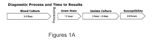
FIGURE 1A is a schematic of a current diagnostic microbiology flow for
isolation and identification
of a pathogen from blood followed by AST. Following blood draw, blood cultures
are grown for 1 to 5
days, followed by a gram stain, culture isolation, and susceptibility testing.
The approximate time to
results for each step of the diagnostic flow is indicated.
7
CA 03011991 2018-07-18
WO 2017/127727
PCT/US2017/014405
FIGURE 1B is a schematic of a diagnostic and therapeutic method in which
pathogen
identification by T2 magnetic resonance (T2MR) in a blood sample obtained from
a subject is followed
immediately by an appropriate targeted therapy against the identified
pathogen.
FIGURE 1C is a schematic of a diagnostic method that includes pathogen
identification by T2MR
in a blood sample obtained from a subject followed by subculture of the
pathogen under optimal
conditions for the identified pathogen. AST is performed on an portion of the
subculture. The
approximate time to results for each step of the method is indicated. A
skilled artisan appreciates that the
length of time required to isolate the culture and test susceptibility may
vary from hours to days based on
organism, titer, and growth conditions.
FIGURE 1D is a schematic of a diagnostic method that includes pathogen
identification and/or
expression analysis by T2MR in a blood sample obtained from a subject followed
immediately by AST or
detection of resistance markers.
FIGURE lE is a schematic of a diagnostic method that includes pathogen
identification by T2MR
in a blood sample obtained from a subject, followed by AST performed using a
T2MR-based detection
method. In this example, T2MR is performed a second time to quantitatively or
semi-quantitatively
measure microbial growth, providing more detailed information than standard
antimicrobial susceptibility
testing.
FIGURE 1F is a schematic of a diagnostic method that includes pathogen
identification and
expression analysis of key transcripts by T2MR in one or more blood samples
obtained from a subject.
Expression analysis is performed to monitor expression of inducible
antimicrobial resistance genes as
well as expression of energy metabolism or other housekeeping genes. This
analysis is optionally
followed by additional blood draws, and repeat of T2MR-based expression
analysis (e.g., real-time PCR
or RNA expression analysis of one or more genes that is characteristic of the
pathogen) to determine
effectiveness of treatment, which may be indicated by the decline in
expression of the one or more genes,
indicating cell growth arrest or death due to successful antimicrobial
therapy. This process may be
repeated as necessary to track antimicrobial susceptibility overtime. In some
embodiments, T2MR-
based direct cell detection (e.g., Lee et al., Nature Medicine 14(8):869-874,
2008) may be used to monitor
pathogen growth.
FIGURE 2 is a schematic showing exemplary downstream steps following positive
detection and
identification of a pathogen by T2MR ("T2 positive") from a blood culture
obtained from a subject.
Following species identification, a blood culture grown in parallel is sampled
at 3-5 hours and the
pathogen is concentrated (e.g., by lysis filtration or centrifugation). The
concentrated pathogen is then
plated to appropriate chromogenic media, enzymatic assays (e.g., a
carbapenemase Nordmann-Poirel
(Carba NP) test to detect carbapenemase-producing bacteria), an agar-based AST
method (e.g., disk
diffusion or ETEST , and/or an automated AST device (e.g., an automated full
panel AST device such as
VITEK 2, PHOENIX , or MICROSCANTm).
FIGURE 3 is a schematic showing exemplary downstream steps following positive
detection and
identification of a pathogen by T2MR ("T2 positive") from a blood culture
obtained from a subject.
Following pathogen identification, a lysis-centrifugation (e.g., ISOLATORTm)
blood culture system is used
to obtain a pellet containing the pathogen. The pellet is directly plated to
appropriate chromogenic media,
8
CA 03011991 2018-07-18
WO 2017/127727
PCT/US2017/014405
an agar-based AST method, and/or subjected to molecular assays (e.g., to
determine the presence
and/or activity of antimicrobial resistance markers).
DETAILED DESCRIPTION OF EMBODIMENTS OF THE INVENTION
The invention provides methods, panels, cartridges, kits, and systems for
rapid and sensitive
detection and identification of pathogens (e.g., identification of a genus to
which the pathogen belongs, or
more specifically, the species) and determination of the pathogen's
susceptibility to antimicrobial agents
for diagnosis and/or treatment of disease, including bloodstream infections
(e.g., bacteremia and
fungemia), sepsis, septic shock, septic arthritis, pneumonia, peritonitis,
osteomyeletis, meningitis,
empyema, urinary tract infection, and systemic inflammatory response syndrome
(SIRS). In some
embodiments, the invention provides methods for determination of an improved
antimicrobial therapy for
a subject suffering from an infection based on the susceptibility of the
infectious pathogen. The invention
also provides methods of treatment for disease, including bloodstream
infections (e.g., bacteremia and
fungemia), sepsis, septic shock, septic arthritis, pneumonia, peritonitis,
osteomyelitis, meningitis,
empyema, urinary tract infection, and SIRS, that involve administration of an
antimicrobial therapy based
on the susceptibility of the pathogen present in a biological sample obtained
from a subject.
The methods, panels, cartridges, and systems of the invention can be used for
rapid detection
and identification, along with rapid AST, of any suitable pathogen. In some
embodiments, the pathogen
is a bacterial pathogen, including Gram-positive bacteria (e.g., Gram-positive
anaerobic bacteria), Gram-
negative bacteria (e.g., Gram-negative anaerobic bacteria), Enterobacteriaceae
spp., Acinetobacter spp.
(e.g., Acinetobacter baumannii), Enterococcus spp. (e.g., Enterococcus faecium
and Enterococcus
faecalis), Klebsiella spp. (e.g., Klebsiella pneumoniae), Pseudomonas spp.
(e.g., Pseudomonas
aeruginosa), Staphylococcus spp. (including, e.g., coagulase-positive species
(e.g., Staphylococcus
aureus) and coagulase-negative (CoNS) species), Streptococcus spp. (e.g., [3-
hemolytic streptococci,
Streptococcus mitis, Streptococcus pneumoniae, Streptococcus agalactiae, and
Streptococcus
pyogenes), Escherichia spp. (e.g., Escherichia coli), Stenotrophomonas spp.
(e.g., Stenotrophomonas
maltophilia), Proteus spp. (e.g., Proteus mirabilis and Proteus vulgaris),
Serratia spp. (e.g., Serratia
marcescens), Citrobacter spp. (e.g., Citrobacter freundii), Enterobacter spp.
(e.g., Enterobacter
aerogenes and Enterobacter cloacae), Borrelia spp. (e.g., Borrelia
burgdorferi, Borrelia afzelii, and
Borrelia garinii), Rickettsia spp. (e.g., Rickettsia rickettsii), Anaplasma
spp. (e.g., Anaplasma
phagocytophilum), Coxiella spp. (e.g., Coxiella burnetii), Ehrlichia spp.
(e.g., Ehrlichia chaffeensis and
Ehrlichia ewingii), Franciscella spp. (e.g., Francisella tularensis),
Clostridium spp. (e.g., Clostridium
botulinum, Clostridium difficile, Clostridium perfringens, and Clostridium
tetani), Bacteroides spp. (e.g,.
Bacteroides fragilis), and Neisseria spp. (e.g., Neisseria meningitides). In
some embodiments, the
pathogen is a fungal pathogen, including Candida spp. (e.g., Candida albicans,
Candida guilliermondii,
Candida glabrata, Candida krusei, Candida lusitaniae, Candida parapsilosis,
and Candida tropicalis),
Saccharomyces spp., Aspergillus spp. (e.g., Aspergillus fumigatus, Aspergillus
clavatus, and Aspergillus
flavus), and Cryptococcus spp. (e.g., Cryptococcus neoformans, Cryptococcus
laurentii, and
Cryptococcus albidus). In some embodiments, the pathogen is a protozoan
pathogen, including Babesia
spp. (e.g., Babesia microti and Babesia divergens).
9
CA 03011991 2018-07-18
WO 2017/127727
PCT/US2017/014405
In some embodiments, the methods and systems of the invention employ magnetic
particles. In
some embodiments, the methods and systems employ an NMR unit, optionally one
or more magnetic
assisted agglomeration (MAA) units, optionally one or more incubation stations
at different temperatures,
optionally one or more vortexers, optionally one or more centrifuges,
optionally a fluidic manipulation
station, optionally a robotic system, and optionally one or more modular
cartridges, as described in
International Patent Application Publication No. WO 2012/054639, which is
incorporated herein by
reference in its entirety. In some embodiments, the methods and systems of the
invention may employ a
conduit-containing device, for example, as described in U.S. Patent
Application Publication No. US
2013/0265054, which is incorporated herein by reference in its entirety. In
some embodiments, the
methods of the invention may involve detecting cells by using magnetic
particles comprising a binding
agent that is operative to bind the cell surface of a pathogen and this
binding event can lead to a change
in measured signal such as a change in the measured T2MR value (see, e.g.,
International Patent
Application Publication No. WO 2012/129281; Skewis et al. Nuclear Magnetic
Resonance
Nanotechnology: Applications in Clinical Diagnostics and Monitoring.
Encyclopedia of Analytical
Chemistry, 2013; Kaittanis et al. Nano Left. 7:380, 2007; Lee et al. Nat. Med.
14:869, 2008; Kulkarni et al.
Anal. Chem. 82:7430, 2010; Chung et al. ACS Nano 5:8834, 2011; Liong et al.
Bioconjug. Chem.
22:2390, 2011; and Lee et al. Angew. Chem. Int. Ed. 48:5657, 2009). In some
embodiments, the
methods of the invention are performed (in full or in part) using a fully-
automated system, e.g., a T2Dx
instrument (T2 Biosystems, Inc., Lexington, Massachusetts, USA). The methods,
systems, devices,
panels, and cartridges of the invention can be used to assay a biological
sample (e.g., whole blood,
serum, plasma, cerebrospinal fluid (CSF), pleural fluid, urine, synovial
fluid, breast milk, sweat, tears,
saliva, semen, feces, vaginal fluid or tissue, sputum, nasopharyngeal aspirate
or swab, lacrimal fluid,
mucous, or epithelial swab (buccal swab), and tissues (e.g., tissue
homogenates), organs, bones, teeth,
among others).
Definitions
The terms "aggregation," "agglomeration," and "clustering" are used
interchangeably in the
context of the magnetic particles described herein and mean the binding of two
or more magnetic
particles to one another, for example, via a multivalent analyte, multimeric
form of analyte, antibody,
nucleic acid molecule, or other binding molecule or entity. In some instances,
magnetic particle
agglomeration is reversible. Such aggregation may lead to the formation of
"aggregates," which may
include amplicons and magnetic particles bearing binding moieties.
The terms "amplification" or "amplify" or derivatives thereof as used herein
mean one or more
methods known in the art for copying a target or template nucleic acid,
thereby increasing the number of
copies of a selected nucleic acid sequence. Amplification may be exponential
or linear. A target or
template nucleic acid may be either DNA or RNA (e.g., mRNA). The sequences
amplified in this manner
form an "amplified region," "amplified nucleic acid," or "amplicon." Primer
probes can be readily designed
by those skilled in the art to target a specific template nucleic acid
sequence.
By "analyte" is meant a substance or a constituent of a sample to be analyzed.
Exemplary
analytes include one or more species of one or more of the following: a
protein, a peptide, a polypeptide,
an amino acid, a nucleic acid, an oligonucleotide, RNA (e.g., mRNA), DNA, an
antibody, a carbohydrate,
CA 03011991 2018-07-18
WO 2017/127727
PCT/US2017/014405
a polysaccharide, glucose, a lipid, a gas (e.g., oxygen or carbon dioxide), an
electrolyte (e.g., sodium,
potassium, chloride, bicarbonate, blood urea nitrogen (BUN), magnesium,
phosphate, calcium, ammonia,
lactate), a lipoprotein, cholesterol, a fatty acid, a glycoprotein, a
proteoglycan, a lipopolysaccharide, a cell
surface marker (e.g., a cell surface protein of a pathogen), a cytoplasmic
marker (e.g., CD4/CD8 or
CD4/viral load), a therapeutic agent, a metabolite of a therapeutic agent, a
marker for the detection of a
weapon (e.g., a chemical or biological weapon), an organism, a pathogen, a
pathogen byproduct, a
parasite (e.g., a protozoan or a helminth), a protist, a fungus (e.g., yeast
or mold), a bacterium, an
actinomycete, a cell (e.g., a whole cell, a tumor cell, a stem cell, a white
blood cell, a T cell (e.g.,
displaying CD3, CD4, CD8, IL2R, CD35, or other surface markers), or another
cell identified with one or
more specific markers), a virus, a prion, a plant component, a plant by-
product, algae, an algae by-
product, plant growth hormone, an insecticide, a man-made toxin, an
environmental toxin, an oil
component, and components derived therefrom.
A "biological sample" is a sample obtained from a subject including but not
limited to whole blood,
serum, plasma, cerebrospinal fluid (CSF), pleural fluid, urine, synovial
fluid, breast milk, sweat, tears,
saliva, semen, feces, vaginal fluid or tissue, sputum, nasopharyngeal aspirate
or swab, lacrimal fluid,
mucous, or epithelial swab (buccal swab), tissues (e.g., tissue homogenates),
organs, bones, teeth,
among others.
A "biomarker" is a biological substance that can be used as an indicator of a
particular disease
state or particular physiological state of an organism, generally a biomarker
is a protein or other native
compound measured in bodily fluid whose concentration reflects the presence or
severity or staging of a
disease state or dysfunction, can be used to monitor therapeutic progress of
treatment of a disease or
disorder or dysfunction, or can be used as a surrogate measure of clinical
outcome or progression.
The term "growth" is used in its broadest sense, and includes changes in cell
size or metabolic
state as well as changes in cell number. "Growth" encompasses the growth of an
individual pathogen
cell, as well as the growth of a population of pathogen cells. Growth may
include cell division of a
pathogen cell into two daughter cells, as well as a pathogen increasing in
size over time without cell
division.
The term "conditions suitable for enhanced growth," as used herein,
encompasses conditions
(e.g., media, growth temperature, additives, oxygen content, and the like)
that lead to enhanced (e.g.,
increased) growth relative to a reference condition, which may be, for
example, a standard condition used
for growth of a microbial species when the identity of the microbial species
is not known.
The term "antimicrobial agent," as used herein, refers to any compound having
an inhibitory (or
antagonistic) effect on the growth of microorganisms, that is, agents that are
capable of at least reducing
the growth rate (e.g., bacteriostatic agents with respect to controlling the
growth of bacteria) as well as
agents that cause toxic effects (e.g., bactericide agents killing bacteria).
An antimicrobial agent may be
selected from small organic or inorganic molecules; saccharines;
oligosaccharides; polysaccharides;
biological macromolecules, e.g., peptides, proteins, and peptide analogs and
derivatives;
peptidomimetics; antibodies and antigen binding fragments thereof; nucleic
acids; nucleic acid analogs
and derivatives; glycogens or other sugars; immunogens; antigens; an extract
made from biological
materials such as bacteria, plants, fungi, or animal cells; animal tissues;
naturally occurring or synthetic
compositions; and any combinations thereof. As used herein, the term
"antimicrobial agent" includes
11
CA 03011991 2018-07-18
WO 2017/127727
PCT/US2017/014405
antibacterial agents, antifungal agents, antiviral agents, antiprotozoal
agents, antiviral agents, and
mixtures thereof.
Exemplary antibacterial agents include, but are not limited to, acrosoxacin,
amifioxacin, amikacin,
amoxycillin, ampicillin, aspoxicillin, azidocillin, azithromycin, aztreonam,
balofloxacin, benzylpenicillin,
biapenem, brodimoprim, cefaclor, cefadroxil, cefatrizine, cefcapene, cefdinir,
cefetamet, ceftmetazole,
cefoxitin, cefprozil, cefroxadine, ceftarolin, ceftazidime, ceftibuten,
ceftobiprole, cefuroxime, cephalexin,
cephalonium, cephaloridine, cephamandole, cephazolin, cephradine,
chlorquinaldol, chlortetracycline,
ciclacillin, cinoxacin, ciprofloxacin, clarithromycin, clavulanic acid,
clindamycin, clofazimine, cloxacillin,
colistin, danofloxacin, dapsone, daptomycin, demeclocycline, dicloxacillin,
difloxacin, doripenem,
doxycycline, enoxacin, enrofloxacin, erythromycin, fleroxacin, flomoxef,
flucloxacillin, flumequine,
fosfomycin, gentamycin, isoniazid, imipenem, kanamycin, levofloxacin,
linezolid, mandelic acid,
mecillinam, meropenem, metronidazole, minocycline, moxalactam, mupirocin,
nadifloxacin, nafcillin,
nalidixic acid, netilmycin, netromycin, nifuirtoinol, nitrofurantoin,
nitroxoline, norfloxacin, ofloxacin,
oxacillin, oxytetracycline, panipenem, pefloxacin, phenoxymethylpenicillin,
pipemidic acid, piromidic acid,
pivampicillin, pivmecillinam, polymixin-b, prulifloxacin, rufloxacin,
sparfloxacin, sulbactam,
sulfabenzamide, sulfacytine, sulfametopyrazine, sulphacetamide, sulphadiazine,
sulphadimidine,
sulphamethizole, sulphamethoxazole, sulphanilamide, sulphasomidine,
sulphathiazole, teicoplanin,
temafioxacin, tetracycline, tetroxoprim, tigecycline, tinidazole, tobramycin,
tosufloxacin, trimethoprim,
vancomycin, and pharmaceutically acceptable salts or esters thereof.
Exemplary antifungal agents include, but are not limited to, polyenes (e.g.,
amphotericin B,
candicidin, filipin, hamycin, natamycin, nystatin, and rimocidin), azoles
(e.g., imidazoles such as
bifonazole, butoconazole, clotrimazole, eberconazole, econazole,
fenticonazole, flutrimazole,
isoconazole, ketoconazole, luliconazole, miconazole, omoconazole, oxiconazole,
sertaconazole,
sulconazole, and tioconazole; triazoles such as albaconazole, efinaconazole,
epoxiconazole, fluconazole,
isavuconazole, itraconazole, posaconazole, propiconazole, ravuconazole,
terconazole, and voriconazole;
and thiazoles such as abafungin), allylamines (e.g., amorolfin, butenafine,
naftifine, and terbinafine),
echinocandins (e.g., anidulafungin, caspofungin, and micafungin), and other
antifungal agents including
but not limited to benzoic acid, ciclopirox olamine, 5-flucytosin,
griseofulvin, haloprogin, tolnaftate,
aminocandin, chlordantoin, chlorphenesin, nifuroxime, undecylenic acid,
crystal violet, and
pharmaceutically acceptable salts or esters thereof.
Exemplary antiprotozoal agents include, but are not limited to, acetarsol,
amphotericin (e.g.,
liposomal amphotericin, amphotericin B, and amphotericin deoxycholate),
arthemether, artsunate,
atovaquone, azanidazole, azithromycin, benznidazole, chloroquine,
ciprofloxacin, clindamycin, diloxanide,
eflornithine, flucytosine, fluconazole, folinic acid, hydroxychloroquine,
iodoquinol, lumefantrine,
macrolides, mefloquine, melarsoprol, metronidazole, miltefosine, nifuratel,
nifurtimox, nimorazole,
nitazoxanide, omidazole, paramomycin, pentamidine, primaquine, proguanil,
propenidazole,
pyrimethamine, quinine, quinidine, secnidazole, sinefungin, sodium
stibogluconate, spiramycin, suramin,
sulfadiazine, sulfamethoxazole, tenonitrozole, temidazole, tinidazole,
trimethoprim, TMP/SMX (co-
timoxazole; trimethoprim and sulfamethoxazole in a 1:5 ratio), and
pharmaceutically acceptable salts or
esters thereof. Additional antiprotozoal agents are described, for example, in
Kappagoda et al. Mayo
Clin. Proc. 86(6):561-583, 2011.
12
CA 03011991 2018-07-18
WO 2017/127727
PCT/US2017/014405
Exemplary antiviral agents include, but are not limited to, abacavir,
acyclovir, adefovir,
amantadine, amprenavir, ampligen, arbidol, atazanavir, atripla, balavir,
brivudine, cidofovir, combivir,
curcumin, darunavir, delavirdine, desciclovir, didanosine, 1-docosanol,
dolutegravir, edoxudine, efavirenz,
emtricitabine, enfuvirtide, entecavir, ecoliever, famciclovir, fiacitabine,
fomivirsen, fosamprenavir,
foscarnet, fosfonet, fusion inhibitors, ganciclovir, ibacitabine, idoxuridine,
imiquimod, imunovir, indinavir,
inosine, integrase inhibitor, interferon (e.g., interferon type I, interferon
type II, and interferon type III),
lamivudine, lopinavir, loviride, maraviroc, moroxydine, methisazone,
nelfinavir, nevirapine, nexavir,
nucleoside analogs, novir, oseltamivir, peginterferon alfa-2a, penciclovir,
peramivir, pleconaril,
podophyllotoxin, protease inhibitors, pyramidine, raltegravir, reverse
transcriptase inhibitors, ribavarin,
rimantadine, ritonavir, saquinavir, sofosbuvir, stavudine, telaprevir,
tenofovir, tenofovir disoproxil,
tipranavir, trifluridine, trizivir, tromontadine, truvada, valacyclovir,
valganciclovir, vicriviroc, vidarabine,
viramidine, zalcitabine, zanamivir, zidovudine, and pharmaceutically
acceptable salts or esters thereof.
By an "isolated" nucleic acid molecule is meant a nucleic acid molecule that
is removed from the
environment in which it naturally occurs. For example, a naturally-occurring
nucleic acid molecule
present in the genome of cell or as part of a gene bank is not isolated, but
the same molecule, separated
from the remaining part of the genome, as a result of, e.g., a cloning event,
amplification, or enrichment,
is "isolated." Typically, an isolated nucleic acid molecule is free from
nucleic acid regions (e.g., coding
regions) with which it is immediately contiguous, at the 5 or 3' ends, in the
naturally occurring genome.
Such isolated nucleic acid molecules can be part of a vector or a composition
and still be isolated, as
such a vector or composition is not part of its natural environment.
As used herein, "linked" means attached or bound by covalent bonds, non-
covalent bonds, and/or
linked via Van der Waals forces, hydrogen bonds, and/or other intermolecular
forces.
The term "magnetic particle" refers to particles including materials of high
positive magnetic
susceptibility such as paramagnetic compounds, superparamagnetic compounds,
and magnetite, gamma
ferric oxide, or metallic iron.
As used herein, "nonspecific reversibility" refers to the colloidal stability
and robustness of
magnetic particles against non-specific aggregation in a liquid sample and can
be determined by
subjecting the particles to the intended assay conditions in the absence of a
specific clustering moiety
(i.e., an analyte or an agglomerator). For example, nonspecific reversibility
can be determined by
measuring the T2 values of a solution of magnetic particles before and after
incubation in a uniform
magnetic field (defined as <5000 ppm) at 0.45T for 3 minutes at 37 C. Magnetic
particles are deemed to
have nonspecific reversibility if the difference in T2 values before and after
subjecting the magnetic
particles to the intended assay conditions vary by less than 10% (e.g., vary
by less than 9%, 8%, 6%, 4%,
3%, 2%, or 1%). If the difference is greater than 10%, then the particles
exhibit irreversibility in the buffer,
diluents, and matrix tested, and manipulation of particle and matrix
properties (e.g., coating and buffer
formulation) may be required to produce a system in which the particles have
nonspecific reversibility. In
another example, the test can be applied by measuring the T2 values of a
solution of magnetic particles
before and after incubation in a gradient magnetic field 1 Gauss/mm-10000
Gauss/mm.
As used herein, the term "NMR relaxation rate" refers to a measuring any of
the following in a
sample Ti, Tz, Ti/T2 hybrid, Tirho, Tzrho, and Tz". The systems and methods of
the invention are designed
to produce an NMR relaxation rate characteristic of whether an analyte is
present in the liquid sample. In
13
CA 03011991 2018-07-18
WO 2017/127727
PCT/US2017/014405
some instances the NMR relaxation rate is characteristic of the quantity of
analyte present in the liquid
sample.
As used herein, the term "Ti/T2 hybrid" refers to any detection method that
combines a Ti and a
T2 measurement. For example, the value of a Ti/T2 hybrid can be a composite
signal obtained through
the combination of, ratio, or difference between two or more different Ti and
T2 measurements. The Ti/T2
hybrid can be obtained, for example, by using a pulse sequence in which Ti and
T2 are alternatively
measured or acquired in an interleaved fashion. Additionally, the Ti/T2 hybrid
signal can be acquired with
a pulse sequence that measures a relaxation rate that is comprised of both Ti
and T2 relaxation rates or
mechanisms.
A "pathogen" means an agent causing disease or illness to its host, such as an
organism or
infectious particle, capable of producing a disease in another organism, and
includes but is not limited to
bacteria, viruses, protozoa, prions, yeast and fungi, or pathogen by-products.
"Pathogen by-products" are
those biological substances arising from the pathogen that can be deleterious
to the host or stimulate an
excessive host immune response, for example pathogen antigen(s), metabolic
substances, enzymes,
biological substances, or toxins. By "pathogen-associated analyte" is meant an
analyte characteristic of
the presence of a pathogen (e.g., a bacterium, fungus, or virus) in a sample.
The pathogen-associated
analyte can be a particular substance derived from a pathogen (e.g., a
protein, nucleic acid, lipid,
polysaccharide, or any other material produced by a pathogen) or a mixture
derived from a pathogen
(e.g., whole cells, or whole viruses). In certain instances, the pathogen-
associated analyte is selected to
be characteristic of the genus, species, or specific strain of pathogen being
detected. Alternatively, the
pathogen-associated analyte is selected to ascertain a property of the
pathogen, such as resistance to a
particular therapy. In some embodiments, a pathogen-associated analyte may be
a target nucleic acid
that has been amplified. In other embodiments, a pathogen-associated analyte
may be a host antibody
or other immune system protein that is expressed in response to an infection
by a pathogen (e.g., an IgM
antibody, an IgA antibody, an IgG antibody, or a major histocompatibility
complex (MHC) protein).
A "genus," as used herein, refers to a grouping of organisms, including
pathogens. In some
embodiments, a genus may be a taxonomic classification, for instance, a
taxonomic domain, a taxonomic
kingdom, a taxonomic phylum, a taxonomic class, a taxonomic order, a taxonomic
family, or a taxonomic
genus. In other embodiments, a genus may be defined by any desired or suitable
characteristics such
as, for example, resistance to an antimicrobial agent or gram staining. It is
to be understood that, in some
instances, a pathogen may belong to more than one genus.
The term "species," as used herein, refers to a basic unit of biological
classification as well as a
taxonomic rank. A skilled artisan appreciates that a species may be defined
based on a number of
criteria, including, for example, DNA similarity, morphology, and ecological
niche. The term encompasses
any suitable species concept, including evolutionary species, phylogenetic
species, typological species,
genetic species, and reproductive species. The term also encompasses
subspecies or strains.
By "pulse sequence" or "RF pulse sequence" is meant one or more radio
frequency pulses to be
applied to a sample and designed to measure, e.g., certain NMR relaxation
rates, such as spin echo
sequences. A pulse sequence may also include the acquisition of a signal
following one or more pulses
to minimize noise and improve accuracy in the resulting signal value.
14
CA 03011991 2018-07-18
WO 2017/127727
PCT/US2017/014405
As used herein, the term "signal" refers to an NMR relaxation rate, frequency
shift, susceptibility
measurement, diffusion measurement, or correlation measurements.
As used herein, reference to the "size" of a magnetic particle refers to the
average diameter for a
mixture of the magnetic particles as determined by microscopy, light
scattering, or other methods.
A "subject" is an animal, preferably a mammal (including, for example, rodents
(e.g., mice or
rats), farm animals (e.g., cows, sheep, horses, and donkeys), pets (e.g., cats
and dogs), or primates (e.g.,
non-human primates and humans)). In particular embodiments, the subject is a
human. A subject may
be a patient (e.g., a patient having or suspected of having a disease
associated with or caused by a
pathogen).
As used herein, the term "substantially monodisperse" refers to a mixture of
magnetic particles
having a polydispersity in size distribution as determined by the shape of the
distribution curve of particle
size in light scattering measurements. The FWHM (full width half max) of the
particle distribution curve
less than 25% of the peak position is considered substantially monodisperse.
In addition, only one peak
should be observed in the light scattering experiments and the peak position
should be within one
standard deviation of a population of known monodisperse particles.
By "T2 relaxivity per particle" is meant the average T2 relaxivity per
particle in a population of
magnetic particles.
As used herein, "unfractionated" refers to an assay in which none of the
components of the
sample being tested are removed following the addition of magnetic particles
to the sample and prior to
the NMR relaxation measurement.
It is contemplated that units, methods, systems, and processes of the claimed
invention
encompass variations and adaptations developed using information from the
embodiments described
herein. Throughout the description, where units and systems are described as
having, including, or
including specific components, or where processes and methods are described as
having, including, or
including specific steps, it is contemplated that, additionally, there are
units and systems of the present
invention that consist essentially of, or consist of, the recited components,
and that there are processes
and methods according to the present invention that consist essentially of, or
consist of, the recited
processing steps. It should be understood that the order of steps or order for
performing certain actions
is immaterial, unless otherwise specified, so long as the invention remains
operable. Moreover, in many
instances two or more steps or actions may be conducted simultaneously.
Magnetic Particles and NMR-based Detection
The methods and systems of the invention may involve use of magnetic particles
and NMR. The
magnetic particles can be coated with a binding moiety (e.g., oligonucleotide,
antibody, etc.) such that in
the presence of analyte, or multivalent binding agent, aggregates are formed.
Aggregation depletes
portions of the sample from the microscopic magnetic non-uniformities that
disrupt the solvent's T2 signal,
leading to an increase in T2 relaxation (see, e.g., Figure 3 of International
Patent Application Publication
No. WO 2012/054639, which is incorporated herein by reference in its
entirety).
The T2 measurement is a single measure of all spins in the ensemble,
measurements lasting
typically 1-10 seconds, which allows the solvent to travel hundreds of
microns, a long distance relative to
the microscopic non-uniformities in the liquid sample. Each solvent molecule
samples a volume in the
CA 03011991 2018-07-18
WO 2017/127727
PCT/US2017/014405
liquid sample and the T2 signal is an average (net total signal) of all
(nuclear spins) on solvent molecules
in the sample; in other words, the T2 measurement is a net measurement of the
entire environment
experienced by a solvent molecule, and is an average measurement of all
microscopic non-uniformities in
the sample.
The observed T2 relaxation rate for the solvent molecules in the liquid sample
is dominated by the
magnetic particles, which in the presence of a magnetic field form high
magnetic dipole moments. In the
absence of magnetic particles, the observed T2 relaxation rates for a liquid
sample are typically long (i.e.,
T2 (water) = approximately 2000 ms, T2 (blood) = approximately 1500 ms). As
particle concentration
increases, the microscopic non-uniformities in the sample increase and the
diffusion of solvent through
these microscopic non-uniformities leads to an increase in spin decoherence
and a decrease in the T2
value. The observed T2 value depends upon the particle concentration in a non-
linear fashion, and on the
relaxivity per particle parameter.
In the aggregation assays of the invention, the number of magnetic particles,
and if present the
number of agglomerant particles, remain constant during the assay. The spatial
distribution of the
particles changes when the particles cluster. Aggregation changes the average
"experience" of a solvent
molecule because particle localization into clusters is promoted rather than
more even particle
distributions. At a high degree of aggregation, many solvent molecules do not
experience microscopic
non-uniformities created by magnetic particles and the T2 approaches that of
solvent. As the fraction of
aggregated magnetic particles increases in a liquid sample, the observed T2 is
the average of the non-
uniform suspension of aggregated and single (unaggregated) magnetic particles.
The assays of the
invention are designed to maximize the change in T2 with aggregation to
increase the sensitivity of the
assay to the presence of analytes, and to differences in analyte
concentration.
In some embodiments, the methods of the invention involve contacting a
solution (e.g., a
biological sample) with between from 1x10⁶ to 1x10¹³ magnetic particles per
milliliter of the liquid sample
(e.g., from 1x10⁶ to 1x10⁸, 1x10⁷ to 1x10⁸, 1x10⁷ to 1x10⁹, 1x10⁸ to 1x10107
1x10⁹ to 1x10¹¹, or 1x101 to
1x10¹³ magnetic particles per milliliter).
In some embodiments, the magnetic particles used in the methods and systems of
the invention
have a mean diameter of from 150 nm to 1200 nm (e.g., from 150 nm to 250 nm,
200 nm to 350 nm, 250
nm to 450 nm, 300 nm to 500 nm, 450 nm to 650 nm, 500 nm to 700 nm, 700 nm to
850 nm, 800 nm to
950 nm, 900 nm to 1050 nm, or from 1000 nm to 1200 nm). For example, in some
embodiments, the
magnetic particles used in the methods of the invention may have a mean
diameter of from 150 nm to
699 nm (e.g., from 150 nm to 250 nm, 200 nm to 350 nm, 250 nm to 450 nm, 300
nm to 500 nm, 450 nm
to 650 nm, or from 500 nm to 699 nm). In other embodiments, the magnetic
particles used in the
methods of the invention may have a mean diameter of from 700 nm to 1200 nm
(e.g., from 700 nm to
850 nm, 800 nm to 950 nm, 900 nm to 1050 nm, or from 1000 nm to 1200 nm). In
particular
embodiments, the magnetic particles may have a mean diameter of from 700 nm to
950 nm (e.g., from
700 nm to 750 nm, 700 nm to 800 nm, 700 nm to 850 nm, or from 700 nm to 900
nm).
In some embodiments, the magnetic particles used in the methods of the
invention may have a T2
relaxivity per particle of from 1x10⁸ to Ix,' 012 ram-15-1 (e.g., from 1x10⁸
to 1x10⁹ mM-15-171x108 to 1x101
ram-15-171x109 to 1x101 mM-15-1, 1x10⁹ to 1x10¹¹ mM-15-1, or from 1x10¹⁰ t0
1x10¹² ram-15-1) .
In some
16
CA 03011991 2018-07-18
WO 2017/127727
PCT/US2017/014405
embodiments, the magnetic particles have a T2 relaxivity per particle of from
1x10⁹ to 1x10¹² mM-1s-1
(e.g., from 1 x10⁹ to 1 x101 mM-1s-1, 1 x10⁹ to 1 x10¹¹ mM-1s-1, or from 1
x101 to 1 x10¹² mM-1s-1).
In some embodiments, the magnetic particles may be substantially monodisperse.
In some
embodiments, the magnetic particles in a liquid sample (e.g., a biological
sample such as whole blood)
may exhibit nonspecific reversibility in the absence of the one or more
analytes and/or multivalent binding
agent. In some embodiments, the magnetic particles may further include a
surface decorated with a
blocking agent selected from albumin, fish skin gelatin, gamma globulin,
lysozyme, casein, peptidase,
and an amine-bearing moiety (e.g., amino polyethyleneglycol, glycine,
ethylenediamine, or amino
dextran.
Medical conditions
The methods of the invention can also be used to monitor and diagnose diseases
and other
medical conditions and to identify improved therapeutic regimens based on the
diagnosis, for instance,
based on AST results. In several embodiments, the methods of the invention may
be used to monitor
and diagnose disease in a multiplexed, automated, no sample preparation
system.
The methods and systems of the invention can be used to identify and monitor
the pathogenesis
of disease in a subject, to select therapeutic interventions, and to monitor
the effectiveness of the
selected treatment. For example, for a patient having or at risk of an
infectious disease, for example,
bloodstream infection (e.g., bacteremia or fungemia) and/or sepsis, the
methods and systems of the
invention can be used to identify the infectious pathogen, pathogen load, and
to monitor white blood cell
count and/or biomarkers indicative of the status of the infection. The
identity of the pathogen (e.g., a
genus to which the pathogen belongs or, more specifically, the species) can be
used to select an
appropriate therapy, for example, using AST, using methods described herein
and/or known in the art. In
some embodiments, the methods may further include administering a therapeutic
agent following
monitoring or diagnosing an infectious disease. The therapeutic intervention
(e.g., a particular
antimicrobial agent) can be monitored as well to correlate the treatment
regimen to the circulating
concentration of antimicrobial agent and pathogen load to ensure that the
patient is responding to
treatment. In some embodiments, antimicrobial resistance markers (e.g.,
antimicrobial resistance genes)
may be monitored following therapeutic intervention.
Exemplary diseases that can be diagnosed and/or monitored by the methods and
systems of the
invention include diseases caused by or associated with microbial pathogens
(e.g., bacterial infection,
fungal infection, viral infection, protozoan infection, Lyme disease,
bloodstream infection (e.g.,
bacteremia, fungemia, or viremia), pneumonia, peritonitis, osteomyeletis,
meningitis, empyema, urinary
tract infection, sepsis, septic shock, and septic arthritis) and diseases that
may manifest with similar
symptoms to diseases caused by or associated with microbial pathogens (e.g.,
SIRS). For example, the
methods and systems of the invention may be used to diagnose and/or monitor a
disease caused by the
following pathogens.
In some embodiments, the disease is caused by a bacterial pathogen, including
Gram-positive
bacteria (e.g., Gram-positive anaerobic bacteria), Gram-negative bacteria
(e.g., Gram-negative anaerobic
bacteria), Enterobacteriaceae spp., Acinetobacter spp. (e.g., Acinetobacter
baumannii), Enterococcus
spp. (e.g., Enterococcus faecium and Enterococcus faecalis), Klebsiella spp.
(e.g., Klebsiella
17
CA 03011991 2018-07-18
WO 2017/127727
PCT/US2017/014405
pneumoniae), Pseudomonas spp. (e.g., Pseudomonas aeruginosa), Staphylococcus
spp. (including, e.g.,
coagulase-positive species (e.g., Staphylococcus aureus) and coagulase-
negative (CoNS) species),
Streptococcus spp. (e.g., [3-hemolytic streptococci, Streptococcus mitis,
Streptococcus pneumoniae,
Streptococcus agalactiae, and Streptococcus pyogenes), Escherichia spp. (e.g.,
Escherichia cob),
Stenotrophomonas spp. (e.g., Stenotrophomonas maltophilia), Proteus spp.
(e.g., Proteus mirabilis and
Proteus vulgaris), Serratia spp. (e.g., Serratia marcescens), Citrobacter spp.
(e.g., Citrobacter freundh),
Enterobacter spp. (e.g., Enterobacter aerogenes and Enterobacter cloacae),
Borrelia spp. (e.g., Borrelia
burgdorferi, Borrelia afzelii, and Borrelia garinii), Rickettsia spp. (e.g.,
Rickettsia rickettsii), Anaplasma
spp. (e.g., Anaplasma phagocytophilum), Coxiella spp. (e.g., Coxiella
burnetii), Ehrlichia spp. (e.g.,
Ehrlichia chaffeensis and Ehrlichia ewingh), Franciscella spp. (e.g.,
Francisella tularensis), Clostridium
spp. (e.g., Clostridium botulinum, Clostridium difficile, Clostridium
perfringens, and Clostridium tetani),
Bacteroides spp. (e.g., Bacteroides fragilis), and Neisseria spp. (e.g.,
Neisseria meningitides). In other
embodiments, the disease is caused by a fungal pathogen, including Candida
spp. (e.g., Candida
albicans, Candida guilliermondii, Candida glabrata, Candida krusei, Candida
lusitaniae, Candida
parapsilosis, and Candida tropicalis), Saccharomyces spp., Aspergillus spp.
(e.g., Aspergillus fumigatus,
Aspergillus clavatus, and Aspergillus flavus), and Cryptococcus spp. (e.g.,
Cryptococcus neoformans,
Cryptococcus laurentii, and Cryptococcus albidus). In yet other embodiments,
the disease is caused by a
protozoan pathogen, including Babesia spp. (e.g., Babesia microti and Babesia
divergens). In some
embodiments, the disease is caused by a pathogen described in Pien et al. Am.
J. Med. 123:819-829,
2010.
Analytes
Embodiments of the invention include methods and systems for detecting and/or
measuring the
concentration of one or more analytes. In several embodiments, the analyte may
be a nucleic acid
derived from an organism (e.g., DNA or RNA (e.g., mRNA)). In some embodiments,
the nucleic acid is
DNA. In other embodiments, the nucleic acid is RNA (e.g., mRNA). In some
embodiments, the nucleic
acid is a target nucleic acid derived from the organism that has been
amplified to form an amplicon.
In some embodiments, the analyte may be derived from a microbial pathogen. For
instance,
pathogen-associated analytes may include or be derived from a bacterial
pathogen including Gram-
positive bacteria (e.g., Gram-positive anaerobic bacteria), Gram-negative
bacteria (e.g., Gram-negative
anaerobic bacteria), Enterobacteriaceae spp., Acinetobacter spp. (e.g.,
Acinetobacter baumannii),
Enterococcus spp. (e.g., Enterococcus faecium and Enterococcus faecalis),
Klebsiella spp. (e.g.,
Klebsiella pneumoniae), Pseudomonas spp. (e.g., Pseudomonas aeruginosa),
Staphylococcus spp.
(including, e.g., coagulase-positive species (e.g., Staphylococcus aureus) and
coagu lase-negative
(CoNS) species), Streptococcus spp. (e.g., [3-hemolytic streptococci,
Streptococcus mitis, Streptococcus
pneumoniae, Streptococcus agalactiae, and Streptococcus pyogenes), Escherichia
spp. (e.g.,
Escherichia cob), Stenotrophomonas spp. (e.g., Stenotrophomonas maltophilia),
Proteus spp. (e.g.,
Proteus mirabilis and Proteus vulgaris), Serratia spp. (e.g., Serratia
marcescens), Citrobacter spp. (e.g.,
Citrobacter freundii), Enterobacter spp. (e.g., Enterobacter aerogenes and
Enterobacter cloacae),
Borrelia spp. (e.g., Borrelia burgdorferi, Borrelia afzelii, and Borrelia
garinii), Rickettsia spp. (e.g.,
Rickettsia rickettsii), Anaplasma spp. (e.g., Anaplasma phagocytophilum),
Coxiella spp. (e.g., Coxiella
18
CA 03011991 2018-07-18
WO 2017/127727
PCT/US2017/014405
burnetiO, Ehrlichia spp. (e.g., Ehrlichia chaffeensis and Ehrlichia ewingh),
Franciscella spp. (e.g.,
Francisella tularensis), Clostridium spp. (e.g., Clostridium botulinum,
Clostridium difficile, Clostridium
perfringens, and Clostridium tetani), Bacteroides spp. (e.g., Bacteroides
fragilis), and Neisseria spp. (e.g.,
Neisseria meningitides). In other embodiments, pathogen-associated analytes
may include or be derived
from a fungal pathogen, including Candida spp. (e.g., Candida albicans,
Candida guilliermondii, Candida
glabrata, Candida krusei, Candida lusitaniae, Candida parapsilosis, and
Candida tropicalis),
Saccharomyces spp., Aspergillus spp. (e.g., Aspergillus fumigatus, Aspergillus
clavatus, and Aspergillus
t7avus), and Cryptococcus spp. (e.g., Cryptococcus neoformans, Cryptococcus
laurentii, and
Cryptococcus albidus). In yet other embodiments, pathogen-associated analytes
may include or be
derived from a protozoan pathogen, including Babesia spp. (e.g., Babesia
microti and Babesia
divergens). In still further embodiments, pathogen-associated analytes may
include or be derived from a
viral pathogen. In some embodiments, pathogen-associated analytes may include
or be derived from a
pathogen associated with bloodstream infection (e.g., bacteremia or fungemia),
invasive bacterial
infection, pneumonia, peritonitis, osteomyeletis, meningitis, empyema, urinary
tract infection, sepsis,
septic shock, septic arthritis, SIRS, or Lyme disease. In some embodiments,
the pathogen-associated
analyte includes or is derived from a pathogen described in Pien et al. Am. J.
Med. supra.
In some embodiments, the analyte is characteristic of a genus of pathogens.
For instance, in
some embodiments, the analyte may be characteristic of bacterial pathogens,
fungal pathogens, viral
pathogens, or protozoan pathogens. In another example, in the case of
bacterial pathogens, the analyte
may be characteristic of Gram-positive bacteria, Gram-negative bacteria,
Enterobacteriaceae spp.,
Acinetobacter spp., Enterococcus spp., Klebsiella spp., Pseudomonas spp.,
Staphylococcus spp.,
Streptococcus spp., Escherichia spp., Stenotrophomonas spp., Proteus spp.,
Serratia spp., Citrobacter
spp., Enterobacter spp., Borrelia spp., Rickettsia spp., Anaplasma spp.,
Coxiella spp., Ehrlichia spp.,
Franciscella spp., Clostridium spp., Bacteroides spp. Neisseria spp., or other
groupings of bacterial
pathogens recognized in the art, for instance, [3-hemolytic streptococci,
fastidious Gram-negative
bacteria, and the like.
In other instances, the analyte is characteristic of a particular pathogen
species. For example, in
some embodiments, the analyte may be characteristic of any of the pathogen
species described above or
that is known in the art (e.g., any species listed in Pien et al., supra).
In some embodiments, a pathogen-associated analyte may be a nucleic acid
derived from any of
the organisms described above. In some embodiments, the nucleic acid is a
target nucleic acid derived
from the organism that has been amplified to form an amplicon. In some
embodiments, the nucleic acid
is DNA. In other embodiments, the nucleic acid is RNA (e.g., mRNA). In some
embodiments, the target
nucleic acid may be a multi-copy locus. Use of a target nucleic acid derived
from a multi-copy locus, in
particular in methods involving amplification, may lead to an increase in
sensitivity in the assay.
Exemplary multi-copy loci may include, for example, ribosomal DNA (rDNA)
operons and multi-copy
plasmids. In other embodiments, the target nucleic acid may be a single-copy
locus. In particular
embodiments, the target nucleic acid may be derived from an essential locus,
for example, an essential
house-keeping gene. In particular embodiments, the target nucleic acid may be
derived from a locus that
is involved in virulence (e.g., a virulence gene). In some instances, the
target nucleic acid may be
derived from an antibiotic resistance gene (e.g., an inducible antibiotic
resistance gene). Any suitable
19
CA 03011991 2018-07-18
WO 2017/127727
PCT/US2017/014405
antibiotic resistance gene known in the art or described herein may be used.
In any of the above
embodiments, a locus may include a gene and/or an intragenic region, for
example, an internally
transcribed sequence (ITS) between rRNA genes (e.g., ITS1, between the 16S and
23S rRNA genes, or
IT52, between the 5S and 23S rRNA genes).
In some embodiments, a target nucleic acid may be (a) genus-specific, (b)
genus-inclusive (in
other words, present in all species in a given genus), (c) compatible with an
amplification/detection
protocol, and/or (d) present in multiple copies). In some embodiments, a
target nucleic acid may be (a)
species-specific, (b) species-inclusive (in other words, present in all
strains or subspecies of a given
species), (c) compatible with an amplification/detection protocol, and/or (d)
present in multiple copies. In
particular embodiments, a target nucleic acid is chromosomally-encoded, which
can help avoid loss by,
for example, plasmid exchange and plasmid curing/transduction events.
In some embodiments, a target nucleic acid may allow for identification of a
genus to which the
pathogen belongs without allowing for identification of the particular
species. In other embodiments, a
target nucleic acid allows for identification of the particular species of the
pathogen, which will typically
identify the genus as well.
In some embodiments, a target nucleic acid may be a control nucleic acid. Such
a control nucleic
acid may serve as a reference for detection of a pathogen.
Acinetobacter target nucleic acids
In some embodiments, a target nucleic acid may include sequence elements that
are specific for
an Acinetobacter spp., for example, Acinetobacter baumannii. For example, in
some embodiments, an
Acinetobacter baumannii target nucleic acid may be amplified in the presence
of a forward primer and a
reverse primer which are specific to Acinetobacter baumannii, as described
below. Detection of such a
target nucleic acid in a sample would typically indicate that an Acinetobacter
baumannii bacterium was
present in the sample. In other embodiments, a target nucleic acid of the
invention may include
sequence elements that are common to all Acinetobacter spp. For example, in
some embodiments, an
Acinetobacter spp. target nucleic acid may be amplified in the presence of a
forward primer and a reverse
primer, each of which is universal to all Acinetobacter spp. Detection of such
a target nucleic acid in a
sample typically would indicate that an Acinetobacter spp. bacterium was
present in the sample. In yet
other embodiments, these approaches may be combined.
In some embodiments, an Acinetobacter spp. target nucleic acid may be derived
from a linear
chromosome or a linear or circular plasmid (e.g., a single-, low-, or multi-
copy plasmid). In some
embodiments, an Acinetobacter spp. target nucleic acid may be derived from an
essential locus (e.g., an
essential housekeeping gene) or a locus involved in virulence (e.g., a gene
essential for virulence). In
some embodiments, an Acinetobacter spp. target nucleic acid may be derived
from a multi-copy locus. In
other embodiments, an Acinetobacter spp. target nucleic acid may be derived
from a multi-copy plasmid.
In some embodiments, an Acinetobacter baumannii target nucleic acid is derived
from a region
that includes parts or all of the internally transcribed sequence (ITS)
between the 5S and 23S rRNA
genes (i.e., the IT52 region). In particular embodiments, an Acinetobacter
baumannii target nucleic acid
may be amplified in the presence of a forward primer that includes the
oligonucleotide sequence 5'-CGT
TTT CCA AAT CTG TAA CAG ACT GGG-3 (SEQ ID NO: 1) or 5'-GGA AGG GAT CAG GTG GTT
CAC
CA 03011991 2018-07-18
WO 2017/127727
PCT/US2017/014405
TCT T-3' (SEQ ID NO: 69) and a reverse primer that includes the
oligonucleotide sequence 5'- AGG ACG
TTG ATA GG TTG GAT GTG GA-3 (SEQ ID NO: 2). For example, in particular
embodiments, an
Acinetobacter baumannii target nucleic acid may be amplified in the presence
of a forward primer that
includes the oligonucleotide sequence 5'-GGA AGG GAT CAG GTG GTT CAC TCT T-3'
(SEQ ID NO:
.. 69) and a reverse primer that includes the oligonucleotide sequence 5'- AGG
ACG TTG ATA GG TTG
GAT GTG GA-3' (SEQ ID NO: 2). In some embodiments, an amplicon produced using
these primers is
detected by hybridization using a 5' capture probe that includes the
oligonucleotide sequence 5'-TGA
GGC TTG ACT ATA CAA CAC C-3' (SEQ ID NO: 15) and/or a 3' capture probe that
includes the
oligonucleotide sequence 5'- CTA AAA TGA ACA GAT AAA GTA AGA TTC AA-3' (SEQ ID
NO: 16) to
detect the presence of Acinetobacter baumannii in a biological sample. In some
embodiments, an
amplicon produced using these primers is detected by hybridization using a 5'
capture probe that includes
the oligonucleotide sequence of SEQ ID NO: 17 and/or a 3' capture probe that
includes the
oligonucleotide sequence of SEQ ID NO: 18 to detect the presence of
Acinetobacter baumannii in a
biological sample. In some embodiments, the 5' capture probe and/or the 3'
capture probe is conjugated
.. to a magnetic nanoparticle.
In some embodiments, a control target nucleic acid for A. baumannii may
comprise the nucleic
acid sequence of SEQ ID NO: 45.
Enterococcus target nucleic acids
In some embodiments, a target nucleic acid may include sequence elements that
are specific for
an Enterococcus spp., for example, Enterococcus faecium or Enterococcus
faecalis. For example, in
some embodiments, an Enterococcus faecium target nucleic acid may be amplified
in the presence of a
forward primer and a reverse primer which are specific to Enterococcus
faecium. Detection of such a
target nucleic acid in a sample would typically indicate that an Enterococcus
faecium bacterium was
present in the sample. In other embodiments, a target nucleic acid may include
sequence elements that
are specific for multiple (e.g., 2, 3, 4, or 5) Enterococcus spp. For example,
in some embodiments, a
target nucleic acid may include sequence elements that are specific for
Enterococcus faecium and
Enterococcus faecalis, as described below. In other embodiments, a target
nucleic acid of the invention
may include sequence elements that are common to all Enterococcus spp. For
example, in some
embodiments, an Enterococcus spp. target nucleic acid may be amplified in the
presence of a forward
primer and a reverse primer, each of which is universal to all Enterococcus
spp. Detection of such a
target nucleic acid in a sample typically would indicate that an Enterococcus
spp. bacterium was present
in the sample. In yet other embodiments, these approaches may be combined.
In some embodiments, an Enterococcus spp. target nucleic acid may be derived
from a linear
.. chromosome or a linear or circular plasmid (e.g., a single-, low-, or multi-
copy plasmid). In some
embodiments, an Enterococcus spp. target nucleic acid may be derived from an
essential locus (e.g., an
essential housekeeping gene) or a locus involved in virulence (e.g., a gene
essential for virulence). In
some embodiments, an Enterococcus spp. target nucleic acid may be derived from
a multi-copy locus. In
particular embodiments, an Enterococcus spp. target nucleic acid may be
derived from a multi-copy
plasmid.
21
CA 03011991 2018-07-18
WO 2017/127727
PCT/US2017/014405
In some embodiments, an Enterococcus spp. target nucleic acid is derived from
a region that
includes parts or all of the ITS between the 23S and 5S rRNA genes. In
particular embodiments, an
target nucleic acid that is specific for Enterococcus faecium and Enterococcus
faecalis may be amplified
in the presence of a forward primer that includes the oligonucleotide sequence
5'-GGT AGC TAT GTA
GGG AAG GGA TAA ACG CTG A-3 (SEQ ID NO: 3) and a reverse primer that includes
the
oligonucleotide sequence 5'-GCG CTA AGG AGC TTA ACT TCT GTG TTC G-3' (SEQ ID
NO: 4). In
some embodiments, an amplicon produced using these primers is detected by
hybridization using a 5'
capture probe that includes the oligonucleotide sequence 5'-AAA ACT TAT ATG
ACT TCA AAT CCA
GTT TT-3' (SEQ ID NO: 19) or 5'-AAA ACT TAT GTG ACT TCA AAT CCA GTT TT-3' (SEQ
ID NO: 70)
.. and/or a 3' capture probe that includes the oligonucleotide sequence 5'-TTT
ACT CAA TAA AAG ATA
ACA CCA CAG-3' (SEQ ID NO: 20) or 5'-TTT ACT CAA TAA AAG ATA ACA CCA CAG T-3'
(SEQ ID
NO 71) to detect the presence of Enterococcus faecium in a biological sample.
In particular
embodiments, an amplicon produced using these primers is detected by
hybridization using a 5' capture
probe that includes the oligonucleotide sequence 5'-AAA ACT TAT GTG ACT TCA
AAT CCA GTT TT-3'
(SEQ ID NO: 70) and/or a 3' capture probe that includes the oligonucleotide
sequence 5'-TTT ACT CAA
TAA AAG ATA ACA CCA CAG T-3' (SEQ ID NO: 71) to detect the presence of
Enterococcus faecium in a
biological sample. In some embodiments, an amplicon produced using these
primers is detected by
hybridization using a 5' capture probe that includes the oligonucleotide
sequence of SEQ ID NO: 21
and/or a 3' capture probe that includes the oligonucleotide sequence of SEQ ID
NO: 22 to detect the
presence of Enterococcus faecium in a biological sample. In some embodiments,
an amplicon produced
using these primers is detected by hybridization using a 5' capture probe that
includes the oligonucleotide
sequence 5'-TGG ATA AGT AAA AGC AAC TTG GTT-3' (SEQ ID NO: 23) and/or a 3'
capture probe that
includes the oligonucleotide sequence 5'-AAT GAA GAT TCA ACT CAA TAA GAA ACA
ACA-3' (SEQ ID
NO: 24) to detect the presence of Enterococcus faecalis in a biological
sample. In some embodiments,
an amplicon produced using these primers is detected by hybridization using a
5' capture probe that
includes the oligonucleotide sequence of SEQ ID NO: 25 and/or a 3' capture
probe that includes the
oligonucleotide sequence of SEQ ID NO: 26 to detect the presence of
Enterococcus faecalis in a
biological sample. In some embodiments, the 5' capture probe and/or the 3'
capture probe is conjugated
to a magnetic nanoparticle.
In some embodiments, a control target nucleic acid for Enterococcus faecium
may comprise the
nucleic acid sequence of SEQ ID NO: 46. In other embodiments, a control target
nucleic acid for
Enterococcus faecium may comprise the nucleic acid sequence of SEQ ID NO: 77.
In some
embodiments, a control target nucleic acid for Enterococcus faecalis may
comprise the nucleic acid
sequence of SEQ ID NO: 47.
Klebsiella target nucleic acids
In some embodiments, a target nucleic acid may include sequence elements that
are specific for
a Klebsiella spp., for example, Klebsiella pneumoniae. For example, in some
embodiments, a Klebsiella
pneumoniae target nucleic acid may be amplified in the presence of a forward
primer and a reverse
primer which are specific to Klebsiella pneumoniae, as described below.
Detection of such a target
nucleic acid in a sample would typically indicate that a Klebsiella pneumoniae
bacterium was present in
22
CA 03011991 2018-07-18
WO 2017/127727
PCT/US2017/014405
the sample. In other embodiments, a target nucleic acid of the invention may
include sequence elements
that are common to all Klebsiella spp. For example, in some embodiments, a
Klebsiella spp. target
nucleic acid may be amplified in the presence of a forward primer and a
reverse primer, each of which is
universal to all Klebsiella spp. Detection of such a target nucleic acid in a
sample typically would indicate
that a Klebsiella spp. bacterium was present in the sample. In yet other
embodiments, these approaches
may be combined.
In some embodiments, a Klebsiella spp. target nucleic acid may be derived from
a linear
chromosome or a linear or circular plasmid (e.g., a single-, low-, or multi-
copy plasmid). In some
embodiments, a Klebsiella spp. target nucleic acid may be derived from an
essential locus (e.g., an
essential housekeeping gene) or a locus involved in virulence (e.g., a gene
essential for virulence). In
some embodiments, a Klebsiella spp. target nucleic acid may be derived from a
multi-copy locus. In
particular embodiments, a Klebsiella spp. target nucleic acid may be derived
from a multi-copy plasmid.
In some embodiments, a Klebsiella pneumoniae target nucleic acid is derived
from a 23S rRNA
gene. In particular embodiments, a Klebsiella pneumoniae target nucleic acid
may be amplified in the
presence of a forward primer that includes the oligonucleotide sequence 5'-GAC
GGT TGT CCC GGT
TTA AGC A-3 (SEQ ID NO: 5) and a reverse primer that includes the
oligonucleotide sequence 5'-GCT
GGT ATC TTC GAC TGG TCT-3' (SEQ ID NO: 6). In some embodiments, an amplicon
produced using
these primers is detected by hybridization using a 5' capture probe that
includes the oligonucleotide
sequence 5'-TAC CAA GGC GCT TGA GAG AAC TC-3' (SEQ ID NO: 27) and/or a 3'
capture probe that
includes the oligonucleotide sequence 5'-CTG GTG TGT AGG TGA AGT C-3' (SEQ ID
NO: 28) to detect
the presence of Klebsiella pneumoniae in a biological sample. In some
embodiments, an amplicon
produced using these primers is detected by hybridization using a 5' capture
probe that includes the
oligonucleotide sequence of SEQ ID NO: 29 and/or a 3' capture probe that
includes the oligonucleotide
sequence of SEQ ID NO: 30 to detect the presence of Klebsiella pneumoniae in a
biological sample. In
some embodiments, the 5' capture probe and/or the 3' capture probe is
conjugated to a magnetic
nanoparticle.
In some embodiments, a control target nucleic acid for Klebsiella pneumoniae
may comprise the
nucleic acid sequence of SEQ ID NO: 48.
Pseudomonas target nucleic acids
In some embodiments, a target nucleic acid may include sequence elements that
are specific for
a Pseudomonas spp., for example, Pseudomonas aeruginosa. For example, in some
embodiments, a
Pseudomonas aeruginosa target nucleic acid may be amplified in the presence of
a forward primer and a
reverse primer which are specific to Pseudomonas aeruginosa, as described
below. Detection of such a
target nucleic acid in a sample would typically indicate that a Pseudomonas
aeruginosa bacterium was
present in the sample. In other embodiments, a target nucleic acid of the
invention may include
sequence elements that are common to all Pseudomonas spp. For example, in some
embodiments, a
Pseudomonas spp. target nucleic acid may be amplified in the presence of a
forward primer and a
reverse primer, each of which is universal to all Pseudomonas spp. Detection
of such a target nucleic
acid in a sample typically would indicate that a Pseudomonas spp. bacterium
was present in the sample.
In yet other embodiments, these approaches may be combined.
23
CA 03011991 2018-07-18
WO 2017/127727
PCT/US2017/014405
In some embodiments, a Pseudomonas spp. target nucleic acid may be derived
from a linear
chromosome or a linear or circular plasmid (e.g., a single-, low-, or multi-
copy plasmid). In some
embodiments, a Pseudomonas spp. target nucleic acid may be derived from an
essential locus (e.g., an
essential housekeeping gene) or a locus involved in virulence (e.g., a gene
essential for virulence). In
some embodiments, a Pseudomonas spp. target nucleic acid may be derived from a
multi-copy locus. In
particular embodiments, a Pseudomonas spp. target nucleic acid may be derived
from a multi-copy
plasmid.
In some embodiments, a Pseudomonas aeruginosa target nucleic acid is derived
from a region
that includes parts or all of the ITS between the 23S and 5S rRNA genes. In
particular embodiments, a
Pseudomonas aeruginosa target nucleic acid may be amplified in the presence of
a forward primer that
includes the oligonucleotide sequence 5'-AGG CTG GGT GTG TAA GCG TTG T-3 (SEQ
ID NO: 7) and a
reverse primer that includes the oligonucleotide sequence 5'-CAA GCA ATT CGG
TTG GAT ATC CGT T-
3' (SEQ ID NO: 8). In some embodiments, an amplicon produced using these
primers is detected by
hybridization using a 5' capture probe that includes the oligonucleotide
sequence 5'-GTG TGT TGT AGG
GTG AAG TCG AC-3' (SEQ ID NO: 31) or 5'-TCT GAC GAT TGT GTG TTG TAA GG-3' (SEQ
ID NO: 73)
and/or a 3' capture probe that includes the oligonucleotide sequence 5'-CAC
CTT GAA ATC ACA TAC
CTG A-3' (SEQ ID NO: 32) or 5'-GGA TAG ACG TAA GCC CAA GC-3' (SEQ ID NO: 74)
to detect the
presence of Pseudomonas aeruginosa in a biological sample. In particular
embodiments, an amplicon
produced using these primers is detected by hybridization using a 5' capture
probe that includes the
oligonucleotide sequence 5'-TCT GAC GAT TGT GTG TTG TAA GG-3' (SEQ ID NO: 73)
and/or a 3'
capture probe that includes the oligonucleotide 5'-GGA TAG ACG TAA GCC CAA GC-
3' (SEQ ID NO: 74)
to detect the presence of Pseudomonas aeruginosa in a biological sample. In
some embodiments, an
amplicon produced using these primers is detected by hybridization using a 5'
capture probe that includes
the oligonucleotide sequence of SEQ ID NO: 33 and/or a 3' capture probe that
includes the
oligonucleotide sequence of SEQ ID NO: 34 to detect the presence of
Pseudomonas aeruginosa in a
biological sample. In some embodiments, the 5' capture probe and/or the 3'
capture probe is conjugated
to a magnetic nanoparticle.
In some embodiments, a control target nucleic acid for Pseudomonas aeruginosa
may comprise
the nucleic acid sequence of SEQ ID NO: 49.
Staphylococcus target nucleic acids
In some embodiments, a target nucleic acid may include sequence elements that
are specific for
a Staphylococcus spp., for example, Staphylococcus aureus. For example, in
some embodiments, a
Staphylococcus aureus target nucleic acid may be amplified in the presence of
a forward primer and a
reverse primer which are specific to Staphylococcus aureus, as described
below. Detection of such a
target nucleic acid in a sample would typically indicate that a Staphylococcus
aureus bacterium was
present in the sample. In other embodiments, a target nucleic acid of the
invention may include
sequence elements that are common to all Staphylococcus spp. For example, in
some embodiments, a
Staphylococcus spp. target nucleic acid may be amplified in the presence of a
forward primer and a
reverse primer, each of which is universal to all Staphylococcus spp.
Detection of such a target nucleic
acid in a sample typically would indicate that a Staphylococcus spp. bacterium
was present in the sample.
24
CA 03011991 2018-07-18
WO 2017/127727
PCT/US2017/014405
In yet other embodiments, these approaches may be combined.
In some embodiments, a Staphylococcus spp. target nucleic acid may be derived
from a linear
chromosome or a linear or circular plasmid (e.g., a single-, low-, or multi-
copy plasmid). In some
embodiments, a Staphylococcus spp. target nucleic acid may be derived from an
essential locus (e.g., an
essential housekeeping gene), a locus involved in virulence (e.g., a gene
essential for virulence), or a
gene involved in antibiotic resistance (e.g., femA and femB). In some
embodiments, a Staphylococcus
spp. target nucleic acid may be derived from a multi-copy locus. In particular
embodiments, a
Staphylococcus spp. target nucleic acid may be derived from a multi-copy
plasmid.
In some embodiments, a Staphylococcus aureus target nucleic acid is derived
from the femAB
.. operon. The femAB operon codes for two nearly identical approximately 50
kDa proteins involved in the
formation of the Staphylococcal pentaglycine interpeptide bridge in
peptidoglycan. These
chromosomally-encoded proteins are considered as factors that influence the
level of methicillin
resistance and as essential housekeeping genes. femB is one gene in the femA/B
operon, also referred
to as graR, the two component response regulator of methicillin resistance.
femB encodes a
aminoacyltransferase, whereas femA encodes a regulatory factor that is
essential for expression of femB
and therefore methicillin resistance expression. In some embodiments, a
Staphylococcus aureus target
nucleic acid is derived from the femA gene. For example, in particular
embodiments, a Staphylococcus
aureus target nucleic acid may be amplified in the presence of a forward
primer that includes the
oligonucleotide sequence 5'-GGT AAT GAATTA CCT /i6diPr/TC TCT GCT GGTTTC TTC
TT-3 (SEQ ID
.. NO: 9) and a reverse primer that includes the oligonucleotide sequence 5'-
ACC AGC ATC TTC
/i6diPr/GC ATC TTC TGT AAA-3' (SEQ ID NO: 10). Note that "/i6diPri" indicates
2,6-Diaminopurine. In
some embodiments, an amplicon produced using these primers is detected by
hybridization using a 5'
capture probe that includes the oligonucleotide sequence 5'-CCA TTT GAA GTT
GTT TAT TAT GC-3'
(SEQ ID NO: 35) and/or a 3' capture probe that includes the oligonucleotide
sequence 5'-GGG AAA TGA
.. TTA ATT ATG CAT TAA ATC-3' (SEQ ID NO: 36) to detect the presence of
Staphylococcus aureus in a
biological sample. In some embodiments, an amplicon produced using these
primers is detected by
hybridization using a 5' capture probe that includes the oligonucleotide
sequence of SEQ ID NO: 37
and/or a 3' capture probe that includes the oligonucleotide sequence of SEQ ID
NO: 38 to detect the
presence of Staphylococcus aureus in a biological sample. In some embodiments,
the 5' capture probe
.. and/or the 3' capture probe is conjugated to a magnetic nanoparticle.
In some embodiments, a Staphylococcus aureus target nucleic acid is derived
from the femB
gene. For example, in other particular embodiments, a Staphylococcus aureus
target nucleic acid may
be amplified in the presence of a forward primer that includes the
oligonucleotide sequence 5'-GAA GTT
ATG TTT /i6diPr/CT ATT CGA ATC GTG GTC CAGT-3' (SEQ ID NO: 11) and a reverse
primer that
includes the oligonucleotide sequence 5'-GTT GTA AAG CCA TGA TGC TCG TAA CCA-
3' (SEQ ID NO:
12). In some embodiments, an amplicon produced using these primers is detected
by hybridization using
a 5' capture probe that includes the oligonucleotide sequence 5'-TT TTT CAG
ATT TAG GAT TAG TTG
ATT-3' (SEQ ID NO: 39) and/or a 3' capture probe that includes the
oligonucleotide sequence 5'-GAT
CCG TAT TGG TTA TAT CAT C-3' (SEQ ID NO: 40) to detect the presence of
Staphylococcus aureus in
a biological sample. In some embodiments, an amplicon produced using these
primers is detected by
hybridization using a 5' capture probe that includes the oligonucleotide
sequence of SEQ ID NO: 41
CA 03011991 2018-07-18
WO 2017/127727
PCT/US2017/014405
and/or a 3' capture probe that includes the oligonucleotide sequence of SEQ ID
NO: 42 to detect the
presence of Staphylococcus aureus in a biological sample. In some embodiments,
the 5' capture probe
and/or the 3' capture probe is conjugated to a magnetic nanoparticle.
In some embodiments, a Staphylococcus aureus target nucleic acid includes all
or a portion of
both the femA gene and the femB gene.
In some embodiments, a control target nucleic acid for Staphylococcus aureus
femA may
comprise the nucleic acid sequence of SEQ ID NO: 50. In some embodiments, a
control target
nucleicacid for Staphylococcus aureus femB may comprise the nucleic acid
sequence of SEQ ID NO: 51.
Escherichia target nucleic acids
In some embodiments, a target nucleic acid may include sequence elements that
are specific for
an Escherichia spp., for example, Escherichia coli. For example, in some
embodiments, an Escherichia
coli target nucleic acid may be amplified in the presence of a forward primer
and a reverse primer which
are specific to Escherichia coli, as described below. Detection of such a
target nucleic acid in a sample
would typically indicate that an Escherichia coli bacterium was present in the
sample. In other
embodiments, a target nucleic acid of the invention may include sequence
elements that are common to
all Staphylococcus spp. For example, in some embodiments, an Escherichia spp.
target nucleic acid may
be amplified in the presence of a forward primer and a reverse primer, each of
which is universal to all
Escherichia spp. Detection of such a target nucleic acid in a sample typically
would indicate that a
Escherichia spp. bacterium was present in the sample. In yet other
embodiments, these approaches may
be combined.
In some embodiments, an Escherichia spp. target nucleic acid may be derived
from a linear
chromosome or a linear or circular plasmid (e.g., a single-, low-, or multi-
copy plasmid). In some
embodiments, an Escherichia spp. target nucleic acid may be derived from an
essential locus (e.g., an
essential housekeeping gene), a locus involved in virulence (e.g., a gene
essential for virulence), or a
gene involved in antibiotic resistance. In some embodiments, an Escherichia
spp. target nucleic acid may
be derived from a multi-copy locus. In particular embodiments, an Escherichia
spp. target nucleic acid
may be derived from a multi-copy plasmid.
In particular embodiments, an Escherichia coli target nucleic acid is derived
from the yfcL gene.
The yfcL gene is within an E. coli-specific Chaperone-Usher Fimbriae gene
cluster (see, e.g., Wurpelet al.
PLoS One Vol 8, e52835, 2013). For example, in other particular embodiments,
Escherichia coli yfcL
may be amplified in the presence of a forward primer that includes the
oligonucleotide sequence 5'-GCA
TTA ATC GAC GGT ATG GTT GAC C-3 (SEQ ID NO: 52) and a reverse primer that
includes the
oligonucleotide sequence 5'-CCT GCT GAA ACA GGT TTT CCC ACA TA-3' (SEQ ID NO:
53). In some
embodiments, an amplicon produced using these primers is detected by
hybridization using a 5' capture
probe that includes the oligonucleotide sequence 5'-AGT GAT GAT GAG TTG TTT
GCC AGT G-3' (SEQ
ID NO: 54) and/or a 3' capture probe that includes the oligonucleotide
sequence 5'-TGA ATT GTC GCC
GCG TGA CCA G-3' (SEQ ID NO: 55) to detect the presence of Escherichia coli in
a biological sample.
In some embodiments, the 5' capture probe and/or the 3' capture probe is
conjugated to a magnetic
nanoparticle.
26
CA 03011991 2018-07-18
WO 2017/127727
PCT/US2017/014405
Candida target nucleic acids
In some embodiments, a target nucleic acid may include sequence elements that
are specific for
a Candida spp. (e.g., Candida albicans, Candida guilliermondii, Candida
glabrata, Candida krusei,
Candida lusitaniae, Candida parapsilosis, and Candida tropicalis). For
example, in some embodiments, a
Candida albicans target nucleic acid may be amplified in the presence of a
forward primer and a reverse
primer which are specific to Candida albicans. Detection of such a target
nucleic acid in a sample would
typically indicate that a Candida albicans cell was present in the sample. In
other embodiments, a target
nucleic acid of the invention may include sequence elements that are common to
all Candida spp. For
example, in some embodiments, a Candida spp. target nucleic acid may be
amplified in the presence of a
forward primer and a reverse primer, each of which is universal to all Candida
spp., as described below.
Detection of such a target nucleic acid in a sample typically would indicate
that a Candida spp. cell was
present in the sample. In yet other embodiments, these approaches may be
combined.
In some embodiments, a Candida spp. target nucleic acid may be derived from a
linear
chromosome or a linear or circular plasmid (e.g., a single-, low-, or multi-
copy plasmid). In some
embodiments, a Candida spp. target nucleic acid may be derived from an
essential locus (e.g., an
essential housekeeping gene) or a locus involved in virulence (e.g., a gene
essential for virulence). In
some embodiments, a Candida spp. target nucleic acid may be derived from a
multi-copy locus. For
example, in some embodiments, a Candida spp. target nucleic acid may be
derived from a ribosomal
DNA operon.
Detection of a Candida species can be performed as described, for example, in
International
Patent Application Publication No. WO 2012/054639, which is incorporated
herein by reference in its
entirety. In particular embodiments, a Candida spp. target nucleic acid may be
amplified in the presence
of a forward primer that includes the oligonucleotide sequence 5'-GGC ATG CCT
GTT TGA GCG TC-3'
(SEQ ID NO: 13) and a reverse primer that includes the oligonucleotide
sequence 5'-GCT TAT TGA TAT
GCT TAA GTT CAG CGG GT-3 (SEQ ID NO: 14). The capture probes listed in Table 1
can be used for
detection of an amplicon produced by these primers to identify the presence of
the indicated Candida
species.
Table 1: Capture Probes for Detection of Candida spp.
Candida Capture Probes Sequence
Candida albicans Probe #1 ACC CAG CGG TTT GAG GGA GAA AC (SEQ ID NO: 56)
Candida albicans Probe #2 AAA GTT TGA AGA TAT ACG TGG TGG ACG TTA (SEQ ID NO:
57)
Candida krusei Probe #1 CGC ACG CGC AAG ATG GAA ACG (SEQ ID NO: 58)
Candida krusei Probe #2 AAG TTC AGC GGG TAT TCC TAC CT (SEQ ID NO: 59)
Candida krusei probe AGC TTT TTG TTG TCT CGC AAC ACT CGC (SEQ ID NO: 60)
Candida glabrata Probe #1 CTA CCA AAC ACA ATG TGT TTG AGA AG (SEQ ID NO:
61)
Candida glabrata Probe #2 CCT GAT TTG AGG TCA AAC TTA AAG ACG TCT G (SEQ ID
NO: 62)
Candida AGT CCT ACC TGA TTT GAG GTC NitIndlAA (SEQ ID NO:
63)
parapsilosis/tropicalis Probe
#1
27
CA 03011991 2018-07-18
WO 2017/127727
PCT/US2017/014405
Candida CCG NitIndiGG GTT TGA GGG AGA AAT (SEQ ID NO: 64)
parapsilosis/tropicalis Probe
#2
Candida tropicalis AAA GTT ATG AAATAA ATT GTG GTG GCC ACT AGC (SEQ ID
NO:
65)
Candida tropicalis ACC CGG GGGTTT GAG GGA GAA A (SEQ ID NO: 66)
Candida parapsilosis AGT CCT ACC TGA TTT GAG GTC GAA (SEQ ID NO: 67)
Candida parapsilosis CCG AGG GTT TGA GGG AGA AAT (SEQ ID NO: 68)
1. NitInd is 5 5-Nitroindole, a base that is capable of annealing with
any of the four DNA bases.
In some methods, a Candida amplicon produced by amplification of a Candida
target nucleic acid
in the presence of a forward primer comprising the oligonucleotide sequence 5'-
GGC ATG CCT GTT TGA
GCG TC-3' (SEQ ID NO: 13) and a reverse primer that includes the
oligonucleotide sequence 5'-GCT
TAT TGA TAT GCT TAA GTT CAG CGG GT-3' (SEQ ID NO: 14) is detected by
hybridization a first
nucleic acid probe and a second nucleic acid probe conjugated to one or more
populations of magnetic
particles. For example, certain embodiments, (i) the Candida species is
Candida albicans, the first probe
includes the oligonucleotide sequence 5'-ACC CAG CGG TTT GAG GGA GAA AC-3'
(SEQ ID NO: 56),
and the second probe includes the oligonucleotide sequence 5'-AAA GTT TGA AGA
TAT ACG TGG TGG
ACG TTA-3' (SEQ ID NO: 57); (ii) the Candida species is Candida krusei and the
first probe and the
second probe include an oligonucleotide sequence selected from: 5'-CGC ACG CGC
AAG ATG GAA
ACG-3' (SEQ ID NO: 58), 5'-AAG TTC AGC GGG TAT TCC TAC CT-3' (SEQ ID NO: 59),
and 5'-AGC
TTT TTG TTG TCT CGC AAC ACT CGC-3' (SEQ ID NO: 60); (iii) the Candida species
is Candida
glabrata, the first probe includes the oligonucleotide sequence: 5'-CTA CCA
AAC ACA ATG TGT TTG
AGA AG-3' (SEQ ID NO: 61), and the second probe includes the oligonucleotide
sequence: 5'-CCT GAT
TTG AGG TCA AAC TTA AAG ACG TCT G-3' (SEQ ID NO: 62); and (iv) the Candida
species is Candida
parapsilosis or Candida tropicalis and the first probe and the second probe
include an oligonucleotide
sequence selected from: 5'-AGT CCT ACC TGA TTT GAG GTCNitIndAA-3' (SEQ ID NO:
63), 5'-CCG
NitIndGG GTT TGA GGG AGA AAT-3' (SEQ ID NO: 64), 5'-AAA GTT ATG AAATAA ATT GTG
GTG
GCC ACT AGC-3' (SEQ ID NO: 65), 5'-ACC CGG GGGTTT GAG GGA GAA A-3' (SEQ ID NO:
66), 5'-
AGT CCT ACC TGA TTT GAG GTC GAA-3' (SEQ ID NO: 67), and 5'-CCG AGG GTT TGA GGG
AGA
AAT-3' (SEQ ID NO: 68). In some embodiments, the first probe comprises the
oligonucleotide sequence
of SEQ ID NO: 43 and the second probe comprises the oligonucleotide sequence
of SEQ ID NO: 44.
Antimicrobial Susceptibility Testing
The invention provides methods that involve determining the susceptibility of
a pathogen to one
or more antimicrobial agents (e.g., antimicrobial susceptibility testing
(AST)) that are based, at least in
part, on the rapid and sensitive detection of the presence and identity of a
pathogen associated with an
infection (e.g., the causative pathogen) by the methods of the invention. In
some embodiments, the
methods of the invention identify a genus to which the pathogen belongs. In
other embodiments, the
methods of the invention identify both the genus and the species to which the
pathogen belongs. In other
28
CA 03011991 2018-07-18
WO 2017/127727
PCT/US2017/014405
embodiments, the methods of the invention identify a genus to which the
pathogen belongs without
identifying the particular species. These methods are facilitated both by the
sensitivity (e.g. 1 CFU/mL
sensitivity, or 95% hit rate at 1 CFU/mL), as well as the fast turnaround time
(typically about 2-8 h, e.g.,
about 2 h, about 3h, about 4 h, about 5 h, about 6 h, about 7h, or about 8 h)
of the detection methods of
the invention. The rapid detection and identification of the pathogen by the
methods of the invention in
turn facilitates rapid and directed determination of susceptibility of the
pathogen to antimicrobial agents.
Following the detection and identification of a pathogen (e.g., at the genus
and/or species level)
by the methods of the invention, a sample (e.g., a pellet obtained from a
biological sample or a culture
thereof) of the pathogen is typically processed such that organisms in a
sample can be inoculated onto
media (e.g., chromogenic media) and/or an AST test system (e.g., an automated
system) to provide
phenotypic AST results. In some embodiments, AST involves comparing the growth
rate of the pathogen
in the presence of the antimicrobial agent to the growth rate of the pathogen
in the absence of the
antimicrobial agent. In other embodiments, AST may involve comparing one or
more functions of the
pathogen in the presence of the antimicrobial agent compared to the absence of
the antimicrobial agent.
For example, the function of the pathogen may be the expression of a gene
associated with the pathogen
(e.g., an antimicrobial resistance gene), an enzymatic activity (e.g., the
activity of a protein encoded by an
antimicrobial resistance gene, such as carbapenemase), production of a
metabolite or toxin, and the like.
In some embodiments, AST may involve measuring properties of blood and/or
growth media that are
affected by the presence of the pathogen, such as electrochemical methods that
measure a change in
impedance, resistance, capacitance, or voltage in the sample, as the pathogen
cultures may be
associated with a change in ionic strength, bulk susceptibility, bulk
capacitance, or resistance; methods
such as those capable of measuring metabolites such as gas chromatography or
mass spectrometry that
measure changes in the amount or distribution of one or more small molecule or
protein metabolites;
optical methods that detect cell growth by measuring changes in the light
transmittance of the sample; or
labeled methods that detect cell growth by means of a labeling moiety such as
a fluorescently-labeled
nucleic acid, peptide nucleic acid (PNA), antibody, aptamer, or other
targeting moiety that allows
measurement of the cells directly.
For example, in some instances, the methods of the invention may involve
obtaining a biological
sample from a subject (e.g., a patient suspected to be suffering from a BSI),
followed by determining the
presence and identity of a pathogen (e.g., at the genus and/or species level)
in a first portion of the
biological sample. In some instances, a second portion of the biological
sample is incubated in parallel in
order to culture the pathogen for downstream AST testing. For example, in
embodiments where the
biological sample is blood, the method may involve determining the presence
and identity of the pathogen
(e.g., at the genus and/or species level) in one portion of a blood sample,
and in parallel inoculating one
or more blood culture bottles to form a pathogen culture for AST testing. Once
the presence and identity
of the pathogen is determined by the methods of the invention, the blood
culture bottles may be sampled
immediately for AST testing, or a subculture can be inoculated in a growth
media that is more favorable
for growth of the pathogen. As one non-limiting example, if the pathogen is
identified as a Candida spp.,
the method of the invention may include inoculating a fungal blood culture
bottle (e.g., BACTECTm Myco/F
Lytic, BD) and/or use of a lysis centrifugation system (e.g., the ISOLATORTm
lysis centrifugation system,
Wampole Laboratories, Cranbury, N.J.) followed by plating onto chromogenic
media (e.g.,
29
CA 03011991 2018-07-18
WO 2017/127727
PCT/US2017/014405
CHROMAGARTm Candida, Chromagar, Paris, France), chocolate agar, and/or
Samouraud Glucose
media to encourage growth of Candida spp. and early growth for the isolated
colonies. The isolated
colonies may be subjected to AST using any of the methods described herein. In
some embodiments,
the inoculated agar plates are used for AST by placing E-TEST strips or disk
diffusion tests on the
media.
In other instances, an additional biological sample is obtained from the
subject following
determination of the presence and identity of the pathogen (e.g., at the genus
and/or species level) in a
first biological sample. In some instances, AST may be performed directly in
the additional biological
sample. In other instances, the additional biological sample is incubated in
media suitable for enhanced
growth of the identified pathogen. For example, if the identified pathogen is
a Candida species, the
method may involve obtaining another blood draw from the subject and
inoculating a fungal blood culture
bottle and/or use of a lysis centrifugation system (e.g., the ISOLATORTm lysis
centrifugation system,
Wampole Laboratories, Cranbury, NJ) followed by plating onto chromogenic media
(e.g.,
CHROMAGARTm Candida, Chromagar, Paris, France), chocolate agar and/or
Samouraud Glucose media
to encourage growth of Candida species and early growth of the isolated
colonies. The isolated colonies
may be subjected to AST using any of the methods described herein. In some
embodiments, the
inoculated agar plates are used for AST by placing ETEST strips or disk
diffusion tests on the media.
In some embodiments, the methods of the invention may involve addition of an
additive to the
growth medium of a culture (e.g., a blood culture) of the pathogen in order to
enhance growth. For
example, in some embodiments, the presence of free heme in a blood sample may
retard growth of a
microbial pathogen. In some embodiments, a heme detoxicification agent may be
added to a blood
sample or a subculture thereof, thereby enhancing growth of a pathogen
present. In some embodiments,
a heme detoxification agent may be an antioxidant, a heme polymerase, a
reducing agent, a buffering
agent, a free radical scavenger, or an agent that inactivates or eliminates
one or more antimicrobial
agents. In some embodiments, the method of the invention may involve removal
of a component of a
growth medium of a culture of the pathogen in order to enhance growth.
In some instances, any of the methods described herein may involve
centrifugation, filtration, lysis
centrifugation, and/or lysis concentration, which may be used, for example, to
concentrate pathogens. In
some instances, magnetic separation may be used to concentrate pathogens (see,
e.g., Aprodu et al. mt.
J. Food. MicrobioL 145(1):561-565, 2011). Any suitable approach for lysis
centrifugation may be used,
including using an ISOLATORTm lysis centrifugation system. In general, lysis
centrifugation may include
obtaining a biological sample (e.g., a blood sample) from a subject, placing
the sample in a culture tube
that includes a lysis agent (e.g., saponin or others described herein) under
conditions suitable for lysis of
cells in the sample (e.g., red and white blood cells), centrifuging the
culture tube to obtain pellets
containing a pathogen, removing the supernatant, and placing the pellet on one
or more media plates,
which may be selected based on the genus and/or species of the pathogen. The
media plates may then
be incubated under conditions suitable for growth (including enhanced growth)
in order to obtain biomass
(e.g., colonies), which can be used in AST analysis. In some instances, the
one or more media plates
may be used for AST, for example, by placing ETEST strips or disk diffusion
tests on the media plates.
A number of lysis centrifugation approaches are known in the art (see, e.g.,
&e'er et al. Acta Tropica
121(2):135-140, 2012 Rossmanith et al. J. MicrobioL Methods 69(3):504-511,
2007; Trovato et al. Clin.
CA 03011991 2018-07-18
WO 2017/127727
PCT/US2017/014405
MicrobioL Infect. 18:E63-E65, 2012; and Idelevich et al. J. Clin. MicrobioL
55(1):97-100, 2017), and any
suitable approach may be used in the context of the invention.
Any suitable method of AST can be used in the methods of the invention. For
example, any
method of AST described in Jorgensen et al. Clinical Infectious Diseases
49:1749-1755, 2009, which is
incorporated herein by reference in its entirety, may be used in the methods
of the invention. For
example, in some instances, the methods of the invention may include a broth
dilution test, a disk
diffusion test, an antimicrobial gradient test, growth on chromogenic media,
an enzyme activity assay,
and/or an automated instrument. In some instances, the broth diffusion test
may include macrobroth or
microdilution trays. In some instances, the antimicrobial gradient test may
include use of an epsilometer
test (e.g., an ETEST , bioMerieux SA, Marcy-l'Etoile, France).
In some instances, the methods of the invention include growth of the pathogen
on chromogenic
media. Any suitable chromogenic media may be used. In some embodiments, the
chromogenic media is
selected based on the identity of the pathogen (e.g., at the genus and/or
species level). For example,
suitable chromogenic media for Staphylococcus aureus (particularly methicillin-
resistant S. aureus,
MRSA) may include BRILLIANCETM MRSA agar (Oxoid, Basingstoke, United Kingdom),
CHROMIDTm
(bioMerieux), MRSASELECTTm (Bio-Rad, Hercules, CA), CHROMAGARTm MRSA
(Chromagar), and
BBLTM CHROMAGARTm (BD Diagnostics). Suitable chromogenic media for
Enterococcus spp. (e.g.,
vancomycin resistant Enterococci, VRE) may include BRILLIANCETM VRE agar
(Oxoid), CHROMIDTm
VRE (bioMerieux), and CHROMAGARTm VRE (Chromagar). Suitable chromogenic media
for gram
negative Bacilli expressing extended spectrum [3-Lactamase (ESBL) may include
BRILLIANCETM ESBL
agar (Oxoid), CHROMIDTm ESBL (bioMerieux), and HARDYCHROMTm ESBL agar (Hardy
Diagnostics,
Santa Maria, CA). Suitable chromogenic media for carbapenem-resistant
Enterobacteriaceae (CRE) may
include BRILLIANCETM CRE agar (Oxoid); HARDYCHROMTm CRE agar (Hardy
Diagnostics);
CHROMIDTm CARBA SMART (bioMerieux); and CHROMAGARTm KPC (Chromagar).
In some embodiments, the methods of the invention may involve AST using
automated systems,
including full-range automated AST devices. A number of automated systems are
known in the art. Non-
limiting examples include the VITEK 2 (bioMerieux), MICROSCAN (Beckman
Coulter, Brea, CA), and
PHOENIXTM (BD Diagnostics) systems. In some embodiments, the methods of the
invention may include
obtaining a pellet of the pathogen from blood culture or a subculture thereof,
for example, using lysis
centrifugation (e.g., SEPSITYPERTm, Bruker Corporation, Billerica, MA), lysis
filtration, or centrifugation
alone, followed by inoculation of an automated system.
In some embodiments, the methods of the invention may involve T2MR-based AST.
In some
embodiments, T2MR is used to determine the growth of pathogen cells present in
a biological sample or
subculture thereof containing antimicrobial agents or controls, for example,
in aliquots taken at time
intervals. In some embodiments, growth is determined by determining the level
of an analyte
characteristic of the pathogen (e.g., a nucleic acid (e.g., DNA or RNA (e.g.,
mRNA), protein, small
molecule, metabolite, growth media component, or cellular biproduct) overtime.
In other embodiments,
growth is determined by direct cell detection of pathogens, for example, as
described in International
Patent Application Publication No. WO 2012/129281; Skewis et al. Nuclear
Magnetic Resonance
Nanotechnology: Applications in Clinical Diagnostics and Monitoring.
Encyclopedia of Analytical
Chemistry, 2013; Kaittanis et al. Nano Left. 7:380, 2007; Lee et al. Nat. Med.
14:869, 2008; Kulkarni et al.
31
CA 03011991 2018-07-18
WO 2017/127727
PCT/US2017/014405
Anal. Chem. 82:7430, 2010; Chung et al. ACS Nano 5:8834, 2011; Liong et al.
Bioconjug. Chem.
22:2390, 2011; and Lee et al. Angew. Chem. mt. Ed. 48:5657, 2009. In some
embodiments, direct cell
detection is achieved using magnetic particles that include binding moieties
operative to bind to the cell
surface of the pathogen. For example, in some embodiments, the binding
moieties are operative to bind
to a surface-exposed protein (e.g., protein A of Staphylococcus aureus or
protein G of Streptococci) or a
cell wall component (e.g., D-alanyl-D-alanine or lipopolysaccharide (LPS)).
Binding of magnetic particles
to the pathogen or pathogen-associated analyte can lead to a change in the
T2MR signal that can be
used to indicate, detect, and/or monitor cell growth.
Any suitable number of antimicrobial agents may be tested in the methods of
the invention. For
example, in some embodiments, about 1 to about 30 (e.g., about 1, about 2,
about 3, about 4, about 5,
about 6, about 7, about 8, about 9, about 10, about 11, about 12, about 13,
about 14, about 15, about 16,
about 17, about 18, about 19, about 20, about 25, or about 30) or more
antimicrobial agents are tested in
the methods of the invention. The antimicrobial agents for testing are
typically selected based on the
identity (e.g., at the genus and/or species level) of the pathogen detected
and identified by the methods of
the invention. In some embodiments, the antimicrobial agents for testing are
based on local resistance
patterns. In some embodiments, the antimicrobial agents for testing are based
on both on the identity of
the pathogen species detected and identified by the methods of the invention
and on local resistance
patterns. The susceptibility or resistance of a pathogen to a given
antimicrobial agent may refer to a
direct interaction between the antimicrobial agent and the pathogen (which can
be measured in vitro) or
the likelihood that the subject will respond to treatment (which may depend in
part on dosing, dose
schedule, site of infection, pharmacokinetics (PK) of the antimicrobial
agent), and/or host defenses).
As a non-limiting example, if the pathogen is identified as an
Enterobacteriaceae (e.g.,
Escherichia spp. (e.g., E. cob), Klebsiella spp. (e.g., K. pneumoniae), or
Enterobacter spp.), the methods
of the invention may include testing from about 1 to about 20 (e.g., about 1,
about 2, about 3, about 4,
about 5, about 6, about 7, about 8, about 9, about 10, about 11, about 12,
about 13, about 14, about 15, a
out 16, about 17, about 18, about 19, or about 20) antimicrobial agents. In
some embodiments, the
antimicrobial agent is selected from one or more (e.g., 1, 2, 3, 4, 5, 6, 7,
8, 9, or 10) of the following:
amp/sulbactam, cefepime, cefotaxime, ceftazidime, ceftriaxone, ciprofloxacin,
gentamicin, imipenem,
meropenem, and/or piperacillin/tazobactam. In some embodiments, the method
involves testing
amp/sulbactam, cefepime, cefotaxime, ceftazidime, ceftriaxone, ciprofloxacin,
gentamicin, imipenem,
meropenem, and piperacillin/tazobactam.
In another example, if the pathogen is identified as a Pseudomonas spp. (e.g.,
P. aeruginosa) or
an Acinetobacter spp. (e.g., A. baumannii), the methods of the invention may
involve testing from about 1
to about 20 (e.g., about 1, about 2, about 3, about 4, about 5, about 6, about
7, about 8, about 9, about
10, about 11, about 12, about 13, about 14, about 15, a out 16, about 17,
about 18, about 19, or about
20) antimicrobial agents. In some embodiments, the antimicrobial agent is
selected from one or more
(e.g., 1, 2, 3, 4, 5, 6, 7, 8, 9, or 10) of the following: aztreonam,
cefepime, ceftazidime, ciprofloxacin,
gentamicin, amikacin, tobramycin, imipenem, meropenem, doxycycline (especially
for Acinetobacter),
piperacillin/tazobactam, and/or colistin. In some embodiments, the method
involves testing aztreonam,
cefepime, ceftazidime, ciprofloxacin, gentamicin, amikacin, tobramycin,
imipenem, meropenem,
doxycycline, piperacillin/tazobactam, and colistin.
32
CA 03011991 2018-07-18
WO 2017/127727
PCT/US2017/014405
In yet another example, if the pathogen is identified as a Staphylococcus
(e.g., S. aureus), the
methods of the invention may involve testing from about 1 to about 12 (e.g.,
about 1, about 2, about 3,
about 4, about 5, about 6, about 7, about 8, about 9, about 10, about 11,
about 12) antimicrobial agents.
In some embodiments, the antimicrobial agent is selected from one or more
(e.g., 1, 2, 3, 4, 5, or 6) of the
following: oxacillin, vancomycin, trimethoprim-sulfamethoxazole (BACTRIMTm),
doxycycline, daptomycin,
and/or linezolid. In some embodiments, the method involves testing oxacillin,
vancomycin,
trimethoprim/sulfa, doxycycline, daptomycin, and linezolid.
In a still further example, if the pathogen is identified as an Enterococcus
(e.g., Enterococcus
faecalis or Enterococcus faecium), the methods of the invention may involve
testing from about 1 to about
12 (e.g., about 1, about 2, about 3, about 4, about 5, about 6, about 7, about
8, about 9, about 10, about
11, about 12) antimicrobial agents. In some embodiments, the antimicrobial
agent is selected from one or
more (e.g., 1, 2, 3, or 4) of the following: ampicillin, vancomycin,
linezolid, and/or high level
aminoglycoside. In some embodiments, the method involves testing ampicillin,
vancomycin, linezolid,
and high level aminoglycoside.
In some embodiments, antimicrobial agents are tested at breakpoint
concentrations. See, for
example, Turnidge et al. Clinical Microbiology Reviews 20(3):391-408, 2007,
the entirety of which is
incorporated herein by reference. Breakpoint concentrations are also referred
to as interpretive criteria,
and may assist a clinician in interpreting AST results to classify a pathogen
as susceptible, intermediate,
or resistant to a given antimicrobial agent. For example, in some embodiments,
a breakpoint
concentration may be the minimum inhibitory concentration (MIC) for any given
antimicrobial agent that
distinguishes wild-type populations of the pathogen from those with acquired
or selected resistance
mechanisms (also known in the art as a "wild-type" breakpoint). In other
embodiments, a breakpoint
concentration may refer to a "clinical breakpoint," which refers to
concentrations that separate pathogen
strains where there is an increased likelihood of treatment success from
pathogen strains where there is
an increased likelihood of treatment failure. In yet other embodiments, the
breakpoint concentration may
be a concentration of antimicrobial agent calculated from knowledge of a
pharmacodynamic (PD)
parameter and the dimension of that parameter that predicts efficacy in vivo
(e.g., a pharmacokinetic/PD
(PK/PD) breakpoint, where data generated in an animal model may be
extrapolated to humans using
mathematical techniques). Breakpoint concentrations can be determined using
methods known in the art
(see, e.g., Turnidge et al. supra). Additionally, breakpoint concentrations
for antimicrobial agents are
published in guidelines from the Clinical and Laboratory Standards Institute
(CLSI), the European Union
Committee on Antimicrobial Susceptibility Testing (EUCAST), and the U.S. Food
and Drug Administration
(FDA). It is to be understood that while breakpoints set by different
organizations may differ in some
aspects, a skilled artisan is able to determine appropriate breakpoint
concentrations for a particular
pathogen species and a particular antimicrobial agent using approaches known
in the art.
The results of AST performed in the methods of the invention may be classified
or interpreted
using any suitable approaches known in the art. For example, a pathogen may be
classified as
"susceptible," "intermediate," or "resistant" to an antimicrobial agent based
on any suitable method. One
set of classifications that may be used with the methods of the invention is
provided by CLSI
(Performance Standards for Antimicrobial Susceptibility Testing; Twenty-Fifth
Informational Supplement
(M100-525), 2015). For example, a susceptible category may include pathogen
isolates that are inhibited
33
CA 03011991 2018-07-18
WO 2017/127727
PCT/US2017/014405
by the usually achievable concentrations of antimicrobial agent when the
recommended dosage is used
for that site of infection. An intermediate category may include pathogen
isolates with antimicrobial agent
MICs that approach usually attainable blood and tissue levels and for which
response rates may be lower
than those for susceptible pathogen isolates. The resistant category may
include pathogen isolates that
are not inhibited by the usually achievable concentrations of the microbial
agent with normal dosage
schedules and/or demonstrate MICs that fall in the range where specific
microbial resistance mechanisms
are likely and that clinical efficacy against the pathogen has not been shown
reliably in treatment studies.
Other classification approaches beyond these exemplary classification
approaches are known in the art
and may be used in the methods of the invention.
In some embodiments, the methods of the invention may involve detecting the
presence and/or
activity of antimicrobial resistance factors (e.g., acquired antimicrobial
resistance genes, including AmpC
8-lactamases (BlaAmpc), extended-spectrum 8-lactamases (ESBLs), ermB, mefA,
mecA, folP, vanA, and
vanB; see, e.g., Thabit et al. Expert Opin. Pharmacother. 16(2):159-177 for
additional antimicrobial
resistance factors). For example, in some embodiments, a sample of the
identified pathogen may be
subjected to a PCR-based (see, e.g., Aminov et al. Methods MoL Biol. 268:3-13,
2004 and Ingram et al.
J. Med. MicrobioL 60:715-721, 2011) or microarray analysis (see, e.g., Call et
al. Antimicrob. Agents
Chemother. 47(10):3290-3295, 2003) to detect the presence of resistance genes.
In other embodiments,
non-agar based chromogenic tests for detection of resistance genes, e.g.,
ESBL/AMPC/CRE, may be
used. The results from these assays may be analyzed in addition to phenotypic
AST results to determine
.. whether an antimicrobial agent is likely to be effective to treat an
infection by a pathogen identified by the
methods of the invention.
In some embodiments, any of the methods of the invention may involve taking
additional blood
draws from the subject. In some embodiments, the additional blood draws are
based on the titer level of
the pathogen in the biological sample. For example, if quantitative or semi-
quantitative T2MR results
indicate that there is a low titer level of the pathogen, one may take
multiple draws from the patient (in the
event of low titer level) or smaller draws (in the event of a high titer
level). These additional blood draws
may be used, for example, for AST or expression analysis (e.g., real-time PCR
or microarray) of genes
characteristic of the pathogen, such as inducible antimicrobial resistance
genes, housekeeping genes, or
other genes. In some embodiments, expression analysis involves measurement of
RNA (e.g., mRNA)
.. levels.
Treatment
In some embodiments, the methods further include administering a therapeutic
agent to a subject
following a diagnosis. Typically, the identification of a particular pathogen
by the methods of the invention
will guide the selection of the appropriate therapeutic agent. In particular,
the methods of the invention
involve administering a therapeutic agent to which the pathogen identified in
a patient sample has been
determined to be susceptible. In some embodiments, the therapeutic agent is an
antimicrobial agent,
e.g., an antibiotic, an antifungal agent, an antiprotozoal agent, an antiviral
agent, or any other therapeutic
agent suitable for treatment and/or prophylaxis of a disease associated with
infection by a pathogen.
For example, for a bacterial infection (e.g., bacteremia), a therapy may
include an antibiotic. In
some instances, an antibiotic may be administered orally. In other instances,
the antibiotic may be
34
CA 03011991 2018-07-18
WO 2017/127727
PCT/US2017/014405
administered intravenously. Exemplary non-limiting antibiotics that may be
used in the methods of the
invention include acrosoxacin, amifioxacin, amikacin, amoxycillin, ampicillin,
aspoxicillin, azidocillin,
azithromycin, aztreonam, balofloxacin, benzylpenicillin, biapenem,
brodimoprim, cefaclor, cefadroxil,
cefatrizine, cefcapene, cefdinir, cefetamet, ceftmetazole, cefoxitin,
cefprozil, cefroxadine, ceftarolin,
ceftazidime, ceftibuten, ceftobiprole, cefuroxime, cephalexin, cephalonium,
cephaloridine, cephamandole,
cephazolin, cephradine, chlorquinaldol, chlortetracycline, ciclacillin,
cinoxacin, ciprofloxacin,
clarithromycin, clavulanic acid, clindamycin, clofazimine, cloxacillin,
colistin, danofloxacin, dapsone,
daptomycin, demeclocycline, dicloxacillin, difloxacin, doripenem, doxycycline,
enoxacin, enrofloxacin,
erythromycin, fleroxacin, flomoxef, flucloxacillin, flumequine, fosfomycin,
gentamycin, isoniazid,
imipenem, kanamycin, levofloxacin, linezolid, mandelic acid, mecillinam,
meropenem, metronidazole,
minocycline, moxalactam, mupirocin, nadifloxacin, nafcillin, nalidixic acid,
netilmycin, netromycin,
nifuirtoinol, nitrofurantoin, nitroxoline, norfloxacin, ofloxacin, oxacillin,
oxytetracycline, panipenem,
pefloxacin, phenoxymethylpenicillin, pipemidic acid, piromidic acid,
pivampicillin, pivmecillinam, polymixin-
b, prulifloxacin, rufloxacin, sparfloxacin, sulbactam, sulfabenzamide,
sulfacytine, sulfametopyrazine,
sulphacetamide, sulphadiazine, sulphadimidine, sulphamethizole,
sulphamethoxazole, sulphanilamide,
sulphasomidine, sulphathiazole, teicoplanin, temafioxacin, tetracycline,
tetroxoprim, tigecycline,
tinidazole, tobramycin, tosufloxacin, trimethoprim, vancomycin, and
pharmaceutically acceptable salts or
esters thereof.
In another embodiment, the treatment method may involve administration of an
antifungal agent,
for example, for treatment of fungemia (e.g., Candidemia). Exemplary
antifungal agents suitable for use in
the invention include, but are not limited to, polyenes (e.g., amphotericin B,
candicidin, filipin, hamycin,
natamycin, nystatin, and rimocidin), azoles (e.g., imidazoles such as
bifonazole, butoconazole,
clotrimazole, eberconazole, econazole, fenticonazole, flutrimazole,
isoconazole, ketoconazole,
luliconazole, miconazole, omoconazole, oxiconazole, sertaconazole,
sulconazole, tioconazole; triazoles
such as albaconazole, efinaconazole, epoxiconazole, fluconazole,
isavuconazole, itraconazole,
posaconazole, propiconazole, ravuconazole, terconazole, voriconazole; and
thiazoles such as
abafungin), allylamines (e.g., amorolfin, butenafine, naftifine, and
terbinafine), echinocandins (e.g.,
anidulafungin, caspofungin, and micafungin), and other antifungal agents
including but not limited to
benzoic acid, ciclopirox olamine, 5-flucytosin, griseofulvin, haloprogin,
tolnaftate, aminocandin,
chlordantoin, chlorphenesin, nifuroxime, undecylenic acid, crystal violet and
pharmaceutically acceptable
salts or esters thereof.
In yet another embodiment, the treatment method may involve administration of
an antiprotozoal
agent, for example, for treatment of infection by Babesia microti. Exemplary
antiprotozoal agents suitable
for use in the invention include, but are not limited to, acetarsol,
amphotericin (e.g., liposomal
amphotericin, amphotericin B, and amphotericin deoxycholate), arthemether,
artsunate, atovaquone,
azanidazole, azithromycin, benznidazole, chloroquine, ciprofloxacin,
clindamycin, diloxanide, eflornithine,
flucytosine, fluconazole, folinic acid, hydroxychloroquine, iodoquinol,
lumefantrine, macrolides,
mefloquine, melarsoprol, metronidazole, miltefosine, nifuratel, nifurtimox,
nimorazole, nitazoxanide,
omidazole, paramomycin, pentamidine, primaquine, proguanil, propenidazole,
pyrimethamine, quinine,
quinidine, secnidazole, sinefungin, sodium stibogluconate, spiramycin,
suramin, sulfadiazine,
CA 03011991 2018-07-18
WO 2017/127727
PCT/US2017/014405
sulfamethoxazole, tenonitrozole, temidazole, tinidazole, trimethoprim,
TMP/SMX, and pharmaceutically
acceptable salts or esters thereof.
In a still further embodiment, the treatment method may involve administration
of an antiviral
agent, for example, for treatment of viremia. Exemplary antiviral agents
suitable for use in the invention
include, but are not limited to, abacavir, acyclovir, acyclovir, adefovir,
amantadine, amprenavir, ampligen,
arbidol, atazanavir, atripla, balavir, brivudine, cidofovir, combivir,
curcumin, darunavir, delavirdine,
desciclovir, didanosine, 1-docosanol, dolutegravir, edoxudine, efavirenz,
emtricitabine, enfuvirtide,
entecavir, ecoliever, famciclovir, fiacitabine, fomivirsen, fosamprenavir,
foscarnet, fosfonet, fusion
inhibitors, ganciclovir, ibacitabine, idoxuridine, imiquimod, imunovir,
indinavir, inosine, integrase inhibitor,
interferon (e.g., interferon type I, interferon type II, and interferon type
III), lamivudine, lopinavir, loviride,
maraviroc, moroxydine, methisazone, nelfinavir, nevirapine, nexavir,
nucleoside analogs, novir,
oseltamivir, peginterferon alfa-2a, penciclovir, peramivir, pleconaril,
podophyllotoxin, protease inhibitors,
pyramidine, raltegravir, reverse transcriptase inhibitors, ribavarin,
rimantadine, ritonavir, saquinavir,
sofosbuvir, stavudine, telaprevir, tenofovir, tenofovir disoproxil,
tipranavir, trifluridine, trizivir, tromontadine,
truvada, valacyclovir, valganciclovir, vicriviroc, vidarabine, viramidine,
zalcitabine, zanamivir, zidovudine,
and pharmaceutically acceptable salts or esters thereof.
An antimicrobial agent or any other therapeutic agent may be administered by
any suitable route.
In some embodiments, an antimicrobial agent or any other therapeutic agent, or
a pharmaceutical
composition thereof, are administered by one or more of a variety of routes,
including parenteral (e.g.,
subcutaneous, intracutaneous, intravenous, intraperitoneal, intramuscular,
intraarticular, intraarterial,
intrasynovial, intrasternal, intrathecal, intralesional, or intracranial
injection, as well as any suitable
infusion technique), oral, trans- or intra-dermal, interdermal, rectal,
intravaginal, topical (e.g.. by powders,
ointments, creams, gels, lotions, and/or drops), mucosa!, nasal, buccal,
enteral, vitreal, intratumoral,
sublingual, intranasal; by intratracheal instillation, bronchial instillation,
and/or inhalation; as an oral spray
and/or powder, nasal spray, and/or aerosol, and/or through a portal vein
catheter. In some embodiments,
a composition may be administered intravenously, intramuscularly,
intradermally, intra-arterially,
intratumorally, subcutaneously, or by inhalation. However, the present
disclosure encompasses the
delivery of compositions of the invention by any appropriate route taking into
consideration likely
advances in the sciences of drug delivery. In general, the most appropriate
route of administration will
depend upon a variety of factors including the nature of the pharmaceutical
composition (e.g., its stability
in various bodily environments such as the bloodstream and gastrointestinal
tract), and the condition of
the patient (e.g., whether the patient is able to tolerate particular routes
of administration).
A dose of an antimicrobial agent or any other therapeutic agent may be
administered at any
suitable frequency, in the same or a different amount, to obtain a desired
drug concentration and/or effect
(e.g., a therapeutic effect). The desired dosage may be delivered, for
example, three times a day, two
times a day, once a day, every other day, every third day, every week, every
two weeks, every three
weeks, or every four weeks. In certain embodiments, the desired dosage may be
delivered using multiple
administrations (e.g., two, three, four, five, six, seven, eight, nine, ten,
eleven, twelve, thirteen, fourteen,
or more administrations). The specific therapeutically effective,
prophylactically effective, or otherwise
appropriate dose level for any particular subject will depend upon a variety
of factors including the
severity and identify of a disorder being treated, if any; the antimicrobial
agent and/or other therapeutic
36
CA 03011991 2018-07-18
WO 2017/127727
PCT/US2017/014405
agent employed; the specific composition employed; the age, body weight,
general health, sex, and diet
of the patient; the time of administration, route of administration, and rate
of excretion of the specific
pharmaceutical composition employed; the duration of the treatment; drugs used
in combination or
coincidental with the specific pharmaceutical composition employed; and like
factors well known in the
medical arts.
In some embodiments, an antimicrobial agent or other therapeutic agent, or a
pharmaceutical
composition thereof, may be administered in combination with another agent,
for example, another
therapeutic agent, a prophylactic agent, and/or a diagnostic agent. By "in
combination with," it is not
intended to imply that the agents must be administered at the same time and/or
formulated for delivery
together, although these methods of delivery are within the scope of the
present disclosure. For example,
one or more compositions including one or more different antimicrobial agents
may be administered in
combination. Compositions can be administered concurrently with, prior to, or
subsequent to, one or
more other desired therapeutics or medical procedures. In general, each agent
will be administered at a
dose and/or on a time schedule determined for that agent. In some embodiments,
the present disclosure
.. encompasses the delivery of compositions of the invention, or imaging,
diagnostic, or prophylactic
compositions thereof in combination with agents that improve their
bioavailability, reduce and/or modify
their metabolism, inhibit their excretion, and/or modify their distribution
within the body.
Assay reagents
The methods described herein may include any suitable reagents, for example,
surfactants,
buffer components, additives, chelating agents, and the like. The surfactant
may be selected from a wide
variety of soluble non-ionic surface active agents including surfactants that
are generally commercially
available under the IGEPAL trade name from GAF Company. The IGEPAL liquid
non-ionic
surfactants are polyethylene glycol p-isooctylphenyl ether compounds and are
available in various
molecular weight designations, for example, IGEPAL CA720, IGEPAL CA630, and
IGEPAL CA890.
Other suitable non-ionic surfactants include those available under the trade
name TETRONIC 909 from
BASF Corporation. This material is a tetra-functional block copolymer
surfactant terminating in primary
hydroxyl groups. Suitable non-ionic surfactants are also available under the
ALPHONIC trade name
from Vista Chemical Company and such materials are ethoxylates that are non-
ionic biodegradables
derived from linear primary alcohol blends of various molecular weights. The
surfactant may also be
selected from poloxamers, such as polyoxyethylene-polyoxypropylene block
copolymers, such as those
available under the trade names SYNPERONIC PE series (ICI), PLURONIC series
(BASF), Supronic,
MONOLAN , PLURACARE , and PLURODAC , polysorbate surfactants, such as TWEEN
20 (PEG-
20 sorbitan monolaurate), and glycols such as ethylene glycol and propylene
glycol.
Such non-ionic surfactants may be selected to provide an appropriate amount of
detergency for
an assay without having a deleterious effect on assay reactions. In
particular, surfactants may be
included in a reaction mixture for the purpose of suppressing non-specific
interactions among various
ingredients of the aggregation assays of the invention. The non-ionic
surfactants are typically added to
the liquid sample prior in an amount from 0.01% (w/w) to 5% (w/w).
37
CA 03011991 2018-07-18
WO 2017/127727
PCT/US2017/014405
The non-ionic surfactants may be used in combination with one or more proteins
(e.g., albumin,
fish skin gelatin, lysozyme, or transferrin) also added to the liquid sample
prior in an amount from 0.01%
(w/w) to 5% (w/w).
Furthermore, the assays, methods, and cartridge units of the invention can
include additional
suitable buffer components (e.g., Tris base, selected to provide a pH of about
7.8 to 8.2 in the reaction
milieu); and chelating agents to scavenge cations (e.g., ethylene diamine
tetraacetic acid (EDTA), EDTA
disodium, citric acid, tartaric acid, glucuronic acid, saccharic acid or
suitable salts thereof).
Sample Preparation and Cell Lysis
The methods and systems of the invention may involve sample preparation and/or
cell lysis. For
example, a pathogen present in a biological sample may be lysed prior to
amplification of a target nucleic
acid. Suitable lysis methods for lysing pathogen cells in a biological sample
(e.g., whole blood, urine, and
tissue homogenates) include, for example, mechanical lysis (e.g., beadbeating
and sonication), heat lysis,
and alkaline lysis. In some embodiments, beadbeating may be performed by
adding glass beads (e.g.,
0.5 mm glass beads) to a biological sample to form a mixture and agitating the
mixture. As an example,
the sample preparation and cell lysis (e.g., beadbeating) may be performed
using any of the approaches
and methods described in WO 2012/054639.
In some embodiments, the methods of the invention involve detection of one or
more pathogen-
associated analytes in a whole blood sample. In some embodiments, the methods
may involve disruption
of red blood cells (erythrocytes). In some embodiments, the disruption of the
red blood cells can be
carried out using an erythrocyte lysis agent (i.e., a lysis buffer, or a
nonionic detergent). Erythrocyte lysis
buffers which can be used in the methods of the invention include, without
limitation, isotonic solutions of
ammonium chloride (optionally including carbonate buffer and/or EDTA), and
hypotonic solutions.
Alternatively, the erythrocyte lysis agent can be an aqueous solution of
nonionic detergents (e.g., nonyl
phenoxypolyethoxylethanol (NP-40), 4-octylphenol polyethoxylate (TRITONTm X-
100), BRIJ 58, or
related nonionic surfactants, and mixtures thereof). The erythrocyte lysis
agent disrupts at least some of
the red blood cells, allowing a large fraction of certain components of whole
blood (e.g., certain whole
blood proteins) to be separated (e.g., as supernatant following
centrifugation) from the white blood cells
or other cells (e.g., pathogen cells (e.g., bacterial cells and/or fungal
cells)) present in the whole blood
sample. Following erythrocyte lysis and centrifugation, the resulting pellet
may be lysed, for example, as
described above. In some embodiments, the resulting pellet is lysed without
any additional washes or
erythrocyte lysis steps.
In some embodiments, the methods of the invention may include (a) providing a
whole blood
sample from a subject; (b) mixing the whole blood sample with an erythrocyte
lysis agent solution to
produce disrupted red blood cells; (c) following step (b), centrifuging the
sample to form a supernatant
and a pellet, discarding some or all of the supernatant, and resuspending the
pellet to form an extract, (d)
lysing cells of the extract (which may include white blood cells and/or
pathogen cells) to form a lysate. In
some embodiments, the method further comprises amplifying one or more target
nucleic acids in the
lysate. In some embodiments, the sample of whole blood is from about 0.5 to
about 10 mL of whole
blood, for example, 0.5 mL, 1 mL, 2 mL, 3 mL, 4 mL, 5 mL, 6 mL, 7 mL, 8 mL, 9
mL, or 10 mL of whole
blood. In some embodiments, the method may include washing the pellet (e.g.,
with a buffer such as TE
38
CA 03011991 2018-07-18
WO 2017/127727
PCT/US2017/014405
buffer) prior to resuspending the pellet and optionally repeating step (c). In
some embodiments, the
method may include 1, 2, 3, 4, 5, or more wash steps. In other embodiments,
the method is performed
without performing any wash step. In some embodiments, the amplifying is in
the presence of whole
blood proteins, non-target nucleic acids, or both. In some embodiments, the
amplifying may be in the
presence of from 0.5 pg to 60 pg (e.g., 0.5 pg, 1 pg, 5 pg, 10 pg, 15 pg, 20
pg, 25 pg, 30 pg, 35 pg, 40
pg, 45 pg, 50 pg, 55 pg, or 60 pg) of subject DNA. In some embodiments, the
subject DNA is from white
blood cells of the subject.
Amplification and Detection of Nucleic Acids from Complex Samples
In several embodiments, the methods and systems of the invention involve
amplification of one or
more nucleic acids. Amplification may be exponential or linear. A target or
template nucleic acid may be
either DNA or RNA (e.g., mRNA). The sequences amplified in this manner form an
"amplified region" or
"amplicon." Primer probes can be readily designed by those skilled in the art
to target a specific template
nucleic acid sequence. In certain preferred embodiments, resulting amplicons
are short to allow for rapid
cycling and generation of copies. The size of the amplicon can vary as needed,
for example, to provide
the ability to discriminate target nucleic acids from non-target nucleic
acids. For example, amplicons can
be less than about 1,000 nucleotides in length. Desirably the amplicons are
from 100 to 500 nucleotides
in length (e.g., 100 to 200, 150 to 250, 300 to 400, 350 to 450, or 400 to 500
nucleotides in length). In
some embodiments, more than one (e.g., 1, 2, 3, 4, 5, 6, 7, 8, 9, 10, or more
than 10) target nucleic acids
may be amplified in one reaction. In other embodiments, a single target
nucleic acid may be amplified in
one reaction. In some embodiments, the invention provides amplification-based
nucleic acid detection
assays conducted in complex samples (e.g., whole blood).
Sample preparation typically involves removing or providing resistance for
common PCR
inhibitors found in complex samples (e.g., body fluids and tissue
homogenates). Common inhibitors are
listed in Table 2 (see also Wilson, App!. Environ. MicrobioL, 63:3741 (1997)).
Inhibitors typically act by
either prevention of cell lysis, degradation or sequestering a target nucleic
acid, and/or inhibition of a
polymerase activity. The most commonly employed polymerase, Taq, is inhibited
by the presence of
0.1% blood in a reaction. Mutant Taq polymerases have been engineered that are
resistant to common
inhibitors (e.g., hemoglobin and/or humic acid) found in blood (Kermekchiev et
al., NucL Acid. Res., 37(5):
e40, (2009)). Manufacturer recommendations indicate these mutations enable
direct amplification from
up to 20% blood. Despite resistance afforded by the mutations, accurate real-
time PCR detection is
complicated due to fluorescence quenching observed in the presence of blood
sample (Kermekchiev et
al., NucL Acid. Res., 37:e40 (2009)).
Table 2. PCR inhibitors and facilitators/methods for overcoming inhibition
Substrate Target Inhibitor Facilitator
feces Escherichia coli >103 bacterial cells ion-exchange column
Treponema Cellular debris causing
CSF nested primers
pallidum nonspecific amplification
mammalian >4 pl of blood/100-ml reaction mix
whole blood 1-2% blood per
reaction
tissue (hemoglobin)
feces Rotavirus unknown dilution cellulose fiber
39
CA 03011991 2018-07-18
WO 2017/127727 PCT/US2017/014405
Substrate Target Inhibitor Facilitator
clinical
Cytomegalovirus unidentified components glass bead
extraction
specimens
human blood thermophilic protease
from
human genes DNA binding proteins
and tissue Thermus strain rt44A
mammalian Mammalian
thermal cycler variations formamide
tissue tissue genetics
mammalian Mammalian DMSO, glycerol, PEG,
thermal cycler variations
tissue tissue genetics organic solvents
clinical Treponema Various substrate-
specific
unknown factors
specimens pallidum physicochemical methods
Genotyping errors; selective/total
forensic semen
Sperm PCR inhibition by vaginal
samples
microorganisms
Salmonella immunomagnetic
feces various body fluids
enterica separation
size exclusion
Various enteric
feces unknown chromatography,
viruses
physicochemical extraction
clinical Herpes simplex endogenous inhibitors,
random repurification, coamplified
specimens virus effects positive control
nonspecific inhibitors, urea, additional primers
and
feces Escherichia coli hemoglobin, heparin,
phenol, reaction cyclers, booster
SDS PCR
Cytomegalovirus
tissue culture HIV glove powder
suspensions, Mycobacterium mercury-based
fixatives, neutral reduced fixation times,
skin biopsies leprae buffered formaline ethanol fixation
clinical Mycobacterium unknown inhibitors in pus, tissue
physicochemical extraction
specimens tuberculosis biopsies, sputum, pleural fluid
mammalian mammalian unknown contaminant of reverse
additional DNA
tissue tissue genetics transcriptase
formalin-fixed phenol/chloroform
Hepatitis C virus ribonucleotide vanadyl complexes
paraffin tissue extraction
nasopharyngea
Bordetella phenol/chloroform
I aspirates and unknown inhibitors
pertussis extraction
swabs
human
mononuclear HIV type I detergents mineral oil
blood cells
human
unidentified heme compound,
bloodstain mitochondria! BSA
hemin
DNA
alternative polymerases
and buffers, chelex,
blood various heparin
spermine, [Mg2+], glycerol,
BSA, heparinase
Mycoplasma N-acetyl-L-cysteine, dithiothreitol,
sputa
pneumonia mucolytic agents
HLA-DRB1 pollen, glove powder, impure
human tissue
genotyping DNA, heparin, hemoglobin
clinical Mycobacterium
unknown competitive internal
control
specimens tuberculosis
diatomaceous earth,
dental plaque many unknown guanidium
isothiocyanate,
ethanol, acetone
ancient
Cytochrome b ammonium acetate,
mammalian unknown
tissues gene ethidium bromide
CA 03011991 2018-07-18
WO 2017/127727
PCT/US2017/014405
Polymerase chain reaction amplification of DNA or cDNA is a tried and trusted
methodology;
however, as discussed above, polymerases are inhibited by agents contained in
crude samples, including
but not limited to commonly used anticoagulants and hemoglobin. Recently
mutant Taq polymerases
have been engineered to harbor resistance to common inhibitors found in blood
and soil. Currently
available polymerases, e.g., HemoKlenTaqTm (New England BioLabs, Inc.,
Ipswich, MA) as well as
OmniTaqTm and OmniKlenTaqTm (DNA Polymerase Technology, Inc., St. Louis, MO)
are mutant (e.g., N-
terminal truncation and/or point mutations) Taq polymerase that render them
capable of amplifying DNA
in the presence of up to 10%, 20% or 25% whole blood, depending on the product
and reaction
conditions (See, e.g., Kermekchiev et al. NucL Acids Res. 31:6139 (2003); and
Kermekchiev et al., NucL
Acid. Res. 37:e40 (2009); and see U.S. Patent No. 7,462,475). Additionally,
PHUSION Blood Direct
PCR Kits (Finnzymes Oy, Espoo, Finland), include a unique fusion DNA
polymerase enzyme engineered
to incorporate a double-stranded DNA binding domain, which allows
amplification under conditions which
are typically inhibitory to conventional polymerases such as Taq or Pfu, and
allow for amplification of DNA
in the presence of up to about 40% whole blood under certain reaction
conditions. See Wang et al., NucL
Acids Res. 32:1197 (2004); and see U.S. Patent Nos. 5,352,778 and 5,500,363.
Furthermore, Kapa
Blood PCR Mixes (Kapa Biosystems, Woburn, MA), provide a genetically
engineered DNA polymerase
enzyme which allows for direct amplification of whole blood at up to about 20%
of the reaction volume
under certain reaction conditions. Despite these breakthroughs, direct optical
detection of generated
amplicons is not possible with existing methods since fluorescence,
absorbance, and other light based
methods yield signals that are quenched by the presence of blood. See
Kermekchiev et al., NucL Acid.
Res. 37:e40 (2009).
A variety of impurities and components of whole blood can be inhibitory to the
polymerase and
primer annealing. These inhibitors can lead to generation of false positives
and low sensitivities. To
reduce the generation of false positives and low sensitivities when amplifying
and detecting nucleic acids
in complex samples, it is desirable to utilize a thermal stable polymerase not
inhibited by whole blood
samples, for example as described above, and include one or more internal PCR
assay controls (see
Rosenstraus et al. J. Clin MicrobioL 36:191 (1998) and Hoofar et al., J. Clin.
MicrobioL 42:1863 (2004)).
For example, the assay can include an internal control nucleic acid that
contains primer binding
regions identical to those of the target sequence to assure that clinical
specimens are successfully
amplified and detected. In some embodiments, the target nucleic acid and
internal control can be
selected such that each has a unique probe binding region that differentiates
the internal control from the
target nucleic acid. The internal control is, optionally, employed in
combination with a processing positive
control, a processing negative control, and a reagent control for the safe and
accurate determination and
identification of an infecting organism in, e.g., a whole blood clinical
sample. The internal control can be
an inhibition control that is designed to co-amplify with the nucleic acid
target being detected. Failure of
the internal inhibition control to be amplified is evidence of a reagent
failure or process error. Universal
primers can be designed such that the target sequence and the internal control
sequence are amplified in
the same reaction tube. Thus, using this format, if the target DNA is
amplified but the internal control is
not it is then assumed that the target DNA is present in a proportionally
greater amount than the internal
control and the positive result is valid as the internal control amplification
is unnecessary. If, on the other
41
CA 03011991 2018-07-18
WO 2017/127727
PCT/US2017/014405
hand, neither the internal control nor the target is amplified it is then
assumed that inhibition of the PCR
reaction has occurred and the test for that particular sample is not valid.
The assays of the invention can include one or more positive processing
controls in which one or
more target nucleic acids is included in the assay (e.g., each included with
one or more cartridges) at 3x
to 5x the limit of detection. The measured T2 for each of the positive
processing controls must be above
the pre-determined threshold indicating the presence of the target nucleic
acid. The positive processing
controls can detect all reagent failures in each step of the process (e.g.,
lysis, PCR, and Tzdetection), and
can be used for quality control of the system. The assays of the invention can
include one or more
negative processing controls consisting of a solution free of target nucleic
acid (e.g., buffer alone). The T2
measurements for the negative processing control should be below the threshold
indicating a negative
result while the T2 measured for the internal control is above the decision
threshold indicating an internal
control positive result. The purpose of the negative control is to detect
carry-over contamination and/or
reagent contamination. The assays of the invention can include one or more
reagent controls. The
reagent control will detect reagent failures in the PCR stage of the reaction
(i.e. incomplete transfer of
master mix to the PCR tubes). The reagent controls can also detect gross
failures in reagent transfer
prior to T2 detection.
In some embodiments, complex biological samples, which may be a liquid sample
(including
whole blood, cerebrospinal fluid, urine, synovial fluid, and tissue biopsy
homogenates (e.g., skin biopsies)
can be directly amplified using about 5%, about 10%, about 20%, about 25%,
about 30%, about 25%,
about 40%, and about 45% or more complex liquid sample in amplification
reactions, and that the
resulting amplicons can be directly detected from amplification reaction using
magnetic resonance (MR)
relaxation measurements upon the addition of conjugated magnetic particles
bound to oligonucleotides
complementary to the target nucleic acid sequence. Alternatively, the magnetic
particles can be added to
the sample prior to amplification. Thus, provided are methods for the use of
nucleic acid amplification in a
complex dirty sample, hybridization of the resulting amplicon to paramagnetic
particles, followed by direct
detection of hybridized magnetic particle conjugate and target amplicons using
magnetic particle based
detection systems. In particular embodiments, direct detection of hybridized
magnetic particle conjugates
and amplicons is via MR relaxation measurements (e.g., Tz, Ti, Ti/T2 hybrid,
Tz", etc). Further provided
are methods which are kinetic, in order to quantify the original nucleic acid
copy number within the
sample (e.g., sampling and nucleic acid detection at pre-defined cycle
numbers, comparison of
endogenous internal control nucleic acid, use of exogenous spiked homologous
competitive control
nucleic acid).
While the exemplary methods described hereinafter relate to amplification
using polymerase
chain reaction ("PCR"), numerous other methods are known in the art for
amplification of nucleic acids
(e.g., isothermal methods, rolling circle methods, etc.). Those skilled in the
art will understand that these
other methods may be used either in place of, or together with, PCR methods.
See, e.g., Saiki,
"Amplification of Genomic DNA" in PCR Protocols, Innis et al., Eds., Academic
Press, San Diego, Calif.,
pp 13-20 (1990); Wharam et al., Nucleic Acids Res. 29:E54 (2001); Hafner et
al., Biotechniques 30:852
(2001). Further amplification methods suitable for use with the present
methods include, for example,
reverse transcription PCR (RT-PCR), ligase chain reaction (LCR), transcription
based amplification
system (TAS), transcription mediated amplification (TMA), nucleic acid
sequence based amplification
42
CA 03011991 2018-07-18
WO 2017/127727
PCT/US2017/014405
(NASBA) method, the strand displacement amplification (SDA) method, the loop
mediated isothermal
amplification (LAMP) method, the isothermal and chimeric primer-initiated
amplification of nucleic acid
(ICAN) method, and the smart amplification system (SMAP) method. These
methods, as well as others
are well known in the art and can be adapted for use in conjunction with
provided methods of detection of
amplified nucleic acid.
The PCR method is a technique for making many copies of a specific template
DNA sequence.
The PCR process is disclosed in U.S. Patent Nos. 4,683,195; 4,683,202; and
4,965,188, each of which is
incorporated herein by reference. One set of primers complementary to a
template DNA are designed,
and a region flanked by the primers is amplified by DNA polymerase in a
reaction including multiple
amplification cycles. Each amplification cycle includes an initial
denaturation, and up to 50 cycles of
annealing, strand elongation (or extension) and strand separation
(denaturation). In each cycle of the
reaction, the DNA sequence between the primers is copied. Primers can bind to
the copied DNA as well
as the original template sequence, so the total number of copies increases
exponentially with time. PCR
can be performed as according to Whelan et al., Journal of Clinical
Microbiology, 33:556 (1995). Various
modified PCR methods are available and well known in the art. Various
modifications such as the "RT-
PCR" method, in which DNA is synthesized from RNA using a reverse
transcriptase before performing
PCR; and the "TaqMane PCR" method, in which only a specific allele is
amplified and detected using a
fluorescently labeled TaqMane probe, and Taq DNA polymerase, are known to
those skilled in the art.
RT-PCR and variations thereof have been described, for example, in U.S. Patent
Nos. 5,804,383;
5,407,800; 5,322,770; and 5,310,652, and references described therein, which
are hereby incorporated
by reference; and TaqMane PCR and related reagents for use in the method have
been described, for
example, in U.S .Patent Nos. 5,210,015; 5,876,930; 5,538,848; 6,030,787; and
6,258,569, which are
hereby incorporated by reference.
In some embodiments, asymmetric PCR is performed to preferentially amplify one
strand of a
double-stranded DNA template. Asymmetric PCR typically involves addition of an
excess of the primer
for the strand targeted for amplification. An exemplary asymmetric PCR
condition is 300 nM of the
excess primer and 75nM of the limiting primer to favor single strand
amplification. In other embodiments,
400 nM of the excess primer and 100 nM of the limiting primer may be used to
favor single strand
amplification.
In some embodiments, including embodiments that employ multiplexed PCR
reactions, hot start
PCR conditions may be used to reduce mis-priming, primer-dimer formation,
improve yield, and/or and
ensure high PCR specificity and sensitivity. A variety of approaches may be
employed to achieve hot
start PCR conditions, including hot start DNA polymerases (e.g., hot start DNA
polymerases with
aptamer-based inhibitors or with mutations that limit activity at lower
temperatures) as well as hot start
dNTPs (e.g., CLEANAMPTm dNTPs, TriLink Biotechnologies).
In some embodiments, a PCR reaction may include from about 20 cycles to about
55 cycles or
more (e.g., about 20, 25, 30, 35, 40, 45, 50, or 55 cycles).
LCR is a method of DNA amplification similar to PCR, except that it uses four
primers instead of
two and uses the enzyme ligase to ligate or join two segments of DNA.
Amplification can be performed in
a thermal cycler (e.g., LCx of Abbott Labs, North Chicago, IL). LCR can be
performed for example, as
according to Moore et al., Journal of Clinical Microbiology 36:1028 (1998).
LCR methods and variations
43
CA 03011991 2018-07-18
WO 2017/127727
PCT/US2017/014405
have been described, for example, in European Patent Application Publication
No. EP0320308, and U.S.
Patent No. 5,427,930, each of which is incorporated herein by reference.
The TAS method is a method for specifically amplifying a target RNA in which a
transcript is
obtained from a template RNA by a cDNA synthesis step and an RNA transcription
step. In the cDNA
synthesis step, a sequence recognized by a DNA-dependent RNA polymerase (i.e.,
a polymerase-binding
sequence or PBS) is inserted into the cDNA copy downstream of the target or
marker sequence to be
amplified using a two-domain oligonucleotide primer. In the second step, an
RNA polymerase is used to
synthesize multiple copies of RNA from the cDNA template. Amplification using
TAS requires only a few
cycles because DNA-dependent RNA transcription can result in 10-1000 copies
for each copy of cDNA
template. TAS can be performed according to Kwoh et al., Proc. Natl. Acad.
Sci. USA 86:1173 (1989).
The TAS method has been described, for example, in International Patent
Application Publication No.
W01988/010315, which is incorporated herein by reference.
Transcription mediated amplification (TMA) is a transcription-based isothermal
amplification
reaction that uses RNA transcription by RNA polymerase and DNA transcription
by reverse transcriptase
to produce an RNA amplicon from target nucleic acid. TMA methods are
advantageous in that they can
produce 100 to 1000 copies of amplicon per amplification cycle, as opposed to
PCR or LCR methods that
produce only 2 copies per cycle. TMA has been described, for example, in U.S.
Patent No. 5,399,491,
which is incorporated herein by reference. NASBA is a transcription-based
method which for specifically
amplifying a target RNA from either an RNA or DNA template. NASBA is a method
used for the
.. continuous amplification of nucleic acids in a single mixture at one
temperature. A transcript is obtained
from a template RNA by a DNA-dependent RNA polymerase using a forward primer
having a sequence
identical to a target RNA and a reverse primer having a sequence complementary
to the target RNA a on
the 3' side and a promoter sequence that recognizes T7 RNA polymerase on the
5' side. A transcript is
further synthesized using the obtained transcript as template. This method can
be performed as
according to Heim, et al., Nucleic Acids Res. 26:2250 (1998). The NASBA method
has been described in
U.S. Patent No. 5,130,238, which is incorporated herein by reference.
The SDA method is an isothermal nucleic acid amplification method in which
target DNA is
amplified using a DNA strand substituted with a strand synthesized by a strand
substitution type DNA
polymerase lacking 5' - >3' exonuclease activity by a single stranded nick
generated by a restriction
enzyme as a template of the next replication. A primer containing a
restriction site is annealed to
template, and then amplification primers are annealed to 5' adjacent sequences
(forming a nick).
Amplification is initiated at a fixed temperature. Newly synthesized DNA
strands are nicked by a
restriction enzyme and the polymerase amplification begins again, displacing
the newly synthesized
strands. SDA can be performed according to Walker, et al., Proc. Natl. Acad.
Sci. USA 89:392 (1992).
SDA methods have been described in U.S. Patent Nos. 5,455,166 and 5,457,027,
each of which are
incorporated by reference.
The LAMP method is an isothermal amplification method in which a loop is
always formed at the
3' end of a synthesized DNA, primers are annealed within the loop, and
specific amplification of the target
DNA is performed isothermally. LAMP can be performed according to Nagamine et
al., Clinical Chemistry
.. 47:1742 (2001). LAMP methods have been described in U.S. Patent Nos.
6,410,278; 6,974,670; and
7,175,985, each of which are incorporated by reference.
44
CA 03011991 2018-07-18
WO 2017/127727
PCT/US2017/014405
The ICAN method is an isothermal amplification method in which specific
amplification of a target
DNA is performed isothermally by a strand substitution reaction, a template
exchange reaction, and a nick
introduction reaction, using a chimeric primer including RNA-DNA and DNA
polymerase having a strand
substitution activity and RNase H. ICAN can be performed according to Mukai et
al., J. Biochem. 142:
273 (2007). The ICAN method has been described in U.S. Patent No. 6,951,722,
which is incorporated
herein by reference.
The SMAP (MITANI) method is a method in which a target nucleic acid is
continuously
synthesized under isothermal conditions using a primer set including two kinds
of primers and DNA or
RNA as a template. The first primer included in the primer set includes, in
the 3' end region thereof, a
sequence (Ac') hybridizable with a sequence (A) in the 3' end region of a
target nucleic acid sequence as
well as, on the 5' side of the above-mentioned sequence (Ac'), a sequence (B)
hybridizable with a
sequence (Bc) complementary to a sequence (B) existing on the 5' side of the
above-mentioned
sequence (A) in the above-mentioned target nucleic acid sequence. The second
primer includes, in the 3'
end region thereof, a sequence (Cc') hybridizable with a sequence (C) in the
3' end region of a sequence
complementary to the above-mentioned target nucleic acid sequence as well as a
loopback sequence (D-
Dc') including two nucleic acid sequences hybridizable with each other on an
identical strand on the 5'
side of the above-mentioned sequence (Cc'). SMAP can be performed according to
Mitani et al., Nat.
Methods 4(3): 257 (2007). SMAP methods have been described in U.S. Patent
Application Publication
Nos. 2006/0160084, 2007/0190531 and 2009/0042197, each of which is
incorporated herein by
reference.
The amplification reaction can be designed to produce a specific type of
amplified product, such
as nucleic acids that are double stranded; single stranded; double stranded
with 3' or 5' overhangs; or
double stranded with chemical ligands on the 5' and 3' ends. The amplified PCR
product can be detected
by: (i) hybridization of the amplified product to magnetic particle bound
complementary oligonucleotides,
where two different oligonucleotides are used that hybridize to the amplified
product such that the nucleic
acid serves as an interparticle tether promoting particle agglomeration; (ii)
hybridization mediated
detection where the DNA of the amplified product must first be denatured;
(iii) hybridization mediated
detection where the particles hybridize to 5' and 3' overhangs of the
amplified product; (iv) binding of the
particles to the chemical or biochemical ligands on the termini of the
amplified product, such as
streptavidin functionalized particles binding to biotin functionalized
amplified product.
The systems and methods of the invention can be used to perform real time PCR
and provide
quantitative information about the amount of target nucleic acid present in a
sample (see, e.g., Figure 52
and Example 18 of WO 2012/054639). Methods for conducting quantitative real
time PCR are provided
in the literature (see for example: RT-PCR Protocols. Methods in Molecular
Biology, Vol. 193. Joe
O'Connell, ed. Totowa, NJ: Humana Press, 2002, 378 pp. ISBN 0-89603-875-0.).
Example 18 of WO
2012/054639 describes use of the methods of the invention for real time PCR
analysis of a whole blood
sample.
The systems and methods of the invention can be used to perform real time PCR
directly in
opaque samples, such as whole blood, using magnetic nanoparticles modified
with capture probes and
magnetic separation. Using real-time PCR allows for the quantification of a
target nucleic acid without
opening the reaction tube after the PCR reaction has commenced.
CA 03011991 2018-07-18
WO 2017/127727
PCT/US2017/014405
In one approach, biotin or avidin labeled primers can be used to perform real-
time PCR. These
labels would have corresponding binding moieties on the magnetic particles
that could have very fast
binding times. This allows for a double stranded product to be generated and
allows for much faster
particle binding times, decreasing the overall turnaround time. The binding
chemistry would be
reversible, preventing the primers from remaining particle bound. In order to
reverse the binding, the
sample can be heated or the pH adjusted.
In another approach, the real-time PCR can be accomplished through the
generation of duplex
DNA with overhangs that can hybridize to the superparamagnetic particles.
Additionally, LNA and/or
fluorinated capture probes may speed up the hybridization times.
In still another approach, the particles are designed to have a hairpin that
buries the binding site
to the amplicon. Heating the particles to a higher melt temperature would
expose the binding site of the
hairpin to allow binding to the target.
In another approach, a probe that hybridizes to an amplicon is tethering two
(or more) particles.
The reaction would be conducted in the presence of a polymerase with 5'
exonuclease activity, resulting
in the cleavage of the inter-particle tether and a subsequent change in Tz.
The polymerase is selected to
have exonuclease activity and compatibility with the matrix of choice (e.g.
blood). In this approach,
smaller particles (e.g., 30 nm CLIO) can be used to reduce steric hindrance of
the hybridization to target
or subsequent enzymatic digestion during polymerization (see, e.g., Heid et
al. Genome Research 1996
6: 986-994).
In another approach, two particle populations can be synthesized to bear
complementary capture
probes. In the absence of amplicon, the capture probes hybridize promoting
particle clustering. Upon
generation of amplicon, the amplicon can compete, hybridize, and displace the
capture probes leading to
particle declustering. The method can be conducted in the presence or absence
of nanoparticles. The
particles free in solution will cluster and decluster due to the thermocycling
(because, e.g., the Tm can be
below 95 C). The Tm of the amplicon binding to one of the particle-immobilized
capture probes can be
designed such that that binding interaction is more favorable than the
particle-to-particle binding
interaction (by, e.g., engineering point mutations within the capture probes
to thermodynamically
destabilize the duplexes). In this embodiment, the particle concentration can
be kept at, e.g., low or high
levels.
Previous work showed that in some cases the presence of particles in the PCR
reaction could
inhibit PCR. For these inhibitory particles, it is envisioned that the
particles could be pulled to the side of
the tube (or other location within the container) to keep them out of solution
during the PCR reaction.
Methods can be used to release the particles back into suspension to allow
them to hybridize to the PCR
product and then pull them back out of solution.
In certain embodiments, the invention features the use of enzymes compatible
with whole blood,
including but not limited to NEB HemoKlenTaq TM, DNAP OmniKlenTaq TM, Kapa
Biosystems whole blood
enzyme, and Thermo-Fisher Finnzymes PHUSION enzyme.
The invention also features quantitative asymmetric PCR. In any of the real-
time PCR methods
of the invention, the method can involve the following steps:
46
CA 03011991 2018-07-18
WO 2017/127727
PCT/US2017/014405
1. aliquoting whole blood into a prepared PCR mastermix containing
superparamagnetic particles;
2. prior to the first PCR cycle, closing the tube until PCR cycling is
completed;
3. loading the tube onto thermal cycler;
4. running "n" cycles of standard PCR thermal cycling;
5. conducting a T2 detection (the exact time duration and steps for this vary
depending on the
biochemical and particle design approach described below); and
6. repeating steps 4 and 5 until enough T2 readings have been taken for an
accurate quantification
of initial target concentration.
The above methods can be used with any of the following categories of
detection of aggregation
or disaggregation described herein, including those described in Table 3.
Table 3: Categories of Detection of Aggregation or Disaggregation
Name Description
Clustering-based detection and = Particles >100 nm or magnetic-separation
compatible.
magnetic separation = Particles removed from solution during PCR
= T2 goes up with amplicon generation
= Agitation during step 5
Clustering-based detection with = Particles >100 nm
particles >100 nm = Particles do not inhibit PCR
= T2 goes up with amplicon generation
= Agitation during step 5
De-clustering-based detection = Particles >100 nm
and magnetic separation = Particles on the side of the tube during
PCR
= T2 goes down with amplicon generation
= Agitation during step 5
De-clustering-based detection = Particles >100 nm
with particles >100 nm = Particles do not inhibit PCR
= T2 goes down with amplicon generation
= Agitation during step 5
Clustering-based detection with = Particles <100 nm (e.g., 30 nm particles)
particles <100 nm = T2 goes down with amplicon appearance (at
least for initial
cycles, T2 may subsequently increase as cluster size
increases)
= Has potential for much more rapid hybridization times
= No agitation required to keep particles suspended
= Particle concentration in nM range
De-clustering-based detection = Particles <100 nm (e.g., 30 nm particles)
with particles <100 nm = T2 goes up with amplicon appearance
= T2 could decrease as the cluster size increase above
100nm
= No agitation required to keep particles suspended
= Has potential for most rapid detection times
= Particle concentration in nM range
Contamination control
One potential problem in the use of PCR as an analytical tool is the risk of
having new reactions
contaminated with old, amplified products. Potential sources of contamination
include a) large numbers
of target organisms in clinical specimens that may result in cross-
contamination, b) plasmid clones
47
CA 03011991 2018-07-18
WO 2017/127727
PCT/US2017/014405
derived from organisms that have been previously analyzed and that may be
present in larger numbers in
the laboratory environment, and c) repeated amplification of the same target
sequence leading to
accumulation of amplification products in the laboratory environment. A common
source of the
accumulation of the PCR amplicon is aerosolization of the product. Typically,
if uncontrolled
aerosolization occurs, the amplicon will contaminate laboratory reagents,
equipment, and ventilation
systems. When this happens, all reactions will be positive, and it is not
possible to distinguish between
amplified products from the contamination or a true, positive sample. In
addition to taking precautions to
avoid or control this carry-over of old products, preferred embodiments
include a blank reference reaction
in every PCR experiment to check for carry-over. For example, carry-over
contamination will be visible on
the agarose gel as faint bands or fluorescent signal when TaqMane probes,
molecular beacons, or
intercalating dyes, among others, are employed as detection mechanisms.
Furthermore, it is preferred to
include a positive sample. As an example, in some embodiments, contamination
control is performed
using any of the approaches and methods described in WO 2012/054639. In some
embodiments, a
bleach solution is used to neutralize potential amplicons, for example, in a
reaction tube of a T2Dx
device being used to perform a method of the invention.
Systems
The invention provides systems for carrying out the methods of the invention,
which may include
one or more NMR units, MAA units, cartridge units, and agitation units, as
described in WO 2012/054639.
A system of the invention may also include an AST unit for determination of
susceptibility of a pathogen to
a selected number of antimicrobial agents. Such systems may further include
other components for
carrying out an automated assay of the invention, such as a PCR unit for the
detection of
oligonucleotides; a centrifuge, a robotic arm for delivery an liquid sample
from unit to unit within the
system; one or more incubation units; a fluid transfer unit (i.e., pipetting
device) for combining assay
reagents and a biological sample to form the liquid sample; a computer with a
programmable processor
for storing data, processing data, and for controlling the activation and
deactivation of the various units
according to a one or more preset protocols; and a cartridge insertion system
for delivering pre-filled
cartridges to the system, optionally with instructions to the computer
identifying the reagents and protocol
to be used in conjunction with the cartridge. Figure 42 of WO 2012/054639
depicts an exemplary system
of the invention.
The systems of the invention can provide an effective means for high
throughput and real-time
detection of analytes present in a bodily fluid from a subject. The detection
methods may be used in a
wide variety of circumstances including, without limitation, identification
and/or quantification of analytes
that are associated with specific biological processes, physiological
conditions, disorders or stages of
disorders. As such, the systems have a broad spectrum of utility in, for
example, disease diagnosis,
parental and forensic identification, disease onset and recurrence, individual
response to treatment
versus population bases, and monitoring of therapy. The devices and systems
can provide a flexible
system for personalized medicine. The system of the invention can be changed
or interchanged along
with a protocol or instructions to a programmable processor of the system to
perform a wide variety of
assays as described herein. The systems of the invention offer many advantages
of a laboratory setting
contained in a desk-top or smaller size automated instrument.
48
CA 03011991 2018-07-18
WO 2017/127727
PCT/US2017/014405
The systems of the invention can be used to simultaneously assay analytes that
are present in
the same liquid sample over a wide concentration range, and can be used to
monitor the rate of change
of an analyte concentration and/or or concentration of PD or PK markers over a
period of time in a single
subject, or used for performing trend analysis on the concentration, or
markers of PD, or PK, whether
they are concentrations of drugs or their metabolites. Thus, the data
generated with the use of the
subject fluidic devices and systems can be utilized for performing a trend
analysis on the concentration of
an analyte in a subject.
For example, a subject (e.g., a patient having or suspected of having a
disease caused by or
associated with a microbial pathogen) may be provided with a plurality of
cartridge units to be used for
detecting a variety of analytes, such as analytes sampled from different
tissues, and at predetermined
times. A subject may, for example, use different cartridge units on different
days of the week. In some
embodiments the software on the system is designed to recognize an identifier
on the cartridge
instructing the system computer to run a particular protocol for running the
assay and/or processing the
data. The protocols on the system can be updated through an external
interface, such as an USB drive
or an Ethernet connection, or in some embodiments the entire protocol can be
recorded in the barcode
attached to the cartridge. The protocol can be optimized as needed by
prompting the user for various
inputs (i.e., for changing the dilution of the sample, the amount of reagent
provided to the liquid sample,
altering an incubation time or MAA time, or altering the NMR relaxation
collection parameters).
A multiplexed assay can be performed using a variety of system designs. For
example, a
multiplexed assay can performed using any of the following configurations:
(i) a spatially-based detection array can be used to direct magnetic particles
to a particular region
of a tube (i.e., without aggregation) and immobilize the particles in
different locations according to the
particular analyte being detected. The immobilized particles are detected by
monitoring their local effect
on the relaxation effect at the site of immobilization. The particles can be
spatially separated by
gravimetric separation in flow (i.e., larger particles settling faster along
with a slow flow perpendicular to
gravity to provide spatial separation based on particle size with different
magnetic particle size
populations being labeled with different targets). Alternatively, of capture
probes can be used to locate
magnetic particles in a particular region of a tube (i.e., without
aggregation) and immobilize the particles
in different locations (i.e., on a functionalized surface, foam, or gel).
Optionally, the array is flow through
system with multiple coils and magnets, each coil being a separate detector
that has the appropriate
particles immobilized within it, and the presence of the analyte detected with
signal changes arising from
clustering in the presence of the analyte. Optionally, once the particles are
spatially separated, each
individual analyte in the multiplexed assay can be detected by sliding a coil
across the sample to read out
the now spatially separated particles.
(ii) A microfluidic tube where the sample is physically split amongst many
branches and a
separate signal is detected in each branch, each branch configured for
detection of a separate analyte in
the multiplexed assay.
(iii) An array of 96 wells (or less or more) where each well has its own coil
and magnet, and each
well is configured for detection of a separate analyte in the multiplexed
assay.
49
CA 03011991 2018-07-18
WO 2017/127727
PCT/US2017/014405
(iv) A sipper or flow through device with multiple independently addressable
coils inside one
magnet or inside multiple mini magnets that can be used for sequential
readings, each reading being a
separate reaction for detection of a separate analyte in the multiplexed
assay.
(v) A sipper or flow through device with multiple independently addressable
wells on a plate
inside one magnet or inside multiple mini magnets that can be used for
sequential readings using a single
sided coil that can be traversed along the plate, each reading being a
separate reaction for detection of a
separate analyte in the multiplexed assay.
(vi) A tube containing two compartments read simultaneously, resulting in one
relaxation curve
which is then fit using bi-exponential fitting to produce the separate
readings for the multiplexed array.
(vii) A microfluidics system where each droplet of liquid is moved around
individually, to produce readings
for the multiplexed array.
(viii) Sequential measurements using magnetic separation and resuspension
requires novel
binding probes or the ability to turn them on and off. This method would be
used for nucleic acid analytes
in which turn on/off mechanism is based mostly on melting temperature (at
higher temperatures hairpin
loops relax, denaturation of double strand binding), and hybridization will
occur at different temperatures.
(ix) Individual capillaries, each equipped with dried particles within them,
allow for small volume
rapid multiplexing of one small aliquot. The dried particles are spatially
separated, and this spatial
separation permits the MR Reader to read each capillary tube independently.
(x) Binding moieties conjugated to nanoparticles are placed in a gel or other
viscous material
forming a region and analyte specific viscous solution. The gel or viscous
solution enhances spatial
separation of more than one analyte in the starting sample because after the
sample is allowed to interact
with the gel, the target analyte can readily diffuse through the gel and
specifically bind to a conjugated
moiety on the gel or viscous solution held nanoparticle. The clustering or
aggregation of the specific
analyte, optionally enhanced via one of the described magnetic assisted
agglomeration methods, and
detection of analyte specific clusters can be performed by using a specific
location NMR reader. In this
way a spatial array of nanoparticles, and can be designed, for example, as a
2d array.
(xi) Magnetic particles can be spotted and dried into multiple locations in a
tube and then each
location measured separately. For example, one type of particle can be bound
to a surface and a second
particle suspended in solution, both of which hybridize to the analyte to be
detected. Clusters can be
formed at the surface where hybridization reactions occur, each surface being
separately detectable.
(xii) A spotted array of nucleic acids can be created within a sample tube,
each configured to
hybridize to a first portion of an array of target nucleic acids. Magnetic
particles can be designed with
probes to hybridize to a second portion of the target nucleic acid. Each
location can be measured
separately. Alternatively, any generic beacon or detection method could be
used to produce output from
the nucleic acid array.
(xiii) An array of magnetic particles for detecting an array of targets can be
included in a single
sample, each configured (e.g., by size, or relaxation properties) to provide a
distinct NMR relaxation
signature with aggregate formation. For example, each of the particles can be
selected to produce
distinct T2 relaxation times (e.g., one set of particles covers 10-200 ms, a
second set from 250-500 ms, a
third set from 550-1100 ms, and so on). Each can be measured as a separate
band of relaxation rates.
(xiv) For detection of analytes of various size or magnetic particles, or
aggregates of various size,
CA 03011991 2018-07-18
WO 2017/127727
PCT/US2017/014405
a single sample with multiple analytes and magnetic particles can undergo
separation in the presence of
a magnetic or electric field (i.e., electrophoretic separation of magnetic
particles coated with analytes), the
separate magnetic particles and/or aggregates reaching the site of a detector
at different times,
accordingly.
(xv) The detection tube could be separated into two (or more) chambers that
each contain a
different nanoparticle for detection. The tube could be read using the reader
and through fitting a multiple
exponential curve such as A*exp(T2_1) + B*exp(T2_2), the response of each
analyte could be determined
by looking at the relative size of the constants A and B and T2_1 and T2_2.
(xvi) Gradient magnetic fields can be shimmed to form narrow fields. Shim
pulses or other RF
based Shimming within a specific field can be performed to pulse and receive
signals within a specific
region. In this way one could envision a stratification of the RF pulse within
a shim and specific
resonance signals could be received from the specific shim. While this method
relies on shimming the
gradient magnetic field, multiplexing would include then, to rely on one of
the other methods described to
get different nanoparticles and the clusters to reside in these different
shims. Thus there would be two
dimensions, one provided by magnetic field shims and a second dimension
provided by varying
nanoparticle binding to more than one analyte. Nanoparticles having two
distinct NMR relaxation signals
upon clustering with an analyte may be employed in a multiplexed assay. In
this method, the observation
that small particles (30-200 nm) cause a decrease in T2 with clustering
whereas large particles (>800 nm)
cause an increase with clustering. The reaction assay is designed as a
competitive reaction, so that with
the addition of the target it changes the equilibrium relaxation signal. For
example, if the T2 relaxation
time is shorter, clusters forming of analyte with small particles are forming.
If on the other hand, the T2
relaxation becomes longer, clusters of analyte with larger particles are
forming. It's probably useful to
change the density/viscosity of the solution with additives such as trehalose
or glucose or glycerol to
make sure the big particles stay in solution. One nanoparticle having binding
moieties to a specific
analyte for whose T2 signal is decreased on clustering may be combined with a
second nanoparticle
having a second binding moiety to a second analyte for whose T2 signal is
increased on clustering. In the
case for which the sample is suspected to have both analytes and the
clustering reaction may cancel
each other out (the increased clustering cancels the decreased clustering),
one could envision an
ordering of the analysis, i.e. addition of competitive binding agents to
detect a competitive binding and
thus T2 signal that would be related to the presence/absence of the analyte of
interest in the sample.
Alternatively, if the increased clustering cancels the decreased clustering in
this multiplexing format, one
could envision use of different relaxation pulse sequences or relaxation
determinants to identify the
presence/absence or concentration of analyte in the sample.
(xvii) Precipitation measurement of particles. In this method, multiple types
of particles designed
to capture different target sequences of nucleic acid are designed So that the
particle size is small
enough that the particles bound with analyte remain suspended in solution.
Sequential addition of an
"initiator" sequence that is complementary to a nucleic acid sequence
conjugated to a second set of
particles (a larger particle, not necessarily having magnetic properties) and
contains a complementary
sequence to the captured target DNA sequence. After hybridization, clusters
will form if the target DNA
sequence is present, e.g. the magnetic nanoparticle conjugated with probe
anneals to one specific
sequence on the target analyte and the other particle binds to another
sequence on the target nucleic
51
CA 03011991 2018-07-18
WO 2017/127727
PCT/US2017/014405
acid sequence. These clusters will be big enough to precipitate (this step may
require a centrifugation
step). In the same reaction, and simultaneously, one could design an
additional magnetic particle,
second particle set to anneal with a second nucleic acid sequence for which
formation of the magnetic
nanoparticle-analyte-second particle clusters do not precipitate. In this way
sequential addition of
particles can result in differential signaling.
(xvii) One possible different detection technique includes phase separated
signals, which would
stem from differing RF coil pulse sequences that are optimized for the
conjugated nanoparticle-analyte
interaction. Optimally, this could be achieved with multiple coils in an array
that would optimize the ability
of the different RF pulses and relaxation signal detection to be mapped and
differentiated to ascertain the
presence/absence of more than one analyte. Multiplexing may also employ the
unique characteristic of
the nanoparticle-analyte clustering reaction and subsequent detection of water
solvent in the sample, the
ability of the clusters to form various "pockets" and these coordinated
clusters to have varying porosity.
For example, linkers having varying length or conformational structures can be
employed to conjugate the
binding moiety to the magnetic nanoparticle. In this way, more than one type
of cluster formed in the
presence of an analyte could be designed having the ability of differing
solvent water flow, and thus
relaxation signal differences, through the aggregated nanoparticle-analyte-
nanoparticle formation. In this
way, two or more linker/binding moiety designs would then allow for detection
of more than one analyte in
the same sample.
(xviii) The methods of the invention can include a fluorinated oil/aqueous
mixture for capturing
particles in an emulsion. In this design one hydrophobic capture particle set
and an aqueous capture set
are used, the hydrophobic capture particle set is designed to bind and
aggregate more readily in an
hydrophobic environment, whereas the aqueous capture particle set is designed
to bind and aggregate in
an aqueous environment. Introduction of an analyte containing sample having
specific analytes that will
bind to either the hydrophobic or aqueous particle, and subsequent mixing in
the detection tube having
both hydrophobic and aqueous solvents, binding and clustering would then
result in a physical separation
of analytes to either the aqueous or hydrophobic phase. The relaxation signal
could be detected in either
solution phase. In the event that the analytes and nanoparticles designed in
this manner are physically
found in an emulsion created by the mixing of the hydrophobic /aqueous phases,
relaxation curves would
be distinguishable in the emulsion phase. The detection tube may have a
capsular design to enhance the
ability to move the capsules through an MR detector to read out the signal.
Further, additional use of a
fluorescent tag to read out probe identity may be employed, i.e., in the case
of two different analytes in
the same aqueous or hydrophobic phase, the addition of a fluorescent tag can
assist determination of the
identity of the analyte. This method is amenable in samples for which limited
isolation or purification of
the target analyte away from the other material in the sample because the
described resonance signals
are independent of sample quality. Further, the addition of the fluorescent
tag can be added in much
higher concentrations that usually added in typical fluorescent studies
because these tags will never
interfere with the relaxation measurements. In this method, oligonucleotide
capture probes that are
conjugated to the magnetic nanoparticles are designed so that specific
restriction endonuclease sites are
located within the annealed section. After hybridization with the sample
forming nanoparticle-analyte
clusters, a relaxation measurement then provides a base signal. Introduction
of a specific restriction
endonuclease to the detection tube and incubation will result in a specific
reduction of the
52
CA 03011991 2018-07-18
WO 2017/127727
PCT/US2017/014405
nanoparticle/analyte cluster after restriction digestion has occurred. After a
subsequent relaxation
measurement, the pattern of signal and restriction enzyme digestion, one can
deduce the target.
(xix) In a combined method, a magnetic nanoparticle is conjugated with two
separate and distinct
binding moieties, i.e. an oligonucleotide and an antibody. This nanoparticle
when incubated with a
sample having both types of analytes in the sample will form nanoparticle-
analyte complexes, and a
baseline T2 relaxation signal will be detectable. Subsequent addition of a
known concentration of one of
the analytes can be added to reduce the clustering formed by that specific
analyte from the sample. After
known analyte addition a subsequent T2 relaxation signal is detected and the
presence/absence of the
sample analyte can be surmised. Further, a second analyte can be added to
compete with the analyte in
the sample to form clusters. Again, after a subsequent T2 relaxation signal
detection the
presence/absence of the second sample analyte can be surmised. This can be
repeated.
Broadly, a multiplexed assay employing the methods of this invention can be
designed so that the
use of one non-superparamagnetic nanoparticle to generate clusters with
analyte from a sample, will
reduce the overall Fe2+ in assay detection vessel and will extend the dynamic
range so that multiple
reactions can be measured in the same detection vessel.
Multiplexing nucleic acid detection can make use of differing hybridization
qualities of the
conjugated magnetic nanoparticle and the target nucleic acid analyte. For
example, capture probes
conjugated to magnetic nanoparticles can be designed so that annealing the
magnetic nanoparticle to the
target nucleic acid sequence is different for more than one nucleic acid
target sequence. Factors for the
design of these different probe-target sequences include G-C content (time to
form hybrids), varying salt
concentration, hybridization temperatures, and/or combinations of these
factors. This method then would
entail allowing various nucleic acid conjugated magnetic nanoparticles to
interact with a sample
suspected of having more than one target nucleic acid analyte. Relaxation
times detected after various
treatments, i.e. heating, addition of salt, hybridization timing, would allow
for the ability to surmise which
suspected nucleic acid sequence is present or absent in the sample.
Use complimentary amplicons to block one reaction and allow serial
hybridizations. In this
method, universal amplification primers are used to amplify more than one
specific nucleic acid sequence
in the starting sample, forming an amplicon pool. Specific oligonucleotides
conjugated to magnetic
nanoparticles are added to the sample and a relaxation measurement is taken.
The sample is then
exposed to a temperature to melt the oligonucleotide-analyte interaction and
addition of a oligonucleotide
that is not attached to a magnetic nanoparticle is added to compete away any
analyte binding to the
magnetic nanoparticle. A second magnetic nanoparticle having a second
oligonucleotide conjugated to it
is then added to form clusters with a second specific target nucleic acid
analyte. Alternatively, the
method could have a step prior to the addition of the second magnetic
nanoparticle that would effectively
sequester the first magnetic nanoparticle from the reaction vessel, i.e.
exposing the reaction vessel to a
magnetic field to move the particles to an area that would not be available to
the second, or subsequent
reaction.
Each of the multiplexing methods above can employ a step of freezing the
sample to slow
diffusion and clustering time and thus alter the measurement of the relaxation
time. Slowing the diffusion
and clustering of the method may enhance the ability to separate and detect
more than one relaxation
time. Each of the multiplexing methods above can make use of sequential
addition of conjugated
53
CA 03011991 2018-07-18
WO 2017/127727
PCT/US2017/014405
nanoparticles followed by relaxation detection after each addition. After each
sequential addition, the
subsequent relaxation baseline becomes the new baseline from the last addition
and can be used to
assist in correlating the relaxation time with presence/absence of the analyte
or analyte concentration in
the sample.
In some embodiments, the method of multiplexing may involve hidden capture
probes. In this
method of multiplexing, oligonucleotides conjugated to the magnetic
nanoparticles are designed so that
secondary structure or a complementary probe on the surface of the particle
hides or covers the
sequence for hybridization initially in the reaction vessel. These hidden
hybridization sequences are then
exposed or revealed in the sample vessel spatially or temporally during the
assay. For example, as
mentioned above, hybridization can be affected by salt, temperature and time
to hybridize. Thus, in one
form of this method, secondary or complementary structures on the
oligonucleotide probe conjugated to
the magnetic nanoparticle can be reduced or relaxed to then expose or reveal
the sequence to hybridize
to the target nucleic acid sample. Further, secondary structures could be
reduced or relaxed using a
chemical compound, e.g., DMSO. Another method to selectively reveal or expose
a sequence for
hybridization of the oligonucleotide conjugated nanoparticle with the target
analyte is to design stem-loop
structures having a site for a restriction endonuclease; subsequent digestion
with a restriction
endonuclease would relax the stem-loop structure and allow for hybridization
to occur. Alternatively, a
chemical cut of the stem-loop structure, releasing one end could make the
sequence free to then
hybridize to the target nucleic acid sequence.
Where the multiplexed array is configured to detect a target nucleic acid, the
assay can include a
multiplexed PCR to generate different amplicons and then serially detect the
different reactions.
The multiplexed assay optionally includes a logical array in which the targets
are set up by binary
search to reduce the number of assays required (e.g., gram positive or
negative leads to different species
based tests that only would be conducted for one group or the other).
The systems of the invention can run a variety of assays, regardless of the
analyte being
detected from a bodily fluid sample. A protocol dependent on the identity of
the cartridge unit being used
can be stored on the system computer. In some embodiments, the cartridge unit
has an identifier (ID)
that is detected or read by the system computer, or a bar code (1D or 2D) on a
card that then supplies
assay specific or patient or subject specific information needed to be tracked
or accessed with the
analysis information (e.g., calibration curves, protocols, previous analyte
concentrations or levels). Where
desired, the cartridge unit identifier is used to select a protocol stored on
the system computer, or to
identify the location of various assay reagents in the cartridge unit. The
protocol to be run on the system
may include instructions to the controller of the system to perform the
protocol, including but not limited to
a particular assay to be run and a detection method to be performed. Once the
assay is performed by the
system, data indicative of an analyte in the biological sample is generated
and communicated to a
communications assembly, where it can either be transmitted to the external
device for processing,
including without limitation, calculation of the analyte concentration in the
sample, or processed by the
system computer and the result presented on a display readout.
For example, the identifier may be a bar code identifier with a series of
black and white lines,
which can be read by a bar code reader (or another type of detector) upon
insertion of the cartridge unit.
Other identifiers could be used, such as a series of alphanumerical values,
colors, raised bumps, RFID, or
54
CA 03011991 2018-07-18
WO 2017/127727
PCT/US2017/014405
any other identifier which can be located on a cartridge unit and be detected
or read by the system
computer. The detector may also be an LED that emits light which can interact
with an identifier which
reflects light and is measured by the system computer to determine the
identity of a particular cartridge
unit. In some embodiments, the system includes a storage or memory device with
the cartridge unit or
the detector for transmitting information to the system computer.
Thus, the systems of the invention can include an operating program to carry
out different assays,
and cartridges encoded to: (i) report to the operating program which pre-
programmed assay was being
employed; (ii) report to the operating program the configuration of the
cartridges; (iii) inform the operating
system the order of steps for carrying out the assay; (iv) inform the system
which pre-programmed routine
to employ; (v) prompt input from the user with respect to certain assay
variables; (vi) record a patient
identification number (the patient identification number can also be included
on the vacutainer holding the
blood sample); (vii) record certain cartridge information (e.g., lot number,
calibration data, assays on the
cartridge, analytic data range, expiration date, storage requirements,
acceptable sample specifics); or
(viii) report to the operating program assay upgrades or revisions (i.e., so
that newer versions of the
assay would occur on cartridge upgrades only and not to the larger, more
costly system).
The systems of the invention can include one or more fluid transfer units
configured to adhere to
a robotic arm (see, e.g., Figures 43A-43C of WO 2012/054639). The fluid
transfer unit can be a pipette,
such as an air-displacement, liquid backed, or syringe pipette. For example, a
fluid transfer unit can
further include a motor in communication with a programmable processor of the
system computer and the
motor can move the plurality of heads based on a protocol from the
programmable processor. Thus, the
programmable processor of a system can include instructions or commands and
can operate a fluid
transfer unit according to the instructions to transfer liquid samples by
either withdrawing (for drawing
liquid in) or extending (for expelling liquid) a piston into a closed air
space. Both the volume of air moved
and the speed of movement can be precisely controlled, for example, by the
programmable processor.
Mixing of samples (or reagents) with diluents (or other reagents) can be
achieved by aspirating
components to be mixed into a common tube and then repeatedly aspirating a
significant fraction of the
combined liquid volume up and down into a tip. Dissolution of reagents dried
into a tube can be done is
similar fashion.
A system can include one or more incubation units for heating the liquid
sample and/or for control
of the assay temperature. Heat can be used in the incubation step of an assay
reaction to promote the
reaction and shorten the duration necessary for the incubation step. A system
can include a heating
block configured to receive a liquid sample for a predetermined time at a
predetermined temperature.
The heating block can be configured to receive a plurality of samples.
The system temperature can be carefully regulated. For example, the system
includes a casing
kept at a predetermined temperature (e.g., 37 C) using stirred temperature
controlled air. Waste heat
from each of the units will exceed what can be passively dissipated by simple
enclosure by conduction
and convection to air. To eliminate waste heat, the system can include two
compartments separated by
an insulated floor. The upper compartment includes those portions of the
components needed for the
manipulation and measurement of the liquid samples, while the lower
compartment includes the heat
generating elements of the individual units (e.g., the motor for the
centrifuge, the motors for the agitation
units, the electronics for each of the separate units, and the heating blocks
for the incubation units). The
CA 03011991 2018-07-18
WO 2017/127727
PCT/US2017/014405
lower floor is then vented and forced air cooling is used to carry heat away
from the system. See, e.g.,
Figures 44A and 44B of WO 2012/054639.
The MR unit may require more closely controlled temperature (e.g., 0.1 C),
and so may
optionally include a separate casing into which air heated at a predetermined
temperature is blown. The
casing can include an opening through which the liquid sample is inserted and
removed, and out of which
the heated air is allowed to escape. See, e.g., Figures 45A and 45B of WO
2012/054639. Other
temperature control approaches may also be utilized.
In some embodiments, any of the preceding systems or a system described herein
may further
include an AST unit configured for testing antimicrobial susceptibility of a
pathogen. In one exemplary
embodiment, the AST unit may include, for example, a tray comprising a
plurality of wells each of which is
suitable for holding a pathogen culture. Aliquots of a pathogen culture may be
automatically dispensed
into the plurality of wells by a dispenser. A suitable panel of antimicrobial
agents may be individually
added to the plurality of wells by a dispenser. The tray may be incubated
under suitable conditions for
growth of the pathogen (e.g., temperature, oxygen content, and the like). The
AST unit may further
include a detection module to detect the growth of the pathogen in the wells
of the tray. The detection
module may be configured for turbidometric, fluorometric, or spectrometric
detection of growth. The
results of the AST testing may be analyzed and stored using a computer.
Cartridge Units
The invention provides methods and systems that may involve one or more
cartridge units to
provide a convenient method for placing all of the assay reagents and
consumables onto the system. For
example, the system may be customized to perform a specific function, or
adapted to perform more than
one function, e.g., via changeable cartridge units containing arrays of micro
wells with customized
magnetic particles contained therein. The system can include a replaceable
and/or interchangeable
cartridge containing an array of wells pre-loaded with magnetic particles, and
designed for detection
and/or concentration measurement of a particular analyte. Alternatively, the
system may be usable with
different cartridges, each designed for detection and/or concentration
measurements of different analytes,
or configured with separate cartridge modules for reagent and detection for a
given assay. The cartridge
may be sized to facilitate insertion into and ejection from a housing for the
preparation of a liquid sample
which is transferred to other units in the system (e.g., a magnetic assisted
agglomeration unit, or an NMR
unit). The cartridge unit itself could potentially interface directly with
manipulation stations as well as with
the MR reader(s). The cartridge unit can be a modular cartridge having an
inlet module that can be
sterilized independent of the reagent module.
For handling biological samples, such as blood samples, there are numerous
competing
requirements for the cartridge design, including the need for sterility for
the inlet module to prevent cross
contamination and false positive test results, and the need to include
reagents in the package which
cannot be easily sterilized using standard terminal sterilization techniques
like irradiation. An inlet module
for sample aliquoting can be designed to interface with uncapped vacutainer
tubes, and to aliquot two a
sample volume that can be used to perform, for example, an assay to detect a
pathogen (see Figures 7D-
7F of WO 2012/054639). The vacutainer permits a partial or full fill. The
inlet module has two hard
plastic parts, that get ultrasonically welded together and foil sealed to form
a network of channels to allow
56
CA 03011991 2018-07-18
WO 2017/127727
PCT/US2017/014405
a flow path to form into the first well overflow to the second sample well. A
soft vacutainer seal part is
used to for a seal with the vacutainer, and includes a port for sample flow,
and a venting port. To
overcome the flow resistance once the vacutainer is loaded and inverted, some
hydrostatic pressure is
needed. Every time sample is removed from a sample well, the well will get
replenished by flow from the
vacutainer.
A modular cartridge can provide a simple means for cross contamination control
during certain
assays, including but not limited to distribution of PCR products into
multiple detection aliquots. In
addition, a modular cartridge can be compatible with automated fluid
dispensing, and provides a way to
hold reagents at very small volumes for long periods of time (in excess of a
year). Finally, pre-dispensing
these reagents allows concentration and volumetric accuracy to be set by the
manufacturing process and
provides for a point of care use instrument that is more convenient as it can
require much less precise
pipetting.
The modular cartridge of the invention is a cartridge that is separated into
modules that can be
packaged and if necessary sterilized separately. They can also be handled and
stored separately, if for
example the reagent module requires refrigeration but the detection module
does not. Figure 6 of WO
2012/054639 shows a representative cartridge with an inlet module, a reagent
module and a detection
module that are snapped together. In this embodiment, the inlet module would
be packaged separately in
a sterile package and the reagent and detection modules would be pre-assembled
and packaged
together.
During storage, the reagent module could be stored in a refrigerator while the
inlet module could
be stored in dry storage. This provides the additional advantage that only a
very small amount of
refrigerator or freezer space is required to store many assays. At time of
use, the operator would retrieve
a detection module and open the package, potentially using sterile technique
to prevent contamination
with skin flora if required by the assay. The Vacutainer tube is then decapped
and the inverted inlet
module is placed onto the tube as shown in Figure 7A of WO 2012/054639. This
module has been
designed to be easily moldable using single draw tooling as shown in Figures
7B and 7C of WO
2012/054639 and the top and bottom of the cartridge are sealed with foil to
prevent contamination and
also to close the channels. Once the tube has been re-sealed using the inlet
module, the assembly is
turned right side up and snapped onto the remainder of the cartridge. The
inlet section includes a well
with an overflow that allows sample tubes with between 2 and 6 ml of blood to
be used and still provide a
constant depth interface to the system automation. It accomplishes this by
means of the overflow shown
in Figure 8 of WO 2012/054639, where blood that overflows the sampling well
simply falls into the
cartridge body, preventing contamination.
Figures 9A-9C of WO 2012/054639 show the means of storing precisely pipetted
small volume
reagents. The reagents are kept in pipette tips that are shown in Figure 9C of
WO 2012/054639. These
are filled by manufacturing automation and then are placed into the cartridge
to seal their tips in tight
fitting wells which are shown in a cutaway view Figure 9B of WO 2012/054639.
Finally, foil seals are
placed on the back of the tips to provide a complete water vapor proof seal.
It is also possible to seal the
whole module with a seal that will be removed by the operator, either in place
of or in addition to the
aforementioned foils. This module also provides storage for empty reaction
vessels and pipette tips for
57
CA 03011991 2018-07-18
WO 2017/127727
PCT/US2017/014405
use by the instrument while the detection module provides storage for capped
200 pl PCR vials used by
the instrument to make final measurements from.
Figures 10-13C of WO 2012/054639 show an alternative embodiment of the
detection module of
the cartridge which is design to provide for contamination control during, for
example, pipetting of post-
PCR (polymerase chain reaction) products. This is required because the billion
fold amplification
produced by PCR presents a great risk of cross contamination and false
positives. However, it is
desirable to be able to aliquot this mixture safely, because low frequency
analytes will have been
amplified up and can be distributed for separate detection or identification.
There are three ways in which
this portion of the cartridge aids in contamination control during this
aliquoting operation.
First, the cartridge contains a recessed well to perform the transfer
operations in as shown in
Figures 10A and 10B of WO 2012/054639. Second, the machine provides airflow
through this well and
down into the cartridge through holes in the bottom of the well, as shown in
Figure 11 of WO
2012/054639. The depth of the well is such that a pipette tip will remain in
the airflow and prevent any
aerosol from escaping. Figure 12 of WO 2012/054639 depicts a bottom view of
the detection module,
showing the bottom of the detection tubes and the two holes used to ensure
airflow. An optional filter can
be inserted here to capture any liquid aerosol and prevent it from entering
the machine. This filter could
also be a sheet of a hydrophobic material like Gore-tex that will allow air
but not liquids to escape.
Finally, there is a special seal cap on each 200u1 tube to provide a make then
break seal for each pipette
tip as it enters the vessel, as shown in Figures 13A-13C of WO 2012/054639. It
is contemplated that the
pipette tip used for aliquoting be stored in this well at all, thus making it
possible for the tip never to leave
the controlled air flow region.
Alternatively, the modular cartridge is designed for a multiplexed assay. The
challenge in
multiplexing assays is combining multiple assays which have incompatible assay
requirements (i.e.,
different incubation times and/or temperatures) on one cartridge. The
cartridge format depicted in
Figures 14A-14C of WO 2012/054639 allows for the combination of different
assays with dramatically
different assay requirements. The cartridge features two main components: (i)
a reagent module (i.e., the
reagent strip portion) that contains all of the individual reagents required
for the full assay panel (for
example, a panel as described below), and (ii) the detection module. In some
embodiments, a cartridge
may be configured to detect from 2 to 24 or more pathogens (e.g., 2, 3, 4, 5,
6, 7, 8, 9, 10, 11, 12, 13, 14,
15, 16, 17, 18, 19, 20, 21, 22, 23, 24, or more pathogens). The detection
modules contain only the parts
of the cartridge that carry through the incubation, and can carry single
assays or several assays, as
needed. The detection module depicted in Figure 14B of WO 2012/054639 includes
two detection
chambers for a single assay, the first detection chamber as the control and
the second detection chamber
for the sample. This cartridge format is expandable in that additional assays
can be added by including
reagents and an additional detection module.
The operation of the module begins when the user inserts the entire or a
portion of the cartridge
into the instrument. The instruments performs the assay actuation, aliquoting
the assays into the
separate detection chambers. These individual detection chambers are then
disconnected from the
reagent strip and from each other, and progress through the system separately.
Because the reagent
module is separated and discarded, the smallest possible sample unit travels
through the instrument,
conserving internal instrument space. By splitting up each assay into its own
unit, different incubation
58
CA 03011991 2018-07-18
WO 2017/127727
PCT/US2017/014405
times and temperatures are possible as each multiplexed assay is physically
removed from the others
and each sample is individually manipulated.
The cartridge units of the invention can include one or more populations of
magnetic particles,
either as a liquid suspension or dried magnetic particles which are
reconstituted prior to use. For
example, the cartridge units of the invention can include a compartment
including from 1 x10⁶ to 1 x10¹³
magnetic particles (e.g., from 1 x10⁶ to 1 x 10⁸, 1 x 10⁷ to 1 x 10⁹, 1 x 10⁸
to 1x10¹⁰, 1 x 10⁹ to 1x10¹¹, 1x101 to
1 x1 0127 1x10¹¹ to 1 x10¹³, or from 1x10⁷ to 5x10⁸ magnetic particles) for
assaying a single liquid sample.
Kits
The invention provides kits configured for T2MR-based detection and
identification of pathogens
(e.g., genus and/or species) and AST. For example, in some embodiments, a kit
includes one or more
populations of magnetic particles and one or more antimicrobial agents. The
kit may include any suitable
number of populations of magnetic particles (e.g., 1, 2, 3, 4, 5, 6, 7, 8, 9,
10, 11, 12, 13, 14, 15, 16, 17,
18, or more, populations). The kit may include any suitable number of
antimicrobial agents (e.g., 1, 2, 3,
4, 5, 6, 7, 8, 9, 10, 11, 12, 13, 14, 15, 16, 17, 18, 19, 20, or more
antimicrobial agents). The magnetic
particles may be a liquid suspension or dried magnetic particles which are
reconstituted prior to use. In
some embodiments, the magnetic particles are conjugated to a binding moiety
configured for detection of
a pathogen-associated analyte. In some examples, the binding moiety is a
nucleic acid probe. In other
embodiments, the binding moiety may be an antibody. In some embodiments, the
kit further includes a
device, for example, as described herein (e.g., a T2Dx device).
In some embodiments, the kit includes one or more populations of magnetic
particles conjugated
to a nucleic acid probe as described herein, for example, a nucleic acid probe
selected from any one of
SEQ ID NOs: 15-42, 54-68, or 70-76.
Panels
The methods, systems, and cartridges of the invention can be configured to
detect a
predetermined panel of pathogens. In some embodiments, the panel may be a
bacterial pathogen panel
configured to individually detect between 1 and 18 (e.g., 1, 2, 3, 4, 5, 6, 7,
8, 9, 10, 11, 12, 13, 14, 15, 16,
17, or 18) pathogens selected from the following: Gram-positive bacteria
(e.g., Gram-positive anaerobic
bacteria), Gram-negative bacteria (e.g., Gram-negative anaerobic bacteria),
Enterobacteriaceae spp.,
Acinetobacter spp. (including Acinetobacter baumannii), Enterococcus spp.
(including Enterococcus
faecium and Enterococcus faecalis), Klebsiella spp. (including Klebsiella
pneumoniae), Pseudomonas
spp. (including Pseudomonas aeruginosa), Staphylococcus spp. (including
coagulase-positive species
(e.g., Staphylococcus aureus) and coagulase-negative (CoNS) species),
Streptococcus spp. (including [3-
hemolytic streptococci, Streptococcus mitis, Streptococcus pneumoniae,
Streptococcus agalactiae, and
Streptococcus pyogenes), Escherichia spp. (including Escherichia cob), a
Stenotrophomonas spp.
(including Stenotrophomonas maltophilia), Proteus spp. (including Proteus
mirabilis and Proteus
vulgaris), Serratia spp. (including Serratia marcescens), Citrobacter spp.
(including Citrobacter freundii),
Enterobacter spp. (including Enterobacter aerogenes and Enterobacter cloacae),
Borrelia spp. (e.g.,
Borrelia burgdorferi, Borrelia afzelii, and Borrelia garinii), Rickettsia spp.
(e.g., Rickettsia rickettsii),
Anaplasma spp. (e.g., Anaplasma phagocytophilum), Coxiella spp. (e.g.,
Coxiella burnetiO, Ehrlichia spp.
59
CA 03011991 2018-07-18
WO 2017/127727
PCT/US2017/014405
(e.g., Ehrlichia chaffeensis and Ehrlichia ewingh), Franciscella spp. (e.g.,
Francisella tularensis),
Clostridium spp. (e.g., Clostridium botulinum, Clostridium difficile,
Clostridium perfringens, and
Clostridium tetani), Bacteroides spp. (e.g., Bacteroides fragilis), and
Candida spp., (including Candida
albicans, Candida guilliermondii, Candida glabrata, Candida krusei, Candida
lusitaniae, Candida
parapsilosis, and Candida tropicalis).
In some embodiments, a pathogen panel is configured to detect Candida spp.,
including one or
more (e.g., 1, 2, 3, 4, 5, 6, or 7) of the following: Candida albicans,
Candida guilliermondii, Candida
glabrata, Candida krusei, Candida lusitaniae, Candida parapsilosis, and
Candida tropicalis). In cases
where multiple species of a genus are detected, the species may be detected
using individual target
nucleic acids or using target nucleic acids that are universal to all of the
species, for example, target
nucleic acids amplified using universal primers. The panel be detected using
the exemplary primers and
probes described herein.
In any of the above embodiments, the panel may be configured to detect one or
more genus of
pathogens, one or more species of pathogens, or a combination thereof.
In some embodiments, the panel may be configured to individually detect one or
more (e.g., 1, 2,
3, 4, 5, 6, or 7) of Acinetobacter baumannfi, Enterococcus faecium,
Enterococcus faecalis, Klebsiella
pneumoniae, Pseudomonas aeruginosa, Staphylococcus aureus, and a Candida spp.
For example, in
some embodiments, the panel is configured to individually detect Acinetobacter
baumannii, Enterococcus
faecium, Enterococcus faecalis, Klebsiella pneumoniae, Pseudomonas aeruginosa,
Staphylococcus
aureus, and a Candida spp. The panel can be detected using the exemplary
primers and probes
described herein.
In any of the above embodiments, the panel may be configured to detect a pan-
bacterial marker.
In any of the above panels, the analyte may be a nucleic acid (e.g., an
amplified target nucleic acid, as
described above), or a polypeptide (e.g., a polypeptide derived from the
pathogen or a pathogen-specific
antibody produced by a host subject, for example, an IgM or IgG antibody). In
some embodiments,
multiple analytes (e.g., multiple amplicons) are used to detect a pathogen.
Examples
The following examples are put forth so as to provide those of ordinary skill
in the art with a
complete disclosure and description of how the methods, devices, systems, and
compositions described
herein are performed, made, and evaluated, and are intended to be purely
exemplary of the invention and
are not intended to limit the scope of what the inventors regard as their
invention.
Example 1: Targeted Therapy Following Pathogen Identification by T2MR
Detection
Rapid determination of the identity of a pathogen in a biological sample can
allow immediate
targeted therapy. In this example, pathogen detection and identification is
performed using a T2Dx
instrument (T2 Biosystems, Lexington, MA). A sample obtained from a subject
(e.g., a 1.7-2 mL whole
blood sample from a human suspected of having a BSI such as Candidemia) is
inserted into the device,
for example, as described in WO 2012/054639. The method described in Example
22 of WO
2012/054639 can be used for detection and identification of Candida species.
Briefly, a sample obtained
from the patient is inserted into the device. If the sample is a whole blood
sample, the next step typically
CA 03011991 2018-07-18
WO 2017/127727
PCT/US2017/014405
includes blood cell lysis and concentration, for instance, by (a) by mixing
the whole blood sample with an
erythrocyte lysis agent solution to produce disrupted red blood cells, (b)
centrifuging the sample to form a
supernatant and a pellet, discarding some or all of the supernatant, and
optionally washing the pellet (for
example, by resuspending the pellet in volume of a buffer, and centrifuging
the pellet), and (c) lysing cells
in the pellet (which may include white blood cells and/or pathogen cells) to
form a lysate. Next, one or
more target nucleic acid(s) is amplified in the lysate, for instance, using
PCR. For detection of Candida
species, a forward primer that includes the oligonucleotide sequence 5'-GGC
ATG CCT GTT TGA GCG
TC-3 (SEQ ID NO: 13) and a reverse primer that includes the oligonucleotide
sequence 5'-GCT TAT TGA
TAT GCT TAA GTT CAG CGG GT-3' (SEQ ID NO: 14) can be used for amplification.
Typically, the
lysate volume used for amplification is approximately 50 pL. Typically, the
target nucleic acid(s) are
amplified without performing any wash steps or other purification steps, such
that amplification occurs in
the presence of whole blood proteins and non-target nucleic acids (e.g.,
subject DNA from white blood
cells). Next, each target nucleic acid is hybridized with magnetic particles
coated with specific probes that
specifically hybridize to the target nucleic acid, leading to changes in the
specific aggregation of the
magnetic particles in the presence of the amplified target nucleic acid. The
capture probes listed in Table
1 may be used for detection and identification of Candida spp. Finally, the
device measures the T2
relaxation response in the sample to determine the presence or absence of the
pathogen.
Upon identification of the pathogen, an appropriate therapy for the pathogen
is administered to
the subject immediately without a requirement for culturing the pathogen for
AST testing (Figure 1B).
.. Coupling T2MR-based species detection and identification can therefore, in
some cases, lead to targeted
therapy within 3-5 hours from the time that the sample is obtained from the
patient.
In one specific example, a method as described above is used to detect the
presence of an
Aspergillus spp., for instance, by amplifying a target nucleic acid with a
forward primer and a reverse
primer that are specific for the genus of Aspergillus and detecting the
presence or absence of the nucleic
acid based on the T2 relaxation response in the sample. If an Aspergillus spp.
pathogen is present in a
sample (e.g., blood) obtained from the subject, voriconazole is administered
to the subject immediately
without a requirement for culturing the Aspergillus spp. for AST testing.
In another specific example, a method as described above is used to detect the
presence of
methicillin-resistant S. aureus. First, one or more target nucleic acids
characteristic of methicillin-resistant
S. aureus, such as a nucleic acid that includes part or all of the mecA gene
and/or a nucleic acid that is
specific for S. aureus, is amplified. The presence or absence of the one or
more nucleic acids is
determined as described above based on the T2 relaxation response for each
sample. If methicillin-
resistant S. aureus is present in the sample, vancomycin is administered to
the subject immediately
without a requirement for culturing the methicillin-resistant S. aureus for
AST testing.
In yet another specific example, a method as described above is used to detect
the presence of
the species Cryptococcus neoformans, for instance, by amplifying a target
nucleic acid with a forward
primer and a reverse primer that are specific for the species Cryptococcus
neoformans and detecting the
presence or absence of the nucleic acid based on the T2 relaxation response in
the sample. If an
Aspergillus spp. pathogen is present in a sample (e.g., blood) obtained from
the subject, amphotericin B
and 5-fluorocytosine are administered to the subject immediately without a
requirement for culturing the
Cryptococcus neoformans for AST testing.
61
CA 03011991 2018-07-18
WO 2017/127727
PCT/US2017/014405
In a still further example, a method as described above is used to detect the
presence of the
species Enterococcus faecium, for instance, by amplifying a target nucleic
acid with a forward primer and
a reverse primer that are specific for the species Enterococcus faecium and
detecting the presence or
absence of the nucleic acid based on the T2 relaxation response in the sample.
If Enterococcus faecium
is present in a sample (e.g., blood) obtained from the subject, daptomycin
and/or linizolid are
administered to the subject immediately without a requirement for culturing
the Enterococcus faecium for
AST testing.
Example 2: Rapid AST Determination following T2MR and Pathogen Subculture
In some instances, T2MR-based methods for pathogen detection and
identification, as described
herein, allow for rapid AST results by allowing for subculture of a pathogen
into more favorable media,
allowing faster growth to yield sufficient biomass for AST testing (see, e.g.,
Figures 1C and 2).
A blood sample is obtained from a subject suspected to be suffering from
Candidemia. A portion
of the blood sample is used to inoculate a blood culture bottle (e.g., a BD
BACTEC TM aerobic blood
culture bottle). The blood culture bottles are incubated as recommended by the
manufacturer. In
parallel, an aliquot of the blood sample obtained from the patient is
subjected to a T2MR-based detection
method, for example, as described in Example 1 above. In this example, the
T2MR-based method
identifies Candida albicans as being present in the blood sample.
Next, based on the identification of Candida albicans in the blood sample, the
blood culture bottle
is sampled by lysis filtration or lysis centrifugation followed by plating of
the cell pellet on to Samouraud
Glucose media, thereby encouraging growth of Candida albicans and early growth
of the isolated
colonies. These colonies are subjected to AST testing, either using an agar-
based AST method (e.g.,
ETESTO) or a full panel AST device (e.g., VITEK 2). Several appropriate anti-
fungal drugs are tested,
e.g., fluconazole, itraconazole, amphotericin B, micafungin, voriconazole, and
caspofungin. In this
example, the AST testing reveals that the Candida albicans isolate present in
the subject's blood sample
is particularly sensitive to fluconazole. Based on this result, an appropriate
dose of fluconazole is
administered to the subject, thereby treating the subject's Candidemia.
Example 3: Rapid AST Determination Following T2MR and Expression Analysis
T2MR-based methods for pathogen detection and identification, as described
herein, allow for
rapid AST results in conjunction with expression analysis to determine whether
the pathogen expresses
one or more genes characteristic of the pathogen, such as antimicrobial
resistance genes and/or
virulence factors (see, e.g., Figure 1D).
A blood sample is obtained from a subject suspected to be suffering from
bacteremia. A portion
of the blood sample is used to inoculate a blood culture bottle (e.g., a BD
BACTECTm aerobic blood
culture bottle). The blood culture bottles are incubated as recommended by the
manufacturer. In
parallel, an aliquot of the blood sample obtained from the patient is
subjected to a T2MR-based detection
method, for example, the method described in Example 22 of WO 2012/054639 in
conjunction with a
T2Dx device. In this example, the T2MR-based method identifies S. aureus as
being present in the
blood sample within 3 hours from the start of the assay.
62
CA 03011991 2018-07-18
WO 2017/127727
PCT/US2017/014405
Next, based on the identification of S. aureus in the blood sample, the blood
culture bottle is
sampled by lysis filtration or lysis centrifugation followed by plating of the
cell pellet on to chromogenic
media (e.g., CHROMAGARTm MRSA (Chromagar)) or directly on to an agar-based
antimicrobial gradient
test (e.g., an ETESTO) in order to test whether the S. aureus isolate from the
subject's blood is sensitive
to oxacillin, vancomycin, trimethoprim-sulfamethoxazole (BACTRIMTm),
doxycycline, daptomycin, and
linezolid. Additionally, T2MR-based real-time PCR is performed as described,
for example, in Example
19 of WO 2012/054639, is used to determine the expression level of antibiotic
resistance markers such
as mecA. Note that the T2MR-based real-time PCR could also have been performed
during the time of
the initial T2MR-based detection and identification of S. aureus.
In this example, the real-time PCR result indicates that the S. aureus
expresses the mecA gene,
which indicates that the S. aureus isolate is likely to be methicillin-
resistant. The results of the growth of
the S. aureus isolate on the chromogenic media also indicate that the S.
aureus isolate is resistant to
methicillin, based on the color of the colonies that form on the plate. The
agar-based antimicrobial
gradient test indicates that the S. aureus isolate is sensitive to
trimethoprim-sulfamethoxazole. Based on
these results, an effective amount of trimethoprim-sulfamethoxazole is
administered to the subject,
thereby treating the bacteremia.
Example 4: T2MR-based Detection and Identification of Pathogens followed by
T2MR-
based AST
In some instances, T2MR-based approaches are used to perform AST without
requiring the use
of conventional AST approaches. Figure lE shows a schematic example in which
T2MR-based detection
and identification of the pathogen followed by additional blood draws taken
from the patient that are
subjected to T2MR-based AST. However, it is also contemplated that T2MR-based
AST could also be
used in place of, or in addition to, any of the AST methods described in the
preceding examples.
A blood sample is obtained from a subject suspected to be suffering from
Candidemia. The blood
sample obtained from the patient is subjected to a T2MR-based detection
method, for example, the
method described in Example 22 of WO 2012/054639 in conjunction with a T2Dx
device. Briefly, PCR
is performed using a forward primer that includes the oligonucleotide sequence
5'-GGC ATG CCT GTT
TGA GCG TC-3 (SEQ ID NO: 13) and a reverse primer that includes the
oligonucleotide sequence 5-
GCT TAT TGA TAT GCT TAA GTT CAG CGG GT-3' (SEQ ID NO: 14). Magnetic particles
conjugated
individually to the capture probes described in Table 1 are used to test for
the presence of Candida
albicans, Candida glabrata, Candida krusei, Candida parapsilosis, and Candida
tropicalis. In this
example, Candida glabrata is identified as being present in the subject's
blood sample within 3 h from the
start of the assay.
Based on the T2MR-based detection and identification of Candida glabrata in
the subject's blood,
a subsequent blood draw is taken directly to media that contain either no
antimicrobial agents or pre-
determined levels of fluconazole, itraconazole, amphotericin B, or
caspofungin. Aliquots are taken from
these media at time intervals, and the aliquots are subjected to T2MR-based
real-time PCR is performed
as described, for example, in Example 19 of WO 2012/054639, to detect the
expression level of a
housekeeping gene such as beta-actin (ACT1) as a proxy for growth of the C.
glabrata isolate in the
media. A growth curve is made by plotting the T2 signal overtime. T2MR may
allow for earlier detection
63
CA 03011991 2018-07-18
WO 2017/127727
PCT/US2017/014405
of growth, thereby shortening the front end and back end of the AST process.
This approach reveals that
the C. glabrata isolate is particularly sensitive to fluconazole. An
appropriate dose of fluconazole is
administered to the subject, thereby treating the subject's Candidemia.
Alternatively, or in addition, an additional blood draw is taken directly to a
lysis centrifugation
system (e.g., ISOLATOR lysis centrifugation system), and a pellet of the C.
glabrata isolate is
subsequently used to inoculate a full panel AST device (e.g., VITEKO 2) in
order to perform AST.
Example 5: Species identification and expression analysis of key transcripts
by T2MR
In some instances, T2MR-based approaches are used for species detection and
expression
analysis of key transcripts, including inducible antimicrobial resistance
genes, housekeeping genes (e.g.,
energy metabolism genes) (Figure 1F).
A blood sample is obtained from a subject suspected to be suffering from
Candidemia. The blood
sample obtained from the patient is subjected to a T2MR-based detection
method, for example, as
described above in Example 4. In this example, Candida albicans is identified
as being present in the
subject's blood sample within 3 h from the start of the assay.
Based on the T2MR-based detection and identification of Candida albicans in
the subject's blood,
a subsequent blood draw is taken directly to media that contain either no
antimicrobial agents or pre-
determined levels of antimicrobial agents such as fluconazole, itraconazole,
amphotericin B, and
caspofungin. Aliquots are taken from these media at time intervals, and the
aliquots are subjected to
T2MR-based real-time PCR is performed as described, for example, in Example 19
of WO 2012/054639,
to detect the expression level of the multiple drug resistance gene MDR1 as
well as a housekeeping gene
such as beta-actin (ACT1) as a proxy for growth of the C. albicans isolate in
the media. This analysis
indicates that the expression of MDR1 is relatively low in the C. albicans
isolate in the subject's blood. A
growth curve is made by plotting the T2 signal overtime. This approach reveals
that the C. albicans
isolate is particularly sensitive to fluconazole. An appropriate dose of
fluconazole is administered to the
subject, thereby treating the subject's Candidemia.
Following the initial administration of fluconazole, subsequent blood draws
are obtained from the
patient and subjected to T2MR-based real-time PCR to determine the expression
level of MDR1 in the C.
albicans isolate present in the subject's blood. This analysis reveals that
the expression level of MDR1 in
the subsequent blood draw is elevated compared to the initial pre-treatment
blood draw. Based on this
result, the therapy is switched from fluconazole to a different agent, e.g.,
amphotericin B, which is
successful in treating the subject.
64
CA 03011991 2018-07-18
WO 2017/127727
PCT/US2017/014405
Example 6: T2MR-based Detection and Identification of Pathogens followed by
T2MR-based AST
Involving Direct Cell Detection
In some instances, T2MR-based approaches are used to perform AST (see, e.g.,
Figure 1E,
which shows a schematic example in which T2MR-based detection and
identification of the pathogen is
followed by additional blood draws taken from the patient that are subjected
to T2MR-based AST). In
some embodiments, T2MR-based AST involves direct cell detection using magnetic
particles that bind to
the cell surface of pathogens.
In one such example, a blood sample is obtained from a subject suspected to be
suffering from
bacteremia. A portion of the blood sample is used to inoculate a blood culture
bottle (e.g., a BD
BACTECTm aerobic blood culture bottle). The blood culture bottles are
incubated as recommended by
the manufacturer. In parallel, an aliquot of the blood sample obtained from
the patient is subjected to a
T2MR-based detection method, for example, the method described in Example 22
of WO 2012/054639 in
conjunction with a T2Dx device. In this example, the T2MR-based method
identifies S. aureus as being
present in the blood sample within 3 hours from the start of the assay.
Based on the T2MR-based detection and identification of S. aureus in the
subject's blood, a
subsequent blood draw is taken directly to media that contain either no
antimicrobial agents or pre-
determined levels of oxacillin, vancomycin, trimethoprim-sulfamethoxazole
(BACTRIMTm), doxycycline,
daptomycin, or linezolid. Aliquots are taken from these media at time
intervals, and the aliquots are
subjected to T2MR-based direct detection of S. aureus cells, as described, for
example, in Example 4 of
International Patent Publication No. WO 2012/129281. S. aureus cells are
detected by labeling with
magnetic nanoparticles. Briefly, magnetic nanoparticles are conjugated with
vancomycin, an antibiotic
that recognizes D-alanyl-D-alanine moieties in the bacterial cell wall. These
magnetic nanoparticles are
added to the aliquots of media containing either no anti-microbial agents or
pre-determined levels of
oxacillin, vancomycin, (trimethoprim-sulfamethoxazole (BACTRIMTm),
doxycycline, daptomycin, or
linezolid. A growth curve is made by plotting the T2 signal overtime. T2MR may
allow for earlier
detection of growth, thereby shortening the front end and back end of the AST
process. This approach
reveals that the S. aureus isolate is particularly sensitive to vancomycin. An
appropriate dose of
vancomycin is administered to the subject, thereby treating the subject's
bacteremia.
Sequence Listing
The following sequences are used throughout the application. "/i6diPri"
indicates 2,6-
Diaminopurine, "Nitlnd" indicates 5 5-Nitroindole, "/5AmMC12/" indicates 5'
amino modifier C12, and
"/3AmM0/" indicates 3' amino modifier.
Sequence SEQ ID
NO:
5'-CGT TTT CCA AAT CTG TAA CAG ACT GGG-3' 1
5'-AGG ACG TTG ATA GG TTG GAT GTG GA-3' 2
5'-GGT AGC TAT GTA GGG AAG GGA TAA ACG CTG A-3' 3
5'-GCG CTA AGG AGC TTA ACT TCT GTG TTC G-3' 4
5'-GAC GGT TGT CCC GGT TTA AGC A-3' 5
5'-GCT GGT ATC TTC GAC TGG TCT-3' 6
CA 03011991 2018-07-18
WO 2017/127727
PCT/US2017/014405
5'-AGG CTG GGT GTG TAA GCG TTG T-3 7
5'-CAA GCA ATT CGG TTG GAT ATC CGT T-3' 8
5'-GGT AAT GAATTA CCT /i6diPr/TC TCT GCT GGTTTC TTC TT-3' 9
5'-ACC AGC ATC TTC /i6diPr/GC ATC TTC TGT AAA-3' 10
5'-GAA GTT ATG TTT /i6diPr/CT ATT CGA ATC GTG GTC CAGT-3' 11
5'-GTT GTA AAG CCA TGA TGC TCG TAA CCA-3' 12
5'-GGC ATG CCT GTT TGA GCG TC-3' 13
5'-GCT TAT TGA TAT GCT TAA GTT CAG CGG GT-3' 14
5'-TGA GGC TTG ACT ATA CAA CAC C-3' 15
5'- CTA AAA TGA ACA GAT AAA GTA AGA TTC AA-3' 16
/5AmMC12/TTT TTT TTT TGA GGC TTG ACT ATA CAA CAC C 17
CTA AAA TGA ACA GAT AAA GTA AGA TTC AAT TTT TTT TT/3AmM0/ 18
5'-AAA ACT TAT ATG ACT TCA AAT CCA GTT TT-3' 19
5'-TTT ACT CAA TAA AAG ATA ACA CCA CAG-3' 20
/5AmMC12/ttt ttt ttt AAA ACT TAT ATG ACT TCA AAT CCA GTT TT 21
TTT ACT CAA TAA AAG ATA ACA CCA CAG Ttt ttt ttt t/3AmM0/ 22
5'-TGG ATA AGT AAA AGC AAC TTG GTT-3' 23
5'-AAT GAA GAT TCA ACT CAA TAA GAA ACA ACA-3' 24
/5AmMC12/ttt ttt ttt TGG ATA AGT AAA AGC AAC TTG GTT 25
AAT GAA GAT TCA ACT CAA TAA GAA ACA ACA ttt ttt ttt/3AmM0/ 26
5'-TAC CAA GGC GCT TGA GAG AAC TC-3' 27
5'-CTG GTG TGT AGG TGA AGT C-3' 28
/5AmMC12/TTT TTT TTT TAC CAA GGC GCT TGA GAG AAC TC 29
CTG GTG TGT AGG TGA AGT CTT TTT TTT T/3AmM0/ 30
5'-GTG TGT TGT AGG GTG AAG TCG AC-3' 31
5'-CAC CTT GAA ATC ACA TAC CTG A-3' 32
/5AmMC12/ttt ttt ttt GTG TGT TGT AGG GTG AAG TCG AC 33
CAC CTT GAA ATC ACA TAC CTG Att ttt ttt t/3AmM0/ 34
5'-CCA TTT GAA GTT GTT TAT TAT GC-3' 35
5'-GGG AAA TGA TTA ATT ATG CAT TAA ATC-3' 36
/5AmMC12/TTT TTT TTT CCA TTT GAA GTT GTT TAT TAT GC 37
GGG AAA TGA TTA ATT ATG CAT TAA ATC TTT TTT TTT /3AmM0/ 38
5'-TT TTT CAG ATT TAG GAT TAG TTG ATT-3' 39
5'-GAT CCG TAT TGG TTA TAT CAT C-3' 40
/5AmMC12/TT TTT TTT TTT TTT CAG ATT TAG GAT TAG TTG ATT 41
GAT CCG TAT TGG TTA TAT CAT CTT TTT TTT T/3AmM0/ 42
5'-AM-TTT TTT TTT TGG AAT AAT ACG CCG ACC AGC-3' 43
5'-AAG GAT CTA TTT CAG TAT GAT GCA GTT TTT TTT T-AM-3' 44
TGCCGAAGCGTTTTCCAAATCTGTAACAGACTGGGCTGATTGAATCTTACTTT 45
ATCTGTTCATTTTAGCTAGAGGTATAACTAAATCAAGTTGTCTTGCATATTTAA
GAATCGATTGATGCTTTATATACAACTGCTTGGGTGTTGTATAGTCAAGCCTCA
CGAGCAATTAGTATTGGTCAGCTTCACATATCACTATGC
GCATGGGAACAGGTGTATCCTTCTCGCTATCGCCACCACACTGGGTGTTGTTT 46
CTTATTGAGTTGAATCTTCATTCACTCAAAACTGGATTGAAGTTTGAATCAAAA
TAACCAAGTTGCTTTTACTTATCCATTCTTTGGTTAAGTCCTCGACCGATTAGT
ATTGGTCCGCTCCAACTATCACTAGCCTTCCACTTCCAA
GCATGGTTACAGGTGTATCCTTCTCGCTATCGCCACCACACTGTGGTGTTATC 47
TTTTATTGAGTAAATTTTGTTCACTCAAAACTGGATTTGAAGTCATATAAGTTTT
TTTCCGAGTTCTTTTCTTTTAACCTATTGGTTAAGTCCTCGATCGATTAGTATCA
GTCCGCTCCATACATCACTGTACTTCCACTCCTGACC
CAGCTCCATCCGCAGGGACTTCACCTACACACCAGCGTGCCTTCTCCCGAAG 48
TTACGGCACCATTTTGCCTAGTTCCTTCACCCGAGTTCTCTCAAGCGCCTTGG
TATTCTCTACCTGACCACCTGTGTCGGTTTGGGGTACGATTTGATGTTACCTG
ATGCTTAGAGGCTTTTCCTGGAAGCAGGGCATTTGTTACTTC
66
CA 03011991 2018-07-18
WO 2017/127727
PCT/US2017/014405
CGCTTGGGCTTACGTCTATCCGGATTCAGGTATGTGATTTCAAGGTGTTTTGC 49
GGTTCATGCGAACTTTCGGTTCGTCGACTTCACCTTACAACACACAATCGTCA
GATTGTTTGGGTGTTATATGGTCAAGCCTCACGGGCAATTAGTACTGGTTAGC
TCAACGCCTC
TTTACCACTAACACCATAGAAATTATAACGGTCAATGCCATGATTTAATGCATA 50
ATTAATCATTTCCCATTGCACTGCATAACTTCCGGCAAAATGACGGAATGCATT
TGATGTACCACCAGCATAATAAACAACTTCAAATGGGTTGATA
TGTGATTTAAACAAGTTTACTAAGGCATCATTTTTCTCGCGACCTTCAAATGGC 51
ACGATATCTTTATCATATAGATGATATAACCAATACGGATCTAATTTAACATATA
AACATTGATGTTGCTGTAAATATTTATCTAACTCTTTTAAATAATAATCAACTAA
TCCTAAATCTGAAAAATCCATT
5'-GCA TTA ATC GAC GGT ATG GTT GAC C-3 52
5'-CCT GCT GAA ACA GGT TTT CCC ACA TA-3' 53
5'-AGT GAT GAT GAG TTG TTT GCC AGT G-3' 54
5'-TGA ATT GTC GCC GCG TGA CCA G-3' 55
ACC CAG CGG TTT GAG GGA GAA AC 56
AAA GTT TGA AGA TAT ACG TGG TGG ACG TTA 57
CGC ACG CGC AAG ATG GAA ACG 58
AAG TTC AGC GGG TAT TCC TAC CT 59
AGC TTT TTG TTG TCT CGC AAC ACT CGC 60
CTA CCA AAC ACA ATG TGT TTG AGA AG 61
CCT GAT TTG AGG TCA AAC TTA AAG ACG TCT G 62
AGT CCT ACC TGA TTT GAG GTC Nitlnd AA 63
CCG Nitlnd GG GTT TGA GGG AGA AAT 64
AAA GTT ATG AAATAA ATT GTG GTG GCC ACT AGC 65
ACC CGG GGGTTT GAG GGA GAA A 66
AGT CCT ACC TGA TTT GAG GTC GAA 67
CCG AGG GTT TGA GGG AGA AAT 68
5'-GGA AGG GAT CAG GTG GTT CAC TCT T-3' 69
5'-AAA ACT TAT GTG ACT TCA AAT CCA GTT TT-3' 70
5'-TTT ACT CAA TAA AAG ATA ACA CCA CAG T-3' 71
/5AmMC12/ttt ttt ttt AAA ACT TAT GTG ACT TCA AAT CCA GTT TT 72
5'-TCT GAC GAT TGT GTG TTG TAA GG-3' 73
5'-GGA TAG ACG TAA GCC CAA GC-3' 74
/5AmMC12/ttt ttt ttt TCT GAC GAT TGT GTG TTG TAA GG 75
GGA TAG ACG TAA GCC CAA GCtt ttt ttt t/3AmM0/ 76
GCA TGG TTA CAG GTG TAT CCT TCT CGC TAT CGC CAC CAC ACT GTG 77
GTG TTA TCT TTT ATT GAG TAA ATT TTG TTC ACT CAA AAC TGG ATT TGA
AGT CAT ATA AGT TTT TTT CCG AGT TCT TTT CTT TTA ACC TAT TGG TTA
AGT CCT CGA TCG ATT AGT ATC AGT CCG CTC CAT ACA TCA CTG TAC
TTC CAC TCC TGA
Other Embodiments
While the invention has been described in connection with specific embodiments
thereof, it will be
understood that it is capable of further modifications and this application is
intended to cover any
variations, uses, or adaptations of the invention following, in general, the
principles of the invention and
including such departures from the present disclosure that come within known
or customary practice
within the art to which the invention pertains and may be applied to the
essential features hereinbefore
set forth, and follows in the scope of the claims.
Other embodiments are within the claims.
67