Note: Descriptions are shown in the official language in which they were submitted.
CA 03011998 2018-07-18
WO 2017/147618 PCT/US2017/020027
SHOULDER ARTHROPLAS TY IMPLANT SYSTEM
CROSS-REFERENCE TO RELATED APPLICATIONS
[001] The present application is a non-provisional of, and claims the
benefit of US
Provisional Patent Application No. 62/300,853 (Attorney Docket No. 44057-
715.101) filed
February 28, 2016; the entire contents of which are incorporated herein by
reference.
BACKGROUND OF THE INVENTION
[002] Shoulder replacement surgeries were first performed in the 1950's in
the United
States to treat severe shoulder fractures. Over the years, the implants used
in shoulder
replacement surgeries have been improved to provide better outcomes and to
expand the
clinical indications for use to include shoulder arthroplasty for degenerative
conditions.
Modern shoulder replacement implants are generally of two designs; anatomic
and reverse.
[003] The anatomic shoulder implants are intended to restore the natural
kinematics of the
shoulder by replacing the humeral head and glenoid with similarly shaped
prosthetic designs
that recreate normal anatomy. The anatomic shoulder implant often has a
spherical humeral
head and a shallow concave glenoid that articulates with the spherical head.
After the intact
humeral head is resected, the anatomic shoulder implants have a stem
configured to be
securely placed down the shaft of the humerus and the spherical head is often
fixed to the
stem via a mechanical taper press fit. The glenoid prosthetic component,
usually made from a
polymer such as ultra-high molecular weight polyethylene (UHMWPE) is either
cemented
directly into the remaining intact glenoid or affixed to a metallic tray,
which is secured to the
native glenoid bone using bone screws, cement, or similar attachment methods.
[004] The reverse shoulder is different from the anatomic shoulder implants
in that the
spherical surface is placed on the remaining intact glenoid and the concave
articular surface is
placed on the humerus. The reverse shoulder also has a stem configured to be
securely placed
down the shaft of the humerus. The polymer concave articular surface is fixed
to the stem
using a mechanical lock. The spherical head, in the reverse shoulder, is fixed
to the remaining
intact glenoid using a base plate.
[005] Anatomic shoulder implants are used in patients to treat a variety of
diseases that
affect the shoulder joint and cause pain. A majority of these patients have
osteoarthritis where
the normal load bearing articular cartilage has eroded away. Reverse shoulders
are generally
used in patients with a weak, irreparably torn or insufficient rotator cuff.
The rotator cuff is
the anatomical term used to describe the group of muscles and their tendons
around the
shoulder joint that stabilizes the shoulder for proper motion of the joint.
The reverse shoulder
1
CA 03011998 2018-07-18
WO 2017/147618 PCT/US2017/020027
implants alter the kinematics of the joint and substitute for the function of
the dysfunctional
rotator cuff so that other muscles like the deltoid muscle can be used to lift
the arm. Reverse
shoulder implants may also be used in other severe cases, such as in cases of
severe glenoid
bone loss, where additional stability is required.
[006] In cases where an anatomic arthroplasty has failed, it is sometimes
appropriate to
revise the anatomic arthroplasty to a reverse arthroplasty. To address this
situation, a few
shoulder arthroplasty systems have been introduced into the market that are
"convertible"
from an anatomic configuration to a reverse configuration. The main advantage
of these
convertible designs is that they obviate the removal of the existing anatomic
humeral stem.
Removal of the stem is technically difficult, associated with longer operative
times, increased
blood loss, and higher complication rates. Most designs are convertible by
using an adaptor
tray that allows the spherical head component of the failed anatomic
arthroplasty to be
exchanged with the adaptor tray which supports the concave humeral component
of the
reverse arthroplasty. These adaptor trays are also called "onlay" designs as
the reverse poly
cup is on top of the resection plane.
[007] The adaptor trays used in these onlay designs are not always ideal
because they add
thickness in the joint that is potentially undesirable. This added thickness
can create "over-
tensioning" of the joint that over-tensions the soft tissue around the joint.
Over-tensioning the
joint can lead to decreased range of motion and also can cause acromial
fractures, which are
difficult to treat. Nonetheless, these are not the only complications that
that can arise with
current shoulder implants.
[008] Another preventable complication that can occur in shoulder
arthroplasty is bone
loss due to stress shielding. Press-fit stem designs which achieve fixation in
one region of the
humerus may preferentially shield another area. The proximal metadiaphyseal
and
metaphyseal stress shielding are caused by stems which achieve secure fixation
distally in the
diaphysis. This may lead to a decrease in the physiologic loads in the
proximal aspect of the
humerus. Without this load, bone loss in this area can occur and potentially
lead to eventual
loosening of the implant. In addition, revising a failed arthroplasty that has
resulted in
significant proximal humeral bone loss is very difficult. There is increased
fragility of the
bone making fracture much more likely. Often, these fractures involve the bony
attachments
of the rotator cuff tendons which often compromises shoulder function. Stem
designs that
have a generally cylindrical shape are particularly problematic because they
require a large
number of sizes to address varying patient anatomy. Anatomically, the humeral
head is not
centered on the shaft (diaphysis) of the humerus. The stem designs that
achieve fixation in the
2
CA 03011998 2018-07-18
WO 2017/147618
PCT/US2017/020027
shaft must, by necessity, have multiple humeral head options with "offset" in
order to
recreate normal anatomy. As a result these stem designs also require a large
inventory of
different sizes and offsets to recreate the normal anatomy.
[009] Current stems can also require more bone removal than is desired by
the surgeon.
Bone sparing designs may allow a greater amount of native bone to be
preserved. For all of
the above reasons, some new stem designs have the potential to be an
improvement over
existing stems.
[0010] Therefore, there is a need for improved shoulder arthroplasty devices
and methods
of use. At least some of the challenges described herein are addressed by the
embodiments
disclosed below.
SUMMARY OF INVENTION
[0011] The present application generally relates to medical devices, systems,
and methods
of use. More preferably, the present application relates to implants and
systems used in
surgical procedures, such as in a shoulder arthroplasty.
[0012] A shoulder arthroplasty implant system and method of use are disclosed
but this is
not intended to be limiting, and other uses are contemplated. The system is
convertible
between an anatomic configuration and a reverse shoulder configuration with an
inlay design
(i.e., no intermediate tray is required to switch between anatomic and reverse
configurations,
which may lead to less over-tensioning of the joint). The stem has been
designed to primarily
load the metaphysis in order to maximize bone compaction, reproduce a more
physiologic
load to the proximal humerus, thereby preventing stress shielding and
loosening. An optimal
shape and size of the system has been derived by a statistical model that
reduces the number
of sizes required to fit the patient population, and wherein each size may fit
its portion of the
population more closely. Additionally, the shape of the stem is designed to
allow for the
stems to be used in both left and right shoulders. Insertion of the stem into
a prepared bone
creates compaction of the bone adding to the stability of the implant.
[0013] In a first aspect, an implant for shoulder arthroplasty comprises a
stem having a
proximal portion, a distal portion, an anterior portion, a posterior portion,
a medial portion,
and a lateral portion. The stem has a size and shape for insertion into an
intramedullary canal
of a humerus bone. The humerus has a metaphysis and a diaphysis. The proximal
portion of
the stem comprises a concave taper decreasing in size in a direction extending
from the
proximal portion toward the distal portion, and the distal portion comprises a
distal taper
decreasing in size in a direction extending from the proximal portion toward
the distal
portion. The distal taper comprises a taper in a direction extending between
the anterior
3
CA 03011998 2018-07-18
WO 2017/147618 PCT/US2017/020027
portion and the posterior portion, and also the distal taper comprises a taper
in a direction
extending between the medial portion and the lateral portion. The shape of the
stem is
configured to load the metaphysis with a load greater than a load on the
diaphysis.
[0014] The implant may further comprise a lateral fin, an anterior fin, and a
posterior fin.
The lateral fin may extend radially outward from the lateral portion of the
stem. The anterior
fin may extend radially outward from the anterior portion of the stem. The
posterior fin may
extend radially outward from the posterior portion of the stem. The lateral,
anterior, and
posterior fins may be configured to engage cancellous bone in the metaphysis
or epiphysis to
provide rotational stability to the stem. A lateral surface of the distal
taper may comprise a
convex curve extending in a direction from the proximal portion toward the
distal portion,
and a medial surface of the distal taper may comprise a concave curve
extending in a
direction from the proximal portion toward the distal portion. The taper in
the direction
extending between the anterior portion and the posterior portion may be
symmetric about a
medial plane of the implant so as to allow bilateral usage in the shoulder.
The taper in the
direction extending between the anterior portion and the posterior portion has
a width which
may be may be substantially equal to a diameter at a distal end of the concave
taper on the
proximal portion of the stem.
[0015] The implant may comprise a cylindrical extrusion disposed adjacent the
proximal
portion of the stem. A first point may be disposed on an anterior portion of
cylindrical
extrusion and a second point may be disposed on a posterior portion of the
cylindrical
extrusion. A third point may be disposed distally away from the first point
and the third point
may be disposed on an anterior portion of the distal taper. A fourth point may
be disposed
distally away from the second point and the fourth point may be disposed on a
posterior
portion of the distal taper. The first, second, third, and fourth points may
define a first total
included angle of a proximal portion of the distal taper. A fifth point may be
disposed at a
distal end of the stem and may be disposed on the anterior portion of the
stem. A sixth point
may be disposed at the distal end of the stem and may be disposed on the
posterior portion of
stem. The third, fourth, fifth, and sixth points may define a second total
included angle of a
distal portion of the distal taper. The second total included angle may be
less than the first
total included angle.
[0016] A distal portion of the stem may comprise an hourglass shaped cross-
section with a
width extending in a direction from the anterior portion toward the posterior
portion that may
be greater than a width at the medial portion or a width at the lateral
portion. A distal portion
of the stem may comprise a cutout section extending through the stem in a
direction from the
4
CA 03011998 2018-07-18
WO 2017/147618 PCT/US2017/020027
anterior portion toward the posterior portion, and the cutout may comprise
medial and lateral
edges which are offset from a medial surface and a lateral surface of the
stem. The cutout
may be configured to carry bone graft material.
[0017] The proximal portion of the stem may comprise a rim that comprises one
or more
protrusions extending outward therefrom, and the one or more protrusions may
be configured
to be received into a corresponding receptacle in an articular cup or a head
component. The
implant may further comprise a collar element disposed circumferentially
around the
proximal portion of the stem. The implant may also comprise one or more
fenestrations
disposed in the proximal portion of the stem. The one or more fenestrations
may extend in a
direction from the proximal portion toward the distal portion, and the one or
more
fenestrations may be sized to allow a surgical instrument to pass
therethrough.
[0018] The implant may further comprise a tapered receptacle disposed in the
proximal
portion of the stem that is configured to receive a cooperating tapered
protrusion disposed on
an articular cup or disposed on a head component. The tapered protrusion may
have a length
that is sized to permit use in an anatomic or reverse arthroplasty, and the
tapered protrusion
may extend through the tapered receptacle thereby permitting an anatomic head
component to
be used with the stem. The implant may further comprise a coating that is
disposed over at
least a portion of the stem. The coating may be configured to promote bone
ingrowth into the
stem. The stem may be a single piece.
[0019] A system for shoulder arthroplasty may comprise any of the implants
described
herein and an articular cup coupled to the stem, or a head component coupled
to the stem.
The articular cup may be coupled directly to the stem without requiring an
intermediate
engaging element such as a tray. An apex of the cup may be disposed distally
of a resection
plane in the humerus.
[0020] In another aspect, a stemless implant for shoulder surgery comprises a
body having
a proximal portion, distal portion, and an outer surface. A cylindrical
extrusion is
substantially perpendicular to and adjacent the proximal portion of the body,
and at least a
portion of the outer surface is configured to contact bone. The outer bone
contacting surface
comprises a concave taper.
[0021] The concave taper may be defined by at least one radius revolved around
a central
axis of the cylindrical extrusion. The implant may further comprise a first
fin extending
radially outward from the bone contacting surface. The first fin may be
configured to provide
rotational stability and tapering from the proximal portion toward the distal
portion. The first
fin may have a width adjacent the proximal portion that is greater than a
width adjacent the
CA 03011998 2018-07-18
WO 2017/147618 PCT/US2017/020027
distal portion. The implant may further comprise a second, third, and fourth
fin. The first fin
may be disposed on a lateral portion of the implant, the second fin may be
disposed on a
medial portion of the implant, the third fin may be disposed on an anterior
portion of the
implant, and the fourth fin may be disposed on a posterior portion of the
implant.
[0022] The cylindrical extrusion may comprise one or more protrusions
extending outward
therefrom, and the one or more protrusion may be configured to be received
into a
corresponding receptacle in an articular cup or a head component. The implant
may further
comprise a collar element disposed circumferentially around a proximal portion
of the
cylindrical extrusion. The implant may also comprise one or more fenestrations
disposed in
the proximal portion of the body. The one or more fenestrations may extend in
a direction
from the proximal portion toward the distal portion, and the one or more
fenestrations may be
sized to allow a surgical instrument to pass therethrough.
[0023] A system for shoulder arthroplasty may comprise any of the implants
described
herein and an articular cup coupled to the body or a head component coupled to
the body.
The articular cup may be coupled directly to the body without requiring any
intermediate
engagement element such as a tray. An apex of the cup may be disposed below a
resection
plane in a humerus bone.
[0024] In another aspect, a method for performing either anatomic or reverse
shoulder
arthroplasty on a shoulder having a humerus bone, comprises performing a
proximal humeral
osteotomy on the humerus, removing proximal bone from the humerus, and
inserting an
implant into the humerus and fixing the implant thereto. The implant loads
metaphysis of the
humerus, and the implant also loads the diaphysis of the humerus. The
metaphysis load is
greater than the diaphysis load.
[0025] The implant may comprise a stem having a proximal portion with a
concave taper
and a distal portion with distal taper.
[0026] Inserting the implant may comprise inserting the stem into the humerus
without
contact between the distal portion and cortical bone of the humerus. The
implant may be
stemless. The method may further comprise coupling an articular cup or a head
component
to the implant. The implant may comprise a stem and coupling the articular cup
to the
implant may comprise coupling the articular cup directly to the stem. An apex
of the cup
may be disposed below a resection plane in the humerus. Inserting the implant
may comprise
engaging one or more fins on the implant with the humerus. The implant may
comprise a
collar element that is disposed adjacent a proximal portion of the implant,
and inserting the
implant comprises advancing the collar element toward a proximal portion of
the humerus.
6
CA 03011998 2018-07-18
WO 2017/147618 PCT/US2017/020027
The stem may comprise one or more fenestrations disposed in the proximal
potion of the
stem, and the method may further comprise passing a surgical instrument
through the one or
more fenestrations.
[0027] These and other embodiments are described in further detail in the
following
description related to the appended drawing figures.
INCORPORATION BY REFERENCE
[0028] All publications, patents, and patent applications mentioned in this
specification are
herein incorporated by reference to the same extent as if each individual
publication, patent,
or patent application was specifically and individually indicated to be
incorporated by
reference.
BRIEF DESCRIPTION OF THE DRAWINGS
[0029] The details of one or more variations of the subject matter described
herein are set
forth in the accompanying drawings and the description below. Other features
and advantages
of the subject matter described herein will be apparent from the description
and drawings,
and from the claims.
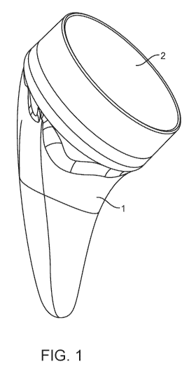
[0030] Figure 1 shows a perspective view of the shoulder arthroplasty system
in a reverse
configuration.
[0031] Figure 2 shows a frontal view of the system with a cup in a reverse
configuration.
[0032] Figure 3 shows a perspective view showing of the stem with head un-
installed
[0033] Figure 4 shows a perspective view of the stem and cup.
[0034] Figure 5 shows an anterior cross-section of the stem installed in a
humerus.
[0035] Figure 6 shows a frontal view of the stem component.
[0036] Figure 7 shows a frontal view of the details of the proximal portion of
the stem
[0037] Figure 8 shows details of the proximal portion of the stem in bone.
[0038] Figure 9 shows a frontal cross-section of the stem and cup.
[0039] Figure 10 shows a cross-section of the stem and cup assembled.
[0040] Figure 11 shows a medial view of the stem component.
[0041] Figure 12 shows a frontal view of the stem component
[0042] Figure 13 shows a frontal view of the stem component and lateral fin
details.
[0043] Figure 14A shows a cross-section of the stem component and Figure 14B
shows a
detailed cross-section of the lateral fin.
[0044] Figure 15A shows a lateral view of the stem component. Figure 15B shows
a frontal
view of an alternate anterior/posterior fin geometry. Figure 15C shows a
perspective view of
the same embodiment.
7
CA 03011998 2018-07-18
WO 2017/147618 PCT/US2017/020027
[0045] Figure 16A shows a frontal view of the stem. Figure 16B shows the
distal cross-
section of the stem.
[0046] Figure 17A shows a medial view of an alternate embodiment of the medial
surface
geometry.
[0047] Figure 18 shows a lateral view of an alternate embodiment of the
lateral fin.
[0048] Figure 19 shows a perspective view of the preferred bone growth coating
placement.
[0049] Figure 20A shows a frontal view of alternate stem geometry with a
cutout for
additional bone graft placement. Figure 20B shows the same embodiment in a
perspective
view.
[0050] Figure 21 shows a frontal view of a stemless configuration.
[0051] Figure 22A shows a cross section of an alternate embodiment of the stem
component with protrusions. Figure 22B shows a perspective view of the same
embodiment.
[0052] Figure 23 shows a cross-section of the head.
[0053] Figure 24A shows a frontal view of an alternative embodiment of the
stem with a
collar. Figure 24B shows a cross-section of the same embodiment.
[0054] Figure 25A shows an auxiliary view of an alternative embodiment of the
stem with
fenestrations for implant removal. Figure 25A is normal to the proximal end of
the stem.
Figure 25B shows a perspective view of the same embodiment.
[0055] Figure 26 shows an alternate bone growth coating placement.
[0056] Figure 27A, 27B, 27C, 27D shows the method of determining the center of
the
resection plane using a disk and pin.
[0057] Figure 28A, 28B, 28C, 28D, 28E, 28F show the reaming procedure to
create the
proximal bone cavity.
[0058] Figure 29 shows a cross-section of the broach in the humerus.
[0059] Figure 30A shows a cross-section of the stemless broach in the humerus.
Figure
30B shows a frontal view of the stemless broach.
DETAILED DESCRIPTION OF THE INVENTION
[0060] Before the present subject matter is further described, it is to be
understood that this
subject matter described herein is not limited to particular embodiments
described, as such
may of course vary. It is also to be understood that the terminology used
herein is for the
purpose of describing particular embodiments only, and is not intended to be
limiting. Unless
defined otherwise, all technical terms used herein have the same meaning as
commonly
understood by one skilled in the art to which this subject matter belongs.
8
CA 03011998 2018-07-18
WO 2017/147618 PCT/US2017/020027
[0061] Disclosed is a shoulder arthroplasty system that is optionally
convertible in use
between an anatomic and a reverse shoulder implantation configuration. The
system includes
a short stem prosthesis that may provide several advantages. In the reverse
shoulder
configuration, the system includes an articular surface cup arranged in an
inlay configuration
on a receptacle of the stem. This may provide for a much more compactly sized
system with
respect to an onlay configuration. The system is configured to preferably
achieve fixation in
the metaphysis to preferably provide rotational and axial stability. The
shoulder arthroplasty
system may be implanted in both a press fit and a cemented configuration.
[0062] The system can be implanted using an installation technique that
preferably removes
as little bone as possible thereby conserving bone in the patient. It is
designed on anatomy
preferably based on a statistical shape model matching that of the humerus, as
described in
detail below. It should be appreciated that a statistical shape model is just
an example, of a
non-limiting means of analysis and that other means of analysis are within the
scope of this
disclosure.
[0063] Figure 1 shows a perspective view of the shoulder arthroplasty system
in a reverse
configuration. The system includes a stem component 1 and a polymer cup 2 in a
reverse
configuration. Figure 2 shows a frontal view of the system with the
aforementioned stem
component 1 that is sized and shaped to be inserted into the humerus. The stem
has a
monoblock or monolithic configuration that is a single piece structure. The
single piece stem
reduces manufacturing costs and hospital inventory requirements compared to a
modular
stem design. The cup 2 has an angled profile that provides a greater range of
motion with less
potential for notching which is loss of bone where the implant comes in
contact with the
glenoid, possibly during some movements. One of skill in the art will
appreciate that the
stem or stemless embodiments may be used with, or without, or in combination
with, any of
the other features described in this specification (e.g. fins, fenestrations,
tapers, etc.).
[0064] The cup component 2 defines a curved articulating surface near to the
resection
plane. This provides minimal lateralization and inferiorization for a
convertible prosthesis,
which leads to a more anatomical reconstruction. The cup component 2 is
interchangeable
with the stem 1. A collection of multiple cup components 2 can be used for a
single,
corresponding stem component wherein each cup component of the collection has
a particular
articulating surface diameter and offset. This permits a user to select a cup
component for use
having a desired surface diameter.
[0065] Figure 3 shows a perspective view of the shoulder arthroplasty system
with a head
component 3 uninstalled from the stem 1. The stem component 1 also includes a
mechanical
9
CA 03011998 2018-07-18
WO 2017/147618 PCT/US2017/020027
taper 5 that serves as a securing mechanism for the head component 3 and when
secured to
the stem 1. The anatomic head component 3 provides an articular surface 6 for
anatomic
shoulder reconstruction.
[0066] Figure 4 provides perspective views of the stem component 1. As
described in detail
below the stem has an anatomic shape that is configured pursuant to a
statistical shape model.
It should be appreciated that a statistical shape model is just an example, of
a non-limiting
means of analysis and that other means of analysis are within the scope of
this disclosure. For
example, other means of analysis including anatomic analysis, geometric
analysis,
anthropometric analysis, mechanical analysis, and kinematic analysis are
within the scope of
this disclosure. In this embodiment, the stem has three fin like protrusions,
a lateral fin 51,
anterior fin 52, and the posterior fin 53 configured to cut into cancellous
bone in the
metaphysis to provide rotational stability when the system is implanted in
bone. The fins are
configured to enhance rotational stability of the stem while avoiding cortical
contact in the
metaphysis. The anterior and posterior fins 52 and 53 may be used
interchangeably when the
stem is used in a left or right side of the patient.
[0067] In Figure 5, a cross section of the stem component inserted into the
humerus is
shown. The proximal end 7 of the stem 1 is positioned along a resection plane
8, which is the
area exposed in surgery when the humeral head is surgically removed prior to
preparation and
implantation. An axis 90 normal to the resection plane 8 is at an obtuse angle
91 from the
long axis of the bone 9.
[0068] Figure 6 shows a frontal view of the stem component. The stem has an
overall taper
shape such that the implant generally increases in size proximally which
improves wedged
fixation. This configuration preserves bone by providing minimal removal of
bone, especially
in the greater tuberosity and humeral metaphysis. The design allows the stem
to be centered
proximally within the resection plane, which thereby allows the spherical head
component of
the anatomic configuration to be also centered on the resection plane,
recreating normal
anatomy. The outer bone contacting surfaces of the stem are configured to
optimize the
proximal bone loading in the proximal metaphysis of the humerus and reduce
loading distally
in the diaphysis. The taper shape has a proximal concave taper 10 that is
generally conical
and a distal taper 11 that is tapered in both the medial-lateral direction as
well as the anterior-
posterior direction. Additional details about the proximal and distal tapers
are described
elsewhere in this specification.
[0069] The stem has a proximal end 7, distal end 12, lateral side 13 and
medial side 14. The
proximal portion of the stem has a short cylindrical extrusion 15
perpendicular to the
CA 03011998 2018-07-18
WO 2017/147618 PCT/US2017/020027
proximal end 7. In the preferred embodiment the cylindrical extrusion 15 is
2.7mm and may
range from 2.5mm to 3mm. While this is a preferred embodiment, the stem design
may have
other dimensions and would work without the cylindrical section where the
proximal portion
of the stem is conical right up to the proximal end 7. The design with the
cylindrical
extrusion 15 is preferred because this truncates the conical taper such that
the diameter of the
proximal end 7 is reduced for the same taper. In a preferred embodiment the
diameter of the
cylindrical extrusion ranges from 30 to 40mm although other dimensions are
possible. The
diameter increases with increasing patient anatomy.
[0070] Figure 7 shows details of the concave taper 10 in the proximal portion
of the stem.
Distal to the cylindrical extrusion 15 the stem geometry transitions to a
concave taper 10. The
proximal end 16 of the concave taper 10 is congruent to the distal end of the
cylindrical
extrusion 15 and extends to a distal end of the taper 17. The concave taper 10
is defined by at
least one radius 18 revolved around the axis 19 through the center of the
cylindrical extrusion
15. The center of the radius 20 used to create the concavity is outside the
cone created by the
revolution of the straight line 21 created between proximal end 16 and the
distal end 17 of the
concave taper 10. In the preferred embodiment the length of the conical
section from the
proximal end 7 to the distal end 17 may be 18mm but other lengths are
possible. In alternate
embodiments, the length of the conical section could range from 12-30mm. The
diameter of
the proximal end of the concave taper 10 is preferably equal to the
cylindrical extrusion 15
described above. The diameter of the distal end of the taper 17 in the
preferred embodiment
may be 9mm and could range from 7 to llmm, although other sizes are also
possible.
[0071] Figure 8 shows the concave taper 10 and the interaction with the
proximal bone
during insertion of the stem 1. The concave taper 10 is designed to compact
bone in the
metaphysis during insertion. The compaction of bone is achieved when the stem
is inserted
into the bone. As the stem advances distally in the humerus, bone is displaced
along the
concave taper 10. The concave taper 10 is an advantageous shape because the
angle formed
between the axis 19 and a tangent line 22 to the concave surface increases as
the stem is
advanced in the bone. This provides ever-increasing compaction until final
placement of the
stem 1 is achieved.
[0072] In the configuration shown in Figure 9, the cup component 2 is
uncoupled from the
receptacle 4 of the stem 1. The cup component attaches primarily via a locking
ring 27, but
secondarily (and especially under load) with the taper 5. The proximal portion
23 of the stem
houses the receptacle for the anatomic head for anatomic shoulder replacement
and the
articular cup 2 for reverse shoulder replacement. In the preferred embodiment,
the stem has
11
CA 03011998 2018-07-18
WO 2017/147618 PCT/US2017/020027
two receptacles; a female taper 5 and a cylindrical cavity 24. The cylindrical
cavity 24 is
concentric to the cylindrical extrusion 15. The cylindrical cavity 24 starts
at the proximal end
7 of the stem and extends distally to flat surface 25 parallel to the proximal
end 7. In the
preferred embodiment, the cylindrical cavity 24 is 3.5mm deep but it may be
other depths.
Between the proximal end 7 and the flat surface 25 an annular groove 26 is
formed to receive
the locking ring 27 of the articular cup 2. A male protrusion 33 has a taper
that corresponds
to the taper 5 in the female receptacle so that the two components engage one
another.
[0073] Figure 9 shows a cross section of the stem component 1 and articular
cup 2. The
female taper 5 is a conical tapered cavity that extends from the flat surface
25 of the
cylindrical cavity 24 to a distal end 28. In the example shown, the axis of
the female taper 29
is congruent with the axis of the cylindrical cavity 30. However, this
embodiment is not
limited to this and the axis may be offset from the cylindrical cavity 30 but
preferably
remains parallel to this axis. The female taper 5 is configured such that the
diameter at the flat
surface 25 is greater than the diameter at the distal end 28. The total
included angle 31 of the
female taper in a preferred embodiment is 4.6 degrees and the length is 12mm,
although other
dimensions are possible. The female taper 5 in the stem receives a conical
protrusion 32 in
the anatomic head 3 and the conical protrusion 33 in the articular cup 2. When
pressed
together, the female taper 5 and the conical protrusion 33 become fixed to one
another with
an interference fit.
[0074] Figure 10 shows a cross-section of the stem component 1 and articular
cup 2 in an
"inlay" configuration. The articular cup 2 is affixed to the stem by the
locking ring 27 in the
annular groove 26 and interference fit between the conical protrusion 33 and
the female taper
5. In the preferred embodiment, the apex 122 of the concave articular surface
6 is distal to the
proximal end 7 that sits flush to the resection plane 8. The inlay design
greatly reduces the
likelihood of over-tensioning the joint. Sufficient bearing thickness is
maintained to support
the loads and wear from patient activity.
[0075] Figure 11 shows a medial view of the stem without the articular cup 2
or anatomic
head 3. The distal taper 11 has an anterior surface 36 and a posterior surface
37. In the
preferred embodiment the distal taper 11 is symmetric about a plane 38 that is
congruent with
the axis 19 of the cylindrical extrusion. The distal taper 11 is generally
tapered in both the
medial-lateral direction and the anterior-posterior direction. The distal
taper 11 has a medial-
lateral width defined by the distance from the medial surface 34 to the
lateral surface 35. The
medial-lateral width decreases distally to create the medial-lateral taper.
The distal taper 11
has an anterior-posterior width defined by the distance from the anterior
surface 36 to the
12
CA 03011998 2018-07-18
WO 2017/147618 PCT/US2017/020027
posterior surface 37. The anterior-posterior width decreases distally to
create the anterior-
posterior taper of the stem. The stem has a relatively thin cross-section. In
the anterior to
posterior direction, the thin cross-section is best described by the distal
anterior-posterior
width 47. The thin cross-section accommodates offset of the humeral head
relative to
intramedullary canal. This eliminates the need for left and right specific
implants and further
reduces inventory requirements. In the medial to lateral direction, the
configuration is still
thin enough to allow rotations to match resection cut without contacting
cortical bone. In
addition, the short length allows further flexibility in the location of the
implant proximally
(angular and position) as the distal stem is not constrained by cortical bone
during
implantation. It also preserves bone by eliminating the need to ream cortical
bone.
[0076] With reference to Figure 11, the anterior-posterior taper of the distal
taper 11 is
configured to reduce loading of the cortical bone in the diaphysis and create
wedge fixation.
The anterior-posterior taper is symmetric about the medial plane 38 of the
implant. The
anterior-posterior width 47 at the distal end is less than the proximal width
48. In the
preferred embodiment, the distal end of the distal taper is rounded in both
the medial-lateral
and anterior-posterior directions. In the preferred embodiment, the anterior-
posterior width 47
at the distal end is 3.5mm and the medial-lateral width at the distal end is
8, although other
widths are possible
[0077] Figure 12 shows details of the medial-lateral taper geometry of the
stem in a frontal
view. The medial lateral taper geometry is configured to conform to but not
contact the
medial and lateral cortex of the humerus. The taper geometry may provide
rotational stability
of the implant. The medial-lateral taper is curved such that the distal end 49
of the stem 1 is
offset laterally a distance 39 from a point intersecting the axis of the
cylindrical extrusion and
the proximal end 7. In the preferred embodiment, this lateral offset is 8.7mm
for the median
size and ranges from 7-13mm, although other distances are possible. The
lateral surface 35 of
the distal taper is defined by a convex curve 40 that extends from the
proximal end to the
distal end 49. The curve starts at a medial offset 41 from the most lateral
edge 42 of the
proximal end 7 and extends to farthest lateral edge of the distal portion 43.
The medial offset
41 of the lateral surface 35 is the distance from the lateral edge of the
cylindrical extrusion 42
to the proximal end of the lateral surface 44. The offset 41 provides space
for a lateral fin like
protrusion that is described in detail below. The curve of the lateral surface
35 is comprised
of at least one radius. The medial surface 36 of the distal taper 11 is
defined by a concave
curve 45 that extends from the proximal end 7 to the distal end 49. The curve
45 starts at the
medial most edge 46 of the proximal end 7 and extends to the farthest medial
edge 92 of the
13
CA 03011998 2018-07-18
WO 2017/147618 PCT/US2017/020027
distal end. It should be noted that the tapered section along the medial edge
may also be
interpreted as a fin-like protrusion and is described in further detail below.
The medial curve
45 is comprised of at least one radius. The medial-lateral taper increases in
width to the distal
end of the cylindrical extrusion 15. The curved medial surface is sized and
shaped to reduce
fracture risk by spreading implantation forces over a larger area during
implantation. The
total length of the implant from the center of the proximal end 7 to the
distal tip ranges from
44mm to 48mm although other lengths are possible. The overall length of the
implant from
the most distal point 49 to the farthest point superior point 50 ranges from
55mm to 61mm
although other lengths are possible.
[0078] Figure 13 shows details of the lateral fin 51. The lateral fin is
defined by a lateral
edge 54 that follows a convex curve from the lateral most edge 42 of the
cylindrical extrusion
15 to a point 55 that is tangent to the lateral surface 35 of the distal taper
11. The convex
curve can be defined by at least one radius.
[0079] The cross-section of the lateral fin protrusion 51, shown in Figure
13B, is
substantially triangular in the preferred embodiment. The apex 56 of the
triangular cross-
section is congruent with the lateral edge 54 previously defined. Alternate
embodiments, may
include rectangular or hemispherical like cross-sections.
[0080] Figure 15 shows a lateral view of the stem component. The anterior and
posterior
fins 52, 53 extend from a point 57 on the distal taper 11 that is distal to
the concave taper 10
and proximal to the distal end 12 of the stem. The anterior and posterior fins
52, 53 extend
from the point 57 to meet the cylindrical extrusion 15. In the preferred
embodiment, the
anterior and posterior fins 52, 53 are concave at the distal end of the fin 58
and convex at
proximal end of the fin 59. The concave portion of the fin is tangent to the
anterior/posterior
surface 60 of the distal taper 11 at the distal end of the fin 58. The convex
portion of the fin is
tangent to the cylindrical extrusion 15 at the proximal end of the fin 59.
Figure 15B and
Figure 15C show an alternate embodiment of the anterior and posterior fins
52,53. In the
alternate embodiment the anterior and posterior fins have a medial surface 109
and a lateral
surface 110. The medial surface 109 and lateral surface 110 are substantially
parallel to each
other. In additional fin geometries the anterior and posterior fins may be
located closer to the
lateral side 13 than the medial side 14.
[0081] Figure 16B, shows in the preferred embodiment, the part of the stem 1
distal to the
anterior and posterior fins of the stem has an hourglass like cross-section.
The cross-section is
shaped such that anterior-posterior width 61 at the medial and lateral sides
is greater than the
center 62 of the cross-section. This preserves bone while keeping any bone
contacting
14
CA 03011998 2018-07-18
WO 2017/147618 PCT/US2017/020027
surfaces at the medial and lateral edges with sufficient contact area to
support implant loads
and avoid compromising the internal cortex.
[0082] Figure 17A shows an alternate embodiment of the anterior-posterior
taper geometry
of the taper. This embodiment provides a larger bone-contacting surface on the
medial and
lateral surfaces and increases the wedge fixation in the metaphysis of the
bone. The distal
taper, in the alternate embodiment, has an anterior posterior width at the
proximal end
defined by the distance between an anterior point 63 on the cylindrical
extrusion 15 and a
posterior point 64 on the cylindrical extrusion 15. At a distance 65 distally
along the distal
taper 11, the anterior-posterior taper is further defined by a second width
defined by the
distance between an anterior point 66 and a posterior point 67. These 4 points
define a total
included angle 68 of the proximal portion of the distal taper 11. A distal
total included angle
can be defined by the second width, the distance between points 66 and 67 and
the anterior 69
and posterior 70 points at the distal end 12 of the stem 1. The total included
angle 71 of the
distal portion of the distal taper is less than the proximal total included
angle 68. Figure 17B
shows a similar geometry but on the lateral fin 51. The lateral fin 51 has
anterior and
posterior surfaces 106 that form an angle 107. The angle 107 is greater than
the angle formed
by the distal taper angle 108
[0083] In a preferred embodiment, Figure 19, the proximal bone contacting
surfaces 72
have a coating 73 for additional boney in-growth. Coatings may include plasma
spray
titanium, hydroxyapatite or similar coatings known in the art to enhance bone
growth. In the
preferred embodiment, the proximal 19-21mm of the stem is coated. However,
additional
embodiments could include coating the entire stem or any sections. Figure 26
shows one such
embodiment of the placement of the bone growth surface where only the fins 51,
52, 53 of
the stem are coated with a bone growth coating 73. The proximal section of the
stem is grit
blasted and the distal portion is polished smooth.
[0084] Figure 20A and 201 shows an alternative embodiment of the distal
portion of the
stem that has a cutout 74 to contain additional bone graft. The medial 75 and
lateral 76 edges
of the cutout 74 is offset from the medial 34 and lateral surfaces 35 of the
stem and extends
through the stem 1 in the anterior - posterior direction. In this embodiment,
the proximal
extent of the cutout 74 is distal to the concave taper 10. The cutout may be
used in any stem
embodiment described herein.
[0085] Figure 21 shows an alternate embodiment of the stem, which does not
have a distal
taper as described in previous embodiments. This configuration is commonly
called a
"stemless" or "canal sparing" implant. The device is surgically implanted in
the same manner
CA 03011998 2018-07-18
WO 2017/147618 PCT/US2017/020027
as the longer, "stemmed", design, and therefore provides the surgeon with an
additional
option without adding complexity. Stemless designs can be used to further
enhance positional
flexibility proximally when metaphyseal fixation is adequate and cementation
is not required.
In the stemless design, like the "stemmed" design, the outer bone-contacting
surface of the
stem has a concave taper 10. The stemless design, like the stemmed design, has
a cylindrical
extrusion 15 perpendicular to the proximal end 7. The concave surface is
defined by at least
one radius that revolves around the axis 19 created by the cylindrical
extrusion 15. The
stemless design also includes at least one fin-like protrusion for rotational
stability. As
previously described, the fin-like protrusions extend from the distal portion
of the stem to
meet the proximal cylindrical section. The fins taper from the distal end 77
to a greater width
at the proximal end of the fin 78. In a preferred embodiment, fins are located
at the lateral 79,
medial 80, anterior 81 and posterior 82 aspects of the implant.
[0086] Figure 22A shows a cross-sectional view of an alternate embodiment of
the
proximal end where the stem 1 has at least one protrusion 83 on the rim 84 of
the proximal
end 7 to prevent rotation of the articular cup 2. A preferred arrangement is
four protrusions
distributed evenly along the rim 84 of the proximal end 7. The protrusion 83
extends from
proximal end 7 of the stem. A rim 84 is created by the outer diameter of the
cylindrical
extrusion 15 and the cylindrical cavity 24. Figure 23A shows the protrusion
extends radially
from the inner surface 85 created by the cylindrical cavity 24 to the outer
surface of the
cylindrical extrusion 15. However, alternative embodiments could include the
protrusion only
extending part of the way from the cylindrical cavity 24 to the cylindrical
extrusion 15. The
articular cup 2 for a reverse shoulder would have mating indents 86, shown in
Figure 22B to
receive the protrusions 83. Any embodiment disclosed herein may include some
or all of
these features.
[0087] Figure 23 shows a cross-sectional side view of the anatomic head
component 3
which may be used with any of the stem embodiments described herein. The
anatomic head
component 3 includes a long taper 32 that couples to the corresponding taper
in the stem to
secure the anatomic head component 3 to the stem. The longer than usual taper
(relative to
currently-existing systems) allows for a single interface for both anatomic
and reverse
arthroplasty. Moreover, the long taper 32 extends through the receptacle in
the stem and is
sized to allow the anatomic head component to be used in the same stem as an
inlay articular
cup (as described above) without an intermediate tray. The anatomic head
component 3
includes a curved, anatomically accurate center and radius curvature of the
articular surface
86 that closely matches normal anatomy of the humeral head. The head component
3 includes
16
CA 03011998 2018-07-18
WO 2017/147618 PCT/US2017/020027
cutout 87 to reduce the weight of the component. The cutout 87 is disposed
between the outer
rim of the head 88 and the central tapered protrusion 32. A rounded periphery
89 provides an
atraumatic surface when in contact with soft tissue.
[0088] Figure 24A shows a frontal view of an alternative embodiment where the
stem has a
collar 95 on the cylindrical portion 15 of the stem 1. This embodiment is also
known as a
"collared" stem and is used with the smallest stem sizes to allow for
placement of the head or
cup, depending on whether an anatomic or a reverse is being implanted. It is
allows the use of
the head or cup in a stem that is too small to accommodate them as the larger
stems do. The
collar 95 has a proximal surface 96, distal surface 97 and radial surface 98.
The proximal
surface 96 of the collar 95 is congruent to the proximal end 7 of the stem.
The distal surface
97 of the collar sits on the resection plane of the humerus. The radial
surface 98 has a
diameter greater than that of the cylindrical extrusion 15. The mating
articular cup 2
component, shown in Figure 24B, has an outer diameter 99 that is equal to the
diameter of the
radial surface 98.
[0089] Figure 25A and 25B shows an alternative embodiment of the stem with
fenestrations. The fenestrations are used to insert instruments if the
prosthesis needs to be
removed. During revision, removal may be made difficult by bone that has grown
onto the
stem. The fenestrations extend from the proximal end 7 distally. In the
embodiment shown in
Figure 23 there are four fenestrations. The first fenestration 100 is located
between the lateral
fin 51 and the posterior fin 53. A second fenestration 101 is located between
the lateral fin 51
and the anterior fin 52. A third fenestration is located between the posterior
fin and the
medial surface 34. The fourth fenestration 103 is located between the anterior
fin and the
medial surface. Alternative embodiments may include a different number of
fenestrations.
[0090] The method to insert the stem is described below. In a first step, the
proximal
humeral osteotomy 111 is made through the anatomic neck of the humerus.
Reference Figure
27A-27D. The osteotomy can be made using a saw, osteotome or equivalent bone
cutting
instrument. The osteotomy 111 is a planar cut made at an angle to the long
axis 9 of the bone.
The osteotomy can be sized using a set of sizing disks 112 and a guide pin 113
placed
through the center of the disk 114 to establish the osteotomy center point
115. The set of
sizing disks 112 include a matching disk for each implant size. The guide pin
113 also acts as
a temporary fixation pin to help hold the disk 112. The disk is a flat
cylinder used to visualize
the diameter of the proximal portion of the stem relative to the resected bone
surface created
by the osteotomy 111. At least two slots 116 are cut into the sizing disk 112
to help visualize
the extent of the resected bone surface and thereby preventing oversizing of
the implant.
17
CA 03011998 2018-07-18
WO 2017/147618 PCT/US2017/020027
[0091] Figure 28A-28F shows the next step being removal of the proximal bone.
The
proximal bone of the humerus is removed to accommodate the proximal portion of
the final
implant. The bone is removed using a proximal reamer 117 that may be attached
to handle for
manual reaming by hand or a power drill. The humerus is reamed such that the
cavity 118
created by the reamer 117 is smaller than the final implant. Figure 28E shows
a cross section
of the reamer 117 in the humerus. Figure 28F shows the reamer over-laid with
the outline of
the concave taper. The area between the concave taper and the reamer is the
volume of bone
that will be compacted during the insertion of the stem.
[0092] The following step, shown in Figure 29, shows a broach 119 that is
utilized to
compact the bone in the epiphysis and to create space for final implantation
of the stem. The
stemless broach 119 is designed to minimize bone cutting and improve bone
compaction.
The broach 119 also serves as the trial for the stem component disclosed
herein. The broach
may also include slots to visualize the extent of the resected bone relative
to broach.
[0093] Figure 30A shows an alternative embodiment of the broach where a
stemless broach
105 may be used to clear bone proximally. The stemless broach 105 may be
inserted over the
guide pin 113 to ensure the bone cavity for the stem is located at the
osteotomy center point
115. The stemless broach 105, shown also in Figure 30B, may be used in both
'stemmed'
implants and stemless implants. In the process of placing the broach, the
metaphyseal bone is
compacted, thus achieving additional stability. The stability of the implant
is then confirmed.
Cementing may optionally be considered if the implant is not satisfactorily
stable such as in
patients with extremely poor bone quality. Once the glenoid is prepared, the
final stem
component is fitted with a humeral head or the reversed cup and the joint
reduced.
[0094] While this specification contains many specifics, these should not be
construed as
limitations on the scope of an invention that is claimed or of what may be
claimed, but rather
as descriptions of features specific to particular embodiments. Certain
features that are
described in this specification in the context of separate embodiments can
also be
implemented in combination in a single embodiment. Conversely, various
features that are
described in the context of a single embodiment can also be implemented in
multiple
embodiments separately or in any suitable sub combination. Moreover, although
features may
be described above as acting in certain combinations and even initially
claimed as such, one
or more features from a claimed combination can in some cases be excised from
the
combination, and the claimed combination may be directed to a sub-combination
or a
variation of a sub combination. Similarly, while operations are depicted in
the drawings in a
particular order, this should not be understood as requiring that such
operations be performed
18
CA 03011998 2018-07-18
WO 2017/147618
PCT/US2017/020027
in the particular order shown or in sequential order, or that all illustrated
operations be
performed, to achieve desirable results. Only a few examples and
implementations are
disclosed. Variations, modifications and enhancements to the described
examples and
implementations and other implementations may be made based on what is
disclosed.
19