Note: Descriptions are shown in the official language in which they were submitted.
CA 03012072 2018-07-20
WO 2016/116897 PCT/IB2016/050323
AUTO-FOCUSING METHOD AND DEVICE
FIELD AND BACKGROUND
The present invention generally relates to the field of optical measurement
and/or
inspection techniques and more specifically relates to an auto-focus method
and device,
particularly useful when viewing non planar surfaces.
Auto focusing is an essential feature in many automated inspection fields such
as the
computer chip industry, biomedical research, data reading/recording in optical
information
carriers, etc. Specifically, when analyzing samples in multiwell plates
including a plurality of
wells on a single plate, auto focusing of a microscope viewing the contents of
the wells can
enable more efficient work procedures as the operator need not focus the
objective on each
well in the plate separately.
Various auto focusing methods for inspection of a multiwell plate have been
disclosed
in the past, such as in US Patent No. 7,109,459. However, when using wells
having a non-
planar bottom, such as a multiwell plate having wells with a U-shaped bottom,
such as are
used, for example, for growing living cells into spheroids, existing autofocus
methods may
require image analysis which is time-consuming.
There is thus a need for a method for auto focusing a microscope on a
multiwell plate,
which is suitable for multi-well plates having wells with a non-planar bottom
surface.
SUMMARY
The present invention generally relates to the field of optical measurement
and/or
inspection techniques and more specifically relates to an auto-focus method
and device,
particularly useful when viewing non planar surfaces.
There is provided in accordance with an embodiment of the invention an auto-
focusing method for determining an in-focus position of a plurality of wells
in at least a
portion of a multi-well plate, the method including:
using a first objective lens having a first magnification to identify, in each
of at least
three wells of a selected subset of the plurality of wells, an in-focus
position of each said well
with respect to the first objective lens;
on the basis of at least three said in-focus positions, computing a plane
along which
the at least three wells will be in focus with respect to at least one
objective lens having a
second magnification that is not greater than the first magnification; and
1
CA 03012072 2018-07-20
WO 2016/116897 PCT/IB2016/050323
using the at least one objective lens to scan, along the plane, at least some
of the
plurality of wells in the portion of the plate.
In some embodiments, the at least one objective lens is the first objective
lens, and the
first magnification is equal to the second magnification. In some embodiments,
the
computing a plane includes computing a plane along which the at least three
wells are in
focus with respect to the first objective lens.
In some embodiments, the at least one objective lens is a second objective
lens,
different from the first objective lens, wherein the second magnification is
smaller than the
first magnification. In some embodiments, computing a plane includes
translating at least
three of the in-focus positions identified using the first objective lens to
corresponding second
in-focus positions with respect to the second objective lens based on optical
characteristics of
the second objective lens; and computing the plane on the basis of at least
three the second
in-focus positions. In some embodiments, computing a plane includes: on the
basis of at least
three said in-focus positions, computing a first plane along which the at
least three wells will
be in focus with respect to the first objective lens; and translating the
first plane to a
corresponding plane along which the at least three wells will be in focus with
respect to the
second objective lens based on optical characteristics of the second objective
lens, thereby to
compute the plane.
In some embodiments, scanning using the at least one objective lens is carried
out
without carrying out additional focusing operations.
In some embodiments, the subset of the plurality of wells includes more than
three of
the plurality of wells.
In some embodiments, identifying an in-focus position includes identifying an
in-
focus position for each well in the subset.
In some embodiments, each of the wells includes generally cylindrical side
walls, and
a bottom surface including a portion of at least one of a sphere, a parabola,
and an ellipse. In
some embodiments, each of the wells has a U-shaped cross-section.
In some embodiments, each of the wells includes generally cylindrical side
walls, and
a planar bottom surface. In some embodiments, the planar bottom surface lies
generally
parallel to a top surface of the multiwell plate, such that the well has a
rectangular cross
section.
In some embodiments, each of the wells is frusto-conical.
In some embodiments, each of the wells has inclined side walls, a planar
bottom, and
a trapezoidal cross section.
2
CA 03012072 2018-07-20
WO 2016/116897 PCT/IB2016/050323
In some embodiments, the method further includes, prior to the using a first
objective
lens, aligning the first objective lens to lie axially over the center of one
of the wells.
In some embodiments, the portion of the plate includes a quadrant of the
plate. In
some embodiments, the portion of the plate includes an entirety of the plate.
There is also provided, in accordance with an embodiment of the invention, an
auto-
focusing method for determining an in-focus position of at least a portion of
a well in a plate,
the method including: using a first objective lens having a first
magnification to identify, at at
least one location of the well, a first in-focus position of at least a
portion of the well with
respect to the first objective lens; identifying, for the first in-focus
position, a corresponding
in-focus position with respect to at least one objective lens having a second
magnification,
based on optical characteristics of the at least one objective lens; and using
the at least one
objective lens to scan, at a height corresponding to the corresponding in-
focus position, at
least the portion of the well, wherein the second magnification is not greater
than the first
magnification.
In some embodiments, the at least one objective lens is the first objective
lens, the
first magnification is equal to the second magnification, and the
corresponding in-focus
position is the first in-focus position.
In some embodiments, the at least one objective lens includes a second
objective lens,
different from the first objective lens, wherein the second magnification is
smaller than the
first magnification. In some such embodiments, the identifying includes
translating the first
in-focus position to the corresponding in-focus position with respect to the
second objective
lens based on optical characteristics of the second objective lens.
In some embodiments, scanning using the at least one objective is carried out
without
carrying out additional focusing operations.
In some embodiments, the well includes generally cylindrical side walls, and a
bottom
surface including a portion of at least one of a sphere, a parabola, and an
ellipse. In some
embodiments, the well has a U-shaped cross-section.
In some embodiments, the well includes generally cylindrical side walls, and a
planar
bottom surface. In some embodiments, the planar bottom surface lies generally
parallel to a
top surface of the plate, such that the well has a rectangular cross section.
In some embodiments, the well is frusto-conical. In some embodiments, the well
has
inclined side walls, a planar bottom, and a trapezoidal cross section.
There is also provided, in accordance with an embodiment of the invention, an
auto-
focusing device for automatically determining an in-focus position of a
plurality of wells
3
CA 03012072 2018-07-20
WO 2016/116897 PCT/IB2016/050323
located in at least a portion of a plate containing wells, the device
including: a computation
component programmed to compute a plane along which at least three wells in
the portion of
the plate would be in focus with respect to an objective lens; a first
objective lens functionally
associated with the computation component, the first objective lens having a
first
magnification, images from the first objective lens being used by the
computation component
for identifying an in-focus position for each of at least three wells of a
selected subset of the
plurality of wells; and at least one objective lens having a second
magnification, the second
magnification not being greater than the first magnification, for scanning at
least some of the
plurality of wells in the portion of the plate along the plane, wherein the
computation
component is configured to compute the plane along which the at least three
wells would be
in-focus with respect to the at least one objective lens on the basis of at
least three the in-
focus positions.
In some embodiments, the at least one objective lens is configured to scan the
plurality of wells along the plane without carrying out additional focusing
operations.
In some embodiments, the at least one objective lens is the first objective
lens, and the
second magnification is equal to the first magnification.
In some embodiments, the at least one objective lens is a second objective
lens,
different from the first objective lens, and the second magnification is
smaller than the first
magnification.
In some embodiments, the computation component is programmed to compute the in-
focus plane by: translating at least three of the in-focus positions
identified using the first
objective lens to corresponding second in-focus positions with respect to the
second objective
lens based on optical characteristics of the second objective lens; and
computing the in-focus
plane on the basis of at least three the second in-focus positions.
In some embodiments, the computation component is programmed to compute the in-
focus plane by: on the basis of at least three the in-focus positions,
computing a first plane
along which the at least three wells will be in focus with respect to the
first objective lens;
and translating the first plane to a corresponding plane along which the at
least three wells
will be in focus with respect to the second objective lens based on optical
characteristics of
the second objective lens, thereby to compute the plane.
In some embodiments, the computation component is programmed to identify an in-
focus position for each well in the subset.
In some embodiments, the device is adapted for use with a plate in which each
of the
wells includes generally cylindrical side walls, and a bottom surface
including at least one of
4
CA 03012072 2018-07-20
WO 2016/116897 PCT/IB2016/050323
a portion of a sphere, a parabola, and a portion of an ellipse. In some
embodiments, the
device is adapted for use with a plate in which each of the wells has a U-
shaped cross section.
In some embodiments, the device is adapted for use with a plate in which each
of the
wells includes generally cylindrical side walls, and a planar bottom surface.
In some
embodiments, the planar bottom surface lies generally parallel to a top
surface of the plate,
such that each of the wells has a generally rectangular cross section.
In some embodiments, the device is adapted for use with a plate in which each
of the
wells is frusto-conical. In some embodiments, the device is adapted for use
with a plate in
which each of the wells has inclined side walls, a planar bottom, and a
trapezoidal cross
section.
In some embodiments, the portion of the plate includes a quadrant of the
plate. In
some embodiments, the portion of the plate includes an entirety of the plate.
There is also provided, in accordance with an embodiment of the invention, an
auto-
focusing device for automatically determining an in-focus position of at least
a portion of a
well, the device including: a computation component programmed to compute an
in-focus
position of the portion of the well; a first objective lens functionally
associated with the
computation component, the first objective lens having a first magnification,
images from the
first objective lens being used by the computation component for identifying,
in at least one
position of the well, a first in-focus position of the well with respect to
the first objective lens;
and at least one objective lens having a second magnification, the second
magnification not
being greater than the first magnification, for scanning at least a portion of
the well at a
height of a corresponding in-focus position of the well with respect to the at
least one
objective lens, wherein the computation component is programmed to identify
the
corresponding in-focus position based on optical characteristics of the at
least one objective
lens.
In some embodiments, the at least one objective lens is configured to scan the
portion
of the well without carrying out additional focusing operations.
In some embodiments, the at least one objective lens is the first objective
lens, the
second magnification is equal to the first magnification, and the
corresponding in-focus
position is the same as the first in-focus position.
In some embodiments, the at least one objective lens is a second objective
lens
different from the first objective lens, and wherein the second magnification
is smaller than
the first magnification.
5
CA 03012072 2018-07-20
WO 2016/116897 PCT/IB2016/050323
In some embodiments, the computation component is programmed to identify the
corresponding in-focus position by translating the first in-focus position to
the corresponding
in-focus position with respect to the second objective lens based on optical
characteristics of
the second objective lens.
In some embodiments, the device is adapted for use with a well including
generally
cylindrical side walls, and a bottom surface including at least one of a
portion of a sphere, a
parabola. and a portion of an ellipse. In some embodiments, the device is
adapted for use with
a well having a U-shaped cross section.
In some embodiments, the device is adapted for use with a well including
generally
cylindrical side walls, and a planar bottom surface. In some embodiments, the
planar bottom
surface lies generally parallel to a top surface of the plate, such that the
well has a generally
rectangular cross section.
In some embodiments, the device is adapted for use with a frusto-conical well.
In
some embodiments, the device is adapted for use with a well having inclined
side walls, a
planar bottom, and a trapezoidal cross section.
Unless otherwise defined, all technical and scientific terms used herein have
the same
meaning as commonly understood by one of ordinary skill in the art to which
the invention
pertains. In case of conflict, the specification, including definitions, will
take precedence.
As used herein, the terms "comprising", "including", "having" and grammatical
variants thereof are to be taken as specifying the stated features, integers,
steps or
components but do not preclude the addition of one or more additional
features, integers,
steps, components or groups thereof. These terms encompass the terms
"consisting of' and
"consisting essentially of'.
As used herein, the indefinite articles "a" and "an" mean "at least one" or
"one or
more" unless the context clearly dictates otherwise.
Embodiments of methods and/or devices of the invention may involve performing
or
completing selected tasks manually, automatically, or a combination thereof.
Some
embodiments of the invention are implemented with the use of components that
comprise
hardware, software, firmware or combinations thereof. In some embodiments,
some
components are general-purpose components such as general purpose computers or
monitors.
In some embodiments, some components are dedicated or custom components such
as
circuits, integrated circuits or software.
For example, in some embodiments, some of an embodiment is implemented as a
plurality of software instructions executed by a data processor, for example
which is part of a
6
CA 03012072 2018-07-20
WO 2016/116897 PCT/IB2016/050323
general-purpose or custom computer. In some embodiments, the data processor or
computer
comprises volatile memory for storing instructions and/or data and/or a non-
volatile storage,
for example, a magnetic hard-disk and/or removable media, for storing
instructions and/or
data. In some embodiments, implementation includes a network connection. In
some
embodiments, implementation includes a user interface, generally comprising
one or more of
input devices (e.g., allowing input of commands and/or parameters) and output
devices (e.g..
allowing reporting parameters of operation and results.
BRIEF DESCRIPTION OF THE FIGURES
Some embodiments of the invention are described herein with reference to the
accompanying figures. The description, together with the figures, makes
apparent to a person
having ordinary skill in the art how some embodiments of the invention may be
practiced.
The figures are for the purpose of illustrative discussion and no attempt is
made to show
structural details of an embodiment in more detail than is necessary for a
fundamental
.. understanding of the invention. For the sake of clarity, some objects
depicted in the figures
are not to scale.
In the Figures:
FIGS. lA and 1B are, respectively, a top plan view of a multiwell plate and a
sectional view of a single well in a multiwell plate, the well having a non-
planar bottom
surface, for which embodiments of the teachings herein may be useful;
FIG. 2 is a block diagram of an embodiment of an imaging device for auto-
focusing
on samples in a multiwell plate in accordance with an embodiment of the
teachings herein;
and
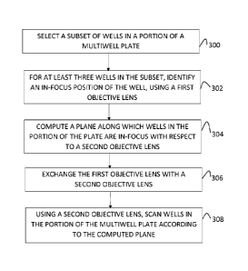
FIG. 3 is a flow chart of an embodiment of a method for auto-focusing an
imaging
device on samples in a multiwell plate in accordance with an embodiment of the
teachings
herein.
DESCRIPTION OF SOME EMBODIMENTS OF THE INVENTION
The principles, uses and implementations of the teachings herein may be better
understood with reference to the accompanying description and figures. Upon
perusal of the
description and figures present herein, one skilled in the art is able to
implement the invention
without undue effort or experimentation.
Before explaining at least one embodiment of the invention in detail, it is to
be
understood that the invention is not limited in its applications to the
details of construction
7
CA 03012072 2018-07-20
WO 2016/116897 PCT/IB2016/050323
and the arrangement of the components and/or methods set forth in the
following description
and/or illustrated in the drawings and/or the Examples. The invention can be
implemented
with other embodiments and can be practiced or carried out in various ways. It
is also
understood that the phraseology and terminology employed herein is for
descriptive purpose
and should not be regarded as limiting.
Reference is now made to Figures 1A and 1B which are, respectively, a top plan
view
of a multiwell plate and a sectional view of a single well in a multiwell
plate, the well having
a non-planar bottom surface, for which embodiments of the teachings herein may
be useful.
As seen in Figure 1A, a multiwell plate 10 has a top surface 11, side surfaces
(not
shown) and, in some embodiments, a bottom surface (not shown). The plate 10
includes a
plurality of wells 12, arranged in a grid formed of columns 14 and rows 16 and
accessible via
apertures 17 in top surface 11. Typically, the rows and columns are enumerated
or otherwise
labeled so as to enable a user to easily reference a specific well 12. The
multiwell plate 10 in
the illustrated embodiment includes 96 wells, though other types of plates,
which include, for
example, a different number of wells, such as 12, 24, or 384 wells, may be
used with the
teachings herein as described in further detail hereinbelow. Typically, the
wells 12 have fixed
distances between one another, and thus are distributed on plate 10 at regular
intervals.
Specifications as to the distanced between wells are standard in the art, and
are typically also
provided by the manufacturer of the plate. Often, the number of wells in the
plate have a 3:2
aspect ratio. As such the wells may be arranged, for example, as a 3x2 grid,
6x4 grid, 12x8
grid, or 24x16 grid.
Turning to Figure 1B, it is seen that the cross section of each well 12 in the
plate 10
may be non-rectangular, such that the well has a non-linear bottom surface. In
the illustrated
embodiment, the well 12 includes a cavity 18 and has a U-shaped cross section,
such that side
walls 20 of the well generally form a cylinder, and a bottom portion 22 of the
well forms part
of a sphere, part of a parabola, or part of an ellipse, thereby defining a
curved bottom surface
to the well. As such, the well typically has a U-shaped cross-section or a
cross-section
somewhat resembling a parabola. Typically, the thickness of side walls 20 and
of bottom
portion 22 is uniform. A rim 26, typically forming part of, and being flush
with or being
raised with respect to, top surface 11 of plate 10, often surrounds the well
12.
Multiwell plates including wells having non-planar bottom surfaces are well
known in
the art, and are commercially available from many manufacturers, such as
Corning
Incorporated Life Sciences of Tewksbury, Massachusetts. Such multi-well plates
are used for
many types of samples, including for growing spheroids, for growing non-
adherent cells such
8
as lynaphocytes and other blood cells, for analysis of 3-dimensional samples
and for handling
. of compounds. Oftentimes, analysis of such samples requires imaging
of the samples within = = = =
the wells.
. . .
. .
.
.
. . . . .
It will be appreciated that due to the curvature of the bottom surface of well
12 the area::
. at which a microscope viewing the well would be in focus is typically very
small, and in some .
cases comprises a single point. As such, existing auto-focusing mechanisms,
such as that,
disclosed in US Patent No. 7,109,459 often do not succeed in focusing on a
sample disposed
within the well. As explained hereinbelow, the method of the teachings herein
enables an
operator to autofocus an imaging device on a well having a non-planar bottom,
such as 11.1.-
shaped wells 12 of Figure TB, without having to manually focus the imaging
device on each
individual well.
It will be appreciated that, though the exemplary illustration shows wells
having a U-
shaped cross section, the method of the teachings herein as described
hereinbeloNv may be
used for other types of wells, such as wells having a planar bottom surface
and. a rectangular.
cross section, or wells of a frusto-conical shape, i.e. comprising a cutoff
cone having inclined
side walls and a planar bottom, and having a generally trapezoidal cross
section, . .
. .
Reference is now made to Figure 2, which s a block diagram of an embodiment of
an.,== = ,
imaging device 200 for auto-focusing on wells in a multiwell plate in
accordance with an = -- -- -
embodiment of the teachings herein.
=
It
will be appreciated that the disclosure herein discusses auto-focusing on
wells
including samples as an. example only, and that the same method and device may
also be used
to auto-focus on. wells not containing a sample, or on a multiwell plate in
which some wells
include a sample and other wells do not.
As seen in Figure 2, imaging device 100 includes a scanning microscope 202,
functionally associated with a sample platform movable along the X, Y, and 1
axes. The
sample platform is configured to have disposed thereon a sample plate 205,
which may be for
example a plate like plate 10 of Figures IA and IB.
Microscope 202 further includes a plurality of objective lenses 206
functionally
associated with an objective lens exchanger 208. At any given time, a single
one of lenses 206
is aligned with a sample platform (not shown) and i5 operational, such that
the sample plate .
. disposed on the sample platform may be viewed through the objective lens
Objective tens -= = ===='=
exchanger 208 is configured. to change the operational lens, used for viewing
the sample,
when a change of objective is required. An example. of such an exchanger is
described, for
example, in WO 2012/097191.
9
CA 3012072 2020-03-30
CA 03012072 2018-07-20
WO 2016/116897 PCT/IB2016/050323
Microscope 202 is functionally associated with at least one illumination
source,
controlled by a controlling unit (not shown). In some embodiments, the
microscope includes
a first illumination source comprising a transmission light source 210a, such
as an LED lamp,
configured to illuminate the sample platform during imaging of a sample plate
205 disposed
thereon. In some embodiments, the microscope further includes a second
illumination source
comprising an excitation light source 210b configured to provide illumination
to yield a
response in a sample carried on or in the sample plate 205, such as providing
illumination to
excite a fluorescent or a luminescent component of the sample. In some
embodiments,
illumination from light source(s) 210a and/or 210b impinges upon one or more
optical
elements 212, such as a mirror, a dichroic cube, a beam splitter, a filter,
and the like, prior to
impinging upon a sample disposed on the sample plate 205. In some embodiments,
illumination from illumination source(s) 210 travels through an optic fiber
213 before
impinging on the sample.
In some embodiments, the image visible by microscope 202 is captured by an
image
capturing unit (not shown), and is transferred to a processing unit 214 for
further processing
and analysis.
Reference is now made to Figure 3, which is a flow chart of an embodiment of a
method for auto-focusing an imaging device on samples in a multiwell plate in
accordance
with an embodiment of the teachings herein.
The method described hereinbelow may be used in an imaging device, such as
imaging device 200 of Figure 2, to automatically determine an in-focus
position of a plurality
of samples disposed in a sample plate, such as plate 10 of Figure 1A, the
plate containing a
plurality of wells. The method may be carried out on a plate including wells
having a non-
planar bottom surface, such as wells 12 of Figure 1B, or on other types of
wells, such as wells
having a planar bottom surface, or frusto-conical wells having inclined side
walls and a
planar bottom, and the like.
As seen at step 300, a subset of the wells in the plate is selected. In some
embodiments, the subset includes at least three wells that each contains a
liquid or a sample,
though this is not necessary for the method disclosed herein. For at least
three of the wells in
the subset, and in some embodiments for all the wells in the subset, an in-
focus position of
the sample included in the well is identified with respect to a first
objective lens having a first
magnification, such as an objective lens 206 of Figure 2, at step 302.
Typically, the first objective lens has a fairly large magnification, such as
for example
20x, 10x or the like.
In some embodiments, the subset includes more than three wells, but in-focus
= positions are identified only for three of the wells in the subset. In
some embodiments, the
=
subset includes more than three wells, and in-focus positions are identified
for more than three -
. = . wells in the subset, but not for all the wells in the subset,
For example, the saset may contain =:õ.:
õ . at least five wells, and in-focus positions are identified for
at least four wells but not for all the .=
õ . wells in the subset. in some embodiments, in-focus positions are
identified for all the wells in = - = =
.the subset,
= The in-focus positions of the samples in the wells of the subset may be
identified using
any suitable method known in the art, including both manual and automatic
methods. In, some
embodiments, the in-focus positiOns are identified substantially as described
in US Patent No.
7,109,459,
In accordance with the teachings of -US Patent No. 7,109,459, in order to
identify the =
in-focus positions, the focal plane of the first objective is spaced from a
surface of the plate,
such as a bottom surface of the plate, a certain distance, for example about
o.ne
The focal plane of the objective is then displaced towards the plate, for
example by displacing
the objective or the plate relative to one another. For example, the objective
lenS may be
disposed below the plate, such that the focal plane of the objective lens is
disposed below the .. = .. = =
surface of the plate and the focal plane is displaced Ve rtically upward
toward the surface of the = = = ==
.
.
plate.
.
, . .
.
.
During displacement of .the focal plane of the objective lens, control
hardware of the =
microscope records the intensity of light reflected from the plate, until the
intensity of the
detected light reaches a maximal value, which, in some embodiments, is higher
than a preset
threshold.. This maximal value of the detected light intensity corresponds to
an in-focus
position of a surface of the plate.
Without wishing to be bound by theory, in the example described above, in
which the
objective lens is disposed below the plate. and the focal plane is initially
disposed below the
plate arid is displaced toward the plate, it is believed that the location at
which maximal light
intensity is observed corresponds to a point at which the focal plane of the
objective lens is
tangential to the curved surface of the well bottom.
Subsequently, in some embodiments, the focal plane of the objective continues
to be
. .
.
.
= displaced toward the plate, until another peak in the intensity of
reflected light is detected, the.
ic=k being defined by a respective threshold value in accordance with the
environment and = = = '; õ == .
thc, sample being tow.d. Without wishing to bo bound by theory, in the exam*
described
above, in which the objective lens is disposed below the plate and the focal
plane is initially
CA 3012072 2020-03-30
CA 03012072 2018-07-20
WO 2016/116897 PCT/IB2016/050323
disposed below the plate and is displaced toward the plate, it is surmised
that this second
peak in the intensity of the reflected light occurs when the focal plane of
the objective lens is
tangent to the intra-well plate bottom, and typically represents an offset
from an in-focus
position of the sample. The magnitude of the offset may be determined manually
by the user.
or may be determined automatically using methods known in the art.
In some embodiments, the offset is computed from the first peak in the
intensity of
detected light, without continuing the search for a second peak in the
intensity of detected
light. In such embodiments, the magnitude of the offset may be determined
manually by the
user, or may be determined automatically using methods known in the art.
It will be appreciated that the direction in which the focal plane is
displaced toward
the plate, and the order in which the peaks in intensity of detected light are
identified, is
dependent on the setup of the imaging device. For example, in some
embodiments. the
objective lens is disposed below the sample plate, but the focal plane of the
objective lens is
disposed above the well bottom, such that the focal plane would be displaced
downward
toward the well bottom. Without wishing to be bound by theory, in such
embodiments, it is
surmised that the first peak in the intensity of the reflected light occurs
when the focal plane
of the objective lens is tangent to the intra-well plate bottom, and typically
represents an
offset from an in-focus position of the sample while the location at which the
second peak in
intensity of the reflected light is detected corresponds to a point at which
the focal plane of
the objective lens is tangential to the curved surface of the well bottom. A
corresponding
situation occurs in other embodiments in which the objective lens is disposed
above the
sample plate, and the focal plane of the objective lens is disposed above the
well bottom, such
that the focal plane would be displaced downward toward the well bottom.
As another example, in some embodiments, the objective lens is disposed above
the
sample plate, but the focal plane of the objective lens is disposed below the
well bottom, such
that the focal plane would be displaced upward toward the well bottom. Without
wishing to
be bound by theory, in such embodiments, it is surmised that the location at
which the first
peak in intensity of the reflected light is detected corresponds to a point at
which the focal
plane of the objective lens is tangential to the curved surface of the well
bottom while the
second peak in the intensity of the reflected light occurs when the focal
plane of the objective
lens is tangent to the intra-well plate bottom, and typically represents an
offset from an in-
focus position of the sample.
In some embodiments, the center of the well, at which the in-focus position
would lie,
is identified based on the plate specifications provided by the manufacturer.
In some
12
CA 03012072 2018-07-20
WO 2016/116897 PCT/IB2016/050323
embodiments, the center of the well is determined using X-Y displacement of
the plate or X-
Y displacement of the objective lens, until the center of a well or the edge
of a well are
identified using suitable light detection parameters and characteristics, as
is known in the art.
At step 304, at least three of the in-focus positions identified at step 302
are used to
.. compute a plane along which at least some of the plurality of wells in the
plate, and typically
all the wells in the plate, are in-focus or close to in-focus with respect to
a second objective
lens, such as an objective lens 206 of Figure 2. The second objective lens has
a second
magnification which is not greater than the first magnification of the first
objective lens. As
described hereinbelow, the wells are scanned using the second objective lens
based on the
location computed plane, by maintaining the position of the second objective
lens during
scanning so that for any given well scanned, the calculated plane and the
focal plane of the
second objective lens are coincident or close to coincident.
In some embodiments, the plane is computed by translating at least three of,
and
typically each of, the in-focus positions identified in step 302 using the
first objective lens to
corresponding second in-focus positions with respect to the second objective
lens, based on
optical characteristics of the second objective lens, and computing a plane
including at least
three of the second in-focus positions.
In some embodiments, the plane is computed by computing, on the basis of at
least
three of the in-focus positions, a first plane along which at least some of
the plurality of wells
in the plate, and typically all the wells in the plate, are in-focus or close
to in-focus with
respect to the first objective lens. The first plane is then translated into
the corresponding
plane along which at least some of the wells, and typically all the wells, are
in-focus or close
to in-focus with respect to the second objective lens, based on optical
characteristics of the
second objective lens.
As mentioned above, the second objective lens has a magnification that is not
greater
than the first magnification of the first objective lens. As such, in some
embodiments the
second magnification is smaller than the first magnification, and may be, for
example, 4x or
2x. In some embodiments, the second magnification is equal to the first
magnification, but
the numerical aperture value of the second objective lens is higher than the
numerical
aperture value of the first objective lens.
In some embodiments, the plane is computed using all the in-focus positions
identified at step 302. In other embodiments, the plane is computed using
fewer than all the
in-focus positions identified at step 302.
13
CA 03012072 2018-07-20
WO 2016/116897 PCT/IB2016/050323
In some embodiments, the plane is computed for a section of the plate, for
example
for a quadrant, using at least three in-focus positions identified, using the
first objective lens,
within that section of the plate. In such embodiments, the method described
herein is repeated
for each section or quadrant of the plate using a different set of in-focus
positions for each
.. such section.
At step 306, which may occur before or after step 304 above, the first
objective lens is
changed to the second objective lens, for example by a suitable hardware
mechanism such as
objective lens exchanger 208 of Figure 2. In some embodiments the first and
second
objective lenses are identical, and step 306 of Figure 3 is omitted.
Finally, at step 308, the wells of the multiwell plate are scanned, or imaged,
along the
plane computed at step 304, using the second objective lens, without carrying
out any
additional focusing operations.
The scanning at step 308 may be carried out using any suitable method known in
the
art, including capturing an image stack, which is particularly useful when
imaging a three
dimensional construct such as a spheroid. In some embodiments, the teachings
herein may
be carried out on a plate having a single well, or on a single well within a
multi-well plate. In
such embodiments, the first objective is used to find an in-focus point of the
sample in the
plate. The in-focus point found using the first objective is translated into
an in-focus point for
the second objective, based on the optical characteristics of the second
objective. The second
objective is then used to scan the plate, when placed at the translated in-
focus point, or at the
height thereof.
It will be appreciated that the teachings herein allow the imaging device to
be in-focus
with respect to the plate regardless of the "expected height difference" and
of the
"unexpected height difference" within the plate. The "expected height
difference" is defined
as the curvature of the plate listed in the specifications of the plate and
that is intended by the
manufacturer to be in the plate, such as having a curved bottom due to the
structure. The
"unexpected height difference" is defined as lack of planarity which is not
intended in the
specification of the plate. Such "unexpected height difference" may be, for
example, due to
differences in the relative heights of the bottoms of the wells; or may be,
for example, due to
deviations from planarity in the virtual surface traced by the scanning
components as the
objective is moved; or the surface upon which the plate rests being non-
parallel with the
virtual surface traced by the scanning components as the objective is moved.
It will be appreciated that certain features of the invention, which are, for
clarity,
described in the context of separate embodiments, may also be provided in
combination in a
14
CA 03012072 2018-07-20
WO 2016/116897 PCT/IB2016/050323
single embodiment. Conversely, various features of the invention, which are.
for brevity,
described in the context of a single embodiment, may also be provided
separately or in any
suitable subcombination or as suitable in any other described embodiment of
the invention.
Certain features described in the context of various embodiments are not to be
considered
essential features of those embodiments, unless the embodiment is inoperative
without those
elements.
Although the invention has been described in conjunction with specific
embodiments
thereof, it is evident that many alternatives, modifications and variations
will be apparent to
those skilled in the art. Accordingly, it is intended to embrace all such
alternatives,
modifications and variations that fall within the scope of the appended
claims.
Citation or identification of any reference in this application shall not be
construed as
an admission that such reference is available as prior art to the invention.
Section headings are used herein to ease understanding of the specification
and should
not be construed as necessarily limiting.