Note: Descriptions are shown in the official language in which they were submitted.
CA 03012212 2018-07-20
WO 2017/136676
PCT/US2017/016434
DETECTION OF EXOSOMES HAVING SURFACE MARKERS
CROSS-REFERENCE TO RELATED APPLICATION
This application claims the benefit of U.S. Application Serial No. 62/291,848
filed on
February 6, 2016, the disclosure of which is hereby incorporated by reference
in its entirety.
FIELD OF THE INVENTION
The present invention relates generally to the detection of particles (e.g.,
nanoparticles,
e.g., exosomes, e.g., extracellular vesicles) comprising biomolecules, e.g.,
biomolecules
associated with cancer, e.g., pancreatic cancer.
BACKGROUND OF THE INVENTION
The ability to detect biological target molecules as well as nanomolecular
particles is
fundamental to our understanding of both cell physiology and disease
progression, as well as for
use in various applications such as early and rapid evaluation, e.g.,
diagnosis of, disease. There
is a need for systems and methods for detecting nanomolecular particles with
high sensitivity and
specificity for the diagnosis, staging, or determination of risk of disease in
a subject.
SUMMARY OF THE INVENTION
Exosomes are small lipid-bilayer enclosed extracellular vesicles ranging from
approximately 30-200 nm in size that circulate in the blood. Exosomes are
secreted by numerous
cell types, including cancer cells. Exosomes derived from cancer cells are
specifically enriched
for the cell surface proteoglycan, glypican-1 (GPC1), among other proteins
from the glypican
1
CA 03012212 2018-07-20
WO 2017/136676
PCT/US2017/016434
family. Described herein are methods of using glypican-positive (e.g., GPC1-
positive) enriched
exosomes as a non-invasive evaluation, diagnostic, and screening tool, and
devices to perform
such methods. For example, methods and devices described herein include
methods of
evaluating a sample, evaluating a subject, or diagnosing a subject,
comprising: contacting a
sample from the subject with a binding agent, e.g., a binding agent specific
for glypican-1,
disposed on a substrate, e.g., a substrate comprising an essentially planar
surface, under
conditions suitable for binding of a circulating extracellular vesicle, e.g.,
an exosome, in the
sample to a binding agent; determining if a circulating extracellular vesicle,
e.g., an exosome,
e.g., an exosome comprising glypican-1, is bound to the binding agent, thereby
evaluating the
sample, evaluating a subject, or diagnosing a subject.
In certain embodiments, a method or device described herein can operate under
interferometric principles of detection, using non-laser light sources, such
as LEDs, as the
illumination source. LEDs are very low-cost, compact, and robust and are ideal
for large scale
use and distribution for diagnostic and research applications. Certain
embodiments described
herein incorporate quantitative molecular binding measurements obtained
through a substrate
microarray imaging system, with the capability to use low-cost incoherent
illumination sources
that enable, high magnification for detection of single biomolecular targets
found in a sample.
Substrate enhanced microarray imaging has the capability to detect the binding
of
biomolecules to a surface at tens of thousands of spots simultaneously in a
label-free fashion. In
certain embodiments, the device described herein includes an incoherent light
source, such as a
light-emitting diode (LED), which can be utilized as the illumination source
for interferometric
principles of detection and measurement. LEDs are very low-cost, compact, and
robust, and are
thus ideal for large-scale use and distribution for diagnostic and research
applications. These
2
CA 03012212 2018-07-20
WO 2017/136676
PCT/US2017/016434
devices and associated methods provide a low-cost incoherent illumination
source that enables a
high magnification embodiment for detection and imaging of nanoparticles,
e.g., extracellular
vesicles, e.g., exosomes comprising biomarkers, in a sample.
In certain embodiments, the devices described herein facilitate a method of
using LED
.. illumination for substrate enhanced detection of nanoparticle extracellular
vesicles such as
exosome biomarkers bound to a surface. Described herein, in one aspect, is a
high-throughput
spectroscopy device that facilitates a method for simultaneously recording a
response of an entire
substrate surface, comprising using at least one incoherent illumination light
source and imaging
the reflected or transmitted light by an imaging device. In certain
embodiments, at least one
incoherent illumination light source is centered around 420 nm, as provided
herein. In certain
embodiments, the substrate comprises a transparent layer than is 60 nm thick,
as provided herein.
In one aspect, the invention is directed to a method of isolating cancer-
derived circulating
extracellular vesicles (e.g., exosomes) comprising: contacting a sample
obtained from a subject
with a surface of a substrate, wherein the surface of the substrate optionally
comprises one or
more binding agents specific for one or more glypicans expressed on a surface
of the circulating
extracellular vesicles, to bind (e.g., via adsorption, e.g., via the one or
more binding agents) (e.g.,
non-covalently, e.g., covalently) circulating extracellular vesicles present
in the sample to the
surface of the substrate, thereby isolating the extracellular vesicles; and
detecting the
extracellular vesicles bound to the surface of the substrate.
In another aspect, the invention is directed to a method of isolating
circulating
extracellular vesicles (e.g., cancer-derived circulating extracellular
vesicles) (e.g., exosomes)
comprising: contacting a sample obtained from a subject with a surface of a
substrate, wherein
the surface of the substrate optionally comprises a first set of one or more
binding agents specific
3
CA 03012212 2018-07-20
WO 2017/136676
PCT/US2017/016434
for one or more glypicans expressed on a surface of the circulating
extracellular vesicles, to bind
(e.g., via adsorption, e.g., via the one or more binding agents) (e.g., non-
covalently, e.g.,
covalently) circulating extracellular vesicles present in the sample to the
surface of the substrate,
thereby isolating the extracellular vesicles; contacting the sample with a
second set of one or
.. more binding agents (e.g., prior to binding of any circulating
extracellular vesicles present in the
sample to the surface of the substrate) (e.g., post binding of any circulating
extracellular vesicles
present in the sample to the surface of the substrate); detecting the
extracellular vesicles bound to
the surface of the substrate.
In certain embodiments, the one or more glypicans comprise a member selected
from the
group consisting of glypican-1, glypican-2, glypican-3, glypican-4 glypican-5,
and glypican-6).
In certain embodiments, the one or more glypican comprises or is glypican-1.
In certain
embodiments, one or more glypican comprises or is glypican-3.
In certain embodiments, the cancer comprises adenocarcinoma. In certain
embodiments,
the cancer comprises lung cancer. In certain embodiments, the lung cancer
comprises a non-
small cell lung cancer or a small cell lung cancer. In certain embodiments,
the cancer comprises
a member selected from the group consisting of esophageal, ovarian, colon,
pancreatic, lung,
breast, tracheal, brain, liver, bladder, stomach, uterine, cervical,
testicular, rectal, skin, and
prostate cancer.
In certain embodiments, the method comprises evaluating the level (e.g.,
quantity, e.g.,
.. number, e.g., concentration) of circulating extracellular vesicles in the
sample. In certain
embodiments, the method comprises evaluating a number of circulating
extracellular vesicles
bound to the substrate or a predetermined portion or area of the substrate,
present in the sample,
or present in the subject from which the sample is obtained.
4
CA 03012212 2018-07-20
WO 2017/136676
PCT/US2017/016434
In certain embodiments, the method comprises providing a value for a parameter
(e.g., an
abundance-parameter) related to the number of circulating extracellular
vesicles bound to the
substrate or a predetermined portion or area of the substrate, present in the
sample, or present in
the subject from which the sample is obtained.
In certain embodiments, the method comprises determining size of the
circulating
extracellular vesicles bound to the substrate (e.g., diameter, e.g., volume)
or a predetermined
portion or area of the substrate, present in the sample, or present in the
subject from which the
sample is obtained (e.g., wherein the diameter is from about 10 nm to about
3100 nm, e.g., from
about 50 nm to about 2000 nm, e.g., from about 50 nm to about 1000 nm, e.g.,
from about 20 nm
to about 300 nm, e.g., from about 30 nm to about 100 nm, e.g., from about 50
nm to about 200
nm, e.g., from about 200 nm to about 3000 nm).
In certain embodiments, the size is the average size of the extracellular
vesicles bound to
the substrate or a predetermined portion or area of the substrate.
In certain embodiments, the method comprises providing a value for a parameter
(a size
parameter) related to the diameter of an extracellular vesicle bound to the
substrate or a
predetermined portion or area of the substrate, present in the sample, or
present in the subject
from which the sample is obtained.
In certain embodiments, the method comprises comparing a value for an
abundance
parameter, a size parameter (e.g., a diameter parameter, e.g., a volume
parameter), or a parameter
related to both size and abundance, with a reference value (e.g., thereby
evaluating the sample,
e.g., thereby characterizing the sample, e.g., thereby diagnosing the
subject).
In certain embodiments, if a value for one or more of an abundance parameter,
a size
parameter, or a parameter related to both size and abundance, meets a
predetermined relationship
5
CA 03012212 2018-07-20
WO 2017/136676
PCT/US2017/016434
with a reference value, classifying the sample or subject. In certain
embodiments, if a value for a
one or more of an abundance parameter, a size parameter, or a parameter
related to both size and
abundance, is greater than a reference value, classifying the sample or
subject, e.g., classifying
the subject as at risk for or having cancer.
In certain embodiments, the reference value is a value determined for a
subject not
having a preselected disorder, e.g., a cancer. In certain embodiments, the
reference value is a
function of one or more of an abundance parameter, a diameter parameter, or a
parameter related
to both diameter and abundance, from a subject not having a preselected
disorder, e.g., a cancer.
In certain embodiments, the value for one or more of an abundance parameter, a
diameter
.. parameter, or a parameter related to both diameter and abundance, is
greater than a reference
value, and the subject is classified as being at risk for or having pancreatic
cancer, e.g.,
pancreatic adenocarcinoma. In certain embodiments, if a value for a one or
more of an
abundance parameter, a diameter parameter, or a parameter related to both
diameter and
abundance, is greater than a reference value, classifying the sample or
subject, e.g., classifying
the subject as at risk for or having cancer.
In certain embodiments, the sample is classified as being indicative of any
one of or
combination of the following: a) the absence of a preselected cancer (e.g.,
pancreatic cancer, e.g.,
pancreatic adenocarcinoma, e.g., breast cancer, e.g., lung cancer, e.g., colon
cancer, e.g.,
glioblastoma, e.g., ovarian cancer); b) the presence of the preselected
cancer; c) the presences of
.. a non-cancerous disorder of a preselected tissue (e.g., the pancreas, e.g.,
lung, e.g., colon, e.g.,
breast, e.g., brain, e.g., ovary); or d) the presence of a preselected pre-
cancerous lesion of the
preselected tissue.
6
CA 03012212 2018-07-20
WO 2017/136676
PCT/US2017/016434
In certain embodiments, the subject is classified as being at an elevated
chance of any one
of or the combination of the following: a) not having the preselected cancer;
b) having the
preselected cancer; c) having the non-cancerous disorder of the preselected
tissue; or d) having
the preselected pre-cancerous lesion of the preselected tissue.
In certain embodiments, the method comprises monitoring or evaluating the
progress or
state of the preselected cancer.
In certain embodiments, the value for one or more of an abundance parameter, a
size
parameter, or a parameter related to both size and abundance, is correlated
with the progress or
state of the preselected cancer.
In certain embodiments, the method comprises, responsive to the evaluation,
classification, or diagnosing, selecting a treatment option for the subject.
In certain embodiments, the method comprises treating the subject, e.g., for
cancer (e.g.,
wherein the cancer comprises a member selected from the group consisting of
esophageal,
ovarian, colon, pancreatic, lung, breast, tracheal, brain, liver, bladder,
stomach, uterine, cervical,
.. testicular, rectal, skin, and prostate cancer).
In certain embodiments, the one or more binding agents comprise a member
selected
from the group consisting of an antibody molecule, a nucleic acid, a
polypeptide, and an
aptamer.
In certain embodiments, the antibody molecule comprises a member selected from
the
group consisting of a monoclonal antibody, a polyclonal antibody, and antigen
binding fragment
thereof (e.g., wherein the one or more binding agents comprise a rodent,
rabbit, mouse, or rat,
anti-human antibody or binding fragment thereof).
7
CA 03012212 2018-07-20
WO 2017/136676
PCT/US2017/016434
In certain embodiments, the antibody molecule specifically binds an antigen
found on the
surface of a cancer cell (e.g., a glypican (e.g., wherein the glypican
comprises a member selected
from the group consisting of glypican-1, glypican-2, glypican-3, glypican-4,
glypican-5, and
glypican-6) (e.g., wherein the antibody molecule specifically binds an
extracellular portion of the
glypican)).
In certain embodiments, a first binding agent that is bound to the surface of
the substrate
(e.g., a first binding agent that binds an extracellular portion of glypican-
1, e.g., a binding agent
that binds an extracellular portion of glypican-3 to the surface of the
substrate) is different from
a second binding agent that to a protein on a surface of the circulating
extracellular vesicles (e.g.,
that binds an extracellular portion of glypican-3, e.g., that binds an
extracellular portion of
glypican-1) (e.g., wherein the protein comprises a member selected from the
group consisting of
CD63, CD81, CD9, Flotillin-1, Mannose binding lectins, and lectins) (e.g.,
wherein the second
binding agent is cancer or disease specific) (e.g., wherein the second binding
agent binds the
circulating extracellular vesicle prior to binding to the surface of the
substrate) (e.g., wherein the
second binding agent is attached to a label (e.g., a nanoparticle, e.g., a
fluorophore).
In certain embodiments, the circulating extracellular vesicle is from a
pancreatic cancer
cell. In certain embodiments, the circulating extracellular vesicle is from a
breast cancer cell.
In certain embodiments, the body fluid comprises plasma, serum, whole blood,
saliva,
cerebrospinal fluid (CSF), or urine.
In certain embodiments, the sample is evaluated with reflectance imaging
system, e.g., an
imaging system described herein.
In certain embodiments, the spectral reflectance imaging system comprises: a
substrate
having a first reflective surface and a partially transparent layer providing
a second reflective
8
CA 03012212 2018-07-20
WO 2017/136676
PCT/US2017/016434
surface; a biolayer bound comprising the first set of the one or more binding
agents to the second
reflective surface; an illumination source, e.g., an illuminating source
comprising at least one
light source that provides light in a narrow frequency band and directing the
frequency band of
light at the substrate (e.g., wherein one of the narrow frequency band
comprises a range of
wavelengths from about 300 nm to about 800 nm, e.g., from about 400 nm to
about 600 nm, e.g.,
from about 405 nm to about 455 nm, e.g., about 420 nm); and an imaging device
directed at the
second reflective surface of the substrate and adapted to produce imaging
signals representative
of light from the illumination source being reflected by the first reflective
surface; the second
reflective surface; and scattered light by particle(s) on the second surface.
In certain embodiments, the first reflective surface is a silicon substrate
and the
transparent layer is silicon oxide (SiO2).
In certain embodiments, the spectral reflectance imaging system further
comprising an
image acquisition and processing system, coupled to the imaging device and
adapted to receive
the imaging signals and under program control, produce an image of the
biolayer/and or
particle(s) on the second reflective surface.
In certain embodiments, the transparent layer is from about 10 nm thick to
about 100 nm
thick, e.g., from about 40 nm thick to about 70 nm thick, e.g., about 60
nanometers thick).
In certain embodiments, the method comprises providing a first specular
reflecting
interface of the substrate with a binding agent for binding a circulating
extracellular vesicle (e.g.,
an exosome comprising a glypican), to the first specular reflecting interface
of the substrate;
providing a second specular reflecting interface that is substantially
parallel to and underlies the
first specular reflecting interface; illuminating the surface with light
substantially centered
around one or more wavelengths of light; imaging light reflected or
transmitted from the
9
CA 03012212 2018-07-20
WO 2017/136676
PCT/US2017/016434
substrate using an imaging device; producing a spectral reflectance image of
the surface of the
substrate; and correlating the features (e.g., diameter of the circulating
extracellular vesicles) on
the image to discrete circulating extracellular vesicles on the surface (e.g.,
thereby evaluating the
size of each of the discrete circulating extracellular vesicles).
In certain embodiments, the transparent layer is about 60 nm thick, wherein
one of the
narrow frequency band is 420 nm (e.g., wherein the imaging occurs while the
substrate is
immersed in aqueous solution, e.g., wherein the imaging occurs after drying
the substrate).
In certain embodiments, the imaging device comprises a camera having a high
magnification objective lens with a high numerical aperture.
In certain embodiments, each wavelength of light is produced by a separate,
narrow band
light source.
In certain embodiments, the imaging device is a monochromatic CCD or CMOS
camera.
In certain embodiments, the surface is illuminated by a light source from a
standard
bright-field microscope optical setup, and wherein the reflected light is
transmitted to an
eyepiece.
In certain embodiments, each wavelength of light is produced by a separate
light emitting
diode (LED), each having a different emission peak wavelengths, and wherein
the imaging
device is a monochromatic camera.
In certain embodiments, the imaging device is a monochromatic CCD or CMOS
camera.
In certain embodiments, the layered substrate comprises anywhere from about 30-
100 nm
(e.g., about 60 nm) of 5i02 layered on a Si wafer. In certain embodiments, the
surface is
illuminated with white light and the imaging device includes a color camera.
In certain
embodiments, the surface is illuminated by an RGB (red green blue) LED and the
imaging
CA 03012212 2018-07-20
WO 2017/136676
PCT/US2017/016434
device includes a color camera. In certain embodiments, the surface is
illuminated by a
broadband light source.
In certain embodiments, the camera further comprises a spatial filter on the
camera's
optical axis.
In certain embodiments, the light is incoherent.
In certain embodiments, each wavelength of light is produced by a separate,
narrow band
light source or by a broadband light source.
In certain embodiments, each wavelength of light is produced by a separate
light emitting
diode (LED), each having a different emission peak wavelength.
In certain embodiments, the imaging device comprises a member selected from
the group
consisting of a monochromatic CCD camera, a CMOS sensor, and a color camera.
In certain
embodiments, the camera further comprises a spatial filter on the camera's
optical axis.
In certain embodiments, detecting the particle comprises detecting the binding
of the
particle on the surface of the layered substrate.
In certain embodiments, the surface of the layered surfaces comprises a
binding agent for
binding a predefined particle and the solution comprises at least one
predefined particle.
In another aspect, the invention is directed to a substrate, e.g., a substrate
described
herein, having disposed thereon a binding agent described herein, e.g., a
binding agent, e.g., an
antibody molecule, specific for glypican (e.g., glypican-1, e.g., glyican-2,
e.g., glypican-3, e.g.,
glypican-4, e.g., glypican-5, e.g., glypican-6). In certain embodiments, the
substrate comprises
an circulating extracellular vesicle (e.g., an exosome) bound to the binding
agent.
In another aspect, the invention is directed to a spectral reflectance imaging
system
comprising: a substrate having a first reflective surface and a thin semi-
transparent layer
11
CA 03012212 2018-07-20
WO 2017/136676
PCT/US2017/016434
providing a second reflective surface; a biolayer bound to the second
reflective surface
comprising one or more binding agents specific for glypican (e.g., glypican-1,
e.g., glypican-2,
e.g., glypican-3, e.g., glypican-4, e.g., glypican-5, e.g., glypican-6); an
illumination source
comprising at least one light source providing light in one narrow frequency
band and directing
the frequency band of light at the substrate; and an imaging device directed
at the second
reflective surface of the substrate and adapted to produce imaging signals
representative of light
from the illumination source being reflected by the first reflective surface
and the second
reflective surface.
In certain embodiments, the first reflective surface is a silicon substrate
and the semi-
transparent layer is silicon oxide (SiO2).
In certain embodiments, the system comprises an image acquisition and
processing
system, coupled to the imaging device and adapted to receive the imaging
signals and under
program control, produce an image of the biolayer on the second reflective
surface.
In certain embodiments, the illumination source produces white light and the
system
further includes a color wheel having at least one filter, each producing a
beam of light in one of
at least three narrow frequency bands that is directed at the substrate.
In another aspect, the invention is directed to a cassette for analysis via
the spectral
reflectance imaging system the cassette comprising a substrate (e.g., a
substrate described herein)
having disposed thereon a binding agent (e.g., an antibody molecule) specific
for an antigen
found on the surface of a cancer cell (e.g., glypican (e.g., glypican-1, e.g.,
glypican-2, e.g.,
glypican-3, e.g., glypican-4, e.g., glypican-5, e.g., glypican-6)).
In another aspect, the invention is directed to a method for detecting the
binding of a
circulating extracellular vesicle (e.g., exosomes) to a surface of a
substrate, the method
12
CA 03012212 2018-07-20
WO 2017/136676
PCT/US2017/016434
comprising: providing a first specular reflecting interface of the substrate
with one or more
binding agents (e.g., a binding agent specific for glypican (e.g., glypican-1,
e.g., glypican-2, e.g.,
glypican-3, e.g., glypican-4, e.g., glypican-5, e.g., glypican-6)) to the
first specular reflecting
interface of the substrate; providing a second specular reflecting interface
that is substantially
parallel to and underlies the first specular reflecting interface;
illuminating the surface with light
substantially centered around one or more wavelengths; imaging light reflected
or transmitted
from the substrate using an imaging device; producing an image of the surface
of the substrate;
and correlating the features on the image to discrete circulating
extracellular vesicle biomarkers
(glypican (e.g., glypican-1, e.g., glypican-2, e.g., glypican-3, e.g.,
glypican-4, e.g., glypican-5,
e.g., glypican-6)) on the surface.
In certain embodiments, the imaging device comprises a camera having a high
magnification objective lens with a high numerical aperture.
In certain embodiments, each wavelength of light is produced by a separate,
narrow band
light source.
In certain embodiments, the imaging device is a monochromatic CCD or CMOS
camera.
In certain embodiments, the surface is illuminated by a light source from a
standard
bright-field microscope optical setup, and wherein the reflected light is
transmitted to an
eyepiece.
In certain embodiments, each wavelength of light is produced by a separate
light emitting
diode (LED), each having a different emission peak wavelengths, and wherein
the imaging
device is a monochromatic camera.
In certain embodiments, the imaging device is a monochromatic CCD or CMOS
camera.
13
CA 03012212 2018-07-20
WO 2017/136676
PCT/US2017/016434
In certain embodiments, the layered substrate comprises anywhere in a range of
30-100
nm of SiO2 layered on a Si wafer (e.g., 60 nm).
In certain embodiments, the surface is illuminated with white light and the
imaging
device includes a color camera.
In certain embodiments, the surface is illuminated by an RGB (red green blue)
LED and
the imaging device includes a color camera.
In certain embodiments, the surface is illuminated by a broadband light
source.
In certain embodiments, the camera further comprises a spatial filter on the
camera's
optical axis.
In another aspect, the invention is directed to a method for detecting a
particle on a
surface of a layered substrate comprising: providing the surface of the
layered substrate with a
binding agent, e.g., a binding agent specific for glypican (e.g., glypican-1,
e.g., glypican-2, e.g.,
glypican-3, e.g., glypican-4, e.g., glypican-5, e.g., glypican-6); contacting
a solution having at
least one circulating extracellular vesicle comprising an exosome biomarker
(glypican (e.g.,
glypican-1, e.g., glypican-2, e.g., glypican-3, e.g., glypican-4, e.g.,
glypican-5, e.g., glypican-6)),
with the surface of the substrate; illuminating the surface with at least one
wavelength of light;
imaging the light reflected or transmitted from the substrate using an imaging
device; and
producing an image of the surface of the substrate to detect the extracellular
circulating vesicle
(e.g., exosome) on the surface of the layered substrate.
In certain embodiments, the layered substrate comprises 5i02 layered on a Si
substrate.
In certain embodiments, the light is incoherent.
In certain embodiments, each wavelength of light is produced by a separate,
narrow band
light source.
14
CA 03012212 2018-07-20
WO 2017/136676
PCT/US2017/016434
In certain embodiments, each wavelength of light is produced by a separate
light emitting
diode (LED), each having a different emission peak wavelength. In certain
embodiments, each
wavelength of light is produced by a white light source. In certain
embodiments, each
wavelength of light is produced by a standard bright-field microscope optical
setup, and wherein
the reflected light is transmitted to an eyepiece.
In certain embodiments, the imaging device is a monochromatic CCD camera or a
color
camera. In certain embodiments, the color camera is a 3-D CCD camera. In
certain
embodiments, the imaging device comprises a camera having a high magnification
objective lens
with a high numerical aperture. In certain embodiments, the camera further
comprises a spatial
filter on the camera's optical axis. In certain embodiments, detecting the
particle comprises
detecting the binding of the exosome nanoparticle on the surface of the
layered substrate.
In certain embodiments, the method comprises sequentially illuminating the
substrate
with light at increasing wavelengths for each subsequent illumination (e.g.,
wherein each
subsequent illuminated wavelength of the plurality of wavelengths has a longer
wavelength than
the previously illuminated wavelength) (e.g., wherein each of the plurality of
wavelengths is
within a range from about from about 500 nm to about 750 nm, e.g., from about
525 nm to about
700 nm) (e.g., wherein the first wavelength of the plurality of wavelengths is
about 420 nm, e.g.,
wherein the second wavelength of the plurality of wavelengths is about 535
nm).
Elements of embodiments involving one aspect of the invention (e.g., methods)
can be
.. applied in embodiments involving other aspects of the invention, and vice
versa.
CA 03012212 2018-07-20
WO 2017/136676
PCT/US2017/016434
BRIEF DESCRIPTION OF THE FIGURES
The foregoing and other objects, aspects, features, and advantages of the
present
disclosure will become more apparent and better understood by referring to the
following
description taken in conduction with the accompanying drawings, in which:
Figure 1 shows a diagrammatic view of a spectral reflectance imaging system
for making
interferometric measurements according to an illustrative embodiment of the
invention;
Figure 2 shows detection of GPC1 exosomes directly from plasma with the
imaging
platform according to this disclosure in a side-by-side comparison of
detection of the same
sample with a scanning electron microscope, according to an illustrative
embodiment of the
invention;
Figure 3 illustrates some properties desired for performing high magnification
substrate
enhanced microarray imaging, according to an illustrative embodiment of the
invention;
Figure 4 depicts using a spatial filter as an option for performing high
magnification
substrate enhanced microarray imaging, according to an illustrative embodiment
of the
invention;
Figure 5 depicts the surface expression of glypican-1 and glypican-3 on
exosomes in
plasma obtained from cancer patients (pancreatic, lung, or breast cancer
patients);
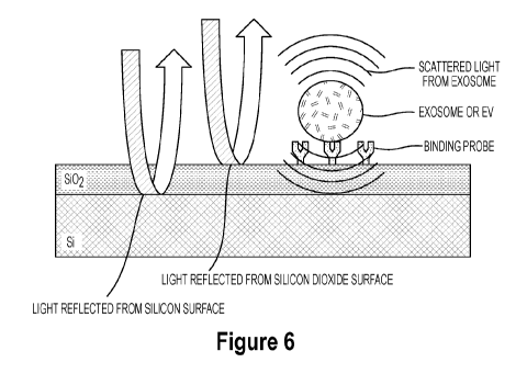
Figure 6 illustrates a schematic of a substrate functionalized with a binding
probe and
how light illuminating the substrate is spectrally reflected from the two
layers of the substrate,
which interferes with the scattered light from particle(s) bound to the one
substrate surface; and
Figure 7A is a picture of the instrument for the imaging of the substrate,
according to an
illustrative embodiment of the invention;
16
CA 03012212 2018-07-20
WO 2017/136676
PCT/US2017/016434
Figure 7B is an image of a spectral reflectance chip (substrate) , according
to an
illustrative embodiment of the invention;
Figure 7C is an image of the spectral reflectance chip disposed within a
microfluidic
cassette, which allows flowing of a sample over the substrate, according to an
illustrative
embodiment of the invention; and
Figure 7D is an illustration of an array of binding probes on substrate,
according to an
illustrative embodiment of the invention.
Figures 8A-8C show a schematic depicting that extracellular vesicles such as
exosomes
can be isolated on a sensor chip through physical absorption or binding
agents, according to
illustrative embodiments of the invention. For example, binding agents can
target bioparticle
surface charge, glycosylation, lipid composition, and/or surface protein.
Figure 8A shows a schematic of a substrate that is functionalized with a
binding agent or
a combination of multiple binding agents (e.g., protein markers such as anti-
CD63, anti-CD9,
anti-CD81, Tim-4 and anti-flotillin-1; e.g., carbohydrate binding lectins such
as Galanthus
nivalis lectin (GNA)), according to an illustrative embodiment on the
invention.
Figure 8B shows a schematic where a combination of markers can be mixed and
then
immobilized on a substrate, according to an illustrative embodiment of the
invention. The
substrate is functionalized with two or more binding agents that are mixed
together.
Figure 8C shows a schematic where a sample is contacted with the
functionalized
substrate to isolate extracellular vesicles and exosomes, according to an
illustrative embodiment
of the invention. The amount of captured vesicles can be quantified using an
interferometric
biosensor (e.g., SP-IRIS).
17
CA 03012212 2018-07-20
WO 2017/136676
PCT/US2017/016434
Figure 9 shows a schematic of a method of isolation of extracellular vesicles
(e.g.,
exosomes) on a substrate, according to an illustrative embodiment of the
invention. A sample is
contacted with a functionalized substrate to isolate extracellular vesicles
and/or exosomes. The
amount of captured vesicles can be quantified using SP-IRIS post isolation or
during isolation.
The substrate containing the captured extracellular vesicles and/or exosomes
is fixed and
permeabilized. Captured extracellular vesicles and/or exosomes can be labeled
with secondary
markers to measure the proportion of bound particles with a secondary marker
which could be
disease specific (e.g., glypican-1).
Figure 10 shows a schematic of a method of isolation of extracellular vesicles
(e.g.,
exosomes) on a substrate, according to an illustrative embodiment of the
invention.
Figure 11 shows signal in percent contrast from 100 nm diameter polystyrene
beads
adsorbed to a SP-IRIS substrate which comprises a silicon with semi-
transparent silicon dioxide
top layer. The nanoparticles are adsorbed to the silicon dioxide top layer.
The percent contrast
is shown for different oxide thickness and wavelength of illumination.
Figure 12 shows signal in percent contrast for a range of nanoparticle
diameters from 40
to 220 nanometers. The particle contrast is plotted for different oxide
thickness and illumination
wavelength.
Figures 13 and 14 show a method of isolating cancer-derived circulating
extracellular
vesicles (e.g., exosomes), according to an illustrative embodiment of the
invention.
Figure 15 shows a method for detecting the binding of a circulating
extracellular vesicle
(e.g., exosomes) to a surface of a substrate, according to an illustrative
embodiment of the
invention.
18
CA 03012212 2018-07-20
WO 2017/136676
PCT/US2017/016434
DETAILED DESCRIPTION
Throughout the description, where compositions are described as having,
including, or
comprising specific components, or where methods are described as having,
including, or
comprising specific steps, it is contemplated that, additionally, there are
compositions of the
present invention that consist essentially of, or consist of, the recited
components, and that there
are methods according to the present invention that consist essentially of, or
consist of, the
recited processing steps.
It should be understood that the order of steps or order for performing
certain action is
immaterial so long as the invention remains operable. Moreover, two or more
steps or actions
may be conducted simultaneously.
The mention herein of any publication, for example, in the Background section,
is not an
admission that the publication serves as prior art with respect to any of the
claims presented
herein. The Background section is presented for purposes of clarity and is not
meant as a
description of prior art with respect to any claim.
In one or more embodiments, the invention is directed to an apparatus that can
detect
binding of nanoparticle extracellular vesicles such as exosomes to binding
agents on a surface of
a substrate. The binding agents can be immobilized on a layered substrate
surface that has a
spectral reflectance signature that is altered upon immobilization of said
nanoparticles on a
binding layer on the substrate surface. In particular, as will be described
herein, the image
processing system detects the extracellular vesicles a function of the change
in reflective
properties of the substrate and an image processing system comprises a forward
model to provide
accurate and quantitative sizing of the extracellular vesicles. In particular,
a preferred
embodiment of the device uses a single wavelength (band) of light to measure
the
19
CA 03012212 2018-07-20
WO 2017/136676
PCT/US2017/016434
interference/mixing of reflected light from the binding layer with the
scattered light from the
particle (scattering of the light). As extracellular vesicles bind to the
binding layer, the scattered
light from these objects interfere with the reflected light from the substrate
surface making the
extracellular vesicles observable on an imaging device as discrete objects
(dots). The substrate is
illuminated with one (or more) wavelengths of light, and if one or more
extracellular vesicle
objects in the sample binds with the binding layer, the nanoparticle target
will appear in the
image as single discrete objects, thereby allowing the detection of the
individual binding of the
nanoparticle targets as well as the quantitative sizing of the extracellular
vesicles. The apparatus
allows for the simultaneous imaging of the entire field of view of a surface
for high-throughput
applications. The apparatus and method has several advantages such as low-
cost, high-
throughput, rapid and portable detection.
Also described herein are methods of use of the device for the detection of a
variety of
biomolecular targets. In some aspects, the devices and methods described
herein provide a high-
throughput method for simultaneously recording a response of an entire
substrate surface,
comprising sampling at least one wavelength using a light source providing
incoherent light,
and imaging the reflected or transmitted light using an imaging device. The
device can include a
light-emitting diode (LEDs) as the illumination source for interferometric
principles of detection.
Interferometric measurements can provide desired sensitivity and resolution
using optical path
length differences (OPD).
Accordingly, described herein are devices and methods for substrate enhanced
detection
of binding of molecules or nanoparticles or extracellular vesicles such as
exosomes to a surface
of a substrate. The device samples the reflectance spectrum by illuminating
the substrate with at
least one wavelength of light, using, for example, an LEDs and recording the
reflectance by an
CA 03012212 2018-07-20
WO 2017/136676
PCT/US2017/016434
imaging device, such as a 2-D arrayed pixel camera. In this way, the
reflectance spectrum for the
whole field-of-view is recorded simultaneously. Using this device and method,
high-throughput
microarray imaging can be accomplished. The invention can also provide high-
magnification
imaging for detection of biomolecular nanoparticle targets in the 30nm to a
few (2-3) microns in
range. Such high-magnification detection can be used, for example, for the
detection of a single
particle on a capture surface.
The instrument and process provide a high-throughput spectroscopy technique
where
sampling at least one wavelength is realized by using a narrowband light
sources, such as an
LED, and the reflected or transmitted light is imaged to an imaging device,
such as a
monochromatic CCD camera, thus allowing the response of the entire imaged
surface to be
recorded simultaneously. The microarray can be fabricated on a layered
substrate (for example:
anywhere from a few nm of 5i02 up to 100 nm of 5i02 layered on a Si wafer). A
preferred
embodiment includes a green LED light source (535 nm) and 100 nm oxide of 5i02
layered on a
Si wafer. A second preferred embodiment includes an ultraviolet LED light
source (420 nm) and
60 nm oxide of 5i02 layered on a Si wafer. A third preferred embodiment, for
use when imaging
in complex media, includes an ultraviolet LED light source (420 nm) and 30-to-
60 nm oxide of
5i02 layered on a Si wafer.
Figure 1 illustrates a diagrammatic view of a spectral reflectance imaging
system100
according to an embodiment of the present invention. The system 100 can
include an
illumination source 101, directing light onto the substrate 122, having an
oxide layer 124 and the
particles 126 to be detected, and an imaging system 130 for capturing images
of the light
reflected by the substrate 122, the oxide layer 124 and the particles 126. The
system 100 can also
include a computer system 140 for controlling the illumination source 101 and
receiving imaging
21
CA 03012212 2018-07-20
WO 2017/136676
PCT/US2017/016434
signals from the imaging system 130. In a preferred embodiment, the
illumination source 101
includes incoherent light source (LED) 102 that provides incoherent light in
one wavelength
having a substantially narrow band of wavelengths. In some embodiments, the
illumination
source 101 can include three or more incoherent light sources 102, 104, 106
that produce
incoherent light in three different wavelengths. The Light Emitting Diodes
(LEDs) or equivalent
light sources, each provide incoherent light at one of the plurality of
wavelengths. In some
embodiments, the illumination source 101 can include an array of illumination
elements,
including one or more illumination elements providing light at the same
wavelength and being
arranged in a geometric (e.g., circular or rectangular), random, or spatially
displaced array. The
light from the illumination source 101 can be directed through a focusing lens
112 and other
optical elements (e.g., polarizing lens, filters and light conditioning
components, not shown) to a
beam splitter 114 that directs the light onto the substrate 122, the oxide
layer 124 and the
particles 126. Optical components can be provided to condition the light to
uniformly illuminate
substantially the entire surface of the layered substrate 122. The light
reflected by the substrate
122, the oxide layer 124 and the particles 126 can be directed through the
beam splitter 114 and
imaging lens 134 into a camera 132 to capture images of the substrate surface.
The camera 132
can be, for example, a CCD camera (color or monochromatic) and produce image
signals
representative of the image. The image signals can be sent from the camera 132
to the computer
system 140 either by a wireless or wired connection.
Computer system 140 can include one or more central processing units (CPUs)
and
associated memory (including volatile and non-volatile memory, such as, RAM,
ROM, flash,
optical and magnetic memory) and a display 146 for presenting information to a
user. The
memory can store one or more computer programs that can be executed by the
CPUs to store and
22
CA 03012212 2018-07-20
WO 2017/136676
PCT/US2017/016434
process the image data and produce images of the substrate surface. Additional
computer
programs can be provided for analyzing the image data and the images to detect
interference
patterns and the particles 126 on the surface of the oxide layer 124 of the
substrate 122.
The computer programs can be executed by the computer to implement a method
according to one or more embodiments of the present invention whereby
interferometric
measurements can be made. The computer programs can control the illumination
source 101
comprising one (or more) LED that can be used to illuminate layered substrate.
The optical path
difference (OPD) between the bottom and top surface causes an interference
pattern. The
interference patterns can be imaged as intensity variations by the CCD camera
132 across the
.. whole substrate at once.
In an alternative embodiment, each incoherent light source can be an optical
fiber (not
shown) that directs the light at the layered substrate 122. Optical components
can be provided to
condition the light to uniformly illuminate substantially the entire surface
of the layered substrate
122
Figures 2A-2B shows detection of GPC1 exosomes directly from plasma with the
imaging platform according to this disclosure in a side-by-side comparison of
detection of the
same sample with a scanning electron microscope. As can be seen in Figure 2A,
the incubated
PDAC patient sample was imaged with the sensor platform disclosed herein and
the sample was
then stained with Osmium Tetroxide (lipid specific stain) to visualize the
sample with an
Electron Microscope as shown in Figure 2B. The incubated PDAC patient sample
was imaged
with the sensor platform was cropped as shown in Figure 2A to allow
visualization of a similar
area to the image from the electron microscope in Figure 2B. As can be seen
from the side-by-
side comparison, common large features from the sample are highlighted in red
to facilitate
23
CA 03012212 2018-07-20
WO 2017/136676
PCT/US2017/016434
comparison between the imaged samples, and the green circles show some of the
imaged small
nanoparticles.
Figure 3 demonstrates the properties desired for performing high magnification
substrate
enhanced microarray imaging. For making high magnification imaging, objectives
with higher
numerical apertures (NA) should be used. Because the light is collected at a
high range of angles,
most of the light averages out (as illustrated in the figure). Also the use of
thin oxide increases
the limit for spatial resolution because of less dispersion in light as it
passes through it.
Figure 4 depicts using a spatial filter as an option for performing high
magnification
substrate enhanced microarray imaging. To maintain the lateral resolution for
single particle
detection and the contrast of the reflectivity curve, it may be desirable to
place a spatial filter on
the collection path that will reject a range of angles of the reflected light.
Simple observation of
interference can be seen on the colors on soap bubbles. One of the ultimate
examples of high
precision measurements using optical interference is the LIGO with attometer
capability.
Figure 5 depicts the surface expression of glypican-1 and glypican-3 on
exosomes in
plasma obtained from cancer patients (pancreatic, lung, or breast cancer
patients). The
measurement was made using the spectral reflectance imaging technique for
counting of
glypican-1 and glypican-3 expressing exosomes from human plasma. The data
shows higher
expression of glypican-1 and/or glypican-3 in cancer patients.
Figure 6 depicts the interferometric scattering of reflected light upon
absorption of
nanoparticle extracellular vesicles such as exosomes to binding agents on a
surface of a
substrate. The reflections from the different layers including the Silicon
surface and the Silicon
dioxide surface interfere with the light reflected from the nanoparticles
captured by the binding
agents cause a change in the reflected light, which can be detected by the
image processing
24
CA 03012212 2018-07-20
WO 2017/136676
PCT/US2017/016434
system. In particular, a reflectance signature of the incident light is
altered by said nanoparticles
on a binding layer on the substrate surface to interfere with the light
reflected from the Silicon
surface and the Silicon Dioxide surface. The imaging system of Figure 1
detects the interference
in the reflection from the extracellular vesicles as compared to reflective
properties of the Silicon
surface and the Silicon Dioxide and an image processing system comprises a
forward model to
provide accurate and quantitative sizing of the extracellular vesicles. A
preferred embodiment of
the imaging device uses a single wavelength (band) of light to measure the
interference/mixing
of reflected light from the binding layer with the scattered light from the
particle (scattering of
the light).
Figure 7A is a picture of the instrument for the imaging of the substrate, as
described
herein. Figure 7B is an image of a spectral reflectance chip (substrate), as
been described herein.
Figure 7C is an image of the spectral reflectance chip disposed within a
microfluidic cassette,
which allows flowing of a sample over the substrate. Figure 7D is an
illustration of an array of
binding probes on substrate, as described herein.
Figures 8A-8C show a schematic depicting that extracellular vesicles such as
exosomes
can be isolated on a sensor chip through physical absorption or binding
agents, according to
illustrative embodiments of the invention. For example, binding agents can
target bioparticle
surface charge, glycosylation, lipid composition, and/or surface protein.
Figure 8A shows a schematic of a substrate that is functionalized with a
binding agent or
a combination of multiple binding agents (e.g., protein markers such as anti-
CD63, anti-CD9,
anti-CD81, Tim-4 and anti-flotillin-1; e.g., carbohydrate binding lectins such
as Galanthus
nivalis lectin (GNA)), according to an illustrative embodiment on the
invention.
CA 03012212 2018-07-20
WO 2017/136676
PCT/US2017/016434
Figure 8B shows alternatively to the schematic depicted in Figure 8A, a
combination of
markers can be mixed and then immobilized on a substrate. The substrate is
functionalized with
two or more binding agents that are mixed together.
Figure 8C shows a schematic where a sample is contacted with the
functionalized
substrate to isolate extracellular vesicles and exosomes, according to an
illustrative embodiment
of the invention. The amount of captured vesicles can be quantified using an
interferometric
biosensing (e.g., SP-IRIS).
Figure 9 shows a schematic of a method of isolation of extracellular vesicles
(e.g,
exosomes) on a substrate, according to an illustrative embodiment of the
invention. A sample is
contacted with a functionalized substrate to isolate extracellular vesicles
and/or exosomes. The
amount of captured vesicles can be quantified using SP-IRIS post isolation or
during isolation.
The substrate containing the captured extracellular vesicles and/or exosomes
is fixed and
permeabilized. Captured extracellular vesicles and/or exosomes can be labeled
with secondary
markers to measure the proportion of bound particles with a secondary marker
which could be
disease specific (e.g., glypican-1).
Figure 10 shows a schematic of a method of isolation of extracellular vesicles
(e.g.,
exosomes) on a substrate, according to an illustrative embodiment of the
invention.
Figure 11 shows signal in percent contrast from 100 nm diameter polystyrene
beads
adsorbed to a SP-IRIS substrate which comprises a silicon with semi-
transparent silicon dioxide
top layer. The nanoparticles are adsorbed to the silicon dioxide top layer.
The percent contrast
is shown for different oxide thickness and wavelength of illumination.
26
CA 03012212 2018-07-20
WO 2017/136676
PCT/US2017/016434
Figure 12 shows signal in percent contrast for a range of nanoparticle
diameters from 40
to 220 nanometers. The particle contrast is plotted for different oxide
thickness and illumination
wavelength.
In some embodiments three or more LEDs with different emission peak
wavelengths can
be used as the light source. In some embodiments where more than one
incoherent light source is
used, the light sources used have a narrow range of wavelength, and the width
between the
wavelengths of each individual light source is small. In some embodiments, one
or two light
sources are used.
In some embodiments described herein, the microarray or binding agent is
fabricated on a
layered substrate comprising anywhere from a few nanometers to 100 nm of SiO2
layered on a Si
wafer. In some embodiments, the microarray or binding agent is fabricated on a
layered substrate
comprising 95-100 nm of 5i02 layered on a Si wafer. In some embodiments, the
microarray or
binding agent is fabricated on a layered substrate comprising 30-60 nm of 5i02
layered on a Si
wafer. A preferred embodiment includes a green LED light source (near 535 nm)
and 100 nm
oxide of 5i02 layered on a Si wafer. A second preferred embodiment includes an
ultraviolet
LED light source (near 420 nm) and 60 nm oxide of 5i02 layered on a Si wafer.
A third
preferred embodiment, for use when imaging in complex media, includes an
ultraviolet LED
light source (near 420 nm) and 30-to-60 nm oxide of 5i02 layered on a Si
wafer. The devices
and methods described herein, can be used, in part, for high magnification
interferometric
measurements, for example, but not limited to, detecting extracellular
vesicles, such as an
exosome biomarker for a cancer, in a given sample.
A "particle," as defined herein, refers to any target to be detected by the
devices and
methods described herein that has a radius from a few nanometers up to a few
microns.
27
CA 03012212 2018-07-20
WO 2017/136676
PCT/US2017/016434
The use of high-magnification interferometric measurements is an approach to
detection
of biomolecular targets and particles. The methods and devices described
herein provide for
imaging through a high magnification objective lens with a high numerical
aperture and placing
a spatial filter on the camera's optical axis. The high numerical aperture
objective lens will allow
imaging at high magnifications and the spatial filter is used to maintain the
contrast of the
interference cause by the layered substrate by only collecting light from a
high angle or a range
of angles of incident light. The optical setup described will allow for
detection of sub-
wavelength structures without losing contrast or lateral resolution.
Another approach to simplifying the imaging device described herein can be to
use a
broadband source and a colored CCD camera in which the spectral sampling is
done by the
camera. Pixels of the camera dedicated for detection of separate colors can be
used to extract the
intensity of light included in a given spectral band, thus enable a spectral
detection scheme.
One advantage to the embodiments with an LED light source is that an LED based
illumination source allows the imaging device to be more robust and portable,
thus allowing field
applications. Another advantage is the high magnification capability of the
invention. High
magnification will allow for the detection of single biomolecular targets on
the binding agent
surface (e.g., > a few nm in length or diameter). In some embodiments, a white
light source or an
RGB LED with a 3CCD or other color camera can be used to capture spectral
information at
three distinct wavelengths to increase temporal resolution. This is beneficial
in studying dynamic
biological interactions, for example.
The device described herein facilitates a method of using an LED illumination
source for
substrate enhanced detection of extracellular vesicles such as exosome
biomarkers in a sample
bound to a surface. The device provides in one aspect a high-throughput
spectroscopy method
28
CA 03012212 2018-07-20
WO 2017/136676
PCT/US2017/016434
for simultaneously recording a response of an entire substrate surface. The
device and methods
can be used in any high-throughput application. One aspect of the invention
thus provides a
platform or a system for high-throughput optical sensing of solid substrates,
comprising an
illuminating source and an imaging device.
In some embodiments the imaging device is a camera. The device can be used for
multiplexed and dynamic detection of extracellular vesicles, such as exosome
biomarkers on a
substrate.
All embodiments of the device can be described as functional modules, which
include
computer executable instructions recorded on computer readable media and which
cause a
computer to perform method steps when executed. The modules can be segregated
by function
for the sake of clarity. However, it should be understood that the modules
need not correspond to
discrete blocks of code and the described functions can be carried out by the
execution of various
code portions stored on various media and executed at various times.
In some embodiments, the device provides a system for obtaining data regarding
optical
.. sensing of a solid substrate comprising a) a determination module
configured to determine
optical information, wherein the optical information comprises sampling a
least one wavelength
using a narrow band light source; b) a storage device configured to store data
output from the
determination module; c) a comparison module adapted to compare the data
stored on the storage
device with a control data, the comparison being a retrieved content; and d) a
display module for
.. displaying a page of the retrieved content for the user on the client
computer, wherein the
retrieved content is a light absorption profile of the substrate, wherein a
certain light absorption
profile is indicative of binding of an exosome biomarker.
29
CA 03012212 2018-07-20
WO 2017/136676
PCT/US2017/016434
In some embodiments, the invention provides a computer program comprising a
computer readable media or memory having computer readable instructions
recorded thereon to
define software modules including a determination module and a comparison
module for
implementing a method on a computer, said method comprising a) determining
with the
determination module optical information, wherein the optical information
comprises sampling
at least one wavelength using a narrow-band light source; b) storing data
output from the
determination module; c) comparing with the comparison module the data stored
on the storage
device with a control data, the comparison being a retrieved content, and d)
displaying a page of
the retrieved content for the user on the client computer, wherein the
retrieved content is a light
absorption profile of the solid substrate, wherein a certain light absorption
profile is indicative of
binding of an exosome biomarker.
The "computer readable medium" can include data and computer-executable
instructions
for performing the steps of the method of the invention. Suitable computer
readable media
include floppy disk, CD-ROM/DVD/DVD-ROM, hard-disk drive, flash memory,
ROM/RAM,
magnetic tapes and etc. The computer executable instructions can be written in
a suitable
computer language or combination of several languages. Basic computational
biology methods
are described in, e.g. Setubal and Meidanis et al.
Introduction to Computational Biology Methods (PWS Publishing Company, Boston,
1997); Salzberg, Searles, Kasif, (Ed.), Computational Methods in Molecular
Biology, (Elsevier,
Amsterdam, 1998); Rashidi and Buehler, Bioinformatics Basics: Application in
Biological
Science and Medicine (CRC Press, London, 2000) and Ouelette and Bzevanis
Bioinformatics: A
Practical Guide for Analysis of Gene and Proteins (Wiley & Sons, Inc., 2nd
ed., 2001).
CA 03012212 2018-07-20
WO 2017/136676
PCT/US2017/016434
In some aspects, the function modules of embodiments of the device include a
determination module, a storage device, a comparison module and a display
module. The
determination module can include computer executable instructions to determine
and provide
optical information using an optical instrument. As used herein, an "optical
instrument" refers to
any instrument that either processes light waves to enhance an image for
viewing, or analyzes
light waves (or photons) to determine one of a number of characteristic
optical properties.
Known determination modules for determining optical properties include, for
example,
but are not limited to, microscopes, cameras, interferometers (for measuring
the interference
properties of light waves), photometers (for measuring light intensity);
polarimeters (for
measuring dispersion or rotation of polarized light), reflectometers (for
measuring the reflectivity
of a surface or object), refractometers (for measuring refractive index of
various materials),
spectrometers or monochromators (for generating or measuring a portion of the
optical spectrum,
for the purpose of chemical or material analysis), autocollimators (used to
measure angular
deflections), and vertometers (used to determine refractive power of lenses
such as glasses,
contact lenses and magnifier lens).
A "spectrograph" or "spectrometer", as defined herein, is an optical
instrument used to
measure properties of light over a specific portion of the electromagnetic
spectrum, typically
used in spectroscopic analysis to identify materials. The variable measured is
most often the
light's intensity but could also, for instance, be the polarization state. The
independent variable is
usually the wavelength of the light, normally expressed as a fraction of a
meter, but sometimes
expressed as a unit directly proportional to the photon energy, such as
wavenumber or electron
volts, which has a reciprocal relationship to wavelength. A spectrometer is
used in spectroscopy
for producing spectral lines and measuring their wavelengths and intensities.
Spectrometer is a
31
CA 03012212 2018-07-20
WO 2017/136676
PCT/US2017/016434
term that is applied to instruments that operate over a very wide range of
wavelengths, from
gamma rays and X-rays into the far infrared. If the region of interest is
restricted to near the
visible spectrum, the study is called spectrophotometry.
Spectrophotometry involves the use of a spectrophotometer. As defined herein,
a
"spectrophotometer" is a photometer (a device for measuring light intensity)
that can measure
intensity as a function of the color, or more specifically, the wavelength of
light. There are many
kinds of spectrophotometers. Among the most important distinctions used to
classify them are
the wavelengths they work with, the measurement techniques they use, how they
acquire a
spectrum, and the sources of intensity variation they are designed to measure.
Other important
.. features of spectrophotometers include the spectral bandwidth and linear
range. There are two
major classes of spectrophotometers; single beam and double beam. A double
beam
spectrophotometer measures the ratio of the light intensity on two different
light paths, and a
single beam spectrophotometer measures the absolute light intensity. Although
ratio
measurements are easier, and generally more stable, single beam instruments
have advantages;
for instance, they can have a larger dynamic range, and they can be more
compact. Historically,
spectrophotometers use a monochromator to analyze the spectrum, but there are
also
spectrophotometers that use arrays of photosensors. Especially for infrared
spectrophotometers,
there are spectrophotometers that use a Fourier transform technique to acquire
the spectral
information quicker in a technique called Fourier Transform InfraRed. The
spectrophotometer
quantitatively substance). The most common application of spectrophotometers
is the
measurement of light absorption, but they can be designed to measure diffuse
or specular
reflectance. Strictly, even the emission half of a luminescence instrument is
a kind of
spectrophotometer.
32
CA 03012212 2018-07-20
WO 2017/136676
PCT/US2017/016434
The optical information determined in the determination module can be saved to
and read
by the storage device. As used herein the "storage device" is intended to
include any suitable
computing or processing apparatus or other device configured or adapted for
storing data or
information. Examples of storage devices suitable for use with the present
invention include
.. stand-alone computing apparatus; communications networks, including local
area networks
(LAN), wide area networks (WAN), Internet, Intranet, and Extranet; and local
and distributed
processing systems including the "cloud." Storage devices also include, but
are not limited to:
magnetic storage media, such as floppy discs, hard disc storage medium, and
magnetic tape;
optical storage media such as compact disc; electronic storage media such as
RAM, ROM,
EPROM, EEPROM and the like; general hard disks and hybrids of these categories
such as
magnetic/optical storage media. The medium is adapted or configured for having
recorded there
on sequence information or expression level information. The data is typically
provided in digital
form that can be transmitted and read electronically, e.g., via the Internet,
on diskette, or any
other mode of electronic or non-electronic communication.
As used herein, "stored" refers to a process for storing information on the
storage device
such that it can be read back from the device. Those skilled in the art can
readily adopt any of the
presently known methods for recording information on known media to generate
manufactures
comprising the sequence information or expression level information.
A variety of software programs and formats can be used to store the optical
information
on the storage device. Any number of data processor structuring formats (e.g.,
text file or
database) can be employed to obtain or create a medium having the information
recorded
thereon.
33
CA 03012212 2018-07-20
WO 2017/136676
PCT/US2017/016434
By providing optical information in computer-readable form, one can use the
optical
information in readable form to compare a specific optical profile with the
optical information
stored within a database of the comparison module. For example, direct
comparison of the
determined optical information from a given sample can be compared to the
control data optical
information (e.g., data obtained from a control sample). The comparison made
in computer-
readable form being the retrieved content from the comparison module, which
can be processed
by a variety of means.
The retrieved content can then be displayed through a "display module".
As used herein, a cassette is defined as configured to contain a
silicon/silicon dioxide
chip with a transparent and high-quality imaging window (COP or polycarbonate)
with a thin
channel of fluid.
As defined herein, a "light emitting diode (LED)" is an electronic light
source based on
the semiconductor diode. When the diode is forward biased (switched on),
electrons are able to
recombine with holes and energy is released in the form of light. This effect
is called
electroluminescence and the color of the light is determined by the energy gap
of the
semiconductor. The LED is usually small in area (less than 1 mm) with
integrated optical
components to shape its radiation pattern and assist in measures the fraction
of light that passes
through a given solution. In a spectrophotometer, a light from the lamp is
guided through a
monochromator, which picks light of one particular wavelength out of the
continuous spectrum.
This light passes through the sample that is being measured. After the sample,
the intensity of the
remaining light is measured with a photodiode or other light sensor, and the
transmittance for
this wavelength is then calculated. In short, the sequence of events in a
spectrophotometer is as
follows: the light source shines through the sample, the sample absorbs light,
the detector detects
34
CA 03012212 2018-07-20
WO 2017/136676
PCT/US2017/016434
how much light the sample has absorbed, the detector then converts how much
light the sample
absorbed into a number, the numbers are e are transmitted to a comparison
module to be further
manipulated (e.g. curve smoothing, baseline correction). Many
spectrophotometers must be
calibrated by a procedure known as "zeroing." The absorbency of some standard
substance is set
as a baseline value, so the absorbencies of all other substances are recorded
relative to the initial
"zeroed" substance. The spectrophotometer then displays % absorbency (the
amount of light
absorbed relative to the initial reflection. Like a normal diode, the LED
consists of a chip of
semiconducting material impregnated, or doped, with impurities to create a p-n
junction. As in
other diodes, current flows easily from the p-side, or anode, to the n-side,
or cathode, but not in
the reverse direction. Charge-carriers¨ electrons and holes¨ flow into the
junction from
electrodes with different voltages. When an electron meets a hole, it falls
into a lower energy
level, and releases energy in the form of a photon. The wavelength of the
light emitted, and
therefore its color, depends on the band gap energy of the materials forming
the p-n junction. In
silicon or germanium diodes, the electrons and holes recombine by a non-
radiative transition
which produces no optical emission, because these are indirect band gap
materials. The materials
used for the LED have a direct band gap with energies corresponding to near-
infrared, visible or
near-ultraviolet light. LEDs are usually built on an n-type substrate, with an
electrode attached to
the p-type layer deposited on its surface. P-type substrates, while less
common, occur as well.
Many commercial LEDs, especially GaN/InGaN, also use sapphire substrate. Most
materials
used for LED production have very high refractive indices. This means that
much light will be
reflected back in to the material at the material/air surface interface. LEDs
of use for the present
invention, include but are not limited to:
CA 03012212 2018-07-20
WO 2017/136676 PCT/US2017/016434
_____ rs-------
. tWaveltir4th . z ':t
:1
Oit= a 1V1 1 Sell:tic:m(1d actor Materiat
1 .1 !Arad =
_______________________________________________________________ 1
'.1 = ... _______
41aaaM.:1 =i ' > 760 'AV ---: 1.9.
1
______________________________________________________________ M OM) I
:'='
" .
: .
= WamMon-
.4...01i:Un'.. Ine.:CSiElk. ( AlatiAri) I
< X < . 1 .,(13 < Ay < iciailiatamaki4mAkoRbsidg
: $60 .2,=,05 = A .o..4.1..tai ch fiallii.ah inthiona 041-
...s:.:41.1$14.t ($ 616a1.1.P)
1 1
1 k3ailiaraiII./=4 iltIo....4.1hlide: (GAP) t
:t
1 .
: 590 ..........x. < .7,03 ,c AN., ,.:. . ...441i..4m a..-
....aitkpbm.:phiatt
.).1$111.ita: : lo
'V'.4111M1I11a1:agIMIRi111A1:M911-11a''19:.:11101 (AY5::EII3P) 1
1
. . .P..411iorita.1..,0-4:00.41.1.) . OR ==4 ::
' t ,....¨ . -..., -----
4,¨õ-- tl----------------4
k= , , i' s - - ,,-,,--= ' -3 , -.6 µ,-i:., '..C''' 'VP) i
. .õ,,,.: ...,...10 <Av õ...,z :, $i.l.s..10 r3): ca, ',..1 .14,0,
..,,,k,0!=,,,.:atN,I., s:a.,..$, ,
1
o.,,...- . jAlimiko.a.t. gi,i3littul3 ibdttlr:13
PlIKsqlNdit (A14:_kkir.ir)
.== = :590 .1.,18 1
i ;:4 M ffB aho..:phittbtckaP) t . .
'
tx' --
t:
t = iNdimagiltlilI.A.RitOde (tri(jaN) i gAili1M-
11:111.1.9.k..F.ick 1
1 .
t
,,.-1 _ = tt(T4Nt
' 500 < A. K; :2,18 <AV < 1.-- '. == ¨ -. - - . -- 1
fir:t=\.-cr4 ,. 570
4- .,0 R.J.a.9ill int .1 ) phospho:.4.. (Cid')
z 1'
'1 . AI LiTS1Si ITU rTS gaitirt. indium
plunbhidc ( A Icklinri.) ,'z
: r :,.:.
., lAlLutitiora u1.4.414n _V.N.W11,ii;k ( A -.C1:41P=1
1
1 i ____
1aZ.akalit qnsii) i
i I
< 41-A3 aro <--alliibm nitriitte dett1t..Nrj t
itliet, .1,7 ,Sli-c..t.tn. c.zd'A de (SIC t a&' ge.htntitt
;õ:.t
1 I
1 I IS tl i cett (Si:=!: tw substrate (LI ntkr
developtmi):0 t
' ,......................-.........- :
........20544424.... .......1.1.204.41.14.2.4.1.....,
...411Ø5441.544424.4........X............11
'= )0 < X < ,76 <AV < 1
,taaaaaef..t
IV t . , ,. .Irs.dittn-i. '.,--atti==-um tkiicte. (taliaN) t
t ri-:.10 = 1,0. i- ' ' ;õ:.t
r--1¨.7.7.--
t mt-ittiVie ' 7FAS <AV <
:1Dttal bliatilred LF0s., .
ttIKEk_
t =#Y-1)04 4,7 )11.x: stith petl vhosphor,
1 ____ . ., ..,..a= ).4.1:m w.4.4.. p,4.p.A. p.......tts... ,
!----- " ----- 1 _____ = -- ' _________________________________ - - -- " t
) i Pixtbord (CI .1
1
.1 t
t . lAks.3:14E3.1trt 31 :(d
-'.$.i.< AV < I- -. - :- " - - " = . t
1
't Itte.s1.440: < 400 eattaatatauuitiatna.aiT4t. \K
.:s = = -4.4:
1Momino .1) gaffium -Mil -,Am Aitzidgz (MGit:m1*) ........ :r tlown 1 .
. .
4-.c. 210 am) 1
1-----1 _______
..,,
IT3i-t-Ez,4 1 1
l'iVh.i:ir :. ' 'AV :-.- 3.,:i .B1,..,.$,Ai V atiodt. :with ydiow
phasphot
.... .. . .. ... P.. . . ... ..,..... ... ..
_I .. ... ....... ... ....... ... ....... ...
....... ... ..1
36
CA 03012212 2018-07-20
WO 2017/136676
PCT/US2017/016434
As defined herein, a substrate surface can include a "specular reflecting
interface." Such
specular reflecting interfaces refer to those surfaces upon which incoming
light undergoes
"specular reflection," i.e., the mirror- like reflection of light (or
sometimes other kinds of wave)
from a surface, in which light from a single incoming direction (a ray) is
reflected into a single
outgoing direction. Such specular reflecting behavior of a surface, substrate
or interface, is
described by the law of reflection, which states that the direction of
incoming light (the incident
ray), and the direction of outgoing light reflected (the reflected ray) make
the same angle with
respect to the surface normal, thus the angle of incidence equals the angle of
reflection;
mathematically defined Oi = Or. A second defining characteristic of specular
reflection is that the
incident, normal, and reflected directions are coplanar. Specular reflection
can be accurately
measured using, for example, a glossmeter. The measurement is based on the
refractive index of
an object. In some embodiments of the aspects described herein, a specular
reflecting interface
comprises a substrate having a transparent dielectric layer, for example a
layer of Silicon Oxide
(5i02) on a Silicon substrate. In some embodiments of aspects herein, the
layer of Silicon Oxide
(5i02) has a layer of binding agent for binding to nanoparticles such as an
exosome biomarker
thereon. In some embodiments, an alternative transparent dielectric layer,
such as silicon nitride
as well as other coatings can be used as a thin transparent or specular
reflecting interface layer.
Applications of the Sensors and Methods
The ability to detect biological extracellular vesicles, e.g., exosomes, e.g.,
exosomes
comprising an exosome biomarker, e.g., a cell surface biomarker, in a sample
is fundamental to
our understanding of both cell physiology and disease progression, as well as
for use in various
applications such as the early and rapid detection. Described herein are
rapid, sensitive, simple to
37
CA 03012212 2018-07-20
WO 2017/136676
PCT/US2017/016434
use, and inexpensive biosensors that are useful for a variety of applications
involving the
detection of nanoparticles, ranging from research and medical diagnostics, to
detection of cancer.
Accordingly, in one aspect, the substrates described herein are used to detect
binding of
extracellular vesicles, e.g., exosomes, e.g., exosomes comprising an exosome
biomarker, e.g., a
cell surface biomarker, in a sample to a substrate layer, wherein binding of a
exosome
biomarkers present in a sample contacted with the substrate layer changes an
optical path length
relative to an optical path length in the absence of the sample, resulting in
an interference pattern
that is detected and measured by the device and methods described herein. In
some embodiment,
the sample that contacts the substrate can have a plurality of biomolecular
targets, such that
multiple extracellular vesicles bind to the substrate layer and are detected
by the devices and
methods described herein.
The devices and substrates can be used to study one or a number of specific
binding
interactions in parallel, i.e., multiplex applications. Binding of one or more
specific extracellular
vesicles in a sample to respective target surfaces can be detected. The
substrate is illuminated
with light, and if one or more nanoparticle targets in the sample binds one or
more targets, they
will appear in the image as single discrete objects allowing the detection of
the individual
binding of the nanoparticle targets. In embodiments where a biosensor
substrate surface
comprises an array of one or more distinct target locations comprising one or
more specific
targets, then the interference pattern is detected from each distinct location
of the substrate.
Thus, in some embodiments, a variety of specific target molecules can be
immobilized in
an array format onto the substrate surface. The substrate is then contacted
with a test sample of
interest comprising potential nanoparticle targets, such as exosome
biomarkers. Only the
exosomes that specifically bind to the target surface are bound to the
substrate. For high-
38
CA 03012212 2018-07-20
WO 2017/136676
PCT/US2017/016434
throughput applications, biosensors can be arranged in an array of arrays,
wherein several
substrates comprising an array of specific binding molecules targets on the
substrate surface are
arranged in an array.
Accordingly, the devices and substrates are used to detect binding of one or
more of a
plurality of nanoparticle targets present in a sample to a biosensor substrate
layer comprising one
or more of a plurality of immobilized target molecules attached to the
substrate layer. For
example, one or more specific immobilized molecules can be arranged in an
array of one or more
distinct locations on the surface of the substrate layer. The one or more
distinct locations can
define microarray spots of about 50-500 microns, or about 150-200 microns in
diameter.
A sample refers to any sample containing a biomolecular target, such as, for
example,
blood, plasma, serum, gastrointestinal secretions, homogenates of tissues or
tumors, synovial
fluid, feces, saliva, sputum, cyst fluid, amniotic fluid, cerebrospinal fluid,
peritoneal fluid, lung
lavage fluid, semen, lymphatic fluid, tears, prostatic fluid, or cellular
lysates. A sample may also
be obtained from an environmental source, such as water sample obtained from a
polluted lake or
other body of water, or a liquid sample obtained from a food source believed
to contaminated.
As used herein the terms "sample" or "biological sample" means any sample,
including,
but not limited to cells, organisms, lysed cells, cellular extracts, nuclear
extracts, components of
cells or organisms, extracellular fluid, media in which cells are cultured,
blood, plasma, serum,
gastrointestinal secretions, homogenates of tissues or tumors, synovial fluid,
feces, saliva,
sputum, cyst fluid, amniotic fluid, cerebrospinal fluid, peritoneal fluid,
lung lavage fluid, semen,
lymphatic fluid, tears and prostatic fluid. In addition, a sample can be a
viral or bacterial sample,
a sample obtained from an environmental source, such as a body of polluted
water, an air
sample, or a soil sample, as well as a food industry sample.
39
CA 03012212 2018-07-20
WO 2017/136676
PCT/US2017/016434
The terms "label" or "tag", as used herein, refer to a composition capable of
producing a
detectable signal indicative of the presence of the target in an assay sample.
Suitable labels
include radioisotopes, nucleotide chromophores, enzymes, substrates,
fluorescent molecules,
chemiluminescent moieties, magnetic particles, bioluminescent moieties, and
the like. As such, a
label is any composition detectable by spectroscopic, photochemical,
biochemical,
immunochemical, electrical, optical or chemical means.
The articles "a" and "an" are used herein to refer to one or to more than one
(i.e., at least
one) of the grammatical object of the article. By way of example, "an element"
means one
element or more than one element. Thus, in this specification and the appended
claims, the
singular forms "a," "an," and "the" include plural references unless the
context clearly dictates
otherwise. Thus, for example, reference to a pharmaceutical composition
comprising "an agent"
includes reference to two or more agents.
As used herein, the term "comprising" means that other elements can also be
present in
addition to the defined elements presented. The use of "comprising" indicates
inclusion rather
than limitation. The term "consisting of' refers to compositions, methods, and
respective
components thereof as described herein, which are exclusive of any element not
recited in that
description of the embodiment. As used herein the term "consisting essentially
of' refers to those
elements required for a given embodiment. The term permits the presence of
elements that do not
materially affect the basic and novel or functional characteristic(s) of that
embodiment of the
invention. Other than in the operating examples, or where otherwise indicated,
all numbers
expressing quantities of ingredients or reaction conditions used herein should
be understood as
modified in all instances by the term "about." The term "about" when used in
connection with
percentages can mean +/- a percentage
CA 03012212 2018-07-20
WO 2017/136676
PCT/US2017/016434
Exosomes
Exosomes are small, membrane-bound vesicles with a size of 40-150 nm (Pan et
al, 1985;
Trams et al, 1981). They are secreted by many different cell types, such as
cancer cells,
.. mesenchymal cells, thrombocytes (Kahlert and Kalluri, Exosomes in tumor
microenvironment
influence cancer progression and metastasis. J. Mol Med. (Berl), 91:431-437,
2013; Heijnen et
al, Activated platelets release two types of membrane vesicles: microvesicles
by surface
shedding and exosomes derived from exocytosis of multivesicular bodies and
alpha-granules.
Blood, 94:3791-3799, 1999; Raposo et al, B lymphocytes secrete antigen-
presenting vesicles.
The Journal of Experimental Medicine, 183: 1161-1172, 1996), immune cells
(Thery et al,
Exosomes: composition, biogenesis and function. Nat. Rev. Immunol, 2:569- 579,
2002),
platelets (Janowska-Wieczorek et al, Microvesicles derived from activated
platelets induce
metastasis and angiogenesis in lung cancer. International Journal of Cancer, 1
13:752-760, 2005.
Jazieh et al, The clinical utility of biomarkers in the management of
pancreatic adenocarcinoma.
Seminars in Radiation Oncology, 24:67-76, 2014), and endothelial cells
(Hergenreider et al,
Atheroprotective communication between endothelial cells and smooth muscle
cells through
miRNAs. Nature Cell Biology, 14:249-256, 2012). The first step in exosomes
biogenesis
involves the inward budding from the limiting membrane of late endosomes
(Trajkovic et al,
Ceramide triggers budding of exosome vesicles into multivesicular endosomes.
Science, 319:
1244-1247, 2008). During this process, exosomes are packed with RNA molecules
and proteins
from the parental cell (Trams et al, Exfoliation of membrane ecto-enzymes in
the form of micro-
vesicles. Biochimica et Biophysica Acta, 645:63-70, 1981; Trajkovic Supra).
After the release
into the extracellular space, tumor-derived exosomes can transfer proteins and
RNAs with
41
CA 03012212 2018-07-20
WO 2017/136676
PCT/US2017/016434
oncogenic activity to recipient cells (Grange et al, Microvesicles released
from human renal
cancer stem cells stimulate angiogenesis and formation of lung premetastatic
niche. Cancer
Research, 71 :5346-5356, 2011; Peinado et al, Melanoma exosomes educate bone
marrow
progenitor cells toward a pro- metastatic phenotype through MET. Nature
Medicine, 18:883-891,
2012). Because exosomes are very stable under different conditions, they can
protect their
biological cargo against degradation and denaturation in the extracellular
environment (Taylor
and Gercel-Taylor, Exosomes/microvesicles: mediators of cancer-associated
immunosuppressive
microenvironments. Seminars in Immunopathology, 33 :441-454, 2011). Genomic
DNA in
circulation is mainly contained in exosomes (Kahlert et al, Identification of
double-stranded
genomic DNA spanning all chromosomes with mutated KRAS and p53 DNA in the
serum
exosomes of patients with pancreatic cancer. The Journal of Biological
Chemistry, 289:3869-
3875, 2014). Exosomes from astrocytes and glioblastoma cells carry
mitochondrial DNA
(Guescini et al, C2C12 myoblasts release micro-vesicles containing mtDNA and
proteins
involved in signal transduction. Experimental Cell Research, 316: 1977-1984,
2010).
.. Furthermore, it has been shown that exosomes from glioblastoma cell lines
contain small
amounts of single-stranded DNA as well as high levels of transposable elements
(Balaj et al.,
Tumour microvesicles contain retrotransposon elements and amplified oncogene
sequences.
Nature Communications, 2: 180, 2011).
Exosomes are found in all body fluids of cancer patients, such as serum,
saliva,
cerebrospinal fluid, bone marrow aspirates, eye exudate/tears, and ascites
(Peinado Supra; Lau et
al, Role of Pancreatic Cancer-derived Exosomes in Salivary Biomarker
Development. The
Journal of Biological Chemistry, 288:26888-26897, 2013; Choi et al, Proteomic
analysis of
microvesicles derived from human colorectal cancer ascites. Proteomics, 1 1
:2745-2751, 2011).
42
CA 03012212 2018-07-20
WO 2017/136676
PCT/US2017/016434
As such, exosomes are promising diagnostic and predictive biomarkers in
cancer. However,
genetic profiling studies on circulating DNA from cancer patients are
confounded by the fact that
the isolated DNA represents all cells of the body, thus making mutation and
genetic defects
challenging (Murtaza et al, Non- invasive analysis of acquired resistance to
cancer therapy by
sequencing of plasma DNA. Nature, 497; 108-1 12, 2013; Yong, Cancer
biomarkers: Written in
blood. Nature, 51 1 :524-526, 2014; Kirk, Breast cancer: Circulating tumour
DNA the better of
the blood biomarkers. Nature Reviews, Clinical Oncology, 10:247, 2013; Crowley
et al, Liquid
biopsy: monitoring cancer-genetics in the blood. Nature Reviews, Clinical
Oncology, 10:472-
484, 2013).
Several exosomes markers have been proposed and include members of the
tetraspanin
family (CD9, CD63, CD81), members of the endosomal sorting complexes required
for transport
(ESCRT; TSG101, Alix), and heat shock proteins (Hsp60, Hsp70, Hsp90) (Taylor
and Gercel-
Taylor, Supra). Epithelial tumor cells secrete exosomes carrying the
epithelial cell adhesion
molecule (EpCAM) (Taylor and Gercel-Taylor, Supra; Silva et al, Analysis of
exosome release
and its prognostic value in human colorectal cancer. Genes, Chromosomes &
Cancer, 51:409-
418, 2012; Runz et al., Malignant ascites-derived exosomes of ovarian
carcinoma patients
contain CD24 and EpCAM. Gynecologic Oncology, 107:563-571, 2007). Melanoma-
derived
exosomes contain the tumor-associated antigen Mart-1 and tyrosinase-related
protein-2 (TYRP2)
(Peinado, Supra; Mears et al, Proteomic analysis of melanoma-derived exosomes
by two-
dimensional polyacrylamide gel electrophoresis and mass spectrometry.
Proteomics, 4:4019-
4031, 2004; Andre et al, Malignant effusions and immunogenic tumour-derived
exosomes.
Lancet, 360:295-305, 2002). Exosomes from gastric cancer, breast cancer, and
pancreatic cancer
carry members of the human epidermal growth factor receptor (HER) family
(Adamczyk et al,
43
CA 03012212 2018-07-20
WO 2017/136676
PCT/US2017/016434
Characterization of soluble and exosomal forms of the EGFR released from
pancreatic cancer
cells. Life Sciences, 89:304-312, 2011; Baran et al, Circulating tumour-
derived microvesicles in
plasma of gastric cancer patients. Cancer Immunology, Immunotherapy: CII,
59:841-850, 2010;
Ciravolo et al, Potential role of HER2-overexpressing exosomes in countering
trastuzumab-
.. based therapy. Journal of Cellular Physiology, 227:658-667, 2012).
The terms "microvesicle" and "exosome," as used herein, refer to a membranous
particle,
wherein at least part of the membrane of the exosomes is directly obtained
from a cell. Most
commonly, exosomes will have a size (average diameter) that is up to 5% of the
size of the donor
cell. Therefore, especially contemplated exosomes include those that are shed
from a cell.
Exosomes may be detected in or isolated from any suitable sample type, such
as, for
example, body fluids. As used herein, the term "sample" refers to any sample
suitable for the
methods provided by the present invention. The sample may be any sample that
includes
exosomes suitable for detection or isolation. Sources of samples include
blood, bone marrow,
pleural fluid, peritoneal fluid, cerebrospinal fluid, urine, saliva, amniotic
fluid, malignant ascites,
.. broncho-alveolar lavage fluid, synovial fluid, breast milk, sweat, tears,
joint fluid, and bronchial
washes. In one aspect, the sample is a blood sample, including, for example,
whole blood or any
fraction or component thereof. A blood sample suitable for use with the
present invention may be
extracted from any source known that includes blood cells or components
thereof, such as
venous, arterial, peripheral, tissue, cord, and the like. For example, a
sample may be obtained
and processed using well-known and routine clinical methods (e.g., procedures
for drawing and
processing whole blood). In one aspect, an exemplary sample may be peripheral
blood drawn
from a subject with cancer.
44
CA 03012212 2018-07-20
WO 2017/136676
PCT/US2017/016434
Exosomes may also be isolated from tissue samples, such as surgical samples,
biopsy
samples, tissues, feces, and cultured cells. When isolating exosomes from
tissue sources it may
be necessary to homogenize the tissue in order to obtain a single cell
suspension followed by
lysis of the cells to release the exosomes. When isolating exosomes from
tissue samples it is
important to select homogenization and lysis procedures that do not result in
disruption of the
exosomes. Exosomes contemplated herein are preferably isolated from body fluid
in a
physiologically acceptable solution, for example, buffered saline, growth
medium, various
aqueous medium, etc.
Exosomes may be isolated from freshly collected samples or from samples that
have been
stored frozen or refrigerated. Although not necessary, higher purity exosomes
may be obtained if
fluid samples are clarified before precipitation with a volume-excluding
polymer, to remove any
debris from the sample. Methods of clarification include centrifugation,
ultracentrifugation,
filtration, or ultrafiltration. Most typically, exosomes can be isolated by
numerous methods well-
known in the art. One preferred method is differential centrifugation from
body fluids or cell
culture supernatants. Exemplary methods for isolation of exosomes are
described in (Losche et
al, Platelet-derived microvesicles transfer tissue factor to monocytes but not
to neutrophils,
Platelets, 15: 109-1 15, 2004; Mesri and Altieri, Endothelial cell activation
by leukocyte
microparticles, J. Immunol, 161 :4382-4387, 1998; Morel et al, Cellular
microparticles: a
disseminated storage pool of bioactive vascular effectors, Curr. Opin.
Hematol, 1 1 : 156-164,
-- 2004). Alternatively, exosomes may also be isolated via flow cytometry as
described in (Combes
et al. , A new flow cytometry method of platelet-derived microvesicle
quantitation in plasma,
Thromb. Haemost., 77:220, 1997).
CA 03012212 2018-07-20
WO 2017/136676
PCT/US2017/016434
One accepted protocol for isolation of exosomes includes ultracentrifugation,
often in
combination with sucrose density gradients or sucrose cushions to float the
relatively low-density
exosomes. Isolation of exosomes by sequential differential centrifugations is
complicated by the
possibility of overlapping size distributions with other microvesicles or
macromolecular
complexes. Furthermore, centrifugation may provide insufficient means to
separate vesicles
based on their sizes. However, sequential centrifugations, when combined with
sucrose gradient
ultracentrifugation, can provide high enrichment of exosomes.
Glypicans
Glypicans constitute one of the two major families of heparin sulfate
proteoglycans, with
the other major family being syndecans. Six glypicans have been identified in
mammals, and are
referred to as GPC1 through GPC6. While six glypicans have been identified in
mammals,
several characteristics remain consistent between these different proteins.
First, the core protein
of all glypicans is similar in size, approximately ranging between 60 and 70
kDa. Additionally,
in terms of amino acid sequence, the location of fourteen cysteine residues is
conserved. For all
members of the glypican family, the C-terminus of the protein is attached to
the cell membrane
covalently via a glycosylphosphatidylinositol (GPI) anchor. To allow for the
addition of the GPI
anchor, glypicans have a hydrophobic domain at the C-terminus of the protein.
Within 50 amino
acids of this GPI anchor, the heparan sulfate chains attach to the protein
core. Glypicans are
critically involved in developmental morphogenesis, and have been implicated
as regulators in
several cell signaling pathways, including Wnt and Hedgehog. Abnormal
expression of
glypicans has been noted in multiple types of cancer, including ovarian
cancer, mesothelioma,
pancreatic cancer, glioma, and breast cancer.
46
CA 03012212 2018-07-20
WO 2017/136676
PCT/US2017/016434
Glypican-1 (also known as GLPC1 and Glypican Proteoglycan 1) is a cell surface
heparan sulfate proteoglycan composed of a core protein anchored to the
cytoplasmic membrane
via a glycosyl phosphatidylinositol linkage. Isoform 1 (the canonical
sequence) is 558 amino
acids and 61.680kDa (UniProtKB Protein Symbol: P35052-GPC1 HUMAN; Protein
Accession:
P35052). Glypican-1 has been associated with cancer-cell derived exosomes
(W02015085096;
Melo et al. Glypican-1 identifies cancer exosomes and detects early pancreatic
cancer (2015)
Nature doi: 10.1038/nature14581). Glypican-2 or GPC2 is associated with
diseases including
mucopolysaccharidoses. The protein is 579 amino acids and 62830 Da(UniProtKB
Protein
Symbol: Q8N158-GPC2 HUMAN; Protein Accession: Q8N158). Glypican-3 (also known
as
GLPC3 and Glypican Proteoglycan 3) is a cell surface heparan sulfate
proteoglycan composed of
a core protein anchored to the cytoplasmic membrane via a glycosyl
phosphatidylinositol
linkage. Isoform 1 (the canonical sequence) is 580 amino acids and 65563 Da
(UniProtKB
Protein Symbol: P51654-GPC3 HUMAN; Protein Accession: P51654). Glypican-4 is
556 amino
acids and 62412 Da (Protein Symbol: 075487-GPC4 HUMAN; Protein Accession:
075487).
The GPC4 gene is adjacent to the 3' end of GPC3 and may also play a role in
Simpson-Golabi-
Behmel syndrome. Glypican-5 is 572 amino acids in length and 63707 Da (Protein
Symbol:
P78333-GPC5 HUMAN; Protein Accession: P78333). Glypican-6 is 555 amino acids
in length
and 62736 Da (Protein Symbol: Q9Y625-GPC6 HUMAN; Protein Accession: Q9Y625).
Diseases associated with GPC6 include omodysplasia 1 and omodysplasia.
Antibodies
Antibodies can be used as binding agents, e.g., an anti-GLPC1 provided on a
surface can
47
CA 03012212 2018-07-20
WO 2017/136676
PCT/US2017/016434
be used to capture exosomes comprising GLPC1 or an anti-GLPC3 provided on a
surface can be
used to capture exosomes comprising GLPC3. As used herein, the term "antibody"
or "antibody
molecule" refers to immunoglobulin molecules and immunologically active
portions of
immunoglobulin (Ig) molecules, i.e., molecules that contain an antigen binding
site that
specifically binds, e.g., immunoreacts with, an antigen. By "specifically
binds" or
"immunoreacts with" is meant that the antibody reacts with one or more
antigenic determinants
of the desired antigen and has a lower affinity for other polypeptides, e.g.,
does not react with
other polypeptides.
The term "antigen-binding site," or "binding portion" refers to the part of
the
immunoglobulin (Ig) molecule that participates in antigen binding. The antigen
binding site is
formed by amino acid residues of the N-terminal variable ("V") regions of the
heavy ("H") and
light ("L") chains. Three highly divergent stretches within the variable
regions of the heavy and
light chains, referred to as hypervariable regions, are interposed between
more conserved
flanking stretches known as "framework regions," or "FRs". The term "FR"
refers to amino acid
sequences which are naturally found between, and adjacent to, hypervariable
regions in
immunoglobulins. In an antibody molecule, the three hypervariable regions of a
light chain and
the three hypervariable regions of a heavy chain are disposed relative to each
other in three
dimensional space to form an antigen-binding surface. The antigen-binding
surface is
complementary to the three-dimensional surface of a bound antigen, and the
three hypervariable
.. regions of each of the heavy and light chains are referred to as
"complementarity-determining
regions," or "CDRs."
The extent of the framework region and CDRs have been defined (see, Kabat,
E.A., et al.
(1991) Sequences of Proteins of Immunological Interest, Fifth Edition, U.S.
Department of
48
CA 03012212 2018-07-20
WO 2017/136676
PCT/US2017/016434
Health and Human Services, NIH Publication No. 91-3242, and Chothia, C. et al.
(1987) J. Mol.
Biol. 196:901-917). Kabat definitions are used herein. Each VH and VL is
typically composed
of three CDRs and four FRs, arranged from amino-terminus to carboxy-terminus
in the amino
acid order: FR1, CDR1, FR2, CDR2, FR3, CDR3, FR4.
In embodiments, an antibody or antibody molecule encompasses full-length
antibodies
and antibody fragments. For example, a full-length antibody is an
immunoglobulin (Ig)
molecule (e.g., an IgG antibody) that is naturally occurring or formed by
normal
immunoglobulin gene fragment recombinatorial processes). In embodiments, an
antibody or
antibody molecule refers to an immunologically active, antigen-binding portion
of an
-- immunoglobulin molecule, such as an antibody fragment.
An antibody fragment, e.g., functional fragment, is a portion of an antibody,
e.g., F(ab1)2,
F(ab)2, Fab', Fab, domain antibody (dAb), variable fragment (Fv), or single
chain variable
fragment (scFv). A functional antibody fragment binds with the same antigen
that is recognized
by the intact antibody. For example, an anti-insulin monoclonal antibody
fragment binds to
-- insulin. The term "antibody fragment" or "functional fragment" also
includes isolated fragments
consisting of the variable regions, such as the "Fv" fragments consisting of
the variable regions
of the heavy and light chains or recombinant single chain polypeptide
molecules in which light
and heavy variable regions are connected by a peptide linker ("scFv
proteins"). In some
embodiments, an antibody fragment does not include portions of antibodies
without antigen
-- binding activity, such as Fc fragments or single amino acid residues.
Antibody fragments
include functional fragments and are encompassed by the terms "antibody" or
"antibody
molecule."
Exemplary antibody molecules include full length antibodies and antibody
fragments,
49
CA 03012212 2018-07-20
WO 2017/136676
PCT/US2017/016434
e.g., dAb (domain antibody), single chain, Fab, Fab', and F(ab')2fragments,
and single chain
variable fragments (scFvs).
A scFv polypeptide molecule is a covalently linked variable heavy chain
(VH)::variable
light chain (VL) heterodimer, which can be expressed from a gene fusion
including VH and VL
encoding genes linked by a peptide-encoding linker. See, e.g., Huston et al.
(1988) Proc Nat
Acad Sci USA 85(16):5879-5883. The N- to C- terminal orientation of the VH and
VL domains
can be in either orientation, e.g., VH¨VL or VL¨VH. Large naive human scFv
libraries have
been created to provide a source of rearranged antibody genes against a
variety of target
molecules. To isolate disease-specific antibodies, libraries can be
constructed from individuals
with certain diseases. See, e.g., Barbas et al., Proc. Natl. Acad. Sci. USA
89:9339-43 (1992); and
Zebedee et al., Proc. Natl. Acad. Sci. USA 89:3175-79 (1992).
The term "polyclonal antibody" refers to a mixture of different antibody
molecules which
react with more than one immunogenic determinant of an antigen. In
embodiments, polyclonal
antibodies can be isolated or purified from mammalian blood, secretions, or
other fluids, or from
eggs. In other embodiments, polyclonal antibodies are made up of a mixture of
different
monoclonal antibodies. In other embodiments, a polyclonal antibody can be
produced as a
recombinant polyclonal antibody.
The term "monoclonal antibody" or "mAb," as used herein, refers to a
population of
antibody molecules that contain only one molecular species of antibody
molecule consisting of a
unique light chain gene product and a unique heavy chain gene product. In
particular, the
complementarity determining regions (CDRs) of the monoclonal antibody are
identical in all the
molecules of the population. MAbs contain an antigen binding site capable of
immunoreacting
with a particular epitope of the antigen characterized by a unique binding
affinity for it.
CA 03012212 2018-07-20
WO 2017/136676
PCT/US2017/016434
Also provided herein are antibody fusion proteins, e.g., recombinantly
produced antigen-
binding molecules in which one or more of the same or different single-chain
antibody or
antibody fragment segments with the same or different specificities are
linked. Valency of an
antibody, e.g., fusion antibody protein, indicates how many binding arms or
sites the antibody
has to a single antigen or epitope; i.e., monovalent, bivalent, trivalent or
multivalent. The
multivalency of the antibody means that it can take advantage of multiple
interactions in binding
to an antigen, thus increasing the avidity of binding to the antigen.
Specificity indicates how
many antigens or epitopes an antibody is able to bind, i.e., monospecific,
bispecific, trispecific,
multispecific. For example, a natural antibody, e.g., an IgG, is bivalent
because it has two
binding arms but is monospecific because it binds to one epitope.
Monospecific, multivalent
antibodies, e.g., antibody fusion proteins, have more than one binding site
for an epitope but only
bind with one epitope. The fusion protein can comprise a single antibody
component, a
multivalent or multispecific combination of different antibody components or
multiple copies of
the same antibody component. The fusion protein can additionally comprise an
antibody or an
antibody fragment and a therapeutic agent. Examples of therapeutic agents
suitable for such
fusion proteins include immunomodulators and toxins. Exemplary toxins include
but are not
limited to ribonuclease (RNase), e.g., recombinant RNase, Diphtheria toxin,
Pseudomonas
exotoxin, monomethyl auristatin E, or mertansine. Additional exemplary toxins
are described
herein. In embodiments, the antibody molecule (e.g., antibody or functional
fragment thereof)
and the therapeutic agent (e.g., toxin) are encoded by a single nucleic acid
molecule. In
embodiments, the antibody molecule (e.g., antibody or functional fragment
thereof) and the
therapeutic agent (e.g., toxin) are disposed on the same polypeptide. In other
embodiments, the
antibody molecule (e.g., antibody or functional fragment thereof) and the
therapeutic agent (e.g.,
51
CA 03012212 2018-07-20
WO 2017/136676
PCT/US2017/016434
toxin) are encoded by separate nucleic acid molecules. In embodiments, the
antibody molecule
(e.g., antibody or functional fragment thereof) and the therapeutic agent
(e.g., toxin) are disposed
on separate polypeptides. A variety of protein or peptide effectors may be
incorporated into a
fusion protein. Conjugates/fusions to toxins are discussed further below.
A multispecific antibody is an antibody that can bind simultaneously to at
least two
targets that are of different structure, e.g., two different antigens, two
different epitopes on the
same antigen, or a hapten and/or an antigen or epitope. For example, one
specificity can be for a
B cell, e.g., an insulin-specific BCR on an insulin-specific B cell, and
another specificity can be
to a different antigen on a B cell. In another example, another specificity
can be to a receptor on
a phagocytosing cell, e.g., macrophage. In another example, another
specificity can be to a
receptor on a dendritic cell. Multispecific, multivalent antibodies are
constructs that have more
than one binding site, and the binding sites are of different specificity.
Humanized, chimeric, or fully human antibody molecules
Also provided herein are humanized, chimeric, or fully human antibody
molecules, e.g.,
full length antibodies, antibody fragments, antibody or antibody fragment
fusions, or antibody or
antibody fragment conjugates.
A humanized antibody is a recombinant protein in which the complementarity
determining regions (CDRs) from an antibody from one species; e.g., a rodent
(e.g., rat or mice)
antibody, are transferred from the heavy and light variable chains of the
rodent antibody into
human heavy and light variable domains. The constant domains of the antibody
molecule are
derived from those of a human antibody.
Methods for humanizing non-human antibodies have been described in the art. in
52
CA 03012212 2018-07-20
WO 2017/136676
PCT/US2017/016434
embodiments, a humanized antibody has one or more amino acid residues
introduced into it from
a source which is non-human. These non-human amino acid residues are often
referred to as
"import" residues, which are typically taken from an "import" variable domain.
Humanization
can be performed following the method of Winter and co-workers (Jones et al.,
Nature, 321: 522-
.. 525 (1986); Reichmann et al., Nature, 332: 323-327 (1988); Verhoeyen et
al., Science, 239:
1534-1536 (1988)), e.g., by substituting hypervariable region sequences for
the corresponding
sequences of a human antibody. Accordingly, such "humanized" antibodies are
chimeric
antibodies (U. S. Patent No. 4,816,567) wherein substantially less than an
intact human variable
domain has been substituted by the corresponding sequence from a non-human
species. In
embodiments, humanized antibodies are antibodies in which some hypervariable
region residues
and possibly some FR residues are substituted by residues from analogous sites
in rodent
antibodies.
The choice of human variable domains, both light and heavy, to be used in
making the
humanized antibodies can play a role in reducing antigenicity. In some
embodiments, according
to the so called "best-fit" method, the sequence of the variable domain of a
rodent antibody is
screened against the entire library of known human variable-domain sequences.
The human
sequence closest to that of the rodent is then accepted as the human framework
region (FR) for
the humanized antibody (Sunset al., J. Immunol., 151: 2296 (1993); Chothia et
al., J. Mol. Biol,
196: 901 (1987)). In embodiments, another method uses a particular framework
region derived
from the consensus sequence of all human antibodies of a particular subgroup
of light or heavy
chains. The same framework may be used for several different humanized
antibodies (Carter et
al., Proc. Natl. Acad. Sci. USA, 89: 4285 (1992); Presta et al., J. Immunol.,
151: 2623 (1993)).
In embodiments, antibodies are humanized with retention of high affinity for
the antigen
53
CA 03012212 2018-07-20
WO 2017/136676
PCT/US2017/016434
and other favorable biological properties. To achieve this goal, in certain
embodiments,
humanized antibodies are prepared by a process of analysis of the parental
sequences and various
conceptual humanized products using three-dimensional models of the parental
and humanized
sequences. Three-dimensional immunoglobulin models are commonly available,
Computer
programs are available which illustrate and display probable three-dimensional
conformational
structures of selected candidate immunoglobulin sequences. Inspection of these
displays permits
analysis of the likely role of the residues in the functioning of the
candidate immunoglobulin
sequence, e.g., the analysis of residues that influence the ability of the
candidate immunoglobulin
to bind its antigen. In this way, FR residues can be selected and combined
from the recipient and
import sequences so that the desired antibody characteristic, such as
preserved or increased
affinity for the target antigen, is achieved. In general, the hypervariable
region residues are
directly and most substantially involved in influencing antigen binding.
In embodiments, a humanized antibody molecule, e.g., humanized antibody
molecule
described herein, comprises one or more non-human (e.g., mouse) CDRs and
comprises human
framework and constant regions (e.g., framework and constant regions from a
human
imm-unoglobulin, e.g., IgGI, IgG2, IgG3, or 1.g,G4).
Antibody binding and affinity
As used herein, the term "epitope" includes any protein determinant capable of
specifically binding to an immunoglobulin, antibody fragment, e.g., an
antibody fragment
described herein, or a B cell receptor (BCR) (e.g., BCR comprising an
immunoglobulin).
Epitopic determinants usually consist of chemically active surface groupings
of molecules such
as amino acids or sugar side chains and usually have specific three
dimensional structural
54
CA 03012212 2018-07-20
WO 2017/136676
PCT/US2017/016434
characteristics, as well as specific charge characteristics. For example,
antibodies may be raised
against N-terminal or C-terminal peptides of a polypeptide.
As used herein, the terms "immunological binding," "immunological binding
properties,"
"specifically binds," or "selectively binds" refer to the non-covalent
interactions of the type
which occur between an immunoglobulin molecule and an antigen for which the
immunoglobulin is specific. The strength, or affinity of immunological binding
interactions can
be expressed in terms of the dissociation constant (Kd) of the interaction,
wherein a smaller Kd
represents a greater affinity. Immunological binding properties of selected
polypeptides can be
quantified using methods well known in the art. One such method entails
measuring the rates of
antigen-binding site/antigen complex formation and dissociation, wherein those
rates depend on
the concentrations of the complex partners, the affinity of the interaction,
and geometric
parameters that equally influence the rate in both directions. Thus, both the
"on rate constant"
(kon) and the "off rate constant" (koff) can be determined by calculation of
the concentrations and
the actual rates of association and dissociation. See, e.g., Nature 361:186-87
(1993). The ratio of
koff/kor, enables the cancellation of all parameters not related to affinity,
and is equal to the
dissociation constant Kd. (See, generally, Davies et al. (1990) Annual Rev
Biochem 59:439-473).
In some embodiments, an antibody molecule described herein specifically binds
an
antigen/epitope (e.g., autoantigen, e.g., islet autoantigen, e.g., insulin; or
a B cell, e.g.,
autoantigen-specific B cell, insulin-specific B cell; or an autoantigen::BCR
complex, e.g.,
insulin::BCR complex) when the equilibrium binding constant (Kd) is less than
or equal to 1 [tM,
e.g., less than or equal to 100 nM, less than or equal to 10 nM, less than or
equal to 100 pM, or
less than or equal to about 1 pM, e.g., as measured by assays such as
radioligand binding assays,
ELISAs, surface plasmon resonance, equilibrium binding assays, or similar
assays known to
CA 03012212 2018-07-20
WO 2017/136676
PCT/US2017/016434
those skilled in the art.
Antibody production
Various procedures known within the art may be used for the production of
antibody
molecules, e.g., antibodies or functional fragments thereof, directed against
a protein or peptide
of the invention, or against derivatives, fragments, analogs homologs or
orthologs thereof (See,
for example, Antibodies: A Laboratory Manual, Harlow E, and Lane D, 1988, Cold
Spring
Harbor Laboratory Press, Cold Spring Harbor, N.Y., incorporated herein by
reference).
In some embodiments, an autoantigen (e.g., islet autoantigen, e.g., an islet
autoantigen
described herein, e.g., insulin), a B cell (e.g., autoantigen-specific B cell,
e.g., insulin-specific B
cell), or an autoantigen::B cell receptor (BCR) complex (e.g., insulin::BCR
complex), can be
utilized as an immunogen in the generation of antibody molecules that
immunospecifically bind
these protein components.
Antibody molecules can be purified by well-known techniques, such as affinity
chromatography using protein A or protein G, e.g., which provide the IgG
fraction of immune
serum. Subsequently, or alternatively, the specific antigen which is the
target of the
immunoglobulin sought, or an epitope thereof, may be immobilized on a column
to purify the
immune specific antibody by immunoaffinity chromatography. Purification of
immunoglobulins
is discussed, for example, by D. Wilkinson (The Scientist, published by The
Scientist, Inc.,
Philadelphia Pa., Vol. 14, No. 8 (Apr. 17, 2000), pp. 25-28).
Monoclonal antibodies can be prepared using hybridoma methods, such as those
described by Kohler and Milstein, Nature, 256:495 (1975). In a hybridoma
method, a mouse,
hamster, or other appropriate host animal, is typically immunized with an
immunizing agent to
56
CA 03012212 2018-07-20
WO 2017/136676
PCT/US2017/016434
elicit lymphocytes that produce or are capable of producing antibodies that
will specifically bind
to the immunizing agent. Alternatively, the lymphocytes can be immunized in
vitro.
In embodiments, the immunizing agent includes the protein antigen, a fragment
thereof or
a fusion protein thereof In accordance with the compositions and methods
described herein, the
immunizing agent comprises an autoantigen, e.g., islet autoantigen, e.g.,
islet autoantigen
described herein, e.g., insulin. Generally, either peripheral blood
lymphocytes are used if cells of
human origin are desired, or spleen cells or lymph node cells are used if non-
human mammalian
sources are desired. The lymphocytes are then fused with an immortalized cell
line using a
suitable fusing agent, such as polyethylene glycol, to form a hybridoma cell
(Goding,
Monoclonal Antibodies: Principles and Practice, Academic Press, (1986) pp. 59-
103).
Immortalized cell lines are usually transformed mammalian cells, particularly
myeloma cells of
rodent, bovine and human origin. In embodiments, rat or mouse myeloma cell
lines are
employed. The hybridoma cells can be cultured in a suitable culture medium
that preferably
contains one or more substances that inhibit the growth or survival of the
unfused, immortalized
.. cells. For example, if the parental cells lack the enzyme hypoxanthine
guanine phosphoribosyl
transferase (HGPRT or HPRT), the culture medium for the hybridomas typically
will include
hypoxanthine, aminopterin, and thymidine ("HAT medium"), which substances
prevent the
growth of HGPRT-deficient cells.
Preferred immortalized cell lines are those that fuse efficiently, support
stable high level
expression of antibody by the selected antibody-producing cells, and are
sensitive to a medium
such as HAT medium. Exemplary immortalized cell lines are murine myeloma
lines, which can
be obtained, for instance, from the Salk Institute Cell Distribution Center,
San Diego, Calif and
the American Type Culture Collection, Manassas, Va. Human myeloma and mouse-
human
57
CA 03012212 2018-07-20
WO 2017/136676
PCT/US2017/016434
heteromyeloma cell lines also have been described for the production of human
monoclonal
antibodies. (See Kozbor, J. Immunol., 133:3001 (1984); Brodeur et al.,
Monoclonal Antibody
Production Techniques and Applications, Marcel Dekker, Inc., New York, (1987)
pp. 51-63)).
The culture medium in which the hybridoma cells are cultured can then be
assayed for
the presence of monoclonal antibodies directed against the antigen. For
example, the binding
specificity of monoclonal antibodies produced by the hybridoma cells is
determined by
immunoprecipitation or by an in vitro binding assay, such as radioimmunoassay
(MA), enzyme-
linked immunoabsorbent assay (ELISA), flow cytometry/FACS, or surface plasmon
resonance.
Such techniques and assays are known in the art. The binding affinity of the
monoclonal
antibody can, for example, be determined by the Scatchard analysis of Munson
and Pollard,
Anal. Biochem., 107:220 (1980). In embodiments, in therapeutic applications of
monoclonal
antibodies, it can be important to identify antibodies having a high degree of
specificity and a
high binding affinity for the target antigen.
After the desired hybridoma cells are identified, the clones can be subcloned
by limiting
dilution procedures and grown by standard methods. (See Goding, Monoclonal
Antibodies:
Principles and Practice, Academic Press, (1986) pp. 59-103). Suitable culture
media for this
purpose include, for example, Dulbecco's Modified Eagle's Medium and RPMI-1640
medium.
Alternatively, the hybridoma cells can be grown in vivo as ascites in a
mammal.
The monoclonal antibodies secreted by the subclones can be isolated or
purified from the
culture medium or ascites fluid by conventional immunoglobulin purification
procedures such as,
for example, protein A-Sepharose, hydroxylapatite chromatography, gel
electrophoresis, dialysis,
or affinity chromatography.
Monoclonal antibodies can also be made by recombinant DNA methods, such as
those
58
CA 03012212 2018-07-20
WO 2017/136676
PCT/US2017/016434
described in U.S. Pat. No. 4,816,567. DNA encoding the monoclonal antibodies
described herein
can be readily isolated and sequenced using conventional procedures (e.g., by
using
oligonucleotide probes that are capable of binding specifically to genes
encoding the heavy and
light chains of murine antibodies). In embodiments, hybridoma cells serve as a
source of such
DNA. Once isolated, the DNA can be placed into expression vectors, which are
then transfected
into host cells such as simian COS cells, Chinese hamster ovary (CHO) cells,
or myeloma cells
that do not otherwise produce immunoglobulin protein, to obtain the synthesis
of monoclonal
antibodies in the recombinant host cells. The DNA also can be modified, for
example, by
substituting the coding sequence for human heavy and light chain constant
domains in place of
the homologous murine sequences (see U.S. Pat. No. 4,816,567; Morrison, Nature
368, 812-13
(1994)) or by covalently joining to the immunoglobulin coding sequence all or
part of the coding
sequence for a non-immunoglobulin polypeptide. Such a non-immunoglobulin
polypeptide can
be substituted for the constant domains of an antibody of the invention, or
can be substituted for
the variable domains of one antigen-combining site of an antibody of the
invention to create a
chimeric bivalent antibody.
Fully human antibodies are antibody molecules in which the entire sequence of
both the
light chain and the heavy chain, including the CDRs, arise from human genes.
Such antibodies
are termed "human" antibodies, or "fully human" antibodies herein. Human
monoclonal
antibodies can be prepared by using trioma technique; the human B-cell
hybridoma technique
(see Kozbor, et al., 1983 Immunol Today 4: 72); and the EBV hybridoma
technique to produce
human monoclonal antibodies (see Cole, et al., 1985 In: Monoclonal Antibodies
And Cancer
Therapy, Alan R. Liss, Inc., pp. 77-96). Human monoclonal antibodies may be
utilized and may
be produced by using human hybridomas (see Cote, et al., 1983. Proc Natl Acad
Sci USA 80:
59
CA 03012212 2018-07-20
WO 2017/136676
PCT/US2017/016434
2026-2030) or by transforming human B-cells with Epstein Barr Virus in vitro
(see Cole, et al.,
1985 In: Monoclonal Antibodies And Cancer Therapy, Alan R. Liss, Inc., pp. 77-
96).
In addition, human antibodies can also be produced using additional
techniques,
including phage display libraries. (See Hoogenboom and Winter, J. Mol. Biol.,
227:381 (1991);
Marks et al., J. Mol. Biol., 222:581 (1991)). Similarly, human antibodies can
be made by
introducing human immunoglobulin loci into transgenic animals, e.g., mice, in
which the
endogenous immunoglobulin genes have been partially or completely inactivated.
Upon
challenge, human antibody production is observed, which closely resembles that
seen in humans
in all respects, including gene rearrangement, assembly, and antibody
repertoire. This approach
is described, for example, in U.S. Pat. Nos. 5,545,807; 5,545,806; 5,569,825;
5,625,126;
5,633,425; 5,661,016, and in Marks et al., Bio/Technology 10, 779-783 (1992);
Lonberg et al.,
Nature 368 856-859 (1994); Morrison, Nature 368, 812-13 (1994); Fishwild et
al, Nature
Biotechnology 14, 845-51 (1996); Neuberger, Nature Biotechnology 14, 826
(1996); and
Lonberg and Huszar, Intern. Rev. Immunol. 13 65-93 (1995).
Human antibodies may additionally be produced using transgenic nonhuman
animals
which are modified so as to produce fully human antibodies rather than the
animal's endogenous
antibodies in response to challenge by an antigen. (See PCT publication
W094/02602). The
endogenous genes encoding the heavy and light immunoglobulin chains in the
nonhuman host
have been incapacitated, and active loci encoding human heavy and light chain
immunoglobulins
are inserted into the host's genome. The human genes are incorporated, for
example, using yeast
artificial chromosomes containing the requisite human DNA segments. An animal
which
provides all the desired modifications is then obtained as progeny by
crossbreeding intermediate
transgenic animals containing fewer than the full complement of the
modifications. The
CA 03012212 2018-07-20
WO 2017/136676
PCT/US2017/016434
preferred embodiment of such a nonhuman animal is a mouse, and is termed the
XenomouseTM
as disclosed in PCT publications WO 96/33735 and WO 96/34096. This animal
produces B cells
which secrete fully human immunoglobulins. The antibodies can be obtained
directly from the
animal after immunization with an immunogen of interest, as, for example, a
preparation of a
polyclonal antibody, or alternatively from immortalized B cells derived from
the animal, such as
hybridomas producing monoclonal antibodies. Additionally, the genes encoding
the
immunoglobulins with human variable regions can be recovered and expressed to
obtain the
antibodies directly, or can be further modified to obtain analogs or fragments
of antibodies such
as, for example, single chain Fv (scFv) molecules.
An exemplary method for producing an antibody described herein, such as a
human
antibody, is disclosed in U.S. Pat. No. 5,916,771. This method includes
introducing an
expression vector that contains a nucleotide sequence encoding a heavy chain
into one
mammalian host cell in culture, introducing an expression vector containing a
nucleotide
sequence encoding a light chain into another mammalian host cell, and fusing
the two cells to
form a hybrid cell. The hybrid cell expresses an antibody containing the heavy
chain and the
light chain. In an embodiment, a method for identifying a clinically relevant
epitope on an
immunogen, and a correlative method for selecting an antibody that binds
immunospecifically to
the relevant epitope with high affinity, are disclosed in PCT publication WO
99/53049.
Vectors
An antibody molecule can be expressed by a vector containing a DNA segment
encoding
the antibody molecule, e.g., antibody molecule described herein.
These can include vectors, liposomes, naked DNA, adjuvant-assisted DNA, gene
gun,
61
CA 03012212 2018-07-20
WO 2017/136676
PCT/US2017/016434
catheters, etc. Vectors include chemical conjugates such as described in WO
93/64701, which
has targeting moiety (e.g. a ligand to a cellular surface receptor), and a
nucleic acid binding
moiety (e.g. polylysine), viral vector (e.g. a DNA or RNA viral vector),
fusion proteins such as
described in PCT/US 95/02140 (WO 95/22618) which is a fusion protein
containing a target
moiety (e.g. an antibody specific for a target cell) and a nucleic acid
binding moiety (e.g. a
protamine), plasmids, phage, etc. The vectors can be chromosomal, non-
chromosomal or
synthetic.
Exemplary vectors include viral vectors, fusion proteins and chemical
conjugates.
Retroviral vectors include moloney murine leukemia viruses. In embodiments,
the viral vector is
a DNA viral vector. Exemplary DNA vectors include pox vectors such as orthopox
or avipox
vectors, herpesvirus vectors such as a herpes simplex I virus (HSV) vector
(see Geller, A. I. et
al., J. Neurochem, 64:487 (1995); Lim, F., et al., in DNA Cloning: Mammalian
Systems, D.
Glover, Ed. (Oxford Univ. Press, Oxford England) (1995); Geller, A. I. et al.,
Proc Natl. Acad.
Sci.: U.S.A. 90:7603 (1993); Geller, A. I., et al., Proc Natl. Acad. Sci. USA
87:1149 (1990),
Adenovirus Vectors (see LeGal LaSalle et al., Science, 259:988 (1993);
Davidson, et al., Nat.
Genet 3:219 (1993); Yang, et al., J. Virol. 69:2004 (1995) and Adeno-
associated Virus Vectors
(see Kaplitt, M. G. et al., Nat. Genet. 8:148 (1994).
Pox viral vectors introduce the gene into the cells cytoplasm. Avipox virus
vectors result
in only a short term expression of the nucleic acid. In embodiments,
adenovirus vectors, adeno-
associated virus vectors and herpes simplex virus (HSV) vectors are used for
introducing the
nucleic acid into cells. The adenovirus vector results in a shorter term
expression (about 2
months) than adeno-associated virus (about 4 months), which in turn is shorter
than HSV
vectors. The particular vector chosen will depend upon the target cell and the
condition being
62
CA 03012212 2018-07-20
WO 2017/136676
PCT/US2017/016434
treated. The introduction can be by standard techniques, e.g. infection,
transfection, transduction
or transformation. Examples of modes of gene transfer include e.g., naked DNA,
CaPO4
precipitation, DEAE dextran, electroporation, protoplast fusion, lipofection,
cell microinjection,
and viral vectors.
The vector can be employed to target essentially any desired target cell. For
example,
stereotaxic injection can be used to direct the vectors (e.g. adenovirus, HSV)
to a desired
location. Additionally, the particles can be delivered by
intracerebroventricular (icy) infusion
using a minipump infusion system, such as a SynchroMed Infusion System. A
method based on
bulk flow, termed convection, has also proven effective at delivering large
molecules to extended
areas of the brain and may be useful in delivering the vector to the target
cell. (See Bobo et al.,
Proc. Natl. Acad. Sci. USA 91:2076-2080 (1994); Morrison et al., Am. J.
Physiol. 266:292-305
(1994)). Other methods that can be used include catheters, intravenous,
parenteral,
intraperitoneal and subcutaneous injection, and oral or other known routes of
administration.
These vectors can be used to express antibody molecules, e.g., antibody
molecules
described herein. Techniques can be adapted for the production of single-chain
antibodies
specific to an antigenic protein of the invention (see e.g., U.S. Pat. No.
4,946,778). In addition,
methods can be adapted for the construction of Fab expression libraries (see
e.g., Huse, et al.,
1989 Science 246: 1275-1281) to allow rapid and effective identification of
monoclonal Fab
fragments with the desired specificity for a protein or derivatives,
fragments, analogs or
homologs thereof. Antibody fragments that contain the idiotypes to a protein
antigen may be
produced by techniques known in the art including, but not limited to: (i) an
F(ab')2 fragment
produced by pepsin digestion of an antibody molecule; (ii) an Fab fragment
generated by reducing
the disulfide bridges of an F(ab')2 fragment; (iii) an Fab fragment generated
by the treatment of the
63
CA 03012212 2018-07-20
WO 2017/136676
PCT/US2017/016434
antibody molecule with papain and a reducing agent and (iv) F, fragments.
Cancers
As used herein, the terms "cancer," "tumor" or "tumor tissue" refer to an
abnormal mass
of tissue that results from excessive cell division, in certain cases tissue
comprising cells which
express, over-express, or abnormally express a hyperproliferative cell
protein. A cancer, tumor or
tumor tissue comprises "tumor cells" which are neoplastic cells with abnormal
growth properties
and no useful bodily function. Cancers, tumors, tumor tissue and tumor cells
may be benign or
malignant. A cancer, tumor or tumor tissue may also comprise "tumor-associated
non-tumor
cells", e.g., vascular cells which form blood vessels to supply the tumor or
tumor tissue. Non-
tumor cells may be induced to replicate and develop by tumor cells, for
example, the induction of
angiogenesis in a tumor or tumor tissue.
Examples of cancer include, but are not limited to, carcinoma, lymphoma,
blastoma,
sarcoma, and leukemia or lymphoid malignancies. More particular examples of
such cancers are
noted below and include: squamous cell cancer (e.g. epithelial squamous cell
cancer), lung
cancer including small-cell lung cancer, non-small cell lung cancer,
adenocarcinoma of the lung
and squamous carcinoma of the lung, cancer of the peritoneum, hepatocellular
cancer, gastric or
stomach cancer including gastrointestinal cancer, pancreatic cancer,
glioblastoma, cervical
cancer, ovarian cancer, liver cancer, bladder cancer, hepatoma, breast cancer,
colon cancer, rectal
.. cancer, colorectal cancer, endometrial cancer or uterine carcinoma,
salivary gland carcinoma,
kidney or renal cancer, prostate cancer, vulvar cancer, thyroid cancer,
hepatic carcinoma, anal
carcinoma, penile carcinoma, as well as head and neck cancer. The term
"cancer" includes
primary malignant cells or tumors (e.g., those whose cells have not migrated
to sites in the
64
CA 03012212 2018-07-20
WO 2017/136676
PCT/US2017/016434
subject's body other than the site of the original malignancy or tumor) and
secondary malignant
cells or tumors (e.g., those arising from metastasis, the migration of
malignant cells or tumor
cells to secondary sites that are different from the site of the original
tumor).
In some embodiments, the cancer is an adenocarcinoma. In some embodiments, the
cancer is selected from breast, lung, head or neck, prostate, esophageal,
tracheal, brain, liver,
bladder, stomach, pancreatic, ovarian, uterine, cervical, testicular, colon,
rectal, and skin. In
some embodiments the caner is an adenocarcinoma of the breast, lung, head or
neck, prostate,
esophagus, trachea, brain, liver, bladder, stomach, pancreas, ovary, uterus
cervix, testicular,
colon, rectum, or skin. In some embodiments the cancer is selected from
pancreatic, lung (e.g.,
small cell or non-small cell), and breast.
Other examples of cancers or malignancies include, but are not limited to:
Acute
Childhood Lymphoblastic Leukemia, Acute Lymphoblastic Leukemia, Acute
Lymphocytic
Leukemia, Acute Myeloid Leukemia, Adrenocortical Carcinoma, Adult (Primary)
Hepatocellular
Cancer, Adult (Primary) Liver Cancer, Adult Acute Lymphocytic Leukemia, Adult
Acute
Myeloid Leukemia, Adult Hodgkin's Disease, Adult Hodgkin's Lymphoma, Adult
Lymphocytic
Leukemia, Adult Non-Hodgkin's Lymphoma, Adult Primary Liver Cancer, Adult Soft
Tissue
Sarcoma, AIDS-Related Lymphoma, AIDS-Related Malignancies, Anal Cancer,
Astrocytoma,
Bile Duct Cancer, Bladder Cancer, Bone Cancer, Brain Stem Glioma, Brain
Tumors, Breast
Cancer, Cancer of the Renal Pelvis and Ureter, Central Nervous System
(Primary) Lymphoma,
Central Nervous System Lymphoma, Cerebellar Astrocytoma, Cerebral Astrocytoma,
Cervical
Cancer, Childhood (Primary) Hepatocellular Cancer, Childhood (Primary) Liver
Cancer,
Childhood Acute Lymphoblastic Leukemia, Childhood Acute Myeloid Leukemia,
Childhood
Brain Stem Glioma, Childhood Cerebellar Astrocytoma, Childhood Cerebral
Astrocytoma,
CA 03012212 2018-07-20
WO 2017/136676
PCT/US2017/016434
Childhood Extracranial Germ Cell Tumors, Childhood Hodgkin's Disease,
Childhood Hodgkin's
Lymphoma, Childhood Hypothalamic and Visual Pathway Glioma, Childhood
Lymphoblastic
Leukemia, Childhood Medulloblastoma, Childhood Non-Hodgkin's Lymphoma,
Childhood
Pineal and Supratentorial Primitive Neuroectodermal Tumors, Childhood Primary
Liver Cancer,
Childhood Rhabdomyosarcoma, Childhood Soft Tissue Sarcoma, Childhood Visual
Pathway and
Hypothalamic Glioma, Chronic Lymphocytic Leukemia, Chronic Myelogenous
Leukemia,
Colon Cancer, Cutaneous T-Cell Lymphoma, Endocrine Pancreas Islet Cell
Carcinoma,
Endometrial Cancer, Ependymoma, Epithelial Cancer, Esophageal Cancer, Ewing's
Sarcoma and
Related Tumors, Exocrine Pancreatic Cancer, Extracranial Germ Cell Tumor,
Extragonadal
Germ Cell Tumor, Extrahepatic Bile Duct Cancer, Eye Cancer, Female Breast
Cancer, Gaucher's
Disease, Gallbladder Cancer, Gastric Cancer, Gastrointestinal Carcinoid Tumor,
Gastrointestinal
Tumors, Germ Cell Tumors, Gestational Trophoblastic Tumor, Hairy Cell
Leukemia, Head and
Neck Cancer, Hepatocellular Cancer, Hodgkin's Disease, Hodgkin's Lymphoma,
Hypergammaglobulinemia, Hypopharyngeal Cancer, Intestinal Cancers, Intraocular
Melanoma,
Islet Cell Carcinoma, Islet Cell Pancreatic Cancer, Kaposi's Sarcoma, Kidney
Cancer, Laryngeal
Cancer, Lip and Oral Cavity Cancer, Liver Cancer, Lung Cancer,
Lymphoproliferative
Disorders, Macroglobulinemia, Male Breast Cancer, Malignant Mesothelioma,
Malignant
Thymoma, Medulloblastoma, Melanoma, Mesothelioma, Metastatic Occult Primary
Squamous
Neck Cancer, Metastatic Primary Squamous Neck Cancer, Metastatic Squamous Neck
Cancer,
Multiple Myeloma, Multiple Myeloma/Plasma Cell Neoplasm, Myelodysplastic
Syndrome,
Myelogenous Leukemia, Myeloid Leukemia, Myeloproliferative Disorders, Nasal
Cavity and
Paranasal Sinus Cancer, Nasopharyngeal Cancer, Neuroblastoma, Non-Hodgkin's
Lymphoma
During Pregnancy, Nonmelanoma Skin Cancer, Non-Small Cell Lung Cancer, Occult
Primary
66
CA 03012212 2018-07-20
WO 2017/136676
PCT/US2017/016434
Metastatic Squamous Neck Cancer, Oropharyngeal Cancer, Osteo-/Malignant
Fibrous Sarcoma,
Osteosarcoma/Malignant Fibrous Histiocytoma, Osteosarcoma/Malignant Fibrous
Histiocytoma
of Bone, Ovarian Epithelial Cancer, Ovarian Germ Cell Tumor, Ovarian Low
Malignant
Potential Tumor, Pancreatic Cancer, Paraproteinemias, Purpura, Parathyroid
Cancer, Penile
Cancer, Pheochromocytoma, Pituitary Tumor, Plasma Cell Neoplasm/Multiple
Myeloma,
Primary Central Nervous System Lymphoma, Primary Liver Cancer, Prostate
Cancer, Rectal
Cancer, Renal Cell Cancer, Renal Pelvis and Ureter Cancer, Retinoblastoma,
Rhabdomyosarcoma, Salivary Gland Cancer, Sarcoidosis Sarcomas, Sezary
Syndrome, Skin
Cancer, Small Cell Lung Cancer, Small Intestine Cancer, Soft Tissue Sarcoma,
Squamous Neck
Cancer, Stomach Cancer, Supratentorial Primitive Neuroectodermal and Pineal
Tumors, T-Cell
Lymphoma, Testicular Cancer, Thymoma, Thyroid Cancer, Transitional Cell Cancer
of the
Renal Pelvis and Ureter, Transitional Renal Pelvis and Ureter Cancer,
Trophoblastic Tumors,
Ureter and Renal Pelvis Cell Cancer, Urethral Cancer, Uterine Cancer, Uterine
Sarcoma, Vaginal
Cancer, Visual Pathway and Hypothalamic Glioma, Vulvar Cancer, Waldenstrom's
.. Macroglobulinemia, Wilms' Tumor, and any other hyperproliferative disease,
besides neoplasia,
located in an organ system listed above.
Binding reagent
Glypican-1 can be captured using any of a variety of binding reagents such as
those
described herein and known in the art in the context of a variety of methods
for measuring and/or
detecting protein levels known in the art and described herein. Any binding
reagent that can
specifically bind to or otherwise detect Glypican-1 glycoproteins as described
herein is
contemplated as a suitable binding reagent. Illustrative binding reagents are
include, but are not
67
CA 03012212 2018-07-20
WO 2017/136676
PCT/US2017/016434
limited to antibodies (including monoclonal antibodies, polyclonal antibodies,
bispecific
antibodies, or antigen-binding fragments thereof, and antibody fragment
including, ScFv, F(ab),
F(ab')2, Fv), isotope labeled peptides, nucleic acid probes, DNA or RNA
aptamers (see e.g., U.S.
Patent Application Pub. No. 20030219801, as well as the use of click chemistry
for target-guided
.. synthesis (Lewis et al., Angewandte Chemie-International Edition, 41, 1053-
, 2002; Manetsch et
al., J. Am. Chem. Soc. 126, 12809-12818, 2004; Ramstrom et al., Nature Rev.
Drug Discov. 1,
26-36, 2002), small molecule compounds, and polymers.
It is understood that the foregoing detailed description and the following
examples are
illustrative only and are not to be taken as limitations upon the scope of the
invention. Various
changes and modifications to the disclosed embodiments, which will be apparent
to those, skilled
in the art, may be made without departing from the spirit and scope of the
present invention.
Further, all patents, patent applications, and publications identified are
expressly incorporated
herein by reference for the purpose of describing and disclosing, for example,
the methodologies
described in such publications that might be used in connection with the
present invention. These
publications are provided solely for their disclosure prior to the filing date
of the present
application. Nothing in this regard should be construed as an admission that
the inventors are not
entitled to antedate such disclosure by virtue of prior invention or for any
other reason. All
statements as to the date or representation as to the contents of these
documents are based on the
information available to the applicants and do not constitute any admission as
to the correctness
of the dates or contents of these documents.
68
CA 03012212 2018-07-20
WO 2017/136676
PCT/US2017/016434
EXAMPLES
The following examples are intended to be illustrative, and are not meant in
any way to
be limiting.
Example 1. Glypican-1 and Glypican-3 Exosome Surface Expression
Human plasma obtained from non-cancer healthy subjects, or subjects having
pancreatic,
lung, or breast cancer was obtained and placed on a sensor chip coated with
antibody against
glypican-1 or glypican-3. The plasma was either placed directly on the chip or
diluted with
buffer and placed on the chip. As shown in Figure 5, the samples derived from
subjects having
cancer showed higher expression of exosomes expressing glypican-1 and/or
glypican-3
compared to the plasma sample from non-cancer human subjects.
Example 2. Illumination of 420 nm wavelength light coupled with a substrate
having an
oxide thickness around 60 nm enables improved nanoparticle detection. Example
demonstrates that the wavelength cannot be continually shortened to improve
contrast.
Figure 11 shows signal in percent contrast from 100 nm diameter polystyrene
beads
adsorbed to a SP-IRIS substrate which comprises a silicon with semi-
transparent silicon dioxide
top layer. The nanoparticles are adsorbed to the silicon dioxide top layer.
The percent contrast
is shown for different oxide thickness and wavelength of illumination.
Figure 12 shows signal in percent contrast for a range of nanoparticle
diameters from 40
to 220 nanometers. The particle contrast is plotted for different oxide
thickness and illumination
wavelength.
In certain embodiments, illuminating a substrate having a 60 nm oxide
thickness with 420
nm of light provides the ability to distinguish discrete small nanoparticles
having a diameter less
than 300 nm on the surface of the substrate. However, nanoparticles larger
than 300 nm can
69
CA 03012212 2018-07-20
WO 2017/136676
PCT/US2017/016434
saturate the camera. To address this issue, after the substrate is illuminated
with light having a
wavelength of 420 nm, the substrate can be sequentially illuminated with light
having
wavelengths greater than about 535 nm to about 700 nm to visualize the larger
particles.
Sequential illumination can improve extraction of additional information of
the particle (e.g.,
.. range of sizing, e.g., physical properties (e.g. index of refraction,
sizing parameters) of the
nanoparticles).
INCORPORATION BY REFERENCE
All publications and patents mentioned herein are hereby incorporated by
reference in
their entirety as if each individual publication or patent was specifically
and individually
indicated to be incorporated by reference.
EQUIVALENTS
Those skilled in the art will recognize, or be able to ascertain using no more
than routine
experimentation, many equivalents to the specific embodiments of the invention
described
herein. Such equivalents are intended to be encompassed by the following
claims.