Note: Descriptions are shown in the official language in which they were submitted.
CA 03012294 2018-07-23
WO 2017/141208 PCT/IB2017/050917
Title: TGFbeta 2 Antibodies
SEQUENCE LISTING
The instant application contains a Sequence Listing which has been submitted
electronically in
ASCII format and is hereby incorporated by reference in its entirety. Said
ASCII copy, created
on February 13, 2017, is named PAT057206-WO-PCT_SL.TXT and is 162,071 bytes in
size.
FIELD OF THE INVENTION
The present invention relates to anti-transforming growth factor beta 2 (TGF-
132) antibodies. In
particular, the invention provides human monoclonal antibodies that bind the
human TGF-132
isoform preferentially over the human TGF-131 or TGF-133 isoforms.
BACKGROUND OF THE INVENTION
Members of the transforming growth factor beta (TGF-13) superfamily are
cytokines which are
associated with a variety of pathological conditions such as fibrosis,
scarring, cancer (Growth
Factors. 2011 Aug;29(4):140-52), specific conditions like Marfan-associated
condition
(U58,597,646) and Epidermolysis bullosa. US 5,571,714 discloses the use of
anti TGF beta
antibodies in treating malignancies and metastatic cancer. Anti-TGFbeta
antibodies have been
used in the treatment of numerous diseases like: lung fibrosis (S.N. Gin i et
al. Thorax 48, 959-
966, 1993); neural scarring (A. Logan et al. Eur. J. Neurosci. 6, 355-363,
1994); arterial injury
(Y.G. Wolf, L.M. Rasmussen & E. Ruoslahti J. Clin. Invest. 93, 1172-1178,
1994);
glomerulonephritis (W.A Border et al. Nature 346, 371-374, 1990); rheumatoid
arthritis (Wahl et
al J. Exp. Medicine 177, 225-230, 1993) and dermal scarring (M. Shah et al.
Lancet 339, 213-
214 1992; M.Shah et al. J.Cell Science 107, 1137-1157, 1994; M. Shah et al.
108, 985-1002,
1995). Therefore, targeting TGF-13 activity is an active area of research
using different
approaches comprising antisense oligonucleotides (Curr Pharm Biotechnol. 2011
Dec;12(12):2203-13.)), small molecule inhibitors of the TGF-13 receptor
kinases (e.g.
LY2109761 targeting TGF-13 receptor type I and ll (Mol Cancer Ther 2008 7;
829)), soluble
receptor ectodomains capturing their natural ligands, and monoclonal
antibodies (reviewed in
Growth Factors. 2011 Aug;29(4):140-52; W09713844).
Transforming growth factor beta 2 (TGF-132) is one member of >30 members of
the TGF-13
protein family. It is closely related to the TGF-131/3 isoforms. All TGF-13
precursor proteins
consist of a N-terminal signal peptide, a large propeptide-segment and a C-
terminal
polypeptide. The latter dimerize to form the active, mature TGF-132 proteins
which are referred
to as TGF-132. Homology between mature TGF-131, TGF-132 and TGF-133 is
relatively high (71-
79%), whereas the propeptide-segments are remarkably unconserved (43-54%
homology).
Moreover, homology of TGF-132 to other TGF-13 family proteins is only <33%,
whereas very high
1
CA 03012294 2018-07-23
WO 2017/141208 PCT/IB2017/050917
homology is observed between TGF-132 from various species including human,
cynomolgus
monkey and mouse (95-100%).
The most widely described TGF-13 signaling pathway is through TGF-13 type ll
receptor, ALK5
and Smad2/3, but many other pathways, including ALK- and Smad-independent
pathways,
have been established. The majority of commercially-available TGF-13
antibodies are either
polyclonal antibodies derived from non-human species or mouse monoclonal
antibodies such
as such as the TGF-[31-specific MAB240 (by R&D Systems TM) for use in Western
blot analyses
(J Immunol Methods. 1999 May 27;225(1-2):87-93). Antibodies for use in
clinical studies in
humans have to meet different requirements than tool antibodies. A critical
requirement is the
reduction of potential immunogenicity by use of chimeric, humanised, or fully
human antibodies.
There are a number of TGF-13 isoform antibodies in clinical development that
meet these
requirements comprising:
1. the fully-human monoclonal antibody GC1008 (Fresolimumab) that neutralizes
the TGF-13
isoforms 1, 2 and 3,
2. the antibody LY2382770 that neutralizes TGF-131,
3. the antibody CAT-192 (Metelimumab) that neutralizes TGF-131, and
4. the antibody CAT-152 (Lerdelimumab, also known as 6B1) has a high affinity
for TGF-132 and
cross-reactivity with TGF-133 (all above reviewed in Growth Factors. 2011
Aug;29(4):140-52).
CAT-152: CAT-152 is a fully human IgG4 antibody that has affinity for TGF-132
(Biacoree
system dissociation constant of 0.89nM) and 9% cross-reactivity with TGF-133
(Biacoree system
dissociation constant of 10nM) whilst showing no detectable binding to TGF-131
(J Immunol
Methods. 1999 Jul 30;227(1-2):17-29; Drugs R&D 2002; 3 (2):106-108;
W09713844). CAT-152
was developed as an adjunct to glaucoma drainage surgery also known as
trabeculectomy
(Drugs R&D 2002; 3 (2):106-108). However, CAT-152 failed to prevent the
progression of
fibrosis in certain glaucoma patients after first-time trabeculectomy in a
phase III trial
(Ophthalmology. 2007 Oct; 114(10):1822-30.).
CAT-192: The TGF-131 specific recombinant human antibody CAT-192 failed to
show evidence
of efficacy in a Phase I/II trial investigating the treatment of early-stage
diffuse cutaneous
systemic sclerosis (Arthritis Rheum. 2007 Jan;56(1):323-33). Moreover, more
adverse events
were observed in patients receiving CAT-192 than in patients receiving placebo
in that trial.
These findings are furthermore supported by results obtained in a mutant mouse
model,
showing that decreased levels of active TGF-131 are associated with multiorgan
inflammation,
lack of Langerhans cells in the epidermis and development of tumors (Proc Natl
Acad Sci U S
A. 2008 Dec 2;105(48)).
The presence of the three closely related TGF-131/2/3 isoforms creates the
need for compounds
that allow a specific detection and neutralisation of those proteins in humans
to avoid any
interference with other pathways which may be associated with adverse or
unwanted events.
2
CA 03012294 2018-07-23
WO 2017/141208 PCT/IB2017/050917
More specifically, this creates the strong medical need for isoform specific
anti-TGF-132
therapeutic antibodies which effectively neutralise TGF-132 and exhibit
preferential binding and
neutralisation of TGF-132 over TGF-131 or TGF-133. Ideally, the specificity of
the TGF-132
antibodies is combined with high binding affinities, because high binding
affinities are
associated with increased potency and lower dosing requirements, contributing
to enhanced
efficacy, safety and lower costs (MAbs. 2012 May-Jun;4(3):341-8). Often
monoclonal antibodies
may require very low (e.g. picomolar) dissociation constants to disrupt very
tight protein: protein
interactions in a disease mechanism (MAbs. 2012 May-Jun;4(3):341-8). This
creates the strong
medical need for isoform specific TGF-132 therapeutic antibodies exhibiting
very low picomolar
dissociation constants which is also solved by the present invention.
SUMMARY OF THE INVENTION
Certain embodiments of the disclosure are described in the following aspects:
1. A human monoclonal anti-TGF-132 antibody or a functional fragment thereof
that neutralizes
the human TGF-13 isoform TGF-132 and does not neutralize human isoform TGF-
133.
2. An antibody or a functional fragment thereof according to aspect 1, which
neutralizes the
human TGF-13 isoform TGF-132 and does not neutralize human isoforms TGF-133
and TGF-131.
3. A human monoclonal anti-TGF-132 antibody or a functional fragment thereof
according to
aspects 1 or 2, wherein neutralization is determined by a Smad dependent
reporter gene assay.
4. An antibody or a functional fragment thereof according to any preceding
aspect, which
neutralises human TGF-132 with an half maximal inhibitory concentration (IC50)
of less than
250pM and which neutralises human TGF-131 and/or TGF-133 with an half maximal
inhibitory
concentration (IC50) of greater than 100nM as determined by a Smad dependent
reporter gene
assay.
5. A human monoclonal anti-TGF-132 antibody or a functional fragment thereof
that binds the
human TGF-13 isoform TGF-132 preferentially over the human isoforms TGF-131
and TGF-133 with
a dissociation constant that is at least 70-fold lower than its dissociation
constant for TGF-131 or
TGF-133, wherein the antibody neutralises human TGF-132.
6. An antibody or a functional fragment thereof according to any of the
preceding aspects,
which binds to human TGF-132 with a KD of 1pM or less.
7. The antibody or functional fragment according to any of aspects 1-6,
wherein said antibody
or functional fragment thereof comprises a heavy chain variable CDR1 region
comprising an
amino acid sequence having at least 95% sequence identity to a sequence
selected from the
group consisting of SEQ ID NOs: 1, 21, 41, 61, 81, 101 or 4, 24, 44, 64, 84,
104 or 124-129; a
3
CA 03012294 2018-07-23
WO 2017/141208 PCT/IB2017/050917
heavy chain variable CDR2 region comprising an amino acid sequence having at
least 95%
sequence identity to a sequence selected from the group consisting of SEQ ID
NOs: 2, 22, 42,
62, 82, 102 or 5, 25, 45, 65, 85, 105; a heavy chain variable CDR3 region
comprising an amino
acid sequence having at least 95% sequence identity to a sequence selected
from the group
consisting of SEQ ID NOs: 3, 23, 43, 63, 83, 103 or 6, 26, 46, 66, 86, 106; a
light chain variable
CDR1 region comprising an amino acid sequence having at least 95% sequence
identity to a
sequence selected from the group consisting of SEQ ID NOs: 11,31, 51, 71, 91,
111 or 14, 34,
54, 74, 94, 114; a light chain variable CDR2 region comprising an amino acid
sequence having
at least 95% sequence identity to a sequence selected from the group
consisting of SEQ ID
NOs: 12, 32, 52, 72, 92, 112 or 15, 35, 55, 75, 95, 115; and a light chain
variable CDR3 region
comprising an amino acid sequence having at least 95% sequence identity to a
sequence
selected from the group consisting of SEQ ID NOs: 13, 33, 53, 73, 93, 113 or
16, 36, 56, 76, 96,
116.
8. The antibody or functional fragment according to any of aspects 1-7,
wherein said antibody
or functional fragment thereof comprises a heavy chain variable region
polypeptide sequence
having at least 95% sequence identity to at least one of SEQ ID NOs: 7, 27,
47, 67, 87, or 107.
9. The antibody or functional fragment according to any of aspects 1-8,
wherein said antibody
or functional fragment thereof comprises a light chain variable region
polypeptide sequence
having at least 95% sequence identity to at least one of SEQ ID NOs: 17, 37,
57, 77, 97, or 117.
10. The antibody or functional fragment according to any of aspects 1-9,
wherein said antibody
or functional fragment thereof comprises a heavy chain variable region
polypeptide sequence
having at least 95% sequence identity to at least one of SEQ ID NOs: 7, 27,
47, 67, 87, or 107
and a light chain variable region polypeptide sequence having at least 95%
sequence identity to
at least one of SEQ ID NOs: 17, 37, 57, 77, 97, or 117.
11. The antibody or functional fragment according to any of aspects 1-10,
wherein said
antibody or functional fragment thereof comprises a heavy chain variable
region CDR1
comprising an amino acid sequence selected from the group consisting of SEQ ID
NOs: 1, 21,
41, 61, 81, 101 or 4, 24, 44, 64, 84, 104 or 124-129; a heavy chain variable
region CDR2
comprising an amino acid sequence selected from the group consisting of SEQ ID
NOs: 2, 22,
42, 62, 82, 102 or 5, 25, 45, 65, 85, 105; a heavy chain variable region CDR3
comprising an
amino acid sequence selected from the group consisting of SEQ ID NOs: 3, 23,
43, 63, 83, 103
or 6, 26, 46, 66, 86, 106; a light chain variable region CDR1 comprising an
amino acid
sequence selected from the group consisting of SEQ ID NOs: 11, 31, 51, 71, 91,
111 or 14, 34,
54, 74, 94, 114; a light chain variable region CDR2 comprising an amino acid
sequence
selected from the group consisting of SEQ ID NOs: 12, 32, 52, 72, 92, 112 or
15, 35, 55, 75, 95,
4
CA 03012294 2018-07-23
WO 2017/141208 PCT/IB2017/050917
115; a light chain variable region CDR3 comprising an amino acid sequence
selected from the
group consisting of SEQ ID NOs: 13, 33, 53, 73, 93, 113 or 16, 36, 56, 76, 96,
116.
12. A human monoclonal anti-TGF-132 antibody or functional fragment according
to any one of
aspects 1-11 comprising:
(a) a heavy chain variable region CDR1 of SEQ ID NO: 1; a heavy chain variable
region CDR2
of SEQ ID NO: 2; a heavy chain variable region CDR3 of SEQ ID NO: 3; a light
chain variable
region CDR1 of SEQ ID NO: 11; a light chain variable region CDR2 of SEQ ID NO:
12; and a
light chain variable region CDR3 of SEQ ID NO: 13,
(b) a heavy chain variable region CDR1 of SEQ ID NO: 21 a heavy chain variable
region CDR2
of SEQ ID NO: 22; a heavy chain variable region CDR3 of SEQ ID NO: 23; a light
chain variable
region CDR1 of SEQ ID NO: 31; a light chain variable region CDR2 of SEQ ID NO:
32; and a
light chain variable region CDR3 of SEQ ID NO: 33,
(c) a heavy chain variable region CDR1 of SEQ ID NO: 41; a heavy chain
variable region CDR2
of SEQ ID NO: 42; a heavy chain variable region CDR3 of SEQ ID NO: 43; a light
chain variable
region CDR1 of SEQ ID NO: 51; a light chain variable region CDR2 of SEQ ID NO:
52; and a
light chain variable region CDR3 of SEQ ID NO: 53,
(d) a heavy chain variable region CDR1 of SEQ ID NO: 61; a heavy chain
variable region CDR2
of SEQ ID NO: 62; a heavy chain variable region CDR3 of SEQ ID NO: 63; a light
chain variable
region CDR1 of SEQ ID NO: 71; a light chain variable region CDR2 of SEQ ID NO:
72; and a
light chain variable region CDR3 of SEQ ID NO: 73,
(e) a heavy chain variable region CDR1 of SEQ ID NO: 81; a heavy chain
variable region CDR2
of SEQ ID NO: 82; a heavy chain variable region CDR3 of SEQ ID NO: 83; a light
chain variable
region CDR1 of SEQ ID NO: 91; a light chain variable region CDR2 of SEQ ID NO:
92; and a
light chain variable region CDR3 of SEQ ID NO: 93, or
(f) a heavy chain variable region CDR1 of SEQ ID NO: 101; a heavy chain
variable region
CDR2 of SEQ ID NO: 102; a heavy chain variable region CDR3 of SEQ ID NO: 103;
a light
chain variable region CDR1 of SEQ ID NO: 111; a light chain variable region
CDR2 of SEQ ID
NO: 112; and a light chain variable region CDR3 of SEQ ID NO: 113.
13. The antibody or functional fragment according to any of aspects 1-12,
wherein said
antibody or functional fragment thereof comprises a full length heavy chain
amino acid
sequence having at least 95% sequence identity to at least one sequence
selected from the
group consisting of SEQ ID NOs: 9, 29, 49, 69, 89, 109.
CA 03012294 2018-07-23
WO 2017/141208 PCT/IB2017/050917
14. The antibody or functional fragment according to any of aspects 1-13,
wherein said
antibody or functional fragment thereof comprises a full length light chain
amino acid sequence
having at least 95% sequence identity to at least one sequence selected from
the group
consisting of SEQ ID NOs: 19, 39, 59, 79, 99, 119.
15. The antibody or functional fragment according to any of aspects 1-14,
wherein said
antibody or functional fragment thereof comprises a full length heavy chain
amino acid
sequence having at least 95% sequence identity to at least one sequence
selected from the
group consisting of SEQ ID NOs: 9, 29, 49, 69, 89, 109 and a full length light
chain amino acid
sequence having at least 95% sequence identity to at least one sequence
selected from the
group consisting of SEQ ID NOs: 19, 39, 59, 79, 99, 119.
16. A human monoclonal anti-TGF-132 antibody comprising:
(a) the variable heavy chain sequence of SEQ ID NO: 7 and variable light chain
sequence of
SEQ ID NO: 17;
(b) the variable heavy chain sequence of SEQ ID NO: 27 and variable light
chain sequence of
SEQ ID NO: 37;
(c) the variable heavy chain sequence of SEQ ID NO: 47 and variable light
chain sequence of
SEQ ID NO: 57;
(d) the variable heavy chain sequence of SEQ ID NO: 67 and variable light
chain sequence of
SEQ ID NO: 77;
(e) the variable heavy chain sequence of SEQ ID NO: 87 and variable light
chain sequence of
SEQ ID NO: 97; or
(f) the variable heavy chain sequence of SEQ ID NO: 107 and variable light
chain sequence of
SEQ ID NO: 117.
17. A human monoclonal anti-TGF-132 antibody comprising:
(a) the heavy chain sequence of SEQ ID NO: 9 and light chain sequence of SEQ
ID NO: 19;
(b) the heavy chain sequence of SEQ ID NO: 29 and light chain sequence of SEQ
ID NO: 39;
(c) the heavy chain sequence of SEQ ID NO: 49 and light chain sequence of SEQ
ID NO: 59;
(d) the heavy chain sequence of SEQ ID NO: 69 and light chain sequence of SEQ
ID NO: 79;
(e) the heavy chain sequence of SEQ ID NO: 89 and light chain sequence of SEQ
ID NO: 99; or
(f) the heavy chain sequence of SEQ ID NO: 109 and light chain sequence of SEQ
ID NO: 119.
18. An anti-TGF-132 antibody according to any previous aspect, wherein said
antibody is of the
I gG1 isotype.
19. A human monoclonal anti-TGF-132 antibody according to any previous aspect,
which has
altered effector function through mutation of the Fc region.
6
CA 03012294 2018-07-23
WO 2017/141208 PCT/IB2017/050917
20. An isolated polynucleotide sequence encoding an antibody or functional
fragment
according to any previous aspect.
21. An isolated polynucleotide sequence according to aspect 20, comprising one
or more of
SEQ ID NOs: 8, 10, 18, 20, 28, 30, 38, 40, 48, 50, 58, 60, 68, 70, 78, 80, 88,
90, 98, 100, 108,
110, 118, 0r120.
22. A cloning or expression vector comprising one or more isolated
polynucleotide sequences
according to aspect 20 or aspect 21.
23. A vector according to aspect 22, wherein said vector comprises one or more
of SEQ ID
NOs: 8, 10, 18, 20, 28, 30, 38, 40, 48, 50, 58, 60, 68, 70, 78, 80, 88, 90,
98, 100, 108, 110, 118,
or 120, or fragment thereof encoding at least one CDR region.
24. A host cell comprising one or more vectors according to aspect 22 or
aspect 23.
25. A process for the production of an antibody or functional fragment thereof
of any one of
aspects 1-19, comprising culturing the host cell of aspect 24 and isolating
said antibody or
functional fragment.
26. A pharmaceutical composition comprising an antibody or functional fragment
thereof
according to any one of the aspects 1-19.
27. A pharmaceutical composition comprising an antibody or functional fragment
thereof of any
of the aspects 12, 15, 16, 17 or 26 for use in therapy.
28. A pharmaceutical composition comprising an antibody or functional fragment
thereof of any
of the aspects 12, 15, 16, 17 or 26 for use as a medicament.
29. A pharmaceutical composition according to aspect 26 to 28, further
comprising a
pharmaceutically acceptable diluent or carrier.
30. A pharmaceutical composition according to aspect 26 to 29, further
comprising one or more
additional active agents.
31. The pharmaceutical composition of aspect 26 to 30 for use in the treatment
of Dupuytren's
disease.
32. The pharmaceutical composition of aspect 26 to 30 for use in the treatment
of Loeys-Dietz-
Syndrome.
33. The pharmaceutical composition of aspect 26 to 30 for use in the treatment
of Marfan-
associated conditions or Marfan's disease.
7
CA 03012294 2018-07-23
WO 2017/141208 PCT/IB2017/050917
34. The pharmaceutical composition of aspect 26 to 30 for use in the treatment
of
Epidermolysis bullosa.
35. The pharmaceutical composition of aspect 26 to 30 for use in the treatment
of
Trabeculectomy.
36. The pharmaceutical composition of aspect 26 to 30 for use in the treatment
of cutaneous
systemic sclerosis.
37. The pharmaceutical composition of aspect 26 to 30 for use in the treatment
of
musculoskeletal disease or disorder.
38. The pharmaceutical composition of aspect 37, wherein said musculoskeletal
disease or
disorder is muscle atrophy, for example caused by a myopathy, such as
myotonia, a congential
myopathy, including nemalene myopathy, multi/minicore myopathy and myotubular
(centronuclear) myopathy, mitochondrial myopathy, familial periodic paralysis,
inflammatory
myopathy, metabolic myopathy, such as caused by a glycogen or lipid storage
disease,
dermatomyositis, polymyositis, inclusion body myositis, myositis ossificans,
rhabdomyolysis and
myoglobinurias; a muscular dystrophy,
such as Duchenne, Becker, myotonic,
fascioscapulohumeral, Emery-Dreifuss, oculopharyngeal, scapulohumeral, limb
girdle,
Fukuyama, a congenital muscular dystrophy, or hereditary distal myopathy;
osteoporosis; a
bone fracture; short stature; dwarfism; prolonged bed rest; voluntary
inactivity; or involuntary
inactivity.
39. A method of treating a patient suffering from a Dupuytren's disease,
comprising
administering an effective dose of a pharmaceutical composition according to
any one of
aspects 26-30 to said patient.
40. A method of treating a patient suffering from a Marfan-associated
condition or Marfan's
disease, comprising administering an effective dose of a pharmaceutical
composition according
to any one of aspects 26-30 to said patient.
41. A method of treating a patient suffering from Epidermolysis bullosa,
comprising
administering an effective dose of a pharmaceutical composition according to
any one of
aspects 26-30 to said patient.
42. A method of treating a patient suffering from Trabeculectomy, comprising
administering an
effective dose of a pharmaceutical composition according to any one of aspects
26-30 to said
patient.
8
CA 03012294 2018-07-23
WO 2017/141208 PCT/IB2017/050917
43. A method of treating a patient suffering from cutaneous systemic
sclerosis, comprising
administering an effective dose of a pharmaceutical composition according to
any one of
aspects 26-30 to said patient.
44. A method of treating a patient suffering from Loeys-Dietz-Syndrome,
comprising
administering an effective dose of a pharmaceutical composition according to
any one of
aspects 26-30 to said patient.
45. A method of treating a patient suffering from musculoskeletal disease or
disorder,
comprising administering an effective dose of a pharmaceutical composition
according to any
one of aspects 26-30 to said patient.
46. A method according to aspect 45, wherein said musculoskeletal disease or
disorder is
muscle atrophy, for example caused by a myopathy, such as myotonia, a
congenital myopathy,
including nemaline myopathy, multi/minicore myopathy and myotubular
(centronuclear)
myopathy, mitochondrial myopathy, familial periodic paralysis, inflammatory
myopathy,
metabolic myopathy, such as caused by a glycogen or lipid storage disease,
dermatomyositis,
polymyositis, inclusion body myositis, myositis ossificans, rhabdomyolysis and
myoglobinurias;
a muscular dystrophy, such as Duchenne, Becker, myotonic,
fascioscapulohumeral, Emery-
Dreifuss, oculopharyngeal, scapulohumeral, limb girdle, Fukuyama, a congenital
muscular
dystrophy, or hereditary distal myopathy; osteoporosis; a bone fracture; short
stature; dwarfism;
prolonged bed rest; voluntary inactivity; or involuntary inactivity.
47. Use of an antibody or functional fragment according to any one of aspects
1-19, the
polynucleotide sequence according to aspect 20 or 21, or the pharmaceutical
composition
according to any one of aspects 26-30 in the manufacture of a medicament for
the treatment of
a Dupuytren's disease, a Marfan-associated conditions or Marfan's disease,
Epidermolysis
bullosa, Trabeculectomy, Loeys-Dietz-Syndrome, cutaneous systemic sclerosis or
a
musculoskeletal disease or disorder.
48. The use of aspect 47, wherein said musculoskeletal disease or disorder is
muscle atrophy,
for example caused by a myopathy, such as myotonia, a congenital myopathy,
including
nemaline myopathy, multi/minicore myopathy and myotubular (centronuclear)
myopathy,
mitochondrial myopathy, familial periodic paralysis, inflammatory myopathy,
metabolic
myopathy, such as caused by a glycogen or lipid storage disease,
dermatomyositis,
polymyositis, inclusion body myositis, myositis ossificans, rhabdomyolysis and
myoglobinurias;
a dystrophy, such as Duchenne, Becker, myotonic, fascioscapulohumeral, Emery-
Dreifuss,
oculopharyngeal, scapulohumeral, limb girdle, Fukuyama, a congenital muscular
dystrophy, or
9
CA 03012294 2018-07-23
WO 2017/141208 PCT/IB2017/050917
hereditary distal myopathy; osteoporosis; a bone fracture; short stature;
dwarfism; prolonged
bed rest; voluntary inactivity; or involuntary inactivity.
49. An antibody or functional fragment thereof which cross-blocks or is cross
blocked by at
least one antibody of aspect 17 from binding to TGFbeta-2.
Brief description of the drawings
Figure 1: is showing a concentrations-response curve of TGF-132-specific AB
M0R13436
binding on human recombinant TGF-132 obtained using Solution Equilibrium
Titration (SET)
Method (Sector Imager 6000 (MSD). KD affinity determination was performed as
described in
the literature (Friquet et al., J Immnunol Meth 77, 305-319. 1985). In order
to improve the
sensitivity and accuracy of the SET method, it was transferred from classical
ELISA to ECL
based technology (Haenel et al., Anal Biochem 339, 182-184. 2005).
Figure 2: is showing concentrations-response curve of TGF-132-specific ABs (A)
M0R14799,
(B) M0R14800, (C) M0R14797 and (D) M0R14809 on human recombinant TGF-132 and
mouse
recombinant TGF-132 obtained using Solution Equilibrium Titration (SET) Method
(Sector Imager
6000 (MSD). Kd affinity determination was performed as described in the
literature (Friquet et
al., J Immnunol Meth 77, 305-319. 1985). In order to improve the sensitivity
and accuracy of the
SET method, it was transferred from classical ELISA to ECL based technology
(Haenel et al.,
Anal Biochem 339, 182-184. 2005).
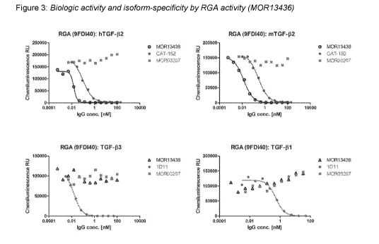
Figure 3: is showing biologic "neutralizing" activity of the TGF-132-specific
AB M0R13436.
Shown are concentration-response curves of the Antibody against the effects of
recombinant
human TGF-132, mouse TGF-132, human TGF-133 and human TGF-131 in HEK293T/17
CAGA-12
(HEK293T-RGA), a luciferase reporter assay specific for phosphorylated Smad-2
and Smad-3.
Recombinant TGF-13s induce Smad-2 and Smad-3 phosphorylation which bind to the
CAGA-12
reporter and causes luciferase gene expression. M0R13436 neutralizes
recombinant human
and mouse TGF-132, but not human TGF-131 and TGF-133. The IgG Antibody
M0R03207 served
as negative control. M0R03207 recognizes an enzyme that is part of human
innate immune
system.
Figure 4: is showing biologic "neutralizing" activity of the TGF-132-specific
ABs M0R14799,
M0R14800, M0R14797 and M0R14809. Shown are concentration-response curves of
the ABs
against the effects of various recombinant human TGF-13 family proteins:
Activin A , Activin B ,
Activin AB , GDF-11 , myostatin , TGF-b2 and TGF-b3 in HEK293T/17 CAGA-12
(HEK293T-
RGA), a luciferase reporter assay specific for phosphorylated Smad-2 and Smad-
3.
CA 03012294 2018-07-23
WO 2017/141208 PCT/IB2017/050917
Recombinant TGF-13s induced Smad-2 and Smad-3 phosphorylation which bind to
the CAGA-12
reporter and causes luciferase gene expression. All ABs neutralize recombinant
human TGF-
132, but not any other TGF-13 protein family members. The IgG Antibody
M0R03207 (assessed
at 100 nM and 33.3 nM) served as negative control. M0R03207 recognizes an
enzyme that is
part of human innate immune system.
Figure 5: is showing biologic "neutralizing" activity of the TGF-132-specific
AB M0R13436.
Shown is a concentration-response curve of the AB counteracting inhibition of
human skeletal
muscle cells (skMC) differentiation by recombinant human TGF-132 (.), mouse
TGF-132 (=) and
human TGF-133 (+). Cells were differentiated up to 120 hours and creatine
kinase (CK) activity, a
well-established skeletal muscle cell differentiation marker, was measured.
All TGF-13s inhibit
CK activity and the Ab counteracts TGF-132, but not TGF-133 responses.
Figure 6: is showing biologic "neutralizing" activity of the TGF-132-specific
ABs (A) MOR14799,
(B) M0R14800, (C) M0R14797 and (D) M0R14809. Shown are concentration-response
curves
of the ABs counteracting inhibition of human skeletal muscle cells (skMC)
differentiation by
recombinant human TGF-132 (.), mouse TGF-132 (=) and human TGF-133 (+).Cells
were
differentiated up to 120 hours and creatine kinase (CK) activity, a well-
established skeletal
muscle cell differentiation marker, was measured. All TGF-13s inhibit CK
activity and the Abs
counteracts TGF-132, but not TGF-133 responses.
Figure 7: is showing bar graphs quantifying immunostaining for collagen I
protein in a
Dupuytren patient sample cultured for seven days with the TGF-132-specific AB
M0R14797 or
the pan- TGF-13 antibody 1D11. Patient tissue was sliced, cultured for 7 days
in the presence of
Ab and then immunostained for collagen I.
Figure 8: is showing immunostainings for collagen I protein in two different
Dupuytren patient
samples cultured for seven days with the TGF-132-specific AB M0R14797 (B/D)
compare to
isotype controls (A/C). Results from patient 1 are shown in A/B and from
patient 2 in O/D.
Patient tissue was sliced, cultured for 7 days in the presence of Ab and then
immunostained for
collagen I.
Figure 9: is showing bar graphs quantifying mRNA expression for collagen I
protein in mouse
kidneys from a unilateral urethral obstruction model (UUO) treated with the
TGF-132-specific AB
M0R13436. Sham- or UUO-operated animals were treated for 14 days with the Ab,
kidneys
removed, mRNA isolated and analysed by qPCR.
Figure 10: Epitope of M0R14797 binding to TGF8-2: (A) Overall structure of two
M0R14797
Fabs binding to TGF8-2 dimer. M0R14797 is shown as surface, TGF8-2 as ribbon.
(B)
Residues of TGF8-2 dimer within 5 A distance to M0R14797 are shown as sticks.
11
CA 03012294 2018-07-23
WO 2017/141208 PCT/IB2017/050917
Figure 11: Paratope of M0R14797 binding to TGF[3-2: Sequence of M0R14797 VH
(A) (SEQ
ID NOS 67 and 67, respectively, in order of appearance) and VL (B) (SEQ ID NOS
77 and 77,
respectively, in order of appearance) are listed. CDR loops (both Kabat and
Chothia definitions)
are boxed. Residues of M0R14797 within 5 A distance to TGF[3-2 dimer are
shaded in grey.
GENERAL DEFINITIONS
In order that the present invention may be more readily understood, certain
terms are first
defined. Additional definitions are set forth throughout the detailed
description.
Comprising: the term "comprising" means "including" e.g. a composition
"comprising" X may
consist exclusively of X or may include something additional e.g. X + Y.
The term "human TGF-13 isoform" is used to describe members of the
transforming growth factor
beta (TGF-13) superfamily, namely the human TGF-13 isoforms TGF-131, TGF-132
and TGF-133,
respectively. The terms "human TGF-13 isoform TGF-132", "TGF-132 isoform",
"TGF[32" and "TGF-
132" and TGFbeta-2 are used synonymously throughout the instant disclosure.
The term "antibody" as referred to herein includes whole antibodies and any
antigen binding
fragment (i.e. "antigen-binding portion") or single chains thereof. A
naturally occurring "antibody"
is a glycoprotein comprising at least two heavy (H) chains and two light (L)
chains inter-
connected by disulfide bonds. Each heavy chain is comprised of a heavy chain
variable region
(abbreviated herein as VH) and a heavy chain constant region. The heavy chain
constant region
is comprised of three domains, CH1, CH2 and CH3. Each light chain is comprised
of a light
chain variable region (abbreviated herein as VL) and a light chain constant
region. The light
chain constant region is comprised of one domain, CL. The VH and VL regions
can be further
subdivided into regions of hypervariability, termed complementarity
determining regions (CDR),
interspersed with regions that are more conserved, termed framework regions
(FR). Each VH
and VL is composed of three CDRs and four FRs arranged from amino-terminus to
carboxy-
terminus in the following order: FR1, CDR1, FR2, CDR2, FR3, CDR3, FR4. The
variable
regions of the heavy and light chains contain a binding domain that interacts
with an antigen.
The constant regions of the antibodies may mediate the binding of the
immunoglobulin to host
tissues or factors, including various cells of the immune system (e.g.
effector cells) and the first
component (Clq) of the classical complement system.
12
CA 03012294 2018-07-23
WO 2017/141208 PCT/IB2017/050917
An "isolated antibody", as used herein, refers to an antibody that is
substantially free of other
antibodies having different antigenic specificities (e.g. an isolated antibody
that specifically
binds antigen-binding portion is substantially free of antibodies that
specifically bind antigens
other than TGFbeta-2).
The term "KID", as used herein, is intended to refer to the dissociation
constant, which is
obtained from the ratio of Kd to Ka (i.e. Kd/Ka) and is expressed as a molar
concentration (M). KD
values for antibodies can be determined using methods well-established in the
art. A method for
determining the KD of an antibody is surface plasmon resonance, such as the
biosensor system
of Biacore , or Solution Equilibrium Titration (SET) (see Friguet B et al.
(1985) J. Immunol
Methods; 77(2): 305-319, and Hanel C et al. (2005) Anal Biochem; 339(1): 182-
184). The term
"Kassoc" Or "Ka" is intended to refer to the association rate of a particular
antibody-antigen
interaction, whereas the term "Kd,s" or "Kd", as used herein, is intended to
refer to the
dissociation rate of a particular antibody-antigen interaction.
Throughout the specification different scientific notations to writing powers
to 10 are used. The
scientific E notation (e.g. 1.1E-15) is used as an alternative to writing
powers of 10. For
example, 0.000000001 Mole can be written as 3.0 x 10⁻⁹ Mole or as 3.0E-9 Mole.
As used herein, the term "ADCC" or "antibody dependent cellular cytotoxicity"
activity refers to
human B cell depleting activity. ADCC activity can be measured by the human B
cell depleting
assays known in the art.
The term "human antibody", as used herein, is intended to include antibodies
having variable
regions in which both the framework and CDR regions are derived from sequences
of human
origin. Furthermore, if the antibody contains a constant region, the constant
region also is
derived from such human sequences, e.g. human germline sequences, or mutated
versions of
human germline sequences or antibody containing consensus framework sequences
derived
from human framework sequences analysis, for example, as described in Knappik,
et al. (2000.
J Mol Biol 296, 57-86). The human antibodies of the invention may include
amino acid residues
not encoded by human sequences (e.g. mutations introduced by random or site-
specific
mutagenesis in vitro or by somatic mutation in vivo; or humanized antibodies).
The term "human monoclonal antibody" refers to antibodies displaying a single
binding
specificity which have variable regions in which both the framework and CDR
regions are
derived from human sequences. In one embodiment, the human monoclonal
antibodies are
produced by a hybridoma which includes a B cell obtained from a transgenic
nonhuman animal,
e.g. a transgenic mouse, having a genome comprising a human heavy chain
transgene and a
light chain transgene fused to an immortalized cell.
13
CA 03012294 2018-07-23
WO 2017/141208 PCT/IB2017/050917
The term "recombinant human antibody", as used herein, includes all human
antibodies that are
prepared, expressed, created or isolated by recombinant means, such as
antibodies isolated
from an animal (e.g. a mouse) that is transgenic or transchromosomal for human
immunoglobulin genes or a hybridoma prepared therefrom, antibodies isolated
from a host cell
transformed to express the human antibody, e.g. from a transfectoma,
antibodies isolated from
a recombinant, combinatorial human antibody library, and antibodies prepared,
expressed,
created or isolated by any other means that involve splicing of all or a
portion of a human
immunoglobulin gene, sequences to other DNA sequences. Such recombinant human
antibodies have variable regions in which the framework and CDR regions are
derived from
human germline immunoglobulin sequences. In certain embodiments, however, such
recombinant human antibodies can be subjected to in vitro mutagenesis (or,
when an animal
transgenic for human Ig sequences is used, in vivo somatic mutagenesis) and
thus the amino
acid sequences of the VH and VL regions of the recombinant antibodies are
sequences that,
while derived from and related to human germline VH and VL sequences, may not
naturally exist
within the human antibody germline repertoire in vivo.
As used herein, "isotype" refers to the antibody class (e.g. IgM, IgE, IgG
such as IgG1 or IgG2)
that is provided by the heavy chain constant region genes.
As used herein, the term "cancer" is meant to include all types of cancerous
growths or
oncogenic processes, metastatic tissues or malignantly transformed cells,
tissues, or organs,
irrespective of histopathologic type or stage of invasiveness. Examples of
cancerous disorders
include, but are not limited to, solid tumors, hematological cancers, soft
tissue tumors, and
metastatic lesions. Examples of solid tumors include malignancies, e.g.,
sarcomas, and
carcinomas (including adenocarcinomas and squamous cell carcinomas), of the
various organ
systems, such as those affecting liver, lung, breast, lymphoid,
gastrointestinal (e.g., colon),
genitourinary tract (e.g., renal, urothelial cells), prostate and pharynx.
Adenocarcinomas
include malignancies such as most colon cancers, rectal cancer, renal-cell
carcinoma, liver
cancer, non-small cell carcinoma of the lung, cancer of the small intestine
and cancer of the
esophagus. Squamous cell carcinomas include malignancies, e.g., in the lung,
esophagus,
skin, head and neck region, oral cavity, anus, and cervix. In one embodiment,
the cancer is a
melanoma, e.g., an advanced stage melanoma. Metastatic lesions of the
aforementioned
cancers can also be treated or prevented using the methods and compositions of
the invention.
Exemplary cancers whose growth can be inhibited using the antibodies molecules
disclosed
herein include cancers typically responsive to immunotherapy. Non-limiting
examples of
preferred cancers for treatment include melanoma (e.g., metastatic malignant
melanoma), renal
cancer (e.g., clear cell carcinoma), prostate cancer (e.g., hormone refractory
prostate
adenocarcinoma), breast cancer, colon cancer and lung cancer (e.g., non-small
cell lung
14
CA 03012294 2018-07-23
WO 2017/141208 PCT/IB2017/050917
cancer). Additionally, refractory or recurrent malignancies can be treated
using the antibody
molecules described herein.
Examples of other cancers that can be treated include bone cancer, pancreatic
cancer, skin
cancer, cancer of the head or neck, cutaneous or intraocular malignant
melanoma, uterine
cancer, ovarian cancer, rectal cancer, anal cancer, gastro-esophageal, stomach
cancer,
liposarcoma, testicular cancer, uterine cancer, carcinoma of the fallopian
tubes, carcinoma of
the endometrium, carcinoma of the cervix, carcinoma of the vagina, carcinoma
of the vulva,
Merkel cell cancer, Hodgkin lymphoma, non-Hodgkin lymphoma, cancer of the
esophagus,
cancer of the small intestine, cancer of the endocrine system, cancer of the
thyroid gland,
cancer of the parathyroid gland, cancer of the adrenal gland, sarcoma of soft
tissue, cancer of
the urethra, cancer of the penis, chronic or acute leukemias including acute
myeloid leukemia,
chronic myeloid leukemia, acute lymphoblastic leukemia, chronic lymphocytic
leukemia, solid
tumors of childhood, lymphocytic lymphoma, cancer of the bladder, multiple
myeloma,
myelodisplastic syndromes, cancer of the kidney or ureter, carcinoma of the
renal pelvis,
neoplasm of the central nervous system (CNS), primary CNS lymphoma, tumor
angiogenesis,
spinal axis tumor, brain stem glioma, pituitary adenoma, Kaposi's sarcoma,
epidermoid cancer,
squamous cell cancer, T-cell lymphoma, environmentally induced cancers
including those
induced by asbestos (e.g., mesothelioma), and combinations of said cancers. In
certain
embodiments, the cancer is a skin cancer, e.g., a Merkel cell carcinoma or a
melanoma. In one
embodiment, the cancer is a Merkel cell carcinoma. In other embodiments, the
cancer is a
melanoma. In other embodiments, the cancer is a breast cancer, e.g., a triple
negative breast
cancer (TNBC) or a HER2-negative breast cancer. In other embodiments, the
cancer is kidney
cancer, e.g., a renal cell carcinoma (e.g., clear cell renal cell carcinoma
(CCRCC) or a non-clear
cell renal cell carcinoma (nccRCC)). In other embodiments, the cancer is a
thyroid cancer, e.g.,
an anaplastic thyroid carcinoma (ATC). In other embodiments, the cancer is a
neuroendocrine
tumor (NET), e.g., an atypical pulmonary carcinoid tumor or an NET in
pancreas,
gastrointestinal (GI) tract, or lung. In certain embodiments, the cancer is a
lung cancer, e.g., a
non-small cell lung cancer (NSCLC) (e.g., a squamous NSCLC or a non-squamous
NSCLC).
As used herein, the term "Programmed Death 1" or "PD-1" relates to the
0D28/CTLA-4 family
member expressed, e.g., on activated CD4+ and CD8+ T cells, Tregs, and B
cells. It negatively
regulates effector T cell signaling and function. PD-1 is induced on tumor-
infiltrating T cells, and
can result in functional exhaustion or dysfunction (Keir et al. (2008) Annu.
Rev. lmmunol.
26:677-704; PardoII et al. (2012) Nat Rev Cancer 12(4):252-64). PD-1 delivers
a coinhibitory
signal upon binding to either of its two ligands, Programmed Death-Ligand 1
(PD-L1) or
Programmed Death-Ligand 2 (PD-L2). PD-L1 is expressed on a number of cell
types, including
CA 03012294 2018-07-23
WO 2017/141208 PCT/IB2017/050917
T cells, natural killer (NK) cells, macrophages, dendritic cells (DCs), B
cells, epithelial cells,
vascular endothelial cells, as well as many types of tumors. High expression
of PD-L1 on
murine and human tumors has been linked to poor clinical outcomes in a variety
of cancers
(Keir et al. (2008) Annu. Rev. lmmunol. 26:677-704; PardoII et al. (2012) Nat
Rev Cancer
12(4):252-64). PD-L2 is expressed on dendritic cells, macrophages, and some
tumors.
Blockade of the PD-1 pathway has been pre-clinically and clinically validated
for cancer
immunotherapy. Both preclinical and clinical studies have demonstrated that
anti-PD-1 blockade
can restore activity of effector T cells and results in robust anti-tumor
response. For example,
blockade of PD-1 pathway can restore exhausted/dysfunctional effector T cell
function (e.g.,
proliferation, IFN-y secretion, or cytolytic function) and/or inhibit Treg
cell function (Keir et al.
(2008) Annu. Rev. lmmunol. 26:677-704; PardoII et al. (2012) Nat Rev Cancer
12(4):252-64).
Blockade of the PD-1 pathway can be effected with an antibody, an antigen
binding fragment
thereof, an immunoadhesin, a fusion protein, or oligopeptide of PD-1, PD-L1
and/or PD-L2. The
amino acid sequence of PD-1, e.g., human PD-1, is known in the art, e.g.,
Shinohara T et al.
(1994) Genomics 23(3):704-6; Finger LR, et al. Gene (1997) 197(1-2):177-87.
Various definitions and aspects of the invention are provided/described in
further detail in the
following subsections.
DETAILED DESCRIPTION OF THE INVENTION
The specific neutralization of one of several highly homologous targets that
are associated with
distinct functions with human monoclonal antibodies remains a major challenge.
This challenge
is more evident in a therapeutic context when cross-reactivity with other
homologous targets
may be associated with adverse or unwanted events. These problems may arise
when targeting
TGF-132 with human monoclonal antibodies due to the presence of the homologous
isoforms
TGF-131 and TGF-133. These TGF-13 isoforms are associated with distinct
functions according to
experimental data from knockout mice. In addition, the inhibition of TGF-131
is known to be
associated with a high risk of adverse events. Furthermore, the inventors
herein disclose the
unrecognized problem that the simultaneous inhibition of TGF-133 and TGF-132
is associated
with valvulopathy, a serious disorder of the valves of the heart, in animal
models. This creates
the so far unresolved need for providing monoclonal therapeutic antibodies
that specifically
neutralize TGF-132 but do not neutralize TGF-133 or TGF-133 and TGF-131. More
specifically,
there is a need for neutralizing anti-TGF-132 antibodies exhibiting very low
picomolar
dissociation constants to be effective in a clinical setting. In addition,
neutralizing anti-TGF-132
antibodies with high binding affinities are associated with an enhanced
efficacy and safety as
they need to be administered in lower doses which are less likely to cause
interference with
other pathways that may be associated with adverse events. Furthermore, the
administration of
16
CA 03012294 2018-07-23
WO 2017/141208 PCT/IB2017/050917
lower doses of monoclonal anti-TGF-132 antibodies is also associated with
lower costs. These
problems are solved by the present invention.
Neutralisation of TGF-beta1/2/3 isoforms is determined in a Smad dependent
reporter gene
assay. The terms "neutralizing antibody" and "antagonistic antibody" are used
synonymously
and are intended to refer to an antibody that inhibits TGFBeta-1,-2 and/or -3
induced signaling
activity in the Smad dependent reporter gene assay with an I050 of less than
or equal to
100nM. The phrase "a human monoclonal anti-TGF-132 antibody or a functional
fragment thereof
that neutralizes the human TGF-13 isoform TGF-132 and does not neutralize
human isoform TGF-
133" or "specifically neutralize TGF-132 but does not neutralize TGF-133 or
TGF-131 (or both TGF-
133 and TGF-[31)" as used in the context of this invention refers to human
monoclonal antibodies
that specifically neutralize TGF-132 with a half maximal inhibitory
concentration (1050) of less
than 150pM but do not neutralize human TGF-131 and/or TGF-133 determined by
the Smad
dependent reporter gene assay (e.g. having an IC 50 for human TGF-131 and/or
TGF-133 of
greater than 100nM). Consequently, a human TGF-132 neutralizing antibody of
the present
disclosure neutralizes human TGF-132 with an half maximal inhibitory
concentration (1050) of
less than e.g. 107pM and neutralizes human TGF-131 and/or TGF-133 with an half
maximal
inhibitory concentration (1050) of greater than 100nM as determined by a Smad
dependent
reporter gene assay. In one embodiment the human TGF-132 neutralizing antibody
of the
present disclosure neutralizes human TGF-132 with an half maximal inhibitory
concentration
(1050) of less than e.g. 107pM and does not neutralize human TGF-131 and/or
TGF-133 because
said antibody exhibits essentially undetectable binding against these proteins
in a Smad
dependent reporter gene assay.
As used herein, the term "conservative sequence modifications" is intended to
refer to amino
acid modifications that do not significantly affect or alter the binding
characteristics of the
antibody containing the amino acid sequence. Such conservative modifications
include amino
acid substitutions, additions and deletions. Modifications can be introduced
into an antibody of
the invention by standard techniques known in the art, such as site-directed
mutagenesis and
PCR-mediated mutagenesis. Conservative amino acid substitutions are ones in
which the
amino acid residue is replaced with an amino acid residue having a similar
side chain. Families
of amino acid residues having similar side chains have been defined in the
art. These families
include amino acids with basic side chains (e.g. lysine, arginine, histidine),
acidic side chains
(e.g. aspartic acid, glutamic acid), uncharged polar side chains (e.g.
glycine, asparagine,
glutamine, serine, threonine, tyrosine, cysteine, tryptophan), nonpolar side
chains (e.g. alanine,
valine, leucine, isoleucine, proline, phenylalanine, methionine), beta-
branched side chains (e.g.
threonine, valine, isoleucine) and aromatic side chains (e.g. tyrosine,
phenylalanine, tryptophan,
histidine). Thus, one or more amino acid residues within the CDR regions of an
antibody of the
17
CA 03012294 2018-07-23
WO 2017/141208 PCT/IB2017/050917
invention can be replaced with other amino acid residues from the same side
chain family, and
the altered antibody can be tested for retained function using the functional
assays described
herein.
An antibody that "cross-reacts" refers to an antibody that binds more than one
antigen, wherein
said binding can, but must not necessarily result in the manipulation
(neutralization, reduction or
activation) of the activity of said antigen(s). Cross reactivity can be
determined using Smad
dependent reporter gene assays and by determining the KD which may be
determined using a
surface plasmon resonance biosensor system, such as a Biacore0 system, or
Solution
Equilibrium Titration.
"A human monoclonal anti-TGF-132 antibody or a functional fragment thereof
that binds the
human TGF-13 isoform TGF-132 and does not bind (cross react) with the human
isoform TGF-133
or TGF-131 or TGF-133 and TGF-131" is intended to also refer to an antibody
that binds to TGF-132
with a KD of about 1pM or less and to TGF-131 or TGF-133 with a KD of about 2
x 10-9 M, or about
x 10-9 M or about 10 x 10-9 M or higher. In certain embodiments, such
antibodies that do not
cross-react with the TGF-133 and TGF-131 antigen actually exhibit essentially
undetectable
binding against these proteins in standard binding assays.
Standard assays to evaluate the binding ability of the antibodies toward TGF-
132 of various
species are known in the art, including for example, ELISAs, western blots and
Radioimmunoassays (RIAs). Suitable assays are described in detail in the
example section.
The binding affinity of the antibodies also can be assessed by standard assays
known in the art,
such as surface plasmon resonance (e.g. Biacore0 system analysis) or Solution
Equilibrium
Titration. Surface plasmon resonance based techniques such as Biacore0 system
can
determine the binding kinetics which allows the calculation of the binding
affinity. Assays to
evaluate the effects of the antibodies on functional properties of TGF-132 are
described in further
detail in the example section.
The present invention provides human monoclonal anti-TGF-132 antibodies or
functional
fragments thereof. More particularly, it provides human monoclonal anti-TGF-
132 antibodies or
functional fragments thereof that neutralize the human TGF-13 isoform TGF-132
(SEQ ID NO:
122) (UniProt ID: P61812 - TGFB2_HUMAN (http://www.uniprot.org/); gene symbol
approved by
the HUGO Gene Nomenclature Committee (HGNC)=TGFB2; HGNC ID= HGNC:11768) and do
not neutralize the human isoform TGF-133 (SEQ ID NO: 123) (UniProt ID: P10600 -
TGFB3_HUMAN; gene symbol HGNC=TGFB3; HGNC ID= HGNC:11769). In particular, the
present invention provides human monoclonal anti-TGF-132 antibodies or
functional fragments
thereof that neutralize the human TGF-13 isoform TGF-132 and do not neutralize
human isoforms
TGF-133 and TGF-131 (SEQ ID NO: 121) ((UniProt ID: P01137 - TGFB1_HUMAN; gene
symbol
18
CA 03012294 2018-07-23
WO 2017/141208 PCT/IB2017/050917
HGNC=TGFB1; HGNC ID= HGNC:11766). TGF-81, TGF-82, or TGF-83 neutralization by
the
antibodies or functional fragments thereof is determined by the Smad dependent
report gene
assay as described in the example section. Preferably, the antibodies or a
functional fragments
thereof which are provided by the present invention neutralise human TGF-82
with an half
maximal inhibitory concentration (1050) of less than 150pM, or less than
107pM, or less than
100pM, or less than 95pM or less than 80pM, or less than 30pM, or less than
20pM, or less
than 10pM, and does not neutralise human TGF-81 or TGF-83 (having an I050
greater than
100nM) as determined by the Smad dependent reporter gene assay. In another
embodiment of
the disclosure, the antibodies or a functional fragments thereof neutralise
human TGF-82 with
an half maximal inhibitory concentration (1050) between about 1pM and about
150pM, or
between about 1pM and about 107pM, or between about 1pM and about 95pM or
between
about 1pM and about 80pM and does not neutralise human TGF-81 or TGF-83
(having an I050
greater than 100nM) as determined by the Smad dependent reporter gene assay.
The present invention also provides human monoclonal anti-TGF-82 antibodies or
functional
fragments thereof that bind the human TGF-8 isoform TGF-82 preferentially over
the human
isoforms TGF-81 and/or TGF-83. In particular, the provided human monoclonal
anti-TGF-82
antibodies or a functional fragment thereof bind the human TGF-8 isoform TGF-
82 with a
dissociation constant that is at least about 70-fold, about 1000-fold, about
2000-fold, about
10000-fold, about 20000-fold, about 200000-fold, about 300000-fold about
1000000-fold, or
about 6000000-fold lower than its dissociation constant for TGF-83, wherein
the antibody or the
functional fragment thereof neutralises human TGF-82 but not TGF-83.
In one embodiment, the dissociation constant of the provided antibodies for
human TGF-82 is
about 2000-fold to about 1000000-fold lower than its dissociation constant for
TGF-81 or TGF-
83. In a another embodiment, the human monoclonal anti-TGF-82 antibodies or a
functional
fragments thereof bind the human TGF-8 isoform TGF-82 with a dissociation
constant that is at
least 2000-fold or at least 1000000-fold lower than its dissociation constant
for TGF-83, wherein
the antibody neutralises human TGF-82 but not TGF-83. The binding affinity of
the antibodies
also can be assessed by standard assays disclosed herein, such as surface
plasmon
resonance (e.g. Biacore0 system analysis) or Solution Equilibrium Titration.
The human
monoclonal anti-TGF-82 antibodies or functional fragments thereof that bind
human TGF-82
preferentially over the human isoforms TGF-81 and TGF-83, binds to TGF-82 with
a
dissociation constant (KD) of about 1pM or less, or about 100fM or less, or
about 50fM or less.
In another embodiment, the disclosed antibodies or functional fragments bind
to the human
TGF-8 isoform TGF-82 with a dissociation constant (KD) of 1pM or less or about
1pM to about
10fM M.
In another embodiment, the human monoclonal anti-TGF-82 antibodies or
functional fragments
thereof bind human TGF-8 isoform TGF-82 preferentially over human TGF-8
isoform TGF-83
19
CA 03012294 2018-07-23
WO 2017/141208 PCT/IB2017/050917
with a dissociation constant that is at least 70-fold lower than its
dissociation constant for human
TGF-13 isoform TGF-133, wherein the antibodies neutralise human TGF-13 isoform
TGF-132 but do
not neutralise human TGF-13 isoform TGF-133, and bind human TGF-13 isoform TGF-
132 with a
dissociation constant (KD) of 1pM or less.
In a preferred embodiment, the human monoclonal anti-TGF-132 antibodies or
functional
fragments thereof bind human TGF-132 preferentially over human TGF-131 and TGF-
133 with a
dissociation constant that is at least 70-fold lower than its dissociation
constant for TGF-131 and
TGF-133, wherein the antibodies neutralise human TGF-132 but does not
neutralise TGF-133 and
TGF-131, and binds human TGF-132 with a dissociation constant (KD) of 1pM or
less.
The anti-TGF-132 antibodies, or antigen binding fragments thereof, as
described herein can be
single chain antibodies, Fab fragments, Fv fragments, F(ab')2 fragments, or
scFv fragments,
and/or IgG isotypes.
None of the prior art antibodies matches the antibodies disclosed herein, in
particular the
antibodies M0R14799, M0R14800, M0R14809, M0R14797, M0R14805 or M0R14787, in
terms of specificity and selectivity. Antibodies of the invention include the
human recombinant
antibodies, isolated and structurally characterized, as described in the
examples. The VH amino
acid sequences of isolated antibodies of the invention are shown in SEQ ID
NOs: 7, 27, 47, 67,
87 and 107, respectively. The VL amino acid sequences of isolated antibodies
of the invention
are shown in SEQ ID NOs: 17, 37, 57, 77, 97 and 117, respectively. Examples of
preferred full
length heavy chain amino acid sequences of antibodies of the invention are
shown in SEQ ID
NOs: 9, 29, 49, 69, 89 and 109, respectively. Examples of preferred full
length light chain amino
acid sequences of antibodies of the invention are shown in SEQ ID NOs: 19, 39,
59, 79, 99 and
119, respectively. Other antibodies of the invention include antibodies that
have been mutated
by amino acid deletion, insertion or substitution, yet have at least 80, 90,
95, 97 or 99 percent
identity to the full length heavy chain amino acid sequences depicted in the
sequences
described above. Further, variable heavy chain nucleotide sequences are shown
in SEQ ID
NOs: 8, 28, 48, 68, 88 and 108, respectively. Variable light chain nucleotide
sequences are
shown in SEQ ID NOs: 18, 38, 58, 78, 98 and 118, respectively. Full length
light chain
nucleotide sequences are shown in SEQ ID NOs: 20, 40, 60, 80, 100 and 120,
respectively. Full
length heavy chain nucleotide sequences are shown in SEQ ID NOs: 10, 30, 50,
70, 90 and
110. Other antibodies of the invention include amino acids or nucleic acids
that have been
mutated, yet have at least 90 or more (i.e. 91, 92, 93, 94, 95, 97, 99 or
more) percent identity to
the sequences described above. Some embodiments include mutant amino acid
sequences
wherein no more than 1, 2, 3, 4 or 5 amino acids have been mutated by amino
acid deletion,
insertion or substitution in the variable regions when compared with the
variable regions
depicted in the sequence described above. Since each of these antibodies binds
the same
target, the VH, VL, full length light chain, and full length heavy chain
sequences (nucleotide
CA 03012294 2018-07-23
WO 2017/141208 PCT/IB2017/050917
sequences and amino acid sequences) can be "mixed and matched" to create other
anti-TGF-
32 antibodies of the invention. TGF-132 binding of such "mixed and matched"
antibodies can be
tested using the binding assays described above and in the examples (e.g.
ELISAs). When
these chains are mixed and matched, a VH sequence from a particular VH/VL
pairing should be
replaced with a structurally similar VH sequence. Likewise a full length heavy
chain sequence
from a particular full length heavy chain/full length light chain pairing
should be replaced with a
structurally similar full length heavy chain sequence. Likewise, a VL sequence
from a particular
VH/VL pairing should be replaced with a structurally similar VL sequence.
Likewise a full length
light chain sequence from a particular full length heavy chain/full length
light chain pairing
should be replaced with a structurally similar full length light chain
sequence. Accordingly, in
one aspect, the invention provides an isolated recombinant anti-TGF-132
antibody or antigen
binding region thereof having: a heavy chain variable region comprising an
amino acid
sequence selected from the group consisting of SEQ ID NOs: 7, 27, 47, 67, 87
and 107; and a
light chain variable region comprising an amino acid sequence selected from
the group
consisting of SEQ ID NOs: 17, 37, 57, 77,97 and 117.
The terms "complementarity determining region," and "CDR," as used herein
refer to the
sequences of amino acids within antibody variable regions which contributes to
the antigen
specificity and binding affinity. In general, there are three CDRs in each
heavy chain variable
region (HCDR1, HCDR2, HCDR3) and three CDRs in each light chain variable
region (LCDR1,
LCDR2, LCDR3). The amino acid sequence boundaries of a given CDR can be
determined
using any of a number of well-known schemes, including those described by
Kabat et al. (1991),
"Sequences of Proteins of Immunological Interest," 5th Ed. Public Health
Service, National
Institutes of Health, Bethesda, MD ("Kabat" numbering scheme). An alternative
method of
determining CDR regions uses the method devised by Chothia (Chothia et al.
1989, Nature,
342:877-883). The Chothia definition is based on the location of the
structural loop regions.
Other systems for defining CDRs exist and are known to the skilled person (see
e.g.
http://www.bioinf.org.uk/abs/). CDRs are assumed to account for the antigen
recognition and
binding and thus to contain antigen binding regions. In the context of this
disclosure CDRs have
been defined using the Kabat and the Chothia numbering system. CDRs defined
using the
Kabat system are designated "Kabat CDRs". CDRs defined using the Chothia
system are
designated "Chothia CDRs". CDRs predicted using the Kabat and Chothia system
mostly
overlap but are not necessarily identical. Hence, an antibody binding region
can also be defined
by merging the CDR amino acid sequences predicted by the Kabat and Chothia
system
("Kabat/Chothia CDR"). It is known that some amino acid residues that actually
bind the antigen
fall outside the CDRs and, consequently, X-ray crystallography and X-ray
diffraction data can
also be used to define additional antibody binding regions/amino acids or
paratope regions.
21
CA 03012294 2018-07-23
WO 2017/141208 PCT/IB2017/050917
The terms "antibody binding regions", "antibody combining sites" and "paratope
region" are
used synonymously throughout this document and refer to those parts of the
variable regions of
both the light and heavy chains of an antibody that interact with the specific
antigen. The
paratope consists of stretches of amino acids or single amino acids comprised
in the variable
regions of antibodies that bind to an antigen by the establishment of chemical
interactions (e.g.
polar-, non-polar-, hydrogen-bonds/-contacts or salt bridges). The CDRs (e.g.
predicted on the
basis of the Kabat/Chothia system) are collectively referred to as the
Kabat/Chothia paratope of
an antibody. In the context of this disclosure a paratope consists of those
amino acid residues
of an antibody that are involved in the antibody/antigen binding/combining,
wherein amino acids
being within 5 A distance to the antigen are considered to be involved in the
antibody/antigen
binding. The paratope may comprise CDR amino acids defined according to
Kabat/Chothia as
well as amino acids comprised in the framework region of the variable light
and/or heavy chain
regions of a given antibody that are involved in the antibody/antigen
binding/combining. Beside
the paratope prediction based on the Kabat/Chothia, the complete paratope of a
given antibody
can be identified using X-ray crystallography/X-ray diffraction data.
The phrase "functional fragments thereof' when used in the context of an
antibody refers to a
protein fragment of a full-length antibody comprising the antigen-binding
regions (single chain
antibodies, Fab fragments, Fv fragments, F(ab')2 fragments, and/or scFv
fragments), wherein (i)
said fragment retains the ability to specifically bind the antigen or (ii)
upon transfer/fusion of said
functional fragment to another antibody (sequence replacements) or antibody
like structure the
ability to specifically bind to an antigen (e.g. a portion of TGFbeta-2) is
retained. Examples of
"functional fragments" of an antibody include a Fab fragment, a monovalent
fragment consisting
of the VL, VH, CL and CH1 domains; a F(ab)2 fragment, a bivalent fragment
comprising two Fab
fragments linked by a disulfide bridge at the hinge region; a Fd fragment
consisting of the VH
and CH1 domains; a Fv fragment consisting of the VL and VH domains of a single
arm of an
antibody; a dAb fragment (Ward et al., 1989 Nature 341:544-546), which
consists of a VH
domain; and an isolated complementarity determining region (CDR). Furthermore,
although the
two domains of the Fv fragment, VL and VH, are coded for by separate genes,
they can be
joined, using recombinant methods, by a synthetic linker that enables them to
be made as a
single protein chain in which the VL and VH regions pair to form monovalent
molecules (known
as single chain Fv (scFv); see e.g. Bird et al., 1988 Science 242:423-426; and
Huston et al.,
1988 Proc. Natl. Acad. Sci. 85:5879-5883). Such single chain antibodies are
also intended to be
encompassed within the term "functional fragment" of an antibody. These
antibody fragments
are obtained using conventional techniques known to those of skill in the art,
and the fragments
are screened for utility in the same manner as are intact antibodies.
22
CA 03012294 2018-07-23
WO 2017/141208 PCT/IB2017/050917
In one aspect the human monoclonal anti-TGF-132 antibodies of the present
disclosure or
functional fragments thereof comprise the following complementarity
determining regions (CDR)
defined by Kabat or Cothia or by Kabat and Cothia
(i) the Kabat CDRs recited in SEQ ID NOs: 1-3, 11-13, 21-23, 31-33, 41-43, 51-
53, 61-63, 71-
73, 81-83, 91-93, 101-103, or 111-113, or
(ii) the Chothia CDRs recited in SEQ ID NOs: 4-6, 14-16, 24-26, 34-36, 44-46,
54-56, 64-66, 74-
76, 84-86, 94-96, 104-106, or 114-116, or
(iii) the Kabat/Chothia CDRs recited on SEQ ID NOs: 124-129.
The human monoclonal anti-TGF-132 antibodies of the present disclosure or
functional
fragments thereof comprise a heavy chain variable region CDR1 comprising an
amino acid
sequence having at least 80%, 90%, 95% or 100% sequence identity to a sequence
selected
from the group consisting of SEQ ID NOs: 1, 21, 41, 61, 81, 101 or 4, 24, 44,
64, 84, 104 or
124-129; a heavy chain variable region CDR2 comprising an amino acid sequence
having at
least 70%, 80%, 90, 95% or 100% sequence identity to a sequence selected from
the group
consisting of SEQ ID NOs: 2, 22, 42, 62, 82, 102 or 5, 25, 45, 65, 85, 105; a
heavy chain
variable region CDR3 comprising an amino acid sequence having at least 70%,
80%, 90 95% or
100% sequence identity to a sequence selected from the group consisting of SEQ
ID NOs: 3,
23, 43, 63, 83, 103 or 6, 26, 46, 66, 86, 106; a light chain variable region
CDR1 comprising an
amino acid sequence having at least 70%, 80%, 90, 95% or 100% sequence
identity to a
sequence selected from the group consisting of SEQ ID NOs: 11, 31, 51, 71, 91,
111 or 14, 34,
54, 74, 94, 114; a light chain variable region CDR2 comprising an amino acid
sequence having
at least 80%, 90, 95% or 100% sequence identity to a sequence selected from
the group
consisting of SEQ ID NOs: 12, 32, 52, 72, 92, 112 or 15, 35, 55, 75, 95, 115;
a light chain
variable region CDR3 comprising an amino acid sequence having at least 70%,
80%, 90, 95%
or 100% sequence identity to a sequence selected from the group consisting of
SEQ ID NOs:
13, 33, 53, 73, 93, 113 or 16, 36, 56, 76, 96, 116.
The human monoclonal anti-TGF-132 antibodies of the present disclosure or
functional
fragments thereof comprise a VH polypeptide sequence having at least 95%, 96%,
97%, 98%,
or 99% sequence identity to at least one of SEQ ID NOs: 7, 27, 47, 67, 87, or
107.
The human monoclonal anti-TGF-132 antibodies of the present disclosure or
functional
fragments thereof comprise a VL polypeptide sequence having at least 95%, 96%,
97%, 98%,
99% or 100% sequence identity to at least one of SEQ ID NOs: 17, 37, 57, 77,
97, or 117.
The human monoclonal anti-TGF-132 antibodies of the present disclosure or
functional
fragments thereof comprise a VH polypeptide sequence having at least 95%, 96%,
97%, 98%,
99% or 100% sequence identity to at least one of SEQ ID NOs: 7, 27, 47, 67,
87, or 107 and a
23
CA 03012294 2018-07-23
WO 2017/141208 PCT/IB2017/050917
VL polypeptide sequence having at least 95%, 96%, 97%, 98%, or 99% sequence
identity to at
least one of SEQ ID NOs: 17, 37, 57, 77, 97, or 117.
The skilled person is aware of methods that can be used to assess identity of
two DNA or
protein sequences. To determine the percent identity of two amino acid
sequences, or of two
nucleic acid sequences, the sequences are aligned for optimal comparison
purposes (e.g., gaps
can be introduced in one or both of a first and a second amino acid or nucleic
acid sequence for
optimal alignment and non-homologous sequences can be disregarded for
comparison
purposes). The comparison of sequences and determination of percent identity
between two
sequences can be accomplished using a mathematical algorithm. In a preferred
embodiment,
the percent identity between two amino acid sequences is determined using the
Needleman
and Wunsch ((1970) J. Mol. Biol. 48:444-453) algorithm which has been
incorporated into the
GAP program in the GCG software package (available at http://www.gcg.com),
using either a
Blossum 62 matrix or a PAM250 matrix, and a gap weight of 16, 14, 12, 10, 8,
6, or 4 and a
length weight of 1, 2, 3, 4, 5, or 6. In yet another preferred embodiment, the
percent identity
between two nucleotide sequences is determined using the GAP program in the
GCG software
package (available at http://www.gcg.com), using a NWSgapdna.CMP matrix and a
gap weight
of 40, 50, 60, 70, or 80 and a length weight of 1, 2, 3, 4, 5, or 6. A
particularly preferred set of
parameters (and the one that should be used unless otherwise specified) are a
Blossum 62
scoring matrix with a gap penalty of 12, a gap extend penalty of 4, and a
frameshift gap penalty
of 5. The percent identity between two amino acid or nucleotide sequences can
be determined
using the algorithm of E. Meyers and W. Miller ((1989) CABIOS, 4:11-17) which
has been
incorporated into the ALIGN program (version 2.0), using a PAM120 weight
residue table, a gap
length penalty of 12 and a gap penalty of 4. The nucleic acid and protein
sequences described
herein can be used as a "query sequence" to perform a search against public
databases. Such
searches can be performed using the NBLAST and XBLAST programs (version 2.0)
of Altschul,
et al. (1990) J. Mol. Biol. 215:403-10. BLAST nucleotide searches can be
performed with the
NBLAST program, score = 100, wordlength = 12 to obtain nucleotide sequences
homologous to
a nucleic acid (SEQ ID NO: 1) molecules of the invention. BLAST protein
searches can be
performed with the XBLAST program, score = 50, wordlength = 3 to obtain amino
acid
sequences homologous to protein molecules of the invention. To obtain gapped
alignments for
comparison purposes, Gapped BLAST can be utilized as described in Altschul et
al., (1997)
Nucleic Acids Res. 25:3389-3402. When utilizing BLAST and Gapped BLAST
programs, the
default parameters of the respective programs (e.g., XBLAST and NBLAST) can be
used. See
http://www. ncbi. nlm . n i h. gov.
The human monoclonal anti-TGF-132 antibodies of the present disclosure or
functional
fragments thereof comprise a heavy chain variable region CDR1 comprising an
amino acid
sequence selected from the group consisting of SEQ ID NOs: 1, 21, 41, 61, 81,
101 or 4, 24,
24
CA 03012294 2018-07-23
WO 2017/141208 PCT/IB2017/050917
44, 64, 84, 104 or 124-129; a heavy chain variable region CDR2 comprising an
amino acid
sequence selected from the group consisting of SEQ ID NOs: 2, 22, 42, 62, 82,
102 or 5, 25,
45, 65, 85, 105; a heavy chain variable region CDR3 comprising an amino acid
sequence
selected from the group consisting of SEQ ID NOs: 3, 23, 43, 63, 83, 103 or 6,
26, 46, 66, 86,
106; a light chain variable region CDR1 comprising an amino acid sequence
selected from the
group consisting of SEQ ID NOs: 11, 31, 51, 71, 91, 111 or 14, 34, 54, 74, 94,
114; a light chain
variable region CDR2 comprising an amino acid sequence selected from the group
consisting of
SEQ ID NOs: 12, 32, 52, 72, 92, 112 or 15, 35, 55, 75, 95, 115; a light chain
variable region
CDR3 comprising an amino acid sequence selected from the group consisting of
SEQ ID NOs:
13, 33, 53, 73, 93, 113 or 16, 36, 56, 76, 96, 116.
Because each of these antibodies can bind to TGF-132, the VH CDR1, 2 and 3
sequences and
VL CDR1, 2 and 3 sequences can be "mixed and matched". TGF-132 binding of such
"mixed and
matched" antibodies can be tested using the binding assays described above and
in the
Examples (e.g. ELISAs). When VH CDR sequences are mixed and matched, the CDR1,
CDR2
and/or CDR3 sequence from a particular VH sequence should be replaced with a
structurally
similar CDR sequence(s). Likewise, when VL CDR sequences are mixed and
matched, the
CDR1, CDR2 and/or CDR3 sequence from a particular VL sequence should be
replaced with a
structurally similar CDR sequence(s). It will be readily apparent to the
ordinarily skilled artisan
that novel VH and VL sequences can be created by substituting one or more VH
and/or VL CDR
region sequences with structurally similar sequences from the CDR sequences
shown herein
for monoclonal antibodies of the present invention.
The human monoclonal anti-TGF-132 antibody of the present invention or a
functional fragment
thereof comprises: (A) a heavy chain variable region CDR1 of SEQ ID NO: 1; a
heavy chain
variable region CDR2 of SEQ ID NO: 2; a heavy chain variable region CDR3 of
SEQ ID NO: 3;
a light chain variable region CDR1 of SEQ ID NO: 11; a light chain variable
region CDR2 of
SEQ ID NO: 12; and a light chain variable region CDR3 of SEQ ID NO: 13.
In another embodiment, the antibody of (A) described above comprises a heavy
chain variable
region CDR1 of SEQ ID NO: 124.
The human monoclonal anti-TGF-132 antibody of the present invention or a
functional fragment
thereof comprises: (B) a heavy chain variable region CDR1 of SEQ ID NO: 4; a
heavy chain
variable region CDR2 of SEQ ID NO: 5; a heavy chain variable region CDR3 of
SEQ ID NO: 6;
a light chain variable region CDR1 of SEQ ID NO: 14; a light chain variable
region CDR2 of
SEQ ID NO: 15; and a light chain variable region CDR3 of SEQ ID NO: 16.
In another embodiment, the antibody of (B) described above comprises a heavy
chain variable
region CDR1 of SEQ ID NO: 124.
In one embodiment the human monoclonal anti-TGF-132 antibody of the present
invention
consist of (C): a variable heavy chain comprising the CDRs of SEQ ID NOs: 1, 2
and 3 and a
CA 03012294 2018-07-23
WO 2017/141208 PCT/IB2017/050917
variable light chain comprising the CDRs of SEQ ID NOs: 11, 12 and 13, wherein
the variable
heavy chain and the variable light chain framework regions of said antibody
having at least
95%, 96%, 97%, 98%, or 99% sequence identity to the framework regions of the
variable heavy
chain (VH) sequence of SEQ ID NO: 7 and framework regions of the variable
light chain (VL)
sequence of SEQ ID NO: 17, respectively.
In one embodiment the human monoclonal anti-TGF-132 antibody of the present
invention
consist of (D): a variable heavy chain comprising the CDRs of SEQ ID NOs: 4, 5
and 6 and a
variable light chain comprising the CDRs of SEQ ID NOs: 14, 15 and 16, wherein
the variable
heavy chain and the variable light chain framework regions of said antibody
having at least
95%, 96%, 97%, 98%, or 99% sequence identity to the framework regions of the
variable heavy
chain (VH) sequence of SEQ ID NO: 7 and framework regions of the variable
light chain (VL)
sequence of SEQ ID NO: 17, respectively.
In one embodiment the human monoclonal anti-TGF-132 antibody of the present
invention
consist of (E): a variable heavy chain comprising the CDRs of SEQ ID NOs: 61,
62 and 63 and
a variable light chain comprising the CDRs of SEQ ID NOs: 71, 72 and 73,
wherein the variable
heavy chain and the variable light chain framework regions of said antibody
having at least
95%, 96%, 97%, 98%, or 99% sequence identity to the framework regions of the
heavy chain
sequence (VH) of SEQ ID NO: 67 and the framework regions of the light chain
sequence (VL) of
SEQ ID NO: 77, respectively.
In one embodiment the human monoclonal anti-TGF-132 antibody of the present
invention or a
functional fragment thereof comprises: (F) a heavy chain variable region CDR1
of SEQ ID NO:
61; a heavy chain variable region CDR2 of SEQ ID NO: 62; a heavy chain
variable region CDR3
of SEQ ID NO: 63; a light chain variable region CDR1 of SEQ ID NO: 71; a light
chain variable
region CDR2 of SEQ ID NO: 72; and a light chain variable region CDR3 of SEQ ID
NO: 73. In
another embodiment, the antibody (F) described above comprises a heavy chain
variable region
CDR1 of SEQ ID NO: 127.
In one embodiment the human monoclonal anti-TGF-132 antibody of the present
invention or a
functional fragment thereof comprises: (F2) a heavy chain variable domain CDR1
of SEQ ID
NO: 61; a heavy chain variable domain CDR2 of SEQ ID NO: 62; a heavy chain
variable
domain CDR3 of SEQ ID NO: 63; a light chain variable domain CDR1 of SEQ ID NO:
71; a light
chain variable domain CDR2 of SEQ ID NO: 72; and a light chain variable domain
CDR3 of
SEQ ID NO: 73, wherein the heavy chain variable domain and the light chain
variable domain
framework regions of said antibody have at least 95% sequence identity to the
framework
regions of the heavy chain variable domain (VH) of SEQ ID NO: 67 and the light
chain variable
domain (VL) of SEQ ID NO: 77, respectively. In another embodiment, the
antibody (F2)
described above comprises a heavy chain variable domain CDR1 of SEQ ID NO:
127.
26
CA 03012294 2018-07-23
WO 2017/141208 PCT/IB2017/050917
In one embodiment the human monoclonal anti-TGF-132 antibody of the present
invention or a
functional fragment thereof comprises (G): a heavy chain variable domain
comprising (i) the
CDRs of SEQ ID NOs: 61, 62 and 63 and (ii) the amino acid Q at position 1 and
the amino acid
R at position 99; and a light chain variable domain comprising (iii) the CDRs
of SEQ ID NOs: 71,
72 and 73, and (iv) the amino acid F at position 49, wherein the heavy chain
variable domain
and the light chain variable domain framework regions of said antibody have at
least 95%
sequence identity to the framework regions of the heavy chain variable domain
sequence (VH)
of SEQ ID NO: 67 and the light chain variable domain sequence (VL) of SEQ ID
NO: 77,
respectively, wherein the position of the amino acids mentioned in (ii) and
(iv) correspond to
positions 1, 99 and 49 of SEQ ID NOs.: 67 and 77, respectively.
In one embodiment the human monoclonal anti-TGF-132 antibody of the present
invention or a
functional fragment thereof comprise: (H) a heavy chain variable region CDR1
of SEQ ID NO:
64; a heavy chain variable region CDR2 of SEQ ID NO: 65; a heavy chain
variable region CDR3
of SEQ ID NO: 66; a light chain variable region CDR1 of SEQ ID NO: 74; a light
chain variable
region CDR2 of SEQ ID NO: 75; and a light chain variable region CDR3 of SEQ ID
NO: 76. In
another embodiment, the antibody (H) described above comprises a heavy chain
variable
region CDR1 of SEQ ID NO: 127.
In one embodiment the human monoclonal anti-TGF-132 antibody of the present
invention or a
functional fragment thereof comprises: (H1) a heavy chain variable domain CDR1
of SEQ ID
NO: 64; a heavy chain variable domain CDR2 of SEQ ID NO: 65; a heavy chain
variable
domain CDR3 of SEQ ID NO: 66; a light chain variable domain CDR1 of SEQ ID NO:
74; a light
chain variable domain CDR2 of SEQ ID NO: 75; and a light chain variable domain
CDR3 of
SEQ ID NO: 76, wherein the heavy chain variable domain and the light chain
variable domain
framework regions of said antibody have at least 95% sequence identity to the
framework
regions of the heavy chain variable domain (VH) of SEQ ID NO: 67 and the light
chain variable
domain (VL) of SEQ ID NO: 77, respectively. In another embodiment, the
antibody (H1)
described above comprises a heavy chain variable domain CDR1 of SEQ ID NO:
127.
In one embodiment the human monoclonal anti-TGF-132 antibody of the present
invention or a
functional fragment thereof comprises (H2): a heavy chain variable domain
comprising (i) the
CDRs of SEQ ID NOs: 64, 65 and 66 and (ii) the amino acid Q at position 1 and
the amino acid
R at position 99; and a light chain variable domain comprising (iii) the CDRs
of SEQ ID NOs: 74,
75 and 76, and (iv) the amino acid F at position 49, wherein the heavy chain
variable domain
and the light chain variable domain framework regions of said antibody have at
least 95%
sequence identity to the framework regions of the heavy chain variable domain
(VH) of SEQ ID
NO: 67 and the light chain variable domain (VL) of SEQ ID NO: 77,
respectively, wherein the
27
CA 03012294 2018-07-23
WO 2017/141208 PCT/IB2017/050917
position of the amino acids mentioned in (ii) and (iv) correspond to positions
1, 99 and 49 of
SEQ ID NO.: 67 and 77, respectively.
In one embodiment the human monoclonal anti-TGF-132 antibody of the present
invention or a
functional fragment thereof comprises (1): a heavy chain variable domain
comprising the
following amino acids: (i) a Q at position 1, a T at position 31, a S at
position 32, a M at position
34, a W at position 55, a N at position 56, a D at position 58, a R at
position 99, a F at position
101, a Y at positions 102 and 103, a S at position 104, a Y at position 106
and a D at position
108 and a light chain variable domain comprising the following amino acids:
(ii) a Y at position
32, a F at position 49, a S at position 53, a L at position 54, a Q at
position 55, a S at position
56, a T at position 91, a N at position 92 and a T at position 93 , wherein
the heavy chain
variable domain and the light chain variable domain framework regions of said
antibody having
at least 95% sequence identity to the framework regions of the heavy chain
variable domain
(VH) of SEQ ID NO: 67 and the light chain variable domain (VL) of SEQ ID NO:
77, respectively,
wherein the position of the amino acids mentioned in (i) and (ii) correspond
to positions 1, 31,
32, 34, 55, 56, 58, 99, 101, 102, 103, 104, 106 and 108 of SEQ ID NO.: 67 and
to positions 32,
49, 53, 54, 55, 56, 91, 92 and 93 of SEQ ID NO.: 77, respectively.
In one embodiment the human monoclonal anti-TGF-132 antibody of the present
invention or a
functional fragment thereof comprises (II): a heavy chain variable domain
comprising (i) amino
acids at positions 1, 31, 32, 34, 55, 56, 58, 99, 101-104, 106 and 108 being,
when bound to the
TGFbeta-2 dimer, within 5 A distance to said antigen and a light chain
variable domain
comprising (ii) amino acids at positions 32, 49, 53-56 and 91-93 , being, when
bound to the
TGFbeta-2 dimer, within 5 A distance to the antigen, wherein the position of
the amino acids
mentioned in (i) and (ii) correspond to positions 1, 31, 32, 34, 55, 56, 58,
99, 101-104, 106 and
108 of SEQ ID NO.: 67 and to positions 32, 49, 53- 56, 91, 92 and 93 of SEQ ID
NO.: 77,
respectively.
In another embodiment, the antibody (12) or a functional fragment thereof
described above
comprises at the positions 1, 31, 32, 34, 55, 56, 58, 99, 101-104, 106 and 108
of the heavy
chain variable domain the same amino acids as those being at the same position
in the heavy
chain variable domain protein of SEQ ID NO: 67 and comprises at the positions
32, 49, 53-56
and 91-93 of the light chain variable domain the same amino acids as those
being at the same
position in the light chain variable domain protein od SEQ ID NO: 77.
In another embodiment, the antibody (13) or a functional fragment thereof
described above
comprises at the positions 1, 31, 32, 34, 55, 56, 58, 99, 101-104, 106 and 108
of the heavy
chain variable domain amino acid having a similar side chain as those amino
acids being at the
same position in the heavy chain variable domain protein of SEQ ID NO: 67 and
comprises at
the positions 32, 49, 53-56 and 91-93 of the heavy chain variable domain amino
acid having a
28
CA 03012294 2018-07-23
WO 2017/141208 PCT/IB2017/050917
similar side chain as those amino acids being at the same position in the
heavy chain variable
domain protein of SEQ ID NO: 77.
In one embodiment the human monoclonal anti-TGF-132 antibody of the present
invention or a
functional fragment thereof comprises (14): a heavy chain variable domain
comprising the
following amino acids: a Q at position 1, a T at position 31, a S at position
32, a M at position
34, a W at position 55, a N at position 56, a D at position 58, a R at
position 99, a F at position
101, a Y at positions 102 and 103, a S at position 104, a Y at position 106
and a D at position
108, being, when bound to the TGFbeta-2 dimer, within 5 A distance to the
antigen and a light
chain variable domain comprising the following amino acids: a Y at position
32, a F at position
49, a S at position 53, a L at position 54, a Q at position 55, a S at
position 56, a T at position
91, a N at position 92 and a T at position 93, being, when bound to the
TGFbeta-2 dimer, within
A distance to the antigen, wherein the heavy chain variable domain and the
light chain
variable domain framework regions of said antibody having at least 95%
sequence identity to
the framework regions of the heavy chain variable domain (VH) of SEQ ID NO: 67
and the light
chain variable domain (VL) of SEQ ID NO: 77, respectively.
In one embodiment the human monoclonal anti-TGF-132 antibody of the present
invention or a
functional fragment thereof comprises: (J) a heavy chain variable region CDR1
of SEQ ID NO:
21 a heavy chain variable region CDR2 of SEQ ID NO: 22; a heavy chain variable
region CDR3
of SEQ ID NO: 23; a light chain variable region CDR1 of SEQ ID NO: 31; a light
chain variable
region CDR2 of SEQ ID NO: 32; and a light chain variable region CDR3 of SEQ ID
NO: 33.
In another embodiment, the antibody (J) described above comprises a heavy
chain variable
region CDR1 of SEQ ID NO: 125.
In one embodiment the human monoclonal anti-TGF-132 antibody of the present
invention
consist of (K): a variable heavy chain comprising the CDRs of SEQ ID NOs: 21,
22 and 23 and
a variable light chain comprising the CDRs of SEQ ID NOs: 31, 32 and 33,
wherein the variable
heavy chain and the variable light chain framework regions of said antibody
having at least
95%, 96%, 97%, 98%, or 99% sequence identity to the framework regions of the
heavy chain
sequence (VH) of SEQ ID NO: 29 and the framework regions of the light chain
sequence (VL) of
SEQ ID NO: 39, respectively.
In one embodiment the human monoclonal anti-TGF-132 antibody of the present
invention or a
functional fragment thereof comprises: (L) a heavy chain variable region CDR1
of SEQ ID NO:
21 a heavy chain variable region CDR2 of SEQ ID NO: 22; a heavy chain variable
region CDR3
of SEQ ID NO: 23; a light chain variable region CDR1 of SEQ ID NO: 31; a light
chain variable
region CDR2 of SEQ ID NO: 32; and a light chain variable region CDR3 of SEQ ID
NO: 33; and
the framework paratope regions of SEQ IDs NO:131 and 132. In another
embodiment, the
29
CA 03012294 2018-07-23
WO 2017/141208 PCT/IB2017/050917
antibody (L) described above comprises a heavy chain variable region CDR1 of
SEQ ID NO:
125.
In one embodiment the human monoclonal anti-TGF-82 antibody of the present
invention or a
functional fragment thereof comprises: (M) a heavy chain variable region CDR1
of SEQ ID NO:
24 a heavy chain variable region CDR2 of SEQ ID NO: 25; a heavy chain variable
region CDR3
of SEQ ID NO: 26; a light chain variable region CDR1 of SEQ ID NO: 34; a light
chain variable
region CDR2 of SEQ ID NO: 35; and a light chain variable region CDR3 of SEQ ID
NO: 36.
In another embodiment, the antibody (M) described above comprises a heavy
chain variable
region CDR1 of SEQ ID NO: 125.
In one embodiment the human monoclonal anti-TGF-82 antibody of the present
invention or a
functional fragment thereof comprises: (N) a heavy chain variable region CDR1
of SEQ ID NO:
24 a heavy chain variable region CDR2 of SEQ ID NO: 25; a heavy chain variable
region CDR3
of SEQ ID NO: 26; a light chain variable region CDR1 of SEQ ID NO: 34; a light
chain variable
region CDR2 of SEQ ID NO: 35; and a light chain variable region CDR3 of SEQ ID
NO: 36; and
a paratope region of SEQ IDs NO:131 and 132. In another embodiment, the
antibody (N)
described above comprises a heavy chain variable region CDR1 of SEQ ID NO:
125.
In one embodiment the human monoclonal anti-TGF-82 antibody of the present
invention or a
functional fragment thereof comprises: (0) a heavy chain variable region CDR1
of SEQ ID NO:
41; a heavy chain variable region CDR2 of SEQ ID NO: 42; a heavy chain
variable region CDR3
of SEQ ID NO: 43; a light chain variable region CDR1 of SEQ ID NO: 51; a light
chain variable
region CDR2 of SEQ ID NO: 52; and a light chain variable region CDR3 of SEQ ID
NO: 53. In
another embodiment, the antibody (0) described above comprises a heavy chain
variable
region CDR1 of SEQ ID NO: 126.
In one embodiment the human monoclonal anti-TGF-82 antibody of the present
invention
consist of (P): a variable heavy chain comprising the CDRs of SEQ ID NOs: 41,
42 and 43 and
a variable light chain comprising the CDRs of SEQ ID NOs: 51, 52 and 53,
wherein the variable
heavy chain and the variable light chain framework regions of said antibody
having at least
95%, 96%, 97%, 98%, or 99% sequence identity to the framework regions of the
heavy chain
sequence (VH) of SEQ ID NO: 47 and the framework regions of the light chain
sequence (VL) of
SEQ ID NO: 57, respectively.
In one embodiment the human monoclonal anti-TGF-82 antibody of the present
invention or a
functional fragment thereof comprises: (Q) a heavy chain variable region CDR1
of SEQ ID NO:
44; a heavy chain variable region CDR2 of SEQ ID NO: 45; a heavy chain
variable region CDR3
of SEQ ID NO: 46; a light chain variable region CDR1 of SEQ ID NO: 54; a light
chain variable
region CDR2 of SEQ ID NO: 55; and a light chain variable region CDR3 of SEQ ID
NO: 56. In
CA 03012294 2018-07-23
WO 2017/141208 PCT/IB2017/050917
another embodiment, the antibody (Q) described above comprises a heavy chain
variable
region CDR1 of SEQ ID NO: 126.
In one embodiment the human monoclonal anti-TGF-132 antibody of the present
invention or a
functional fragment thereof comprise: (R) a heavy chain variable region CDR1
of SEQ ID NO:
81; a heavy chain variable region CDR2 of SEQ ID NO: 82; a heavy chain
variable region CDR3
of SEQ ID NO: 83; a light chain variable region CDR1 of SEQ ID NO: 91; a light
chain variable
region CDR2 of SEQ ID NO: 92; and a light chain variable region CDR3 of SEQ ID
NO: 93. In
another embodiment, the antibody (R) described above comprises a heavy chain
variable
region CDR1 of SEQ ID NO: 128.
In one embodiment the human monoclonal anti-TGF-132 antibody of the present
invention
consist of (S): a variable heavy chain comprising the CDRs of SEQ ID NOs: 81,
82 and 83 and
a variable light chain comprising the CDRs of SEQ ID NOs: 91, 92 and 93,
wherein the variable
heavy chain and the variable light chain framework regions of said antibody
having at least
95%, 96%, 97%, 98%, or 99% sequence identity to the framework regions of the
heavy chain
sequence (VH) of SEQ ID NO: 87 and the framework regions of the light chain
sequence (VL) of
SEQ ID NO: 97, respectively.
In one embodiment the human monoclonal anti-TGF-132 antibody of the present
invention or a
functional fragment thereof comprises: (T) a heavy chain variable region CDR1
of SEQ ID NO:
84; a heavy chain variable region CDR2 of SEQ ID NO: 85; a heavy chain
variable region CDR3
of SEQ ID NO: 86; a light chain variable region CDR1 of SEQ ID NO: 94; a light
chain variable
region CDR2 of SEQ ID NO: 95; and a light chain variable region CDR3 of SEQ ID
NO: 96. In
another embodiment, the antibody (T) described above comprises a heavy chain
variable region
CDR1 of SEQ ID NO: 128.
In one embodiment the human monoclonal anti-TGF-132 antibody of the present
invention or a
functional fragment thereof comprises: U) a heavy chain variable region CDR1
of SEQ ID NO:
101; a heavy chain variable region CDR2 of SEQ ID NO: 102; a heavy chain
variable region
CDR3 of SEQ ID NO: 103; a light chain variable region CDR1 of SEQ ID NO: 111;
a light chain
variable region CDR2 of SEQ ID NO: 112; and a light chain variable region CDR3
of SEQ ID
NO: 113. In another embodiment, the antibody (U) described above comprises a
heavy chain
variable region CDR1 of SEQ ID NO: 129.
In one embodiment the human monoclonal anti-TGF-132 antibody of the present
invention
consist of (V): a variable heavy chain comprising the CDRs of SEQ ID NOs: 101,
102 and 103
and a variable light chain comprising the CDRs of SEQ ID NOs: 111, 112 and
113, wherein the
variable heavy chain and the variable light chain framework regions of said
antibody having at
least 95%, 96%, 97%, 98%, or 99% sequence identity to the framework regions of
the heavy
31
CA 03012294 2018-07-23
WO 2017/141208 PCT/IB2017/050917
chain sequence (VH) of SEQ ID NO: 107 and the framework regions of the light
chain sequence
(VL) of SEQ ID NO: 117, respectively.
In one embodiment the human monoclonal anti-TGF-132 antibody of the present
invention or a
functional fragment thereof comprises: (V\/) a heavy chain variable region
CDR1 of SEQ ID NO:
104; a heavy chain variable region CDR2 of SEQ ID NO: 105; a heavy chain
variable region
CDR3 of SEQ ID NO: 106; a light chain variable region CDR1 of SEQ ID NO: 114;
a light chain
variable region CDR2 of SEQ ID NO: 115; and a light chain variable region CDR3
of SEQ ID
NO: 116. In another embodiment, the antibody (V\/) described above comprises a
heavy chain
variable region CDR1 of SEQ ID NO: 129.
In another preferred embodiment, the human monoclonal anti-TGF-132 antibodies
(A) to (V\/)
described above or functional fragments thereof bind human TGF-132
preferentially over human
TGF-131 and TGF-133 with a dissociation constant that is at least 2,000-fold
lower than its
dissociation constant for TGF-131 or TGF-133, wherein the antibodies
neutralise human TGF-132
but do not neutralise TGF-133, and bind human TGF-132 with a dissociation
constant (KD) of 1pM
or less. In another embodiment the human monoclonal anti-TGF-132 antibodies of
the present
disclosure comprise a full length heavy chain amino acid sequence having at
least 95%, 96%,
97%, 98%, 99% or 100% sequence identity to at least one sequence selected from
the group
consisting of SEQ ID NOs: 9, 29, 49, 69, 89 and 109.
In one embodiment the human monoclonal anti-TGF-132 antibodies of the present
disclosure
comprise a full length light chain amino acid sequence having at least 95%,
96%, 97%, 98%,
99% or 100% sequence identity to at least one sequence selected from the group
consisting of
SEQ ID NOs: 19, 39, 59, 79, 99 and 119.
In one embodiment the human monoclonal anti-TGF-132 antibodies of the present
disclosure
comprise full length heavy chain amino acid sequence having at least 95%, 96%,
97%, 98%,
99% or 100% sequence identity to at least one sequence selected from the group
consisting of
SEQ ID NOs: 9, 29, 49, 69, 89 and 109 and a full length light chain amino acid
sequence having
at least 95%, 96%, 97%, 98%, 99% or 100% sequence identity to at least one
sequence
selected from the group consisting of SEQ ID NOs: 19, 39, 59, 79, 99 and 119.
In one aspect the invention further provides human monoclonal anti-TGF-132
antibodies or
functional fragments thereof comprising:
(a) the variable heavy chain sequence of SEQ ID NO: 7 and variable light chain
sequence of
SEQ ID NO: 17; (b) the variable heavy chain sequence of SEQ ID NO: 27 and
variable light
chain sequence of SEQ ID NO: 37; (c) the variable heavy chain sequence of SEQ
ID NO: 47
and variable light chain sequence of SEQ ID NO: 57; (d) the variable heavy
chain sequence of
SEQ ID NO: 67 and variable light chain sequence of SEQ ID NO: 77; (e) the
variable heavy
chain sequence of SEQ ID NO: 87 and variable light chain sequence of SEQ ID
NO: 97; or (f)
32
CA 03012294 2018-07-23
WO 2017/141208 PCT/IB2017/050917
the variable heavy chain sequence of SEQ ID NO: 107 and variable light chain
sequence of
SEQ ID NO: 117.
In another embodiment, the human monoclonal anti-TGF-132 antibodies or
functional fragments
thereof bind human TGF-132 preferentially over human TGF-131 and TGF-133 with
a dissociation
constant that is at least 2,000-fold lower than its dissociation constant for
TGF-131 or TGF-133,
wherein the antibodies neutralise human TGF-132 but do not neutralise TGF-133,
and bind human
TGF-132 with a dissociation constant (KD) of 1pM or less, wherein the
antibodies or functional
fragments thereof comprise: (a) the variable heavy chain sequence of SEQ ID
NO: 7 and
variable light chain sequence of SEQ ID NO: 17; (b) the variable heavy chain
sequence of SEQ
ID NO: 27 and variable light chain sequence of SEQ ID NO: 37; (c) the variable
heavy chain
sequence of SEQ ID NO: 47 and variable light chain sequence of SEQ ID NO: 57;
(d) the
variable heavy chain sequence of SEQ ID NO: 67 and variable light chain
sequence of SEQ ID
NO: 77; (e) the variable heavy chain sequence of SEQ ID NO: 87 and variable
light chain
sequence of SEQ ID NO: 97; or (f) the variable heavy chain sequence of SEQ ID
NO: 107 and
variable light chain sequence of SEQ ID NO: 117.
The invention further provides human monoclonal anti-TGF-132 antibodies
comprising:
(a) the heavy chain sequence of SEQ ID NO: 9 and light chain sequence of SEQ
ID NO: 19;
(b) the heavy chain sequence of SEQ ID NO: 29 and light chain sequence of SEQ
ID NO: 39;
(c) the heavy chain sequence of SEQ ID NO: 49 and light chain sequence of SEQ
ID NO: 59;
(d) the heavy chain sequence of SEQ ID NO: 69 and light chain sequence of SEQ
ID NO: 79;
(e) the heavy chain sequence of SEQ ID NO: 89 and light chain sequence of SEQ
ID NO: 99; or
(f) the heavy chain sequence of SEQ ID NO: 109 and light chain sequence of SEQ
ID NO: 119.
M0R14799: The monoclonal antibody of the present disclosure herein referred to
as
M0R14799 comprises a heavy chain sequence of SEQ ID NO: 9 and a light chain
sequence of
SEQ ID NO: 19. M0R14799 comprises a variable heavy chain sequence of SEQ ID
NO: 7 and
a variable light chain sequence of SEQ ID NO: 17. M0R14799 comprises a heavy
chain
variable region CDR1 of SEQ ID NO: 1; a heavy chain variable region CDR2 of
SEQ ID NO: 2;
a heavy chain variable region CDR3 of SEQ ID NO: 3; a light chain variable
region CDR1 of
SEQ ID NO: 11; a light chain variable region CDR2 of SEQ ID NO: 12; and a
light chain variable
region CDR3 of SEQ ID NO: 13. In another embodiment, M0R14799 comprises a
heavy chain
variable region CDR1 of SEQ ID NO: 124. The heavy chain and the heavy chain
variable region
of M0R14799 are encoded by the polynucleotide sequences of SEQ IDs: 10 and 8,
respectively. The light chain and the light chain variable region of M0R14799
are encoded by
the polynucleotide sequences of SEQ IDs: 20 and 18, respectively. The KD value
of the
antibody M0R14799 for (i) hTGFbeta-2 is 1.08E-15 M, (ii) with no detectable
binding to
TGFbeta-1 and (iii) a KD value for hTGFbeta-3 of 1.99E-11 M (see Table 4).
From this follows,
33
CA 03012294 2018-07-23
WO 2017/141208 PCT/IB2017/050917
that the KD value of M0R14799 for TGF[32 is more than 18000-fold lower than
for TGFbeta 3.
Even when applying a more conservative interpretation of the KD value
measurements of the
M0R14799, to consider possible measurement inaccuracies resulting from the
extremely low
KD values close to the detection limit of such an assay, by agreeing on a KD
value of not more
than 100fM, the KD value of M0R14799 for TGF[32 would still be 200-fold lower
than for
TGFbeta 3. These data illustrate the surprising superiority of M0R14799 over
the prior art
antibodies, because based on the data published by Thompson et al (J Immunol
Methods. 1999
Jul 30;227(1-2):17-29), the KD value of the prior art antibody CAT-152 for (i)
hTGFbeta-2 is
0.89nM and (ii) the KD value for hTGFbeta-3 is 10nM. From this follows, that
the KD value of
CAT-152 for TGF[32 is just 11-fold lower than for TGFbeta 3. Based on the
experimental data
provided herein, the KD value of CAT-152 for TGF[32 is 7.3E-11M and the KD
value for
hTGFbeta-3 is 1.18E-09M, which results in a KD value of CAT-152 for TGF[32
that is only 16-fold
lower than for TGFbeta 3.
Based on the experimental data provided herein, the KD value of 1D11 for
TGF[32 is 1.17 E-10M
and the KD value for hTGFbeta-3 is 2.0 E-11M, which results in a KD value of
1D11 for TGF[32
that is only 17-fold lower than for TGFbeta 3.
M0R14800: The monoclonal antibody of the present disclosure herein referred to
as
M0R14800 comprises a heavy chain sequence of SEQ ID NO: 29 and a light chain
sequence
of SEQ ID NO: 39. M0R14800 comprises a variable heavy chain sequence of SEQ ID
NO: 27
and a variable light chain sequence of SEQ ID NO: 37. M0R14800 comprises a
heavy chain
variable region CDR1 of SEQ ID NO: 21 a heavy chain variable region CDR2 of
SEQ ID NO:
22; a heavy chain variable region CDR3 of SEQ ID NO: 23; a light chain
variable region CDR1
of SEQ ID NO: 31; a light chain variable region CDR2 of SEQ ID NO: 32; and a
light chain
variable region CDR3 of SEQ ID NO: 33. In another embodiment, M0R14800
comprises a
heavy chain variable region CDR1 of SEQ ID NO: 125. The heavy chain and the
heavy chain
variable region of M0R14800 are encoded by the polynucleotide sequences of SEQ
IDs: 30
and 28, respectively. The light chain and the light chain variable region of
M0R14800 are
encoded by the polynucleotide sequences of SEQ IDs: 40 and 38, respectively.
The KD value of
the antibody M0R14800 for (i) hTGFbeta-2 is 4.34E-16 M (conservative
estimation 100fM), (ii)
with no detectable binding to TGFbeta-1 and (iii) no detectable binding to
TGFbeta-3 (Table 4).
Hence, the antibody M0R14800 does not react in any way with TGFbeta 1 or
TGFbeta 3.
These data illustrate the surprising superiority of M0R14800 over the prior
art antibodies.
In another embodiment of the disclosure, the monoclonal antibody M0R14800
comprises a
heavy chain variable region CDR1 of SEQ ID NO: 21 a heavy chain variable
region CDR2 of
SEQ ID NO: 22; a heavy chain variable region CDR3 of SEQ ID NO: 23; a light
chain variable
region CDR1 of SEQ ID NO: 31; a light chain variable region CDR2 of SEQ ID NO:
32; and a
34
CA 03012294 2018-07-23
WO 2017/141208 PCT/IB2017/050917
light chain variable region CDR3 of SEQ ID NO: 33; and the framework paratope
regions of
SEQ IDs NO:131 and 132. In another embodiment, the antibody M0R14800 described
above
comprises a heavy chain variable region CDR1 of SEQ ID NO: 125.
M0R14809: The monoclonal antibody of the present disclosure herein referred to
as
M0R14809 comprises a heavy chain sequence of SEQ ID NO: 49 and a light chain
sequence
of SEQ ID NO: 59. M0R14809 comprises a variable heavy chain sequence of SEQ ID
NO: 47
and a variable light chain sequence of SEQ ID NO: 57. M0R14809 comprises a
heavy chain
variable region CDR1 of SEQ ID NO: 41; a heavy chain variable region CDR2 of
SEQ ID NO:
42; a heavy chain variable region CDR3 of SEQ ID NO: 43; a light chain
variable region CDR1
of SEQ ID NO: 51; a light chain variable region CDR2 of SEQ ID NO: 52; and a
light chain
variable region CDR3 of SEQ ID NO: 53. In another embodiment, the antibody
M0R14809
comprises a heavy chain variable region CDR1 of SEQ ID NO: 126. The heavy
chain and the
heavy chain variable region of M0R14809 are encoded by the polynucleotide
sequences of
SEQ IDs: 50 and 48, respectively. The light chain and the light chain variable
region of
M0R14809 are encoded by the polynucleotide sequences of SEQ IDs: 60 and 58,
respectively.
The KD value of the antibody M0R14809 for (i) hTGFbeta-2 is 5.24E-14M
(conservative
estimation 100fM), (ii) with no detectable binding to TGFbeta-1 and (iii) no
detectable binding to
TGFbeta-3 (see Table 4). Hence, the antibody M0R14809 does not react in any
way with
TGFbeta-1 or TGFbeta-3. These data illustrate the surprising superiority of
M0R14809 over the
prior art antibodies.
M0R14797: The monoclonal antibody of the present disclosure herein referred to
as
M0R14797 comprises a heavy chain sequence of SEQ ID NO: 69 and a light chain
sequence
of SEQ ID NO: 79. M0R14797 comprises a variable heavy chain sequence of SEQ ID
NO: 67
and a variable light chain sequence of SEQ ID NO: 77. M0R14797 comprises a
heavy chain
variable region CDR1 of SEQ ID NO: 61; a heavy chain variable region CDR2 of
SEQ ID NO:
62; a heavy chain variable region CDR3 of SEQ ID NO: 63; a light chain
variable region CDR1
of SEQ ID NO: 71; a light chain variable region CDR2 of SEQ ID NO: 72; and a
light chain
variable region CDR3 of SEQ ID NO: 73. In another embodiment, the antibody
M0R14797
comprises a heavy chain variable region CDR1 of SEQ ID NO: 127. The heavy
chain and the
heavy chain variable region of M0R14797 are encoded by the polynucleotide
sequences of
SEQ IDs: 70 and 68, respectively. The light chain and the light chain variable
region of
M0R14797 are encoded by the polynucleotide sequences of SEQ IDs: 80 and 78,
respectively.
The KD value of the antibody M0R14797 for (i) hTGFbeta-2 is 4.46E-15 M, (ii)
with no
detectable binding to TGFbeta-1 and (iii) a KD value for hTGFbeta-3 of 2.74E-
08 M (Table 4).
From this follows, that the KD value of MOR14797 for TGFbeta-2 is 6 million-
fold lower than for
CA 03012294 2018-07-23
WO 2017/141208 PCT/IB2017/050917
TGFbeta-3. Even when applying a more conservative interpretation of the KD
value
measurements of the M0R14799, to consider possible measurement inaccuracies
resulting
from the extremely low KD values close to the detection limit of such an
assay, by agreeing on a
KD value of not more than 100fM, the KD value of M0R14797 for TGF[32 would
still be 270000-
fold lower than for TGFbeta-3. These data illustrate the surprising
superiority of M0R14797
over the prior art antibodies.
In one embodiment the human monoclonal anti-TGF-132 antibody of the present
invention or a
functional fragment thereof comprises: a heavy chain variable domain
comprising (i) the CDRs
of SEQ ID NOs: 61, 62 and 63 and (ii) the amino acid Q at position 1 and the
amino acid R at
position 99; and a light chain variable domain comprising (iii) the CDRs of
SEQ ID NOs: 71, 72
and 73, and (iv) the amino acid F at position 49, wherein the heavy chain
variable domain and
the light chain variable domain framework regions of said antibody have at
least 98% sequence
identity to the framework regions of the heavy chain variable domain sequence
(VH) of SEQ ID
NO: 67 and the light chain variable domain sequence (VL) of SEQ ID NO: 77,
respectively,
wherein the position of the amino acids mentioned in (ii) and (iv) correspond
to positions 1, 99
and 49 of SEQ ID NOs.: 67 and 77, respectively.
In one embodiment the human monoclonal anti-TGF-132 antibody of the present
invention or a
functional fragment thereof comprises: a heavy chain variable domain
comprising (i) the CDRs
of SEQ ID NOs: 64, 65 and 66 and (ii) the amino acid Q at position 1 and the
amino acid R at
position 99; and a light chain variable domain comprising (iii) the CDRs of
SEQ ID NOs: 74, 75
and 76, and (iv) the amino acid F at position 49, wherein the heavy chain
variable domain and
the light chain variable domain framework regions of said antibody have at
least 98% sequence
identity to the framework regions of the heavy chain variable domain (VH) of
SEQ ID NO: 67
and the light chain variable domain (VL) of SEQ ID NO: 77, respectively,
wherein the position of
the amino acids mentioned in (ii) and (iv) correspond to positions 1, 99 and
49 of SEQ ID NO.:
67 and 77, respectively.
M0R14805: The monoclonal antibody of the present disclosure herein referred to
as
M0R14805 comprises a heavy chain sequence of SEQ ID NO: 89 and light chain
sequence of
SEQ ID NO: 99. M0R14805 comprises a variable heavy chain sequence of SEQ ID
NO: 87 and
variable light chain sequence of SEQ ID NO: 97. M0R14805 comprises a heavy
chain variable
region CDR1 of SEQ ID NO: 81; a heavy chain variable region CDR2 of SEQ ID NO:
82; a
heavy chain variable region CDR3 of SEQ ID NO: 83; a light chain variable
region CDR1 of
SEQ ID NO: 91; a light chain variable region CDR2 of SEQ ID NO: 92; and a
light chain variable
region CDR3 of SEQ ID NO: 93. In another embodiment, the antibody M0R14805
comprises a
heavy chain variable region CDR1 of SEQ ID NO: 128. The heavy chain and the
heavy chain
variable region of M0R14805 are encoded by the polynucleotide sequences of SEQ
IDs: 90
36
CA 03012294 2018-07-23
WO 2017/141208 PCT/IB2017/050917
and 88, respectively. The light chain and the light chain variable region of
M0R14805 are
encoded by the polynucleotide sequences of SEQ IDs: 100 and 98, respectively.
The KD value
of the antibody M0R14805 for (i) hTGFbeta-2 is 9.70E-13M, (ii) with no
detectable binding to
TGFbeta-1 and (iii) a KD value for hTGFbeta-3 of 7.05E-11M (see Table 4). From
this follows,
that the KD value of M0R14805 for TGFbeta-2 is 73-fold lower than for TGFbeta-
3. These data
illustrate the surprising superiority of MOR14805 over the prior art
antibodies.
M0R14787: The monoclonal antibody of the present disclosure herein referred to
as
M0R14787 comprises a heavy chain sequence of SEQ ID NO: 109 and light chain
sequence of
SEQ ID NO: 119. M0R14787 comprises a variable heavy chain sequence of SEQ ID
NO: 107
and variable light chain sequence of SEQ ID NO: 117. M0R14787 comprises a
heavy chain
variable region CDR1 of SEQ ID NO: 101; a heavy chain variable region CDR2 of
SEQ ID NO:
102; a heavy chain variable region CDR3 of SEQ ID NO: 103; a light chain
variable region
CDR1 of SEQ ID NO: 111; a light chain variable region CDR2 of SEQ ID NO: 112;
and a light
chain variable region CDR3 of SEQ ID NO: 113. In another embodiment, the
antibody
M0R14787 comprises a heavy chain variable region CDR1 of SEQ ID NO: 129. The
heavy
chain and the heavy chain variable region of M0R14787 are encoded by the
polynucleotide
sequences of SEQ IDs: 110 and 108, respectively. The light chain and the light
chain variable
region of M0R14787 are encoded by the polynucleotide sequences of SEQ IDs: 120
and 118,
respectively. The KD value of the antibody M0R14787 for (i) hTGFbeta-2 is
7.38E-16 M, (ii) with
no detectable binding to TGFbeta-1 and (iii) no detectable binding to TGFbeta-
3 (see Table 4).
Hence, the antibody M0R14787 does not react in any way with TGFbeta-1 or
TGFbeta-3.
These data illustrate the surprising superiority of M0R14787 over the prior
art antibodies.
Neutralizing anti-TGF-132 antibodies with high binding affinities/selectivity
are less likely to cause
interference with other pathways that may be associated with adverse events.
The antibodies of
the invention do not bind and/or neutralize the TGFbeta isoforms 1 and 3.
Furthermore, the
antibodies of the invention do not bind other proteins being active in the
same or related
metabolic pathways. In particular, the antibodies of the invention do not bind
to the human
proteins GDF-11, GDF-8, Activin A, Activin B and Activin A/B ("counter-
targets"). Accordingly, in
one embodiment the invention relates to the herein disclosed antibodies
M0R14799,
M0R14800, M0R14809, M0R14797, M0R14805 or M0R14787, wherein said antibodies do
not neutralize to the counter-targets GDF-11, GDF-8, Activin A, Activin B and
Activin A/B up to
100nM IgG antibodies.
In certain embodiments, an antibody of the invention has a heavy chain
variable region
comprising CDR1, CDR2, and CDR3 sequences and a light chain variable region
comprising
CDR1, CDR2, CDR3 sequences, wherein one or more of these sequences have
specified
amino acid sequences based on the antibodies described herein or conservative
modifications
37
CA 03012294 2018-07-23
WO 2017/141208 PCT/IB2017/050917
thereof, and wherein the antibodies retain the desired functional properties
of the anti-TGFbeta-
2 antibodies of the invention. Accordingly, the invention provides an isolated
recombinant anti-
TGFbeta-2 antibody, or a functional fragment thereof, consisting of a heavy
chain variable
region comprising CDR1, CDR2, and CDR3 sequences and a light chain variable
region
comprising CDR1, CDR2, CDR3 (a) selected from the group consisting of SEQ ID
NOs: 1-3, 11-
13, 21-23, 31-33, 41-43, 51-53, 61-63, 71-73, 81-83, 91-93, 101-103, 111-113
and 124-129 or
conservative modifications thereof, or b) selected from the group consisting
of SEQ ID NOs: 4-
6, 14-16, 24-26, 34-36, 44-46, 54-56, 64-66, 74-76, 84-86, 94-96, 104-106, 114-
116 and 124-
129 or conservative modifications thereof. Preferably such an antibody
exhibits at least one of
the following functional properties: (i) neutralising human TGF-132 with an
half maximal inhibitory
concentration (1050) of less than 150pM, or less than 107pM, or less than
100pM, or less than
80pM or less than 30pM, or less than 20pM, or less than 10pM, and neutralising
human TGF-131
or TGF-133 with an half maximal inhibitory concentration (1050) of greater
than 100nM as
determined by a Smad dependent reporter gene assay, or (ii) neutralising human
TGF-132 with
an half maximal inhibitory concentration (1050) between about 1pM and about
150pM, or
between about 1pM and about 107pM, or between about 1pM and about 80pM and
neutralising
human TGF-131 or TGF-133 with an half maximal inhibitory concentration (1050)
of greater than
100nM as determined by a Smad dependent reporter gene assay. Such antibodies
can be, for
example, human antibodies, humanized antibodies or chimeric antibodies.
In other embodiments, an antibody of the invention optimized for expression in
a mammalian
cell has a full length heavy chain sequence and a full length light chain
sequence, wherein one
or more of these sequences have specified amino acid sequences based on the
antibodies
described herein or conservative modifications thereof, and wherein the
antibodies retain the
desired functional properties of the anti-TGFbeta-2 antibodies of the
invention. Accordingly, the
invention provides an isolated monoclonal anti-TGFbeta-2 antibody optimized
for expression in
a mammalian cell consisting of a full length heavy chain sequence of SEQ ID
NO: 9, 29, 49, 69,
89 and 109 and a full length light chain sequences of SEQ ID NO: 19, 39, 59,
79, 99, or 119,
wherein conservative modifications have been introduced into said sequences.
In various
embodiments, the antibody may exhibit one or both of the functional properties
listed above.
Such antibodies can be, for example, human antibodies, humanized antibodies or
chimeric
antibodies.
The human monoclonal anti-TGF-132 antibodies of the present invention or
functional fragments
thereof may be of an IgG1, IgG2, IgG3, IgG4, IgA, IgE, IgM or IgD isotype,
particularly chosen
from e.g. human heavy chain constant regions of IgG1, IgG2, IgG3 and IgG4
isotype. As used
herein, "isotype" refers to the antibody class (e.g. IgM, IgE, IgG such as
IgG1 or IgG2) that is
provided by the heavy chain constant region genes.
38
CA 03012294 2018-07-23
WO 2017/141208 PCT/IB2017/050917
The human monoclonal anti-TGF-132 antibodies or functional fragments of the
present invention
may have an altered effector function through mutation of the Fc region. In a
further
embodiment, the antibodies of the invention are of the IgG1 isotype and have
an altered effector
function through mutation of the Fc region. In one embodiment, said altered
effector function is
reduced antibody-dependent cellular cytotoxicity (ADCC) and complement-
dependent
cytotoxicity (CDC) activity. In one embodiment, said altered effector function
is silenced ADCC
and CDC activity. Antibodies can also be converted into the silent IgG1LALA
format in which
leucines at positions 234 and 235 were mutated to alanines to abrogate FcRy
binding and
attenuate effector functions. Methods for altering an antibody constant region
are known in the
art. Antibodies with altered function, e.g., altered affinity for an effector
ligand, such as FcR on a
cell, or the Cl component of complement can be produced by replacing at least
one amino acid
residue in the constant portion of the antibody with a different residue (see
e.g., EP388151;
US5,624,821). Similar type of alterations could be described which if applied
to the murine, or
other species immunoglobulin would reduce or eliminate these functions.
Antibodies that bind to the same epitope as anti-TGF-132 antibodies of the
invention
In another embodiment, the invention provides antibodies that bind to the same
epitope as the
various specific anti-TGF-132 antibodies of the invention described herein.
Thus, the invention
provides an antibody or a functional fragment thereof that binds to an epitope
recognised by an
antibody comprising the heavy chain variable domain of SEQ ID NO: 67, and the
light chain
variable domain of SEQ ID NO: 77. Thus, the invention provides an antibody or
a functional
fragment thereof that binds to an epitope recognised by the antibody M0R14797.
Following the
Crystallization and structure determination the binding regions of preferred
antibodies of the
invention have been more clearly defined. Thus, the invention provides an
antibody that binds to
an epitope of the human TGF[3-2 protein comprising the amino acids LEU 304,
ASN 316, PRO
351, TYR 352, LEU 353, TRP 354, SER 355, SER 356, GLN 359, ARG 362, VAL 363,
LEU
366, THR 369 and ILE 370 of chain A of the human TGF[3-2 protein and LYS 327,
GLY 331,
TRP 332, LYS 333, TRP 334, TYR 392, ILE 394, LYS 399, GLU 401 and LEU 403 of
chain B of
the human TGF[3-2 protein.
Thus, the invention provides an antibody or a functional fragment thereof that
binds to an
epitope recognised by an antibody comprising the heavy chain variable domain
of SEQ ID NO:
27, and the light chain variable domain of SEQ ID NO: 37. Thus, the invention
provides an
antibody or a functional fragment thereof that binds to an epitope recognised
by the antibody
M0R14800. Thus, the invention provides an antibody that binds to an epitope of
the human
TGF[3-2 protein comprising the amino acids GLY 29, TRP 30, LYS 31, TRP 32, TYR
90, TYR
91, ILE 92, GLU 99, LEU 101 and MET 104 of chain A of the human TGF[3-2
protein and ALAI,
LEU 2, ASP 3, ALA 5, TYR 6, ASN 10, ASN 14, PRO 49, TYR 50, LEU 51, TRP 52,
SER 53,
39
CA 03012294 2018-07-23
WO 2017/141208 PCT/IB2017/050917
SER 54, ASP 55, GLN 57, ARG 60, VAL 61, LEU 64, GLN 81, LYS 110 of chain B of
the human
TGF[3-2 protein.
Engineered and modified antibodies
An antibody of the invention further can be prepared using an antibody having
one or more of
the VH and/or VL sequences shown herein as starting material to engineer a
modified antibody,
which modified antibody may have altered properties from the starting
antibody. An antibody
can be engineered by modifying one or more residues within one or both
variable regions (i.e.
VH and/or VL), for example within one or more CDR regions and/or within one or
more
framework regions. Additionally or alternatively, an antibody can be
engineered by modifying
residues within the constant region(s), for example to alter the effector
function(s) of the
antibody. One type of variable region engineering that can be performed is
antibody binding
region/paratope or CDR grafting. Because paratope sequences are responsible
for antibody-
antigen interactions, it is possible to express recombinant antibodies that
mimic the properties
of specific naturally occurring antibodies by constructing expression vectors
that include
CDR/paratope sequences from the specific naturally occurring antibody grafted
onto framework
sequences from a different antibody with different properties (see, e.g.
Riechmann, L. et al.,
1998 Nature 332:323-327; Jones, P. et al., 1986 Nature 321:522-525; Queen, C.
et al., 1989
Proc. Natl. Acad. Sci. U.S.A. 86:10029-10033; U.S. Patent No. 5,225,539 to
Winter, and U.S.
Patent Nos. 5,530,101; 5,585,089; 5,693,762 and 6,180,370 to Queen etal.).
Accordingly, another embodiment of the invention pertains to an isolated
monoclonal anti-
TGFbeta-2 antibody, or a functional fragment thereof, comprising a mixture of
complementarity
determining region sequences selected from the group of sequences recited in
(a) SEQ ID NOs:
1-3, 11-13, 21-23, 31-33, 41-43, 51-53, 61-63, 71-73, 81-83, 91-93, 101-103,
111-113 and 124-
129 or b) SEQ ID NOs: 4-6, 14-16, 24-26, 34-36, 44-46, 54-56, 64-66, 74-76, 84-
86, 94-96,
104-106, 114-116 and 124-129. Thus, such antibodies contain the VH and VL CDR
sequences
of the disclosed antibodies, yet may contain different framework sequences
from these
antibodies. Such framework sequences can be obtained from public DNA databases
or
published references that include germline antibody gene sequences. For
example, germline
DNA sequences for human heavy and light chain variable region genes can be
found in the
"VBase" human germline sequence database (available on the Internet at www.mrc-
cpe.cam.ac.uk/vbase), as well as in Kabat, E. A., et al., [supra]; Tomlinson,
I. M., et al., 1992 J.
fol. Biol. 227:776-798; and Cox, J. P. L. et al., 1994 Eur. J lmmunol. 24:827-
836.
An example of framework sequences for use in the antibodies of the invention
are those that
are structurally similar to the framework sequences used by selected
antibodies of the
invention, e.g. consensus sequences and/or framework sequences used by
monoclonal
antibodies of the invention. The VH CDR1, 2 and 3 sequences, and the VL CDR1,
2 and 3
CA 03012294 2018-07-23
WO 2017/141208 PCT/IB2017/050917
sequences, can be grafted onto framework regions that have the identical
sequence as that
found in the germline immunoglobulin gene from which the framework sequence
derive, or the
CDR sequences can be grafted onto framework regions that contain one or more
mutations as
compared to the germline sequences. For example, it has been found that in
certain instances it
is beneficial to mutate residues within the framework regions to maintain or
enhance the antigen
binding ability of the antibody (see e.g. US5,530,101; US5,585,089;
US5,693,762 and
US6,180,370). Another type of variable region modification is to mutate amino
acid residues
within the VH and/or VL CDR1, CDR2 and/or CDR3 regions to thereby improve one
or more
binding properties (e.g. affinity) of the antibody of interest, known as
"affinity maturation." Site-
directed mutagenesis or PCR-mediated mutagenesis can be performed to introduce
the
mutation(s) and the effect on antibody binding, or other functional property
of interest, can be
evaluated in in vitro or in vivo assays as described herein and provided in
the examples.
Conservative modifications (as discussed herein) can be introduced. The
mutations may be
amino acid substitutions, additions or deletions. Moreover, typically no more
than one, two,
three, four or five residues within a CDR region are altered.
Grafting antigen-binding domains into alternative frameworks or scaffolds
A wide variety of antibody/immunoglobulin frameworks or scaffolds can be
employed so long as
the resulting polypeptide includes at least one binding region which
specifically binds to
TGFbeta-2. Such frameworks or scaffolds include the 5 main idiotypes of human
immunoglobulins, or fragments thereof (such as those disclosed elsewhere
herein), and include
immunoglobulins of other animal species, preferably having humanized aspects.
Single heavy-
chain antibodies such as those identified in camelids are of particular
interest in this regard.
Novel frameworks, scaffolds and fragments continue to be discovered and
developed by those
skilled in the art.
Camelid antibodies
Antibody proteins obtained from members of the camel and dromedary family
(Came/us
bactrianus and Came/us dromaderius) including new world members such as llama
species
(Lama paccos, Lama glama and Lama vicugna) have been characterized with
respect to size,
structural complexity and antigenicity for human subjects. Certain IgG
antibodies from this
family of mammals as found in nature lack light chains, and are thus
structurally distinct from
the typical four chain quaternary structure having two heavy and two light
chains, for antibodies
from other animals (see W094/04678). A region of the camelid antibody which is
the small
single variable domain identified as VHH can be obtained by genetic
engineering to yield a small
protein having high affinity for a target, resulting in a low molecular weight
antibody-derived
protein known as a "camelid nanobody" (see U55,759,808; Stijlemans, B. et al.,
2004 J Biol
Chem 279: 1256-1261; Dumoulin, M. et al., 2003 Nature 424: 783-788;
Pleschberger, M. et al.
2003 Bioconjugate Chem 14: 440-448; Cortez-Retamozo, V. et al. 2002 Int J
Cancer 89: 456-
41
CA 03012294 2018-07-23
WO 2017/141208 PCT/IB2017/050917
62; and Lauwereys, M. et al. 1998 EMBO J 17: 3512-3520). Engineered libraries
of camelid
antibodies and antibody fragments are commercially available, for example,
from Ablynx,
Ghent, Belgium. As with other antibodies of non-human origin, an amino acid
sequence of a
camelid antibody can be altered recombinantly to obtain a sequence that more
closely
resembles a human sequence, i.e. the nanobody can be "humanized". Thus the
natural low
antigenicity of camelid antibodies to humans can be further reduced. The
camelid nanobody
has a molecular weight approximately one-tenth that of a human IgG molecule,
and the protein
has a physical diameter of only a few nanometers. One consequence of the small
size is the
ability of camelid nanobodies to bind to antigenic sites that are functionally
invisible to larger
antibody proteins, i.e. camelid nanobodies are useful as reagents detect
antigens that are
otherwise cryptic using classical immunological techniques, and as possible
therapeutic agents.
Thus yet another consequence of small size is that a camelid nanobody can
inhibit as a result of
binding to a specific site in a groove or narrow cleft of a target protein,
and hence can serve in a
capacity that more closely resembles the function of a classical low molecular
weight drug than
that of a classical antibody. The low molecular weight and compact size
further result in camelid
nanobodies being extremely thermostable, stable to extreme pH and to
proteolytic digestion,
and poorly antigenic. Another consequence is that camelid nanobodies readily
move from the
circulatory system into tissues, and even cross the blood-brain barrier and
can treat disorders
that affect nervous tissue. Nanobodies can further facilitate drug transport
across the blood
brain barrier (see US2004/0161738). These features combined with the low
antigenicity to
humans indicate great therapeutic potential. Further, these molecules can be
fully expressed in
prokaryotic cells such as E. coli and are expressed as fusion proteins with
bacteriophage and
are functional. Accordingly, a feature of the present invention is a camelid
antibody obtained by
grafting the CDRs sequences of the heavy or light chain of the human
antibodies of the
invention into nanobody or single domain antibody framework sequences, as
described for
example in W094/04678.
Non-antibody scaffold
Known non-immunoglobulin frameworks or scaffolds include, but are not limited
to, Adnectins
(fibronectin) (Compound Therapeutics, Inc., Waltham, MA), ankyrin (Molecular
Partners AG,
Zurich, Switzerland), domain antibodies (Domantis, Ltd (Cambridge, MA) and
Ablynx nv
(Zwijnaarde, Belgium)), lipocalin (Anticalin) (Pieris Proteolab AG, Freising,
Germany), small
modular immuno-pharmaceuticals (Trubion Pharmaceuticals Inc., Seattle, WA),
maxybodies
(Avidia, Inc. (Mountain View, CA)), Protein A (Affibody AG, Sweden) and
affilin (gamma-
crystallin or ubiquitin) (Scil Proteins GmbH, Halle, Germany), protein epitope
mimetics (Polyphor
Ltd, Allschwil, Switzerland).
(i) Fibronectin scaffold
42
CA 03012294 2018-07-23
WO 2017/141208 PCT/IB2017/050917
The fibronectin scaffolds are based preferably on fibronectin type III domain
(e.g. the tenth
module of the fibronectin type III (10 Fn3 domain)). The fibronectin type III
domain has 7 or 8
beta strands which are distributed between two beta sheets, which themselves
pack against
each other to form the core of the protein, and further containing loops
(analogous to CDRs)
which connect the beta strands to each other and are solvent exposed. There
are at least three
such loops at each edge of the beta sheet sandwich, where the edge is the
boundary of the
protein perpendicular to the direction of the beta strands (US 6,818,418).
These fibronectin-
based scaffolds are not an immunoglobulin, although the overall fold is
closely related to that of
the smallest functional antibody fragment, the variable region of the heavy
chain, which
comprises the entire antigen recognition unit in camel and llama IgG. Because
of this structure,
the non-immunoglobulin antibody mimics antigen binding properties that are
similar in nature
and affinity to those of antibodies. These scaffolds can be used in a loop
randomization and
shuffling strategy in vitro that is similar to the process of affinity
maturation of antibodies in vivo.
These fibronectin-based molecules can be used as scaffolds where the loop
regions of the
molecule can be replaced with CDRs of the invention using standard cloning
techniques.
Framework or Fc engineering
Engineered antibodies of the invention include those in which modifications
have been made to
framework residues within VH and/or VL, e.g. to improve the properties of the
antibody. Typically
such framework modifications are made to decrease the immunogenicity of the
antibody. For
example, one approach is to "backmutate" one or more framework residues to the
corresponding germline sequence. More specifically, an antibody that has
undergone somatic
mutation may contain framework residues that differ from the germline sequence
from which the
antibody is derived. Such residues can be identified by comparing the antibody
framework
sequences to the germline sequences from which the antibody is derived. To
return the
framework region sequences to their germline configuration, the somatic
mutations can be
"backmutated" to the germline sequence by, for example, site-directed
mutagenesis or PCR-
mediated mutagenesis. Such "backmutated" antibodies are also intended to be
encompassed
by the invention. Another type of framework modification involves mutating one
or more
residues within the framework region, or even within one or more CDR regions,
to remove T-cell
epitopes to thereby reduce the potential immunogenicity of the antibody. This
approach is also
referred to as "deimmunization" and is described in further detail in
U52003/0153043. In
addition or alternative to modifications made within the framework or CDR
regions, antibodies of
the invention may be engineered to include modifications within the Fc region,
typically to alter
one or more functional properties of the antibody, such as serum half-life,
complement fixation,
Fc receptor binding, and/or antigen-dependent cellular cytotoxicity.
Furthermore, an antibody of
the invention may be chemically modified (e.g. one or more chemical moieties
can be attached
43
CA 03012294 2018-07-23
WO 2017/141208 PCT/IB2017/050917
to the antibody) or be modified to alter its glycosylation, again to alter one
or more functional
properties of the antibody. In one embodiment, the hinge region of CH1 is
modified such that
the number of cysteine residues in the hinge region is altered, e.g. increased
or decreased. This
approach is described further in US5,677,425. The number of cysteine residues
in the hinge
region of CH1 is altered to, for example, facilitate assembly of the light and
heavy chains or to
increase or decrease the stability of the antibody. In another embodiment, the
antibody is
modified to increase its biological half-life. Various approaches are
possible. For example, one
or more of the following mutations can be introduced: T252L, T254S, T256F, as
described in
US6,277,375. Alternatively, to increase the biological half-life, the antibody
can be altered within
the CH1 or CL region to contain a salvage receptor binding epitope taken from
two loops of a
CH2 domain of an Fc region of an IgG, as described in US5,869,046 and
US6,121,022. In yet
other embodiments, the Fc region is altered by replacing at least one amino
acid residue with a
different amino acid residue to alter the effector functions of the antibody.
For example, one or
more amino acids can be replaced with a different amino acid residue such that
the antibody
has an altered affinity for an effector ligand but retains the antigen-binding
ability of the parent
antibody. The effector ligand to which affinity is altered can be, for
example, an Fc receptor or
the Cl component of complement. This approach is described in further detail
in US5,624,821
and US5,648,260, both by VVinter et al. In particular, residues 234 and 235
may be mutated. In
particular, these mutations may be to alanine. Thus in one embodiment the
antibody of the
invention has a mutation in the Fc region at one or both of amino acids 234
and 235. In another
embodiment, one or both of amino acids 234 and 235 may be substituted to
alanine.
Substitution of both amino acids 234 and 235 to alanine results in a reduced
ADCC activity. In
yet another embodiment, the Fc region is modified to increase the ability of
the antibody to
mediate antibody dependent cellular cytotoxicity (ADCC) and/or to increase the
affinity of the
antibody for an Fey receptor by modifying one or more amino acids. This
approach is described
further in W000/42072. Moreover, the binding sites on human IgG1 for FeyRI,
FeyRII, FeyRIII
and FcRn have been mapped and variants with improved binding have been
described (see
Shields, R.L. et al., 2001 J. Biol. Chen. 276:6591-6604). In still another
embodiment, the
glycosylation of an antibody is modified. For example, an aglycoslated
antibody can be made
(i.e. the antibody lacks glycosylation). Glycosylation can be altered to, for
example, increase the
affinity of the antibody for the antigen. Such carbohydrate modifications can
be accomplished
by; for example, altering one or more sites of glycosylation within the
antibody sequence. For
example, one or more amino acid substitutions can be made that result in
elimination of one or
more variable region framework glycosylation sites to thereby eliminate
glycosylation at that
site. Such aglycosylation may increase the affinity of the antibody for
antigen. Such an
approach is described in further detail in U.S. Patent Nos. 5,714,350 and
6,350,861 by Co et al.
Additionally or alternatively, an antibody can be made that has an altered
type of glycosylation,
44
CA 03012294 2018-07-23
WO 2017/141208 PCT/IB2017/050917
such as a hypofucosylated antibody having reduced amounts of fucosyl residues
or an antibody
having increased bisecting GIcNac structures. Such altered glycosylation
patterns have been
demonstrated to increase the ADCC ability of antibodies. Such carbohydrate
modifications can
be accomplished by, for example, expressing the antibody in a host cell with
altered
glycosylation machinery. Cells with altered glycosylation machinery have been
described in the
art and can be used as host cells in which to express recombinant antibodies
of the invention to
thereby produce an antibody with altered glycosylation. For example, EP
1,176,195 by Hang et
al. describes a cell line with a functionally disrupted FUT8 gene, which
encodes a fucosyl
transferase, such that antibodies expressed in such a cell line exhibit
hypofucosylation.
Therefore, in one embodiment, the antibodies of the invention are produced by
recombinant
expression in a cell line which exhibit hypofucosylation pattern, for example,
a mammalian cell
line with deficient expression of the FUT8 gene encoding fucosyltransferase.
W003/035835
describes a variant CHO cell line, Lec13 cells, with reduced ability to attach
fucose to Asn(297)-
linked carbohydrates, also resulting in hypofucosylation of antibodies
expressed in that host cell
(see also Shields, R.L. et al., 2002 J. Biol. Chem. 277:26733-26740).
W099/54342 describes
cell lines engineered to express glycoprotein-modifying glycosyl transferases
(e.g. beta(1,4)-N
acetylglucosaminyltransferase III (GnTIII)) such that antibodies expressed in
the engineered cell
lines exhibit increased bisecting GIcNac structures which results in increased
ADCC activity of
the antibodies (see also Umana et al., 1999 Nat. Biotech. 17:176-180).
Alternatively, the
antibodies of the invention can be produced in a yeast or a filamentous fungi
engineered for
mammalian-like glycosylation pattern, and capable of producing antibodies
lacking fucose as
glycosylation pattern (see for example EP1297172B1).
Nucleic acid molecules encoding antibodies of the invention
The term "nucleic acid" or "polynucleotide" refers to deoxyribonucleic acids
(DNA) or ribonucleic
acids (RNA) and polymers thereof in either single- or double-stranded form.
Unless specifically
limited, the term encompasses nucleic acids containing known analogues of
natural nucleotides
that have similar binding properties as the reference nucleic acid and are
metabolized in a
manner similar to naturally occurring nucleotides. Specifically, degenerate
codon substitutions
may be achieved by generating sequences in which the third position of one or
more selected
(or all) codons is substituted with mixed-base and/or deoxyinosine residues
(Batzer et al.,
Nucleic Acid Res. 19:5081 (1991); Ohtsuka et al., J. Biol. Chem. 260:2605-2608
(1985); and
Rossolini et al., Mol. Cell. Probes 8:91-98 (1994)). Another aspect of the
invention pertains to
nucleic acid molecules that encode the antibodies of the invention. Examples
of full length light
chain nucleotide sequences are shown in SEQ ID NOs: 20, 40, 60, 80, 100 or
120. Examples of
full length heavy chain nucleotide sequences are shown in SEQ ID NOs: 10, 30,
50, 70, 90 or
110. The nucleic acids may be present in whole cells, in a cell lysate, or may
be nucleic acids in
a partially purified or substantially pure form. A nucleic acid is "isolated"
or "rendered
CA 03012294 2018-07-23
WO 2017/141208 PCT/IB2017/050917
substantially pure" when purified away from other cellular components or other
contaminants,
e.g. other cellular nucleic acids or proteins, by standard techniques,
including alkaline/SDS
treatment, CsCI banding, column chromatography, agarose gel electrophoresis
and others well
known in the art. See, F. Ausubel, et al., ed. 1987 Current Protocols in
Molecular Biology,
Greene Publishing and Wiley lnterscience, New York. A nucleic acid of the
invention can be, for
example, DNA or RNA and may or may not contain intronic sequences. In an
embodiment, the
nucleic acid is a cDNA molecule. The nucleic acid may be present in a vector
such as a phage
display vector, or in a recombinant plasmid vector. Nucleic acids of the
invention can be
obtained using standard molecular biology techniques. For antibodies expressed
by hybridomas
(e.g. hybridomas prepared from transgenic mice carrying human immunoglobulin
genes as
described further below), cDNAs encoding the light and heavy chains of the
antibody made by
the hybridoma can be obtained by standard PCR amplification or cDNA cloning
techniques. For
antibodies obtained from an immunoglobulin gene library (e.g. using phage
display techniques),
nucleic acid encoding the antibody can be recovered from various phage clones
that are
members of the library. Also included within the scope of the invention are
variant nucleic acid
sequences that comprise one or more deletions, additions or substitutions. In
one embodiment,
the invention comprises one or more of SEQ ID NOs: 8, 10, 18, 20, 28, 30, 38,
40, 48, 50, 58,
60, 68, 70, 78, 80, 88, 90, 98, 100, 108, 110, 118, or 120. Due to the
degeneracy of the genetic
code, an amino acid may be encoded by more than one codon. Thus, it is
possible to amend
the nucleotide sequence, while the translated amino acid sequence remains the
same.
Once DNA fragments encoding VH and VL segments are obtained, these DNA
fragments can be
further manipulated by standard recombinant DNA techniques, for example to
convert the
variable region genes to full-length antibody chain genes, to Fab fragment
genes or to an scFv
gene. In these manipulations, a VL- or VH-encoding DNA fragment is operatively
linked to
another DNA molecule, or to a fragment encoding another protein, such as an
antibody
constant region or a flexible linker. The term "operatively linked", as used
in this context, is
intended to mean that the two DNA fragments are joined in a functional manner,
for example,
such that the amino acid sequences encoded by the two DNA fragments remain in-
frame, or
such that the protein is expressed under control of a desired promoter. The
isolated DNA
encoding the VH region can be converted to a full-length heavy chain gene by
operatively linking
the VH-encoding DNA to another DNA molecule encoding heavy chain constant
regions (CH1,
CH2 and CH3). The sequences of human heavy chain constant region genes are
known in the
art (see e.g. Kabat, E. A., et al. [supra]) and DNA fragments encompassing
these regions can
be obtained by standard PCR amplification. The heavy chain constant region can
be an IgG1,
IgG2, IgG3, IgG4, IgA, IgE, IgM or IgD constant region. In some embodiments,
the heavy chain
constant region is selected among IgG1 isotypes. For a Fab fragment heavy
chain gene, the VH-
encoding DNA can be operatively linked to another DNA molecule encoding only
the heavy
46
CA 03012294 2018-07-23
WO 2017/141208 PCT/IB2017/050917
chain CH1 constant region. The isolated DNA encoding the VL region can be
converted to a full-
length light chain gene (as well as to a Fab light chain gene) by operatively
linking the VL-
encoding DNA to another DNA molecule encoding the light chain constant region,
CL. The
sequences of human light chain constant region genes are known in the art (see
e.g. Kabat, E.
A., et al. [supra]) and DNA fragments encompassing these regions can be
obtained by standard
PCR amplification. The light chain constant region can be a kappa or a lambda
constant region.
To create an scFv gene, the VH- and VL-encoding DNA fragments are operatively
linked to
another fragment encoding a flexible linker, e.g. encoding the amino acid
sequence (Gly4 -
Ser)3, such that the VH and VL sequences can be expressed as a contiguous
single-chain
protein, with the VL and VH regions joined by the flexible linker (see e.g.
Bird et al., 1988
Science 242:423-426; Huston etal., 1988 Proc. Natl. Acad. Sci. USA 85:5879-
5883; McCafferty
etal., 1990 Nature 348:552-554).
Generation of monoclonal antibodies of the invention
Monoclonal antibodies (mAbs) can be produced by a variety of techniques,
including
conventional monoclonal antibody methodology e.g. the standard somatic cell
hybridization
technique of Kohler and Milstein (1975 Nature 256: 495). Many techniques for
producing
monoclonal antibody can be employed e.g. viral or oncogenic transformation of
B lymphocytes.
An animal system for preparing hybridomas is the murine system. Hybridoma
production in the
mouse is a well-established procedure. Immunization protocols and techniques
for isolation of
immunized splenocytes for fusion are known in the art. Fusion partners (e.g.
murine myeloma
cells) and fusion procedures are also known.
Generation of hybridomas producing human monoclonal antibodies
To generate hybridomas producing human monoclonal antibodies of the invention,
splenocytes
and/or lymph node cells from immunized mice can be isolated and fused to an
appropriate
immortalized cell line, such as a mouse myeloma cell line. The resulting
hybridomas can be
screened for the production of antigen-specific antibodies. For example,
single cell suspensions
of splenic lymphocytes from immunized mice can be fused to one-sixth the
number of P3X63-
Ag8.653 nonsecreting mouse myeloma cells (ATCC, CRL 1580) with 50% PEG. Cells
are
plated at approximately 2 x 145 in flat bottom microtiter plates, followed by
a two week
incubation in selective medium containing 20% fetal Clone Serum, 18% "653"
conditioned
media, 5% origen (IGEN), 4mM L-glutamine, 1mM sodium pyruvate, 5mM HEPES,
0:055mM 2-
mercaptoethanol, 50 units/ml penicillin, 50mg/m1 streptomycin, 50mg/m1
gentamycin and 1X
HAT (Sigma; the HAT is added 24 hours after the fusion). After approximately
two weeks, cells
can be cultured in medium in which the HAT is replaced with HT. Individual
wells can then be
screened by ELISA for human monoclonal IgM and IgG antibodies. Once extensive
hybridoma
growth occurs, medium can be observed usually after 10-14 days. The antibody
secreting
hybridomas can be replated, screened again, and if still positive for human
IgG, the monoclonal
47
CA 03012294 2018-07-23
WO 2017/141208 PCT/IB2017/050917
antibodies can be subcloned at least twice by limiting dilution. The stable
subclones can then be
cultured in vitro to generate small amounts of antibody in tissue culture
medium for
characterization. To purify human monoclonal antibodies, selected hybridomas
can be grown in
two-liter spinner-flasks for monoclonal antibody purification. Supernatants
can be filtered and
concentrated before affinity chromatography with protein A-sepharose
(Pharmacia). Eluted IgG
can be checked by gel electrophoresis and high performance liquid
chromatography to ensure
purity. The buffer solution can be exchanged into PBS, and the concentration
can be
determined by 0D280 using 1.43 extinction coefficient. The monoclonal
antibodies can be
aliquoted and stored at -80 C.
Generation of transfectomas producing monoclonal antibodies
Antibodies of the invention also can be produced in a host cell transfectoma
using, for example,
a combination of recombinant DNA techniques and gene transfection methods as
is well known
in the art (e.g. Morrison, S. (1985) Science 229:1202). For example, to
express the antibodies,
or antibody fragments thereof, DNAs encoding partial or full-length light and
heavy chains, can
be obtained by standard molecular biology techniques (e.g. PCR amplification
or cDNA cloning
using a hybridoma that expresses the antibody of interest) and the DNAs can be
inserted into
expression vectors such that the genes are operatively linked to
transcriptional and translational
control sequences. In this context, the term "operatively linked" is intended
to mean that an
antibody gene is ligated into a vector such that transcriptional and
translational control
sequences within the vector serve their intended function of regulating the
transcription and
translation of the antibody gene. The expression vector and expression control
sequences are
chosen to be compatible with the expression host cell used. The antibody light
chain gene and
the antibody heavy chain gene can be inserted into separate vector or, more
typically, both
genes are inserted into the same expression vector. The antibody genes are
inserted into the
expression vector by standard methods (e.g. ligation of complementary
restriction sites on the
antibody gene fragment and vector, or blunt end ligation if no restriction
sites are present). The
light and heavy chain variable regions of the antibodies described herein can
be used to create
full-length antibody genes of any antibody isotype by inserting them into
expression vectors
already encoding heavy chain constant and light chain constant regions of the
desired isotype
such that the VH segment is operatively linked to the CH segment(s) within the
vector and the VL
segment is operatively linked to the CL segment within the vector.
Additionally or alternatively,
the recombinant expression vector can encode a signal peptide that facilitates
secretion of the
antibody chain from a host cell. The antibody chain gene can be cloned into
the vector such that
the signal peptide is linked in frame to the amino terminus of the antibody
chain gene. The
signal peptide can be an immunoglobulin signal peptide or a heterologous
signal peptide (i.e. a
signal peptide from a non-immunoglobulin protein). In addition to the antibody
chain genes, the
recombinant expression vectors of the invention carry regulatory sequences
that control the
48
CA 03012294 2018-07-23
WO 2017/141208 PCT/IB2017/050917
expression of the antibody chain genes in a host cell. The term "regulatory
sequence" is
intended to include promoters, enhancers and other expression control elements
(e.g.
polyadenylation signals) that control the transcription or translation of the
antibody chain genes.
Such regulatory sequences are described, for example, in Goeddel (Gene
Expression
Technology. Methods in Enzymology 185, Academic Press, San Diego, CA 1990). It
will be
appreciated by those skilled in the art that the design of the expression
vector, including the
selection of regulatory sequences, may depend on such factors as the choice of
the host cell to
be transformed, the level of expression of protein desired, etc. Regulatory
sequences for
mammalian host cell expression include viral elements that direct high levels
of protein
expression in mammalian cells, such as promoters and/or enhancers derived from
cytomegalovirus (CMV), Simian Virus 40 (5V40), adenovirus (e.g. the adenovirus
major late
promoter (AdMLP)), and polyoma. Alternatively, nonviral regulatory sequences
may be used,
such as the ubiquitin promoter or P-globin promoter.
Still further, regulatory elements
composed of sequences from different sources, such as the SRa promoter system,
which
contains sequences from the 5V40 early promoter and the long terminal repeat
of human T cell
leukemia virus type 1 (Takebe, Y. et al., 1988 Mol. Cell. Biol. 8:466-472). In
addition to the
antibody chain genes and regulatory sequences, the recombinant expression
vectors of the
invention may carry additional sequences, such as sequences that regulate
replication of the
vector in host cells (e.g. origins of replication) and selectable marker
genes. The selectable
marker gene facilitates selection of host cells into which the vector has been
introduced (see,
e.g. U.S. Patent Nos. 4,399,216, 4,634,665 and 5,179,017). For example,
typically the
selectable marker gene confers resistance to drugs, such as G418, hygromycin
or
methotrexate, on a host cell into which the vector has been introduced.
Selectable marker
genes include the dihydrofolate reductase (DHFR) gene (for use in dhfr- host
cells with
methotrexate selection/amplification) and the neo gene (for G418 selection).
Accordingly, in
another embodiment the invention relates to a cloning or expression vector as
described above
comprising one or more of SEQ ID NOs: 8, 10, 18, 20, 28, 30, 38, 40, 48, 50,
58, 60, 68, 70, 78,
80, 88, 90, 98, 100, 108, 110, 118, or 120, or fragments thereof encoding at
least one CDR
region. For expression of the light and heavy chains, the expression vector(s)
encoding the
heavy and light chains is transfected into a host cell by standard techniques.
The various forms
of the term "transfection" are intended to encompass a wide variety of
techniques commonly
used for the introduction of exogenous DNA into a prokaryotic or eukaryotic
host cell, e.g.
electroporation, calcium-phosphate precipitation, DEAE-dextran transfection
and the like. It is
theoretically possible to express the antibodies of the invention in either
prokaryotic or
eukaryotic host cells. Expression of antibodies in eukaryotic cells, in
particular mammalian host
cells, is discussed because such eukaryotic cells, and in particular mammalian
cells, are more
likely than prokaryotic cells to assemble and secrete a properly folded and
immunologically
49
CA 03012294 2018-07-23
WO 2017/141208 PCT/IB2017/050917
active antibody. Prokaryotic expression of antibody genes has been reported to
be ineffective
for production of high yields of active antibody (Boss, M. A. and Wood, C. R.,
1985 Immunology
Today 6:12-13). Mammalian host cells for expressing the recombinant antibodies
of the
invention include Chinese Hamster Ovary (CHO cells) (including dhfr- CHO
cells, described
Urlaub and Chasin, 1980 Proc. Natl. Acad. Sci. USA 77:4216-4220 used with a DH
FR
selectable marker, e.g. as described in R.J. Kaufman and P.A. Sharp, 1982 Mol.
Biol. 159:601-
621), NSO myeloma cells, COS cells and 5P2 cells. In one embodiment the host
cells are CHO
K1PD cells. In particular, for use with NSO myeloma cells, another expression
system is the GS
gene expression system shown in W087/04462, W089/01036 and EP 338,841. In one
embodiment, mammalian host cells for expressing the recombinant antibodies of
the invention
include mammalian cell lines deficient for FUT8 gene expression, for example
as described in
U56,946,292B2. Accordingly in one aspect, the invention provides a host cell
comprising one or
more of the vectors described above or disclosed herein.
When recombinant expression vectors encoding antibody genes are introduced
into mammalian
host cells, the antibodies are produced by culturing the host cells for a
period of time sufficient
to allow for expression of the antibody in the host cells or secretion of the
antibody into the
culture medium in which the host cells are grown. Antibodies can be recovered
from the culture
medium using standard protein purification methods. Accordingly, the invention
provides a
process for the production of an TGFbeta-2 antibody of the invention or a
functional fragment
thereof, comprising culturing a host cell as described above and isolating the
antibody or
functional fragment thereof.
Pharmaceutical compositions
In another aspect, the present invention provides a composition, e.g. a
pharmaceutical
composition, containing the TGFbeta-2 antibodies of the disclosure or
functional fragments
thereof. In one embodiment the composition is formulated together with a
pharmaceutically
acceptable carrier. Pharmaceutical compositions of the invention also can be
administered in
combination therapy, i.e. combined with other agents. For example, the TGFbeta-
2 antibodies
or functional fragments thereof can be combined with at least one muscle
mass/strength
increasing agent, for example IGF-1 or variants of IGF-1, an anti-myostatin
antibody, an anti-
Activin antibody, a myostatin propeptide, a myostatin decoy protein that binds
ActRIIB but does
not activate it, a ActRIIB decoy receptor, a 32 agonist, a Ghrelin agonist, a
SARM, GH
agonists/mimetics or follistatin. Treatment with TGFbeta-2 antibodies of the
invention or
functional fragments thereof may be combined with corticosteroids, immune
suppressive
agents, anti-cytokine agents, anti-cancer drugs; growth factors such as
erythropoietin, G-CSF,
GM-CSF, or others; drugs used for the treatment of diabetes (including insulin
and oral
CA 03012294 2018-07-23
WO 2017/141208 PCT/IB2017/050917
hypoglycemic agents), anti-tuberculosis drugs, and antibiotics. Combinations
may include both
small molecule and biomolecule agents.
The pharmaceutical compositions of the invention may be administered as the
sole active agent
or in conjunction with an ActRIIB antibody, ActRIIA antibody or an ActRIIA and
ActRIIB pan
specific antibody. For example, the drug of the invention may be used in
combination with an
ActRIIB antibody as disclosed in W02010125003. The term "pharmaceutically
acceptable"
means approved by a regulatory agency of the Federal or a state government or
listed in the
U.S. Pharmacopeia or other generally recognized pharmacopeia for use in
animals, and more
particularly in humans. The term "carrier" refers to a diluent, adjuvant,
excipient, or vehicle with
which the therapeutic is administered. Such pharmaceutical carriers can be
sterile liquids, such
as water and oils, including those of petroleum, animal, vegetable or
synthetic origin, such as
peanut oil, soybean oil, mineral oil, sesame oil and the like. Suitable
pharmaceutical excipients
include starch, glucose, lactose, sucrose, gelatin, malt, rice, flour, chalk,
silica gel, sodium
stearate, glycerol monostearate, talc, sodium chloride, dried skim milk,
glycerol, propylene,
glycol, water, ethanol and the like. The composition, if desired, can also
contain minor amounts
of wetting or emulsifying agents, or pH buffering agents. These compositions
can take the form
of solutions, suspensions, emulsion, tablets, pills, capsules, powders,
sustained-release
formulations and the like. The composition can be formulated as a suppository,
with traditional
binders and carriers such as triglycerides. Oral formulation can include
standard carriers such
as pharmaceutical grades of mannitol, lactose, starch, magnesium stearate,
sodium saccharine,
cellulose, magnesium carbonate, etc. Examples of suitable pharmaceutical
carriers are
described in "Remington's Pharmaceutical Sciences" by E. W. Martin. In a
preferred
embodiment, the composition is formulated in accordance with routine
procedures as a
pharmaceutical composition adapted for intravenous administration to human
beings. Where
necessary, the composition may also include a solubilizing agent and a local
anesthetic such as
lidocaine to ease pain at the site of the injection. Where the composition is
to be administered
by infusion, it can be dispensed with an infusion bottle containing sterile
pharmaceutical grade
water or saline. Where the composition is administered by injection, an
ampoule of sterile water
for injection or saline can be provided so that the ingredients may be mixed
prior to
administration. Pharmaceutically acceptable carrier include any and all
solvents, dispersion
media, coatings, antibacterial and antifungal agents, isotonic and absorption
delaying agents,
and the like that are physiologically compatible. The carrier should be
suitable for intravenous,
intramuscular, subcutaneous, parenteral, spinal or epidermal administration
(e.g. by injection or
infusion). Depending on the route of administration, the active compound, i.e.
antibody,
immunoconjuage, or bispecific molecule, may be coated in a material to protect
the compound
from the action of acids and other natural conditions that may inactivate the
compound. A
pharmaceutical composition of the invention also may include a
pharmaceutically acceptable
51
CA 03012294 2018-07-23
WO 2017/141208 PCT/IB2017/050917
anti-oxidant. Examples of pharmaceutically acceptable antioxidants include:
water soluble
antioxidants, such as ascorbic acid, cysteine hydrochloride, sodium bisulfate,
sodium
metabisulfite, sodium sulfite and the like; oil-soluble antioxidants, such as
ascorbyl palmitate,
butylated hydroxyanisole (BHA), butylated hydroxytoluene (BHT), lecithin,
propyl gallate, alpha-
tocopherol, and the like; and metal chelating agents, such as citric acid,
ethylenediamine
tetraacetic acid (EDTA), sorbitol, tartaric acid, phosphoric acid, and the
like.
Examples of suitable aqueous and nonaqueous carriers that may be employed in
the
pharmaceutical compositions of the invention include water, ethanol, polyols
(such as glycerol,
propylene glycol, polyethylene glycol, and the like), and suitable mixtures
thereof, vegetable
oils, such as olive oil, and injectable organic esters, such as ethyl oleate.
Proper fluidity can be
maintained, for example, by the use of coating materials, such as lecithin, by
the maintenance
of the required particle size in the case of dispersions, and by the use of
surfactants.
These compositions may also contain adjuvants such as preservatives, wetting
agents,
emulsifying agents and dispersing agents. Prevention of presence of
microorganisms may be
ensured both by sterilization procedures, supra, and by the inclusion of
various antibacterial and
antifungal agents, for example, paraben, chlorobutanol, phenol sorbic acid,
and the like. It may
also be desirable to include isotonic agents, such as sugars, sodium chloride,
and the like into
the compositions. In addition, prolonged absorption of the injectable
pharmaceutical form may
be brought about by the inclusion of agents which delay absorption such as,
aluminum
monostearate and gelatin. Pharmaceutically acceptable carriers include sterile
aqueous
solutions or dispersions and sterile powders for the extemporaneous
preparation of sterile
injectable solutions or dispersion. The use of such media and agents for
pharmaceutically active
substances is known in the art. Except insofar as any conventional media or
agent is
incompatible with the active compound, use thereof in the pharmaceutical
compositions of the
invention is contemplated. Supplementary active compounds can also be
incorporated into the
compositions. Therapeutic compositions typically must be sterile and stable
under the
conditions of manufacture and storage. The composition can be formulated as a
solution,
microemulsion, liposome, or other ordered structure suitable to high drug
concentration. The
carrier can be a solvent or dispersion medium containing, for example, water,
ethanol, polyol
(for example, glycerol, propylene glycol, and liquid polyethylene glycol, and
the like), and
suitable mixtures thereof. The proper fluidity can be maintained, for example,
by the use of a
coating such as lecithin, by the maintenance of the required particle size in
the case of
dispersion and by the use of surfactants. In many cases, one can include
isotonic agents, for
example, sugars, polyalcohols such as mannitol, sorbitol, or sodium chloride
in the composition.
Prolonged absorption of the injectable compositions can be brought about by
including in the
composition an agent that delays absorption for example, monostearate salts
and gelatin.
Sterile injectable solutions can be prepared by incorporating the active
compound in the
52
CA 03012294 2018-07-23
WO 2017/141208 PCT/IB2017/050917
required amount in an appropriate solvent with one or a combination of agents
enumerated
above, as required, followed by sterilization microfiltration. Generally,
dispersions are prepared
by incorporating the active compound into a sterile vehicle that contains a
basic dispersion
medium and the required other agents from those enumerated above. In the case
of sterile
powders for the preparation of sterile injectable solutions, the methods of
preparation are
vacuum drying and freeze-drying (Iyophilization) that yield a powder of the
active agent plus any
additional desired agent from a previously sterile-filtered solution thereof.
Accordingly, the
invention provided pharmaceutical composition comprising TGFbeta-2 antibodies
of the
disclosure or functional fragments thereof, further comprising a
pharmaceutically acceptable
diluent or carrier.
The amount of active agent which can be combined with a carrier material to
produce a single
dosage form will vary depending upon the subject being treated, and the
particular mode of
administration. The amount of active agent which can be combined with a
carrier material to
produce a single dosage form will generally be that amount of the composition
which produces
a therapeutic effect. Generally, out of one hundred percent, this amount will
range from about
0.0001 percent to about 15 percent of active agent, from about 0.01 percent to
about 10
percent, or from about 0.1 percent to about 5 percent of active agent in
combination with a
pharmaceutically acceptable carrier. Dosage regimens are adjusted to provide
the optimum
desired response (e.g. a therapeutic response). For example, a single bolus
may be
administered, several divided doses may be administered over time or the dose
may be
proportionally reduced or increased as indicated by the exigencies of the
therapeutic situation. It
is especially advantageous to formulate parenteral compositions in dosage unit
form for ease of
administration and uniformity of dosage. Dosage unit form as used herein
refers to physically
discrete units suited as unitary dosages for the subjects to be treated; each
unit contains a
predetermined quantity of active compound calculated to produce the desired
therapeutic effect
in association with the required pharmaceutical carrier. The specification for
the dosage unit
forms of the invention are dictated by and directly dependent on the unique
characteristics of
the active compound and the particular therapeutic effect to be achieved, and
the limitations
inherent in the art of compounding such an active compound for the treatment
of sensitivity in
individuals. A therapeutically effective amount of TGFbeta-2 antibodies of the
disclosure or
functional fragments thereof or composition comprising said TGFbeta-2
antibodies or functional
fragments thereof, ranges from about 0.001 to 150 mg/kg, or 0.01 to 30 mg/kg,
and more
usually 0.1 to 10 mg/kg of the host body weight. The skilled person knows to
identify a suitable
effective dose, which will vary depending on the route of administration (e.g.
intravenously or
subcutaneously). An exemplary treatment regime entails administration once per
day, once
every week, once every two weeks, once every three weeks, once every four
weeks or once a
month. Such administration may be carried out intravenously or subcutaneously.
Dosage
53
CA 03012294 2018-07-23
WO 2017/141208 PCT/IB2017/050917
regimens for TGFbeta-2 antibodies of the disclosure or functional fragments
thereof include
0.01 mg/kg body weight or 0.1 mg/kg body weight or 0.5 mg/kg body weight or 1
mg/kg body
weight or 3 mg/kg body weight or 10 mg/kg body weight by intravenous
administration.
Alternatively, the composition can be a sustained release formulation, in
which case less
frequent administration is required. Dosage and frequency vary depending on
the half-life of the
antibody in the patient. The dosage and frequency of administration can vary
depending on
whether the treatment is prophylactic or therapeutic. In prophylactic
applications, a relatively low
dosage is administered at relatively infrequent intervals over a long period
of time. Some
patients continue to receive treatment for the rest of their lives. In
therapeutic applications, a
relatively high dosage at relatively short intervals is sometimes required
until progression of the
disease is reduced or terminated or until the patient shows partial or
complete amelioration of
symptoms of disease. Thereafter, the patient can be administered a
prophylactic regime. Actual
dosage levels of the active agents in the pharmaceutical compositions of the
present invention
may be varied so as to obtain an amount of the active agent which is effective
to achieve the
desired therapeutic response for a particular patient, composition, and mode
of administration,
without being toxic to the patient. The selected dosage level will depend upon
a variety of
pharmacokinetic factors including the activity of the particular compositions
of the present
invention employed, or the ester, salt or amide thereof, the route of
administration, the time of
administration, the rate of excretion of the particular compound being
employed, the duration of
the treatment, other drugs, compounds and/or materials used in combination
with the particular
compositions employed, the age, sex, weight, condition, general health and
prior medical
history of the patient being treated, and like factors well known in the
medical arts. An
exemplary treatment regime entails administration once per week, once every
two weeks, once
every three weeks, once every four weeks, once a month, once every 3 months or
once every
three to 6 months. Alternatively, the antibody may be administered about once
a year or once
only. Such administration may be carried out intravenously or subcutaneously.
Dosage
regimens for an anti-TGFbeta-2 antibody of the invention may include 0.1 mg/kg
body weight or
1 mg/kg body weight or 3 mg/kg body weight by intravenous administration, with
the antibody
being given using one of the following exemplified dosing schedules: every
four weeks for six
dosages, then every three months; every three weeks; 3 mg/kg body weight once
followed by 1
mg/kg body weight every three weeks. Administration of a therapeutically
effective dose of
TGFbeta-2 antibodies of the disclosure or functional fragments thereof
comprised in the
compositions of the invention can result in a decrease in severity of disease
symptoms, an
increase in frequency and duration of disease symptom-free periods, or a
prevention of
impairment or disability due to the disease affliction i.e. a reduction of
strong fibrous tissue
between the handpalm and fingers, permanently disrupting the fine movement
ability in
Dupuytren's disease patients. Patients will receive an effective amount of the
polypeptide active
54
CA 03012294 2018-07-23
WO 2017/141208 PCT/IB2017/050917
ingredient i.e. an amount that is sufficient to detect, treat, ameliorate, or
prevent the disease or
disorder in question. Therapeutic effects may also include reduction in
physical symptoms. The
optimum effective amount and concentration of a therapeutic protein for any
particular subject
will depend upon various factors, including the patient's age size health
and/or gender, the
nature and extent of the condition, the activity of the particular therapeutic
protein, the rate of its
clearance by the body, and also on any possible further therapeutic(s)
administered in
combination with the therapeutic protein. The effective amount delivered for a
given situation
can be determined by routine experimentation and is within the judgment of a
clinician. Dosage
can be by a single dose schedule or a multiple dose schedule.
A composition of the present invention can be administered by one or more
routes of
administration using one or more of a variety of methods known in the art. As
will be
appreciated by the skilled artisan, the route and/or mode of administration
will vary depending
upon the desired results. Routes of administration for the therapeutic
proteins of the invention
include intravenous, intramuscular, intradermal, intraperitoneal,
subcutaneous, spinal or other
parenteral routes of administration, for example by injection or infusion. The
phrase "parenteral
administration" as used herein means modes of administration other than
enteral and topical
administration, usually by injection, and includes, without limitation,
intravenous, intramuscular,
intraarterial, intrathecal, intracapsular, intraorbital, intracardiac,
intradermal, intraperitoneal,
transtracheal, subcutaneous, subcuticular, intraarticular, subcapsular,
subarachnoid, intraspinal,
epidural and intrastemal injection and infusion. In one embodiment the
antibody comprising
composition is administered intravenously. In another embodiment the antibody
is administered
subcutaneously. Alternatively, an TGFbeta-2 antibodies of the disclosure or
functional
fragments thereof comprised in the compositions of the invention can be
administered by a non-
parenteral route, such as a topical, epidermal or mucosal route of
administration, for example,
intranasally, orally, vaginally, rectally, sublingually or topically.
The active compounds can be prepared with carriers that will protect the
compound against
immediate release, such as a controlled release formulation, including
implants, transdermal
patches, and microencapsulated delivery systems. Biodegradable, biocompatible
polymers can
be used, such as ethylene vinyl acetate, polyanhydrides, polyglycolic acid,
collagen,
polyorthoesters, and polylactic acid. Many methods for the preparation of such
formulations are
patented or generally known to those skilled in the art. See, e.g. Sustained
and Controlled
Release Drug Delivery Systems, J.R. Robinson, ed., Marcel Dekker, Inc., New
York, 1978.
Therapeutic compositions can be administered with medical devices known in the
art. For
example, in one embodiment, a therapeutic composition of the invention can be
administered
with a needleless hypodermic injection device, such as the devices shown in
U.S. Patent Nos.
5,399,163; 5,383,851; 5,312,335; 5,064,413; 4,941,880; 4,790,824 or 4,596,556.
Examples of
well-known implants and modules useful in the present invention include: U.S.
Patent No.
CA 03012294 2018-07-23
WO 2017/141208 PCT/IB2017/050917
4,487,603, which shows an implantable micro-infusion pump for dispensing
medication at a
controlled rate; U.S. Patent No. 4,486,194, which shows a therapeutic device
for administering
medicants through the skin; U.S. Patent No. 4,447,233, which shows a
medication infusion
pump for delivering medication at a precise infusion rate; U.S. Patent No.
4,447,224, which
shows a variable flow implantable infusion apparatus for continuous drug
delivery; U.S. Patent
No. 4,439,196, which shows an osmotic drug delivery system having multi-
chamber
compartments; and U.S. Patent No. 4,475,196, which shows an osmotic drug
delivery system.
Many other such implants, delivery systems, and modules are known to those
skilled in the art
and include those made by MicroCHIPSTM (Bedford, MA).
In certain embodiments, compositions comprising the TGFbeta-2 antibodies of
the disclosure or
functional fragments thereof can be formulated to ensure proper distribution
in vivo. For
example, the blood-brain barrier (BBB) excludes many highly hydrophilic
compounds. To
ensure that the therapeutic compounds of the invention cross the BBB (if
desired); they can be
formulated, for example, in liposomes. For methods of manufacturing liposomes,
see, e.g. U.S.
Patents 4,522,811; 5,374,548; and 5,399,331. The liposomes may comprise one or
more
moieties which are selectively transported into specific cells or organs, thus
enhance targeted
drug delivery (see, e.g. V.V. Ranade, 1989 J. Olin Pharmacol. 29:685).
Exemplary targeting
moieties include folate or biotin (see, e.g. U.S. Patent 5,416,016);
mannosides (Umezawa et al.,
1988 Biochem. Biophys. Res. Commun. 153:1038); antibodies (P.G. Bloeman et
al., 1995
FEBS Lett. 357:140; M. Owais et al., 1995 Antimicrob. Agents Chernother.
39:180); surfactant
protein A receptor (Briscoe et al., 1995 Am. J. Physio1.1233:134); p120
(Schreier et al., 1994 J.
Biol. Chem. 269:9090); see also K. Keinanen; M.L. Laukkanen, 1994 FEBSLett.
346:123; J.J.
Killion; I.J. Fidler, 1994 lmrnunomethods 4:273.
Patient groups
Patients who can benefit from the proposed treatment include patients
recovering from acute or
critical illness requiring intensive care or prolonged hospitalization (more
than 1 week); frail
elderly patients with sarcopenia; young adults recovering from severe trauma,
such as motor
vehicle accidents, severe burns, combat injuries, and other traumatic
injuries; patients with
chronic diseases known to cause cachexia, as listed above; and patients with
muscle diseases,
as listed above. Since loss of muscle is a common complication of most
illnesses that are either
severe or prolonged, it is anticipated that reversal of muscle wasting will
speed the recovery and
return to function of patients who experience muscle loss regardless of the
root cause of this
loss. Furthermore patients with pathological conditions such as Dupuytren's
disease (a benign
fibroproliferative disease of the hand, characterized by the excessive
production of extracellular
matrix (ECM) proteins, which form a strong fibrous tissue between the handpalm
and fingers,
permanently disrupting the fine movement ability) fibrosis, scarring, cancer,
specific conditions
56
CA 03012294 2018-07-23
WO 2017/141208 PCT/IB2017/050917
like Marfan-associated condition, Epidermolysis bullosa, in particular
dystrophic Epidermolysis
bullosa or junctional Epidermolysis bullosa.
Trabeculectomy and cutaneous systemic sclerosis may benefit from the proposed
treatment.
Additionally, patients with glaucoma, age-related macular degeneration,
diabetic retinopathy
and fuchs dystrophy may benefit from the proposed treatment. Furthermore,
patients with
hyperproliferative disorders like Morbus Ledderhose, Peyronies' disease,
Knuckle pads, Frozen
shoulder, Fibromatosis on back of the hand Carpal tunnel syndrome, Fasciitis
nodularis,
Aggressive fibromatosis, Vibration white finger Moving fingers (also known as
hand-arm
vibration syndrome (HAVS)), Plantar fibromatosis and Garrods' pad may benefit
from the
proposed treatment. Fibrosis can occur in many tissues within the body,
typically as a result of
inflammation or damage, and examples include: lungs, liver, heart, knee,
shoulder, other joints.
Hence, patients with degenerative and fibrotic diseases such as scleroderma,
hypertrophic
scaring and Keloids, idiopathic pulmonary fibrosis, cardiac fibrosis (also
fibrotic myocarditis),
atrial fibrosis, endomyocardial fibrosis, liver fibrosis (e.g. NASH), lung
fibrosis, kidney fibrosis
(e.g. Alport syndrome), cystic fibrosis, systemic sclerosis, primary
myelofibrosis, arthrofibrosis,
artherosclerosis and mediastinal fibrosis may benefit from the proposed
treatment.
Uses and methods of the invention
The antibodies of the present invention have therapeutic utilities. For
example, these molecules
can be administered to a subject and may be used in the treatment of a
disease, prophylaxis
and for delaying the onset of disease symptoms. The invention provides a
polypeptide, nucleic
acid or pharmaceutical composition of the invention for use in therapy or as a
medicament. The
invention further provides a polypeptide, nucleic acid or pharmaceutical
composition of the
invention for use in the treatment of a pathological disorder. The invention
further provides use
of a polypeptide, nucleic acid or pharmaceutical composition of the invention
in the manufacture
of a medicament for the treatment of a pathological disorder. The invention
further provides a
method of treating a patient suffering from a pathological disorder comprising
administering a
therapeutically effective amount of a polypeptide, nucleic acid or
pharmaceutical composition of
the invention to said patient. The term "subject" as used herein can be a
mammal, e.g., a
primate, preferably a higher primate, e.g., a human (e.g., a patient having,
or at risk of having, a
disorder described herein). In one embodiment, the subject is a human subject,
e.g., a human
patient having a disorder or condition characterized by abnormal TGFbeta-2
functioning. The
invention provides a method of treating a patient suffering from a
pathological disorder
comprising administering a therapeutically effective amount of a TGFbeta-2
antibody of the
disclosure or functional fragments thereof. Accordingly, the invention
provides a method of
treating a patient suffering from a pathological disorder comprising
administering a
57
CA 03012294 2018-07-23
WO 2017/141208 PCT/IB2017/050917
therapeutically effective amount the antibodies M0R14799, M0R14800, M0R14809,
M0R14797, M0R14805 or M0R14787. The invention also provides TGFbeta-2
antibodies of
the disclosure or functional fragments thereof for use in therapy.
Accordingly, the invention
provides the antibodies M0R14799, M0R14800, M0R14809, M0R14797, M0R14805 or
M0R14787 for use in therapy. As used herein, a "pathological disorder"
includes, but is not
limited to, musculoskeletal diseases or disorders, muscle atrophy, muscular
dystrophy,
Dupuytren's disease, sarcopenia, traumatic injuries, chronic diseases known to
cause cachexia
(e.g. cancer), pathological conditions such as fibrosis, Marfan-associated
condition,
Epidermolysis bullosa, in particular dystrophic Epidermolysis bullosa or
junctional Epidermolysis
bullosa, Trabeculectomy, Loeys-Dietz-Syndrome, cutaneous systemic sclerosis,
hyperproliferative disorders like Morbus Ledderhose, Peyronies' disease,
Knuckle pads, Frozen
shoulder, Fibromatosis on back of the hand Carpal tunnel syndrome, Fasciitis
nodularis,
Aggressive fibromatosis, Vibration white finger Moving fingers, Plantar
fibromatosis and
Garrods' pad can benefit from the proposed treatment and degenerative and
fibrotic diseases
such as scleroderma, hypertrophic scaring and Keloids, idiopathic pulmonary
fibrosis, cardiac
fibrosis (also fibrotic myocarditis), liver fibrosis (e.g. NASH), lung
fibrosis, kidney fibrosis (e.g.
Alport), cystic fibrosis, systemic sclerosis, primary myelofibrosis,
atherosclerosis, glaucoma,
age-related macular degeneration, diabetic retinopathy and fuchs dystrophy.
Epidermolysis
bullosa refers to any form of Epidermolysis Bullosa, but particularly to
dystrophic Epidermolysis
bullosa and junctional Epidermolysis bullosa.
There are many causes of muscle atrophy, including as a result of treatment
with a
glucocorticoid such as cortisol, dexamethasone, betamethasone, prednisone,
methylprednisolone, or prednisolone. The muscle atrophy can also be a result
of denervation
due to nerve trauma or a result of degenerative, metabolic, or inflammatory
neuropathy (e.g.,
Guillian-Barre syndrome, peripheral neuropathy, or exposure to environmental
toxins or drugs).
In addition, the muscle atrophy can be a result of myopathy, such as myotonia;
a congential
myopathy, including nemalene myopathy, multi/minicore myopathy and myotubular
(centronuclear) myopathy; mitochondrial myopathy; familial periodic paralysis;
inflammatory
myopathy; metabolic myopathy, such as caused by a glycogen or lipid storage
disease;
dermatomyositisis; polymyositis; inclusion body myositis; myositis ossificans;
rhabdomyolysis
and myoglobinurias. The myopathy may be caused by a muscular dystrophy
syndrome, such as
Duchenne, Becker, myotonic, fascioscapulohumeral, Emery-Dreifuss,
oculopharyngeal,
scapulohumeral, limb girdle, Fukuyama, a congenital muscular dystrophy, or
hereditary distal
myopathy. In addition, the muscle atrophy can be a result of an adult motor
neuron disease
such as amyotrophic lateral sclerosis; infantile spinal muscular atrophy,
juvenile spinal muscular
atrophy, autoimmune motor neuropathy with multifocal conductor block,
paralysis due to stroke
or spinal cord injury, skeletal immobilization due to trauma, prolonged bed
rest, voluntary
58
CA 03012294 2018-07-23
WO 2017/141208 PCT/IB2017/050917
inactivity, involuntary inactivity, metabolic stress or nutritional
insufficiency, cancer, AIDS,
fasting, a thyroid gland or adrenal gland or pituitary gland disorder,
diabetes, benign congenital
hypotonia, central core disease, liver diseases (examples such as fibrosis,
cirrhosis), sepsis,
renal failure, congestive heart failure, ageing, space travel or time spent in
a zero gravity
environment. In another embodiment of the disclosure, the pharmaceutical
composition of the
invention can be used for the treatment of pathological disorders listed
above. Accordingly, the
pharmaceutical composition of the invention can be used for the treatment of
Dupuytren's
disease, or be used for the treatment of muscle dystrophy or be used for the
treatment of
sarcopenia, or be used for the treatment of fibrotic conditions, or be used
for the treatment of
Marfan-associated condition, or be used for the treatment of Epidermolysis
bullosa, in particular
dystrophic Epidermolysis bullosa or junctional Epidermolysis bullosa, Loeys-
Dietz-Syndrome or
be used for the treatment of Trabeculectomy, or be used for the treatment of
cutaneous
systemic sclerosis. In one embodiment the invention provides the antibodies
M0R14799,
M0R14800, M0R14809, M0R14797, M0R14805 or M0R14787 for use in the treatment of
Dupuytren's disease, or the treatment of muscle dystrophy or the treatment of
sarcopenia, or
the treatment of fibrotic conditions, or the treatment of Marfan-associated
condition, or the
treatment of Epidermolysis bullosa, in particular dystrophic Epidermolysis
bullosa or junctional
Epidermolysis bullosa, or the treatment of Trabeculectomy, or the treatment of
cutaneous
systemic sclerosis or the treatment of Loeys-Dietz-Syndrome. In a particular
embodiment, the
pharmaceutical composition of the invention, comprising an antibody selected
from the group
consisting of M0R14799, M0R14800, M0R14809, M0R14797, M0R14805 and M0R14787
can be used for the treatment of Dupuytren's disease or the treatment of Loeys-
Dietz-
Syndrome. In another particular embodiment of the disclosure the
pharmaceutical composition
of the invention comprising an antibody selected from the group consisting of
M0R14799,
M0R14800, M0R14809, M0R14797, M0R14805 and M0R14787 can be used for the
treatment of muscle dystrophy. Further conditions include cachexia, cachexia
associated with a
rheumatoid arthritis and cachexia associated with cancer. In another
embodiment the disclosure
relates to a method of treating a muscle disorder, the method comprising
administering a
therapeutically effective amount, as described above, of the antibodies of the
inventions of
functional fragments thereof. Accordingly, the disclosure relates to a method
of treating a
muscle disorder, the method comprising administering a therapeutically
effective amount of an
antibody selected from the group consisting of M0R14799, M0R14800, M0R14809,
M0R14797, M0R14805 and M0R14787. The need of treatment with the disclosed
polypeptides or compositions comprising them to increase muscle mass can
result from one of
the above mentioned conditions, particularly as a consequence of a
musculoskeletal disease or
disorder, such as muscle atrophy wherein the muscle disorder is a muscle
atrophy selected
59
CA 03012294 2018-07-23
WO 2017/141208 PCT/IB2017/050917
from the group consisting of obesity-associated sarcopenia, sarcopenia, and
diabetes-
associated muscle atrophy.
Furthermore, the invention relates to the use of the antibodies of the
inventions or functional
fragments thereof, in particular an antibody selected from the group
consisting of M0R14799,
M0R14800, M0R14809, M0R14797, M0R14805 and M0R14787, for the manufacture of a
medicament for the treatment of Dupuytren's disease, a Marfan-associated
conditions or
Marfan's disease, Epidermolysis bullosa, Trabeculectomy, Loeys-Dietz-Syndrome,
cutaneous
systemic sclerosis or a musculoskeletal disease or disorder.
TGF-13 belongs to a large family of structurally-related cytokines including,
e.g., bone
morphogenetic proteins (BM Ps), growth and differentiation factors, activins
and inhibins.
Under normal conditions, TGF-13 maintains homeostasis and limits the growth of
epithelial,
endothelial, neuronal and hematopoietic cell lineages, e.g., through the
induction of anti-
proliferative and apoptotic responses. Canonical and non-canonical signaling
pathways are
involved in cellular responses to TGF-13. Activation of the TGF-13/Smad
canonical pathway can
mediate the anti-proliferative effects of TGF-13. The non-canonical TGF-13
pathway can activate
additional intra-cellular pathways, e.g., mitogen-activated protein kinases
(MAPK),
phosphatidylinositol 3 kinase/Protein Kinase B, Rho-like GTPases (Tian et al.
Cell Signal. 2011;
23(6):951-62; Blobe et al. N Engl J Med. 2000; 342(18):1350-8), thus
modulating epithelial to
mesenchymal transition (EMT) and/or cell motility.
Alterations of TGF-13 signaling pathway are associated with human diseases,
e.g., cancers,
cardio-vascular diseases, fibrosis, reproductive disorders, and wound healing.
Without wishing
to be bound by theory, it is believed that the role of TGF-13 in cancer is
dependent on the
disease setting (e.g., tumor stage and genetic alteration) and/or cellular
context. For example, in
late stages of cancer, TGF-13 can modulate a cancer-related process, e.g., by
promoting tumor
growth (e.g., inducing EMT), blocking anti-tumor immune responses, increasing
tumor-
associated fibrosis, or enhancing angiogenesis (Wakefield and Hill Nat Rev
Cancer. 2013;
13(5):328-41).
Preclinical evidence indicates that TGF-13 plays an important role in immune
regulation
(Wojtowicz-Praga Invest New Drugs. 2003; 21(1):21-32; Yang et al. Trends
lmmunol. 2010;
31(6):220-7). TGF-13 can down-regulate the host-immune response via several
mechanisms,
e.g., shift of the T-helper balance toward Th2 immune phenotype; inhibition of
anti-tumoral Th1
type response and M1-type macrophages; suppression of cytotoxic CD8+ T
lymphocytes (CTL),
NK lymphocytes and dendritic cell functions, generation of CD4+CD25+ T-
regulatory cells; or
promotion of M2-type macrophages with pro-tumoral activity mediated by
secretion of
immunosuppressive cytokines (e.g., 11_10 or VEGF), pro-inflammatory cytokines
(e.g., 1L6,
TNFa, or 11_1) and generation of reactive oxygen species (ROS) with genotoxic
activity (Yang et
CA 03012294 2018-07-23
WO 2017/141208 PCT/IB2017/050917
al. Trends lmmunol. 2010; 31(6):220-7; Truty and Urrutia Pancreatology. 2007;
7(5-6):423-35;
Achyut et al Gastroenterology. 2011; 141(4): 1167-78).
Without wishing to be bound by theory, it is believed that resistance to PD-1
immunotherapy is
associated with the presence of a transcriptional signature which includes,
e.g., genes
connected to TGF-13 signaling and TGF-p-dependent processes, e.g., wound
healing or
angiogenesis (Hugo et al. Cell. 2016; 165(1):35-44). TGF-13 blockade extends
the therapeutic
window of a therapy that inhibits the PD-1/PD-L1 axis. TGF-13 inhibitors can
affect the clinical
benefits of PD-1 immunotherapy, e.g., by modulating tumor microenvironment,
e.g.,
vasculogenesis, fibrosis, or factors that affect the recruitment of effector T
cells (Yang et al.
Trends lmmunol. 2010; 31(6):220-7; Wakefield and Hill Nat Rev Cancer. 2013;
13(5):328-41;
Truty and Urrutia Pancreatology. 2007; 7(5-6):423-35).
Without wishing to be bound by theory, it is also believed that a number of
elements of the anti-
tumor immunity cycle express both PD-1 and TGF-13 receptors, and PD-1 and TGF-
13 receptors
are likely to propagate non-redundant cellular signals. For example, in mouse
models of
autochthonous prostate cancer, the use of either a dominant-negative form of
TGFBRII, or
abrogation of TGF-13 production in T cells delays tumor growth (Donkor et al.
Immunity. 2011;
35(1):123-34; Diener et al. Lab Invest. 2009; 89(2):142-51). Studies in the
transgenic
adenocarcinoma of the mouse prostate (TRAMP) mice showed that blocking TGF-13
signaling in
adoptively transferred T cells increases their persistence and antitumor
activity (Chou et al. J
lmmunol. 2012; 189(8):3936-46). The antitumor activity of the transferred T
cells may decrease
over time, partially due to PD-1 upregulation in tumor-infiltrating
lymphocytes, supporting a
combination of PD-1 and TGF-13 inhibition as described herein. The use of
neutralizing
antibodies against either PD-1 or TGF-13 can also affect regulatory T-cells
(Tregs), given their
high expression levels of PD-1 and their responsiveness to TGF-13 stimulation
(Riella et al. Am J
Transplant. 2012; 12(10):2575-87), supporting a combination of PD-1 and TGF-13
inhibition to
treat cancer, e.g., by enhancing the modulation of Tregs differentiation and
function.
Without wishing to be bound by theory, it is believed that cancers can use TGF-
13 to escape
immune surveillance to facilitate tumor growth and metastatic progression. For
example, in
certain advanced cancers, high levels of TGF-13 are associated with tumor
aggressiveness and
poor prognosis, and TGF-13 pathway can promote one or more of cancer cell
motility, invasion,
EMT, or a stem cell phenotype. Immune regulation mediated by cancer cells and
leukocyte
populations (e.g., through a variety of cell-expressed or secreted molecules,
e.g., IL-10 or TGF-
[3) may limit the response to checkpoint inhibitors as monotherapy in certain
patients. In certain
embodiments, a combined inhibition of TGF-13 with a checkpoint inhibitor
(e.g., an inhibitor of
PD-1) is used to treat a cancer that does not respond, or responds poorly, to
a checkpoint
inhibitor (e.g., anti-PD-1) monotherapy, e.g., a pancreatic cancer or a
colorectal cancer (e.g., a
microsatellite stable colorectal cancer (MSS-CRC)). In other embodiments, a
combined
61
CA 03012294 2018-07-23
WO 2017/141208 PCT/IB2017/050917
inhibition of TGF-13 with a checkpoint inhibitor (e.g., an inhibitor of PD-1)
is used to treat a
cancer that shows a high level of effector T cell infiltration, e.g., a lung
cancer (e.g., a non-small
cell lung cancer), a breast cancer (e.g., a triple negative breast cancer), a
liver cancer (e.g., a
hepatocellular carcinoma), a prostate cancer, or a renal cancer (e.g., a clear
cell renal cell
carcinoma). In one embodiment the disclosure relates to a method of treating a
cancer in a
subject, comprising administering to the subject a combination of a TGF-13-2
antibody an
inhibitor of PD-1 (e.g., an anti-PD-1 antibody). In one embodiment, the
combination includes the
TGF-13-2 antibodies disclosed herein or functional fragments thereof and an
inhibitor of PD-1
(e.g., an anti-PD-1 antibody). In one embodiment, the TGF-13-2 antibodies
disclosed herein or
functional fragments thereof are administered at a dose between 0.1 mg/kg and
15 mg/kg (e.g.,
between 0.3 mg/kg and 12 mg/kg or between 1 mg/kg and 6 mg, e.g., about 0.1
mg/kg, 0.3
mg/kg, 1 mg/kg, 3 mg/kg, 6 mg/kg, 12 mg/kg, or 15 mg/kg), e.g., once every
three weeks, e.g.,
intraveniously, and an antibody capable of binding to human Programmed Death-1
(PD-1)
protein comprising a VH comprising the amino acid sequence of SEQ ID NO: 159
and a VL
comprising the amino acid sequence of SEQ ID NO: 169 is administered at a dose
between 50
mg and 500 mg (e.g., between 100 mg and 400 mg, e.g., at a dose of about 100
mg, 200 mg,
300 mg, or 400 mg), e.g., once every 3 weeks or once every 4 weeks, e.g, by
intravenous
infusion. In some embodiments, the antibody capable of binding to human
Programmed Death-
1 (PD-1) protein comprising a VH comprising the amino acid sequence of SEQ ID
NO: 159 and
a VL comprising the amino acid sequence of SEQ ID NO: 169 is administered at a
dose
between 100 mg and 300 mg (e.g., at a dose of about 100 mg, 200 mg, or 300
mg), e.g., once
every 3 weeks, e.g., by intravenous infusion.
In some embodiments, the TGF-13-2 antibodies disclosed herein or functional
fragments thereof
are administered at a dose of about 0.1 mg/kg or 0.3 mg/kg, e.g., once every 3
weeks, e.g., by
intravenous infusion, and the antibody capable of binding to human Programmed
Death-1 (PD-
1) protein comprising a VH comprising the amino acid sequence of SEQ ID NO:
159 and a VL
comprising the amino acid sequence of SEQ ID NO: 169 is administered at a dose
of about 100
mg, e.g., once every 3 weeks, e.g., by intravenous infusion. In some
embodiments, the TGF-13-2
antibodies disclosed herein or functional fragments thereof are administered
at a dose of about
0.3 mg/kg, e.g., once every 3 weeks, e.g., by intravenous infusion, and the
antibody capable of
binding to human Programmed Death-1 (PD-1) protein comprising a VH comprising
the amino
acid sequence of SEQ ID NO: 159 and a VL comprising the amino acid sequence of
SEQ ID
NO: 169 is administered at a dose of about 100 mg or 300 mg, e.g., once every
3 weeks, e.g.,
by intravenous infusion. In some embodiments, the TGF-13-2 antibodies
disclosed herein or
functional fragments thereof are administered at a dose of about 1 mg/kg, 3
mg/kg, 6 mg/kg, 12
mg/kg, or 15 mg/kg, e.g., once every 3 weeks, e.g., by intravenous infusion,
and the antibody
capable of binding to human Programmed Death-1 (PD-1) protein comprising a VH
comprising
62
CA 03012294 2018-07-23
WO 2017/141208 PCT/IB2017/050917
the amino acid sequence of SEQ ID NO: 159 and a VL comprising the amino acid
sequence of
SEQ ID NO: 169 is administered at a dose of about 300 mg, e.g., once every 3
weeks, e.g., by
intravenous infusion.
In some embodiments, the TGF-13-2 antibodies disclosed herein or functional
fragments thereof
are administered at a dose between 0.1 mg and 0.2 mg (e.g., about 0.1 mg/kg),
e.g., once
every three weeks, e.g., by intravenous infusion, and the antibody capable of
binding to human
Programmed Death-1 (PD-1) protein comprising a VH comprising the amino acid
sequence of
SEQ ID NO: 159 and a VL comprising the amino acid sequence of SEQ ID NO: 169
is
administered at a dose between 50 mg and 200 mg (e.g., about 100 mg), e.g.,
once every three
weeks, e.g., by intravenous infusion.
In some embodiments, the TGF-13-2 antibodies disclosed herein or functional
fragments thereof
are administered at a dose between 0.2 mg and 0.5 mg (e.g., about 0.3 mg/kg),
e.g., once
every three weeks, e.g., by intravenous infusion, and the antibody capable of
binding to human
Programmed Death-1 (PD-1) protein comprising a VH comprising the amino acid
sequence of
SEQ ID NO: 159 and a VL comprising the amino acid sequence of SEQ ID NO: 169
is
administered at a between 200 mg and 400 mg (e.g., about 300 mg), e.g., once
every three
weeks, e.g., by intravenous infusion.
In some embodiments, the TGF-13-2 antibodies disclosed herein or functional
fragments thereof
are administered at a dose between 0.5 mg and 2 mg (e.g., about 1 mg/kg),
e.g., once every
three weeks, e.g., by intravenous infusion, and the antibody capable of
binding to human
Programmed Death-1 (PD-1) protein comprising a VH comprising the amino acid
sequence of
SEQ ID NO: 159 and a VL comprising the amino acid sequence of SEQ ID NO: 169
is
administered at a between 200 mg and 400 mg (e.g., about 300 mg), e.g., once
every three
weeks, e.g., by intravenous infusion.
In some embodiments, the TGF-13-2 antibodies disclosed herein or functional
fragments thereof
are administered at a dose between 2 mg and 5 mg (e.g., about 3 mg/kg), e.g.,
once every
three weeks, e.g., by intravenous infusion, and the antibody capable of
binding to human
Programmed Death-1 (PD-1) protein comprising a VH comprising the amino acid
sequence of
SEQ ID NO: 159 and a VL comprising the amino acid sequence of SEQ ID NO: 169
is
63
CA 03012294 2018-07-23
WO 2017/141208 PCT/IB2017/050917
administered at a between 200 mg and 400 mg (e.g., about 300 mg), e.g., once
every three
weeks, e.g., by intravenous infusion.
In some embodiments, the TGF-13-2 antibodies disclosed herein or functional
fragments thereof
are administered at a dose between 5 mg and 10 mg (e.g., about 6 mg/kg), e.g.,
once every
three weeks, e.g., by intravenous infusion, and the antibody capable of
binding to human
Programmed Death-1 (PD-1) protein comprising a VH comprising the amino acid
sequence of
SEQ ID NO: 159 and a VL comprising the amino acid sequence of SEQ ID NO: 169
is
administered at a between 200 mg and 400 mg (e.g., about 300 mg), e.g., once
every three
weeks, e.g., by intravenous infusion.
In some embodiments, the TGF-13-2 antibodies disclosed herein or functional
fragments thereof
are administered at a dose between 10 mg and 15 mg (e.g., about 12 mg/kg),
e.g., once every
three weeks, e.g., by intravenous infusion, and the antibody capable of
binding to human
Programmed Death-1 (PD-1) protein comprising a VH comprising the amino acid
sequence of
SEQ ID NO: 159 and a VL comprising the amino acid sequence of SEQ ID NO: 169
is
administered at a between 200 mg and 400 mg (e.g., about 300 mg), e.g., once
every three
weeks, e.g., by intravenous infusion.
In some embodiments, the TGF-13-2 antibodies disclosed herein or functional
fragments thereof
are administered at a dose between 10 mg and 20 mg (e.g., about 15 mg/kg),
e.g., once every
three weeks, e.g., by intravenous infusion, and the antibody capable of
binding to human
Programmed Death-1 (PD-1) protein comprising a VH comprising the amino acid
sequence of
SEQ ID NO: 159 and a VL comprising the amino acid sequence of SEQ ID NO: 169
is
administered at a between 200 mg and 400 mg (e.g., about 300 mg), e.g., once
every three
weeks, e.g., by intravenous infusion.
In some embodiments, the TGF-13-2 antibodies disclosed herein or functional
fragments thereof
are administered before the inhibitor of PD-1 (e.g., the antibody capable of
binding to human
Programmed Death-1 (PD-1) protein comprising a VH comprising the amino acid
sequence of
SEQ ID NO: 159 and a VL comprising the amino acid sequence of SEQ ID NO: 169
is
administered. In other embodiments, the TGF-13-2 antibodies disclosed herein
or functional
fragments thereof are administered after the inhibitor of PD-1 (e.g., the
antibody capable of
binding to human Programmed Death-1 (PD-1) protein comprising a VH comprising
the amino
acid sequence of SEQ ID NO: 159 and a VL comprising the amino acid sequence of
SEQ ID
NO: 169 is administered. In certain embodiments, the TGF-13-2 antibodies
disclosed herein or
functional fragments thereof and the inhibitor of PD-1 (e.g., the antibody
capable of binding to
human Programmed Death-1 (PD-1) protein comprising a VH comprising the amino
acid
sequence of SEQ ID NO: 159 and a VL comprising the amino acid sequence of SEQ
ID NO:
169, are administered separately with at least a 30-minute (e.g., at least 1,
1.5, or 2 hours)
break between the two administrations.
64
CA 03012294 2018-07-23
WO 2017/141208 PCT/IB2017/050917
In one embodiment, the TGF-13-2 antibodies disclosed herein or functional
fragments thereof are
administered in combination with the antibody capable of binding to human
Programmed Death-
1 (PD-1) protein comprising a VH comprising the amino acid sequence of SEQ ID
NO: 159 and
a VL comprising the amino acid sequence of SEQ ID NO: 169 to treat a
pancreatic cancer, a
colorectal cancer (e.g., a microsatellite stable colorectal cancer (MSS-CRC)),
a lung cancer
(e.g., a non-small cell lung cancer), a breast cancer (e.g., a triple negative
breast cancer), a
liver cancer (e.g., a hepatocellular carcinoma), a prostate cancer, or a renal
cancer (e.g., a clear
cell renal cell carcinoma).
In one embodiment the TGF-13-2 antibodies or functional fragments thereof
administered in
combination with the antibody capable of binding to human Programmed Death-1
(PD-1) protein
comprising a VH comprising the amino acid sequence of SEQ ID NO: 159 and a VL
comprising
the amino acid sequence of SEQ ID NO: 169 are the antibodies MOR14799,
MOR14800,
MOR14809, MOR14797, MOR14805 or MOR14787. In one embodiment the TGF-13-2
antibody
or functional fragment thereof administered in combination with the antibody
capable of binding
to human Programmed Death-1 (PD-1) protein comprising a VH comprising the
amino acid
sequence of SEQ ID NO: 159 and a VL comprising the amino acid sequence of SEQ
ID NO:
169 is the antibody MOR14797.
The cancer can be, e.g., a cancer described herein, such as lung cancer
(squamous), lung
cancer (adenocarcinoma), head and neck cancer, cervical cancer (squamous),
stomach cancer,
thyroid cancer, skin cancer, melanoma, nasopharyngeal cancer (e.g.,
differentiated or
undifferentiated metastatic or locally recurrent nasopharyngeal carcinoma),
kidney cancer,
neuroendocrine tumor (NET), ovarian cancer, fallopian tube cancer, colorectal
cancer, or breast
cancer. In certain embodiments, the cancer is a skin cancer, e.g., a Merkel
cell carcinoma or a
melanoma. In one embodiment, the cancer is a Merkel cell carcinoma. In other
embodiments,
the cancer is a melanoma. In other embodiments, the cancer is a breast cancer,
e.g., a triple
negative breast cancer (TN BC) or a HER2-negative breast cancer. In other
embodiments, the
cancer is kidney cancer, e.g., a renal cell carcinoma (e.g., clear cell renal
cell carcinoma
(CCRCC) or a non-clear cell renal cell carcinoma (nccRCC)). In other
embodiments, the cancer
is a thyroid cancer, e.g., an anaplastic thyroid carcinoma (ATC). In other
embodiments, the
cancer is a neuroendocrine tumor (NET), e.g., an atypical pulmonary carcinoid
tumor, or an
NET in pancreas, gastrointestinal (GI) tract, or lung. In certain embodiments,
the cancer is a
lung cancer, e.g., a non-small cell lung cancer (NSCLC) (e.g., a squamous
NSCLC or a non-
squamous NSCLC). In certain embodiments, the cancer is an ovarian cancer. In
certain
embodiments, the cancer is a fallopian tube cancer. In certain embodiments,
the cancer is a
colorectal cancer (CRC) (e.g., a microsatellite instability-high colorectal
cancer (MSI-high CRC)
or a microsatellite stable colorectal cancer (MSS CRC)).
CA 03012294 2018-07-23
WO 2017/141208 PCT/IB2017/050917
In another aspect, a method of treating a subject, e.g., reducing or
ameliorating, a
hyperproliferative condition or disorder (e.g., a cancer), e.g., solid tumor,
a hematological
cancer, soft tissue tumor, or a metastatic lesion, in a subject is provided.
The method includes
administering to the subject one or more of the TGF-8-2 antibody/PD-1
inhibitor combinations
disclosed herein.
Certain embodiments of the disclosure are described in the following aspects:
50. A method of treating a cancer, comprising administering to a subject in
need thereof a
human monoclonal anti-TGF-82 antibody comprising
(a) the variable heavy chain sequence of SEQ ID NO: 7 and variable light chain
sequence of
SEQ ID NO: 17;
(b) the variable heavy chain sequence of SEQ ID NO: 27 and variable light
chain sequence of
SEQ ID NO: 37;
(c) the variable heavy chain sequence of SEQ ID NO: 47 and variable light
chain sequence of
SEQ ID NO: 57;
(d) the variable heavy chain sequence of SEQ ID NO: 67 and variable light
chain sequence of
SEQ ID NO: 77;
(e) the variable heavy chain sequence of SEQ ID NO: 87 and variable light
chain sequence of
SEQ ID NO: 97; or
(f) the variable heavy chain sequence of SEQ ID NO: 107 and variable light
chain sequence of
SEQ ID NO: 117.
and an antibody molecule capable of binding to human Programmed Death-1 (PD-1)
protein in
an amount effective to treat the cancer, wherein the antibody molecule
comprises a heavy chain
variable region (VH) comprising a VHCDR1 amino acid sequence of SEQ ID NO: 153
, a
VHCDR2 amino acid sequence of SEQ ID NO: 154, and a VHCDR3 amino acid sequence
of
SEQ ID NO: 155; and a light chain variable region (VL) comprising a VLCDR1
amino acid
sequence of SEQ ID NO: 163, a VLCDR2 amino acid sequence of SEQ ID NO: 164,
and a
VLCDR3 amino acid sequence of SEQ ID NO: 165.
Si. The method of aspect 50, wherein the antibody capable of binding to
human
Programmed Death-1 (PD-1) protein comprises a VH comprising the amino acid
sequence of
SEQ ID NO: 159 and a VL comprising the amino acid sequence of SEQ ID NO: 169.
52. The method of aspect 50 or Si, wherein the antibody capable of binding
to human
Programmed Death-1 (PD-1) protein comprises a heavy chain comprising the amino
acid
sequence of SEQ ID NO: 161 and a light chain comprising the amino acid
sequence of SEQ ID
NO: 171.
53. The method of aspect 50, wherein the antibody capable of binding to
human
Programmed Death-1 (PD-1) protein comprises a VH comprising the amino acid
sequence of
SEQ ID NO: 159 and a VL comprising the amino acid sequence of SEQ ID NO: 149.
66
CA 03012294 2018-07-23
WO 2017/141208 PCT/IB2017/050917
54.
The method of aspect 50 or 53, wherein the antibody capable of binding to
human
Programmed Death-1 (PD-1) protein comprises a heavy chain comprising the amino
acid
sequence of SEQ ID NO: 161 and a light chain comprising the amino acid
sequence of SEQ ID
NO: 151.
Exemplary PD-1 Inhibitors
In one embodiment, the PD-1 inhibitor is an anti-PD-1 antibody molecule. In
one embodiment,
the PD-1 inhibitor is an anti-PD-1 antibody molecule as described in US
2015/0210769,
published on July 30, 2015, entitled "Antibody Molecules to PD-1 and Uses
Thereof,"
incorporated by reference in its entirety.
In one embodiment, the anti-PD-1 antibody molecule comprises at least one,
two, three, four,
five or six complementarity determining regions (CDRs) (or collectively all of
the CDRs) from a
heavy and light chain variable region comprising an amino acid sequence shown
in the
sequence table (e.g., from the heavy and light chain variable region sequences
of BAP049-
Clone-E or BAP049-Clone-B disclosed in the sequence table), or encoded by a
nucleotide
sequence shown in the sequence table. In some embodiments, the CDRs are
according to the
Kabat definition (e.g., as set out in the sequence table). In some
embodiments, the CDRs are
according to the Chothia definition (e.g., as set out in the sequence table).
In some
embodiments, the CDRs are according to the combined CDR definitions of both
Kabat and
Chothia (e.g., as set out in the sequence table). In one embodiment, the
combination of Kabat
and Chothia CDR of VH CDR1 comprises the amino acid sequence GYTFTTYWMH (SEQ
ID
NO: 203 . In one embodiment, one or more of the CDRs (or collectively all of
the CDRs) have
one, two, three, four, five, six or more changes, e.g., amino acid
substitutions (e.g., conservative
amino acid substitutions) or deletions, relative to the amino acid sequence
shown in the
sequence table, or encoded by a nucleotide sequence shown in the sequence
table.
In one embodiment, the anti-PD-1 antibody molecule comprises a heavy chain
variable region
(VH) comprising a VHCDR1 amino acid sequence of SEQ ID NO: 153; a VHCDR2 amino
acid
sequence of SEQ ID NO: i54; and a VHCDR3 amino acid sequence of SEQ ID NO:
155; and a
light chain variable region (VL) comprising a VLCDR1 amino acid sequence of
SEQ ID NO: 163,
a VLCDR2 amino acid sequence of SEQ ID NO: 164, and a VLCDR3 amino acid
sequence of
SEQ ID NO: 165, each disclosed in the sequence table.
In one embodiment, the antibody molecule comprises a VH comprising a VHCDR1
encoded by
the nucleotide sequence of SEQ ID NO: 173; a VHCDR2 encoded by the nucleotide
sequence
of SEQ ID NO: 174; and a VHCDR3 encoded by the nucleotide sequence of SEQ ID
NO: 175;
and a VL comprising a VLCDR1 encoded by the nucleotide sequence of SEQ ID NO:
179; a
VLCDR2 encoded by the nucleotide sequence of SEQ ID NO: 180; and a VLCDR3
encoded by
the nucleotide sequence of SEQ ID NO: 181, each disclosed in the sequence
table.
67
CA 03012294 2018-07-23
WO 2017/141208 PCT/IB2017/050917
In one embodiment, the anti-PD-1 antibody molecule comprises a VH comprising
the amino
acid sequence of SEQ ID NO: 139, or an amino acid sequence at least 85%, 90%,
95%, or 99%
identical or higher to SEQ ID NO: 139. In one embodiment, the anti-PD-1
antibody molecule
comprises a VL comprising the amino acid sequence of SEQ ID NO: 169, or an
amino acid
sequence at least 85%, 90%, 95%, or 99% identical or higher to SEQ ID NO: 169.
In one
embodiment, the anti-PD-1 antibody molecule comprises a VL comprising the
amino acid
sequence of SEQ ID NO: 149, or an amino acid sequence at least 85%, 90%, 95%,
or 99%
identical or higher to SEQ ID NO: 149. In one embodiment, the anti-PD-1
antibody molecule
comprises a VH comprising the amino acid sequence of SEQ ID NO: 139 and a VL
comprising
the amino acid sequence of SEQ ID NO: 169. In one embodiment, the anti-PD-1
antibody
molecule comprises a VH comprising the amino acid sequence of SEQ ID NO: 139
and a VL
comprising the amino acid sequence of SEQ ID NO: 149.
In one embodiment, the antibody molecule comprises a VH encoded by the
nucleotide
sequence of SEQ ID NO: 140, or a nucleotide sequence at least 85%, 90%, 95%,
or 99%
identical or higher to SEQ ID NO: 140. In one embodiment, the antibody
molecule comprises a
VL encoded by the nucleotide sequence of SEQ ID NO: 170 or 150, or a
nucleotide sequence
at least 85%, 90%, 95%, or 99% identical or higher to SEQ ID NO: 170 or 150.
In one
embodiment, the antibody molecule comprises a VH encoded by the nucleotide
sequence of
SEQ ID NO: 140 and a VL encoded by the nucleotide sequence of SEQ ID NO: 170
or 150.
In one embodiment, the anti-PD-1 antibody molecule comprises a heavy chain
comprising the
amino acid sequence of SEQ ID NO: 141, or an amino acid sequence at least 85%,
90%, 95%,
or 99% identical or higher to SEQ ID NO: 141. In one embodiment, the anti-PD-1
antibody
molecule comprises a light chain comprising the amino acid sequence of SEQ ID
NO: 171, or
an amino acid sequence at least 85%, 90%, 95%, or 99% identical or higher to
SEQ ID NO:
171. In one embodiment, the anti-PD-1 antibody molecule comprises a light
chain comprising
the amino acid sequence of SEQ ID NO: 151, or an amino acid sequence at least
85%, 90%,
95%, or 99% identical or higher to SEQ ID NO: 151. In one embodiment, the anti-
PD-1
antibody molecule comprises a heavy chain comprising the amino acid sequence
of SEQ ID
NO: 141 and a light chain comprising the amino acid sequence of SEQ ID NO:
171. In one
embodiment, the anti-PD-1 antibody molecule comprises a heavy chain comprising
the amino
acid sequence of SEQ ID NO: 141 and a light chain comprising the amino acid
sequence of
SEQ ID NO: 151.
In one embodiment, the antibody molecule comprises a heavy chain encoded by
the nucleotide
sequence of SEQ ID NO: 162, or a nucleotide sequence at least 85%, 90%, 95%,
or 99%
identical or higher to SEQ ID NO: 162. In one embodiment, the antibody
molecule comprises a
light chain encoded by the nucleotide sequence of SEQ ID NO: 172 or 152, or a
nucleotide
sequence at least 85%, 90%, 95%, or 99% identical or higher to SEQ ID NO: 172
or 152. In
68
CA 03012294 2018-07-23
WO 2017/141208 PCT/IB2017/050917
one embodiment, the antibody molecule comprises a heavy chain encoded by the
nucleotide
sequence of SEQ ID NO: 142 and a light chain encoded by the nucleotide
sequence of SEQ ID
NO: 172 or 152.
Generally, unless specifically indicated, the anti-PD-1 antibody molecules can
include any
combination of one or more Kabat CDRs and/or Chothia CDRs, e.g., described in
the sequence
table enclosed herein. Under all definitions, each VH and VL typically
includes three CDRs and
four frameworks (FR), arranged from amino-terminus to carboxy-terminus in the
following order:
FR1, CDR1, FR2, CDR2, FR3, CDR3, FR4.
Other Exemplary PD-1 Inhibitors
In one embodiment, the anti-PD-1 antibody molecule is Nivolumab (Bristol-Myers
Squibb), also
known as MDX-1106, MDX-1106-04, ONO-4538, BMS-936558, or OPDIVOO. Nivolumab
(clone 504) and other anti-PD-1 antibodies are disclosed in US 8,008,449 and
WO
2006/121168, incorporated by reference in their entirety. In one embodiment,
the anti-PD-1
antibody molecule comprises one or more of the CDR sequences (or collectively
all of the CDR
sequences), the heavy chain or light chain variable region sequence, or the
heavy chain or light
chain sequence of Nivolumab disclosed in the sequence table.
In one embodiment, the anti-PD-1 antibody molecule is Pembrolizumab (Merck &
Co), also
known as Lambrolizumab, MK-3475, M K03475, SCH-900475, or KEYTRUDAO.
Pembrolizumab and other anti-PD-1 antibodies are disclosed in Hamid, 0. et al.
(2013) New
England Journal of Medicine 369 (2): 134-44, US 8,354,509, and WO 2009/114335,
incorporated by reference in their entirety. In one embodiment, the anti-PD-1
antibody molecule
comprises one or more of the CDR sequences (or collectively all of the CDR
sequences), the
heavy chain or light chain variable region sequence, or the heavy chain or
light chain sequence
of Pembrolizumab disclosed in the sequence table.
In one embodiment, the anti-PD-1 antibody molecule is Pidilizumab (CureTech),
also known as
CT-011. Pidilizumab and other anti-PD-1 antibodies are disclosed in
Rosenblatt, J. et al. (2011)
J lmmunotherapy 34(5): 409-18, US 7,695,715, US 7,332,582, and US 8,686,119,
incorporated
by reference in their entirety. In one embodiment, the anti-PD-1 antibody
molecule comprises
one or more of the CDR sequences (or collectively all of the CDR sequences),
the heavy chain
or light chain variable region sequence, or the heavy chain or light chain
sequence of
Pidilizumab disclosed in the sequence table.
In one embodiment, the anti-PD-1 antibody molecule is MEDI0680 (Medimmune),
also known
as AMP-514. MED10680 and other anti-PD-1 antibodies are disclosed in US
9,205,148 and WO
2012/145493, incorporated by reference in their entirety. In one embodiment,
the anti-PD-1
antibody molecule comprises one or more of the CDR sequences (or collectively
all of the CDR
sequences), the heavy chain or light chain variable region sequence, or the
heavy chain or light
chain sequence of MEDI0680.
69
CA 03012294 2018-07-23
WO 2017/141208 PCT/IB2017/050917
In one embodiment, the anti-PD-1 antibody molecule is REGN2810 (Regeneron). In
one
embodiment, the anti-PD-1 antibody molecule comprises one or more of the CDR
sequences
(or collectively all of the CDR sequences), the heavy chain or light chain
variable region
sequence, or the heavy chain or light chain sequence of REGN2810.
In one embodiment, the anti-PD-1 antibody molecule is PF-06801591 (Pfizer).
In one
embodiment, the anti-PD-1 antibody molecule comprises one or more of the CDR
sequences
(or collectively all of the CDR sequences), the heavy chain or light chain
variable region
sequence, or the heavy chain or light chain sequence of PF-06801591.
In one embodiment, the anti-PD-1 antibody molecule is BGB-A317 or BGB-108
(Beigene). In
one embodiment, the anti-PD-1 antibody molecule comprises one or more of the
CDR
sequences (or collectively all of the CDR sequences), the heavy chain or light
chain variable
region sequence, or the heavy chain or light chain sequence of BGB-A317 or BGB-
108.
In one embodiment, the anti-PD-1 antibody molecule is INCSHR1210 (Incyte),
also known as
INCSHR01210 or SHR-1210. In one embodiment, the anti-PD-1 antibody molecule
comprises
one or more of the CDR sequences (or collectively all of the CDR sequences),
the heavy chain
or light chain variable region sequence, or the heavy chain or light chain
sequence of
INCSHR1210.
In one embodiment, the anti-PD-1 antibody molecule is TSR-042 (Tesaro), also
known as
ANB011. In one embodiment, the anti-PD-1 antibody molecule comprises one or
more of the
CDR sequences (or collectively all of the CDR sequences), the heavy chain or
light chain
variable region sequence, or the heavy chain or light chain sequence of TSR-
042.
Further known anti-PD-1 antibodies include those described, e.g., in WO
2015/112800, WO
2016/092419õ WO 2015/085847, WO 2014/179664, WO 2014/194302, WO 2014/209804,
WO
2015/200119, US 8,735,553, US 7,488,802, US 8,927,697, US 8,993,731, and US
9,102,727,
incorporated by reference in their entirety. In one embodiment, the anti-PD-1
antibody is an
antibody that competes for binding with, and/or binds to the same epitope on
PD-1 as, one of
the anti-PD-1 antibodies described herein. In one embodiment, the PD-1
inhibitor is a peptide
that inhibits the PD-1 signaling pathway, e.g., as described in US 8,907,053,
incorporated by
reference in its entirety. In one embodiment, the PD-1 inhibitor is an
immunoadhesin (e.g., an
immunoadhesin comprising an extracellular or PD-1 binding portion of PD-L1 or
PD-L2 fused to
a constant region (e.g., an Fc region of an immunoglobulin sequence). In one
embodiment, the
PD-1 inhibitor is AMP-224 (B7-DCIg (Amp!immune), e.g., disclosed in WO
2010/027827 and
WO 2011/066342, incorporated by reference in their entirety).
In accordance with the foregoing the present invention provides in a yet
further aspect:
Where the antibodies of the invention are administered in conjunction with
another active agent,
dosages of the co-administered combination compound will of course vary
depending on the
type of co-drug employed, on the specific drug employed, on the condition
being treated and so
CA 03012294 2018-07-23
WO 2017/141208 PCT/IB2017/050917
forth. Also within the scope of the invention are kits consisting of the
compositions comprising
human antibodies of the invention and instructions for use. The kit can
further contain at least
one additional reagent, or one or more additional antibodies of the invention.
Kits typically
include a label indicating the intended use of the contents of the kit. The
term label includes any
writing, or recorded material supplied on or with the kit, or which otherwise
accompanies the kit.
The kit may further comprise tools for diagnosing whether a patient belongs to
a group that will
response to an anti-TGFbeta-2 antibody treatment. Such kits may comprise an
antibody of the
invention in lyophilised form, a diluent and instructions for use. The
invention having been fully
described, it is further illustrated by the following examples and claims,
which are illustrative and
are not meant to be further limiting.
SEQUENCES/SEQUENCE TABLE
MOR14 7 99
SEQ ID NO: 1 HCDR1 RYYVA
(Kabat)
SEQ ID NO: 2 HCDR2 WIDPGQSNTRYSPSFQG
(Kabat)
SEQ ID NO: 3 HCDR3 MLAWGWFDY
(Kabat)
SEQ ID NO: 4 HCDR1 GYSFTRY
(Chothia)
SEQ ID NO: 5 HCDR2 DPGQSN
(Chothia)
SEQ ID NO: 6 HCDR3 MLAWGWFDY
(Chothia)
SEQ ID NO: 7 VH
QVQLVQSGAEVKKPGESLKISCKGSGYSFTRYYVAWVRQMPGKGLEWMGWIDPGQSNTR
YSPSFQGQVTISADKSISTAYLQWSSLKASDTAMYYCARMLAWGWFDYWGQGTLVTVSS
SEQ ID NO: 8 DNA VH
caggtgcagctggtgcagtcaggcgccgaagtgaagaagcccggcgagtcactgaagat
tagctgtaaaggctcaggctatagcttcactaggtactacgtggcctgggtgagacaga
tgcccggcaagggcctggagtggatgggctggatcgaccccggccagtctaacactaga
tatagccctagctttcagggccaggtgacaattagcgccgataagtctattagcaccgc
ctacctgcagtggtctagcctgaaggctagtgacaccgctatgtactactgcgctagaa
tgctggcctggggctggttcgactactggggccagggcaccctggtgacagtgtctagc
SEQ ID NO: 9 Heavy
QVQLVQSGAEVKKPGESLKISCKGSGYSFTRYYVAWVRQMPGKGLEWMGWIDPGQSNTR
Chain
YSPSFQGQVTISADKSISTAYLQWSSLKASDTAMYYCARMLAWGWFDYWGQGTLVTVSS
ASTKGPSVFPLAPSSKSTSGGTAALGCLVKDYFPEPVTVSWNSGALTSGVHTFPAVLQS
SGLYSLSSVVTVPSSSLGTQTYICNVNHKPSNTKVDKRVEPKSCDKTHTCPPCPAPELL
GGPSVFLFPPKPKDTLMISRTPEVTCVVVDVSHEDPEVKFNWYVDGVEVHNAKTKPREE
QYNSTYRVVSVLTVLHQDWLNGKEYKCKVSNKALPAPIEKTISKAKGQPREPQVYTLPP
SREEMTKNQVSLTCLVKGFYPSDIAVEWESNGQPENNYKTTPPVLDSDGSFFLYSKLTV
DKSRWQQGNVFSCSVMHEALHNHYTQKSLSLSPGK
SEQ ID NO: 10 DNA
caggtgcagctggtgcagtcaggcgccgaagtgaagaagcccggcgagtcactgaagat
Heavy
tagctgtaaaggctcaggctatagcttcactaggtactacgtggcctgggtgagacaga
Chain
tgcccggcaagggcctggagtggatgggctggatcgaccccggccagtctaacactaga
tatagccctagctttcagggccaggtgacaattagcgccgataagtctattagcaccgc
ctacctgcagtggtctagcctgaaggctagtgacaccgctatgtactactgcgctagaa
tgctggcctggggctggttcgactactggggccagggcaccctggtgacagtgtctagc
gctagcaccaagggcccaagtgtgtttcccctggcccccagcagcaagtctacttccgg
cggaactgctgccctgggttgcctggtgaaggactacttccccgagcccgtgacagtgt
cctggaactotggggctotgacttccggcgtgcacaccttccccgccgtgctgcagagc
agcggcctgtacagcctgagcagcgtggtgacagtgccctccagctctctgggaaccca
gacctatatctgcaacgtgaaccacaagcccagcaacaccaaggtggacaagagagtgg
agcccaagagctgcgacaagacccacacctgccccccctgcccagctccagaactgctg
ggagggccttccgtgttcctgttcccccccaagcccaaggacaccctgatgatcagcag
gacccccgaggtgacctgcgtggtggtggacgtgtcccacgaggacccagaggtgaagt
tcaactggtacgtggacggcgtggaggtgcacaacgccaagaccaagcccagagaggag
cagtacaacagcacctacagggtggtgtccgtgctgaccgtgctgcaccaggactggct
gaacggcaaagaatacaagtgcaaagtctccaacaaggccctgccagccccaatcgaaa
71
ZL
ofregoqbgbpopbqbbq000pobbbpoobbbbqopqopboqqbbqobbbbqoobbqobq
ppfregobobqopqopqbqpqoboopopbgbpqobbppbqoobpqoqbbgbpobqoopqo
oboopofreqqpqoqbppqpboobobpqqppopbqbbpoobbbpoqqqobpqoopbpqpq
pfreqopoppqoqbpoobb0000pboqpbbqobbbqpbbgbpbbqoobbbppobb000bq
pbpopbpbqbbbqoobbgbopqopqbbpqopoggobpqpqobbpogobbpppqbqobpq
Tefrepbqopogbpbobb000bppbppbgbppboobobbpogbpobqbbqobpobqbbpo HA VNU GZ :ON
GI 02S
SSALATILSOSMAG3MSMVqKTurOAANVIGSV=SMOgAVISISMOVSTIA0503SdSA
HINSOSdOIMSW=ISMSdKOHAMVAAAHL3SASSSMOSTWIS25dMMA2VSSOAq0A0 HA LZ :ON GI 02S
(PTT-Tg0T-TO)
AG3MSMVqW EHOON 9Z :ON GI 02S
(PTT-Tg0T-TO)
NSOSdO aTOON SZ :ON GI 02S
(PTT-Tg0T-TO)
AHL3SAS THOON tZ :ON GI 02S
(Tegpm)
AG3MSMVqW EHOON EZ :ON GI 02S
(Tegpm)
503SdSAHINSOSdOIM aTOON ZZ :ON GI 02S
(Tegpm)
VAXAH THOON -CZ :ON GI 02S
00 97 PJOH
obpobgbpboopp0000bbgboopbppppbbgboopobpobbbpbopoo
opbqbbpoobqobpopqooqbbpopoobpbppbbqbpobpbooppopbqoobpbqoopq
obpobpooboobopqbppoppoppobpbpobppobp00000poopopbpbbqbobboo
bbppbqb000DEpobpopboobbppbbqoobbqboopbqboobobbp000pqoqq3ubo
bpoTebqoobqbqbbqpoopoobbppoppoobbpobqobpbbpbobpobp000000qq.
bq000pbqbobp00000bqobbppqoobpoobbbqobqboopbqobppqopobbpbbob
boqqbqbbTebTebqoobpqoqopbopqqbpqobobqopqopqopboobbpbopbobbo
obbpogobbfregogoqpqopbq000pqoboopoppobbobpqppgogobbobpqqqbbo
bpbc000Tepbbobpqoopbpqpbpoqbppqpbopqoqpbqbbqobqba0000bbpoob uTPT-TO
b000frepbpobpoqpqbbqopogobopqopqobbobbbqooppqpbobbobpqbqoopo qT-1671
Tepb-egoboopbpoobbbq000bbgbpoqbgbpoqbq0000bpoqopbqobpbopqobp VNU OZ :ON GI
02S
SO2IdVAIM2AISS2HIAOOSASHIISMMO2dIgSgA
SSVVAMNNSOMSdIII2ASVMAdSSOVMMVALAYSdA3OSIgOAgIVMNVOq22SSdd3
ILASdVVMd0SgAL'IMISSS3ATAWISSGASVOAAGY2GSVOV2ISTIgIVINSSNSSS321 uTPT-TO
2dISSdHOSMGATAgAdV05dMOOAMHVAASS'INGSSOLITurIOSTYASASqd0=2AS qT-167-I 61
:ON GI 02S
bqobgboopbqobppqopobbpbbob
boqqbqbbTebTebqoobpqoqopbopqqbpqobobqopqopqopboobbpbopbobbo
obbpogobbfregogoqpqopbq000pqoboopoppobbobpqppqoqobbobpqqqbbo
bphopooTepbbobpgoopbpqpbpogbppqpbopqoqpbqbbqobgb000pobbpoob
b000frepbpobpoqpqbbqopogobopqopqobbobbbqooppqpbobbbpqbqoopo
Tepb-egoboopbpoobbbq000bbgbpoqbgbpoqbq0000bpoqopbqobpbopqobp IA VNU GT :ON
GI 02S
qA=MISSS3ANIATISSGASVOAAGY2GSVOV2ISTIgIVINSSNSSS321
2dISSdHOSMGATAgAdV05dMOOAMHVAASS'INGSSOLITurIOSTYASASqd0=2AS IA LT :ON GI
02S
(PTT-Tg0T-TO)
WIATISSGA EHOOq 91 :ON GI 02S
(PTT-Tg0T-TO)
SMO alGO'l ST :ON GI 02S
(PTT-Tg0T-TO)
AASS'ING THOOq tT :ON GI 02S
(Tegpm)
ATAWISSGASV EHOOq ET :ON GI 02S
(Tegpm)
SdHOSMO HUI T :ON GI 02S
(Tegpm)
HVAASS'INGSS THOOq TT :ON GI 02S
bppobboopobpbqoobpbq000qbppbp000popqopooppopob
q000bbpbopobqpbqbobpobqobpoqqbqboppobbbpobpobbqbbpooqbppopb
bgboopbqobppobpopqbqooggoggobpobbopbobpopbbqobgbpop00000ppo
pbppopqoppoppbpb000bpoobboppobpbpbbbqbpbbqbooboqpqpbobp0000
pqoqqobbfrepbqbbqoqbqoopbq000qbqbbpooppbppoopbqpbpbbpbbboobp
0000pobg000popqbqbbp0000bpbbbopoohpoobbbppoobbppobpoqppopbp
LI6OSO/LIOZEII/I3c1 80Ziti/LIOZ OM
EZ-L0-8TOZ V6ZZTOE0 VD
CL
obpobgbpboopp0000bbgboopOppppbbgboopobpobbbpbopoo
opbqbbpoobqobpopqooqbbpopoobpbppbbqbpobpbooppopbqoobpbqoopq
obpobpooboobopqbppoppoppobpbpobppobp00000poopoopbpbbqbobboo
bbppbqb0000bpobpopboobbppbbqoobbqboopbqboobobbp000pqoqqopbo
bpoTebqoobqbqbbq000poobbppoppoobbpobqobpbbpbobpobp0000000qq.
Og000pbgbobp000005qobbppqoobpoobbbqobqboopbqobppqopobbpfbob
boqqbqbbTebTebqoobpqoqopbopqqbpqobobqopqopqopboobbpbopbobbo
obbpogobbfregogoqpqopbq000pqoboopoppobbobpqppqoqobbobpqqqbbo
bpb0000Tepbbobpqoopbpqpbobpqopopbopqoqpbqbbqobqba0000bbpoob uTPT-TO
b000fcepOpobpoqpqbbqopogobopqopqobbobbbqooppqpbobbobpqbqoopo qT-1671
Tepb-egoboopbpoobbbq000bbgbpoqbgbpoqbq0000bpoqopOgobpbopqobp VNU Ot :ON GI
02S
SO2IdVAIM2AISS2NIAOOSASHNSMMO2dIgSgA
SSVVAMNNSOMSdIII2ASVMAdSSOVMMVALAVSdA3OSIgOATILVMNVOq22SSdd3
ILASdVVMd0SgAL'IMISSS3ATAWISSGASVOAAGY2GSVOV2ISLYILVINSSNSSS321 uTPT-TO
2dISSdHOSIGAIAgAdV05dMOOAMNVAASS'INGSSOLIHVIOSTYASASqd0=2AS qT-167I 6E :ON
GI 02S
OgobgboopOgobppqopobbpbbob
boqqbqbbTebTebqoobpqoqopbopqqbpqobobqopqopqopboobbpbopbobbo
obbpogobbfregogoqpqopbq000pqoboopoppobbobpqppqoqobbobpqqqbbo
freb000Tepbbobpqoopbpqpbobpqopopbopqoqpbqbbqobqbz0000bbpoob
b000bppbpobpoqpqbbqopogobopqopqobbobbbqooppqpbobbobpqbqoopo
Tepb-egoboopbpoobbbq000bbgbpoqbgbpoqbq0000bpoqopOgobpbopqobp IA VNU GE :ON
GI 02S
TAL'IMISSS3ANIATISSGASVOAAGY2GSVOV2ISLYILVINSSNSSS321
2dISSdHOSIGAIAgAdV05dMOOAMNVAASS'INGSSOLIHVIOSTYASASqd0=2AS IA LE :ON GI
02S
(PTT-Tg0T-TO)
WIATISSGA EHOOg 9E :ON GI 02S
(PTT-Tg0T-TO)
SIG alGO'l SE :ON GI 02S
(PTT-Tg0T-TO)
AASS'ING THOOg tE :ON GI 02S
(Tegpm)
ATAWISSGASV EHOOg EE :ON GI 02S
(Tegpm)
SdHOSIG ZE :ON GI 02S
(Tegpm)
NVAASS'INGSS THOOg TE :ON GI 02S
Oppobb0000bpbqoobpbq000gbppbp000popqopooppopob
g000bbpbopobqpbgbobpobqobpoqqbgboppobbbpobpobbqbbpooqbppopb
bgboopOgobppobpopqbqooggoqqobpobbopbobpopbbqobqbp0000000poo
pOppopqoppoppbpb000bpoobboppobpbpbbbqbpbbqbooboqpqpbobp0000
pqoqqobbfrepbqbbqoqbqoopbq000qbqbbpooppOppoopbqpbpbbpbbboobp
000000bqopopopqbqbbp0000bpbbbopoobpoobbbppoobbppobpoqppopbp
pppboTep0000hpoofq=obbppoppoogogbpppobgbppopqppOpppobboppb
qobbqopbbpoopobqobqboopbqobqbooqbqbbqbbbpopqoopobpoppopqbpo
frebbp6p6p000bppoopbppooboppopobqbbpbbqbobbopbbgbopqbbqoppoq
qbppbqbbpbpopopbbpbop000qbgbopbbqbbqbbqbobqoopbqbbpb00000pb
bpobpoTebqpbq000popbbpp000bpp0000000qqbqooqqbqbooqqoobbbpbb
bqobqoppbpooqobp000bq0000000bqoopop000pOppopbobqobpbpp000bp
66q6p6pOppopbbqbfrepoopoppbp000bppopooppbqboppobqoqpqpqoopb
p000ppbbbqoqoqobpooq000bqbpopbqbbqbobpobpbqoobpopqbqoobbobp
obpbpobqobqboob0000qqoopopobqbobbooqqopbqogobbbbqoqoppbbgoo
qbqbpopfq.b000bpb0000qqopqopbbppbqbbqoobqq6664000bqobqoppbbo
bbooqqopqoqbppobpobpop000bbq0000qqqbqbqbpp000bbbppoopobpqob
obpqoqbqbpopbqbbq000pobbbpoobbbbqopqopboqqbbqobbbbqoobbqobq
ppfreqobobqopqopqbqpq3boopopbqbpqobbppbqoobpqoqbbqbpobqoopqo
oboopofreqqpqoqbppqpboobobpqqppopbqbbpoobbbpoqqqobpqoopbpqpq
pfreqopoppgogbpoobb0000pboqpbbqobbbqpbbgbpbbqoobbbppobb000bq uTPT-TO
pbpopbpbqbbbqoobbgbopqopqbbpqopoggobpqpqobbpogobbpppqbqobpq AAPGil
Tefrepbqopogbpbobb000bppOppbgbppboobobbpogbpobqbbqobpobqbbpo VNU OE :ON GI
02S
MSdSgSgSMOLANNWTY2NWASOS3ANSOOMHSMO
AL'IMS,V133SSOSCFIAddLIMANN2dOSNS2M2AVIGSdA3SMA=ISAONMIN22HS
ddgIAA0d2HdOSMVMSIIM2IdVdTYMNSAMOMA2MSN'IMOOFFIALgASAAHAISNAO
2221dMIMVNNA2ASGAAMN3MA2dO2NSAGAAAOLA2dIHSINTLOMdMdd3q3ASdSS
712dVdOddOINIMGOSMd2AHMGAMINSdMIINANOIALOISgSSSdALAASS'ISAMS
SOTAVd3INASSITurSSNMSALAd2d3AGMAgOSqVVISSSISMSSdYld3ASdSMISV
SSALATILSOSMAG3M5M7WHVOAANVIGS=SSMOgAVISISMOVSILA0503SdSA uTPT-TO
HINSOSdOIMSW=ISMSdKOHAMVAXAHL3SSSOMOSIWIS25dMMA2VSSONIOAO AAPGil 6Z :ON GI
02S
LI6OSO/LIOZEII/I3c1 80Ziti/LIOZ OM
EZ-L0-8TOZ V6ZZTOE0 VD
17L
(PTT-Tg0T-TO)
3AgAGOIL EHOOq 9q :ON GI 02S
(PTT-Tg0T-TO)
N12 qg :ON GI 02S
(PTT-Tg0T-TO)
AASS'ING THOOq tq :ON GI 02S
(Tegpm)
A3AgAGOLISV EHOOq Eq :ON GI 02S
(Tegpm)
SdHNNL
HUIZq :ON GI 02S
(Tegpm)
AVAASS'INGSS THOOq TS :ON GI 02S
bppobb0000bpbqoobpbq000gbppbp000popqopooppopobq000bbpbopob
Tebgbobpobqobpoqqbgboppobbbpobpobbqbbpooqbppopbbqboopbqobpp
obpopqbgooggoggobpobbopbobpopbbqobgbpop00000poppbppopqoppop
pbpb000bpoobboppobpbpbbbqbpbbqbooboqpqpbobp0000pqoqqobbbppb
qbbqoqbqoopernooqbqbbpooppbppoopbqpbpbbpbbboobp000000bqoop
opqbqbbp0000bpbbbopoobpoobbbppoobbppobpoqppopbppppboqpp0000
bpoobq000bbppoppooqoqbpppobqbppopqppbpppobboppbqobbqopbbpoo
pobqobgboopbqobgbooqbqbbqbbbpopqoopobpoppopqbpobpbbpbpbp000
frepoopbppooboppopobqbbpbbqbobbopbbgbopqbbqoppoqqbppbqbbpbpo
oopbbpbop000qbqbopbbqbbqbfq.bobqoopbqbbpb00000pbbpobpoqpbqpb
q=oPopbbpp000bpp=00000qqbqooqqbqbooqqoobbbpbbbqobqoppbpoo
qobP000bq0000000bqoopop000pbppopbobqobpbpp000bpbbgbp6p6ppop
66q66-2poopoppobp000bppopooppbgboppobqoqpqpqoopbp000ppbbbqoq
oqobpooq000bqbpopbqbfq.bobpobpbqoobpopqbgoobbobpobpbpobqobqb
oob0000qqoopopobqbobbooqqopbqoqobbbbqoqoppbbgooqbgbpopbgboo
obph0000qqopqopbbppbqbbqoobqq666g000bqobqoppbbobbooqqopqoqb
ppobpobp00000bbq0000qqqbqbqbppoopbbbppoopobpqobobpqoqbgbpop
bqbbq000pobbbpoobbbbqopqopboqqoobpoqopobqooqpbqopogbppqpbog
ppfreqobobqopqopqbqpqob3opopbgbpqobbppbqoobpqoqbbgbpobqoopqo
oboopofreqqpqoqbppqpboobobpqqppopbqbbpoobbbpoqqqobpqoopbpqpq
pfreqopopbobpqopobboocopboqpqqppbbbqpbbgbpbbqoobbbppobb000bq uTPT-TO
pbpopbpbqbbbqooboqpbbqopqpbpqopoqqobpqpqobbpogobbpppqbqobpq AAPGil
Tefrepbqopogbpbobb000bppbppbgbppboobobbpogbpobqbbqobpobqbbpo VNU OS :ON GI
02S
MSdSgSgSMOLAHNIITY2HWASOS3ANSOOMHSMGAL'IM
S2V-133SSOSCF1AddLIMANN2dOSNS2M2AVIGSdA3SMA=ISAONMIN2221Sd=
AA0d2HdOSMVMSIIM2IdVdTYMNSAMOMA2MSN'IMOOFFIALgASAAHAISNA02221d
MIMVNHA2ASGAAMN3MA2dO2HSAGAAAOLA2dIHSINTLOMdMdd3q3ASd55712d
/dOddOIHIMGOSMd2AHMGAMINSdMIINANOIALOISgSSSdALAASS'ISAMSSOgA
/d3LHASSITurSSNMSALAd2d3AGMAgOSqVVISSSISMSSdYld3ASdSMISYSSAI
ATILSOSMAG3VSHMISMODIVOAANVIGS=SSMOgAVISISMOVSILA0503SaSA uTPT-TO
HIGSISdOIISHM2q5MSdKOHAMVIMAHL3SASSSMOSIWIS2SdMMA2VSSOAq0A0 AAPGil 6' :ON
GI 02S
ofregoqbgbpop
bqbbqooppobbbpoobbbbqopqopboggoobpoqopobgooqpbqopogbppqpbog
ppfreqobobqopqopqbqpqoboopopbgbpqobbppbgoobpqoqbbgbpobqoopqo
oboopobpqqpqoqbppqpboobobpqqppopbqbbpoobbbpoqqqobpq000bpqpq
pfreqopopbobpqopobb0000pboqpqqppbbbqpbbgbpbbqoobbbppobb000bq
pbpopbpbqbbbqoobogebbqopqpbpqopoggobpqpqobbpogobbpppqbqobpq
Tefrepbqopogbpbobb000bppbppbgbppboobobbpogbpobqbbqobpobqbbpo HA VNU Gt :ON
GI 02S
SSAI
ATILSOSMAG3VSHMISMODIVOAANVIGS=SSMOgAVISISMOVSILA0503SdSA
HIGSISdOIISHM2q5MSdKOHAMVIMAHL3SASSSMOSIWIS2SdMMA2VSSOAq0A0 HA Lt :ON GI
02S
(PTT-Tg0T-TO)
AG3VSHMISMOI EHOON 9' :ON GI 02S
(PTT-Tg0T-TO)
OSISdO aTOON St :ON GI 02S
(PTT-Tg0T-TO)
AHL3SAS THOON tt :ON GI 02S
(Tegpm)
AG3VSHMISMOI EHOON Et :ON GI 02S
(Tegpm)
503SdSAHLOSISdOII aTOON Zt :ON GI 02S
(Tegpm)
VIMAH THOON It :ON GI 02S
608T2=101n1
LI6OSO/LIOZEII/I3c1 80Ziti/LIOZ OM
EZ¨L0-8TOZ V6ZZTOE0 VD
SL
ppbpoogobp000bq0000000bqoopop000pOppopbobqobpbpp000bpbbgbpb
pOppopbbqbbpp3opoppobp000bppopooppbqboppobqoqpqpqoopbp000pp
666gogogobpoog000bgbpopbqbbgbobpobpbqoobpopqbqoobbobpobpbpo
bqobqboob0000qqoopopobqbobbooqqopbqoqobbbbqoqoppbbgooqbgbpo
pbqb000bpb0000qqopqopbbppbqbbqoobqq666q000bqobqoppbbobbooqq.
opqoqbppobpobp00000bbq0000qqqbqbqbpp000bbbppoopobpqobobpqoq
bqfreopbqbbqopopobbelpoobbbbqobpqpboqqopqobbobpqpqopqoqqopbpb
pqobobqopqopqoopqoboopopbbqb0000pbbqpqppqopbqpqopOgobgbfq.66
poTepOppgogoopopbbppgogoqpqopbqobbpqoppppbqoobpqopqoqopgoog
frepTebopboppbbqopqoqpopoqobbqobbqbpbbq000bbppobbq00000bpopb uTPT-TO
pqq-ebbqobbbgbobbbqppbbobpqopobpbqoobpqqqobbobpqqq=pobqoopb AAPGil
g000pOg000pbpoqopqoopppbqbbq000bq000bbpogbpbpbpbqopopbqbbpo VNU OL :ON GI
02S
MSdSgSgSMOLAHNIITY2HWASOS3ANSOOMHSMGAI
qMS2V-133SSOSCF1AddLIMANN2dOSNS2M2AVIGSdA3SMA=ISAONMIN2221Sdd
ILAA0d2HdOSMVMSIIM2IdVdTYMNSAMOMA2MSN'IMOOFFIALgASAAHAISNA022
HdMIMVNHA2ASGAAMN3MA2dO2HSAGAAAOLA2dIHSINTLOMdMdd3q3ASd5571
2dVdOddOIHIMGOSMd2AHMGAMINSdMIINANOIALOISgSSSdALAASS'ISAMSSO
qAcurd3LHASSITurSSNMSALAd2d3AGMAgOSqVVISSSISMSSdYld3ASdSMISVSS
ALATILSOSMSG3ASSAA3M:TVOAAIVIGAdOWNINIgAAONMSIGMSLYIHIWISISAS uTPT-TO
MOGNMAIHY1M2TYMSdd0HIMSASKSSISqS3SS3LOITYLLOIdMATurdSS2HILAO AAPGil 69 :ON
GI 02S
ofregog
bqbpopbqbbq000pobbfrepobbbbqobpqpboqqopqobbobpqpqopqoqqopbpb
pqobobqopqopqoopqoboopopbbqb0000pbbqpqppqopbqpqopOgobgbfq.66
poTepOppgogoopopbbppgogoqpqopbqobbpqoppppbqoobpqopqoqopgoog
frepTebopboppbbqopqoqpopoqobbqobbqbpbbq000bbppobbq00000bpopb
pqq-ebbqobbbgbobbbqppbbobpqopobpbqoobpqqqobbobpqqqoppobqoopb
g000pOg000pbpoqopqoopppbqbbq000bq000bbpogbpbpbpbqopopbqbbpo HA VNU 89 :ON
GI 02S
SS
ALATILSOSMSG3ASSAA3M:TVOAAIVIGAdOWNINIgAAONMSIGMSLYIHIWISISAS
MOGNMAIHY1M2TYMSdd0HIMSASKSSISqS3SS3LOITYLLOIdMATurdSS2HILAO HA L9 :ON GI
02S
(PTT-Tg0T-TO)
SO3ASSAA32 EHOON 99 :ON GI 02S
(PTT-Tg0T-TO)
GONMA aTOON q9 :ON GI 02S
(PTT-Tg0T-TO)
WSSISqS35 THOON t9 :ON GI 02S
(Tegpm)
SO3ASSAA32 EHOON E9 :ON GI 02S
(Tegpm)
IWISISASMOGNMAIN aTOON Z9 :ON GI 02S
(Tegpm)
SASKSSI THOON 19 :ON GI 02S
L6L-P=101,1
obpobgbpboopp0000bbgboopOppppbbgboopobpobbbpbop000pb
qbbpoobqobpopqooqbbpopoobpbppbbqbpobpb00000pbqoobpbqoopqobp
obpoohoohopqbppoppoppobpbpobppobpoopoopoopoopbpbbgbobboobbp
p6q50000bpobpopboobbppbbqoobbqboopbqboobobbpooppqoqqoubobpo
Tebqoobqbqbbq000poobbppoppoobbpobqobpbbpbobpobp0000000qqbqo
oopbgbobp00000hgobbppgoobpoobbbqobgboopOgobppqopobbpbbobbog
qbgboqqbqbbqoopqopbbpoqopqopqbpqobobqopqopqopboobbpbopbobbo
obbpogobbfregogoqpqopbq000pqoboopoppobbobpqppqoqobbobpqqqbbo
bpb0000Tepbbobpqoopbpqppoppqopbpbopqoqpbqbbqobqb00000bbpoob uTPT-TO
b000frepOpobpoqpqbbqopqoobopqopqobbobbbqooppqpbobbobpqbqoopo qT-1671
Tepb-egoboopbpoobbbq000bbgbpoqbgbpoqbq0000bpoqopOgobpbopqobp VNU 09 :ON GI
02S
SO2IdVAIM2AISS2HIAOOSASHIISMMO2dIgSgAS
SVVAMNNSOMSdIII2ASVMAdSSOVMMVALAYSdA3GSYMAILVMNVOq22SSdd3q
LASdVVMd0SgAL'IMISSS3A3AgAGOLISVOAAGY2GSVOV2ISLYILVINSSNSSS321 uTPT-TO
2dISS(MINNI2AIAgAdV05dMOOAMAVAASS'INGSSOLD:TYLOSTYASASqd0=2AS qT-167-1 6q
:ON GI 02S
OgobgboopOgobppqopobbpbbobbog
qbgboqqbqbbqoopqopbbpoqopqopqbpqobobqopqopqopboobbpbopbobbo
obbpogobbfregogoqpqopbq000pqoboopoppobbobpqppqoq3bbobpqqqbbo
Oph0000Tepbbobpqoopbpqppoppqopbpbopqoqpbqbbqobgb000pobbpoob
b000frepOpobpoqpqbbqopqoobopqopqobbobbbqooppqpbobbbpqbqoopo
Tepb-egoboopbpoobbbq000bbgbpoqbgbpoqbq0000bpoqopOgobpbopqobp qA VNU GS :ON
GI 02S
gAL'IMISSS3A3AgAGOLISVOAAGY2GSVOV2ISLYILVINSSNSSS321
2dISS(MINNI2AIAgAdV05dMOOMAVAASS'INGSSOLD:TYLOSTYASASqd0=2AS qA Lq :ON GI
02S
LI6OSO/LIOZEII/I3c1 80Ziti/LIOZ OM
EZ-L0-8TOZ V6ZZTOE0 VD
9Z
SSALATILSOSMAG3MSMVqNHVOAANVIGS=SSMOgAVISISMOVSILA0503SdSA
HINSISdOIMSW=SMSdKOHAMVAAAHL3SASSSMOSIWIS25dMMA2VSSOAq0A0 HA L8 :ON GI 02S
(PTT-Tg0T-TO)
AG3MSMVqW EHOON 98 :ON GI 02S
(PTT-Tg0T-TO)
NSISdG aTOON q8 :ON GI 02S
(PTT-Tg0T-TO)
AHL3SAS THOON 178 :ON GI 02S
(Tegpm)
AG3MSMVqW EHOON EG :ON GI 02S
(Tegpm)
503SdSAHINSISdOIM aTOON Z8 :ON GI 02S
(Tegpm)
VAXAH THOON 18 :ON GI 02S
809T2101,1
obgbpbobbbbpoppoggobpbppoopbgb0000bpooqbqoobbbpoo
p3oopbqbfrebobqoobopqbqbbppqpobppbpbopqopbo3b6ppobpbq000pbqo
oopobpobp6goohpopqoopooqopbbppobpopbbpobpboopogbobpbpbbpoob
poppobbobpbpobg000boppopbbqbbppbbqbpobqbbppoobbpbbb00000pqo
qqoppoppbqobgoobqbqbbgbobpooboopobbobpbppbqobpobpbopbobp000
0000ggogpoqqbgbobp000goboobbqbbopqbobppqqpbpbbqbbppqopobbbp
oobboqqoopoppbqpqopoppqopbpobpoqbqopqopqoopqoboqqopbbpb000b
pobqoofreqbpqqpqopbq000poqqopboopobbpoqobbpoqobbobpqqqbbpqoq
000bgbobbpogbpobgoobpqogoobobboggoqpbqobqobppq0000bbppobboo uTPT-TO
obppfreobpoqpqbbqoppbqoopqoppgogoqpqpbbpogogoobpbpqbqoopoqpq qq671
opbgbp&eqpbobbbqbgbpqobobpbqoobpqbpqoopogbpogopbqpbpoqqpqpb VNU 08 :ON GI
02S
O2SHN3SMIAdSSMOHIA2OVAAMHM2AGVMSqLq
ISS'ISAISOMSGO2LAS2OSNSSOTYNGAMMOAMV2HdA3N=OAASVISSW-102GSd
d3I3ASdVVAIHMI2AMISOS3INNINI000AALV3G2dOgSSIIIL3GISSSSSS3HS uTPT-TO
dASSOgSSVS3I7DMVMSdMOOAM=NSIGOSYdOLILAHOSASVS'ISSdSOINOIG qq671 6L :ON GI
02S
bppqq-ebpbbqbbppqopobbbp
oobboqqoopoppbqpqopoppqopbpobpoqbqopqopqoopqoboqqopbbpb000b
pobqoofreqbpqqpqopbq000poqqopboopobbpoqobbpoqobbobpqqqbbpqoq
000bgbobbpogbpobgoobpqogoobobboggoqpbqobqobppg3000bbppobboo
obppfreobpoqpqbbqoppbqoopqoppgogoqpqpbbpogogoobpbpqbqoopoqpq
opbgbp&eqpbobbbqbgbpqobobpbqoobpqbpqoopogbpogopbqpbpoqqpqpb JA VNU GL :ON
GI 02S
MI2AMISOS3INNINIOOOAALV3G2d091SSIIIL3GISSSSSS3HS
dASSOgSSVS3I7DMVMSdMOOAM=NSIGOSYdOLILAHOSASVS'ISSdSOINOIG IA LL :ON GI 02S
(PTT-Tg0T-TO)
NWINI EHOOq 9L :ON GI 02S
(PTT-Tg0T-TO)
SVS alGO'l SC, :ON GI 02S
(PTT-Tg0T-TO)
ANSIGOS THOOq tL :ON GI 02S
(Tegpm)
INNINI00 EHOOq EL :ON GI 02S
(Tegpm)
S09ISS HUI L
:ON GI 02S
(Tegpm)
NgANSIGOSV-21 THOOq IL :ON GI 02S
bppobb0000bpbqoobpbq000gbppbp000popqopooppopobq000bb
pbopobqpbgbobpobqobpoqqbqboppobbbpobpobbqbbpooqbppopbbqboop
bqobppobpopqbgooggoggobpobbopbobpopbbqobqbp0000000poopbppop
qoppoppbpb000bpoobboppobpbpbbbqbpbbqbooboqpqpbobp0000pqoqqo
bbfrepbqbbqoqbqoopbq000qbqbbpooppbppoopbqpbpbbpbbboobp000000
bqopopopqbqbbp0000bpbbbopoobpoobbbppoobbppobpoqppopbppppbog
pp0000bpoobq000bbppoppooqoqbpppobqbppopqppbpppobboppbqobbqo
pbbpoopobqobgboopbqobgbooqbqbbqbbbpopqoopobpoppopqbpobpbbpb
pbp000fiepoopbppooboppopobqbbpbbqbobbopbbgbopqbbqoppoqqbppbq
bbpbp000ubbpbop000qbqbopbbqbbqbbqbobqoopbqbbpb00000pbbpobpo
TebTebg000popbbpp000bpp0000000qqbqooqqbgbooggoobbbpbbbqobqo
LI6OSO/LIOZEII/I3c1 80Ziti/LIOZ OM
EZ-L0-8TOZ V6ZZTOE0 VD
LL
SO2IdVAIM2AISS2HIAOOSASHIISMMO2dIgSgA
SSVVAMNNSOMSdIII2ASVMAdSSOVMMVALAYSdA3GSYMAgIVMNVOq22SSdd3
ILASdVVMd0SgAL'IMISSS3ATAWISSGASVOAAGY2GSVOV2ISLYILVINSSNSSS321 uTPT-TO
2dISSdHOSMGAIAgAdV05dMOOAMHVAASS'INGSSOLIHVIOSTYASASqd0=2AS qT-167I 66 :ON
GI 02S
bqobgboopbqobppqopobbpbbob
boqqbqbbTebTebqoobpqoqopbopqqbpqobobqopqopqopboobbpbopbobbo
obbpogobbfregogoqpqopbq000pqoboopoppobbobpqppqoqobbobpqqqbbo
bpboaooTepbbobpqoopbpqpbpoqbppqpbopqoqpbqbbqobqb00000bbpoob
b000fcepbpobpoqpqbbqopogobopqopqobbobbbqooppqpbobbbpqbqoopo
Tepb-egoboopbpoobbbq000bbgbpoqbgbpoqbq0000bpoqopbqobpbopqobp IA VNU 86 :ON
GI 02S
qA=MISSS3ANIATISSGASVOAAGY2GSVOV2ISLYILVINSSNSSS321
2dISSdHOSMGAIAgAdV05dMOOAMHVAASS'INGSSOLIHVIOSTYASASqd0=2AS IA L6 :ON GI
02S
(PTT-Tg0T-TO)
WIATISSGA EHOOg 96 :ON GI 02S
(PTT-Tg0T-TO)
SMO alGO'l 86 :ON GI 02S
(PTT-Tg0T-TO)
AASS'ING THOOg t6 :ON GI 02S
(Tegpm)
ATAWISSGASV EHOOg 6 :ON GI 02S
(Tegpm)
SdHOSMO alGO'l Z6 :ON GI 02S
(Tegpm)
HVAASS'INGSS THOOg 16 :ON GI 02S
bppobb0000bpbqoobp6g000gbppbp000popqopooppopob
g000bbpbopobqpbgbobpobqobpoqqbqboppobbbpobpobbqbbppoqbppopb
bgboopbqobppobpopqbqooqqoqqobpobbopbobpopbbqobqbp0000000poo
pbppopqoppoppbpb000bpoobboppobpbpbbbqbpbbqbooboqpqpbobp0000
pqoqqobbfrepbqbbqoqbqoppbq000qbqbbpooppbppoopbqpbpbbpbbboobp
000000bq000popqbqbbp0000bpbbbopoobpoobbbppoobbppobpoqppopbp
pppboTep0000bpoobq000bbppoppooqoqbpppobqbppopqppbpppobboppb
gobbqopbbpoopobqobgboopbqobgbooqbqbbqbbbpopqoopobpoppopqbpo
frebbp6p6p000fiepoopbp-eooLoppopobqbbpbbqbobbopbbqbopqbbqoppoq
qbppbqbbpbp000pbbpbop000qbqbopbbqbbqbbqbobqoopbqbbpbop000pb
bpobpoTebqpbq000popbbpp000bppoop0000qqbqooqqbgbooggoobbbpbb
bqobqoppbpooqobp000bq0000000bqoopop000pbppopbobqobpbpp000bp
66q6p6p6ppopbbqbfrepoopoppbp000bppopooppbqboppobqoqpqpqoopb
p000ppbbbqoqoq3bpooq000bqbpopbqbfq.bobpobpbqoobpopqbgoobbobp
obpbpobqobqboobopooqqoopopobqbobbooqqopbqoqobbbbqoqoppbbgoo
qbqbpopfq.b000bpb0000qq3pqopbbppbqbbqoobqq666q000bqobqoppbbo
bbooqqopqoqbppobpobp00000bbqocooqqqbqbqbpp000bbbppoopobpqob
obpqoqbqbpopbqbbq000pobbbpoobbbbqopqopboqqbbqobbbbqopbbqobq
ppfreqobobqopqopqbqpqoboopopbqbpqobbppbqoobpqoqbbgbpobloopqo
oboopofreqqpqoqbppqpboobobpqqppopbqbbpoobbbpoqqqobpqopobpqpq
pfreqopoppgogoopobf=oopboqpbbqobbbqpbbgbpbbqoobbbppobb000bq uTPT-TO
pbpopbpbqbbbqoobbgbopqopqbbpqopoggobpqpqobbpogobbpppqbqobpq AAPGil
Tefrepbqopogbpbobb000bppbppbgbppboobobbpogbpobqbbqobpobqbbpo VNU 06 :ON GI
02S
MSdSgSgSMOLAHNIITY2HWASOS3ANSOOMHSMO
AL'IMS2V133SSOSCFIAddLIMANN2dOSNS2M2AVIGSdA3SMA=ISAONMIN22HS
ddgIAA0d2HdOSMVMSIIM2IdVdTYMNSAMOMA2MSN'IMOOFFIALgASAAHAISNAO
2221dMIMVNHA2ASGAAMN3MA2dO2HSAGAAAOLA2dIHSINTLOMdMdd3q3ASdSS
712dVdOddOIHIMGOSMd2AHMGAMINSdMIINANOIALOISgSSSdALAASS'ISAMS
SOgArYd3LHASSITurSSNMSALAd2d3AGMAgOSqVVISSSISNSSdYld3ASdSMISV
SSALATILSOSMAG3M5M7WHVOAANVIGS=SSMOgAVISISMOVSILA0503SdSA uTPT-TO
HINSISdOIMSW=ISMSdKOHAMVAXAHL3SASSSMOSIWIS25dMMA2VSSOAq0A0 AAPGil 68 :ON GI
02S
ofregoqbgbpopbqbbq000pobbbpoobbbbqopqopboqqbbqobbbbqoobbqobq
ppfreqobobqopqopqbqpqob3opopbqbpqobbppbqoobpqoqbbgbpobgoopqo
oboopofreqqpqoqbppqpboohobpqqppopbqbbpoobbbpoqqqobpqopobpqpq
pfreqopoppqoqoopobb0000pboqpbbqobbbqpbbgbpbbqoobbbppobb000bq
pbpopbpbqbbbqoobbqbopqopqbbpqopoqqobpqpqobbpogobbpppqbqobpq
Tefrepbqopogbpbobb000bppbppbgbppboobobbpogbpobqbbqobpobqbbpo HA VNU 88 :ON
GI 02S
LI6OSO/LIOZEII/I3c1 80Ziti/LIOZ OM
EZ-L0-8TOZ V6ZZTOE0 VD
8Z
bppobb0000bpbqoob
pOg000gbppbp000popqopooppopobq000bbpbopobqpbgbobpobqobpoqqb
qboppobbbpobpobbqbbpoogerepopbbgboopOgobppobpopqbqooggoggobp
obbopbobpopbbqobgbp0000000poopOppopqoppoppbpb000bpoobboppob
pfrebbbgbpbbgbooboTeqpbobp0000pqoqqobbbppbqbbqoqbqoppbq000qb
qbbpooppOppoopbqpbpbbpbbboobp000000fq.3oppopqbqbbp0000bpbbbo
poobpoobbbppoobbppobpoqppopOppppboqpp0000hpoobq000bbppoppoo
gogbpppobgbppopqppOpppobboppOgobbqopbbpoopobqobqboopbqobqbo
oqbqbbqbbbpopqoopobpoppopqbpobpbbpbpbp000fcepoopbppooboppopo
bgbfrebbgbobbopbbgbopqbbqoppoqqbppbqbbpbp000pbbpbopopoqbqbop
66q6bgbbgbobgoopbqbbpb0000pbbpobpoqpbqpbq000popbbppopobppo
000000qqbqooqqbqbooqqoobbbpbbbqobqoppbpooqobp000bq0000000bq
oopop000pOppopbobqobpOpp000bpbbqbp6pOppopbbqbfrepoopoppDEpoo
obppopooppbgboppobqoqpqpqoopbp000ppbbbqoqoqobpooq000bqbpopb
qbbgbobpobpbqoobpopqbqoobbobpobpbpobqobqboob0000qqoopopobqb
obbooqqopbqogobbbbqoqoppbbgooqbgbpopbqb0005pb0000qqopqopbbp
pbqbbqoobqq666g000bqobqoppbbobbooqqopqoqbppobpobpoopoobbqoo
ooqqqbqbgbpp000bbbppoopobpqobobpqoqbgbpopbqbbq000pobbbpoobb
bbqbgbopboggobbobbbqpqpqopobpboopbqobqbbqobqbbqboggopbobbpp
frefregobobqopqopqbgboob3opopbbpbpoqpbpbqoobpqoqbqoppbbqpqpqo
oboopobpqopqoqbpbopbooboopoqpqopbgbpbpobbbpoqqqpppbpogobopq
freog000poobbbbgbppgoobgboqpbgbobbbqpbbgbpbbqoobbbpoobbp0000 uTPT-TO
bbpopbpbqbbbqobpqqpqobopqobpqogoggoopobbobbgbpqobpppqbqobpb AAPGil OTT
qbpppbgbobpqogobb000pppOppbgbppboobobbpogbpobqbbqobpobqbbpo VNU :ON GI
02S
MSdSgSgSMOLAHNHgV2HWASOS3ANSOOMISMGAL'IMSAq33S
SOSCF1AddLIMANN2dOSNS2M2AVIGSdA3SMA=ISAONMIN2221SddgIAA0d2H
dOSMVMSILM2IdVdqVMNSAMOMA2MSN'IMOOFFIALgASAAHAISNA02221dMIMVNII
AEASGAAMN3MA2dO2HSAGAAAOLA2dIHSINTLOMdMdd3q3ASd55712dVdOddO
IHIMGOSMd2AHMGAMINSdMIINANOIALOISgSSSdALAASS'ISAMSSOgAVd3LHA
SSIqVSSNMSALAd2d3AGMAgOSqVVISSSISMSSdYld3ASdSMISVSSALATILSOS
MAG3SSWAH=AgAA3(152HVOAAAVIG2SH'ISSq2NAVISIS2OVIIIAHS03MOVA uTPT-TO 601
OdIISMMdAIASHM2q505dVOHAMSIVASS3ISSSVMOSAMASS5dMMA2VSSOAq0A0 AAPGil :ON
GI 02S
ofregoqbgbpopbqbbq000pobbbpoobb
bbqbgbopboggobbobbbqpqpqopobpboopbqobqbbqobqbbqboggopbobbpp
frefregobobqopqopqbgbooboopopbbpbpoqpbpbqoobpqoqbqoppbbqpqpqo
oboouobpqopqoqbpbophoohoppoqpqopbgbpbpobbbpoqqqpppbpogobopq
freog000poobbbbgbppgoobgboqpbgbobbbqpbbgbpbbqoobbbpoobbp0000
bbpopbpbqbbbqobpqqpqobopqobpqogoggoopobbobbgbpqobpppqbqobpb GOT
qbpppbgbobpqogobb000pppOppbgbppboobobbpogbpobqbbqobpobqbbpo HA VNU :ON
GI 02S
SSALATILSOS
MAG3SSWAH=AgAA3(152HVOAAAVIG2SH'ISSq2NAVISIS2OVIIIAHS03MOVA LOT
OdIISMMdAIASHM2q505dVOHAMSIVASS3ISSSVMOSAMASS5dMMA2VSSOAq0A0 HA :ON GI
02S
(PTT-Tg0T-TO) 901
AG3SSWAH=AgAA3(152 EHOON :ON GI 02S
(PTT-Tg0T-TO) SOT
IISMMdA aTOON :ON GI 02S
(PTT-Tg0T-TO) tOT
ASS3ISS THOON :ON GI 02S
(Tec[P)j) SOT
AG3SSWAH=AgAA3(152 EHOON :ON GI 02S
(Tec[P)j) ZOT
503MOVA0dIISMMdAIA aTOON :ON GI 02S
(Tec[P)j) TOT
SIVAS THOON :ON GI 02S
LALT2101,1
obpobgbpboopp0000bbgboopOppppbbgboopobpobbbpbopoo
opbqbbpoobqobpopqooqbbpopoobpbppbbqbpobpbooppopbqoobpbqoopq
obpobpooboobopqbppoppoppobpbpobppobp00000poopoopbpbbqbobboo
bbppbqb000Dbpobpopboobbppbbqoobbqboopbgboobobbp000pqoqqopbo
bpoTebqoobqbqbbq000poobbppoppoobbpobqobpbbpbobpobpo=0000qq.
Og000pbgbobpoopoobqobbppgoobpoobbbqobqboopbqobppqopobbpfbob
boqqbqbbTebTebqoobpqoqopbopqqbpqobobqopqopqopboobbpbopbobbo
obbpogobbfregogoqpqopbq000pqoboopoppobbobpqppgogobbobpqqqbbo
bpbc000Tepbbobpqoopbpqpbpoqbppqpbopqoqpbqbbqobqba0000bbpoob uTPT-TO
b000frepOpobpoqpqbbqopogobopqopqobbobbbqooppqpbobbobpqbqoopo qT-1671 001
Tepb-egoboopbpoobbbq000bbgbpoqbgbpoqbq0000bpoqopOgobpbopqobp VNU :ON GI
02S
L1600/LIOZE11/13.1 80Ziti/LIOZ OM
EZ-L0-8TOZ V6ZZTOE0 VD
CA 03012294 2018-07-23
WO 2017/141208 PCT/IB2017/050917
SEQ ID NO: LCDR1 RASQSIDEYLN
111 (Kabat)
SEQ ID NO: LCDR2 AGSNLQS
112 (Kabat)
SEQ ID NO: LCDR3 LQGYSYPRT
113 (Kabat)
SEQ ID NO: LCDR1 SQSIDEY
114 (Chothia)
SEQ ID NO: LCDR2 AGS
115 (Chothia)
SEQ ID NO: LCDR3 GYSYPR
116 (Chothia)
SEQ ID NO: VL
DIQMTQSPSSLSASVGDRVTITCRASQSIDEYLNWYQQKPGKAPKLLIYAGSNLQSGVP
117 SRFSGSGSGTDFTLTISSLQPEDFATYYCLQGYSYPRTFGQGTKVEIK
SEQ ID NO: DNA VL
gatattcagatgactcagtcacctagtagcctgagcgctagtgtgggcgatagagtgac
118
tatcacctgtagagcctctcagtctatcgacgagtacctgaactggtatcagcagaagc
ccggcaaggcccctaagctgctgatctacgccggctctaacctgcagtcaggcgtgccc
tctaggtttagcggctcaggctcaggcaccgacttcaccctgactatctctagcctgca
gcccgaggacttcgctacctactactgtctgcagggctatagctaccctagaaccttcg
gccagggcactaaggtggagattaag
SEQ ID NO: Light
DIQMTQSPSSLSASVGDRVTITCRASQSIDEYLNWYQQKPGKAPKLLIYAGSNLQSGVP
119 Chain
SRFSGSGSGTDFTLTISSLQPEDFATYYCLQGYSYPRTFGQGTKVEIKRTVAAPSVFIF
PPSDEQLKSGTASVVCLLNNFYPREAKVQWKVDNALQSGNSQESVTEQDSKDSTYSLSS
TLTLSKADYEKHKVYACEVTHQGLSSPVTKSFNRGEC
SEQ ID NO: DNA
gatattcagatgactcagtcacctagtagcctgagcgctagtgtgggcgatagagtgac
120 Light
tatcacctgtagagcctctcagtctatcgacgagtacctgaactggtatcagcagaagc
Chain
ccggcaaggcccctaagctgctgatctacgccggctctaacctgcagtcaggcgtgccc
tctaggtttagcggctcaggctcaggcaccgacttcaccctgactatctctagcctgca
gcccgaggacttcgctacctactactgtctgcagggctatagctaccctagaaccttcg
gccagggcactaaggtggagattaagcgtacggtggccgctcccagcgtgttcatcttc
ccccccagcgacgagcagctgaagagcggcaccgccagcgtggtgtgcctgctgaacaa
cttctaccoccgggaggccaaggtgcagtggaaggtggacaacgccctgcagagoggca
acagccaggagagcgtcaccgagcaggacagcaaggactccacctacagcctgagcagc
accctgaccctgagcaaggccgactacgagaagcataaggtgtacgcctgcgaggtgac
ccaccagggcctgtccagccccgtgaccaagagcttcaacaggggcgagtgc
SEQ ID NO: Protein
MPPSGLRLLLLLLPLLWLLVLTPGRPAAGLSTCKTIDMELVKRKRIEAIRGQILSKLRL
121 TGFbeta
ASPPSQGEVPPGPLPEAVLALYNSTRDRVAGESAEPEPEPEADYYAKEVTRVLMVETHN
1
EIYDKFKQSTHSIYMFFNTSELREAVPEPVLLSRAELRLLRLKLKVEQHVELYQKYSNN
Uni Prot
SWRYLSNRLLAPSDSPEWLSFDVTGVVRQWLSRGGEIEGFRLSAHCSCDSRDNTLQVDI
ID:
NGFTTGRRGDLATIHGMNRPFLLLMATPLERAQHLQSSRHRRALDTNYCFSSTEKNCCV
P01137
RQLYIDFRKDLGWKWIHEPKGYHANFCLGPCPYIWSLDTQYSKVLALYNQHNPGASAAP
CCVPQALEPLPIVYYVGRKPKVEQLSNMIVRSCKCS
SEQ ID NO: Protein
MHYCVLSAFLILHLVTVALSLSTCSTLDMDQFMRKRIEAIRGQILSKLKLTSPPEDYPE
122 TGFp2
PEEVPPEVISIYNSTRDLLQEKASRRAAACERERSDEEYYAKEVYKIDMPPFFPSENAI
Uni Prot PPTFYRPYFRIVRFDVSAMEKNASNLVKAEFRVFRLQNPKARVPEQRIELYQILKSKDL
ID:
TSPTQRYIDSKVVKTRAEGEWLSFDVTDAVHEWLHHKDRNLGFKISLHCPCCTFVPSNN
P61812
YIIPNKSEELEARFAGIDGTSTYTSGDQKTIKSTRKKNSGKTPHLLLMLLPSYRLESQQ
TNRRKKRALDAAYCFRNVQDNCCLRPLYIDFKRDLGWKWIHEPKGYNANFCAGACPYLW
SSDTQHSRVLSLYNTINPEASASPCCVSQDLEPLTILYYIGKTPKIEQLSNMIVKSCKC
SEQ ID NO: Protein
MKMHLQRALVVLALLNFATVSLSLSTCTTLDFGHIKKKRVEAIRGQILSKLRLTSPPEP
123 TGFbeta
TVMTHVPYQVLALYNSTRELLEEMHGEREEGCTQENTESEYYAKEIHKFDMIQGLAEHN
3
ELAVCPKGITSKVFRFNVSSVEKNRTNLFRAEFRVLRVPNPSSKRNEQRIELFQILRPD
Uni Prot EHIAKQRYIGGKNLPTRGTAEWLSFDVTDTVREWLLRRESNLGLEISIHCPCHTFQPNG
ID:
DILENTHEVMEIKFKGVDNEDDHGRGDLGRLKKQKDHHNPHLILMMIPPHRLDNPGQGG
P10600
QRKKRALDTNYCFRNLEENCCVRPLYIDFRQDLGWKWVHEPKGYYANFCSGPCPYLRSA
DTTHSTVLGLYNTLNPEASASPCCVPQDLEPLTILYYVGRTPKVEQLSNMVVKSCKCS
SEQ ID NO:124 HCDR1 GYSFTRYYVA
(Kabat SEQ ID
NO:1 /Chothia
SEQ ID NO: 4)
79
CA 03012294 2018-07-23
WO 2017/141208 PCT/IB2017/050917
SEQ ID NO:125 HCDR1 GYSFTRYYVA
(Kabat SEQ ID
NO:21/Chothia
SEQ ID NO:
24)
SEQ ID NO:126 HCDR1 GYSFTRYWIA
(Kabat SEQ ID
NO: 41
/Chothia SEQ
ID NO:44)
SEQ ID NO:127 HCDR1 GFSLSTSGMGVG
(Kabat SEQ ID
NO: 61
/Chothia SEQ
ID NO: 64)
SEQ ID NO:128 HCDR1 GYSFTRYYVA
(Kabat SEQ ID
NO: 81
/Chothia SEQ
ID NO: 84)
SEQ ID NO:129 HCDR1 GGTFSSYAIS
(Kabat SEQ ID
NO: 101
/Chothia SEQ
ID NO: 104)
SEQ ID NO:131 VL NSGN
paratope
sequence
MOR14800
SEQ ID NO:132 VL GSGT
paratope
sequence
MOR14800
BAP049-Clone-
B HC
SEQ ID NO: HCDR1 TYWMH
133 (Kabat)
SEQ ID NO: HCDR2 NIYPGTGGSNFDEKFKN
134 (Kabat)
SEQ ID NO: HCDR3 WTTGTGAY
135 (Kabat)
SEQ ID NO: HCDR1 GYTFTTY
136 (Chothia)
SEQ ID NO: HCDR2 YPGTGG
137 (Chothia)
SEQ ID NO: HCDR3 WTTGTGAY
138 (Chothia)
SEQ ID NO: VH
EVQLVQSGAEVKKPGESLRISCKGSGYTFTTYWMHWVRQATGQGLEWMGNIYPGTGGSN
139
FDEKFKNRVTITADKSTSTAYMELSSLRSEDTAVYYCTRWTTGTGAYWGQGTTVTVSS
SEQ ID NO: DNA VH
Gaggtgcagctggtgcagtcaggcgccgaagtgaagaagcccggcgagtcactgagaat
140
tagctgtaaaggttcaggctacaccttcactacctactggatgcactgggtccgccagg
ctaccggtcaaggcctcgagtggatgggtaatatctacccoggcaccggcggctctaac
ttcgacgagaagtttaagaatagagtgactatcaccgccgataagtctactagcaccgc
ctatatggaactgtctagcctgagatcagaggacaccgccgtctactactgcactaggt
ggactaccggcacaggcgcctactggggtcaaggcactaccgtgaccgtgtctagc
SEQ ID NO: HC
EVQLVQSGAEVKKPGESLRISCKGSGYTFTTYWMHWVRQATGQGLEWMGNIYPGTGGSN
141
FDEKFKNRVTITADKSTSTAYMELSSLRSEDTAVYYCTRWTTGTGAYWGQGTTVTVSSA
STKGPSVFPLAPCSRSTSESTAALGCLVKDYFPEPVTVSWNSGALTSGVHTFPAVLQSS
GLYSLSSVVTVPSSSLGTKTYTCNVDHKPSNTKVDKRVESKYGPPCPPCPAPEFLGGPS
VFLFPPKPKDTLMISRTPEVTCVVVDVSQEDPEVQFNWYVDGVEVHNAKTKPREEQFNS
TYRVVSVLTVLHQDWLNGKEYKCKVSNKGLPSSIEKTISKAKGQPREPQVYTLPPSQEE
MTKNQVSLTCLVKGFYPSDIAVEWESNGQPENNYKTTPPVLDSDGSFFLYSRLTVDKSR
WQEGNVFSCSVMHEALHNHYTQKSLSLSLG
CA 03012294 2018-07-23
WO 2017/141208 PCT/IB2017/050917
SEQ ID NO: DNA HC
gaggtgcagctggtgcagtcaggcgccgaagtgaagaagcccggcgagtcactgagaat
142
tagctgtaaaggttcaggctacaccttcactacctactggatgcactgggtccgccagg
ctaccggtcaaggcctcgagtggatgggtaatatctaccccggcaccggcggctctaac
ttcgacgagaagtttaagaatagagtgactatcaccgccgataagtctactagcaccgc
ctatatggaactgtctagcctgagatcagaggacaccgccgtctactactgcactaggt
ggactaccggcacaggcgcctactggggtcaaggcactaccgtgaccgtgtctagcgct
agcactaagggcccgtccgtgttccccctggcaccttgtagccggagcactagcgaatc
caccgctgccctcggctgcctggtcaaggattacttcccggagcccgtgaccgtgtcct
ggaacagcggagccctgacctccggagtgcacaccttccccgctgtgctgcagagctcc
gggctgtactcgctgtcgtcggtggtcacggtgccttcatctagcctgggtaccaagac
ctacacttgcaacgtggaccacaagccttccaacactaaggtggacaagcgcgtcgaat
cgaagtacggcccaccgtgcccgccttgtoccgcgccggagttcctoggcggtocctcg
gtctttctgttcccaccgaagcccaaggacactttgatgatttcccgcacccctgaagt
gacatgcgtggtcgtggacgtgtcacaggaagatccggaggtgcagttcaattggtacg
tggatggcgtcgaggtgcacaacgccaaaaccaagccgagggaggagcagttcaactcc
acttaccgcgtcgtgtccgtgctgacggtgctgcatcaggactggctgaacgggaagga
gtacaagtgcaaagtgtccaacaagggacttcctagctcaatcgaaaagaccatctcga
aagccaagggacagccccgggaaccccaagtgtataccctgccaccgagccaggaagaa
atgactaagaaccaagtotcattgacttgccttgtgaagggcttctacccatcggatat
cgccgtggaatgggagtccaacggccagccggaaaacaactacaagaccacccctccgg
tgctggactcagacggatccttcttcctctactcgcggctgaccgtggataagagcaga
tggcaggagggaaatgtgttcagctgttctgtgatgcatgaagccctgcacaaccacta
cactcagaagtccctgtccctctccctggga
BAP049-Clone-
B LC
SEQ ID NO: LCDR1 KSSQSLLDSGNQKNFLT
143(Kabat)
SEQ ID NO: LCDR2 WASTRES
144(Kabat)
SEQ ID NO: LCDR3 QNDYSYPYT
145(Kabat)
SEQ ID NO: LCDR1 SQSLLDSGNQKNF
146(Chothia)
SEQ ID NO: LCDR2 WAS
147(Chothia)
SEQ ID NO: LCDR3 DYSYPY
148(Chothia)
SEQ ID NO: VL
EIVLTQSPATLSLSPGERATLSCKSSQSLLDSGNQKNFLTWYQQKPGKAPKLLIYWAST
149 RESGVPSRFSGSGSGTDFTFTISSLQPEDIATYYCQNDYSYPYTFGQGTKVEIK
SEQ ID NO: DNA VL
Gagatcgtcctgactcagtcacccgctaccctgagcctgagccctggcgagcgggctac
150
actgagctgtaaatctagtcagtcactgctggatagcggtaatcagaagaacttcctga
cctggtatcagcagaagcccggtaaagcccctaagctgctgatctactgggcctctact
agagaatcaggcgtgocctotaggtttagcggtagcggtagtggcaccgacttcacctt
cactatctctagcctgcagcccgaggatatcgctacctactactgtcagaacgactata
gctacccctacaccttcggtcaaggcactaaggtcgagattaag
SEQ ID NO: LC
EIVLTQSPATLSLSPGERATLSCKSSQSLLDSGNQKNFLTWYQQKPGKAPKLLIYWAST
151
RESGVPSRFSGSGSGTDFTFTISSLQPEDIATYYCQNDYSYPYTFGQGTKVEIKRTVAA
PSVFIFPPSDEQLKSGTASVVCLLNNFYPREAKVQWKVDNALQSGNSQESVTEQDSKDS
TYSLSSTLTLSKADYEKHKVYACEVTHQGLSSPVTKSFNRGEC
SEQ ID NO: DNA LC
Gagatcgtcctgactcagtcacccgctaccctgagcctgagccctggcgagcgggctac
152
actgagctgtaaatctagtcagtcactgctggatagcggtaatcagaagaacttcctga
cctggtatcagcagaagcccggtaaagcccctaagctgctgatctactgggcctctact
agagaatcaggcgtgocctctaggtttagcggtagcggtagtggcaccgacttcacctt
cactatctctagcctgcagcccgaggatatcgctacctactactgtcagaacgactata
gctacccctacaccttcggtcaaggcactaaggtcgagattaagcgtacggtggccgct
cccagcgtgttcatottcccccccagcgacgagcagctgaagagoggcaccgccagcgt
ggtgtgcctgctgaacaacttctaccccegggaggccaaggtgcagtggaaggtggaca
acgccctgcagagcggcaacagccaggagagcgtcaccgagcaggacagcaaggactcc
acctacagcctgagcagcaccctgaccctgagcaaggccgactacgagaagcataaggt
gtacgcctgcgaggtgacccaccagggcctgtccagccccgtgaccaagagcttcaaca
ggggcgagtgc
BAP049-Clone-
E HC
SEQ ID NO: HCDR1 TYWMH
81
CA 03012294 2018-07-23
WO 2017/141208 PCT/IB2017/050917
153 (Kabat)
SEQ ID NO: HCDR2 NIYPGTGGSNFDEKFKN
154 (Kabat)
SEQ ID NO: HCDR3 WTTGTGAY
155 (Kabat)
SEQ ID NO: HCDR1 GYTFTTY
156 (Chothia)
SEQ ID NO: HCDR2 YPGTGG
157 (Chothia)
SEQ ID NO: HCDR3 WTTGTGAY
158 (Chothia)
SEQ ID NO: VH
EVQLVQSGAEVKKPGESLRISCKGSGYTFTTYWMHWVRQATGQGLEWMGNIYPGTGGSN
159
FDEKFKNRVTITADKSTSTAYMELSSLRSEDTAVYYCTRWTTGTGAYWGQGTTVTVSS
SEQ ID NO: DNA VH
Gaggtgcagctggtgcagtcaggcgccgaagtgaagaagcccggcgagtcactgagaat
160
tagctgtaaaggttcaggctacaccttcactacctactggatgcactgggtccgccagg
ctaccggtcaaggcctcgagtggatgggtaatatctaccccggcaccggcggctctaac
ttcgacgagaagtttaagaatagagtgactatcaccgccgataagtctactagcaccgc
ctatatggaactgtctagcctgagatcagaggacaccgccgtctactactgcactaggt
ggactaccggcacaggcgcctactggggtcaaggcactaccgtgaccgtgtctagc
SEQ ID NO: HC
EVQLVQSGAEVKKPGESLRISCKGSGYTFTTYWMHWVRQATGQGLEWMGNIYPGTGGSN
161
FDEKFKNRVTITADKSTSTAYMELSSLRSEDTAVYYCTRWTTGTGAYWGQGTTVTVSSA
STKGPSVFPLAPCSRSTSESTAALGCLVKDYFPEPVTVSWNSGALTSGVHTFPAVLQSS
GLYSLSSVVTVPSSSLGTKTYTCNVDHKPSNTKVDKRVESKYGPPCPPCPAPEFLGGPS
VFLFPPKPKDTLMISRTPEVTCVVVDVSQEDPEVQFNWYVDGVEVHNAKTKPREEQFNS
TYRVVSVLTVLHQDWLNGKEYKCKVSNKGLPSSIEKTISKAKGQPREPQVYTLPPSQEE
MTKNQVSLTCLVKGFYPSDIAVEWESNGQPENNYKTTPPVLDSDGSFFLYSRLTVDKSR
WQEGNVFSCSVMHEALHNHYTQKSLSLSLG
SEQ ID NO: DNA HC
gaggtgcagctggtgcagtcaggcgccgaagtgaagaagcccggcgagtcactgagaat
162
tagctgtaaaggttcaggctacaccttcactacctactggatgcactgggtccgccagg
ctaccggtcaaggcctcgagtggatgggtaatatctacccoggcaccggcggctctaac
ttcgacgagaagtttaagaatagagtgactatcaccgccgataagtctactagcaccgc
ctatatggaactgtctagcctgagatcagaggacaccgccgtctactactgcactaggt
ggactaccggcacaggcgcctactggggtcaaggcactaccgtgaccgtgtctagcgct
agcactaagggcccgtccgtgttccccctggcaccttgtagccggagcactagcgaatc
caccgctgccctcggctgcctggtcaaggattacttcccggagcccgtgaccgtgtcct
ggaacagcggagccctgacctccggagtgcacaccttccccgctgtgctgcagagctcc
gggctgtactcgctgtcgtcggtggtcacggtgccttcatctagcctgggtaccaagac
ctacacttgcaacgtggaccacaagccttccaacactaaggtggacaagcgcgtcgaat
cgaagtacggcccaccgtgcccgccttgtcccgcgccggagttcctcggcggtccctcg
gtctttctgttcccaccgaagcccaaggacactttgatgatttcccgcacccctgaagt
gacatgcgtggtcgtggacgtgtcacaggaagatccggaggtgcagttcaattggtacg
tggatggcgtcgaggtgcacaacgccaaaaccaagccgagggaggagcagttcaactcc
acttaccgcgtcgtgtccgtgctgacggtgctgcatcaggactggctgaacgggaagga
gtacaagtgcaaagtgtccaacaagggacttcctagctcaatcgaaaagaccatctcga
aagccaagggacagccccgggaaccccaagtgtataccctgccaccgagccaggaagaa
atgactaagaaccaagtotcattgacttgccttgtgaagggcttctacccatcggatat
cgccgtggaatgggagtccaacggccagccggaaaacaactacaagaccacccctccgg
tgctggactcagacggatccttcttcctctactcgcggctgaccgtggataagagcaga
tggcaggagggaaatgtgttcagctgttctgtgatgcatgaagccctgcacaaccacta
cactcagaagtccctgtccctctccctggga
BAP049-Clone-
E LC
SEQ ID NO: LCDR1 KSSQSLLDSGNQKNFLT
163 (Kabat)
SEQ ID NO: LCDR2 WASTRES
164 (Kabat)
SEQ ID NO: LCDR3 QNDYSYPYT
165 (Kabat)
SEQ ID NO: LCDR1 SQSLLDSGNQKNF
166 (Chothia)
SEQ ID NO: LCDR2 WAS
167 (Chothia)
SEQ ID NO: LCDR3 DYSYPY
168 (Chothia)
82
CA 03012294 2018-07-23
WO 2017/141208 PCT/IB2017/050917
SEQ ID NO: VL
EIVLTQSPATLSLSPGERATLSCKSSQSLLDSGNQKNFLTWYQQKPGQAPRLLIYWAST
169 RESGVPSRFSGSGSGTDFTFTISSLEAEDAATYYCQNDYSYPYTFGQGTKVEIK
SEQ ID NO: DNA VL
Gagatcgtcctgactcagtcacccgctaccctgagcctgagccctggcgagcgggctac
170
actgagctgtaaatctagtcagtcactgctggatagcggtaatcagaagaacttcctga
cctggtatcagcagaagcccggtcaagcccctagactgctgatctactgggcctctact
agagaatcaggcgtgccctctaggtttagcggtagcggtagtggcaccgacttcacctt
cactatctctagcctggaagccgaggacgccgctacctactactgtcagaacgactata
gctacccctacaccttcggtcaaggcactaaggtcgagattaag
SEQ ID NO: LC
EIVLTQSPATLSLSPGERATLSCKSSQSLLDSGNQKNFLTWYQQKPGQAPRLLIYWAST
171
RESGVPSRFSGSGSGTDFTFTISSLEAEDAATYYCQNDYSYPYTFGQGTKVEIKRTVAA
PSVFIFPPSDEQLKSGTASVVCLLNNFYPREAKVQWKVDNALQSGNSQESVTEQDSKDS
TYSLSSTLTLSKADYEKHKVYACEVTHQGLSSPVTKSFNRGEC
SEQ ID NO: DNA LC
Gagatcgtcctgactcagtcacccgctaccctgagcctgagccctggcgagcgggctac
172
actgagctgtaaatctagtcagtcactgctggatagcggtaatcagaagaacttcctga
cctggtatcagcagaagcccggtcaagcccctagactgctgatctactgggcctctact
agagaatcaggcgtgccctctaggtttagcggtagcggtagtggcaccgacttcacctt
cactatctctagcctggaagccgaggacgccgctacctactactgtcagaacgactata
gctaccoctacaccttoggtcaaggcactaaggtcgagattaagcgtacggtggccgct
cccagcgtgttcatcttcccccccagcgacgagcagctgaagagcggcaccgccagcgt
ggtgtgcctgctgaacaacttctacccccgggaggccaaggtgcagtggaaggtggaca
acgccctgcagagcggcaacagccaggagagcgtcaccgagcaggacagcaaggactcc
acctacagcctgagcagcaccctgaccctgagcaaggccgactacgagaagcataaggt
gtacgcctgcgaggtgacccaccagggcctgtccagccccgtgaccaagagcttcaaca
ggggcgagtgc
BAP049-Clone-
B HC
SEQ ID NO: HCDR1 ACCTACTGGATGCAC
173 (Kabat)
SEQ ID NO: HCDR2 AATATCTACCCCGGCACCGGCGGCTCTAACTTCGACGAGAAGTTTAAGAAT
174 (Kabat)
SEQ ID NO: HCDR3 TGGACTACCGGCACAGGCGCCTAC
175 (Kabat)
SEQ ID NO: HCDR1 GGCTACACCTTCACTACCTAC
176 (Chothia)
SEQ ID NO: HCDR2 TACCCCGGCACCGGCGGC
177 (Chothia)
SEQ ID NO: HCDR3 TGGACTACCGGCACAGGCGCCTAC
178 (Chothia)
BAP049-Clone-
B LC
SEQ ID NO: LCDR1 AAATCTAGTCAGTCACTGCTGGATAGCGGTAATCAGAAGAACTTCCTGACC
179 (Kabat)
SEQ ID NO: LCDR2 TGGGCCTCTACTAGAGAATCA
180 (Kabat)
SEQ ID NO: LCDR3 CAGAACGACTATAGCTACCCCTACACC
181 (Kabat)
SEQ ID NO: LCDR1 AGTCAGTCACTGCTGGATAGCGGTAATCAGAAGAACTTC
182 (Chothia)
SEQ ID NO: LCDR2 TGGGCCTCT
183 (Chothia)
SEQ ID NO: LCDR3 GACTATAGCTACCCCTAC
184 (Chothia)
BAP049-Clone-
E HC
SEQ ID NO: HCDR1 ACCTACTGGATGCAC
185 (Kabat)
SEQ ID NO: HCDR2 AATATCTACCCCGGCACCGGCGGCTCTAACTTCGACGAGAAGTTTAAGAAT
186 (Kabat)
SEQ ID NO: HCDR3 TGGACTACCGGCACAGGCGCCTAC
187 (Kabat)
SEQ ID NO: HCDR1 GGCTACACCTTCACTACCTAC
188 (Chothia)
SEQ ID NO: HCDR2 TACCCCGGCACCGGCGGC
189 (Chothia)
83
CA 03012294 2018-07-23
WO 2017/141208 PCT/IB2017/050917
SEQ ID NO: HCDR3 TGGACTACCGGCACAGGCGCCTAC
190 (Chothia)
BAP049-Clone-
E LC
SEQ ID NO: LCDR1 AAATCTAGTCAGTCACTGCTGGATAGCGGTAATCAGAAGAACTTCCTGACC
191 (Kabat)
SEQ ID NO: LCDR2 TGGGCCTCTACTAGAGAATCA
192 (Kabat)
SEQ ID NO: LCDR3 CAGAACGACTATAGCTACCCCTACACC
193 (Kabat)
SEQ ID NO: LCDR1 AGTCAGTCACTGCTGGATAGCGGTAATCAGAAGAACTTC
194 (Chothia)
SEQ ID NO: LCDR2 TGGGCCTCT
195 (Chothia)
SEQ ID NO: LCDR3 GACTATAGCTACCCCTAC
196 (Chothia)
Nivolumab
SEQ ID NO: HC
QVQLVESGGGVVQPGRSLRLDCKASGITFSNSGMHWVRQAPGKGLEWVAVIWYDGSKRY
197
YADSVKGRFTISRDNSKNTLFLQMNSLRAEDTAVYYCATNDDYWGQGTLVTVSSASTKG
PSVFPLAPCSRSTSESTAALGCLVKDYFPEPVTVSWNSGALTSGVHTFPAVLQSSGLYS
LSSVVTVPSSSLGTKTYTCNVDHKPSNTKVDKRVESKYGPPCPPCPAPEFLGGPSVFLF
PPKPKDTLMISRTPEVTCVVVDVSQEDPEVQFNWYVDGVEVHNAKTKPREEQFNSTYRV
VSVLTVLHQDWLNGKEYKCKVSNKGLPSSIEKTISKAKGQPREPQVYTLPPSQEEMTKN
QVSLTCLVKGFYPSDIAVEWESNGQPENNYKTTPPVLDSDGSFFLYSRLTVDKSRWQEG
NVFSCSVMHEALHNHYTQKSLSLSLGK
SEQ ID NO: LC
EIVLTQSPATLSLSPGERATLSCRASQSVSSYLAWYQQKPGQAPRLLIYDASNRATGIP
198
ARFSGSGSGTDFTLTISSLEPEDFAVYYCQQSSNWPRTFGQGTKVEIKRTVAAPSVFIF
PPSDEQLKSGTASVVCLLNNFYPREAKVQWKVDNALQSGNSQESVTEQDSKDSTYSLSS
TLTLSKADYEKHKVYACEVTHQGLSSPVTKSFNRGEC
Pembrolizumab
SEQ ID NO: HC
QVQLVQSGVEVKKPGASVKVSCKASGYTFTNYYMYWVRQAPGQGLEWMGGINPSNGGTN
199
FNEKFKNRVTLTTDSSTTTAYMELKSLQFDDTAVYYCARRDYRFDMGFDYWGQGTTVTV
SSASTKGPSVFPLAPCSRSTSESTAALGCLVKDYFPEPVTVSWNSGALTSGVHTFPAVL
QSSGLYSLSSVVTVPSSSLGTKTYTCNVDHKPSNTKVDKRVESKYGPPCPPCPAPEFLG
GPSVFLFPPKPKDTLMISRTPEVTCVVVDVSQEDPEVQFNWYVDGVEVHNAKTKPREEQ
FNSTYRVVSVLTVLHQDWLNGKEYKCKVSNKGLPSSIEKTISKAKGQPREPQVYTLPPS
QEEMTKNQVSLTCLVKGFYPSDIAVEWESNGQPENNYKTTPPVLDSDGSFFLYSRLTVD
KSRWQEGNVFSCSVMHEALHNHYTQKSLSLSLGK
SEQ ID NO: LC
EIVLTQSPATLSLSPGERATLSCRASKGVSTSGYSYLHWYQQKPGQAPRLLIYLASYLE
200
SGVPARFSGSGSGTDFTLTISSLEPEDFAVYYCQHSRDLPLTFGGGTKVEIKRTVAAPS
VFIFPPSDEQLKSGTASVVCLLNNFYPREAKVQWKVDNALQSGNSQESVTEQDSKDSTY
SLSSTLTLSKADYEKHKVYACEVTHQGLSSPVTKSFNRGEC
Pidilizumab
SEQ ID NO: HC
QVQLVQSGSELKKPGASVKISCKASGYTFTNYGMNWVRQAPGQGLQWMGWINTDSGEST
201
YAEEFKGRFVFSLDTSVNTAYLQITSLTAEDTGMYFCVRVGYDALDYWGQGTLVTVSSA
STKGPSVFPLAPSSKSTSGGTAALGCLVKDYFPEPVTVSWNSGALTSGVHTFPAVLQSS
GLYSLSSVVTVPSSSLGTQTYICNVNHKPSNTKVDKRVEPKSCDKTHTCPPCPAPELLG
GPSVFLFPPKPKDTLMISRTPEVTCVVVDVSHEDPEVKFNWYVDGVEVHNAKTKPREEQ
YNSTYRVVSVLTVLHQDWLNGKEYKCKVSNKALPAPIEKTISKAKGQPREPQVYTLPPS
REEMTKNQVSLTCLVKGFYPSDIAVEWESNGQPENNYKTTPPVLDSDGSFFLYSKLTVD
KSRWQQGNVFSCSVMHEALHNHYTQKSLSLSPGK
SEQ ID NO: LC
EIVLTQSPSSLSASVGDRVTITCSARSSVSYMHWFQQKPGKAPKLWIYRTSNLASGVPS
202
RFSGSGSGTSYCLTINSLQPEDFATYYCQQRSSFPLTFGGGTKLEIKRTVAAPSVFIFP
PSDEQLKSGTASVVCLLNNFYPREAKVQWKVDNALQSGNSQESVTEQDSKDSTYSLSST
LTLSKADYEKHKVYACEVTHQGLSSPVTKSFNRGEC
SEQ ID NO: GYTFTTYWMH
203
MODES FOR CARRYING OUT THE INVENTION
HuCAL PLATINUM Pannings
84
CA 03012294 2018-07-23
WO 2017/141208 PCT/IB2017/050917
The phagemid library is based on the HuCAL concept (Knappik et al., J Mol
Biol 296, 57-86.
2000) and employs the CysDisplay technology for displaying the Fab on the
phage surface (
WO 01/05950).
Bead-based Panning against TGF-132
Prior to a bead-based panning, the antigen had to be immobilized to carboxy
beads
(Dynabeads M-270 Carboxylic Acid, lnvitrogen). Per phage pool, 1x10⁷ antigen
coated beads
were blocked with an equal volume of PBS/0.1% Tween20/5% milk powder. In
parallel, for each
panning HuCAL PLATINUM phage-antibodies were blocked with an equal volume of
PBS/0.05% Tween20/5% milk powder/5% BSA. For removal of bead-binding phage,
pre-
adsorption of blocked phage particles was performed twice using 0.5-1.0x10⁷
BSA coated
beads each. Then, blocked antigen coated beads were added to the pre-adsorbed
and blocked
phage particles and incubated for 1-2h at RT (room temperature) on a rotator.
Phage particles
bound to the antigen coated beads were collected with a magnetic separator.
Unspecific bound
phage were washed off by several washing steps using PBS/0.05% Tween20 and PBS
and
specifically bound phage were eluted from antigen coated beads using DDT. The
DTT eluate
was then transferred into E. coli TG1and incubated for 45min in a water bath
at 37 C for phage
infection. The bacterial pellets were resuspended in 2xYT medium, plated on
LB/Cam agar
plates and incubated o/n. Colonies were scraped off the plates and were used
for phage
rescue, polyclonal amplification of selected clones, and phage production.
VVith purified phage
the next panning round was started. The second and third round of bead-based
panning was
performed according to the protocol of the first round except for more
stringent washing
conditions were applied.
Solution Panning Against TGF-132
Prerequisite for a solution panning was biotinylation of the antigen and
confirmation of retained
activity of biotinylated antigen. During solution panning, the Fab displaying
phage and the
biotinylated antigen were incubated in solution which facilitated the
accessibility of the antigen
by the phage.
Solution Panning Protocol with Streptavidin-Coupled Magnetic Beads
Per phage pool, 4 mg Streptavidin beads (Dynabeads M-280 Streptavidin,
lnvitrogen) were
blocked in lx Chemiblocker. In parallel, for each panning, HuCAL PLATINUM
phage-
antibodies were blocked with Chemiblocker/ 0.1% Tween20. Subsequently, for
removal of
Streptavidin- or bead-binding phage, pre-adsorption of blocked phage particles
was performed
twice using Streptavidin beads each. Then, 100nM biotinylated antigens TGF-132
was added to
the pre-adsorbed and blocked phage particles and incubated for 1-2h at RT on a
rotator. The
phage-antigen complexes were captured using 2mg blocked Streptavidin beads and
phage
particles bound to the Streptavidin beads were collected with a magnetic
separator. Unspecific
bound phage were washed off by several washing steps using PBS/0.05% Tween20
and PBS.
CA 03012294 2018-07-23
WO 2017/141208 PCT/IB2017/050917
Specifically bound phage were eluted from Streptavidin beads using DDT. The
DTT eluate was
then transferred into E. coli TG1and the mix of TG1 and DTT eluate was
incubated for phage
infection. The bacterial pellets were resuspended in 2xYT medium, plated on
LB/Cam agar
plates and incubated o/n. Colonies were scraped off the plates and were used
for phage
rescue, polyclonal amplification of selected clones, and phage production.
VVith purified phage
the next panning round was started. The second and third round of the solution
panning was
performed according to the protocol of the first round except for decreased
amounts of antigen
and more stringent washing conditions.
RapMAT Pannings
In order to obtain specific antibodies with increased affinities, RapMAT
pannings were
performed (Prassler et al., lmmunotherapy 1, 571-583. 2009). For this purpose,
two rounds of
Bead-based or Solution pannings were performed with antigen TGF-132 as
described above.
a) For LCDR3 RapMAT : After the 2nd round of panning, Fab-encoding fragments
of phage
derived pMORPH30 vector DNA were enzymatically digested and TRIM TM LCDR3
maturation
cassettes were inserted by enzymatic ligation (Virnekas et al., 1994) to
diversify LCDR3s.
b) For HCDR2 RapMAT : After the 2nd round of panning, Fab-encoding fragments
of phage
derived pMORPH 30 vector DNA were enzymatically digested and TRIM TM HCDR2
maturation
cassettes (Virnekas et al., Nucleic Acids Res 22, 5600-5607. 1994) were
inserted by enzymatic
ligation to diversify HCDR2s.
Ligation mixtures were electroporated in E. coli TOP1OF" cells yielding in >
5x10⁶ independent
colonies. The generated libraries were amplified and subjected to two more
rounds of panning
with increased stringency using antigen concentrations of 5nM and 0.5nM for
the 3rd and 4th
round of solution pannings, respectively.
Subcloning and Screening Scale Expression of Selected Fab Fragments
To facilitate rapid expression of soluble Fab, the Fab encoding inserts of the
selected
HuCAL PLATINUM phage were subcloned from pMORPH 30 display vector into
pMORPH x11 expression vector pMORPH x11_Fab_FH. After transformation of E.
coli TG1-F-
single clone expression and preparation of periplasmic extracts containing
HuCAL -Fab
fragments were performed as described previously (Rauchenberger et al., J Biol
Chem 278,
38194-38205. 2003).
Generation of Masterplates
Chloramphenicol resistant single clones were picked into the wells of a
sterile 384-well
microtiter plate pre-filled with 2xYT-CG (34 pg/ml chloramphenicol (Cam); 0.1%
Glucose)
medium and grown o/n at 37 C. Next morning, sterile 2xYT media containing
glycerol was
added into each well of the masterplates; plates were sealed with aluminum
foil and stored at -
80 C. The following two chapters describe the preparation of lysates from Fab-
expressing
86
CA 03012294 2018-07-23
WO 2017/141208 PCT/IB2017/050917
E. coli in 384- and 96-well format, respectively. These expression plates are
later used for
screening approaches.
Preparation of Fab Containing Bacterial Lysates for ELISA and FMAT Screening
pl of each o/n culture were transferred to a sterile 384-well microtiter plate
pre-filled with 40 pl
2xYT-CG medium (34 pg/ml chloramphenicol (Cam); 0.1% Glucose) per well. Plates
were
incubated at 37 C until the cultures were slightly turbid and 2xYT medium
containing Cam and
IPTG was added per well. After overnight incubation BEL buffer (2.5mg/m1
lysozyme, 4mM
EDTA, 1OU/pl Benzonase) was added and plates were incubated for 1 hour.
Preparation of Fab Containing Bacterial Lysates for pSMAD3 SureFire@ Assay
5 pl of each o/n culture were transferred to a sterile 96-well microtiter
plate pre-filled with 100 pl
2xYT-CG medium (34 pg/ml Cam; 0.1% Glucose) per well. Plates were incubated at
22 C until
the cultures were slightly turbid and 2xYT medium containing CAM and IPTG was
added per
well. Next morning, bacteria were spun down by centrifugation, supernatant was
discarded and
BEL buffer (2.5 mg/ml lysozyme, Complete (w/o EDTA; Roche), 20 [Jim!
Benzonase) was
added into each well. Plates were incubated for 2 hand BEL-lysates (lysates
produced by using
the BEL buffer) were centrifuged to spin down bacterial cell debris. Fab
containing supernatants
were used for screening purposes (e.g. pSMAD3 SureFiree Assay).
Screening of Fab-Containing Raw Bacterial Lysates
ELISA Screening
Using ELISA screening, single Fab clones are identified from panning output
for binding to the
target antigen human TGF-132 protein. Fabs are tested using Fab containing
crude E. coli
lysates describe above.
ELISA Screening on Directly Coated Antigen
This approach was used for screening of some RapMAT pannings prior to SET.
MaxisorpTM
384-well plates were coated with mouse TGF-132 at concentrations of 5 pg/ml or
1 pg/ml,
respectively, in PBS. After blocking of plates with 5% skim milk powder in
PBS, Fab-containing
E. coli lysates were added. Binding of Fabs was detected by F(ab)2 specific
goat anti-human
IgG conjugated to alkaline phosphatase (Jackson lmmuno Research, #109-055-097)
(diluted
1:5000) using Attophos fluorescence substrate (Roche, #11681982001).
Fluorescence emission
at 535 nm was recorded with excitation at 430 nm.
ELISA Screening of Biotinylated Antigen (Fab capture ELISA)
The specificity of anti-TGF-132 Fab antibodies to human TGF-132 and mouse TGF-
132 and
crossreactivity to rat TGF-133, human TGF-131 was evaluated in an ELISA
setting.
For this, if not stated otherwise, MaxisorpTM 384-well plates were coated with
Fd fragment
specific sheep anti-human IgG (The binding site, #PC075) diluted 1:1000 in
PBS. After blocking
87
CA 03012294 2018-07-23
WO 2017/141208 PCT/IB2017/050917
with 5% skim milk powder in PBS, Fab-containing E. coli lysates were added.
Subsequently the
captured HuCAL Fab fragments were allowed to bind to biotinylated antigens.
The following
concentrations of biotinylated antigens were used for screening:
- 1 pg/ml biotinylated human TGF-132 / biotinylated transferrin (for
counter screening on
irrelevant biotinylated protein) for Fab capture ELISA based screening of
standard
pannings;
- 0.01 pg/ml biotinylated human TGF-132 for Fab capture ELISA based
screening of some
RapMATO pannings prior to SET;
Anti-TGF-132 Fab antibodies bound biotinylated antigens were detected by
incubation with
streptavidin conjugated to alkaline phosphatase (phosphatase (Invitrogen
(Zymed), #43-4322))
followed by addition of AttoPhos fluorescence substrate (Roche, #11681982001).
Fluorescence
emission at 535 nm was recorded with excitation at 430 nm.
FMAT Screening
Using FMAT (Fluorometric Microvolume Assay Technology) screening, single Fab
clones are
identified from panning output for binding to the target antigen immobilized
on beads (antigens
or BSA were immobilized on Carboxy beads as described above). Fabs are tested
using Fab
containing crude E. coli lysates (prepared as described above).
Fab-containing lysates and beads were blocked in PBS/0.1% Tween20/3% BSA.
Blocked
lysates, blocked beads and CyTm5-conjugated AffiniPure F(ab')2 fragment goat
anti-human IgG
(Jackson ImmunoResearch, # 109-176-097) were transferred to the FMAT plates.
Plates were
incubated for -3 hours at room temperature in the dark. FMAT readings were
taken using the
8200 Cellular Detection System (Applied Biosystems) according to the
manufacturer's
instructions.
SET Screening
Affinity ranking was in principle performed as described above. For ranking of
the matured
binders by Solution Equilibrium Titration based on the principles described by
(Haenel et al.,
Anal Biochem 339, 182-184 2005), a constant amount of diluted BEL extract
(lysates produced
by using the BEL buffer: 2.5mg/m1 lysozyme, 4mM EDTA, 10U/pl Benzonase) was
equilibrated
overnight with different concentrations of antigen.
Then the mixture was transferred to MSD plates which were previously coated
with antigen, and
after incubation and washing, a suitable MSD-Sulfo-Tag labeled detection
antibody was added.
Subsequently, the concentration of unbound Fab was quantified via ECL
detection using the
Sector Imager 6000 (Meso Scale Discovery, Gaithersburg, MD, USA). Results were
processed
using XLfit (IDBS) software, applying the corresponding fit model (see below)
to estimate
affinities and thus identify clones most improved by the maturation.
88
CA 03012294 2018-07-23
WO 2017/141208 PCT/IB2017/050917
Expression and Purification of HuCAL Fab Fragments
Micro-expression and -purification of His-tagged HuCAL Fab Fragments in E.
coli
Expression of Fab fragments encoded by pMORPH x11_Fab_FH in E. coli TG1 F
cells was
carried out in 50m1 Falcons using 25m1 of 2xYT medium supplemented with 0.1%
glucose,
34pg/m1 chloramphenicol and 0.1mM IPTG (isopropyl-11-D-thiogalacto-
pyranoside). Cultures
were shaken at 30 C for 18 h. Cells were harvested and disrupted using a
combination of
lysozyme and Bug Buster Protein Extraction Reagent (Novagen, Germany). His6-
tagged Fab
fragments ("His6" disclosed as SEQ ID NO: 204) were isolated via IMAC (Qiagen,
Germany).
Buffer exchange to lx Dulbecco's PBS (pH 7.2) was performed using D-Tube96Tm-
Dialyzer
(MWCO 6-8 kDa, Novagen, Germany). Protein concentrations were determined by UV-
spectrophotometry. The purity of representatively selected samples was
analyzed in denaturing,
reducing 15% SDS-PAGE.
Expression and Purification of His-tagged HuCAL Fab Fragments in E. coli
Expression of Fab fragments encoded by pMORPH x11_Fab_FH in E. coli TG1 F
cells was
carried out in shake flask cultures using 500 ml of 2xYT medium supplemented
with 0.1%
glucose and 34 pg/ml chloramphenicol. Cultures were shaken at 30 C until the
0D600 reached a
value of 0.5. Fab expression was induced by addition of IPTG (isopropyl-11-D-
thiogalactopyranoside) at a final concentration of 0.75mM and further
cultivation for 20h at
30 C. Cells were harvested and disrupted using lysozyme. His6-tagged Fab
fragments ("His6"
disclosed as SEQ ID NO: 204) were isolated via IMAC (Bio-Rad, Germany) and
eluted using
imidazole. Buffer exchange to lx Dulbecco's PBS (pH 7.2) was performed using
PD10 columns
(GE Healthcare, Germany). Samples were sterile filtered (0.2pm). Protein
concentrations were
determined by UV-spectrophotometry. The purity of the samples was analyzed in
denaturing,
reducing 15% SDS-PAGE. The homogeneity of Fab preparations was determined in
native
state by size exclusion chromatography (HP-SEC) with calibration standards.
Functional Bio-Assays
Reporter Gene Assay (RGA)
Cultivation of HEK293T/17 Cell Lines
HEK293T/17 cells were maintained in DMEM (Dulbecco's Modified Eagle's Medium)
containing
10% FBS (Fetal Bovine Serum), 2mM L-glutamine, penicillin (50 IE/m1), and
streptomycin
(50pg/m1). Cells were grown in an incubator at 37 C and 5% CO2 and subcultured
every 3-4
days. Cells were detached using AccutaseTM and then transferred into a new
flask containing
fresh medium. HEK293T/17 cells stably transfected with CAGA-12 luc were
cultured as
described above for parental HEK293T/17 cells but cell growth medium was
supplemented with
4mM L-glutamine and 3pg/m1 blasticidin in addition to FBS, penicillin and
streptomycin.
89
CA 03012294 2018-07-23
WO 2017/141208 PCT/IB2017/050917
TGF-I3-protein Induced Luciferase Reporter Gene Assay
To determine the capacity of anti-TGF-82 antibodies to inhibit TGF-82-induced
signaling and
specificity against other TGF-131, TGF-133, myostatin, Activins and GDF-11, a
reporter gene
assay using the stable reporter cell line HEK293T/17 CAGA-12 luc was
performed. The CAGA-
12 luciferase reporter construct carries the luciferase gene downstream of a
minimal promoter
and multiple CAGA boxes which are specific for phosphorylated Smad-2 and Smad-
3. Addition
of purified recombinant TGF-82 (but also TGF-133, TGF-131, myostatin, GDF-11
and Activins)
induce Smad-2 and Smad-3 phosphorylation and thus binding to the CAGA-12
reporter and
leads to luciferase gene expression. At 90% confluency of HEK293T/17 CAGA-12
luc cells,
cells were detached using accutase and diluted in assay medium (DMEM
supplemented with
2% FBS and 2mM L Glutamine) to a concentration of 2.5x10⁵ cells/ml.
Subsequently, 100p1
cells per well were seeded into white flat-bottomed 96-well plates (BD Falcon,
# 353296) and
incubated at 37 C and 5% CO2 overnight. The next day, the TGF-132 antibodies
(Fab or IgG) at
the desired concentrations and the TGF-8 proteins were mixed in PBS. Final
concentrations of
antigens in the mixtures correspond to their respective -ECK) concentrations
in this assay and
are as follows: TGF-81/2/3 proteins - 12pM (active dimer), Activin A, GDF-8,
GDF-11 - 1nM
(active dimer) and Activin B, Activin AB - 100pM (active dimer). Antigens and
antibodies were
incubated for 30 minutes at room temperature. Assay medium was removed from
the cells and
the mixtures were added to the seeded cells. After overnight incubation, assay
medium was
removed and 70 pl of PBS (with CaCl2 and MgCl2) was added to each well. After
adaptation to
room temperature, 50 pl of freshly prepared BriteLite Plus reagent
(PerkinElmer, # 6016761)
was added to each well. After 5 min incubation time, the luminescence was read
in a
luminometer (GeniosPro, Tecan). The half maximal inhibitory concentration
(1050 values) was
calculated using Prism software (GraphPad Software) after full titration of
the respective
antibodies.
Receptor/Ligand Interaction Elisa
To assess whether the inhibitory Fabs act via blocking the TGF-132 binding
sites of human TGF-
r3RIII/Fc TGF-13 receptor-TGF-13 interaction ELISAs were performed.
TGF-132 - TGF-pRill Interaction Assay
For this, Maxisorp TM 384-well plates were coated overnight at 4 C with 2
pg/ml (-22nM) TGF-
8R111 (R&D Systems, #LF1309051). 1.25pg/mI(-100nM) biotinylated human TGF-82
was
incubated for 30min with different Fab / IgG concentrations and then added to
the coated wells
blocked with Chemiblocker. Bound biotinylated human TGF-82 was detected by
incubation with
streptavidin conjugated to alkaline phosphatase (Invitrogen (Zymed), #43-4322)
followed by
CA 03012294 2018-07-23
WO 2017/141208 PCT/IB2017/050917
addition of AttoPhos fluorescence substrate (Roche, #11681982001).
Fluorescence emission at
535nm was recorded with excitation at 430nm.
pSMAD3 SureFire Assay
For this functional screening specially prepared Fab-containing bacterial
lysates (BEL buffer)
were used to determine TGF-13-induced Smad3 phosphorylation in HEK293T cells
with the
AlphaScreen SureFire for phospho-SMAD3 (Ser423/425) kit (PerkinElmer#
TGRSM3S10K)
and AlphaScreen IgG(ProteinA) Detection Kit (# 67606170). To determine TGF-
132/3
neutralization capacity of BEL lysates (lysates produced by using the BEL
buffer), 8.000 cells
per well from the reporter cell line HEK293T (CAGA-12 luciferase) were seeded
into a collagen
coated proxiplateTM in assay medium, (DMEM supplemented with 2% FBS and 2mM L-
Glutamine) and cultivated at 37 C and 5% CO2 overnight. The next day a master
plate was
prepared containing 25p1 of the BEL lysates and 75p1 of serum-free assay
medium
supplemented with 0.3ng/m1 TGF-132 or 3, respectively. Assay medium was
removed from the
cells by flicking and tabbing the plates on paper towels. Then 15 pl from the
master plate were
transferred to the HEK293T cells and stimulated at 37 C and 5% CO2 for 1h.
Medium was
removed and 4p1 lx lysis buffer was added to each well. Plates were stored
until analysis at -
80 C. For analysis 5p1 "acceptor bead mix" were added for two hours, followed
by the addition
of 2p1 "donor bead mix" and incubated for further two hours. Plates were read
on AlphaScreen
plate reader using AlphaScreen settings.
Skeletal Muscle Cell Differentiation Assay (Creatine Kinase (CK) Assay)
Human skeletal muscle cells (HsKMCs; Lonza) were cultured in 96-well plates
(7500 cells/well)
using growth medium (GM) consisting of skeletal muscle basal medium (skBM;
Lonza)
supplemented with 20% FCS (PAA). Differentiation was initiated 24 to 48 hours
after seeding by
changing to serum-free differentiation medium (DM) consisting of skBM. Effects
on HuSKMCs
differentiation were assessed by adding either 0.3ng/m1 TGF-132 and the
respective IgG1s at 7
different concentrations (up to 3 g/m1) at the onset of differentiation and
cells were differentiated
into myotubes for up to 120h. Cells were washed three times with PBS and then
lysed with
Reporter lysis buffer (Promega) and stored till measurement at -80 C. CK
activity was
measured using the CK (IFCC) reagent (Thermo Electron). The CK reagent was
prepared
according to the manufacturer's instructions. To allow adjustment to room
temperature (RT) the
assay plate was taken out of the -80 C freezer at least 90 minutes prior to
measurement. After
adding 75p1 CK reagent to each well (see plate design) the assay plate was
immediately
transferred to an ELISA reader and absorbance was read at 340 nm for 20 min,
reading interval
1 min. To calculate CK activity a standard curve (0.02-40 pg/ml) was freshly
prepared in
Reporter lysis buffer using CK from rabbit muscle (#10127566001; Roche
Diagnostics).
91
CA 03012294 2018-07-23
WO 2017/141208 PCT/IB2017/050917
Protein content was determined using the bicinchoninic acid (BOA) reagent
(#BCA-1; Sigma).
BOA reagent was prepared according to manufacturer's instructions. After
adding 200 pl BOA
reagent to each well the assay plate was incubated for 30 min at 37 C and
absorbance was
then read at 562 nm. To calculate protein content a protein standard curve
(0.25-1 mg/ml in
Reporter lysis buffer) was prepared. OK activity was calculated as follows
(see also Florini et al.
1989). Absorbance values at each of the 21 time points recorded were averaged
and plotted.
From the linear part of these plots the maximal slope was determined by linear
regression
(using SigmaPlot software) and converted into OK activity (mUnits) with the
help of the OK
standard curve. Values were then normalized with the corresponding protein
content and OK
activity was expressed either as mUnits/mg protein or percentage of control
(OK activity
Concentration-response data were evaluated, if possible, by sigmoid curve
fitting (using XLfit
software) yielding E050 values (concentration causing half-maximal effects)
and Ema, (maximal
effects).
Affinity Determination
Affinity Determination Using Surface Plasmon Resonance (Biacore)
For direct antigen immobilisation standard EDC-NHS amine coupling chemistry
was used. 0M5
chips (Biacore, Sweden) were coated with appropriate resonance units (RU) of
human- or
mouse-TGFbeta-2/Fc (according to the activity of the antigens) in 10mM acetate
buffer, pH 4.5.
For the reference flow cell, a respective amount of HSA was used. Regeneration
was done with
5p1 10mM Glycine/HCI buffer pH1.5. Alternatively, the antigens were not
immobilized directly,
but captured on a 0M5 chip, which was modified with an anti-human-Fc antibody
(Fc capture
kit, GE Healthcare / Biacore). On the reference flow cell, capture antibody
was immobilized, but
no antigen captured. Regeneration was achieved using 2 injections of 5pL 3M
MgCl2. Kinetic
measurements were done in Dulbecco's PBS at a flow rate of 20p1/min using a
serial dilution
row of Fab samples. The Fab concentrations ranged from 15.6 to 500nM.
Injection time for each
concentration was 1min. The dissociation time was set to at least 2min (or
more, according to
determined affinity). A blank injection of running buffer was used for double
referencing. All
sensorgrams were fitted globally using BIA evaluation software 3.2 (Biacore,
Sweden).
Affinity Determination of Selected Anti-Human TGF-f32 Fabs and IgGs using
Solution
Equilibrium Titration (SET) Method (Sector Imager 6000 (MSD)
For KD determinations, monomer fractions of antibody protein were used (at
least 90%
monomer content, analyzed by analytical SEC; 5uperdex75 (Amersham Pharmacia)
for Fab, or
Tosoh G3000SVVXL (Tosoh Bioscience) for IgG, respectively). Affinity
determination in solution
was basically performed as described in the literature (Friquet et al., J
Immnunol Meth 77, 305-
319. 1985). In order to improve the sensitivity and accuracy of the SET
method, it was
92
CA 03012294 2018-07-23
WO 2017/141208 PCT/IB2017/050917
transferred from classical ELISA to ECL based technology (Haenel et al., Anal
Biochem 339,
182-184. 2005). 1 mg/ml goat-anti-human (Fab)2 fragment specific antibodies
(Dianova) were
labeled with MSD-Sulfo-TAGTM NHS-Ester (Meso Scale Discovery, Gaithersburg,
MD, USA)
according to the manufacturer's instructions. The experiments were carried out
in polypropylene
microtiter plates and PBS pH 7.4 containing 0.5% BSA and 0.02% Tween-20 as
assay buffer.
Unlabeled antigen was diluted in a 2n series, starting with a concentration at
least 10 times
higher than the expected KD. Wells without antigen were used to determine Bmax
values; wells
containing only assay buffer were used to determine background. After addition
of appropriate
amount of binder (antibody concentration similar to or below the expected KD,
60 pl final
volume), the mixture was incubated overnight at RT. MSD plates were coated
with antigen (30
pl per well). After washing the plate with PBS with 0.02% Tween-20, the
equilibrated samples
were transferred to those plates (30 pl per well) and incubated for 20 min.
After washing, 30 pl
per well of the MSD-Sulfo-Tag labeled detection antibody (anti-human (Fab)2,
final dilution
typically 1:2,000) was added to the MSD plate and incubated for 30 min at RT
on an Eppendorf
shaker (700 rpm). After washing the MSD plate and adding 30 p1/well MSD Read
Buffer T with
surfactant, electrochemiluminescence signals were detected using a Sector
Imager 6000 (Meso
Scale Discovery, Gaithersburg, MD, USA).
The data was evaluated with XLfit (IDBS) software applying customized fitting
models. For KD
determination of Fab molecules the following fit model was used (according to
(Haenel et al.,
Anal Biochem 339, 182-184.2005), modified according to (Abraham et al., J Mol
Recognit. 9,
456-461.1996):
(
y = B max Bin" (Fab], + x + ¨([Fab] + x + C D)2 44Fabl,
2[Fab],
[Fab]t: applied total Fab concentration
x: applied total soluble antigen concentration (binding sites)
Bmax: maximal signal of Fab without antigen
KD: affinity
Affinity determination of Fab
KD determination of anti-TGF-132 Fab was basically performed as follows: For
all dilutions,
human TGF-132 was prediluted with 4 mM HCI to 100 pg/mL. For coating, the
antigens were
further diluted in PBS to 0.05 pg/ml (TGF-132), and coated o/n at 4 C on
standard MSD plates.
Subsequently MSD plates were blocked with 3% BSA in PBS for 1 h at RT.
Binding to TGF-131/3 and counter-targets
For SET based analysis of binding to TGF-131, TGF-133 and counter-targets
(Myostatin, GDF-11,
Activin A, Activin B and Activin AB) the specific antigen, human TGF-131 or
TGF-133 respectively,
93
CA 03012294 2018-07-23
WO 2017/141208 PCT/IB2017/050917
was coated as described. The proteins to be tested were used for titration and
diluted in a 2n
series. The starting concentration was 2 pM for TGF-131, 0.05 pg/ml for TGF-
133 or 100 nM for all
counter targets, respectively.
Epitope Binning
MaxisorpTM 384-well plates were coated overnight at 4 C with 2.5 pg/ml of TGF-
132 specific
Fabs diluted in PBS. Biotinylated TGF-132 proteins were incubated for one hour
at room
temperature with Fabs in solution with a constant TGF-132 concentration of 0.1
pg/ml (-8 nM)
and a titration of Fabs starting with a 12.5 molar excess of Fabs (5 pg/ml
(100 nM)) compared to
TGF-132 proteins. Fab - TGF-132 complexes were then added to Fab coated wells
blocked with
BSA for 30 minutes. Bound biotinylated TGF-132 were detected by incubation
with streptavidin
conjugated to alkaline phosphatase (Invitrogen (Zymed), #43-4322) followed by
addition of
AttoPhos fluorescence substrate (Roche, #11681982001). Fluorescence emission
at 535 nm
was recorded with excitation at 430 nm.
Conversion to IgG
In order to express full length IgG, variable domain fragments of heavy (VH)
and light chains
(VL) were subcloned from Fab expression vectors into pMorph 4_hIg1f_LALA_kappa
or
pMorph 4_hIg1f_LALA_Iambda vector for human IgG1f LALA.
Production of HuCAL lmmunoglobulins
Transient Exploratory Expression and Purification of Human IgG using pMORPHO4
vector
system. Eukaryotic HKB11 cells were transfected with pMORPH 4 expression
vector DNA
encoding both heavy and light chains of IgGs. Cell culture supernatant was
harvested on day 3
or 7 post transfection and subjected to standard Protein A affinity
chromatography (MabSelect
SURE, GE Healthcare). If not stated otherwise, buffer exchange was performed
to lx
Dulbcecco's PBS (pH 7.2, lnvitrogen) and samples were sterile filtered (0.2 pm
pore size).
Purity of IgG was analyzed under denaturing, reducing and non-reducing
conditions using a
Labchip System (Caliper Life Sciences, USA or Agilent, USA) or on SDS-PAGE.
Protein
concentrations were determined by UV-spectrophotometry and HP-SEC was
performed to
analyze IgG preparations in native state.
Pannings, antibody identification and characterization
Therapeutic antibodies which neutralize the biological activities of the
antigens TGF-132 protein
were generated by selection of clones having high binding affinities, using as
the source of
antibody variant proteins a commercially available phage display library, the
MorphoSys HuCAL
PLATINUM library. The HuCAL PLATINUM phagemid library is based on the HuCAL
concept
(Knappik et al., J Mol Biol 296, 57-86, 2000) and employs the CysDisplay
technology for
displaying the Fab on the phage surface (WO 01/05950).
94
CA 03012294 2018-07-23
WO 2017/141208 PCT/IB2017/050917
Five panning strategies were performed, three bead-based and two solution
pannings.
RapMAT pannings were performed using the outputs of the second rounds of
standard
pannings. To increase the probability of selecting highly diverse HuCAL
antibodies the library
pools were kept mainly separate. To increase the probability of generating
high affinity
antibodies, the second round outputs of the standard pannings were used to
perform RapMAT .
H-CDR2 and L-CDR3 were diversified separately. Standard panning output was
screened for
binding to human TGF-82. Screening method was chosen based on the panning
strategy: bead
pannings were screened using FMAT approach and solution pannings were screened
using
Fab capture ELISA approach. Counter screening against TGF-81 was performed
using FMAT.
p5mad3 SureFire based screening was performed for pannings against rh TGF-82.
RapMAT
panning output was screened mainly for the most affine hits using SET
approach. p5mad3
SureFire based screening was performed for pannings against rh TGF-82 as part
of standard
vs. RapMAT pannings. Using these methods, inhibition of hTGF-82 was analyzed.
Stringent
ELISA screening to select for hTGF-82 and TGF-83 negative clones was applied.
Additionally,
SET screening included counter-screening against TGF-81 to exclude high affine
(KD on TGF-
81 <25 nM) TGF-81 binders. Out of the primary hits identified in primary
screening of standard
pannings 27 monoclonal TGF-82 inhibitory antibodies identified in p5mad3
SureFire based
screening consolidated. Subsequently, 10/27 unique consolidated clones were
expressed and
purified in exploratory scale. These purified Fab proteins were then also
analyzed in the RGA.
Primary hits identified in primary screening of RapMAT pannings were micro-
expressed and -
purified in Fab-FH format and subsequently analysed for inhibition of hTGF-82,
mTGF-82, TGF-
83 and TGF-81 in the RGA. Combined with mTGF-82 inhibitory hits identified in
p5mad3
SureFire screening approach 38 unique consolidated antibodies were then
expressed in
exploratory scale in Fab-FH format and also analysed in the RGA. The most
potent secondary
hits were selected for the consolidation step which included the analysis of
binding to different
TGF-8 isoforms in ELISA and sequencing. In total 190 unique Fabs could be
consolidated from
all panning and screening strategies. Sequence analysis of 190 unique
consolidated antibodies
showed:
- 90 HCDR3 families;
- 89 kappa and 101 lambda antibodies.
The 190 consolidated Fabs were expressed in E. coli and purified. 169/190
expressed Fabs
passed quality control and were characterised in the RGA for inhibition of
human TGF-82,
mouse TGF-82, human TGF-83 and human TGF-81. Based on the results of this
characterisation, the most promising 116 antibodies were selected for
conversion into
hIgG1f_LALA format via RapCLON E
IgG Characterisation Including selection-developability assessement (s-DAS)
CA 03012294 2018-07-23
WO 2017/141208 PCT/IB2017/050917
116 IgGs were expressed in eukaryotic HKB11 cells and purified. 90/116
expressed IgGs
passed the quality control and were characterised in the RGA for inhibition of
human TGF-82,
mouse TGF-82, human TGF-83 and human TGF-81. A high proportion of antibodies
were found
to have high or moderate developability risk due to aggregation propensity.
VVithin extended s-
DAS analysis, hydrophobicity analysis was also performed. The results revealed
high proportion
of very hydrophobic antibodies. High hydrophobicity was found to correlate
with aggregation
tendency of antibodies: higher proportion of antibodies with high / moderate
hydrophobicity was
found among antibodies stated to have high or moderate developability risk in
comparison with
antibodies stated to have low developability risk.
Characterisation of Candidates (before germlining and PTM removal)
56 antibodies that passed s-DAS showed affinity and potency in the RGA. Based
on activities in
the RGA and OK assay and extended s-DAS data (s-DAS + hydrophobicity
assessment). Six
anti TGF-132 antibodies were selected for in vivo characterisation and PTM
removal/germlining.
These 6 selected antibodies (M0R12773, M0R13436, M0R13438, M0R12413, M0R13416,
M0R13426) were characterized in various assays summarized below.
Affinity Determination
KD values of antibody antigen interactions were determined using SET according
to the
principles and conditions described above. TGF-82 specific Ab M0R13436) and KD
values for
selected TGF-132 antibodies are summarizsed in Table 1. The KD success
criterion for this
project was set to be below 200pM for human TGF-82. All selected TGF-82
specific antibody
candidates fulfilled this criterion. TGF-82 specific should preferably bind to
mouse TGF-82 with
5-fold relaxed criteria. 4 out of the 6 selected TGF-82 specific antibodies
fulfilled the criteria.
Table 1 KD determination by SET measurement (before germlining /PTM removal)
Number hTGF-b2 KD [pM] mTGF-b2 KD [pM] hTGF-b3 KD [pM]
M0R12773 23 2700 No signal
M0R13436 4 18 n.a.
M0R13438 2 110 n.a.
M0R12413 8 2000 n.a.
M0R13416 2 130 n.a.
M0R13426 8 4 n.a.
n.a. no affinity up to 2 .M
Cross-reactivity to Counter Targets
96
CA 03012294 2018-07-23
WO 2017/141208 PCT/IB2017/050917
Binding of final antibodies (before PTM removal and germlining) to TGF-131 and
five counter
targets (Myostatin, GDF-11, Activin A, Activin B and Activin AB) was analyzed
using SET
according to the principles and conditions described above. All selected TGF-
132-specific Abs
fulfilled the success criteria.
Activity in RGA
Activities of final antibodies (before PTM removal and germlining) were
analyzed for inhibition of
hTGF-132, mTGF-132, TGF-133 and TGF-131 in the RGA using conditions described
above. An
example of the RGA activity data for M0R13436 is given in Figure 3. The 1050
in RGA success
criterion for this project was set to be below 200 pM for human TGF-132. All
selected TGF-132
specific antibodies candidates fulfilled this criterion.
Activity in CK Assay
Activities of final antibodies (before PTM removal and germlining) were
analyzed in the OK
assay using conditions described above. The OK assay data correlated well with
the RGA data
with generally lower potencies in the OK assay. The IC50 in OK assay success
criterion for this
project was set to be below 1000 pM for human TGF-132 and TGF-133 antibodies,
respectively.
Example for concentrations-response curves of M0R13436 are presented in Figure
5
Receptor-ligand Interaction Assay
Ability of final antibodies (before PTM removal and germlining) to inhibit TGF-
132 ¨ TGF-13RIII
binding was analyzed in receptor-ligand interaction assay using conditions
described above.
Germlining/Engineering of Final Candidates
Table 2: Sequences of TGF-132 specific antibodies were analysed for necessary
PTM removal
and germlining.
PTMs to remove Germlining to be done
Number VH VL
cc\I Q cc\I Q Q cc\I cc\I
a a a a a a IL IL IL IL
0 0 0 0 0 0
> > > > > > > >
M0R12773 VH2 1(1 - - - - - - 2 - - - -
- - -
M0R13436 VH5 2,3 - NS - - NS - - - - - 6 - 3 -
M0R13438 VH5 2,3 - NS - - - - - - - - 6 - 3 -
M0R12413 VH1A K1 - - DG - - - - -
97
CA 03012294 2018-07-23
WO 2017/141208 PCT/IB2017/050917
In total 23 derivatives were generated in pM4_hIgG1f_LALA_kappa or
pM4_hIgGlf_LALA_
lambda after PTM removal and germlining. Table provides an overview of
antibody derivatives
after PTM removal and germlining.
Table 3: Antibody derivatives after PTM removal / germlining
Parental Derivative PTM removal/ germlining detail
Antibody Antibody
MOR12773 14797 VH2 germlining: aa positions according to Chothia
M0R13436 14799 N54Q (HCDR2) + X3 germlining to VL3_3j + N51K (LCDR2)
14800 N54Q (HCDR2) + X3 germlining to VL3_3j + N51T (LCDR2)
14805 N54T (HCDR2) + X3 germlining to VL3_3j + N51K (LCDR2)
M0R13438 14809 N54T (HCDR2) + X3 germlining to VL3_3j
M0R12413 14787 D95E (HCDR3)
Activity in RGA
Activities of final antibodies (after PTM removal and germlining) were
analyzed for inhibition of
hTGF-132, mTGF-132, TGF-133 and TGF-131 in the RGA using conditions described
above. Data
are summarized in Table 4
Activity in CK Assay
Activities of final antibodies (after PTM removal and germlining) were
analyzed in the CK assay
using conditions described above. The CK assay data are summarized in table 4.
Table 4: Binding and biologic activity of TGF-b2-specific Ab
RGA (CAGA-12 luc/Smad-2/3 RGA; BiaCORE (surface plasmon CK (IC 50
[p1V1])
IC 50 [p1V1]) resonance; KD [M])
Ab Human Mouse Human Human Human Mouse Human Human Mouse
TGF-I32 TGF-I32 TGF-I33 TGF-I31 TGF-I32 TGF-I32 TGF-I33 TGF-I32 TGF-I32
MOR 8.4 329 n.e. n.e 4.46E-15 6.33E-13 2.74E-08 105 1504
14797
MOR 78 138 n.e. n.e 1.08E-15 2.77E-15 1.99E-11 1050 909
14799
MOR 29 123 n.e. n.e 4.34E-16 1.09E-15 >1 E-06 491 1200
14800
MOR 106 248 n.e. n.e. 9.70E-13 1.31E-16 7.05E-11 315 629
14805
98
CA 03012294 2018-07-23
WO 2017/141208 PCT/IB2017/050917
MOR 94 598 n.e. n.e. 5.24E-14 2.36E-15 > 1 E-06 220 800
14809
MOR 2.8 n.e. n.e. n.e. 7.38E-16 1.89E-12 >1 E-06 366 3000
14787
CAT 7.3E-11 1.175E-
-152 26 298 1234 n.e. 09
1D11 4370 n.e. 32 108 1.17E-10 2.0E-11
CK: Skeletal Muscle Cell Differentiation Assay (Creatine Kinase); n.e. no
effect up to 100 nM
Cross-reactivity to Counter Targets (RGA)
Cross-reactivity of final antibodies (after PTM removal and germlining) to TGF-
133 and five
counter targets (Myostatin, GDF-11, Activin A, Activin B and Activin AB) was
analyzed in the
RGA using conditions described above and are shown in figure 4.
In summary, no concentration dependent inhibition of TGF-133 and five counter
targets could be
detected for any of the antibodies tested. Final antibodies were also tested
for inhibition of TGF-
131. None of the antibodies showed concentration dependent inhibition of TGF-
131 up to 100 nM
I gG.
Statistical Analysis
Results are expressed as mean +/-SEM. Statistical analysis was carried out
using Dunnett's
multiple comparison test following one-way analysis of variance. Treatment
(anti-TGF-132
antibodies M0R14797, M0R14799, M0R14800, M0R14805, M0R14809 and M0R14787 were
tested for difference to control antibody) and differences were considered to
be significant when
the probability value was < 0.05: *: P < 0.05, **: P < 0.01, NS: no
significance versus IgG
control. Statistical analyses were performed by GraphPad Prism version 5.0
(GraphPad
Software, Inc). Body weight were calculated by subtracting body weight at day
0, and muscle
weight was normalized by body weight at day 0 (initial body weight).
Ex vivo testing of anti-TGF-I32 antibodies in tissues form patients suffering
from the
Dupuytren's disease
The TGF-132-specific Ab M0R14797 was tested and compared to the pan-TGF-13 Ab
1D11 in a
recently described 3-D ex vivo culturing system of Dupuytren's contracture
disease
(Karkampouna et al. 2014, Molecular Therapy ¨ Nucleic Acids, (2014) 3, e142)
to determine its
anti-fibrotic activity measuring fibrosis markers by immunohistochemistry and
mRNA. Results
are shown in Figs. 7 and 8. The ex vivo model was done as described in
Karkampouna et al.
2014. Patient tissue was sliced and cultured on nitrocellular membranes in the
presence of
99
CA 03012294 2018-07-23
WO 2017/141208 PCT/IB2017/050917
antibodies at 3 ,g/mlfor seven days. At day 7 tissue slices were collected
for mRNA isolation or
fixed and processed for cryoembeding if used for immunohistochemistry
analysis.
For immunohistochemistry 5 p.m sections were cut at and stained with
antibodies against
Collagen1 and a-smooth muscle actin. Nuclei were assessed using the nuclear
dye DAPI. 3
images per section were acquired with a confocal microscope of labelled
specimens and
quantified by imaging analysis. For RNA analysis total RNA was isolation and
quantitative PCR
(qPCR) was performed. Results are expressed as mRNA expression normalized to
expression
of house-keeping genes. In cultures from three different patients, M0R14797 3
,g/m1 effectively
reduced the fibrosis marker collagen 1 and a-smooth muscle actin (SMA; see
figure 7) as
measured by immunohistochemistry; M0R14797 was more efficacious than the pan-
TGF-13 AB
1D11. In cultures from two additional patients, a dose-dependent (0.03 ¨ 3
g/ml) reduction of
the fibrosis markers collagen 1 and a-smooth muscle actin were shown. Examples
of collagen 1
immunostaining inhibition of the TGF-132-specific Ab M0R14797 is shown in
Figure 8.
M0R14797 also reduced expression of various fibrosis markers on mRNA level.
In vivo testing in the unilateral ureteral obstruction (UUO) mouse kidney
fibrosis model
The TGF-132-specific Ab M0R13436 in a mouse kidney fibrosis model, unilateral
ureteral
obstruction (UUO) to determine its anti-fibrotic activity measuring fibrosis
markers by mRNA.
Results are shown in Figs. 9. The ex vivo model was done as described in
Kitamoto et al. 2009
(J Pharmacol Sci, 111, 285-292). Briefly, UUO was performed by complete
ligation of the left
ureter and treated for 14 days with the TGF-132-specific AB M0R14346 at 5
mg/kg. Kidneys
were collected for mRNA isolation. For RNA analysis total RNA was isolation
and quantitative
PCR (qPCR) was performed. Results are expressed as mRNA expression normalized
to
expression of house-keeping genes.
X-ray Crystal Structure of the Human TG93-2 / M0R14797 Fab Complex
The crystal structure of a human TGF8-2 (residues 303 - 414, SEQ ID NO: 122)
bound to the
Fab fragment of M0R14797 (SEQ ID NO: 69 (partial sequence) and 79, Table 5)
was
determined. As detailed below, the individual components of the complex were
produced using
separate expression systems and were combined to generate the purified
complex. X-ray
crystallography was then employed to generate diffraction data for TGF8-2
bound to the
M0R14797 Fab, elucidating the antigen-binding site.
Table 5: Proteins used for crystal structure determination
SEQ ID
Construct Amino acid sequence in one letter code
NO
100
CA 03012294 2018-07-23
WO 2017/141208
PCT/IB2017/050917
QVTLRESGPALVKPTQTLTLTCT FS GFSL ST SGMGVGWI RQPPGKALEWLAHIYWNDDK 69 (partial
SYST S LKT RLT I SKDT SKNQVVLTMTNMDPVDTATYYCARDFYYSGYFDSWGQGTLVTV
M0R14797 S SAS TKGP SVFP LAP S SKST SGGTAALGCLVKDYFPEPVTVSWNS GALT SGVHTFPAVL
sequence;
QS SGLYSLS SVVTVP S SS LGTQTYI CNVNHKP SNT KVDKRVEP KS CDKTHT C
residues 1-
Fab heavy
229 of
chain
SEQ ID
NO: 69)
M0R14797 DI QMTQ SPSS LSASVGDRVT I T CRASQDI SNYLNWYQQKPGKAPKLL I FGASSLQSGVP
SRFS GS GS GT DFTLT I SSLQPEDFATYYCQQTNTMNT FGQGTKVEIKRTVAAP SVFI FP
Fab light P S DEQLKS GTASVVCLLNNFYP REAKVQWKVDNALQS GNSQESVT EQDS KDSTYS LS
ST 79
LT LS KADYEKHKVYACEVTHQGLS S PVTKSFNRGEC
chain
Protein Production
Fab production from papain cleavage of IgG: M0R14797 Fab was produced by
cleavage of
full-length M0R14797 IgG with immobilized papain (Thermo Scientific Pierce).
M0R14797 IgG
at 20 mg/ml in 20 mM sodium phosphate (pH 7.0), 10 mM EDTA was mixed with
immobilized
papain at a weight ratio of 80:1, and the mixture was incubated on a rotating
platform overnight
at 37 C. The Fab and Fc fragments were then separated from the immobilized
papain using a
gravity flow column. The collected flow through was then loaded onto a HiTrap
MabSelect
SURE column (GE Healthcare Life Sciences) to remove any Fc fragments. The Fab-
containing
flow through was then concentrated and loaded onto a HiLoad 16/600 Superdex
200 column
(GE Healthcare Life Sciences) equilibrated with 20 mM HEPES (pH 7.5), 150 mM
NaCI. Peak
fractions underwent analysis by SDS-PAGE, and the combined fractions were then
used to form
a complex with TGF[3-2.
In vitro reconstitution of the TG93-2 - M0R14797 Fab complex: The Fab at a
concentration
of 1 mg/ml was acidified by adding 1:10 (v/v) of 1 M trisodium citrate (pH
3.5). Purified TGF[3-2
was then mixed with M0R14797 Fab at a molar ratio of 1:1.5 (concentration
measured by OD
A280). The complex was neutralized with 1:5 (v/v) of 1 M Tris-HCI (pH 8.5) and
was then
concentrated before being loaded onto a HiLoad 16/600 Superdex 200 column (GE
Healthcare
Life Sciences) equilibrated with 20 mM HEPES (pH 7.5), 150 mM NaCI. Peak
fractions
underwent analysis by SDS-PAGE and selected fractions were combined for
crystallization
trials.
Crystallization and structure determination
The TGF[3-2 - MOR14797 complex was concentrated to 20 mg/ml, followed by
centrifugation at
20,000 x g for 10 min prior to dispensing crystallization screens. Crystals
were grown at 20 C
using a sitting drop vapor diffusion setup. 0.1 pl of the TGF[3-2 - M0R14797
complex was
mixed with 0.1 pl of a reservoir solution containing 0.1 M HEPES (pH 7.5) and
70% (v/v) MPD,
and the drop was equilibrated against 45 pl of the reservoir solution.
Crystals were flash-cooled
101
CA 03012294 2018-07-23
WO 2017/141208 PCT/IB2017/050917
directly in liquid nitrogen and were sent to the Advanced Photon Source
(Argonne National
Laboratory, USA) for collection of diffraction data at beamline 17-ID.
Datasets were processed
using autoPROC (version 1.1.6, Global Phasing Ltd.) to a resolution of 2.4 A
in space group
P212121 with cell dimensions a = 77.2 A, b = 122.1 A, c = 130.1 A, a= 90 , p=
90 , and y= 90 .
The structure of the complex was solved by molecular replacement using Phaser
(version 2.5.5,
McCoy et al., J. Appl. Cryst. 40, 658-674, 2007), with the final model being
built in COOT
(version 0.8.7, Emsley et al., Acta Cryst. D66, 486-501, 2010) and refined
using Phenix (version
1.11.1, Afonine et al., Acta Cryst. D66, 213-221, 2010). The Rwork and Rfree
values are 17.5%
and 23.0%, respectively. The RMSD values for the bond lengths and angles were
0.008 A and
1.054 .
The epitope is defined as the residues of TGF[3-2 that contain atoms within 5
A of any atom of
the M0R14797 Fab, identified using PyMOL (Version 1.8, SchrOdinger, LLC.) and
listed in
Table 6. There are two copies of the TGF[3-2/M0R14797 Fab complex in the
asymmetric unit
(the smallest unique unit in the crystal), and only those antibody-contacting
residues that are
common in both copies are listed as epitope residues.
Epitope and paratope of M0R14797 binding to TG93-2
The crystal structure of the TGF[3-2 - MOR14797 Fab complex was used to
identify the epitope
and paratope of M0R14797 binding to TGF[3-2. The surface on TGF[3-2
interacting with
M0R14797 Fab is formed by several noncontiguous sequences as detailed in Table
6. These
residues form the three-dimensional conformational epitope that is recognized
by the
M0R14797 Fab (Fig. 1). The surface on M0R14797 interacting with TGF[3-2 is
formed by
several noncontiguous sequences as detailed in Table 6. These residues form
the three-
dimensional paratope of M0R14797 binding to TGF[3-2 (Fig. 11).
Table 6: Interactions between human TGF[3-2 and the M0R14797 Fab. TGF[3-2
residues were
numbered based upon UniProt entry P61812 (SEQ ID NO: 122) and the two chain of
the TGF[3-
2 dimer are labeled as A and B. Fab residues are numbered based upon their
linear amino acid
sequence (SEQ ID NO: 69 (partial sequence) and 79) and corresponding chains
are labeled
("H" for heavy chain, "L" for light chain). TGF[3-2 residues shown have at
least one atom within
A of an atom in the MOR14797 Fab, to account for potential water mediated
interactions.
TGF6-2 M0R14797
Amino acid Number Chain Amino acid Number Chain
LEU 304 A GLN 1
ASN 316 A SER 56
LYS 327 B TYR 32
GLY 331 B TYR 32
102
CA 03012294 2018-07-23
WO 2017/141208
PCT/IB2017/050917
TRP 332 B TYR 32 L
PHE 101 H
TYR 102 H
LYS 333 B TYR 32 L
THR 91 L
ASN 92 L
THR 93 L
TRP 334 B TRP 55 H
TYR 102 H
TYR 103 H
PRO 351 A SER 56 L
TYR 352 A GLN 55 L
PHE 101 H
SER 104 H
TYR 106 H
ASP 108 H
LEU 353 A GLN 1 H
MET 34 H
ARG 99 H
PHE 101 H
ASP 108 H
TRP 354 A GLN 1 H
SER 355 A GLN 1 H
SER 356 A GLN 1 H
GLN 359 A SER 32 H
TYR 102 H
ARG 362 A THR 31 H
SER 32 H
MET 34 H
PHE 101 H
VAL 363 A PHE 101 H
TYR 102 H
LEU 366 A PHE 49 L
PHE 101 H
TYR 102 H
THR 369 A PHE 49 L
SER 53 L
LEU 54 L
GLN 55 L
ILE 370 A PHE 49 L
SER 53 L
103
CA 03012294 2018-07-23
WO 2017/141208 PCT/IB2017/050917
TYR 392 B TRP 55
ASN 56
TYR 103
ILE 394 B ASN 56
ASP 58
TYR 103
LYS 399 B ASN 56
GLU 401 B SER 32
TRP 55
ASN 56
LEU 403 B SER 32
TYR 102
Epitope of M0R14800 binding to TG93-2
Table 7: Interactions between human TGF[3-2 and the M0R14800 Fab. TGF[3-2
residues were
numbered based upon UniProt entry P61812 (SEQ ID NO: 122) and the two chain of
the TGF[3-
2 dimer are labeled as A and B. TGF[3-2 residues shown have at least one atom
within 5 A of an
atom in the MOR14797 Fab, to account for potential water mediated
interactions.
Protein Amino acid Sequence position
TGF_B2_chain B 1(A LA). 1
TGF_B2_chain B 2(LEU). 2
TGF_B2_chain B 3(ASP). 3
TGF_B2_chain B 5(ALA). 5
TGF_B2_chain B 6(TYR). 6
TGF_B2_chain B 10(ASN). 10
TGF_B2_chain B 14(ASN). 14
TGF_B2_chain A 29(GLY). 29
TGF_B2_chain A 30(TRP). 30
TGF_B2_chain A 31(LYS). 31
TGF_B2_chain A 32(TRP). 32
TGF_B2_chain B 49(PRO). 49
TGF_B2_chain B 50(TYR). 50
TGF_B2_chain B 51(LEU). 51
TGF_B2_chain B 52(TRP). 52
TGF_B2_chain B 53(SER). 53
TGF_B2_chain B 54(SER). 54
TGF_B2_chain B 55(ASP). 55
TGF_B2_chain B 57(GLN). 57
TGF_B2_chain B 60(ARG). 60
TGF_B2_chain B 61(VAL). 61
TGF_B2_chain B 64(LEU). 64
TGF_B2_chain B 81(GLN). 81
TGF_B2_chain A 90(TYR). 90
TGF_B2_chain A 91(TYR). 91
TGF_B2_chain A 92(ILE). 92
104
CA 03012294 2018-07-23
WO 2017/141208 PCT/IB2017/050917
TGF_B2_chain A 99(GLU). 99
TGF_B2_chain A 101(LEU). 101
TGF_B2_chain A 104(MET) 104
TGF_B2_chain B 110(LYS) 110
105