Note: Descriptions are shown in the official language in which they were submitted.
CA 03012330 2018-07-23
WO 2017/127123 PCT/US2016/017848
STEM CELLS FOR WOUND HEALING
FIELD OF THE INVENTION
The present invention provides a method for treating wounds by applying cells
as described in
this application. In one aspect the method provides treatment for cutaneous
wounds. In general
embodiments the cells are delivered to the wound without being attached to a
functionalized substrate in
the delivery vehicle.
BACKGROUND OF THE INVENTION
The skin is the body's first line of defense from injury and microorganisms
and plays an
important role in the physical function. Traumatic injuries, burns and chronic
ulcers may cause severe
damage of the skin, which affects the primary immune function of the skin
barrier and then may be
accompanied with systemic risk.
Optimum healing of a cutaneous wound requires the processes of inflammation,
re-
epithelialization, granulation tissue formation, angiogenesis, wound
contraction and extracellular matrix
(ECM) reconstruction, which contribute to skin tissue regeneration after
traumatic injury.
Wound healing is an intricate process in which the skin tissue repairs itself
after injury. In normal
skin, the epidermis (surface layer) and dermis (deeper layer) form a
protective barrier against the external
environment. When the barrier is broken, an orchestrated cascade of
biochemical events is quickly set
into motion to repair the damage. This process is divided into predictable
phases: blood clotting
(hemostasis), inflammation, the growth of new tissue (proliferation), and the
remodeling of tissue
1
CA 03012330 2018-07-23
WO 2017/127123 PCT/US2016/017848
(maturation). Sometimes blood clotting is considered to be part of the
inflammation stage instead of its
own stage.
= Hemostasis (blood clotting): Within the first few minutes of injury,
platelets in the blood begin to
stick to the injured site. This activates the platelets, causing a few things
to happen. They change
into an amorphous shape, more suitable for clotting, and they release chemical
signals to promote
clotting. This results in the activation of fibrin, which forms a mesh and
acts as "glue" to bind
platelets to each other. This makes a clot that serves to plug the break in
the blood vessel,
slowing/preventing further bleeding.
= Inflammation: During this phase, damaged and dead cells are cleared out,
along with bacteria and
other pathogens or debris. This happens through the process of phagocytosis,
where white blood
cells "eat" debris by engulfing it. Platelet-derived growth factors are
released into the wound that
cause the migration and division of cells during the proliferative phase.
= Proliferation (growth of new tissue): In this phase, (lymph)angiogenesis,
collagen deposition,
granulation tissue formation, epithelialization, and wound contraction occur.
In angiogenesis,
vascular endothelial cells form new blood vessels, while lymphatic endothelial
cells contribute to
the formation of new lymphatic vessels. In fibroplasias and granulation tissue
formation,
fibroblasts grow and form a new, provisional extracellular matrix (ECM) by
excreting collagen
and fibronectin. Concurrently, restoration of the epidermis occurs, in which
epithelial cells
proliferate and "crawl" atop the wound bed, providing cover for the new
tissue. In wound
contraction, myofibroblasts decrease the size of the wound by gripping the
wound edges and
contracting using a mechanism that resembles that in smooth muscle cells. When
the cells' roles
are close to complete, unneeded cells undergo apoptosis.
= Maturation (remodeling): During maturation and remodeling, collagen is
realigned along tension
lines, and cells that are no longer needed are removed by programmed cell
death, or apoptosis.
2
CA 03012330 2018-07-23
WO 2017/127123 PCT/US2016/017848
The wound healing process is not only complex but also fragile, and it is
susceptible to
interruption or failure leading to the formation of non-healing chronic
wounds. Factors that contribute to
non-healing chronic wounds are diabetes, venous or arterial disease,
infection, and metabolic deficiencies
of old age.
Wounds can result from a variety of causes, including for example trauma,
disease, action of
micro-organisms and exposure to foreign materials. Wound healing it not only
important to achieve
wound closure, but is also important to restore tissue functionality and to
provide a barrier function
against infection. Delayed wound healing is a significant contributor to
morbidity in subjects. In some
situations, the wound healing process is dysfunctional, leading to the
development of chronic wounds.
Chronic wounds have major impacts on the physical and mental health,
productivity, morbidity, mortality
and cost of care for affected individuals.
Chronic wounds are defined as wounds that fail to heal after 3 months. Venous
stasis ulcers,
diabetic ulcers, pressure ulcers, and ischemic ulcers are the most common
chronic wounds. Many of the
dressing options that attempt to heal venous stasis ulcers are a variation on
the classic paste compression
bandage, Unna's boot. These wounds can sometimes have large amounts of
exudates that require
frequent debridement. Alginates, foams and other absorptive can be used in
this situation. Because
chronic wounds heal by slightly different mechanisms than those of acute
wounds, experimentation with
growth factors is being investigated. Regranex and Procuren (Curative Health
Services, Inc.,
Hauppauge, N.Y.) are the only medications approved by the U.S. Food and Drug
Administration (FDA).
Wound care encourages and speeds wound healing via cleaning and protection
from reinjury or
infection. Depending on each patient's needs, it can range from the simplest
first aid to entire nursing
specialties such as wound, ostomy, and continence nursing and burn center
care.
3
CA 03012330 2018-07-23
WO 2017/127123 PCT/US2016/017848
Each year, over 1.5 million skin wounds are due to burns and over 1 million
skin wounds are due
to skin cancer. Each year, skin wounds result in about 75,000 inpatient cases
and 12,000 deaths, and in
2005, about $3.3 billion dollars were spent on wound care.
In the body, skin wound healing involves fibroblast secretion of a provisional
matrix, a process
that usually begins 7 days post-injury. However, the currently available
tissue engineered skin substitutes
are decellularized human skin, such as Alloderm , which are used for humans in
cases of chronic skin
wounds (e.g., due to diabetes, vasculitis, malnutrition, infection), acute
skin wounds (e.g., burns, skin
cancer), skin malformation, etc. Such decellularized skin substitutes lack
adnexal structures (e.g.,
sebaceous glands, hair follicles, melanocytes), a rete ridge pattern at the
epidermal-dermal junction, and
other vital living components that promote wound healing. Furthermore, high
risk of infection remains in
heterologous transplantation of the currently available skin substitutes.
Since the regeneration of both dermal and epidermal skin layers are critical
for successful wound
healing with limited scar formation and infection, new models are needed that
are "true" skin substitutes.
The most commonly used conventional modality to assist in wound healing
involves the use of
wound dressings. A variety of different types of dressings are used to assist
with wound healing. Some
treatments have also utilized the provision of minerals and vitamins to assist
with wound healing.
However, these types of treatment modalities have met with little success. As
such, current clinical
approached used to promote wound healing include protection of the wound bed
from mechanical trauma,
control of surface microbial burden through antibiotics, antiseptics and other
antimicrobial compounds,
and the use of some types of growth factors. However, these approaches all
have a variety of
disadvantages.
4
CA 03012330 2018-07-23
WO 2017/127123 PCT/US2016/017848
The healing of wounds is an example where the delivery of cells has
therapeutic potential.
Despite advances in the understanding of the principles underlying the wound
healing process, the
therapeutic options for wound treatment still remain limited. Cell delivery
strategies provide a potential
therapeutic avenue.
While the delivery of cells has therapeutic potential, the use of cell
delivery still remains limited
for a number of reasons. For example, considerations such as how cells should
be delivered, substrate
selection, attachment of cells, efficiency of cell transfer and/or the ability
of cells to retain their
therapeutic properties are important to therapeutic outcome.
Researchers have used stem cells from different sources to treat traumatic
skin injury, to
accelerate the regeneration and reconstruction of the skin defects (Yaojiong
et al., Stem Cells, 25(10):
2648-59, 2007). However, there are still problems with stem cell therapies,
such as limited sources of
stem cells. Accordingly, there is a continuing need to identify new cells
and/or means for delivery of
cells, for therapeutic purposes.
Despite these advances in the art, a need exists in the art for new and better
methods and devices
for restoring the natural process of wound healing at a lesion, the repair of
which requires tissue
remodeling and restoration.
BRIEF SUMMARY OF THE INVENTION
The present invention provides a method for treating certain wounds by
applying to those wounds
certain cells as described herein for healing the wound.
CA 03012330 2018-07-23
WO 2017/127123 PCT/US2016/017848
Routes of delivery include, but are not limited to, topical administration
forms. Examples of
forms of topical administration include delivery by way of a gel, an ointment,
a cream, a lotion, a foam,
an emulsion, a suspension, a spray, an aerosol, a solution, a liquid, a
powder, a semi-solid, a gel, a jelly; a
solid, a paste, a tincture, a liniment, a degradable carrier, a
pharmaceutically acceptable carrier, a fluid, a
reservoir, a liquid, a gel, an implant, such as a PVA-loaded sponge, collagen
gel solution, membrane
preparation, such as placental membranes, amniotic membranes, collagen sponge,
fibrin or other protein
glue, in fluid communication suspension in a pharmaceutically acceptable
carrier, for example, saline,
sugars, for example, dextrose, isotonic aqueous diluent solution, powder, a
skin substitute, such as a
protein, e.g., fibrin, or membrane preparation, decellularized tissue
preparations, for example,
decellularized skin preparations, a scaffold, including hydrogel, Matrigel,
spongastan, fibronectin, PLGA,
collagen gel, fibrin spray, or other protein spray or membrane spray.
Administration may also be by means of a patch, bandage, gauze, or dressing,
wherein the
bandage, patch, gauze, or dressing does not contain a functionalized substrate
to which the cells are
attached and from which they migrate to the wound, such as, chemical
modification with an alkyl group,
such as, an alkylamine group. Other forms of topical delivery are
contemplated.
Delivery may also be intradermal or subcutaneous with any of the forms
mentioned above with
respect to topical delivery.
The cells may be delivered by local injection to the wound in any of the
appropriate carriers, such
as those mentioned above, with respect to topical administration.
The cells may be implanted in a wound with any of the above delivery vehicles
as appropriate, for
example, in a PVA-loaded sponge.
6
CA 03012330 2018-07-23
WO 2017/127123 PCT/US2016/017848
In certain embodiments the cells are not delivered in a bandage, gauze, patch,
or dressing. In
more specific embodiments the cells are not delivered in any of these vehicles
wherein the vehicles
comprise a functionalized substrate. In more specific embodiments the vehicles
do not include a
functionalized substrate that is a chemical modification, such as with an
alkyl group, such as an
alkylamine group.
However, the cells may be delivered by means of functionalized substrates that
do not include
chemical modifications with alkyl groups. Thus, the cells could be delivered
by way of substrates
functionalized with protein or other biological material that is derived from
tissues or mimic those found
in tissues such as membrane preparations, including, but not limited to,
amniotic membrane.
In specific excluded embodiments, the cells are not delivered by means of a
device (such as
bandage, gauze, dressing, or patch) that is chemically modified with an alkyl
group and, particularly, an
alkylamine group.
In one aspect, the cells are delivered to the wound but not in a cell-laden
patch, bandage, or
dressing. In a specific embodiment the cells are not attached to a
functionalized substrate.
The cells described herein may be administered to the wound in a
pharmaceutically acceptable
carrier. Pharmaceutically-acceptable carriers include, but are not limited to,
water, glucose, glycerol,
saline, ethanol, liquid oil, such as palmitates, polyethylene glycol, tween,
and SDS, among others.
In certain embodiments, the pharmaceutical composition is suitable for
delivery to a subject by
one or more of intravenous administration, by aerosolized administration, by
parenteral
administration, by implant, by subcutaneous injection, intraarticularly,
rectally, intranasally,
intraocularly, vaginally, or transdermally.
7
CA 03012330 2018-07-23
WO 2017/127123 PCT/US2016/017848
In certain embodiments, the pharmaceutical composition comprises other
compounds that
enhance, stabilize or maintain the activity of the cells for delivery and/or
their delivery or transfer.
In certain embodiments, it may be desirable to administer the pharmaceutical
composition
parenterally (such as directly into the joint space) or intraperitoneally. For
example, solutions or
suspensions can be prepared in water suitably mixed with a surfactant such as
hydroxy-propylcellulose.
Dispersions can also be prepared in glycerol, liquid polyethylene glycols and
mixtures thereof in oils.
In certain embodiments, it may be desirable to administer the composition by
injection. Forms
suitable for injectable use include sterile aqueous solutions or dispersions
and sterile powders for the
extemporaneous preparation of sterile injectable solutions or dispersions. A
carrier can be a solvent or
dispersion medium containing, for example, water, ethanol, polyol (e.g.,
glycerol, propylene glycol and
liquid polyethylene glycol), suitable mixtures thereof, and vegetable oils.
In certain embodiments, it may be desirable to administer the composition
intravenously.
Compositions containing the composition described herein suitable for
intravenous administration may
be formulated by a skilled person.
In certain embodiments the composition may be administered by injection, e.g.,
as a cell
suspension, in a foam or paste, i.e., by 3D support consisting of polymers or
other molecules, meshes, or
micro-carriers.
In one aspect, the present invention provides a method for treating a wound to
the skin, which
comprises administering to the skin wound a composition comprising stem cells.
The wound to the skin
8
CA 03012330 2018-07-23
WO 2017/127123 PCT/US2016/017848
can be limited or extensive. It can be confined to the epidermis or can also
involve the dermis, fatty layer,
muscle, and even bone. Thus, the wound can extend to cutaneous and
subcutaneous tissues.
The wound may be selected from the group consisting of lacerations, scrapes,
burns, incisions,
punctures, wounds caused by a projectile and epidermal wounds, skin wound,
chronic wound, acute
wound, external wound, internal wound, congenital wound, ulcer, pressure
ulcer, diabetic ulcer, tunnel
wound, wound caused during or as an adjunct to a surgical procedure, venous
skin ulcer, and avascular
necrosis.
In one embodiment the wounds are of a class that arise because of insufficient
blood and/or
lymphatic circulation. Within this class, species include, in particular,
chronic wounds that result from
this insufficient circulation, such as, diabetic ulcers, venous skin ulcers,
and avascular necrosis. In
particular cutaneous wounds may be treated by the methods of the invention. It
is understood, however,
that these cutaneous wounds, particularly when chronic, can affect the
subcutaneous layers and may
actually expose deeper muscle and even bone tissue. This can be the case with
diabetic foot ulcers,
venous leg ulcers and burns.
The term "wound" includes, for example, an injury to a tissue, including open
wounds, delayed or
difficult to heal wounds, and chronic wounds. Examples of wounds may include
both open and closed
wounds. The term "wound" also includes, for example, injuries to the skin and
subcutaneous tissue and
injuries initiated in different ways and with varying characteristics.
In certain embodiments, the wound comprises an external wound. In certain
embodiments, the
wound comprises an open wound. In certain embodiments, the wound comprises a
chronic wound. In
certain embodiments, the wound comprises a chronic wound or an ulcer.
9
CA 03012330 2018-07-23
WO 2017/127123 PCT/US2016/017848
For external wounds, typically these wounds are classified into one of four
grades depending on
the depth of the wound: i) Grade I wounds limited to the epithelium; ii) Grade
II wounds extending into
the dermis; iii) Grade III wounds extending into the subcutaneous tissue; and
iv) Grade IV (or full-
thickness wounds) wounds wherein bones are exposed.
The invention is directed to methods of promoting cutaneous wound healing,
including,
administering to a patient an effective amount of stem cells, thereby
resulting in at least one of accelerated
wound closure, rapid re-epithelialization, improved (lymph)angiogenesis and
improved tissue remodeling,
relative to untreated controls.
Positive results in wound healing include, but are not limited to, enhanced
epithelialization,
granulation tissue formation and angiogenesis, accelerated wound closure,
deposition of granulation
tissue, increased wound bursting strength with increased collagen content,
increased wound tensile
strength, reduced scarring, and reduced wound size.
Wounds include cutaneous wounds. They also include wounds that reach all
layers of the dermis,
including, the subcutaneous and fat layers, i.e., the underlying tissues as
well. The invention applies to
chronic wounds, wounds that result from obesity or diabetes, non-healing
diabetic wounds, diabetic
wounds in general, diabetic foot ulcers, burns, neuropathic foot ulcers,
diabetic neuropathic ulcers, and
chronic cutaneous ulcers. Wounds may result in the cutaneous and subcutaneous
tissues by underlying
causes, such as, lack of sufficient blood circulation or lymphatic
circulation. Methods of the present
invention and compositions of the present invention, thus, promote re-
epithelialization, i.e., wound
closure whether full or partial.
In accordance with a further aspect of the present invention, there is
provided a method of
promoting wound healing in a subject. The method comprises administering to
the subject stem cells in
CA 03012330 2018-07-23
WO 2017/127123 PCT/US2016/017848
an amount effective to promote wound healing in the subject. In one embodiment
the subject is human.
However, the invention includes veterinary subjects (e.g., dogs, cats, pigs,
horses, etc.).
There are three phases of normal wound healing including, bleeding and
coagulation, acute
inflammation, cell migration, proliferation, differentiation, angiogenesis, re-
epithelialization, and
synthesis and remodeling of extracellular matrix. All of these events occur in
three overlapping phases,
specifically, inflammatory, proliferative, and remodeling. The cells in the
present application can be used
in one or more of these phases. They need not be used, but may be used, in all
three of these phases.
Chronic wounds are those that fail to progress through the three normal stages
of healing. This
results in tissue injury that is not repaired within the typical time period.
These may result from various
underlying disorders that include, but are not limited to, diabetes, pressure,
vascular insufficiency, burns,
and vasculitis (Borue, et al.; Am J Pathol (2004) 165:1767-1772). The cells in
the present application can
be used in one or more of these stages.
The stem cells are administered to the animal in an amount effective to
promote wound healing in
the animal. The animal may be a mammal, and the mammal may be a primate,
including human and non-
human primates. In general, the stem cells are administered in an amount of
from about 1 x 10⁵ cells/kg
to about 1 x 10⁷ cells/kg, preferably from about 1 x 10⁶ cells/kg to about 5 x
106 cells/kg. In a specific
embodiment 2-4 x 10⁷ cells/kg are administered. The exact amount of stem cells
to be administered is
dependent upon a variety of factors, including the age, weight, and sex of the
patient, and the extent and
severity of the wound being treated.
The stem cells may be administered in conjunction with an acceptable
pharmaceutical carrier.
The stem cells may be administered systemically. The stem cells may be
administered directly to a
wound, a fluid or reservoir containing the stem cells such as PBS, buffered
salts, cell media, PlasmaLyte.
11
CA 03012330 2018-07-23
WO 2017/127123 PCT/US2016/017848
In some embodiments the cells are delivered with additional factors. These
include, but are not
limited to, one or more of antiflammatory and antimicrobial factors, including
defensins, N-Gal, IL-1RA,
angiogenic factors, such as, VEGF, bFGF, PDGF, epithelial cell stimulatory
proteins, including KGF and
EGF and antiscarring proteins TGFI33, IFNa2, and HGF.
The cells to which the invention is directed may express pluripotency markers,
such as oct4.
They may also express markers associated with extended replicative capacity,
such as telomerase. Other
characteristics of pluripotency can include the ability to differentiate into
cell types of more than one
germ layer, such as two or three of ectodermal, endodermal, and mesodermal
embryonic germ layers.
Such cells may or may not be immortalized or transformed in culture. The cells
may be highly expanded
without being transformed and also maintain a normal karyotype. For example,
in one embodiment, the
non-embryonic stem, non-germ cells may have undergone at least 10-40 cell
doublings in culture, such as
50, 60, or more, wherein the cells are not transformed and have a normal
karyotype. The cells may
differentiate into at least one cell type of each of two of the endodermal,
ectodermal, and mesodermal
embryonic lineages and may include differentiation into all three. Further,
the cells may not be
tumorigenic, such as, not producing teratomas. If cells are transformed or
tumorigenic, and it is desirable
to use them for infusion, such cells may be disabled so they cannot form
tumors in vivo, as by treatment
that prevents cell proliferation into tumors. Such treatments are well known
in the art.
Cells include, but are not limited to, the following numbered embodiments:
1. Isolated expanded non-embryonic stem, non-germ cells, the cells having
undergone at least
10-40 cell doublings in culture, wherein the cells express oct4, are not
transformed, and have a normal
karyotype.
12
CA 03012330 2018-07-23
WO 2017/127123 PCT/US2016/017848
2. The non-embryonic stem, non-germ cells of 1 above that further express one
or more of
telomerase, rex-1, rox-1, or sox-2.
3. The non-embryonic stem, non-germ cells of 1 above that can differentiate
into at least one cell
type of at least two of the endodermal, ectodermal, and mesodermal embryonic
lineages.
4. The non-embryonic stem, non-germ cells of 3 above that further express one
or more of
telomerase, rex-1, rox-1, or sox-2.
5. The non-embryonic stem, non-germ cells of 3 above that can differentiate
into at least one cell
type of each of the endodermal, ectodermal, and mesodermal embryonic lineages.
6. The non-embryonic stem, non-germ cells of 5 above that further express one
or more of
telomerase, rex-1, rox-1, or sox-2.
7. Isolated expanded non-embryonic stem, non-germ cells that are obtained by
culture of non-
embryonic, non-germ tissue, the cells having undergone at least 40 cell
doublings in culture, wherein the
cells are not transformed and have a normal karyotype.
8. The non-embryonic stem, non-germ cells of 7 above that express one or more
of oct4,
telomerase, rex-1, rox-1, or sox-2.
9. The non-embryonic stem, non-germ cells of 7 above that can differentiate
into at least one cell
type of at least two of the endodermal, ectodermal, and mesodermal embryonic
lineages.
13
CA 03012330 2018-07-23
WO 2017/127123 PCT/US2016/017848
10. The non-embryonic stem, non-germ cells of 9 above that express one or more
of oct4,
telomerase, rex-1, rox-1, or sox-2.
11. The non-embryonic stem, non-germ cells of 9 above that can differentiate
into at least one
cell type of each of the endodermal, ectodermal, and mesodermal embryonic
lineages.
12. The non-embryonic stem, non-germ cells of 11 above that express one or
more of oct4,
telomerase, rex-1, rox-1, or sox-2.
13. Isolated expanded non-embryonic stem, non-germ cells, the cells having
undergone at least
10-40 cell doublings in culture, wherein the cells express telomerase, are not
transformed, and have a
normal karyotype.
14. The non-embryonic stem, non-germ cells of 13 above that further express
one or more of
oct4, rex-1, rox-1, or sox-2.
15. The non-embryonic stem, non-germ cells of 13 above that can differentiate
into at least one
cell type of at least two of the endodermal, ectodermal, and mesodermal
embryonic lineages.
16. The non-embryonic stem, non-germ cells of 15 above that further express
one or more of
oct4, rex-1, rox-1, or sox-2.
17. The non-embryonic stem, non-germ cells of 15 above that can differentiate
into at least one
cell type of each of the endodermal, ectodermal, and mesodermal embryonic
lineages.
14
CA 03012330 2018-07-23
WO 2017/127123 PCT/US2016/017848
18. The non-embryonic stem, non-germ cells of 17 above that further express
one or more of
oct4, rex-1, rox-1, or sox-2.
19. Isolated expanded non-embryonic stem, non-germ cells that can
differentiate into at least one
cell type of at least two of the endodermal, ectodermal, and mesodermal
embryonic lineages, said cells
having undergone at least 10-40 cell doublings in culture.
20. The non-embryonic stem, non-germ cells of 19 above that express one or
more of oct4,
telomerase, rex-1, rox-1, or sox-2.
21. The non-embryonic stem, non-germ cells of 19 above that can differentiate
into at least one
cell type of each of the endodermal, ectodermal, and mesodermal embryonic
lineages.
22. The non-embryonic stem, non-germ cells of 21 above that express one or
more of oct4,
telomerase, rex-1, rox-1, or sox-2.
The cells described above can be prepared from any desirable tissue source,
including, but not
limited to, bone marrow, umbilical cord blood, umbilical cord matrix,
peripheral blood, placenta,
placental blood, muscle, brain, kidney, and other solid organs. They can also
be derived from excreted
fluids, such as urine and menstrual blood.
In one embodiment, the cells are derived from human tissue.
In specific embodiments the wound contains epithelial damage.
In certain embodiments the cells themselves need not be delivered. The
therapeutic effects may
CA 03012330 2018-07-23
WO 2017/127123 PCT/US2016/017848
be achieved by factors that are secreted by the cells. For example, when the
cells are cultured the
beneficial factors may be secreted into the cell culture medium. Therefore,
the medium, itself, may be
used in the various embodiments disclosed in the application. Alternatively,
extracts of the conditioned
medium may be used, the extracts containing the beneficial factors by which
the cells provide a
therapeutic result in wound healing as described in this application. Thus
wherever cells may be
delivered, the conditioned medium or extracts thereof may be substituted or
added.
BRIEF DESCRIPTION OF THE FIGURES
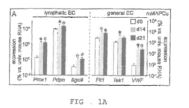
FIGS. 1A ¨ 1M: FIGS 1A and B; Diagram representing expression of general
(right) and lymphatic-
specific (left) endothelial cell (EC) markers, shown as % versus universal
mouse RNA in undifferentiated
mMAPCs (A) or versus universal human RNA in undifferentiated hMAPCs (dO,
white), at 14 (d14, gray)
and 21 (d21, black) days of differentiation. Data represent mean SEM of 5-6
independent
differentiations. *P<0.05 versus dO by Kruskal-Wallis test with Dunn's post-
hoc test. FIG. 1C; FACS
histogram (representative of n=3) showing LYVE1 protein expression (full line)
versus isotype control
(dashed line) in mMAPCs at d14. APC: allophycocyanine. FIG. 1D; Diagram
representing LYVE1
expression, shown as fold-increase versus undifferentiated hMAPCs (dO, white),
or at d9 in the presence
of VEGF-A (light gray), VEGF-C (dark gray) or a combination (black). Data
represent mean SEM of
n=3. *P<0.05 versus dO by 1-way ANOVA with Tuckey's post-hoc test. FIGS 1E-G;
Representative
images of human lymphatic EC (hLEC) spheroids exposed to LEC media (E; 'L') or
conditioned media
from mMAPCs ('mCM' ; F), and corresponding quantification (G; data represent
mean SEM of n=4;
*P<0.05 versus 1' by Mann-Whitney-U test). FIG. 1H; Diagram representing the
effect of mouse
('mCM') or human ('hCM') MAPC-CM on LEC proliferation, expressed as % versus
LEC media. Data
represent mean SEM of n=3-6. *P<0.05 versus 'LEC' by Mann-Whitney-U test. FIGS
1I-M;
Representative images of LECs migrated across the membrane of a transwell
(revealed by Wright-Giemsa
staining) in the presence of non-conditioned mMAPC media (NCM; J), mMAPC-CM
(K), non-
16
CA 03012330 2018-07-23
WO 2017/127123 PCT/US2016/017848
conditioned hMAPC media (NCM; L) or hMAPC-CM (M) and the corresponding
quantification (I; data
represent mean SEM of n=4; *P<0.05 versus corresponding NCM condition by Mann-
Whitney-U test).
Scale bars: 50 jim (E,F); 100 pm (J-M).
FIG. 2A ¨ 2P: FIG 2A; Wound width in mice treated with PBS (open circles) or
mMAPCs (filled
circles). Data represent mean SEM. n=5; *P<0.05 versus PBS by repeated
measures ANOVA and Fisher
post-hoc test. FIG. 2B; Merged brightfield/fluorescent image of the wound
(indicated by arrows) area of
a mouse transplanted with eGFP+ mMAPCs 4d earlier. Note the mMAPCs are close
to blood vessels
(indicated by arrowheads) leading towards the wound bed. FIGS. 2C and D;
Representative pictures of
CD31-stained (brown) cross-sections of 10d-old wounds from mice treated with
PBS (C) or mMAPCs
(D). FIGS. 2E-G; Representative pictures of LYVE1-stained (red) cross-sections
of 10d-old wounds
from mice treated with PBS (E) or mMAPCs (F), and corresponding quantification
(G; data represent
mean SEM. *P<0.05 versus PBS by Mann-Whitney-U test; n=4-5). FIG. 2H; Confocal
image of a
cross-section of a mouse transplanted with eGFP+ mMAPCs 10d earlier revealing
occasional co-
localization (arrowhead) of eGFP with LYVE1 (red). FIGS. 21 and J;
Representative images of cross-
sections of wounds treated with PBS (I) or hMAPCs (J) 5d earlier, stained for
pancytokeratin (PCK;
brown; arrowheads indicate wound borders, horizontal lines indicate distance
covered by the epidermis).
FIGS. 2K and L; Representative images of CD31-stained (brown) cross-sections
of wounds treated with
PBS (K) or hMAPCs (L) 10d earlier. FIGS. 2M-0; Representative pictures of
LYVE1-stained (red)
cross-sections of 10d-old wounds from mice treated with PBS (M) or hMAPCs (N),
and corresponding
quantification (0; data represent mean SEM. *P<0.05 versus PBS by unpaired
Student's t-test; n=6-8).
FIG. 2P; Image of a wound cross-section of a mouse transplanted with hMAPCs
10d earlier revealing
occasional co-localization (arrowheads) of hVimentin (green) with LYVE1 (red).
Hematoxylin and DAPI
were used to reveal nuclei in C,D,I-L and E,F,M,N, respectively. Scale bars:
10 gm (H,P); 100 gm (E,F);
150 [tm (K,L); 400 gm (C,D,I,J,M,N); 2 mm (B).
17
CA 03012330 2018-07-23
WO 2017/127123 PCT/US2016/017848
FIG. 3A ¨ 3D: FIG 3A; Image displaying the skin flap model. R1/R2 indicate the
areas from which
images in panel B-D are shown. Arrows/A' indicate injection spots of
fluorescently-labeled dextran for
lymphangiography or MAPCs/PBS, respectively, and arrowheads show the area
through which blood
supply to the skin flap is preserved. FIGS. 3B-D; Representative merged
pictures of
brightfield/fluorescent images 15min after injection of dextran (FITC-labeled
in B,D or Rhodamin-B-
labeled in C) of regions R1 (left; and enlarged image of the corresponding
inset (i; middle)) and R2 (right)
of mice injected 2w earlier with PBS (B), mMAPCs (C) or hMAPCs (D). Arrowheads
indicate filled
afferent lymphatic vessels. LN: lymph node. Dashed lines in R1/R2 delineate
border of the opened skin or
the flap border, respectively. Scale bars: 100 [tin (B;i1, C;i2+R2, D;i3); 250
[tin (B;R1+R2, C;R1,
D;R1+R2); and 500 [tm (A).
FIGS. 4A -4L: FIGS 4A-D; Representative pictures of Flt4-stained (brown) skin
wound cross-sections
(around the location of dextran injection) from mice treated with PBS (A),
mMAPCs (`mM'; B) or
hMAPCs ChM'; C), and corresponding quantification (D; data represent mean SEM.
*P<0.05 versus
PBS by Kruskal-Wallis with Dunn's post-hoc test; n=6). FIGS 4E-H;
Representative pictures of skin
wound cross-sections (around the location of dextran injection) from mice
treated with PBS (E),
mMAPCs (`mM'; F) or hMAPCs ('hM'; G) revealing functional (dextran-perfused)
lymphatic vessels
(green or red) in cell-treated mice, and corresponding quantification (H; data
represent mean SEM.
*P<0.05 versus PBS by Kruskal-Wallis with Dunn's post-hoc test; n=5-10). Inset
(ii) in E shows the
corresponding region stained for Proxl (red). Note the diffuse fluorescence
signal in E representing
FITC-dextran that failed to be taken up by lymphatic vessels. FIG. 41; Merged
brightfield/fluorescent
image of the wound area of a mouse transplanted with eGFP+ mMAPCs (injection
spots indicated by
arrowheads) 2w earlier. FIG. 4J; Merged green/red fluorescent images of the
wound area of a mouse
transplanted with eGFP+ mMAPCs (arrow) 4w earlier. Note the Rhodamin-dextran-
filled lymphatic
vessels (red; arrowheads) in the vicinity of the transplanted cells. FIG. 4K;
Cross-section through the
area around the wound, revealing transplanted eGFP+ mMAPCs adjacent to
functional (Rhodamin-
18
CA 03012330 2018-07-23
WO 2017/127123 PCT/US2016/017848
dextran-filled, red; lumen indicated by asterisks) lymphatic vessels. FIG. 4L;
High power magnification
of the wound area transplanted with eGFP+ mMAPCs 2w earlier revealing that
occasionally these cells
become part of the endothelial lining (arrowheads) of functional (Rhodamin-
dextran-filled, in red)
lymphatic vessels. Hematoxylin and DAPI were used to reveal nuclei in A-C, and
E-G,K, respectively.
Scale bars: 25 pm (L); 50 [tm (E-G); 100 [tm (A-C,J,K); 500 pm (I).
FIG. 5A ¨ 5G: FIG. 5A; Merged brightfield/fluorescent image of the right
axillary region of a mouse
transplanted with an eGFP+ lymph node (LN; arrowhead) and treated with
Matrigel containing hMAPCs
16w earlier. The area covered with solidified Matrigel and the open skin
border are indicated by a
dashed and full white lines, respectively. FIG. 5B; Diagram representing the
extent of edema in the right
upper limb (determined by MRI and shown as right/left ratio in AU) in mice
treated with Matrigel
containing PBS or hMAPCs 4w or 16w after LN transplantation. *P<0.05 versus w4
by unpaired
Student's t-test (n=4-9). FIGS. 5C and D; Representative T2-weighted MR images
of the antebrachial
regions of mice treated with Matrigel containing PBS (C) or hMAPCs (D),
recorded 16w after LN
transplantation. Hyperintense areas (arrows) indicate accumulation of fluid
due to edema. L: left; R: right.
FIGS. 5E and F; Merged brightfield/fluorescent image of the right axillary
region of a mouse
transplanted with an eGFP+ LN (arrowhead) and treated with Matrigel
containing PBS (E) or hMAPCs
(F) 16w earlier. Inset (ii) zooms in on the boxed area in F. Note the
significantly improved drainage of
the Rhodamin-labeled lectin (red) in hMAPC-treated mice recorded 15min after
injection (injection spot
indicated by arrow). The border of the opened skin is indicated by white
lines. FIG 5G; Merged
brightfield/fluorescent image zooming in on an eGFP+ LN (green) transplanted
in a mouse treated with
Matrigel containing hMAPCs 16w earlier, revealing drainage of the Rhodamin-
labeled lectin (red) into
the LN. Arrowheads indicate afferent lymph vessel. Scale bars: 200 gm (G); 3
mm (A,E,F).
FIGS. 6 A ¨ 6N: FIGS. 6A-C; Brightfield images of the blood vessel network
leading up to the
transplanted lymph node (LN) of mice treated with Matrigel containing PBS (A)
or hMAPCs ('hM' ; B)
19
CA 03012330 2018-07-23
WO 2017/127123 PCT/US2016/017848
16w earlier, and corresponding quantification (C; data represent mean SEM.
*P<0.05 versus PBS by
Mann-Whitney-U test; n=6). FIG. 6D; Merged brightfieldifluorescent image of an
eGFP+ LN
transplanted in a mouse treated with Matrigel containing hMAPCs 16w earlier
revealing that the LN is
irrigated by numerous blood vessels. FIG. 6E and F; Merged
brightfield/fluorescent images zooming in
on a DsRed+ LN transplanted in mice treated with Matrigel containing hMAPCs
8w earlier revealing
extensive branching of the LN vascular network. Inset (ii) corresponds to the
boxed area in F. FIG 6G;
Merged IF image of a Proxl/eGFP-stained section in a mouse treated with
Matrigel +hMAPCs 16w
earlier revealing that part of the branches are lymphatic (Proxl+,
arrowheads). Inset (i2) corresponds to
the boxed area in G. FIGS. 6H-J; LYVEl-stained (red) cross-sections of PBS (H)
or hMAPC-treated
('hM'; I) mice in the area around the sutures at 8w after LN transplantation
and corresponding
quantification (I; data represent mean SEM. *P<0.05 versus PBS by Student's t-
test; n=5-8). FIGS. 6K-
M; Fluorescence images of the area around the transplanted eGFP+ LN (lined by
a dashed line in K;
adjacent section stained for Proxl in green is shown in L; Proxl/smooth muscle
a-actin (aSMA in red,
indicated by arrowheads; double staining in M zooms in on the boxed area in
K,L; and FIG. 6N
represents the same area on an adjacent cross-section stained for LYVE1 in
red) revealing
Prox 1 aSMALYVEL draining lymphatic collector vessels in mice treated 16w
earlier with Matrigel
containing hMAPCs. Asterisks in L-N indicate lymph (which artifactually
fluoresces upon exposure to
tyramide-based amplification). White arrows in A,B,E-L indicate the sutures
used to fix the transplanted
LN. Scale bars: 20 pm (M,N); 50 jim (G,K,L); 100 pm (D); 150 gm (F;i1); 200
[tm (E,H,I); 500 [tm (F); 1
mm (A,B).
FIGS. 7A ¨ 7H: FIG. 7A; Diagram representing wound length (in mm) in mice
treated with PBS (n=5:
open circles) or mMAPCs (n=5; filled circles) until 10d after wounding. Data
represent mean SEM.
*P<0.05 versus PBS by repeated measures ANOVA with Fisher post-hoc test. FIGS.
7B and C;
Representative brightfield pictures of linear wounds on the back of mice
treated with PBS (B) or murine
(m)MAPCs (C) 10d after wounding. FIGS. 7D and E; Representative pictures of
cross-sections of 10d-
CA 03012330 2018-07-23
WO 2017/127123 PCT/US2016/017848
old wounds from mice treated with PBS (D) or mMAPCs (E) stained with H&E. Note
the significantly
smaller wound gap (the edges of which are indicated by arrowheads) in mMAPC-
treated mice. FIG. 7F;
Merged picture of red and green fluorescent image of a wound cross-section
revealing no co-localization
of CD45 (in green) with LYVE1 (in red). FIG. 7G; Merged picture of
brightfieldifluorescent image of
the wound bed 24h after seeding of eGFP-labeled hMAPCs revealing homogenous
distribution of eGFP+
hMAPCs across the wound area. FIG. 7H; Image of a vimentin-stained (green)
wound cross-section of a
mouse transplanted with hMAPCs 10d earlier revealing persistence of large
patches of hMAPCs
homogenously distributed across the wound bed. The dermo-epidermal junction is
indicated by a dashed
line. DAPI was used as nuclear counterstain in H. Scale bars: 20 m in F; 100
pm in H; 300 pm in D,E; 1
mm in G; and 2 mm in B,C.
FIGS. 8A ¨81: FIGS. 8A-D; Representative pictures of cross-sections of the
skin wound (around the
location of transplantation indicated by 'X' in FIG. 3A) from mice treated
with PBS (A), mMAPCs
('mM'; B) or hMAPCs ('hM' ; C) stained for CD31 (in brown), and corresponding
quantification (D; data
represent mean SEM. *P<0.05 versus PBS by Kruskal-Wallis test with Dunn's
post-hoc test; n=5).
FIGS 8E-H; Representative pictures of cross-sections of the skin wound (around
the location of dextran
injection indicated by arrow in FIG. 3A) from mice treated with PBS (E),
mMAPCs ('mM'; F) or
hMAPCs ('hM' ; G) stained for LYVE1 (red in E,G; green in F), and
corresponding quantification (H;
data represent mean SEM. *P<0.05 versus PBS by Kruskal-Wallis test with
Dunn' s post-hoc test; n=6).
FIG. 81; Merged picture of green (FITC-labeled dextran), red (Proxl) and far-
red (smooth muscle cell-a-
actin; aSMA) fluorescent microscopic images of the wound area (around the
location of transplantation
indicated by 'X' in FIG. 3A) of a mouse transplanted with hMAPCs 2w earlier,
revealing a functional
aSMA-coated (arrowheads) Proxl+ lymphatic (pre-)collector vessel in addition
to two small functional
Proxr/aSMA lymphatic capillaries (lined by white dashed lines). The
autofluorescent muscle cells of the
fascia are lined by a red dashed line. Scale bars: 10 pm in I; and 100 pm in A-
C ,E-G.
21
CA 03012330 2018-07-23
WO 2017/127123 PCT/US2016/017848
FIGS. 9A ¨ 9H: FIGS. 9A and B; T2 maps corresponding to the T2-weighted MR
images shown in FIG.
5C,D of the antebrachial regions of mice treated with Matrigel containing PBS
(A) or hMAPCs (B),
recorded 16w after LN transplantation. L: left; R: right. FIG. 9C; Merged
picture of green and red
fluorescent microscopic images of the right axillary region of a mouse
transplanted with a DsRed+ LN
and treated with Matrigel containing hMAPCs 8w earlier. Note the afferent
lymphatic vessel filled with
FITC-labeled lectin (in green), indicated by arrowheads. FIGS. 9D and E;
Merged pictures of brightfield
and green fluorescent images of the right axillary region of mice transplanted
with an eGFP+ LN and
treated with Matrigel containing PBS (D) or hMAPCs (E) 16w earlier, revealing
a more elaborate blood
vessel network irrigating the transplanted LN of hMAPC-treated mice. FIG. 9F;
Merged picture of a red
and green fluorescent image of a cross-section of the right axillary region of
a mouse transplanted with an
eGFP+ LN and treated with hMAPCs 16w earlier, revealing persisting vimentin-
stained (in red) hMAPCs
surrounding the transplanted LN. FIG. 9G; Merged picture of brightfield and
green fluorescent images
of the right axillary region of a mouse transplanted with an eGFP+ LN and
treated with Matrigel
containing hMAPCs 4w earlier, revealing outward branching of the
(lymph)vascular network. FIG. 9H;
Merged picture of an eGFP-stained cross-section of the right axillary region
of a mouse transplanted with
an eGFP+ LN and treated with Matrigel containing hMAPCs 16w earlier,
revealing outward branches of
the (lymph)vascular network. Permanent sutures fixing the transplanted LN are
indicated by arrows in C-
E. LN body is lined by a white dashed line in F-H. DAPI was used to reveal
nuclei in F,H. Scale bars: 25
pm in F; 100 gm in H; 150 gm in G; and 250 pm in C-E.
DETAILED DESCRIPTION OF THE INVENTION
It should be understood that this invention is not limited to the particular
methodology, protocols,
and reagents, etc., described herein and, as such, may vary. The terminology
used herein is for the
purpose of describing particular embodiments only, and is not intended to
limit the scope of the disclosed
invention, which is defined solely by the claims.
22
CA 03012330 2018-07-23
WO 2017/127123 PCT/US2016/017848
The section headings are used herein for organizational purposes only and are
not to be construed
as in any way limiting the subject matter described.
The methods and techniques of the present application are generally performed
according to
conventional methods well-known in the art and as described in various general
and more specific
references that are cited and discussed throughout the present specification
unless otherwise indicated.
See, e.g., Sambrook et al., Molecular Cloning: A Laboratory Manual, 3rd ed.,
Cold Spring Harbor
Laboratory Press, Cold Spring Harbor, N.Y. (2001) and Ausubel et al., Current
Protocols in Molecular
Biology, Greene Publishing Associates (1992), and Harlow and Lane, Antibodies:
A Laboratory Manual,
Cold Spring Harbor Laboratory Press, Cold Spring Harbor, N.Y. (1990).
Definitions
"A" or "an" means herein one or more than one; at least one. Where the plural
form is used
herein, it generally includes the singular.
The term "bandage" as used in this application is synonymous with the terms
"dressing" or
"patch" as they refer to a functionalized substrate to which cells are
attached. These devices have been
referred to as cell-laden bandages, cell-laden patches, and cell-laden
dressings. In these embodiments the
cells that are attached to the substrate, when applied in operable proximity
to the wound, leave the patch,
dressing, or bandage and migrate to the wound. In some instances these
bandages/patches may be
comprised of a coating of plasma polymer. As mentioned this can be comprised
of a functionalized
substrate to which the cells are attached.
23
CA 03012330 2018-07-23
WO 2017/127123 PCT/US2016/017848
A "clinically-relevant" number of cells refers to a number of cells that is
sufficient to effect a
clinical response; that is, a prevention, reduction, amelioration, etc. of an
undesirable pathological
condition in a subject. A particular embodiment pertains to a number of cells
that is sufficient to create a
master cell bank.
"Co-administer" means to administer in conjunction with one another, together,
coordinately,
including simultaneous or sequential administration of two or more agents.
"Comprising" means, without other limitation, including the referent,
necessarily, without any
qualification or exclusion on what else may be included. For example, "a
composition comprising x and
y" encompasses any composition that contains x and y, no matter what other
components may be present
in the composition. Likewise, "a method comprising the step of x" encompasses
any method in which x is
carried out, whether x is the only step in the method or it is only one of the
steps, no matter how many
other steps there may be and no matter how simple or complex x is in
comparison to them. "Comprised of
and similar phrases using words of the root "comprise" are used herein as
synonyms of "comprising" and
have the same meaning.
"Comprised of' is a synonym of "comprising" (see above).
"Conditioned cell culture medium" is a term well-known in the art and refers
to medium in which
cells have been grown. Herein this means that the cells are grown for a
sufficient time to secrete the
factors that are effective to achieve any of the results described in this
application.
Conditioned cell culture medium refers to medium in which cells have been
cultured so as to
secrete factors into the medium. For the purposes of the present invention,
cells can be grown through a
sufficient number of cell divisions so as to produce effective amounts of such
factors so that the medium
24
CA 03012330 2018-07-23
WO 2017/127123 PCT/US2016/017848
has the effects. Cells are removed from the medium by any of the known methods
in the art, including,
but not limited to, centrifugation, filtration, immunodepletion (e.g., via
tagged antibodies and magnetic
columns), and FACS sorting.
"Effective amount" generally means an amount which provides the desired local
or systemic
effect. For example, an effective amount is an amount sufficient to effectuate
a beneficial or desired
clinical result. The effective amounts can be provided all at once in a single
administration or in fractional
amounts that provide the effective amount in several administrations. The
precise determination of what
would be considered an effective amount may be based on factors individual to
each subject, including
their size, age, injury, and/or disease or injury being treated, and amount of
time since the injury occurred
or the disease began. One skilled in the art will be able to determine the
effective amount for a given
subject based on these considerations which are routine in the art. As used
herein, "effective dose" means
the same as "effective amount."
"Effective route" generally means a route which provides for delivery of an
agent to a desired
compartment, system, or location. For example, an effective route is one
through which an agent can be
administered to provide at the desired site of action an amount of the agent
sufficient to effectuate a
beneficial or desired clinical result.
Use of the term "includes" is not intended to be limiting.
"Increase" or "increasing" means to induce entirely where there was no pre-
existing presence or
to increase the degree of.
The term "isolated" refers to a cell or cells which are not associated with
one or more cells or one
or more cellular components that are associated with the cell or cells in
vivo. An "enriched population"
CA 03012330 2018-07-23
WO 2017/127123 PCT/US2016/017848
means a relative increase in numbers of a desired cell relative to one or more
other cell types in vivo or in
primary culture.
However, as used herein, the term "isolated" does not indicate the presence of
only stem cells.
Rather, the term "isolated" indicates that the cells are removed from their
natural tissue environment and
are present at a higher concentration as compared to the normal tissue
environment. Accordingly, an
"isolated" cell population may further include cell types in addition to stem
cells and may include
additional tissue components. This also can be expressed in terms of cell
doublings, for example. A cell
may have undergone 10, 20, 30, 40 or more doublings in vitro or ex vivo so
that it is enriched compared to
its original numbers in vivo or in its original tissue environment (e.g., bone
marrow, peripheral blood,
adipose tissue, etc.).
"MAPC" is an acronym for "multipotent adult progenitor cell." It refers to a
cell that is not an
embryonic stem cell or germ cell but has some characteristics of these. MAPC
can be characterized in a
number of alternative descriptions, each of which conferred novelty to the
cells when they were
discovered. They can, therefore, be characterized by one or more of those
descriptions. First, they have
extended replicative capacity in culture without being transformed
(tumorigenic) and with a normal
karyotype. Second, they may give rise to cell progeny of more than one germ
layer, such as two or all
three germ layers (i.e., endoderm, mesoderm and ectoderm) upon
differentiation. Third, although they are
not embryonic stem cells or germ cells, they may express markers of these
primitive cell types so that
MAPCs may express one or more of Oct 3/4 (aka, Oct 3A or Oct 4), rex-1, and
rox-1. They may also
express one or more of sox-2 and SSEA-4. Fourth, like a stem cell, they may
self-renew, that is, have an
extended replication capacity without being transformed. This means that these
cells express telomerase
(i.e., have telomerase activity). Accordingly, the cell type that was
designated "MAPC" may be
characterized by alternative basic characteristics that describe the cell via
some of its novel properties.
26
CA 03012330 2018-07-23
WO 2017/127123 PCT/US2016/017848
The term "adult" in MAPC is non-restrictive. It refers to a non-embryonic
somatic cell.
MAPCs are karyotypically normal and do not form teratomas or other tumors in
vivo. This acronym was
first used in U.S. Patent No. 7,015,037 to describe a pluripotent cell
isolated from bone marrow.
However, cells with pluripotential markers and/or differentiation potential
have been discovered
subsequently and, for purposes of this invention, may be equivalent to those
cells first designated
"MAPC." Descriptions of the MAPC type of cell are provided in the Summary of
the Invention above.
MAPC represents a more primitive progenitor cell population than MSC
(Verfaillie, C.M.,
Trends Cell Biol 12:502-8 (2002), Jahagirdar, B.N., et al., Exp Hematol,
29:543-56 (2001); Reyes, M. and
C.M. Verfaillie, Ann N Y Acad Sci, 938:231-233 (2001); Jiang, Y. et al., Exp
Hematol, 30896-904
(2002); and Jiang, Y. et al., Nature, 418:41-9. (2002).
"Progenitor cells" are cells produced during differentiation of a stem cell
that have some, but not
all, of the characteristics of their terminally-differentiated progeny.
Defined progenitor cells, such as
"cardiac progenitor cells," are committed to a lineage, but not to a specific
or terminally differentiated cell
type. The term "progenitor" as used in the acronym "MAPC" does not limit these
cells to a particular
lineage. A progenitor cell can form a progeny cell that is more highly
differentiated than the progenitor
cell.
Selection could be from cells in a tissue. For example, in this case, cells
would be isolated from a
desired tissue, expanded in culture, selected for a desired characteristic,
and the selected cells further
expanded.
"Self-renewal" refers to the ability to produce replicate daughter stem cells
having differentiation
potential that is identical to those from which they arose. A similar term
used in this context is
"proliferation."
27
CA 03012330 2018-07-23
WO 2017/127123 PCT/US2016/017848
"Serum-free medium" refers to medium in which serum is not present or, if
present, is at levels at
which the components of the serum have no effect on the growth or variability
of the cells (i.e., are not
actually necessary, such as residual or trace amounts).
"Stem cell" means a cell that can undergo self-renewal (i.e., progeny with the
same
differentiation potential) and also produce progeny cells that are more
restricted in differentiation
potential.
"Subject" means a vertebrate, such as a mammal, such as a human. Mammals
include, but are
not limited to, humans, dogs, cats, horses, cows, and pigs.
As used herein, the term "wound" means a breach in the integrity of a tissue,
e.g., skin, which can
be caused by acute trauma or underlying pathological causes such as the
cutaneous and subcutaneous
wounds that have been described in this application.
Wounds may be derived from sources including, but not limited to, autoimmune-
disease,
rejection of transplanted organs, burns, cuts, lacerations, and ulcerations,
including skin ulcerations and
diabetic ulcerations.
The stem cells may be administered to an animal to repair epithelial damage
caused by burns,
cuts, lacerations, and ulcerations, including, but not limited to, skin
ulcerations and diabetic ulcerations.
Examples of wounds may include both open and closed wounds. In certain
embodiments, the
wound comprises an external wound. In certain embodiments, the wound comprises
an open wound.
28
CA 03012330 2018-07-23
WO 2017/127123 PCT/US2016/017848
In certain embodiments, the wound comprises a chronic wound. In certain
embodiments, the wound
comprises a chronic wound or an ulcer.
In certain embodiments, the composition is suitable for topical application,
topical
administration or topical delivery to a subject. Topical formulations are as
described herein. Other forms
of delivery of cells are contemplated.
The dose and frequency of topical administration may be determined by one of
skill in the art.
Examples of forms for topical administration include delivery by way of a gel,
an ointment, a
cream, a lotion, a foam, an emulsion, a suspension, a spray, an aerosol, a
solution, a liquid, a powder, a
semi-solid, a gel, a jelly, a suppository; a solid, an ointment, a paste, a
tincture, a liniment, a patch, or
release from a bandage, gauze or dressing. Other forms of topical delivery are
contemplated.
Methods for incorporating substrates into products for topical release are
known in the art, for
example as described in Boateng J.S. et al (2008) "Wound healing dressings and
drug delivery systems:
a review" J. Pharm Sci. 97(8): 2892-2923 and "Delivery System Handbook for
Personal Care and
Cosmetic Products: Technology" (2005) by Meyer Rosen, published William Andrew
Inc, Norwich New
York.
In certain embodiments, the composition is suitable for delivery to a subject
by one or more of
intravenous administration, by aerosolized administration, by parenteral
administration, by implant, by
subcutaneous injection, intraarticularly, rectally, intranasally,
intraocularly, vaginally, or transdermally.
In certain embodiments, the composition comprises other compounds that
enhance, stabilize or
maintain the activity of the cells for delivery and/or their delivery or
transfer.
29
CA 03012330 2018-07-23
WO 2017/127123 PCT/US2016/017848
In certain embodiments, it may be desirable to administer the composition by
injection. Forms
suitable for injectable use include sterile aqueous solutions or dispersions
and sterile powders for the
extemporaneous preparation of sterile injectable solutions or dispersions. A
carrier can be a solvent or
dispersion medium containing, for example, water, ethanol, polyol (e.g.,
glycerol, propylene glycol and
liquid polyethylene glycol), suitable mixtures thereof, and vegetable oils.
In certain embodiments, it may be desirable to administer the composition
intravenously.
Compositions containing the composition described herein suitable for
intravenous administration may
be formulated by a skilled person.
In certain embodiments, the subject is a human or animal subject. In certain
embodiments, the
subject is a human subject.
In certain embodiments, the subject is a mammalian subject, a livestock animal
(such as a horse,
a cow, a sheep, a goat, a pig), a domestic animal (such as a dog or a cat) and
other types of animals
such as monkeys, rabbits, mice, laboratory animals, birds and fish. Other
types of animals are
contemplated. Veterinary applications of the present disclosure are
contemplated. Use of any of
the aforementioned animals as animal models is also contemplated.
The present disclosure provide a method of healing or treating a wound, the
method comprising
delivering cells to the wound using a product or a composition as described
herein.
MAPC
CA 03012330 2018-07-23
WO 2017/127123 PCT/US2016/017848
Human MAPCs are described in U.S. Patent 7,015,037. MAPCs have been identified
in other
mammals. Murine MAPCs, for example, are also described in U.S. Patent
7,015,037. Rat MAPCs are
also described in U.S. Patent No. 7,838,289. These references are incorporated
by reference for
describing MAPCs, their phenotype and culture.
Isolation and Growth of MAPCs
Methods of MAPC isolation are known in the art. See, for example, U.S. Patent
7,015,037, and
these methods, along with the characterization (phenotype) of MAPCs, are
incorporated herein by
reference. MAPCs can be isolated from multiple sources, including, but not
limited to, bone marrow,
placenta, umbilical cord and cord blood, muscle, brain, liver, spinal cord,
blood or skin. It is, therefore,
possible to obtain bone marrow aspirates, brain or liver biopsies, and other
organs, and isolate the cells
using positive or negative selection techniques available to those of skill in
the art, relying upon the genes
that are expressed (or not expressed) in these cells (e.g., by functional or
morphological assays such as
those disclosed in the above-referenced applications, which have been
incorporated herein by reference).
Rodent MAPCs have also been obtained by improved methods described in Breyer
et al.,
Experimental Hematology, 34:1596-1601 (2006) and Subramanian et al., Cellular
Programming and
Reprogramming: Methods and Protocols; S. Ding (ed.), Methods in Molecular
Biology, 636:55-78
(2010), incorporated by reference for these methods. Human MAPCs have been
obtained by improved
methods that are described in Roobrouck et al. Stem Cells 29:871-882 (2011),
incorporated by reference
for these methods.
MAPCs from Human Bone Marrow as Described in U.S. Patent 7,015,037
31
CA 03012330 2018-07-23
WO 2017/127123 PCT/US2016/017848
MAPCs do not express the common leukocyte antigen CD45 or erythroblast
specific
glycophorin-A (Gly-A). The mixed population of cells was subjected to a Ficoll
Hypaque separation. The
cells were then subjected to negative selection using anti-CD45 and anti-Gly-A
antibodies, depleting the
population of CD45+ and Gly-A+ cells, and the remaining approximately 0.1% of
marrow mononuclear
cells were then recovered. Cells could also be plated in fibronectin-coated
wells and cultured as
described below for 2-4 weeks to deplete the cells of CD45+ and Gly-A+ cells.
In cultures of adherent
bone marrow cells, many adherent stromal cells undergo replicative senescence
around cell doubling 30
and a more homogenous population of cells continues to expand and maintains
long telomeres.
Alternatively, positive selection could be used to isolate cells via a
combination of cell-specific
markers. Both positive and negative selection techniques are available to
those of skill in the art, and
numerous monoclonal and polyclonal antibodies suitable for negative selection
purposes are also
available in the art (see, for example, Leukocyte Typing V, Schlossman, et
al., Eds. (1995) Oxford
University Press) and are commercially available from a number of sources.
Techniques for mammalian cell separation from a mixture of cell populations
have also been
described by Schwartz, et al., in U. S. Patent No. 5,759,793 (magnetic
separation), Basch et al., 1983
(immunoaffinity chromatography), and Wysocki and Sato, 1978 (fluorescence-
activated cell sorting).
Cells may be cultured in low-serum or serum-free culture medium. Serum-free
medium used to
culture MAPCs is described in U.S. Patent 7,015,037. Commonly-used growth
factors include but are not
limited to platelet-derived growth factor and epidermal growth factor. See,
for example, U.S. Patent Nos.
7,169,610; 7,109,032; 7,037,721; 6,617,161; 6,617,159; 6,372,210;6,224,860;
6,037,174; 5,908,782;
5,766,951; 5,397,706; and 4,657,866; all incorporated by reference for
teaching growing cells in serum-
free medium.
32
CA 03012330 2018-07-23
WO 2017/127123 PCT/US2016/017848
Additional Culture Methods
In additional experiments the density at which MAPCs are seeded can vary from
about 100
cells/cm2 or about 150 cells/cm2 to about 10,000 cells/cm2, including about
200 cells/cm2 to about 1500
cells/cm2 to about 2000 cells/cm2. The density can vary between species.
Additionally, optimal density
can vary depending on culture conditions and source of cells. It is within the
skill of the ordinary artisan
to determine the optimal seeding density for a given set of culture
conditions.
Also, effective atmospheric oxygen concentrations of less than about 10%,
including about 1-5%
and, especially, 3-5%, can be used at any time during the isolation, growth
and differentiation of MAPCs
in culture.
Cells may be cultured under various serum concentrations, e.g., about 2-20%.
Fetal bovine serum
may be used. Higher serum may be used in combination with lower oxygen
tensions, for example, about
15-20%. Cells need not be selected prior to adherence to culture dishes. For
example, after a Ficoll
gradient, cells can be directly plated, e.g., 250,000-500,000/cm2. Adherent
colonies can be picked,
possibly pooled, and expanded.
In one embodiment, high serum (around 15-20%) and low oxygen (around 3-5%)
conditions are
used for the cell culture. For example, adherent cells from colonies can be
plated and passaged at
densities of about 1700-2300 cells/cm2 in 18% serum and 3% oxygen (with PDGF
and EGF).
In an embodiment specific for MAPCs, supplements are cellular factors or
components that
allow MAPCs to retain the ability to differentiate into cell types of more
than one embryonic lineage,
such as, all three lineages. This may be indicated by the expression of
specific markers of the
33
CA 03012330 2018-07-23
WO 2017/127123 PCT/US2016/017848
undifferentiated state, such as Oct 3/4 (a.k.a. 0ct4 or Oct 3A) and/or markers
of high expansion capacity,
such as, telomerase.
For all the components listed below, see U.S. 7,015,037, which is incorporated
by reference for
teaching these components.
Stem cells often require additional factors that encourage their attachment to
a solid support, such
as fibronectin. One embodiment of the present invention utilizes fibronectin.
See, for example, Ohashi et
al., Nature Medicine, 13:880-885 (2007); Matsumoto et al., J Bioscience and
Bioengineering, 105:350-
354 (2008); Kirouac et al., Cell Stem Cell, 3:369-381 (2008); Chua et al.,
Biomaterials, 26:2537-2547
(2005); Drobinskaya et al., Stem Cells, 26:2245-2256 (2008); Dvir-Ginzberg et
al., FASEB J, 22:1440-
1449 (2008); Turner et al., J Biomed Mater Res Part B: Appl Biomater, 82B:156-
168 (2007); and
Miyazawa et al., Journal of Gastroenterology and Hepatology, 22:1959-1964
(2007)).
Once established in culture, cells can be used fresh or frozen and stored as
frozen stocks, using,
for example, DMEM with 20%-40% FCS and 10% DMSO. In one embodiment, 20% FCS is
used.
Other methods for preparing frozen stocks for cultured cells are also
available to those of skill in the art.
For the purposes of this application, the additional culture methods as well
as the other culture
methods also apply to bioreactor methods, with respect to the medium
components and conditions
described above. As an example, in an exemplified embodiment, the oxygen
concentration is 5%, serum
is about 19% and both EGF and PDGF are added to the medium.
Pharmaceutical Formulations
34
CA 03012330 2018-07-23
WO 2017/127123 PCT/US2016/017848
U.S. 7,015,037 is incorporated by reference for teaching pharmaceutical
formulations. In certain
embodiments, the cell populations are present within a composition adapted for
and suitable for delivery,
i.e., physiologically compatible.
In some embodiments the purity of the cells (or conditioned medium) for
administration to a
subject is about 100% (substantially homogeneous). In other embodiments it is
95% to 100%. In some
embodiments it is 85% to 95%. Particularly, in the case of admixtures with
other cells, the percentage
can be about 10%-15%, 15%-20%, 20%-25%, 25%-30%, 30%-35%, 35%-40%, 40%-45%,
45%-50%,
60%-70%, 70%-80%, 80%-90%, or 90%-95%. Or isolation/purity can be expressed in
terms of cell
doublings where the cells have undergone, for example, 10-20, 20-30, 30-40, 40-
50 or more cell
doublings.
The choice of formulation for administering the cells for a given application
will depend on a
variety of factors. Prominent among these will be the species of subject, the
nature of the condition being
treated, its state and distribution in the subject, the nature of other
therapies and agents that are being
administered, the optimum route for administration, survivability via the
route, the dosing regimen, and
other factors that will be apparent to those skilled in the art. For instance,
the choice of suitable carriers
and other additives will depend on the exact route of administration and the
nature of the particular
dosage form.
Final formulations of the aqueous suspension of cells/medium will typically
involve adjusting the
ionic strength of the suspension to isotonicity (i.e., about 0.1 to 0.2) and
to physiological pH (i.e., about
pH 6.8 to 7.5). The final formulation will also typically contain a fluid
lubricant.
In some embodiments, cells/medium are formulated in a unit dosage injectable
form, such as a
solution, suspension, or emulsion. Pharmaceutical formulations suitable for
injection of cells/medium
CA 03012330 2018-07-23
WO 2017/127123 PCT/US2016/017848
typically are sterile aqueous solutions and dispersions. Carriers for
injectable formulations can be a
solvent or dispersing medium containing, for example, water, saline, phosphate
buffered saline, polyol
(for example, glycerol, propylene glycol, liquid polyethylene glycol, and the
like), and suitable mixtures
thereof.
The skilled artisan can readily determine the amount of cells and optional
additives, vehicles,
and/or carrier in compositions to be administered in methods of the invention.
Typically, any additives
(in addition to the cells) are present in an amount of 0.001 to 50 wt % in
solution, such as in phosphate
buffered saline. The active ingredient is present in the order of micrograms
to milligrams, such as about
0.0001 to about 5 wt %, preferably about 0.0001 to about 1 wt %, most
preferably about 0.0001 to about
0.05 wt % or about 0.001 to about 20 wt %, preferably about 0.01 to about 10
wt %, and most preferably
about 0.05 to about 5 wt %.
36
CA 03012330 2018-07-23
WO 2017/127123 PCT/US2016/017848
EXAMPLE
MAPC Support Lymphatic Vessel Growth in Lymphedema
MAPCs have lymphvasculogenic and lymphangiogenic potential
The inventors investigated whether MAPCs have the inherent capacity to give
rise to LECs. First,
they confirmed that MAPCs gain expression of general EC markers upon VEGF-A
exposure (Figures 1A,
B). Proxl , the masterswitch of lymphatic differentiation, was significantly
induced in MAPCs at 2w of
endothelial differentiation and its expression levels remained stable until 1w
later (Figures 1A,B). Proxl
induction may also have triggered expression of additional lymphatic genes
(i.e., Pdpn and Itg9a), known
to be upregulated by forced Prox 1 expression(36). A fraction (21 6%) of MAPCs
exposed to VEGF-A
also expressed LYVE1 (shown at the protein level for mMAPCs; Figure 1C).
Notably, induction of
lymphatic marker gene expression in hMAPCs was not further improved in the
presence of
lymphangiogenic GF VEGF-C (shown for LYVE1 in FigurelD; PROX1 fold-induction
versus dO was also
comparable upon exposure to VEGF-A, VEGF-C or a combination: 26 10, 26 14 and
26 11,
respectively; n=4). COUP-TFII, a transcription factor co-determining lymphatic
competence of
ECs(36,37), was expressed at high relatively constant levels throughout the
differentiation process (not
shown). Thus, MAPCs have the inherent capacity to initiate a LEC
differentiation program.
The inventors reasoned that MAPCs might have an effect on lymphangiogenesis by
cross-talking
to LECs, as MAPCs are known to secrete VEGF-A, which is responsible for the
trophic effects of MSCs
on LECs. 72h MAPC supernatant significantly stimulated LEC sprouting,
proliferation and migration
(Figures 1E-M). Thus, MAPCs may support the formation of lymphatic vessels by
a combination of
direct and indirect effects.
37
CA 03012330 2018-07-23
WO 2017/127123 PCT/US2016/017848
MAPCs contribute to physiological lymphangiogenesis during wound healing
Wound healing requires growth of new blood and lymphatic vessels (Maruyama,
K., et al. 2007.
Am J Pathol (2007) 170:1178-1191). Transplantation of mMAPCs from mice
ubiquitously expressing
enhanced (e)GFP shortly after a linear back skin incision in C57B1/6 mice
resulted in a significant
acceleration of wound closure (FIG. 2A and FIG. 7A) and the occurrence of
smaller scars (FIG. 7B-E)
compared to PBS-injection. While all mMAPC-injected wounds were completely
reepithelialized, 60% of
PBS-treated wounds were only partially covered with neo-epidermis at 10d. In
vivo fluorescence imaging
revealed that 4d after injection, eGFP+ mMAPCs were located in close vicinity
to blood vessels growing
towards the wound bed (FIG. 2B). In accordance, mMAPC transplantation boosted
de novo growth of
CD31+ vessels in the wound center by two-fold (number of CD31+ vessels/area
(mm2): 76 4 in mMAPC-
treated versus 36 16 in PBS-injected mice; n=5, P<0.05 by Mann-Whitney-U test;
FIG. 2C,D). In
agreement with earlier limb ischemia studies (Aranguren, X.L., et al. J Gin
Invest (2008) 118:505-514),
direct contribution to CD31+ ECs was modest in this wound healing model mMAPCs
also significantly
increased LYVE1+ lymphatic capillary growth by 3-fold and occasionally
contributed to differentiated
LECs (FIGS. 2E-H). The vast majority of LYVE1+ cells were lymphatic
endothelial cells (LECs) and not
macrophage intermediates ¨ previously suggested to contribute to lymphatic
vessels in transplanted
kidneys (Kerjaschki, D., et al. Nat Med (2006) 12:230-234) ¨ since they did
not co-localize with CD45, a
panleukocytic marker (FIG. 7F).
hMAPCs applied onto circular wounds in athymic nude mice significantly
promoted healing.
Live imaging and cross-sections through the wound area upon transplantation of
hMAPCs showed their
homogenous distribution in the wound bed (FIGS. 7G,H). hMAPCs accelerated
epithelial coverage (%
coverage at 5d: 46 5 in hMAPC-treated versus 7 2 in vehicle-treated wounds;
n=6, P<0.05 by Mann-
Whitney-U test; FIGS. 2I,J). All wounds were completely reepithelialized at
d10 in hMAPC-treated mice
38
CA 03012330 2018-07-23
WO 2017/127123 PCT/US2016/017848
versus only 46% of PBS-treated mice. hMAPC transplantation improved wound
vascularization by about
two-fold (% CD31+ area in the wound borders at 5d and the entire wound at 10d:
11 1 and 13 1 in
hMAPC-treated versus 6 1 and 6 1 in PBS-injected wounds; n=6-8, P<0.05 by
Student's t-test;
FIGS.2K,L). hMAPCs significantly boosted lymphangiogenesis as evidenced by the
three-fold increased
LYVE1+ fractional area (FIGS. 2M-0). Double immunofluorescence (IF) staining
for Proxl and smooth
muscle a-actin (aSMA) revealed that in this short-duration wound model, the
vast majority (97 2%) of
lymphatic vessels in the granulation tissue at 10d were capillaries devoid of
aSMA coverage. Again, in
situ LEC differentiation of hMAPCs happened only occasionally, shown by the co-
localization of the
hMAPC-derived vimentin signal and LYVE1 staining (FIG. 2P). Thus, MAPCs
significantly accelerated
wound healing in part by boosting capillary lymphangiogenesis mostly
indirectly through a trophic effect
on host LECs.
MAPCs regenerate lymphatic vessels in a secondary lymphedema model
To test the potential of MAPCs to restore lymph flow in secondary lymphedema,
lymph drainage
to the axillary lymph nodes (LNs) was discontinued by means of a full-
thickness skin incision in the
abdomen (FIG. 3A) (Saaristo, A., et al. FASEB J (2004) 18:1707-1709). This
abrogated normal lymph
drainage in the majority (7/10) of PBS-treated animals shown by the lack of
fluorescent dye crossing the
wound border 2w following skin incision (FIG. 3B; Tablel). MAPC
transplantation around the wound
border almost completely (in 5/6 and 6/6 cases for mMAPC- or hMAPC-treated
mice, respectively)
restored lymph drainage across this border (FIGS. 3C,D; Table 1). While
drainage to the axillary LN was
only obtained in 1/10 PBS-injected mice, 3/6 mMAPC-injected and 6/6 hMAPC-
injected mice showed
LN drainage after 2w. In a second set of mice injected with PBS or mMAPCs,
fluorescent dye crossed the
wound border in 5/5 mMAPC-treated mice and LN drainage was restored in 4/5,
while there was no
restoration of drainage across the wound border and into the axillary LN in
any of the PBS-injected mice
4w after skin incision (Tablel). Histological analysis of the skin wound area
around the transplantation
39
CA 03012330 2018-07-23
WO 2017/127123 PCT/US2016/017848
sites revealed that, in addition to a 1.8-fold expansion of CD31+ blood
vessels (FIGS. 8A-D), MAPC-
injected mice had a -two-three-fold increase in Fltr (VEGFR3+) and LYVE1+
fractional area in the
wound borders (FIGS. 4A-D + 8E-H, respectively) 2w after skin incision. The
average number of
functional lymphatic vessels per cross-section filled with fluorescently-
labeled dextran around the
incision at 2w was significantly increased by MAPC injection (FIGS. 4E-H).
Notably, some mMAPCs
persisted until 2-4w, lodged in the vicinity of draining lymphatic vessels and
occasionally became part of
their endothelial lining (FIGS. 4I-L). Compared to the wound healing models,
deep (sparsely) aSMA-
coated Proxl+ (pre-)collector vessels were more frequently observed here (FIG.
81), yet the majority
(67 5%) of skin lymphatics was still devoid of aSMA coating. Nevertheless,
hMAPC transplantation
significantly increased the number of draining (pre-)collectors by 3-fold
(Tablel). Thus, MAPCs restored
the lymphatic functional deficit in secondary lymphedema by bridging the gap
in the pre-existing
lymphatic network across the wound border.
MAPCs reconnect transplanted lymph nodes to the host lymphatic network
Thus far, the results show that MAPC transplantation increases
lymphangiogenesis and restores
lymphatic drainage mainly by boosting lymphatic capillary growth. However, the
underlying problem of
secondary lymphedema most often relates to damaged LNs and lymphatic
collectors to which the
lymphatic capillaries normally connect. Hence, an appropriate remedy must not
only imply lymphatic
capillary expansion but also restoration of lymphatic collector vessels. A
stringent model was applied in
which axillary LNs and their surrounding lymphatic (collector) network are
surgically ablated, such that
drainage of a LN transplanted in this area becomes critically dependent on
restoration of lymphatic
collectors and their reconnection to the distant lymphatic network (Tammela,
T., et al. Nat Med (2007)
13:1458-1466). To test the potential of hMAPCs, they were applied in Matrigel
around a transplanted
LN derived from mice ubiquitously expressing dsRed or eGFP in the right
axillary cavity (FIG. 5A).
Transplantation of the LN alone (and covering it with Matrigel containing
PBS) failed to resolve
CA 03012330 2018-07-23
WO 2017/127123 PCT/US2016/017848
inflammation-induced edema in the right upper limb, evident from the
accumulation of interstitial fluid
measured by magnetic resonance imaging (MRI) 4w and 16w after surgery upon
challenge of the paw
with mustard oil ¨ an inflammatory agent (FIGS. 5B,C + 9A). At 16w, fluid
accumulation was
significantly less prominent upon application of hMAPCs around the
transplanted LN, suggesting
functional restoration of lymph drainage from the front paw to the axillary
region (FIGS. 5B,D + 9B).
Indeed, lymphangiography revealed that lymph fluid drainage was significantly
improved in hMAPC-
treated mice and that the injected fluorescent dye reached the transplanted LN
in ¨35% and 50-60% of
hMAPC-treated mice, 8w and 16w after transplantation, respectively, a result
that was reproduced with
two hMAPC clones and not at all with PBS-treated mice (FIGS. 5E-G + 9C; Table
2). This suggested that
hMAPC transplantation was able to functionally reconnect the transplanted LN
to the distant lymphatic
network. Notably, while all LNs implanted along with hMAPCs persisted, half of
them could not be
found back in PBS-injected mice at 16w, suggesting a positive effect of hMAPC
transplantation on LN
survival (Table 2). Moreover, unlike in hMAPC-treated mice, the mean size of
the transplanted LN was
decreased in PBS-treated mice (Table 2). Inspection of the skin area leading
up to the transplanted LN
revealed a two-fold more elaborate blood vascular network in hMAPC-treated
mice (FIGS. 6A-C) with
significantly more blood vessels in the immediate surroundings of the LNs,
compared to PBS-injected
mice (FIGS. 6D + 9D,E). Some hMAPCs persisted until 16w and were found in the
vicinity of the
transplanted LN (FIG. 9F). All transplanted LNs in hMAPC-treated mice showed
signs of (outward)
branching of their internal (lymph)vascular network from 4w onwards, while
this was never observed in
PBS-treated mice (FIGS. 6E-G + 9G,H; Table 2). At 8w, hMAPC transplantation
resulted in a significant
4-fold expansion of LYVE1+ lymphatic vessels in the area surrounding the LN as
compared to PBS-
treatment (FIGS. 6H-J). Finally, to test whether the beneficial effect of
hMAPCs was related to functional
reconnection of lymphatic collector vessels, aSMA/Prox 1 IF stainings were
performed on cross-sections
taken from the area around the transplanted LNs and found lymph-filled
ProxraSMA+ collectors (FIGS.
6K-M). Collector identity was confirmed by negative staining for LYVE1 (FIG.
6N). Collectively,
hMAPCs restored lymph drainage following LN transplantation by promoting LN
survival and outward
41
CA 03012330 2018-07-23
WO 2017/127123 PCT/US2016/017848
branching and by reconnecting the transplanted LN to the endogenous vessel
network through collector
vessels.
METHODS
MAPC derivation and differentiation
The mMAPC clone was derived from BM of adult C57B1/6 mice with ubiquitous eGFP
expression (C57B1/6-Tg-eGFP). mMAPCs were derived and maintained under low 02
(5%) and low-
serum (2%) conditions, as described (Aranguren, X.L., et al. 2008. J Clin
Invest (2008) 118:505-514.).
hMAPC clones were established according to derivation and culture methods
described earlier
(Roobrouck, V.D., et al. Stem Cells (2011) 29:871-882.). Cell cultures were
routinely tested for
mycoplasma contamination. Endothelial differentiation was performed by
exposure to recombinant
(r)hVEGF-A165 or rhVEGF-C (R&D Systems), as described (Roobrouck, V.D., et al.
Stem Cells (2011)
29:871-882). The references that describe the MAPC derivation above are
incorporated by reference for
these methods.
Human MAPCs were isolated from bone fragments (femur) and hMab isolated from
skeletal
muscle fragments (quadriceps femoris) of children (5- to 15-year old)
undergoing orthopedic surgery,
after obtaining informed consent in accordance with the guidelines of the
Medical Ethics Committee of
the University Hospitals Leuven. hMAPCs were generated by flushing the bone
fragment and plating the
total cell fraction at 0.5 x 10⁶ cells per centimeter square in medium
consisting of 60% Dulbecco's
modified Eagle's medium (DMEM) low glucose (Gibco, Invitrogen, Carlsbad, CA,
www.invitrogen.com), 40% MCDB-201 (Sigma-Aldrich, St. Louis, MO,
www.sigmaaldrich.com),
supplemented with 50 nM dexamethasone, iO4 M L-ascorbic acid, 1 x selenium-
insulin-transferrin (ITS),
42
CA 03012330 2018-07-23
WO 2017/127123 PCT/US2016/017848
0.5 x linoleic acid-bovine serum albumin (all from Sigma-Aldrich), 1%
penicillin/streptomycin (Gibco,
Invitrogen), along with 2% Serum Supreme (Lonza BioWhittaker, Basel,
Switzerland www. Lonza.com),
and human platelet derived growth factor BB (PDGF-BB) (R&D Systems,
Minneapolis, MN,
www.mdsystems.com) and human EGF (Sigma-Aldrich) (both lOng/m1). Human MAPC
cultures were
maintained under hypoxic conditions (5% 02) in a 5.5% CO2 humidified incubator
at a density of 400
cells per centimeter square and were passaged every 2-3 days. Clonal
populations were obtained by
plating 5 cells per well in a 96-well or 48-well plate between passages 2 and
12.
Isolation and culture of the cells can also be performed as previously
described in Reyes, M., et
al. J Gin Invest (2002) 109:337-346. Bone marrow is obtained from healthy
donors. Bone marrow
mononuclear cells obtained by Ficoll-Paque density gradient centrifugation are
depleted of CD45 + and
glycophorinA+ cells by means of micromagnetic beads. The eluted cells are
99.5% negative for both
CD45 and glyA. Cells are plated into 96-well plates at a concentration of 5 x
103 cells/200[d. This is
done in the same medium described above. When cells are around 50% confluent
they are trypsinized
and passaged into bigger plates at a concentration of 2 x 10³ ¨ 8 x 10³/cm2
and further expanded.
Isolation and culture of the cells can also be performed as previously
described in Reyes et al. Blood
98:2615-2625. The method is essentially the same as that just described except
that, after collecting the
cells that are glyA and CD45, cells can be plated into 96-well plates at a
concentration of 5-10 x 103/ml.
In all these conditions the medium is the same. These references are
incorporated by reference for
reporting methods for the isolation and culture of the cells.
Murine cells were derived from BM of C57BL/6 mice with ubiquitous GFP
expression.
mMAPCs were derived and maintained under low 02 (5%) and low-serum (2%)
conditions (Ulloa-
Montoya, F., et al. Genome Biol. (2007) 8:R163.). The mMAPCs can also be
derived according to Breyer
et al. Experimental Hematology 34:1596-1601 (2006). These references are
incorporated by reference
for providing the methods of deriving the cells.
43
CA 03012330 2018-07-23
WO 2017/127123 PCT/US2016/017848
RNA isolation, cDNA preparation, qRT-PCR and flow cytometry
Total RNA from cell lysates was extracted using Trizol reagent (Invitrogen)
or RLT lysis buffer
(Qiagen). mRNA was reverse transcribed using Superscript III Reverse
Transcriptase (Invitrogen) and
cDNA underwent 40 amplification rounds on an ABI PRISM 7700 cycler,
PerkinElmer/Applied
Biosystems) for SYBR-Green-based qRT-PCR, as described (Aranguren, X.L., et
al. J Cell Sci (2013)
126:1165-1175). mRNA levels were normalized using GAPDH as housekeeping gene.
To analyze
LYVE1 expression on the surface of differentiated mMAPCs, cells were harvested
by gentle
trypsinization and analyzed by FACS as described in the extended methods.
In vitro LEC functional assays
Cell culture and CM collection. Human lung LECs were purchased from Lonza
(Merelbeke,
Belgium) and cultured in EBM2 supplemented with EGM-2-MV bulletkit (Lonza).
For CM collection,
MAPCs were seeded at high density in serum-free basal media and CM was
collected after 72h and
frozen in aliquots at -80 C until further use.
LEC proliferation. LECs were seeded at 2,000 cells/cm2 in regular LEC growth
medium onto
gelatin-coated 96-well plates. Following their attachment, medium was replaced
by a 1:1 mix of serum-
free LEC medium and MAPC-CM or 100% serum-free LEC medium as reference
condition. After 24h,
cell proliferation was assessed with the WST-1 cell proliferation assay kit
(Cayman Chemical).
LEC migration. Transwell inserts (containing polycarbonate filters with 8 ,m
pore size; Costar,
Corning) were coated overnight with gelatin. The bottom compartment of a 24-
well plate was filled with
non-conditioned media (NCM) or MAPC-CM. Following rehydration, inserts were
placed into the 24-
44
CA 03012330 2018-07-23
WO 2017/127123 PCT/US2016/017848
well plate and each was loaded with EGM-2-MV/0.5% FBS containing 5x10⁴ LECs.
Following
incubation for 24h at 37 C/5% CO2, cells were fixed in methanol and stained
with Wright-Giemsa's
staining solution (Sigma WG32). Inserts were lifted and cells on the upper
side of the membranes were
removed. Pictures of the inserts were taken and transmigrated cells were
manually counted.
LEC sprouting. LEC spheroids were allowed to form by applying 25[d droplets
(containing 1,000
LECs in a 20% methylcellulose/EGM-2-MV mixture) onto non-attachment plates and
incubating them
upside down at 37 C/5%CO2. The next day, spheroids were carefully washed in
PBS/2%FBS, collected
by gentle centrifugation, resuspended in methylcellulose/FBS/collagen (Purecol
Advanced Biomatrix)
and seeded into 24-well plates. Following incubation for 30min at 37 C/5% CO2,
mMAPC-CM (1:1 mix
with serum-free LEC media) or 100% serum-free LEC media as reference condition
was added on top of
the collagen/spheroid gel. Pictures were taken 24h later and the number of
sprouts per spheroid was
determined by manual counting.
Mouse models
As MAPCs do not express MHC-I and ¨ consequently ¨ are sensitive to NK cell-
mediated
clearance, all mice were injected i.p. with anti-asialo GM1 Ab's (Wako
Chemicals, Osaka, Japan) 1-2h
before transplantation and every 10d thereafter. These antibodies selectively
eliminate NK cells without
affecting macrophage or lymphocyte function (Seaman, W.E., et al. J Immunol
(1987) 138:4539-4544).
Linear wound model: At day 0, a 12-mm linear skin incision was inflicted on
the back of
anesthetized 12-w-old C57B1/6 male mice. Immediately after wounding, mice were
injected in the muscle
fascia underneath the skin wound with 1x10⁶mMAPCs (resuspended in PBS) or PBS
alone divided over
three equally spaced injection spots. To avoid wound infection, mice were
housed individually in cages
without bedding. Wound dimensions were measured daily under anesthesia using
digital calipers
CA 03012330 2018-07-23
WO 2017/127123 PCT/US2016/017848
(VWRI819-0012, VWR). At d4, brightfield and fluorescence pictures of the wound
area were taken and
at d10, mice were euthanized, the residual skin wound and underlying muscle
tissue were dissected out,
fixed and prepared for embedding.
Circular wound model: At day 0, 12-w-old athymic nude Foxnl male mice (Harlan)
were
anesthetized and under sterile and temperature-controlled (37 C) conditions,
standardized full-thickness
wounds were made with a 0.5 cm biopsy puncher (Stiefel Laboratories, Offenbach
am Main, Germany)
on the back of the mouse. A silicone ring was sutured around the wound and
wounds were treated with
PBS or 5x10⁵ hMAPCs. In a subset of mice, hMAPCs were transduced with an eGFP-
encoding lentivirus
before transplantation. An occlusive dressing (TegadermTm, 3M, Diegem,
Belgium) was used to keep the
wound moist and was renewed every other day. At 5d or 10d after wounding, mice
were euthanized, skin
wounds were dissected out, rinsed and post-fixed. Following fixation, skin
fragments were separated in
two equal pieces at the midline of the wound and processed for embedding.
Skin flap model: At day 0, 12-w-old athymic nude Foxnl male mice (Harlan) were
anesthetized
and the lymphatic network in the abdominal skin was severed by elevating an
epigastric skin flap and
suturing it back to its original position, as described (Saaristo, A., et al.
FASEB J (2004) 18:1707-1709).
Continuous blood supply to the flap was insured by retaining a vascular
pedicle (FIG. 3A). One day after
resuturing the skin flap, 1x10⁶ mMAPCs, 1x10⁶ hMAPCs or PBS (divided over 4
injection spots; FIG.
3A) were injected around the wound edges. Two or 4w later, the axillary
regions were exposed and
axillary LN drainage was monitored by microlymphangiography after intradermal
injection of FITC-
dextran (MW 2,000 kDa, Sigma-Aldrich; hMAPCs) or Rhodamin-B-isothiocyanate-
dextran (MW 70
kDa, Sigma-Aldrich; mMAPCs) under the wound border (FIG. 3A). Brightfield and
fluorescence pictures
were taken at 15 min and mice were subsequently euthanized, the skin wound
area around the cell
engraftment/microlymphangiography areas excised, fixed and processed for
embedding.
46
CA 03012330 2018-07-23
WO 2017/127123 PCT/US2016/017848
LN transplantation model: At day 0, 12-w-old athymic nude Foxnl female mice
(Harlan) were
anesthetized and to visualize the LNs, the right axilla region was exposed and
mice were injected with a
3% Evans Blue solution in the palm of the right paw after which LNs were
removed (along with the
surrounding lymphatic (collector) vessels). A pocket just caudal of the
axillary vessels was prepared.
Donor LNs were dissected from mice ubiquitously expressing DsRed (B6.Cg-Tg(CAG-
DsRed*MST)1Nagy/J; for mice receiving hMAPCs or PBS and followed up for 4w or
8w) or enhanced
(e)GFP (C57BL/6-Tg(CAG-EGFP)10sb/J; for mice receiving hMAPCs or PBS and
followed up for 4w
or 16w) and cut in two halves through the hilus. The cut LN was subsequently
implanted into the
recipient pocket and fixed in place with permanent sutures (MonosofTm). Cold
GF-reduced Matrigel
(Beckton Dickinson) mixed with 0.5x10⁶ hMAPCs or PBS was applied into the
pocket and allowed to
solidify. The skin was subsequently closed and the wound covered with
TegadermTm dressing. Four,
eight or sixteen weeks later, mice were anesthetized and subjected to
microlymphangiography following
injection of FITC-conjugated L. esculentum lectin (Vector Laboratories; in
DsRed+ LN recipients) or
Texas Red-conjugated L. esculentum lectin (in eGFP+ LN recipients) in the palm
of the right paw.
Drainage of the implanted LN was monitored for 15min and brightfield and
fluorescence pictures were
taken at the end. Mice were subsequently euthanized, the axilla regions
containing the transplanted LN
excised, fixed and processed for embedding. Two additional sets of mice were
subjected to in vivo MRI,
following inflammatory stimulation by injection of mustard oil (to elicit
vascular hyperpermeability and
aggravate edema), as described in the extended methods.
Histology and morphometry
Morphometric analyses were performed on 7 pm paraffin sections, 10 m
cryosections or
brightfield pictures of exposed skin regions by blinded observers. Lymphatic
(determined on LYVE1-,
Flt4-or Proxl/aSMA-stained sections) or blood (determined on CD31-stained
sections) vessel density and
epithelial coverage (determined on pancytokeratin-stained sections) was scored
on at least 10 randomly
47
CA 03012330 2018-07-23
WO 2017/127123 PCT/US2016/017848
chosen fields per mouse, covering a distance of 700 pm. Functional lymphatics
(determined on
cryosections of mice injected with fluorescently-labeled dextran) were counted
on 8-10 consecutive
sections per mouse, thereby scanning the entire wound area visible on each
section. The fractional area of
the blood vessel network leading up to the transplanted LNs was determined on
digitally reconstructed
images of the entire region of interest. For stainings on paraffin sections,
slides were deparaffinated and
rehydrated, cryosections were incubated in PBS for five min prior to the
staining procedure. H&E
staining was performed as previously described (Aranguren, X.L., et al. J Clin
Invest (2008) 118:505-
514). IF and IHC staining procedures for CD31, Flt4, pancytokeratin, LYVE1
(combined or not with
CD45 or vimentin), Proxl/aSMA, vimentin and (Prox1/)eGFP are described in the
supplement and a list
of primary Ab's is provided in Table 4. All Images were recorded on a Zeiss
Axiovert 200M microscope,
a Zeiss Axio Imager Z1 or a Zeiss LSM510 confocal microscope equipped with a
Zeiss MRc5 camera
and Axiovision 4.8 software.
Statistics
n in results text and Figure/Table legends designates the number of replicates
(i.e., each
performed on different passages of a given MAPC clone; in vitro) or separate
animals (in vivo). Data
normality was tested by the Shapiro-Wilk test. Comparisons among two groups
was performed by
Student's t-test in case of normal distribution or by Mann-Whitney-U test in
cases where data were not
normally distributed or normality could not be assumed. Multiple-group
comparisons were done by 1-
way ANOVA with Tuckey's post-hoc test (normal distribution) or Kruskal-Wallis
test with Dunn's post-
hoc test (no normality assumption). Wound size was evaluated by repeated
measure ANOVA, followed
by Fisher least-significant-difference test. All analyses were performed with
Graphpad Prism (version
6.0).
48
CA 03012330 2018-07-23
WO 2017/127123 PCT/US2016/017848
EXTENDED METHODS
MAPC derivation and differentiation
The murine (m)MAPC clone was derived from BM of adult C57B1/6 mice with
ubiquitous eGFP
expression (C57B1/6-Tg-eGFP). mMAPCs were derived and maintained under low 02
(5%) and low-
serum (2%) conditions, as previously described (Aranguren, X.L., et al. J Clin
Invest (2008) 118:505-
514). Human (h)MAPC clones were established at KU Leuven (clone 1 or hMAPC1 at
the Endothelial
Cell Biology Unit; clone 2 or hMAPC2 at the Stem Cell Institute Leuven),
according to derivation and
culture methods described earlier (Roobrouck, V.D., et al. Stem Cells (2011)
29:871-882). Cell cultures
were routinely tested for mycoplasma contamination. Endothelial
differentiation was performed by
exposure of the cells to recombinant (r)hVEGF-A165 or rhVEGF-C (both from
R&DSystems), as
described (Roobrouck, V.D., et al. Stem Cells (2011) 29:871-882).
RNA isolation, cDNA preparation, qRT-PCR and flow cytometry
Total RNA from cell lysates was extracted using Trizol reagent (Invitrogen)
or RLT lysis buffer
(Qiagen). mRNA was reverse transcribed using Superscript III Reverse
Transcriptase (Invitrogen) and
cDNA underwent 40 amplification rounds on an ABI PRISM 7700 cycler
PerkinElmer/Applied
Biosystems) for SYBR-Green-based qRT-PCR, as described (Aranguren, X.L.et al.
J Cell Sci (2013)
126:1164-1175.). mRNA levels were normalized using GAPDH as housekeeping gene.
To analyze
LYVE1 expression on the surface of differentiated mMAPCs, cells were harvested
by gentle
trypsinization, washed with FACS staining buffer (PBS+1mmol/L EDTA+25mmo1/L
HEPES+1% BSA)
and incubated with primary antibody (Upstate) or the corresponding rabbit IgG
isotype for 20 min at
room temperature in the dark. After washing with FACS buffer, cells were
incubated with biotinylated
goat-anti-rabbit secondary antibodies for 20 min at room temperature in the
dark. Next, samples were
49
CA 03012330 2018-07-23
WO 2017/127123 PCT/US2016/017848
washed and incubated in the dark for 20 min with allophycocyanin (APC)-labeled
streptavidin. To select
for viable cells, 7-AAD was added 10 min before running the samples on a FACS
Aria I (Beckton
Dickinson) for analysis.
In vitro LEC functional assays
Cell culture and conditioned media collection. Human lung LECs were purchased
from Lonza
(Merelbeke, Belgium) and cultured in EBM2 supplemented with EGM-2-MV bulletkit
(Lonza). For CM
collection, MAPCs were seeded at high density in serum-free basal media and CM
was collected after
72h and frozen in aliquots at -80 C until further use.
LEC proliferation. To test the effect of MAPC-CM on LEC proliferation, LECs
were seeded at a
density of 2,000 cells/cm2 in regular LEC growth medium onto gelatin-coated 96-
well plates. Following
their attachment, medium was replaced by a 1:1 mix of serum-free LEC medium
and MAPC-CM or
100% serum-free LEC medium as reference condition. After 24h, cell
proliferation was assessed with the
WST-1 Cell Proliferation Assay kit. Briefly, 10 [d of WST-1 mixture was added
to each well, cells were
incubated at 37 C for 2h and the absorbance of each well was measured on a Bio-
Tek microplate reader
(BRS, Belgium) at a wavelength of 450 nm.
LEC migration. To estimate the effect of MAPC-CM on LEC migration, a Boyden
chamber assay
was performed. Briefly, transwell inserts (containing polycarbonate filters
with 8 [tin pore size; Costar,
Corning) were coated overnight with 0.2% gelatin. The bottom compartment of a
24-well plate was filled
with 0.3 ml NCM or with 0.3 ml of mMAPC or hMAPC-CM. Following rehydration for
lh with
deionized water, inserts were placed into the 24-well plate and each was
loaded with 0.3 ml EGM-2-
MV/0.5% FBS containing 5x10⁴ LECs. Following incubation for 24h at 37 C/5%
CO2, cells were fixed in
methanol for 30 min at -20 C. Next, cells were stained with Wright-Giemsa's
staining solution (Sigma
CA 03012330 2018-07-23
WO 2017/127123 PCT/US2016/017848
WG32) for 7 min and rinsed with deionized water for 10 min. Inserts were
lifted and cells on the upper
side of the membranes were removed by gentle rubbing using a cotton swab.
Pictures of the inserts were
taken with a Zeiss MRc5 camera mounted onto an Axiovert200M microscope and
equipped with
Axiovision 4.8 software, and transmigrated cells were manually counted in 3
random fields per insert at
20x magnification.
LEC sprouting. To test the effect of mMAPC-CM on LEC sprouting, LEC spheroids
were
allowed to form by applying 25 [d droplets (containing 1,000 LECs in a 20%
methylcellulose/EGM-2-
MV mixture) onto non-attachment plates and incubating them upside down at 37
C/5%CO2. The next
day, spheroids were carefully washed in PBS/2%FBS, collected by gentle
centrifugation, carefully
resuspended in methylcellulose/FBS/collagen (Purecol Advanced Biomatrix) and
seeded into 24-well
plates (0.5 ml/well). Following incubation of 30 min at 37 C/5% CO2, 0.5 ml
mMAPC-CM (1:1 mix with
serum-free LEC media) or 100% serum-free LEC media as reference condition was
added on top of the
collagen/spheroid gel. Pictures were taken 24h later with a Zeiss MRm camera
mounted on a Zeiss
Axiovert200M microscope and the number of sprouts per spheroid was determined
by manual counting.
Mouse models
As MAPCs do not express MHC-I and ¨ consequently ¨ are sensitive to NK cell-
mediated
clearance, all mice were injected i.p. with 20 I.L1 anti-asialo GM1 Ab's (Wako
Chemicals, Osaka, Japan;
20x diluted in PBS) 1-2h before transplantation and every 10d thereafter.
These antibodies selectively
eliminate NK cells without affecting macrophage or lymphocyte function
(Seaman, W.E., et al. J
Immunol (1987) 138:4539-4544.).
Linear wound model: At day 0, a 12-mm linear skin incision was inflicted with
a scalpel on the
back of 12-w-old C57B1/6 male mice after they were anesthetized with a mixture
of 100 mg/kg ketamine
51
CA 03012330 2018-07-23
WO 2017/127123 PCT/US2016/017848
and 10 mg/kg xylazine. Immediately after wounding, mice were injected in the
muscle fascia underneath
the skin wound with 1x10⁶ mMAPCs (resuspended in PBS) or PBS alone divided
over three equally
spaced injection spots. To avoid wound infection, mice were housed
individually in cages without
bedding. Wound dimensions were measured daily under isoflurane anesthesia
using digital calipers
(VWRI819-0012, VWR). At day 4, brightfield and fluorescence pictures of the
wound area were taken
with a Zeiss MRc5 camera mounted on a Zeiss Lumar microscope. At d10, mice
were euthanized, the
residual skin wound and underlying muscle tissue were dissected out, fixed in
zinc-paraformaldehyde and
prepared for embedding in paraffin or optimal cutting temperature (OCT) and
sectioning.
Circular wound model: At day 0, 12-w-old athymic nude Foxnl male mice (Harlan)
were
anesthetized with an i.p. injection of ketamine/xylazine. Atropine (0.01
mg/kg) was administered i.p. as
premedication. Under sterile and temperature-controlled (37 C) conditions,
standardized full-thickness
wounds were made with a 0.5 cm biopsy puncher (Stiefel Laboratories, Offenbach
am Main, Germany)
on the back of the mouse in the mid-dorsal region. A silicone ring was fixed
(using Histoacryl tissue
adhesive, Braun, Diegem, Belgium) and sutured around the wound and wounds were
treated with saline
or 5x10⁵ hMAPCs. In a separate subset of mice, hMAPCs were transduced with an
eGFP-encoding
lentivirus before transplantation. An occlusive dressing (TegadermTm, 3M,
Diegem, Belgium) was used to
keep the wound moist. All wounded mice were housed individually to avoid
fighting and to prevent
removal of the occlusive wound dressing. Every other day, the occlusive
dressing was renewed under
isoflurane anesthesia. At 5d or 10d after wounding, mice were euthanized and
square skin fragments
including the circular wound area and a rim of normal skin were dissected out,
rinsed in PBS and post-
fixed overnight at 4 C using zinc-paraformaldehyde. Following fixation, skin
fragments were separated in
two equal pieces at the midline of the wound and processed for paraffin or OCT
embedding and
sectioning.
52
CA 03012330 2018-07-23
WO 2017/127123 PCT/US2016/017848
Skin flap model: At day 0, 12-w-old athymic nude Foxnl male mice (Harlan) were
anesthetized
with an i.p. injection of ketamine/xylazine. The lymphatic network in the
abdominal skin was severed by
elevating an epigastric skin flap and suturing it back to its original
position, as previously described
(Saaristo, A., et al. FASEB J (2004) 18:1707-1709.). Continuous blood supply
to the flap was insured by
retaining a vascular pedicle including the right inferior epigastric artery
and vein (FIG. 3A). One day after
resuturing the skin flap, 1x10⁶ mMAPCs, 1x10⁶ hMAPCs or PBS (divided over 4
injection spots; FIG.
3A) were injected around the wound edges. Two or four weeks later, the
axillary regions were exposed
and axillary lymph node drainage was monitored by microlymphangiography for 15
min after intradermal
injection of 10 [d FITC-dextran (MW 2,000 kDa, Sigma-Aldrich; hMAPCs) or 10 [d
Rhodamin-B-
isothiocyanate-dextran (MW 70 kDa, Sigma-Aldrich; mMAPCs) under the wound
border (FIG. 3A).
Brightfield and fluorescence pictures were taken at 15 min with a Zeiss MRc5
camera mounted onto a
Zeiss Lumar microscope. Mice were subsequently euthanized, the skin wound area
around the cell
engraftment/microlymphangiography areas excised, fixed and processed for
paraffin or OCT embedding
and sectioning.
Lymph node transplantation model: At day 0, 12-w-old athymic nude Foxnl female
recipient
mice (Harlan) were anesthetized with an i.p. injection of ketamine (100 mg/kg)
and xylazine (10 mg/kg).
To visualize the lymph nodes, the right axilla region was exposed and mice
were injected with a 3%
Evans Blue solution in the palm of the right paw after which lymph nodes were
removed along with the
surrounding lymphatic (collector) vessels. A pocket just caudal of the
axillary vessels, aligned by the
lateral axillary fat pad, the M. pectoralis and the M. latissimus dorsi was
prepared. Donor lymph nodes
were dissected from mice ubiquitously expressing DsRed (B6.Cg-Tg(CAG-
DsRed*MST)1Nagy/J; for
mice receiving hMAPCs or PBS and followed up for 4w or 8w) or enhanced (e)GFP
(C57BL/6-Tg(CAG-
EGFP)10sb/J; for mice receiving hMAPCs or PBS and followed up for 4w or 16w)
and cut in two halves
through the hilus. The cut lymph node was subsequently implanted into the
recipient pocket (hilus
oriented medially and cut surface facing upwards) and fixed in place with two
permanent sutures (using
53
CA 03012330 2018-07-23
WO 2017/127123 PCT/US2016/017848
9-0 nylon non-absorbable suture, MonosofTm). Cold growth factor-reduced
Matrigel (100 [d; Beckton
Dickinson) mixed with 0.5x10⁶ hMAPCs or PBS was applied into the pocket and
allowed to solidify for
min. The skin was subsequently closed and the wound covered with TegadermTm
dressing. Four, eight
or sixteen weeks later, mice were anesthetized with a ketamine/xylazine
mixture and subjected to
microlymphangiography following injection of 10 [L1 FITC-conjugated L.
esculentum lectin (Vector
Laboratories; in recipients of DsRed+ donor lymph nodes) or 10 [d Texas Red-
conjugated L. esculentum
lectin (in recipients of eGFP+ lymph nodes) in the palm of the right paw.
Drainage of the implanted lymph
node was monitored for 15 min and brightfield and fluorescence pictures were
taken at the end with a
Zeiss MRc5 camera mounted onto a Zeiss Lumar microscope. Mice were
subsequently euthanized, the
axilla regions containing the transplanted lymph node excised, fixed and
processed for paraffin or OCT
embedding and sectioning. Two additional sets of mice were subjected to in
vivo magnetic resonance
imaging (MRI; as described (Tammela, T., et al. Nat Med (2007) 13:1458-1466)
at 4w or 16w after
lymph node transplantation. Briefly, mice were anesthetized with isoflurane
and mustard oil (diluted 1/5
in mineral oil) was applied with a cotton stick on both fore limbs for 2 x 15
min to elicit vascular
hyperpermeability and aggravate edema. Mice were allowed to recover for
another 30 min before MRI
recording. Temperature and respiration were monitored throughout the
experiment and maintained at
37 C and 100 - 120 breaths per min. MR images were acquired with a 9.4T
Biospec small animal MR
scanner (Bruker Biospin, Ettlingen, Germany) equipped with a horizontal bore
magnet and an actively
shielded gradient set of 600mT per m (117 mm inner diameter) using a 7 cm
linearly polarized resonator
for transmission and an actively decoupled dedicated 2 cm diameter surface
coil for receiving (Rapid
Biomedical, Rimpar, Germany). After the acquisition of 2D localization scans;
3D T2 weighted images,
2D T2 parameter maps and 2D diffusion weighted images were acquired to
determine the level of edema.
Specific parameters were: 3D rapid acquisition with refocused echoes (RARE)
sequence, repetition time
(TR): 1300 ms, effective echo time (TE): 22.9 ms, rare factor: 6, matrix size:
256x48x48, field of view
(FOV): 2.5x0.7x1.5 cm, resolution: 98x146x312 tim3; 2D T2 maps: TR: 3500 ms,
10 TE's between: 10 -
100 ms, matrix size: 256x256, FOV: 2x2 cm, 15 transverse slices with slice
thickness: 0.3 mm and gap
54
CA 03012330 2018-07-23
WO 2017/127123 PCT/US2016/017848
0.3 mm, in plane resolution: 78 tim2; diffusion weighted MRI: spin echo
sequence; b-value of 1500 s
mm2, TR: 25 msõ TE: 3,000 ms, matrix size: 128x128, FOV: 2x2 cm, 8 transverse
slices of 1 mm
thickness. Processing of the 3D T2 weighted images was done by determining the
volume with a signal
intensity above a common threshold value using home-written software developed
with Mevislab (Mevis
Medical Solutions, Bremen, Germany) reported as ratio's between the lymph node
implanted site versus
the control site. Calculation of the T2 parameter maps of the manually
delineated edema of the paws (or
an area of the same size and located in the same region in the absence of
edema) was done using
Paravison 5.1 (Bruker Biospin).
Histology and morphometry
Morphometric analyses were performed on 7 gm paraffin sections, 10 gm
cryosections or
brightfield pictures of exposed skin regions by blinded observers. Lymphatic
(determined on LYVE1-,
Flt4-or Proxl/aSMA-stained sections) or blood (determined on CD31-stained
sections) vessel density and
epithelial coverage (determined on pancytokeratin-stained sections) was scored
on at least 10 randomly
chosen fields per mouse, covering a distance of 700 pm. Functional lymphatics
(determined on
cryosections of mice injected with fluorescently-labeled dextran) were counted
on 8-10 consecutive
sections per mouse, thereby scanning the entire wound area visible on each
section. The fractional area of
the blood vessel network leading up to the transplanted lymph nodes was
determined on digitally
reconstructed images of the entire region of interest. For stainings on
paraffin sections, slides were
deparaffinated and rehydrated, cryosections were incubated in PBS for five min
prior to the staining
procedure. H&E staining was performed as previously described (1). For CD31,
Flt4 or pancytokeratin
immunohistochemical staining, antigen retrieval was performed by boiling in
target retrieval solution
s1699 (Sigma). After cooling down in TBS, endogenous peroxidase activity was
quenched in 0.3% H202
in methanol. Slides were incubated with primary Ab overnight. A list of
primary Ab's is provided in
Table 4. After washing in TBS, slides were incubated for 2h with biotinylated
rabbit-anti-rat (CD31 and
CA 03012330 2018-07-23
WO 2017/127123 PCT/US2016/017848
Flt4) or goat anti-mouse (pancytokeratin) Ab's and the detection signal was
amplified with a tyramide
signal amplification system (Perkin Elmer, NEL700A). Nuclei were revealed by
hematoxylin
counterstaining and slides were mounted with DPX mountant (Sigma). For LYVE1
immunofluorescence
(IF) staining, antigen retrieval was performed by boiling in target retrieval
solution s1699 (Sigma). After
cooling down in TBS, endogenous peroxidase activity was quenched in 0.3% H202
in methanol. Slides
were incubated with primary Ab overnight. After washing in TBS, slides were
incubated for 2h with
biotinylated goat-anti-rabbit Ab and the detection signal was amplified with a
tyramide-Cy3 or tyramide-
fluorescein signal amplification system (Perkin Elmer, NEL704A or NEL701A).
When combined with
CD45 IF staining, slides were subsequently incubated with primary anti-CD45 Ab
overnight, followed by
a 2h incubation with goat-anti-rat-Alexa488. For GFP or vimentin IF staining,
antigen retrieval was
performed by boiling in citrate buffer (pH=6). After overnight incubation with
primary Ab, slides were
incubated for lh with Alexa-conjugated donkey-anti-chicken (GFP) or goat-anti-
mouse (vimentin) Ab's.
For combined LYVEl/vimentin IF staining, antigen retrieval was performed by
boiling in citrate buffer
(pH=6) and tissues were permeabilized by incubation in Triton 0.1% in PBS.
After overnight incubation
with primary Abs, slides were incubated for lh with goat-anti-mouse-Alexa488
and goat-anti-rabbit-
Alexa568. For combined Proxl/aSMA IF staining, antigen retrieval was performed
by boiling in citrate
buffer (pH=6) and tissues were permeabilized by incubation in Triton 0.1% in
PBS. After overnight
incubation with Proxl primary Ab, slides were incubated for lh with biotin-
conjugated goat-anti-rabbit
Ab and the detection signal was amplified with a tyramide-Cy3 or tyramide-
fluorescein signal
amplification system (Perkin Elmer, NEL704A or NEL701A). Slides were
subsequently stained with
Cy3-conjugated aSMA for 2h or with unconjugated SMA followed by goat-anti-
mouse-Alexa660. For
combined Proxl/eGFP IF staining, antigen retrieval was performed by boiling in
citrate buffer (pH=6)
and tissues were permeabilized by incubation in Triton 0.1% in PBS. After
overnight incubation with
Proxl and eGFP primary Ab's, slides were incubated for lh with biotin-
conjugated goat-anti-rabbit and
Alexa488-conjugated donkey-anti-chicken Ab's and the Proxl detection signal
was amplified with a
tyramide-Cy3 signal amplification system (Perkin Elmer). IF-stained slides
were sealed with ProLong
56
CA 03012330 2018-07-23
WO 2017/127123 PCT/US2016/017848
Gold Antifade Reagent with DAPI (Life Technologies; P36931) or without in case
nuclei were revealed
by Hoechst staining. All Images were recorded on a Zeiss Axiovert 200M
microscope, a Zeiss Axio
Imager Z1 or a Zeiss LSM510 confocal microscope equipped with a Zeiss MRc5
camera and Axiovision
4.8 software.
Statistics
n in results text and Figure/Table legends designates the number of replicates
(i.e., each
performed on different passages of a given MAPC clone; in vitro) or separate
animals (in vivo).
Normality of the data was tested by the Shapiro-Wilk test. Comparisons among
two groups was
performed by Student's t-test in case of normal distribution or by Mann-
Whitney-U test in cases where
data were not normally distributed or normality could not be assumed. Multiple-
group comparisons were
done by 1-way ANOVA with Tuckey's post-hoc test (normal distribution) or
Kruskal-Wallis test
followed by Dunn's post-hoc test (no normality assumption). Wound size was
evaluated by repeated
measures ANOVA, followed by Fisher least-significant-difference test. All
analyses were performed with
Graphpad Prism (version 6.0).
57
CA 03012330 2018-07-23
WO 2017/127123 PCT/US2016/017848
TABLES
Table 1. Lymphangiography in skin flap model
PBS mMAPCs hMAPCs
Day post operation 14 28 14 28 14
Wound border crossing (%) 30.0 0.0 83.3 100.0 100.0
Lymph node filling (%) 10.0 0.0 50.0 80.0 100.0
Dextran+Prox1 aSMA (pre-)
collectors (average number 3 1 ND ND ND 10 3A
per cross-section)
Data represent fraction of mice revealing the functional feature mentioned in
the left column or the
mean SEM. (PBS: n=10 for each time point; mMAPCs: n=6 for 14d and n=5 for 28d;
hMAPCs: n=6).
ND, not determined. AP<0.05 versus PBS by unpaired Student's t-test.
58
CA 03012330 2018-07-23
WO 2017/127123 PCT/US2016/017848
Table 2. Lymphangiography in LN transplantation model
PBS hMAPC1 hMAPC2
Week 4 8 16 4 8 16 4 8 16
Survival (%) 100.0 83.0 50.0
100.0 100.0 100.0 100.0 100.0 100.0
Size (mm2) 0.74 0.25 0.35 0.64 1.20 1.08
+ + + + + + ND ND ND
0.20 0.10 0.24 0.13 0.18A 0.12A
Branching (%) 0.0 0.0 0.0
100.0 100.0 100.0 100.0 100.0 100.0
Filling (%) 0.0 0.0 0.0 0.0 33.3 62.5 0.0 37.5
50.0
Data represent fraction of mice revealing the functional feature of the
transplanted LN mentioned in the
left column or mean SEM. (PBS: n=10, 6 and 6 for 4w, 8w and 16w, respectively;
hMAPC1: n=10, 6
and 8 for 4w, 8w and 16w, respectively; hMAPC2: n=6, 8 and 4 for 4w, 8w and
16w, respectively); ND:
not determined. AP<0.05 versus corresponding PBS condition by Mann-Whitney-U
test.
59
CA 03012330 2018-07-23
WO 2017/127123
PCT/US2016/017848
Table 4. List of antibodies for histology
Antigen Target species Supplier, catalog N
CD31 mouse Beckton Dickinson, 557355
LYVE1 mouse + human Upstate Biotechnology, 07-538
Pancytokeratin (PCK) mouse Sigma, C-2562
Flt4 mouse eBioscience, 14-5988-82
Smooth muscle a-actin (SMA) mouse + human Sigma C-6148 or A5228
CD45 mouse Beckton Dickinson, 553076
Prox1 mouse + human Angiobio, 11-002
Vimentin human DAKO, Clone V9
eGFP chicken Abcam, ab13970