Note: Descriptions are shown in the official language in which they were submitted.
CA 03013072 2018-07-27
WO 2017/136511 PCT/US2017/016152
Assessment of Intestinal Barrier Function to Improve Treatment of Inflammatory
Bowel Disease
Reference to Related Application
[0001] This patent application claims benefit of U.S. provisional application
serial no.
62/290,201, filed February 2, 2016 and U.S. provisional application serial no.
62/434,741,
filed December 15, 2016, the entireties of each of which applications are
hereby incorporated
by reference.
Background
[0002] The present invention relates to the fields of biology and medicine.
[0003] It would be useful to identify inflammatory bowel disease patients who
will benefit
from treatment.
Summary of the Embodiments
[0004] The invention provides methods for identifying an agent that will be
beneficial to a
patient with inflammatory bowel disease.
[0005] In a first aspect, the invention provides a method for identifying an
agent beneficial to
treat a patient with inflammatory bowel disease comprising (a) analyzing (or
determining) a
status of an intestinal barrier in the patient to obtain a patient status; and
(b) categorizing the
patient status as severe dysfunction or moderate dysfunction, wherein a
patient with a patient
status categorized as being severe dysfunction is identified as a patient who
will benefit from
treatment with an anti-TNF agent and/or an anti-IL-12/23 agent, and a patient
with a patient
status categorized as being moderate dysfunction is identified as a patient
who will benefit
from treatment with an anti-integrin agent, an anti-janus kinase agent, and/or
a sphingosine-
1 -phosphate receptor agonist agent.
[0006] In another aspect, the invention provides a method of identifying a
status of an
intestinal barrier in a patient with inflammatory bowel disease, wherein the
status is severe
dysfunction or moderate dysfunction, comprising analyzing (or determining) the
status of the
intestinal barrier of the patient, wherein if said status is identified as
being severe
1
CA 03013072 2018-07-27
WO 2017/136511 PCT/US2017/016152
dysfunction, the method further comprises treating said patient with an anti-
TNF agent
and/or an anti-IL-12/23 agent, and wherein if said status is identified as
having moderate
dysfunction, the method further comprises treating said patient with an anti-
integrin agent, an
anti-janus kinase agent, and/or a sphingosine-1-phosphate receptor agonist
agent.
[0007] In another aspect, the invention provides a method of treating a
patient with
inflammatory bowel disease, comprising (a) analyzing (or determining) the
status of an
intestinal barrier to determine if the status is severe dysfunction or
moderate dysfunction; and
(b) treating said patient with an agent, wherein: (i) if said patient is
identified as having
severe dysfunction, the agent is an anti-TNF agent and/or an anti-IL-12/23
agent, and (ii) if
said patient is identified as having moderate dysfunction, the agent is an
anti-integrin agent,
an anti-janus kinase agent, and/or a sphingosine-l-phosphate receptor agonist
agent.
[0008] In some embodiments of various aspects of the invention, the
inflammatory bowel
disease is Crohn's disease, ulcerates colitis, indeterminate colitis, or
chemotherapy-induced
colitis.
[0009] In various embodiments of various aspects of the invention, the status
of the intestinal
barrier is analyzed (or determined) by calculating or measuring an amount of
activated
caspase expression in intestinal epithelial cells of the intestinal barrier.
In some
embodiments, the activated caspase is activated caspase 1. In some
embodiments, the
activated caspase is activated caspase 3. In some embodiments, the activated
caspase is a
combination of activated caspase 1 and activated caspase 3. In some
embodiments, the
activated caspase is a ratio of an amount of expression of activated caspase 1
to an amount of
expression of activated caspase 3.
[0010] In some embodiments, an increase in the amount of activated caspase
expression by
about four fold to about seven fold in the patient as compared to the amount
of activated
caspase expression in intestinal epithelial cells of an intestinal barrier of
one or more healthy
volunteers indicates that the patient status is severe dysfunction. In some
embodiments, an
increase in the amount of activated caspase expression by between about two
fold to about
four fold in the patient as compared to the amount of activated caspase
expression in
intestinal epithelial cells of an intestinal barrier of one or more healthy
volunteers indicates
that the patient status is moderate dysfunction.
2
CA 03013072 2018-07-27
WO 2017/136511 PCT/US2017/016152
[0011] In some embodiments, the status of the intestinal barrier is analyzed
or determined by
counting a number of gaps in histological staining of an intestinal surface at
the intestinal
barrier. In some embodiments, an increase in the number of gaps by about four
fold to about
seven fold in the patient as compared to a number of gaps in an intestinal
surface at an
intestinal barrier of one or more healthy volunteers indicates that the
patient status is severe
dysfunction. In some embodiments, an increase in the number of gaps by between
about two
fold to about four fold in the patient as compared to a number of gaps in an
intestinal surface
at an intestinal barrier of one or more healthy volunteers indicates that the
patient status is
moderate dysfunction.
[0012] In some embodiments of various aspects of the invention, the status of
the intestinal
barrier is analyzed or determined using confocal laser endomicroscopy, multi-
photo confocal
microscopy or fluorescent microscopy of the intestinal lining and barrier.
[0013] In some embodiments of various aspects of the invention, the anti-TNF
agent is
selected from the group consisting of adalimumab, infliximab, certolizumab
pegol,
golimumab, etanercept, and apremilast. In some embodiments, the anti-janus
kinase agent is
tofacitinib. In some embodiments, the anti-IL-12/23 agent is ustekinumab. In
some
embodiments, the sphingosine-l-phosphate receptor agonist agent is ozanimod or
fingolimod.
In some embodiments, the anti-integrin agent is selected from the group
consisting of
vedolizumab, natalizumab, and etrolizumab.
Brief Description of the Drawings
[0014] The file of this patent contains at least one drawing executed in
color. Copies of this
patent with color drawings will be provided by the Office upon request and
payment of the
necessary fee.
[0015] Figure 1 is a schematic diagram showing the mucosal barrier based
therapeutic
approach that will optimize response to anti-integrin and anti-TNF agents.
[0016] Figure 2 is a photographic image showing staining for activated caspase
1 in
intestinal epithelial cells (IECs). The white arrow points to IECs staining
positive for
activated caspase 1, and the white arrowhead points to intra-epithelial
lymphocytes staining
positive for both activated caspase 1 and for CD3 (a T cell marker).
3
CA 03013072 2018-07-27
WO 2017/136511 PCT/US2017/016152
[0017] Figures 3A and 3B are photographic images showing the staining of
intestinal
epithelial cells (IECs) for nuclear fragmentation using a commercially
available TUNEL
stain (Fig. 3A) or for activated caspase 3 (Fig. 3B). In Fig. 3A, the white
arrows point to
TUNEL-positive cells (i.e., cells with nuclear fragmentation). In Fig. 3B, the
white arrows
point to activated caspase-3 positive cells.
[0018] Figure 4 is a bar graph showing the significant difference in the
number of activated
caspase 1 positive cells in IBD patients with disease (left column) or in IBD
patients in
remission (right column), as determined by endoscopy. The mean activated
caspase-1
positive cells were: 1.5 in the endoscopic remission group, versus 3.5 in the
diseased group
(p=0.038).
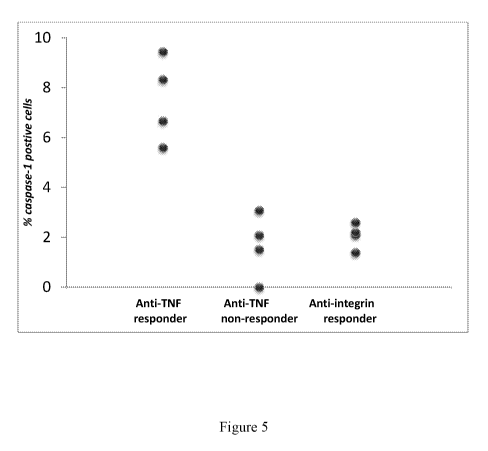
[0019] Figure 5 is a dot plot showing the percentage of activated caspase 1
positive intestinal
epithelial cells (IECs) cells taken from IBD patients who beneficially
responded to anti-TNF
therapy (left column, "Anti-TNF responder"), from IBD patients who did not
beneficially
respond to anti-TNF therapy (middle column, "Anti-TNF non-responder"), and
from IBD
patients who beneficially responded to anti-integrin therapy (right column,
"Anti-integrin
responder").
Detailed Description of Specific Embodiments
[0020] The invention stems, in part, form the discovery that assessment of the
intestinal
barrier function of a patient is predictive for determining whether that
patient is suffering
from or is disposed to suffer from a bowel disorder such as chronic
inflammatory bowel
disease or irritable bowel syndrome. Such a patient thus identified may
benefit from
treatment with an agent that treats inflammatory bowel diseases (or, less
commonly, irritable
bowel syndrome), such as an agent (e.g., a biologics) that blocks c1437
integrin (e.g.,
vedolizumab, a monoclonal antibody sold under the trademark Entyvio by Takeda
Pharmaceuticals, Cambridge, Massachusetts) or an agent (e.g., a biologics)
that blocks tumor
necrosis factor.
[0021] The published patents, patent applications, websites, company names,
and scientific
literature referred to herein establish the knowledge that is available to
those with skill in the
art and are hereby incorporated by reference in their entirety to the same
extent as if each
4
CA 03013072 2018-07-27
WO 2017/136511 PCT/US2017/016152
was specifically and individually indicated to be incorporated by reference.
Any conflict
between any reference cited herein and the specific teachings of this
specification shall be
resolved in favor of the latter.
[0022] Terms defined or used in the description and the claims shall have the
meanings
indicated, unless context otherwise requires. Technical and scientific terms
used herein have
the meaning commonly understood by one of skill in the art to which the
present invention
pertains, unless otherwise defined. Any conflict between an art-understood
definition of a
word or phrase and a definition of the word or phrase as specifically taught
in this
specification shall be resolved in favor of the latter. As used herein, the
following terms
have the meanings indicated. As used in this specification, the singular forms
"a," "an" and
"the" specifically also encompass the plural forms of the terms to which they
refer, unless the
content clearly dictates otherwise. The term "about" is used herein to mean
approximately,
in the region of, roughly, or around. When the term "about" is used in
conjunction with a
numerical range, it modifies that range by extending the boundaries above and
below the
numerical values set forth. In general, the term "about" is used herein to
modify a numerical
value above and below the stated value by a variance of 20%.
[0023] The intestinal epithelium is a single-cell layer that constitutes the
largest and most
important barrier against the external environment. Thus, this intestinal
epithelial layer shall
be referred to herein as an "intestinal barrier". The intestinal barrier acts
as a selectively
permeable barrier, permitting the absorption of nutrients, electrolytes, and
water while
maintaining an effective defense against intraluminal toxins, antigens, and
enteric flora. The
lining of the intestine which makes up the intestinal barrier undergoes
continuous
physiologic renewal: stem cells located at the base of the crypts mature and
migrate up the
villi. The mature epithelial cells are eventually shed at the tip of the
villi.
[0024] Studies published over the past two decades have convincingly shown
that intestinal
barrier disruption plays a crucial role in the pathogenesis of intestinal
inflammation and in
the severity of inflammatory bowel disease (IBD), such as Cohn's disease (CD)
and
ulcerative colitis (UC). Crohn's disease is a chronic relapsing inflammatory
bowel disorder
(IBD). Clinical relapse occurs in 30-60% of patients within one year of
medically induced
remission. Studies over the past two decades have convincingly demonstrated
that barrier
CA 03013072 2018-07-27
WO 2017/136511 PCT/US2017/016152
disruption plays a significant and important role in the pathogenesis of
intestinal
inflammation and in the severity of Crohn's disease.
[0025] Methods for detecting intestinal cell barrier dysfunction have been
described (see,
e.g., PCT Publication No. W02014/039699 and US patent publication no. US
2015/0202329, both incorporated by reference herein their entireties). Barrier
disruption not
only exposes the subepithelial immune system to resident microbes but also
induces the
secretion of TNF-a and other pro-inflammatory cytokines (Neish AS: Microbes in
gastrointestinal health and disease. Gastroenterology 2009, 136(1):65-80). The
cytokine
secretion in turn induces more shedding of epithelial cells and promotes
further inflammation
and barrier dysfunction (Watson AJ, Duckworth CA, Guan Y, Montrose MR:
Mechanisms
of epithelial cell shedding in the Mammalian intestine and maintenance of
barrier function.
Annals of the New York Academy of Sciences 2009, 1165:135-142).
[0026] Older assays for barrier function such as the lactulose/mannitol test
(May GR,
Sutherland LR, Meddings JB: Is small intestinal permeability really increased
in relatives of
patients with Crohn's disease? Gastroenterology 1993, 104(6):1627-1632) have
not been
useful clinically, because the size of the sugar molecules used in the test
(about 10-10 m) are
not reflective of those of the resident microbes (about 10-6 m).
[0027] More recently, the advent of confocal laser endomicroscopy (CLE) has
enabled the
real-time assessment of mucosal barrier function in vivo (Kiesslich R et al.,
"Identification of
epithelial gaps in human small and large intestine by confocal endomicroscopy.
Gastroenterology 2007, 133(6):1769-1778; Liu JJ, et al., "Epithelial cell
extrusion leads to
breaches in the intestinal epithelium", Inflammatory bowel diseases 2013,
19(5):912-921).
The density of epithelial gaps (also known as extrusion zones) in the
intestinal surface as
observed by CLE has been shown to be a surrogate marker for mucosal barrier
function (Liu
JJ, et al., "Mind the gaps: confocal endomicroscopy showed increased density
of small bowel
epithelial gaps in inflammatory bowel disease," Journal of clinical
gastroenterology 2011,
45(3):240-245). Gap density is defined as the total number of epithelial gaps
per a set
number of total cells (e.g., 1000 cells). The epithelial gaps or extrusion
zones may be
potential entry sites for luminal microbes into the host. Epithelial gap
density has also been
validated by conventional light microscopy as a measure of epithelial cell
extrusion (Liu JJ,
et al., "Epithelial gaps in a rodent model of inflammatory bowel disease: a
quantitative
6
CA 03013072 2018-07-27
WO 2017/136511 PCT/US2017/016152
validation study," Clinical and Translational Gastroenterology 2011, 2:e3).
The epithelial
gap density¨a validated measure of epithelial cell extrusion against
conventional light
microscopy (Liu JJ, et al., "Epithelial gaps in a rodent model of inflammatory
bowel disease:
a quantitative validation study". Clinical and Translational Gastroenterology
2011, 2:e3] ¨
is increased in nearly half of UC patients (Turcotte JF et al., "Breaks in the
wall: increased
gaps in the intestinal epithelium of irritable bowel syndrome patients
identified by confocal
laser endomicroscopy (with videos)", Gastrointestinal Endoscopy 2013,
77(4):624-630) and
is a linear predictor of moderate to severe flare within a one-year follow-up
period (Turcotte
JF, et al., "Increased epithelial gaps in the small intestine are predictive
of hospitalization and
surgery in patients with inflammatory bowel disease," Clinical and
Translational
Gastroenterology 2012, 3:e19).
[0028] Epithelial gaps appear to be potential sites for the entry of luminal
microbes into the
host (Liu JJ, et al.: Epithelial cell extrusion leads to breaches in the
intestinal epithelium.
Inflammatory Bowel Diseases 2013, 19(5):912-921). The severity of mucosal
barrier
dysfunction to luminal microbes as measured by gap density on CLE therefore,
is likely to be
predictive of disease relapse. Elevated epithelial gap densities are found in
60% of Crohn's
disease (CD) patients and in 45% of ulcerative colitis (UC) patients (Turcotte
JF, et al.,
"Breaks in the wall: increased gaps in the intestinal epithelium of irritable
bowel syndrome
patients identified by confocal laser endomicroscopy (with videos),"
Gastrointestinal
Endoscopy 2013, 77(4):624-630) and are reported to be a linear predictor of
moderate to
severe flare within a one-year follow-up period (Turcotte JF et al.,
"Increased epithelial gaps
in the small intestine are predictive of hospitalization and surgery in
patients with
inflammatory bowel disease," Clinical and Translational Gastroenterology 2012,
3:e19).
Moreover, gap densities determined by CLE correlated strongly with the levels
of activated
caspases expressed in mucosal biopsy samples as determined by quantitative
analysis of
immunohistochemical staining (unpublished). The correlation between gap
density on CLE
and mucosal biopsy analysis will enable the use of intestinal biopsy samples
for barrier
function analysis.
[0029] A recent study of molecular imaging using CLE in the intestine of
Crohn's patients
revealed that careful patient selection based on the status of their mucosal
TNF receptor
expression can increase the clinical response rate to anti-TNF antibody
therapy to over 90%
7
CA 03013072 2018-07-27
WO 2017/136511 PCT/US2017/016152
(Atreya R, et al., "In vivo imaging using fluorescent antibodies to tumor
necrosis factor
predicts therapeutic response in Crohn's disease," Nature Medicine 2014,
20(3):313-318).
This result highlights the role of mucosal TNF levels in determining the
response rate to
biologic agents. IBD patients with higher gap densities have been found to
display increased
mucosal pro-inflammatory cytokine levels in their mucosal biopsy specimens
(Liu JJ et al.,
"Epithelial cell extrusion leads to breaches in the intestinal epithelium,"
Inflammatory bowel
diseases 2013, 19(5):912-921).
[0030] The present invention stems, in part, from the discovery that IBD
patients with severe
barrier dysfunction resulting in enhanced mucosal TNF levels will have a
beneficial response
to an agent that treats bowel disorders. Such agents that treat IBD include,
without limitation,
anti-TNF agents and agents that inhibit interleukin-12 and interleukin-23. CD
patients with
enhanced TNF levels were found to display a greater than 90% response rate to
anti-TNF
therapy (Atreya et al., "In vivo imaging using fluorescent antibodies to tumor
necrosis factor
predicts therapeutic response in Crohn's disease." Nature Medicine 2014,
20(3):313-318).
[0031] Patients with severe barrier dysfunction will also have a beneficial
response (i.e., will
respond favorably) to an agent that inhibits interleukin-12 and interleukin-23
(IL-12 and IL-
23, respectively). IL-12 and IL-23 share a common p40 subunit. 1L12 is made up
of the IL-
12/2300 and IL-12p35 subunits, and 1L-23 comprises 1L-23p19 and IL-1212300.
Such an
agent includes, without limitation, ustekinurnab, which is sold under the
trademark Stelara
by Johnson & Johnson Corp., New Jersey, USA. An agent that inhibits LL-12 and
1L-23 will be
referred to herein as an "anti-IL12/23 agent)
[0032] Conversely, those patients with lesser mucosal barrier dysfunction,
and, in some
embodiments, without increased mucosal TNF activity, only had a 10% response
rate to anti-
TNF therapy. In other words, patients with a moderate barrier dysfunction did
not have a
beneficial response to anti-TNF therapy. Instead, these patients are more
likely to have a
beneficial response to anti-integrin therapy. In Crohn's patients with lower
(i.e., "moderate")
range of gap density (e.g., 3% or less), the response rate to anti-integrin
therapy was found to
be 100% at the two-year follow-up examination (unpublished).
[0033] Patients with moderate intestinal barrier dysfunction (e.g., without
increased mucosal
TNF activity) will also have a beneficial response to sphingosine-l-phosphate
receptor
agonists, such as fingolimod (tradename Gilenya , available from Novartis AG
Corp.,
8
CA 03013072 2018-07-27
WO 2017/136511 PCT/US2017/016152
Switzerland) or ozaniniod (developed by Receptos, Inc. and currently available
from
Celgene, Inc.), and/or an agent that inhibits a janus kinase family member,
such as tofacitinib
(tradenam es Xeljanze and Jakvinuse, available from Pfizer, Inc.).
[0034] As used herein, by the term "anti-TNF therapy" is meant the
administration, to a
patient (e.g., a human patient), of an agent that inhibits tumor necrosis
factor (referred to as
TNF or TNF alpha). Several such anti-TNF agents are commercially available and
have
been approved for use in human patients in the USA by the U.S. Food and Drug
Administration. Any anti-TNF agent, where a biological or a small molecule, is
contemplated in the invention. In some embodiments, anti-TNF agent may be
adalimumab
(trade name Humira , sold by Abbie, Chicago, Illinois, USA), infliximab (trade
name
Remicadet, sold by Janssen Biotech, Inc., Horsham, Pennsylvania, USA),
certolizumab
pegol (trade name Cimziat, sold by UCB S.A., Brussels, Belgium), golimumab
(trade name
Simponi , sold by Janssen Biotech, Inc., Horsham, Pennsylvania, USA), or
etanercept (trade
name Enbrelg, sold by Amgen and Pfizer). Yet additional non-limiting anti-TNF
agents are
those that inhibit production of TNF (e.g., TNF-alpha) by cells by, for
example, inhibiting
enzymes (e.g., protein kinases) in the cells to inhibit their production of
TNF. One such anti-
TNF agent that acts to prevent TNF-alpha production is apremilast (trade name
Otezla, sold
by Celgene Corp, New Jersey, USA).
[0035] As used herein, by the term "anti-integrin therapy" is meant the
administration, to a
patient, of an agent that inhibits an integrin from forming an adhesion with
its natural target.
Thus, an anti-integrin agent, when bound to the integrin, partially or
completely prevents the
integrin from binding its target. Integrins are family of transmembrane
receptors that appear
on a variety of cells. They are heterodimers comprised of two chains¨an alpha
chain and a
beta chain. In mammals, there are eighteen alpha chains and eight beta chains,
so a
particular integrin may be referred to by which alpha chain and which beta
chain it has.
Some non-limiting examples of integrins are the a1f31 integrin (also called
VLA-1), the a4f37
integrin (also called LPAM-1), and the ad32 integrin (also called LFA-1). An
anti-integrin
agent is an agent that inhibits (i.e., blocks) any integrin family member
(i.e., inhibits one or
more integrin family member).
[0036] In some embodiments, the anti-integrin agent is vedolizumab which
targets LPAM-1
(the trade name of vedolizumab is Entyviog, and vedolizumab is sold by
Millennium
9
CA 03013072 2018-07-27
WO 2017/136511 PCT/US2017/016152
Pharmaceuticals (Cambridge, Massachusetts, USA), a subsidiary of Takeda
Pharmaceuticals,
Japan). In some embodiments, the anti-integrin agent is natalizumab which
targets alpha 4
chain integrin(s). The trade name of natalizumab is Tysabri , and natalizumab
is sold by
BioGen Idec (Cambridge, Massachusetts, USA) and Elan (Dublin, Ireland). In
some
embodiments, the anti-integrin agent is etrolizumab (available from Genentech,
South San
Francisco, California, USA) which targets the (37 chain integrin(s) (e.g.,
integrins a4(37 and
aE(37). Additional anti-integrin agents are described in Kawamoto et al.,
Autimmune
Diseases, vol. 2012, Article ID 357101, herein incorporated by reference.
[0037] It shall be understood that the amount of any agent (e.g., anti-TNF
agent or anti-
integrin) that administered to a patient to "treat" that patient will be
administered in a
therapeutically effective amount, as determined by ordinarily skilled
physicians,
pharmacologists, and toxicologists, that make take into account the weight and
age of the
patient. In any event, where the drug has been approved by a regulatory
authority (e.g., the
U.S. Food and Drug Administration), a therapeutically effective amount of anti-
TNF agent is
an amount approved by the regulatory authority.
[0038] Of course, the route of administration can be by any route and will be
determined
based on the agent and the patient. For example, a small molecule such as
apremilast may be
administered orally, while a biological such as etanercept may be administered
by
subcutaneous injection. All other routes of administration of a
therapeutically effective
amount of an agent to treat an IBD patient are contemplated herein and
include, without
limitation, parenteral (e.g., intravenous, intrathecal, subcutaneous) or
enteral (e.g., orally or
rectally) or other routes (e.g., intranasal, intradermal, intravitreal,
subcutaneous, transdermal,
topical, intraperitoneal, intravaginal, and intramuscular).
[0039] The invention is based, in part, on the discovery that the mucosal
intestinal barrier
status while inflammatory bowel disease (IBD) patients are on IBD therapy is
predictive of
clinical and endoscopic remission over time in response to that therapy (e.g.,
treatment with
an anti-TNF agent). Thus, the invention is based, in part, on the discover
that determining
the mucosal intestinal barrier function status in patients with inflammatory
bowel disease
(IBD) is an important tool for predicting therapeutic response to a
therapeutic agent, such as
an anti-TNF agent, an anti-integrin agent, or an anti-IL-12 and IL-23 agent
(e.g.,
ustekinumab ) . First of all, IBD patients with higher gap densities have
higher mucosal pro-
w
CA 03013072 2018-07-27
WO 2017/136511 PCT/US2017/016152
inflammatory cytokine levels (see Liu JJ, et al: "Epithelial cell extrusion
leads to breaches in
the intestinal epithelium." Inflammatory bowel diseases 2013, 19(5):912-921).
Second, the
highest rates of response to biologic therapy for Crohn's disease are seen in
post-operative
patients, with over 90% endoscopic remission rate at one year (see Regueiro M,
Schraut W,
Baidoo L, Kip KE, Sepulveda AR, Pesci M, Harrison J, Plevy SE: Infliximab
prevents
Crohn's disease recurrence after ileal resection. Gastroenterology 2009,
136(2):441-450
e441; quiz 716). Third, prominent barrier dysfunction was observed at the
anastomotic site
in animal models of ileal resection (unpublished).
[0040] It has been discovered that normalization of mucosal intestinal barrier
function for
IBD patients (Crohn's disease, ulcerative colitis, indeterminate colitis, or
chemotherapy-
induced colitis) on IBD therapy to healthy control (e.g., from healthy
volunteers) levels is
predictive of clinical and endoscopic remission for a significant period of
time (e.g., one
year). Correspondingly, abnormal mucosal intestinal barrier function on
biologic therapy is
predictive of lack of clinical response and disease relapse.
[0041] Therefore, barrier dysfunction is a potent predictor of therapeutic
response to an IBD
therapy, such as administration of an anti-TNF agent or an anti-integrin agent
in IBD
patients.
[0042] Accordingly, in a first aspect, the invention provides a method for
identifying an
agent beneficial to treat a patient with inflammatory bowel disease comprising
(a) analyzing
(or determining) a status of an intestinal barrier in the patient to obtain a
patient status; and
(b) categorizing the patient status as severe dysfunction or moderate
dysfunction, wherein a
patient with a patient barrier status categorized as being severe dysfunction
is identified as a
patient who will benefit from treatment with an agent (e.g., a biologic)
selected from the
group consisting of an anti-TNF agent, an anti-IL-12/23 agent, and a
combination thereof,
and a patient with a patient barrier status categorized as being moderate
dysfunction is
identified as a patient who will benefit from treatment with an agent (e.g., a
biologic)
selected from the group consisting of an anti-integrin agent, an anti-janus
kinase agent a
sphingosine- I-phosphate receptor agonist agent, a combination of two or more
of an anti-
integrin agent, an anti-janus kinase agent a sphingosine- 1 -phosphate
receptor agonist agent.
[0043] In another aspect, the invention provides a method of identifying a
status of an
intestinal barrier in a patient with inflammatory bowel disease, wherein the
status is severe
11
CA 03013072 2018-07-27
WO 2017/136511 PCT/US2017/016152
dysfunction or moderate dysfunction, comprising analyzing (or determining) the
status of the
intestinal barrier of the patient, wherein if said status is identified as
being severe
dysfunction, the method further comprises treating said patient with an agent
selected from
the group consisting of an anti-TNF agent, an anti-IL-12/23 agent, and a
combination
thereof, and wherein if said status is identified as having moderate
dysfunction, the method
further comprises treating said patient with an agent selected from the group
consisting of an
anti-integrin agent, an anti-janus kinase agent a sphingosine- 1-phosphate
receptor agonist
agent, a combination of two or more of an anti-integrin agent, an anti-janus
kinase agent a
sphingosine- I-phosphate receptor against agent.
[0044] In another aspect, the invention provides a method of treating a
patient with
inflammatory bowel disease, comprising (a) analyzing (or determining) the
status of an
intestinal barrier to determine if the status is severe dysfunction or
moderate dysfunction; and
(b) treating said patient with an agent, wherein: (i) if said patient is
identified as having
severe dysfunction, the agent is selected from the group consisting of an anti-
TNF agent, an
anti-IL-12/23 agent, and a combination thereof, and (ii) if said patient is
identified as having
moderate dysfunction, the agent is selected from the group consisting of an
anti-integrin
agent, an anti-janus kinase agent a sphingosine-l-phosphate receptor agonist
agent, a
combination of two or more of an anti-integrin agent, an anti-janus kinase
agent a
sphingosine- I -phosphate receptor agonist agent.
[0045] The invention stems, in part, from the discovery that the status of the
intestinal barrier
of an inflammatory bowel disease patient can reveal which agent would be most
beneficial in
treating the patient. As used herein, by "beneficial" is meant that the IBD
symptoms of the
patient are alleviated when the patient is treated (e.g., by oral
administration) of a
therapeutically effective amount of an anti-TNF or an anti-integrin agent. The
patient thus
treated is referred to as a patient who has a beneficial response to the
treatment.
[0046] Symptoms of IBD are well known and include, without limitation,
diarrhea, fever
(e.g., low-grade fever), abdominal pain and cramping, blood in the stool
(hematochezia),
bleeding ulcers, bloating, bowel obstruction, unintended weight loss, and
anemia. Crohn's
disease, ulcerates colitis, indeterminate colitis, and chemotherapy-induced
colitis are all
forms of inflammatory bowel disease. Note that chemotherapy-induced colitis,
unlike other
forms of IBD, is not predictable, as it occurs in a minority (less than 30%)
of patients who
12
CA 03013072 2018-07-27
WO 2017/136511 PCT/US2017/016152
have been treated with a chemotherapeutic drug such as a checkpoint inhibitor
drug.
[0047] In some embodiments, the status of the intestinal barrier is determined
by measuring
or calculating the amount of activated caspase expressed in intestinal
epithelial cells at the
intestinal surface of the intestinal barrier. For example, the amount of
activated caspase can
be determined by staining a sample (e.g., a biopsy sample) from the patient
with a detectably
labeled antibody that specifically binds to an activated caspase molecule
(e.g., activated
caspase 1 or activated caspase 3). The amount of activated caspase can also be
determined
by staining a sample from the patient with a detectably labeled peptide that
binds to activated
caspase. It should be noted that by being detectably labeled, the peptide or
antibody can be
directly labeled (e.g., with a fluorescent label or chromatogenic tag) or can
be detected by
being bound during secondary staining with an detectably labeled secondary
antibody (e.g.,
the anti-caspase antibody is a murine monoclonal antibody and the secondary
antibody is a
fluorescently labeled rabbit anti-mouse antibody).
[0048] In some embodiments, the activated caspase is activated caspase 1. In
some
embodiments, the activated caspase is activated caspase 3. In some
embodiments, the
activated caspase is a combination of activated caspase 1 and activated
caspase 3. In some
embodiments, the activated caspase is a ratio of an amount of expression of
activated caspase
to an amount of expression of activated caspase 3.
[0049] Typically, intestinal epithelial cells at the intestinal barrier of
people who do not have
intestinal diseases (e.g., do not have IBD or IBS symptoms) express low levels
of activated
caspase (e.g., express low levels of activated caspase 1 or activated caspase
3). Such people
who do not have IBD may be referred to as a healthy volunteer. Accordingly, in
some
embodiments, an amount of activated caspase expression in that patient that is
about four
fold to about seven fold higher than the amount of activated caspase
expression in intestinal
epithelial cells of an intestinal barrier of one or more healthy volunteers
indicates that the
patient status is severe dysfunction. In some embodiments, an amount of
activated caspase
expression in that patient that is about 4 fold to about 7 fold higher than
the amount of
activated caspase expression in intestinal epithelial cells of an intestinal
barrier of one or
more healthy volunteers indicates that the patient status is severe
dysfunction. Of course, a
patient expressing activated caspase that is more than about 7 fold higher
than the amount of
activated caspase expression in intestinal epithelial cells of an intestinal
barrier of one or
13
CA 03013072 2018-07-27
WO 2017/136511 PCT/US2017/016152
more healthy volunteers indicates that the patient status is severe
dysfunction In some
embodiments, an amount of activated caspase expression in that patient that is
about 5 fold to
about 6.5 fold higher than the amount of activated caspase expression in
intestinal epithelial
cells of an intestinal barrier of one or more healthy volunteers indicates
that the patient status
is severe dysfunction.
[0050] In some embodiments, an amount of activated caspase expression in the
patient that is
between about 1.5 fold to about 4.5 fold higher than the amount of activated
caspase
expression in intestinal epithelial cells of an intestinal barrier of one or
more healthy
volunteers indicates that the patient status is moderate dysfunction and is
not severe
dysfunction. In some embodiments, an amount of activated caspase expression in
the patient
that is between about 2 fold to about 4.5 fold higher than the amount of
activated caspase
expression in intestinal epithelial cells of an intestinal barrier of one or
more healthy
volunteers indicates that the patient status is moderate dysfunction and is
not severe
dysfunction. In some embodiments, an amount of activated caspase expression in
the patient
that is between about 2 fold to about 4 fold higher than the amount of
activated caspase
expression in intestinal epithelial cells of an intestinal barrier of one or
more healthy
volunteers indicates that the patient status is moderate dysfunction and is
not severe
dysfunction.
[0051] The expression level amount of activated caspase in a healthy volunteer
can be
pooled and averaged with other healthy volunteers. For example, if you have
two healthy
volunteers, and one has no activated caspase 1 expression and the other has
1.0% activated
caspase 1 expression, the average is 0.5% activated caspase 1 expression in
the intestinal
epithelial cells of the intestinal barriers of healthy volunteers.
[0052] In some embodiments, the amount of activated caspase expression in
intestinal
epithelial cells of an intestinal barrier of a healthy volunteer is 0.5%.
Thus, if a patient has
1.5% activated caspase 1 expression (i.e., has 1.5 out of 100 intestinal
epithelial cells
expressing activated caspase 1), that patient will be categorized as having
moderate
dysfunction of the status of his intestinal barrier. Conversely, if a patient
has 5.0% activated
caspase 1 expression (i.e., has 5 out of 100 intestinal epithelial cells
expressing activated
caspase 1), that patient will be categorized as having severe dysfunction of
the status of his
intestinal barrier.
14
CA 03013072 2018-07-27
WO 2017/136511 PCT/US2017/016152
[0053] Note that the amount of activated caspase expressed by a healthy
volunteer will
depend upon several factors including the reagent used to detect the activated
caspase (e.g.,
the peptide inhibitor, Ac-YVAD (tyr-val-ala-asp)-CMK, from Enzo described
below that
inhibits activated caspase 1 or an antibody that specifically binds to
activated caspase 1 such
as the antibody from Cell Signaling Technology, Inc. described below).
[0054] In some embodiments, the amount of activated caspase expression in
intestinal
epithelial cells of an intestinal barrier of a healthy volunteer is 0.5%.
Thus, if a patient has
3% activated caspase 1 expression (i.e., has 1.5 out of 100 intestinal
epithelial cells
expressing activated caspase 1), that patient will be categorized as having
moderate
dysfunction of the status of his intestinal barrier because the patient has a
3 fold higher
expression of activated caspase 1 than the healthy volunteer. Correspondingly,
if a patient
has 6% activated caspase 1 expression (i.e., has 6 out of 100 intestinal
epithelial cells
expressing activated caspase 1), that patient will be categorized as having
severe dysfunction
of the status of his intestinal barrier because the patient has a 6 fold
higher expression of
activated caspase 1 than the healthy volunteer.
[0055] In some embodiments, the amount of activated caspase expression in
intestinal
epithelial cells of an intestinal barrier of a healthy volunteer is
approximately 0.5%. Thus, if
a patient has 1.5% activated caspase 1 expression (i.e., has 1.5 out of 100
intestinal epithelial
cells expressing activated caspase 1), that patient will be categorized as
having moderate
dysfunction of the status of his intestinal barrier because the patient has a
3 fold higher
expression of activated caspase 1 than the healthy volunteer. Correspondingly,
if a patient
has 3.0% activated caspase 1 expression (i.e., has 3 out of 100 intestinal
epithelial cells
expressing activated caspase 1), that patient will be categorized as having
severe dysfunction
of the status of his intestinal barrier because the patient has a 6 fold
higher expression of
activated caspase 1 than the healthy volunteer (i.e., 3% is 6 fold higher than
0.5%).
[0056] Where there is no number or percentage value available for "the amount
of activated
caspase expression in intestinal epithelial cells of an intestinal barrier of
one or more healthy
volunteers", that amount shall understood to be in the range of about 0.5 to
1.0 cells out of
100, or 0.5% to 1.0% expression.
[0057] In some embodiments, the status of the intestinal barrier is determined
by counting a
number of gaps in routine histological staining of the intestinal lining. For
example, the
CA 03013072 2018-07-27
WO 2017/136511 PCT/US2017/016152
residual spaces left in between cells in the intestinal surface after
extrusion of epithelial cells,
also called extrusion zones, can be counted on well preserved intestinal
specimens and
normalized to the total number of epithelial cells to reflect the barrier
status. The samples
can be stained using conventional histologic staining techniques, including
but not limited to
hernatoxylin and eosin siam. alcian blue and nuclear fist red.
[0058] In some embodiments, the status of the intestinal barrier is determined
by measuring
gap density (i.e., number of gaps) using confocal endomicroscopy of the
intestinal surface.
For example, the intestinal samples can be stained with a nuclear (such as
DAPI) stain and
cytoskeletal (e.g., actin) stain and imaged using multi-photon confocal
microscopy ex-vivo.
[0059] Ordinarily, a healthy volunteer will have very little intestinal
barrier gaps, and so that
number of gaps in a healthy volunteer is ordinarily under 0.5 gaps per 100
intestinal
epithelial cells.
[0060] However, where there is no number or percentage value available for the
number of
gaps or the gap density of one or more healthy volunteers, that amount shall
understood to be
approximately 0.5 gaps per 100 intestinal cells, or approximately 0.5%.
[0061] In some embodiments, the mucosal (or intestinal) barrier status or the
degree of
intestinal barrier dysfunction can be characterized by a combination stain for
activated
caspase-1 and/or activated caspase-3 of intestinal epithelial cells, and anti-
CD3 of
intraepithelial lymphocytes. The total number of intestinal epithelial cells
can be quantitated
using nuclear stains (e.g., DAPI). The staining methods are detailed in
Protocol A "Staining
protocol for paraffin-embedded mucosal biopsy samples" below.
[0062] The degree of intestinal barrier dysfunction can be derived by either
the total number
of activated caspase-1 positive cells normalized to the total number of
intestinal epithelial
cells (e.g., as determined by nuclear stain); or a relative ratio of activated
caspase-1 positive
to activated caspase-3 positive cells, or a combination of activated caspase-1
positive and
activated caspase-3 positive cells normalized to the total number of
intestinal epithelial cells.
[0063] The intestinal barrier status or the degree of barrier dysfunction can
also be
characterized by a combination stain for TUNEL stain which will stain positive
for both
activated caspase-1 and activated caspase -3 epithelial cells, minus the
activated caspase-3
positively stained cells; with or without anti-CD3 stain for intraepithelial
lymphocytes. The
total number of intestinal epithelial cells can be quantitated using nuclear
stains, e.g. DAPI.
16
CA 03013072 2018-07-27
WO 2017/136511 PCT/US2017/016152
The staining methods are detailed in protocol B "TUNEL staining protocol for
paraffin-
embedded mucosal biopsy samples using commercially-available staining kits",
below.
[0064] In some embodiments, the intestinal (i.e., mucosal) barrier dysfunction
can
alternatively be characterized by staining for active interleukin 1-beta (IL-
113) and/or IL-18,
both of which are surrogate markers of activated caspase-1. Antibodies that
specifically bind
to active (i.e., mature) interleukin 1-beta (IL-113) and antibodies that
specifically bind to IL-
18 are known (see, e.g., Cleaved-IL-113 (Asp116) (D3A3Z) Rabbit mAb #83186,
Cell
Signaling Technology, Inc., Danvers, Massachusetts, USA, and Anti-IL18
antibody
(ab71495), Abcam, Cambridge, Massachusetts, USA).
[0065] In vivo, the intestinal surface may be stained with intravenous dye
(e.g., fluorescein)
with or without a nuclear stain (e.g., acriflavine), and imaged using confocal
laser
endomicroscope. Gap density on confocal laser endomicroscopy is a validated
measure of
extrusion zones.
[0066] The status of the intestinal barrier is significantly compromised in
inflammatory
bowel disease (IBD) patients as compared to the status of an intestinal
barrier from a healthy
volunteer (e.g., a person, aged 18 to 70) who does not have gastrointestinal
symptoms.
[0067] It will be understood that each therapeutic agent (or, for example,
each target of a
group of therapeutic agents) may present itself with its own specific profile
for caspase 1
and/or caspase 3 staining to show optimal therapeutic efficacy. For example,
for an anti-
TNF agent (depending upon which agent), one non-limiting profile for positive
caspase-1
stain is estimated to be above between about 4 to 7 positive activated caspase-
1 cells per 100
intestinal epithelial cells on the biopsy samples, or between about 4% to
about 7%,
depending on the specific anti-TNF, as well as the disease condition (e.g.,
Crohn's disease,
ulcerative colitis, or indeterminate colitis).
[0068] In some embodiments, the anti-TNF agent is selected from the group
consisting of
adalimumab, infliximab, certolizumab, pegol, golimumab, and etanercept. As
described
below, the therapeutic response rate to an anti-TNF agent in patients with
inflammatory
bowel disease (e.g., Crohn's patients or ulcerative colitis) is expected to be
above 70, when
the criteria above for severe dysfunction is met (e.g., positively stained
cells or gap density of
over between about 4% to 7% for Crohn's disease or ulcerative colitis).
[0069] In some embodiments, the anti-integrin agent is selected from the group
consisting of
17
CA 03013072 2018-07-27
WO 2017/136511 PCT/US2017/016152
vedolizumab, natalizumab, and etrolizumab. As described below, the response
rate to these
anti-integrin agents in IBD patients (e.g., Crohn's or ulcerative colitis
patients) is expected to
be above about 70 to 80% when the criteria for moderate barrier dysfunction is
met, (e.g.,
positively stained cells or gap density of less than about 4%).
[0070] The following examples are not meant to limit the invention in any way.
[0071] Staining Protocols A and B
[0072] In general, staining (i.e., contacting a sample with a binding agent,
where the binding
can be detected) can be performed as follows.
[0073] Protocol A: Staining protocol for paraffin-embedded mucosal biopsy
samples
[0074] Step I. Deparaffinization
[0075] Place the slides in a rack, and perform the following sequential washes
in Coplin jars
or other container:
[0076] Wash 1. Xylene: 2 x 5 minutes
[0077] Wash 2. 100% ethanol: 2 x 5 minutes
[0078] Wash 4. 95% ethanol: 3 minutes
[0079] Wash 5. 70 % ethanol: 3 minutes
[0080] Wash 6. 50 % ethanol: 3 minutes
[0081] Wash 7. Distilled H20: 2 x 3 minutes
[0082] Keep the slides in the distilled water until ready to perform antigen
retrieval. In some
embodiments, do not allow the slides to dry from this point onwards, as drying
out may
cause non-specific antibody binding and therefore high background staining on
the tissue.
[0083] Step II. Antigen retrieval
[0084] 1. Pre-heat a water bath and antigen retrieval solution (10 mM sodium
citrate buffer)
to 95 C.
[0085] The 10 mM Sodium Citrate Buffer is 10mM sodium citrate, 0.05% Tween 20,
pH
6.0, and is made as follows:
Tr-sodium citrate (dihydrate) 2.94 g
Distilled water 1000 ml
Mix to dissolve. Adjust pH to 6.0 with 1N HC1.
Add 0.5 ml Tween 20, mix well, and store at 4 C
18
CA 03013072 2018-07-27
WO 2017/136511 PCT/US2017/016152
[0086] 2. Place slides in pre-heated antigen retrieval solution (enough to
cover the slides by
about 1 to about 8 centimeters). As glass containers may crack in the heat, in
some
embodiments, glass containers are not used. In some embodiments, a plastic
tupperware
container with a lid to prevent evaporation may be used. In some embodiment,
an empty box
with a lid (e.g., a box that used to hold pipet tips for a micropipetter) may
be used. In
embodiments, a weight is added on the cover of the container to prevent the
container from
floating around.
[0087] 3. Incubate the slides for 20 minutes at 95 C.
[0088] 4. When 20 minutes have elapsed, remove the container and slides from
the water
bath. Allow the slides to cool at room temperate, still immersed in the
antigen retrieval
solution, before removing them from the container.
[0089] 5. Continue to the immunohistochemical staining protocol (i.e., Step
III).
[0090] Step III. Immunostaining
[0091] All incubations should be carried out in a humidified chamber to avoid
drying of the
tissue.
[0092] A shallow, plastic box with a sealed lid and wet tissue paper in the
bottom works
well. In some embodiments, the slides do not directly contact the paper. In
some
embodiments, the slides can lay flat. In some embodiments, the slides are
positions so that
the reagents do not drain off
[0093] 1. Wash slides in 1X PBS (phosphate buffered saline) with 0.025% Triton
X-100 for
minutes with gentle agitation. Repeat with a second wash for a total of 2
washes.
[0094] 2. Remove slides from wash buffer and dry excess liquid from slides
using a
Kimwipe or other delicate task wipe with low lint and low electrostatic
discharge. Use a
PAP pen or other similar pen to draw a circle around the tissue to create a
hydrophobic
barrier around the sample.
[0095] 3. Pipet approximately 100uL of blocking solution onto the tissue,
ensuring that the
tissue section is completely covered, and incubate the tissues at room
temperate for 2 hours.
The Blocking solution contains: 1X PBS with 10% normal goat serum and 1% BSA
(bovine
serum albumin)
[0096] 4. Use a Kimwipe to blot out any excess blocking solution from the
tissue and add
approximately 100uL of primary antibody solution to each slide. Incubate
overnight at 4 C.
19
CA 03013072 2018-07-27
WO 2017/136511 PCT/US2017/016152
The Primary antibody solution contains 1X PBS with 1% BSA with Caspase-1 p20
antibody
at 1:250 dilution (using, for example, the Cleaved caspase-1 (Asp297)(D57A2)
rabbit mAb
available from Cell Signaling Technologies (Danvers, Massachusetts), cat#
4199) and with
CD3e antibody at 1:100 dilution (using, for example, the CD3e/CD3 epsilon
antibody (SPV-
T3b) human, raised in mouse; Invitrogen (Carlsbad, California) cat# 07-0303)
[0097] Note that staining of activated caspase 1 can also be accomplished by
immunoblotting with a peptide inhibitor, such as the Ac-YVAD (tyr-val-ala-asp)-
CMK
inhibitor commercially available from Enzo Life Sciences, Farmingdale, New
York, and
described in PCT Publication No. W02014/039699 and US patent publication no.
US
2015/0202329, both incorporated by reference herein their entireties). The
peptide Ac-
YVAD-CMK (Ac-Tyr-Val-Ala-Asp-chloromethylketone) is a cell permeable,
irreversible
inhibitor of cap spase -1.
[0098] 5. Drain off excess primary antibody solution and wash the slides in 1X
PBS for 5
minutes with gentle agitation. Repeat twice for a total of 3 washes.
[0099] 6. Pipet approximately 100uL of secondary antibody solution onto the
tissues and
incubate at room temperature (e.g., 25 C) for 1 hour in the dark. The
Secondary antibody
solution contains 1X PBS with 1% BSA with Goat anti-rabbit AlexaFluor 488 at
1:3000
dilution (using, for example, the Goat anti-rabbit (H+L) Superclonal secondary
antibody,
AlexaFluor conjugate 488; Invitrogen, cat# PIA27034) and with Goat anti-mouse
AlexaFluor
555 at 1:3000 dilution (using, for example, the Goat anti-mouse IgG (H+L),
AlexaFluor
conjugate 555; Invitrogen, cat# A21424
[00100] 7. Blot off excess antibody solution and wash the slides in 1X PBS
for 5
minutes, with gentle agitation. Repeat once for a total of two washes. Perform
washes in the
dark.
[00101] 8. Incubate slides in 1X PBS containing 0.3 ug/mL (0.654 nM)
DAPI(4',6-
Diamidino-2-Phenylindole, Dilactate), commercially available for example, from
Molecular
Probes, cat# D3571 for 10 minutes with gentle agitation in the dark. For
example, 12uL of
5mg/mL DAPI stock into 200mL PBS can be used for a final wash + stain in one
step.
[00102] 9. Drain off excess liquid, wipe around the sections with tissue
paper, and
mount coverslips onto the tissue. This can be done using a mounting agent such
as, for
example, ProLong Diamond Antifade Mountant (Molecular Probes (Eugene, Oregon),
cat#
CA 03013072 2018-07-27
WO 2017/136511 PCT/US2017/016152
P36970). Allow the slides to cure (i.e., the tissue with the mounting agent
sets and hardens
with time) overnight at room temperature in the dark before imaging.
[00103] Slides are imaged using either multi-photon microscopy or
fluorescent
microscopy, using wavelengths corresponding to the fluorochrome used. Note
that confocal
laser endomicroscopy can be performed on patient samples during the time of
endoscopy,
while multi-photon confocal microscopy or fluorescent microscopy can be done
on the slides
stained with immunohistochemical (IHC) stains (e.g., using labeled monoclonal
antibodies
specific for activated caspase 1).
[00104] For the antibodies used in the above protocol A, emission spectra
for the dyes
are as follows: DAPI was imaged at 455nm, anti-CD3 was imaged at 555nm, and
anti-
Caspase 1 was imaged at 488nm. Figure 2 shows a representative image of
intestinal
epithelial cells stained (i.e., immunostained) for activated caspase 1. The T
cells present in
the slide are identified by co-staining with an antibody that specifically
binds to CD3, a cell
surface molecule associated with the T cell receptor in T cells. In Figure 2,
the white arrow
points to a green-stained intestinal epithelial cell that stained positive for
expression of
caspase 1, and the white arrow head (i.e., triangle) points to a red-stained T
cell (an intra-
epithelial lymphocyte, or TEL) that stained positive for expression of both
CD3 and caspase
1.
[00105] Protocol B: TUNEL staining protocol for paraffin-embedded mucosal
biopsy samples using commercially-available staining kits.
[00106] TUNEL (Terminal deoxynucleotidyl transferase dUTP nick end
labeling)
staining is a method for detecting DNA fragmentation by labeling the terminal
ends of
nucleic acids. Since apoptosis causes fragmentation of DNA, the TUNEL assay is
a common
method for DNA fragmentation that results from apoptotic signaling cascades.
The assay
relies on the presence of nicks in the DNA which can be identified by terminal
deoxynucleotidyl transferase or TdT, an enzyme that will catalyze the addition
of dUTPs that
are secondarily labeled with a marker.
[00107] Step I. Deparaffinization
[00108] Place the slides in a rack, and perform the following sequential
washes in
Coplin jars or other container:
21
CA 03013072 2018-07-27
WO 2017/136511 PCT/US2017/016152
[00109] 1. Xylene: 2 x 5 minutes
[00110] 2. 100% ethanol: 2 x 5 minutes
[00111] 4. 95% ethanol: 3 minutes
[00112] 5. 70 % ethanol: 3 minutes
[00113] 6. 50 % ethanol: 3 minutes
[00114] 7. Distilled H20: 2 x 3 minutes
[00115] Keep the slides in the distilled water until ready to perform
antigen retrieval.
Do not allow the slides to dry from this point onwards. Drying out may cause
non-specific
antibody binding and therefore high background staining on the tissue.
[00116] Step II. Antigen retrieval
[00117] 1. Pre-heat a water bath and antigen retrieval solution (10 mM
sodium citrate
buffer) to 95 C. Sodium Citrate Buffer (10mM sodium citrate, 0.05% Tween 20,
pH 6.0) is
made as follows:
Tr-sodium citrate (dihydrate) 2.94 g
Distilled water 1000 ml
Mix to dissolve. Adjust pH to 6.0 with 1N HC1.
Add 0.5 ml Tween 20, mix well, and store at 4 C
[00118] 2. Place slides in pre-heated antigen retrieval solution (enough
to cover the
slides by several a few centimeters). Avoid using glass containers as these
may crack in the
heat. A plastic Tupperware with a lid to prevent evaporation works well or
empty tip boxes
with lids also work. Add a weight on the cover to prevent the container from
floating
around.
[00119] 3. Incubate the slides for 20 minutes at 95C.
[00120] 4. When 20 minutes have elapsed, remove the container and slides
from the
water bath. Allow the slides to cool at room temperate, still immersed in the
antigen retrieval
solution, before removing them from the container.
[00121] Step II. Nuclear Staining
[00122] At this point, follow the protocol provided by the commercially-
available kit.
Below is an abbreviated and adapted protocol for the Trevigen TACS 2 TdT-
Fluor In Situ
Apoptosis Detection Kit, commercially available from Trevigen (Gaithersburg,
Maryland)
Cat #: 4812-30-K.
22
CA 03013072 2018-07-27
WO 2017/136511 PCT/US2017/016152
[00123] 1. Immerse sample in 1X PBS for 10 minutes with gentle agitation.
[00124] 2. Cover sample with 50 11.1 of Proteinase K Solution for 15
minutes. The
Proteinase K Solution (per sample) contains as follows: 50 11.1 Apoptosis
GradeTM Water and
1 11.1 Proteinase K
[00125] 3. Wash the samples in deionized water for 2 minutes. Repeat with
a second
wash.
[00126] 4. Immerse the samples in 1X TdT Labeling Buffer for 5 minutes.
The TdT
Labeling Buffer contains 45 ml Deionized Water and 5 ml 10X TdT Labeling
Buffer (from
the Trevigen kit).
[00127] 5. Cover sample with 50 11.1 of Labeling Reaction Mix (from the
Trevigen kit)
and incubate for 60 minutes at 37 C in a humidity chamber. The Labeling
Reaction Mix per
sample contains 1 11.1 TdT dNTP, 111.150X Cation (Mg2+, Mn2+, or Co2+ ), 111.1
TdT
Enzyme (Avoid repeated freeze-thaw), and 50 11.1 1X TdT Labeling Buffer (from
the
Trevigen kit)
[00128] 6. Immerse the samples in lx TdT Stop Buffer for 5 minutes. The
TdT Stop
Buffer contains 45 ml Deionized Water and 5 ml 10X TdT Stop Buffer (from the
Trevigen
kit)
[00129] 7. Wash the samples twice in 1X PBS, for 2 minutes each.
[00130] 8. Cover sample with 50 11.1 of Strep-Fluor Solution and incubate
for 20
minutes in the dark. The Strep-Fluor Solution contains 200 11.1 1X PBST (1X
PBS with
0.05% Tween 20) and 1 11.1 Strep-Fluorescein.
[00131] 9. Wash the samples three times in 1X PBS, 2 minutes each.
[00132] 10. Mount glass coverslip using 90% glycerol and view under
fluorescence
microscope using a 495 nm filter.
[00133] Figures 3A and 3B show representative images of intestinal
epithelial cells
stained (i.e., immunostained) for TUNEL (e.g., using Protocol B above) (Fig.
3A) and
activated caspase 3 (e.g., using Protocol A above) (Fig. 3B). In Figure 3A,
the arrows points
to intestinal epithelial cells staining positive for nuclear fragmentation
using a commercial kit
staining for TUNEL-positive cells. In Figure 3B, the arrows point to
intestinal epithelial
cells staining positive for expression of caspase 3.
23
CA 03013072 2018-07-27
WO 2017/136511 PCT/US2017/016152
[00134] Example I
[00135] The major aim of this Example I is to improve patient selection
for
vedolizumab for the treatment of Crohn's disease and ulcerative colitis by
assessing the
functional status of the intestinal barrier prior to therapy. Biologic
therapies such as anti-
tumor necrosis factor (TNF), anti-IL12/23 agents, and anti-integrin agents
have had a clinical
response rate of about 50% for the past decade. Vedolizumab, a monoclonal
antibody that
specifically binds to integrin ct437 and sold under the name ENTYVIO by
Takeda
Pharmaceuticals) was approved for clinical use in 2014 and has been widely
used in patients
who have failed to respond to other anti-TNF agents. However, its clinical
response rate,
particularly for Crohn's disease, was low compare to anti-TNF agents. This
study has been
designed to improve the therapeutic response to vedolizumab through better
patient selection.
[00136] Studies over the past two decades have convincingly demonstrated
that barrier
disruption plays a significant and important role in the pathogenesis of
mucosal inflammation
in inflammatory bowel disease (IBD). Barrier disruption exposes the sub-
epithelial immune
system to resident microbes and induces the secretion of TNF-a and other pro-
inflammatory
cytokines, which in turn induces shedding of epithelial cells from the
intestine and promotes
further inflammation and increased barrier dysfunction.
[00137] This Example I is based on the discovery that assessment of the
intestinal
barrier function of biopsy samples can serve as an important prognostic tool
for predicting
the clinical response to vedolizumab in patients suffering from inflammatory
bowel disease
(e.g., Crohn's disease (CD) patients and ulcerative colitis patients). In some
embodiments,
the assessment of the intestinal barrier function of biopsy samples can serve
as an important
prognostic tool for predicting the clinical response to vedolizumab in
patients likely to suffer
in the future from irritable bowel syndrome and patients likely to suffer in
the future from
inflammatory bowel disease
[00138] To do this, the clinical response to vedolizumab in Crohn's
patients and
ulcerative colitis patients who have been stratified by the mucosal barrier
function status of
pre-treatment biopsy samples from the terminal ileum is determined. A
determination will be
made as to whether the barrier status of the colon reflects that of the
terminal ileum.
24
CA 03013072 2018-07-27
WO 2017/136511 PCT/US2017/016152
[00139] This Example I will allow improvement of patient selection, which
will
improve the therapeutic response to vedolizumab and enable clinicians to alter
the course of
disease and improve patient outcomes in a more cost-effective manner.
[00140] For this Example I, a study of pre-treatment mucosal biopsy
samples collected
from CD patients who are being treated or had previously been treated with
vedolizumab (an
anti-integrin agent) will be conducted. These biopsy samples will be analyzed
for intestinal
barrier function using previously reported techniques with appropriate
modifications
(unpublished). The clinical response of CD patients and ulcerative colitis
patients to
vedolizumab will be stratified by the baseline functional status of the
mucosal barrier in each
sample. The response rate is expected to be in the range of 70 to 80% in
patients with gap
density or positively stained activated caspases in the range of under 3 to 4%
for
vedolizumab for the treatment of both Crohn's disease and ulcerative colitis.
[00141] The primary objective of this Example I is to improve patient
selection for
vedolizumab therapy based on the pre-treatment functional status of the
mucosal barrier,
thereby improving clinical response to the therapy. The primary study end-
point will be the
determination of therapeutic responses to vedolizumab in Crohn's patients and
ulcerative
colitis patients stratified by pre-treatment barrier status. The secondary end-
point will be the
completion of a sensitivity analysis determining the cut-point for mucosal
barrier
dysfunction: this cut-point will yield the highest clinical response rate to
vedolizumab in CD
patients and ulcerative colitis patients for those who are below the value;
while the lowest
response rate for those who are above the value. The cut-point is the value
that separates
mild from severe barrier dysfunction. Since mucosal TNF activation does not
occur in mild
barrier dysfunction which is below the value, vedolizumab is the ideal
therapy. In more
severe barrier dysfunction, which is above the cut-point, mucosal TNF
activation occurs and
anti-TNF therapy is the more appropriate treatment. The mucosal barrier based
therapeutic
approach that will optimize response to anti-integrin and anti-TNF agents are
shown in
Figure 1.
[00142] The secondary objective of this Example I is to determine whether
barrier
function status of the colon biopsies have similar predictive value for
therapy as the terminal
ileal samples. Assessment of mucosal barrier function with cell extrusion has
been evaluated
and validated in the terminal ileum of IBD patients and rodent model of
disease (Liu JJ,
CA 03013072 2018-07-27
WO 2017/136511
PCT/US2017/016152
Davis EM, Wine E, Lou Y, Rudzinski JK, Alipour M, Boulanger P, Thiesen AL,
Sergi C,
Fedorak RN et al: Epithelial cell extrusion leads to breaches in the
intestinal epithelium.
Inflammatory bowel diseases 2013, 19(5):912-921; Liu JJ, Rudzinski JK, Mah SJ,
Thiesen
AL, Bao H, Wine E, Ogg SC, Boulanger P, Fedorak RN, Madsen KL: Epithelial gaps
in a
rodent model of inflammatory bowel disease: a quantitative validation study.
Clinical and
translational gastroenterology 2011, 2:e3, Kiesslich R, Duckworth CA, Moussata
D,
Gloeckner A, Lim LG, Goetz M, Pritchard DM, Galle PR, Neurath MF, Watson AJ:
Local
barrier dysfunction identified by confocal laser endomicroscopy predicts
relapse in
inflammatory bowel disease. Gut 2012, 61:1146-1153). Loss of mucosal barrier
function
also occurs in the duodenum of both adult and pediatric Crohn's patients (see
Lim LG,
Neumann J, Hansen T, Goetz M, Hoffman A, Neurath ME, Galle PR, Chan YH,
Kiesslich R,
Watson AJ: Confocal endomicroscopy identifies loss of local barrier function
in the
duodenum of patients with Crohn's disease and ulcerative colitis. Inflammatory
bowel
diseases 2014, 20(5):892-900; and unpublished results). This finding suggests
that innate
immune-mediated epithelial cell deaths (pyroptosis) may occur throughout the
gastrointestinal tract. CD is a disease that can affect tissues anywhere from
the mouth to the
anus. Thus, staining of oral/buccal mucosal biopsies or swabs for activated
caspase staining
may have similar predictive value for IBD therapy.
[00143]
Although the exact trigger(s) for the innate immune activation in Crohn's
disease have not been identified, the offending agent(s) are likely passing
through the entire
gastrointestinal tract. The innate immune activation resulting in mucosal
barrier dysfunction
observed in the terminal ileum is likely to occur in the colon as well. The
objective of the
second aim is to determine how closely the mucosal barrier function status in
the colon
reflects that of the terminal ileum. To reach that objective, regression
analysis will be used to
test the working hypothesis that markers of barrier function status measured
in the colon can
be used to predict with reasonable accuracy the same markers measured at the
same time in
the terminal ileum. In a cohort of 100 Crohn's patients, the barrier function
of the
conventional biopsy samples from the terminal ileum and the colon will be
compared to
evaluate the correlation of barrier function status in the two areas.
Examination will then be
made of how closely mucosal barrier function status in the colon reflects the
barrier status in
the terminal ileum.
26
CA 03013072 2018-07-27
WO 2017/136511 PCT/US2017/016152
[00144] To achieve the objective of improving patient selection and thus
clinical
response in CD patients, two study aims are set forth below:
[00145] Aim 1: To determine the clinical response to vedolizumab in
Crohn's patients
stratified by pre-treatment mucosal barrier function in the terminal ileum.
[00146] Aim 2: To determine whether the mucosal barrier function status of
the colon
reflects that of the terminal ileum.
[00147] Primary end-points
[00148] Aim 1: Clinical response in CD patients is defined as a reduction
of the
Harvey-Bradshaw Index (HBI) by 5 points or more from pre-treatment baseline.
Clinical
remission is defined as an HBI of less than 5 (Harvey and Bradshaw, "A simple
index of
Crohn's-disease activity", Lancet 1980, 1(8167):514). For patients who do not
have their
HBI recorded, the assessment of their clinical status made during the most
recent office visit
will be used. The clinical responses and remission rates of Crohn's patients
with mild and
severe barrier dysfunction will be compared using Fisher's exact test.
[00149] Aim 2: Histologic evaluation of the conventional biopsy samples
obtained
from terminal ileum and the colon will be carried out. For barrier function
assessment,
activated caspase staining analysis will be performed. Standard methods will
be employed,
such as, for example, staining the biopsy samples by contacting the samples
with, for
example, fluorescently labeled antibodies that specifically bind to activated
caspase 1 and
caspase 3. Such antibodies are commercially available (e.g., from Cell
Signaling
Technology, Inc., Danvers, Massachusetts; from Abcam, Cambridge,
Massachusetts; Santa
Cruz Biotechnology, Santa Cruz, California). Mucosal biopsy samples from the
terminal
ileum and the colon will be stained and analyzed for activated caspases. For
each sample, the
total number of cells in the epithelial surface and the number of cells
positively stained for
activated caspase-1 or caspase-3 will be manually counted and recorded by a
blinded expert
pathologist. The total number of cells staining positive for activated caspase-
1 and caspase-3
will be combined and expressed as percentage of the total number of epithelial
cells counted
over all the biopsy samples. For each patient, eight images of cross-sectional
views of villi
will be obtained from the four biopsy samples. A minimum of three villi with
proper cross-
sectional orientations will be selected from all the biopsy samples for manual
counting.
27
CA 03013072 2018-07-27
WO 2017/136511 PCT/US2017/016152
Activated caspase staining will be expressed as a continuous variable of
positive cell per
1000 cells counted.
[00150] Secondary end-points
[00151] Aim 1: To perform a sensitivity analysis to determine the cut-
point
(threshold) for using barrier dysfunction as an indicator for treatment with
vedolizumab in
Crohn's patients.
[00152] Patients and Methods
[00153] Inclusion criteria:
[00154] 1. Patient was diagnosed with Crohn's disease or ulcerative
colitis based
on standard clinical, radiological, endoscopic and histological criteria.
[00155] 2. Biopsies were obtained prior to the initiation of
vedolizumab therapy,
which had been prescribed because of moderate to severe flare, steroid
dependence, failure
of biologic therapies, or allergies or adverse reactions to other agents.
[00156] 3. Patient age is from 18 to 75 years.
[00157] Exclusion criteria:
[00158] 1. Patient has had previous exposure to vedolizumab.
[00159] 2. Biopsies of the terminal ileum, colon and/or rectum were not
obtained
during the colonoscopy.
[00160] 3. Biopsies were obtained after vedolizumab therapy.
[00161] Study Procedure
[00162] Patients on vedolizumab therapy will be screened using a clinical
study
information questionnaire. Relevant information will be collected and entered
into the study
database. Pre-treatment intestinal biopsy samples that fulfill the inclusion
and exclusion
criteria listed above will be retrieved from each study center for 100 CD
patients who are
currently or were previously on vedolizumab therapy. A detailed sample size
calculation is
provided in Statistical Methods section below. Mucosal biopsies from the
terminal ileum and
colon (proximal, distal colon, and the rectum) will be retrieved as either
sectioned specimens
or as paraffin blocks and sent to Central Arkansas Veterans Healthcare System
(CAVHS) for
analysis of barrier function status. For each patient, 2 to 4 biopsies from
all locations listed
above will be analyzed. For each patient, we will also conduct a retrospective
review of the
28
CA 03013072 2018-07-27
WO 2017/136511 PCT/US2017/016152
medical history and the endoscopic findings of the colonoscopies. The clinical
responses to
vedolizumab therapy and disease remission rates will be evaluated and
stratified by barrier
function status.
[00163] Study Duration
[00164] Institutional Review Board (IRB) approval and chart review is
anticipated to
take 3 to 6 months for each study center. Biopsy samples will be sent for
analysis at CAVHS
over approximately a 12-month period. Data analysis will take 6 months to
complete after all
samples are analyzed. Thus, the total study duration will be about 24 months.
[00165] Study Enrollment and Timelines
[00166] The five study sites with significant vedolizumab experience have
already
obtained the biopsy specimens for the proposed study from clinically indicated
colonosocopies. After obtaining IRB approval¨which should take no more than
three
months for a retrospective study that poses no additional risk to patients,
specimens will be
pulled from the pathology archives of each institution. Since the
colonoscopies will likely
have been done within the past three years, most institutions should have the
biopsy samples
stored on site. Even if some samples have been sent for storage and longer
retrieval times
are required, an average rate of 10 samples arriving each month are projected
for a total
duration of no more than a year before all study samples have been received
and analyzed.
[00167] Statistical Methodology
[00168] Sample Size Calculation
[00169] The sample size calculation was performed for Aim 1 and based on
the
assumption of a difference in clinical response rate of 50% between patients
with severe and
mild mucosal barrier dysfunction, i.e. 20% versus 70% response rate to
vedolizumab therapy.
These response rates are conservative estimates based on previous reports of
77% difference
in response rate, i.e. 15% versus 92% in TNF antibody negative vs. positive
patients (Atreya
R et al., Nat Med 2014, 20(3):313-318). A total of 30 patients (15 per group)
would be
needed to achieve 80% statistical power with a type I error (a) of 0.05. To
obtain 30 patient
samples of sufficient quality for analysis of barrier function, we estimate
that we will need to
obtain samples from approximately 100 patients. Many patients (estimated to be
30%) will
not have had ileal biopsies. Among the remaining 70% that did have ileal
biopsies, we
anticipate that approximately half of the samples will not contain tissues
suitable for barrier
29
CA 03013072 2018-07-27
WO 2017/136511 PCT/US2017/016152
function analysis. The analysis for barrier function can only be performed on
mucosal tissue
with intact villous architecture, and clinicians often take biopsies only from
diseased areas.
Hence, only 30 to 35 of the 70 ileal samples are expected to be appropriate
for barrier
analysis. It is nonetheless anticipated that sufficient samples will be
available in each group
(i.e., 15 with mild and 15 with severe barrier dysfunction) to allow the
statistical
comparisons to be completed.
[00170] Statistical Analysis
[00171] For the primary study end-point of Aim 1 are expressed as binary
response
variables (clinical response or remission). As noted above, Fisher's exact
test will be the
most appropriate method for the comparison of proportions between the two
mucosal barrier
function groups. For each aim, the estimated odds ratio and a 95% confidence
interval will
be used to describe the association between barrier function status and the
study end-points.
[00172] For secondary end-point of Aims 1, a receiver operating
characteristic (ROC)
curve will be constructed to describe the relationship between barrier
function and clinical
response. The overall ability of barrier function to discriminate between
patients who do and
do not achieve clinical response via the area under the ROC curve (AUROC) will
be
described. The values of barrier function which optimize discrimination based
on the
following two metrics: (a) total sensitivity + specificity; and (b) overall
classification rate
will be reported. For each threshold value, the proportion of patients above
and below the
threshold and the associated sensitivity, specificity, overall classification
rate, and likelihood
ratio statistics will be reported.
[00173] For Aim 2, markers of barrier function status will be activated
caspase
staining observed on mucosal biopsy samples. The markers will be measured in
the colon
and the terminal ileum. Data for each marker will be summarized by organ site
as a mean, an
SD, a median, quartiles, and a range. For each marker for each patient, we
will display the
relationship between the marker's values measured in the terminal ileum versus
the colon as
a scatterplot annotated with both a Pearson and a Spearmann correlation
coefficient. Using
the scatterplots as a guide, we will regress the marker's values measured in
the terminal
ileum on its values measured in the colon. Specifically, the data will be
randomly divided 2:1
into a training set and a test set, use the training set to develop the
regression model, and use
the test set to determine how accurately the regression model predicts marker
values in the
CA 03013072 2018-07-27
WO 2017/136511 PCT/US2017/016152
terminal ileum. To develop the regression model, a traditional least-squares
regression will
be used as the primary tool. Quadratic or other nonlinear terms will be
included if the
scatterplot's shape warrants them. Back-up tools will include median
regression and Poisson
regression. Point estimates with standard errors for the regression model's
intercept, slope,
and any nonlinear terms that may have been included will be reported. To
characterize how
well the regression model predicts terminal-ileum marker values in the test
set, a scatterplot
will be used to plot the test set's observed values in conjunction with the
regression model's
predicted means, 90% confidence interval, and 90% prediction interval. The
number of test-
set data points that fall outside the 90% prediction interval will be noted. A
root-mean-square
prediction error (RMSPE) from the residuals between observed and predicted
terminal-ileum
marker values in the test set will be computed and reported on. The ratio of
the RMSPE to
the SD of terminal-ileum values in the entire data set (110 data points) will
be used to judge
the quality of prediction, with small ratios (<25%) implying good quality.
[00174] Future studies examining the cost-effectiveness of vedolizumab
treatment
based on the new therapeutic paradigm will be undertaken. Economic analysis of
this
personalized approach will be important in demonstrating cost savings to the
payers.
Prospective validation study of the results from this retrospective study is
also needed to
apply the barrier assessment technique to the general patient population.
[00175] It is expected that the results from this Example 1 will show that
a majority of
patients (i.e., 50% or more of the patients) whose biopsy samples show a
moderate amount of
dysfunction at the intestinal barrier will have a beneficial response to
treatment with
vedolizumab. By "moderate" is meant that the level of active caspase 1 and/or
active
caspase 3 in these patients is about 1.5% to less than about 4-5% (e.g., less
than about 4%).
In other words, about 1.5 cells to fewer than about 5 cells per 100 intestinal
epithelial cells
tested will be positive for staining for active caspase 1 and/or active
caspase 3. In another
example, "moderate" also means that the number of gaps in the intestinal
barrier is between
about 1.5 to 5 gaps per 100 epithelial cells. Note that in normal intestinal
epithelial cells
from healthy volunteers, there is less than 1% caspase 1 positive cells (i.e.,
fewer than one
caspase 1 positive cell per 100 cells).
[00176] Putting this example into practice, 27 patients with a median age
of 59 years,
with a median disease duration of 11 years (range 1 year to 36 years) were
analyzed. Of
31
CA 03013072 2018-07-27
WO 2017/136511 PCT/US2017/016152
these 27 patients, 11 patients had Crohn's disease and 16 patients had
ulcerative colitis.
Following treatment with aminosalacylates, steroids, immunomodulators or
biologics, or
combinations of any of the above therapies with a minimum duration of therapy
of 3 weeks
(range 3 weeks to 10 years), as shown in Fig. 4, eight patients were in
endoscopic remission
and nineteen still had evidence of disease. The patients who responded
favorably to the
therapy with biologic therapy (either single therapy or in combination with
immunodulators
and/or aminosalacylates) and thus were in endoscopic remission had lower
activated caspase
levels in their IECs than those patients who did not respond favorably to
therapy (or were or
were just on aminosalacylates, or immunomodulators) and thus were in
endoscopic disease
(see Fig. 4). The mean activated caspase-1 positive cells were: 1.5 +/- 0.75
in the endoscopic
remission group, versus 3.5 +/- 2.5 in the diseased group (p=0.038).
[00177] Note that clinical remission for ulcerative colitis is defined as
partial Mayo
score of less than 2 points, without any sub-score >1; mild disease for UC is
defined as
having a Mayo score 2 to 4, and moderate disease for UC has a Mayo score 5 to
6. For
Crohn's disease, remission defined as Harvey-Bradshaw Index (HBI) of less than
5, mild
Crohn's disease has a HBI index of 5 to 7, and moderate Crohn's disease has a
HBI index of
8 to 16.
[00178] Note that in Figure 4, the intestinal epithelial cells (IECs)
stains were
erformed with immunohistochemistry (IHC) staining of intestinal biopsy samples
taken from
these patients using an anti-caspase 1 antibody.
[00179] Example 2
[00180] The major objective of this Example 2 is to develop an improved
rate of
response to biologic therapy for Crohn's disease (CD) based on the functional
status of the
mucosal barrier. The two aims of the study are to (/) evaluate the predictive
value of barrier
dysfunction for the therapeutic response to the anti-TNF agent certolizumab
pegol or
adalimumab in CD patients, and (2) determine the prognostic value of mucosal
barrier
dysfunction for disease relapse in CD patients on certolizumab pegol or on
adalimumab.
[00181] In other words, the objective of this Example 2 is to determine the
predictive
value of mucosal barrier dysfunction for a therapeutic response to
certolizumab pegol in CD
patients. This objective is based on a hypothesis that barrier dysfunction is
a potent predictor
32
CA 03013072 2018-07-27
WO 2017/136511 PCT/US2017/016152
of response to anti-TNF therapy. This hypothesis is based on the following
observations:
(/) IBD patients with higher gap densities have higher mucosal pro-
inflammatory cytokine
levels (see Liu et al., "Epithelial cell extrusion leads to breaches in the
intestinal epithelium",
Inflammatory Bowel Diseases 2013, 19(5):912-921); (2) the highest rates of
response to
biologic therapy for Crohn's disease are seen in post-operative patients, with
over 90%
endoscopic remission rate at one year, and (3) prominent barrier dysfunction
is observed at
the anastomotic site in animal models of ileal resection (unpublished
observations). The
response rate is expected to be in the range of 70 to 80% for Crohn's patients
with gap
density or positively stained cells of over 6 to7%.
[00182] A recent study of molecular imaging of the intestine with CLE in
IBD patients
has shown that anti-TNF therapy can result in a short-term clinical response
rate of over 90%
(Atreya et al., Nat Med 2014, 20(3):313-318). This result highlights the role
of mucosal TNF
levels in determining the response rate to biologic agents. IBD patients with
higher gap
densities have increased mucosal pro-inflammatory cytokine levels in their
mucosal biopsy
specimens (see Liu JJ, et al., Inflammatory bowel diseases 2013, 19(5):912-
921). Thus, CD
patients with barrier dysfunction have increased mucosal TNF levels and are
therefore more
likely to respond (e.g., in a beneficial way) to certolizumab pegol or to or
on adalimumab.
Specifically, it may be possible to improve the long-term clinical response
rate to
certolizumab pegol from 50% in all CD patients to 80% to 90% in CD with
barrier
dysfunction.
[00183] Inclusion Criteria
[00184] 1. Diagnosis of Crohn's disease based on standard clinical,
radiological,
endoscopic and histological criteria.
[00185] 2. Patient about to be started on certolizumab pegol therapy or
on
adalimumab therapy because of moderate to severe flare, steroid dependence,
failure of other
therapies, or allergies or adverse reactions to other biologic agents.
[00186] 3. Age 18 to 75 years.
[00187] Exclusion Criteria
[00188] 1. Pregnancy/nursing.
[00189] 2. Known allergies to fluorescein or shellfish.
33
CA 03013072 2018-07-27
WO 2017/136511 PCT/US2017/016152
[00190] 3. Impaired renal function (serum creatinine over 1.5 mg/dL).
[00191] 4. Uncontrolled or severe asthma.
[00192] 5. Active infection.
[00193] 6. Positive tuberculosis skin test.
[00194] 7. History of systemic lupus.
[00195] 8. Demyelinating neurological disease.
[00196] 9. Congestive heart failure.
[00197] 10. Prior or current treatment with anti-TNF agents.
[00198] 11. Extensively resected small bowel.
[00199] The central hypothesis of this Example 2 is that mucosal barrier
dysfunction is
an important predictive tool for therapeutic response and disease relapse in
CD patients.
[00200] Study Design
[00201] A prospective, blinded, observational study will be conducted of
CD patients
on or about to be initiated on certolizumab pegol therapy or on adalimumab
therapy.
Mucosal barrier dysfunction will be evaluated as a predictor for clinical
response and relapse
in patients on certolizumab pegol therapy or on adalimumab therapy.
[00202] Study Procedure
[00203] Each patient will undergo a colonoscopy with probe-based confocal
laser
endomicroscopy (pCLE) and mucosal biopsies. Blood and stool samples will be
collected at
baseline and at the one-year follow-up or at time of relapse. Detailed study
methods are
outlined below.
[00204] Biological measures in blood and fecal samples. All samples will
be coded so
that technicians will be blinded to the clinical status of each study patient.
C-reactive protein
levels in the blood will be measured using rate nephelometry. Blood serum
samples will be
collected at the time of colonoscopy or baseline clinic visit. Fecal
calprotectin will be
measured in luminal aspirate samples collected at the time of colonoscopy.
Elevated fecal
calprotectin indicates the migration of neutrophils to the intestinal mucosa,
which occurs
during intestinal inflammation, including inflammation caused by inflammatory
bowel
disease. All serum and fecal calprotectin samples will be stored at ¨80 C
until the time of
assay.
34
CA 03013072 2018-07-27
WO 2017/136511 PCT/US2017/016152
[00205] Standard colonoscopy and biopsy data collection. High-definition
white-light
colonoscopy (without magnification or optical/digital enhancement technology)
will be
performed on all patients at baseline and at the one-year follow-up
examination or at the first
episode of disease flare. Endoscopic activity will be assessed using the
simple endoscopic
score for Crohn's disease (SES-CD). During these colonoscopies, four standard
mucosal
biopsy samples will be taken from the optical biopsies via pCLE will be
performed: the
terminal ileum and rectum. The tissue samples will be stored in liquid
nitrogen while
awaiting histopathologic and microbiome analysis.
[00206] Confocal laser endomicroscopy. Probe-based confocal laser
endomicroscopy
(pCLE) will be performed using previously reported methods (Hsiung et al.,
"Detection of
colonic dysplasia in vivo using a targeted heptapeptide and confocal
microendoscopy",
Nature Medicine 2008, 14(4):454-458; Liu JJ, et al., "Increased epithelial
gaps in the small
intestines of patients with inflammatory bowel disease: density matters".
Gastrointestinal
Endoscopy 2011, 73(6):1174-1180). In brief, following intubation of the
terminal ileum, 2.5
to 5 mL of 10% fluorescein will be administered intravenously to produce
contrast in the
image. An antispasmodic agent (glucagon or butylscopolamine) may also be given
to reduce
peristalsis and minimize movement artifacts in the images. Frame-by-frame
confocal images
of the terminal ileum (a survey of three sites for a total of 5 minutes) and
of the rectum (a
three-minute survey) will be collected using a previously described technique
(Neumann et
al., "Assessment of Crohn's disease activity by confocal laser
endomicroscopy,"
Inflammatory Bowel Diseases 2012, 18(12):2261-2269). The total duration for
recording of
the pCLE images will be about 10 minutes per patient. Prior to study patient
enrollment, each
pCLE operator will undergo hands-on training to standardize the image
acquisition
techniques employed across all centers.
[00207] Post-hoc pCLE image analysis will be performed by two blinded
expert
reviewers using previously established criteria (Neumann et al., "Assessment
of Crohn's
disease activity by confocal laser endomicroscopy," Inflammatory Bowel
Diseases 2012,
18(12):2261-2269). Epithelial gap density is a validated and reproducible
measure of
epithelial cell extrusion in the intestine, and it is also a surrogate marker
for intestinal
permeability. Each gap density measurement will be derived from the images of
a minimum
of three villi that have the highest number of epithelial gaps (normalized to
the total number
CA 03013072 2018-07-27
WO 2017/136511 PCT/US2017/016152
of epithelial cells in the villi) (Liu JJ et al., "Mind the gaps: confocal
endomicroscopy
showed increased density of small bowel epithelial gaps in inflammatory bowel
disease,"
Journal of Clinical Gastroenterology 2011, 45(3):240-245; Liu JJ, et al.,
"Increased
epithelial gaps in the small intestines of patients with inflammatory bowel
disease: density
matters". Gastrointestinal Endoscopy 2011, 73(6):1174-1180). Scoring of
intestinal
inflammation will be performed using previously established criteria for
endomicroscopic
assessment of inflammation in patients with Crohn's disease (see, e.g.,
Turcotte et al.
"Increased epithelial gaps in the small intestine are predictive of
hospitalization and surgery
in patients with inflammatory bowel disease," Clinical and Translational
Gastroenterology
2012, 3:e19]. These criteria include the number and tortuosity of colonic
crypts, the
appearance of the intestinal villi, the presence of microerosions, vascularity
within the lamina
propria, the number of goblet cells, the presence of cellular infiltrate
within the lamina
propria, and the density of the epithelial gaps.
[00208] Histological evaluation. Biopsies from the imaged mucosal areas
will be fixed
in formalin, embedded in paraffin, sectioned, stained with H&E, and analyzed
by an expert
pathologist who is blinded to the clinical status of each study patient.
Histological scoring for
the severity of acute inflammation will be based on the neutrophilic
infiltration of the
biopsied tissue. The histological score ranges from 0 (no acute inflammation)
to 3 (massive
acute inflammation), and was initially developed for ulcerative colitis (Riley
et al.,
"Microscopic activity in ulcerative colitis: what does it mean?" Gut 1991,
32(2):174-178). In
the absence of a validated histological scoring system for Crohn's disease,
this scoring
system was modified to quantify the degree of mucosal inflammation in the
biopsied tissue.
The modified score is composed of six indicators, each of which is rated on a
scale of none
(0) to severe (3 points). An experienced blinded pathologist will use this
system to determine
the degree of mucosal inflammation in each set of biopsies. The most severe
changes within
each set will be taken for grading. The six indicators are acute inflammatory
cell infiltrate
(neutrophils in the lamina propria), crypt abscesses, mucin depletion, altered
surface
epithelial integrity, chronic inflammatory cell infiltrate (mononuclear cells
in the lamina
propria), and crypt architectural irregularities. After morphologic evaluation
by the
pathologist, the histologic images will then be sent to a second pathologist
for computerized
image analysis to determine the predictive value of the images for disease
relapse.
36
CA 03013072 2018-07-27
WO 2017/136511 PCT/US2017/016152
[00209] Microbiome analysis. DNA from one mucosal biopsy sample and from
one
luminal aspirate per patient will be extracted using a Qiagen Stool/Tissue kit
and sent to an
expert molecular microbiologist for analysis by 16S rRNA gene targeted
sequencing using
next-generation genetic sequencing instrumentation (HiSeq 2000, Illumina Inc.,
San Diego,
CA). The 16S sequence reads will be analyzed using the "mothur" bioinformatics
software
package (http://www.mothur.org/) and classified using a Bayesian classifier
from the
Ribosomal Database Project from the Center for Microbial Ecology at Michigan
State
University (http://rdp.cme.msu.edu/classifier/classifier.jsp).
[00210] Clinical follow-up. Patients will be seen in clinic at baseline and
every three
months thereafter for one year in total (if the patient remains in remission)
or for a shorter
period if relapse occurs. Relapse is defined here as a Crohn's Disease
Activity Index (CDAI)
of more than 150 with an increase of at least 60 CDAI points from baseline.
Patients will be
instructed to communicate with the physician or research coordinator if they
develop
symptoms suggestive of an exacerbation, at which time a visit will be arranged
to confirm
the relapse. At each clinic visit and/or at the time of relapse, the CDAI will
be calculated,
blood will be drawn for measurement of C-reactive protein, stool samples will
be taken for
'determination of fecal calprotectin level, and adherence with maintenance
medication will
be verified. Colonoscopies (with pCLE) will be arranged to assess the
endoscopic severity of
the disease in patients with relapse or at the end of one year.
[00211] Primary End-points
[00212] Aim 1: To determine the clinical response rate at 3 months
following
induction therapy with certolizumab pegol or with Adalimumab. Clinical
response is defined
here as a decrease in CDAI of greater than 70. Clinical remission is defined
as CDAI of less
than 150. The clinical responses and remission rates of patients with normal
and impaired
barrier function (as shown by normal vs. elevated gap density) will be
compared using
Fisher's exact test.
[00213] Aim 2: To determine the relapse rate following induction therapy
with
certolizumab pegol in patients with mucosal barrier dysfunction and compare it
to that of
those without such dysfunction over one year of follow-up. Clinical relapse is
defined here
as a CDAI of greater than 150 with the increase of at least 60 points from
baseline. The
37
CA 03013072 2018-07-27
WO 2017/136511 PCT/US2017/016152
relapse rates of patients with normal and impaired barrier function will be
compared using
Fisher's exact test.
[00214] Secondary End-points
[00215] Aim 1: To determine the clinical response rate and to acquire
endoscopic
evidence of mucosal improvement at one year after induction therapy with
certolizumab
pegol or with Adalimumab. Alterations in barrier function and microbial
species induced by
certolizumab pegol or with Adalimumab will be explored. The correlations
between clinical
response and endoscopic remission rate of patients with normal and impaired
barrier function
will be assessed.
[00216] Exploratory analysis of barrier dysfunction and microbiome
analysis will be
performed in collaboration with Dr. Brett Finlay of the University of British
Columbia.
[00217] Aim 2: To correlate mucosal barrier dysfunction observed via pCLE
(following induction therapy with certolizumab pegol or with Adalimumab) with
(a)
histologic evaluations of mucosal biopsy specimens, ( b) the mucosal
microbiome, and (c)
biomarkers. In addition, features of the mucosal architecture seen using pCLE
will be
correlated with histologic findings.
[00218] Population and Number of Subjects
[00219] For Aim 1, CD patients with moderate to severe flare who are about
to be
placed on certolizumab pegol (N= 30) or on Adalimumab (N= 30) will be
recruited. The
sample size calculation was based on the assumption of a difference in
clinical response rate
of 55% between patients with normal and impaired mucosal barrier function
(e.g., 25%
versus 80% response rate). A total of 30 patients (15 per group) would be
needed to achieve
80% statistical power with a type I error (a) of 0.05.
[00220] For Aim 2, CD patients who are on or initiating certolizumab pegol
therapy
(N= 30) or on Adalimumab therapy (N= 30) will be recruited. The sample size
calculation
was based on results obtained from our previous studies of disease relapse in
IBD. To detect
a difference in relapse rate of 50% between patients with normal and impaired
mucosal
barrier function (e.g., 5% versus 55% relapse at one-year follow-up), a total
of 30 patients
(15 per group) would be required to achieve 80% statistical power with a type
I error (a) of
0.05.
38
CA 03013072 2018-07-27
WO 2017/136511 PCT/US2017/016152
[00221] CD patients undergoing routine colonoscopy prior to initiation of
certolizumab treatment will have pCLE of the terminal ileum and rectum
performed as in
previous studies. Mucosal architecture and barrier function will also be
examined by pCLE.
The addition of pCLE to standard colonoscopy, although adding to the cost of
the procedure,
is expected to significantly improve the predictive value of colonoscopy for
response to
certolizumab in CD. Mucosal biopsies and luminal aspirates will be collected
for histologic,
cytokine, and microbial analyses. Biomarkers (C-reactive protein (CRP) and
fecal
calprotectin) will also be analyzed for comparison purposes.
[00222] Safety Variables
[00223] Adverse events related to the study procedure (confocal laser
endomicroscopy) will be reported to the study site Institutional Review Board
(IRB) and to
the principal investigator. Since this study is designed to evaluate the
predictive value of
mucosal barrier dysfunction for response to clinically indicated certolizumab
pegol therapy, a
Data and Safety Monitoring Board will not be formed.
[00224] Duration
[00225] Patient enrollment is anticipated to take approximately 12 months,
with a
follow-up period of 12 months per study patient. Thus, the total study
duration will be about
24 months. Data analysis will take another 3 months. The study is expected to
be completed
in 27 months.
[00226] Statistical Methodology
[00227] Sample Size Calculation
[00228] The study sample size of 30 patients for each Aim (15 with normal
mucosal
barrier function and 15 with barrier dysfunction) was determined to provide
80% statistical
power to address both Aims 1 and 2. Because of the relatively small numbers of
patients
involved, Fisher's exact test will be used for the comparison of the groups'
event rates.
Through simulations, we estimate that having 15 patients in each group will
provide
approximately 78% power to detect a difference in clinical response rates of
55 percentage
points (Aim 1) and 82% power to detect a difference in relapse rates of 5% vs
55% (Aim 2).
(The secondary study end-points are exploratory and were not considered for
the purposes of
sample size estimation.)
[00229] Statistical Analysis
39
CA 03013072 2018-07-27
WO 2017/136511 PCT/US2017/016152
[00230] Both of the primary study end-points are expressed as binary
response
variables (clinical response at 3 months and disease relapse at one year). As
noted above,
Fisher's exact test will be the most appropriate method for the comparison of
proportions
between the two mucosal barrier function groups. For each Aim, the estimated
odds ratio and
a 95% confidence interval will be used to describe the association between
barrier function
status and the study end-points.
[00231] For secondary Aim 1, the concordance between the binary clinical
response
and endoscopic remission outcomes will be displayed via a 2 x 2 table. The
agreement
between the two outcomes will be quantified via odds ratio, percent agreement,
and Cohen's
kappa coefficient. These analyses will be carried out for the total cohort (N=
30) as well as
separately in each mucosal barrier function group (N = 15 each). Differences
in the strength
of the relationships between the outcomes with respect to barrier function
status will be
assessed via a logistic regression model.
[00232] For secondary Aim 2, the histologic evaluation will result in
scores ranging
from 0 to 3 for each of six indicators, resulting in a total score ranging
from 0 to 18. Each of
these individual and total scores will be correlated with the gap density and
the degree of
inflammation determined by CLE (scored 0 to 3). For all comparisons, non-
parametric
methods (particularly Spearman's rank correlation and Somers' D, a measure of
ordinal
association) will be used be used to describe the concordance between
parameters.
[00233] Example 3
[00234] The objective of this Example 3 is to determine the predictive
value of
mucosal barrier dysfunction for a therapeutic response to golimumab in UC
patients. This
objective is based on our hypothesis that barrier dysfunction is a potent
predictor of response
to anti-TNF therapy. This hypothesis is based on the following observations:
(/) IBD patients
with higher gap densities have higher mucosal pro-inflammatory cytokine levels
(Liu JJ,
Davis EM, Wine E, Lou Y, Rudzinski JK, Alipour M, Boulanger P, Thiesen AL,
Sergi C,
Fedorak RN et al: Epithelial cell extrusion leads to breaches in the
intestinal epithelium.
Inflammatory bowel diseases 2013, 19(5):912-921); (2) the highest rates of
response to
biologic therapy for Crohn's disease are seen in post-operative patients, with
over 90%
endoscopic remission rate at one year (Regueiro M, Schraut W, Baidoo L, Kip
KE,
CA 03013072 2018-07-27
WO 2017/136511 PCT/US2017/016152
Sepulveda AR, Pesci M, Harrison J, Plevy SE: Infliximab prevents Crohn's
disease
recurrence after ileal resection. Gastroenterology 2009, 136(2):441-450 e441;
quiz 716); and
(3) prominent barrier dysfunction is observed at the anastomotic site in
animal models of
ileal resection (unpublished observations). Therefore, barrier dysfunction is
expected to be a
potent predictor of therapeutic response to golimumab in UC patients. The
response rate is
expected to be in the range of 70 to 80% for ulcerative colitis patients with
gap density or
positively stained cells of over 6 to7%.
[00235] A recent study of molecular imaging of the intestine with CLE in
IBD patients
has shown that selecting patients who express mucosal TNF receptors can
improve the short-
term clinical response rate to over 90% (Atreya R, Neumann H, Neufert C,
Waldner MJ,
Billmeier U, Zopf Y, Willma M, App C, Munster T, Kessler H et al: In vivo
imaging using
fluorescent antibodies to tumor necrosis factor predicts therapeutic response
in Crohn's
disease. Nat Med 2014, 20(3):313-318). This result highlights the role of
mucosal TNF levels
in determining the response rate to biologic agents. We have previously shown
that IBD
patients with higher gap densities have increased mucosal pro-inflammatory
cytokine levels
in their mucosal biopsy specimens (Liu JJ, et al., "Epithelial cell extrusion
leads to breaches
in the intestinal epithelium," Inflammatory bowel diseases 2013, 19(5):912-
921). Thus, it
would be reasonable to assume that UC patients with barrier dysfunction have
increased
mucosal TNF levels and are therefore more likely to respond to golimumab.
Specifically, it
may be possible to improve the short-term and long-term clinical response rate
to golimumab
from 50% in all UC patients to 80% to 90% in selected UC patients, i.e. those
patients with
demonstrable barrier dysfunction. In view of the costs and complications
associated with
biologic therapy, an urgent and unmet need in the treatment of UC patients is
the
development of a prediction model that can identify patients who will respond
favorably to
golimumab.
[00236] The primary study end-point is clinical response/remission rate at
three
months and one year following induction therapy with golimumab. An analysis
will be
performed at 3 months following induction, i.e. 27 months after study
initiation, with the
final analysis to be performed at 36 months after study initiation. Clinical
response is
defined as a reduction of Mayo score by 30% and points, with a rectal
bleeding sub-
41
CA 03013072 2018-07-27
WO 2017/136511 PCT/US2017/016152
score of 1 or a decrease in the bleeding sub-score of 1. Clinical remission is
defined as a
Mayo score of 2 points, without any sub-score >1.
[00237] The secondary end-points are endoscopic and histologic evidence of
mucosal
improvement at one year. Endoscopic mucosal improvement is defined as a Mayo
endoscopy
sub-score of 0 or 1. Patients will be followed for one year after initial
colonoscopy with
CLE.
[00238] Correlation of endomicroscopic findings with mucosal biopsies for
histologic,
microbiologic and cytokine profiles are performed in a secondary analysis.
[00239] Example 4
[00240] In this Example 4, biopsy samples containing intestinal epithelial
cells were
collected from patients suffering from inflammatory bowel disease. The
biopsied cells were
stained for activated caspase 1 expression using immunohistochemistry (IHC) as
described
above, and each patient categorized as a patient with high dysfunction (e.g.,
having activated
caspase 1 of greater than about 4.5 % (e.g., expression from about 5% to about
10%
expression)) or a patient with moderate dysfunction (e.g., having activated
caspase 1 of
between about 1.5% to less than about 4.5%).
[00241] The patients were then randomly administered a therapeutically
effective
amount of either an anti-TNF agent or an anti-integrin agent.
[00242] Specifically, for these studies, twelve patients, with a median
age of 44, and a
median disease duration of 7.5 years (range of one year to eighteen years),
were analyzed.
Four patients had biopsy proven Crohn's disease, and the eight had biopsy-
proven ulcerative
colitis (UC). Six of the UC patients were treated with golimumab (a monoclonal
antibody to
TNF-alpha that is sold under the trademark Simponig by Johnson & Johnson
Corp.) One of
the UC patients and one of the Crohn's disease patients were treated with
infliximab, a
chimeric monoclonal antibody that works against TNFa and is sold under the
trademark
Remicade by Johnson & Johnson Corp. The remaining three Crohn's disease
patients and
one UC patient were treated with vedolizamab, which is a monoclonal antibody
that binds to
integrin 1407 and is sold under the trademark Entyvio by Millenium
Pharmaceuticals, Inc.
[00243] Figure 5 shows the activated caspase levels of the patients before
the start of
therapy, and separates them into columns of patients who responded favorably
to anti-TNF
42
CA 03013072 2018-07-27
WO 2017/136511 PCT/US2017/016152
therapy ("Anti-TNF responder"), patients who did not responded favorably to
anti-TNF
therapy ("Anti-TNF non-responder"), and patients who responded favorably to
anti-integrin
therapy ("Anti-integrin responder")
[00244] As shown in Figure 5, patients who had severe intestinal barrier
dysfunction
(between about 5% to about10% activated caspase 1 positive cells) responded
well to therapy
with the anti-TNF agent. Patients who had moderate intestinal barrier
dysfunction (between
about 1% to 4% activated caspase 1 positive cells) did not have a beneficial
response to the
anti-TNF agent ("anti-TNF non-responder"). For patients with moderate
intestinal barrier
dysfunction who did not respond to the anti-TNF agent, some were subsequently
treated with
an anti-integrin agent (vedolizumab) and responded favorably (see "Anti-
integrin responder"
in Fig. 5).
[00245] With a minimum of 6 months clinical follow up (range 6 to 18
months) for the
12 patients on biologic therapy, 4 out of the 4 patients with severe barrier
dysfunction on
anti-TNF agents were in clinical remission; 4 patients with mild to moderate
barrier
dysfunction on anti-TNF agents had continued clinical symptoms of mild to
moderate
severity; the 4 patients on with mild to moderate barrier dysfunction on anti-
integrin therapy,
however, all achieved clinical remission. Here clinical remission for
ulcerative colitis is
defined as a partial Mayo score of less than 2 points, without any sub-score
>1; for Crohn's
disease, remission defined as a Harvey-Bradshaw Index (HBI) of less than 5.
[00246] All the patients whose data are shown in Fig. 5 were started on an
anti-TNF
agent (see left and middle columns in Fig. 5). For the patients who did not
respond to anti-
TNF therapy, colonoscopy was performed again while they were on the anti-TNF
agent, and
they still showed moderate barrier dysfunction. Those patients were treated
with
vedolizumab (the anti-integrin agent that binds to integrin a437) and
responded favorably
(see far right column of Fig. 5).
[00247] The embodiments of the invention described above are intended to
be merely
exemplary; numerous variations and modifications will be apparent to those
skilled in the art.
All such variations and modifications are intended to be within the scope of
the present
invention as defined in any appended claims.
[00248] What is claimed is:
43