Note: Descriptions are shown in the official language in which they were submitted.
METHODS AND DEVICES FOR INSPECTION OF PIPELINES
CROSS-REFERENCE(S) TO RELATED APPLICATION(S)
This application claims the benefit of U.S. Provisional Application
No. 62/290742, filed February 3, 2016.
BACKGROUND
Metal piping is prone to cracks. Typically, the cracks develop as relatively
shallow defects caused by, for example, material fatigue. Over time, the
cracks become
longer and deeper, and, given enough time, will compromise the structural
integrity of the
pipe. Therefore, pipes are from time to time inspected to detect the presence
and severity
of cracks.
FIGURE 1 is a schematic view of crack detection in accordance with prior art.
Some conventional technologies generate ultrasonic waves by a piezoelectric
transducer
or an electromagnetic acoustic transducer (EMAT) into a solid material 6
(e.g., a metal
plate). The piezoelectric transducer includes a vibrating crystal 2 and a
couplant 4
(e.g., gel or fluid) that transfers vibrations onto the solid material 6
(e.g., a steel plate). In
another conventional technology, the EMAT 15 produces vibrations in the solid
material 6. The EMAT 15 includes a permanent magnet 10 coupled with a coil 12.
When the alternating current (AC) flows in the coil 12, magnetic field of the
permanent
magnet 10 interacts with magnetic field created by the AC current in the coil
12 to
generate eddy currents in the solid material 6. The energy of these eddy
currents are
transferred to the crystal lattice of the solid material, producing an
ultrasonic wave.
When the ultrasonic waves reach a crack 5, a reflected ultrasonic wave is
generated.
These reflected waves can be detected by a receiver that is also an EMAT. At
the
receiving EMAT (not shown), the interaction of the reflected ultrasonic waves
with the
magnetic field of the receiving EMAT induces electrical currents in the
receiving EMAT
coil circuit. These induced currents can be measured, and further analyzed to
characterize the crack 5. Figure 1 schematically illustrates the so-called
Lorentz force
type EMAT. However, the description generally applies to magnetostriction type
of
.. EMATs as well.
FIGURE 2 is a partially schematic, isometric view of crack detection in pipes
in
accordance with prior art. Illustrated crack detection system 50 includes
several EMAT
-1-
Date Recue/Date Received 2020-09-01
CA 03013160 2018-07-27
WO 2017/136692 PCT/US2017/016457
transmitters 15-T interspersed with several EMAT receivers 15-R. These EMAT
transmitters/receivers are distributed over the inner surface of a pipe 1. The
individual
EMAT transmitters 15-T generate ultrasound waves 40-F and 40-B in the material
of the
pipe 1, as explained with reference to Figure 1. When the ultrasound waves
encounter
the crack 5, the reflected ultrasound waves are generated and detected by one
or more
EMAT receivers 15-R. A distance from the EMAT receiver 15-R to the crack can
be
calculated based on the known time difference between the time when the
ultrasound
waves were transmitted by an EMAT transmitter 15-T and the time when the
reflected
ultrasound waves were received by an EMAT receiver 15-R. However, the
conventional
system 50 is only suitable for pipes having relatively large diameter, because
of the
required distance between the transmitters and receivers makes them unsuitable
for the
pipes having small diameter. Furthermore, multiple EMAT transmitters 14-T will
cause
multiple reflected ultrasound waves that may be difficult to interpret by the
EMAT
receivers 14-R due to "signal congestion" at the EMAT receivers. These
multiple signals
arriving to the EMAT receivers may need to travel many rounds about the
circular pipe to
sufficiently attenuate, all the while taxing the limited bandwidth of the EMAT
receiver.
Accordingly, there remains a need for efficient detection of pipe cracks,
especially for the
pipes with relatively small diameters.
DESCRIPTION OF THE DRAWINGS
The foregoing aspects and many of the attendant advantages of this invention
will
become more readily appreciated as the same become better understood by
reference to
the following detailed description, when taken in conjunction with the
accompanying
drawings, wherein:
FIGURE 1 is a schematic view of crack detection in accordance with prior art.
FIGURE 2 is a partially schematic, isometric view of crack detection in pipes
in
accordance with prior art.
FIGURES 3A and 3B are isometric views of systems for detecting cracks in pipes
in accordance with an embodiment of the presently disclosed technology.
FIGURE 4 is a side view of a system for detecting cracks in pipes in
accordance
with an embodiment of the presently disclosed technology.
FIGURE 5 is a schematic cross-sectional view of a printed circuit board in
accordance with an embodiment of the presently disclosed technology.
-2-
CA 03013160 2018-07-27
WO 2017/136692 PCT/US2017/016457
FIGURE 6 is a schematic view of a width of a trace in accordance with an
embodiment of the presently disclosed technology.
FIGURE 7 is a schematic top view of a partial masking of the coil in
accordance
with an embodiment of the presently disclosed technology.
FIGURE 8 is a graph of amplitudes of transmitter (TX) signal in accordance
with
an embodiment of the present technology.
FIGURES 9A-9C are graphs of receiver (RX) signal in accordance with an
embodiment of the present technology.
FIGURE 10A is a schematic view of coils of RX in accordance with an
embodiment of the present technology.
FIGURE 10B is a graph of RX signal in accordance with an embodiment of the
present technology.
FIGURE 11A is a schematic view of coils of RX in accordance with an
embodiment of the present technology.
FIGURE 11B is a graph of RX signal in accordance with an embodiment of the
present technology.
FIGURES 12-14 are flow diagrams of signal processing methods in accordance
with embodiments of the present technology.
DETAILED DESCRIPTION
Specific details of several embodiments of representative systems and methods
for
detecting corrosion under insulation are described below. The systems and
methods can
be used for detecting and characterizing cracks (also referred to as "flaws")
in, for
example, piping, tanks or vessels. A person skilled in the relevant art will
also
understand that the technology may have additional embodiments, and that the
technology may be practiced without several of the details of the embodiments
described
below with reference to Figures 3A-14.
In some embodiments, individual EMATs are clustered together into a
multichannel EMAT transmitter (TX) to increase the strength of the ultrasonic
waves and
to impart directivity to the ultrasonic waves. For example, the AC current in
the coils of
individual EMATs can be phase-offset to produce stronger ultrasonic waves
(also referred to as "signal") in a preferred direction, and weaker ultrasonic
waves in the
opposite direction. Furthermore, individual EMATs can be clustered together
into a
multichannel EMAT receiver (RX). Multiple individual EMATs of the EMAT RX can
-3-
CA 03013160 2018-07-27
WO 2017/136692 PCT/US2017/016457
improve acquisition and interpretation of the received signal to better
determine location
and size of the crack in the pipe.
In some embodiments, "blind spots," i.e., the areas where the crack is
difficult or
impossible to detect are reduced due to the directivity of the emitted and
received
ultrasound waves. For example, the blind spots can be reduced by determining a
ratio of
forward- and backward-propagating ultrasonic waves as received by the
multichannel
EMAT RX and decomposed by a controller or a computer. In particular, in some
embodiments the location and severity of the crack can be determined by
decomposing
the received ultrasonic waves into forward and backward waves. Furthermore, a
modal
noise, which is often present in the RX signal, can be reduced with the
multichannel
EMAT RX.
In some embodiments, the system is relatively small and advantageous for pipes
having small diameter. In some embodiments, the inventive methods and systems
are
suitable for pipes used in the oil and gas industry. The inventive technology
is applicable
to Lorentz force type EMATs and to magnetostriction type of EMATs.
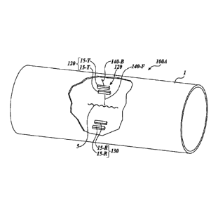
FIGURES 3A and 3B are isometric views of systems for detecting cracks in pipes
in accordance with an embodiment of the presently disclosed technology. Figure
3A
shows a system 100A that includes a multichannel EMAT TX 120 having two
individual
EMATs 15-T. In other embodiments, the multichannel EMAT TX 120 may include
different number of individual EMATs.
In some embodiments, the actuating coils of the individual EMATs 15-T generate
ultrasonic waves travelling in opposite directions. For example, using
appropriately time-
delayed AC currents in the coils of the multichannel EMAT TX 120, the EMAT
generates a relatively strong circumferential ultrasonic waves 140-F (also
referred to as
"forward waves" or "forward-propagating waves"), and a relatively weak
circumferential
waves 140-B (also referred to as "backward waves" or "backward-propagating
waves") in
the pipe 1. The ultrasonic waves generated by the EMATS 15-T are also referred
to as
the unidirectional waves because the waves predominantly propagate in one
direction
(e.g., in the forward direction or in the backward direction), as opposed to,
for example,
the ultrasonic waves propagating circularly away from the source. In some
embodiments,
the forward-propagating waves 140-F may have amplitude that is several times
greater
than that of the backward-propagating waves 140-B.
-4-
CA 03013160 2018-07-27
WO 2017/136692 PCT/US2017/016457
Provided that an EMAT RX 130 is not directly opposite from the EMAT TX 120
(i.e., the RX and TX are not exactly 1800 apart in polar direction), the
ultrasonic
waves 140-F/140-B reach the EMAT RX 130 at different times. Similarly, wave
reflections off the crack 5 may also reach the EMAT RX at different times. In
general,
when the signals that the EMAT RX senses do not overlap in time, the EMAT RX
experiences smaller signal congestion. Furthermore, in at least some
embodiments, when
the EMAT RX 130 includes multiple individual EMATs 15-R, the EMAT RX can
detect
directivity of the received ultrasound signal, further improving determination
of location
of the crack 5.
In the illustrated embodiment, the EMATs TXs/RXs are located inside the pipe.
However, in some embodiments the EMATs TXs/RXs may be located
circumferentially
around the pipe. Collectively, EMAT TX 120 and EMAT RX 130 may be referred to
as
EMAT transceiver or EMAT TRX.
Figure 3B shows a system 100B that includes a multichannel EMAT TX 120
having four individual EMATs 15-T. In other embodiments, the multichannel EMAT
TX 120 may include different number of individual EMATs. Generally, when the
ultrasonic waves 140-A/140-B propagate circumferentially, they may make many
circumferential rounds in the pipe before their energy dissipates. In some
embodiments,
these multiple rounds of the ultrasonic waves 140-A/140-B increase signal
congestion at
the EMAT RX. In the illustrated embodiment, the EMAT TX 120 is inclined with
respect the axis of the pipe (i.e., the EMAT TX 120 is not perpendicular with
respect to
the axis of the pipe), therefore generating the ultrasonic waves that
propagate in a spiral
direction (also referred to as a "helical direction") away from the EMAT TX
120. As a
result, the forward-propagating wave 140-F and the backward-propagating wave
140-B
escape the area of the EMAT TX 120 after a certain number of rotations,
depending on
the magnitude of angle a and the axial width of the EMAT TX 120. In some
embodiments, because the ultrasound waves 140-F/140B spirally propagate in the
axial
direction, a longer axial segment of the pipe can be inspected before
repositioning the
system 100B in the axial direction. In some embodiments, the EMAT RX 130 may
be
axially offset (upstream or downstream) from the EMAT TX 120.
FIGURE 4 is a side view of a system for detecting cracks in pipes in
accordance
with an embodiment of the presently disclosed technology. The illustrated
system
includes the EMAT TX 120 and the EMAT RX 130 that are circumferentially
offset. The
-5-
EMAT TX 120 includes a permanent magnet 100-T and the EMAT RX 130 includes a
permanent magnet 100-R. Collectively, EMAT TX 120 and EMAT RX 130 may be
referred to as EMAT transceiver or EMAT TRX.
Furthermore, each of the EMAT TX 120 and EMAT RX 130 includes four
.. coils 210-T/210-R, respectively. In other embodiments, different numbers of
coils are
possible. In operation, the AC currents in the coils 210-T can be phase-offset
to produce
a stronger ultrasound wave 140-F in one direction, and a weaker ultrasound
wave 140-B
in the opposite direction. Analogously, in at least some embodiments, the
coils 210-R of
the EMAT RX 130 will sense the incoming ultrasonic wave at slightly offset
times. As a
.. result, a direction of the detected ultrasonic wave may be determined using
analog or
digital signal processing. Therefore, the EMAT RX 130 can discriminate among
the
transmitted ultrasound waves 140-F/140-B and reflected ultrasound waves 140-F-
R/140-
B-R based on their differing directions, therefore enabling more precise
determination of
the location and/or severity of the crack 5 in comparison to the systems that
can only
.. detect the magnitude of the received ultrasound waves. In some embodiment,
the signal
to noise ratio (SNR) can also be improved based on using the multichannel EMAT
TX
and/or RX. Some suitable digital processing methods for analyzing the sensed
ultrasound
waves are described below with reference to Figures 10A-14.
FIGURE 5 is a schematic cross-sectional view of a printed circuit board
.. (PCB) 200 in accordance with an embodiment of the presently disclosed
technology. The
PCB 200 includes three collocated coils 210 that can be laid out in the
routing layers of
the PCB. The illustrated collocated coils 210 are mutually offset by a
distance d, but their
turns overlap in the illustrated side view (the insulating material of the PCB
prevents
electrical contact between coils 210 that are laid out in their individual
routing layers). In
some embodiments, the phase offset among the AC currents in the coils 210 can
be
selected to increase amplitude of the ultrasound waves in one direction, and
to decrease
their amplitude in the opposite direction. For example, the phase offset
between the
adjacent coils 210 may correspond to one quarter of the wavelength of the
ultrasound
wave (//4). In some embodiments, the phase offset can be controlled with a
controller C
(e.g., a digital microcontroller, an analog controller, a computer, etc.).
Analogously, the
controller C may be configured to detect the phase offset in the coils 210 of
the
EMAT RX. In some embodiments, the PCB 200 includes a protective material 250,
for
-6-
Date Recue/Date Received 2021-07-29
example, an electrically insulating material that prevents electrical contact
between the
pipe 1 and the coils 210.
FIGURE 6 is a schematic view of a width of a trace in accordance with an
embodiment of the presently disclosed technology. The illustrated trace is a
segment of
the coil 210. Without being bound by theory, it is believed that wider traces
reduce
modal noise in the signal received by the EMAT RX. In some embodiments, a
width t of
the trace corresponds to more than 40% or up to 50% of the available space L
(distance)
between the adjacent traces.
FIGURE 7 is a schematic top view of a partial masking of the coil 210 in
accordance with an embodiment of the presently disclosed technology. In the
illustrated
embodiment, the coil 210 is separated from the surface of the pipe by a
blocking foil 300
having an opening 310 that can be elliptical, circular, rectangular, diamond
shaped, etc.
In some embodiments, the blocking foil 300 is a metal foil, for example a
steel foil that
blocks EM radiation. The opening 310 provides a path for the EM radiation to
the
surface of the pipe, while the material of the blocking foil 300 at least
partially blocks the
EM radiation to the surface of the pipe. As a result, the blocking foil 300
partially
restricts eddy current in the pipe. Without being bound by theory, it is
believed that the
blocking foil 300 makes ultrasound waves 140-F/140-B narrower (i.e., the
directivity of
the ultrasound waves is better defined). Additionally, in at least some
embodiments, the
EMAT RX 130 is subjected to less modal noise and the sideband suppression is
improved. As a result, the silent region of the signal is increased.
FIGURE 8 is a graph of amplitudes of transmitter (TX) signal in accordance
with
an embodiment of the present technology. The horizontal axis represents time,
and the
vertical axis represents the amplitude of the ultrasound wave ("signal"). Two
signals are
shown: the forward-propagating signal 140-F and the backward-propagating
signal
140-B. In the illustrated embodiment, the forward-propagating signal 140-F has
a larger
amplitude than the backward-propagating signal 140-B. For example, the
amplitude of
the forward-propagating waves 140-F may have amplitude that is 4 times, 5
times, or
more than 10 times greater for two channel EMAT and nearly 20 times greater
for 4
channel EMAT than that of the backward-propagating waves 140-B.
The width of the amplitude is At, and the period of the signal is T. For a
large
diameter pipe and single channel EMAT an optimal At can be determined to
minimize the
blind spots. However, in some embodiments, reducing the width At can only
partially
-7-
Date Recue/Date Received 2021-07-29
CA 03013160 2018-07-27
WO 2017/136692 PCT/US2017/016457
minimize/eliminate blind spots because of the multimodality and dispersion of
the
ultrasonic waves. For example, ultrasonic waves (even when unidirectional) in
thin-
walled structures (such as plates and pipes) are characterized by multiple
wave packets
having a frequency-dependent velocity (also known as "wave dispersion").
Further, at a
given frequency, these multiple wave packets (or modes) may propagate with
distinct
velocities (also known as "multimodality"). Furthermore, the wave dispersion
and
multimodality tend to be more pronounced in the pipes having smaller
diameters. In
some embodiments of the inventive technology, a reduction of the blind spots
at the
EMAT RX is achieved by (a) limiting the frequency bandwidth of the ultrasonic
waves,
and (b) reducing the number of modes in the waves generated at a given
frequency.
Limiting the frequency bandwidth involves using sinusoidal signals with
multiple
cycles or a relatively large At. Additionally, coils 210 having a large number
of turns
also reduce the number of modes.
Reducing the number of modes involves using multichannel EMAT RX, without
increasing the overall dimensions of the transducer module. In some
embodiments, this
economy in size is obtained because the multiple EMAT coils 210 can be
overlaid on a
printed circuit board (PCB).
FIGURES 9A-9C are graphs of EMAT RX signal in accordance with an
embodiment of the present technology. The horizontal axis in both graphs shows
time
in tts. For an ultrasound wave with known frequency, a length of time 2zR/vg
on the
horizontal axis corresponds to the time needed for the single to make full
circle through
the pipe, where R is the radius of the pipe, and vg is the group velocity of
the ultrasonic
guided wave mode. In the context of guided waves, vg generally changes with
frequency f. However, when the guided wave mode is characterized by a constant
velocity relative to frequency, then the value of vg is also given by fX,
where X is the
wavelength of the ultrasound wave mode. The vertical axis in both graphs shows
signal
strength in V as detected by the EMAT RX 130.
The graph in Figure 9A further illustrates the complexity of the signal
obtained
from EMAT RX 130 in a pipe. In the illustrated embodiment, the reflected
ultrasound
waves from the pipe crack are absent (e.g., because no crack exists in a given
segment of
the pipe). Regions F and B correspond to the forward-propagating and backward-
propagating ultrasound signal detected by the EMAT RX. In some embodiments,
the
amplitude of the forward-propagating signal (region F) is larger than the
amplitude of the
-8-
CA 03013160 2018-07-27
WO 2017/136692 PCT/US2017/016457
backward-propagating signal (region B) because the EMAT TX emits a stronger
signal in
the preferred, forward direction. Region CT corresponds to electrical cross-
talk between
the TX and RX. For example, driving the coils 210 of the EMAT TX with AC
current
may electromagnetically couple with the coils 210 of the EMAT RX to produce
the signal
illustrated in the region CT even in absence of the ultrasound waves at the
location of
EMAT RX. Furthermore, in some embodiments, the EMAT TX and EMAT RX may
share the same power supply, which causes the electromagnetic noise at the
EMAT RX
when the EMAT TX is excited. Due to the presence of relatively large signals
in
regions CT, F and B, detecting the ultrasound waves reflected from the crack
can be
difficult in these regions (also referred to as "blind spot" regions).
Furthermore, even
outside of the regions CT, F and B, the EMAT RX may be detecting modal noise
MN
that, if not removed, can be mistakenly interpreted as an indication of the
crack in the
pipe.
The graph in Figure 9B shows EMAT RX signal that includes reflected ultrasound
waves from the pipe crack. Regions F, B and CT generally represent same types
of
signals as those described with reference to Figure 9A. Additionally, the EMAT
RX
detects a signal REFL corresponding to the ultrasound wave reflected from the
crack 5.
A region DEF corresponds to the region where such signal may be detectable. In
some
embodiments, the region(s) DEF is at least partially masked by the regions F,
B,
and/or CT (and vice versa) thus generally decreasing the sensitivity of the
method and,
conversely, increasing the blind spots. The reduction of the blind spots
therefore
increases the sensitivity of the system to the cracks in the pipe. In some
embodiments,
elimination of the blind spots cannot be fully accomplished by the
unidirectional
EMAT TX 120 alone.
The graph in Figure 9C shows EMAT RX signal that does not include reflected
ultrasound waves from the pipe crack. The forward-propagating wave is shown in
solid
line, and the backward-propagating wave is shown in dashed line. In absence of
the pipe
crack, the region between the strong forward-propagating wave and the strong
backward-
propagating wave should have relatively small signal amplitude. However, the
modal
noise increases the signal amplitude in this, otherwise quiet, region.
Reduction in the
number of modes is explained with reference to Figures 10A-11B below.
FIGURE 10A is a schematic view of coils of EMAT RX in accordance with an
embodiment of the present technology. Two coils 210-R are represented by
different line
-9-
CA 03013160 2018-07-27
WO 2017/136692 PCT/US2017/016457
types: one coil 210-R is represented by solid line, and the other coil 210-R
is represented
by dashed line. In operation, the illustrated coils 210-R may be excited such
that there is
a phase offset from the time when one coil 210-R is excited with AC current to
the time
when the other coil is excited with AC current. As explained above, the phase-
offset
excitation may produce ultrasonic waves having stronger amplitude in one
direction, and
a weaker amplitude in another direction. Additionally, multiple coils 210-R
also help in
reducing the modal noise MN, as explained with reference to Figure 10B below.
FIGURE 10B is a graph of RX signal in accordance with an embodiment of the
present technology. The horizontal axis represents the wavenumber of the EMAT
RX
signal in rad/m. The vertical axis represents normalized spectrum. The peaks
in the
normalized spectrum represent modes of in the detected RX signal. When the
EMAT RX
includes only one coil 210-R, the EMAT RX detects all modal peaks 1-8 In at
least
some embodiments, preferably the EMAT RX detects just one modal peak, the
other,
unwanted modal peaks representing the modal noise MN. When the EMAT RX
includes
two coils 210-R, some modal peaks that are part of modal noise MN are
eliminated. For
example, the modal peaks drawn in dashed lines (modal peaks 2, 4, 6, and 7)
are
eliminated.
FIGURE 11A is a schematic view of coils of EMAT RX in accordance with an
embodiment of the present technology. The illustrated EMAT RX includes four
coils 210-R represented by different line types. In operation, the illustrated
coils 210-R
may be excited with a phase offset, resulting in the ultrasonic waves having
stronger
amplitude in one direction, and a weaker amplitude in another direction.
Additionally, an
increase in the number of coils 210-R can help to further reduce the modal
noise MN, as
explained with reference to Figure 11B below.
FIGURE 11B is a graph of RX signal in accordance with an embodiment of the
present technology. The horizontal axis represents the wavenumber of the EMAT
RX
signal in rad/m. The vertical axis represents normalized spectrum. The peaks
in the
normalized spectrum represent modes of in the detected RX signal. When the
EMAT RX
includes only one coil 210-R, modal peaks 1-8 are detected by the EMAT RX, the
other
modal peaks representing the undesired modal noise MN. When the EMAT RX
includes
four coils 210-R, some modal peaks that are part of modal noise MN are
eliminated. For
example, the modal peaks drawn in dashed lines (modal peaks 2, 3, 4, 5, 6, and
7) are
-10-
CA 03013160 2018-07-27
WO 2017/136692 PCT/US2017/016457
eliminated. Analogously, further modal peaks may be eliminated by increasing
the
number of coils 210-R.
FIGURE 12 is a flow diagram of a signal processing method 120 in accordance
with an embodiment of the present technology. In step 121, a unidirectional
ultrasonic
wave (signal) 121 is transmitted to a multi-channel receiver, for example the
EMAT
RX 120 having two coils 210 that can be individually measured. In step 122,
the
individual channels of the multichannel receiver are scanned to produce signal
Si from
the first channel (e.g., the first coil) and S2 from the second channel (e.g.,
the second
coil). In other embodiments, different number of signals S can be produced by
the
EMAT RX, depending on the number of channels (coils) of the EMAT RX. In step
123,
the signals Si and S2 are decomposed into forward-propagating signal Sf and
backward-
propagating signal Sb relative to the EMAT TRX The decomposition of the
signals Si
and S2 into Sf and Sb is explained with reference to Figure 14 below.
In step 124, the RMS-es for the forward-propagating signal Sf and backward-
propagating signal Sb are calculated, and the ratio of the RMS-es is
calculated to as a
measure of side-band suppression. For example, in some embodiments the modal
peaks
on the negative wavenumber axis (also referred to as the "sidebands") can be
suppressed
using a dual coil (i.e., dual channel) EMAT TX 130. In general, the term
"improvement
of sideband suppression" refers to a decrease of the sidebands. The term
"degradation of
sideband suppression" refers to an increase of the sidebands. In some
embodiments, if
there are no flaws (e.g., cracks) in the pipe, the side-band suppression
remains the same.
If, however, a flaw exists, then the reflected ultrasonic wave may cause an
apparent
degradation in the sideband suppression. In step 125, the change in the
apparent sideband
suppression efficiency can be compared with pre-calibrated lookup table to
obtain the
flaw size. If the flaw size is directly measureable, then it may also be
locatable. In
step 126, to locate the flaw, a peak of Sb and its time of arrival, Tb is
found.
Multiplying Tb with the group velocity for the ultrasound provides the
location of the
flaw.
In at least some embodiments, the method 120 works well even in the presence
of
seams and uniform corrosion in the inside of the pipe, when larger lengths of
data (higher
than 720 of wave traversal) are used. Generally, the sideband suppression
converges to
uniform values in the presence of flaws, if larger data lengths are used.
-11-
CA 03013160 2018-07-27
WO 2017/136692 PCT/US2017/016457
FIGURE 13 is a flow diagram of a signal processing method 130 in accordance
with an embodiment of the present technology. The method 130 can find a time
delay
between the two signals, for example the signals received by the individual
coils of the
multichannel EMAT RX. In step 131, a unidirectional ultrasonic wave (signal)
121 is
transmitted to a multi-channel receiver, for example the EMAT RX 120 having
two
coils 210. In step 122, the individual channels of the multichannel receiver
are scanned
to produce signal Si from the first channel (e.g., first coil) and S2 from the
second
channel (e.g., second coil). In step 133, data Si and S2 from the two channels
of the
EMAT RX are normalized so that they have equal amplitude. In some embodiments,
in
step 134 the following minimization problem is solved:
min IS1 + a x S2I, such that al 1 (Eq. 1)
a
The solution of Equation 1 can be interpreted as: find the value of a for the
minimum possible norm (root mean square) of the quantity Si + a x S2 (after
normalizing signals Si and S2 as in, for example, step 133). Once the value of
a is
found, the time delay between the signals received by the individual coils of
the
multichannel EMAT RX can be calculated in step 135 using the formula:
At = ¨2af COS-1 a (Eq. 2)
where f is the center frequency of the input signal. In some embodiments, the
implementation of Eq. 2 results in sideband suppression of 2-4 dB.
In some embodiments, instead of Eq. 1, an average of main lax Si + S21 and
min IS1 + a x S21 can provide improved sideband suppression. The method 130
may be
a
generalized to a multi-channel EMAT having more than two channels by, for
example,
applying the algorithms in Eqs. 1 and 2 to data from two channels at a time,
while
keeping a common EMAT RX channel for each pair to assure that the time delays
will be
relative to such a common channel. The method 130 can also be implemented on
the
transmit side (e.g., with EMAT TX having multiple channels).
FIGURE 14 is a flow diagram of a signal processing method 140 in accordance
with an embodiment of the present technology. In some embodiments, the method
140
may eliminate or at least reduce a need for the RX electronics (e.g., analog
to digital
converter) with high sampling frequency, while achieving high sideband
suppression.
The signals Si and S2 may be obtained using, for example, methods described
with
reference to Figures 12 and 13. With the method 140, time delays are applied
to
signal S2.
-12-
CA 03013160 2018-07-27
WO 2017/136692 PCT/US2017/016457
In step 141, zero padding is applied to signal S2. In some embodiments, the
number of zero data may depend on capacity of the onboard memory. In step 142,
a fast
Fourier transform (FFT) of the zero-padded signal is determined. In step 143,
the FFT
results from step 142 are multiplied by a vector e-121-f' to obtain vector
32), where f is
a vector of frequencies whose values and range depend on the original sampling
frequency of S2 and the size of the zero-padded version of S2. In step 144, an
inverse FFT (IFFT) is perfoimed over the 32 . In step 145, the results of step
144 are
truncated such that the length of the data (e.g., the length of the time
series) is back to the
original length of S2. The resulting S2 is an accurate time delayed version of
the
acquired S2. In some embodiments, the accuracy of the time delay is
proportional to the
length of the zero-padding. In step 146, this resulting S2 from step 145 may
be added
to S1 to determine a forward-propagating wave Sf Analogously, in step 147 the
resulting S2 from step 145 may be subtracted from Si to determine a backward-
propagating wave Sb. In some embodiments, if the original sampling frequency
is much
larger than the frequency of interest, this algorithm need not be followed.
Instead, the
delay can be obtained by truncating the end of the signal and adding zeros at
the
beginning of the signal.
Many embodiments of the technology described above may take the form of
computer- or controller-executable instructions, including routines executed
by a
programmable computer or controller. Those skilled in the relevant art will
appreciate
that the technology can be practiced on computer/controller systems other than
those
shown and described above. The technology can be embodied in a special-purpose
computer, controller or data processor that is specifically programmed,
configured or
constructed to perform one or more of the computer-executable instructions
described
above. Accordingly, the terms "computer" and "controller" as generally used
herein refer
to any data processor and can include Internet appliances and hand-held
devices
(including palm-top computers, wearable computers, cellular or mobile phones,
multi-
processor systems, processor-based or programmable consumer electronics,
network
computers, mini computers and the like). Information handled by these
computers can be
presented at any suitable display medium, including a CRT display or LCD.
From the foregoing, it will be appreciated that specific embodiments of the
technology have been described herein for purposes of illustration, but that
various
modifications may be made without deviating from the disclosure. Moreover,
while
-13-
CA 03013160 2018-07-27
WO 2017/136692 PCT/US2017/016457
various advantages and features associated with certain embodiments have been
described above in the context of those embodiments, other embodiments may
also
exhibit such advantages and/or features, and not all embodiments need
necessarily exhibit
such advantages and/or features to fall within the scope of the technology.
Accordingly,
the disclosure can encompass other embodiments not expressly shown or
described
herein
-14-