Note: Descriptions are shown in the official language in which they were submitted.
CA 03013168 2018-07-30
WO 2017/137542
PCT/EP2017/052974
HUMAN ANTIBODIES AND BINDING FRAGMENTS THEREOF TO TENASCIN
The present invention relates to antibodies or binding fragments thereof,
specific the fibrinogen-like
globe (FBG) domain of a tenascin, such as tenascin-C, compositions comprising
the antibodies and
use of any one of the same in the diagnosis, determination of prognosis,
and/or treatment of
disorders, for example disorders associated with chronic inflammation, as well
as methods of making
said antibodies.
BACKGROUND
Inflammation is the complex biological response of tissues to harmful stimuli,
such as
pathogens, tissue damage, or irritants. It is a protective attempt by the
tissue to remove the injurious
stimuli as well as initiate the healing process for the tissue. Abnormalities
associated with
inflammation comprise a large, unrelated group of disorders which underlie a
variety of human
diseases (inflammatory disorders). Examples of diseases with an inflammatory
aspect include (but
are not limited to) asthma, autoimmune disease, glomerulonephritis, allergy
(hypersensitivities),
cancer, inflammatory bowel diseases, reperfusion injury, rheumatoid arthritis
and transplant
rejection. Rheumatoid arthritis (RA) is a typical example of a chronic
inflammatory condition.
Toll-like receptors (TLRs) play a key role in driving the production of
inflammatory
mediators in RA and blockade of TLR function may be of significant clinical
benefit (reviewed in
Brentano (2005) and O'Neill (2002)). This family of receptors forms an
integral part of the immune
system. TLRs mediate host defence against infection and injury by recognising
both pathogen-
associated molecular patterns (PAM Ps) and damage-associated molecular
patterns (DAMPs)
(Matzinger (2002)). DAMPs are endogenous pro-inflammatory molecules generated
upon tissue
injury and include intracellular molecules released from damaged or necrotic
cells, fragments of
extracellular matrix (ECM) molecules or ECM molecules up regulated upon injury
(reviewed in
Bianchi (2007) and Gordon (2002)).
Upon activation, TLRs promote both innate and adaptive immune responses
including
stimulation of expression of pro-inflammatory cytokines and MMPs (Medzhitov
(2002)). TLRs are
expressed at high levels in synovial tissue from RA patients (Radstake (2004),
Roelofs (2005), Sacre
(2007), and (Sacre, 2008) and mice with targeted deletions or loss of function
mutations in TLR4 are
protected from experimental arthritis (Choe (2003) and Lee (2005).
Tenascin-C (TNC) is an ECM glycoprotein that is associated with tissue injury
and wound
repair. Tenascin-C is not normally expressed in healthy adult tissue but, in
adults, is specifically and
transiently up-regulated during acute inflammation and persistently expressed
in chronic
inflammation (reviewed in Chiquet-Ehrismann (2003)). Immunohistochemical
studies show that
little tenascin-C is expressed in normal human joints but levels are greatly
increased in RA synovia,
in areas of inflammation and fibrosis, specifically below the synovial lining,
in the invading pannus
and around blood vessels (Cutolo (1992), MacCachren (1992) and Salter (1993)).
There is also a
significant increase in tenascin-C levels in synovial fluid from RA patients
(Chevalier (1994) and
Hasegawa (2007)) and in RA cartilage (Salter (1993) and Chevalier (1994)).
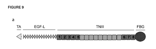
Tenascin-C is a large hexameric protein of 1.5 million Da. Each chain
comprises different
domains, including an assembly domain (TA), EGF-like repeats (EGF-L),
fibronectin type III-like
1
CA 03013168 2018-07-30
WO 2017/137542
PCT/EP2017/052974
repeats (TNIII) and a fibrinogen-like globe (FBG) (reviewed in Orend (2005)).
The sequences of
tenascin-C and its domains are shown in Figure 9.
Tenascin-C has been shown to be an endogenous activator of TLR4 and it has
been
demonstrated that this molecule is required for destructive joint inflammation
(W02010/103289).
Tenascin-C was shown to be capable of activating cells in the joint and the
primary active domain of
tenascin-C has been mapped to the fibrinogen-like globe (FBG), a 227 amino
acid (26.9 kDa) globular
domain at the C terminal of the molecule (Sin i (1991)).
Addition of FBG to synovial membrane cultures from RA patients enhanced the
spontaneous
release of pro-inflammatory cytokines. It also stimulated synthesis of TNF-a,
IL-6 and IL-8 in primary
human macrophages and IL-6 in RA synovial fibroblasts via activation of TLR4
and MyD88 dependent
signalling pathways.
It has been shown that, as in the case of LPS, TLR4 expression is necessary
for induction of
cytokine synthesis by FBG. However, unlike LPS, neither CD14 nor MD-2 appears
to be required for
TLR-4 activation. CD14 is dispensable for activation of TLR4 by other ligands.
It is not required for
TLR4 to respond to lipid A in a MyD88 dependent manner (Jiang (2005)),
fibronectin EDA (extra
domain A) can activate mast cells even in the absence of CD14 (Gondokaryono
(2007)) and
hyaluronic acid activation of human monocytic THP-1 cells requires a complex
of TLR4, CD44 and
MD-2, but not CD14 (Taylor (2007)).
Formation of distinct receptor complexes by each TLR4 ligand may facilitate
recruitment of
different intracellular adapter/signalling molecules. This may account for the
differential cellular
responses we observe with FBG and LPS. Similarly, hyaluronic acid activation
of the TLR4 and CD44
complex induces a pattern of gene expression in mouse alveolar macrophage cell
lines that is
different to LPS (Taylor (2007)).
The tightly regulated pattern of expression of tenascin-C makes it an
attractive target for
treating chronic inflammation. It is predominantly absent from healthy adults,
however expression
is specifically induced upon tissue injury. During acute inflammation tenascin-
C is transiently
expressed: induction often precedes inflammation and both mRNA and protein are
absent from the
tissue by the time inflammation is resolved (reviewed in Chiquet-Ehrismann
(2003)).
Persistent expression of tenascin-C has now been shown to be associated with
chronic
inflammation. In addition to RA, increased tenascin-C levels are observed in
other autoimmune
diseases including multiple sclerosis (Gutowski (1999)) and Sjogrens disease
(Amin (2001)), and in
non-healing wounds and diabetic and venous ulcers (Loots (1998)). De novo
synthesis of tenascin-C
correlates well with the intensity of inflammation in diseases of the oral
mucosa and plasma levels of
tenascin-C are a reliable indicator for the activity of inflammatory bowel
diseases before and after
medication or surgery (reviewed in Chiquet-Ehrismann (2003)).
W02010/103289 describes the use of agents for modulation of a chronic
inflammatory
response wherein the agent modulates the biological activity of tenascin-C and
their use in treating
conditions associated with chronic inflammation.
Clark et al. (1997) (52) describes a murine antibody specific for the FBG
domain, which
interfers with "lymphocyte rolling". The latter is believed to be a measure of
cell migration, and
unrelated to cell activation and production of inflammatory cytokines.
2
CA 03013168 2018-07-30
WO 2017/137542
PCT/EP2017/052974
The inventors have designed antibodies and fragments thereof with properites
that are
suitable for use in therapy, in particular human antibodies, with very high
affinity to the fibrinogen-
like globe (FBG) domain of tenascin-C, and which neutralise the biological
activity of FBG. These high
affinity antibodies are useful in a variety of therapeutic methods, such as
those which use anti-FBG
antibody molecules in the diagnosis or treatment of tenascin-C related
disorders, particularly those
associated with chronic inflammation, including rheumatoid arthritis (RA). The
antibodies are also
useful in related diagnostic and prognostic methods. The antibodies are
disclosed in
W02016/020702, incorporated herein by reference. This application discloses an
antibody B12
from which an antibody 165_12_C3 (referred to herein as C3) was derived.
SUMMARY OF INVENTION
The present disclosure relates to a modified antibody or binding fragment
thereof referred
to C3* wherein a potential T cell epitope has been removed from the light
chain framework to reduce
the immunogenicity, and variants of the B12 antibody comprising said
modification in the light chain.
Thus there is provided:
1. An antibody or binding fragment specific to Tenascin (for example specific
to Tenscin C)
comprising a sequence as shown in SEQ ID NO: 22 or 23, in particular, SEQ ID
NO: 22.
2. An antibody or binding fragment according to paragraph 1 further
comprising VH with CDRH1
of SEQ ID NO: 3, CDRH2 of SEQ ID NO: 4 and a CDRH3 independenity selected from
SEQ ID NO:
5, 12, 14, 16, 18, 24, 26, 28, 30 and 32, or a variant thereof wherein up to 5
amino acids are
changed in the CDRs of the VH and VL.
3. An antibody or binding fragment according to paragraph 1 or 2 comprising
a VH selected from
SEQ ID NO: 6, 13, 15, 17, 19, 21, 25, 27, 29, 31, 33 and a variant thereof
wherein up to 5 amino
acids in the sequence are changed.
4. An antibody or binding fragment according to any one of paragraphs 1 to 3,
which is a Fab or
Fab' fragment.
5. An antibody or binding fragment according to any one of paragraphs 1 to
3, which is a ful length
antibody.
6. An antibody or binding fragment according to paragraph 5, wherein the heavy
chain has a
sequence as shown in SEQ ID NO: 1.
7. An antibody or binding fragment according to paragraph 5 or 6, wherein
the heavy chain has a
sequence as shown in SEQ ID NO: 2.
8. A pharmaceutical composition comprising an antibody or binding fragment
according to any one
of paragraphs 1 to 7.
9. An antibody or binding fragment according to any one of paragraphs 1 to 7,
or a pharmaceutical
composition according to paragraph 8, for use in treatment.
10. An antibody, binding fragment or composition for use according to
paragraph 9, wherein the use
is for the treatment of an inflammatory disorder, for example a chronic
inflammatory disorder,
for example a disorder disclosed herein, such as rheumatoid arthritis.
11. An antibody or binding fragment according to any one of paragraphs 1 to 7,
or a pharmaceutical
composition according to paragraph 8, for use in the manufacture of a
medicament for the
3
CA 03013168 2018-07-30
WO 2017/137542
PCT/EP2017/052974
treatment of a chronic inflammatory response, for example a disorder disclosed
herein, such as
rheumatoid arthritis.
12. A method of treatment comprising administering a therapeutically effect
amount of an antibody
or binding fragment according to any one of paragraph 1 to 7 or a
pharmaceutical composition
according to paragraph 8.
13. A method according to paragraph 12, wherein the treatment is for an
inflammatory disorder, for
example a chronic inflammatory disorder, such as rheumatoid arthritis.
14. A polynucleotide encoding an antibody or binding fragment according to any
one of paragraph
1 to 7.
15. A vector comprising a polynucleotide according to paragraph 13.
16. A host cell (for example a mammalian cell) comprising a polynucleotide of
paragraph 12 or a
vector of paragraph 13.
17. A process of producings (making) an antibody or binding fragment according
to the present
disclosure comprising the step of culturing a host cell, as defined in
paragraph 16, to express
said antibody or binding fragment.
Thus there is provided an antibody or binding fragment specific to Tenascin
(for example
specific to Tenscin C) comprising a light chain sequence as shown in SEQ ID
NO: 1:
D I QMTQSP S SLSASVGDRVT I TCRASQY I QGFLNWYQQKPGKAPKLL I YDASNLETGVP
SRFSGSGSGTDFTLT I S S
LQPEDFATYYCQQSYSTPQTFGQGTKVD I KRTVAAP SVF I FPP
SDEQLKSGTASVVCLLNNFYPREAKVQWKVDNAL
QSGNSQESVTEQDSKDSTYSLS STLTLSKADYEKHKVYACEVTHQGLS SPVTKSFNRGEC
The antibody or binding fragment of the present disclosure further comprises a
VH
comprising CDRH1 of SEQ ID NO: 3, CDRH2 of SEQ ID NO: 4 and a CDRH3
independenity selected
from SEQ ID NO: 5, 12, 14, 16, 18, 20, 24, 26, 28, 30 and 32, or a variant
thereof wherein up to 5 amino
acids are changed in the CDRs of the VH and VL (in particular wherein the
binding affinity for human
Tenascin C in maintained at a similar value as that of the "parent/starting"
antibody.
In one embodiment the VH in the antibody or binding fragment of the present
disclosure is
independently selected from SEQ ID NO: 6, 13, 15, 17, 19, 21, 25, 27, 29, 31,
33 and a variant thereof
wherein up to 5 amino acids in the sequence are changed.
In one embodiment the VH is in a heavy chain as shown in SEQ ID NO: 2:
QVQLVE SGGGLVQPGRS LRL SCAASGF TEDDYAMHWVRQAPGKGLEWVSG I SGSGGSTYYADSVKGRFT I
SRDNAKN
SLYLQMNSLRAEDTALYYCAKSYQSDEDAFD IWGQGTMVTVS SAS TKGP SVFP LAPC SRS T SE S
TAALGCLVKDYFP
EPVTVSWNSGALTSGVHTFPAVLQS SGLYSLS SVVTVP S S SLGTKTYTCNVDHKP
SNTKVDKRVESKYGPPCPPCPA
PEFLGGP SVFLEPPKPKDTLMI
SRTPEVTCVVVDVSQEDPEVQFNWYVDGVEVHNAKTKPREEQFNSTYRVVSVLTV
LHQDWLNGKEYKCKVSNKGLP S S I EKT I SKAKGQPREPQVYTLPP SQEEMTKNQVSLTCLVKGFYP SD
IAVEWESNG
QPENNYKT TPPVLD SDGSFFLYSRLTVDKSRWQEGNVF SC SVMHEALHNHYTQKS L S L S LG
Thus in one embodiment the antibody of the present disclosure is a full length
antibody or a
molecule comprising a full length antibody, for example an IgG, such as IgG1,
IgG2, IgG3 or IgG4.
4
CA 03013168 2018-07-30
WO 2017/137542
PCT/EP2017/052974
In one embodiment the antibody or binding fragment thereof, is specific to an
FBG domain,
in particular an FBG domain of Tenascin-C.
In one embodiment the antibody or binding fragment according to the present
disclosure
have affinity to human Tenascin-C of 100nM or higher affinity, such as 50nM or
higher, in particular
45, 44, 43, 42, 41, 40, 39, 38, 37,36, 35, 34, 33, 32, 31, 30, 29, 28, 27,26,
25, 24, 23, 22, 21, 20, 19, 18,
17, 16, 15, 14, 13, 12, 11, 10, 9, 8, 7, 6, 5, 4, 3, 2, 1 or 0.5 nM or higher
affinity (which of course has a
lower numerical value).
The antibodies and binding fragments according to the present disclosure may
be less
immunogenic that the corresponding parent antibody.
In one embodiment the antibody or binding fragment according to the present
disclosure is
conjugated to a payload.
The antibodies and binding fragments according to the present disclosure may
express better
than the corresponding parent antibody, for example 1.5, 2, 2.5 or 3 times
better expression.
The antibodies and binding fragments according to the present disclosure have
comparable
properties, such as affinity, to the corresponding parent antibody. However,
in some instances they
antibodies or binding fragments herein may improved properties or activity
over the corresponding
parent antibodies.
DETAILED DISCLOSURE
"Antibody" as employed herein includes substantially intact antibody
molecules, as well as
chimeric antibodies, humanised antibodies, human antibodies (wherein at least
one amino acid is
mutated relative to the naturally occurring human antibodies), single chain
antibodies, multispecific
antibodies (such as bispecific antibodies), antibody heavy chains, antibody
light chains, homodimers
and heterodimers of antibody heavy and/or light chains, and antigen binding
fragments and
derivatives of the same.
By "antigen-binding fragment" we mean a functional fragment of an antibody
that is capable
of binding to the FBG domain of tenascin-C.
Antibody binding fragment and antigen binding fragment are employed
interchangeably
herein unless the context indicates otherwise.
Example of antibody binding fragments include to Fab, modified Fab, Fab',
modified Fab',
F(ab')2, Fv, Fab-Fv, Fab-dsFy, single domain antibodies (e.g. VH or VL or
VHH), scFv, bi, tri or tetra-
valent antibodies, Bis-scFv, diabodies, triabodies, tetrabodies and epitope-
binding fragments of any
of the above (see for example Holliger and Hudson, 2005, Nature Biotech.
23(9):1126-1136; Adair
and Lawson, 2005, Drug Design Reviews - Online 2(3), 209-217). The methods for
creating and
manufacturing these antibody fragments are well known in the art (see for
example Verma et al.,
1998, Journal of Immunological Methods, 216, 165-181). Other antibody
fragments for use in the
present invention include the Fab and Fab' fragments described in
International patent applications
W02005/003169, W02005/003170 and W02005/003171.
Examples of a multispecific antibody comprising a fully length antibody
include a DVD-Ig,
IgG-scFv, scFv-IgG, and IgG-V.
IgG-scFy as employed herein is a full length antibody with a scFy on the C-
terminal of each of
the heavy chains or each of the light chains.
5
CA 03013168 2018-07-30
WO 2017/137542
PCT/EP2017/052974
scFv-IgG as employed herein is a full length antibody with a scFy on
the N-terminal of each of
the heavy chains or each of the light chains.
V-IgG as employed herein is a full length antibody with a variable
domain on the N-terminal
of each of the heavy chains or each of the light chains.
IgG-V as employed herein is a full length antibody with a variable domain
on the C-terminal
of each of the heavy chains or each of the light chains
DVD-Ig (also known as dual V domain IgG) is a full length antibody
with 4 additional variable
domains, one on the N-terminus of each heavy and each light chain.
In one embodiment the antibody binding fragment is or comprises a Fab or Fab'
fragment.
Antibody binding fragments that comprise a Fab or Fab' fragment include
Fabdab, Fab'dab,
FabFv, Fab'Fv, FabdsFy, Fab-scFv, Fab'-scFv, Fab-(scFv)2, Fab'-(scFv)2, DiFab,
DiFab'.
Fabdab as employed herein refers to a Fab fragment with a domain
antibody appended to the
heavy or light chain thereof, optionally via a linker.
Fab'dab as employed herein refers to a Fab' fragment with a domain
antibody appended to
the heavy or light chain thereof, optionally via a linker.
FabFv as employed herein refers to a Fab fragment with an additional
variable region
appended to the C-terminal of each of the following, the CH1 of the heavy
chain and
CL of the light chain see for example W02009/040562. The format may be
provided
as a PEGylated version thereof see for example W02011/061492,
Fab'Fy as employed herein is similar to FabFv, wherein the Fab portion is
replaced by a Fab'.
The format may be provided as a PEGylated version thereof.
FabdsFy as employed herein refers to a FabFv wherein an intra-Fv
disulfide bond stabilises the
appended C-terminal variable regions, see for example W02010/035012. The
format
may be provided as a PEGylated version thereof
Fab-scFy (also referred to as a bibody) as employed herein is a Fab
molecule with a scFy
appended on the C-terminal of the light or heavy chain, optionally via a
linker.
Fab'-scFy as employed herein is a Fab' molecule with a scFy appended on
the C-terminal of the
light or heavy chain, optionally via a linker.
DiFab as employed herein refers to two Fab molecules linked via
their C-terminus of the
heavy chains.
DiFab' as employed herein refers to two Fab' molecules linked via one
or more disulfide
bonds in the hinge region thereof.
DiFab and DiFab' molecules include chemically conjugated forms thereof.
Examples of linkers are disclosed in the sequence listing in SEQ ID NO: 45 to
86, and further
includes the sequence PPP, and the hinge sequences disclosed in SEQ ID NO: 36
to 44.
The antibody or antigen-binding fragment, derivative or variant thereof
according to the
present disclosure may down-regulate the biological activity of, for example
tenascin-C.
The antibody or antigen-binding fragment, derivative or variant thereof
according to the
present disclosure may be an inhibitor of transcription of, for example
tenascin-C.
The antibody or antigen-binding fragment, derivative or variant thereof
according to the
present disclosure may be an inhibitor of translation of, for example tenascin-
C.
6
CA 03013168 2018-07-30
WO 2017/137542
PCT/EP2017/052974
Thus in one embodiment the antibody or binding fragment, derivate or variant
thereof
according to the present disclosure may down-regulate expression of the a
tenascin, such as tenascin-
C, in particular in vivo.
The antibody or antigen-binding fragment, derivative or variant thereof of the
first aspect of
the invention may be an inhibitor of the binding properties of tenascin-C. For
example, the antibody
or antigen-binding fragment, derivative or variant thereof may alter the
conformation of tenascin-C
such that it is no longer able to bind to its receptor or receptors, (in
particular binding and/or activity
of the FBG domain of tenascin-C is inhibited).
The antibody or antigen-binding fragment, derivative or variant thereof of the
present
disclosure may be a competitive binding inhibitor of tenascin-C. It will be
appreciated by persons
skilled in the art that the antibody or antigen-binding fragment, derivative
or variant thereof may
also inhibit the biological activity of the tenascin (such as tenascin-C) by
blocking tenascin-C receptor
function either directly (by acting as a tenascin-C receptor antagonist) or
indirectly (by binding
intermediary or assisting molecules).
The antibody or antigen-binding fragment, derivative or variant thereof of the
first aspect of
the invention may be an antagonist of the TLR-4 receptor. By an antagonist of
TLR4 we include
indirect antagonism. The antigen-binding fragment, derivative or variant
thereof might prevent
tenascin-C activation of TLR4 or also of any other receptor.
It will be appreciated by persons skilled in the art that inhibition of the
biological activity of
a tenascin (such as tenascin-C, in particular the FBG domain thereof) by an
antibody or antigen-
binding fragment, derivative or variant thereof of the invention may be in
whole or in part. For
example, the antibody or antigen-binding fragment, derivative or variant
thereof may inhibit the
biological activity of a tenascin (such as tenascin-C, in particular the FBG
domain thereof) by at least
10%, preferably at least 20%, 30%, 40%, 50%, 60%, 70%, 80% or 90%, such as
100% compared to
the biological activity of the tenascin (such as tenascin-C) on inflammatory
cells which have not been
exposed to the antibody or antigen-binding fragment, derivative or variant
thereof.
In one embodiment, the antibody or antigen-binding fragment, derivative or
variant thereof
is monoclonal.
In one embodiment there is provided a polynucleotide encoding an antibody or
binding
fragment according to the present disclosure, for example the heavy and light
chain of the antibody
or bindng fragment can be encoded on the same or different polynucleotide
strand. Thus as
employed herein "polynucleotide encoding) includes one polynucleotide encoding
both the heavy
and light chain or two separate polynucleotides one encoding the heavy chain
and one encoding the
light chain.
In one aspect, there is provided a vector comprising the polynucleotide as
described above.
General methods by which the vectors may be constructed, transfection methods
and culture
methods are well known to those skilled in the art. In this respect, reference
is made to "Current
Protocols in Molecular Biology", 1999, F. M. Ausubel (ed), Wiley Interscience,
New York and the
Maniatis Manual produced by Cold Spring Harbor Publishing.
In another aspect, there is provided a host cell comprising the polynucleotide
or vector as
described above. Any suitable host cell/vector system may be used for
expression of the DNA
sequences encoding the antibody molecule of the present invention. Bacterial,
for example E. coli,
7
CA 03013168 2018-07-30
WO 2017/137542
PCT/EP2017/052974
and other microbial systems may be used or eukaryotic, for example mammalian,
host cell expression
systems may also be used. Suitable mammalian host cells include CHO, myeloma
or hybridoma cells.
The present invention also provides a process for the production of an
antibody molecule
according to the present invention comprising culturing a host cell containing
a vector (and/or DNA)
of the present invention under conditions suitable for leading to expression
of protein from DNA
encoding the antibody molecule of the present invention, and isolating the
antibody molecule.
The antibody molecule may comprise only a heavy or light chain polypeptide, in
which case
only a heavy chain or light chain polypeptide coding sequence needs to be used
to transfect the host
cells. For production of products comprising both heavy and light chains, the
cell line may be
transfected with two vectors, a first vector encoding a light chain
polypeptide and a second vector
encoding a heavy chain polypeptide. Alternatively, a single vector may be
used, the vector including
sequences encoding light chain and heavy chain polypeptides.
In one embodiment the antibody or binding fragment is provided as a
pharmaceutical
formulation comprising one or more excipients, diluents and/or carriers.
Accordingly, there is
provided a pharmaceutical composition comprising an antibody or binding
fragment as described
above.
It will be appreciated by persons skilled in the art that the antibody or
antigen-binding
fragment, derivative or variant thereof of the invention will generally be
administered in admixture
with a suitable pharmaceutical excipient diluent or carrier selected with
regard to the intended route
of administration and standard pharmaceutical practice (for example, see
Remington: The Science
and Practice of Pharmacy, 19th edition, 1995, Ed. Alfonso Gennaro, Mack
Publishing Company,
Pennsylvania, USA).
For example, the antibody or antigen-binding fragment, derivative or variant
thereof of the
invention can be administered orally, buccally or sublingually in the form of
tablets, capsules, ovules,
elixirs, solutions or suspensions, which may contain flavouring or colouring
agents, for immediate-,
delayed- or controlled-release applications.
Such tablets may contain excipients such as microcrystalline cellulose,
lactose, sodium citrate,
calcium carbonate, dibasic calcium phosphate and glycine, disintegrants such
as starch (preferably
corn, potato or tapioca starch), sodium starch glycollate, croscarmellose
sodium and certain complex
silicates, and granulation binders such as polyvinylpyrrolidone,
hydroxypropylmethylcellulose
(HPMC), hydroxy-propylcellulose (HPC), sucrose, gelatin and acacia.
Additionally, lubricating agents
such as magnesium stearate, stearic acid, glyceryl behenate and talc may be
included.
Solid compositions of a similar type may also be employed as fillers in
gelatin capsules.
Suitable excipients in this regard include lactose, starch, cellulose, milk
sugar or high molecular
weight polyethylene glycols. For aqueous suspensions and/or elixirs, the
compounds of the
invention may be combined with various sweetening or flavouring agents,
colouring matter or dyes,
with emulsifying and/or suspending agents and with diluents such as water,
ethanol, propylene
glycol and glycerin, and combinations thereof. Alterntively, capsules may be
filled with a liquid
formulation.
The antibody or antigen-binding fragment, derivative or variant thereof of the
invention can
also be administered parenterally, for example, intravenously, intra-
articularly, intra-arterially,
intraperitoneally, intrathecally, intraventricularly, intrasternally,
intracranially, intra-muscularly or
8
CA 03013168 2018-07-30
WO 2017/137542
PCT/EP2017/052974
subcutaneously, by intracavernosal injection, or they may be administered by
infusion techniques.
They are best used in the form of a sterile aqueous solution which may contain
other substances, for
example, enough salts or glucose to make the solution isotonic with blood. The
aqueous solutions
should be suitably buffered (preferably to a pH of from 3 to 9), if necessary.
The preparation of
suitable parenteral formulations under sterile conditions is readily
accomplished by standard
pharmaceutical techniques well known to those skilled in the art.
Formulations suitable for parenteral administration include aqueous and non-
aqueous
sterile injection solutions which may contain anti-oxidants, buffers,
bacteriostats and solutes which
render the formulation isotonic with the blood of the intended recipient; and
aqueous and non-
aqueous sterile suspensions which may include suspending agents and thickening
agents. The
formulations may be presented in unit-dose or multi-dose containers, for
example sealed ampoules
and vials, and may be stored in a freeze-dried (lyophilised) condition
requiring only the addition of
the sterile liquid carrier, for example water for injections, immediately
prior to use. Extemporaneous
injection solutions and suspensions may be prepared from sterile powders,
granules and tablets.
Example approaches: 1) Excipients such as buffers and detergents (usually
Tween) are
added to inhibit aggregation in aqueous formulations; 2) Freeze drying with
appropriate excipients
to provide bulk, stability and cosmetic appeal to the cake; 3) Formation of a
glassy sugar using
compounds such as trehalose.
For oral and parenteral administration, or other routes of administration, to
human patients,
the daily dosage level of the antibody or antigen-binding fragment, derivative
or variant thereof of
the invention will usually be from 1jig to 1000 mg per adult (i.e. from about
0.015 to 15 mg/kg),
administered in single or divided doses.
As an example, the dosage level may be from about 0.5mg/kg to about 10 mg/kg,
the
administration regimen may be twice or three times weekly, the administration
may be intravenous.
In another embodiment the dosing regimen may be in the range once a week to
once a month
delivered intravenously or by subcutaneous injection.
The antibody or antigen-binding fragment, derivative or variant thereof of the
invention can
also be administered intranasally or by inhalation and are conveniently
delivered, for example in the
form of a dry powder inhaler, pump, spray or nebuliser an aerosol spray
presentation from a
pressurised container with the use of a suitable propellant, such as
dichlorodifluoromethane,
trichlorofluoro-methane, dichlorotetrafluoro-ethane, a hydrofluoroalkane such
as 1,1,1,2-
tetrafluoroethane (HFA 134A3 or 1,1,1,2,3,3,3-heptafluoropropane (HFA 227EA3),
carbon dioxide
or other suitable gas. In the case of a pressurised aerosol, the dosage unit
may be determined by
providing a valve to deliver a metered amount. The pressurised container,
pump, spray or nebuliser
may contain a solution or suspension of the active antibody or antigen-binding
fragment, derivative
or variant thereof, such as using a mixture of ethanol and the propellant as
the solvent, which may
additionally contain a lubricant, such as sorbitan trioleate. Capsules and
cartridges (made, for
example, from gelatin) for use in an inhaler or insufflator may be formulated
to contain a powder mix
of an antibody or binding fragment of the invention and a suitable powder base
such as lactose or
starch.
Aerosol or dry powder formulations are suitably arranged so that each dose (or
metered dose
or 'puff') contains at least 1 jig of an antibody or antigen-binding fragment,
derivative or variant
9
CA 03013168 2018-07-30
WO 2017/137542
PCT/EP2017/052974
thereof of the invention for delivery to the patient. It will be appreciated
that the overall daily dose
with an aerosol will vary from patient to patient, and may be administered in
a single dose or, more
usually, in divided doses throughout the day.
Alternatively, the antibody or antigen-binding fragment, derivative or variant
thereof of the
invention can be administered in the form of a suppository or pessary, or they
may be applied
topically in the form of a lotion, solution, cream, ointment or dusting
powder. The compounds of the
invention may also be transdermally administered, for example, by the use of a
skin patch. They may
also be administered by the ocular route.
For ophthalmic use, the antibody or antigen-binding fragment, derivative or
variant thereof
of the invention can be formulated as micronised suspensions in isotonic, pH
adjusted, sterile saline,
or, suitably, as solutions in isotonic, pH adjusted, sterile saline,
optionally in combination with a
preservative such as a benzylalkonium chloride. Alternatively, they may be
formulated in an
ointment such as petrolatum.
For application topically to the skin, the antibody or antigen-binding
fragment, derivative or
variant thereof of the invention can be formulated as a suitable ointment
containing the active
compound suspended or dissolved in, for example, a mixture with one or more of
the following:
mineral oil, liquid petrolatum, white petrolatum, propylene glycol,
polyoxyethylene
polyoxypropylene compound, emulsifying wax and water. Alternatively, they can
be formulated as a
suitable lotion or cream, suspended or dissolved in, for example, a mixture of
one or more of the
following: mineral oil, sorbitan monostearate, a polyethylene glycol, liquid
paraffin, polysorbate 60,
cetyl esters wax, cetearyl alcohol, 2-octyldodecanol, benzyl alcohol and
water.
Formulations suitable for topical administration in the mouth include lozenges
comprising
the active ingredient in a flavoured basis, usually sucrose and acacia or
tragacanth; pastilles
comprising the active ingredient in an inert basis such as gelatin and
glycerin, or sucrose and acacia;
and mouth-washes comprising the active ingredient in a suitable liquid
carrier.
In one embodiment a sustained-release drug delivery system is employed, such
as a
microspheres. These are designed specifically to reduce the frequency of
injections. An example of
such a system is Nutropin Depot which encapsulates recombinant human growth
hormone (rhGH)
in biodegradable microspheres that, once injected, release rhGH slowly over a
sustained period.
Alternatively, the antibody or antigen-binding fragment, derivative or variant
thereof of the
present invention can be administered by a surgically implanted device that
releases the drug, for
example directly to the required site.
Electroporation therapy (EPT) systems can also be employed for the
administration of the
antibody or antigen-binding fragment, derivative or variant thereof. A device
which delivers a pulsed
electric field to cells increases the permeability of the cell membranes to
the drug, resulting in a
significant enhancement of intracellular drug delivery.
The antibody or antigen-binding fragment, derivative or variant thereof can
also be delivered
by electroincorporation (El). El occurs when small particles of up to 30
microns in diameter on the
surface of the skin experience electrical pulses identical or similar to those
used in electroporation.
In El, these particles are driven through the stratum corneum and into deeper
layers of the skin. The
particles can be loaded or coated with drugs or genes or can simply act as
"bullets" that generate
pores in the skin through which the drugs can enter.
CA 03013168 2018-07-30
WO 2017/137542
PCT/EP2017/052974
An alternative method of antibody or antigen-binding fragment, derivative or
variant thereof
delivery is the thermo-sensitive ReGel injectable. Below body temperature,
ReGel is an injectable
liquid while at body temperature it immediately forms a gel reservoir that
slowly erodes and
dissolves into known, safe, biodegradable polymers. The active drug is
delivered over time as the
biopolymers dissolve.
Antibody or antigen-binding fragment, derivative or variant thereof
pharmaceuticals can also
be delivered orally. One such system employs a natural process for oral uptake
of vitamin B12 in the
body to co-deliver proteins and polypeptides. By employing the vitamin B12
uptake system, the
protein or polypeptide can move through the intestinal wall. Complexes are
produced between
vitamin B12 analogues and the drug that retain both significant affinity for
intrinsic factor (IF) in the
vitamin B12 portion of the complex and significant bioactivity of the
"antibody" portion of the
complex.
The composition of the present disclosure may further comprise at least one
other agent.
Such a further agent may be an anti-inflammatory agent which includes but is
not limited to
non-steroidal anti-inflammatory agent (NSAID), a disease modifying anti-
rheumatic drug (DMARD),
a statin (including HMG-CoA reductase inhibitors such as simvastatin), a
biological agent
(biologicals), a steroid, an immunosuppressive agent, a salicylate and/or a
microbicidal agent. Non-
steroidal anti-inflammatory agents include anti-metabolite agents (such as
methotrexate) and
anti-inflammatory gold agents (including gold sodium thiomalate,
aurothiomalate or gold salts, such
as auranofin). Biologicals include anti-TNF agents (including adalimumab,
etanercept, infliximab,
anti-IL-1 reagents, anti-IL-6 reagents, anti-B cell reagents (retoximab), anti-
T cell reagents (anti-CD4
antibodies), anti-IL-15 reagents, anti-CLTA4 reagents, anti-RAGE reagents),
antibodies, soluble
receptors, receptor binding proteins, cytokine binding proteins, mutant
proteins with altered or
attenuated functions, RNAi, polynucleotide aptamers, antisense
oligonucleotides or omega 3 fatty
acids. Steroids (also known as corticosteroids) include cortisone,
prednisolone or dexamethasone
may also be employed in a combination thearpy with an antibody or binding
fragment according to
the present disclosure. Immunosuppressive agents for use in a combination
therapy according to
the present disclosure include cyclosporin, FK506, rapamycin, mycophenolic
acid. Salicylates for use
in said combination therapy include aspirin, sodium salicylate, choline
salicylate and magnesium
salicylate. Microbicidal agents include quinine and chloroquine. For example,
the antibody or
antigen-binding fragment, derivative or variant thereof may be administered in
combination with
one or more of an NSAID, DMARD, or an immunosuppressant in treatment regime
comprising an
antibody or binding fragment according to the present disclosure.
In one embodiment there is provided an antibody or antigen-binding fragment of
the present
disclosure, or a derivative or variant thereof or composition as defined for
use in therapy.
In one embodiment the antibody or antigen-binding fragment of the present
disclosure, or a
derivative or variant thereof or composition is employed for the treatment of
a pathological condition
such as an inflammatory condition/disorder and/or an autoimmune disease, for
example a chronic
inflammatory condition, in particular rheumatoid arthritis.
In one aspect of the invention there is provided the use of an antibody or
antigen-binding
fragment, derivative or variant thereof or composition according to the
present disclosure in the
11
CA 03013168 2018-07-30
WO 2017/137542
PCT/EP2017/052974
manufacture of a medicament for the treatment and/or diagnosis of a
pathological condition
disclosed herein, for example chronic inflammatory condition.
In one embodiment there is provided a method of treating a pathological
condition disclosed
herein, such as chronic inflammatory condition comprising administering to a
subject a
therapeutically effective amount of an antibody or antigen-binding fragment,
derivative or variant
thereof or composition according to the present disclosure.
The pathological condition or disorder, may, for example be selected from the
group
comprising or consisting of arthritis such as rheumatoid arthritis, asthma
such as severe asthma,
chronic obstructive pulmonary disease (COPD), pelvic inflammatory disease,
Alzheimer's Disease,
inflammatory bowel disease, Crohn's disease, ulcerative colitis, Peyronie's
Disease, coeliac disease,
gallbladder disease, Pilonidal disease, peritonitis, psoriasis, vasculitis,
surgical adhesions, stroke,
Type I Diabetes, lyme disease, meningoencephalitis, autoimmune uveitis, immune
mediated
inflammatory disorders of the central and peripheral nervous system such as
multiple sclerosis,
lupus (such as systemic lupus erythematosus) and Guillain-Barr syndrome,
Atopic dermatitis,
autoimmune hepatitis, fibrosing alveolitis, Grave's disease, IgA nephropathy,
idiopathic
thrombocytopenic purpura, Meniere's disease, pemphigus, primary biliary
cirrhosis, sarcoidosis,
scleroderma, Wegener's granulomatosis, other autoimmune disorders,
pancreatitis, trauma
(surgery), graft-versus-host disease, transplant rejection, heart disease
including ischaemic diseases
such as myocardial infarction as well as atherosclerosis, intravascular
coagulation, bone resorption,
osteoporosis, osteoarthritis, periodontitis, hypochlorhydia and cancer,
including breast cancer, lung
cancer, gastric cancer, ovarian cancer, hepatocellular cancer, colon cancer,
pancreatic cancer,
esophageal cancer, head & neck cancer, kidney, and cancer, in particular renal
cell carcinoma,
prostate cancer, liver cancer, melanoma, sarcoma, myeloma, neuroblastoma,
placental
choriocarcinoma, cervical cancer, and thyroid cancer, and the metastatic forms
thereof.
In one embodiment the autoimmune disease is selected from the group comprising
or
consisting of Acute disseminated encephalomyelitis (adem), acute necrotizing
hemorrhagic
leukoencephalitis, Addison's disease, adrenal insufficiency, hypocortisolism,
alopecia areata,
amyloidosis, ankylosing spondylitis, spondyloarthritis, Strumpell-marie
disease, anti-GBM/anti-TBM
nephritis, antiphospholipid syndrome (aps), autoimmune angioedema, autoimmune
aplastic anemia,
autoimmune dysautonomia, autoimmune hepatitis, autoimmune hyperlipidemia,
autoimmune
immunodeficiency, autoimmune inner ear disease (AIED), autoimmune
lymphoproliferative
syndrome (ALPS), Canale-Smith syndrome, autoimmune myocarditis, autoimmune
oophoritis,
autoimmune pancreatitis (AIP), autoimmune polyglandular syndromes( types I, II
& III),
autoimmune retinopathy (AR), autoimmune thrombocytopenic purpura (ATP),
autoimmune thyroid
disease, autoimmune urticaria, axonal/neuronal neuropathies, balo disease,
Behcet's disease,
bullous pemphigoid, cardiomyopathy, Castleman disease, coeliac disease, chagas
disease, chronic
inflammatory demyelinating polyneuropathy (CIDP), chronic recurrent multifocal
ostomyelitis
(CRMO) , Churg-Strauss syndrome, cicatricial pemphigoid/benign mucosal
pemphigoid (CP), Crohn's
disease, inflammatory bowel disease, colitis, enteritis, ileitis, Cogans
syndrome, cold agglutinin
disease, congenital heart block, Coxsackie myocarditis, crest disease,
cryoglobulinemia,
demyelinating neuropathies, dermatitis herpetiformis, Duhring's disease,
dermatomyositis, diabetes,
type I, discoid lupus erythematosus (DLE), Dressler's syndrome, endometriosis,
epidermolysis
12
CA 03013168 2018-07-30
WO 2017/137542
PCT/EP2017/052974
bullosa (EB) and eb acquisita (EBA), eosinophilic gastroenteritis,
esophagitis, eosinophilic fasciitis,
schulman's syndrome, erythema nodosum , experimental allergic
encephalomyelitis, Evans
syndrome, fibrosing alveolitis, giant cell arteritis (temporal arteritis),
giant cell myocarditis,
glomerulonephritis (non-proliferative: focal segmental glomerulosclerosis and
membranous
glomerulonephritis. proliferative: IgA nephropathy), goodpasture's syndrome,
granulomatosis with
polyangiitis (GPA) (formerly called Wegener's granulomatosis), Graves'
disease, Guillain-Barre
syndrome , Miller Fisher syndrome, acute motor axonal neuropathy, acute motor
sensory axonal
neuropathy, acute panautonomic neuropathy, Bickerstaff's brainstem
encephalitis, Hashimoto's
encephalitis, Hashimoto's thyroiditis, hemolytic anemia, Henoch-Schonlein
purpura, herpes
gestationis, hypogammaglobulinemia, idiopathic pulmonary fibrosis, idiopathic
thrombocytopenic
purpura (ITP), IgA nephropathy (IGAN), berger's syndrome, synpharyngitic
glomerulonephritisõ IgA
pemphigus, IgG4-related sclerosing disease, immune-regulated infertilityõ
inclusion body myositis,
insulin-dependent diabetes mellitus, interstitial cystitis, Isaac's syndrome,
neuromyotonia ,juvenile
arthritis, juvenile myositis, Kawasaki syndrome, Lambert-Eaton syndrome,
leukocytoclastic
vasculitis, lichen planus, lichen sclerosus, ligneous conjunctivitis, linear
IgA dermatosis (LAD),
pemphigoid, lupus (SLE), lyme diseaseõ Meniere's disease, microscopic
polyangiitis (MPA), mixed
connective tissue disease (MCTD), monoclonal gammaopathy, Mooren's ulcer,
Mucha-Habermann
disease, multiple sclerosis, myasthenia gravis, myositis, narcolepsy,
neuromyelitis optica (devic's),
neuromyotonia, Isaac's syndrome (acquired, paraneoplastic, hereditary),
neutropenia, ocular
cicatricial pemphigoid, optic neuritis, oophoritis, opsoclonus-myoclonus
syndrome, orchitis,
palindromic rheumatism, pandas (pediatric autoimmune neuropsychiatric
disorders associated with
streptococcus), paraneoplastic autoimmune multiorgan syndrome (PAMS),
paraneoplastic
cerebellar degeneration, paraneoplastic pemphigus (PNP), paroxysmal nocturnal
hemoglobinuria
(PNH), Parry Romberg syndrome, Parsonnage-Turner syndrome, pars planitis
(peripheral uveitis),
pempgigoid gestationis (PG), pemphigus vulgaris (PV), pemphigus folliaceus
(PF), peripheral
neuropathy, perivenous encephalomyelitis, pernicious anemia, Poems syndrome,
polyarteritis
nodosa (PAN), polymyalgia rheumatic, polymyositis, postmyocardial infarction
syndrome,
postpericardiotomy syndrome, progesterone dermatitis primary biliary
cirrhosis, Hanot syndrome,
primary sclerosing cholangitis (PSC), sclerosong cholangitis, psoriasis,
psoriatic arthritis, pyoderma
gangrenosum, pure red cell aplasia, Rasmussen's encephalitis, chronic focal
encephalitis (CFE),
Raynauds phenomenon, reactive arthritis, Reiter's syndrome, recoverin-
associated retinopathy
(RAR), reflex sympathetic dystrophy, Reiter's syndrome, relapsing
polychondritis, restless legs
syndrome, retroperitoneal fibrosis, rheumatic fever, rheumatoid arthritis,
sarcoidosis, Schmidt
syndrome, scleritis, scleroderma, systemic sclerosis, sjogren's syndrome,
sperm & testicular
autoimmunity, stiff person/man syndrome, subacute bacterial endocarditis
(SBE), Susac's syndrome,
sympathetic ophthalmia, Takayasu's arteritis, temporal arteritis/giant cell
arteritis, thromboangiitis
obliterans, Buerger's disease, thrombocytopenic purpura (TTP), Tolosa-Hunt
syndrome, transverse
myelitis, ulcerative colitis, undifferentiated connective tissue disease
(UCTD), uveitis, polymyalgia
rheumatica, Takayasu's arteritis, temporal arteritis, Buerger's disease,
cutaneous vasculitis,
Kawasaki disease, polyarteritis nodosa, Behcet's syndrome, Churg-Strauss
syndrome, cutaneous
vasculitis, Henoch-Schonlein purpura, microscopic polyangiitis, Wegener's
granulomatosis, golfer's
13
CA 03013168 2018-07-30
WO 2017/137542
PCT/EP2017/052974
vasculitis, vesiculobullous dermatosis, and Vitiligowegener's granulomatosis
(now termed
granulomatosis with polyangiitis (GPA).
In one embodiment the autoimmune disease is selected from the group comprising
or
consisting of ANCA vasculitis, IgA nephropathy (Berger's), pemphigus
vulgaris/bullous pemphigoid,
ITP, primary biliary cirrhosis, autoimmune thyroiditis (Grave's disease),
hashimoto's disease, lupus
nephritis, membranous glomerulonephritis (or membranous nephropathy), APS,
myasthenia gravis,
neuromyelitis optica, primary Sjogren'sõ autoimmune neutropaenia, autoimmune
pancreatitis,
dermatosmyositis, autoimmune uveitis, autoimmune retinopathy, Behcet's
disease, IPF, systemic
sclerosis, liver fibrosis, autoimmune hepatitis, primary sclerosing
cholangitis, vitiligo, goodpasture's
syndrome, pulmonary alveolar proteinosis, chronic autoimmune urticarial,
psoriasis, rheumatoid
arthritis, psoriatic arthritis, axial spodyloarthritis, transplantation
(including GvHD), asthma, COPD,
giant cell arteritis, refractory autoimmune cytopaenias, Evans syndrome
(autoimmune haemolytic
anaemia), type I diabetes, sarcoidosis, polymyositis, ulcerative colitis,
Crohn's disease, coeliac disease,
Waldenstrom's macroglobulinaemia, focal segmental glomerulosclerosis, chronic
Lyme disease
(Lyme borreliosis), lichen planus, Stiff person syndrome, dilated
cardiomyopathy, autoimmune
(lymphocytic) oophoritis, epidermolysis bullosa acquisita, autoimmune atrophic
gastritis, pernicious
anaemia, atopic dermatitis, atherosclerosis, multiple sclerosis, Rasmussen's
encephalitis, Guillain-
Barre syndrome, acquired neuromyotonia, stroke.
In one embodiment the antibody or antigen-binding fragment, derivative or
variant thereof,
composition, according to the present disclosure is employed for the treatment
of a chronic
inflammatory condition wherein the condition associated with inappropriate
inflammation. Such
conditions include, but are not limited to, rheumatoid arthritis (RA),
autoimmune conditions,
inflammatory bowel diseases, non-healing wounds, multiple sclerosis, cancer,
atherosclerosis,
sjogrens disease, diabetes, lupus erythrematosus (including systemic lupus
erythrematosus), asthma,
fibrotic diseases (including liver cirrhosis), pulmonary fibrosis, and UV
damage and psoriasis.
Chronic inflammation is a debilitating and serious condition associated with
many of the
above diseases and is characterised by persistent inflammation at a site of
infection or injury, or
persistent inflammation of an unknown origin, or in relation to altered immune
responses such as in
autoimmune disease.
Thus in one embodiment the antibody or antigen-binding fragment, derivative or
variant
thereof, composition or method according to the present disclosure is employed
in the treatment of
a chronic inflammatory condition wherein the condition is associated with any
condition associated
with inappropriate inflammation. Such conditions include, but are not limited
to, rheumatoid
arthritis (RA), autoimmune conditions, inflammatory bowel diseases, non-
healing wounds, multiple
sclerosis, cancer, atherosclerosis, sjogrens disease, diabetes, lupus
erythrematosus (including
systemic lupus erythrematosus), asthma, fibrotic diseases (including liver
cirrhosis), pulmonary
fibrosis, UV damage and psoriasis.
In one embodiment the antibody or antigen-binding fragment, derivative or
variant thereof,
composition or method according to the present disclosure is employed in the
treatment of a
condition selected from axial spondyloarthropathy, primary biliary
cholangitis, and allergy.
Rheumatoid arthritis (RA) is a typical example of, though by no means the
only, a chronic
inflammatory condition. RA is characterised by synovial inflammation and
destruction of joint
14
CA 03013168 2018-07-30
WO 2017/137542
PCT/EP2017/052974
cartilage and bone mediated by persistent synthesis of pro-inflammatory
cytokines and matrix
metalloproteinases (MMPs).
In one embodiment the antibody or antigen-binding fragment, derivative or
variant thereof
or composition according to the present disclosure may be used, for example,
for one or more of the
following: to diagnose chronic inflammatory condition status in a subject; to
assess the likelihood of
a subject developing a chronic inflammatory condition; to determine the
prognosis for a subject with
a chronic inflammatory condition; to monitor disease progression of a chronic
inflammatory
condition; and/or to monitor effectiveness or response of a subject to a
treatment for chronic
inflammatory condition.
In one embodiment there is provided an antibody or antigen-binding fragment,
derivative or
variant thereof or composition according to the present disclosure for use in
the diagnosis of a
chronic inflammatory condition and/or the determination of prognosis of a
patient with a chronic
inflammatory condition.
In one embodiment there is provided a method of diagnosing a chronic
inflammatory
condition and/or determination of the prognosis of a patient with a chronic
inflammatory condition
comprising detecting the presence or absence or amount of the FBG domain of
tenascin-C using an
antibody or antigen-binding fragment, derivative or variant thereof or
composition according to the
present disclosure.
The prognosis determined may, for example, be a worsening of the chronic
inflammatory
condition. Alternatively, the prognosis may be a reduction (i.e. improvement)
in the chronic
inflammatory condition, or the prognosis may be that the chronic inflammatory
condition stays the
same (i.e. remains constant without worsening or improving).
In one embodiment the method of diagnosis is an in vitro method.
Thus in one embodiment anantibody or binding fragment according to the present
disclosure
is conjugate to label, for example a labe that can detected, quatified and/or
monitored such as a
radiolabel or fluorescent label.
The appropriate treatment may comprise the administration of an effective
amount of an an
antibody or antigen-binding fragment, derivative or variant thereof, or
composition according to the
present disclosure optionally in combination with one or more of the
following; DMARDS (such as
methotrexate); anti-TNF drug; an anti-IL17 therapy; a T-cell co-stimulation
modulator (such as
OrenciaTM - abatacept): an interleukin-6 (IL-6) inhibitor (such as ActemraTM -
tocilizumab); an anti-
CD20 antibody (such as RituxanTM - rituxumab; a B cell activating factor (such
as anti-BAFF); an
inhibitor of janus kinase (JAK) (such as TofacitinibTm); an inhibitor of
spleen tyrosine kinase (Syk)
(such as FostamatinibTm); antiTNC antibodies or antibodies to citrullinated
tenascin-C domains;
and/or an agent that modulates the biological activity of citrullinated and/or
non-citrullinated
tenascin-C.
In a particular embodiment, the appropriate treatment according to the present
disclosure
targets the FBG domain of tenascin-C.
In one embodiment, the method of diagnosis or method of determining the
appropriate
treatment comprises performing one or more of: immunoassays; spectrometry;
western blot; ELISA;
immunoprecipitation; slot or dot blot assay; isoelectric focussing; SDS-PAGE;
antibody microarray;
CA 03013168 2018-07-30
WO 2017/137542
PCT/EP2017/052974
immunohistological staining; radio immuno assay (RIA); fluoroimmunoassay;
and/or an
immunoassay using an avidin-biotin or streptoavidin-biotin system.
In one aspect there is provided a kit of parts comprising:
(i) an antibody or antigen-binding fragment, derivative or variant thereof
or
composition according to the present disclosure.
(ii) administration means
(iii) instructions for their use
In one embodiment, the kit may further optionally comprise
(iv) at least one other agent.
According to a further aspect of the invention there is provided a kit of
parts for use in
determining the chronic inflammatory condition status of a subject comprising:
(i) an antibody or antigen-binding fragment, derivative or variant thereof
or
composition according to the present disclosure; and
(ii) instructions for use
Further Definitions
"Amino acid change" as employed herein refers to substituting or deleting an
amino acid, in
particular substituting an amino acid refers to replacing an amino acid in
sequences with a different
(alternative) amino acid.
"Inflammation"as employed herein refers to local accumulation of fluid, plasma
proteins, and
white blood cells that is initiated by tissue injury, infection or a local
immune response.
"Acute inflammation" as employed herein refers to the initial stages
(initiation) of
inflammation and the short-term transient inflammatory response immediately
after injury,
infection or local immune response. Typically, acute inflammation is rapidly
resolved, lasting from a
matter of minutes to no longer that a few days.
"Chronic inflammation" as employed herein refers to persistent and/or non-
resolved
inflammation. It is often associated with inappropriate destruction of healthy
tissue. This may be
progressive and last over a period of weeks or longer. Chronic inflammation is
typically associated
with persistent infection or disease including, but not limited to, autoimmune
conditions.
"Chronic joint inflammation" as employed herein refers to persistent
inflammation that is
progressive and unremitting over a period of weeks to months, resulting in
distortion of the affected
joint and radiographic evidence of cartilage and bone destruction as observed
in human disease
(Kelly, Harris, Ruddy and Sledge, Textbook of Rheumatology 4th Edition).
In experimental murine models, chronic joint inflammation is characterised by
inflammation
that does not subside and causes inappropriate tissue destruction, even over a
relatively short period
of time. This is characterised (and can be identified) histologically by the
prolonged presence of
inflammatory cells in the synovium and joint space, chondrocyte death, and
cartilage and bone
erosion.
"Chronic inflammatory condition status", as employed herein includes the
diagnosis of,
determining the prognosis of and/or determining the appropriate treatment for
a subject with or
without a chronic inflammatory condition.
16
CA 03013168 2018-07-30
WO 2017/137542
PCT/EP2017/052974
"Fragment" as employed herein refers to at least four amino acids, for example
at least 4, 5,
6, 7, 8, 9, 10, 11, 12, 13, 14, 15, 16, 17, 18, 19, 20, 25, 30, 35, 40, 45, 50
amino acids.
The percent sequence identity between two polypeptides may be determined using
suitable
computer programs, for example the GAP program of the University of Wisconsin
Genetic Computing
Group and it will be appreciated that percent identity is calculated in
relation to polynucleotides
whose sequences have been aligned optimally.
The alignment may alternatively be carried out using the Clustal W program (as
described in
Thompson et al., 1994, Nuc. Acid Res. 22:4673-4680).
The parameters used may be as follows:
Fast pairwise alignment parameters: K-tuple
(word) size; 1, window size; 5, gap penalty; 3, number of top diagonals; 5.
Scoring method: x percent.
Multiple alignment parameters: gap open penalty; 10, gap extension penalty;
0.05. Scoring
matrix: BLOSUM.
Alternatively, the BESTFIT program may be used to determine local sequence
alignments.
The term "subject" or "individual" means all animals including humans.
Examples of subjects
include humans, cows, dogs, cats, goats, sheep, and pigs. The term "patient"
means a subject or
individual having a disorder in need of treatment. Generally the subject
and/or patient will be a
human.
As used herein, 'pharmaceutical formulation' refers to a therapeutically
effective formulation
according to the present disclosure.
A 'therapeutically effective amount', or 'effective amount', or
'therapeutically effective', as
used herein, refers to that amount which provides a therapeutic effect for a
given condition and
administration regimen. This is a predetermined quantity of active material
calculated to produce a
desired therapeutic effect in association with the required additive and
diluent, i.e. a carrier or
administration vehicle. Further, it is intended to mean an amount sufficient
to reduce and/or prevent,
a clinically significant deficit in the activity, function and response of the
host/patient. Alternatively,
a therapeutically effective amount is sufficient to cause an improvement in a
clinically significant
condition in a host/patient. As is appreciated by those skilled in the art,
the amount of an active agent
(such as an antibody or binding fragment according to the present disclosure)
may vary depending
on its specific activity. Suitable dosage amounts may contain a predetermined
quantity of active
composition calculated to produce the desired therapeutic effect in
association with the required
diluent. In the methods and use for manufacture of compositions of the
invention, a therapeutically
effective amount of the active component is provided. A therapeutically
effective amount can be
determined by the ordinary skilled medical or veterinary worker based on
patient characteristics,
such as age, weight, sex, condition, complications, other diseases, etc., as
is well known in the art.
The term payloads as used herein includes, for example, biologically active
proteins, for
example enzymes, other antibody or antibody fragments, synthetic or naturally
occurring polymers,
nucleic acids and fragments thereof e.g. DNA, RNA and fragments thereof,
radionuclides, particularly
radioiodide, radioisotopes, chelated metals, nanoparticles and reporter groups
such as fluorescent
compounds or compounds which may be detected by NMR or ESR spectroscopy.
Other payloads may include chelated radionuclides such as 111In and 90Y,
Lu177,
Bismuth213, Californium252, Iridium192 and Tungsten188/Rhenium188; or drugs
such as but not
limited to, alkylphosphocholines, topoisomerase I inhibitors, taxoids and
suramin.
17
CA 03013168 2018-07-30
WO 2017/137542
PCT/EP2017/052974
Other payloads include proteins, peptides and enzymes. Enzymes of interest
include, but are
not limited to, proteolytic enzymes, hydrolases, lyases, isomerases,
transferases. Proteins,
polypeptides and peptides of interest include, but are not limited to,
immunoglobulins, toxins such
as abrin, ricin A, pseudomonas exotoxin, or diphtheria toxin, a protein such
as insulin, tumour
necrosis factor, a-interferon, 13-interferon, nerve growth factor, platelet
derived growth factor or
tissue plasminogen activator, a thrombotic agent or an anti-angiogenic agent,
e.g. angiostatin or
endostatin, or, a biological response modifier such as a lymphokine,
interleukin-1 (IL-1), interleukin-
2 (IL-2), granulocyte macrophage colony stimulating factor (GM-CSF),
granulocyte colony
stimulating factor (G-CSF), nerve growth factor (NGF) or other growth factor
and immunoglobulins.
Other payloads may include detectable substances useful for example in
diagnosis. Examples
of detectable substances include various enzymes, prosthetic groups,
fluorescent materials,
luminescent materials, bioluminescent materials, radioactive nuclides,
positron emitting metals (for
use in positron emission tomography), and nonradioactive paramagnetic metal
ions. See generally
U.S. Patent No. 4,741,900 for metal ions which can be conjugated to antibodies
for use as diagnostics.
Suitable enzymes include horseradish peroxidase, alkaline phosphatase, beta-
galactosidase, or
acetylcholinesterase; suitable prosthetic groups include streptavidin, avidin
and biotin; suitable
fluorescent materials include umbelliferone, fluorescein, fluorescein
isothiocyanate, rhodamine,
dichlorotriazinylamine fluorescein, dansyl chloride and phycoerythrin;
suitable luminescent
materials include luminol; suitable bioluminescent materials include
luciferase, luciferin, and
aequorin; and suitable radioactive nuclides include 1251, 1311, 111In and
99Tc.
In another example the payload may increase the half-life of the antibody in
vivo, and/or
reduce immunogenicity of the antibody and/or enhance the delivery of an
antibody across an
epithelial barrier to the immune system. Examples of suitable effector
molecules of this type include
polymers, albumin, albumin binding proteins or albumin binding compounds such
as those described
in W005/117984.
Where the effector molecule is a polymer it may, in general, be a synthetic or
a naturally
occurring polymer, for example an optionally substituted straight or branched
chain polyalkylene,
polyalkenylene or polyoxyalkylene polymer or a branched or unbranched
polysaccharide, e.g. a
homo- or hetero- polysaccharide.
Specific optional substituents which may be present on the above-mentioned
synthetic
polymers include one or more hydroxy, methyl or methoxy groups.
Specific examples of synthetic polymers include optionally substituted
straight or branched
chain poly(ethyleneglycol), poly(propyleneglycol) poly(vinylalcohol) or
derivatives thereof,
especially optionally substituted poly(ethyleneglycol) such as
methoxypoly(ethyleneglycol) or
derivatives thereof.
Specific naturally occurring polymers include lactose, amylose, dextran,
glycogen or
derivatives thereof.
"Derivatives" as used herein is intended to include reactive derivatives, for
example thiol-
selective reactive groups such as maleimides and the like. The reactive group
may be linked directly
or through a linker segment to the polymer. It will be appreciated that the
residue of such a group
will in some instances form part of the product as the linking group between
the antibody fragment
and the polymer.
18
CA 03013168 2018-07-30
WO 2017/137542
PCT/EP2017/052974
The size of the polymer may be varied as desired, but will generally be in an
average
molecular weight range from 500Da to 50000Da, for example from 5000 to 40000Da
such as from
20000 to 40000Da. The polymer size may in particular be selected on the basis
of the intended use
of the product for example ability to localize to certain tissues such as
tumors or extend circulating
half-life (for review see Chapman, 2002, Advanced Drug Delivery Reviews, 54,
531-545).
Thus, for example, where the product is intended to leave the circulation and
penetrate tissue,
for example for use in the treatment of a tumour, it may be advantageous to
use a small molecular
weight polymer, for example with a molecular weight of around 5000Da. For
applications where the
product remains in the circulation, it may be advantageous to use a higher
molecular weight polymer,
for example having a molecular weight in the range from 20000Da to 40000Da.
Suitable polymers include a polyalkylene polymer, such as a
poly(ethyleneglycol) or,
especially, a methoxypoly(ethyleneglycol) or a derivative thereof, and
especially with a molecular
weight in the range from about 15000Da to about 40000Da.
In one example antibodies for use in the present invention are attached to
poly(ethyleneglycol) (PEG) moieties. In one particular example the antibody is
an antibody fragment
and the PEG molecules may be attached through any available amino acid side-
chain or terminal
amino acid functional group located in the antibody fragment, for example any
free amino, imino,
thiol, hydroxyl or carboxyl group. Such amino acids may occur naturally in the
antibody fragment or
may be engineered into the fragment using recombinant DNA methods (see for
example US
5,219,996; US 5,667,425; W098/25971, W02008/038024). In one example the
antibody molecule
of the present invention is a modified Fab fragment wherein the modification
is the addition to the
C-terminal end of its heavy chain one or more amino acids to allow the
attachment of an effector
molecule. Suitably, the additional amino acids form a modified hinge region
containing one or more
cysteine residues to which the effector molecule may be attached. Multiple
sites can be used to attach
two or more PEG molecules.
Suitably PEG molecules are covalently linked through a thiol group of at least
one cysteine
residue located in the antibody fragment. Each polymer molecule attached to
the modified antibody
fragment may be covalently linked to the sulphur atom of a cysteine residue
located in the fragment.
The covalent linkage will generally be a disulphide bond or, in particular, a
sulphur-carbon bond.
Where a thiol group is used as the point of attachment appropriately activated
effector molecules, for
example thiol selective derivatives such as maleimides and cysteine
derivatives may be used. An
activated polymer may be used as the starting material in the preparation of
polymer-modified
antibody fragments as described above. The activated polymer may be any
polymer containing a
thiol reactive group such as an a-halocarboxylic acid or ester, e.g.
iodoacetamide, an imide, e.g.
maleimide, a vinyl sulphone or a disulphide. Such starting materials may be
obtained commercially
(for example from Nektar, formerly Shearwater Polymers Inc., Huntsville, AL,
USA) or may be
prepared from commercially available starting materials using conventional
chemical procedures.
Particular PEG molecules include 20K methoxy-PEG-amine (obtainable from
Nektar, formerly
Shearwater; Rapp Polymere; and SunBio) and M-PEG-SPA (obtainable from Nektar,
formerly
Shearwater).
In one embodiment, the antibody is a modified Fab fragment or diFab which is
PEGylated, i.e.
has PEG (poly(ethyleneglycol)) covalently attached thereto, e.g. according to
the method disclosed in
19
CA 03013168 2018-07-30
WO 2017/137542
PCT/EP2017/052974
EP 0948544 or EP1090037 [see also "Poly(ethyleneglycol) Chemistry,
Biotechnical and Biomedical
Applications", 1992, J. Milton Harris (ed), Plenum Press, New York,
"Poly(ethyleneglycol) Chemistry
and Biological Applications", 1997, J. Milton Harris and S. Zalipsky (eds),
American Chemical Society,
Washington DC and "Bioconjugation Protein Coupling Techniques for the
Biomedical Sciences", 1998,
M. Aslam and A. Dent, Grove Publishers, New York; Chapman, A. 2002, Advanced
Drug Delivery
Reviews 2002, 54:531-545]. In one example PEG is attached to a cysteine in the
hinge region. In one
example, a PEG modified Fab fragment has a maleimide group covalently linked
to a single thiol group
in a modified hinge region. A lysine residue may be covalently linked to the
maleimide group and to
each of the amine groups on the lysine residue may be attached a
methoxypoly(ethyleneglycol)
polymer having a molecular weight of approximately 20,000Da. The total
molecular weight of the
PEG attached to the Fab fragment may therefore be approximately 40,000Da.
Particular PEG molecules include 243-(N-maleimido)propionamido]ethyl amide of
N,N'-
bis(methoxypoly(ethylene glycol) MW 20,000) modified lysine, also known as
PEG2MAL4OK
(obtainable from Nektar, formerly Shearwater).
Alternative sources of PEG linkers include NOF who supply GL2-400MA2 (wherein
m in the
structure below is 5) and GL2-400MA (where m is 2) and n is approximately 450:
N"
HaC0-(CH2CH20)n
H3c0-(cH2cH20)-)) , H 0
1 1
(0-12),, ,..... A
1\1 \
0 '51- ----I
0
m is 2 or 5
That is to say each PEG is about 20,000Da. Further alternative PEG effector
molecules of the
following type:
CF130-(CH2CH20)n
0
10 NjLii
C1130-(C112C I-120)n
)"
0
are available from Dr Reddy, NOF and Jenkem.
CA 03013168 2018-07-30
WO 2017/137542
PCT/EP2017/052974
In one embodiment there is provided an antibody which is PEGylated (for
example with a
PEG described herein), attached through a cysteine amino acid residue at or
about amino acid 226 in
the chain, for example amino acid 226 of the heavy chain (by sequential
numbering).
In the context of this specification "comprising" is to be interpreted as
"including".
Aspects of the invention comprising certain elements are also intended to
extend to
alternative embodiments "consisting" or "consisting essentially" of the
relevant elements.
Where technically appropriate, embodiments of the invention may be combined.
Technical references such as patents and applications are incorporated herein
by reference.
Any embodiments specifically and explicitly recited herein may form the basis
of a disclaimer
either alone or in combination with one or more further embodiments.
Aspects of the present disclosure are described in the sequences and the
figures, which may
form the basis of an amendment. The disclosure of the figures and sequences
have general
application to the teaching of the present disclosure and not intended to
considered as simply very
specific combinations of features.
Examples embodying an aspect of the invention will now be described by way of
illustration only,
with reference to the following figures:
BRIEF DESCRIPTION OF FIGURES
Figure 1 (A) graph showing results of in vitro binding assay for TLR4
and Fc-His-FBG. (B)
graph showing results of experiments to demonstrate ability of monoclonal Ab
C3 to
disrupt binding of FBG and TLR4 in vitro.
Figure 2 (A) graphs showing effect on pro-inflammatory cytokine release
by human M2
macrophages stimulated with recombinant human TNC-FBG after incubation with
MAb C3. (B) graphs showing effect on pro-inflammatory cytokine release by
human
M2 macrophages stimulated with recombinant murine TNC-FBG after incubation
with MAb C3.
Figure 3 (A) graphs showing effect on pro-inflammatory cytokine release
by human M2
macrophages stimulated with recombinant human TNC-FBG after incubation with
MAb B12. (B) graphs showing effect on pro-inflammatory cytokine release by
human
M2 macrophages stimulated with recombinant human TNC-FBG after incubation
with MAb B12 at laboratory/larger scale.
Figure 4 Graphs show effect on pro-inflammatory cytokine release by RA
synovial fibroblasts
stimulated with recombinant TNC-FBG after incubation with MAb C3.
Figure 5 Scatter-plot showing tenascin-C levels vs clinical score in
synovial fluid wash-out
from rat paws measured by ELISA.
Figure 6 Graph showing clinical score of rats over time following
treatment with different
dosages of C3 MAb.
Figure 7 Graph showing hind paw volumes of rats over time following
treatment with different
dosages of C3 MAb.
Figure 8 Table showing primer sequences used.
Figure 9 (A) schematic diagram showing tenascin-C domain structure. (B)
Legend.
21
CA 03013168 2018-07-30
WO 2017/137542
PCT/EP2017/052974
EXAMPLES
Example 1 - Generation of purified tenascin-c FBG as antigen and assay
reagents
Purified soluble proteins containing the FBG domain of tenascin-C (TNC FBG)
were generated for use
as antigens in antibody selections and as reagents in subsequent screening and
characterisation
assays. To enable selection strategies for isolation of antibodies that bind
tenascin-C of multiple
mammalian species, a range of DNA expression constructs were synthesised,
which incorporated the
TNC FBG domain of either human, mouse, rat or dog. A human tenascin-R FBG
construct was also
prepared for identification of antibodies that displayed unwanted binding to
this homologue.
Constructs were produced as 6His-tagged proteins with either a rat CD4 or
human IgG1 Fc tag
coupled to either a C- or N-terminal FBG domain as described below.
Protein expression constructs
All synthetic DNA constructs for antigen expression were synthesised and
sequence confirmed by
Genscript (Piscataway, USA). FBG domains were cloned into the mammalian
expression vectors
pBIOCAM4 or BIOCAMS, which fuse the expressed domains with either a rat Cd4
(domains 3 and 4)
tag (Chapple et al, 2006) or a human IgG1 Fc tag (Falk et al, 2012)
respectively. The vectors were
modified from the pCMV/myc/ER plasmid (Invitrogen) (Falk et al, 2012), which
contains an
endoplasmic reticulum (ER) signal sequence derived from the mouse VH chain,
for secretion of
expressed proteins. For all constructs which resulted in an N-terminal FBG
(e.g. FBG-Fc-His or FBG-
rCd4-His) the digested PCR products were ligated with NcoI/NotI cut pBIOCAM4
or pBIOCAM5
vectors. For all constructs which resulted in a C-terminal FBG (e.g. Fc-His-
FBG or rCd4-His-FBG),
digested PCR products were ligated with BamHI/HindIII cut pBIOCAM4 or pBIOCAM5
vectors. The
primers used to amplify the FBG domains are listed in Figure 3. All constructs
were sequence
confirmed. To facilitate ELISA screening, an insert encoding a His-tag
(primers 2574 and 2575) was
cloned between the BamHI and HindIII sites (replacing the His-FLAG tag) for
the expression plasmid
with a FBG-X (N-terminal FBG) fusion. Full length tenascin C was cloned
directly from the Genscript
pUC57 plasmid by digestion with BstXI and BamHI and cloned into the
BstXI/BamHI cut expression
vector pFBG-Fc-His6. To create His-FBG constructs, primers were designed to
PCR from an rCd4-His-
FBG expression plasmid and the PCR product, encoding His-FBG, was digested
with XhoI and HindIII
and cloned into the XhoI/HindIII digested pBIOCAM5.
Protein expression and cell culture
Transfection quality plasmid DNA was prepared using the Machery Nagel
Nucleobond Xtra Midi kit
(740410.50, Fisher Scientific, UK). HEK293F suspension cells and Freestyle
media, for antigen and
antibody expression, and RPMI media were from Life Technologies (Paisley, UK).
Transfection of
HEK293F cells was carried out as described previously (Chapple et al, 2006).
Protein purification and QC
Protein affinity purification employed either Ni-NTA agarose or immobilised
recombinant protein A
resin.
For purification of His-tagged proteins, culture supernatants were mixed with
Ni-NTA agarose
(1018240, Qiagen, Crawley, UK) for 1h and the resin transferred to Proteus 1-
step midi spin columns
(Generon, UK) for centrifugation (200 x g, 2min). Unbound proteins were washed
out with phosphate
buffered saline (PBS) supplemented with 20 mM imidazole (pH 8). Bound proteins
were eluted in
fractions through addition of 300 mM imidazole in PBS (pH 8) and column
centrifugation (200 x g,
22
CA 03013168 2018-07-30
WO 2017/137542
PCT/EP2017/052974
2min). Pooled fractions containing eluted protein were placed in Gebaflex Midi
dialysis tubes
(Generon D010; molecular weight cut-off 3.5 kDa) and dialysed against PBS.
Fc-tagged proteins and antibodies expressed as human IgG4 were purified using
protein A sepharose
(PC-A25, Generon, Maidenhead, UK). Culture supernatants were clarified by
centrifugation (2500 x
g, 15min) and mixed with protein A sepharose overnight at 4 C before transfer
of the resin to Proteus
1-step midi spin columns (Generon, UK). Columns were centrifuged (200 x g,
2min) and washed with
PBS to remove unbound protein. Fc-tagged or IgG4 proteins were eluted in
fractions from the protein
A with 0.2 M glycine (pH 2.8) into Tris-HC1 (pH 8) by centrifugation (200 x g,
2min). Eluted fractions
were pooled and dialysed against PBS in Gebaflex Maxi dialysis tubes (Generon
D045; molecular
weight cut-off 8 kDa).
Proteins were analysed for purity and concentration by SDS-PAGE (4-12% gel)
and
spectrophotometry (0D280 using theoretical extinction coefficient). Where
purified proteins were
used in cell-based assays the endotoxin content was first determined by
limulus amoebocyte lysate
chromogenic endotoxin assay (Pierce). Proteins were not used if endotoxin
levels exceeded 1
endotoxin unit per milligram (i.e. 1 EU/mg).
Example 2 - Isolation of primary anti-FBG antibodies
Antibody phage display
Antibodies against tenascin-C FBG domain were isolated using the Iontas Ltd
proprietary human
antibody phage display library, which was constructed using DNA isolated from
43 human
lymphocyte donors. Selections, phage rescues and subcloning into pSANG10
(Martin et al, 2006)
were all performed as described previously (Schofield et al, 2007) using
techniques that are well
known in the art.
Two rounds of panning selections were performed on immobilised TNC FBG fused
to human IgG1 Fc
or rCd4 at either the N terminus of the fusion partner (e.g. FBG-Fc, FBG-rCd4)
or at the C terminus
(Fc-FBG, rCd4-FBG). Phage antibody libraries containing either kappa (x) or
lambda (A) variable light
chains (VL) were panned separately to facilitate later sub-cloning to Fab
expression vectors
containing either constant light (CO kappa (x) or lambda (A) chains.
Polyclonal phage populations were prepared from the selected populations and
were tested in ELISA
(polyclonal phage ELISA) using ELISA plates coated with TNC FBG antigen or
appropriate fusion
partner (Fc or rCd4). After incubation with phage, plates were washed, and
bound phage detected
using peroxidase-conjugated anti-M13 antibodies. Enrichment of antigen-
specific binders between
rounds 1 and 2 of selection and a greater proportion of FBG binders compared
to anti-Fc or -rCd4
phage in the round 2 output populations, indicating that the selections were
successful.
Confirmation of scFv binding to antigen and cross-reactivity assay by ELISA
Round 2 selection outputs were expressed as individual scFy clones to confirm
antigen recognition
in ELISA binding assays. Output populations were sub-cloned into the bacterial
expression vector
pSANG10 (Martin et al, 2006), transformed into E.coli BL21 (DE3), and
individual transformants
were induced in 96-well plates as described previously (Schofield et al,
2007). E.coli supernatants
were collected and assayed for binding of scFy to TNC FBG using DELFIA-based
ELISA, using
europium-labelled anti-FLAG detection antibodies.
23
CA 03013168 2018-07-30
WO 2017/137542
PCT/EP2017/052974
The most successful selections with the A library were based on panning
against the antigens rCd4-
FBG and Fc-FBG (selections 147 and 148). For the K library, the most
successful selections were
obtained with the antigens FBG-rCd4 (150), rCd4-FBG (152) and Fc-FBG (153).
The 79 positive
clones from this ELISA screen were selected for further analysis.
Cross-reactivity ELISA showed that 67/79 (85%) of anti-human FBG scFy were
cross-reactive to
mouse TNC FBG. DNA sequence analysis of the anti-FBG scFy indicated excellent
sequence diversity.
For example, selections 147 and 148 from the VL A library contained 92% unique
variable heavy (VH)
complementarity determining region 3 (CDR3) sequences, and selections 150, 152
and 153 from the
VL K library contained 67%, 91% and 100% unique variable VH CDR3 sequences,
respectively.
A further 1425 clones isolated from the most effective selections were
screened by ELISA and this
resulted in the identification of an additional 401 scFy with FBG-binding
specificity. These clones,
together with the 79 scFy identified in initial ELISAs were chosen for further
evaluation.
The 1425 clones were further tested in a specificity ELISA in which each scFy
was tested for binding
to human Tenascin R FBG and also to human, mouse, rat and dog TNC FBG. Clones
were ranked
according to the ELISA signal obtained for binding to Tenascin C divided by
the signal for Tenascin R
FBG binding. The top 250 clones with a ratio above 50 were taken for
subcloning and further analysis.
Example 3- Screening of primary anti-FBG antibodies in a functional assay
Anti-FBG scFy were reformatted either as bivalent scFv-Fc or as monomeric Fabs
for evaluation of
their activity as inhibitors of FBG-evoked signalling in a whole cell assay
system.
The top 50 anti-FBG scFv, ranked by primary ELISA signal, for each of the
selections 147, 148, 150,
152 and 153 were sub-cloned into the mammalian expression plasmid pBIOCAM5
(Falk et al, 2012)
as individual selection populations and expressed by transient transfection in
HEK293F cells
(Chapple et al, 2006). For Fab expression, pooled A or K scFy variable heavy
(VH) and variable light
(VL) inserts were cloned into a dual promoter Fab expression vector (pFab-dual-
K or pFab-dual-Aõ
depending on the light chain germ-line) using a proprietary Iontas Ltd
protocol. Culture
supernatants were screened for activity in the THP-1 cell assay and selected
scFV-Fc and Fab hits
were affinity purified for re-assaying and confirmation of inhibitory
activity.
THP1-Blueng reporter cell assay
Tenascin-C has been shown to elicit the generation of cytokines in
inflammatory cells and fibroblasts
by interaction of the FBG domain with cellular TLR4 (Midwood et al, 2009). The
receptor signalling
cascade leading to generation of inflammatory cytokines such as TNFa, IL-8 and
IL-6 involves
activation of the transcription factor NF-KB. This process can be studied in
'reporter' cell lines
modified to respond to NF-KB activation with generation of an easily measured
protein signal. The
THP1-BlueTm reporter cell line (InvivoGen; Toulouse, France) is derived from
the human THP-1
monocyte cell line and stably expresses an NF-KB-inducible secreted alkaline
phosphatase (SEAP)
reporter construct. These cells also constitutively express cell surface TLR4,
which enables the
signalling activity of TNC FBG fusion proteins to be readily measured using
colorimetric or
fluorimetric quantitation of SEAP in culture supernatants using medium- to
high-throughput assay
methods.
Activity at low FBG concentrations is critical to the success of any screening
assay; if the
concentrations of FBG required to produce a robust increase in the reporter
protein are too high then
24
CA 03013168 2018-07-30
WO 2017/137542
PCT/EP2017/052974
the expression levels and concentrations of scFv, Fc-ScFy or Fab constructs
required to fully inhibit
any such signal would be unacceptable for a screen. Fc-FBG produces a robust
SEAP signal at low nM
levels in this cell assay (CD4-FBG did not produce a response in this
concentration range).
THP1-BlueTm cells were cultured and passaged in supplemented RPMI media
according to supplier's
protocols (http://www.invivogen.com/PDF/THP1_Blue_NF_kB_TDS.pdf), except that
cells were
grown in ultra-low attachment T75 flasks. For assays, THP1-BlueTm cells were
added to 96-well tissue
culture plates (100,000 cells/well) containing Fc-FBG (3 or 10 nM) in RPMI
medium in a total volume
of 170 il. Culture supernatants containing expressed scFv-Fc or Fab, or
affinity purified antibody in
PBS, was added in a volume of 30 il and cells were incubated for 18h at 37 C.
Supernatants were
harvested and assayed for either SEAP using the Attophos AP fluorimetric
quantitation system
(S1000; Promega) or IL-8 content using the DuoSet ELISA development system
(DY208; R&D
Systems, UK) according to the supplier's instructions. Data were plotted and
curves fitted using
Prism software (GraphPad).
Screening of anti-FBG antibodies as HEK293F culture supernatants highlighted
putative inhibitors of
Fc-His-FBG evoked signalling in THP1-BlueTm cells of which 9 were confirmed
when re-assayed as
purified scFv-Fc or Fab. Fc-His-FBG is key to having the potecy assays work.
Monomeric FBG does
not elicit any cytokine response in THP-1Blue and human cells.
Example 4- Functional characterisation of primary anti-FBG antibodies
ELISA cross-reactivity assays
The panel of 9 human FBG signalling inhibitors identified in the THP1-BlueTm
functional assay was
evaluated by ELISA for cross-reactivity to rat, mouse, and dog FBG. Binding to
the human tenascin-R
FBG homologue was also determined. Assay wells were coated with human, rat,
mouse, and dog TNC
FBG-rCD4, or human TNR FBG-rCd4 fusion proteins and binding of Fabs was
detected using anti-
kappa or anti-lambda mAb followed by Europium-conjugated anti-mouse mAb. ELISA
results
revealed that the C3 antibody showed good cross-reactivity to other mammalian
homologues of
human TNC FBG, with lower apparent binding to human TNR FBG. These were:
Determination of binding affinity by surface plasm on resonance
The affinity and association and dissociation kinetics of selected Fabs for
binding to the human, rat
and mouse TNC FBG, and human TNR FBG were measured by surface plasmon
resonance (SPR) at
25 C. Experiments were performed using a BIAcore T100 instrument with CM5
sensor chip
according to the protocol provided with the Human Fab Capture Kit (GE, 28-9583-
25). Varying
concentrations of rCd4-FBG were injected into a flow-cell with immobilised Fab
and a reference flow-
cell. After reference signal subtraction, the data was fitted to a global 1:1
fit using theT100
BIAevaluation software.
The calculated kinetic constants are shown in Table 3. The rank order of
affinity of Fabs for human
TNC FBG was B12 (110 pM) >. All Fabs displayed low nanomolar affinity for
rodent TNC FBG, and
affinities for human TNR FBG were typically greater than 60-fold lower than
human TNR FBG.
Inhibitory potency assays
The potency of purified Fabs for neutralisation of huFc-His-FBG activity was
determined in the THP1-
B1ueTm assay, using measures of TLR4-mediated secreted alkaline phosphatase
and IL-8 cytokine
production. Assays were conducted as described in Example 2, except that
purified Fabs were added
CA 03013168 2018-07-30
WO 2017/137542
PCT/EP2017/052974
to assay wells at a range of concentrations (0.3 - 100 nM) to enable
calculation of ICso values using
Prism software (GraphPad).
The C3 antibody of the present disclosure is derived from an antibody referred
to as B12.
Kinetics
Steady
Fab FBG
ID Ka
State
(nM) (M-1s-1) x 105 (s-1) x 10-4
Hu TNC 0.111 26.62 3.0 N/A
Mu TNC 13 52.15 675.5 18.7
B12
Rat TNC 7.9 94.59 747.9 N/A
Hu TNR 33.9 13.96 472.5 36.1
Table 1 - Anti-FBG Fab binding kinetic data determined by surface plasmon
resonance (SPR)
spectroscopy. KD, equilibrium dissociation constant; Ka, association constant;
Kd, dissociation
constant
Example 5- Generation and isolation of optimised antibodies to huTNC FBG
domain
Affinity maturation by targeted CDR mutagenesis
Anti-FBG antibody B12 was selected for affinity maturation. Targeted CDR
mutagenesis was carried
out by randomising VH and VL CDR3 residues in blocks of 6 amino acids using
Kunkel mutagenesis
(Fellouse and Sidhu, 2007; Kunkel et al., 1987; Sidhu and Weiss, 2004). Due to
the longer VH CDR3s
(10-16 residues) for the given clones randomisation was done in three
overlapping blocks and the
VL CDR3s (9 residues) were randomised in two overlapping blocks.
Randomisations were carried
out using NNS (N= A/G/C/T and S= G/C) degenerate primers that could encode any
of the 20 amino
acids (and only a single amber stop codon) at a given position from 32 codon
combinations. The
following library was created.
Library Sub library Size Combined size
B12 VH 3.1 1.8 x 10⁹
B12 VH B12 VH 3.2 1.6 x 10⁹ 6.1 x 10⁹
B12 VH 3.3 1.7 x 10⁹
B12 VL 3.1 2.6 x 10⁹
B12 VL 7.7 x 10⁹
B12 VL 3.2 5.1 x 10⁹
Table 2 - Estimated sizes of the CDR3 randomised libraries
High stringency ph age display selections
Phage-antibody selections on streptavidin Dynabeads were performed as
described previously
(Dyson et al, 2011). Multiple rounds of solution-phase selections were carried
out on biotinylated
rCd4-His-FBG to enrich for affinity improved clones. The optimum antigen
concentrations for each
round were determined empirically by selecting against a range of antigen
concentrations and
comparing the output numbers with a no-antigen control. The stringency of
selection was increased
by reducing the amount of antigen used in each round. No further rounds of
selection were carried
out after the selection window (the fold difference between phage titres from
selection outputs and
no antigen control) dropped below 10. Hence, three rounds of selection were
carried out on
biotinylated human rCd4-His-FBG for all libraries except B12 which was
subjected to a fourth round
26
CA 03013168 2018-07-30
WO 2017/137542 PCT/EP2017/052974
of selection due to the large selection windows observed at round 3. All
libraries were subjected to
deselection against streptavidin beads and tenascin-R (100 nM for rounds 1 to
3 and 1 nM for round
4) at each round of selection to avoid unwanted cross reactivity to
streptavidin or tenascin-R. In
addition, a hybrid selection strategy in which the human and mouse antigens
were alternated
between rounds of selection was performed for the B12 randomised libraries
only. The reason for
performing this extra selection on the B12 libraries was the large difference
in affinity observed for
the B12 parental antibody binding to human and mouse rCd4-his-FBG.
Furthermore, an additional
round of selection was carried out to select for antibody clones with superior
off-rates. In off-rate
selections, phage were allowed to bind to the biotinylated antigen (1 nM in
this case), and a large
excess of non-biotinylated antigen (500 nM) was subsequently added to the
reaction and incubated
for 20h or 40h. The non-biotinylated antigen serves as a competitor and
captures the phage
antibodies that dissociate from the biotinylated antigen, i.e. only the
antibodies with longer off-rates
will be recovered at the end of the selection (Hawkins et al., 1992; Zahnd et
al., 2010). The output
phage titres for each round of selection together with calculated selection
windows are shown in
Tables 3 to 5 below.
Selection
Selection
CDR3
1 nM 0 nM
window for window for
randomised 10 nM Selection
Selection Selection 10 nM 1 nM
libraries
selection
selection
B12 VH 6 x 10⁷ 2.6 x 10⁷ 1 x 10⁵ 600 260
B12 VL 6 x 10⁷ 5 x 10⁷ 2 x 10⁵ 300
250
Table 3 - Selection output titres. Round 1 selections. Phage output titres
were determined as
described previously (Schofield et al, 2007).
Selection
200 pM 50 pM
Selection
CDR3 randomised 0 nM window for
Selectio Selectio
window for 50
libraries Selection 200 pM
pM selection
selection
B12 VH 1 x 10⁸ 6.75 x 2 x 104
5000
3375
107
B12 VL 1.2 x 10⁸ 8.1 x 10⁷ 4 x 10⁴ 3000 2025
B12 VH on mu TNC FBG 7 x 10⁶ 2 x 10⁴ 350
B12 VL on mu TNC FBG 7.5 x 10⁶ 4 x 10⁴ 187
Table 4 - Selection output titres. Round 2 selections. Phage output titres
were determined as
described previously (Schofield et al, 2007).
Selection Selection
CDR3
5 pM 1 pM 0 nM window window for
randomised
Selection Selection Selection
for 5 pM 1 pM
libraries
selection
selection
B12 VH 1.5 x 10⁷ 4 x 10⁶ <1 x 10⁵ >150 >40
B12 VL 2.7 x 10⁷ 3.5 x 10⁶ <1 x 10⁵ >270
>35
27
CA 03013168 2018-07-30
WO 2017/137542
PCT/EP2017/052974
Hybrid Selection Selection
selections on 20 pM 5 pM 0 pM window window for
B12 libraries Selection Selection Selection
for 20 pM 5 pM
(Hu-mu-hu) selection selection
B12 VH 1 x 10⁸ 7.7 x 10⁶ <1 x 10⁵ >1000
>77
B12 VL 1.3 x 10⁸ 1.8 x 10⁷ <1 x 10⁵ >1300
>78
Table 5 - Selection output titres. Round 3 selections. Phage output titres
were determined as
described previously (Schofield et al, 2007).
ELISA screen
An anti-FLAG capture ELISA was performed to screen for clones that had an
improved affinity for
-- mouse FBG binding compared with the parental antibodies.
E. coil clones harbouring scFy pSANG10 expression plasmids were induced in 96-
well plates with
auto-induction media as described previously (Schofield et al, 2007). E. coil
supernatants were
harvested for ELISA assays. ELISA used the DELFIA (dissociation enhanced
lanthanide fluorescent
immunoassay) system with Europium-labelled anti-FLAG antibody (Sigma, Aldrich,
UK). Black
-- immunosorb plates (Nunc) were coated overnight with anti-FLAG M2 antibody
(Sigma, F3165, 5
ug/m1 in PBS, 50 ul per well), in wells blocked by the addition of 2% milk
powder, PBS (PBS-M, 300
I per well). Plates were washed three times with PBS-T (PBS, 0.1% Tween-20)
and three times with
PBS followed by the addition of a 1:2 dilution of 96-well auto-induction
culture supernatants
containing expressed scFy in PBS-M (50 ul per well). The plates were incubated
for 1h, washed as
-- above and biotinylated mouse or human rCd4-His-FBG (5 ug/m1 in PBS-M, 50
ul) added to each well.
Plates were incubated for a further 1h, washed and Strepravidin-Eu added
(Perkin Elmer, 1 ug/ml,
PBS-M, 50 [11), incubated for 30min, washed and DELFIA enhancement solution
added (50 ul) and
plates read on a Perkin Elmer Fusion plate reader (excitation = 320 nm,
emission 620 nm).
In this assay differences in scFy expression level are normalised because the
expression levels of scFy
-- in auto-induction cultures saturate the anti-FLAG coated wells. Therefore,
the signals obtained in the
assay reflect the amount of biotinylated rCd4-His-FBG bound after washing,
which will be a function
of the off-rate of that clone for mouse or human FBG. ELISA screening of the
selection output from
the B12 sub-library revealed clones with improved binding to mouse TNC FBG.
HTRF screen
-- An HTRF- based competition assay was developed to screen for antibody
variants with improved
binding to human TNC FBG.
All samples and reagents were prepared in assay buffer (50 mM NaPO4, 0.1% BSA,
0.4 M KF, pH 7.0)
at 4x the stated concentration. 5 ul of each reagent was subsequently added to
low volume 384-well
assay plates (Greiner, 784075) to give a final reaction volume of 20 ul. IgG
antibodies were labelled
-- using the d2 labelling kit (CisBio, 62D2DPEA) as directed by the
manufacturer. Streptavidin
europium cryptate (CisBio, 610SAKLA, Lot# 25C) was used at a final
concentration of 1.8 ng active
moiety (SA) per 20 ul reaction as recommended by the manufacturer.
Biotinylated rCd4-His-FBG was
prepared using EZ-link Sulfo-NHS-LC-Biotin reagent (Thermo Scientific, 21327)
the extent of
biotinylation was quantified using biotinylation fluorescence quantitation kit
(Thermo Scientific,
-- 46610). Where appropriate, supernatants containing scFy (prepared as
described above for ELISA
assays) were added to the 384-well assay plate at a final dilution of 1/20
(i.e. 1/5 dilution in assay
28
CA 03013168 2018-07-30
WO 2017/137542
PCT/EP2017/052974
buffer followed by addition of Sul diluted sample to the 20 ul FRET assay).
The concentrations of d2-
labelled B12 IgG used for screening were 1.25 nM. Unless otherwise stated,
biotinylated rCd4-His-
FBG (biotin:protein ratio = 1.8:1) was present at either 2.2 nM (in assays
using the 2A5 IgG antibody)
or 1 nM (in experiments using B12 IgG). Samples were incubated for
approximately 1h at room
temperature and the FRET signal was determined using a BMG Pherastar
instrument: excitation =
320 nm; emission = 620 nm and 665 nm; integration start time = 60us;
integration time = 500us; 100
flashes per well. For competition assays containing culture supernatant,
biotinylated rCd4-His-FBG
antigen was pre-incubated with streptavidin europium cryptate for 45min prior
to addition of
reagents to the assay plate. All FRET signals are presented as AR, where R =
(E665/E620 x 104) and
AR = (Rsample - Rbackground fluorescence).
Culture supernatants containing unlabelled scFy clones from affinity selected
mutant libraries were
tested for inhibition of the interaction between FBG and the fluorophore-
labelled parental IgG
antibody. The relative ranking of clones exhibiting FRET signals within the
useful range in both
assays was broadly unchanged, indicating that they were competing for similar
epitopes. Hence, all
B12 scFy variants from affinity maturation selections were screened for their
ability to inhibit the
binding of B12 IgG molecules to human TNC FBG. The parental clones, expressed
as scFvs in parallel
with the affinity matured clones, were used as benchmarks.
ScFy were sequenced and a panel of clones with unique VH or VL CDR3 sequences
was selected for
further study in human IgG4 format, based on their binding to mouse and human
TNC FBG in the
ELISA and HTRF assays, respectively.
%
inhibition
Total % inhibition of FRET signal
CDR3 Selection by
parent
clones
Library type scFv
tested
0- 25- 51- 76- 86- 91-
96% 2A5 B12
25% 50% 75% 85% 90% 95%
B12 VH 100 fM 46 6 2 3 8 5 6 16 19
86
B12 VH Hybrid
46 3 3 5 5 3 9 18 19
86
5pM
Table 6 - HTRF screen for clones with improved affinity for human rCD4-FBG.
Variants of antibody B12 showed .4-fold improvement for mouse FBG binding, and
91% inhibition
of HTRF signal. In total, 31 clones fitting these criteria with unique CDR3
sequences were identified
below.
Library Clone name CDR sequence
165_13_B1 VMSSMEDAFDI SEQ
ID NO: 12
165_13_B6 GQKGEGDTFDI SEQ
ID NO: 14
165_13_D1 GTRGEGDTFDI SEQ
ID NO: 16
B12 VH
165_13_C3 SYQSDEDAFDI SEQ
ID NO: 18
165_13_D4 GTVGEGDTFDI SEQ
ID NO: 24
165_13_A4 DKYPVLDTFD I SEQ
ID NO: 26
29
CA 03013168 2018-07-30
WO 2017/137542
PCT/EP2017/052974
165_13_B3 ALARGH DTFD I SEQ ID NO: 28
165_13_E1 DI SAVMDVPQT SEQ ID NO: 30
180_11_F5 VMRTGLDTFDI SEQ ID NO: 32
Table 7 - Heavy or light chain CDR3 sequences of clones identified with
improved binding to mouse
and human TNC FBG and chosen for conversion to human IgG format for further
study.
These are heavy or light chain sequences of antibody clones that bind to human
and mouse TNC FBG
and thus have potential utility in the methods, uses, compositions and
compounds of the present
invention. For example, antibodies that bind TNF FBG having these CDR3
sequences may be useful
in identifying, inhibiting the function of, detecting and purifying TNC or TNC
FBG.
Conversion to IgG4 format and determination of binding kinetics
The 31 scFy of interest were sub-cloned into a human IgG4 expression vector
for generation of
antibodies as human IgG4 with a hinge-stabilising mutation (5241P; Angal et
al, 1993). IgG4
antibodies were transiently expressed in HEK-293F cells and culture
supernatants were screened
using surface plasmon resonance spectroscopy for ranking of their off-rates
for binding to human
and mouse TNC FBG, and human TNR FBG. Briefly, surface plasmon resonance (SPR)
experiments
were performed using a BIAcore T100 instrument and followed the protocol
according to the Human
antibody capture kit protocol (GE, BR-1008-39). For off-rate screening, 10,000
response units (RU)
of anti-human Fc IgG (GE, BR-1008-39) was immobilised on flow-cells (FC1 and
FC2) of a Series 5
CM5 dextran sensor chip (BR-1005-30) using EDC / NHS cross-linking chemistry
according to the
amine coupling kit protocol (GE, BR-1000-50). Culture supernatants containing
expressed IgG4 were
diluted 1:2 with 2xPBS-T and injected into FC2 (flowrate 5 jil/min, 60s
contact time) to enable
antibody capture at 25 C. Antibody capture levels ranged from 308 to 1975 RU
depending on the
expression level of the antibody in the supernatant.
A fixed concentration of antigen (15 nM of human and mouse TNC rCd4-His-FBG
and 100 nM of
human TNR rCd4-His-FBG) was injected with a flow-path via FC 1 (reference flow
cell) and FC 2
(antibody capture flow cell), with a flow rate of 30 jil/min, and the
association and dissociation
phases measured over 1 and Smin time periods, respectively. Regeneration of
the binding surface
employed 3M MgC12 with 30s contact time. Off rates were determined by
reference cell subtraction
and fitting the sensogram experimental data assuming a 1:1 interaction using
BIAevaluation
software (GE, BR-1005-97). Results of the off-rate screen are summarised in
the Table 8 below.
kd (s-1 x 10⁻⁴) for rCD4-His-FBG
Clone name
Human TNC FBG Mouse TNC FBG Human TNR FBG
165_13_C3 0.00095 0.033 120
162_02_C3 0.0149 20 6350
B12 parent 1.5 300 1001
Table 8 - Surface plasmon resonance screen for ranking of human IgG4 anti-FBG
off-rates
Clones were ranked according to low off-rate for human and mouse TNC rCd4-His-
FBG, and high-off
rate for human TNR rCd4-His-FBG. The 3 highest-ranking antibodies from each
library were
CA 03013168 2018-07-30
WO 2017/137542 PCT/EP2017/052974
prioritised for more detailed kinetic analysis as purified IgG4. These clones
are shown in Table 9
below.
Clone VH CDR3
B12 parent DISAVPDTFDI SEQ ID NO: 5
165_13_B1 VMSSMEDAFDI SEQ ID NO: 12
165_13_1)1 GTRGEGDTFDI SEQ ID NO: 16
165_13_C3 SYQSDEDAFDI SEQ ID NO: 18
Table 9 - Heavy chain CDR3 amino acid sequences of B12 mutants with improved
FBG binding off-
rate characteristics (bol-underlined shows the amino acids that were changed
in B12 parent).
Detailed kinetic parameters were evaluated for the 9 prioritised IgG4
antibodies. Binding
characteristics were determined for interaction with human, rat and dog TNC
rCD4-His-FBG, and
human TNR rCD4-His-FBG. Kinetic assays followed essentially the same protocols
as for the off-rate
determinations described above, with some modifications as follows. To improve
the accuracy of
kinetic parameter determination, anti-human Fc IgG was immobilised at lower
levels (2229 RU),
resulting in a corresponding reduction in the amount of anti-FBG IgG4
captured. Purified anti-FBG
IgG4 was diluted to a concentration of 3.5 nM in PBS, pH 7.4, 0.05% Tween-20
and injected into FC2
at a flow rate of 10
60s contact time. This typically resulted in an average of 80 RU of
antibody
captured (range: 55 RU to 90 RU). Antigens were prepared by doubling dilution
in PBS, pH 7.4, 0.05%
Tween-20 (highest concentration 100 nM except mouse rCD4-His-FBG which was 7
nM). Assays were
performed at 37 C (30 120s contact time; mouse rCD4-His-FBGFBG 10
60s contact
time), with both the flow cell and injection chamber equilibrated to this
temperature. As before,
kinetic parameters were determined by reference cell subtraction and fitting
the sensogram
experimental data assuming a 1:1 interaction using BIAevaluation software (GE,
BR-1005-97).
All nine antibodies displayed improved binding to mouse TNC FBG domain
compared to the non-
affinity matured parent clones, and antibodies 165_13_B1, 165_13_C3, and
160_01_A4 exhibited sub-
nanomolar KD values for binding to human TNC FBG, with >70-fold lower affinity
to the human TNR
FBG analogue:
Antibody rCD4-His-FBG KD Ka Kd
(nM) (M-ls-1) x 104 (s-1) x
1O-
IgG4 Parent Species Tenascin
Human TNC 0.24 47.1 11.2
B12 B12
Mouse TNC 4.5 30 13.8
Human TNC 0.26 72.7 18.8
Mouse TNC 0.96 73.3 7.06
165_13_B1 B12 Rat TNC 2.20 31.1 68.4
Dog TNC 2.85 65.5 187
Human TNR 94.4 12.2 1149
Human TNC 0.072 116 8.3
16513C3 B12
_ _
Mouse TNC 0.46 97.2 4.45
31
CA 03013168 2018-07-30
WO 2017/137542
PCT/EP2017/052974
Rat TNC 1.22 38.9 47.3
Dog TNC 1.80 59.7 108
Human TNR 35.8 12.0 431
Table 10 - Anti-FBG IgG4 binding kinetic data determined by surface plasmon
resonance at 37 C.
Example 7- Anti-FBG IgG4 binding to citrullinated FBG
The binding affinity of antibody B12 to citrullinated FBG was determined by
surface plasmon
resonance (SPR). B12 was expressed as a human IgG4 with the hinge-stabilising
5241P mutation
using the QMCF expression technology (Icosagen, Estonia) and purified by
protein A affinity
chromatography (MabSelect Sure; GE Healthcare).
Citrullination of human TNC FBG
Purified human His-FBG was citrullinated using either peptidylarginine
deiminase 2 (PAD2; MQ-
16.201-2.5, Modiquest, NL) or peptidylarginine deiminase 4 (PAD4; MQ-16.203-
2.5, Modiquest, NL)
according to the supplier's instructions. Briefly, His-FBG was diluted to
1mg/m1 in the supplied
deimination buffer (0.1 M Tris-HC1 pH 7.5, 10 mM CaC12, 5 mM dithiothreitol)
and 250 ul mixed with
125 mU of either PAD2 or PAD4 enzyme followed by incubation at 37 C for 2h.
Citrullination was
confirmed by amino acid analysis of the enzymatically-treated samples.
Aliquots of His-FBG in
deimination buffer were incubated for 2h at 37 C in the absence of added PAD
enzyme, for use as
non-citrullinated control protein. Citrullinated and unmodified His-FBG
proteins were used in SPR
experiments as described below.
Surface plasmon resonance
SPR experiments were performed on a BIAcore 3000 instrument. Anti-human IgG
(GE Healthcare)
was covalently coupled to the surface of a CM5 sensor chip using amino
coupling chemistry. The
amount of the coupled anti-human IgG expressed in RU units varied between 6500-
7000 (6.5-7.0
ng/mm2). B12-hIgG4 (1-13 nM) was attached to the immobilised anti-human IgG in
HBS-EP buffer
(10 mM Hepes, 0.15 M NaC1, 2.5 mM EDTA and 0.005% Tween-20) at 25 C. Binding
of the His-FBG
variants to the immobilised B12-hIgG4 was also measured in HBS-EP buffer at 25
C. The flow rate
was Sul/min in the immobilization experiments and 20 ul/min for kinetic
analyses. The sensor chip
surface was regenerated using 3 M MgC12. Data were analysed using
BIAevaluation program 4.1 (GE
Healthcare).
Analysis of B12-IgG4 binding to citrullinated His-FBG revealed that the
kinetic parameters were
essentially unchanged when compared to values obtained for binding to
unmodified His-FBG. These
results indicate that anti-FBG antibodies of the B12 lineage would be expected
to bind both
citrullinated and non-citrullinated forms of TNC FBG in therapeutic or
diagnostic applications:
Analyte KD (M) Koo (M-1s-1) Koff
(s-1)
His-FBG (1.7 0.3) x 1040 (4.1 0.6) x 106 (6.8
0.9) x 10-4
His-FBG + PAD2 (3.2 0.3) x 1040 (3.0 0.4) x 106 (9.6
0.8) x 10-4
His-FBG + PAD4 (3.2 0.7) x 1040 (2.6 0.6) x 106 (7.8
0.4) x 10-4
Table 11 - Kinetic parameters for interaction of B12-hIgG4 with the His-FBG
variants. Each kinetic
parameter represents the mean s.d. of 3 independent determinations.
32
CA 03013168 2018-07-30
WO 2017/137542
PCT/EP2017/052974
Example 8- Detection of TNC FBG in human RA tissue using immunohistochemistry
Immunohistochemistry studies were performed to determine whether anti-FBG
antibodies
effectively recognise endogenous forms of the human TNC FBG protein in human
tissue. Tenascin-C
is expressed at sites of chronic inflammation and its localisation within the
inflamed synovium of
joints from individuals with rheumatoid arthritis has previously been
demonstrated by
immunohistochemistry using commercially available antibodies (Goh et al, 2010;
Salter DM, 1993).
The B12 antibody was expressed as mouse IgG2a format using the QMCF expression
technology
(Icosagen, Estonia) and purified by Protein G affinity chromatography followed
by Superdex 200 gel
filtration. Control mouse IgG1 anti-tenascin-C antibody (Clone 4F10TT; Takara
Clontech), which
recognises an EGF domain of full-length human tenascin-C was used as a
positive control comparator.
Mouse IgG1 (Dako X0931) or IgG2a (Dako X0943) against an irrelevant bacterial
antigen were used
as control primary antibodies to determine the level of non-specific
background staining with these
isotypes. Frozen sections of human knee joint synovium from donors with
confirmed RA diagnosis
(Asterand, UK) were equilibrated to room temperature, fixed (10min) in 1:1 v/v
acetone/methanol,
and transferred to wash buffer. Immunostaining was performed using a Dako
Autostainer with
Envision Flex reagents (Dako K8010) according to manufacturer's protocols.
Briefly, fixed tissue
slides were placed onto the automated stainer and blocked (peroxidase block,
Smin; protein block,
10min, Dako X0909) before 30min application of primary antibody (B12 or Clone
4F1OTT; 1, 2, or 4
ug/m1). In some controls, slides were not exposed to primary antibody. After
washing, HRP-labelled
goat anti-mouse secondary antibody was applied (20min) and slides were washed
again, followed by
10min application of DAB+ Chromogen. Slides were washed, counterstained with
haematoxylin and
coverslipped for microscopic visualisation of staining.
In cryosections of RA synovium that were fixed using acetone / methanol, the
anti-TNC FBG
B12 mouse IgG2a showed a very similar pattern of staining to that obtained
with the positive control
antibody Clone 4F10TT. Specific immunostaining was observed in the synovium,
fibrous capsule,
vasculature and within the interstitium. There was no staining within lymphoid
aggregates. Some
non-specific immunostaining was present in non-immune control treated tissues.
These results
confirm and extend previous reports of tenascin-C expression within RA
synovium, demonstrating
that B12 is an effective agent for binding endogenous tenascin-C at sites of
inflammation and further
indicating that FBG is an accessible target in RA.
Example 9 - Antibody sequences
CDRs in VH and VL sequences indicated by boxes.
Antibody B12
VH CDR1: DYAMH (SEQ ID NO: 3)
VH CDR2: GISGSGGSTYYADSVKG (SEQ ID NO: 4)
VH CDR3: DISAVPDTFDI (SEQ ID NO: 5)
VH amino acid sequence:
QVQLVESGGGLVQPGRSLRLSCAASGFTFD DYAM H WVRQAPG KG LEWVS G ISGSGGSTYYADSVKG
RFTISRDNAK
NSLYLQMNSLRAEDTALYYCAKDISAVPDTFDIWGQGTMVTVSS (SEQ ID NO: 6)
VI CDR1: RASQYIQGFLN (SEQ ID NO: 7)
VI CDR2: DASN LET (SEQ ID NO: 8)
VI CDR3: QQSYSTPQT (SEQ ID NO: 9)
33
CA 03013168 2018-07-30
WO 2017/137542
PCT/EP2017/052974
VL amino acid sequence:
DIQMTQSPASLPTPVGDRVTITCRASQYIQGFLNWYQQKPGKAPKLLIYDASNLETGVPSRFSGSGSGTDFTLTISSLQ
P
EDFATYYCQQSYSTPQTFGQGTKVDIKR (SEQ ID NO: 10); or
DIQMTQSPASLPTPVGDRVTITCRASQYIQGFLNWYQQKPGKAPKLLIYDASNLETGVPSRFSGSGSGTDFTLTISSLQ
P
EDFATYYCQQSYSTPQTFGQGTKVDIK (SEQ ID NO: 11)
Antibody B12*
VH CDR1: DYAMH (SEQ ID NO: 3)
VH CDR2: GISGSGGSTYYADSVKG (SEQ ID NO: 4)
VH CDR3: DISAVPDTFDI (SEQ ID NO: 5)
VH amino acid sequence:
QVQLVESGGGLVQPGRSLRLSCAASGFTFDDYAMHWVRQAPGKGLEWVSGISGSGGSTYYADSVKGRFTISRDNAKN
SLYLQMNSLRAEDTALYYCAKDISAVPDTFDIWGQGTMVTVSS (SEQ ID NO: 6)
VL CDR1: RASQYIQGFLN (SEQ ID NO: 7)
VL CDR2: DASN LET (SEQ ID NO: 8)
VL CDR3: QQSYSTPQT (SEQ ID NO: 9)
VL amino acid sequence:
DIQMTQSPSSLSASVGDRVTITCRASQYIQGFLNWYQQKPGKAPKLLIYDASNLETGVPSRFSGSGSGTDFTLTISSLQ
PE
DFATYYCQQSYSTPQTFGQGTKVDIKR (SEQ ID NO: 22); or
DIQMTQSPSSLSASVGDRVTITCRASQYIQGFLNWYQQKPGKAPKLLIYDASNLETGVPSRFSGSGSGTDFTLTISSLQ
PE
DFATYYCQQSYSTPQTFGQGTKVDIK (SEQ ID NO: 23), or
a light chain amino acid sequence:
DIQMTQSPSSLSASVGDRVTITCRASQYIQGFLNWYQQKPGKAPKLLIYDASNLETGVPSRFSGSGSGTDFTLTISSLQ
PE
DFATYYCQQSYSTPQTFGQGTKVDIKRTVAAPSVFIFPPSDEQLKSGTASVVCLLNNFYPREAKVQWKVDNALQSGNS
QESVTEQDSKDSTYSLSSTLTLSKADYEKHKVYACEVTHQGLSSPVTKSFNRGEC (SEQ ID NO: 1),
Antibody 165 13 B1* (derived from B12)
VH CDR1: DYAMH (SEQ ID NO: 3)
VH CDR2: GISGSGGSTYYADSVKG (SEQ ID NO: 4)
VH CDR3: VMSSMEDAFDI (SEQ ID NO: 12)
VH amino acid sequence:
QVQLVESGGGLVQPGRSLRLSCAASGFTFD DYAM H WVRQAPG KG LEWVS G ISGSGGSTYYADSVKG
RFTISRDNAK
NSLYLQMNSLRAEDTALYYCAKVMSSMEDAFDIWGQGTMVTVSS (SEQ ID NO: 13)
VL CDR1: RASQYIQGFLN (SEQ ID NO: 7)
VL CDR2: DASNLET (SEQ ID NO: 8)
VL CDR3: QQSYSTPQT (SEQ ID NO: 9)
VL amino acid sequence:
DIQMTQSPSSLSASVGDRVTITCRASQYIQGFLNWYQQKPGKAPKLLIYDASNLETGVPSRFSGSGSGTDFTLTISSLQ
PE
DFATYYCQQSYSTPQTFGQGTKVDIKR (SEQ ID NO: 22); or
DIQMTQSPSSLSASVGDRVTITCRASQYIQGFLNWYQQKPGKAPKLLIYDASNLETGVPSRFSGSGSGTDFTLTISSLQ
PE
DFATYYCQQSYSTPQTFGQGTKVDIK (SEQ ID NO: 23), or
a light chain amino acid sequence:
34
CA 03013168 2018-07-30
WO 2017/137542
PCT/EP2017/052974
DIQMTQSPSSLSASVGDRVTITCRASQYIQGFLNWYQQKPGKAPKLLIYDASNLETGVPSRFSGSGSGTDFTLTISSLQ
PE
DFATYYCQQSYSTPQTFGQGTKVDIKRTVAAPSVFIFPPSDEQLKSGTASVVCLLNNFYPREAKVQWKVDNALQSGNS
QESVTEQDSKDSTYSLSSTLTLSKADYEKHKVYACEVTHQGLSSPVTKSFNRGEC (SEQ ID NO: 1).
Antibody 165 13 B6* (derived from B12)
VH CDR1: DYAMH (SEQ ID NO: 3)
VH CDR2: GISGSGGSTYYADSVKG (SEQ ID NO: 4)
VH CDR3: GQKGEGDTFDI (SEQ ID NO: 14)
VH amino acid sequence:
QVQLVESGGGLVQPGRSLRLSCAASGFTFD DYAM H WVRQAPG KG LEWVS G ISGSGGSTYYADSVKG
RFTISRDNAK
NSLYLQMNSLRAEDTALYYCAKGQKGEGDTFDI1WGQGTMVTVSS (SEQ ID NO: 15)
VL CDR1: RASQYIQGFLN (SEQ ID NO: 7)
VL CDR2: DASNLET (SEQ ID NO: 8)
VL CDR3: QQSYSTPQT (SEQ ID NO: 9)
VL amino acid sequence:
DIQMTQSPSSLSASVGDRVTITCRASQYIQGFLNWYQQKPGKAPKLLIYDASNLETGVPSRFSGSGSGTDFTLTISSLQ
PE
DFATYYCQQSYSTPQTFGQGTKVDIKR (SEQ ID NO: 22); or
DIQMTQSPSSLSASVGDRVTITCRASQYIQGFLNWYQQKPGKAPKLLIYDASNLETGVPSRFSGSGSGTDFTLTISSLQ
PE
DFATYYCQQSYSTPQTFGQGTKVDIK (SEQ ID NO: 23), or
a light chain amino acid sequence:
DIQMTQSPSSLSASVGDRVTITCRASQYIQGFLNWYQQKPGKAPKLLIYDASNLETGVPSRFSGSGSGTDFTLTISSLQ
PE
DFATYYCQQSYSTPQTFGQGTKVDIKRTVAAPSVFIFPPSDEQLKSGTASVVCLLNNFYPREAKVQWKVDNALQSGNS
QESVTEQDSKDSTYSLSSTLTLSKADYEKHKVYACEVTHQGLSSPVTKSFNRGEC (SEQ ID NO: 1).
Antibody 165 13 D1 (derived from B12)
VH CDR1: DYAMH (SEQ ID NO: 3)
VH CDR2: GISGSGGSTYYADSVKG (SEQ ID NO: 4)
VH CDR3: GTRGEGDTFDI (SEQ ID NO: 16)
VH amino acid sequence:
QVQLVESGGGLVQPGRSLRLSCAASGFTFD DYAM H WVRQAPG KG LEWVS G ISGSGGSTYYADSVKG
RFTISRDNAK
NSLYLQMNSLRAEDTALYYCAKGTRGEGDTFDIWGQGTMVTVSS (SEQ ID NO: 17)
VL CDR1: RASQYIQGFLN (SEQ ID NO: 7)
VL CDR2: DASNLET (SEQ ID NO: 8)
VL CDR3: QQSYSTPQT (SEQ ID NO: 9)
VL amino acid sequence:
DIQMTQSPSSLSASVGDRVTITCRASQYIQGFLNWYQQKPGKAPKLLIYDASNLETGVPSRFSGSGSGTDFTLTISSLQ
PE
DFATYYCQQSYSTPQTFGQGTKVDIKR (SEQ ID NO: 22); or
DIQMTQSPSSLSASVGDRVTITCRASQYIQGFLNWYQQKPGKAPKLLIYDASNLETGVPSRFSGSGSGTDFTLTISSLQ
PE
DFATYYCQQSYSTPQTFGQGTKVDIK (SEQ ID NO: 23),
a light chain amino acid sequence:
CA 03013168 2018-07-30
WO 2017/137542
PCT/EP2017/052974
DIQMTQSPSSLSASVGDRVTITCRASQYIQGFLNWYQQKPGKAPKLLIYDASNLETGVPSRFSGSGSGTDFTLTISSLQ
PE
DFATYYCQQSYSTPQTFGQGTKVDIKRTVAAPSVFIFPPSDEQLKSGTASVVCLLNNFYPREAKVQWKVDNALQSGNS
QESVTEQDSKDSTYSLSSTLTLSKADYEKHKVYACEVTHQGLSSPVTKSFNRGEC (SEQ ID NO: 1).
Antibody 165 13 C3 (derived from B12)
VH CDR1: DYAMH (SEQ ID NO: 3)
VH CDR2: GISGSGGSTYYADSVKG (SEQ ID NO: 4)
VH CDR3: SYQSDEDAFDI (SEQ ID NO: 18)
VH amino acid sequence:
QVQLVESGGGLVQPGRSLRLSCAASGFTFD DYAM H WVRQAPG KG LEWVS G ISGSGGSTYYADSVKG
RFTISRDNAK
NSLYLQMNSLRAEDTALYYCAKSYQSDEDAFDI1WGQGTMVTVSS (SEQ ID NO: 19)
VL CDR1: RASQYIQGFLN (SEQ ID NO: 7)
VL CDR2: DASNLET (SEQ ID NO: 8)
VL CDR3: QQSYSTPQT (SEQ ID NO: 9)
VL amino acid sequence:
DIQMTQSPASLPTPVGDRVTITCRASQYIQGFLNWYQQKPGKAPKLLIYDASNLETGVPSRFSGSGSGTDFTLTISSLQ
P
EDFATYYCQQSYSTPQTFGQGTKVDIKR (SEQ ID NO: 10); or
DIQMTQSPASLPTPVGDRVTITCRASQYIQGFLNWYQQKPGKAPKLLIYDASNLETGVPSRFSGSGSGTDFTLTISSLQ
P
EDFATYYCQQSYSTPQTFGQGTKVDIK (SEQ ID NO: 11)
Antibody 165 13 C3* (derived from B12)
VH CDR1: DYAMH (SEQ ID NO: 3)
VH CDR2: GISGSGGSTYYADSVKG (SEQ ID NO: 4)
VH CDR3: SYQSDEDAFDI (SEQ ID NO: 18)
VH amino acid sequence:
QVQLVESGGGLVQPGRSLRLSCAASGFTFDDYAMHWVRQAPGKGLEWVSGISGSGGSTYYADSVKGRFTISRDNAKN
SLYLQMNSLRAEDTALYYCAKSYQSDEDAFDIWGQGTMVTVSS (SEQ ID NO: 19)
VL CDR1: RASQYIQGFLN (SEQ ID NO: 7)
VL CDR2: DASNLET (SEQ ID NO: 8)
VL CDR3: QQSYSTPQT (SEQ ID NO: 9)
VL amino acid sequence:
DIQMTQSPSSLSASVGDRVTITCRASQYIQGFLNWYQQKPGKAPKLLIYDASNLETGVPSRFSGSGSGTDFTLTISSLQ
PE
DFATYYCQQSYSTPQTFGQGTKVDIKR (SEQ ID NO: 22); or
DIQMTQSPSSLSASVGDRVTITCRASQYIQGFLNWYQQKPGKAPKLLIYDASNLETGVPSRFSGSGSGTDFTLTISSLQ
PE
DFATYYCQQSYSTPQTFGQGTKVDIK (SEQ ID NO: 23), or
a light chain amino acid sequence:
DIQMTQSPSSLSASVGDRVTITCRASQYIQGFLNWYQQKPGKAPKLLIYDASNLETGVPSRFSGSGSGTDFTLTISSLQ
PE
DFATYYCQQSYSTPQTFGQGTKVDIKRTVAAPSVFIFPPSDEQLKSGTASVVCLLNNFYPREAKVQWKVDNALQSGNS
QESVTEQDSKDSTYSLSSTLTLSKADYEKHKVYACEVTHQGLSSPVTKSFNRGEC (SEQ ID NO: 1).
Antibody 165 13 D4* (derived from B12)
36
CA 03013168 2018-07-30
WO 2017/137542
PCT/EP2017/052974
VH CDR1: DYAMH (SEQ ID NO: 3)
VH CDR2: GISGSGGSTYYADSVKG (SEQ ID NO: 4)
VH CDR3: GTVGEGDTFDI (SEQ ID NO: 24)
VH amino acid sequence:
QVQLVESGGGLVQPGRSLRLSCAASGFTFD DYAM H WVRQAPG KG LEWVS G ISGSGGSTYYADSVKG
RFTISRDNAK
NSLYLQMNSLRAEDTALYYCAKGTVGEGDTFDI1WGQGTMVTVSS (SEQ ID NO: 25)
VL CDR1: RASQYIQGFLN (SEQ ID NO: 7)
VL CDR2: DASNLET (SEQ ID NO: 8)
VL CDR3: QQSYSTPQT (SEQ ID NO: 9)
VL amino acid sequence:
DIQMTQSPSSLSASVGDRVTITCRASQYIQGFLNWYQQKPGKAPKLLIYDASNLETGVPSRFSGSGSGTDFTLTISSLQ
PE
DFATYYCQQSYSTPQTFGQGTKVDIKR (SEQ ID NO: 22); or
DIQMTQSPSSLSASVGDRVTITCRASQYIQGFLNWYQQKPGKAPKLLIYDASNLETGVPSRFSGSGSGTDFTLTISSLQ
PE
DFATYYCQQSYSTPQTFGQGTKVDIK (SEQ ID NO: 23),
a light chain amino acid sequence:
DIQMTQSPSSLSASVGDRVTITCRASQYIQGFLNWYQQKPGKAPKLLIYDASNLETGVPSRFSGSGSGTDFTLTISSLQ
PE
DFATYYCQQSYSTPQTFGQGTKVDIKRTVAAPSVFIFPPSDEQLKSGTASVVCLLNNFYPREAKVQWKVDNALQSGNS
QESVTEQDSKDSTYSLSSTLTLSKADYEKHKVYACEVTHQGLSSPVTKSFNRGEC (SEQ ID NO: 1).
Antibody 165 13 A4* (derived from B12)
VH CDR1: DYAMH (SEQ ID NO: 3)
VH CDR2: GISGSGGSTYYADSVKG (SEQ ID NO: 4)
VH CDR3: DKYPVLDTFDI (SEQ ID NO: 26)
VH amino acid sequence:
QVQLVESGGGLVQPGRSLRLSCAASGFTFD DYAM H WVRQAPG KG LEWVS G ISGSGGSTYYADSVKG
RFTISRDNAK
NSLYLQMNSLRAEDTALYYCAKDKYPVLDTFDI1WGQGTMVTVSS (SEQ ID NO: 27)
VL CDR1: RASQYIQGFLN (SEQ ID NO: 7)
VL CDR2: DASNLET (SEQ ID NO: 8)
VL CDR3: QQSYSTPQT (SEQ ID NO: 9)
VL amino acid sequence:
DIQMTQSPSSLSASVGDRVTITCRASQYIQGFLNWYQQKPGKAPKLLIYDASNLETGVPSRFSGSGSGTDFTLTISSLQ
PE
DFATYYCQQSYSTPQTFGQGTKVDIKR (SEQ ID NO: 22); or
DIQMTQSPSSLSASVGDRVTITCRASQYIQGFLNWYQQKPGKAPKLLIYDASNLETGVPSRFSGSGSGTDFTLTISSLQ
PE
DFATYYCQQSYSTPQTFGQGTKVDIK (SEQ ID NO: 23),or
a light chain amino acid sequence:
DIQMTQSPSSLSASVGDRVTITCRASQYIQGFLNWYQQKPGKAPKLLIYDASNLETGVPSRFSGSGSGTDFTLTISSLQ
PE
DFATYYCQQSYSTPQTFGQGTKVDIKRTVAAPSVFIFPPSDEQLKSGTASVVCLLNNFYPREAKVQWKVDNALQSGNS
QESVTEQDSKDSTYSLSSTLTLSKADYEKHKVYACEVTHQGLSSPVTKSFNRGEC (SEQ ID NO: 1).
Antibody 165 13 B3* (derived from B12)
37
CA 03013168 2018-07-30
WO 2017/137542
PCT/EP2017/052974
VH CDR1: DYAMH (SEQ ID NO: 3)
VH CDR2: GISGSGGSTYYADSVKG (SEQ ID NO: 4)
VH CDR3: ALARGHDTFDI (SEQ ID NO: 28)
VH amino acid sequence:
QVQLVESGGGLVQPGRSLRLSCAASGFTFD DYAM H WVRQAPG KG LEWVS G ISGSGGSTYYADSVKG
RFTISRDNAK
NSLYLQMNSLRAEDTALYYCAKALARGHDTFDI1WGQGTMVTVSS (SEQ ID NO: 29)
VL CDR1: RASQYIQGFLN (SEQ ID NO: 7)
VL CDR2: DASNLET (SEQ ID NO: 8)
VL CDR3: QQSYSTPQT (SEQ ID NO: 9)
VL amino acid sequence:
DIQMTQSPSSLSASVGDRVTITCRASQYIQGFLNWYQQKPGKAPKLLIYDASNLETGVPSRFSGSGSGTDFTLTISSLQ
PE
DFATYYCQQSYSTPQTFGQGTKVDIKR (SEQ ID NO: 22); or
DIQMTQSPSSLSASVGDRVTITCRASQYIQGFLNWYQQKPGKAPKLLIYDASNLETGVPSRFSGSGSGTDFTLTISSLQ
PE
DFATYYCQQSYSTPQTFGQGTKVDIK (SEQ ID NO: 23), or
a light chain amino acid sequence:
DIQMTQSPSSLSASVGDRVTITCRASQYIQGFLNWYQQKPGKAPKLLIYDASNLETGVPSRFSGSGSGTDFTLTISSLQ
PE
DFATYYCQQSYSTPQTFGQGTKVDIKRTVAAPSVFIFPPSDEQLKSGTASVVCLLNNFYPREAKVQWKVDNALQSGNS
QESVTEQDSKDSTYSLSSTLTLSKADYEKHKVYACEVTHQGLSSPVTKSFNRGEC (SEQ ID NO: 1).
Antibody 165 13 El* (derived from B12)
VH CDR1: DYAMH (SEQ ID NO: 3)
VH CDR2: GISGSGGSTYYADSVKG (SEQ ID NO: 4)
VH CDR3: DISAVMDVPQT (SEQ ID NO: 30)
VH amino acid sequence:
QVQLVESGGGLVQPGRSLRLSCAASGFTFD DYAM H WVRQAPG KG LEWVS G ISGSGGSTYYADSVKG
RFTISRDNAK
NSLYLQMNSLRAEDTALYYCAKDISAVMDVPQTWGQGTMVTVSS (SEQ ID NO: 31)
VL CDR1: RASQYIQGFLN (SEQ ID NO: 7)
VL CDR2: DASNLET (SEQ ID NO: 8)
VL CDR3: QQSYSTPQT (SEQ ID NO: 9)
VL amino acid sequence:
DIQMTQSPSSLSASVGDRVTITCRASQYIQGFLNWYQQKPGKAPKLLIYDASNLETGVPSRFSGSGSGTDFTLTISSLQ
PE
DFATYYCQQSYSTPQTFGQGTKVDIKR (SEQ ID NO: 22); or
DIQMTQSPSSLSASVGDRVTITCRASQYIQGFLNWYQQKPGKAPKLLIYDASNLETGVPSRFSGSGSGTDFTLTISSLQ
PE
DFATYYCQQSYSTPQTFGQGTKVDIK (SEQ ID NO: 23), or
a light chain amino acid sequence:
DIQMTQSPSSLSASVGDRVTITCRASQYIQGFLNWYQQKPGKAPKLLIYDASNLETGVPSRFSGSGSGTDFTLTISSLQ
PE
DFATYYCQQSYSTPQTFGQGTKVDIKRTVAAPSVFIFPPSDEQLKSGTASVVCLLNNFYPREAKVQWKVDNALQSGNS
QESVTEQDSKDSTYSLSSTLTLSKADYEKHKVYACEVTHQGLSSPVTKSFNRGEC (SEQ ID NO: 1),
38
CA 03013168 2018-07-30
WO 2017/137542
PCT/EP2017/052974
Antibody 180 11 F5* (derived from B12)
VH CDR1: DYAMH (SEQ ID NO: 3)
VH CDR2: GISGSGGSTYYADSVKG (SEQ ID NO: 4)
VH CDR3: VMRTGLDTFDI (SEQ ID NO: 32)
VH amino acid sequence:
QVQLVESGGGLVQPGRSLRLSCAASGFTFD DYAM H WVRQAPG KG LEWVS G ISGSGGSTYYADSVKG
RFTISRDNAK
NSLYLQMNSLRAEDTALYYCAKVMRTGLDTFDIWGQGTMVTVSS (SEQ ID NO: 33)
VI CDR1: RASQYIQGFLN (SEQ ID NO: 7)
VI CDR2: DASNLET (SEQ ID NO: 8)
VL CDR3: QQSYSTPQT (SEQ ID NO: 9)
VL amino acid sequence:
DIQMTQSPSSLSASVGDRVTITCRASQYIQGFLNWYQQKPGKAPKLLIYDASN
LETGVPSRFSGSGSGTDFTLTISSLQPE
DFATYYCQQSYSTPQTFGQGTKVDIKR (SEQ ID NO: 22); or
DIQMTQSPSSLSASVGDRVTITCRASQYIQGFLNWYQQKPGKAPKLLIYDASN
LETGVPSRFSGSGSGTDFTLTISSLQPE
DFATYYCQQSYSTPQTFGQGTKVDIK (SEQ ID NO: 23), or
a light chain amino acid sequence:
DIQMTQSPSSLSASVGDRVTITCRASQYIQGFLNWYQQKPGKAPKLLIYDASN
LETGVPSRFSGSGSGTDFTLTISSLQPE
DFATYYCQQSYSTPQTFGQGTKVD I KRTVAAPSVFI FPPSDEQLKSGTASVVCLLN N
FYPREAKVQWKVDNALQSG NS
QESVTEQDSKDSTYSLSSTLTLSKADYEKHKVYACEVTHQGLSSPVTKSFNRGEC (SEQ ID NO: 1).
IgG4 165 13 C3 (constant region with hinge modification as described in Angal
1993)
Reference: Angal Si, King DJ, Bodmer MW, Turner A, Lawson AD, Roberts G,
Pedley B, Adair JR. Mol
Immunol. 1993 Jan;30 (4105-8.
QVQLVESGGGLVQPGRSLRLSCAASGFTFDDYAM HWVRQAPGKGLEWVSGISGSGGSTYYADSVKGRFTISRDNAKN
SLYLQM NSLRAEDTALYYCAKSYQSD EDAFDIWGQGTM VTVSSASTKG
PSVFPLAPCSRSTSESTAALGCLVKDYFPEP
VTVSWNSGALTSGVHTFPAVLOSSGLYSLSSVVTVPSSSLGTKTYTCNVDHKPSNTKVDKRVESKYGPPCPPCPAPEFL
G
GPSVFLFPPKPK DTLM ISRTPEVTCVVVDVSQEDPEVQFNWYVDGVEVH
NAKTKPREEQFNSTYRVVSVLTVLHQDW
LNG KEYKCKVSN KGLPSSI EKTISKAKGQPREPQVYTLPPSQE EMTKNQVSLTCLVKG
FPSDIAVEWESNGQPEN NYKTT
PPVLDSDGSFFLYSRLTVDKSRWQEGNVFSCSVMHEALHNHYTQKSLSLSLGK (SEQ ID NO: 34)
Antibody B12
Framework Germlined: VH amino acid sequence:
EVQLVESGGGLVQPGRSLRLSCAASGFTFD DYAM
HWVRQAPGKGLEWVSGISGSGGSTYYADSVKYRFTISRDNAKN
SLYLQMNSLRAEDTALYYCAKDISAVPDTFDIWGQGTMVTVSS (SEQ ID NO: 35)
CDRs changed as a result of the germlined sequence:
VH CDR2: GISGSGGSTYYADSVKY (SEQ ID NO: 20)
Example 13 - Activity of the C3 antibody in vitro
In order to confirm that the monoclonal antibody C3 (165_13_C3) acts by
disrupting the
binding of TNC-FBG to its receptor TLR4, first an in vitro binding assay was
developed for TLR4 and
Fc-His-FBG then the effect of pre-incubation of Fc-His-FBG with C3 was
determined.
39
CA 03013168 2018-07-30
WO 2017/137542
PCT/EP2017/052974
Recombinant human TLR4 (R&D systems) (lug/m1 (14.6nM)) in PBS (or PBS alone)
was
bound to a 96-well plate. After blocking (10% BSA) the indicated
concentrations of Human Fc-His-
FBG was added and detection was carried out by incubation of an anti-human
IgG1 MAb (AbD Serotec,
clone 2C11) at lug/ml, an anti-mouse HRP conjugated secondary antibody (AbD
Serotec, STAR13B)
at lug/ml, and TMB substrate. The results are shown in Figure 1A, n=4 mean and
SEM shown. This
experiment shows that Fc-His-FBG binds TLR4 in vitro in a dose dependent
manner.
As shown in Figure 1B, monoclonal Ab C3 disrupts the binding FBG and TLR4 in
vitro.
Recombinant human TLR4 in PBS (or PBS alone) was bound to a 96-well plate,
after blocking
recombinant human Fc-His-TNC-FBG (100nM) which had been pre-incubated with C3
Mab or isotype
control antibody was added. Detection was carried out by successive incubation
of antibody directed
against the Fc portion of the protein, an anti-mouse HRP conjugated secondary
antibody and TMB
substrate. The percentage inhibition in the C3 pre-incubated samples was
calculated compared to
the isotype control samples (IC50 = 44.5nM). n=4.
Example 14- Anti-inflammatory effect of antibodies B12 and C3
It was confirmed that the anti-TNC-FBG antibodies B12, and C3 (165_13_C3) have
an anti-
inflammatory effect in a biological system. To do this, human monocytes were
isolated from
peripheral blood (London blood bank) by Ficoll gradient and counter-flow
centrifugation. The
monocytes were then differentiated with 10Ong/m1 M-CSF (Peprotec) for 5 days
to produce M2
macrophages.
As shown by the results in Figure 2A, recombinant human Fc-TNC FBG (1uM) or
LPS (Enzo)
(1ng/m1) was pre-incubated for 30 min at RT with MAb C3 (1, 0.2, and 0.04uM)
or isotype control
(Eureka) MAb (1uM) before being added in triplicate to Human M2 macrophage
cultures. After 24 h
supernatants were taken and subjected to IL-8, IL-6 and TNF cytokine ELISA (BD
Biosciences), n=3.
These results show that at 1uM C3 greatly reduces the pro-inflammatory
cytokine release by human
M2 macrophages stimulated with TNC-FBG, this reduction is statistically
significant for both IL-8 and
TNF. As expected C3 has no effect on LPS-induced cytokine release.
Figure 2B shows results from the experiment where recombinant murine Fc-TNC
FBG (1uM)
was pre-incubated for 30 min at RT with MAb C3 (1, 0.2 and 0.04uM) or isotype
control MAb (Eureka)
(1uM) before being added in triplicate to Human M2 macrophage cultures. After
24 h supernatants
were taken and subjected to cytokine ELISA. n= 3 or over, mean and SEM shown.
Again C3 at 1uM
greatly reduced the murine Fc-TNC-FBG-induced cytokine release by macrophages,
indicating good
cross-species reactivity of the antibody.
To confirm that the FBG-induced cytokine release was induced by the FBG rather
than the Fc
portion of the protein, a protein where the Fc portion is mutated to be
inactive (Fc-Mut-FBG) was
used, Anti-TNC-FBG antibodies, B12, C3 (165_13_C3) and A4 (160_01_A4) were
also tested for
activity against this molecule. Fc-Mut-FBG (1uM) and C3, A4 or B12 (1uM) were
pre-incubated for
30 min at RT before being added to human M2 macrophage cultures. After 24 h
supernatants were
taken and subjected to cytokine ELISA. n=3, mean and SEM shown. Results are
shown in Figure 2C.
This experiment confirms that Fc-His-FBG-induced cytokine synthesis is not due
to the Fc portion
signalling through Fc-receptors. Further, it shows that pre-incubation of the
related antibodies B12
and A4, as well as C3 greatly reduce FBG-induced cytokine release by human M2
macrophages.
CA 03013168 2018-07-30
WO 2017/137542
PCT/EP2017/052974
Figure 3A shows that Monoclonal antibody B12 reduces the production of pro-
inflammatory
cytokines by primary human macrophages stimulated with human TNC-FBG. In that
experiment,
recombinant Human tenascin-C FBG (1uM) was pre-incubated for 30 min at RT with
MAb B12 (1, 0.1,
0.01 or 0.001uM) or isotype control MAb (1uM) before being added in triplicate
to Human M2
macrophage cultures. After 24 h supernatants were taken and subjected to
cytokine ELISA, n=1. Here
again we see that the B12 antibody pre-incubation reduces FBG-induced cytokine
release, in this
donor IL-8 gives a minimal response.
Figure 3B shows that monoclonal antibody C3 produced at laboratory or larger
scale show
the same level of efficacy in blockade of FBG-induced cytokine synthesis by
primary human
macrophages.
To take the C3 antibody into animal studies, IgG4 B12 165-13-C3 product was
cloned,
expressed and purified at a leading contract manufacturing organisation using
a commercial GS-CHO
expression. cDNAs for the heavy and light chain variable regions were
optimised for CHO expression
and synthesised (with commercial signal sequences) by Life Technologies prior
to cloning into the
expression vectors. CHO cells were transfected as pools and the highest
expressing pool was taken
forward into large-scale shake flask production (22L - 11 x 2L in 5L shake
flasks.). Proprietary feeds
were administered on day 4 and 8 prior to harvesting the culture on day 12.
Material was centrifuged
prior to depth filtration and filter sterilisation. Approximately a 5.5 fold
concentration of material
was performed using tangential flow filtration (30kDa molecular weight cut
off) and the resulting
concentrate was filter sterilised again prior to MabSelect SuRe purification.
The product was eluted
and product was neutralised and then concentrated / diafiltered to
approximately 11mg/mL in
20mM Na0Ac, pH 5.5, 150mM NaCl. Reduced and non-reduced SDS-PAGE analysis
together with size
exclusion - HPLC showed material that was highly pure and greater than 98%
monomer. Endotoxin
was less than 0.1Eu per mg.
In this experiment the potency of the larger scale antibody batch was compared
to the current
smaller scale batch. Recombinant Human tenascin-C FBG (1uM) was pre-incubated
for 30 min at RT
with MAb C3 (1, 0.2 and 0.04uM) or isotype control MAb (1uM) before being
added in triplicate to
Human M2 macrophage cultures. After 24 h supernatants were taken and subjected
to cytokine
ELISA. n=1, Ico = laboratory scale Lon= larger scale material. This experiment
shows that both
batches of antibodies show equal potency in the reduction of FBG-induced
cytokine synthesis, i.e. the
results are consistent irrespective of production.
Example 15- monoclonal antibody C3 (165 13 C3) reduces the production of pro-
inflammatory
cytokines by RA synovial fibroblasts stimulated with human TNC-FBG.
It has been reported that synovial fibroblasts could be an important source of
pro-
inflammatory cytokine release in RA (R Bucala et al. (1991) Constitutive
Production of Mitogenic and
Inflammatory Cytokines by Rheumatoid Synovial Fibroblasts. J. Exp. Med.
173:569-574), it was
therefore tested whether the C3 antibody also showed similar effects on FBG-
induced cytokine
release as in the macrophages.
Human RA fibroblasts were grown out of donor RA synovial tissue by digestion
of the tissue
in RPMI (Lonza) containing 0.5mg/m1Liberase (Roche) and 0.2mg/m1DNase (Roche)
and incubation
at 37 C for 1 - 1.5h. The resulting tissue was pipetted through a 200 jim
nylon mesh; the material that
did not pass through the mesh was put into a petri-dish containing RPMI with
10% FBS (Life
41
CA 03013168 2018-07-30
WO 2017/137542
PCT/EP2017/052974
technologies) and 1% pen/strep (Life technologies) and incubated at 37 C for 5
days. After 5 days
synovial fibroblasts grow out of the tissue and the remaining tissue was
removed from the RA
synovial fibroblast (RASF) culture which was subsequently maintained in DMEM
(Lonza) containing
10% FBS and 1% pen/strep. For this experiment RASF were plated out at 1x10⁴
cells/well.
Recombinant Human TNC-FBG (1uM) was pre-incubated for 30 min at RT with MAb C3
(1, 0.2 and
0.04uM) or isotype control MAb (1uM) before being added in triplicate to the
synovial fibroblast
cultures. After 24 h supernatants were taken and subjected to cytokine ELISA.
n= 1, mean and SEM
shown (see Figure 4).
These results indicate that C3 acts to reduce FBG induced pro-inflammatory
cytokine release
(both IL-8 and IL-6) in RA synovial fibroblasts, showing that this is a
potential mechanism in multiple
cell types found in the inflamed RA joint.
Example 16 - Levels of Tenascin-C in rat modeL
Expression of tenascin-C in both mouse and rat CIA (collagen-induced
arthritis) models was
confirmed and disease activity shown to correlate with clinical score.
Figure 5 shows the results of an experiment measuring the levels of tenascin-C
in synovial
fluid wash-out from the paws of rats at the conclusion of two separate CIA
studies (KWS). Tenascin-
C levels were measured by ELISA (IBL, large (FN III-B) kit). The measured TNC
level was then
correlated with the clinical score associated with that paw designated by KWS.
This experiment
shows that the higher the clinical score for the paw, the higher the level of
TNC seen in the synovial
fluid from that paw. This indicates that the rat CIA model is a good model for
testing of the C3
antibody.
Example 17- Evaluation of C3 antibody in a rat model of collagen-induced
arthritis
IgG4 C3 (165_13_C3) was tested for therapeutic activity in the standard rat
collagen induced
arthritis model. Adult male Lewis rats were randomly allocated to experimental
groups and allowed
to acclimatise for one week. On Day 0, animals were administered with 500 il
of a 1 mg/ml emulsion
of type II bovine collagen in incomplete Freund's adjuvant (CII/IFA) by intra-
dermal injection in the
lower back. On Day 7, animals received a second injection of CII/IFA.
Injections were performed
under gas (isoflurane) anaesthesia. Treatments were administered according to
the Administration
Schedule shown below in Table 12.
Table 12. Administration Schedule
Route Disease
Group Treatment Dose Regimen Induction
1 Vehicle (0.9% NaC1) n/a IV
2 Control IgG4 1 10 mg/kg IV
Twice weekly*, Day 0, Day 7:
3 IgG4 165_13_C3 1 mg/kg IV
Day 0-End CII/IFA, ID
4 IgG4 165_13_C3 3 mg/kg IV
5 IgG4 165_13_C3 10 mg/kg IV
1 Fully human IgG4 isotype control, preclinical grade, (ET904, Eureka
Therapeutics), n/a: not
applicable, IV: intra-venous injections, ID: intra-dermal injections, CII/IFA:
Type II collagen and
Incomplete Freund's Adjuvant emulsion, * Day 0, Day 3, Day 7, Day 10, Day 14,
Day 17, Day 21 and
Day 24
42
CA 03013168 2018-07-30
WO 2017/137542
PCT/EP2017/052974
From Day 7 until the end of the experiment, animals were scored three times
per week for clinical
signs of arthritis by an experimenter blind to the treatments. On Day 0, Day
14, Day 21 and Day 28,
paw volumes were measured using a plethysmometer by an experimenter blind to
the treatments.
Results
Non-specific clinical observations
From Day 0 until the end of the experiment, animals were checked daily for non-
specific clinical signs
to include abnormal posture (hunched), abnormal coat condition (piloerection)
and abnormal
activity levels (reduced or increased activity). One animal in Group 6 (ID
#6.9, antibody 10 mg/kg-
treated) did not recover from the isoflurane anaesthesia on Day 21. Animals
did not show any non-
specific clinical signs such as abnormal posture, abnormal coat condition and
abnormal activity levels.
One animal in Group 1 (ID #1.10, vehicle-treated) was culled on Day 22, prior
to the end of the
experiment, due to the severity of the clinical signs of arthritis.
Clinical scores
From Day 7 until the end of the experiment, animals were scored three times
per week for clinical
signs of arthritis to include front and hind limb swelling. The experimenter
was blind to the
treatments. Each limb was scored on a five-point scale: (0) absence of
swelling, (1) slight swelling
and/or erythema, (2) mild swelling, (3) moderate swelling and (4) severe
swelling and/or joint
rigidity. A clinical score was calculated for each animal by adding the score
of each limb. Data
provided in Figure 6 were graphed (Mean SEM for each experimental group) and
analysed by two-
way ANOVA followed by Dunnett's post-test for multiple comparisons between
experimental groups.
The last recorded score for the vehicle-treated animal #1.10 was used after
Day 22. Data recorded
from animal #6.9 were excluded from the analysis. Clinical scores in the
vehicle-treated group
significantly increased from Day 17 until the end of the experiment on Day 28
when compared to the
clinical scores measured on Day 7 (p < 0.0001). Control IgG4 and IgG4 C3 1
mg/mL dose groups did
not induce any significant difference when compared to the vehicle-treated
group between Day 7 and
the end of the experiment on Day 28. IgG4 C3 administered at 3 mg/kg, induced
a significant
reduction of the clinical scores when compared to the vehicle-treated group on
Day 24 (p < 0.01).
IgG4 C3 administered at 10 mg/kg, induced a significant reduction of the
clinical scores when
compared to the vehicle-treated group from Day 22 until the end of the
experiment on Day 28 (p <
0.01).
Paw volumes
On Day 0, Day 14, Day 21 and Day 28, hind paw volumes were measured using a
plethysmometer
(water-displacement device). Measurements were performed under gas
(isoflurane) anaesthesia.
The experimenter was blind to the treatment. Right and left hind paw volumes
from each animal on
each experimental day were averaged. Figure 7 shows graphed data (Mean SEM
for each
experimental group). Data were analysed by two-way ANOVA followed by Dunnett's
post-test for
multiple comparisons between experimental groups. The last recorded value for
the vehicle-treated
animal #1.10 was used on Day 28. Data recorded from animal #6.9 were excluded
from the analysis.
Paw volumes measured in the vehicle-treated group increased significantly from
Day 14 until the
end of the experiment on Day 28 when compared to the paw volumes measured on
Day 0 (p <0.01
on Day 14, p < 0.0001 on Day 21 and Day 28). The control IgG4 and 1mg/kg IgG4
C3 dose groups did
not induced any difference in hind paw volumes when compared to the vehicle-
treated group
43
CA 03013168 2018-07-30
WO 2017/137542
PCT/EP2017/052974
between Day 0 and Day 28. IgG4 C3 administered at 3 mg/kg induced a
significant decrease of the
hind paw volumes when compared to the vehicle-treated group on Day 28 (p <
0.01). IgG4 C3
administered at 10 mg/kg induced a significant decrease of the hind paw
volumes when compared
to the vehicle-treated group on Day 21 (p <0.05) and Day 28 (p < 0.01).
Conclusions
The test antibody, IgG4 C3 (165_13_C3), when administered at 3 mg/kg or 10
mg/kg, significantly
reduced the severity of the clinical signs.
Example 18 -Protocol for In Vivo Testing
Adult male Lewis rats randomly allocated to experimental groups and allowed to
acclimatise for one
week are employed. On Day 0, animals are administered with 500 il of a 1 mg/ml
emulsion of type
II bovine collagen in incomplete Freund's adjuvant (CII/IFA) by intra-dermal
injections in the lower
back. On Day 7, animals receive a second injection of CII/IFA. Injections are
performed under gas
(isoflurane) anaesthesia. Treatments are administered according to the
Administration Schedule
below. From Day 0 until the end of the experiment, animals will be weighed
three times per week.
From Day 7 until the end of the experiment, animals are scored three times per
week for clinical signs
of arthritis by an experimenter blind to the treatments. On Day 0, Day 14, Day
21 and Day 28, paw
volumes are measured using a plethysmometer by an experimenter blind to the
treatments.
Treatment Groups and Dosages
Treatment groups and dosages are summarised in Table 13. Vehicle for test
compounds was a 0.9%
Sodium Chloride solution (Saline). Administration volume for intra-venous
injection was 5 ml/kg.
All groups are n=10.
Table 13 Treatment groups and dosages
Treatment Disease
Group
Route Regimen Induction
Three times per week: Days 0, 2,
1 Vehicle (Saline) IV
4, 7, 9, 11, 14, 16, 18, 21, 23, 25
2 Methotrexate 1 mg/kg IP Twice weekly: Day 0-End
IgG41 165_13_C3 (NSCT- Three times per week: Days 0, 2,
3
121), 10 mg/kg 4, 7, 9, 11, 14, 16, 18, 21, 23, 25
Day 0: CII/IFA
IgG41 165_13_C3 (NSCT-
4 Days 0, 1, 4, 7, 10, 14, 17, 21, 24 Day 7: CII/IFA
121), 30 mg/kg
IV
IgG41 165_13_C3* (NSCT- Three times per week: Days 0, 2,
5
141), 10 mg/kg 4, 7, 9, 11, 14, 16, 18, 21, 23, 25
IgG41 165_13_C3*(NSCT-
6 Days 0, 1, 4, 7, 10, 14, 17, 21, 24
141), 30 mg/kg
n/a: not applicable, IV: intra-venous injection, IP: intra-peritoneal
injection, CII: Type II collagen, IFA:
incomplete Freund's adjuvant, 1: Hinge modified IgG4 (5241P; Angal et al,
1993).
Clinical scores
44
CA 03013168 2018-07-30
WO 2017/137542
PCT/EP2017/052974
From Day 7 until the end of the experiment, animals are scored three times per
week for clinical signs
of arthritis to include front and hind limb swelling. The experimenter is
blind to the treatments. Each
limb is scored on a five-point scale: (0) absence of swelling, (1) slight
swelling and/or erythema, (2)
mild swelling, (3) moderate swelling and (4) severe swelling and/or joint
rigidity. A clinical score is
calculated for each animal by adding the score of each limb.
In the model methotrexate, which is a control, reduces clinical systems. The
C3 antibody reduces
clinical systems, the C3* antibody shows activity similar or greater than C3
in this model.
Example 19 -Biacore analysis of C3 and C3* antibodies human and mouse TNC FBG
and human
TNR FBG
Kinetic assay: Characterisation of binding of human, and mouse TNC rCd4-His-
FBG and human TNR
rCd4-His-FBG to NSCT antibodies
SPR experiments were performed to test whether the germline changes made in
165_13_C3* have
retained the affinity or specificity of 165_13_C3. SPR experiments were
performed using a BIAcore
T200 instrument (GE Healthcare) according to the protocol of the Human
antibody capture kit (GE,
BR-1008-39).
For the kinetic assay -1,500 - 1,900 response units (RU) of anti-human Fc IgG
(GE, BR-1008- 39)
was immobilised in all flow-cells (FC1-4) on a Series S CM5 sensor chip (BR-
1005-30) using EDC/NHS
cross-linking chemistry according to the amine coupling kit protocol (GE, BR-
1000-50). Purified
NSCT antibody was diluted in HBS-P+ (Hepes pH 7.4, 150 mM NaC1, 0.05% Tween
20) running buffer
to a concentration of 3.5 nM and injected into FC2 and FC4 (using a flowrate
10 ut/min and 115 s
contact time) at 25 C. Antibody capture levels ranged typically from 70 RU
(for TNR interactions) to
90 RU (for TNC interactions).
A 1:1 dilution series of antigen in HBS-P+ (30 nM of human and mouse TNC rCd4-
His-FBG and 480
nM of human TNR rCd4-His-FBG) was injected with a flow-path via the reference
flow cell (FC1 and
FC3, respectively) and the antibody capture flow cell (FC2 and FC4,
respectively) with a flow rate of
jil/min. The association and dissociation phases were measured over a 3.5 and
30 min time period,
respectively, for human and mouse TNC rCd4-His-FBG, and 2 and 8 min time
period for human TNR
rCd4-His-FBG. For interaction analysis with human and mouse TNC rCD4-His-FBG
regeneration of
30 free capture antibody surfaces after each antigen injection was done by
injection of 3 M MgC12 for
60 s and for each sample injection a fresh NSCT antibody was captured. Carry
over of antigen was
prohibited by an extra needle wash step with 50% DMSO before every injection.
Runs were
performed at the more physiologically relevant temperature of 37 C.
Kinetic parameters were determined by reference cell subtraction and fitting
the sensorgram
experimental data to a 1:1 interaction model using the BIAcore T200 Evaluation
software version 2Ø
Conclusion: 165_13_C3 and 165_13_C3* show comparable binding to the TNC FBG
and TNR FBG
proteins tested.
CA 03013168 2018-07-30
WO 2017/137542
PCT/EP2017/052974
Protein KD (M) Tmax SA ka SE kd SE Chi2
U-
(RU) (%) (1/Ms) (ka) (1/s) (kd) (RU2) valu
e
hTNR 5.27e-8 13 184 1.127e6 3.2e3 0.059 1.7e-4 0.0260 1
hTNC 1.24e-9 17 298 3.398e6 6.4e2 0.0042 6.8e-7 0.214 1
C3
mTNC 4.33e-10 17 303 9.272e6 5.0e3 0.0040 2.3e-6 0.0801 1
hTNR 5.31e-8 15 233 1.108e6 1.0e3 0.058 4.2e-5 0.0656 1
C3* hTNC 4.43e-10 14 297 9.350e6 5.8e3 0.0041 2.7e-6 0.0750
1
mTNC 6.70e-10 14 304 6.275e6 6.8e3 0.0042 2.2e-6 0.644 2
Table 14 Kinetic binding data for initial protein samples determined by
surface plasmon
resonance (SPR) spectroscopy at 37 C. K D, equilibrium constant (M); T Rmax ,
theoretical maximal
binding level of ligand in response units (RU); SA, surface activity in
percent (%); ka, association
constant (1/Ms); kd, dissociation constant (1/s); Chi2, value is a statistical
measure of the closeness
of fit (typically <2). The U-value is an additional indicator of the parameter
significance. This is a
parameter that represents the uniqueness of the calculated rate constants and
R max, determined
by testing the dependence of fitting on correlated variations between selected
variables. Lower
values indicate greater confidence in the results. A high value (above about
10) indicates that the
reported kinetic constants contain no useful information.
Example 20 Expression of 165 13 C3 and 165 13 C3* in a CHO-pool shake-flask
production
165_13_C3 and 165_13_C3* were cloned into a GS-CHO expression vector with a
hinge modified
IgG4 heavy chain constant region. Cell lines were generated and material
expressed and purified as
described in example 14. Antibody titre data from the cell culture supernatant
prior to
concentration and affinity purification is provided in table 15. Expression of
IgG4 165_13_C3 * was
more than three-fold higher than IgG4 165_13_C3.
Culture
Product Lot Number (L) Titre
(mg/L)
Volume
IgG4 165_13_C3* 357-180516-1 10 375
Pooled 237-230115-01 and 237-260115-01 111
IgG4 165_13_C3 22
IgG4 165_13_C3 20
443-231116-01 110
Table 15: Titre from cell culture supernatant at the end of production
46