Note: Descriptions are shown in the official language in which they were submitted.
CA 03013358 2018-07-31
WO 2017/120621
PCT/US2017/018658
SYSTEM AM) METHOD .FOR ADJUSTING DENTAL X-RAY EXPOSURE
Background
fQ0011 X-ray imaging is commonly used as a diagnostic topl: in dental.
settings.
Traditinnally, film was used tia.Thek,ray detector:. The film. ia inserted:
ititrytheparients
.rnooth. behind the. teeth and the outside of the patient`s:jaw la:exposed to
apnise ofix.lays,
The teeth *Orb theX4ays,. and hence, .create contact images of the. teeth :on
the OA which
are then used by .the dentist.todiagnosethestate of the teeth.
[000.21 This system had: anuniberof problems that.hayelicen eveivometo some
degree by replaeling the film sitha diktital image .sensor:: Tntsuch systems,
A santin** is
uziedio.eonveathe x-rays. to visible ighi The light is poRig*ted by 0. fiber
optic bundle, and
aCMOS image sensor of.st.thsnitati ally the sametype as :000 in:digital
pnbtography is: nst,A to
deteetthe generated light .that. team: thetollitham
[0031 The tin e aild.:00stiaproteing.the filth are stibstantialirta film-based
tiy*m.
Co4yeratig the film images.Vdignal -roma for Storagein.a modem record keeping
..s2istetti
ftirther. increases the 0:S(0f:such systems both iti.tertriSOf equipment and .
operator time
Digital image:Sensors avoid the fitnipntesSing and provide a. final image that
isitt afPrin
compatible With.:4igitat tecord4otage
[00041 Determining the .eortectexpOsure presents problems. with Mtn which :
are:
partially solved by the use Of digital sensors. . The dynamic range .of the
filth is: typically leas.
than .t factorOf 100. That is,:ithe ratio: of the highest light level that:.
can he recorded lit:id-tout
.fitoutatiog the film to the lowest level that can be detected is of the order
.0 t(a. This
dynamic range .i,..stiffielehtUyprevidenseahle images ifilie=cxpostuel&
correctly Set,.
HOWeet, settingTheexposure presents challenges.. : If *Ott of an x=ray
exposure is used,
the image will lad:- detail:in the: darker areas. If the way exposure is too
high, part Of the
image will be overexposed and ittagaeiegt detail will be present. 71.1)e
coriect in
general, depends to somedegree on the stractare: thp patient's
face;.:M.theriatiettr.s. fade
absorbs part:oft:he x-ray before.* Ni-rays.:reach the teeth,
CA 03013358 2018-07-31
WO 2017/120621
PCT/US2017/018658
NOM :01giratsensOrS have .sUbstantiallyhigher dynamic ranges than :Mtn.
Henee,.
even if oxposort.is abOveotbelow the optimum level needed to plate the in
the
lirimr 'range of theserisot:respousefhnetion,iauseahleitnage on he Obtained by
digitally
prodosibgtbo image oweverpuoviding an optimal exposure is aull 8041006os,,
[0006] Digital sensors also require isubstantially iess: x-ray exposure to
form a usable
image.. In general, it is advantageous to reduce the xlay exposure to which
the patient is.
subjected to as low a level as possible. -ffsnico, 4ijotoi
...ioluors.:bolditfte: promise of reducing
this exposure. However, this promise ..h000nlypartially been achieved by
current digital x-ray
:systems.
1;000.7.1in general, the.xqaygeneratirig gyAtto...is independent from the
imaging.
..Sensor. For example,: a= dehtilit:whci clOit0 to 8*-.itth ftvm.. it to
&digital sensor, typically
uses.thelleaties existing x-ray 'system and Merely reduces the exposure to a
level that
provides satisfactory images The techniciari. pikes the digital sensor in the
pWient's moudi
and then triggers the x-ray systentto piovide apulse.of x-raysthat is
sufficient to provide the
desired image. The digital sensor sends the image to a computer that processes
the
.storestedata in the electronic patieritinformationsySterp.
[9008] SySternsin Which the digital sensor detects he start a tb...e.M.ypttise
and
beginsits exposure .: when the pulSeis:deteeted.have been %%gated. Such
systems can also.
end the exposure when sufficient x-rays been detected by the same E4igot.
enoehaniot
However, these Syslehis do not. have any me4iami$01 for turning Off the .x4ay
generator when
there is Snflicientdat&16.fimm an imapõ and hence,: tbo.patioltU subjected to
additional x-
ray exposure that provides no therapeutic benefit,
[9009:i sthemes: in Which oradiationideteetOr associated with the imaging.
array
Measures.thex-raydose during the exposure otd then turns Off the
bigh,;..voltage supply to the
x,raygencrator hove:. also. two .suggested,: Thts,et.ertU te011* an st.4ay
generator .*Ayhieh
:the high-voltage can be potitrolled by The :oledt4Ohles that tionitOt. the
exposure measuring
device andlor the imaging qatettt, However, most dental offices utilize, x-ray
systems. that
were designed for tihrAastd x-ray imaging and do
not.*ovide.the.remote.control.vsttttn.
refoired=byttehatimmatieexpoal.tre systems. In these legacy systems, the
operator initiates
CA 03013358 2018-07-31
WO 2017/120621
PCT/US2017/018658
3
an eXposure ofa predetermined length by pressinga 'button, TO
protectitheoperatOr from
repeated eXposures. the button is placed at a õremote:lot:00a relative to the
patieritand x,ray
head, in addition,: the electronics that control the x.-,rayexpoStIredO: not
present: a readily
aecossibk location thatean be used..tofashion a tentOte control interface
that. can:be:used by
automatic exposure control systems.
slIMIMPY
[001 0:1 The present .ioyentionincludes.,an Itray imaging :system. and a
method .for
retrofitting existing x-ray generatoit.toalloWthoSe.generstors to he
controlled by &digital
ray imaging system, The:x-ray imogitit.?; oyoort includes an imaging array and
an image
.cOntroPer.. 7rho imogingatray i entiglired to be
positioned.withinapatient"smouth, the
iMaging ar-ray.:acquiringan image Of the patent steeth when the .patienfs head
is illuminated
with xrays. The ithatOng array includes an x-ray.doSimeterthin provides an
X=rayexpos.uN:
signal indiCatiVeof auk-ray exposure received the:imaging:array, The image
connoller is
coupled to the imaging array andreceivestexlay. exposure signal, the image
controller
ineludes. a ..first Wireless link that cornrols::an.:way.generatOr by
initiating a ge.litoonimed
way exposure,
[90.1 11 In one aspect of the invention, the proptograrintedx-ray exposure is
leas than
the.minimumway exposure 'needed to prOVidea.SansWtOry image of the.patienrs
teeth,.
The image controller repeatedly initiates the pre-programmed k-
rayeitpostironntil.the
exposures taken:.togetherprovide.a..satiSfactOryimage tbepatientcslIeetb,
190:121 In au dier aspect of.th:0 dw.iiiiiigtotitioller includes a second
wireless link that !ittivrroxs the pre,-;Otogratathed x-ray exposure. Theimage
contteller
interrupti/ITthepreiprOgathitted x-ray exposure in response : to the way
expesto signal
indicating thoto.oxposuto.gaoian to provide &satisfactory image of the
patients teeth had.
been r000.iye0.
[0013 Tn a still tlitlii.e.aspect: of theinveraion, the image controller is
manufactnred.
by a first eenunereial entity and the system includes an x-ray g0110.r4tpr
tonasctwed by a
second commereial entity thatr different :Train th e first cormorigai
.entim.:the xsoy generator
CA 03013358 2018-07-31
WO 2017/120621
PCT/US2017/018658
4
is sold separately as:a:stand alone unit for N4dyipatutifig: The first
wireless link
communicates with a 'wirelos controlled awfioh *IN)
generator; the Wireless controlled
switch replao.ing manually contmlied switch the stand alOne dint that
initiates: x-ray
igeneration.
1:90141 :hi another aspect of the fry:y.0th)* the second wireless link
controls a wireless
controlled switch that inogirupt$: the :$t,t)etatiot Of x-rays by the x-ray
generator; The second:
wireless link intermpts a power line that supplies power to an. x-ray
generating tube in the: x,
ray generator;
Brief Description of the Drawinos
[0015) Figtire I illustrates the arrangement of the various components in: a
dental x-
ray analysis of a patients teeth.
[00I6] Figure 2 illustrates one ......................................
embodiment of an x-ray exposure : system according to
the present invention
[00171 Figure 3 illustrates smother otfihodiide* of a digital x-ray system
according to
the present invention,:
Detaiied Description
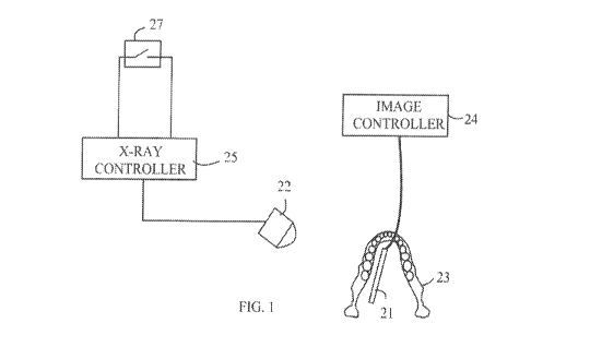
MOM The ifiwitigr in 114kb:the present :inventionprovide.S its advantages can
be
more easily understood With reference to Figure 1, Which illustrates the
arrangement of the
various components in a &Mat k-tay analysis of a patients teeth. Typically, an
filming
Roam 21 is placed in the patieheg mouth 'so that 8.-rays from an x-ray
waive 22 will pass
through the patients teeth 23 and be detected by the imaging array.
Thehnaging, play is
controlled by image Controller 24 that processes the data from:nnaging sensor
21 to provide
an image Of the patient!Siteeth that is stored in an electronic file
associated with the patient.
X-ray sOfiroe::22 is controlled from a separate X-ray controller 25 that:
includes .a high voltage
power Supply that applies an acceleration potential that cause electrons to
strike :a Metal target
to generate: the lk-fa,`.tk. Many
dental :setiings,:the x-ray source was:pureliased byte dentist
CA 03013358 2018-07-31
WO 2017/120621
PCT/US2017/018658
to be used it a film-based diagnostic system. Thedigitat imaging system is
typieally
pin-chased as a reptaeetnent for the film component oldie old system
and is completely
independent of the older x-ray source.
1001 9J this film replacement mode, the old .way system is operated by
.rnandally
dosing a switch 27 that initiates a high voltage pulse .of the appropriate
length to provide the.
.desired x-ray exposure: Prior art systems in which 21 is controlled in
response to the generation of X-rays are known. In such systems, the imaging
array is placed
in a:ready. state prior to: switch :27 beingelos0.by.an operator entering .
the Appropriate
cortnnand to image eon troller24 Inthessysteins imaging:seri:Or 21
:includes..a.dose
.ineasuring sutpsysterp that detects:. the. got of the x-ray. po Ise
:Sy.StemS. based on measuring
currents generated. in the imaging *Of or
a..secondary.Sensor associated with imaging
sensor 21 .are known to :the art In these Systernsõ the irottgli* array is
reset at the start of the.
x-ray pulse. The image is.appitnntated: until:the X-ray source is turned Off
The imaging array
C11. CO/KT detect:the eessatiort of thic. x-ray evOsitretisingthe same stun
that detecfpdjhe:
start of the ptilse of x-rays or merely wait a 'predetermined :length of
time,. in either case, the
hiltage.ireadOut. and processed after the x-ray exposarel.haa:hemcompleted,
[9.020] in this type: of syStern,:the x-ray exposure determined by the on
the
way .contract, The settings are t*seally not varied to account for different.
patients facial
abaOrptiOn of the x-rays. . etc,. As a reSult the settings Are typically set
ito.sorne.safe..sotting that
etwoi..4 sufficient mtOoStire fOr .a ltpatitAK. .A0cOrdiqfY many patients
receive a larger
exposure than neeesSaiy:.
[00211 The presentinvention.overcometheseprohlems. by retrofitting the
..00nventional.X4aysyStom with q:trigpt. switch that
ean..be.010Mled'hythetontreler that
mOnitorStiteimaging. array and byexecating:alVeXspOSWt.thal Consists. of
ii.Sequerice of Short
pulses of X-rays rather than opelohg.pulse. Refer now to Figure 2,, which
illustrates one
:embodiment :of an :x-ray exposure systein according to the. present
invention. In digital x-ray
;ystem::40,;..the: trigger switch that initiated an x-ray burst from
.k4ayhead.4.1 has been
replaced by. a:remote controllable switch 42, S.i*,.0 thloeation of
i.eiriete:coraroll able switch.
42 is not necessarily to image controller 51 that Ontrolaintaging array 525
remote
controllable switeh 42 preferablyeeridrantkates with image controller 51 via a
wireless
CA 03013358 2018-07-31
WO 2017/120621
PCT/US2017/018658
:.Oottorittoicatiot. link. 53 such .that a:eable is: not needed
betwortremott:tOrittoil able switch 42
and linage Controller :51. Remote controllable sWitch..4Z.allows. image
:011061W 5:1. to trigger
pulSe of x-rays.
[00221 :linaging.arroy 52 includes an x-ray doe detector that the
dose of x.;,.
rays.receiVed at imaging array 52 Any of a .ntimbetOf systeins.
tbr.tneasuring.the x-ray dose
can he utilized:. For expmple.)07.0es striking theinAgirig.iforiwAr.fj.knmrn.
to :give ..rise to;
currents that can be rneasure For example, nt. Onto guard. riritthat is
normally used to
shield the itnagibg.array from transients gettetated by other .6t.cuitr.yin
a.CMOS imaging
array is used 4,a/ix-Tay detector by measuring the eurrent that: flows
:between th,.U. guard. ring
and a.p.o.w.er rail in another example, theitttaging array is readout after
each pulse of .-rays
and. the total .sub,evosure determined by. examining: thesums. of the signals
front a
predetermined set of pixels.
[00.23]. As noted abovo.;..the.11.tray source is operated in a series of shot
:04p0,.hi
general. the .exposure of each pulse is set hycorronlson.the x-ray source. In
the present
these controls are set such that the pulse dcItyaeci: in response to. each
trigger pulse
is a knaft fraction of the required. :dose to provide an Image of the desired
.quality. For
example, each pulse could .be set to one,tenth.ofthe expoteddeSe,..S0 that: a
total Of about ten
pulses are needed to provide the expospre In embodiments in Which
4.$epArAtetioSirrreter is
used, image- controller Si initializes.:itnagint.array52.0d:sends the first
pie Of x-rays.
!triage: controller 5.1 thenteads out the dosimeter =404:de.-0.5.triiitio the
number of pulses that:
wilrbeneeded.to.:provide thc..torr0.ex.poswt linage eontr011er.51 then
pillse$: the .x,-ray
source for the determined number of pulses In one aspect of the invention,
image controiler
5.1 TeadS out the dosimeter at one or more intermediate points in the
eXpositrelo verify that
the initial estimate of delred exposure dine was correct.
1Ø02..41 jntmotheteMbodittient,the dOsciS.Set to a smelt fraction of the
estimated correct dose:and:tho: dosimeter is readout.. linage controller 51
then instructsthe
operatorto 8,et the x-ray exposure tOa value determined by that dose
Theirnaging:army:iS
then reset:and:the x-ray systetti triggered for the calculated exposoretime,.
This embodiment
pliminates.thedod. ti me between .x'-ray pulses; 'however, it requires more
skill :and time on. the:
part of the Nowt:
CA 03013358 2018-07-31
WO 2017/120621
PCT/US2017/018658
7
00751 Therein6w:controllable Omer switch ofthepresent invention provides a
sisnplemethod.for upgrading n ebnventional .st4ey source or use .with:the
dignal .x-ray
system, The sWiteh: typically .inalcos::a connection between two contneta that
are held...at:a
potentiattelatiVe to one anothet,. This potential.differenee can be
toppo4.tp:poW:dvt.hp=Tadip
link between the reitete .06ot-rabic switeh...andthecontr011er of the digital
x-ray system. If
Ahepoker available itonithiasounze is lnsurficient;::.a: battery .thatis
charged from the source
over. a long period of time can be utilized to power the.wireless.
00261 In the above-described embodiments, the Oche eontrellable switch that
proVides:thelink between the.image:contr011er :and the x-ray controller is
inserted into the .:717,
lay Controller by replacing thexlay trigger $.witeh: that is:::c601SMinly
itaeditt existing
generating systems. Since the, trigger sw,.:.iteb does not sot the length Or
the x-raypulse, some.
other strategy is needed to turnoff' the x-ray generator when
thk;t:patienuhasreeeiVed the..
desired level of exposure. ln one aspect:olthe invention, a Second
radioxontrolied switch.. is
inserted within the x-ray generation system to interrupt the high
voltagetothevoy head
Thia.second.switelt allowatheimoging eontkslleftoteiminatc the exposure at the
.apprOpriato
time without significantly altering theumietlying .electronics :of the A.-
my.genetution system..
Refer now to figurel, which illustrates inother embodiment of a. digital x-ray
system
according to the proseor invention:. TO.SiMplify:thefollowing: discussion,
those elements of
digital: *ray SYSteMØ0thataerve functions analogona to elements 'of x-ray
system 40
discussed above have been given
the:siie:numericatdesignationatta.theetomentabf digital
system 40 and will net be diSeussed in detail here.
[0037] Digital x-ray system 60 is based:0.01e observation that )t-ray head 41
typically
includes an x-ray generating tube that is powered by k.signal source that
&mks; electrons to
be needlerated..intO.a. metal target: The signal source typically incindes a
high voltage power
supply line that provides the potential for the 'acceleration.. Horioo,
the'::444ayedtpet..ean:be
terminated by interrupting this high vOltage power line t$Iing:a second radio
remote .controlled
:sMtt-il. 67 that is operated by a second wireless link 64. In this:
embodiment, image controller
.61.controlathe .xray process and provides the user interface fbr the :control
of that process,
The x-ray controller 257:is set to pro* an ovowit that .issutlieieritt6 ensure
that all.
patients canTeCeiv.e at least the desired.x,ray aluminajoi if the entire is
allowed to
CA 03013358 2018-07-31
WO 2017/120621
PCT/US2017/018658
8
proceed. When the x-ray imaging process is triggered by auser inputting=
appropriate
corninapdtoirptige::pontrolter 61. via tad. interface 66, image controller 61
uses .the ..wireless.
intelfacebetweeh. antennas $3 and 65 to triggodiewaygenerator
with...wirelesslii* 64
closed, After image 001.011061 .detects that the desitax-ray.exposure:haa been
Obtained,
Image controller 61 opens indio remote Controlled .switch 67 using the
wireless bnk between
wirOess. link 04 and. antenna 65. Thetemainderof the programmed exposure in x-
ray
.0400er 25 does.not.generate *rays:.
.[60281 .The aboVe.,.described.embodiinerits of thopresent invention can be
advantageously used in dental settings having existing x-ray .generators. .
Typically. these x-
ray .generators are.made.hy a different.,..rriannfacturer. or L10.11141 p /via
entity than the
manufacturer of the digital image controller. The present invention providesa
method for
sloing the x-raygenerater to the digital nnaging systent that reottiNs:
only,Mininial. changes
the x-ray generator. Thew changcs:can.be provided on .ite.in,adental.Offiee
.that already.
has an :x4aygenerator: or bytho manufacturer of 'digital ko.y.imaglng.sysWth.
that then sell s...4.
complete system to the dentist,
100291 Tlio.Sboyondoseribod embodiments of the ptesent invention have been
provided toillustrate:Various aspects of the invention. HOwever, iris to be
understood that
di:Eft:two iispoMa of the proseiti. invention that are shown in.diffesent
specific embodiments
ean: be combined to proVide .other einbodinients Of the present invention. in
addition, various
modifications to the present invention will become apparent from the
foi7cgoitig.desetiptim
00:00empaiiyitig:d6vviogs. ACcOrdingly, the present invention is to be limited
solely by the
scope of the .follotiltig claims