Note: Descriptions are shown in the official language in which they were submitted.
A MEDICAL SUCTION TOOL FOR A EUSTACHIAN TUBE
FIELD OF THE INVENTION
The present invention relates generally to medical
devices, and particularly to methods and systems for
suctioning material from a Eustachian tube of a patient.
BACKGROUND OF THE INVENTION
Some ear-nose-throat (ENT) procedures require removing
material, such as liquid or mucus, from the Eustachian tube
and sinuses of a patient.
For example, U.S. Patent Application Publication
2013/0303968 describes methods and devices for providing a
gas pathway between the nasopharynx and the Eustachian
tube. One device may include a lumen with a valve. A
portion of the valve may be tethered to adjacent muscle.
Another portion of the valve may be tethered to adjacent
cartilage.
U.S. Patent Application Publication 2014/0296898
describes various methods and devices used for remodeling
or changing the shape, size or configuration of a sinus
ostium or duct or other anatomical structure in the ear,
nose or throat, removing matter from the ear, nose or
throat, delivering diagnostic or therapeutic substances or
performing other diagnostic or therapeutic procedures.
SUMMARY OF THE INVENTION
An embodiment of the present invention that is
described herein provides a method that includes inserting
into a patient body a medical suction tool, which includes
a hollow first tube for removing material away from a
Eustachian tube of a patient, and a hollow second tube
disposed around the first tube. The medical suction tool is
navigated to the Eustachian tube. The Eustachian tube is
sealed by coupling an outer surface of the second tube to
1
CA 3013716 2018-08-09
an inner surface of the Eustachian tube. While the
Eustachian tube is sealed by the second tube, the material
is removed away from the Eustachian tube via the first
tube.
In some embodiments, at least one of the first and
second tubes is flexible. In other embodiments, navigating
the medical suction tool includes tracking a position of
the medical suction tool using a position sensor of a
position tracking system, which is coupled to a distal end
of the medical suction tool and produces position signals
that are indicative of the position of the position sensor.
In yet other embodiments, tracking the position includes
tracking a magnetic position sensor that includes a single
coil.
In an embodiment, inserting the medical suction tool
includes inserting the medical suction tool through a nose
of the patient. In another embodiment, the method includes
cleaning the Eustachian tube by moving the medical suction
tool along sidewalls of the Eustachian tube. In yet another
embodiment, removing the material includes drawing the
material away from the Eustachian tube by applying suction
using a suction apparatus coupled to the medical suction
tool.
There is additionally provided, in accordance with an
embodiment of the present invention, a medical suction tool
that includes a hollow first tube and a hollow second tube.
The hollow first tube is coupled to a suction apparatus and
is configured to remove material away from a Eustachian
tube of a patient. The hollow second tube is disposed
around the first tube, and configured to seal the
Eustachian tube by coupling an outer surface of the second
tube to an inner surface of the Eustachian tube.
There is further provided, in accordance with an
embodiment of the present invention, a method for producing
2
CA 3013716 2018-08-09
a medical suction tool, the method includes coupling to a
suction apparatus a hollow first tube for removing material
away from a Eustachian tube of a patient. A hollow second
tube is disposed around the first tube for sealing the
Eustachian tube by coupling an outer surface of the second
tube to an inner surface of the Eustachian tube.
The present invention will be more fully understood
from the following detailed description of the embodiments
thereof, taken together with the drawings in which:
BRIEF DESCRIPTION OF THE DRAWINGS
Fig. 1 is a schematic, pictorial illustration of a
sinuplasty procedure using a sinuplasty system, in
accordance with an embodiment of the present invention;
Fig. 2 is a sectional side view of an ear and a
suction module, in accordance with an embodiment of the
present invention; and
Fig. 3 is a flow chart that schematically illustrates
a method for suctioning material from a Eustachian tube, in
accordance with an embodiment of the present invention.
DETAILED DESCRIPTION OF EMBODIMENTS
OVERVIEW
Some medical procedures, such as sinuplasty, require
removing material from anatomical cavities, such as a
Eustachian tube of a patient inner ear. The Eustachian tube
is typically narrow and cannot be accessed through the
outer ear without damaging the ear drum and organs of the
inner ear. In principle, it is possible to insert a suction
device into a cavity (e.g., sinus) located some distance
away from the Eustachian tube, but the suction will be
ineffective due to lack of sealing between the suction
device and the Eustachian tube.
3
CA 3013716 2018-08-09
Embodiments of the present invention that are
described hereinbelow provide improved techniques for
suctioning undesired material, such as mucus and infectious
fluid, from the Eustachian tube. In some embodiments, a
suction module for suctioning the undesired material
comprises a suction tool coupled to a suction apparatus,
such as a medical suction pump.
In some embodiments, the suction tool comprises a
hollow flexible internal tube, which is coupled, at its
proximal end, to the suction pump, and configured to remove
the undesired material away from the Eustachian tube. The
suction tool further comprises a hollow flexible external
tube, which is disposed around the internal tube. The
external tube is configured to seal the Eustachian tube by
coupling its outer surface to an inner surface of the
Eustachian tube.
In some embodiments, a position sensor of a position
tracking system is coupled to the distal tip of the suction
tube and configured to produce position signals that are
indicative of a position of the suction tool in an organ,
such as an ear-nose-throat (ENT) system, of the patient.
In some embodiments, the suction module is
electrically connected to a processor, which is configured
to receive pre-acquired anatomical images, such as
computerized tomography (CT) images depicting respective
segmented two-dimensional (2D) slices of the patient ENT,
from a CT system, and to register between coordinate
systems of the CT system and the position tracking system.
In some embodiments, the processor is further
configured to track the position of the distal tip in the
patient body, and to display a marker on a respective
anatomical image, indicative of the position of the
position sensor in the anatomical image.
4
CA 3013716 2018-08-09
In some embodiments, during the sinuplasty procedure,
the physician inserts the suction tool, through the patient
nose, and navigates the suction tool to an ostium of the
Eustachian tube, using the marker displayed on the
anatomical image.
In some embodiments, the physician seals the
Eustachian tube by coupling the outer surface of the
external tube to the inner surface of the Eustachian tube.
Subsequently, the physician removes the undesired
material from the Eustachian tube by applying suction using
the suction pump coupled to the proximal end of the suction
tool. After concluding the material removal, the physician
extracts the suction tool, through the patient nose, out of
the body of the patient.
SYSTEM DESCRIPTION
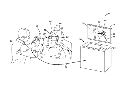
Fig. 1 is a schematic pictorial illustration of a
sinuplasty procedure using a sinuplasty system 20, in
accordance with an embodiment of the present invention. In
some embodiments, sinuplasty system 20 comprises an ear-
nose-throat (ENT) suction module 28, which is configured to
remove matter, such as infection, liquid or mucus, from a
Eustachian tube 50 of a patient 22.
In some embodiments, suction module 28 comprises a
distal end, such as an ENT suction tool 38, which a
physician 24 inserts into a nose 26 of patient 22. Module
28 further comprises a handheld suction apparatus 30,
coupled to a proximal end of suction tool 38 and configured
to assist physician 24 in navigating tool 38 into
Eustachian tube 50 and in applying suction, so as to remove
the matter away from Eustachian tube 50. ENT suction tool
is depicted in further detail in Fig. 2 below.
In an embodiment, system 20 further comprises a
magnetic position tracking system, which is configured to
5
CA 3013716 2018-08-09
track the position of one or more position sensors in the
head of patient 22. The magnetic position tracking system
comprises magnetic field-generators 44 and one or more
position sensors shown in Fig. 2 below. The position
sensors generate position signals in response to the sensed
external magnetic fields from the field generators, thereby
enabling a processor 34 to map the position of each sensor
as will be described below.
This method of position sensing is implemented in
various medical applications, for example, in the CARTOTm
system, produced by Biosense Webster Inc. (Diamond Bar,
Calif.) and is described in detail in U.S. Patents
5,391,199, 6,690,963, 6,484,118, 6,239,724, 6,618,612 and
6,332,089, in PCT Patent Publication WO 96/05768, and in
U.S. Patent Application Publications 2002/0065455 Al,
2003/0120150 Al and 2004/0068178 Al, whose disclosures are
all incorporated herein by reference.
System 20 further comprises a location pad 40, which
comprises field-generators 44 fixed on a frame 46. In the
exemplary configuration shown in Fig. 1, pad 40 comprises
five field-generators 44 but may comprise any other
suitable number of generators 44. Pad 40 further comprises
a pillow (not shown) placed under a head 41 of patient 22,
such that generators 44 are located at fixed, known
positions external to patient 22.
In some embodiments, system 20 comprises a console 33,
which comprises a processor 34, typically a general-purpose
computer, with suitable front end and interface circuits
for receiving signals from tool 28 having a magnetic sensor
attached thereon (shown in Fig. 2 below), via a cable 32,
and for controlling other components of system 20 described
herein.
In some embodiments, processor 34 is configured to map
the position of each position sensor so as to estimate the
6
CA 3013716 2018-08-09
position and orientation of a distal tip (shown in Fig. 2
below) of tool 28 in the coordinate system of the optical
position tracking system.
In some embodiments, processor 34 is configured to
receive one or more anatomical images, such as computerized
tomography (CT) images depicting respective segmented two-
dimensional (2D) slices of a head 41 of a patient 22,
obtained using an external CT system (not shown). The term
"segmented" refers to displaying various types of tissues
identified in each slice by measuring respective
attenuation of the tissues in the CT system.
Console 33 further comprises input devices 39 and a
user display 36, which is configured to display the data
(e.g., images) received from processor 34 or inputs
inserted by a user (e.g., physician 24).
In some embodiments, processor 34 is configured to
display from among the CT images, one or more selected
slices, such as an image 35, on user display 36. In the
example of Fig. 1, image 35 is a sectional view of an ear
48 of patient 22, such that image 35 comprises Eustachian
tube 50.
Console 33 comprises a driver circuit (not shown),
which is configured to drive field-generators 44 with
suitable signals so as to generate magnetic fields in a
predefined working volume around head 41.
Fig. 1 shows only elements related to the disclosed
techniques, for the sake of simplicity and clarity. System
20 typically comprises additional modules and elements that
are not directly related to the disclosed techniques, and
thus, intentionally omitted from Fig. 1 and from the
corresponding description.
Processor 34 may be programmed in software to carry
out the functions that are used by the system, and to store
data in a memory (not shown) to be processed or otherwise
7
CA 3013716 2018-08-09
1
used by the software. The software may be downloaded to the
processor in electronic form, over a network, for example,
or it may be provided on non-transitory tangible media,
such as optical, magnetic or electronic memory media.
Alternatively, some or all of the functions of processor 34
may be carried out by dedicated or programmable digital
hardware components.
Fig. 2 is a sectional side view of ear 48 and suction
module 28, in accordance with an embodiment of the present
invention. During the sinuplasty procedure, physician 24
inserts suction tool 38, typically through nose 26, into an
ostium 66 of Eustachian tube 50. Note that suction tool 38
is coupled to suction apparatus 30, located externally to
patient 22 and may be used by physician 24 for navigating
suction tool 38 from nose 26 to Eustachian tube 50 of ear
48. Additionally or alternatively, any other suitable
apparatus may be used by physician for the navigation of
suction tool 38.
Reference is now made to an inset 62 showing the
distal end of suction tool 38. In some embodiments, suction
tool 38 comprises an internal hollow tube 54, which is
typically flexible but can also be rigid, and an external
hollow tube 58 disposed around internal hollow tube 54.
External hollow tube 58 is typically flexible but may also
be rigid.
In some embodiments, tubes 54 and 58 are typically
made from polymers, such as polyurethane and polyamide, or
from any other suitable biocompatible material. In an
embodiment, suction tube 38 is produced such that an outer
diameter 80 of the distal end of tube 38 has a similar
value to an inner diameter (i.e., between inner surfaces
68) of Eustachian tube 50, e.g., about 1 mm. Note that the
inner diameter of Eustachian tube 50 may be measured, e.g.,
in image 35, before performing the sinuplasty procedure, so
8
CA 3013716 2018-08-09
that physician 24 may select a suction tool having a
suitable external diameter that can fit into Eustachian
tube 50. In this embodiment, when physician inserts suction
tool 38 into ostium 66, suction tool 38 is configured to
seal Eustachian tube 50 by coupling an outer surface 70 of
external tube 58 to inner surface 68 of Eustachian tube 50.
Reference is now made back to inset 62. In some
embodiments, openings in a distal tip 74 of hollow tubes 54
and 58 form an opening 72, through which suction tool 38
removes material away (e.g., liquid, mucus, infection,
dirt) from cavities in an inner ear 60, and/or from
Eustachian tube 50.
In some embodiments, suction tool 38 comprises a
strengthening element, such as a wire 56 that extends along
at least part (e.g., the distal end) of external tube 58
and is coupled thereto. In an embodiment, wire 56 is made
from an alloy of nickel-titanium, such as nitinol, or any
other suitable material, and is configured to mechanically
strengthen external tube 58.
In some embodiments, suction tool 38 further comprises
a position sensor 64 of the position tracking system
described in Fig. 1 above. In some embodiments, position
sensor 64 comprises a single coil configured to generate
position signals, or any other suitable number of coils. In
the example of Fig. 2 position sensor 64 is coupled to
distal tip 74 of suction tool 38, such that sensor 64 does
not block opening 72. In this configuration, sensor 64 may
be coupled between an outer surface of tube 54 and an inner
surface of tube 58, such that electrical leads (not shown)
connected to sensor 64 are disposed between these surfaces
and configured to conduct the position signal sensed by
position sensor 64 to cable 32.
9
CA 3013716 2018-08-09
In other embodiments, sensor 64 may be coupled to
surface 70 of tube 58, or in any other suitable
configuration in distal tip 74.
In alternative embodiments, sensor 64 may be coupled
along suction tool 38 at any suitable offset relative to
distal tip 74, such that processor 34 applies the offset to
calculate the position of distal tip 74 in the coordinate
system of the position tracking system.
In some embodiments, processor 34 is configured to
register between coordinate systems of the CT system and
the position tracking system. In an embodiment, processor
34 is configured to display, based on the registered
coordinate systems, an indication of the position of distal
tip 74 in image 35, so as to assist physician 24 in
navigating distal tip 74 into ostium 66.
In some embodiments, suction tool 38 is further
configured to clean Eustachian tube 50 from any undesired
material (e.g., dirt, mucus, infectious fluid), for
example, by peeling and suctioning the dirt from surface
68. The cleaning may be carried out by an operator, such
as physician 24, which moves suction tool 38, e.g., back
and forth between ostium 66 and an ostium 76, along surface
68 of Eustachian tube 50. In some embodiments, physician 24
can see the position of distal tip 74 in image 35, so as to
pull suction tool 38 back before reaching ostium 76.
The configurations of suction module 28, and
particularly, suction tool 38 described in Figs. 1 and 2
above, are depicted purely by way of example. In
alternative embodiments, module 28 and tool 38 may comprise
any suitable configuration, having any suitable size and
shape and arranged so that suction tool 38 can be navigated
to, and snugly fitted into Eustachian tube 50 to enable
suctioning material therefrom and cleaning surface 68 and
other parts of Eustachian tube 50. For example, opening 72
CA 3013716 2018-08-09
may be replaced by one or more openings along the distal
end of suction tool 38, this configuration enables removing
material from other cavities or anatomical tubes having a
different geometry. Note that the flexibility of tubes 54
and 58 provides physician 24 with the capability to
navigate and insert suction tool to various organs, such as
in brain surgical applications or in small blood vessels in
the body of patient 22.
Fig. 3 is a flow chart that schematically illustrates
a method for suctioning material from Eustachian tube 50,
in accordance with an embodiment of the present invention.
The method begins with an insertion step 100, in which
physician 24 insert suction tool 38, which comprises
internal hollow tube 54 and external hollow tube 58
disposed around tube 54, into patient nose 26.
At a navigation step 102, physician 24 navigates
suction tool 38 to ostium 66 of Eustachian tube 50, using
the tracked position of sensor 64 displayed in image 35. At
a cleaning step 104, which is an optional step in this
method, physician 24 cleans inner walls 68 of Eustachian
tube 50 by moving suction tool 38 back and forth in along
walls 68, thereby peeling undesired material off walls 68
of Eustachian tube 50.
At a sealing step 106, physician 24 seals Eustachian
tube 50 by coupling outer surface 70 of external tube 58 to
inner surface 68 of Eustachian tube 50. At a suctioning
step 108, physician 24 removes undesired material, such as
dirt, mucus, and infectious fluid, from Eustachian tube 50,
using suction apparatus 30, which is coupled to the
proximal end of suction tool 38. At an extraction step 110,
which concludes this method, physician 24 extracts suction
tool 38 out of the body of patient 22, through patient nose
26.
11
CA 3013716 2018-08-09
In alternative embodiments, physician 24 may change
the order of at least some of the steps of the method. For
example, physician 24 may carry out cleaning step 104 after
suctioning step 108, and then repeat sealing step 106 and
suctioning step 108 so as to clean the material peeled off
walls 68 at cleaning step 104.
Although the embodiments described herein mainly
address removing material from the Eustachian tube, the
methods and systems described herein can also be used in
other applications, such as in suctioning undesired
material from any other cavity of the ENT system or any
other anatomical system of the body.
It will be appreciated that the embodiments described
above are cited by way of example, and that the present
invention is not limited to what has been particularly
shown and described hereinabove. Rather, the scope of the
present invention includes both combinations and sub-
combinations of the various features described hereinabove,
as well as variations and modifications thereof which would
occur to persons skilled in the art upon reading the
foregoing description and which are not disclosed in the
prior art. Documents incorporated by reference in the
present patent application are to be considered an integral
part of the application except that to the extent any terms
are defined in these incorporated documents in a manner
that conflicts with the definitions made explicitly or
implicitly in the present specification, only the
definitions in the present specification should be
considered.
12
CA 3013716 2018-08-09