Note: Descriptions are shown in the official language in which they were submitted.
CA 03013808 2018-08-06
WO 2017/153567 1-
PCT/EP2017/055652
-
POSTERIOR OCULAR FIBROSIS INHIBITION BY ANTAGONIZING PLACENTAL
GROWTH FACTOR
FIELD OF THE INVENTION
The invention is situated in the field of ocular therapies. In particular it
refers to antagonists of
placental growth factor for interfering with posterior ocular fibrosis.
BACKGROUND TO THE INVENTION
The retina of an eye (most posterior segment of the eye; back of the eye) is
part of the central
nervous system (CNS). As such the retina's wound-healing response is similar
to the wound-
healing response of the brain which Friedlander refers to as gliosis (fibrosis
mediated by glial
cells). This in contrast to wound-healing responses in non-CNS tissues or
organs in general and
anterior ocular segments (front of the eye) such as cornea and trabecular
meshwork specifically,
which is referred to as fibrosis (fibrosis mediated by fibroblasts)
(Friedlander 2007, J Clin
Invest, 117:576-586).
Any type of retinal disease or disorder accompanied by or caused by
inflammation and/or
neovascularization leads to gliosis and fibrous scarring. Ultimately, this
gliosis, or posterior
ocular fibrosis, leads to severe vision loss and blindness. Although a number
of drugs are
available to suppress neovascularization (e.g. pegaptanib sodium and
ranibizumab; and, off-
label, bevacizumab; all targeting vascular endothelial growth factor, VEGF),
these do not
minimize gliosis/posterior ocular fibrosis (Friedlander, J Clin Invest 2007,
117:576-586). It is
described that long-term use of anti-VEGF therapy can even lead to increased
posterior ocular
fibrosis. From the CATT-trial, for instance, it is known that 24.7 % of the
patients with age-
related macular edema (AMD) will develop a posterior fibrotic scar after 2
years of anti-VEGF
therapy (bevacizumab or ranibizumab) (Daniel et al., Ophthalmology 2014,
121:656-666).
.. Recently, Van Bergen et al. (Invest Ophthalmol Vis Sci 2015, 56:5280-5289)
used the
experimental murine model of laser-induced choroidal neovascularization (CNV)
to
demonstrate reduction of posterior ocular fibrosis by means of antibodies
targeting LOX (lysyl
oxidase) or LOXL2 (lysyl oxidase-like 2). In a similar model, Rakic et al.
(Invest Ophthalmol
Vis Sci 2003, 44:3186-3193) identified placental growth factor (P1GF) as one
of the growth
CA 03013808 2018-08-06
WO 2017/153567 -2-
PCT/EP2017/055652
factors contributing to CNV, more in particular contributing to
neovascularization and lesion
size 14 days after inducing laser injury. Hollborn et al. (Graefe's Arch Clin
Exp Ophthalmol
2006, 244:732-741) determined that in vitro grown human retinal pigment
epithelial (RPE) cells
stimulated by transforming growth factor- 0 (TGF-I3) produce increased amounts
of P1GF and
VEGF, leading to the suggestion that during diabetic retinopathy, TGF- 0-
caused P1GF-
secretion by RPE cells may contribute to cell migration as part of the
formation of fibrovascular
membranes. The presence of myofibroblasts in these membranes can cause
tractional retinal
detachment and retinal hemorrhage.
Cao et al. (Invest Ophthalmol Vis Sci 2010, 51:6009-6017) investigated the
effect of a VEGF-
Trap (binding both VEGF and P1GF) on CNV induced by subretinal injection of
Matrigel. The
authors observed arrested CNV growth and reduced inflammatory and fibrotic
responses.
Matrigel, however, contains several growth factors including basic fibroblast
growth factor
(bFGF), epidermal growth factor (EGF), insulin-like growth factor 1 (IGF-1),
TGF-13, platelet-
derived growth factor (PDGF), nerve growth factor (NGF), and connective tissue
growth factor
(CTGF) (Hughes et al., Proteomics 2010, 10:1886-1890). Results obtained with
this model can
therefore not be compared to results obtained with the laser-induced CNV model
which does
not introduce an external cocktail of growth factors in the eye.
A beneficial effect of P1GF-neutralizing antibodies has been described for
many disorders
including pathological angiogenesis, pathological arteriogenesis,
inflammation, tumor
formation, vascular leakage, and pulmonary hypertension (WO 01/85796),
osteoporosis (WO
2004/002524), tissue adhesion (WO 03/063904), liver cirrhosis (W02007/003609),
Philadelphia chromosome positive leukemia (WO 2010/037864) and trabeculectomy
outcome
(WO 2013/07971); see also Fischer et al. (Cell 2007, 131:463-475), Van
Steenkiste et al.
(Gastroenterology 2009, 137:2112-2124), Coenegrachts et al. (Cancer Research
2010, 70:6537-
6547), Van de Veire et al. (Cell 2010, 141:178-190), Schmidt et al. (Cancer
Cell 2011, 19:740-
53), Snuderl et al. (Cell 2013, 152:1065-1076), Van Bergen et al. (J Cell Mol
Med 2013,
17:1632-1643).
Specifically, Van de Veire et al. (2010) noted inhibition by P1GF-neutralizing
antibodies of
ocular angiogenesis, ocular inflammation and choroidal vessel leakage after
laser-induced CNV
(thus in part confirming and extending the data of Rakic et al., Invest
Ophthalmol Vis Sci 2003,
44:3186-3193).
CA 03013808 2018-08-06
WO 2017/153567 3-
PCT/EP2017/055652
-
The beneficial effect of P1GF-neutralizing antibodies on post-operative tissue
adhesion (WO
03/063904) and failure of trabeculectomy Van Bergen et al. (J Cell Mol Med
2013, 17:1632-
1643) may, at least in part, be attributed to apparent inhibition of
fibroblast-mediated fibrosis.
Scarring (fibrosis) is thought to contribute to bleb failure after glaucoma
filtration
surgery/trabeculectomy (Li et al. 2006; Free papers Glaucoma: microbiology and
bloodflow
and IOP; Role of vascular endothelial growth factor and placental growth
factor in glaucoma
and scar formation after glaucoma filtration surgery). In this context, it was
shown that an
antibody blocking the VEGF-R2 receptor did, albeit to a lower extent than a
P1GF-neutralizing
antibody, increase bleb survival (decrease scarring) after glaucoma filtration
surgery/trabeculectomy (WO 2013/07971). Both an antibody blocking the VEGF-R2
receptor
and a P1GF-neutralizing antibody were thus capable of reducing anterior ocular
fibrosis.
Friedlander (J Clin Invest 2007, 117:576-586) summarized the lack of action of
VEGF-
antagonists on posterior ocular fibrosis. The difference in action of VEGF-
antagonists on
posterior and anterior ocular fibrosis indicates a difference in the processes
between posterior
ocular fibrosis and anterior ocular fibrosis.
SUMMARY OF THE INVENTION
The invention relates to monospecific placental growth factor (P1GF)
antagonists for use in
treating, preventing, or delaying progression of ocular posterior fibrosis in
a subject.
Alternatively, the monospecific placental growth factor (P1GF) antagonist is
for use in treating,
preventing, or delaying progression of ocular posterior fibrosis without
inducing ocular
posterior neurodegeneration in a subject. As a further embodiment the
monospecific placental
growth factor (P1GF) antagonist for the above uses envisages further treating,
preventing, or
delaying progression of ocular posterior inflammation and/or ocular posterior
neovascularization and/or vessel leakage and/or for use in maintaining or
improving the visual
acuity of a subject with an eye of which the retina is damaged.
The invention further relates to monospecific placental growth factor (P1GF)
antagonists for
use in maintaining or improving the visual acuity of a subject with an eye of
which the retina is
damaged.
CA 03013808 2018-08-06
WO 2017/153567 4-
PCT/EP2017/055652
-
In any of the above, the monospecific placental growth factor (P1GF)
antagonist alone can be
administered to an eye.
Alternatively, in the above uses, the monospecific placental growth factor
(P1GF) antagonist
may be administered to an eye after wash out of a vascular endothelial growth
factor (VEGF)
antagonist or a VEGF-receptor (VEGFR) antagonist previously administered to
the same eye.
In a further alternative, a vascular endothelial growth factor (VEGF)
antagonist or a VEGF-
receptor (VEGFR) antagonist is administered to an eye after wash out of the
monospecific
placental growth factor (P1GF) antagonist previously administered to the same
eye.
Further alternatives are envisaged including the combined administration of a
monospecific
placental growth factor (P1GF) antagonist and a second active compound. As
such, in any of
the above-described uses, the monospecific placental growth factor (P1GF)
antagonist may be
administered to an eye in combination with a second active compound wherein
said second
active compound is different from a vascular endothelial growth factor (VEGF)
antagonist and
different from a VEGF-receptor (VEGFR) antagonist.
Alternatively, the monospecific placental growth factor (P1GF) antagonist is
administered to an
eye in combination with a second active compound wherein said second active
compound is
different from a vascular endothelial growth factor (VEGF) antagonist and
different from a
VEGF-receptor (VEGFR) antagonist; and wherein said administration is after
wash out of a
vascular endothelial growth factor (VEGF) antagonist or VEGF-receptor (VEGFR)
antagonist
previously administered to the same eye.
In a further alternative, a vascular endothelial growth factor (VEGF)
antagonist or a VEGF-
receptor (VEGFR) antagonist is administered to an eye after wash out of the
monospecific
placental growth factor (P1GF) antagonist previously administered to the same
eye in
combination with a second active compound wherein said second active compound
is different
from a vascular endothelial growth factor (VEGF) antagonist and different from
a VEGF-
receptor (VEGFR) antagonist.
When a monospecific placental growth factor (P1GF) antagonist is combined with
a second
active compound, both can be administered to the eye each in a separate
composition. The
CA 03013808 2018-08-06
WO 2017/153567 5-
PCT/EP2017/055652
-
second active agent can be administered prior to, concurrent with, or after
the administration of
the monospecific placental growth factor (P1GF) antagonist.
Alternatively, both can be administered to the eye combined in a single
composition. Second
active compounds in this context may be one active compound or a combination
of more than
one active compound. In particular, but not limiting, such second active
compound may be an
anti-inflammatory compound, an anti-angiogenic compound, an anti-fibrotic
compound, an
AGE-inhibiting compound, an ALE-inhibiting compound, an AGE-breaking compound,
a
carbonic anhydrase inhibitor, an NMDA-receptor antagonist, a kainate receptor
antagonist, an
AMPA-receptor antagonist, a neuroprotective agent, an agent for controlling
the intra-ocular
pressure, an anti-apoptotic agent, an antiviral compound, an antibiotic
compound, an antifungal
compound, an antihistamine, an anticoagulant, a thrombolytic compound, an anti-
mitotic agent,
an anesthetic agent, and agent inducing mydriasis, an agent inducing
cycloplegia, an agent
inducing posterior vitreous detachment (complete or incomplete), an agent
inducing vitreous
liquefaction, an integrin inhibitor, an anti-edema agent.
Any use of the monospecific placental growth factor (P1GF) antagonist as
hereinabove
described may further be combined with photodynamic therapy, laser
photocoagulation,
radiation therapy or vitreal surgery.
The monospecific placental growth factor (P1GF) antagonist for any use as
described
hereinabove may be further characterized in that the posterior ocular fibrosis
is occurring
concurrent with or after retinal damage. Such posterior ocular fibrosis may
for instance be
occurring in age-related macular edema, diabetic retinopathy, (diabetic)
macular edema, any
type of retinopathy, neovascular glaucoma, retinal detachment or retinal
hemorrhage.
The monospecific placental growth factor (P1GF) antagonist for any use as
described
hereinabove may be further characterized in that it is a P1GF-neutralizing
antibody or a P1GF-
neutralizing fragment of an antibody, an antisense RNA, a small interfering
RNA, an aptamer,
or a ribozyme. Herein, a P1GF-neutralizing antibody or a P1GF-neutralizing
antibody fragment
may be one comprising the 6 CDRs comprised in the heavy chain defined in SEQ
ID NO:7 and
in the light chain defined in SEQ ID NO:8. In particular, these CDRs are as
defined in SEQ ID
NOs: 1 to 6 when applying the IMGT-method to SEQ ID NOs:7 and 8.
CA 03013808 2018-08-06
WO 2017/153567 -6-
PCT/EP2017/055652
The invention also relates to an isolated P1GF-neutralizing antibody, or a
P1GF-neutralizing
antibody fragment thereof, comprising the 3 heavy chain CDRs comprised in the
heavy chain
defined in SEQ ID NO:12 and the 3 light chain CDRs comprised in the light
chain defined in
SEQ ID NO:13.
FIGURE LEGENDS
FIGURE 1. Leukocyte infiltration in the laser-induced CNV model.
(A) In the CNV model, 5 days after lasering, treatment with anti-P1GF antibody
5D11D4 (1.5
and 3.1 g) decreased leukocyte infiltration as compared to IgG-treated mice
(P<0.05);
aflibercept (2.4 and 20 g) and the high-dose oftriamcino lone acetonide
(TAAC; 40 g) showed
comparable effects. Anti-VEGFR2 antibody DC101 administration (3.1 g) did not
show any
anti-inflammatory effects. (B) Equimolar comparison 5 days after lasering.
Treatment with anti-
P1GF antibody 5D11D4 (3.1 g) decreased leukocyte infiltration as compared to
IgG treated
mice (P<0.05); equimolar concentrations of aflibercept (Eylea ) (2.4 g) showed
similar effect.
Anti-VEGFR2 antibody DC101 (3.1 g) and TAAC administration (4 g) did not show
any anti-
inflammatory effects. Data are mean SEM.
FIGURE 2. Posterior ocular collagen deposition in the laser-induced CNV model.
(A) In the CNV model, 30 days after lasering, as compared to PBS treated mice,
treatment with
anti-P1GF antibody 5D11D4 (1.5 and 3.1 g) decreased collagen deposition
(P<0.05); which
was similar to the effect of a high-molar concentration of triamcinolone
acetonide (TAAC;
40 g). Anti-VEGFR2 antibody DC101 and aflibercept (Eylea ) administration
(both 3.1 g)
did not show any anti-fibrotic effects. (B) Equimolar comparison 30 days after
lasering.
Treatment with anti-P1GF antibody 5D11D4 (3.1 g) decreased collagen
deposition as
compared to IgG treated mice (P<0.05). Anti-VEGFR2 antibody DC101 (3.1 g),
aflibercept
(2.4 g) and TAAC administration (4 g) did not show any anti-fibrotic effects.
Data are mean
SEM.
FIGURE 3. Retinal ganglion cell (RGC) survival.
Retinal ganglion cell survival was assessed after 2 (Figure 3A), 4 (Figure 3B)
and 6 weeks
(Figure 3C) of intraperitoneal injections with control IgG, anti-P1GF antibody
5D11D4 and
anti-VEGF-R2 antibody DC101 (all 25 mg/kg, 3 times per week). The RGCs/retinal
area was
CA 03013808 2018-08-06
WO 2017/153567 7-
PCT/EP2017/055652
-
not significantly different between the 3 treatment groups after 2, 4 and 6
weeks in C57B1/6
mice (N=6; P>0.05), whereas a significant reduction was present in Swiss mice
(N=6; P<0.05).
As shown in Figure 3D, TUNEL staining confirming the number of apoptotic cells
per retinal
area in the ganglion cell layer was comparable in the anti-P1GF antibody
5D11D4 versus control
IgG treated mice after 6 weeks (N=6; P>0.05). A trend of increase in apoptotic
cells was present
for the anti-VEGF-R2 antibody DC101 treated C57B1/6 mice (n=6; P=0.10),
whereas the
increase was significant in the Swiss group (n=6; P<0.001). Data represent
mean SEM.
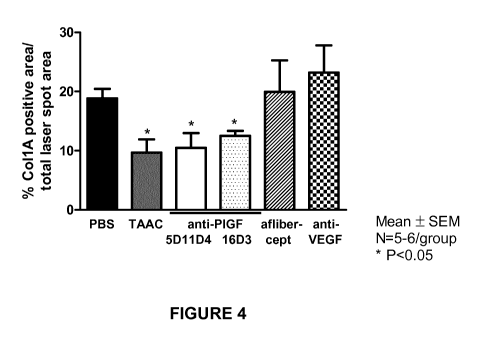
FIGURE 4. Posterior ocular collagen deposition in the laser-induced CNV mouse
model.
Posterior ocular collagen deposition, compared PBS treated eyes, was assessed
after intravitreal
administration of anti-mouse P1GF antibody 5D11D4, of anti-human P1GF antibody
16D3, of
anti-murine VEGF antibody B20 (all 3.1 gg/eye), of aflibercept (equimolar
amount of 2.4
gg/eye), and of triamcinolone acetonide (TAAC; 40gg/eye). Both anti-P1GF
antibodies
significantly decreased collagen deposition (P<0.05); which was similar to
administration of
TAAC (40gg/eye). Administration of an equimolar amount of aflibercept or of
anti-VEGF
antibody B20 did not reduce fibrosis compared to PBS treated eyes (P<0.05).
Data represent
mean SEM.
FIGURE 5. RGC density in eyes of diabetic mice (streptozotocin-induced
diabetes).
Eight weeks after the onset of diabetes, the number of RGCs (250 gm from
either side of optic
nerve) was not significantly different between eyes receiving no treatment (---
), receiving
intravitreal administration of anti-P1GF antibody 5D11D4 (5.4 gg/eye), or
receiving PBS
injection. In contrast, administration of anti-VEGFR2 antibody DC101
significantly reduced
the RGC density with 20% (P<0.05). Data represent mean SEM.
FIGURE 6. Pericyte coverage in retinal vessels in the laser-induced CNV mouse
model.
Treatment with anti-P1GF antibody 5D11D4 (25 mg/kg) increases vessel
maturation in CNV as
analysed at day 14 after lasering. Intraperitoneal administration of anti-P1GF
antibody (3 times
per week) started immediately after lasering and until upon sacrifice.
Treatment with anti-P1GF
antibody 5D11D4 (25 mg/kg) increased the aSMA (smooth muscle cell actin)
positive area, as
compared to treatment with control IgG antibody 1C8 ("IgG", n=10, P<0.05). The
effect of
administration of anti-VEGF-R2 antibody DC101 (25 mg/kg) treatment was not
significant, as
compared to administration of control IgG (n=10, P>0.05). Data represent mean
SEM.
CA 03013808 2018-08-06
WO 2017/153567 -8-
PCT/EP2017/055652
DETAILED DESCRIPTION OF THE INVENTION
In work leading to the invention, the effect of different angiogenesis-
inhibitors on different
aspects of laser-induced choroidal neovascularization (CNV) was compared.
The angiogenesis-inhibitors involved are an antibody blocking the vascular
endothelial growth
factor receptor 2 (VEGF-R2) (receptor of VEGF-A), a murine placental growth
factor (P1GF)-
neutralizing antibody (as described in WO 01/85796; and see below), a human
P1GF-
neutralizing antibody (as described in WO 2006/099698; and see below), an anti-
murine VEGF
antibody B20 (Liang et al. 2006, J Biol Chem 281:951-961), and aflibercept
(capturing both
VEGF-A, VEGF-B and P1GF; tradename Eylea0). The aspects of CNV that were
studied are
inflammation, neovascularization, vessel leakage (including effect on vessel
pericytes), and
posterior ocular fibrosis. The effect on retinal ganglion cells was
investigated in naive mice and
in a diabetic mouse model. The murine antibody against VEGFR-2 and against
P1GF, as well
as aflibercept all reduced neovascularization and vessel leakage. Strikingly,
only aflibercept
and the P1GF-neutralizing antibody were able to reduce inflammation
(comparable reduction at
comparable dose), whereas the VEGF-R2 receptor-blocking antibody did not
reduce
inflammation. The inflammation-reducing effect of aflibercept thus is
attributable to its P1GF-
capturing feature. In relation to P1GF-neutralizing antibodies, these data
confirm earlier
published observations (Van de Veire et al., Cell 2010, 141:178-190).
In striking contrast therewith, however, the current work identified P1GF
inhibitors as only
agents being able to reduce posterior ocular fibrosis. Such effect was not
seen with aflibercept,
neither with an antibody blocking VEGF, nor with an antibody blocking the VEGF-
R2 receptor.
This is very surprising as aflibercept, although able to neutralize P1GF, did
not reduce posterior
ocular fibrosis. This goes against the sometimes conceived or expressed
conviction that P1GF
and VEGF are just alternative growth factor acting similarly in the VEGF-VEGFR
pathways.
It is furthermore surprising as these results, in confirming the difference
between posterior
ocular fibrosis and anterior ocular fibrosis, indicate that blocking P1GF
action in the back of the
eye holds potential for halting posterior ocular fibrosis, this in contrast to
VEGF-inhibition
(potentially increasing posterior ocular fibrosis, see Background section).
Observed further in this work was a lack of toxicity of a P1GF antagonist on
RGCs, this in
contrast to a significant RGC apoptosis rate induced by an antibody blocking
the VEGF-R2
receptor.
CA 03013808 2018-08-06
WO 2017/153567 9-
PCT/EP2017/055652
-
In view of the above, the invention therefore relates to monospecific
placental growth factor
(P1GF) antagonists for use in treating, preventing, or delaying progression of
ocular posterior
fibrosis in a subject. Alternatively, the monospecific placental growth factor
(P1GF) antagonist
is for use in treating, preventing, or delaying progression of ocular
posterior fibrosis without
inducing ocular posterior neurodegeneration in a subject. As a further
embodiment the
monospecific placental growth factor (P1GF) antagonist for the above uses
envisages further
treating, preventing, or delaying progression of ocular posterior inflammation
and/or ocular
posterior neovascularization and/or vessel leakage.
Ocular posterior fibrosis is associated with the healing of any retinal wound,
damage, or trauma
(collectively referred to herein as retinal damage). Fibrosis occurring due to
the healing
response/process occurring at the back of the eye (posterior zone of the eye)
is referred to as
gliosis (fibrosis mediated by glial cells) by Friedlander (J Clin Invest 2007,
117:576-586), see
also the Background section hereinabove.
A specific antagonist is an antagonist that blocks, neutralizes or otherwise
abolishes (e.g.
inhibits) the action of the antagonist's target molecule, and not, or not
significantly, the action
of another molecule (therewith a non-target molecule). The blocking,
neutralization or
otherwise abolishing of the action of the target molecule thus is selective.
The blocking,
neutralization or otherwise abolishing of the action of the target molecule
can be partial (e.g.
anywhere between 5% and 95% residual activity left = anywhere between 95% and
5%
inhibition) or near complete (e.g. more than 95 % inhibition).
In case of a ligand-receptor interaction, the ligand can be the sole ligand of
a (not necessarily
sole) receptor; or multiple ligands can bind to the same receptor in which
case all or some
ligands may bind to the same site of the receptor, or all or some ligands each
may bind to a
different site of the receptor. Specific antagonism of a ligand is always
possible. In case of
specific receptor inhibition, this would be possible by targeting either in
case of a sole receptor
or in case of targeting a unique binding site in the receptor for a target
ligand.
The blocking, neutralization or otherwise abolishing of the action of the
target molecule by a
selective antagonist usually implies physical interaction between the
antagonist and the target
molecule. This does not exclude binding of the selective antagonist to non-
target molecules but
the (biological) action of latter should then not be, or not significantly be,
blocked, neutralized
CA 03013808 2018-08-06
WO 2017/153567 -10-
PCT/EP2017/055652
or otherwise abolished. Alternatively, the (biological) action of the target
molecule is inhibited
to a much higher extent, e.g. 25-fold, 50-fold, 100-fold or more, compared to
the inhibition of
the non-target molecule, thus creating selectivity. Comparison of inhibition
can be expressed
e.g. in terms of concentration of the antagonist required to inhibit 50% of
the (biological)
activity of a molecule (IC50 value).
In particular, a specific antagonist is a monospecific antagonist. This
implies that the antagonist
is targeting (in the sense of blocking, neutralizing, or otherwise abolishing
the action as
described above) only one specific molecule. This does not exclude
multivalency of the
(mono)specific antagonist. Such antagonist thus could have multiple binding
sites, each ofthese
interacting with the same part of the molecule; or each of these, or some of
these interacting
with distinct parts of the target molecule. In toto, however, the antagonist
is specific, or
monospecific, for one and the same molecule, i.e. the same target molecule.
The concept of
specificity and monospecificity furthermore extends to multiple isoforms of a
molecule. For
instance, bevacizumab is a monoclonal antibody inhibiting multiple isoforms of
vascular
endothelial growth factor A (VEGF-A) and is therefore a monospecific VEGF-A
antagonist.
Although the concept of monospecificity is well-known in the antibody field,
it extends to small
molecules (e.g. class I retinoids are monospecific (ant)agonists of only one
type of retinoic acid
receptor, this compared to class II retinoids that are non-specific ¨ Gehin et
al., Chem Biol
1999, 6:519-529), as well as to e.g. antisense oligonucleotides, siRNAs, and
aptamers
(traditionally monospecific, but bispecific antisense oligonucleotides,
siRNAs, and aptamers
are known, e.g., Rubenstein & Guinan, In vivo 2010, 24:489-494; Anderson et
al.,
Oligonucleotides 2003, 13:303-312; and Schrand et al., Cancer Immunol Res
2014, 2:867-877,
respectively). A trivalent but otherwise monospecific ribozyme has been
described by Bai et al.
(AIDS Res Hum Retrovir 2001, 17:385-399).
Human placental growth factor, hP1GF, was first disclosed by Maglione et al.
(Proc Natl Acad
Sci USA 1991, 88:9267-9271) and refers to 4 isoformic variants of the
polypeptide accessible
under GenBank accession no. P49763, of which P1GF1 and P1GF2 (also referred to
as P1GF-1
and P1GF-2) are the most well-known. The full-length reference sequence of
human P1GF-2
(i.e. the mature protein lacking the 18-amino acid signal sequence; hP1GF2) is
included
hereafter:
LPAVPPQQWALSAGNGSSEVEVVPFQEVWGRSYCRALERLVDVVSEYPSEVEHMFSPSCVSL
CA 03013808 2018-08-06
WO 2017/153567 11 PCT/EP2017/055652
- -
LRCTGCCGDENLHCVPVETANVTMQLLKIRSGDRPSYVELTFSQHVRCECRPLREKMKPERR
RPKGRGKRRREKQRPTDCHLCGDAVPRR (SEQ ID NO:10 ) . Compared to hP1GF2, the
heparin binding domain with sequence RRPKGRGKRRREKQRPTDCHL (SEQ ID NO: 11)
is absent in hP1GF1. The alternative abbreviation "PGF" for placental growth
factor is often
being used nowadays. In the context of monospecific antagonists of P1GF, such
monospecificity thus can extend to all isoforms of P1GF. A "specific inhibitor
of P1GF" as used
herein thus is a molecule or compound that inhibits the function of P1GF,
inhibits P1GF
expression or inhibits P1GF signaling without interfering with, or without
significantly
interfering with (selectively interfering with), the physiological function of
other molecules. In
particular, a selective P1GF inhibitor will not interfere with the function of
VEGF. Thus, as a
non-limiting example, a compound specifically directed against P1GF (e.g. an
anti-P1GF
antibody) is a (mono)specific inhibitor, while compounds that also target VEGF
(such as
VEGFR1-based compounds and VEGF-Trap or VEGF-Trap-like compounds) or target
VEGF/P1GF-shared receptors (e.g. an antibody against VEGFR1, or sVEGFR-1) is
typically a
non-specific inhibitor as these are not (mono)specific P1GF antagonists. VEGF
antagonists and
VEGF-receptor antagonists thus are not (mono)specific P1GF antagonists.
P1GF-neutralizing antibodies have been disclosed in for instance WO 01/85796,
WO
2006/099698 (see also Nielsen & Sengelov, Expert Opin Biol Ther 2012, 12:795-
804), WO
2011/088111 and by e.g. Bais et al. (Cell 2010, 141:166-177 ¨one of these,
C9.V2 being used
by Snuderl et al., Cell 2013, 152:1065-1076). In particular, the human P1GF-
neutralizing
antibody 16D3 disclosed in WO 2006/099698 comprises VH CDR1 with sequence
GYTFTDYY (SEQ ID NO:1), VH CDR2 with sequence IYPGSGNT (SEQ ID NO:2), VH
CDR3 with sequence VRDSPFFDY (SEQ ID NO:3), VL CDR1 with sequence
QSLLNSGMRKSF (SEQ ID NO:4), VL CDR2 with sequence WAS (SEQ ID NO:5), and VL
CDR3 with sequence KQSYHLFT (SEQ ID NO:6). The hybridoma expressing the murine
antibody was deposited by Thromb-X (Herestraat 49, B-3000 Leuven) with the
BCCM/LMBP
(Belgian Co-ordinated Collections of Microorganisms/Plasmid Collection
Laboratorium voor
Moleculaire Biologie), University of Ghent, Technologiepark 927, B-9052
Zwijnaarde,
Belgium, on 29 March 2005 with biological deposit accession number LMBP
6399CB.
Humanized VH, VL, and scFv amino acid sequences exemplified in WO 2006/099698
are:
Humanized VH amino acid sequence:
CA 03013808 2018-08-06
WO 2017/153567 -12-
PCT/EP2017/055652
QVQLQQSGAELVKPGASVKISCKASGYTFTDYYINWVKLAPGQGLEWIGWIYPGSGNTKYNE
KFKGKATLTIDTSSSTAYMQLSSLTSEDTAVYFCVRDSPFFDYWGQGTLLTVSS(SEQ
ID
NO:7)
Humanized VL amino acid sequence:
DIVLTQSPDSLAVSLGERVTMNCKSSQSLLNSGMRKSFLAWYQQKPGQSPKLLIYWASTRES
GVPDRFTGSGSGTDFTLTISSVQAEDVAVYYCKQSYHLFTFGSGTKLEIK(SEQ ID NO:8)
Humanized scFv amino acid sequence (6-His tag omitted compared to sequence in
WO
2006/099698):
QVQLQQSGAELVKPGASVKISCKASGYTFTDYYINWVKLAPGQGLEWIGWIYPGSGNTKYNE
KFKGKATLTIDTSSSTAYMQLSSLTSEDTAVYFCVRDSPFFDYWGQGTLLTVSSGGGGSGGG
GSGGGGSDIVLTQSPDSLAVSLGERVTMNCKSSQSLLNSGMRKSFLAWYQQKPGQSPKLLIY
WASTRESGVPDRFTGSGSGTDFTLTISSVQAEDVAVYYCKQSYHLFTFGSGTKLEIKGSYPY
DVPDYAGS (SEQ ID NO:9)
The murine P1GF-neutralizing antibody 5D11D4 as used in WO 01/85796 is
characterized by
the heavy- and light chain amino acid sequences given hereafter.
Heavy chain 5D11D4: FR1-CDR1-FR2-CDR2-FR3-CDR3-FR4
QVQLQQPGAELVRPGASVKL S CKAS GYT FTNYWINWVKQRPGQGLEWI GNI Y P S DS FTNYNQ
KFKDKATLTVDKS S S TAYMHL S S PT S DDSAVYYCTRDYRYDAVYALDYWGQGT SVTVS S
(SEQ ID NO:12), wherein CDR H1, CDR H2, and CDR H3 are defined by the amino
acid
sequences NYWIN (SEQ ID NO:14), NI Y P S DS FTNYNQKFKD (SEQ ID NO:15), and
DYRYDAVYALDY (SEQ ID NO:16), respectively.
Light chain 5D11D4: FR1-CDR1-FR2-CDR2-FR3-CDR3-FR4
QIVLTQS PAIMSAS PGEKVT I IC SAS S SVS F I HWFQQKPGT S PKLWIYGT SNLASGVPARFS
GS GS GT S S SLT I SRMEAEDAATYYCQQRSRYPYTFGGGTKLE IK (SEQ ID NO:13), wherein
CDR Li, CDR L2, and CDR L3 are defined by the amino acid sequences SASS SVS F
I H (SEQ
ID NO:17), (SEQ ID NO:18), and QQRSRYPYT (SEQ ID NO:19), respectively.
The above murine P1GF-neutralizing antibody, as well as P1GF-neutralizing
fragments thereof,
as well as humanized versions of such antibody or antibody fragment, form a
further aspect of
the current invention. In particular, the invention relates to an isolated
P1GF-neutralizing
antibody, or a P1GF-neutralizing antibody fragment thereof, comprising the 3
heavy chain
CDRs comprised in the heavy chain defined in SEQ ID NO:12 and the 3 light
chain CDRs
comprised in the light chain defined in SEQ ID NO:13, wherein the CDRs are
delineated by
CA 03013808 2018-08-06
WO 2017/153567 -13-
PCT/EP2017/055652
any of the well-known methodologies as described below. In particular, the
CDRs as defined
in SEQ ID NOs: 14 to 19 where delineated applying the Kabat-method to SEQ ID
NOs:12 and
13. Alternatively, the invention relates to a murine P1GF-neutralizing
antibody or a murine
P1GF-neutralizing antibody fragment competing with 5D11D4 for binding to
murine P1GF, or
binding to the same murine P1GF-epitope as bound by 5D11D4.
The determination of the CDR regions in an antibody sequence may depend on the
algorithm/methodology applied (Kabat-, Chothia-, Martin (enhanced Chothia),
IMGT
(ImMunoGeneTics information system)-numbering schemes; see,
e.g.
http ://www.bio inf. org .uk/ab s/index . html#kab atnum and
http://www.imgt.org/IMGTScientificChart/Numbering/IMGTnumbering.html), which
can
give rise to differences in CDR sequence length and/or -delineation. The CDRs
of the anti-P1GF
antibodies described in WO 01/85796 and WO 2006/099698 can therefore be
alternatively
described as the CDR sequences as present in the given respective heavy- and
light-chain
sequences, and as determined or delineated according to a well-known
methodology such as
according to the Kabat-, Chothia-, Martin (enhanced Chothia), or IMGT-
numbering scheme.
The CDR sequences defined in SEQ ID NOs:1 to 6, for instance, have, according
to described
WO 2006/099698, been delineated from the 16D3 anti-P1GF antibody by means of
the IMGT-
method. Applying another method may result in CDR sequences (slightly)
different from those
defined in SEQ ID NOs:1-6.
Herein, a P1GF-neutralizing antibody or a P1GF-neutralizing antibody fragment
may be one
comprising 6 CDRs of anti-human P1GF antibody 16D3, namely the 3 VH CDRs
comprised in
the heavy chain defined in SEQ ID NO:7 and the 3VL CDRs comprised in the light
chain
defined in SEQ ID NO:8, wherein the CDRs are delineated by any of the well-
known
methodologies as described above. In particular, these CDRs are as defined in
SEQ ID NOs: 1
to 6 when applying the IMGT-method to SEQ ID NOs:7 and 8. Outside and flanking
the
complementarity determining regions, a P1GF-neutralizing antibody or a P1GF-
neutralizing
antibody fragment may be comprising suitable framework regions (FR), such as
those derivable
from the VH defined in SEQ ID NO:7 and from the VL defined in SEQ ID NO:8, or
any
humanized version thereof Alternatively, the P1GF-neutralizing antibody or a
P1GF-
neutralizing antibody fragment may be one competing with 16D3 for binding to
P1GF, or
binding to the same P1GF-epitope as bound by 16D3. The antibody 16D3 binds to
human P1GF
as well as, albeit with lower affinity, to murine P1GF.
CA 03013808 2018-08-06
WO 2017/153567 -14-
PCT/EP2017/055652
In particular said neutralizing anti-P1GF antibody may be any type of antibody
or any fragment
of any thereof that is capable of binding to P1GF and of inhibiting an
activity of P1GF. In
particular, said anti-P1GF antibody or fragment thereof may be neutralizing an
activity of P1GF,
thus may be a neutralizing anti-P1GF antibody or neutralizing anti-P1GF
antibody fragment.
Such antibodies include all types of antibodies known in the art, such as
human or humanized
antibodies, cameloid antibodies, shark antibodies, nanobodies, (single) domain
antibodies,
miniaturized antibodies (e.g. small modular immunopharmaceuticals, SMIPs),
unibodies, etc.,
and any fragment of any thereof. Exemplary antibody fragments include Fab,
F(ab')2, scFv,
scFV-Fc, minibody, V-NAR, VhH. (Nelson, mAbs 2010, 2:77-83; Holliger & Hudson,
Nat
Biotechnol 2005, 23:1126-1136).
P1GF antisense RNAs are known in the art (e.g. Yonekura et al., J Biol Chem
1999, 274:35172-
35178; Levati et al., Int J Oncol 2011, 38:241-247), as well as P1GF siRNA for
RNA
interference (e.g. Li et al., Oncogene 2013, 32:2952-2962; Nourinia et al., J
Ophthalmic Vis
Res 2013, 8:4-8) and anti-P1GF ribozymes (e.g. Chen et al., J Cell Biochem
2008, 105:313-
320).
A non-exhaustive list of VEGF- and VEGFR-inhibiting compounds is included
hereafter.
Monospecific VEGF-inhibiting agents include the antibody bevacizumab (binding
all VEGF-
A isoforms), or antibody fragment ranibizumab (binding all VEGF-A isoforms),
the RNA-
aptamer pegaptanib (binding only one VEGF-A isoform) and abicipar (VEGF-A-
specific
designed ankyrin repeat protein (darpin)). Aflibercept is a multipecific
inhibitor capturing both
VEGF-A, VEGF-B, and P1GF). VEGFR-2(Flk-1) blocking agents include the antibody
DC101
(produced by hybridoma cell line ATCC HB-11534). VEGFR-1(Flt-1) blocking
agents include
peptides (Taylor & Goldenberg 2007, Mol Cancer Ther 6:524-531; Bae et al.
2005, Clin Cancer
Ther 11:2651-2661; Ponticelli et al. 2008, J Biol Chem 283:34250-34259) and
antibodies (e.g.
as described in WO 2006/055809).
"Treatment/treating" refers to any rate of reduction, delaying or retardation
of the progress of a
disease or disorder, or a single symptom thereof, compared to the progress or
expected progress
of the disease or disorder, or a single symptom thereof, when left untreated.
More desirable, the
treatment results in no/zero progress of a disease or disorder (i.e.
"inhibition") or a single
symptom thereof, or even in any rate of regression of the already developed
disease or disorder,
or in any rate of regression of a single symptom of the already developed
disease or disorder.
CA 03013808 2018-08-06
WO 2017/153567 -15-
PCT/EP2017/055652
Treatment/treating also refers to achieving a significant amelioration of one
or more clinical
symptoms associated with a disease or disorder, or of any single symptom
thereof. Depending
on the situation, the significant amelioration may be scored quantitatively or
qualitatively.
Qualitative criteria may e.g. be patient well-being. In the case of
quantitative evaluation, the
significant amelioration is typically a more than 10%, more than 20%, more
than 25%, more
than 30%, more than 40%, more than 50%, more than 60%, more than 70%, more
than 75%,
more than 80%, more than 90%, more than 95%, or a 100% or more improvement
over the
situation prior to treatment. The time-frame over which the improvement is
evaluated will
depend on the type of criteria/disease observed and can be determined by the
person skilled in
the art.
In some instances, a treatment can be prophylactic, meaning that it results in
preventing the
onset of a disease or disorder, or of a single symptom thereof In the current
context for instance,
the development of ocular posterior fibrosis takes time and can in principle
starts to occur
concurrent with or after any type of retinal damage. If such retinal damage is
recognized early
enough, then a monoselective P1GF antagonist could be administered as of these
early stages to
prevent the onset of significant development of ocular posterior fibrosis.
Likewise, it is known
that in patients diagnosed with retinal damage in one eye (due to a
pathology), the fellow or
companion eye, although maybe yet healthy, may become subject to the same
retinal damage
(due to the pathology) (e.g. Stalmans, Graefes Arch Clin Exp Ophthalmol 2016,
doi
10.1007/s00417-016-3294-1) . In such cases, prophylactic treatment ofthe
fellow or companion
eye with a monoselective P1GF antagonist may be considered in order to prevent
posterior
ocular fibrosis to occur once the retinal damage is a fact. A monoselective
P1GF antagonist
could in other words be used to prevent ocular posterior fibrosis. Another
circumstance in which
a monoselective P1GF antagonist could be used to prevent ocular posterior
fibrosis is in
combination with (e.g. shortly after) surgical vitrectomy. As retinal damage
may occur as a
side-effect of surgical vitrectomy, it can be envisaged to prevent posterior
ocular fibrotic
responses to such damage from occurring.
Any damage to the retina can trigger chronic wound healing responses including
posterior
ocular fibrosis and scarring. Abnormalities in retinal and choroidal
vasculature, all damaging
the retina, are at the basis of many sight-threatening diseases including age-
related macular
degeneration, diabetic retinopathy, retinopathy of prematurity, any type of
retinopathy,
neovascular glaucoma, and macular edema and complications such as
vitreomacular traction or
CA 03013808 2018-08-06
WO 2017/153567 -16-
PCT/EP2017/055652
symptomatic vitreomacular adhesion (causing fraction of the vitreous on the
retina), retinal and
vitreous hemorrhage, retinal detachment, macular holes etc. Retinal damage can
also be the
result of vitreomacular traction or (symptomatic) vitreomacular adhesion, or
be the result of
neuro degenerative assaults (see further).
Age-related macular degeneration (AMD) is divided in dry AMD (non-neovascular)
and wet
AMD (neovascular). Wet AMD is characterized by choroidal neovascularization
(CNV). In the
developed world, AMD is one of the main causes of severe and irreversible loss
of central
vision, and ultimately, blindness. CNV is often assessed by fluorescent
angiography (evidenced
by hyperfluorescent proliferating and/or leaking vessels) or by optical
coherence tomography
(OCT), but the patient's visual acuity determination is the most relevant
clinical parameter.
CNV can also develop with pathologic myopia or with the ocular histoplasmosis
syndrome.
Subretinal fibrosis occurs during AMD (Friedlander, J Clin Invest 2007,
117:576-586).
Several ways of treating AMD, in particular wet AMD, exist:
- photodynamic therapy (PDT): uses photosensitive drugs (eg, verteporfin) that
can be
administered systemically (e.g. intravenous), followed by activation with
nonthermal light to
achieve selective vaso-occlusion of the arteriolarized neovessels, i.e.
selective destruction of
CNV
- anti-inflammatory agents: steroids, corticosteroids or other
immunosuppressants, for instance
intravitreal, subtenon or subconjunctival dexamethasone, triamcinolone
acetonide (TAAC), or
fluocinolone acetonide; often these agents also exert antiangiogenic,
antifibrotic and
antipermeability (anti-edematous) effects. Sustained-release steroid implants
(e.g. Ozurdex ,
Iluvien ) offer advantages over e.g. multiple intravitreal injections. Other
anti-inflammatory
agents can target cytokines such as tumor necrosis factor a (TNFa), e.g.
infliximab (Olson et
al., Arch Ophthalmol 2007, 125:1221-1224). Inhibition of the complement system
is another
route for obtaining anti-inflammatory effects. Complement system inhibitors
include the
complement factor C5 inhibiting aptamer avacincaptad pegol sodium (Zimure);
and an
inhibitor of the complement factor C3, POT-4, being a derivative of the cyclic
peptide
compstatin (Querques et al., Ophthalmic Res 2015, 53:194-199).
- anti-VEGF agents: bevacizumab (off-label), ranibizumab, aflibercept,
pegaptanib sodium; or
other such as the DARPin-based abicipar pegol, the single-chain anti-VEGF
antibody
brolucizumab, the VEGF-Trap variant conbercept (Barakat & Dugel, Retinal
Physician 2015,
12:26-36; Querques et al., Ophthalmic Res 2015, 53:194-199); or such as the
VEGF-Trap
CA 03013808 2018-08-06
WO 2017/153567 -17-
PCT/EP2017/055652
variant VEGF-Grab (Lee et al., Mol Cancer Ther 2015, 14:470-479). Pazopanib, a
multi-
tyrosine kinase inhibitors blocking VEGFR1-, VEGFR2- , VEGFR3-, PDGFRa and
PDGFRI3-
receptors is likewise under evaluation (Querques et al., Ophthalmic Res 2015,
53:194-199).
- thermal laser ablation, laser photocoagulation
- ionizing radiation/radiation therapy (Finger et al., Ophthalmology 1996,
103:878-889)
- anti-angiogenic agents other than anti-VEGF agents: agents for instance
inhibiting platelet
derived growth factor (PDGF), basic fibroblast growth factor (bFGF),
transforming growth
factor 0 (TGFI3); or such as squalamine (Barakat & Dugel, Retinal Physician
2015, 12:26-36).
Anti-PDGF-B agents under clinical evaluation include the pegylated aptamer
pegpleranib
.. sodium (Fovista ) (Querques et al., Ophthalmic Res 2015, 53:194-199).
- anti-fibrotic agents: agents for instance inhibiting connective tissue
growth factor (CTGF); 5-
fluorouracil (5-FU)
- combination therapies: PDT + anti-inflammatory agent; PDT + anti-VEGF
agent; PDT + anti-
inflammatory agent + anti-VEGF agent (Yip et al., Br J Ophthalmol 2009, 93:754-
758; Shah
et al., Retina 2009, 29:133-148); anti-VEGF agent + anti-PDGF agent (mentioned
in Spaide,
Retina 2009, 29:S5-S7)
- vitreal surgery (surgical excision of subfoveal CNV via pars plana
vitrectomy; surgical
translocation of the fovea, for subfoveal CNV; the resulting juxtafoveal or
extrafoveal CNV
can then be treated with standard laser photocoagulation or PDT).
Diabetic retinopathy (DR) is, likewise to AMD, divided in two stages. The
early stage is non-
neovascular and is termed non-proliferative diabetic retinopathy (NPDR),
itself subdivided in
mild, moderate, and severe NPDR. The advanced stage is neovascular and termed
proliferative
diabetic retinopathy (PDR). Vision loss due to (advanced) DR may occur once
the macula is
affected ("diabetic maculopathy"). Diabetic macular edema (DME) may occur at
any DR stage
but is more frequently associated with later-stage DR and is characterized by
vascular leakage
leading to swelling of the macula. Further classifications of diabetic
maculopathy include it
being central (affecting fovea) or non-central (not affecting fovea); focal or
diffuse (depending
on extent of edema); ischemic or non-ischemic; and tractional or non-
tractional. An important
.. aspect of multifactorial DR is neurodegeneration. (Stitt et al., Prog Retin
Eye Res 2016, 51:156-
186). Epiretinal fibrosis occurs during DR (Friedlander, J Clin Invest 2007,
117:576-586).
Several ways of treating DR exist (Stitt et al., Prog Retin Eye Res 2016,
51:156-186; Park &
Roh, J Diabet Res 2016, article ID 1753584):
CA 03013808 2018-08-06
WO 2017/153567 -18-
PCT/EP2017/055652
- controlling diabetes in general (hyperglycemia, dyslipidemia,
hypertension, smoking)
- when DME occurs: laser photocoagulation (focal or grid laser treatment,
or newer concepts
such as subthreshold diode micropulse laser photocoagulation (SDM), retinal
rejuvenation
therapy (2RT) and selective retina therapy (SRT)), anti-VEGF agents, and
corticosteroids (e.g.
triamcino lone, dexamethasone, fluocino lone) or non-steroidal anti-
inflammatory drugs
(NSAIDs), or a combination of any of these. See also the section herein on AMD
for more
elaborate information on anti-VEGFs and anti-inflammatory agents.
- when PDR occurs: pan-retinal laser photocoagulation; vitreal surgery
(vitrectomy); anti-
VEGF agents or steroids to halt further progression
The aim of any treatment is to stabilize the patient's visual acuity (i.e. to
prevent further
deterioration of visual acuity) but ideally also to improve the patient's
visual acuity (VA), this
compared to the patient's visual acuity at the onset of the treatment.
Different methods for
determining VA are discussed by e.g. Vanden Bosch and Wall (Eye 1997, 11:411-
417) and
computerized methods of VA testing have been introduced (e.g. Beck et al., Am
J Ophthalmol
2003, 135:194-205).
The invention therefore also relates to monospecific placental growth factor
(P1GF) antagonists
for use in maintaining or improving the visual acuity of a subject with an eye
of which the retina
is damaged.
Retinal ganglion cells (RGCs) and glial cells are vulnerable to metabolic
stress conditions.
Degeneration of these cells is occurring in ocular pathologies such as
diabetic retinopathy (DR),
age-related macular degeneration (AMD), and glaucoma. Factors contributing to
cell
death/apoptosis include advanced glycation endproducts (AGEs), advanced
lipoxidation
endproducts (ALEs), free radical species, high intraocular pressure (lOP),
hypoxia (Schmidt et
al., Curr Neuropharmacol 2008, 6:164-178; Barber et al., Prog
Neuropsychopharmacol Biol
Psychiatry 2003, 27:283-290).
A number of AGE-inhibiting compounds is known, including aminoguanidine (and
derivatives
thereof), pyridoxamine, 2,3 diaminophenazine (2,3DAP), thiazolidine
derivatives (e.g. OPB-
9195), carnosine, tenilsetam, thiamine, benfotiamine, "Lalezari-Rahbar" (LR)
compounds, and
derivatives of edaravone (reviewed in Nagai et al., Diabetes 2012, 61:549-559;
see e.g. Table
1 and Figure 2 in this reference). Other AGE inhibitors include inhibitors of
angiotensin
CA 03013808 2018-08-06
WO 2017/153567 -19-
PCT/EP2017/055652
converting enzyme (ACE), e.g. ramipril, benazepril, temocaprilat, AVE8048;
angiotensin
receptor blockers (ARBs), e.g. losartan, valsartan, olmesartan, R147176; and
antihypertensive
agents, e.g. hydralazine (reviewed in Nagai et al., Diabetes 2012, 61:549-559;
see e.g. Table 1
in this reference).
Further known is a number of AGE-breaking compounds, including N-
phenacylthiazolium
bromide, and a derivative thereof known as ALT-711 or alagebrium, and
pyridinium analogs
TRC4186 and TRC4149 (reviewed in Nagai et al., Diabetes 2012, 61:549-559; see
e.g. Table
1 and Figure 3 in this reference).
It is believed that most AGE inhibitors are potentially also ALE inhibitors
(Baynes & Thorpe,
Free Radic Biol Med 2000, 28:1708-1716). ALE inhibitors further include
compounds capable
of neutralizing ALE precursors generated from lipid peroxidation, e.g.
hydrazine and hydrazine
derivatives (e.g. hydralazine, dihydralazine, aminoguanidine, OPB-9195),
vitamin B6 and
vitamin B6 derivatives (e.g. pyridoxamine, pyridoxal isonicotyl hydrazones).
Amino-acid
derivatives such as carnosine, histidyl hydrazide, N-acetyl cysteine and S-
adenosylmethionine
have also been considered as ALE-inhibitors. Further ALE-inhibitors include
ACE inhibitors
(e.g. captotril, enalapril, fosinopril), ARB inhibitors (e.g. losartan,
candesartan), and
antioxidants. ALE-inhibitors described above were reviewed by Negre-Salvayre
et al. (Br J
Pharmacol 2008, 153:6-20).
Compounds aimed at reducing apoptosis (anti-apoptotic agents) include carbonic
anhydrase
blockers (e.g. dorzolamide (Schmidt et al., Br J Ophthalmol 1998, 82:758-
762)). Another
carbonic anhydrase blocker, i.e. acetazolamide, was disclosed to decrease
cystoid macular
edema in patients with retinitis pigmentosa as well as in diabetic macular
edema (Giusti et al.,
Int Ophthalmol 2002, 24:79-88).
The excitatory amino acid glutamate released during metabolic stress
contributes to the
initiation of RGC death through binding to the N-methyl-D-aspartate receptor
(NMDA-
receptor), in turn leading to excessive levels of intracellular calcium.
Blockers of the NMDA-
receptor are known to protect RGCs and include MK-801 (dizocilpine; 5-methy1-
10,11-
dihydro-5H-dipenzocyclohepta-5,10-iminomaleate) (e.g. Weber et al., Graefes
Arch Clin Exp
Ophthalmol 1995, 233:360-365), memantine (e.g. Vorwerk et al., Invest
Ophthalmol Vis Sci
1996, 37:1618-1624), dextromethorphan (Yoon & Marmor, Arch Ophthalmol 1989,
107:409-
CA 03013808 2018-08-06
WO 2017/153567 -20-
PCT/EP2017/055652
411), amino -pho sphonovaleric acid (DeVries & Schwartz, Nature 1999, 397:157-
160), and
ketamine (Sleigh et al. Trends Anaesthesia Critical Care 2014, 4:76-81).
Calcium antagonists
such as nimodipine also protect RGCs (e.g. Grosskreutz et al., Curr Eye Res
1999, 18:363-367).
At least 2 other non-NMDA excitatory amino acid receptors are widespread in
the retina and
are likely involved in signal transmission between photoreceptor or bipolar
cells and ganglion
cells: the kainate receptor and the 2-amino-3-hydroxy-5-methyt1-4-
isoxazolepropionic acid
(AMPA) receptor (DeVries & Schwartz, Nature 1999, 397:157-160). Inhibitors of
these non-
NMDA excitatory amino acid receptors, e.g. cis-2-3-piperidine dicarboxylic
acid (cis-PDA),
exert retinal neuroprotective effects (Weber et al., Graefes Arch Clin Exp
Ophthalmol 1995,
233:360-365). Other inhibitors of kainate and AMPA receptors include 6-cyano-7-
nitroquinoxaline-2,3-dione (CNQX), and examples of selective AMPA receptor
antagonists
are the 2,3-benzodiazepine compounds GYKI52466 and GYKI53655 (Paternain et
al., Neuron
1995, 14:185-189).
Combination of NMDA- and non-NMDA-receptor antagonists may increase the
protection
against retinal neurodegeneration (Mosinger et al., Exp Neurol 1991, 113:10-
17).
Other neuroprotective factors include insulin, neuroprotectin D1, brain-
derived neurotrophic
factor (BDNF), glial cell line derived neurotrophic factor (GDNF), ciliary
neurotrophic factor
(CNTF), nerve growth factor (NGF), adrenomedullin (AM), pigment epithelium-
derived factor
(PEDF), somatostatin (SST), interstitial retinol-binding protein (IRBP) (Simo
& Hernandez,
Trends Endocrinol Metabol 2014, 25:23-33).
In view oftheir action, any ofthe above exemplified compounds (non-exhaustive
list) is capable
of protecting neuronal cells, in particular retinal neuronal cells, to some
extent, the whole group
therefore in the current context being defined as neuroprotective compounds,
in particular
retinal neuroprotective compounds.
As already indicated, the invention relates to monospecific placental growth
factor (P1GF)
antagonists for use in treating, preventing, or delaying progression of ocular
posterior fibrosis
in a subject. Alternatively, the monospecific placental growth factor (P1GF)
antagonist is for
use in treating, preventing, or delaying progression of ocular posterior
fibrosis without inducing
ocular posterior neurodegeneration in a subject. As a further embodiment the
monospecific
placental growth factor (P1GF) antagonist for the above uses envisages further
treating,
CA 03013808 2018-08-06
WO 2017/153567 -21-
PCT/EP2017/055652
preventing, or delaying progression of ocular posterior inflammation and/or
ocular posterior
neovascularization and/or vessel leakage.
The invention also relates to monospecific placental growth factor (P1GF)
antagonists for use
in maintaining or improving the visual acuity of a subject with an eye of
which the retina is
damaged.
It is clear that, in the above, the monospecific placental growth factor
(P1GF) antagonist alone
can be administered to an eye, i.e. without administering another compound
different from the
monospecific P1GF antagonist.
Alternatively, the monospecific placental growth factor (P1GF) antagonist may
be administered
to an eye after wash out of a vascular endothelial growth factor (VEGF)
antagonist or a VEGF-
receptor (VEGFR) antagonist previously administered to the same eye.
In a further alternative, a vascular endothelial growth factor (VEGF)
antagonist or a VEGF-
receptor (VEGFR) antagonist is administered to an eye after wash out of the
monospecific
placental growth factor (P1GF) antagonist previously administered to the same
eye.
Further alternatives are envisaged including the combined administration of a
monospecific
placental growth factor (P1GF) antagonist and a second active compound. As
such, in any of
the above-described uses, the monospecific placental growth factor (P1GF)
antagonist may be
administered to an eye in combination with a second active compound wherein
said second
active compound is different from a vascular endothelial growth factor (VEGF)
antagonist and
different from a VEGF-receptor (VEGFR) antagonist.
Alternatively, the monospecific placental growth factor (P1GF) antagonist is
administered to an
eye in combination with a second active compound wherein said second active
compound is
different from a vascular endothelial growth factor (VEGF) antagonist and
different from a
VEGF-receptor (VEGFR) antagonist; and wherein said administration is after
wash out of a
vascular endothelial growth factor (VEGF) antagonist or VEGF-receptor (VEGFR)
antagonist
previously administered to the same eye.
CA 03013808 2018-08-06
WO 2017/153567 -22-
PCT/EP2017/055652
In a further alternative, a vascular endothelial growth factor (VEGF)
antagonist or a VEGF-
receptor (VEGFR) antagonist is administered to an eye after wash out of the
monospecific
placental growth factor (P1GF) antagonist previously administered to the same
eye in
combination with a second active compound wherein said second active compound
is different
from a vascular endothelial growth factor (VEGF) antagonist and different from
a VEGF-
receptor (VEGFR) antagonist.
When a monospecific placental growth factor (P1GF) antagonist is combined with
a second
active compound, both can be administered to the eye each in a separate
composition (each in
the same or in a different pharmaceutically acceptable formulation). The
second active agent
can be administered prior to, concurrent with, or after the administration of
the monospecific
placental growth factor (P1GF) antagonist. Alternatively, both can be
administered to the eye
combined in a single composition (in the same pharmaceutically acceptable
formulation).
Combinations of P1GF antagonist (with or without a further second active
compound) and
VEGF- or VEGFR-antagonist as described above can take many forms. For
instance,
administration of P1GF antagonist at the one hand and of VEGF- or VEGFR-
antagonist at the
other hand could be alternated (starting with either one in a first
administration). Alternatively,
a first administration of P1GF-antagonist, or of VEGF- or VEGFR-antagonist,
respectively,
could be followed by multiple subsequent administrations of VEGF- or VEGFR-
antagonist, or
of P1GF antagonist, respectively. In a further alternative, a first and second
administration of
P1GF-antagonist, or of VEGF- or VEGFR-antagonist, respectively, could be
separated by
multiple subsequent administrations of VEGF- or VEGFR-antagonist, or of P1GF
antagonist,
respectively.
Second active compounds in this context may be one active compound or a
combination of
more than one active compound. In particular, but not limiting, such second
active compound
may be an anti-inflammatory compound, an anti-angiogenic compound, an anti-
fibrotic
compound, an AGE-inhibiting compound, an ALE-inhibiting compound, an AGE-
breaking
compound, a carbonic anhydrase inhibitor, an NMDA-receptor antagonist, a
kainate receptor
antagonist, an AMPA-receptor antagonist, a neuroprotective agent, an agent for
controlling the
intra-ocular pressure, an anti-apoptotic agent, an antiviral compound, an
antibiotic compound,
an antifungal compound, an antihistamine, an anticoagulant, a thrombolytic
compound, an anti-
mitotic agent, an anesthetic agent, and agent inducing mydriasis, an agent
inducing cycloplegia,
CA 03013808 2018-08-06
WO 2017/153567 -23- PCT/EP2017/055652
an agent inducing posterior vitreous detachment (complete or incomplete), an
agent inducing
vitreous liquefaction, an integrin inhibitor, an anti-edema agent.
In view of the state of the art, any use of the monospecific placental growth
factor (P1GF)
antagonist as hereinabove described may of course be combined with
photodynamic therapy,
laser photocoagulation, radiation therapy or vitreal surgery.
Any use of the monospecific placental growth factor (P1GF) antagonist as
hereinabove
described may be characterized further in that the visual acuity of the
subject is stabilized or
improved (see "treatment/treating" for explanation of e.g. amelioration =
improvement).
Any of the above can also be redrafted as methods for treating, preventing, or
delaying
progression of ocular posterior fibrosis in a subject. In particular the
subject is a mammal, more
in particular a human.
"Administering" means any mode of contacting that results in interaction
between an agent
(e.g. monospecific P1GF antagonist) or composition comprising the agent (such
as a
medicament) and an object (cell, tissue, organ, body lumen) with which said
agent or
composition is contacted. The interaction between the agent or composition and
the object can
.. occur starting immediately or nearly immediately with the administration of
the agent or
composition, can occur over an extended time period (starting immediately or
nearly
immediately with the administration of the agent or composition), or can be
delayed relative to
the time of administration of the agent or composition. More specifically the
"contacting"
results in delivering an effective amount of the agent, composition or
medicament to the object.
The term "effective amount" refers to the dosing regimen of the agent (e.g.
monospecific P1GF
antagonist) or composition comprising the agent (e.g. medicament). The
effective amount will
generally depend on and will need adjustment to the mode of contacting or
administration. The
effective amount ofthe agent, composition or medicament, more particular its
active ingredient,
is the amount required to obtain the desired clinical outcome or therapeutic
or prophylactic
effect without causing significant or unnecessary toxic effects. To obtain or
maintain the
effective amount, the agent, composition or medicament may be administered as
a single dose
or in multiple doses. The effective amount may further vary depending on the
severity of the
condition that needs to be treated or the expected severity of the condition
that needs to be
CA 03013808 2018-08-06
WO 2017/153567 -24-
PCT/EP2017/055652
prevented or treated; this may depend on the overall health and physical
condition of the patient
and usually the treating doctor's or physician's assessment will be required
to establish what is
the effective amount. The effective amount may further be obtained by a
combination of
different types of contacting or administration. In the context of the present
invention the
effective amount may more particularly be obtained by either one or more of
administration of
topical eye drops, administration by injection into the anterior chamber of an
eye,
administration by subconjunctival injection, administration by intravitreal
injection, systemic
administration, sustained- or slow-release administration (e.g. re-fillable
eye implant, container
with recombinant cells expressing the agent, erodible gel implant loaded with
the agent, gene
therapeutic modalities). Administration of a monospecific P1GF antagonist
(with or without
administration of a second active agent) by means of ocular injection
typically is kept to a
minimum, i.e., the frequency of repeat injections is kept to a minimum and can
be adjusted to
the further course of the eye disease or disorder, or any single symptom
thereof.
The wash out period in the current context is the period during which an agent
administered to
the eye is washed out from the eye, e.g. due to clearing from the eye (e.g.
into the systemic
circulation or into tear fluid) or due to intraocular degradation or
intraocular neutralization. In
practice, the wash out period, i.e. the number of wash out hours or days, is
the period during
which no therapy is delivered or at the end of which the concentration of the
active compound
has decreased to or below the effective concentration. Alternatively, the wash
out period is the
period between two deliveries of therapeutic agents that can be the same or
can be different.
The wash out period will usually depend on the nature and dosing of the agent,
i.e., by its
pharmacokinetic properties, which are determined during the (pre-)clinical
development of a
potential new drug. Specifically in case of ocular drug administration by
injection, the wash out
period will preferably be long enough to avoid a high frequency of repeat
injections.
An "agent for controlling the intra-ocular pressure" is an agent that
stabilizes or lowers the
intra-ocular pressure. Such medicaments include adrenergic blocking agents
(beta blockers or
sympatholytic drugs such as betaxolol, carteolol, levobunolol, metipanolol and
timolol),
adrenergic stimulating agents (sympathomimetic drugs such as aproclonidine,
epinephrine,
hydroxyamphetamine, phenylephrine, naphazoline and tetrahydrozaline), carbonic
anhydrase
inhibitors (such as systemic acetozolamide, and topical brinzolamide and
dorzolamide), miotics
(cholinergic stimulating agents, parasympathomimetic drugs such as carbachol
and
pilocarpine), osmotic agents (such as glycerin and mannitol), prostaglandin
and prostaglandin
analogues (prostamides, bimatoprost, unoprostone isopropyl, travoprost,
latanoprost, natural
CA 03013808 2018-08-06
WO 2017/153567 -25-
PCT/EP2017/055652
prostaglandin, prostaglandin F2a, and FP prostanoid receptor agonists). When
such
medicaments are not efficient (or not anymore), then glaucoma filtration
surgery is a viable
treatment.
"Anticoagulants" include hirudins, heparins, coumarins, low-molecular weight
heparin,
thrombin inhibitors, platelet inhibitors, platelet aggregation inhibitors,
coagulation factor
inhibitors, anti-fibrin antibodies and factor VIII-inhibitors (such as those
described in WO
01/04269 and WO 2005/016455).
"Thrombolytic agents" include urokinase, streptokinase, tissue-type
plasminogen activator
(tPA), urokinase-type plasminogen activator (uPA) and staphylokinase or any
variant or
derivative of any thereof such as APSAC (anisoylated plasminogen streptokinase
activator
complex), alteplase, reteplase, tenecteplase, and scuPA (single chain uPA),
plasmin or any
truncated variant thereof such as midiplasmin, miniplasmin, deltaplasmin and
microplasmin.
"Anti-inflammatory agents" include steroids (e.g. predniso lone,
methylpredniso lone, cortisone,
hydrocortisone, prednisone, triamcino lone, dexamethasone) and non-steroidal
anti-
inflammatory agents (NSAIDs; e.g. acetaminophren, ibuprofen, aspirin), see
also agents
described higher.
"Antiviral agents" include trifluridine, vidarabine, acyclovir, valacyclovir,
famciclovir, and
do xuridine .
"Antibacterial agents" or antibiotics include ampicillin, penicillin,
tetracycline, oxytetracycline,
framycetin, gatifloxacin, gentamicin, tobramycin, bacitracin, neomycin and
polymyxin.
"Anti-mycotic/fungistatic/antifungal agents" include fluconazo le,
amphotericin, clotrimazole,
econazo le, itraconazole, miconazo le, 5-fluorocytosine, ketoconazo le and
natamycin.
"Anti-angiogenic agents" include agents described higher as well as, mini-
trypthophanyl-tRNA
synthetase (TrpRS) (Wakasugi et al., Proc Natl Acad Sci USA 2002, 99:173-177),
anecortave
acetate, combrestatin A4 prodrug, AdPEDF (adenovector capable of expressing
pigment
epithelium-derived factor), inhibitors of TGF-I3, Sirolimus (rapamycin),
endostatin, and
possibly integrin inhibitors (US 9,018,352).
CA 03013808 2018-08-06
WO 2017/153567 -26-
PCT/EP2017/055652
"Anti-mitotic agents" include mitomycin C and 5-fluorouracyl.
"Antihistamine" includes ketitofen fumarate and pheniramine maleate.
"Anesthetics" include benzocaine, butamben, dibucaine, lidocaine,
oxybuprocaine, pramoxine,
proparacaine, proxymetacaine, tetracaine and amethocaine.
Other adjunct agents or drugs that can be used in conjunction with the
monospecific P1GF
antagonist include scopoloamine, atropine or tropicamide, to induce mydriasis
(pupillary
.. dilation) and/or cycloplegia (paralysis of the eye focusing muscle).
"Anti-edema agents" include inhibitors of plasma kallikrein (e.g. ecallantide;
and KVD001, in
phase I for treating DME, KalVista Pharmaceuticals; see WO 2014/006414) and
some anti-
inflammatory agents (see higher).
Whereas vitreous liquefaction seems a prerequisite for the induction of
posterior vitreous
detachment (PVD), liquefaction in itself does not lead to PVD, which was
established by
intravitreal administration of hyaluronidase. This protease is able to induce
vitreous
liquefaction but fails to produce a separation of the posterior vitreous from
the inner limiting
membrane (PVD) (Sebag et al., Trans Am Ophthalmol Soc 2005, 103:473-494;
Williams,
Ophthalmology 2008, 108:1902-1905; Lopez-Lopez et al., Curr Diabetes Rev 2009,
5:57-62).
It has been demonstrated that combined ocular injection of chondroitinase ABC
and MMP-3
can lead to PVD in the rabbit eye. Yet, this study also indicated that
intravitreal injection of
chondroitinase ABC together with MMP-3 resulted in liquefaction in all treated
eyes (100%),
while only 62.5 % injected eyes exhibited PVD, indicating that 37.5 % of the
experimental eyes
displayed only vitreous liquefaction and no PVD (Meng & Zeng, Zhonghua Yan Ke
Za Zhi
2004, 40:625-631).
Several enzymes including plasmin, co llagenase, hyaluronidase, dispase,
chondroitinase,
urokinase and nattokinase have been analyzed for their potential to induce
pharmacologic
vitreolysis. It has been demonstrated that plasmin and its truncated form
microplasmin have the
capacity to induce PVD in animal models as well as post-mortem human eyes (US
5,304,118;
GB2393121; W02004/052228; Stalmans et al., New Engl J Med 2012, 367:606-615).
Ocriplasmin (Jetrea , ThromboGenics NV) is indeed the first approved drug that
can be used
CA 03013808 2018-08-06
WO 2017/153567 -27-
PCT/EP2017/055652
as a non-chirurgical treatment for focal symptomatic vitreomacular adhesion
(sVMA). Other,
non-enzymatic, agents inducing PVD include urea and urea derivatives (e.g. WO
00/51620),
and integrin inhibitors (e.g. US 9,018,352).
The vitreous humor is a clear gel that occupies the space between the lens and
the retina and it
helps the eye to maintain its round shape. The vitreous gel consists mainly
out of water
molecules and only 1 % macromolecules such as collagen, hyaluronic acid, and
glycoproteins.
These macromolecules form a network and establish a stable gel-like structure.
Normal
adhesion at the vitreo-retinal interface is mediated by interactions between
the posterior vitreous
cortex and the inner limiting membrane of the retina (Sebag et al. 2005, Trans
Am Ophthalmol
Soc 103:473-494). The inner limiting membrane mainly consists of collagen,
fibronectin and
laminin. Vitreo-retinal diseases comprise eye disorders, which can cause
vision loss, due to
aberrant interactions between the inner limiting membrane and the vitreous
gel/posterior
vitreous cortex. Such aberrant interactions often induce retinal damage, in
turn inducing
posterior ocular fibrotic responses. Anomalies at the vitreo-retinal interface
can lead to
permanent loss of vision and lead to symptoms or diseases such as partial
posterior vitreous
detachment, retinal tear, retinal detachment, symptomatic vitreomacular
adhesion/traction,
macular hole, idiopathic and secondary epiretinal membrane, proliferative
vitreo-retinopathy,
proliferative diabetic retinopathy, diabetic macular edema, cystoid macular
edema, and age-
related macular degeneration. The abnormal mechanical traction of the vitreous
on the retina is
presumed to be the underlying factor in many eye/ocular/retinal diseases and
maculopathies
(Skeie & Mahajan, PLOS One 2013, 8:e82140; Shao & Wei, Chin Med J 2014,
127:1566-
1571). Depending on the traction site of the vitreous on the retina, different
effects may emerge.
Pulling on blood vessels may cause retinal and vitreous hemorrhage and may
stimulate retinal
neovascularization. Traction in the macular area may cause vitreo-macular
traction syndrome,
macular pucker, macular holes, and/or diabetic macular edema. If the traction
site is in the
periphery, retinal tears and/or retinal detachments may occur. If the optic
disc is affected by
anomalous traction of the vitreous, vitreo-papillary traction syndrome and
aggravation of
neovascularization of the optic disc, proliferative diabetic vitreoretinopathy
and/or central
retinal vein occlusion may result (Sebag, Graefe's Arch Clin Exp Ophthalmol
2004, 242:690-
698).
Symptomatic vitreomacular adhesion (sVMA) is an eye condition in which the
vitreous gel has
an aberrantly strong adhesion to the retina. Over time, the gel tends to pull
forward and this can
CA 03013808 2018-08-06
WO 2017/153567 -28-
PCT/EP2017/055652
cause retinal distortions resulting in visual deficits (i.e. metamorphopsia).
In more advanced
stages, sVMA (sometimes referred to as vitreomacular traction, VMT) can even
cause a focal
retinal tear or macular hole, which can lead to blindness. Typical for a VMT-
associated macular
hole is that the retina is not interrupted over its full-thickness (in
contrast to full-thickness
macular hole wherein all retinal layers are interrupted). Vitreous traction
can be treated by
means of a surgical intervention known as vitrectomy. Surgical vitrectomy is a
standard
treatment for sVMA, but this mechanical procedure to relieve vitreous traction
remains critical
and carries the high risk of damage to the retina. For this reason several
proteases have been
tested as an adjunct to vitrectomy or even to replace vitrectomy and/or for
induction of
pharmacological vitreolysis or pharmacological posterior vitreous detachment
(PVD).
Molecular therapy has the potential to improve visual outcomes and overcome
the risks
associated with surgical/mechanical vitrectomy. Several enzymes such as
plasmin, collagenase,
hyaluronidase, dispase, chondroitinase, urokinase and nattokinase have been
analyzed for their
potential to induce pharmacologic vitreolysis. As discussed above, ocriplasmin
(a truncated
variant of plasmin) has recently been registered and is currently the only
available product
capable of inducing PVD and capable of promoting macular hole closure. A
possible synonym
for VMA is anomalous PVD, defined as a partial PVD concurrent with persisting
attachment
of vitreous to the retina in the macular region. The attachment is of
anomalous strength and can
result in deformation of the retina. The partial PVD more particular occurs in
the perifoveal
area. VMA becomes symptomatic VMA (sVMA) when associated with any symptom(s)
and/or
disease(s). Focal or broad (s)VMA may occur, over a distance of less than or
equal to 1500 gm
or of over 1500 gm, respectively. More details on definitions and
classifications can be found
in Duker et al. (Ophthalmology 2013, 120:2611-2619). The current standard
technology for
determining the presence of (partial of full) PVD or (s)VMA is optical
coherence tomography
(OCT).
Vitrectomy, vitreolysis, vitreous liquefaction and/or induction of PVD is o f
benefit for a number
of eye conditions such as vitreous floaters (motile debris/deposits of
vitreous within the
normally transparent vitreous humour which can impair vision), retinal
detachment (a blinding
condition which may be caused by e.g. vitreal fraction), macular pucker (scar
tissue on macula;
macula is required for sharp, central vision; macular pucker is also known as
epi- or preretinal
membrane, cellophane maculopathy, retina wrinkle, surface wrinkling
retinopathy, premacular
fibrosis, or internal limiting membrane disease), diabetic retinopathy
(proliferative or non-
CA 03013808 2018-08-06
WO 2017/153567 -29-
PCT/EP2017/055652
proliferative) which may result in vitreal hemorrhage and/or formation of
fibrous scar tissue on
the retina (which may cause retinal detachment), macular holes (hole in macula
causing a blind
spot and caused by vitreal traction, injury or a traumatic event; can be full-
thickness or not),
vitreous hemorrhage (caused by diabetic retinopathy, injuries, retinal
detachment or retinal
tears, subarachnoidal bleedings (Terson syndrome), or blocked vessels),
subhyaloid
hemorrhage (bleeding under the hyaloid membrane enveloping the vitreous),
macular edema
(deposition of fluid and/or protein on or under the macula of the eye) and
macular degeneration
(starting with the formation of drusen; occurs in dry and wet form; if
correlated with age coined
age-related macular degeneration).
Full thickness macular holes are categorized as small (less than or equal to
250 gm), medium
(over 250 gm but less than or equal to 400 gm) or large (over 400 gm) (Duker
et al.,
Ophthalmology 2013, 120:2611-2619).
Age-related macular degeneration (AMD) and diabetic retinopathy (DR) patients
can suffer
from additional vitreo-retinal complications such as partially detached
vitreous with traction,
epiretinal membrane, tractional retinal detachment or macular hole for which
PVD or sVMA
resolution or VMT resolution could be beneficial. AMD and DR are both
multifactorial eye
disorders and ischemic damage plays a major role in their pathophysiology.
Surgical and
enzymatic PVD (or sVMA resolution or VMT resolution) seems to have a
protective role
against hypoxia-induced complications in AMD and DR, as PVD is associated with
increased
vitreal and retinal oxygenation. Moreover, it has been described that an
aberrantly strong
attached vitreous and/or vitreomacular traction is correlated with an
increased risk of
progressing to exudative AMD and proliferative DR (Williams et al.,
Ophthalmology 2001,
108:1902-1905; Haller et al., Ophthalmology 2010, 117:1087-1093; Roller et
al.,
Ophthalmology 2010, 117:1381-1386). Furthermore, vitreo-retinal traction is a
major
pathological cause of visual deficits in DR, since it can induce diabetic
macular edema. It has
indeed been observed that release of mechanical traction on the retina by
means of PVD can
lead to reduction in diabetic macular edema (Williams et al., Ophthalmology
2001, 108:1902-
1905; Haller et al., Ophthalmology 2010, 117:1087-1093).
The scientific literature indicates that surgical vitrectomy is successful in
the treatment of a
plethora of ocular diseases and disorders. Exemplary references are included
hereafter in
support of the wide therapeutical applicability of vitrectomy, including
pharmacological
vitrectomy. Ondes et al. (Jpn J Ophthalmol 2000, 44:91-93) discusses that the
frequency of
CA 03013808 2018-08-06
WO 2017/153567 -30-
PCT/EP2017/055652
PVD is less in eyes with age-related macular degeneration (AMD) than in normal
eyes and
proposes a mechanism by which vitreoretinal adherence (i.e., the absence of
PVD) negatively
influences AMD. Stefansson et al. (Invest Ophthalmol Vis Sci 1990, 31:284-289)
discloses a
possible mechanism for halting diabetic retinal neovascularization by
vitrectomy, i.e.
prevention of hypoxia. Hypoxia is a factor known to induce compensatory
neovascularization
as complication of vein occlusion (retinal or other). Stefansson et al. 1990
experimentally
induced retinal vein occlusion and noticed that retinal oxygen deprivation is
less severe in
vitrectomized eyes. Preventing the neovascularization complication of retinal
vein occlusion
clearly is a method of treatment. Moreover, newly formed retinal vessels often
are brittle and
thereby prone to occlusion or rupture. Successful treatment of endophthalmitis
by vitrectomy
in combination with antibiotics is disclosed by e.g. Snip et al. (Am J
Ophthalmol 1976, 82:699-
704). Tachi et al. (Semin Ophthalmol 1998, 13:20-30) observed improvement of
diabetic
macular edema after spontaneous vitreous detachment or after vitrectomy.
Controlling diabetic
macular edema not responding to laser treatment was reported as well to
benefit from vitreolysis
(Lovestam-Adrian & Larsson, Int Ophthalmol 2005, 26:21-26). Resolution of
cystoid macular
edema as well as improvement of visual acuity was obtained after vitrectomy as
described by
e.g. Federman et al. (Ophthalmology 1980, 87:622-628). Vallat (Graefes Arch
Clin Exp
Ophthalmol 1986, 224:238-239) describes successful surgical treatment, by
means of
vitrectomy, of retinal detachment from macular hole caused by vitreous
fraction. The impact of
vitrectomy on proliferative diabetic retinopathy is discussed in e.g. Federman
et al.
(Ophthalmology 1979, 86:276-282), the impact being such that continuous
regression of the
disease was observed with time after vitrectomy. Similar conclusions were made
for
pharmacologic vitreolysis by Li et al. (Invest Ophthalmol Vis Sci 2013, 54:
4964-4970). Hong
et al. (Am J Ophthalmol 2001, 131:133-134) disclose the usefulness of
vitrectomy in treating
visually significant vitreous opacities that may develop as a complication of
retinitis
pigmentosa.
Pharmacologic vitreolysis, alone or as adjunct to surgical vitrectomy, is
advocated to tackle the
adverse effects of anomalous PVD, vitreous traction or VMT even early in eye
diseases such
as to prevent progression of the eye disease (Sebag, Graefe's Arch Clin Exp
Ophthalmol 2004,
242:690-698).
A "pharmaceutically acceptable formulation" is, in the context of the current
invention more
particular an "ophthalmologically acceptable formulation". A formulation in
general is a
CA 03013808 2018-08-06
WO 2017/153567 -31-
PCT/EP2017/055652
composition comprising a carrier, diluent or adjunvant compatible with the one
or more active
ingredients to be formulated, the whole formulation being compatible with the
intended use in
the intended tissue or organ, etc. Examples of pharmaceutically acceptable
formulations as well
as methods for making them can be found, e.g., in Remington's Pharmaceutical
Sciences (e.g.
20th Edition; Lippincott, Williams & Wilkins, 2000) or in any Pharmacopeia
handbook (e.g.
US-, European- or International Pharmacopeia).
"Lubricants" include propylene glycerol,
glycerin, carboxymethylcellulo se,
hydroxypropylmethylcellulose, soy lecithin, polyvinyl alcohol, white
petrolatum, mineral oil,
povidone, carbopol 980, polysorbate 80, dextran 70.
CA 03013808 2018-08-06
WO 2017/153567 -32-
PCT/EP2017/055652
EXAMPLES
1. INTRODUCTION
This study investigated the dose-response efficacy of P1GF inhibition by an
anti-P1GF antibody
(mouse PLGF-inhibiting antibody 5D11D4 or human P1GF-inhibiting antibody 16D3,
ThromboGenics, Leuven, Belgium) on one or more of neovascularization,
inflammation and
collagen deposition in a mouse CNV model; and compared it to the effect of
equimolar
concentrations of an anti-VEGF-R2 antibody (DC101, produced by hybridoma cell
line ATCC
HB-11534), aflibercept (Eylea , Bayer), triamcino lone acetonide (TAAC;
Kenacort , Bristol-
Myers Squibb), and anti-murine VEGF antibody B20 (Liang et al. 2006, J Biol
Chem 281:951-
961). TAAC was used as reference for inflammation and fibrosis. A treatment
schedule of a
single injection of 14 TAAC was selected based on the activity in mouse CNV
model
described by Takata et al. (Takata et al., Sci Rep 2015, 5:9898). The effects
of the anti-P1GF
antibody 5D11D4 and the anti-VEGF-R2 antibody DC101 on survival of retinal
ganglion cells
was investigated in naïve mice and in diabetic mice.
When referring hereinafter to e.g. injection of or with anti-P1GF (or 5D11D4
or 16D3) or of or
with anti-VEGFR2 (or DC101), this is to be understood as injections of or with
the above-
mentioned anti-P1GF antibody 5D11D4 or anti-P1GF antibody 16D3, or of or with
the above-
mentioned anti-VEGF-R2 antibody DC101. Likewise, e.g. treated with anti-P1GF,
with
5D11D4 or with 16D3 is to be understood as treated with the above-mentioned
anti-P1GF
antibody 5D11D4 or with the above-mentioned anti-P1GF antibody 16D3.
2. MATERIALS AND METHODS
All experimental animal procedures were approved by the Institutional Animal
Care and
Research Advisory Committee of the KU Leuven, according to the 2010/63/EU
Directive. All
animal procedures were performed in accordance with the ARVO Statement for the
Use of
Animals in Ophthalmic and Vision Research.
2.1.Mouse CNV model
Mice (C57BL/6J, male, 8-10 weeks old) were anesthetized by intraperitoneal
injection (135
L) of a mixture of ketamine hydrochloride (Anesketin; 115 mg/L) and
medetomidine
(Domitor; 1 mg/mL) and their pupils dilated with one eye drop (50 L)
Tropicamide
(TropicolTm; 5mg/mL; Thea, Research Papers Clermont-Ferrand, France). Three
laser burns
CA 03013808 2018-08-06
WO 2017/153567 33 PCT/EP2017/055652
- -
were placed in one eye with a green laser at the 9, 12, 3 o'clock positions
around the optic disk
using a slit lamp delivery system with a hand-held cover slide as a contact
lens moisturized with
Genteal GelTM (Novartis, Vilvoorde, Belgium). Each spot was placed with a spot
size of 100gm,
laser duration of a 50 milliseconds and a power of 320mW. Only spots, which
showed bubble
production, a sign of rupture of the Bruch's membrane and known to be
necessary for triggering
neovascularization, inflammation and fibrosis, were included. Finally, 300 iut
atipamezole
(Antisedan; 5 mg/mL) was injected intraperitoneally to reverse the effect of
medetomidine and
to reduce sedation time.
2.2. Intravitreal administration
At different time points after lasering, the compounds were administered
intravitreally (IVT).
The animal was anesthetized with a mixture of ketamine hydrochloride/
medetomidine and the
eye was treated with a drop of 0.4% oxybuprocaine (Unicaine; Thea Pharma).
Intravitreal
injections (1 [iL) to one eye according to Tables 1 to 3 were performed by
using an analytic
science syringe (SGE Analytic Science) and glass capillaries with a diameter
of 50 ¨ 70 gm at
the end, controlled by the UMP3I Microsyringe Injector and Micro4 Controller
(all from World
Precision Instruments Inc., Hertfordshire, UK). Finally, atipamezo le
(Antisedan) was injected
intraperitoneally to reverse the effect of medetomidine and to reduce sedation
time.
2.3. Processing and histology
On the day of sacrifice, mice were killed by cervical dislocation and the
lasered eyes were
enucleated and fixed in 1% (w/v) paraformaldehyde overnight. To analyze the in
vivo efficacy
of the different compounds, the retina was removed from the dissected
posterior segments.
These posterior eye cups, which included retinal pigment epithelium (RPE), the
choroid and
the sclera were stored in phosphate buffered saline (PBS).
2.3.1. Quantification of inflammation
On day 5 after lasering, a rat anti-mouse CD45 and F4/80 antibody (1/100;
Pharmingen,
Erembodegem, Belgium) was used overnight to stain all leukocytes and
macrophages,
respectively, diluted in Tris-buffered saline (TBS)-Triton 0.3% (v/v). The
following day, the
tissues were incubated for 2 hours with rabbit anti-rat biotin labeled
antibody (1/300;
DakoCytomation A/S, Copenhagen, Denmark), diluted in TBS-Triton 0.3%. Antibody
binding
was visualized by fluorescent staining using streptavidin-Alexa-568 (1/200;
Molecular Probes,
Life Technologies, Eugene, OR, USA) in TBS-Triton 0.3% for 2 hours. The
flatmounts of the
CA 03013808 2018-08-06
WO 2017/153567 34-
PCT/EP2017/055652
-
posterior eye cups were mounted with Prolong Gold with 4', 6-diamidino-2-
phenylindole
(DAPI, Molecular Probes). Images were obtained using a microscope with a
digital camera
TABLE 1. Study Design
Treatment Endpoint D5 - Endpoint D7 -
End point D30 - fibrosis
inflammation neovascularization
and leakage
... o .,,,
.,-.-A- o
= = 'Arf, A 't A
=
A:4 !--1 ¨
o a.) "O Q4 ¨
o Q4
o --- , -cs =.,57
. = =
_ ct _ ct
g o
A g E=
=^' 12.4' g E= cL4'
=^' g E=
*^'
E-1 1-1 E-1 1-1 E-1 1-1
1 5D11D4 3.1 g DO CD45 and F4/80 DO FITC + FA DO, 4,10 and 20 Coll
a
2 5D11D4 1.5m DO CD45 and F4/80 DO
FITC +FA DO, 4, 10 and 20 Coll a
3 5D11D4 0.77 g DO CD45 and F4/80 DO
FITC +FA DO, 4, 10 and 20 Coll a
4 IgG (1C8) 3.1 g DO CD45 and F4/80 DO
FITC +FA DO, 4, 10 and 20 Coll a
DC101 3.1 g DO CD45 and F4/80 DO FITC
+FA DO, 4, 10 and 20 Coll a
6 aflibercept 2.4m DO CD45 and F4/80 DO
FITC +FA DO, 4, 10 and 20 Coll a
7 aflibercept 20 or
DO CD45 and F4/80 DO FITC +FA DO, 4, 10 and 20 Coll a
40 lag
8 TAAC 401Lig DO CD45 and F4/80 / /
DO, 4, 10 and 20 Coll a
9 TAAC 41Lig DO CD45 and F4/80 / /
DO, 4, 10 and 20 Coll a
PBS NA DO CD45 and F4/80 / / DO, 4,
10 and 20 Coll a
11 aflibercept NA DO CD45 and F4/80 / /
DO, 4, 10 and 20 Coll a
buffer
12 TAAC NA DO CD45 and F4/80 / /
DO, 4, 10 and 20 Coll a
buffer
5 IgG: irrelevant IgG antibody 1C8; DC101: murine anti-murine VEGFR-2
antibody; TAAC
(Kenacort 0): triamcino lone acetonide; 5D11D4: murine anti-murine P1GF
antibody; PBS:
phosphate-buffered saline; IVT: intravitreal; Coll a: collagen la;
aflibercept: Eylea0; DO, D4,
D5, D7, D10, D20, D30: day 0, day 4, day 5, day 7, day 10, day 20, day 30,
respectively; FITC:
fluorescein isothiocyanate conjugated dextran; FA: fluorescein angiography.
(Axiocam MrC5; Carl Zeiss, Oberkochen, Germany) at a magnification of 20 X.
Morphometric
analyses were performed using commercial software (Axiovision; Zeiss,
Oberkochen,
CA 03013808 2018-08-06
WO 2017/153567 35-
PCT/EP2017/055652
-
Germany). The density of inflammation was determined by calculating the CD45-
positive area
as a proportion of the total lesion.
2.3.2. Quantification of neovascularization
On day 7 after lasering, angiogenesis was investigated using retrobulbar
perfusion with 200 uL,
of fluorescein isothiocyanate (FITC)-conjugated dextran (50 mg/mL, Mr 2 x 10⁶
Da; Sigma-
Aldrich, Diegem, Belgium) for 2 minutes. The flat mounts of the posterior eye
cups were
mounted with Prolong Gold with 4', 6-diamidino-2-phenylindole (DAPI, Molecular
Probes).
Images were obtained using a microscope with a digital camera (Axiocam MrC5;
Carl Zeiss,
Oberkochen, Germany) at a magnification factor of 20. Morphometric analyses
were performed
using commercial software (Axiovision; Zeiss, Oberkochen, Germany). The
density of blood
vessels was quantified by calculating the FITC-dextran-positive area as a
proportion to the total
CNV lesion area in the samples. Fluorescein angiography (FA) was performed on
day 6 after
laser to investigate vascular leakage.
2.3.3. Quantification of collagen deposition
To stain for the presence of collagen type 1 (Coll a) protein in the laser
spots at day 30 after
laser, a rabbit anti-collagen antibody (Abcam, 1/270) was used overnight,
diluted in Tris-
buffered saline (TBS)-Triton 0.5% (v/v) at 4 C. The following day, the tissues
were incubated
for 2 hours with goat anti rabbit IgG Alexa Fluor 555 (Life Technologies; A-
21428), diluted
1/100 in TBS-Triton 0.3% (v/v) at 4 C. The flat mounts of the posterior eye
cups were mounted
with Prolong Gold with 4', 6-diamidino-2-phenylindole (DAPI, Molecular
Probes). Images
were obtained using a microscope with a digital camera (Axiocam MrC5; Carl
Zeiss,
Oberkochen, Germany) at a magnification factor of 20. Morphometric analyses
were performed
using commercial software (Axiovision; Zeiss, Oberkochen, Germany). The
density of collagen
deposition was determined by calculating the Coll a-positive area as a
proportion of the total
lesion.
2.4. Neuroretinal safety
To investigate the safety of anti-P1GF and anti-VEGF-R2 on the retinal
ganglion cell layer,
naive mice (C75B1/6 or Swiss mice) were injected with 25 mg/kg of anti-P1GF,
anti-VEGF-R2
or control IgG 3 times a week for 6 weeks. Afterwards the mice were sacrificed
and
immunohistochemistry for the neuronal marker NeuN was performed. The samples
were
incubated overnight with the primary mouse anti-NeuN (Chemicon MAB377) 1/500.
As
CA 03013808 2018-08-06
WO 2017/153567 -36-
PCT/EP2017/055652
secondary antibody, rabbit anti-mouse biotin-labeled 1/400 (Dako E0646) was
added for 45
minutes. Subsequently, the sections were incubated with Streptavidin-HRP 1/100
in TNB for
30 minutes, followed by amplification with Biotin (kit NEL700) 1/50 in
amplification buffer
for 8 minutes and again incubated with Streptavidin-HRP 1/100 for 30 minutes
(all from Perkin
Elmer, Life Sciences). Hereafter, a 3,3-diaminobenzidine (DAB) staining (Fluka-
Sigma
Aldrich) was performed by adding peroxide to the tissue and a counterstaining
was done with
Harris hematoxylin (Merck). Viable RGCs were quantified 2 times on the same
serial section
on a defined length of the retina (250pm) on either side of the optic nerve.
A TUNEL (terminal deoxynucleotidyl transferase-mediated biotinylated UTP nick
end
labeling) staining was performed to investigate apoptotic signal. Serial
sections were
deparaffinized, washed and treated with proteinase K (1/500; Qiagen, Venlo,
the Netherlands)
for 15 minutes. Next, sections are incubated with the TUNEL reaction mixture
(In Situ Cell
Death Detection Kit, POD; Roche, Mannheim, Germany) at 37 C for 1 hour.
Afterwards, the
slides were mounted with Prolong Gold containing DAPI (Molecular Probes). Each
section was
scanned systematically from the temporal to the nasal ora serrata for
fluorescent cells indicative
of apoptosis. Positive labeled cells in the INL were counted.
2.5. Streptozotocin (STZ)-induced diabetic mouse model
C57BL/6J mice (male, 3-5 weeks old, Charles River) were rendered diabetic with
five
consecutive daily intraperitoneal injections of streptozotocin (STZ; Sigma
Aldrich, St. Louis,
MO, USA), at 50 mg/kg. STZ was freshly dissolved (15-20 minutes prior use) in
6.6 mL Na-
Citrate (CAM) buffer, yielding in a 7.5 mg/mL concentration at pH of 4.7.
Control non-diabetic
mice received five consecutive injections of CAM buffer alone. Development of
diabetes was
defined by blood glucose levels higher than 300 mg/dL and was monitored weekly
after the
first STZ or CAM injection by use of a glucose meter and strips (Glucomen,
Menarini). Only
animals with consistently elevated glucose levels for 3 weeks were used in the
study. At 7 weeks
after diabetes onset, mice of the different diabetes groups were randomized
for the various
treatment groups. Intraperitoneal injection of 10 times-diluted (60 mg/kg
final dose) sodium
pentobarbital (Nembutal, 60 mg/mL; CEVA, Sante Animale, Brussels, Belgium) was
used to
induce general anesthesia and the eye was treated with a drop of unicain 0.4%
(Thea Pharma,
France). Intravitreal injection(s) of 5D11D4 (5.4 lug, anti-P1GF antibody),
DC101 (6.2 lug, anti-
VEGFR-2 antibody) or PBS were administered in one eye (see Table 2). Eight
weeks after
CA 03013808 2018-08-06
WO 2017/153567 37-
PCT/EP2017/055652
-
diabetes onset (1 week after start IVT treatment), mice were sacrificed to
investigate retinal
ganglion cell (RGC) density. On the day of sacrifice, mice were killed by
cervical dislocation
and eyes were enucleated and fixed in 1% (w/v) paraformaldehyde overnight.
TABLE 2. Study Design Mouse STZ model
Treatment End point W8 ¨ RGC density
o
o 52, ol. .
C9 = 0 =
C:i 0
..
g E" CI
PC1.44
..
E-1 1-1
1 no NA NA
Brn3a staining
2 PBS NA
Week 7 after diabetes onset (every 2 days) Brn3a staining
3 5D11D4 5.4 [ig/eye Week 7 after diabetes onset (every 2 days)
Brn3a staining
4 DC101 6.2 [ig/eye Week 7 after diabetes onset
Brn3a staining
DC101 : murine anti-murine VEGFR-2 antibody; 5D 1 1D4: murine anti-murine P1GF
antibody;
PBS: phosphate-buffered saline; IVT: intravitreal; Brn3a: see 2.5.1; RGC:
retinal ganglion cell;
W8: week 8.
2.5.1. Quantification of RGC density in mouse STZ model.
In the mouse STZ model, viable RGCs were visualized by Brn3a immunostaining at
8 weeks
after diabetes onset (1 week after start IVT injections). Eyes were enucleated
and placed in 1%
(w/v) formaldehyde ON. Retinal sections (7 gm) of the injected eyes were
stained with Brn3a
mouse monoclonal antibody (dilution 1/100, MAB1585-Millipore) to visualize the
viable
RGCs. The Vector M.O.M.Tm Immunodetection Kit (BMK-2202) was used to process
the
retinal cross-sections for Brn3a immunoreactivity. BRN3A (POU4F1) is a class
IV POU
domain-containing transcription factor highly expressed in the developing
sensory nervous
system and in cells of the B- and T-lymphocytic lineages (Gerrero et al. 1993,
Proc Natl Acad
Sci USA 90:10841-10845) and is a reliable marker for retinal ganglion cells
(Nadal-Nicolas et
al. 2009, Invest Ophthalmol Vis Sci 50:3860-3868). Metamorph software (Leica,
Wetzlar,
Germany) was used to count viable RGCs. RGC density was measured by a masked
reader in
the central retina at two locations, on the anterior and posterior side of the
optic nerve, based
on the localization of the vascular leakage in this model. Images of 3
different serial sections
CA 03013808 2018-08-06
WO 2017/153567 -38-
PCT/EP2017/055652
containing the optic nerve head were used (six measurements in total). To
quantify RGC
density, ganglion cell nuclei were counted 250 gm form the optic nerve head on
a defined length
of the retina (250 gm on either side of the optic nerve head).
.. 2.6. Pericvte coverage and posterior fibrosis in mouse CNV model
The mouse CNV model as described in Example 2.1 was used. At different time
points after
lasering, compounds were administered intraperitoneally (IP), or
intravitreally (IVT), as
indicated in Table 3.
.. TABLE 3. Study Design Mouse CNV model
Treatment Endpoint D14 - End point D30 -
fibrosis
pericyte coverage
o = o
52,
ol.
=
ci.) =,
ct ol. = -cs
=- ct
Et1 E p., r2.4' g E=
r2.4'
E-1 1-1
1 IgG 25 mg/kg 3x/week SMA / /
staining
2 DC101 25 mg/kg 3x/week SMA / /
staining
3 5D11D4 25 mg/kg 3x/week SMA / /
staining
4 PBS NA / /
DO, 4 , 10 and 20 Coll a staining
5 TAAC 40 [tg/eye / /
DO, 4, 10 and 20 Coll a staining
6 5D11D4 3.1 [tg/eye / / DO, 4, 10 and 20
Coll a staining
7 16D3 3.1 [tg/eye / / DO, 4, 10 and 20
Coll a staining
8 B20 3.1 [tg/eye / / DO, 4, 10 and 20
Coll a staining
9 aflibercept 2.4 [tg/eye DO, 4, 10 and 20
Coll a staining
IgG: irrelevant IgG antibody 1C8; DC101: murine anti-murine VEGFR-2 antibody;
TAAC:
triamcinolone acetonide; 5D11D4: murine anti-murine P1GF antibody; 16D3:
murine anti-
human P1GF antibody; B20: murine anti-murine VEGF antibody; PBS: phosphate-
buffered
saline; IP: intraperitoneal; IVT: intravitreal; SMA: smooth muscle cell actin;
Coll a: collagen
la; DO, D4, D10, D14, D20, D30: day 0, day 4, day 10, day 14, day 20, day 30,
respectively.
CA 03013808 2018-08-06
WO 2017/153567 39-
PCT/EP2017/055652
-
2.6.1. Quantification of pericyte coverage in mouse CNV.
To study pericyte coverage as a marker of vessel maturation, smooth muscle
cell actin (SMA)
was examined on serial 7mm paraffin sections. Immunohistochemistry was
performed with rat
anti-aSMA antibody (Sigma Aldrich) 1/500 diluted in tris natriumchloride
blocking reagent
.. (TNB) as primary antibody and rabbit-anti-mouse-HRP (horseradish
peroxidase) labeled as
secondary antibody for 45 minutes (Dako) 1/100 in TNB with pre-immune mouse
serum 10%
(v/v). The amount of SMA cells present in the lesion was determined by the use
of commercial
software (Axiovision; Zeiss, Oberkochen, Germany).
2.7. Alternating anti-P1GF and anti-VEGF in mouse CNV model
In a study design similar to that as depicted in Table 1 and Table 3, and for
the purpose of
determining the effect on collagen deposition/posterior ocular fibrosis,
different dosing
regimens of anti-P1GF antibody 5D11D4 and of anti-VEGF antibody B20 are
compared. One
such study design is given in Table 4. Group 1 of this study is expected to
yield results similar
to those obtained with aflibercept. This study is in part (group 2) initiating
the determination of
the washout period (from a murine eye), as well as initiating exploration of
alternating anti-
P1GF/anti-VEGF combinations as described higher herein.
TABLE 4. Study design mouse CNV model
group treatment Dose/eye time point number
of
DO D4 D I 0 D20 animals
1 5D11D4 3.1 iLig + 5D11D4 5D11D4 5D11D4 5D11D4 6
+ B20 3.1 iLig + B20 + B20 + B20 + B20
2 5D11D4 3.1 iLig or 5D11D4 B20 5D11D4 B20
6
or B20 3.1 iLig
3 5D11D4 3.1 iLig or B20 5D11D4 5D11D4 5D11D4 6
or B20 3.1 iLig
2.8. Statistical analysis
Comparisons between two experimental groups were performed using unpaired
Student's t-
tests. To compare more than two groups, one-way ANOVA analysis with treatment
as variable
was performed using Graph Pad Prism 5 with a Bonferroni post hoc analysis
test.
CA 03013808 2018-08-06
WO 2017/153567 -40-
PCT/EP2017/055652
A dataset of 'Leuven Biostatistics and Statistical Bioinformatics Centre' (L-
BioStat) was used
to determine statistical power. Power was given for an independent two-sided t-
test with an
alpha of 0.05 to detect a difference in means between two groups assuming
equal variance and
equal group size. Data are represented as mean standard error of the mean
(SEM), unless
.. stated otherwise.
3. RESULTS
To determine the therapeutic anti-angiogenic, anti-leakage anti-inflammatory
and anti-anti-
fibrotic potential of P1GF inhibition in a murine model of CNV, mice were
treated with IVT or
IP injections of 5D11D4, 16D3, DC101, B20, IgG, aflibercept, TAAC and their
respective
buffers (Table 1). All animals were clinically examined every other day and
inflammation was
investigated at day 5 after laser, neovascularization/leakage (including
pericyte coverage) at
day 7 or 14 and fibrosis was investigated at day 30 after laser. No treatment-
related differences
in pre- and post-treatment body weights at day 10, 20 and 30 were detected
(data not shown).
3.1. Inflammation (study design: Table 1)
Analysis of F4/80 staining confirmed the previously published results (Van de
Veire et al., Cell
2010, 141:178-190) that anti-P1GF was able to significantly decrease the
infiltration of
macrophages with 48%, whereas DC101 had no effect. Administration of
aflibercept also
.. reduced macrophage infiltration with 52%, as compared to IgG (P<0.05).
For the first time, the effect of leukocytes were investigated. Five days
after lasering, CD45-
immunohistochemical staining showed a dose-dependent significant reduction in
leukocyte
infiltration in the eyes of the group treated with the anti-P1GF antibody.
Indeed, inflammation
was significantly reduced with 36% and 46% of 1.5 g and 3.1 g 5D11D4,
respectively (n=10-
25; P<0.05 compared to IgG). This was comparable to 2.4 g and 20 g of
aflibercept which
induced a similar reduction of approximately 50%, respectively (P<0.05). Only
the highest
concentration of TAAC (40 g) decreased the leukocyte infiltration (P<0.05),
whereas lower
concentrations did not have an effect. Importantly, a single administration of
DC101 had no
effect on leukocyte infiltration (Figure 1A). After comparing the effect of
equimolar
concentrations of the different compounds, it can be concluded that both
5D11D4 and
aflibercept reduce leukocyte infiltration with 50%, whereas DC101 and TAAC had
no effect
(Figure 1B). Of note, TAAC only had an anti-inflammatory effect when
administered at the
CA 03013808 2018-08-06
WO 2017/153567 -41-
PCT/EP2017/055652
highest dose of 40 g. All the respective buffers, PBS, aflibercept- and TAAC-
buffer were not
different from IgG (P>0.05; data not shown).
3.2. Neovascularization and leakage (study design: Table 1)
Previously published results were confirmed that inhibition of P1GF equally
reduced
neovascularization and leakage in the mouse CNV model, as compared to DC101
(Van de Veire
et al., Cell 2010, 141:178-190). These effects were also comparable to those
of aflibercept.
3.3. Collagen deposition (study design: Tables 1 and 3)
Thirty days after lasering, collagen determination by immunohistochemical
staining showed a
dose-dependent and significant reduction in fibrosis in the eyes of the group
treated with the
anti-P1GF antibody. Indeed, collagen deposition was significantly reduced with
43% and 44%
of 1.5 g and 3.1 g 5D11D4, respectively (n=5-18; P<0.05 compared to IgG).
This was
comparable to the highest concentration of TAAC (40 g) whereas lower
concentrations did not
have an effect. In contrast, repeated administration of DC101 (3.1 g) and
aflibercept (2.4 g
and 20 g) had no effect on collagen deposition (Figure 2A). After comparing
the effect of
equimolar concentrations of the different compounds, it can be concluded that
only 5D11D4
delays the process of wound healing, whereas aflibercept, DC101 and TAAC had
no effect
(Figure 2B). Of note, TAAC only had an anti-fibrotic effect when administered
at the highest
dose of 40 g. All the respective buffers, PBS, aflibercept- and TAAC- buffer
were not different
from IgG (P>0.05; data not shown).
In a repeat experiment, a further anti-P1GF antibody, 16D3 (anti-human P1GF),
and an anti-
VEGF-A antibody (B20), were included. Thirty days after lasering, collagen
determination by
immunohistochemical staining showed a significant reduction in fibrosis in the
eyes of the
group treated with both anti-P1GF antibodies. Indeed, collagen deposition was
significantly
reduced with 44% and 34% of 3.1 g/eye 5D11D4 or 16D3, respectively (n=5-6;
P<0.05
compared to PBS). This effect was comparable to the administration of the
steroid, TAAC
(40 g/eye), showing a significant reduction of 49% in collagen deposition
(P<0.05, Figure 4).
Administration of equimolar amounts of aflibercept (2.4 g/eye) and the anti-
VEGF antibody
B20 (3.1 g/eye) did not reduce fibrosis compared to PBS (P<0.05; Figure 4).
3.4. Neuroretinal safety / naïve mice
CA 03013808 2018-08-06
WO 2017/153567 -42-
PCT/EP2017/055652
To study the effect of P1GF and VEGF-R2 inhibition on retinal ganglion cells
(RGCs), C57B1/6
mice were injected for 2, 4 and 6 weeks with 5D11D4, DC101 or isotype matched
irrelevant
control IgG (all three antibodies injected 3 times per week,
intraperitoneally) and counted the
number of RGCs on NeuN staining. The ganglion cell density (RGC/retinal area)
was not
significantly different between the 3 treatment groups at the three described
time points: 5100
for control IgG versus 4600 for 5D11D4 and 5500 for DC101 (n=6; P=NS). A TUNEL
staining
confirmed that the number of apoptic cells per retinal area in the ganglion
cell layer was
comparable in the aP1GF versus control IgG treated mice after 6 weeks: 16 2
for control IgG
versus 20 4 for 5D11D4. A trend for increase in apoptotic cells were present
for the DC101
treated mice: 35 4 apoptotic cells per retinal area (n=6; P=0.10).
Subsequently, these
experiments were repeated in Swiss mice that carry the retinal degeneration
gene mutation (Rd
gene) and develop photoreceptor degeneration at the age of P19-24 (Caravaggio
and Bonting,
Exp Eye Res 1963, 2:12-19). Indeed, Swiss mice treated with DC101 exhibited an
increased
number of apoptotic cells with 33% in the ganglion cell layer on TUNEL
staining (p<0.001)
and a reduced RGC density of 32% after 4 weeks (n=6, p=0.05). In contrast anti-
P1GF did not
induce any alteration in RGC density or apoptotic rate (Figure 3 A-D).
3.5. RGC density in mouse STZ model (study design: Table 2)
To visualize the RGC density after compound administration, retinal sections
were stained with
a Brn3a mouse monoclonal antibody at 8 weeks after diabetes onset. The number
of RGC in
the central retina (250 gm form the optic nerve head on either sides of the
optic nerve) of
diabetic mice injected with STZ (no IVT) was significantly lower compared to
non-diabetic
mice (12.8 1.0/250 m retinal length versus 15.7 0.8/250 m retinal length,
respectively,
P=0.05; Figure 5). RGC density after administration of 5D11D4 did not
significantly differ
from the PBS injected mice, whereas DC101 injection significantly reduced the
RGC density
with 20%, as compared to buffer (P<0.05, Figure 5).
3.6. Pericyte coverage in mouse CNV model (study design: Table 3)
Pericyte coverage was investigated at 14 days after lasering in the mouse CNV
model. Analysis
showed that the area of aSMA-positive vessels within the lesions was
comparable between IgG-
treated mice and mice injected with anti-VEGF-R2 antibody (25 mg/kg) (n=10,
p>0.05),
whereas an increase of 35% in pericyte-covered vessels was present in the anti-
P1GF-treated
CA 03013808 2018-08-06
WO 2017/153567 43-
PCT/EP2017/055652
-
group. This means that anti-P1GF treatment (25 mg/kg) improved the maturation
of choroidal
blood vessels (n=10, p<0.05; Figure 6).
4. CONCLUSIONS
It can be concluded that P1GF-inhibiting or ¨neutralizing antibodies are able
to reduce fibrosis,
as well as reducing neovascularization, leakage, and inflammation, all of this
without affecting
RGC survival.
Of these, the effect on fibrosis is unique to the monospecific P1GF antagonist
as not shared by
VEGF-inhibitors (VEGF- and VEGFR2-inhibitors) and dual VEGF-/P1GF-inhibitors.
Of
importance, fibrosis was studied at day 30 after lasering, i.e. at a time
point at which the collagen
deposition seems to slow down (Van Bergen et al., Invest Ophthalmol Vis Sci
2015, 56:5280-
5289).
.. The absence of neurodegenerative properties of the monospecific P1GF
antagonist is further
remarkable as also not shared by VEGF inhibitors. The effect of monospecific
P1GF antagonists
on RGC was so far unknown. Izawa et al. (Invest Ophthalmol Vis Sci
2015,56:6914-6924)
previously reported protective effects of an anti-P1GF antibody against light-
induced
photoreceptor degeneration in an experimental model resembling dry age-related
macular
degeneration (dry AMD). This property of anti-P1GF antibodies is shared with
anti-VEGF
antibodies (Cachafeiro et al., Cell Death Dis 2013, 4:e781). In contrast with
the current results,
Inoue et al. (J Neurosci Res 2014, 92:329-337) reported a neuroprotective
effect exerted by
P1GF itself, which seems negated by anti-P1GF. A possible explanation for this
is that Inoue et
al. used in vitro cultured cells, which is different from the current study
performed in vivo. The
dual-specific VEGF/P1GF inhibitor aflibercept was also reported to increase
retinal pigment
epithelium (RPE) cell death (Julien et al., Br J Ophthalmol 2014, 98:813-825).
In view of the above, monospecific P1GF antagonists differentiate themselves
from VEGF
inhibitors which are currently the gold standard therapies in clinical
practice.