Language selection

Search

Patent 3013946 Summary

Third-party information liability

Some of the information on this Web page has been provided by external sources. The Government of Canada is not responsible for the accuracy, reliability or currency of the information supplied by external sources. Users wishing to rely upon this information should consult directly with the source of the information. Content provided by external sources is not subject to official languages, privacy and accessibility requirements.

Claims and Abstract availability

Any discrepancies in the text and image of the Claims and Abstract are due to differing posting times. Text of the Claims and Abstract are posted:

  • At the time the application is open to public inspection;
  • At the time of issue of the patent (grant).
(12) Patent Application: (11) CA 3013946
(54) English Title: METHOD AND SYSTEM FOR IMPROVING LATERAL RESOLUTION IN OPTICAL SCANNING MICROSCOPY
(54) French Title: PROCEDE ET SYSTEME D'AMELIORATION DE RESOLUTION LATERALE DE MICROSCOPIE A BALAYAGE OPTIQUE
Status: Deemed Abandoned and Beyond the Period of Reinstatement - Pending Response to Notice of Disregarded Communication
Bibliographic Data
(51) International Patent Classification (IPC):
  • G02B 21/00 (2006.01)
  • G01N 33/483 (2006.01)
  • G02B 21/06 (2006.01)
(72) Inventors :
  • DE KONINCK, YVES (Canada)
  • PICHE, MICHEL (Canada)
  • THIBON, LOUIS (Canada)
(73) Owners :
  • UNIVERSITE LAVAL
(71) Applicants :
  • UNIVERSITE LAVAL (Canada)
(74) Agent: ROBIC AGENCE PI S.E.C./ROBIC IP AGENCY LP
(74) Associate agent:
(45) Issued:
(86) PCT Filing Date: 2017-02-15
(87) Open to Public Inspection: 2017-08-24
Availability of licence: N/A
Dedicated to the Public: N/A
(25) Language of filing: English

Patent Cooperation Treaty (PCT): Yes
(86) PCT Filing Number: 3013946/
(87) International Publication Number: CA2017050195
(85) National Entry: 2018-08-08

(30) Application Priority Data:
Application No. Country/Territory Date
62/295,819 (United States of America) 2016-02-16

Abstracts

English Abstract

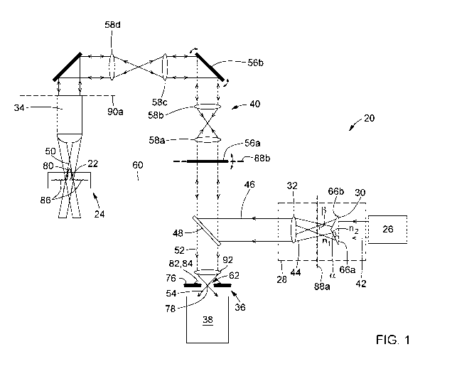
A method and system for improving lateral resolution in optical microscopy are provided. The method includes generating a source optical beam and passing the source optical beam successively through an axicon, a Fourier-transform lens and an objective to convert the source optical beam into an excitation Bessel-type beam having a central lobe and at least one side lobe. The method also includes focusing the excitation beam onto a focal plane of the objective within or on a sample to generate a sample light signal, and spatially filtering the sample light signal. The spatial filtering includes rejecting light originating from outside of the focal plane and light generated by the at least one side lobe of the excitation beam. The spatial filtering also includes permitting passage, as a filtered light signal, of light generated by the central lobe of the excitation beam. The method further includes detecting the filtered light signal.


French Abstract

L'invention concerne un procédé et un système d'amélioration de résolution latérale de microscopie optique. Le procédé consiste à générer un faisceau optique source et à faire passer le faisceau optique source successivement à travers un axicon, une lentille à transformée de Fourier et un objectif pour convertir le faisceau optique source en un faisceau d'excitation de type Bessel possédant un lobe central et au moins un lobe latéral. Le procédé consiste également à focaliser le faisceau d'excitation sur un plan focal de l'objectif à l'intérieur d'un échantillon, ou sur ce dernier, afin de générer un signal lumineux d'échantillon, et à filtrer spatialement le signal lumineux d'échantillon. Le filtrage spatial consiste à rejeter de la lumière provenant de l'extérieur du plan focal et de la lumière générée par ledit lobe latéral du faisceau d'excitation. Le filtrage spatial consiste également à laisser passer, en tant que signal lumineux filtré, de la lumière générée par le lobe central du faisceau d'excitation. Le procédé consiste aussi à détecter le signal lumineux filtré.

Claims

Note: Claims are shown in the official language in which they were submitted.


37
CLAIMS
1. A method for improving lateral resolution in optical microscopy, the method
comprising:
(a) generating a source optical beam;
(b) converting the source optical beam into an excitation Bessel-type beam
having a
central lobe and at least one side lobe, the converting comprising:
(i) passing the source optical beam through an axicon, thereby converting the
source optical beam into an intermediate Bessel-type beam;
(ii) passing the intermediate Bessel-type beam through a Fourier-transform
lens,
thereby converting the intermediate Bessel-type beam into an annular beam; and
(iii) passing the annular beam through an objective, thereby converting the
annular
beam into the excitation Bessel-type beam;
(c) focusing the excitation Bessel-type beam onto a focal plane of the
objective within or
on a sample, thereby generating a sample light signal from the sample;
(d) spatially filtering the sample light signal, the spatial filtering
comprising rejecting, from
the sample light signal, light originating from outside of the focal plane of
the objective
and light generated by the at least one side lobe of the excitation Bessel-
type beam,
and permitting passage, as a filtered light signal, of light generated by the
central lobe
of the excitation Bessel-type beam; and
(e) detecting the filtered light signal.
2. The method of claim 1, wherein step (a) comprises generating a laser beam
as the source
optical beam.
3. The method of claim 1 or 2, wherein step (a) comprises generating a
Gaussian beam as
the source optical beam, sub-step (i) of step (b) comprises generating a
Bessel-Gauss beam
as the intermediate Bessel-type beam, and sub-step (iii) of step (b) comprises
generating a
Bessel-Gauss beam as the excitation Bessel-type beam.
4. The method of any one of claims 1 to 3, wherein step (a) comprises
generating the source
optical beam in a wavelength range extending from 200 nanometers to 5
micrometers.

38
5. The method of any one of claims 1 to 4, further comprising a step of
adjusting a focal
length of the Fourier-transform lens so that a back focal plane of the Fourier-
transform lens
coincides with a center of the intermediate Bessel-type beam produced by the
axicon.
6. The method of any one of claims 1 to 5, wherein step (d) comprises passing
the sample
light signal through an aperture.
7. The method of claim 6, wherein step (d) further comprises adjusting at
least one of a size,
a shape and a position of the aperture in accordance with a width and a
position of the central
lobe of the excitation Bessel-type beam.
8. The method of claim 7, wherein adjusting at least one of the size, the
shape and the
position of the aperture comprises adjusting a linear dimension of the
aperture in a range
from 1 micrometer to 1 millimeter.
9. The method of any one of claims 1 to 8, further comprising a step of
scanning the
excitation Bessel-type beam over the sample.
10. An optical microscopy system comprising:
an optical source configured to generate a source optical beam;
beam-conditioning optics disposed in a path of the source optical beam, the
beam-
conditioning optics comprising:
an axicon positioned and configured to convert the source optical beam into an
intermediate Bessel-type beam; and
a Fourier-transform lens positioned and configured to convert the intermediate
Bessel-type beam into an annular beam;
an objective disposed in a path of the annular beam for converting the annular
beam
into an excitation Bessel-type beam having a central lobe and at least one
side lobe, the
objective focusing the excitation Bessel-type beam onto a focal plane of the
objective
within or on a sample, thereby generating a sample light signal from the
sample;

39
a spatial filter disposed in a path of the sample light signal, the spatial
filter being
configured to reject, from the sample light signal, light originating from
outside of the focal
plane of the objective and light generated by the at least one side lobe of
the excitation
Bessel-type beam, and permitting passage, as a filtered light signal, of light
generated by
the central lobe of the excitation Bessel-type beam; and
a detector configured to detect the filtered light signal.
11. The optical microscopy system of claim 10, wherein the optical source is a
laser source
configured to generate a laser beam as the source optical beam.
12. The optical microscopy system of claim 11, wherein the system is
configured for one of
confocal laser scanning microscopy and two-photon laser scanning microscopy.
13. The optical microscopy system of any one of claims 10 to 12, wherein the
optical source
is configured to generate a Gaussian beam as the source optical beam, the
axicon is
positioned and configured to generate a Bessel-Gauss beam as the intermediate
Bessel-type
beam, and the objective is positioned and configured to generate a Bessel-
Gauss beam as
the excitation Bessel-type beam.
14. The optical microscopy system of any one of claims 10 to 13, further
comprising a
switching module disposed between the optical source and the beam-conditioning
optics, the
switching module being configured for operation between a first operating
mode, wherein the
switching module directs the source optical beam onto the beam-conditioning
optics, and a
second operating mode, wherein the switching module directs the source optical
beam along
a path that bypasses the beam-conditioning optics.
15. The optical microscopy system of any one of claims 10 to 14, wherein the
optical source
is configured to generate the source optical beam in a wavelength range
extending from 200
nanometers to 5 micrometers.

40
16. The optical microscopy system of any one of claims 10 to 15, wherein the
axicon is a
refractive axicon.
17. The optical microscopy system of claim 16, wherein the axicon has an
axicon angle
ranging from 1° to 5°.
18. The optical microscopy system of any one of claims 10 to 17, wherein the
Fourier-
transform lens has an adjustable focal length.
19. The optical microscopy system of any one of claims 10 to 18, further
comprising a
scanning module configured to relay the annular beam generated by the Fourier-
transform
lens to the objective and to scan the excitation Bessel-type beam over the
sample.
20. The optical microscopy system of any one of claims 10 to 19, wherein the
axicon and the
Fourier-transform lens are separated from each other by a distance such that a
back focal
plane of the Fourier-transform lens coincides with a center of the
intermediate Bessel-type
beam produced by the axicon.
21. The optical microscopy system of any one of claims 10 to 20, wherein the
Fourier-
transform lens has a front focal plane and the objective has a back-aperture
plane, the front
focal plane of the Fourier-transform lens being optically conjugate with the
back-aperture
plane of the objective.
22. The optical microscopy system of any one of claims 10 to 21, wherein the
spatial filter
comprises a light-blocking portion surrounding an aperture, the light-blocking
portion being
configured to reject the light originating from outside of the focal plane and
the light generated
by the at least one side lobe of the excitation Bessel-type beam, and the
aperture being
configured to permit passage therethrough of the light generated by the
central lobe of the
excitation Bessel-type beam.

41
23. The optical microscopy system of claim 22, wherein the aperture has a size
ranging from
1 micrometer to 1 millimeter.
24. The optical microscopy system of claim 22 or 23, wherein the aperture has
at least one of
an adjustable size, an adjustable shape and an adjustable position.
25. The optical microscopy system of any one of claims 22 to 24, wherein the
aperture is
circular.

Description

Note: Descriptions are shown in the official language in which they were submitted.


CA 03013946 2018-08-08
WO 2017/139885 PCT/CA2017/050195
1
METHOD AND SYSTEM FOR IMPROVING LATERAL RESOLUTION
IN OPTICAL SCANNING MICROSCOPY
TECHNICAL FIELD
[0001] The general technical field relates to optical microscopy and, in
particular, to a method
and system for improving lateral resolution in optical microscopy, notably in
laser scanning
microscopy.
BACKGROUND
[0002] Laser scanning microscopy provides a range of techniques for performing
fluorescence imaging of biological samples. By way of example, confocal and
multiphoton
(e.g., two-photon) microscopes are commonly used for imaging narrow sections
of biological
structures having features of interest tagged with fluorescent markers. In
such applications, a
laser beam is focused by an objective lens to a diffraction-limited focal spot
inside or on the
surface of the specimen. Following illumination by the laser beam, fluorescent
light is emitted
from the focal spot which, along with scattered and reflected laser light, is
collected by the
objective lens, separated from the illumination light, and detected by a
photodetector. By
scanning the sample in three dimensions (3D), a volumetric image of the sample
may be
obtained pixel by pixel, where the brightness of each pixel is indicative of
the relative intensity
of detected light emanating from the corresponding focal volume.
[0003] Confocal and multiphoton microscopy can provide excellent optical
sectioning
capabilities, with depths of field of the order of a few micrometers (pm).
Using these
techniques, multiple in-focus images of thin sections located at different
depths inside a thick
sample can be acquired sequentially and subsequently combined to provide 3D
imaging
capabilities. However, although confocal and multi-photon microscopes are
usually favored in
biological applications due to their z-sectioning capabilities, their lateral
resolution at the focal
spot remains similar to that of wide-field microscopes.

Representative Drawing
A single figure which represents the drawing illustrating the invention.
Administrative Status

2024-08-01:As part of the Next Generation Patents (NGP) transition, the Canadian Patents Database (CPD) now contains a more detailed Event History, which replicates the Event Log of our new back-office solution.

Please note that "Inactive:" events refers to events no longer in use in our new back-office solution.

For a clearer understanding of the status of the application/patent presented on this page, the site Disclaimer , as well as the definitions for Patent , Event History , Maintenance Fee  and Payment History  should be consulted.

Event History

Description Date
Application Not Reinstated by Deadline 2022-08-16
Time Limit for Reversal Expired 2022-08-16
Deemed Abandoned - Failure to Respond to a Request for Examination Notice 2022-05-16
Letter Sent 2022-02-15
Deemed Abandoned - Failure to Respond to Maintenance Fee Notice 2021-08-16
Letter Sent 2021-02-15
Common Representative Appointed 2020-11-07
Common Representative Appointed 2019-10-30
Change of Address or Method of Correspondence Request Received 2018-12-04
Inactive: Cover page published 2018-08-16
Inactive: Notice - National entry - No RFE 2018-08-16
Inactive: IPC assigned 2018-08-14
Application Received - PCT 2018-08-14
Inactive: First IPC assigned 2018-08-14
Letter Sent 2018-08-14
National Entry Requirements Determined Compliant 2018-08-08
Small Entity Declaration Determined Compliant 2018-08-08
Application Published (Open to Public Inspection) 2017-08-24

Abandonment History

Abandonment Date Reason Reinstatement Date
2022-05-16
2021-08-16

Maintenance Fee

The last payment was received on 2020-02-03

Note : If the full payment has not been received on or before the date indicated, a further fee may be required which may be one of the following

  • the reinstatement fee;
  • the late payment fee; or
  • additional fee to reverse deemed expiry.

Patent fees are adjusted on the 1st of January every year. The amounts above are the current amounts if received by December 31 of the current year.
Please refer to the CIPO Patent Fees web page to see all current fee amounts.

Fee History

Fee Type Anniversary Year Due Date Paid Date
Registration of a document 2018-08-08
Basic national fee - small 2018-08-08
MF (application, 2nd anniv.) - small 02 2019-02-15 2019-01-16
MF (application, 3rd anniv.) - small 03 2020-02-17 2020-02-03
Owners on Record

Note: Records showing the ownership history in alphabetical order.

Current Owners on Record
UNIVERSITE LAVAL
Past Owners on Record
LOUIS THIBON
MICHEL PICHE
YVES DE KONINCK
Past Owners that do not appear in the "Owners on Record" listing will appear in other documentation within the application.
Documents

To view selected files, please enter reCAPTCHA code :


To view images, click a link in the Document Description column. To download the documents, select one or more checkboxes in the first column and then click the "Download Selected in PDF format (Zip Archive)" or the "Download Selected as Single PDF" button.

List of published and non-published patent-specific documents on the CPD .

If you have any difficulty accessing content, you can call the Client Service Centre at 1-866-997-1936 or send them an e-mail at CIPO Client Service Centre.


Document
Description 
Date
(yyyy-mm-dd) 
Number of pages   Size of Image (KB) 
Claims 2018-08-07 5 160
Abstract 2018-08-07 1 69
Drawings 2018-08-07 15 775
Description 2018-08-07 1 40
Representative drawing 2018-08-07 1 14
Courtesy - Certificate of registration (related document(s)) 2018-08-13 1 106
Notice of National Entry 2018-08-15 1 193
Reminder of maintenance fee due 2018-10-15 1 112
Commissioner's Notice - Maintenance Fee for a Patent Application Not Paid 2021-03-28 1 529
Courtesy - Abandonment Letter (Maintenance Fee) 2021-09-06 1 552
Commissioner's Notice: Request for Examination Not Made 2022-03-14 1 541
Commissioner's Notice - Maintenance Fee for a Patent Application Not Paid 2022-03-28 1 562
Courtesy - Abandonment Letter (Request for Examination) 2022-06-12 1 553
Patent cooperation treaty (PCT) 2018-08-07 1 64
International search report 2018-08-07 2 83
National entry request 2018-08-07 12 318
Declaration 2018-08-07 2 135