Sélection de la langue

Search

Sommaire du brevet 3013946 

Énoncé de désistement de responsabilité concernant l'information provenant de tiers

Une partie des informations de ce site Web a été fournie par des sources externes. Le gouvernement du Canada n'assume aucune responsabilité concernant la précision, l'actualité ou la fiabilité des informations fournies par les sources externes. Les utilisateurs qui désirent employer cette information devraient consulter directement la source des informations. Le contenu fourni par les sources externes n'est pas assujetti aux exigences sur les langues officielles, la protection des renseignements personnels et l'accessibilité.

Disponibilité de l'Abrégé et des Revendications

L'apparition de différences dans le texte et l'image des Revendications et de l'Abrégé dépend du moment auquel le document est publié. Les textes des Revendications et de l'Abrégé sont affichés :

  • lorsque la demande peut être examinée par le public;
  • lorsque le brevet est émis (délivrance).
(12) Demande de brevet: (11) CA 3013946
(54) Titre français: PROCEDE ET SYSTEME D'AMELIORATION DE RESOLUTION LATERALE DE MICROSCOPIE A BALAYAGE OPTIQUE
(54) Titre anglais: METHOD AND SYSTEM FOR IMPROVING LATERAL RESOLUTION IN OPTICAL SCANNING MICROSCOPY
Statut: Réputée abandonnée et au-delà du délai pour le rétablissement - en attente de la réponse à l’avis de communication rejetée
Données bibliographiques
(51) Classification internationale des brevets (CIB):
  • G02B 21/00 (2006.01)
  • G01N 33/483 (2006.01)
  • G02B 21/06 (2006.01)
(72) Inventeurs :
  • DE KONINCK, YVES (Canada)
  • PICHE, MICHEL (Canada)
  • THIBON, LOUIS (Canada)
(73) Titulaires :
  • UNIVERSITE LAVAL
(71) Demandeurs :
  • UNIVERSITE LAVAL (Canada)
(74) Agent: ROBIC AGENCE PI S.E.C./ROBIC IP AGENCY LP
(74) Co-agent:
(45) Délivré:
(86) Date de dépôt PCT: 2017-02-15
(87) Mise à la disponibilité du public: 2017-08-24
Licence disponible: S.O.
Cédé au domaine public: S.O.
(25) Langue des documents déposés: Anglais

Traité de coopération en matière de brevets (PCT): Oui
(86) Numéro de la demande PCT: 3013946/
(87) Numéro de publication internationale PCT: CA2017050195
(85) Entrée nationale: 2018-08-08

(30) Données de priorité de la demande:
Numéro de la demande Pays / territoire Date
62/295,819 (Etats-Unis d'Amérique) 2016-02-16

Abrégés

Abrégé français

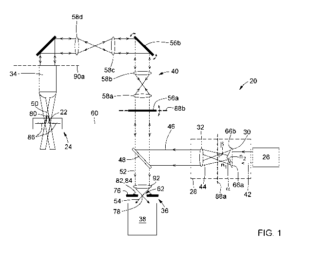
L'invention concerne un procédé et un système d'amélioration de résolution latérale de microscopie optique. Le procédé consiste à générer un faisceau optique source et à faire passer le faisceau optique source successivement à travers un axicon, une lentille à transformée de Fourier et un objectif pour convertir le faisceau optique source en un faisceau d'excitation de type Bessel possédant un lobe central et au moins un lobe latéral. Le procédé consiste également à focaliser le faisceau d'excitation sur un plan focal de l'objectif à l'intérieur d'un échantillon, ou sur ce dernier, afin de générer un signal lumineux d'échantillon, et à filtrer spatialement le signal lumineux d'échantillon. Le filtrage spatial consiste à rejeter de la lumière provenant de l'extérieur du plan focal et de la lumière générée par ledit lobe latéral du faisceau d'excitation. Le filtrage spatial consiste également à laisser passer, en tant que signal lumineux filtré, de la lumière générée par le lobe central du faisceau d'excitation. Le procédé consiste aussi à détecter le signal lumineux filtré.


Abrégé anglais

A method and system for improving lateral resolution in optical microscopy are provided. The method includes generating a source optical beam and passing the source optical beam successively through an axicon, a Fourier-transform lens and an objective to convert the source optical beam into an excitation Bessel-type beam having a central lobe and at least one side lobe. The method also includes focusing the excitation beam onto a focal plane of the objective within or on a sample to generate a sample light signal, and spatially filtering the sample light signal. The spatial filtering includes rejecting light originating from outside of the focal plane and light generated by the at least one side lobe of the excitation beam. The spatial filtering also includes permitting passage, as a filtered light signal, of light generated by the central lobe of the excitation beam. The method further includes detecting the filtered light signal.

Revendications

Note : Les revendications sont présentées dans la langue officielle dans laquelle elles ont été soumises.


37
CLAIMS
1. A method for improving lateral resolution in optical microscopy, the method
comprising:
(a) generating a source optical beam;
(b) converting the source optical beam into an excitation Bessel-type beam
having a
central lobe and at least one side lobe, the converting comprising:
(i) passing the source optical beam through an axicon, thereby converting the
source optical beam into an intermediate Bessel-type beam;
(ii) passing the intermediate Bessel-type beam through a Fourier-transform
lens,
thereby converting the intermediate Bessel-type beam into an annular beam; and
(iii) passing the annular beam through an objective, thereby converting the
annular
beam into the excitation Bessel-type beam;
(c) focusing the excitation Bessel-type beam onto a focal plane of the
objective within or
on a sample, thereby generating a sample light signal from the sample;
(d) spatially filtering the sample light signal, the spatial filtering
comprising rejecting, from
the sample light signal, light originating from outside of the focal plane of
the objective
and light generated by the at least one side lobe of the excitation Bessel-
type beam,
and permitting passage, as a filtered light signal, of light generated by the
central lobe
of the excitation Bessel-type beam; and
(e) detecting the filtered light signal.
2. The method of claim 1, wherein step (a) comprises generating a laser beam
as the source
optical beam.
3. The method of claim 1 or 2, wherein step (a) comprises generating a
Gaussian beam as
the source optical beam, sub-step (i) of step (b) comprises generating a
Bessel-Gauss beam
as the intermediate Bessel-type beam, and sub-step (iii) of step (b) comprises
generating a
Bessel-Gauss beam as the excitation Bessel-type beam.
4. The method of any one of claims 1 to 3, wherein step (a) comprises
generating the source
optical beam in a wavelength range extending from 200 nanometers to 5
micrometers.

38
5. The method of any one of claims 1 to 4, further comprising a step of
adjusting a focal
length of the Fourier-transform lens so that a back focal plane of the Fourier-
transform lens
coincides with a center of the intermediate Bessel-type beam produced by the
axicon.
6. The method of any one of claims 1 to 5, wherein step (d) comprises passing
the sample
light signal through an aperture.
7. The method of claim 6, wherein step (d) further comprises adjusting at
least one of a size,
a shape and a position of the aperture in accordance with a width and a
position of the central
lobe of the excitation Bessel-type beam.
8. The method of claim 7, wherein adjusting at least one of the size, the
shape and the
position of the aperture comprises adjusting a linear dimension of the
aperture in a range
from 1 micrometer to 1 millimeter.
9. The method of any one of claims 1 to 8, further comprising a step of
scanning the
excitation Bessel-type beam over the sample.
10. An optical microscopy system comprising:
an optical source configured to generate a source optical beam;
beam-conditioning optics disposed in a path of the source optical beam, the
beam-
conditioning optics comprising:
an axicon positioned and configured to convert the source optical beam into an
intermediate Bessel-type beam; and
a Fourier-transform lens positioned and configured to convert the intermediate
Bessel-type beam into an annular beam;
an objective disposed in a path of the annular beam for converting the annular
beam
into an excitation Bessel-type beam having a central lobe and at least one
side lobe, the
objective focusing the excitation Bessel-type beam onto a focal plane of the
objective
within or on a sample, thereby generating a sample light signal from the
sample;

39
a spatial filter disposed in a path of the sample light signal, the spatial
filter being
configured to reject, from the sample light signal, light originating from
outside of the focal
plane of the objective and light generated by the at least one side lobe of
the excitation
Bessel-type beam, and permitting passage, as a filtered light signal, of light
generated by
the central lobe of the excitation Bessel-type beam; and
a detector configured to detect the filtered light signal.
11. The optical microscopy system of claim 10, wherein the optical source is a
laser source
configured to generate a laser beam as the source optical beam.
12. The optical microscopy system of claim 11, wherein the system is
configured for one of
confocal laser scanning microscopy and two-photon laser scanning microscopy.
13. The optical microscopy system of any one of claims 10 to 12, wherein the
optical source
is configured to generate a Gaussian beam as the source optical beam, the
axicon is
positioned and configured to generate a Bessel-Gauss beam as the intermediate
Bessel-type
beam, and the objective is positioned and configured to generate a Bessel-
Gauss beam as
the excitation Bessel-type beam.
14. The optical microscopy system of any one of claims 10 to 13, further
comprising a
switching module disposed between the optical source and the beam-conditioning
optics, the
switching module being configured for operation between a first operating
mode, wherein the
switching module directs the source optical beam onto the beam-conditioning
optics, and a
second operating mode, wherein the switching module directs the source optical
beam along
a path that bypasses the beam-conditioning optics.
15. The optical microscopy system of any one of claims 10 to 14, wherein the
optical source
is configured to generate the source optical beam in a wavelength range
extending from 200
nanometers to 5 micrometers.

40
16. The optical microscopy system of any one of claims 10 to 15, wherein the
axicon is a
refractive axicon.
17. The optical microscopy system of claim 16, wherein the axicon has an
axicon angle
ranging from 1° to 5°.
18. The optical microscopy system of any one of claims 10 to 17, wherein the
Fourier-
transform lens has an adjustable focal length.
19. The optical microscopy system of any one of claims 10 to 18, further
comprising a
scanning module configured to relay the annular beam generated by the Fourier-
transform
lens to the objective and to scan the excitation Bessel-type beam over the
sample.
20. The optical microscopy system of any one of claims 10 to 19, wherein the
axicon and the
Fourier-transform lens are separated from each other by a distance such that a
back focal
plane of the Fourier-transform lens coincides with a center of the
intermediate Bessel-type
beam produced by the axicon.
21. The optical microscopy system of any one of claims 10 to 20, wherein the
Fourier-
transform lens has a front focal plane and the objective has a back-aperture
plane, the front
focal plane of the Fourier-transform lens being optically conjugate with the
back-aperture
plane of the objective.
22. The optical microscopy system of any one of claims 10 to 21, wherein the
spatial filter
comprises a light-blocking portion surrounding an aperture, the light-blocking
portion being
configured to reject the light originating from outside of the focal plane and
the light generated
by the at least one side lobe of the excitation Bessel-type beam, and the
aperture being
configured to permit passage therethrough of the light generated by the
central lobe of the
excitation Bessel-type beam.

41
23. The optical microscopy system of claim 22, wherein the aperture has a size
ranging from
1 micrometer to 1 millimeter.
24. The optical microscopy system of claim 22 or 23, wherein the aperture has
at least one of
an adjustable size, an adjustable shape and an adjustable position.
25. The optical microscopy system of any one of claims 22 to 24, wherein the
aperture is
circular.

Description

Note : Les descriptions sont présentées dans la langue officielle dans laquelle elles ont été soumises.


CA 03013946 2018-08-08
WO 2017/139885 PCT/CA2017/050195
1
METHOD AND SYSTEM FOR IMPROVING LATERAL RESOLUTION
IN OPTICAL SCANNING MICROSCOPY
TECHNICAL FIELD
[0001] The general technical field relates to optical microscopy and, in
particular, to a method
and system for improving lateral resolution in optical microscopy, notably in
laser scanning
microscopy.
BACKGROUND
[0002] Laser scanning microscopy provides a range of techniques for performing
fluorescence imaging of biological samples. By way of example, confocal and
multiphoton
(e.g., two-photon) microscopes are commonly used for imaging narrow sections
of biological
structures having features of interest tagged with fluorescent markers. In
such applications, a
laser beam is focused by an objective lens to a diffraction-limited focal spot
inside or on the
surface of the specimen. Following illumination by the laser beam, fluorescent
light is emitted
from the focal spot which, along with scattered and reflected laser light, is
collected by the
objective lens, separated from the illumination light, and detected by a
photodetector. By
scanning the sample in three dimensions (3D), a volumetric image of the sample
may be
obtained pixel by pixel, where the brightness of each pixel is indicative of
the relative intensity
of detected light emanating from the corresponding focal volume.
[0003] Confocal and multiphoton microscopy can provide excellent optical
sectioning
capabilities, with depths of field of the order of a few micrometers (pm).
Using these
techniques, multiple in-focus images of thin sections located at different
depths inside a thick
sample can be acquired sequentially and subsequently combined to provide 3D
imaging
capabilities. However, although confocal and multi-photon microscopes are
usually favored in
biological applications due to their z-sectioning capabilities, their lateral
resolution at the focal
spot remains similar to that of wide-field microscopes.

Dessin représentatif
Une figure unique qui représente un dessin illustrant l'invention.
États administratifs

2024-08-01 : Dans le cadre de la transition vers les Brevets de nouvelle génération (BNG), la base de données sur les brevets canadiens (BDBC) contient désormais un Historique d'événement plus détaillé, qui reproduit le Journal des événements de notre nouvelle solution interne.

Veuillez noter que les événements débutant par « Inactive : » se réfèrent à des événements qui ne sont plus utilisés dans notre nouvelle solution interne.

Pour une meilleure compréhension de l'état de la demande ou brevet qui figure sur cette page, la rubrique Mise en garde , et les descriptions de Brevet , Historique d'événement , Taxes périodiques et Historique des paiements devraient être consultées.

Historique d'événement

Description Date
Demande non rétablie avant l'échéance 2022-08-16
Le délai pour l'annulation est expiré 2022-08-16
Réputée abandonnée - omission de répondre à un avis relatif à une requête d'examen 2022-05-16
Lettre envoyée 2022-02-15
Réputée abandonnée - omission de répondre à un avis sur les taxes pour le maintien en état 2021-08-16
Lettre envoyée 2021-02-15
Représentant commun nommé 2020-11-07
Représentant commun nommé 2019-10-30
Requête pour le changement d'adresse ou de mode de correspondance reçue 2018-12-04
Inactive : Page couverture publiée 2018-08-16
Inactive : Notice - Entrée phase nat. - Pas de RE 2018-08-16
Inactive : CIB attribuée 2018-08-14
Demande reçue - PCT 2018-08-14
Inactive : CIB en 1re position 2018-08-14
Lettre envoyée 2018-08-14
Exigences pour l'entrée dans la phase nationale - jugée conforme 2018-08-08
Déclaration du statut de petite entité jugée conforme 2018-08-08
Demande publiée (accessible au public) 2017-08-24

Historique d'abandonnement

Date d'abandonnement Raison Date de rétablissement
2022-05-16
2021-08-16

Taxes périodiques

Le dernier paiement a été reçu le 2020-02-03

Avis : Si le paiement en totalité n'a pas été reçu au plus tard à la date indiquée, une taxe supplémentaire peut être imposée, soit une des taxes suivantes :

  • taxe de rétablissement ;
  • taxe pour paiement en souffrance ; ou
  • taxe additionnelle pour le renversement d'une péremption réputée.

Les taxes sur les brevets sont ajustées au 1er janvier de chaque année. Les montants ci-dessus sont les montants actuels s'ils sont reçus au plus tard le 31 décembre de l'année en cours.
Veuillez vous référer à la page web des taxes sur les brevets de l'OPIC pour voir tous les montants actuels des taxes.

Historique des taxes

Type de taxes Anniversaire Échéance Date payée
Enregistrement d'un document 2018-08-08
Taxe nationale de base - petite 2018-08-08
TM (demande, 2e anniv.) - petite 02 2019-02-15 2019-01-16
TM (demande, 3e anniv.) - petite 03 2020-02-17 2020-02-03
Titulaires au dossier

Les titulaires actuels et antérieures au dossier sont affichés en ordre alphabétique.

Titulaires actuels au dossier
UNIVERSITE LAVAL
Titulaires antérieures au dossier
LOUIS THIBON
MICHEL PICHE
YVES DE KONINCK
Les propriétaires antérieurs qui ne figurent pas dans la liste des « Propriétaires au dossier » apparaîtront dans d'autres documents au dossier.
Documents

Pour visionner les fichiers sélectionnés, entrer le code reCAPTCHA :


Pour visualiser une image, cliquer sur un lien dans la colonne description du document. Pour télécharger l'image (les images), cliquer l'une ou plusieurs cases à cocher dans la première colonne et ensuite cliquer sur le bouton "Télécharger sélection en format PDF (archive Zip)" ou le bouton "Télécharger sélection (en un fichier PDF fusionné)".

Liste des documents de brevet publiés et non publiés sur la BDBC .

Si vous avez des difficultés à accéder au contenu, veuillez communiquer avec le Centre de services à la clientèle au 1-866-997-1936, ou envoyer un courriel au Centre de service à la clientèle de l'OPIC.


Description du
Document 
Date
(aaaa-mm-jj) 
Nombre de pages   Taille de l'image (Ko) 
Revendications 2018-08-07 5 160
Abrégé 2018-08-07 1 69
Dessins 2018-08-07 15 775
Description 2018-08-07 1 40
Dessin représentatif 2018-08-07 1 14
Courtoisie - Certificat d'enregistrement (document(s) connexe(s)) 2018-08-13 1 106
Avis d'entree dans la phase nationale 2018-08-15 1 193
Rappel de taxe de maintien due 2018-10-15 1 112
Avis du commissaire - non-paiement de la taxe de maintien en état pour une demande de brevet 2021-03-28 1 529
Courtoisie - Lettre d'abandon (taxe de maintien en état) 2021-09-06 1 552
Avis du commissaire - Requête d'examen non faite 2022-03-14 1 541
Avis du commissaire - non-paiement de la taxe de maintien en état pour une demande de brevet 2022-03-28 1 562
Courtoisie - Lettre d'abandon (requête d'examen) 2022-06-12 1 553
Traité de coopération en matière de brevets (PCT) 2018-08-07 1 64
Rapport de recherche internationale 2018-08-07 2 83
Demande d'entrée en phase nationale 2018-08-07 12 318
Déclaration 2018-08-07 2 135