Note: Descriptions are shown in the official language in which they were submitted.
CA 03014659 2018-08-14
WO 2017/151907
PCT/US2017/020434
METHODS FOR CONTROLLED PROLIFERATION OF STEM CELLS /
GENERATING INNER EAR HAIR CELLS USING GSK-3-ALPHA INHIBITORS
CROSS-REFERENCE TO RELATED APPLICATIONS
[0001] This application claims priority under 35 U.S.C. 119(e) to U.S.
Application No.
62/302,803, filed March 2, 2016; and U.S. Application No. 62/303,099, filed
March 3, 2016,
each of which is incorporated by reference in its entirety.
BACKGROUND
Technical Field
[0002] The present disclosure relates to compositions and methods for inducing
the self-
renewal of stem/progenitor supporting cells, including inducing the
stem/progenitor cells to
proliferate while maintaining in the daughter cells the capacity to
differentiate into tissue
cells.
Description of the Related Art
[0003] Stem cells exhibit an extraordinary ability to generate multiple cell
types in the body.
Besides embryonic stem cells, tissue specific stem cells serve a critical role
during
development as well as in homeostasis and injury repair in the adult. Stem
cells renew
themselves through proliferation as well as generate tissue specific cell
types through
differentiation. The characteristics of different stem cells varies from
tissue to tissue, and are
determined by their intrinsic genetic and epigenetic status. However, the
balance between
self-renewal and differentiation of different stem cells are all stringently
controlled.
Uncontrolled self-renewal may lead to overgrowth of stem cells and possibly
tumor
formation, while uncontrolled differentiation may exhaust the stem cell pool,
leading to an
impaired ability to sustain tissue homeostasis. Thus, stem cells continuously
sense their
environment and appropriately respond with proliferation, differentiation or
apoptosis. It
would be desirable to drive regeneration by controlling the timing and extent
of stem cell
proliferation and differentiation. Controlling the proliferation with small
molecules that are
cleared over time would allow for control of the timing and extent of stem
cell proliferation
and differentiation. Remarkably, tissue stem cells from different tissues
share a limited
number of signaling pathways for the regulation of their self-renewal and
differentiation,
albeit in a very context dependent manner. Some of these pathways are the Wnt
and GSK3-
beta pathways.
1
CA 03014659 2018-08-14
WO 2017/151907
PCT/US2017/020434
[0004] Lgr5 is expressed across a diverse range of tissues and has been
identified as a
biomarker of adult stem cells in a variety of tissues such as the gut
epithelia (Barker et al.
2007), kidney, hair follicle, and stomach (Barker et al, 2010; Haegebarth &
Clevers, 2009).
For example, it was first published in 2011, that mammalian inner ear hair
cells are derived
from LGR5+ cells (Chai et al, 2011, Shi et al. 2012). Lgr5 is a known
component of the
Wnt/beta-catenin pathway, which has been shown to play major roles in
differentiation,
proliferation, and inducing stem cell characteristics (Barker et al. 2007).
[0005] Permanent damage to the hair cells of the inner ear results in
sensorineural hearing
loss, leading to communication difficulties in a large percentage of the
population. Hair cells
are the receptor cells that transduce the acoustic stimulus. Regeneration of
damaged hair cells
would provide an avenue for the treatment of a condition that currently has no
therapies other
than prosthetic devices. Although hair cells do not regenerate in the
mammalian cochlea, new
hair cells in lower vertebrates are generated from epithelial cells, called
supporting cells,
which surround hair cells.
[0006] Prior work has focused on transdifferentiation of supporting cells into
hair cells
through activation or forced expression of genes that lead to hair cell
formation, with a
particular focus on mechanisms to enhance expression of Atohl (Bermingham et
al., 1999;
Zheng and Gao, 2000; Izumikawa et al., 2005; Mizutari et al., 2013).
Interestingly, cells
transduced with Atohl vectors have been shown to acquire vestibular phenotypes
(Kawamoto
et al., 2003; Huang et al., 2009; Yang et al., 2012, 2013), and lack complete
development. As
mentioned, upregulating Atohl via gene insertion has been shown to create non-
cochlear cell
types that behave in a manner that is not found within the native cochlea. In
addition, these
methods increase hair cell numbers but decrease supporting cell numbers. Since
supporting
cells are known to have specialized roles (Ramirez-Camancho 2006, Dale and
Jagger 2010),
loss of these cells could create problems in proper cochlear function.
[0007] Thus, there remains a long felt need to protect auditory cells before
injury,
preserve/promote the function of existing cells after injury, regenerate
structure including
neurons and hair cells after injury, and regenerate cochlear supporting cells
or hair cells after
injury. As disclosed below, in certain embodiments, the present disclosure
provides methods
for preventing and treating auditory dysfunctions.
BRIEF DESCRIPTION OF THE DRAWINGS
2
CA 03014659 2018-08-14
WO 2017/151907
PCT/US2017/020434
[0008] Figures 1A-1B show expansion of Lgr5-GFP inner ear supporting cells in
multiple
conditions. Figure 1A shows Brightfield and GFP fluorescence images of Lgr5-
GFP inner ear
progenitor cells cultured for 10 days in media containing Growth Factor (GF) =
[EGF, bFGF,
IGF-11, VPA (V), combined with either CHIR99021 (C), a molecule that
preferentially
inhibits GSK3r3, or AZD1080, a molecule that preferentially inhibits GSK3a.
Figure 1B
shows quantification of Lgr5-GFP inner ear progenitor cells cultured for 10
days in media
containing GF=[EGF, bFGF, IGF-11, VPA (V), combined with either CHIR99021 (C),
a
molecule that preferentially inhibits GSK3r3, or AZD1080, a molecule that
preferentially
inhibits GSK3a.
[0009] Figures 2A-2B show Expansion of Lgr5-GFP inner ear supporting cells in
multiple
conditions. Figure 2B shows brightfield and GFP fluorescence images of Lgr5-
GFP inner ear
progenitor cells cultured for 10 days in media containing GF=[EGF, bFGF, IGF-
11, VPA (V),
combined with either CHIR99021 (C), a molecule that preferentially inhibits
GSK3r3, or
GSK3 inhibitor XXII, a molecule that equally inhibits GSK3a and GSK3r3. Figure
2B shows
quantification of Lgr5-GFP inner ear progenitor cells cultured for 10 days in
media
containing GF = [EGF, bFGF, IGF-11, VPA (V), combined with either CHIR99021
(C), a
molecule that preferentially inhibits GSK3r3, or GSK3 inhibitor XXII, which
has a higher
GSK3a-inhibition preference than CHIR99021.
[0010] Figure 3 shows increased number of hair cells in organ of Corti treated
ex vivo.
Organ of Corti treated with GSK3-inhibitor XXII showed increased Lgr5-GFP
expression, 2
rows of inner hair cells, and 6 rows of outer hair cells. This is an increase
over the normal 1
row of inner hair cells and 3 rows of outer hair cells normally seen in the
cochlea.
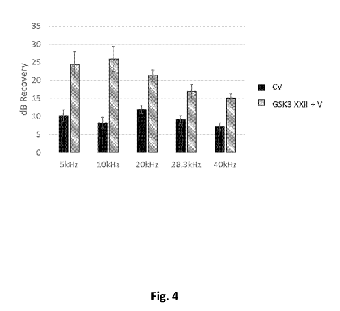
[0011] Figure 4 shows hearing recovery in noise damaged CBA/CaJ mice. Animals
treated
with Valproic Acid and CHIR99021 (CV), where CHIR99021 preferentially inhibits
GSK3r3
compared to GSKa, showed significant recovery across all tested frequencies
(n=32).
Animals treated with VPA (V) and GSK3-inhibitor XXII (a molecule that has a
higher
GSK3a-inhibition preference than CHIR99021 (n=6)).
BRIEF SUMMARY
[0012] In one aspect the present disclosure provides a method for
proliferation of stem cells
comprising contacting a cell population with an effective amount of a GSK3-
alpha inhibitor,
or a pharmaceutically-acceptable salt thereof In some embodiments, the method
further
comprises contacting the cell population with a differentiation inhibitor,
e.g., an HDAC
3
CA 03014659 2018-08-14
WO 2017/151907
PCT/US2017/020434
inhibitor or a Notch agonist. In certain embodiments, the differentiation
inhibitor is valproic
acid.
[0013] Among the various aspects of the present disclosure, therefore, may be
noted a
method for activating the Wnt pathway in a cell population to increase the
capacity of the
population for self-renewal, i.e., the capacity for repeated generation of
daughter cells with
equivalent proliferation and 'cell fate specification' potential, and
differentiation, i.e., the
capacity for generation of daughter cells specified for differentiation. In
one embodiment,
the cell population is a cochlear supporting cell population. Preferably, the
Wnt pathway is
activated upstream of the c-myc gene in members of the population and without
any genetic
modification of the population. Instead, the Wnt pathway is preferably
activated by small
molecules that transiently induce such activity. Additionally, the supporting
cell population
preferably includes supporting cells that are LGR5+ and endogenous to the
Organ of Corti.
[0014] A further aspect of the present disclosure is a method for inducing the
self-renewal of
stem/progenitor supporting cells comprised by a cochlear cell population. That
is, the
stem/progenitor supporting cells are induced to proliferate (i.e., divide and
form daughter
cells) while maintaining, in the daughter cells, the capacity to differentiate
into hair cells. In
contrast, if the stem/progenitor supporting cells were merely induced to
proliferate (without
maintaining multi-potency), the daughter cells would lack the capacity to
divide into hair
cells. Further, merely enforcing differentiation of a pre-existing
stem/progenitor cell
population has the potential to exhaust the stem cell pool.
[0015] Proliferation is preferably activated by small molecules that
transiently induce such
activity. Additionally, in certain embodiments the supporting cell population
preferably
includes supporting cells that are LGR5+ and endogenous to the Organ of Corti.
[0016] In various embodiments, the Wnt pathway is activated with a GSK3-alpha
inhibitor.
In some embodiments, the Wnt pathway is activated with a plurality of GSK3-
alpha
inhibitors. In some embodiments, the GSK3-alpha inhibitor used according to
the methods
herein includes one or more of the GSK3-alpha inhibitors disclosed herein. In
one
embodiment, the one or more GSK3-alpha inhibitor is one or more of the GSK3-
alpha
inhibitors in Table 1.
[0017] In certain embodiments, therefore, the present disclosure provides
methods to induce
self-renewal of a population of supporting cells by activating pathways and
mechanisms (e.g.,
the Wnt pathway) that are involved in inducing stem cell properties to create
"induced
4
CA 03014659 2018-08-14
WO 2017/151907
PCT/US2017/020434
pluripotent stem cells", e.g., via contacting the cells with a GSK3-alpha
inhibitor. Preferably,
the pathways are activated with small molecules (e.g., any one or more of the
GSK3-alpha
inhibitors in Table 1). For example, a composition when applied in vitro to a
supporting cell
population induces the population to proliferate to a high degree and in high
purity in a Stem
Cell Proliferation Assay, and also allows the population to differentiate into
a high purity
population of a tissue cell in a Stem Cell Differentiation Assay. In one such
embodiment, the
composition induces and maintains stem cell properties by proliferating to
produce stem cells
that can divide for many generations and maintain the ability to have a high
proportion of the
resulting cells differentiate into tissue cells. Further, the proliferating
stem cells express stem
cell markers which may include one or more of Lgr5, 5ox2, Opeml, Phex, 1in28,
Lgr6, cyclin
D1, Msxl, Myb, Kit, Gdnf3, Zic3, Dppa3, Dppa4, Dppa5, Nanog, Esrrb, Rexl,
Dnmt3a,
Dnmt3b, Dnmt31, Utfl, Tcll, 0ct4, Klf4, Pax6, 5ix2, Zicl, Zic2, 0tx2, Bmil,
CDX2,
STAT3, Smadl, 5mad2, smad2/3, smad4, smad5, and smad7.
[0018] In certain embodiments, the disclosure provides a method for expanding
a population
of cochlear cells in a cochlear tissue comprising a parent population of
cells, the method
comprising contacting the cochlear tissue with a stem cell proliferator to
form an expanded
population of cells in the cochlear tissue, wherein
the stem cell proliferator is capable of (i) forming a proliferation assay
final cell
population from a proliferation assay initial cell population over a
proliferation assay
time period in a stem cell proliferation assay and (ii) forming a
differentiation assay
final cell population from a differentiation assay initial cell population
over a
differentiation assay time period in a stem cell differentiation assay
wherein:
(a) the proliferation assay initial cell population has (i) a proliferation
assay initial
number of total cells, (ii) a proliferation assay initial number of Lgr5+
cells, (iii) a
proliferation assay initial number of hair cells, (iv) a proliferation assay
initial Lgr5+
cell fraction that equals the ratio of the proliferation assay initial number
of Lgr5+
cells to the proliferation assay initial number of total cells, and (v) a
proliferation
assay initial hair cell fraction that equals the ratio of the proliferation
assay initial
number of hair cells to the proliferation assay initial number of total cells;
(b) the proliferation assay final cell population has (i) a proliferation
assay final
number of total cells, (ii) a proliferation assay final number of Lgr5+ cells,
(iii) a
proliferation assay final number of hair cells, (iv) a proliferation assay
final Lgr5+ cell
CA 03014659 2018-08-14
WO 2017/151907
PCT/US2017/020434
fraction that equals the ratio of the proliferation assay final number of
Lgr5+ cells to
the proliferation assay final number of total cells and (v) a proliferation
assay final
hair cell fraction that equals the ratio of the proliferation assay final
number of hair
cells to the proliferation assay final number of total cells;
(c) the differentiation assay initial cell population has (i) a
differentiation assay initial
number of total cells, (ii) a differentiation assay initial number of Lgr5+
cells, (iii) a
differentiation assay initial number of hair cells, (iv) a differentiation
assay initial
Lgr5+ cell fraction that equals the ratio of the differentiation assay initial
number of
Lgr5+ cells to the differentiation assay initial number of total cells, and
(v) a
differentiation assay initial hair cell fraction that equals the ratio of the
differentiation
assay initial number of hair cells to the differentiation assay initial number
of total
cells;
(d) the differentiation assay final cell population has (i) a differentiation
assay final
number of total cells, (ii) a differentiation assay final number of Lgr5+
cells, (iii) a
differentiation assay final number of hair cells, (iv) a differentiation assay
final Lgr5+
cell fraction that equals the ratio of the differentiation assay final number
of Lgr5+
cells to the differentiation assay final number of total cells, and (v) a
differentiation
assay final hair cell fraction that equals the ratio of the differentiation
assay final
number of hair cells to the differentiation assay final number of total cells;
(e) the proliferation assay final number of Lgr5+ cells exceeds the
proliferation assay
initial number of Lgr5+ cells by a factor of at least 10; and
(0 the differentiation assay final number of hair cells is a non-zero number.
In certain such embodiments, the stem cell proliferator comprises Sternness
Driver (e.g., a
GSK3-alpha inhibitor). In certain embodiments, the stem cell proliferator
comprises a
Differentiation Inhibitor. In certain embodiments, the stem cell proliferator
comprises a
Sternness Driver and a Differentiation Inhibitor. In certain embodiments, the
stem cell
proliferator is a GSK3-alpha inhibitor (e.g., a GSK3-alpha inhibitor shown in
Table 1) and
the method further comprises contacting cochlear cells in the cochlear tissue
with a
differentiation inhibitor. In some embodiments, the differentiation inhibitor
is an HDAC
inhibitor or a Notch agonist. In some embodiments, the differentiation
inhibitor is valproic
acid.
6
CA 03014659 2018-08-14
WO 2017/151907
PCT/US2017/020434
[0019] In certain embodiments, the disclosure provides a method for increasing
the cell
density of supporting cells in a population of cochlear cells, the method
comprising activating
pathways and mechanisms that induce stem cell properties in the supporting
cells,
proliferating the activated supporting cells (while maintaining the multi-
potent character of
the supporting cells in the newly formed daughter cells) and thereafter
allowing (or even
inducing) the expanded population to differentiate into hair cells to form an
expanded
cochlear cell population wherein the cell density of hair cells in the
expanded cochlear cell
population exceeds the cell density of hair cells in the original (non-
expanded) cochlear cell
population. In some embodiments, the supporting cell population is an in vitro
supporting cell
population. In some embodiments, the supporting cell population is an in vivo
supporting cell
population. Additionally, the proliferation stage is preferably controlled to
substantially
maintain the native organization of the cochlear structure. In such
embodiments, the
proliferating is induced by one or more GSK3-alpha inhibitors (e.g., one or
more of the GSK-
alpha inhibitors shown in Table 1) that transiently induce such activity,
rather than by
induction of c-myc and without any genetic modification of the population. In
some
embodiments, such methods further comprise contacting the cells with a
differentiation
inhibitor. In some embodiments, the differentiation inhibitor is an HDAC
inhibitor or a
Notch agonist. In some embodiments, the differentiation inhibitor is valproic
acid.
Additionally, in certain embodiments the supporting cell population preferably
includes
supporting cells that are LGR5+ and endogenous to the Organ of Corti.
[0020] In certain embodiments, the disclosure provides a method for increasing
the cell
density of Lgr5+ supporting cells in a population of cochlear cells, the
method comprising
activating pathways and mechanisms that induce or maintain stem cell
properties in the Lgr5+
supporting cells, proliferating the activated Lgr5+ supporting cells (while
maintaining such
stem cell properties) and thereafter allowing (or even inducing) the expanded
population to
differentiate into hair cells to form an expanded cochlear cell population
wherein the cell
density of hair cells in the expanded cochlear cell population exceeds the
cell density of hair
cells in the original (non-expanded) cochlear cell population. In some
embodiments, the
Lgr5+ supporting cell population is an in vitro Lgr5+ stem cell population. In
some
embodiments, the Lgr5+ supporting cell population is an in vivo supporting
cell population.
Additionally, in certain embodiments the proliferation stage is preferably
controlled to
substantially maintain the native organization of the cochlear structure. In
such
embodiments, the proliferating is induced by one or more GSK3-alpha inhibitors
(e.g., one or
7
CA 03014659 2018-08-14
WO 2017/151907
PCT/US2017/020434
more of the GSK-alpha inhibitors shown in Table 1). In some embodiments, the
GSK3-alpha
inhibitors transiently induce such activity, rather than by induction of c-myc
and without any
genetic modification of the population. In some embodiments, such methods
further
comprise contacting the cells with a differentiation inhibitor. In some
embodiments the
differentiation inhibitor is an HDAC inhibitor or a Notch agonist. In some
embodiments, the
differentiation inhibitor is valproic acid.
[0029] In certain embodiments, a composition containing a Sternness Driver
(e.g., a GSK-
alpha inhibitor) and a Differentiation Inhibitor (e.g., a Notch agonist or an
HDAC inhibitor
such as valproic acid) is administered to a cochlear cell population to induce
proliferation of
stem cells and to inhibit differentiation of the stem cells until the desired
expansion of the
stem cell population is achieved. Thereafter, the expanded population is
permitted (or
optionally even induced) to differentiate into hair cells. Additionally, the
proliferation stage
is preferably controlled to substantially maintain the native organization of
the cochlear
structure. In some embodiments, the Sternness Driver and Differentiation
inhibitor are small
molecules. In some embodiments, the stem cell population is an in vivo stem
cell population.
In some embodiments, the stem cell population is an in vitro stem cell
population. In some
embodiments, the stem cell population is an in vivo Lgr5+ stem cell
population. In some
embodiments, the stem cell population is an in vitro Lgr5+ stem cell
population. In some
embodiments, the Sternness Driver is a GSK-alpha inhibitor. In some
embodiments, the
differentiation inhibitor is an HDAC inhibitor or a Notch agonist. In some
embodiments, the
differentiation inhibitor is valproic acid.
[0030] In certain embodiments, the disclosure provides a method for increasing
the cell
density of hair cells in an initial population of cochlear cells, the initial
population (which
may be an in vivo or an in vitro population) comprises hair cells, Lgr-
supporting cells, and
Lgr5+ supporting cells. The method comprises administering to the initial
population a
Sternness Driver and Differentiation inhibitor. In some embodiments, the
Sternness Driver is
a GSK-alpha inhibitor. In some embodiments, the differentiation inhibitor is
an HDAC
inhibitor or a Notch agonist. In some embodiments, the differentiation
inhibitor is valproic
acid.
[0021] In certain embodiments, the method produces stem cells in a Stem Cell
Proliferation
Assay that express stem cells markers Lgr5+. In certain embodiments, if a
mixed population
of Lgr5+ and non-Lgr5+ stems are placed in a Stem Cell Proliferation Assay,
the method
increases the fraction of cells in the population that are Lgr5+.
8
CA 03014659 2018-08-14
WO 2017/151907
PCT/US2017/020434
[0031] Expanding supporting cell populations to a degree that destroys the
native
organization of the cochlear structure could inhibit cochlear function.
Driving proliferation
of existing supporting cells with a small molecule signal may allow for a more
controlled
regeneration of hair cells than using gene delivery, which is incapable of
targeting a specific
cell type and permanently alters a cell's genetic information. An
approximately normal
cochlear structure is desired with rows of hair cells that have supporting
cells between them,
and hair cells do not contact other hair cells. Further, it would be desirable
to avoid using
genetic modification to drive proliferation to create large cell aggregations
in the cochlea that
disrupt the organ's anatomy.
[0022] In certain embodiments, the disclosure provides a composition
comprising a Sternness
Driver that may be used to drive the selective expansion of cochlea supporting
cells. In some
cases, a Sternness Driver may also induce differentiation of the supporting
cells to hair cells if
a Differentiation Inhibitor is not present at an Effective Differentiation
Inhibition
Concentration. Examples of Sternness Drivers that may drive both proliferation
and
differentiation include GSK3-alpha inhibitors. In certain of these
embodiments, the
composition comprises a Sternness Driver and a Differentiation Inhibitor,
where in the
Sternness Driver and the Differentiation Inhibitor are different agents. In
one such
embodiment, the Sternness Driver is a GSK3-alpha inhibitor (e.g., one of the
GSK3-alpha
inhibitors disclosed herein) and the Differentiation Inhibitor is valproic
acid.
[0023] In certain embodiments, the disclosure provides a method for increasing
the cell
density of hair cells in an initial population of cochlear cells comprising
hair cells and
supporting cells. The method comprises selectively expanding the number of
supporting
cells in the initial population to form an intermediate cochlear cell
population wherein the
ratio of the number of supporting cells to hair cells in the intermediate
cochlear cell
population exceeds the ratio of the number of supporting cells to hair cells
in the initial
cochlear cell population. The method further comprises generating hair cells
in the
intermediate cochlear cell population to form an expanded cochlear cell
population wherein
the ratio of the number of hair cells to supporting cells in the expanded
cochlear cell
population exceeds the ratio of the number of hair cells to supporting cells
in the intermediate
cochlear cell population.
[0024] In certain embodiments, the disclosure provides a method for increasing
the number
of Lgr5+ supporting cells or increasing the Lgr5+ activity in an initial
population of cochlear
cells, wherein the initial population comprises supporting cells and hair
cells. For example,
9
CA 03014659 2018-08-14
WO 2017/151907
PCT/US2017/020434
in one such method an intermediate population is formed in which the number of
Lgr5+
supporting cells is expanded relative to the initial population.
Alternatively, in one such
method an intermediate population is formed in which the Lgr5+ activity of the
supporting
cells relative to the initial population is increased. Alternatively, a method
where the number
of Lgr5+ cells is increased relative to the initial cell population by
activating Lgr5+ expression
in cell types that normally lack or have very low levels of Lgr5+. By way of
further example,
an intermediate population is formed in which the number of Lgr5+ supporting
cells is
expanded and the Lgr5 activity is increased relative to the initial cochlear
cell population.
Thereafter, hair cells in the intermediate cochlear cell population may be
generated to form
an expanded cochlear cell population wherein the ratio of hair cells to
supporting cells in the
expanded cochlear cell population exceeds the ratio of the number of hair
cells to supporting
cells in the intermediate cochlear cell population.
[0025] In some embodiments, the methods of the present disclosure comprise a
first
Proliferation Period with an Effective Sternness Driver Concentration and an
Effective
Differentiation Inhibition Concentration of a Differentiation Inhibitor,
followed by a
Differentiation Period with an Effective Sternness Driver Concentration and
without an
Effective Differentiation Inhibition Concentration of a Differentiation
Inhibitor. In some such
embodiments, the Sternness Driver and Differentiation Inhibitor are provided
to the cochlear
cells in a formulation that releases the Sternness Driver and Differentiation
inhibitor at
different rates. In some such embodiments, the Sternness Driver is a GSK3-
alpha inhibitor
(e.g., one or more of the GSK3-alpha inhibitors disclosed in Table 1). In some
such
embodiments, the Sternness Driver is a GSK3-alpha inhibitor (e.g., one or more
of the GSK3-
alpha inhibitors disclosed in Table 1) and the Differentiation Inhibitor is an
HDAC inhibitor
or a Notch agonist. In some such embodiments, the Sternness Driver is a GSK3-
alpha
inhibitor (e.g., one or more of the GSK3-alpha inhibitors disclosed in Table
1) and the
Differentiation Inhibitor is valproic acid.
[0032] In certain embodiments, the disclosure provides a method of and
compositions for
generating hair cells, the method comprising: administering or causing to be
administered to
a stem cell population (e.g., of an in vitro, ex vivo, or in vivo
sample/subject) a composition
comprising both of (i) and (ii): (i) a GSK3-alpha inhibitor (or a derivative
or
pharmaceutically-acceptable salt thereof) and (ii) valproic acid (or a
derivative or analog or
pharmaceutically-acceptable salt thereof), thereby proliferating stem cells in
the stem cell
population and resulting in an expanded population of stem cells; and exposing
the expanded
CA 03014659 2018-08-14
WO 2017/151907
PCT/US2017/020434
population of stem cells to a GSK3-alpha inhibitor (or a derivative or
pharmaceutically-
acceptable salt thereof) and, optionally a Differentiation Inhibitor (e.g., a
Notch agonist or an
HDAC inhibitor, e.g., valproic acid), thereby facilitating generation of inner
ear hair cells
from the expanded population of stem cells.
[0026] In certain embodiments, the disclosure provides methods for preventing
and treating
auditory dysfunction. For example, in certain embodiments, the disclosure
provides methods
for preventing or treating auditory impairments in a subject comprising
administering to said
subject an effective amount of a GSK3-alpha inhibitor (e.g., any one or more
of the GSK3-
alpha inhibitors disclosed in Table 1)
[0027] In certain embodiments, the present disclosure also relates to ex-vivo
uses of cells
described herein. For example, approaches described herein can be used for hi
and for
discovery purposes. For example, certain embodiments of the present disclosure
are useful
for identifying agents that proliferate hair cell progenitors and/or increase
numbers of hair
cells, and also agents that protect supporting cells and/or hair cells (e.g.
to support their
survival), and also for identifying agents that are toxic or not toxic to
supporting cells or
differentiated progeny including hair cells.
[0028] In certain embodiments, the disclosure provides for methods for
inhibiting the loss or
death of the cells of the auditory system in a subject comprising
administering to said subject
an effective amount of a composition described herein (e.g., comprising a GSK3-
alpha
inhibitor and optionally a differentiation inhibitor (e.g., a Notch agonist or
an HDAC
inhibitor, e.g., valproic acid) or derivative thereof or pharmaceutically-
acceptable salt thereof
and an acceptable carrier or excipient, thereby inhibiting loss or death of
the cells of the
auditory system in the subject or regenerating structure including hair cells
and/or neurons.
[0029] In certain embodiments, the disclosure provides methods for maintaining
or
promoting the growth of cells of the auditory system in a subject comprising
administering to
said subject a composition comprising as an agent described herein (comprising
a GSK3-
alpha inhibitor and optionally a Differentiation Inhibitor (e.g., a Notch
agonist or an HDAC
inhibitor, e.g., valproic acid) or derivative thereof or pharmaceutically-
acceptable salt thereof
in an effective amount so as to augment or initiate endogenous repair, thereby
maintaining or
promoting the growth of cells of the auditory system in the subject.
[0033] Also described herein is a method for expanding a population of
cochlear cells in a
cochlear tissue comprising a parent population of cells, the parent population
including
11
CA 03014659 2018-08-14
WO 2017/151907
PCT/US2017/020434
supporting cells and a number of Lgr5+ cells, the method comprising contacting
the cochlear
tissue with a stem cell proliferator to form an expanded population of cells
in the cochlear
tissue, wherein the stem cell proliferator is capable (i) in a stem cell
proliferation assay of
increasing the number of Lgr5+ cells in a stem cell proliferation assay cell
population by a
factor of at least 10 and (ii) in a stem cell differentiation assay of forming
hair cells from a
cell population comprising Lgr5+ cells.
[0034] Also described herein is a method for expanding a population of
cochlear cells in a
cochlear tissue comprising a parent population of cells, the parent population
including
supporting cells, the method comprising contacting the cochlear tissue with a
stem cell
proliferator to form an expanded population of cells in the cochlear tissue.
The stem cell
proliferator can be capable of (i) forming a proliferation assay final cell
population from a
proliferation assay initial cell population over a proliferation assay time
period in a stem cell
proliferation assay and (ii) forming a differentiation assay final cell
population from a
differentiation assay initial cell population over a differentiation assay
time period in a stem
cell differentiation assay wherein: (a) the proliferation assay initial cell
population has (i) a
proliferation assay initial number of total cells, (ii) a proliferation assay
initial number of
Lgr5+ cells, (iii) a proliferation assay initial number of hair cells, (iv) a
proliferation assay
initial Lgr5+ cell fraction that equals the ratio of the proliferation assay
initial number of
Lgr5+ cells to the proliferation assay initial number of total cells, and (v)
a proliferation assay
initial hair cell fraction that equals the ratio of the proliferation assay
initial number of hair
cells to the proliferation assay initial number of total cells; (b) the
proliferation assay final
cell population has (i) a proliferation assay final number of total cells,
(ii) a proliferation
assay final number of Lgr5+ cells, (iii) a proliferation assay final number of
hair cells, (iv) a
proliferation assay final Lgr5+ cell fraction that equals the ratio of the
proliferation assay final
number of Lgr5+ cells to the proliferation assay final number of total cells
and (v) a
proliferation assay final hair cell fraction that equals the ratio of the
proliferation assay final
number of hair cells to the proliferation assay final number of total cells;
(c) the
differentiation assay initial cell population has (i) a differentiation assay
initial number of
total cells, (ii) a differentiation assay initial number of Lgr5+ cells, (iii)
a differentiation assay
initial number of hair cells, (iv) a differentiation assay initial Lgr5+ cell
fraction that equals
the ratio of the differentiation assay initial number of Lgr5+ cells to the
differentiation assay
initial number of total cells, and (v) a differentiation assay initial hair
cell fraction that equals
the ratio of the differentiation assay initial number of hair cells to the
differentiation assay
12
CA 03014659 2018-08-14
WO 2017/151907
PCT/US2017/020434
initial number of total cells; (d) the differentiation assay final cell
population has (i) a
differentiation assay final number of total cells, (ii) a differentiation
assay final number of
Lgr5+ cells, (iii) a differentiation assay final number of hair cells, (iv) a
differentiation assay
final Lgr5+ cell fraction that equals the ratio of the differentiation assay
final number of Lgr5+
cells to the differentiation assay final number of total cells, and (v) a
differentiation assay
final hair cell fraction that equals the ratio of the differentiation assay
final number of hair
cells to the differentiation assay final number of total cells; (e) the
proliferation assay final
number of Lgr5+ cells exceeds the proliferation assay initial number of Lgr5+
cells by a factor
of at least 10; and (f) the differentiation assay final number of hair cells
is a non-zero number.
[0035] The proliferation assay final number of Lgr5+ cells can be greater than
the
proliferation assay initial number of Lgr5+ cells by a factor of at least 50,
or by a factor of at
least 100. The expanded population of cells in the cochlear tissue can include
a greater
number of hair cells than does the parent population. The proliferation assay
final Lgr5+ cell
fraction can be greater than the differentiation assay initial Lgr5+ cell
fraction by at least a
factor of 2. The differentiation assay final hair cell fraction can be greater
than the
proliferation assay initial hair cell fraction by at least a factor of 2. The
proliferation assay
final hair cell fraction can be at least 25% less than the proliferation assay
initial hair cell
fraction. The proliferation assay final Lgr5+ cell fraction can be at least
10% greater than
proliferation assay initial Lgr5+ cell fraction. One of more morphological
characteristics of
the cochlear tissue can be maintained. Native morphology can be maintained.
The stem cell
proliferator can be dispersed in a biocompatible matrix, which can be a
biocompatible gel or
foam. The cochlear tissue can be an in vivo cochlear tissue or an ex vivo
cochlear tissue.
The method can produce a population of Lgr5+ cells that are in s-phase. The at
least one stem
cell proliferator can include both a sternness driver and a differentiation
inhibitor. For
example, in some embodiments, the stem cell proliferator includes a sternness
driver that is a
GSK3-alpha inhibitor and a differentiation inhibitor. In some embodiments, the
stem cell
proliferator includes a sternness driver that is a GSK3-alpha inhibitor and a
differentiation
inhibitor that is an HDAC inhibitor or a Notch agonist. In some embodiments,
the stem cell
proliferator includes a sternness driver that is a GSK3-alpha inhibitor and a
differentiation
inhibitor that is valproic acid. Contacting can provide to the cochlear
tissue: in an initial
phase, at least an effective proliferation concentration of the sternness
driver and at least an
effective differentiation inhibition concentration of the differentiation
inhibitor; and in a
subsequent phase, at least an effective proliferation concentration of the
sternness driver and
13
CA 03014659 2018-08-14
WO 2017/151907
PCT/US2017/020434
less than an effective differentiation inhibition concentration of the
differentiation inhibitor.
The cochlear tissue can be in a subject, and contacting the cochlear tissue
with the
composition can be achieved by administering the composition transtympanically
to the
subject. Contacting the cochlear tissue with the composition can result in
improved auditory
functioning of the subject.
[0036] Also described herein is a method of treating a subject who has, or is
at risk of
developing, hearing loss. The method can include transtympanically
administering to a
cochlear tissue of the subject a GSK3-alpha inhibitor. In some such
embodiments, the
method further comprises administering a differentiation inhibitor to the
tissue. In one such
embodiment, the differentiation inhibitor is an HDAC inhibitor or a Notch
agonist. In one
such embodiment, the differentiation inhibitor is valproic acid.
[0037] Certain embodiments relate to pharmaceutical compositions, comprising a
pharmaceutically-acceptable carrier and a stem cell proliferator that is a
GSK3-alpha
inhibitor, or a pharmaceutically-acceptable salt thereof In some embodiments,
the
composition is adapted for administration to the inner ear and/or middle ear.
In some
instances, the composition is adapted for local administration to the round
window
membrane. In some embodiments, the composition is adapted for intratympanic or
transtympanic administration, for example, to cochlear tissue.
[0038] In some embodiments, the GSK3-alpha inhibitor is dispersed in a
biocompatible
matrix. In certain embodiments, the biocompatible matrix is a biocompatible
gel or foam.
[0039] Some compositions further comprise a differentiation inhibitor. In
particular
embodiments, the differentiation inhibitor is selected from an HDAC inhibitor
and a Notch
agonist, or a pharmaceutically-acceptable salt thereof In some embodiments,
the HDAC
inhibitor is valproic acid, or a derivative or analog or pharmaceutically-
acceptable salt
thereof
[0040] In some embodiments, the GSK3-alpha inhibitor has a GSK3-alpha/GSK3-
beta
selectivity ratio that is at least about 0.5x, or 0.6x, 0.7x, 0.8x, 0.9x,
1.0x, 1.1x, 1.2x, or 1.3x,
1.4x, 1.5x, 2x, 3x, 4x, 5x, 6x, 7x, 8x, 9x, 10x, 15x, 20x, 25x, 30x, 40x, 50x,
60x, 70x, 80x,
90x, or 100x. In some embodiments, the GSK3-alpha inhibitor has potency
against both
GSK3-alpha and GSK3-beta, wherein the potency is less than about 100 nM for
inhibiting
both GSK3-alpha and GSK3-beta, or less than about 50 nM, 20 nM, 10 nM, 5 nM, 2
nM, or
less than about 1 nM for inhibiting both GSK3-alpha and GSK3-beta. In some
embodiments,
14
CA 03014659 2018-08-14
WO 2017/151907
PCT/US2017/020434
the GSK3-alpha inhibitor has a GSK3-alpha/CDK selectivity ratio that is at
least about 10x or
15x, 20x, 25x, 30x, 35x, 40x, 45x, 50x, 60x, 60x, 80x, 90x, or at least about
100x. In some
embodiments, the GSK3-alpha inhibitor has a GSK3-alpha/MAPK selectivity ratio
that is at
least about 10x or 15x, 20x, 25x, 30x, 35x, 40x, 45x, 50x, 60x, 60x, 80x, 90x,
or at least
about 100x. In some embodiments, the GSK3-alpha inhibitor has a GSK3-alpha/ERK
selectivity ratio that is at least about 10x or 15x, 20x, 25x, 30x, 35x, 40x,
45x, 50x, 60x, 60x,
80x, 90x, or at least about 100x. In some embodiments, the GSK3-alpha
inhibitor has a
GSK3-alpha/MEK selectivity ratio that is at least about 10x or 15x, 20x, 25x,
30x, 35x, 40x,
45x, 50x, 60x, 60x, 80x, 90x, or at least about 100x. In some embodiments, the
GSK3-alpha
inhibitor comprises potency for GSK3-alpha that ranges from about mM to about
1000nM;
from about 100 nM to about 1000 nM; from about 10 nM to about 100nM; of from
about
mM to about 10 nM.
[0041] In some embodiments, the pharmaceutical composition comprises a
poloxamer. In
particular embodiments, the poloxamer comprises at least one of Poloxamer 188
and
Poloxamer 407 or mixtures thereof In some embodiments, the poloxamer is in a
concentration between about 5 wt% and about 25 wt% relative to the
composition. In
particular embodiments, the poloxamer is in a concentration between about 10
wt% and about
23 wt% relative to the composition. In some embodiments, the poloxamer is in a
concentration between about 15 wt% and about 20 wt% relative to the
composition. In
specific embodiments, the poloxamer is in a concentration is approximately 17
wt% relative
to the composition.
[0042] In some compositions, the GSK3-alpha inhibitor is at a concentration of
about 0.01
uM to 1000 mM, about 0.1 uM to 1000 mM, about 1 uM to 100 mM, about 10 uM to
10 mM,
about 1 uM to 10 uM, about 10 uM to 100 uM, about 100 uM to 1000 uM, about 1
mM to 10
mM, or about 10 mM to 100 mM; or at a concentration ratio of about 0.01 to
1,000,000 fold
relative to its effective activity in an in vitro activity assay, or about 0.1
to 100,000 fold
relative to its effective activity in an in vitro activity assay, or about 1
to 10,000 fold relative
to its effective activity in an in vitro activity assay, or about 100 to 5000
fold relative to its
effective activity in an in vitro activity assay, or about 50 to 2000 fold
relative to its effective
activity in an in vitro activity assay, or about 100 to 1000 fold relative to
its effective activity
in an in vitro activity assay, or at about 1000 fold relative to its effective
activity in an in vitro
activity assay; or at a concentration of about 0.01 nM to 1000 uM, about 0.1
nM to 1000 uM,
CA 03014659 2018-08-14
WO 2017/151907
PCT/US2017/020434
about 1 nM to 100 uM, about 10 nM to 10 uM, about 1 nM to 10 nM, about 10 nM
to 100
nM, about 100 nM to 1000 nM, about 1 uM to 10 uM, or about 10 uM to 100 uM.
[0043] In some embodiments, the GSK3-alpha inhibitor is GSK3 inhibitor XXII,
which is at
a concentration of about 0.1 uM to 1000 mM, about 1 uM to 100 mM, 10 uM to 10
mM,
about 100 uM to 10 mM, or 100 uM to 1 mM, or about 1, 2, 3, 4, 5, 6, 7, 8, 9,
or 10 mM; or at
a concentration ratio of about 0.1 to 1,000,000 fold relative to its effective
activity in an in
vitro activity assay, or about 1 to 100,000 fold relative to its effective
activity in an in vitro
activity assay, or about 10 to 10,000 fold relative to its effective activity
in an in vitro activity
assay, or about 100 to 1000 fold relative to its effective activity in an in
vitro activity assay,
or about 1000 fold relative to its effective activity in an in vitro activity
assay; or at a
concentration of about 0.1 nM to 1000 uM, about 1 nM to 100 uM, about 10 nM to
10 uM,
about 100 nM to 1 uM, or about 0.5 uM.
[0044] In certain embodiments, the GSK3-alpha inhibitor is AZD1080, which is
at a
concentration of about 0.1 uM to 1000 mM, about 1 uM to 1000 mM, about 10 uM
to 100
mM, about 100 uM to 10 mM, about 1 mM to 10 mM, or about 1, 2, 3, 4, 5, 6, 7,
8, 9, or 10
mM; or at a concentration ratio of about 0.1 to 1,000,000 fold relative to its
effective activity
in an in vitro activity assay, or about 1 to 100,000 fold relative to its
effective activity in an in
vitro activity assay, or about 10 to 10,000 fold relative to its effective
activity in an in vitro
activity assay, or about 100 to 1000 fold relative to its effective activity
in an in vitro activity
assay, or about 1000 fold relative to its effective activity in an in vitro
activity assay; or at a
concentration of about 1 nM to 1000 uM, about 10 nM to 1000 uM, about 100 nM
to 100 uM,
about 1 uM to 10 uM, or about 1, 2, 3, 4, 5, 6, 7, 8, 9, or 10 uM.
[0045] In some embodiments, the HDAC inhibitor is at a concentration of about
0.01 uM to
100,000 mM, about 1 uM to 10,000 mM, about 10 uM to 10,000 mM, about 100 uM to
1000
mM, about 1 uM to 10 uM, about 10 uM to 100 uM, about 100 uM to 1000 uM, about
1000
uM to 10 mM, about 10 mM to 100 mM, about 100 mM to 1000 mM, or about 1000 mM
to
10,000 mM; or at a concentration ratio of about 0.1 to 1,000,000 fold relative
to its effective
activity in an in vitro activity assay, or about 1 to 100,000 fold relative to
its effective activity
in an in vitro activity assay, or about 10 to 10,000 fold relative to its
effective activity in an in
vitro activity assay, or about 100 to 1000 fold relative to its effective
activity in an in vitro
activity assay; or about 1000 fold relative to its effective activity in an in
vitro activity assay;
or at a concentration of about 0.01 nM to 100,000 uM, about 1 nM to 10,000 uM,
about 10
nM to 10,000 uM, about 100 nM to 1000 uM, about 1 nM to 10 nM, about 10 nM to
100 nM,
16
CA 03014659 2018-08-14
WO 2017/151907
PCT/US2017/020434
about 100 nM to 1000 nM, about 1 uM to 10 uM, about 10 uM to 100 uM, about 100
uM to
1000 uM, or about 1000 uM to 10,000 uM.
[0046] In certain embodiments, the HDAC inhibitor is valproic acid, which is
at a
concentration of about 10 uM to 100,000 mM, about 1 mM to 10,000 mM, about 10
mM to
10,000 mM, about 100 mM to 10,000 mM, about 200 mM to 2000 mM, about 1000 mM,
or
about 600 mM; or at a concentration ratio of about 0.1 to 1,000,000 fold
relative to its
effective activity in an in vitro activity assay, or about 1 to 100,000 fold
relative to its
effective activity in an in vitro activity assay, or about 10 to 10,000 fold
relative to its
effective activity in an in vitro activity assay, or about 100 to 1000 fold
relative to its
effective activity in an in vitro activity assay, or about 1000 fold relative
to its effective
activity in an in vitro activity assay; or at a concentration of about 10 nM
to 100,000 uM, 1
uM to 10,000 uM, about 10 uM to 10,000 uM, about 100 uM to 10,000 uM, about
200 uM to
2000 uM, or about 1000 uM.
[0047] In certain embodiments, the effective activity is measured in an Lgr5
proliferation
assay, as described herein.
[0048] In certain compositions, the GSK3-alpha inhibitor is capable in a stem
cell
proliferation assay of increasing the number of Lgr5+ cells in a stem cell
proliferation assay
cell population by a factor of at least about 1.25, 1.5, 1.75, 2, 3, 5, 10, or
20, and is optionally
selected from Table 1. In some compositions, the a GSK3-alpha inhibitor is
capable in a stem
cell differentiation assay of forming hair cells from a cell population
comprising Lgr5+ cells
[0049] The pharmaceutical compositions can be used in any one or more of the
methods
described herein, including the use of the compositions for expanding a
population of
cochlear cells in a cochlear tissue. The pharmaceutical compositions can also
be used for
treating a subject who has, or is at risk of developing, hearing loss.
[0050] Also included are methods of generating Myo7a+ cochlear cells. The
methods can
include contacting Lgr5+ cochlear cells with a GSK3-alpha inhibitor, thereby
generating an
expanded population of Lgr5+ cells;, thereby generating Myo7a+ cochlear cells.
[0051] Other objects and features will be in part apparent and in part pointed
out hereinafter.
DETAILED DESCRIPTION
Definitions
17
CA 03014659 2018-08-14
WO 2017/151907
PCT/US2017/020434
[0052] In this application, the use of "or" includes "and/or" unless stated
otherwise. As used
in this application, the term "comprise" and variations of the term, such as
"comprising" and
"comprises," are not intended to exclude other additives, components, integers
or steps. By
"consisting of' is meant including, and limited to, whatever follows the
phrase "consisting
of" Thus, the phrase "consisting of' indicates that the listed elements are
required or
mandatory, and that no other elements may be present. By "consisting
essentially of' is
meant including any elements listed after the phrase, and limited to other
elements that do not
interfere with or contribute to the activity or action specified in the
disclosure for the listed
elements. Thus, the phrase "consisting essentially of' indicates that the
listed elements are
required or mandatory, but that other elements are optional and may or may not
be present
depending upon whether or not they materially affect the activity or action of
the listed
elements.
[0053] As used in this application, the terms "about" and "approximately" are
used as
equivalents. Any numerals used in this application with or without
about/approximately are
meant to cover any normal fluctuations appreciated by one of ordinary skill in
the relevant
art. In certain embodiments, the term "approximately" or "about" refers to a
range of values
that fall within 25%, 20%, 19%, 18%, 17%, 16%, 15%, 14%, 13%, 12%, 11%, 10%,
9%, 8%,
7%, 6%, 5%, 4%, 3%, 2%, 1%, or less in either direction (greater than or less
than) of the
stated reference value unless otherwise stated or otherwise evident from the
context (except
where such number would exceed 100% of a possible value).
[0054] "Administration" refers to introducing a substance into a subject. In
some
embodiments, administration is auricular, intraauricular, intracochlear,
intravestibular, or
transtympanically, e.g., by injection. In some embodiments, administration is
directly to the
inner ear, e.g. injection through the round window, otic capsule, or
vestibular canals. In some
embodiments, administration is directly into the inner ear via a cochlear
implant delivery
system. In some embodiments, the substance is injected transtympanically to
the middle ear.
In certain embodiments "causing to be administered" refers to administration
of a second
component after a first component has already been administered (e.g., at a
different time
and/or by a different actor).
[0055] An "antibody" refers to an immunoglobulin polypeptide, or fragment
thereof, having
immunogen binding ability.
18
CA 03014659 2018-08-14
WO 2017/151907
PCT/US2017/020434
[0056] As used herein, an "agonist" is an agent that causes an increase in the
expression or
activity of a target gene, protein, or a pathway, respectively. Therefore, an
agonist can bind
to and activate its cognate receptor in some fashion, which directly or
indirectly brings about
this physiological effect on the target gene or protein. An agonist can also
increase the
activity of a pathway through modulating the activity of pathway components,
for example,
through inhibiting the activity of negative regulators of a pathway.
Therefore, a "Wnt
agonist" can be defined as an agent that increases the activity of Wnt
pathway, which can be
measured by increased TCF/LEF-mediated transcription in a cell. Therefore, a
"Wnt agonist"
can be a true Wnt agonist that bind and activate a Frizzled receptor family
member, including
any and all of the Wnt family proteins, an inhibitor of intracellular beta-
catenin degradation,
and activators of TCF/LEF.
[0057] An "antagonist" refers to an agent that binds to a receptor, and which
in turn
decreases or eliminates binding by other molecules.
[0058] "Antisense" refers to a nucleic acid sequence, regardless of length,
that is
complementary to the coding strand or mRNA of a nucleic acid sequence.
Antisense RNA
can be introduced to an individual cell, tissue or organoid. An anti-sense
nucleic acid can
contain a modified backbone, for example, phosphorothioate,
phosphorodithioate, or other
modified backbones known in the art, or may contain non-natural
internucleoside linkages.
[0059] As referred to herein, a "complementary nucleic acid sequence" is a
nucleic acid
sequence capable of hybridizing with another nucleic acid sequence comprised
of
complementary nucleotide base pairs. By "hybridize" is meant pair to form a
double-
stranded molecule between complementary nucleotide bases (e.g., adenine (A)
forms a base
pair with thymine (T), as does guanine (G) with cytosine (C) in DNA) under
suitable
conditions of stringency. (See, e.g., Wahl, G. M. and S. L. Berger (1987)
Methods Enzymol.
152:399; Kimmel, A. R. (1987) Methods Enzymol. 152:507).
[0060] "Auricular administration" refers to a method of using a catheter or
wick device to
administer a composition across the tympanic membrane to the inner ear of the
subject. To
facilitate insertion of the wick or catheter, the tympanic membrane may be
pierced using a
suitably sized syringe or pipette. The devices could also be inserted using
any other methods
known to those of skill in the art, e.g., surgical implantation of the device.
In particular
embodiments, the wick or catheter device may be a stand-alone device, meaning
that it is
inserted into the ear of the subject and then the composition is controllably
released to
19
CA 03014659 2018-08-14
WO 2017/151907
PCT/US2017/020434
the inner ear. In other particular embodiments, the wick or catheter device
may be attached
or coupled to a pump or other device that allows for the administration of
additional
compositions. The pump may be automatically programmed to deliver dosage units
or may
be controlled by the subject or medical professional.
[0061] "Cell Aggregate" as used herein shall mean a body cells in the Organ of
Corti that
have proliferated to form a cluster of a given cell type that is greater than
40 microns in
diameter and/or produced a morphology in which greater than 3 cell layers
reside
perpendicular to the basilar membrane. A "Cell Aggregate" can also refer a
process in which
cell division creates a body of cells that cause one or more cell types to
breach the reticular
lamina, or the boundary between endolymph and perilymph
[0062] "Cell Density" as used herein in connection with a specific cell type
is the mean
number of that cell type per area in a Representative Microscopy Sample. The
cell types may
include but are not limited to Lgr5+ cells, hair cells, or supporting cells.
The Cell Density
may be assessed with a given cell type in a given organ or tissue, including
but not limited to
the cochlea or Organ of Corti. For instance, the Lgr5+ Cell Density in the
Organ of Corti is
the Cell Density of Lgr5+ cells as measured across the Organ of Corti.
Typically, supporting
cells and Lgr5+ cells will be enumerated by taking cross sections of the Organ
of Corti.
Typically, hair cells will be enumerated by looking down at the surface of the
Organ of Corti,
though cross sections may be used in some instances, as described in a
Representative
Microscopy Sample. Typically, Cell Density of Lgr5+ cells will be measured by
analyzing
whole mount preparations of the Organ of Corti and counting the number of Lgr5
cells across
a given distance along the surface of the epithelia, as described in a
Representative
Microscopy Sample. Hair cells may be identified by their morphological
features such as
bundles or hair cell specific stains (e.g., Myosin VIIa, Prestin, vGlut3,
Pou4f3, Espin,
conjugated-Phalloidin, PMCA2, Ribeye, Atohl, etc.). Lgr5+ cells may be
identified by
specific stains or antibodies (e.g., Lgr5-GFP transgenic reporter, anti-Lgr5
antibody, etc.)
[0063] "Cochlear Concentration" as used herein will be the concentration of a
given agent as
measured through sampling cochlear fluid. Unless otherwise noted, the sample
should
contain a substantial enough portion of the cochlear fluid so that it is
approximately
representative of the average concentration of the agent in the cochlea. For
example, samples
may be drawn from a vestibular canal, and a series of fluid samples drawn in
series such that
individual samples are comprised of cochlear fluid in specified portions of
the cochlea
CA 03014659 2018-08-14
WO 2017/151907
PCT/US2017/020434
[0064] "Complementary nucleic acid sequence" refers to a nucleic acid sequence
capable of
hybridizing with another nucleic acid sequence comprised of complementary
nucleotide base
pairs.
[0065] "Cross-Sectional Cell Density" as used herein in connection with a
specific cell type
is the mean number of that cell type per area of cross section through a
tissue in a
Representative Microscopy Sample. Cross sections of the Organ of Corti can
also be used to
determine the number of cells in a given plane. Typically, hair cells Cross-
sectional Cell
Density will be measured by analyzing whole mount preparations of the Organ of
Corti and
counting the number of hair cells across a given distance in cross sections
taken along a
portion of the epithelia, as described in a Representative Microscopy Sample.
Typically,
Cross-sectional Cell Density of Lgr5+ cells will be measured by analyzing
whole mount
preparations of the Organ of Corti and counting the number of Lgr5+ cells
across a given
distance in cross sections taken along a portion of the epithelia, as
described in a
Representative Microscopy Sample. Hair cells may be identified by their
morphological
features such as bundles or hair cell specific stains (suitable stains include
e.g., Myosin VIIa,
Prestin, vGlut3, Pou4f3, conjugated-Phalloidin, PMCA2, Atohl, etc.). Lgr5+
cells may be
identified by specific stains or antibodies (suitable stains and antibodies
include fluorescence
in situ hybridization of Lgr5 mRNA, Lgr5-GFP transgenic reporter system, anti-
Lgr5
antibodies, etc.).
[0066] "Decreasing" refers to decreasing by at least 5%, for example, 5, 6, 7,
8, 9, 10, 15,
20, 25, 30, 35, 40, 45, 50, 55, 60, 65, 70, 75, 80, 85, 90, 95, 99 or 100%,
for example, as
compared to the level of reference.
[0067] "Decreases" also means decreases by at least 1-fold, for example, 1, 2,
3, 4, 5, 6, 7,
8, 9, 10, 15, 20, 30, 40, 50, 60, 70, 80, 90, 100, 200, 500, 1000-fold or
more, for example, as
compared to the level of a reference.
[0068] "Differentiation Inhibitor" as used herein is an agent which may
inhibit
differentiation of an inner ear stem cell into an inner ear hair cell. Some
differentiation
inhibitors maintain expression of post-natal Stem Cell Markers. Some
Differentiation
Inhibitors include, without limitation, Notch agonists and HDAC inhibitors.
[0069] "Differentiation Period" as used herein is the duration of time in
which there is an
Effective Stemness Driver Concentration without an Effective Differentiation
Inhibition
Concentration.
21
CA 03014659 2018-08-14
WO 2017/151907
PCT/US2017/020434
[0070] "Effective Concentration" may be the Effective Sternness Driver
Concentration for a
Sternness Driver or the Effective Differentiation Inhibition Concentration for
a
Differentiation Inhibitor.
[0071] "Effective Differentiation Inhibition Concentration" is the minimum
concentration of
a Differentiation Inhibitor that does not allow more than a 50% increase in
the fraction of the
total population of cells that are hair cells at the end of the Stem Cell
Proliferation Assay
compared to the start of the Stem Cell Proliferation Assay In measuring the
Effective
Differentiation Inhibition Concentration, a Hair Cell stain for cells may be
used with flow
cytometry to quantify hair cells for a mouse strain that is not an Atohl-GFP
mouse.
Alternatively, and Atohl-GFP mouse strain may be used.
[0072] "Effective Release Rate" (mass/time) as used herein is the Effective
Concentration
(mass/volume) * 30 uL / 1 hour.
[0073] "Effective Sternness Driver Concentration" is the minimum concentration
of a
Sternness Driver that induces at least 1.5-fold increase in number of LGR5+
cells in a Stem
Cell Proliferation Assay compared to the number of Lgr5+ cells in a Stem Cell
Proliferation
Assay performed without the Sternness Driver and with all other components
present at the
same concentrations.
[0074] "Eliminate" means to decrease to a level that is undetectable.
[0075] "Engraft" or "engraftment" refers to the process of stem or progenitor
cell
incorporation into a tissue of interest in vivo through contact with existing
cells of the tissue.
"Epithelial progenitor cell" refers to a multipotent cell which has the
potential to become
restricted to cell lineages resulting in epithelial cells.
[0076] "Epithelial stem cell" refers to a multipotent cell which has the
potential to become
committed to multiple cell lineages, including cell lineages resulting in
epithelial cells.
[0077] "Fragment" refers to a portion of a polypeptide or nucleic acid
molecule. This
portion contains, preferably, at least 10%, 20%, 30%, 40%, 50%, 60%, 70%, 80%,
or 90% of
the entire length of the reference nucleic acid molecule or polypeptide. A
fragment may
contain 10, 20, 30, 40, 50, 60, 70, 80, 90, or 100, 200, 300, 400, 500, 600,
700, 800, 900, or
1000 nucleotides or amino acids.
[0078] "GSK3-alpha," "GSK3a," and "GSK3A" as used interchangeably herein are
acronyms for glycogen synthase kinase 3 alpha.
22
CA 03014659 2018-08-14
WO 2017/151907
PCT/US2017/020434
[0079] "GSK3-alpha inhibitor" is a compound or composition that inhibits the
activity of
GSK3-alpha (e.g., any one of the GSK3-alpha inhibitors disclosed herein, e.g.,
in Table 1).
[0080] "GSK3-beta," "GSK-313," and "GSK-3B" as used interchangeably herein are
acronyms for glycogen synthase kinase 3 beta.
[0081] "GSK3-beta inhibitor" is a compound or composition that inhibits the
activity of
GSK3beta.
[0082] "GSK3-alpha potency" refers to the ICso value of a GSK3-alpha
inhibitor. The ICso
values can be measured using any suitable method known in the art. In one
embodiment,
activity assays include approximately 0.2 uM enzyme (e.g., GSK3-alpha) alone
or with 10
p,M known peptide substrates. Peptides are subjected to a kinase reaction (10
mM HEPES
[pH 7.01, 10 mM MgCl2, 200 p,M EDTA, 100 p,M cold ATP, 1 pCi [y- 32P1 ATP) to
determine time-dependent phosphate incorporations into the peptides.
[0083] "GSK3-beta potency" refers to the ICso value of a GSK3-beta inhibitor.
[0084] "GSK3-alpha selective" refers to a GSK3-alpha inhibitor that has a
higher potency
against GSK3-alpha, than it does against another target, e.g., GSK3-beta, a
CDK, MAPK,
ERK, or MEK.
[0085] The "GSK3-alpha/CDK selectivity ratio" for a given compound (e.g., a
GSK3-alpha
inhibitor) is the ratio of the compound's ICso for a CDK divided by its ICso
for GSK3-alpha.
[0086] The "GSK3-alpha/GSK3-beta selectivity ratio" for a given compound
(e.g., a GSK3-
alpha inhibitor) is the ratio of the compound's IC50 for GSK3-beta divided by
its IC50 for
GSK3-alpha.
[0087] The "GSK3-alpha/ERK selectivity ratio" for a given compound (e.g., a
GSK3-alpha
inhibitor) is the ratio of the compound's ICso for ERK divided by its IC50 for
GSK3-alpha.
[0088] The "GSK3-alpha/MAPK selectivity ratio" for a given compound (e.g., a
GSK3-
alpha inhibitor) is the ratio of the compound's ICso for MAPK divided by its
IC50 for GSK3-
alpha.
[0089] The "GSK3-alpha/MEK selectivity ratio" for a given compound (e.g., a
GSK3-alpha
inhibitor) is the ratio of the compound's ICso for MEC divided by its IC50 for
GSK3-alpha.
[0090] "Hybridize" refers to pairing to form a double-stranded molecule
between
complementary nucleotide bases (e.g., adenine (A) forms a base pair with
thymine (T), as
23
CA 03014659 2018-08-14
WO 2017/151907
PCT/US2017/020434
does guanine (G) with cytosine (C) in DNA) under suitable conditions of
stringency. (See,
e.g., Wahl, G. M. and S. L. Berger (1987) Methods Enzymol. 152:399; Kimmel, A.
R. (1987)
Methods Enzymol. 152:507).
[0091] An "inhibitor" refers to an agent that causes a decrease in the
expression or activity
of a target gene or protein, respectively. An "antagonist" can be an
inhibitor, but is more
specifically an agent that binds to a receptor, and which in turn decreases or
eliminates
binding by other molecules.
[0092] As used herein, an "inhibitory nucleic acid" is a double-stranded RNA,
RNA
interference, miRNA, siRNA, shRNA, or antisense RNA, or a portion thereof, or
a mimetic
thereof, that when administered to a mammalian cell results in a decrease in
the expression of
a target gene. Typically, a nucleic acid inhibitor comprises at least a
portion of a target
nucleic acid molecule, or an ortholog thereof, or comprises at least a portion
of the
complementary strand of a target nucleic acid molecule. Typically, expression
of a target
gene is reduced by 10%, 25%, 50%, 75%, or even 90-100%.
[0093] "In Vitro Lgr5 activity" refers to the level of expression or activity
of Lgr5 in an in
vitro population of cells. It may be measured, for example, in cells derived
from a Lgr5-GFP
expressing mouse such as a B6.129P2-Lgr5tml(cre/ERT2)Cle/J mouse (also known
as Lgr5-
EGFP-IRES-creERT2 or Lgr5-GFP mouse, Jackson Lab Stock No: 008875) by
dissociating
cells to single cells, staining with propidium iodide (PI), and analyzing the
cells using a flow
cytometer for Lgr5-GFP expression. Inner ear epithelial cells from wild-type
(non-Lgr5-
GFP) mice that passing the same culturing and analyzing procedures can be used
as a
negative control. Typically, two population of cells are shown in the
bivariate plot with
GFP/FITC as one variable, which include both GFP positive and GFP negative
populations.
Lgr5-positive cells are identified by gating GFP positive cell population. The
percentage of
Lgr5-positive cells are measured by gating GFP positive cell population
against both GFP
negative population and the negative control. The number of Lgr5-positive
cells is calculated
by multiplying the total number of cells by the percentage of Lgr5-positive
cells. For cells
derived from non-Lgr5-GFP mice, Lgr5 activity can be measured using an anti-
Lgr5 antibody
or quantitative-PCR on the Lgr5 gene.
[0094] "In Vivo Lgr5 activity" as used herein is the level of expression or
activity of Lgr5 in
a subject. It may be measured, for example, by removing an animal's inner ear
and
measuring Lgr5 protein or Lgr5 mRNA. Lgr5 protein production can be measured
using an
24
CA 03014659 2018-08-14
WO 2017/151907
PCT/US2017/020434
anti-Lgr5 antibody to measure fluorescence intensity as determined by imaging
cochlear
samples, where fluorescence intensity is used as a measure of Lgr5 presence.
Western blots
can be used with an anti-Lgr5 antibody, where cells can be harvested from the
treated organ
to determine increases in Lgr5 protein. Quantitative-PCR or RNA in situ
hybridization can be
used to measure relative changes in Lgr5 mRNA production, where cells can be
harvested
from the inner ear to determine changes in Lgr5 mRNA. Alternatively, Lgr5
expression can
be measured using an Lgr5 promoter driven GFP reporter transgenic system,
where the
presence or intensity GFP fluoresce can be directly detected using flow
cytometry, imaging,
or indirectly using an anti-GFP antibody.
[0095] "Increases" also means increases by at least 1-fold, for example, 1, 2,
3, 4, 5, 6, 7, 8,
9, 10, 15, 20, 30, 40, 50, 60, 70, 80, 90, 100, 200, 500, 1000-fold or more,
for example, as
compared to the level of a as compared to the level of a reference standard.
[0096] "Increasing" refers to increasing by at least 5%, for example, 5, 6, 7,
8, 9, 10, 15, 20,
25, 30, 35, 40, 45, 50, 55, 60, 65, 70, 75, 80, 85, 90, 95, 99, 100% or more,
for example, as
compared to the level of a reference.
[0097] "Intraauricular administration" refers to administration of a
composition to the
middle or inner ear of a subject by directly injecting the composition.
[0098] "Intracochlear" administration refers to direct injection of a
composition across the
tympanic membrane and across the round window membrane into the cochlea.
[0099] "Intravestibular" administration refers to direct injection of a
composition across the
tympanic membrane and across the round window membrane into the vestibular
organs.
[00100] "Isolated" refers to a material that is free to varying degrees from
components which
normally accompany it as found in its native state. "Isolate" denotes a degree
of separation
from original source or surroundings.
[00101] "Lgr5" is an acronym for the Leucine-rich repeat-containing G-protein
coupled
receptor 5, also known as G-protein coupled receptor 49 (GPR49) or G-protein
coupled
receptor 67 (GPR67). It is a protein that in humans is encoded by the Lgr5
gene.
[00102] "Lgr5 activity" is defined as the level of activity of Lgr5 in a
population of cells. In
an in vitro cell population, Lgr5 activity may be measured in an in vitro Lgr5
Activity assay.
In an in vivo cell population, Lgr5 activity may be measured in an in vivo
Lgr5 Activity
assay.
CA 03014659 2018-08-14
WO 2017/151907
PCT/US2017/020434
[00103] "Lgr5 + cell" or "Lgr5-positive cell" as used herein is a cell that
expresses Lgr5.
"Lgr5- cell" as used herein is a cell that is not Lgr5+.
[00104] "Lineage Tracing" as used herein is using a mouse line that enables
fate tracing of
any cell that expresses a target gene at the time of reporter induction. This
can include hair
cell or supporting cells genes (Sox2, Lgr5, MyosinVIIa, Pou4f3, etc). For
example, lineage
tracing may use an Lgr5-EGFP-IRES-creERT2 mouse crossed with a reporter mouse,
which
upon induction, allows one to trace the fate of cells that expressed Lgr5 at
the time of
induction. By further example, Lgr5 cells can be isolated into single cells
and cultured in a
Stem Cell Proliferation Assay to generate colonies, then subsequently
differentiated in a
Differentiation Assay and analyzed for cell fate by staining for hair cell
and/or supporting cell
proteins and determinning the reporter colocalization with either hair cell or
supporting cell
staining to determine the Lgr5 cells' fate. In addition, lineage tracing can
be performed in
cochlear explants to track supporting cell or hair cell fate within the intact
organ after
treatment. For example, Lgr5 cell fate can be determined by isolating the
cochlea from a
Lgr5-EGFP-IRES-creERT2 mouse crossed with a reporter mouse, and inducing the
reporter
in Lgr5 cells before or during treatment. The organ can then be analyzed for
cell fate by
staining for hair cell and/or supporting cell proteins and determinning the
reporter
colocalization with either hair cell or supporting cell staining to determine
the Lgr5 cells'
fate. In addtion, lineage tracing can be performed in vivo track supporting
cell or hair cell fate
within the intact organ after treatment. For example, Lgr5 cell fate can be
determined
inducing a reporter in an Lgr5-EGFP-IRES-creERT2 mouse crossed with a reporter
mouse,
treating the animal, then isolating the cochlea. The organ can then be
analyzed for cell fate by
staining for hair cell and/or supporting cell proteins and determinning the
reporter
colocalization with either hair cell or supporting cell staining to determine
the Lgr5 cells'
fate. Lineage tracing may be performed using alternative reporters of interest
as is standard
in the art.
[00105] "Mammal" refers to any mammal including but not limited to human,
mouse, rat,
sheep, monkey, goat, rabbit, hamster, horse, cow or pig.
[00106] "Mean Release Time" as used herein is the time in which one-half of an
agent is
released into phosphate buffered saline from a carrier in a Release Assay.
[00107] "Native Morphology" as used herein is means that tissue organization
largely reflects
the organization in a healthy tissue.
26
CA 03014659 2018-08-14
WO 2017/151907
PCT/US2017/020434
[00108] "Non-human mammal", as used herein, refers to any mammal that is not a
human.
[00109] As used in relevant context herein, the term "number" of cells can be
0, 1, or more
cells.
[00110] "Organ of Corti" as used herein refers to the sensory cells (inner and
outer hair cells)
of the hearing organ located in the cochlea.
[00111] "Organoid" or "epithelial organoid" refers to a cell cluster or
aggregate that
resembles an organ, or part of an organ, and possesses cell types relevant to
that particular
organ.
[00112] "Population" of cells refers to any number of cells greater than 1,
but is preferably at
least 1X10³ cells, at least 1X10⁴ cells, at least at least 1X10⁵ cells, at
least 1X10⁶ cells, at
least 1X10⁷ cells, at least 1X10⁸ cells, at least 1X10⁹ cells, or at least
1X101 cells.
[00113] "Progenitor cell" as used herein refers to a cell that, like a stem
cell, has the tendency
to differentiate into a specific type of cell, but is already more specific
than a stem cell and is
pushed to differentiate into its "target" cell.
[00114] "Proliferation Period" as used herein is the duration of time in which
there is an
Effective Sternness Driver Concentration and a Differentiation Inhibition
Concentration of a
Differentiation Inhibitor.
[00115] In certain embodiments, the "purity" of any given compound in a
composition may
be specifically defined. For instance, certain compositions may comprise a
compound that is
at least 80, 85, 90, 91, 92, 93, 94, 95, 96, 97, 98, 99, or 100% pure,
including all decimals in
between, as measured, for example and by no means limiting, by high
performance liquid
chromatography (HPLC), a well-known form of column chromatography used
frequently in
biochemistry and analytical chemistry to separate, identify, and quantify
compounds.
[00116] "Reference" means a standard or control condition (e.g., untreated
with a test agent
or combination of test agents).
[00117] "Release Assay" as used herein is a test in which the rate of release
of an agent from
a Biocompatible Matrix through dialysis membrane to a saline environment. An
exemplary
Release Assay may be performed by placing 30 microliters of a composition in 1
ml
Phosphate Buffered Saline inside saline dialysis bag with a suitable cutoff,
and placing the
dialysis bag within 10 mL of Phosphate Buffered Saline at 37 C. The dialysis
membrane
size may be chosen based on agent size in order to allow the agent being
assessed to exit the
27
CA 03014659 2018-08-14
WO 2017/151907
PCT/US2017/020434
membrane. For small molecule release, a 3.5-5 kDa cutoff may be used. The
agent may be a
Sternness Driver, Differentiation Inhibitor, or other agent. The Release Rate
for a
composition may change over time and may be measured in 1 hour increments.
[00118] "Representative Microscopy Sample" as used herein describes a
sufficient number
of fields of view within a cell culture system, a portion of extracted tissue,
or an entire
extracted organ that the average feature size or number being measured can
reasonably be
said to represent the average feature size or number if all relevant fields
were measured. For
example, in order to assess the hair cell counts at a frequency range on the
Organ of Corti,
ImageJ software (NIH) can used to measure the total length of cochlear whole
mounts and
the length of individual counted segments. The total number of inner hair
cells, outer hair
cells, and supporting cells can be counted in the entire or fraction of any of
the four cochlear
segments of 1200-1400 um (apical, mid-apical, mid-basal, and basal) at least 3
fields of view
at 100um field size would be reasonably considered a Representative Microscopy
Sample. A
Representative Microscopy sample can include measurements within a field of
view, which
can be measured as cells per a given distance. A Representative Microscopy
sample can be
used to assess morphology, such as cell-cell contacts, cochlear architecture,
and cellular
components (e.g., bundles, synapses).
[00119] "Rosette Patterning" is a characteristic cell arrangement in the
cochlea in which <5%
hair cells are adjacent to other hair cells.
[00120] The term "sample" refers to a volume or mass obtained, provided,
and/or subjected
to analysis. In some embodiments, a sample is or comprises a tissue sample,
cell sample, a
fluid sample, and the like. In some embodiments, a sample is taken from (or
is) a subject
(e.g., a human or animal subject). In some embodiments, a tissue sample is or
comprises
brain, hair (including roots), buccal swabs, blood, saliva, semen, muscle, or
from any internal
organs, or cancer, precancerous, or tumor cells associated with any one of
these. A fluid may
be, but is not limited to, urine, blood, ascites, pleural fluid, spinal fluid,
and the like. A body
tissue can include, but is not limited to, brain, skin, muscle, endometrial,
uterine, and cervical
tissue or cancer, precancerous, or tumor cells associated with any one of
these. In an
embodiment, a body tissue is brain tissue or a brain tumor or cancer. Those of
ordinary skill
in the art will appreciate that, in some embodiments, a "sample" is a "primary
sample" in that
it is obtained from a source (e.g., a subject); in some embodiments, a
"sample" is the result of
processing of a primary sample, for example to remove certain potentially
contaminating
components and/or to isolate or purify certain components of interest.
28
CA 03014659 2018-08-14
WO 2017/151907
PCT/US2017/020434
[00121] "A selectivity ratio" for a given compound is the ratio of the
compound's potency
against a control enzyme as compared to its potency against a test enzyme. For
example, the
"GSK3-alpha/GSK3-beta selectivity ratio" for a given inhibitor is the ratio of
the compound's
IC50 for GSK3-beta divided by its IC50 for GSK3-alpha. Similarly, the GSK3-
alpha/MEK
selectivity ratio for a given inhibitor is the ratio of the compound's IC50
for MEK divided by
its IC50 for GSK3-alpha.
[00122] "Self-renewal" refers to the process by which a stem cell divides to
generate one
(asymmetric division) or two (symmetric division) daughter cells with
development
potentials that are indistinguishable from those of the mother cell. Self-
renewal involves both
proliferation and the maintenance of an undifferentiated state.
[00123] "siRNA" refers to a double stranded RNA. Optimally, an siRNA is 18,
19, 20, 21,
22, 23 or 24 nucleotides in length and has a 2 base overhang at its 3' end.
These dsRNAs can
be introduced to an individual cell or culture system. Such siRNAs are used to
downregulate
mRNA levels or promoter activity.
[00124] "Stem cell" refers to a multipotent cell having the capacity to self-
renew and to
differentiate into multiple cell lineages.
[00125] "Stem Cell Differentiation Assay" as used herein is an assay to
determine the
differentiation capacity of stem cells. In an exemplary Stem Cell
Differentiation Assay, the
number of cells for an initial cell population is harvested from a Atohl-GFP
mouse between
the age of 3 to 7 days, by isolating the Organ of Corti sensory epithelium,
dissociating the
epithelium into single cells, and passing the cells through a 40um cell
strainer.
Approximately 5000 cells are entrapped in 40 ill of culture substrate (for
example: Matrigel
(Corning, Growth Factor Reduced)) and placed at the center of wells in a 24-
well plate with
500 ill of an appropriate culture media, growth factors and agent being
tested. Appropriate
culture media and growth factors include Advanced DMEM/F12 with media
Supplements
(1X N2, lx B27, 2 mM Glutamax, 10 mM HEPES, 1 mM N-acetylcysteine, and 100
U/ml
Penicillin/100 g/m1 Streptomycin) and growth factors (50 ng/ml EGF, 50 ng/ml
bFGF, and
50 ng/ml IGF-1) as well as the agent(s) being assessed are added into each
well. Cells are
cultured for 10 days in a standard cell culture incubator at 37 C and 5% CO2,
with media
change every 2 days. These cells are then cultured by removing the Stem Cell
Proliferation
Assay agents and replacing with Basal culture media and molecules to drive
differentiation.
An appropriate Basal culture media is Advanced DMEM/F12 supplemented with 1X
N2, 1X
29
CA 03014659 2018-08-14
WO 2017/151907
PCT/US2017/020434
B27, 2 mM Glutamax, 10 mM HEPES, 1 mM N-acetylcysteine, and 100 U/ml
Penicillin/100
ug/m1 Streptomycin and appropriate molecules to drive differentiation are 3 uM
CHIR99021
and 5 tM DAPT for 10 days, with media change every 2 days. The number of hair
cells in a
population may be measured by using flow cytometry for GFP. Hair cell
differentiation level
can further be assessed using qPCR to measure hair cell marker (e.g., Myo7a)
expression
level normalized using suitable and unregulated references or housekeeping
genes (e.g.,
Hprt). Hair cell differentiation level can also be assessed by immunostaining
for hair cell
markers (e.g. Myosin7a, vGlut3, Espin, PMCAs, Ribeye, conjugated-phalloidin,
Atohl,
Pou4f3, etc.). Hair cell differentiation level can also be assessed by Western
Blot for
Myosin7a, vGlut3, Espin, PMCAs, Prestin, Ribeye, Atohl, Pou4f3.
[00126] "Stem Cell Assay" as used herein is an assay in which a cell or a cell
population are
tested for a series of criteria to determine whether the cell or cell
population are stem cells or
enriched in stem cells or stem cell markers. In a stem cell assay, the
cell/cell population are
tested for stem cell characteristics such as expression of Stem Cell Markers,
and further
optionally are tested for stem cell function, including the capacity of self-
renewal and
differentiation.
[00127] "Stem Cell Proliferator" as used herein is a compound (or composition,
if so
indicated) that induces an increase in a population of cells which have the
capacity for self-
renewal and differentiation.
[00128] "Stem Cell Proliferation Assay" as used herein is an assay to
determine the capacity
for agent(s) to induce the creation of stem cells from a starting cell
population. In an
exemplary Stem Cell Proliferation Assay, the number of cells for an initial
cell population is
harvested from a Lgr5-GFP mouse such as a B6.129P2-Lgr5tml(cre/ERT2)Cle/J
mouse (also
known as Lgr5-EGFP-IRES-creERT2 or Lgr5-GFP mouse, Jackson Lab Stock No:
008875)
between the age of 3 to 7 days, by isolating the Organ of Corti sensory
epithelium and
dissociating the epithelium into single cells. Approximately 5000 cells are
entrapped in 40 ul
of culture substrate (for example: Matrigel (Coming, Growth Factor Reduced))
and placed at
the center of wells in a 24-well plate with 500 ul of an appropriate culture
media, growth
factors and agent being tested. Appropriate culture media and growth factors
include
Advanced DMEM/F12 with media Supplements (1X N2, 1X B27, 2 mM Glutamax, 10 mM
HEPES, 1 mM N-acetylcysteine, and 100 U/ml Penicillin/100 ug/m1 Streptomycin)
and
growth factors (50 ng/ml EGF, 50 ng/ml bFGF, and 50 ng/ml IGF-1) as well as
the agent(s)
being assessed are added into each well. Cells are cultured for 10 days in a
standard cell
CA 03014659 2018-08-14
WO 2017/151907
PCT/US2017/020434
culture incubator at 37 C and 5% CO2, with media change every 2 days. The
number of
Lgr5+cells is quantified by counting the number of cells identified as Lgr5+
in an In
Vitro Lgr5 activity assay. The fraction of cells that are Lgr5 + is quantified
by dividing the
number of cells identified as Lgr5 + in a cell population by the total number
of cells present in
the cell population. The average Lgr5 + activity of a population is quantified
by measuring the
average mRNA expression level of Lgr5 of the population normalized using
suitable and
unregulated references or housekeeping genes (e.g., Hprt). The number of hair
cells in a
population may be measured by staining with hair cell marker (e.g.,
MyosinVIIa), or using an
endogenous reporter of hair cell genes (e.g., Pou4f3-GFP, Atohl-nGFP) and
analyzing using
flow cytometry. The fraction of cells that are hair cells is quantified by
dividing the number
of cells identified as hair cells in a cell population by the total number of
cells present in the
cell population. Lgr5 activity can be measured by qPCR.
[00129] "Stem Cell Markers" as used herein can be defined as gene products
(e.g. protein,
RNA, etc.) that specifically expressed in stem cells. One type of stem cell
marker is gene
products that are directly and specifically support the maintenance of stem
cell identity.
Examples include Lgr5 and Sox2. Additional stem cell markers can be identified
using
assays that were described in the literatures. To determine whether a gene is
required for
maintenance of stem cell identity, gain-of-function and loss-of-function
studies can be used.
In gain-of-function studies, over expression of specific gene product (the
stem cell marker)
would help maintain the stem cell identity. While in loss-of-function studies,
removal of the
stem cell marker would cause loss of the stem cell identity or induced the
differentiation of
stem cells. Another type of stem cell marker is gene that only expressed in
stem cells but
does not necessary to have specific function to maintain the identity of stem
cells. This type
of markers can be identified by comparing the gene expression signature of
sorted stem cells
and non-stem cells by assays such as micro-array and qPCR. This type of stem
cell marker
can be found in the literature. (e.g. Liu Q. et al., Int .1- Biochem Cell
Biol. 2015 Mar;60:99-
111. http://www.ncbi.nlm.nih.gov/pubmed/25582750). Potential stem cell markers
include
Ccdc121, Gdf10, Opcml, Phex, etc. The expression of stem cell markers such as
Lgr5 or
5ox2 in a given cell or cell population can be measure using assays such as
qPCR,
immunohistochemistry, western blot, and RNA hybridization. The expression of
stem cell
markers can also be measured using transgenic cells express reporters which
can indicate the
expression of the given stem cell markers, e.g. Lgr5-GFP or 5ox2-GFP. Flow
cytometry
analysis can then be used to measure the activity of reporter expression.
Fluorescence
31
CA 03014659 2018-08-14
WO 2017/151907
PCT/US2017/020434
microscopy can also be used to directly visualize the expression of reporters.
The expression
of stem cell markers may further be determined using microarray analysis for
global gene
expression profile analysis. The gene expression profile of a given cell
population or purified
cell population can be compared with the gene expression profile of the stem
cell to
determine similarity between the 2 cell populations. Stem cell function can be
measured by
colony forming assay or sphere forming assay, self-renewal assay and
differentiation assay.
In colony (or sphere) forming assay, when cultured in appropriate culture
media, the stem cell
should be able to form colonies, on cell culture surface (e.g. cell culture
dish) or embedded in
cell culture substrate (e.g. Matrigel) or be able to form spheres when
cultured in suspension.
In colony/sphere forming assay, single stem cells are seeded at low cell
density in appropriate
culture media and allowed to proliferate for a given period of time (7-10
days). Colony
formed are then counted and scored for stem cell marker expression as an
indicator of
stemness of the original cell. Optionally, the colonies that formed are then
picked and
passaged to test its self-renewal and differentiation potential. In self-
renewal assay, when
cultured in appropriate culture media, the cells should maintain stem cell
marker (e.g. Lgr5)
expression over at least one (e.g., 1, 2, 3, 4, 5, 10, 20, etc.) cell
divisions. In a Stem Cell
Differentiation Assay, when cultured in appropriate differentiation media, the
cells should be
able to generate hair cell which can be identified by hair cell marker
expression measured by
qPCR, immunostaining, western blot, RNA hybridization or flow cytometry.
[00130] "Stemness Driver" as used herein is a composition that induces
proliferation of
LGR5+ cells, upregulates Lgr5 in cells, or maintains Lgr5 expression in cells,
while
maintaining the potential for self-renewal and the potential to differentiate
into hair cells.
Generally, stemness drivers upregulate at least one biomarker of post-natal
stem cells.
Stemness Drivers include but are not limited to Wnt agonists and GSK3Beta
inhibitors.
[00131] "Subject" includes humans and mammals (e.g., mice, rats, pigs, cats,
dogs, and
horses). In some embodiments, subjects are be mammals, particularly primates,
especially
humans. In some embodiments, subjects are livestock such as cattle, sheep,
goats, cows,
swine, and the like; poultry such as chickens, ducks, geese, turkeys, and the
like; and
domesticated animals particularly pets such as dogs and cats. In some
embodiments (e.g.,
particularly in research contexts) subject mammals will be, for example,
rodents (e.g., mice,
rats, hamsters), rabbits, primates, or swine such as inbred pigs and the like.
[00132] "Supporting Cell" as used herein in connection with a cochlear
epithelium comprises
epithelial cells within the organ of Corti that are not hair cells. This
includes inner pillar cells,
32
CA 03014659 2018-08-14
WO 2017/151907
PCT/US2017/020434
outer pillar cells, inner phalangeal cells, Deiter cells, Hensen cells,
Boettcher cells, and/or
Claudius cells.
[00133] By "statistically significant", it is meant that the result was
unlikely to have occurred
by chance. Statistical significance can be determined by any method known in
the art.
Commonly used measures of significance include the p-value, which is the
frequency or
probability with which the observed event would occur, if the null hypothesis
were true. If
the obtained p-value is smaller than the significance level, then the null
hypothesis is rejected.
In simple cases, the significance level is defined at a p-value of 0.05 or
less.
[00134] "Substantially" or "essentially" means nearly totally or completely,
for instance, 95%
or greater of some given quantity.
[00135] "Synergy" or "synergistic effect" is an effect which is greater than
the sum of each
of the effects taken separately; a greater than additive effect.
[00136] "TGF-beta inhibitor" as used herein is a composition that reduces
activity of TGF-
beta.
[00137] "Tissue" is an ensemble of similar cells from the same origin that
together carry out
a specific function including, for example, tissue of cochlear, such as the
Organ of Corti.
[00138] "Transtympanic" administration refers to direct injection of a
composition across the
tympanic membrane into the middle ear.
[00139] "Treating" as used herein in connection with a cell population means
delivering a
substance to the population to effect an outcome. In the case of in vitro
populations, the
substance may be directly (or even indirectly) delivered to the population. In
the case of in
vivo populations, the substance may be delivered by administration to the host
subject.
[00140] "Valproic acid" (VPA) has chemical formula C8H1602 and the following
alternate
name: 2-propylpentanoic acid. Its chemical structure is as follows:
0
[00141] In some embodiments, reference herein to "valproic acid" or "VPA"
means "valproic
acid, or a pharmaceutically-acceptable salt thereof"
[00142] "Wnt activation" as used herein is an activation of the Wnt signaling
pathway.
33
CA 03014659 2018-08-14
WO 2017/151907
PCT/US2017/020434
[00143] "Pharmaceutically-acceptable salt" includes both acid and base
addition salts.
[00144] "Pharmaceutically-acceptable acid addition salt" refers to those salts
which retain the
biological effectiveness and properties of the free bases, which are not
biologically or
otherwise undesirable, and which are formed with inorganic acids such as, but
are not limited
to, hydrochloric acid, hydrobromic acid, sulfuric acid, nitric acid,
phosphoric acid and the
like, and organic acids such as, but not limited to, acetic acid, 2,2-
dichloroacetic acid, adipic
acid, alginic acid, ascorbic acid, aspartic acid, benzenesulfonic acid,
benzoic acid, 4-
acetamidobenzoic acid, camphoric acid, camphor- 10-sulfonic acid, capric acid,
caproic acid,
caprylic acid, carbonic acid, cinnamic acid, citric acid, cyclamic acid,
dodecylsulfuric acid,
ethane- 1 ,2-disulfonic acid, ethanesulfonic acid, 2-hydroxyethanesulfonic
acid, formic acid,
fumaric acid, galactaric acid, gentisic acid, glucoheptonic acid, gluconic
acid, glucuronic
acid, glutamic acid, glutaric acid, 2-oxo-glutaric acid, glycerophosphoric
acid, glycolic acid,
hippuric acid, isobutyric acid, lactic acid, lactobionic acid, lauric acid,
maleic acid, malic
acid, malonic acid, mandelic acid, methanesulfonic acid, mucic acid,
naphthalene-1,5-
disulfonic acid, naphthalene-2-sulfonic acid, 1-hydroxy-2-naphthoic acid,
nicotinic acid, oleic
acid, orotic acid, oxalic acid, palmitic acid, pamoic acid, propionic acid,
pyroglutamic acid,
pyruvic acid, salicylic acid, 4-aminosalicylic acid, sebacic acid, stearic
acid, succinic acid,
tartaric acid, thiocyanic acid, / toluenesulfonic acid, trifluoroacetic acid,
undecylenic acid,
and the like.
[00145] "Pharmaceutically-acceptable base addition salt" refers to those salts
which retain the
biological effectiveness and properties of the free acids, which are not
biologically or
otherwise undesirable. These salts are prepared from addition of an inorganic
base or an
organic base to the free acid. Salts derived from inorganic bases include, but
are not limited
to, the sodium, potassium, lithium, ammonium, calcium, magnesium, iron, zinc,
copper,
manganese, aluminum salts and the like. For example, inorganic salts include,
but are not
limited to, ammonium, sodium, potassium, calcium, and magnesium salts. Salts
derived from
organic bases include, but are not limited to, salts of primary, secondary,
and tertiary amines,
substituted amines including naturally occurring substituted amines, cyclic
amines and basic
ion exchange resins, such as ammonia, isopropylamine, trimethylamine,
diethylamine,
triethylamine, tripropylamine, diethanolamine, ethanolamine, deanol, 2-
dimethylaminoethanol, 2-diethylaminoethanol, dicyclohexylamine, lysine,
arginine, histidine,
caffeine, procaine, hydrabamine, choline, betaine, benethamine, benzathine,
ethylenediamine,
glucosamine, methylglucamine, theobromine, triethanolamine, tromethamine,
purines,
34
CA 03014659 2018-08-14
WO 2017/151907
PCT/US2017/020434
piperazine, piperidine, N-ethylpiperidine, polyamine resins and the like.
Example organic
bases used in certain embodiments include isopropylamine, diethylamine,
ethanolamine,
trimethylamine, dicyclohexylamine, choline and caffeine.
[00146] A description of exemplary embodiments of the disclosure follows.
[00147] The present disclosure relates to methods and compositions for
activating the Wnt
pathway and/or inhibiting GSK3-alpha activity.
[00148] In some aspects the present disclosure provides a method for
controlled proliferation
of stem cells comprising an initial phase of inducing sternness while
inhibiting differentiation
and a subsequent phase of differentiation of the stem cells into tissue cells
comprising
administering to a cell population an effective amount of a GSK3-alpha
inhibitor or a
pharmaceutically-acceptable salt thereof, optionally in combination with a
Differentiation
Inhibitor (e.g., a Notch agonist or an HDAC inhibitor, e.g., valproic acid).
In some
embodiments, the Differentiation Inhibitor is an HDAC inhibitor or a Notch
agonist. In some
embodiments, the Differentiation Inhibitor is valproic acid, or a
pharmaceutically-acceptable
salt thereof
[00149] In certain aspects the present disclosure relates to methods to
prevent, reduce or treat
the incidence and/or severity of disorders or diseases associated with absence
or lack of
certain tissue cells. In one aspect the present disclosure relates to methods
to prevent, reduce
or treat the incidence and/or severity of inner ear disorders and hearing
impairments
involving inner ear tissue, particularly inner ear hair cells, their
progenitors, and optionally,
the stria vascularis, and associated auditory nerves. Of particular interest
are those conditions
that lead to permanent hearing loss where reduced number of hair cells may be
responsible
and/or decreased hair cell function. Also of interest are those arising as an
unwanted side-
effect of ototoxic therapeutic drugs including cisplatin and its analogs,
aminoglycoside
antibiotics, salicylate and its analogs, or loop diuretics. In certain
embodiments, the present
disclosure relates to inducing, promoting, or enhancing the growth,
proliferation or
regeneration of inner ear tissue, particularly inner ear supporting cells and
hair cells.
[00150] Among other things, the methods presented here are useful for the
preparation of
pharmaceutical formulations for the prophylaxis and/or treatment of acute and
chronic ear
disease and hearing loss, dizziness and balance problems especially of sudden
hearing loss,
acoustic trauma, hearing loss due to chronic noise exposure, presbycusis,
trauma during
implantation of the inner ear prosthesis (insertion trauma), dizziness due to
diseases of the
CA 03014659 2018-08-14
WO 2017/151907
PCT/US2017/020434
inner ear area, dizziness related and/or as a symptom of Meniere's disease,
vertigo related
and/or as a symptom of Meniere's disease, tinnitus, and hearing loss due to
antibiotics and
cytostatics and other drugs.
[00151] When cochlea supporting cell populations are treated with the
compound, whether
the population is in vivo or in vitro, the treated supporting cells exhibit
stem-like behavior in
that the treated supporting cells have the capacity to proliferate and
differentiate and, more
specifically, differentiate into cochlear hair cells. Preferably, the
composition induces and
maintains the supporting cells to produce daughter stem cells that can divide
for many
generations and maintain the ability to have a high proportion of the
resulting cells
differentiate into hair cells. In certain embodiments, the proliferating stem
cells express stem
cell markers which may include Lgr5, Sox2, Opeml, Phex, 1in28, Lgr6, cyclin
DI, Msxl,
Myb, Kit, Gdnf3, Zic3, Dppa3, Dppa4, Dppa5, Nanog, Esrrb, Rexl, Dnmt3a,
Dnmt3b,
Dnmt31, Utfl, Tcll, 0ct4, Klf4, Pax6, Six2, Zicl, Zic2, 0tx2, Bmil, CDX2,
STAT3, Smadl,
Smad2, smad2/3, smad4, smad5, and/or smad7.
[00152] In some embodiments, the method of the present disclosure may be used
to maintain,
or even transiently increase sternness (i.e., self-renewal) of a pre-existing
supporting cell
population prior to significant hair cell formation. In some embodiments, the
pre-existing
supporting cell population comprises inner pillar cells, outer pillar cells,
inner phalangeal
cells, Deiter cells, Hensen cells, Boettcher cells, and/or Claudius cells.
Morphological
analyses with immunostaining (including cell counts) and lineage tracing
across a
Representative Microscopy Samples may be used to confirm expansion of one or
more of
these cell-types. In some embodiments, the pre-existing supporting cells
comprise Lgr5+
cells. Morphological analyses with immunostaining (including cell counts) and
qPCR and
RNA hybridization may be used to confirm Lgr5 upregulation amongst the cell
population.
[00153] Advantageously, the methods of the present disclosure achieve these
goals without
the use of genetic manipulation. Germ-line manipulation used in many academic
studies is
not a therapeutically desirable approach to treating hearing loss. In general,
the therapy
preferably involves the administration of a small molecule, peptide, antibody,
or other non-
nucleic acid molecule or nucleic acid delivery vector unaccompanied by gene
therapy. In
certain embodiments, the therapy involves the administration of a small
organic molecule.
Preferably, hearing protection or restoration is achieved through the use of a
(non-genetic)
therapeutic that is injected in the middle ear and diffuses into the cochlea.
36
CA 03014659 2018-08-14
WO 2017/151907
PCT/US2017/020434
[00154] The cochlea relies heavily on all present cell types, and the
organization of these
cells is important to their function. As supporting cells play an important
role in
neurotransmitter cycling and cochlear mechanics. Thus, maintaining a rosette
patterning
within the organ of Corti may be important for function. Cochlear mechanics of
the basilar
membrane activate hair cell transduction. Due to the high sensitivity of
cochlear mechanics, it
is also desirable to avoid masses of cells. In all, maintaining proper
distribution and relation
of hair cells and supporting cells along the basilar membrane, even after
proliferation, is
likely a desired feature for hearing as supporting cell function and proper
mechanics is
necessary for normal hearing.
[00155] In one embodiment of the present disclosure, the cell density of hair
cells in a
cochlear cell population is expanded in a manner that maintains, or even
establishes, the
rosette pattern characteristic of cochlear epithelia.
[00156] In accordance with one aspect of the present disclosure, the cell
density of hair cells
may be increased in a population of cochlear cells comprising both hair cells
and supporting
cells. The cochlear cell population may be an in vivo population (i.e.,
comprised by the
cochlear epithelium of a subject) or the cochlear cell population may be an in
vitro (ex vivo)
population. If the population is an in vitro population, the increase in cell
density may be
determined by reference to a Representative Microscopy Sample of the
population taken prior
and subsequent to any treatment. If the population is an in vivo population,
the increase in
cell density may be determined indirectly by determining an effect upon the
hearing of the
subject with an increase in hair cell density correlating to an improvement in
hearing.
[00157] In one embodiment, supporting cells placed in a Stem Cell
Proliferation Assay in the
absence of neuronal cells form ribbon synapses.
[00158] In a native cochlea, patterning of hair cells and supporting cells
occurs in a manner
parallel to the basilar membrane. In one embodiment of the present disclosure,
the
proliferation of supporting cells in a cochlear cell population is expanded in
a manner that the
basilar membrane characteristic of cochlear epithelia.
[00159] In one embodiment, the number of supporting cells in an initial
cochlear cell
population is selectively expanded by treating the initial cochlear cell
population with a
composition of the present disclosure (e.g., a composition containing an
Effective
Concentration of a Stemness Driver such as, e.g., a GSK3-alpha inhibitor and
an Effective
Concentration of a Differentiation Inhibitor (e.g., a Notch agonist or an HDAC
inhibitor such
37
CA 03014659 2018-08-14
WO 2017/151907
PCT/US2017/020434
as valproic acid) to form an intermediate cochlear cell population and wherein
the ratio of
supporting cells to hair cells in the intermediate cochlear cell population
exceeds the ratio of
supporting cells to hair cells in the initial cochlear cell population. The
expanded cochlear
cell population may be, for example, an in vivo population, an in vitro
population or even an
in vitro explant. In one such embodiment, the ratio of supporting cells to
hair cells in the
intermediate cochlear cell population exceeds the ratio of supporting cells to
hair cells in the
initial cochlear cell population. For example, in one such embodiment the
ratio of supporting
cells to hair cells in the intermediate cochlear cell population exceeds the
ratio of supporting
cells to hair cells in the initial cochlear cell population by a factor of
1.1. By way of further
example, in one such embodiment the ratio of supporting cells to hair cells in
the intermediate
cochlear cell population exceeds the ratio of supporting cells to hair cells
in the initial
cochlear cell population by a factor of 1.5. By way of further example, in one
such
embodiment the ratio of supporting cells to hair cells in the intermediate
cochlear cell
population exceeds the ratio of supporting cells to hair cells in the initial
cochlear cell
population by a factor of 2. By way of further example, in one such embodiment
the ratio of
supporting cells to hair cells in the intermediate cochlear cell population
exceeds the ratio of
supporting cells to hair cells in the initial cochlear cell population by a
factor of 3. In each of
the foregoing embodiments, the capacity of a composition of the present
disclosure to expand
a cochlear cell population as described in this paragraph may be determined by
means of a
Stem Cell Proliferation Assay.
[00160] In one embodiment, the number of stem cells in a cochlear cell
population is
expanded to form an intermediate cochlear cell population by treating a
cochlear cell
population with a composition of the present disclosure (e.g., a composition
containing an
Effective Concentration of a Stemness Driver such as, e.g., a GSK3-alpha
inhibitor and an
Effective Concentration of a Differentiation Inhibitor (e.g., a Notch agonist
or an HDAC
inhibitor such as valproic acid) wherein the cell density of stem cells in the
intermediate
cochlear cell population exceeds the cell density of stem cells in the initial
cochlear cell
population. The treated cochlear cell population may be, for example, an in
vivo population,
an in vitro population or even an in vitro explant. In one such embodiment,
the cell density
of stem cells in the treated cochlear cell population exceeds the cell density
of stem cells in
the initial cochlear cell population by a factor of at least 1.1. For example,
in one such
embodiment the cell density of stem cells in the treated cochlear cell
population exceeds the
cell density of stem cells in the initial cochlear cell population by a factor
of at least 1.25.
38
CA 03014659 2018-08-14
WO 2017/151907
PCT/US2017/020434
For example, in one such embodiment the cell density of stem cells in the
treated cochlear
cell population exceeds the cell density of stem cells in the initial cochlear
cell population by
a factor of at least 1.5. By way of further example, in one such embodiment
the cell density
of stem cells in the treated cochlear cell population exceeds the cell density
of stem cells in
the initial cochlear cell population by a factor of at least 2. By way of
further example, in one
such embodiment the cell density of stem cells in the treated cochlear cell
population exceeds
the cell density of stem cells in the initial cochlear cell population by a
factor of at least 3. In
vitro cochlear cell populations may expand significantly more than in vivo
populations; for
example, in certain embodiments the cell density of stem cells in an expanded
in vitro
population of stem cells may be at least 4, 5, 6, 7, 8, 9, 10, 15, 20, 25, 30,
35, 40, 45, 50, 75,
100, 200, 300, 400, 500, 600, 700, 800, 900, 1000, 2000 or even 3000 times
greater than the
cell density of the stem cells in the initial cochlear cell population. In
each of the foregoing
embodiments, the capacity of a composition of the present disclosure to expand
a cochlear
cell population as described in this paragraph may be determined by means of a
Stem Cell
Proliferation Assay.
[00161] In accordance with one aspect of the present disclosure, a cochlea
supporting cell
population is treated with a composition of the present disclosure (e.g., a
composition
containing an Effective Concentration of a Stemness Driver such as, e.g., a
GSK3-alpha
inhibitor and an Effective Concentration of a Differentiation Inhibitor (e.g.,
a Notch agonist
or an HDAC inhibitor, e.g., valproic acid) to increase the Lgr5 activity of
the population. For
example, in one embodiment a GSK3-alpha inhibitor has the capacity to increase
and
maintain the Lgr5 activity of an in vitro population of cochlea supporting
cells by factor of at
least 1.2. By way of further example, in one such embodiment the GSK3-alpha
inhibitor has
the capacity to increase the Lgr5 activity of an in vitro population of
cochlea supporting cells
by factor of 1.5. By way of further example, in one such embodiment the GSK3-
alpha
inhibitor has the capacity to increase the Lgr5 activity of an in vitro
population of cochlea
supporting cells by factor of 2, 3, 5 10, 100, 500, 1000, 2000 or even 3000.
Increases in Lgr5
activity may also be observed for in vivo populations but the observed
increase may be
somewhat more modest. For example, in one embodiment the GSK3-alpha inhibitor
has the
capacity to increase the Lgr5 activity of an in vivo population of cochlea
supporting cells by
at least 5%. By way of further example, in one such embodiment the GSK3-alpha
inhibitor
has the capacity to increase the Lgr5 activity of an in vivo population of
cochlea supporting
cells by at least 10%. By way of further example, in one such embodiment the
GSK3-alpha
39
CA 03014659 2018-08-14
WO 2017/151907
PCT/US2017/020434
inhibitor has the capacity to increase the Lgr5 activity of an in vivo
population of cochlea
supporting cells by at least 20%. By way of further example, in one such
embodiment the
GSK3-alpha inhibitor has the capacity to increase the Lgr5 activity of an in
vivo population
of cochlea supporting cells by at least 30%. In each of the foregoing
embodiments, the
capacity of the GSK3-alpha inhibitor for such an increase in Lgr5 activity may
be
demonstrated, for example, in an In Vitro Lgr5 + Activity Assay and in an in
vivo population
may be demonstrated, for example, in an In Vivo Lgr5 + Activity Assay, as
measured by
isolating the organ and performing morphological analyses using
immunostaining,
endogenous fluorescent protein expression of Lgr5 (e.g., Lgr5, Sox2), and qPCR
for Lgr5.
[00162] In addition to increasing the Lgr5 activity of the population, the
number of Lgr5+
supporting cells in a cochlea cell population may be increased by treating a
cochlea cell
population containing Lgr5 + supporting cells (whether in vivo or in vitro)
with a composition
of the present disclosure (e.g., a composition containing an Effective
Concentration of a
Stemness Driver such as, e.g., a GSK3-alpha inhibitor and an Effective
Concentration of a
Differentiation Inhibitor (e.g., a Notch agonist or an HDAC inhibitor such as
valproic acid).
In general, the cell density of the stem/progenitor supporting cells may
expand relative to the
initial cell population via one or more of several mechanisms. For example, in
one such
embodiment, newly generated Lgr5 + supporting cells may be generated that have
increased
stem cell propensity (i.e., greater capacity to differentiate into hair cell).
By way of further
example, in one such embodiment no daughter Lgr5 + cells are generated by cell
division, but
pre-existing Lgr5 + supporting cells are induced to differentiate into hair
cells. By way of
further example, in one such embodiment no daughter cells are generated by
cell division, but
Lgr5- supporting cells are activated to a greater level of Lgr5 activity and
the activated
supporting cells are then able to differentiate into hair cells. Regardless of
the mechanism, in
one embodiment a composition (e.g., a composition comprising a GSK3-alpha
inhibitor) of
the present disclosure has the capacity to increase the cell density of Lgr5 +
supporting cells in
an in vitro isolated cell population of cochlea supporting cells by factor of
at least 5. By way
of further example, in one such embodiment the composition (e.g., a
composition comprising
a GSK3-alpha inhibitor) has the capacity to increase the cell density of Lgr5
+ supporting cells
in an in vitro population of cochlea supporting cells by factor of at least
10. By way of
further example, in one such embodiment the composition (e.g., a composition
comprising a
GSK3-alpha inhibitor) has the capacity to increase the cell density of Lgr5 +
supporting cells
in an in vitro population of cochlea supporting cells by factor of at least
100, at least 500, at
CA 03014659 2018-08-14
WO 2017/151907
PCT/US2017/020434
least 1000 or even at least 2000. Increases in the cell density of Lgr5 +
supporting cells may
also be observed for in vivo populations but the observed increase may be
somewhat more
modest. For example, in one embodiment the composition (e.g., a composition
comprising a
GSK3-alpha inhibitor) has the capacity to increase the cell density of Lgr5
supporting cells
in an in vivo population of cochlea supporting cells by at least 5%. By way of
further
example, in one such embodiment the composition (e.g., a composition
comprising a GSK3-
alpha inhibitor) has the capacity to increase the cell density of Lgr5 +
supporting cells in an in
vivo population of cochlea supporting cells by at least 10%. By way of further
example, in
one such embodiment the composition (e.g., a composition comprising a GSK3-
alpha
inhibitor) has the capacity to increase the cell density of Lgr5 + supporting
cells in an in vivo
population of cochlea supporting cells by at least 20%. By way of further
example, in one
such embodiment the composition (e.g., a composition comprising a GSK3-alpha
inhibitor)
has the capacity to increase the cell density of Lgr5 + supporting cells in an
in vivo population
of cochlea supporting cells by at least 30%. The capacity of the composition
(e.g., a
composition comprising a GSK3-alpha inhibitor) for such an increase in Lgr5 +
supporting
cells in an in vitro population may be demonstrated, for example, in a Stem
Cell Proliferation
Assay or in an appropriate in vivo assay. In one embodiment, a composition
(e.g., a
composition comprising a GSK3-alpha inhibitor) of the present disclosure has
the capacity to
increase the number of Lgr5 + cells in the cochlea by inducing expression of
Lgr5 in cells with
absent or low detection levels of the protein, while maintaining Native
Morphology. In one
embodiment, a composition (e.g., a composition comprising a GSK3-alpha
inhibitor) of the
present disclosure has the capacity to increase the number of Lgr5 + cells in
the cochlea by
inducing expression of Lgr5 in cells with absent or low detection levels of
the protein, while
maintaining Native Morphology and without producing Cell Aggregates.
[00163] In addition to increasing the cell density of Lgr5 + supporting cells,
in one
embodiment the method of the present disclosure has the capacity to increase
the ratio of
Lgr5 + cells to hair cells in a cochlear cell population. In one embodiment,
the number of
Lgr5 + supporting cells in an initial cochlear cell population is selectively
expanded by
treating the initial cochlear cell population with a composition of the
present disclosure (e.g.,
a composition containing an Effective Concentration of a Sternness Driver such
as, e.g., a
GSK3-alpha inhibitor and an Effective Concentration of a Differentiation
Inhibitor (e.g., a
Notch agonist or an HDAC inhibitor such as valproic acid) to form an expanded
cell
population and wherein the number of Lgr5 + supporting cells in the expanded
cochlear cell
41
CA 03014659 2018-08-14
WO 2017/151907
PCT/US2017/020434
population at least equals the number of hair cells. The expanded cochlear
cell population
may be, for example, an in vivo population, an in vitro population or even an
in vitro explant.
In one such embodiment, the ratio of Lgr5+ supporting cells to hair cells in
the expanded
cochlear cell population is at least 1:1. For example, in one such embodiment
the ratio of
Lgr5+ supporting cells to hair cells in the expanded cochlear cell population
is at least 1.5:1.
By way of further example, in one such embodiment the ratio of Lgr5+
supporting cells to
hair cells in the expanded cochlear cell population is at least 2:1. By way of
further example,
in one such embodiment the ratio of Lgr5+ supporting cells to hair cells in
the expanded
cochlear cell population is at least 3:1. By way of further example, in one
such embodiment
the ratio of Lgr5+ supporting cells to hair cells in the expanded cochlear
cell population is at
least 4:1. By way of further example, in one such embodiment the ratio of
Lgr5+ supporting
cells to hair cells in the expanded cochlear cell population is at least 5:1.
In each of the
foregoing embodiments, the capacity of the composition (e.g., the composition
comprising a
GSK3-alpha inhibitor) of the present disclosure to expand a cochlear cell
population as
described in this paragraph may be determined by means of a Stem Cell
Proliferation Assay.
[00164] In certain embodiments, the method increases the fraction of the Lgr5+
cells to total
cells on the sensory epithelium by at least 10%, 20%, 50%, 100%, 250% 500%,
1000% or
5000%.
[00165] In certain embodiments, the method increases the Lgr5+ cells until
they become at
least 10, 20, 30, 50, 70, or 85 % of the cells on the sensory epithelium, e.g.
the Organ of
Corti.
[00166] In general, excessive proliferation of supporting cells in the cochlea
is preferably
avoided. In one embodiment, the method of the present disclosure has the
capacity to expand
a cochlear cell population without creating a protrusion of new cells beyond
the native
surface of the cochlea, e.g. a Cell Aggregate. In some embodiments, 30 days
after placing a
composition (e.g., a composition comprising a GSK3-alpha inhibitor) on the
round window
membrane, the cochlear tissue has Native Morphology. In some embodiments, 30
days after
placing the composition (e.g., the composition comprising a GSK3-alpha
inhibitor) on the
round window membrane, the cochlear tissue has Native Morphology and lacks
Cell
Aggregates. In some embodiments, 30 days after placing the composition (e.g.,
the
composition comprising a GSK3-alpha inhibitor) on the round window membrane,
the
cochlear tissue has Native Morphology and at least 10, 20, 30, 50, 75, 90, 95,
98, or even at
least 99% of the Lgr5+ cells in the Organ of Corti are not part of Cell
Aggregates.
42
CA 03014659 2018-08-14
WO 2017/151907
PCT/US2017/020434
[00167] In addition to expanding supporting cell populations, generally, and
Lgr5 + supporting
cells, specifically, as described above, the method of the present disclosure
has the capacity
to maintain, in the daughter cells, the capacity to differentiate into hair
cells. In in vivo
populations, the maintenance of this capacity may be indirectly observed by an
improvement
in a subject's hearing. In in vitro populations, the maintenance of this
capacity may be
directly observed by an increase in the number of hair cells relative to a
starting population or
indirectly by measuring LGR5 activity, SOX2 activity or one or more of the
other stem cell
markers identified elsewhere herein.
[00168] In one embodiment, the capacity of the method to increase the
sternness of a
population of cochlear supporting cells, in general, or a population of Lgr5 +
supporting cells,
in particular, may be correlated with an increase of Lgr5 activity of an in
vitro population of
isolated Lgr5 + cells as determined by an Lgr5 Activity Assay. As previously
noted, in one
such embodiment, the composition (e.g., the composition comprising a GSK3-
alpha
inhibitor) has the capacity to increase the Lgr5 activity of stem cells in the
intermediate cell
population by a factor of 5 on average relative to the Lgr5 activity of the
cells in the initial
cell population. By way of further example, in one such embodiment the method
has the
capacity to increase the Lgr5 activity of the stem cells genes in the
intermediate cell
population by a factor of 10 relative to the Lgr5 activity of the cells in the
initial cell
population. By way of further example, in one such embodiment the method has
the capacity
to increase the Lgr5 activity of the stem cells in the intermediate cell
population by a factor of
100 relative to the Lgr5 activity of the cells in the initial cell population.
By way of further
example, in one such embodiment the method has the capacity to increase the
Lgr5 activity of
the stem cells in the intermediate cell population by a factor of 1000
relative to the Lgr5
activity of the cells in the initial cell population. In each of the foregoing
embodiments, the
increase in the activity of stem cells in the cell population may be
determined in vitro by
immunostaining or endogenous fluorescent protein expression for target genes
and analysis
of their relative intensities via imaging analysis or flow cytometry, or using
qPCR for target
stem cell genes. The identity of the resulting stem cell population may
optionally be further
determined by stem cell assays including stem cell marker expression assay,
colony forming
assay, self-renewal assay and differentiation assay as defined in Stem cell
assay.
[00169] In some embodiments, the method applied to an adult mammal produces a
population of adult mammalian Lgr5 + cells that are in S-phase.
43
CA 03014659 2018-08-14
WO 2017/151907
PCT/US2017/020434
[00170] In one embodiment, after applying a GSK3-alpha inhibitor to the round
window of a
mouse, the in vivo Lgr5+ Activity of a cell population in the Organ of Corti
increases 1.3x,
1.5x, up to 20x over baseline for a population that has not been exposed to
the composition
(e.g., the composition comprising a GSK3-alpha inhibitor). In some
embodiments, applying
the composition (e.g., the composition comprising a GSK3-alpha inhibitor) to
the round
window of a mouse increases the average In vivo Lgr5+ Activity for cells in
the Organ of
Corti is increased 1.3x, 1.5x, up to 20x over baseline for a population that
has not been
exposed to the composition (e.g., the composition comprising a GSK3-alpha
inhibitor).
[00171] In certain embodiments, the method increases the Lgr5+ cells until
they become at
least 10%, 7.5%, 10%, up to 100% of the supporting cell population by number.
[00172] In some cases, a Sternness Driver may also induce differentiation of
the supporting
cells to hair cells if a Differentiation Inhibitor is not present at an
Effective Differentiation
Inhibition Concentration. Examples of Sternness Drivers that may drive both
proliferation
and differentiation include GSK3-alpha inhibitors such as, e.g., those
disclosed in Table 1.
[00173] In some embodiments, a Sternness Driver may be used to drive the
proliferation of
Lgr5+ stem cells. In some cases, a Sternness Driver may also induce
differentiation of Lgr5+
cells to hair cells if a Differentiation Inhibitor is not present at an
Effective Differentiation
Inhibition Concentration. Examples of Sternness Drivers that may drive both
proliferation
and differentiation include GSK3-alpha inhibitors such as, e.g., those
disclosed in Table 1. In
some embodiments, the Differentiation inhibitor is also a Sternness Driver. In
some
embodiments, the Differentiation inhibitor is Valproic Acid, which may be a
Sternness
Driver. In some embodiments, if a Differentiation Inhibitor is also a
Sternness Driver, the
concentration of the Differentiation Inhibitor is below the Effective
Differentiation Inhibition
Concentration during the Differentiation Period.
[00174] In certain embodiments, the composition (e.g., the composition
comprising a GSK3-
alpha inhibitor) has the capacity to increase the percentage of Lgr5+ cell in
a cochlea by 5%,
10%, 25%, 50%, or 80%. In certain embodiments, the present disclosure provides
a
composition comprising a combination of (i) a GSK3-alpha inhibitor and (ii)
valproic acid.
In some embodiments, use of such a combination composition comprising (i) and
(ii) above
has the capacity to increase the percentage of Lgr5+ cell in a cochlea by 5%,
10%, 25%, 50%,
or 80%.
44
CA 03014659 2018-08-14
WO 2017/151907
PCT/US2017/020434
Sternness Drivers
[00175] Exemplary GSK3-alpha inhibitors within the present disclosure appear
in Table 1.
Table 1. Exemplary GSK3-alpha Inhibitors
Column A Column B Potency in Potency
Ratio of
nM in nM Alpha to
Beta
Class Agent CAS Number GSK3- GSK3-
alpha beta
Pyrazole GSK-3b XXII 1195901-31-5 2.3 2.0
0.87
Pyrazole AT 7519 844442-38-2 89
Pyrazole Compound 4a 1627557-91-8 8
Pyrazole Compound 4t 1627558-10-4 <5
Pyrazole Compound 4z 1627558-16-0 5
Pyrazolopyridines Compound 14 583038-63-5 1
Pyrazolopyridines Compound 23 583038-76-0 1
Pyrazolopyridines Pyrazolopyridine 34 583039-27-4 7
Pyrazolopyridazines Compound 18 405223-20-3 0.95
Pyrazolopyridazines Compound 19 405223-71-4 0.19
Oxadiazoles Compound 15b 1374671-66-5 2 (230) 185
92 (>4.3)
(>1K)
Oxadiazoles Compound 14d 1374671-64-3 6 316 52
Oxadiazoles Compound 27 1820758-44-8 42 140 3.3
Oxindole AZD1080 612487-72-6 6.9 31 4.5
Isonicotinamides Compound 39 1772824-10-8 0.34 1.9 5.6
Isonicotinamides Compound 29 1772823-37-6 1.7 5.2 3.0
Isonicotinamides Compound 33 1772823-64-9 2 5.9 2.9
Maleimide Tivantinib 905854-02-6 659 1865 2.8
Maleimide 15 264217-24-5 76 160 2.1
Triazolpyrimidine Compound 90 91322-11-1 330 628 1.9
Triazolpyrimidine Compound 92 1043429-30-6 9 13 1.4
Organometallic Compound E -0 S1 1291104-51-2 0.9 6 6.8
1292843-11-8
Organometallic Compound 3 1498285-39-4 3 10 3.3
1498285-48-5
Organometallic Compound (R)-DW12 1047684-07-0 0.5 1 2
Pyrazolo- BRD4003 chiral 1597439-60-5 4800 10,200
2.1
tetrahydroquinolinone
Pyrazolo- BRD4003 chiral 1597439-59-2 161 232 1.4
tetrahydroquinolinone
Pyrazolo- Compound 8 1597439-01-4 18 87 4.8
tetrahydroquinolinone
Pyrazolo- Compound 9 1597439-02-5 62 156 2.5
tetrahydroquinolinone 2056261-29-9
Pyrazolo- Compound 11 1597439-12-7 32 102 3.2
tetrahydroquinolinone
Pyrazolo- BRD1172 1597438-86-2 3 10 3.3
tetrahydroquinolinone
CA 03014659 2018-08-14
WO 2017/151907
PCT/US2017/020434
Pyrazolo- Compound 16 1597440-17-9 8 26 3.2
tetrahydroquinolinone
Pyrazolo- BRD1652 1597438-93-1 0.4 4 10
tetrahydroquinolinone
Urea AR-A014418 487021-52-3 28 116 4.1
CREB knockdown
ACS Chem. Biol. 2016, 11, 1952-1963
PLoS One (2016), 11(4), e0153075
[00176] In some embodiments, the GSK3-alpha inhibitor of the present
disclosure comprises
potency for GSK3-alpha that is less than 1nM. In some embodiments, the GSK3-
alpha
inhibitor of the present disclosure comprises potency for GSK3-alpha that is 1-
10 nM. In
some embodiments, the GSK3-alpha inhibitor of the present disclosure comprises
potency for
GSK3-alpha that is between 10 nM and 100 nM. In some embodiments, the GSK3-
alpha
inhibitor of the present disclosure comprises potency for GSK3-alpha that is
between 100 nM
and 1000 nM.
[00177] In some embodiments the GSK3-alpha inhibitor is a pyrazolopyridine
compound
disclosed in Witherington, J. et.al., Bioorganic & Medicinal Chemistry Letters
(2003),
13(18), 3055-3057; Witherington, J. et.al., Bioorganic & Medicinal Chemistry
Letters (2003),
13(18), 3059-3062; or Witherington, J. et.al., Bioorganic & Medicinal
Chemistry Letters
(2003), 13(9), 1577-1580, each of which is incorporated herein by reference in
its entirety.
[00178] In some embodiments the GSK3-alpha inhibitor is a pyrazolopyridazine
compound
disclosed in Witherington, J. et.al., Bioorganic & Medicinal Chemistry Letters
(2003), 13(9),
1581-1584, incorporated herein by reference in its entirety.
[00179] Potency vs GSK3-alpha and GSK3-beta can be determined as described in
the
experimental procedures in the following references: Unoa, Y. et.al., Brain
Research (2009),
1296, 148; Neumann, T. et.al., B. Journal of Medicinal Chemistry (2015),
58(22), 8907-8919,
each of which is incorporated herein by reference in its entirety.
[00180] In some embodiments, compounds with a structure similar to potent GSK3-
alpha
inhibitors will show potent GSK-3-alpha inhibition as well. Accordingly, in
some
embodiments, the GSK3-alpha inhibitor is a pyrazoles that form a binding motif
similar to
GSK-3 beta )0(II inhibitor CAS 1195901-31-5:
46
CA 03014659 2018-08-14
WO 2017/151907
PCT/US2017/020434
_pN
H 0
/ \
N.
GSK3 Inhibitor XXII
[00181] In some embodiments, the GSK3-alpha inhibitor comprises a structure:
0 R
R / 0
N,
[00182] In some embodiments, the GSK3-alpha inhibitor is a compound disclosed
in Unoa,
Y., Iwashitaa et.al., Brain Research (2009) 1296, 148; or Tsukamoto, T.; WO
2006085685
[00183] In some embodiments, the GSK3-alpha inhibitor is a compound disclosed
in Wyatt,
P.G. et.al., J. Med. Chem. 2008, 51, 4986-4999; W02005002552A2,
W02005012256A1,
W02007129062A1, W02007129066A1, W02008001101A2, W02008001115A2;
US20110002879 Al; WO 2005002576 A2; WO 2006070192; W02006077414 Al;
W02006077416; W02006077419 W02006077424; WO 2006077425 Al; WO 2006070198
Al WO 2006070195; WO 2006077426 A2; WO 2006003440 Al; WO 2007129062;
W02006077428; WO 2006070202; WO 2008007113 A2; WO 2008007122 A2;
W02008007123 A2; or WO 2008009954, each of which is incorporated herein by
reference
in its entirety.
[00184] In some embodiments, the GSK3-alpha inhibitor is a compound disclosed
in Urich,
R. et.al., Journal of Medicinal Chemistry (2014), 57(18), 7536-7549,
incorporated herein by
reference in its entirety.
[00185] In some embodiments, the GSK3-alpha inhibitor is a compound disclosed
in
CN102060772 or Lu, Y. et.al., Chemical Pharmaceutical Bulletin (2014), 62(3),
238-246,
each of which is incorporated herein by reference in its entirety
[00186] In some embodiments, the GSK3-alpha inhibitor of the present
disclosure inhibits
both GSK3-alpha and GSK3-beta. In some embodiments, the GSK3-alpha inhibitor
has
potency against GSK3-alpha and GSK3-beta, wherein the potency is less than
about 100 nM
for inhibiting both GSK3-alpha and GSK3-beta, or less than about 50 nM, 20 nM,
10 nM, 5
nM, 2 nM, or less than 1 nM for inhibiting both GSK3-alpha and GSK3-beta.
47
CA 03014659 2018-08-14
WO 2017/151907
PCT/US2017/020434
[00187] In some embodiments, the GSK3-alpha inhibitor is selective for GSK3-
alpha over
GSK3-beta. In some embodiments, the GSK3-alpha inhibitor comprises a GSK3-
alpha/GSK3-beta selectivity ratio that is at least about 0.5x, or at least
about 0.6x, 0.7x, 0.8x,
0.9x, 1.0x, 1.1x,1.2x, or 1.3x, 1.4x, 1.5x, 2x, 3x, 4x, 5x, 6x, 7x, 8x, 9x,
10x, 15x, 20x, 25x,
30x, 40x, 50x, 60x, 70x, 80x, 90x, or 100x, including all integers (e.g., 61x,
62x, 63x) and
ranges (e.g., from about lx to about 100x; from about 20x to about 90x; from
about 40x to
about 60x) between of GSK3-alpha/GSK3-beta selectivity ratios.
[00188] In some embodiments, the GSK3-alpha inhibitor of the present
disclosure comprises
potency against GSK3-alpha and GSK3-beta, wherein the potencies against GSK3-
alpha and
GSK3-beta differ by about 100% or by about 50 %; about 25%; about 20%; or
about 5%. In
some embodiments, the GSK3-alpha inhibitor of the present disclosure comprises
potency
against GSK3-alpha and GSK3-beta, wherein the potencies against GSK3-alpha and
GSK3-
beta are not detectably different.
[00189] In some embodiments, the GSK3-alpha inhibitor is a compound with GSK3-
alpha
selectivity over GSK3-beta selected from (i) the oxadiazole compounds
disclosed in Lo
Monte, F. et.al., Journal of Medicinal Chemistry (2012), 55(9), 4407-4424;
Neumann, T.
et.al., Journal of Medicinal Chemistry (2015), 58(22), 8907-8919; (ii) the
indole compounds
disclosed in Georgievska, B. et.al., Journal of neurochemistry (2013), 125(3),
446-56; (iii) the
isonicotinamide compounds disclosed in Luo, G. et.al., Journal of Medicinal
Chemistry
(2016), 59(3), 1041-1051; (iv) the maleimides compounds disclosed in Remsing
Rix, L.L.
et.al., ACS Chemical Biology (2014), 9(2), 353-358 or Bertrand, J. A. et.al.,
Journal of
Molecular Biology (2003), 333(2), 393-407; (v) the diaminopyrimidine and
azapurine
compounds disclosed in Lum, C. et.al., Bioorganic & Medicinal Chemistry
Letters (2008),
18(12), 3578-358; and (vi) the metal Complexes disclosed in Kramer, T.;
Schmidt, B.; Lo
Monte, F. International Journal of Alzheimer's Disease (2012), 381029, 32;
Feng, L. et.al,
Journal of the American Chemical Society (2011), 133(15), 5976-5986; Bregman,
H. et.al.,
Journal of the American Chemical Society (2004), 126(42), 13594-13595; Atilla-
Gokcumen,
G. E.; Di Costanzo, L.; Meggers, E. JBIC, Journal of Biological Inorganic
Chemistry (2011),
16(1), 45-50, each of which is incorporated herein by reference in its
entirety.
[00190] In some embodiments, the GSK3-alpha inhibitor of the present
disclosure does not
comprise any detectable potency for inhibiting a cyclin-dependent kinase
(CDK)(e.g., in vitro
against a panel of CDKs such as CDK1, CDK2, CDK3, CDK4, CDK5, CDK6, CDK7, CDK9
using a standard radiometric in vitro kinase assay in which the ability of the
CDKs to
48
CA 03014659 2018-08-14
WO 2017/151907
PCT/US2017/020434
incorporate radioactive ATP into a known peptide substrate is tested in the
presence of the
inhibitor. In some embodiments, the GSK3-alpha inhibitor of the present
disclosure is
selective for GSK3-alpha over CDK. In some embodiments, the GSK3-alpha
inhibitor
comprises a GSK3-alpha/CDK selectivity ratio that is at least about 10x, at
least about 15x, at
least about 20x, at least about 25x, at least about 30x, at least about 35x,
at least about 40x, at
least about 45x, at least about 50x, at least about 60x, at least about 60x,
at least about 80x, at
least about 90x, at least about 100x, at least about 200x, at least about
500x, at least about
1000x, or at least about 15,000x, including all integers (e.g., 81x, 82x, 83x)
and ranges (e.g.,
from about 10x to about 1000x; from about 200x to about 900x; from about 400x
to about
600x) between of GSK3-alpha/CDK selectivity ratios.
[00191] In some embodiments, the GSK3-alpha inhibitor of the present
disclosure does not
comprise any detectable potency for inhibiting a Mitogen-activated protein
kinase (MAPK)
in vitro using a standard radiometric in vitro kinase assay in which the
ability of the MAPK
to incorporate radioactive ATP into a known peptide substrate is tested in the
presence of the
inhibitor. In some embodiments, the GSK3-alpha inhibitor of the present
disclosure is
selective for GSK-alpha over MAPK. In some embodiments, the GSK3-alpha
inhibitor
comprises a GSK3-alpha/MAPK selectivity ratio that is at least about 10x, at
least about 15x,
at least about 20x, at least about 25x, at least about 30x, at least about
35x, at least about 40x,
at least about 45x, at least about 50x, at least about 60x, at least about
60x, at least about 80x,
at least about 90x, at least about 100x, at least about 200x, at least about
500x, at least about
1000x, or at least about 15,000x, including all integers (e.g., 81x, 82x, 83x)
and ranges (e.g.,
from about 10x to about 1000x; from about 200x to about 900x; from about 400x
to about
600x) between of GSK3-alpha/MAPK selectivity ratios.
[00192] In some embodiments, the GSK3-alpha inhibitor of the present
disclosure does not
comprise any detectable potency for inhibiting a extraceilutar-signalregulaied
kinases (ERK)
in vitro using a standard radiometric in vitro kinase assay in which the
ability of the ERK to
incorporate radioactive ATP into a known peptide substrate is tested in the
presence of the
inhibitor. In some embodiments, the GSK3-alpha inhibitor of the present
disclosure is
selective for GSK-alpha over ERK. In some embodiments, the GSK3-alpha
inhibitor
comprises a GSK3-alpha/ERK selectivity ratio that is at least about 10x, at
least about 15x, at
least about 20x, at least about 25x, at least about 30x, at least about 35x,
at least about 40x, at
least about 45x, at least about 50x, at least about 60x, at least about 60x,
at least about 80x, at
least about 90x, at least about 100x, at least about 200x, at least about
500x, at least about
49
CA 03014659 2018-08-14
WO 2017/151907
PCT/US2017/020434
1000x, or at least about 15,000x, including all integers (e.g., 81x, 82x, 83x)
and ranges (e.g.,
from about 10x to about 1000x; from about 200x to about 900x; from about 400x
to about
600x) between of GSK3-alpha/ERK selectivity ratios.
[00193] In some embodiments, the GSK3-alpha inhibitor of the present
disclosure does not
comprise any detectable potency for inhibiting a Mitogen-activated protein
kinase kinase
(MEK) in vitro using a standard radiometric in vitro kinase assay in which the
ability of the
MEK to incorporate radioactive ATP into a known peptide substrate is tested in
the presence
of the inhibitor. In some embodiments, the GSK3-alpha inhibitor of the present
disclosure is
selective for GSK-alpha over MEK. In some embodiments, the GSK3-alpha
inhibitor
comprises a GSK3-alpha/MEK selectivity ratio that is at least about 10x, at
least about 15x,
at least about 20x, at least about 25x, at least about 30x, at least about
35x, at least about 40x,
at least about 45x, at least about 50x, at least about 60x, at least about
60x, at least about 80x,
at least about 90x, at least about 100x, at least about 200x, at least about
500x, at least about
1000x, or at least about 15,000x, including all integers (e.g., 81x, 82x, 83x)
and ranges (e.g.,
from about 10x to about 1000x; from about 200x to about 900x; from about 400x
to about
600x) between of GSK3-alpha/MEK selectivity ratios.
[00194] Classes of GSK3-alpha inhibitors for use in various embodiments of the
compositions and methods disclosed herein include but are not limited to those
listed in
Column A of Table 1. Specific GSK3-alpha inhibitors for use in various
embodiments of the
compositions and methods disclosed herein include but are not limited to those
listed in
Column B of Table 1.
Combinations of Agents
[00195] In certain embodiments, the composition comprises an agent within the
classes of
Table 1, Column A (or a derivative or pharmaceutically-acceptable salt
thereof) and a
Differentiation Inhibitor. In certain embodiments, the composition comprises
an agent within
the classes of Table 1, Column A (or a derivative or pharmaceutically-
acceptable salt thereof)
and an HDAC inhibitor (or a derivative or pharmaceutically-acceptable salt
thereof) or a
Notch agonist (or a derivative or pharmaceutically-acceptable salt thereof).
In certain
embodiments, the composition comprises an agent within the classes of Table 1,
Column A
(or a derivative or pharmaceutically-acceptable salt thereof) and valproic
acid (or a derivative
or analog or pharmaceutically-acceptable salt thereof). Delivery Profile of
Combined Agents.
CA 03014659 2018-08-14
WO 2017/151907
PCT/US2017/020434
[00196] Exemplary HDAC Inhibitors within the present disclosure appear in
Table 4.
Table 4 - HDAC Inhibitors
Column A Column B CAS Number
Class Agent
Aliphatic Acid Valproic Acid 99-66-1
Aliphatic Acid Phenyl butyrate 1821-12-1
Aliphatic Acid Butyrate 107-92-6
Aliphatic Acid 2-hexy1-4-pentynoic acid 96017-59-3
Aliphatic Acid S-2-hexy1-4-pentynoic acid 185463-37-0
Aliphatic Acid R-2-hexy1-4-pentynoic acid 185463-38-1
Aliphatic Acid 2-penty1-4-pentynoic acid 176638-49-6
Aliphatic Acid R-2-penty1-4-pentynoic acid 675831-45-5
Aliphatic Acid S-2-penty1-4-pentynoic acid 675831-46-6
Aliphatic Acid 2-propylpent-4-ynoic acid 24102-11-2
Aliphatic Acid 2-ethyl-4-Pentynoic acid 245079-04-3
Aliphatic Acid 3-propyl-heptanoic acid 96185-13-6
2,2,3,3-Tetramethylcycloprop
Aliphatic Acid anecarboxylic acid 15641-58-4
1-Methy1-1 -
Aliphatic Acid cyclohexanecarboxylic acid 1123-25-7
4-oxo-644-(1-piperidinyl)phenyll-
Aliphatic Acid (5E)-5-Hexenoic acid, 1632052-48-2
3-[4-(4-pheny1-1-piperazinyl)
Aliphatic Acid phenyl-(2E)- 2-Propenoic acid 1632052-55-1
4-oxo-644-(4-pheny1-1-
piperazinyl)pheny11-(5E)-5-
Aliphatic Acid Hexenoic acid 1632052-51-7
Aliphatic Acid Ester AN-9 122110-53-6
Amine 932718-22-4 932718-22-4
Benzamide Entinostat (MS-275) 209783-80-2
Benzamide Mocetinostat (MGCD0103) 726169-73-9
Benzamide Tacedinaline 112522-64-2
Benzamide BML-210 537034-17-6
Benzamide NKL 22 537034-15-4
Benzamide RGFP109 1215493-56-3
Benzamide RGFP136 1215493-97-2
Benzamide RGFP966 1357389-11-7
Benzamide 4SC-202 1186222-89-8
Benzamide HDAC Inhibitor IV 537034-15-4
Benzamide Chidamide 743438-44-0
Benzamide TC-H 106, HDAC Inhibitor VII 937039-45-7
Cyclic peptide Romidepsin 128517-07-7
Cyclic peptide Trapoxin A 133155-89-2
Cyclic peptide HC Toxin 83209-65-8
Cyclic peptide Apicidin 183506-66-3
Cyclic Peptide Thailandepsin A 1269219-30-8
51
CA 03014659 2018-08-14
WO 2017/151907
PCT/US2017/020434
Table 4 - HDAC Inhibitors
Column A Column B CAS Number
Class Agent
Cyclic peptide Dihydrochlamydocin 52574-64-8
Epoxide (¨)-Depudecin 139508-73-9
Epoxide Parthenolide 20554-84-1
Hydroxamate Trichostatin A (TSA)
Hydroxamate Trichostatin A (TSA) 58880-19-6
Hydroxamate SAHA (Zolinza, vorinostat) 149647-78-9
Hydroxamate 4-iodo-SAHA 1219807-87-0
Hydroxamate SBHA 38937-66-5
Hydroxamate CBHA 174664-65-4
Hydroxamate LAQ-824 591207-53-3
Hydroxamate PDX-101 (belinostat) 866323-14-0
Hydroxamate LBH-589 (panobinostat) 404950-80-7
Hydroxamate ITF2357 (Givinostat) 497833-27-9
Hydroxamate PCI-34051 950762-95-5
Hydroxamate PCI-24781 (Abexinostat) 783355-60-2
Hydroxamate Tubastatin A 1252003-15-8
Hydroxamate CUDC101 1012054-59-9
Hydroxamate Oxamflatin 151720-43-3
Hydroxamate ITF2357 497833-27-9
Hydroxamate Bufexamac 2438-72-4
Hydroxamate APHA Compound 8 676599-90-9
Hydroxamate HDAC Inhibitor XXIV 854779-95-6
Hydroxamate Tubacin 537049-40-4
Hydroxamate Butyiy1hydroxamic acid 4312-91-8
Hydroxamate MC 1568 852475-26-4
Hydroxamate SB939 (Pracinostat) 929016-96-6
Hydroxamate 4SC-201 (Resminostat) 864814-88-0
Hydroxamate Tefinostat (CHR-2845) 914382-60-8
Hydroxamate CHR-3996 1256448-47-1
Hydroxamate NSC 57457 6953-61-3
Hydroxamate CG200745 936221-33-9
Hydroxamate ACY1215 1316214-52-4
Hydroxamate Nexturastat A 1403783-31-2
Hydroxamate Droxinostat 99873-43-5
Hydroxamate Scriptaid 287383-59-9
Hydroxamate BRD9757 1423058-85-8
Hydroxamate HPOB 1429651-50-2
Hydroxamate CAY10603 1045792-66-2
Hydroxamate HDAC6 Inhibitor III 1450618-49-1
Hydroxamate M344 251456-60-7
Hydroxamate 4-(dimethylamino)-N-[6- 193551-00-7
52
CA 03014659 2018-08-14
WO 2017/151907
PCT/US2017/020434
Table 4 - HDAC Inhibitors
Column A Column B CAS Number
Class Agent
(hydroxyamino)-6-oxohexy1l-
benzamide
Hydroxamate (S)-HDAC-42 935881-37-1
Hydroxamate HNHA 926908-04-5
Hydroxamate Pyroxamide 382180-17-8
Hydroxamate HDAC Inhibitor VI 926908-04-5
Hydroxamate HDAC Inhibitor II 174664-65-4
Hydroxamate L1V1K235 1418033-25-6
Hydroxamate HDAC-IN-1 1239610-44-6
Hydroxamate VAHA 106132-78-9
Ketone - CF3 Compound 6e 946500-31-8
Ketone - CF3 Compound 6H 946500-39-6
Ketone - CF3 Compound 27 946499-86-1
Ketone Compound 43 891259-76-0
Ketone - a-ketoamides 436150-82-2 436150-82-2
Polyketide Ratjadone A 163564-92-9
Silylalcohol 1587636-32-5 1587636-32-5
Sulphonyl Urea 960130-17-0 960130-17-0
Sulphonamide 1587636-33-6 1587636-33-6
Sulphonamide 329967-25-1 329967-25-1
Thiol 1428536-05-3 1428536-05-3
Thiol 908860-21-9 908860-21-9
Thiol 828920-13-4 828920-13-4
Thiol 1368806-68-1 1368806-68-1
Thiol 827036-76-0 827036-76-0
Thioester TCS HDAC6 20b 956154-63-5
Thioester PTACH 848354-66-5
Thioester KD 5170 940943-37-3
Thioester HDAC Inhibitor XXII 848354-66-5
Thioketone SIRT1/2 Inhibitor VII 143034-06-4
Tropones 46189-88-2 46189-88-2
Tropones 1411673-95-4 1411673-95-4
Non classical T1V1P269 1314890-29-3
Non classical Tasquinimod 254964-60-8
[00197] Exemplary Notch agonists within the present disclosure appear in Table
3.
Table 3 - Notch Agonists
Column A Column B CAS Number
Natural receptor Ligands Jagged 1 Protein
53
CA 03014659 2018-08-14
WO 2017/151907
PCT/US2017/020434
Jagged 2 Protein
Delta-like 1 Protein
Delta-like 2 Protein
Delta-like 3 Protein
Delta-like 4 Protein
DSL peptide Protein
Delta 1 Protein
Delta D Protein
Receptor antibodies Notch 1 antibody Protein
Inhibition of Suppressor of
Deltex-mediated receptor
ubiquitination/ degradation
Downregulation of negative Notchless Protein
modulators of Notch activity
Numb Protein
Portion of Jag-1 residue 188-204 CDDYYYGFGCNKFCRPR Peptide
[00198] In some embodiments, a Sternness Driver may be used to drive the
proliferation of
Lgr5+ stem cells. In some cases, a Sternness Driver may also induce
differentiation of
LGR5+ cells to hair cells if a Differentiation Inhibitor is not present at an
Effective
Differentiation Inhibition Concentration. One example of Sternness Drivers
that may drive
both proliferation and differentiation includes GSK3-alpha inhibitors. In some
embodiments,
the Differentiation Inhibitor may be an HDAC inhibitor (or a derivative or
pharmaceutically-
acceptable salt thereof) or a Notch agonist (or a derivative or
pharmaceutically-acceptable
salt thereof). In some embodiments, the Differentiation Inhibitor is valproic
acid (or a
derivative or pharmaceutically-acceptable salt thereof). In certain
embodiments, the stem cell
population is of an in vivo subject, and the method is a treatment for hearing
loss and/or
vestibular dysfunction (e.g., wherein the generation of inner ear hair cells
from the expanded
population of stem cells results in partial or full recovery of hearing loss
and/or improved
vestibular function). In certain embodiments, the stem cell population is of
an in vivo subject,
and the method further comprises delivering a drug to the subject (e.g., for
treatment of a
disease and/or disorder unrelated to hearing loss and/or vestibular
dysfunction) at a higher
concentration than a known safe maximum dosage of the drug for the subject
(e.g., the known
safe maximum dosage if delivered in the absence of the generation of inner ear
hair cells
resulting from the method) (e.g., due to a reduction or elimination of a dose-
limiting
ototoxicity of the drug).
54
CA 03014659 2018-08-14
WO 2017/151907
PCT/US2017/020434
[00199] In certain embodiments, the method further comprises performing high
throughput
screening using the generated inner ear hair cells. In certain embodiments,
the method
comprises using the generated inner ear hair cells to screen molecules for
toxicity against
inner ear hair cells. In certain embodiments, the method comprises using the
generated inner
ear hair cells to screen molecules for ability to improve survival of inner
ear hair cells (e.g.,
inner ear hair cells exposed to said molecules).
[00200] In some aspects, the disclosure is directed to a method of producing
an expanded
population of stem cells, the method comprising: administering or causing to
be administered
to a stem cell population (e.g., of an in vitro, ex vivo, or in vivo
sample/subject) a GSK3-alpha
inhibitor (e.g., a GSK3-alpha inhibitor disclosed in Table 1), optionally in
combination with a
Differentiation Inhibitor. In some embodiments, the Differentiation Inhibitor
is an HDAC
inhibitor or a Notch agonist. In some embodiments, the Differentiation
Inhibitor is valproic
acid.
[00201] In certain embodiments, the administering step is carried out by
performing one or
more injections into the ear (e.g., transtympanically into the middle ear
and/or inner ear).
[00202] In certain embodiments, the administering step comprises administering
the GSK3-
alpha inhibitor in a sustained manner. In some embodiments, the administering
step further
comprises administering a Differentiation Inhibitor in combination with the
GSK3-alpha
inhibitor. In some embodiments, the GSK3-alpha inhibitor and the
Differentiation Inhibitor
are administered as a single combination composition. In some embodiments the
GSK3-
alpha inhibitor and the Differentiation Inhibitor are administered
sequentially, as separate
compositions.
[00203] In certain embodiments, the administering step comprises administering
the GSK3-
alpha inhibitor in a sustained manner, wherein the administering step further
comprises
administering an HDAC inhibitor or a Notch agonist in combination with the
GSK3-alpha
inhibitor. In some embodiments, the GSK3-alpha inhibitor and the HDAC
inhibitor or Notch
agonist are administered as a single combination composition. In some
embodiments the
GSK3-alpha inhibitor and the HDAC inhibitor or Notch agonist are administered
sequentially, as separate compositions.
[00204] In certain embodiments, the administering step comprises administering
the GSK3-
alpha inhibitor in a sustained manner, wherein the administering step further
comprises
administering valproic acid in combination with the GSK3-alpha inhibitor. In
some
CA 03014659 2018-08-14
WO 2017/151907
PCT/US2017/020434
embodiments, the GSK3-alpha inhibitor and the valproic acid are administered
as a single
combination composition. In some embodiments the GSK3-alpha inhibitor and the
valproic
acid are administered sequentially, as separate compositions.
[00205] In certain embodiments, the stem cells are inner ear stem cells and/or
supporting
cells.
[00206] In certain embodiments, the method further comprises performing high
throughput
screening using the generated expanded population of stem cells. In certain
embodiments,
the method further comprises using the generated stem cells to screen
molecules for toxicity
against stem cells and/or their progeny. In certain embodiments, the method
comprises using
the generated stem cells to screen molecules for ability to improve survival
of stem cells
and/or their progeny.
[00207] In some aspects, the disclosure is directed to a method of treating a
subject who has,
or is at risk of developing, hearing loss and/or vestibular dysfunction, the
method comprising:
identifying a subject who has experienced, or is at risk for developing,
hearing loss and/or
vestibular dysfunction, administering or causing to be administered a GSK3-
alpha inhibitor
and optionally a Differentiation Inhibitor. In some embodiments, the
Differentiation
Inhibitor is an HDAC inhibitor or a Notch agonist. In some embodiments, the
Differentiation
inhibitor is valproic acid.
[00208] In certain embodiments, the stem cell population comprises Lgr5+
cells. In certain
embodiments, the stem cell population comprises post-natal cells. In certain
embodiments,
the stem cell population comprises epithelial stem cells. In certain
embodiments, stem cells
include progenitor cells.
[00209] In certain embodiments, the administering step comprises administering
or causing
to be administered to the subject VPA (e.g., in a pharmaceutically-acceptable
form (e.g.,
salt)). In certain embodiments, the step of administering is carried out by
performing one or
more injections into the ear (e.g., transtympanically into the middle ear
and/or inner ear).
[00210] In some aspects, the disclosure is directed to a method of generating
inner ear hair
cells, the method comprising: proliferating stem cells in an initial stem cell
population (e.g.,
of an in vitro, ex vivo, or in vivo sample/subject), resulting in an expanded
population of stem
cells (e.g., such that the expanded population is a factor of at least 1.25,
1.5, 1.75, 2, 3, 5, 10,
or 20 greater than the initial stem cell population); and facilitating
generation of inner ear hair
cells from the expanded population of stem cells.
56
CA 03014659 2018-08-14
WO 2017/151907
PCT/US2017/020434
[00211] In some aspects, the disclosure is directed to a method of generating
inner ear hair
cells, the method comprising administering a GSK3-alpha inhibitor (e.g., in a
pharmaceutically-acceptable form (e.g., salt)) to a cell population in an
inner ear of a subject,
thereby facilitating generation of inner ear hair cells.
[00212] In some aspects, the disclosure is directed to a method of generating
inner ear hair
cells, the method comprising: proliferating post-natal LGR5+ cells in an
initial population
(e.g., of an in vitro, ex vivo, or in vivo sample/subject), resulting in an
expanded population of
LGR5+ cells (e.g., such that the expanded population is a factor of at least
1.25, 1.5, 1.75, 2,
3, 5, 10, or 20 greater than the initial stem cell population), said expanded
population of
LGR5+ cells resulting in generation of inner ear hair cells. In certain
embodiments, stem
cells include progenitor cells.
[00213] In some aspects, the disclosure is directed to a method of treating a
disease or
disorder, the method comprising: proliferating post-natal Lgr5+ epithelial
cells in an initial
population of a subject (in vivo), resulting in an expanded population of
Lgr5+ epithelial cells
(e.g., such that the expanded population is a factor of at least 1.25, 1.5,
1.75, 2, 3, 5, 10, or 20
greater than the initial post-natal Lgr5+ epithelial cell population).
[00214] In some embodiments, Lgr5+ cells are differentiated into hair cells.
Compositions and Administration
[00215] Certain embodiments relate to pharmaceutical, prophylactic, or
therapeutic
compositions, comprising a pharmaceutically-acceptable carrier and a stem cell
proliferator
that is a GSK3-alpha inhibitor, or a pharmaceutically-acceptable salt thereof
In some
embodiments, as noted above, a composition is adapted for administration to
the inner ear
and/or middle ear, for example, local administration to the round window
membrane or
intratympanic or transtympanic administration, for example, to cochlear
tissue.
[00216] The phrase "pharmaceutically-acceptable" is employed herein to refer
to those
compounds, materials, compositions, and/or dosage forms which are, within the
scope of
sound medical judgment, suitable for use in contact with the tissues of human
beings and
animals without excessive toxicity, irritation, allergic response, or other
problem or
complication, commensurate with a reasonable benefit/risk ratio.
[00217] As used herein "pharmaceutically-acceptable carrier, diluent or
excipient" includes
without limitation any adjuvant, carrier, excipient, glidant, sweetening
agent, diluent,
preservative, dye/colorant, flavor enhancer, surfactant, wetting agent,
dispersing agent,
57
CA 03014659 2018-08-14
WO 2017/151907
PCT/US2017/020434
suspending agent, stabilizer, isotonic agent, solvent, surfactant, or
emulsifier which has been
approved by the United States Food and Drug Administration as being acceptable
for use in
humans or domestic animals. Exemplary pharmaceutically-acceptable carriers
include, but
are not limited to, to sugars, such as lactose, glucose and sucrose; starches,
such as corn
starch and potato starch; cellulose, and its derivatives, such as sodium
carboxymethyl
cellulose, ethyl cellulose and cellulose acetate; tragacanth; malt; gelatin;
talc; cocoa butter,
waxes, animal and vegetable fats, paraffins, silicones, bentonites, silicic
acid, zinc oxide; oils,
such as peanut oil, cottonseed oil, safflower oil, sesame oil, olive oil, corn
oil and soybean
oil; glycols, such as propylene glycol; polyols, such as glycerin, sorbitol,
mannitol and
polyethylene glycol; esters, such as ethyl oleate and ethyl laurate; agar;
buffering agents, such
as magnesium hydroxide and aluminum hydroxide; alginic acid; pyrogen- free
water; isotonic
saline; Ringer's solution; ethyl alcohol; phosphate buffer solutions; and any
other compatible
substances employed in pharmaceutical formulations.
[00218] Certain compositions comprise at least one biocompatible matrix. The
term
"biocompatible matrix" as used herein is a polymeric carrier that is
acceptable for
administration to humans for the release of therapeutic agents. In some
instances, a
biocompatible matrix may be a biocompatible gel or foam.
[00219] Certain compositions comprise at least on poloxamer. Poloxamers are
triblock
copolymers formed of (i.e., hydrophilic poly(oxyethylene) blocks and
hydrophobic
poly(oxypropylene) blocks) configured as a triblock of poly(oxyethylene)-
poly(oxypropylene)-poly(oxyethylene). Poloxamers are one class of block
copolymer
surfactants having a propylene oxide block hydrophobe and an ethylene oxide
hydrophile.
Poloxamers are commercially available (e.g., Pluronic0 polyols are available
from BASF
Corporation). Alternatively, poloxamers can be synthesized by known
techniques.
[00220] Exemplary poloxamers include Poloxamer 124, Poloxamer 188, Poloxamer
237,
Poloxamer 338, and Poloxamer 407. In some embodiments, the poloxamer comprises
mixtures of two or more of Poloxamer 124, Poloxamer 188, Poloxamer 237,
Poloxamer 338
or Poloxamer 407. In some embodiments, the mixture of two or more poloxamers
comprise
Poloxamer 407 and Poloxamer 124. In certain embodiments the poloxamer
comprises at least
one of Poloxamer 188 and Poloxamer 407 or mixtures thereof In some
embodiments, the
poloxamer is Poloxamer 407.
58
CA 03014659 2018-08-14
WO 2017/151907
PCT/US2017/020434
[00221] In some embodiments, the poloxamer is in a concentration between about
5 wt% and
about 25 wt% relative to the composition, or about 5 wt%, 6 wt%, 7 wt%, 8 wt%,
9 wt%, 10
wt%, 11 wt%, 12 wt%, 13 wt%, 14 wt%, 15 wt%, 16 wt%, 17 wt%, 18 wt%, 19 wt%,
20
wt%, 21 wt%, 22 wt%, 23 wt%, 24 wt%, or 25 wt% relative to the composition. In
certain
embodiments, the poloxamer is in a concentration between about 10 wt% and
about 23 wt%
relative to the composition. In some embodiments the poloxamer is in a
concentration
between about 15 wt% and about 20 wt% relative to the composition. In
particular
embodiments, the poloxamer is in a concentration is approximately 17 wt%
relative to the
composition.
[00222] In some embodiments, wetting agents, emulsifiers and lubricants, such
as sodium
lauryl sulfate and magnesium stearate, as well as coloring agents, release
agents, coating
agents, sweetening, flavoring and perfuming agents, preservatives and
antioxidants can also
be present in the compositions.
[00223] Certain compositions comprise at least one antioxidant. Examples of
pharmaceutically-acceptable antioxidants include: (1) water soluble
antioxidants, such as
ascorbic acid, cysteine hydrochloride, sodium bisulfate, sodium metabisulfite,
sodium sulfite
and the like; (2) oil-soluble antioxidants, such as ascorbyl palmitate,
butylated
hydroxyanisole (BHA), butylated hydroxytoluene (BHT), lecithin, propyl
gallate, alpha-
tocopherol, and the like; and (3) metal chelating agents, such as citric acid,
ethylenediamine
tetraacetic acid (EDTA), sorbitol, tartaric acid, phosphoric acid, and the
like.
[00224] In specific embodiments, the viscosity of the composition at about
body temperature
is substantially different (e.g., lesser, greater) than the viscosity of the
composition at room
temperature.
[00225] In some embodiments, the composition comprises a buffer. For example,
in certain
instances, the buffer is physiological saline or phosphate-buffered saline
(PBS).
[00226] In some embodiments, the composition is at or near physiological pH.
For instance,
in some embodiments, the composition has a pH of between about 6 and about 8,
including
all integers, decimals, and ranges in between, for example, about 6 to about
6.5 to about 7 to
about 7.5 to about 8. In specific embodiments, the composition has a pH of
about 7.4 ( 0.2).
[00227] In certain embodiments, the GSK3-alpha inhibitor is present in a
pharmaceutical
composition at an effective or otherwise defined concentration or
concentration range. For
example, in certain embodiments, the GSK3-alpha inhibitor is present in a
composition at a
59
CA 03014659 2018-08-14
WO 2017/151907
PCT/US2017/020434
concentration of about 0.01 uM to 1000 mM, about 0.1 uM to 1000 mM, about 1 uM
to 100
mM, about 10 uM to 10 mM, about 1 uM to 10 uM, about 10 uM to 100 uM, about
100 uM
to 1000 uM, about 1 mM to 10 mM, or about 10 mM to 100 mM; or at a
concentration ratio
of about 0.01 to 1,000,000 fold relative to its effective activity in an in
vitro activity assay, or
about 0.1 to 100,000 fold relative to its effective activity in an in vitro
activity assay, or about
1 to 10,000 fold relative to its effective activity in an in vitro activity
assay, or about 100 to
5000 fold relative to its effective activity in an in vitro activity assay, or
about 50 to 2000 fold
relative to its effective activity in an in vitro activity assay, or about 100
to 1000 fold relative
to its effective activity in an in vitro activity assay, or at about 1000 fold
relative to its
effective activity in an in vitro activity assay; or at a concentration of
about 0.01 nM to 1000
uM, about 0.1 nM to 1000 uM, about 1 nM to 100 uM, about 10 nM to 10 uM, about
1 nM to
nM, about 10 nM to 100 nM, about 100 nM to 1000 nM, about 1 uM to 10 uM, or
about
10 uM to 100 uM, including all integers and ranges in between. In some
embodiments, the
effective activity is measured in an Lgr5 proliferation assay, as described
herein.
[00228] In some embodiments, the GSK3-alpha inhibitor is GSK3 inhibitor XXII,
which is in
a composition at a concentration of about 0.1 uM to 1000 mM, about 1 uM to 100
mM, 10
uM to 10 mM, about 100 uM to 10 mM, or 100 uM to 1 mM, or about 1, 2, 3, 4, 5,
6, 7, 8, 9,
or 10 mM; or at a concentration ratio of about 0.1 to 1,000,000 fold relative
to its effective
activity in an in vitro activity assay, or about 1 to 100,000 fold relative to
its effective activity
in an in vitro activity assay, or about 10 to 10,000 fold relative to its
effective activity in an in
vitro activity assay, or about 100 to 1000 fold relative to its effective
activity in an in vitro
activity assay, or about 1000 fold relative to its effective activity in an in
vitro activity assay;
or at a concentration of about 0.1 nM to 1000 uM, about 1 nM to 100 uM, about
10 nM to 10
uM, about 100 nM to 1 uM, or about 0.5 uM, including all integers and ranges
in between. In
some embodiments, the effective activity is measured in an Lgr5 proliferation
assay, as
described herein.
[00229] In particular embodiments, the GSK3-alpha inhibitor is AZD1080, which
is at a
concentration of about 0.1 uM to 1000 mM, about 1 uM to 1000 mM, about 10 uM
to 100
mM, about 100 uM to 10 mM, about 1 mM to 10 mM, or about 1, 2, 3, 4, 5, 6, 7,
8, 9, or 10
mM; or at a concentration ratio of about 0.1 to 1,000,000 fold relative to its
effective activity
in an in vitro activity assay, or about 1 to 100,000 fold relative to its
effective activity in an in
vitro activity assay, or about10 to 10,000 fold relative to its effective
activity in an in vitro
activity assay, or about 100 to 1000 fold relative to its effective activity
in an in vitro activity
CA 03014659 2018-08-14
WO 2017/151907
PCT/US2017/020434
assay, or about 1000 fold relative to its effective activity in an in vitro
activity assay; or at a
concentration of about 1 nM to 1000 uM, about 10 nM to 1000 uM, about 100 nM
to 100 uM,
about 1 uM to 10 uM, or about 1, 2, 3, 4, 5, 6, 7, 8, 9, or 10 uM, including
all integers and
ranges in between. In some embodiments, the effective activity is measured in
an Lgr5
proliferation assay, as described herein.
[00230] In certain embodiments, the HDAC inhibitor is present in a
pharmaceutical
composition at an effective or otherwise defined concentration or
concentration range. For
example, in certain embodiments, the HDAC inhibitor is present in a
composition at a
concentration of about 0.01 uM to 100,000 mM, about 1 uM to 10,000 mM, about
10 uM to
10,000 mM, about 100 uM to 1000 mM, about 1 uM to 10 uM, about 10 uM to 100
uM,
about 100 uM to 1000 uM, about 1000 uM to 10 mM, about 10 mM to 100 mM, about
100
mM to 1000 mM, or about 1000 mM to 10,000 mM; or at a concentration ratio of
about 0.1
to 1,000,000 fold relative to its effective activity in an in vitro activity
assay, or about 1 to
100,000 fold relative to its effective activity in an in vitro activity assay,
or about 10 to
10,000 fold relative to its effective activity in an in vitro activity assay,
or about 100 to 1000
fold relative to its effective activity in an in vitro activity assay; or
about 1000 fold relative to
its effective activity in an in vitro activity assay; or at a concentration of
about 0.01 nM to
100,000 uM, about 1 nM to 10,000 uM, about 10 nM to 10,000 uM, about 100 nM to
1000
uM, about 1 nM to 10 nM, about 10 nM to 100 nM, about 100 nM to 1000 nM, about
1 uM to
uM, about 10 uM to 100 uM, about 100 uM to 1000 uM, or about 1000 uM to 10,000
uM,
including all integers and ranges in between. In some embodiments, the
effective activity is
measured in an Lgr5 proliferation assay, as described herein.
[00231] In some embodiments, the HDAC inhibitor is valproic acid, which is at
a
concentration of about 10 uM to 100,000 mM, about 1 mM to 10,000 mM, about 10
mM to
10,000 mM, about 100 mM to 10,000 mM, about 200 mM to 2000 mM, about 1000 mM,
or
about 600 mM; or at a concentration ratio of about 0.1 to 1,000,000 fold
relative to its
effective activity in an in vitro activity assay, or about 1 to 100,000 fold
relative to its
effective activity in an in vitro activity assay, or about 10 to 10,000 fold
relative to its
effective activity in an in vitro activity assay, or about 100 to 1000 fold
relative to its
effective activity in an in vitro activity assay, or about 1000 fold relative
to its effective
activity in an in vitro activity assay; or at a concentration of about 10 nM
to 100,000 uM, 1
uM to 10,000 uM, about 10 uM to 10,000 uM, about 100 uM to 10,000 uM, about
200 uM to
2000 uM, or about 1000 uM, including all integers and ranges in between. In
some
61
CA 03014659 2018-08-14
WO 2017/151907
PCT/US2017/020434
embodiments, the effective activity is measured in an Lgr5 proliferation
assay, as described
herein..
[00232] Compounds or compositions described herein can be formulated in any
manner
suitable for a desired delivery route, e.g., transtympanic injection,
transtympanic wicks and
catheters, cochlear implants, and injectable depots. In some instances,
compositions or
formulations include one or more physiologically-acceptable components,
including
derivatives or prodrugs, solvates, stereoisomers, racemates, or tautomers
thereof with any
physiologically acceptable carriers, diluents, and/or excipients.
[00233] As noted above, certain compositions are adapted for, and certain
methods employ,
administration to the middle ear or inner ear, for example, by local
administration to the
round window membrane. The membrane of the round window is the biological
barrier to the
inner ear space and represents the major obstacle for the local treatment of
hearing
impairment. The administered drug must overcome this membrane to reach the
inner ear
space. The drug can operatively (e.g., injection through the tympanic
membrane) be placed
locally to the round window membrane and can then penetrate through the round
window
membrane. Substances that penetrate the round window typically distribute in
the perilymph
and thus reach the hair cells and supporting cells.
[00234] The pharmaceutical compositions or formulations may also contain a
membrane
penetration enhancer, which supports the passage of the agents mentioned
herein through the
round window membrane. Accordingly, liquid, gel or foam formulations may be
used. It is
also possible to apply the active ingredient orally or to employ a combination
of delivery
approaches.
[00235] Certain compositions are adapted for, and certain methods employ,
administration to
the middle ear or inner ear, for example, by intratympanic or transtympanic
administration.
Intratympanic (IT) delivery of drugs to the ear is increasingly used for both
clinical and
research purposes. Some groups have applied drugs in a sustained manner using
microcatheters and microwicks, while the majority have applied them as single
or as repeated
IT injections (up to 8 injections over periods of up to 2 weeks).
[00236] Intratympanically applied drugs are thought to enter the fluids of the
inner ear
primarily by crossing the round window (RW) membrane. Calculations show that a
major
factor controlling both the amount of drug entering the ear and the
distribution of drug along
the length of the ear is the duration the drug remains in the middle ear
space. Single, 'one-
62
CA 03014659 2018-08-14
WO 2017/151907
PCT/US2017/020434
shot' applications or applications of aqueous solutions for few hours'
duration result in steep
drug gradients for the applied substance along the length of the cochlea and
rapidly declining
concentration in the basal turn of the cochlea as the drug subsequently
becomes distributed
throughout the ear.
[00237] Other injection approaches include by osmotic pump, or, by combination
with
implanted biomaterial, and more preferably, by injection or infusion.
Biomaterials that can
aid in controlling release kinetics and distribution of drug include hydrogel
materials,
degradable materials. One class of materials that is most preferably used
includes in situ
gelling materials. All potential materials and methodologies mentioned in
references
(Almeida H, Amaral MH, Lobao P, Lobo JM, Drug Discov Today 2014;19:400-12;
Wise
AK, Gillespie LN, J Neural Eng 2012;9:065002; Surovtseva EV, Johnston AH,
Zhang W, et
al, Int .1- Pharmaceut 2012; 424:121-7; Roy S, Glueckert R, Johnston AH, et
al.,
Nanomedicine 2012; 7:55-63; Rivera T, Sanz L, Camarero G, Varela-Nieto I,.
Curr Drug
Deliv 2012;9:231-42; Pararas EE, Borkholder DA, Borenstein JT, Adv Drug Deliv
Rev 2012;
64:1650-60; Li ML, Lee LC, Cheng YR, et al., IEEE T Bio-Med Eng 2013; 60:2450-
60;
Lajud SA, Han Z, Chi FL, et al., J Control Release 2013;166:268-76; Kim DK,
Park SN,
Park KH, et al., Drug Deliv 2014; Engleder E, Honeder C, Klobasa J, Wirth M,
Arnoldner C,
Gabor F, Int .1- Pharmaceut 2014;471:297-302; Bohl A, Rohm HW, Ceschi P, et
al., J Mater
Sci Mater Med 2012;23:2151-62; Hoskison E, Daniel M, Al-Zahid S, Shakesheff
KM,
Bayston R, Birchall JP, Ther Deliv 2013;4:115-24; Staecker H, Rodgers B,
Expert Opin Drug
Deliv 2013;10:639-50; Pritz CO, Dudas J, Rask-Andersen H, Schrott-Fischer A,
Glueckert R,
Nanomedicine 2013;8:1155-72), which are included herein by reference in their
entirety.
Other materials include collagen or other natural materials including fibrin,
gelatin, and
decelluarized tissues. Gelfoam may also be suitable.
[00238] Delivery may also be enhanced via alternate means including but not
limited to
agents added to the delivered composition such as penetration enhancers, or
could be through
devices via ultrasound, electroporation, or high speed jet.
[00239] Methods described herein can also be used for inner ear cell types
that may be
produced using a variety of methods know to those skilled in the art including
those cell types
described in PCT Application No. W02012103012 Al.
[00240] With regard to human and veterinary treatment, the amount of a
particular agent(s)
that is administered may be dependent on a variety of factors, including the
disorder being
63
CA 03014659 2018-08-14
WO 2017/151907
PCT/US2017/020434
treated and the severity of the disorder; activity of the specific agent(s)
employed; the age,
body weight, general health, sex and diet of the patient; the time of
administration, route of
administration, and rate of excretion of the specific agent(s) employed; the
duration of the
treatment; drugs used in combination or coincidental with the specific
agent(s) employed; the
judgment of the prescribing physician or veterinarian; and like factors known
in the medical
and veterinary arts.
[00241] The agents described herein may be administered in a therapeutically
effective
amount to a subject in need of treatment. Administration of compositions
(e.g., compositions
comprising one or more GSK3-alpha inhibitor and optionally a Differentiation
Inhibitor (e.g.,
(e.g., a Notch agonist or an HDAC inhibitor, e.g., valproic acid) described
herein can be via
any of suitable route of administration, for example, by intratympanic
administration. Other
routes include ingestion, or alternatively parenterally, for example
intravenously, intra-
arterially, intraperitoneally, intrathecally, intraventricularly,
intraurethrally, intrasternally,
intracranially, intramuscularly, intranasally, subcutaneously, sublingually,
transdermally, or
by inhalation or insufflations, or topical by ear instillation for absorption
through the skin of
the ear canal and membranes of the eardrum. Such administration may be as a
single or
multiple oral dose, defined number of ear drops, or a bolus injection,
multiple injections, or
as a short- or long-duration infusion. Implantable devices (e.g., implantable
infusion pumps)
may also be employed for the periodic parenteral delivery over time of
equivalent or varying
dosages of the particular formulation. For such parenteral administration, the
compounds are
preferably formulated as a sterile solution in water or another suitable
solvent or mixture of
solvents. The solution may contain other substances such as salts, sugars
(particularly glucose
or mannitol), to make the solution isotonic with blood, buffering agents such
as acetic, citric,
and/or phosphoric acids and their sodium salts, and preservatives.
[00242] Compositions described herein can be administered by a number of
methods
sufficient to deliver the composition to the inner ear. Delivering a
composition to the inner
ear includes administering the composition to the middle ear, such that the
composition may
diffuse across the round window to the inner ear. It also includes
administering a composition
to the inner ear by direct injection through the round window membrane. Such
methods
include, but are not limited to auricular administration, by transtympanic
wicks or catheters,
or parenteral administration, for example, by intraauricular, transtympanic,
or intracochlear
injection.
64
CA 03014659 2018-08-14
WO 2017/151907
PCT/US2017/020434
[00243] In particular embodiments, the compounds, compositions and
formulations of the
disclosure are locally administered, meaning that they are not administered
systemically.
[00244] In one embodiment, a syringe and needle apparatus is used to
administer compounds
or compositions to a subject using auricular administration. A suitably sized
needle is used to
pierce the tympanic membrane and a wick or catheter comprising the composition
is inserted
through the pierced tympanic membrane and into the middle ear of the subject.
The device
may be inserted such that it is in contact with the round window or
immediately adjacent to
the round window. Exemplary devices used for auricular administration include,
but are not
limited to, transtympanic wicks, transtympanic catheters, round window
microcatheters
(small catheters that deliver medicine to the round window), and Silverstein
MicrowicksTM
(small tube with a "wick" through the tube to the round window, allowing
regulation by
subject or medical professional).
[00245] In some embodiments, a syringe and needle apparatus is used to
administer
compounds or compositions to a subject using transtympanic injection,
injection behind the
tympanic membrane into the middle and/or inner ear. The formulation may be
administered
directly onto the round window membrane via transtympanic injection or may be
administered directly to the cochlea via intracochlear injection or directly
to the vestibular
organs via intravestibular injection.
[00246] In some embodiments, the delivery device is an apparatus designed for
administration of compounds or compositions to the middle and/or inner ear. By
way of
example only: GYRUS Medical GmbH offers micro-otoscopes for visualization of
and drug
delivery to the round window niche; Arenberg has described a medical treatment
device to
deliver fluids to inner ear structures in U.S. Pat. Nos. 5,421,818; 5,474,529;
and 5,476,446,
each of which is incorporated by reference herein for such disclosure. U.S.
patent application
Ser. No. 08/874,208, which is incorporated herein by reference for such
disclosure, describes
a surgical method for implanting a fluid transfer conduit to deliver
compositions to the inner
ear. U.S. Patent Application Publication 2007/0167918, which is incorporated
herein by
reference for such disclosure, further describes a combined otic aspirator and
medication
dispenser for transtympanic fluid sampling and medicament application.
[00247] In some embodiments, a compound or composition disclosed herein is
administered
to a subject in need thereof once. In some embodiments, a compound or
composition
disclosed herein is administered to a subject in need thereof more than once.
In some
CA 03014659 2018-08-14
WO 2017/151907
PCT/US2017/020434
embodiments, a first administration of a compound or composition disclosed
herein is
followed by a second, third, fourth, or fifth administration of a compound or
composition
disclosed herein.
[00248] The number of times a compound or composition is administered to an
subject in
need thereof depends on the discretion of a medical professional, the
disorder, the severity of
the disorder, and the subject's response to the formulation. In some
embodiments, the
compound or composition disclosed herein is administered once to a subject in
need thereof
with a mild acute condition. In some embodiments, a compound or composition
disclosed
herein is administered more than once to a subject in need thereof with a
moderate or severe
acute condition. In the case wherein the subject's condition does not improve,
upon the
doctor's discretion the compound or composition may be administered
chronically, that is, for
an extended period of time, including throughout the duration of the subject's
life in order to
ameliorate or otherwise control or limit the symptoms of the subject's disease
or condition.
[00249] In the case wherein the subject's status does improve, upon the
doctor's discretion
the compound or composition may administered continuously; alternatively, the
dose of drug
being administered may be temporarily reduced or temporarily suspended for a
certain length
of time (i.e., a "drug holiday"). The length of the drug holiday varies
between 2 days and 1
year, including by way of example only, 2 days, 3 days, 4 days, 5 days, 6
days, 7 days, 10
days, 12 days, 15 days, 20 days, 28 days, 35 days, 50 days, 70 days, 100 days,
120 days, 150
days, 180 days, 200 days, 250 days, 280 days, 300 days, 320 days, 350 days,
and 365 days.
The dose reduction during a drug holiday may be from 10%- 100%, including by
way of
example only 10%, 15%, 20%, 25%, 30%, 35%, 40%, 45%, 50%, 55%, 60%, 65%, 70%,
75%, 80%, 85%, 90%, 95%, and 100%.
[00250] Once the subject's hearing and/or balance has improved, a maintenance
dose can be
administered, if necessary. Subsequently, the dosage or the frequency of
administration, or
both, is optionally reduced, as a function of the symptoms, to a level at
which the improved
disease, disorder or condition is retained. In certain embodiments, subjects
require
intermittent treatment on a long-term basis upon any recurrence of symptoms.
EXAMPLES
Assay: Mouse Strains
[00251] Lgr5-EGFP-IRES-Cre-ER mice (Barker et al., 2007)
(http://jaxmice.jax.org/strain/008875.html) were used to analyze the effects
of small
66
CA 03014659 2018-08-14
WO 2017/151907
PCT/US2017/020434
molecules on cochlear stem cell expansion. Atohl-nGFP mice (Lumpkin et al.,
2003)
(provided by Dr. Jane Johnson) were used to identify differentiated hair
cells.
[00252] Isolation of stem cells from the inner ear: All animal studies were
conducted under
an approved institutional protocol according to National Institutes of Health
guidelines. For
experiments with neonatal mice (postnatal days 1-3), the cochleae were
dissected in HBSS
and the organ of Corti was separated from the stria vascularis and the
modiolus. The organs
of Corti were then treated with Cell Recovery Solution (Corning) for 1 h to
separate cochlear
epithelium from the underlying mesenchyme. Epithelia were then collected and
treated with
TrypLE (Life Technologies) for 15-20 minutes at 3T C. Single cells obtained by
mechanical
trituration were filtered (40 p.m) and suspended in Matrigel (Corning) for 3D
culture.
Expansion of Lgr5-Positive Cells
[00253] Cells were cultured in a 1:1 mixture of DMEM and F12, supplemented
with
Glutamax (GIBCO), N2, B27 (Invitrogen), EGF (50 ng/mL; Chemicon), bFGF (50
ng/mL;
Chemicon), IGF1 (50 ng/mL; Chemicon) and a GSK3-alpha inhibitor. Media were
changed
every other day.
[00254] Differentiation of Lgr5-Positive Progenitor Cells Stem cell colonies
were
differentiated in a 1:1 mixture of DMEM and F12, supplemented with Glutamax
(GIBCO),
N2, B27 (Invitrogen), with the addition of molecules that drive
differentiation, or after
removal of growth factors without the addition of molecules that drive
differentiation. Small
molecules were added to the culture to test their effect on differentiation.
The optimal
differentiation conditions were achieved with 3 [tM of a GSK3-alpha inhibitor.
Analysis
[00255] Lgr5-positive cells were quantified after 10 days (D10) in culture in
multiple
conditions. Cell colonies were dissociated into single cells using TrypLE
(Gibco). The cells
were then stained with propidium iodide (PI) and analyzed using a flow
cytometer for Lgr5-
GFP expression. The number of GFP-positive cells and the percentage of GFP-
positive cells
were quantified.
[00256] Atohl-nGFP-positive cells were quantified at day 0 (DO) and day 10
(D10) of
differentiation treatment to determine the number of hair cells that had
differentiated. Cell
colonies were incubated in Cell Recovery Solution to release the colonies from
Matrigel and
dissociated into single cells using TrypLE. The total number and percentage of
GFP-positive
cells were quantified using a flow cytometer for multiple culture conditions.
ANOVA was
67
CA 03014659 2018-08-14
WO 2017/151907
PCT/US2017/020434
used to compare means across conditions, and the two-tailed Student's T-test
was used to
compare each condition to the treatment with the highest yield.
Example 1
[00257] Cell culture: Heterozygous Lgr5-EGFP-IRES-CreERT2 (Lgr5-GFP) mice were
obtained from Jackson Labs, and neonatal P2-P5 mice were used for cell
isolation. Organ of
Corti were isolated from Lgr5-GFP mice and further dissociated into single
cells using Tryple
(Life Technologies). Cells were then cultured as previously described (Yin et
al, 2014).
Briefly, cells were entrapped in Matrigel and plated at the center of wells in
a 24-well plate.
Following polymerization of Matrigel, 500 pl of culture media (Advanced
DMEM/F12 with
N2 and B27) was added, containing growth factors including EGF (50 ng/ml)
(epidermal
growth factor), bFGF (50 ng/ml) (fibroblast growth factor), and IGF1 (50
ng/ml) (insulin-like
growth factor 1), Valproic acid sodium salt (1mM), and the small molecules
including
CHIR99021 (3 p,M), or AZD1080 (varying concentrations), or GSK3 inhibitor XXII
(varying
concentrations).
[00258] For single cell isolation, the cochleae were isolated from the otic
capsule and the
modiolar tissue (auditory nerve) and stria vascularis (ion transport
epithelia) were removed.
The organ of Corti was then placed in Tryple (Life Technologies) for 15-20
minutes at 37 C.
Single cells obtained by mechanical trituration were filtered by cell strainer
(40 p.m). Single
cells were then mixed in Matrigel and cultured in a 3D culture system. In each
well
containing Lgr5 cells in 3D culture, either CHIR99021, AZD1080, or GSK
inhibitor XXII
were added as described above, in the presence of background growth factors
and VPA.
[00259] Lgr5 cell quantification: Cell culture media was removed and Tryple
was added.
After incubation at 37 C for 15-20 min, colonies were dissociated into single
cells using
mechanical trituration. Live Lgr5-GFP cell number was counted using PI
staining and the
Lgr5-GFP.
[00260] For cochlear explant studies, organ of Corti isolated from Lgr5-GFP
mice were
treated with either CHIR99021+VPA or GSK3-inhibitor XXII+VPA for 3 days. Organ
of
Corti were plated on a cover slip and bathed in media containing the drugs.
Growth factors
were not present. CHIR99021 (3uM), GSK3-inhibitor XXII (.3uM), VPA (1mM).
[00261] For the mouse studies, a 17-19% (w/w) stock solution of poloxamer 407
gel (Sigma-
Aldrich) was prepared by slowly adding it to cold lx phosphate-buffered saline
at pH 7.4.
This solution is liquid when refrigerated or at room temperature but
solidifies at body
68
CA 03014659 2018-08-14
WO 2017/151907
PCT/US2017/020434
temperature. The gel was tinted blue with Evans blue dye (50 ppm) for
visualization during
administration.
[00262] A formulation was then prepared with 87.6mg/m1VPA and 55.6mg/m1
CHIR99021
or 87.6mg/m1VPA and 50mg/m1GSK3 inhibitor XXII in 17-19% Poloxamer 407 with 5%
DMSO.
[00263] CBA/CaJ mice were deafened in a noise chamber by exposure to an 8-16
kHz octave
band noise band for 2 hours at 120 dB SPL. 24 hours after noise exposure,
their hearing was
assessed by measuring auditory brainstem responses (ABRs). The minimum sound
pressure
level (SPL) required for visual detection of ABR Wave I was determined at 5,
10, 20, 28.3,
and 40 kHz. Following ABR measurements, a trans-tympanic injection of a
Poloxamer gel
drug mixture was delivered to fill the middle ear cavity. After 30 days, ABR
was assessed
and the improvement in hearing threshold from 24hrs to 30 days after noise
exposure was
determined. The results are shown in Figure 4.
Results
[00264] Lgr5 cells are present within a subset of supporting cells within the
cochlear
epithelium. Using an Lgr5-GFP mouse line, we tested the activation or
inhibition of multiple
GSK3 alpha and beta-inhibitors to expand single Lgr5+ supporting cells
isolated from the
cochlea in a Matrigel based 3D culture system. Inner ear epithelial cells have
been shown to
be able to be cultured as neuro-spheres in the presence of growth factors
including epidermal
growth factor (EGF or E), basic fibroblast growth factor (bFGF or F), and
insulin like growth
factor 1 (IGF-1 or I) (Li et al., 2003). However, in this condition, no Lgr5-
GFP cell growth is
observed. Inhibiting the glycogen synthase kinase 33 (GSK3r3) inhibitor with
CHIR99021
(CHIR or C) in combination with the histone deacetylase (HDAC) inhibitor,
valproic acid
(VPA or V), along with EGF, bFGF, and IGF-1 addition has been shown to enhance
Lgr5
cell proliferation in cochlear Lgr5 cells (McLean et al.). Here, as shown in
the Figures, it was
found that molecules with stronger GSKa-inhibition preference than CHIR99021
promoted
the expansion of Lgr5-GFP cells, and large colonies of Lgr5-GFP+ cells were
observed in the
culture of the AZD1080 and GSK inhibitor XXII in a background of EGF, bFGF,
IGF-1, and
VPA (FIG. 1A and 2A). Flow cytometry analysis also revealed that a combination
of CHIR
and VPA increased Lgr5-GFP cell number similar to AZD108 and VPA or GSK
inhibitor
XXII and VPA in background growth factors (Fig 1B and 2B).
69
CA 03014659 2018-08-14
WO 2017/151907
PCT/US2017/020434
[00265] Organ of Corti treated ex vivo with a molecule (GSK3 inhibitor XXII)
that has a
higher preference for GSK3a-inhibition compared to CHIR99021, in combination
with VPA
(V), showed evidence with showed increase in hair cell number.
[00266] When administered to the noise-damaged inner ear, a molecule (GSK3
inhibitor
XXII) with a higher preference for GSK3a-inhibition compared to CHIR99021,in
combination with VPA (V), showed evidence of hearing recovery.
[00267] As shown in Figure 1 and Figure 2, a cocktail containing growth
factors (GF), VPA
(V), and GSK3-inhibitors promoted the proliferation and GFP expression of
Lgr5+ inner ear
progenitor cells in vitro, and permitted the expansion of these cells.
[00268] The results of Figure 3 show increased number of hair cells in organ
of Corti treated
ex vivo a molecule (GSK3 inhibitor XXII) that has a higher preference for
GSK3a-inhibition
than CHIR99021, in combination with VPA (V).
[00269] The results of Figure 4 show hearing recovery in noise-damaged CBA/CaJ
mice.
Specifically, treatment with GSK3 XXII + VPA, a molecule with a higher
preference for
GSK3a inhibition, lead to greater hearing recovery than CHIR99021 + VPA (CV).
A 10 dB
improvement in threshold creates a doubling in loudness for a given sound and
is considered
to be clinically meaningful. The CV formulation achieved a 10 dB recovery
while the
VPA/GSK3 inhibitor XXII formulation achieved even greater recoveries.
[00270] From the foregoing description, it will be apparent that variations
and modifications
may be made to the invention described herein to adopt it to various usages
and conditions.
Methods and materials are described herein for use in the present invention;
other, suitable
methods and materials known in the art can also be used. The materials,
methods, and
examples are illustrative only and not intended to be limiting. Such
embodiments are also
within the scope of the following claims. The recitation of a listing of
elements in any
definition of a variable herein includes definitions of that variable as any
single element or
combination (or subcombination) of listed elements. The recitation of an
embodiment herein
includes that embodiment as any single embodiment or in combination with any
other
embodiments or portions thereof The teachings of all patents, published
applications and
references cited herein are incorporated by reference in their entirety. While
this invention
has been particularly shown and described with references to example
embodiments thereof,
it will be understood by those skilled in the art that various changes in form
and details may
CA 03014659 2018-08-14
WO 2017/151907
PCT/US2017/020434
be made therein without departing from the scope of the invention encompassed
by the
appended claims.
71