Note: Descriptions are shown in the official language in which they were submitted.
CA 03014772 2018-08-15
WO 2017/151687 PCT/US2017/020053
COMPOSITIONS AND METHODS FOR REDUCING PRION LEVELS
PRIORITY CLAIM
[0001] This application claims priority to U.S. Provisional Patent
Application Serial No.
62/302,006, filed March 1, 2016. The above referenced application is
incorporated herein by
reference as if restated in full.
[0002] All references cited herein, including but not limited to patents
and patent applications,
are incorporated by reference in their entirety.
BACKGROUND
[0003] Prion diseases or transmissible spongiform encephalopathies (TSEs)
are fatal
infectious neurodegenerative disorders in man and animals (Prusiner 1982,
1998). Examples are
Creutzfeldt-Jakob disease (CJD), variant CJD (vCJD), variably protease-
sensitive prionopathy
(VPSPr), Gerstmann- Straussler-Scheinker syndrome (GSS), fatal familial
insomnia (FFI), and
kuru in humans; bovine spongiform encephalopathy (BSE) or mad cow disease in
cattle, scrapie
in sheep and goat, and chronic wasting disease (CWD) in cervids. Prions use
template-directed
refolding of the normal cellular prion protein (PrPc) into the pathologic
isoform PrPsc for
propagation (Prusiner 1982, 1998). This epigenetic process does not involve
the coding of nucleic
acids in the infectious agent and is solely based on change in protein
conformation.
[0004] In humans, prion disease can be initiated by a spontaneous event,
with genetic linkage
passing from generation to generation within families, or acquired by
infection. Examples of routes
for infectious prion transmission include blood transfusions, dura mater
grafts, and contaminated
human growth hormone or contaminated medical instruments (iatrogenic prion
diseases).
Although rare, every year about 8,000 people die of sporadic and genetic prion
diseases worldwide,
and patients with genetic predisposition to prion infection can be diagnosed
long before the onset
of clinical disease presentation. As a result of BSE, there is evidence that
between 1:10,000 and
1:20,000 in the general population of U.K. are infected with vCJD prions and
are incubating the
disease. So far, there is no established therapy or prophylaxis for human
prion diseases. The major
limitations of experimental anti-prion drugs include severe side effects
observed in animal models
and inability of the investigational drug to cross the blood brain barrier
(BBB).
1
CA 03014772 2018-08-15
WO 2017/151687 PCT/US2017/020053
[0005] Cell culture models persistently infected with prions are typically
used to screen
potential anti-prion compounds for activity (Nunziante et al., 2003; Gilch et
al., 2008; Krammer
et al., 2009). In these models, treated and control cells are analyzed for the
amount of PrPsc, which
serves as a surrogate marker for prion infectivity. In this physiological
system, the cellular and
molecular requirements for conversion and cellular turnover of prions are
considered, whereas
most in vitro assays only test for interference in the physical interaction of
PrPc and PrPsc
(Nunziante et al., 2003; Gilch et al., 2008; Krammer et al., 2009). These
requirements include, for
example, the proper subcellular localization and trafficking of PrPc and PrPsc
as well as the
degradation kinetics of PrPsc. Validation of potential drug targets can be
performed in prion-
infected animal models.
[0006] A promising experimental anti-prion strategy is the induction of
autophagy. Autophagy
is a basic cellular program for degradation and recycling of cytosolic
proteins, protein aggregates,
and organelles. Published data shows that autophagy is a potent modifier of
the cellular clearance
of prions and that drug induced autophagy shifts the delicate equilibrium
between propagation and
clearance of prions towards the latter (Ertmer et al., 2004, 2007; Aguib et
al., 2009; Heiseke et al.,
2009, 2010). There is proof-of-concept evidence that drug-induced activation
of autophagy can
delay or diminish prion diseases in animal models (Aguib et al., 2009; Heiseke
et al., 2009).
[0007] AR-12 (a.k.a. OSU-03012) has been previously shown to exhibit anti-
tumor, anti-
viral, anti-fungal and anti-bacterial activity. It is thought that AR-12
induces autophagy of cells
harboring intracellular microbes. However, the anti-prion activity of AR-12
has not been
previously shown.
SUMMARY
[0008] Aspects described herein provide methods and compositions for
reducing the level of
prions in prion-infected cells, tissues or organs, by exposing prion-infected
cells, tissues or organs
to AR-12 by administering AR-12 to a host with a prion infection in an amount
sufficient to reduce
the level of prions in the prion-infected cells, tissues or organs by at least
about 90% compared to
prion-infected cells, tissues, organs that have not been exposed to AR-12 in
short-term treatments
(e.g., 3 days) and to substantially cure infected cells from prion infection
in long-term treatments
(e.g., 20 days). In this aspect, the term "substantially cure" means reducing
the amount of prions
in infected cells by about 100% or below the detectable level.
2
CA 03014772 2018-08-15
WO 2017/151687 PCT/US2017/020053
[0009] In another aspect, prion infected cells, tissues or organs are
exposed to AR-12 in an
amount sufficient to achieve a concentration of at least about 1 M in the
prion-infected cells,
tissues or organs. In another aspect, the concentration can be between about 1
M and 3 M.
[00010] The AR-12 analog AR-14 is also effective in reducing the level of
prions in prion-
infected cells. In one aspect, AR-14 can reduce the prion level in prion-
infected cells, tissues, or
organs by at least about 90% at nanomolar levels (e.g., less than about 1 M)
in short-term
treatments (e.g., 3 days) and to substantially cure infected cells from prion
infection in long-term
treatments with a 2 M treatment concentration (e.g., 20 days).
BRIEF DESCRIPTION OF THE DRAWINGS
[00011] The feature and nature of the present disclosure will become more
apparent from the
detailed description set forth below when taken in conjunction with the
accompanying drawings.
[00012] Figure 1 is an immunoblot showing the relative proteinase K (PK)
resistance of PrPsc
compared to PrPc (e.g., left panel/scheme shows a typical 3-band pattern,
right panel/immunoblot
shows comparative pattern of PrPc and PrPsc);
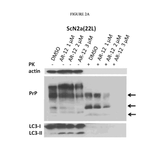
[00013] Figure 2A is an exemplary immunoblot showing the PrP banding pattern
following
treatment of persistently prion infection neuronal cells (ScN2a) treated for
72 hours with three
concentrations of AR-12, with (right panel) and without proteinase K digestion
(PK) (left panel);
actin (upper panel) was used as a loading control;
[00014] Figure 2B illustrates an exemplary Autophagy Assay measuring LC3-II
induction for
control (DSMO) and three concentrations of AR-12;
[00015] Figure 2C illustrates an exemplary XTT Cytotoxicity Assay for control
(DSMO) and
five concentrations of AR-12; Triton X-100 treatment was used as a positive
control (induction
of cell death); asterisks indicate concentrations with statistically
significant toxicity;
[00016] Figure 3A is an exemplary immunoblot showing the PrP banding pattern
for
persistently prion infected ScCAD5 neuronal cells treated with DSMO (control)
or 1, 2, 3, 4 or 5
M AR-12 for 72 hours, with (right side) and without PK (left side); actin
(upper panel) was
used as a loading control;
3
CA 03014772 2018-08-15
WO 2017/151687 PCT/US2017/020053
[00017] Figure 3B illustrates an exemplary XTT Cytotoxicity Assay for control
(DSMO) and
five concentrations of AR-12; Triton X-100 treatment was used as a positive
control (induction
of cell death); asterisks indicate concentrations with statistically
significant toxicity;
[00018] Figure 4A is an exemplary immunoblot showing the PrP banding pattern
for
persistently prion infected ScMEF fibroblast cells (infected with prion
strains 22L, Me7 and
RML) treated with DSMO (control) or 3 i.tM AR-12 for 72 hours, with and
without PK and actin
(upper panel) used as a loading control;
[00019] Figure 4B illustrates an exemplary XTT Cytotoxicity Assay for control
(DSMO) and
five concentrations of AR-12; Triton X-100 treatment was used as a positive
control (induction
of cell death); asterisks indicate concentrations with statistically
significant toxicity;
[00020] Figure 5A is an exemplary immunoblot showing the PrP banding pattern
following
treatment of persistently prion infection neuronal cells (ScN2a) treated for
72 hours with three
concentrations of AR-14 (0.5, 1 and 2 with (right panel) and without
proteinase K digestion
(PK) (left panel); actin (upper panel) was used as a loading control;
[00021] Figure 5B illustrates an exemplary XTT Cytotoxicity Assay for control
(DSMO) and
six concentrations of AR-14; Triton X-100 treatment was used as a positive
control (induction of
cell death); asterisks indicate concentrations with statistically significant
toxicity;
[00022] Figure 5C is an exemplary immunoblot showing the PrP banding pattern
for
persistently prion infected ScN2A cells (prion strain 22L) treated for 72
hours with AR-14 at the
indicated nanomolar concentrations, with or without PK.
[00023] Figure 6A is an exemplary immunoblot showing the PrP banding pattern
(with or
without PK) for persistently prion infected ScN2A cells treated with DMSO, AR-
12 (3 1..1M) or
AR-14 (2 ilM) for 4 days (upper panel), 20 days (second panel), 20 days with
drug, followed by
4 days without (third panel), and 20 days with drug, followed by 20 days
without drug treatment
(lower panel) with LC3-I/II used as marker for autophagy;
[00024] Figure 6B is an exemplary RT-QuIC assay showing prion conversion
activity in
uninfected N2a cells (panels A, E, I and M), ScN2a cells treated with DMSO
(panels B, F, J and
N), ScN2a cells treated with 3 i.tM of AR-12 (panels C, G, K and 0), and ScN2a
cells treated
with 2 tM of AR-14 (panels D, H, L and P) for 4 days (A-D), 20 days (E-H), 20
days with drug,
4
CA 03014772 2018-08-15
WO 2017/151687 PCT/US2017/020053
followed by 4 days without (I-L), and 20 days with drug, followed by 20 days
without drug
treatment (M-P) with dilutions of cell lysates and the test cut-off indicated
at the bottom;
[00025] Figure 6C is an exemplary immunoblot showing the PrP banding pattern
(with or
without PK) for persistently prion infected ScMEF cells (prion strain Me7)
treated with DMSO,
AR-12 (3 1..1M) or AR-14 (211.M) for 4 days (upper panel), 20 days (second
panel), 20 days with
drug, followed by 4 days without (third panel), and 20 days with drug,
followed by 20 days
without drug treatment (lower panel) with LC3-I/II used as marker for
autophagy;
[00026] Figure 6D is an exemplary RT-QuIC assay showing prion conversion
activity in
uninfected MEF cells (panels A, E, I and M), ScMEF cells treated with DMSO
(panels B, F, J
and N), ScMEF cells treated with 3 of AR-12 (panels C, G, K and 0), and
ScMEF cells
treated with 2 tM of AR-14 (panels D, H, L and P) for 4 days (A-D), 20 days (E-
H), 20 days
with drug, followed by 4 days without (I-L), and 20 days with drug, followed
by 20 days without
drug treatment (M-P) with dilutions of cell lysates and the test cut-off
indicated at the bottom;
[00027] Figure 7 is an exemplary immunoblot showing induction of autophagy by
short-term
treatment (2, 4 and 6 hours) with AR-12 (3 1..1M) or AR-14 (2 ilM) in N2a, MEF
and CADS cells;
DMSO treatment was used as negative control; actin (upper panel) served as a
loading control;
Increase of LC3-II band indicates autophagy induction and treatment with
bafilomycin Al (BA1)
used to block autophagy flux;
[00028] Figure 8A shows the establishment of a knock-out of Atg5 (autophagy
gene) clone of
N2A persistently prion-infected cells;
[00029] Figure 8B is an exemplary immunoblot showing the PrP banding pattern
for wild-
type ScN2A cells (left panel) and autophagy-deficient Atg5-K0 ScN2a cells
(right panel) after
treatment with control (DSMO) or AR-12 at three concentrations, with or
without PK and actin
used as a loading control; and
[00030] Figure 8C is an exemplary immunoblot showing the PrP banding pattern
for wild-
type ScN2A cells (left panel) and autophagy-deficient Atg5-K0 ScN2a cells
(right panel) after
treatment with control (DSMO) or AR-14 at three concentrations, with or
without PK and actin
used as a loading control.
CA 03014772 2018-08-15
WO 2017/151687 PCT/US2017/020053
DETAILED DESCRIPTION
[00031] The disclosed methods and compositions below may be described both
generally as
well as specifically. It should be noted that when the description is specific
to an aspect, that
aspect should in no way limit the scope of the methods. All references cited
herein are hereby
incorporated by reference in their entirety.
[00032] The term "prion," as used herein, refers to unconventional infectious
particles which
are the causal agents of prion diseases in humans and animals and fatal
infectious
neurodegenerative disorders. Prions are composed of the pathological isoform
PrPsc of the prion
protein, which serves as a surrogate marker for prion infectivity.
[00033] Aspects described herein provide methods of reducing the level of
prions in prion-
infected cells, tissues or organs, by exposing prion-infected cells, tissues
or organs to AR-12 in an
amount sufficient to reduce the level of prions in the prion-infected cells,
tissues or organs by at
least about 90% compared to prion-infected cells, tissues, organs that have
not been exposed to
AR-12. In another aspect, the prion level is reduced by at least about 50%
(short-term treatment,
e.g., 3 days) or prion-infected cells are substantially cured from prion
infection (long-term
treatment, e.g., 20 days).
[00034] In yet another aspect, prion infected cells, tissues or organs are
exposed to AR-12 in an
amount sufficient to achieve a concentration of at least about 1 p.M in the
prion-infected cells,
tissues or organs. In another aspect, prion infected cells, tissues or organs
are exposed to AR-12 in
an amount sufficient to achieve a concentration of between about 1 1.11\4 and
3 1.1.1\4 in the prion
infected cells, tissues, or organs. In a further aspect, the amount of AR-12
the prion infected cells,
tissues, and organs are exposed to is sufficient to reduce the prion level by
about 50% to about
90%, or substantially cures the prion-infected cells in long-term treatments.
In these aspects,
exposure to AR-12 does not result in substantial cytotoxicity of the prion
infected cells.
[00035] As used herein, the term "cytotoxicity" refers to the quality or
effect of a chemical,
drug or compound being toxic to cells. The toxic effects on individual cells.
The toxic effect on
individual cells can then result in cell death, tissue necrosis and organ
dysfunction or failure.
[00036] Aspects described herein provide methods of reducing the level of
prions in prion-
infected cells, tissues or organs, by exposing prion-infected cells, tissues
or organs to AR-14 in an
6
CA 03014772 2018-08-15
WO 2017/151687 PCT/US2017/020053
amount sufficient to reduce the level of prions in the prion-infected cells,
tissues or organs by at
least about 90% compared to prion-infected cells, tissues, organs that have
not been exposed to
AR-14. In another aspect, the prion level is reduced by at least about 50%
(short-term treatment,
e.g., 3 days) or prion-infected cells are substantially cured from prion
infection (long-term
treatment, e.g., 20 days).
[00037] In yet another aspect, prion infected cells, tissues or organs are
exposed to AR-14 in an
amount sufficient to achieve a concentration of at least about 0.5 p.M in the
prion-infected cells,
tissues or organs. In another aspect, prion infected cells, tissues or organs
are exposed to AR-14 in
an amount sufficient to achieve a concentration of between about 0.5 jtM and 2
1.11\4 in the prion
infected cells, tissues, or organs. In a further aspect, the amount of AR-14
the prion infected cells,
tissues, and organs are exposed to is sufficient to reduce the prion level by
about 50% to about
90%, or substantially cures the prion-infected cells in long-term treatments.
In these aspects,
exposure to AR-14 does not result in substantial cytotoxicity of the prion
infected cells.
[00038] As used herein, the term AR-12 refers to (C26H19F3N40 and 2-amino-N-(4-
(5-
(phenanthren-2-y1)-3-(trifluoromethyl)-1H-pyraz ol-1 -yl)phenyl)acetami de)),
having the
following structure:
CF3
N -N
01"--
HN
'NH2
[00039] The term "AR-12" also includes, for example, analogs of AR-12 (e.g.,
the compounds
described in U.S. Patents 7,576,116, 8,546,441, 8,541,460, 8,039,502, and
8,080,574 hereby
incorporated by reference in their entirety).
[00040] As used herein, AR-14 refers to a compound having the following
structure:
7
CA 03014772 2018-08-15
WO 2017/151687 PCT/US2017/020053
/---r-CF3
02,
_--
\ j 0
HN
H 2N
[00041] The relative resistance of PrPsc to proteinase K (PK) digestion can be
used as a
diagnostic tool to distinguish between PrPc and PrPsc. In this aspect, lysates
of cells or tissues are
digested with proteinase K (PK) under defined standard conditions and analyzed
in an immunoblot.
As shown in Figure 1, PrPc is completely sensitive to PK digestion. PrPsc is
only partially sensitive
to PK digestion and becomes degraded solely at the N-terminus. The left panel
of Figure 1 shows
situation schematically, right panel depicts a typical immunoblot result. For
persistently prion-
infected cells, a typical 3-banding pattern is obtained following PK digestion
(+PK), representing
N-terminally truncated un-glycosylated, single- and double-glycosylated PrPSc
(see arrows).
[00042] Proteinase K (PK) was obtained from Roth (Karlsruhe, Germany),
Pefabloc inhibitor
was from Roche (Mannheim, Germany). Cell culture media and solutions were
obtained from
Invitrogen (Karlsruhe, Germany). N-Lauryl-sarcosine was purchased from Sigma-
Aldrich
(Munich, Germany). Immunoblotting was done using the enhanced
chemiluminiscence blotting
technique (ECL plus) from Amersham Corporation (Buckinghamshire, UK). The test
AR
compounds were dissolved in DMSO at a stock solution of 1 mM and stored at ¨20
C. The
monoclonal anti-PrP antibody (mAb) 4H11 was generated using a dimeric murine
PrP as an
immunogen (Ertmer et al., 2004). Mouse anti-j3-actin mAb was from Sigma, mouse
anti-LC3 mAb
was obtained from nanoTools (nanoTools Antikorpertechnik GmbH & Co. KG,
Teningen,
Germany). Peroxidase-conjugated immunoglobulins for immunoblot analysis were
obtained from
Dianova (Hamburg, Germany).
[00043] Cell culture
8
CA 03014772 2018-08-15
WO 2017/151687 PCT/US2017/020053
[00044] The mouse neuroblastoma cell line N2a [American Type Culture
Collection (ATCC)
CCL-131] and the persistently-prion infected ScN2a cell lines (22L-ScN2a, RML-
ScN2a) have
been described (Schatzl et al., 1997; Gilch et al., 2001; Taguchi et al.,
2013). N2a cells deficient
for Atg5 were prepared by CRISPR-Cas9 technology and characterized in
immunoblot and DNA
sequencing for successful Atg5 knock-out. Characterized single cell clones
were then infected with
22L prions as described previously (Maas et al. 2007). Wild type mouse
embryonic fibroblasts
(MEF) have been described before (Kuma et al., 2004) and have been
persistently infected with
mouse-adapted prion strains 22L, RN/IL and Me7. CAD5 (a central nervous system
catecholaminergic cell line; Qi et al, 1997) and persistently prion-infected
22L-ScCAD cells were
prepared as above. MEF cells were maintained in Dulbecco's modified Eagle's
medium (DMEM),
N2a cells in Opti-MEM Glutamax medium, both media containing 10% fetal calf
serum (FCS),
penicillin/streptomycin and glutamine in a 5% CO2 atmosphere. CAD5 cells were
cultured in
OptiMEM Glutamax medium containing 10% bovine growth serum (BGS) and
penicillin/streptomycin in a 5% CO2 atmosphere.
[00045] Cell lysis, proteinase K (PK) analysis and immunoblot
[00046] Immunoblot analyses were performed as previously described (Schatzl et
al., 1997;
Gilch et al., 2001; Taguchi et al., 2013). Confluent cells were lysed in cold
lysis buffer (10 mM
Tris-HC1, pH 7.5; 100 mM NaCl; 10 mM EDTA; 0.5% Triton X-100; 0.5% sodium
deoxycholate
(DOC)) for 10 min. For proteinase K (PK) treatment, post-nuclear lysates were
divided into two
halves. One half was incubated with PK (20 [tg/m1) for 30 min at 37 C and
digestion was stopped
by addition of proteinase inhibitors (0.5 mM Pefabloc) and directly
precipitated with methanol.
The sample without PK treatment was directly supplemented with proteinase
inhibitors and
precipitated with methanol. After centrifugation for 25 min at 3,500 rpm (4
C), the pellets were
re-dissolved in TNE buffer (50 mM Tris-HC1 pH 7.5, 150 mM NaCl, 5 mM EDTA) and
gel loading
buffer (7% SDS, 30% glycerine, 20% Et-SH, 0,01% Bromphenol blue in 90 mM Tris-
HC1 pH 6.8)
was added. After boiling for 5 min an aliquot was analyzed on 12.5% SDS-PAGE.
Proteins were
electrotransferred to polyvinylidene difluoride (PVDF) membrane (Amersham).
Membranes were
blocked with non-fat dry milk (5%) in Tris-buffered saline (TBST) (0.05% Tween
20, 100 mM
NaCl, 10 mM Tris-HC1; pH 7.8), incubated overnight with the appropriate
antibody at 4 C and
stained using enhanced chemiluminiscence blotting (ECL plus) kit from
Amersham. To achieve
equal loading of total protein for different samples in immunoblot analysis
(e.g. comparison of
9
CA 03014772 2018-08-15
WO 2017/151687 PCT/US2017/020053
PrPsc amounts in different samples, or LC3-II levels), the same number of
cells were plated and
grown under identical conditions over specific periods. Cells were
subsequently lysed in the same
amount of lysis buffer. Precipitated proteins of each sample were resuspended
in the same amount
of TNE buffer and supplemented with identical amounts of gel loading buffer.
Equal volumes of
each sample were then analyzed by 12.5% SDS-PAGE. In addition, immunoblots
were stripped
with anti-J3-actin antibody to verify equal amounts of total protein loaded on
gel for each sample.
To allow comparison of endogenous LC3-II levels, intensity of LC3-II signals
were measured
relative to actin signals by densitometry analysis.
[00047] Viability assay (XTT)
[00048] The viability of a cell population upon treatment with different
compounds was
determined with the XTT assay (Roche, Mannheim, Germany). Viability testing
was mainly
performed in uninfected cells. Data from our and other groups showed that
viability in uninfected
and persistently prion-infected N2a, CADS or MEF cells is substantially the
same. Cells were
plated at a density of 1.5 x 10⁴ cells per well in 96 well plates. The
following day, cells were treated
for 72 h with various concentrations of the indicated compounds. Subsequently,
5011.1 of the XTT
reagent was added to each well. After incubation for 4 h, the absorption at
450 nm was measured
with a FLUOstar Omega plate reader (BMG LABTECH, Offenburg, Germany). The
average
absorption of four control wells was set as 100% viability. The viability of
treated cells was
compared to the viability of DMSO (negative control) or Triton X-100 (positive
control) treated
cells.
[00049] Real-time quacking-induced conversion assay (RT-QuIC)
[00050] A. Preparation of recombinant protein.
[00051] Preparation of recombinant prion proteins was performed as described
(Orru et al.,
2012). Briefly, mouse PrP (aa 23-231) was cloned into pET-41 plasmids,
transformed into E. coil
Rosetta, and bacteria cultured in LB media supplemented with kanamycin (0.05
mg/ml) and
chloramphenicol (0.034 mg/ml). The Overnight Express Autoinduction System
(Novagen, USA)
was used to induce protein expression. Inclusion bodies were isolated from
pelleted cells using
Bug Buster Master Mix (Novagen, USA) and stored at -20 C. For purification of
recombinant
PrP, inclusion bodies were solubilized in (8 M guanidine-HC1, 100 mM Na-
phosphate, 10 mM
Tris-HC1, pH 8.0) and incubated on the rocker for 1 h at RT. Ni-NTA Superflow
resin beads
CA 03014772 2018-08-15
WO 2017/151687 PCT/US2017/020053
(Quiagen, USA) were incubated in denaturing buffer (6 M guanidine-HC1, 100 mM
Na-phosphate,
pH 8.0) for 1 h at RT. Solubilized inclusion bodies were centrifuged at 16,000
x g for 5 min, the
supernatant added to the beads and incubated for 1 h with gentle rocking.
Beads were then packed
into a XK 16 glass column (GE Healthcare Life Sciences; USA; length 200 mm).
Using an
Amersham AKTA Explorer FPLC unit running with Unicorn software (5 version, GE
Healthcare
Life Sciences, USA), protein was refolded by a gradient from 100% denaturing
buffer to 100%
refolding buffer (100 mM Na-phosphate, 10 mM Tris-HC1, pH 8.0) over 4 h. The
column was
washed for 30 min with refolding buffer and proteins eluted using a linear
gradient from 100%
refolding buffer to 100% elution buffer (500 mM imidazole, 100 mM Na-
phosphate, 10 mM Tris-
HC1, pH 5.8). The central portions of the A280 UV peak were collected into
dialysis buffer (10
mM Na-phosphate, pH 5.8). Purified protein was filtered using a 0.22 p.m
filter, transferred into a
Slide-A-Lyzer dialysis cassette (MW 10 kDa; Thermo- Scientific, UA) placed
into a 4 1 beaker
with dialysis buffer overnight at 4 C with continuous stirring. Following
dialysis, the protein
solution was filtered again with a prewashed 0.22 p.m Argos syringe filter.
Protein concentration
was measured using BCA protein assay (Thermo-Scientific, 23227), the solution
aliquoted and
kept in -80 C until use.
[00052] B. RT-QuIC assay. Real-time QuIC was performed as described (John et
al., 2013).
Briefly, reactions were set up in assay buffer containing 20 mM Na-phosphate,
pH7.4, 300 mM
NaCl, 1 mM EDTA, 10 M Thioflavin T and 0.1 mg/ml rPrP substrate. Ninety-eight
pi aliquots
were added to the wells of a black-walled 96-well optical bottom plate (Nalge
Nunc International,
Nunc, USA). Tenfold serial dilutions of brain homogenate or cell homogenate
were prepared in
0.5 ml microtubes. Quadruplicate reactions were seeded with 2 1 of test
solution for a final
reaction volume of 100 pl. Reactions contained a final concentration of 0.002%
SD S. Plates were
sealed with Nunc Amplification Tape (Nalge Nunc International) and incubated
in a FLUOstar
Omega (BMG Labtech, Cary, NC, USA) plate reader for 30 h. Reactions were
incubated at 42 C,
with cycles of 60 s shaking (700 revolutions per minute) and 60 s of rest
throughout the incubation.
ThT fluorescence measurements (450 nm excitation and 480 nm emission) were
taken every 15
min. RT-QuIC data were averaged from four replicate wells and average values
plotted against
reaction time. Samples were scored positive if at least 50% of replicates
reached a ThT
fluorescence cut-off, which was calculated based on the average ThT
fluorescence plus 5 x
standard deviation.
11
CA 03014772 2018-08-15
WO 2017/151687 PCT/US2017/020053
[00053] As shown in Figure 2A, persistently prion (prion strain 22L) infected
mouse neuronal
cells (ScN2a cell line) were treated for 72 hours with the indicated
concentrations of AR-12, and
the cells were subjected to immunoblot analysis. Solvent only-treated cells
(DMSO) were used as
control. Cell lysates were split into two halves and one treated with
proteinase K (PK; 20 1/ml,
30 min at 37 C) and subjected to SDS-PAGE and immunoblot analysis.
[00054] The immunoblot was developed with anti-PrP monoclonal antibody (mAb)
4H11 and
the blot was re-probed with mAbs for actin (gel loading) and LC3 (autophagy
marker). PrPsc (right
side, +PK; 3 glycoforms indicated by arrows) was dose-dependently reduced, to
undetectable
levels when treated for 3 days with a concentration of 3 i.tM (=100%
reduction). Since PrPsc has a
very long half-life time in cultured cells (>24 h; see Ertmer et al., 2004),
such a strong anti-prion
effect after 3 days of treatment strongly indicates that AR-12 induces PrPsc
clearance as opposed
to inhibiting PrPsc propagation. LC3-II was induced about 2-fold, indicating
induction of
autophagy.
[00055] Figure 2B shows an exemplary quantification of the autophagy induction
at the
indicated concentration of AR-12. Figure 2C shows that exposing AR-12 to the
cells was done at
non-toxic concentrations (XTT toxicity assay).
[00056] Figure 3A shows an exemplary effect of AR-12 administration on
persistently prion-
infected CADS cells (mouse neuronal cell line) over time. The CADS neuronal
cells persistently
infected with prions (mouse-adapted scrapie strain 22L; termed ScCAD5) were
treated for 72
hours with 1-5 i.tM AR-12, and cells subjected to immunoblot analysis. Solvent
only-treated cells
(DMSO) were used as control. Cell lysates were split into two halves, and one
treated with
proteinase K (PK; 20 1/ml, 30 min at 37 C) and subjected to SDS-PAGE and
immunoblot
analysis. The immunoblot was developed with anti-PrP mAb 4H11, and the blot
was re-probed
with mAb for actin (gel loading; upper panel). PrPsc levels (+PK; 3 glycoforms
indicated by
arrows) were reduced, although slightly less than in ScN2a cells. This result
was expected, as
ScCAD5 cells harbor more PrPsc and therefore would need a longer treatment
period. This data
show that AR-12 is effective in another neuronal mouse cell type (derived from
the central nervous
system), confirming results described above for ScN2a cells.
[00057] Figure 3B shows that exposing AR-12 to the ScCAD5 cells was done at
non-toxic
concentrations (XTT toxicity assay).
12
CA 03014772 2018-08-15
WO 2017/151687 PCT/US2017/020053
[00058] Figure 4A shows an exemplary effect of AR-12 administration to
persistently prion-
infected mouse embryonic fibroblasts (ScMEFs, infected with mouse-adapted
scrapie prion strains
22L, Me7 or RML). These fibroblast cells were treated for 72 hours with 3 tM
AR-12, and the
cells were subjected to immunoblot analysis. Solvent only-treated cells (DMSO)
were used as
control. Cell lysates were split into two halves and one treated with
proteinase K (PK; 20 1/ml,
30 min at 37 C) and subjected to SDS-PAGE and immunoblot analysis. The
immunoblot was
developed with anti-PrP mAb 4H11 and the blot was re-probed with mAb for actin
(gel loading;
upper panel). PrPsc (+PK; indicated by 3 arrows) was strongly reduced. As
shown in Figure 5A,
AR-12 is also effective in a non-neuronal cell type, indicating a broad range
of anti-prion activity
that is not cell type-dependent. AR-12 was effective against three different
prion strains (22L,
RML and Me7), indicating a broad range of anti-prion activity against
different prion strains.
[00059] Figure 4B shows that exposing AR-12 to the ScMEF cells was done at non-
toxic
concentrations (XTT toxicity assay).
[00060] Figure 5A shows persistently prion (prion strain 22L) infected mouse
neuronal cells
(ScN2a cell line) treated for 72 hours with the indicated concentrations of AR-
14 (0.5, 1 and 2
Solvent only-treated cells (DMSO) were used as control. The cells were
subjected to
immunoblot analysis. Cell lysates were split into two halves and one treated
with proteinase K
(PK; 20 1/ml, 30 min at 37 C) and subjected to SDS-PAGE and immunoblot
analysis. The
immunoblot was developed with anti-PrP mAb 4H11 and the blot was re-probed
with mAb for
actin (gel loading). PrPsc (+PK; 3 glycoforms indicated by arrows) was dose-
dependently reduced,
to undetectable levels when treated for 3 days with a concentration of 2
(=100% reduction).
Since PrPsc has a very long half-life time in cultured cells (>24 h; see
Ertmer et al., 2004), such a
substantial anti-prion effect after 3 days of treatment strongly indicates
that AR-14 induces PrPsc
clearance as opposed to inhibiting PrPsc propagation.
[00061] Figure 5B shows that exposing AR-14 to the ScN2a cells was done at non-
toxic
concentrations (XTT toxicity assay).
[00062] Figure 5C shows an exemplary effect of AR-14 on persistently prion
infected ScN2a
cells (prion strain 22L) at various nanomolar concentrations (0.5, 0.75 and
1.0 PrPsc (+PK,
indicated by 3 arrows) was dose-dependently reduced, with AR-14 effective
already at 0.75 M.
AR-14 showed anti-prion effects at nanomolar concentrations.
13
CA 03014772 2018-08-15
WO 2017/151687 PCT/US2017/020053
[00063] Figure 6A shows that a long-term treatment of ScN2a cells with AR-12
and AR-14
cures the cells of prion infection. ScN2a cells (neuronal) were treated with
AR-12 (3 [tM) or AR-
14 (2 M). DMSO-treated cells were used as a control. Treatment was continued
for 20 days (five
passages). Then, the treatment was stopped, and cells were passaged for
another 20 days (five
passages) without drug. At passages one and five with treatment (first and
second panel), or after
drug-withdrawal (third and fourth panel), cells were lysed. Cell lysates were
split into two halves,
one half was treated with proteinase K (PK; 20 g/ml, 30 min at 37 C) and
subjected to
immunoblot analysis. The immunoblot was developed with anti-PrP mAb 4H11, anti-
LC3
(autophagy marker) and anti-actin for gel loading. The PrPsc signal completely
disappeared during
drug treatment and did not reappear after drug withdrawal.
[00064] Figure 6B shows that prion conversion activity is lost in long-term AR-
12 or AR-14
treated ScN2a cells. RT-QuIC assay was performed using recombinant mouse PrP
as a substrate.
Each quadruplicate RT-QuIC reaction was seeded with 2 gl cell lysate (at
dilutions 10-1 to 10-4) of
ScN2a cells treated with AR-12 (3 [tM) (C, G), AR-14 (2 [tM) (D, H) or solvent
only (DMSO; B,
F). Data shown are for passage one (P1) and passage five (P5). Uninfected N2a
cells were used as
negative test control (A, E). Panels in third and fourth row show ScN2a cells
after treatment
discontinuation for AR-12 (K, 0), AR-14 (L, P) or solvent only (DMSO; J, N).
Data shown are
for passage one (P1) and passage five (P5) after drug withdrawal. Uninfected
N2a cells were used
as negative test control (I, M). The average increase of thioflavin-T
fluorescence of replicate wells
is plotted as a function of time. Y-axis represents relative fluorescent units
(RFU) and x-axis time
in hours. Cut-off values were shown as dotted line. ScN2a cells treated with
AR-12 lost prion
conversion activity (tested until passage five after terminating the AR-12
treatment).
[00065] Figure 6C shows that a long-term treatment of ScMEF cells with AR-12
and AR-14
permanently cures the cells of prion infection. ScMEF cells (non-neuronal)
were treated with AR-
12 (3 [tM) or AR-14 (2 M). DMSO-treated cells were used as a control.
Treatment was continued
for 20 days (five passages). Then, the treatment was stopped, and cells were
passaged for another
20 days (five passages) without drug. At passages one and five with treatment
(first and second
panel) or after drug-withdrawal (third and fourth panel), cells were lysed.
Cell lysates were split
into two halves, one half was treated with proteinase K (PK; 20 g/ml, 30 min
at 37 C), and
subjected to immunoblot analysis. The immunoblot was developed with anti-PrP
mAb 4H11, anti-
LC3 (autophagy marker) and anti-actin for gel loading. PrPsc signal completely
disappeared
14
CA 03014772 2018-08-15
WO 2017/151687 PCT/US2017/020053
during drug treatment and did not reappear after drug withdrawal. Long-term
treatment with AR-
12 and AR-14 cured neuronal and non-neuronal cells from prion infection.
[00066] Figure 6D shows that prion conversion activity is lost in long-term AR-
12 or AR-14
treated ScMEF cells. RT-QuIC assay was performed using recombinant mouse PrP
as substrate.
Each quadruplicate RT-QuIC reaction was seeded with 2 gl cell lysate (at
dilutions 104 to 10-a) of
ScMEF cells treated with AR-12 (3 1..1M) (C, G), AR-14 (2 1..1M) (D, H) or
solvent only (DMSO;
B, F). Data shown are for passage one (P1) and passage five (P5). Uninfected
MEF cells were used
as negative assay control (A, E). Panels in third and fourth row show ScMEF
cells after treatment
discontinuation for AR-12 (K, 0), AR-14 (L, P) or solvent only (DMSO; J, N).
Data shown are
for passage one (P1) and passage five (P5) after drug withdrawal. Uninfected
MWF cells were
used as negative test control (I, M). The average increase of thioflavin-T
fluorescence of replicate
wells is plotted as a function of time. Y-axis represents relative fluorescent
units (RFU) and x-axis
time in hours. Cut-off values are shown as dotted lines. ScMEF cells treated
with AR-12 or AR-
14 lost prion conversion activity (tested until passage five after terminating
the AR-12 treatment).
[00067] Figure 7 shows that AR-12 and AR-14 induce autophagy in N2a, MEF and
CADS cells,
indicating that AR-12 and AR-14-mediated anti-prion effect involve autophagy.
N2a, MEF and
CADS cells were treated with either AR-12 (3 1..1M) or AR-14 (2
respectively, for 2, 4 or 6
hours. Bafilomycin Al treatment was used to alter the lysosomal function and
to block the
autophagic flux. This control demonstrates that AR-12 and AR-14 induce
autophagy and do not
block autophagic flux. Solvent only-treated cells (DMSO) were used as
treatment vehicle control.
Cells were lysed, subjected to immunoblot analysis, and the immunoblots were
developed with an
anti-LC3 mAb as autophagy marker and an actin mAb (gel loading control; upper
panel). Both
AR-12 and AR-14 showed a time dependent and pronounced increase in LC3-II
levels (lower
band). This induction was lower than that of BAl-treated cells, which had the
highest expression
level of LC3-II due to blocking of autophagic flux and lysosomal function.
These data indicate
that AR-12 and AR-14 are strong inducers of autophagy in all three tested cell
lines. Interestingly,
the lowest induction was found in CADS cells, which correlates with the
weakest effects on PrPsc
levels in these cells. Formal testing of the impact of autophagy was done
using cells compromised
in autophagy and comparing them to wild-type cells (see Figures 8A and 8C).
CA 03014772 2018-08-15
WO 2017/151687 PCT/US2017/020053
[00068] Figure 8A shows the establishment ofN2a cells with a knock-out in the
autophagy gene
ATG5. Using CRISPR/Cas-9 technology, insertions and deletions were introduced
into exon 5 and
6 of the ATG5 gene, resulting in premature stop codons. Individual cell clones
were generated and
analyzed for ATG5 knock-out by DNA sequencing and immunoblot analysis. Various
positive
clones were then persistently infected with prions (strain 22L). Immunoblot
shows ATG5-K0
ScN2a cells, probed for Atg5, LC3 and actin. There is no Atg5 and LC3-II band
(lane 1 vs. lane
2), indicating knock-out of ATG5 and complete deficiency in autophagy.
[00069] Figures 8B shows the kinetics of AR-12 mediated reduction of PrPsc in
wild-type and
Atg5-K0 ScN2a cells, indicating partial involvement of autophagy competency in
AR-12
mediated anti-prion effects. Persistently prion infected wild-type (left
panel) and ATG5-K0 (right
panel) ScN2a cells were treated for 72 hours with 1 to 3
AR-12 and the cells subjected to
immunoblot analysis. Solvent only-treated cells (DMSO) were used as control.
Cell lysates were
split into two halves, and one half was treated with proteinase K (PK; 20
1/ml, 30 min at 37 C)
and subjected to SDS-PAGE and immunoblot analysis. The immunoblot was
developed with anti-
PrP mAb 4H11, and the blot was re-probed with mAb for actin (gel loading;
upper panel). PrPsc
(right side of panels, +PK; indicated by 3 arrows) was reduced in both
situations, although with
different kinetics for wild-type and Atg5-K0 ScN2a cells. At 3
of AR-12, the reduction of
PrPsc in wild-type ScN2a cells was 90% or greater, while in Atg5-K0 ScN2a
cells, the reduction
was about 50%. These results indicate autophagy competency is involved in AR-
12 mediated anti-
prion effects.
[00070] Figures 8C shows the kinetics of AR-14 mediated reduction of PrPsc in
wild-type and
Atg5-K0 ScN2a cells, indicating partial involvement of autophagy competency in
AR-14
mediated anti-prion effects. Persistently prion infected wild-type (left
panel) and ATG5-K0 (right
panel) ScN2a cells were treated for 72 hours with 0.5, 1 and 2
AR-14 and the cells subjected
to immunoblot analysis. Solvent only-treated cells (DMSO) were used as
control. Cell lysates were
split into two halves, and one half was treated with proteinase K (PK; 20
1/ml, 30 min at 37 C)
and subjected to SDS-PAGE and immunoblot analysis. The immunoblot was
developed with anti-
PrP mAb 4H11, and the blot was re-probed with mAb for actin (gel loading;
upper panel). PrPsc
(right side of panels, +PK; indicated by 3 arrows) was reduced in both
situations, although with
different kinetics for wild-type and Atg5-K0 ScN2a cells. At 2
AR-14, the reduction of PrPsc
in wild-type ScN2a cells was 100%, whereas in Atg5-K0 ScN2a cells, the
reduction was about
16
CA 03014772 2018-08-15
WO 2017/151687 PCT/US2017/020053
50%. These results indicate autophagy competency is involved in AR-14 mediated
anti-prion
effects.
[00071] AR-12 or AR-14, as described herein, can be administered orally,
parenterally (IV, IM,
depot-IM, SQ, and depot-SQ), sublingually, intranasally (inhalation),
intrathecally, topically, in
the pulmonary system or airways (e.g., nebulization, aerosol) or rectally.
Dosage forms known to
those of skill in the art are suitable for delivery of AR-12 and AR-14
described herein. In one
aspect, AR-12 and AR-14 is administered orally.
[00072] AR-12 or AR-14 can be formulated into suitable pharmaceutical
preparations such as
creams, gels, suspensions, tablets, capsules, or elixirs for oral
administration or in sterile solutions
or suspensions for parenteral administration. AR-12 or AR-14 can be formulated
into
pharmaceutical compositions using techniques and procedures well-known in the
art.
[00073] In one aspect, about 0.1 to 1000 mg, about 5 to about 100 mg, or about
10 to about 50
mg of the AR-12 or AR-14, or a physiologically acceptable salt or ester can be
compounded with
a physiologically acceptable vehicle, carrier, excipient, binder,
preservative, pain reliever,
stabilizer, flavor, etc., in a unit dosage form as called for by accepted
pharmaceutical practice. The
amount of active substance in compositions or preparations comprising AR-12 or
AR-14 is such
that a suitable dosage and concentration in a host in the range indicated is
obtained.
[00074] In another aspect, the compositions can be formulated in a unit dosage
form, each
dosage containing from about 1 to about 1000 mg, about 1 to about 500 mg, or
about 10 to about
100 mg of the active ingredient. The term "unit dosage from" refers to
physically discrete units
suitable as unitary dosages for human subjects and other mammals, each unit
containing a
predetermined quantity of active material calculated to produce the desired
therapeutic effect, in
association with a suitable pharmaceutical excipient.
[00075] In one aspect, AR-12 or AR-14 alone or AR-12 or AR-14 and one or more
additional
active or inert ingredients, is mixed with a suitable pharmaceutically
acceptable carrier to form a
composition. Upon mixing or addition of the compound(s), the resulting mixture
may be a cream,
gel, solution, suspension, emulsion, or the like. Liposomal suspensions may
also be used as
pharmaceutically acceptable carriers. These may be prepared according to
methods known to those
skilled in the art. The form of the resulting mixture depends upon a number of
factors, including
the intended mode of administration and the solubility of the compound in the
selected carrier or
17
CA 03014772 2018-08-15
WO 2017/151687 PCT/US2017/020053
vehicle. In one aspect, the effective concentration is sufficient for
lessening or ameliorating at least
one symptom of the disease, disorder, or condition treated and may be
empirically determined.
[00076] Pharmaceutical carriers or vehicles suitable for administration of AR-
12 or AR-14
described herein include any such carriers suitable for the particular mode of
administration. In
addition, the active materials can also be mixed with other active materials
that do not impair the
desired action, or with materials that supplement the desired action, or have
another action. The
compounds may be formulated as the sole pharmaceutically active ingredient in
the composition
or may be combined with other active ingredients (e.g., Congo Red,
anthracyclines, sulfated
polyanions, suramin, imatinib/Gleevec , rapamycin, trehalose, lithium,
tamoxifen, piperazine
derivatives, diphenylpyrazole-derived compounds, flupirtine, tetrapyrroles,
quinacrine,
chlorpromazine, pentosan polysulphate, D-penicillamine, active and passive
anti-prion
vaccination, doxycycline, donepezil, rivastigmine, galantamine and memantine).
[00077] In another aspect, if AR-12 or AR-14 exhibits insufficient solubility,
methods for
solubilizing may be used. Such methods are known and include, but are not
limited to, using co-
solvents such as dimethylsulfoxide (DMSO), using surfactants such as TWEEN,
and dissolution
in aqueous sodium bicarbonate. Derivatives of the compounds, such as salts or
prodrugs, may also
be used in formulating effective pharmaceutical compositions.
[00078] The concentration of the compound is effective for delivery of an
amount upon
administration that lessens or ameliorates at least one symptom of the
disorder for which the
compound is administered. Typically, the compositions are formulated for
single dosage
administration.
[00079] In another aspect, AR-12 or AR-14 as described herein may be prepared
with carriers
that protect them against rapid elimination from the body, such as time-
release formulations or
coatings. Such carriers include controlled release formulations, such as, but
not limited to,
microencapsulated delivery systems. The active compound can be included in the
pharmaceutically acceptable carrier in an amount sufficient to exert a
therapeutically useful effect
in the absence of undesirable side effects on the patient treated. The
therapeutically effective
concentration may be determined empirically by testing the compounds in known
in vitro and in
vivo model systems for the treated disorder.
18
CA 03014772 2018-08-15
WO 2017/151687 PCT/US2017/020053
[00080] In another aspect, AR-12 or AR-14 and compositions described herein
can be enclosed
in multiple or single dose containers. The enclosed compounds and compositions
can be provided
in kits, for example, including component parts that can be assembled for use.
For example, AR-
12 or AR-14 in lyophilized form and a suitable diluent may be provided as
separated components
for combination prior to use. A kit may include AR-12 and a second therapeutic
agent for co-
administration. AR-12 and second therapeutic agent may be provided as separate
component parts.
A kit may include a plurality of containers, each container holding one or
more unit dose of AR-
12 or AR-14 described herein. In one aspect, the containers can be adapted for
the desired mode
of administration, including, but not limited to suspensions, tablets, gel
capsules, sustained-release
capsules, and the like for oral administration; depot products, pre-filled
syringes, ampoules, vials,
and the like for parenteral administration; and patches, medipads, gels,
suspensions, creams, and
the like for topical administration.
[00081] The concentration of AR-12 or AR-14 in the pharmaceutical composition
will depend
on absorption, inactivation, and excretion rates of the active compound, the
dosage schedule, and
amount administered as well as other factors known to those of skill in the
art.
[00082] In another aspect, the active ingredient may be administered at once,
or may be divided
into a number of smaller doses to be administered at intervals of time. It is
understood that the
precise dosage and duration of treatment is a function of the disease being
treated and may be
determined empirically using known testing protocols or by extrapolation from
in vivo or in vitro
test data. It is to be noted that concentrations and dosage values may also
vary with the severity of
the condition to be alleviated. It is to be further understood that for any
particular subject, specific
dosage regimens should be adjusted over time according to the individual need
and the
professional judgment of the person administering or supervising the
administration of the
compositions, and that the concentration ranges set forth herein are exemplary
only and are not
intended to limit the scope or practice of the claimed compositions.
[00083] If oral administration is desired, the compound can be provided in a
composition that
protects it from the acidic environment of the stomach. For example, the
composition can be
formulated in an enteric coating that maintains its integrity in the stomach
and releases the active
compound in the intestine. The composition may also be formulated in
combination with an
antacid or other such ingredient.
19
CA 03014772 2018-08-15
WO 2017/151687 PCT/US2017/020053
[00084] Oral compositions will generally include an inert diluent or an edible
carrier and may
be compressed into tablets or enclosed in gelatin capsules. For the purpose of
oral therapeutic
administration, the active compound or compounds can be incorporated with
excipients and used
in the form of tablets, capsules, or troches. Pharmaceutically compatible
binding agents and
adjuvant materials can be included as part of the composition.
[00085] The tablets, pills, capsules, troches, and the like can contain any
of the following
ingredients or compounds of a similar nature: a binder such as, but not
limited to, gum tragacanth,
acacia, corn starch, or gelatin; an excipient such as microcrystalline
cellulose, starch, or lactose; a
disintegrating agent such as, but not limited to, alginic acid and corn
starch; a lubricant such as,
but not limited to, magnesium stearate; a glidant, such as, but not limited
to, colloidal silicon
dioxide; a sweetening agent such as sucrose or saccharin; and a flavoring
agent such as peppermint,
methyl salicylate, or fruit flavoring.
[00086] When the dosage unit form is a capsule, it can contain, in addition to
material of the
above type, a liquid carrier such as a fatty oil. In addition, dosage unit
forms can contain various
other materials, which modify the physical form of the dosage unit, for
example, coatings of sugar
and other enteric agents. The compounds can also be administered as a
component of an elixir,
suspension, syrup, wafer, chewing gum or the like. A syrup may contain, in
addition to the active
compounds, sucrose as a sweetening agent and certain preservatives, dyes and
colorings, and
flavors.
[00087] The active materials can also be mixed with other active materials
that do not impair
the desired action, or with materials that supplement the desired action. AR-
12 or AR-14 can be
used, for example, in combination with an antibiotic, antifungal, antiviral,
pain reliever, or
cosmetic.
[00088] In one aspect, solutions or suspensions used for parenteral,
intradermal, subcutaneous,
inhalation, or topical application can include any of the following
components: a sterile diluent
such as water for injection, saline solution, fixed oil, a naturally occurring
vegetable oil such as
sesame oil, coconut oil, peanut oil, cottonseed oil, and the like, or a
synthetic fatty vehicle such as
ethyl oleate, and the like, alcohols, polyethylene glycol, glycerin, propylene
glycol, or other
synthetic solvent; antimicrobial agents such as benzyl alcohol and methyl
parabens; antioxidants
such as ascorbic acid and sodium bisulfite; chelating agents such as
ethylenediaminetetraacetic
CA 03014772 2018-08-15
WO 2017/151687 PCT/US2017/020053
acid (EDTA); buffers such as acetates, citrates, and phosphates; and agents
for the adjustment of
tonicity such as sodium chloride and dextrose. Parenteral preparations can be
enclosed in
ampoules, disposable syringes, or multiple dose vials made of glass, plastic,
or other suitable
material. Buffers, preservatives, antioxidants, and the like can be
incorporated as required.
[00089] Where administered intravenously, intramuscularly, or
intraperitoneally, suitable
carriers include, but are not limited to, physiological saline, phosphate
buffered saline (PBS), and
solutions containing thickening and solubilizing agents such as glucose,
polyethylene glycol,
polypropylene glycol, ethanol, N-methylpyrrolidone, surfactants and mixtures
thereof. Liposomal
suspensions including tissue-targeted liposomes may also be suitable as
pharmaceutically
acceptable carriers. These may be prepared according to methods known in the
art.
[00090] In another aspect, AR-12 or AR-14 may be prepared with carriers that
protect the
compound against rapid elimination from the body, such as time-release
formulations or coatings.
Such carriers include controlled release formulations, such as, but not
limited to, implants and
microencapsulated delivery systems, and biodegradable, biocompatible polymers
such as collagen,
ethylene vinyl acetate, polyanhydrides, polyglycolic acid, polyorthoesters,
polylactic acid, and the
like. Methods for preparation of such formulations are known to those skilled
in the art.
[00091] In yet another aspect, compounds employed in the methods of the
disclosure may be
administered enterally or parenterally. When administered orally, compounds
employed in the
methods of the disclosure can be administered in usual dosage forms for oral
administration as is
well known to those skilled in the art. These dosage forms include the usual
solid unit dosage
forms of tablets and capsules as well as liquid dosage forms such as
solutions, suspensions, and
elixirs. When the solid dosage forms are used, they can be of the sustained
release type so that the
compounds employed in the methods described herein need to be administered
only once or twice
daily.
[00092] The dosage forms can be administered to the patient (e.g., human or
non-human
animal) 1, 2, 3, or 4 times daily. AR-12 or AR-14 as described herein can be
administered either
three or fewer times, or even once or twice daily or every other day.
[00093] The terms "therapeutically effective amount" and "therapeutically
effective period of
time" are used to denote treatments at dosages and for periods of time
effective to reduce the prion
infection in cells, tissues or organs. As noted above, such administration can
be parenteral, oral,
21
CA 03014772 2018-08-15
WO 2017/151687 PCT/US2017/020053
sublingual, transdermal, topical, intranasal, or intrarectal. In one aspect,
when administered
systemically, the therapeutic composition can be administered at a sufficient
dosage to attain a
blood level of the compounds of from about 0.1 uM to about 20 uM. For
localized administration,
much lower concentrations than this can be effective, and much higher
concentrations may be
tolerated. One of skill in the art will appreciate that such therapeutic
effect resulting in a lower
effective concentration of AR-12 or AR-14 may vary considerably depending on
the tissue, organ,
or the particular animal or patient to be treated. It is also understood that
while a patient may be
started at one dose, that dose may be varied overtime as the patient's
condition changes.
[00094] It should be apparent to one skilled in the art that the exact dosage
and frequency of
administration will depend on the particular compounds employed in the methods
of the disclosure
administered, the particular condition being treated, the severity of the
condition being treated, the
age, weight, general physical condition of the particular patient, and other
medication the
individual may be taking as is well known to administering physicians or
veterinarians who are
skilled in this art.
[00095] Not every element described herein is required. Indeed, a person of
skill in the art will
find numerous additional uses of and variations to the methods described
herein, which the
inventors intend to be limited only by the claims. All references cited herein
are incorporated by
reference in their entirety.
22
CA 03014772 2018-08-15
WO 2017/151687 PCT/US2017/020053
REFERENCES
1. Prusiner, S.B. (1982). Science 216, 136-144.
2. Prusiner, S,B, (1998). Proc. Natl. Acad. Sci. USA 95, 13363-13383.
3. Nunziante, M., Gilch, S., Schatz!, H.M. (2003). Chembiochem. 4, 1268-
1284.
4. Gilch, S., Krammer, C., Schatz!, H.M. (2008). Expert Opin. Biol. Ther.
8, 923-940.
5. Krammer, C., Vorberg, I, Schatz!, H.M., Gilch, S. (2009). Infect.
Disord. Drug Targets 9,
3-14.
6. Ertmer, A., Gilch, S., Yun, S.W., Flechsig, E., Klebl, B., Stein-
Gerlach, M., Klein, M.A.,
Schatz!, H.M. (2004). J. Biol. Chem. 279, 41918-41927.
7. Ertmer, A., Huber, V., Gilch, S., Yoshimori, T., Erfle, V., Duyster, J.,
Elsasser, H.P.,
Schatz!, H.M. (2007). Leukemia 21, 936-942.
8. Aguib, Y., Heiseke, A., Gilch, S., Riemer, C., Baier, M., Schatz!, H.M.,
Ertmer, A. (2009).
Autophagy 5, 361-369.
9. Heiseke, A., Aguib, Y., Riemer, C., Baier, M., Schatz!, H.M. (2009). J.
Neurochem. 109,
25-34.
10. Heiseke, A., Aguib, Y., Schatz!, H.M. (2010). Curr. Issues Mol. Biol. 12,
87-98.
11. Schatz!, H., Laszlo, L., Holtzman, D.M., Tatzelt, J. Weiner, R.I., Mobley,
W., Prusiner,
S.B. (1997). J. Virol. 71, 8821-8831.
12. Gilch, S., K. F. Winklhofer, M. H. Groschup, M. Nunziante, R. Lucassen,
C. Spielhaupter,
W. Muranyi, D. Riesner, J. Tatzelt, H.M. Schatz!. (2001). EMBO J. 20, 3957-
3366.
13. Taguchi, Y., Mistica, A. M., Kitamoto, T., Schatz!, H. M. (2013) PLoS
Pathog. 9, e1003466.
14. Maas, E., Geissen, M., Groschup, M.H., Rost, R., Onodera, T., Schatz!,
H.M., Vorberg I.
(2007). J. Biol. Chem. 282, 18702-18710.
15. Qi, Y., Wang, J.K., McMillian, M., Chikaraishi, D.M. (1997). J.
Neurosci. 17, 1217-1225.
16. Kuma, A., Hatano, M., Matsui, M., Yamamoto, A., Nakaya, H., Yoshimori, T.,
Ohsumi,
Y., Tokuhisa, T., Mizushima, N. (2004). Nature 432, 1032-1036.
17. Orru, C.D., Wilham, J.M., Vascellari, S., Hughson, A.G., Caughey, B.
(2012). Prion 6,
147-152.
18. John, T.R., Schatz!, H.M., Gilch, 5.(2013). Prion 7, 253-258.
23