Note: Descriptions are shown in the official language in which they were submitted.
CA 03015233 2018-08-20
WO 2017/151928
PCT/US2017/020458
CHEMICALLY-SELECTIVE IMAGER FOR IMAGING FLUID
OF A SUBSURFACE FORMATION AND METHOD OF USING SAME
BACKGROUND
[0001] The present disclosure relates generally to formation evaluation.
More
specifically, the present disclosure relates to formation evaluation
techniques, such as
imaging of subsurface formations and fluids therein.
[0002] Exploration may be used to locate valuable hydrocarbons, such as oil
and gas.
Rigs are located at wellsites to drill wellbores and deploy tools downhole to
locate
subsurface reservoirs. Downhole tools, such as drilling tools, are advanced
into the
wellbore. Downhole tools, such as wireline tools, are deployed by a cable into
the wellbore
to collect subsurface samples for evaluation.
[0003] Downhole tools are often provided with logging devices, such as a
nuclear
magnetic resonance device to image subsurface formations. Downhole tools are
also
provided with sampling tools, such as probes, to draw samples of subsurface
fluid into the
downhole tool, and coring tools, such as axial and sidewall coring devices, to
cut samples
of subsurface formations. Examples of downhole tools are provided in US Patent
Nos.
6047239 and 6897652.
[0004] Collected samples are captured in the downhole tool and retrieved to
the surface.
Samples are taken to labs for testing. Tests are performed on the samples to
determine the
presence of hydrocarbons. In some cases, core samples may be tested using
nuclear
magnetic resonance. Examples of testing are provided in US Patent Nos.
9133709,
8499856, 6220371 and 4769602.
[0005] Despite advancement in formation testing and sampling, there remains a
need for
techniques and tools capable of accurately evaluating subsurface formations.
SUMMARY OF THE INVENTION
[0006] In at least one aspect, the present invention is directed to an imager
for imaging
fluid of a subsurface formation. The imager includes a housing having a
sidewall defining
a passage to receive a core sample of the subsurface formation therethrough.
The housing
is positioned in a downhole tool and has a fluid inlet to receive fluid from
the subsurface
formation and into the passage. The imager also includes a permanent magnet
positioned in
the sidewall of the housing oriented to direct a magnetic field through the
passage, a radio
frequency coil positioned in the sidewall of the housing between the permanent
magnet and
1
CA 03015233 2018-08-20
WO 2017/151928
PCT/US2017/020458
the passage oriented to direct a radio frequency field through the passage, a
magnetic field
gradient positioned in the sidewall of the housing between the permanent
magnet and the
radio frequency coil to selectively direct a gradient field through the
passage, and a
chemically-selective imager operatively connected to the radio frequency coil
to selectively
pulse frequencies according to a pulse sequence whereby individual fluid
measurements of
the core sample are generated.
[0007] In another aspect, the present invention is directed to a method of
imaging fluid
positioned in a subsurface formation. The method involves positioning a core
sample of
the subsurface formation in a passage of an imager in a downhole tool,
flooding the core
sample by passing a sample of the fluid from the formation into the passage,
and imaging
the core sample. The imaging involves directing a magnetic field through the
passage in a
direction along a longitudinal axis of the passage, selectively directing a
gradient field
through the passage, selectively pulsing by directing a radio frequency field
through the
passage in a direction orthogonal to the direction of the magnetic field and
the longitudinal
axis of the passage, and generating images of the fluid in the core sample
during the
pulsing.
[0008] Finally, in another aspect, the present invention is directed to a
method of imaging
fluid located in a subsurface formation. The method involves positioning a
core sample of
the subsurface formation in a fluid filled passage of an imager, directing a
magnetic field
through the passage in a direction along a longitudinal axis of the passage,
selectively
directing a gradient field through the passage, directing a radio frequency
field through the
passage in the direction along the longitudinal axis of the passage,
selectively acquiring
nuclear magnetic resonance measurements of the fluid in the core sample by
selectively
pulsing frequencies of the radio frequency field to the core sample and
applying the
magnetic field gradient to the core sample according to a pre-determined, k-
space sampling
plot, and generating images of the fluid in the core sample by performing
compressed
sensing on the on the acquired nuclear magnetic resonance measurements.
2
CA 03015233 2018-08-20
WO 2017/151928
PCT/US2017/020458
BRIEF DESCRIPTION OF THE DRAWINGS
[0009] A more particular description of the disclosure, briefly summarized
above, may
be had by reference to embodiments thereof that are illustrated in the
appended drawings.
It is to be noted, however, that the appended drawings illustrate example
embodiments of
this disclosure and are, therefore, not to be considered limiting of its
scope. The figures are
not necessarily to scale, and certain features and certain views of the
figures may be shown
exaggerated in scale or in schematic in the interest of clarity and
conciseness.
[0010] Fig. 1A is a schematic diagram depicting a wellsite with a downhole
tool
deployed into a wellbore penetrating a subsurface formation having fluid
therein, the
downhole tool having an imager therein.
[0011] Fig. 1B is an expanded view of a pore in subterranean formation.
[0012] Fig. 2 is a schematic diagram depicting an imager for imaging core
samples taken
from the subsurface formation.
[0013] Figs. 3A and 3B are schematic diagrams depicting imaging processes for
imaging
fluid in the core sample.
[0014] Figs. 4A ¨ 4C are schematic diagrams depicting 1D, 2D, and 3D pulse
sequences,
respectively, generated during the imaging.
[0015] Figs. 5A-5C are images of fluids in the core sample.
[0016] Fig. 6 is a flow chart depicting a method of imaging fluid in a
subsurface
formation.
DETAILED DESCRIPTION OF THE INVENTION
[0017] In the following description, numerous details are set forth to provide
an
understanding of the present disclosure. However, it will be understood by
those skilled in
the art that the present disclosure may be practiced without these details and
that numerous
variations or modifications from the described embodiments are possible.
[0018] Techniques for chemically-selective imaging of a subsurface formation
are
disclosed. These techniques involve performing magnetic resonance imaging
(MRI) (or
nuclear magnetic resonance (NMR) imaging) of core samples of the subsurface
formation.
The imaging may be performed in situ and/or at the surface using a device
capable of
selectively applying magnetic field pulses oscillating at radio frequency at
the core
samples. The chemically-selective imaging may image any NMR-active species
(e.g. 1H or
3
CA 03015233 2018-08-20
WO 2017/151928
PCT/US2017/020458
23Na) using single or multi-tuned probes. Contrast imaging (e.g., relaxation
and/or
diffusion) may also be performed for comparison.
[0019] The imaging may be performed to selectively measure various fluids,
such as
hydrocarbons (e.g. crude oil or dodecane) and aqueous fluids (e.g., water,
brine, etc.), in
the core sample. Such techniques may be used to image the various fluids in
the formation
separately or in combination. In particular, the imaging may be used to
differentiate
between aqueous fluids and hydrocarbons in the core samples. These images may
be used,
for example, to characterize fluid parameters, such as rate of flow and type
of
hydrocarbons produced. Information gathered from such imaging may be used, for
example, to identify specific fluids, individually image fluids, evaluate the
formation
containing the fluid, determine downhole parameters, detect valuable
hydrocarbons,
provide information for planning oilfield operations, among others.
[0020] The imaged fluids may be selectively imaged using, for example, 1D, 2D
or 3D
pulse sequences. To facilitate the imaging (e.g., to reduce acquisition time),
various
imaging sequences, such as fast imaging (rapid acquisition with relaxation
enhancement
(RARE) pulse sequencing) for collecting reduced sample sizes of the data and
compressed
sensing (CS) for reconstructing images from the reduced sample sizes, may be
used. Fast
imaging techniques may be used in combination with compressed sensing to
reduce the
image acquisition time which may be used, for example, to minimize the time
that a tool is
spent downhole performing the imaging.
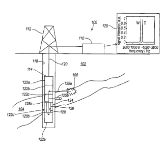
[0021] Fig. 1A is a schematic diagram depicting a wellsite 100 for performing
subsurface
operations. The wellsite 100 is positioned about a subterranean formation 102
having a
reservoir 104 with fluid therein. The formation 102 has one or more pores 108
with fluid
therein. As shown in Fig. 1B, an expanded view of pore 108, a portion 106a of
the fluid
may be positioned centrally within the pore 108 and a portion 106b may be
positioned in
recesses 110 of the pore 108. The central portion 106a may represent
retrievable fluids and
the portion 106b may represent trapped fluids within the pore 108.
[0022] As shown, the wellsite 100 includes a rig 112, a downhole tool 114, and
a surface
unit 116. The downhole tool 114 is deployed into a wellbore 118 to measure
subsurface
parameters. The downhole tool 114 as shown is a wireline tool deployed into
the wellbore
118 via a wireline cable 120, but any downhole tool (e.g., drilling, coiled
tubing,
production, and/or other tool) may be used. The wireline cable 120 is in
communication
with the surface unit 116 for passing signals therebetween. The surface unit
116 may be
4
CA 03015233 2018-08-20
WO 2017/151928
PCT/US2017/020458
used to collect data from the downhole tool 114 and/or to send signals (e.g.,
power,
command, etc.) to the downhole tool 114.
[0023] The downhole tool 114 of Fig. 1A may include a variety of components
for
performing various operations. As shown, the downhole tool 114 includes
electronics
122a, a fluid sampler 122b, a core sampler 122c, an imager 122d, and a
collector 122e.
The electronics may include various devices, such as power, control,
processing,
communication (e.g., telemetry), and/or other devices used in downhole
operations.
[0024] The fluid sampler 122b may be a conventional sampling tool capable of
drawing
fluid samples from the subsurface formation into the downhole tool 114. The
core sampler
122c may be a conventional sidewall coring tool capable of cutting core
samples 125b
from a wall of the wellbore surrounding the formation. While Fig. 1A shows a
sidewall
coring example, an axial coring tool may be provided in the downhole tool 114.
The
collector 122e may be a receptacle for storing the collected samples. An
example of a
wireline tool with sampling capabilities is provided in US Patent No. 6047239.
[0025] The imager 122d as shown includes a housing 124 with a sidewall 126
defining a
passage 128 therethrough. The fluid sampler 122b and the core sampler 122c may
be
positioned about (e.g., uphole from) the imager 122d to pass fluid samples
125a and core
samples 125b, respectively, through an inlet 128a and into the passage 128 for
measurement as schematically indicated by the arrows. The imager 122d may
include, for
example, a magnetic resonance imager (MRI) configured to receive the fluid and
core
samples 125a,b as is described further herein.
[0026] The core sample 125b may be imaged using the imager 122d. The core
sample
125b may be passed through a passage inlet 128a (e.g., a door) located in the
housing
disposed to receive the core sample into the passage whereupon the core sample
may be
positioned in the passage 128 of the imager 122d. The core sample 125b may be
saturated
with the fluid sample (or other fluid) 125a using core flooding as indicated
by the arrows.
The fluid and core samples 125a,b may be released (e.g., dropped) from the
passage 128
through an outlet 128b (e.g., a door) and into the collector 122e for storage
and/or retrieval.
The downhole tool 114 may be provided with various devices to facilitate
and/or control
sampling and/or imaging. For example, the fluid sample 125a may be free to
pass through
the imager 122d, or controlled using fluid control devices, such as flowlines,
valves, etc.
[0027] The imager 122d may be capable of performing an MRI on the core sample
within the downhole tool 114. The core sample 125b may be saturated with the
sampled
5
CA 03015233 2018-08-20
WO 2017/151928
PCT/US2017/020458
fluid during imaging. The measurements taken by the imager 122d may be
collected by the
surface unit 116 and outputs 129, such as a plot, may be generated therefrom
as is
described further herein.
[0028] Additional measurements may be taken using sensor(s) and/or other
devices to
.. determine various subsurface parameters, such as downhole conditions,
formation
parameters, fluid parameters, etc. For example, the downhole tool 114 may be
provided
with optical fluid analyzers, gauges, spectrometers, transducers, etc. that
may collect
additional measurements, such as composition, temperature, pressure, etc. The
collected
images and/or measurements may be evaluated to determine various subsurface
parameters.
[0029] Fig. 2 is a cross-sectional view of a contrast imager 222 which may be
positioned
downhole (see, e.g., the imager 122d of Fig. 1A) or at a surface location
(e.g., stand alone,
lab facility, etc.). As shown in this view, the contrast imager 222 includes a
housing 224
with a sidewall 226 defining a passage 228 which may be similar to the imager
122d of
Fig. 1A. The imager may be oriented in any direction to facilitate operation.
[0030] The passage 228 is shaped to receive a core sample 225b (e.g., core
sample 125b
of Fig. 1A) and fluid 225a (e.g., the fluid sample 125a of Fig. 1A). The core
sample 225b
may be a cylindrically shaped sample that may be disposed into the linear
passage 228 of
the housing 224 as shown, or of other shapes and/or dimensions. Devices may be
provided
.. to automatically insert and/or remove one or more samples into/out of the
passage 228.
[0031] The fluid 225a may be passed through the passage 228 during testing as
indicated
by the axial arrow. A flowline and/or other flow control devices may
optionally be
provided to selectively pass the fluid 225a into the passage 228 to provide
the desired core
flooding. The fluid 225a may flow through the passage 228 at a desired flow
rate, or be
.. enclosed therein to remain stationary during testing. The fluid 225a may be
dumped from
the passage 228 as desired. The fluid 225a may be any fluid passed through the
core
sample 225b during testing. In an example, the fluid 225a is in situ fluid
from the
formation used to replicate subsurface conditions.
[0032] As indicated by the curved arrow, the imager and/or the core sample
225b may
optionally be rotated (e.g., by a rotating shaft driven by a motor) to change
the orientation
of the core sample 225b from an angle al to a2 during imaging. The rotation of
the core
sample 225b relative to the housing 224 allows for signal selection along the
coordinates
6
CA 03015233 2018-08-20
WO 2017/151928
PCT/US2017/020458
based on the orientation of the direction of the fields BO and B1 to the
orientation of the
core sample 225b as it rotates.
[0033] The housing 224 has a sensor array including a permanent magnet 230a, a
radio
frequency coil 230b, and an applied magnetic field gradient(s) 230c. The
permanent
magnet 230a is positioned in the sidewall 226 and is radially disposed about
the passage
228 to encircle the core sample 225b therein. The permanent magnet 230a may be
any
permanent magnet, such as a Halbach magnet, arranged to generate a magnetic
field (BO)
oriented to the z-axis of the passage 228.
[0034] The radio frequency coil 230b is positioned in the sidewall 226 and is
radially
disposed about the passage 228 to encircle the core sample 225b therein. The
radio
frequency coil 230b is positioned between the passage 228 and the permanent
magnet
230a. The radio frequency coil 230b may be a coil arranged to generate a
magnetic field
B1 oscillating at a radio frequency along the x or y-axis of the passage 228.
The magnetic
field gradients 230c are positioned between the permanent magnet 230a and the
radio
frequency coil 230b.
[0035] The housing 224 may be provided with or coupled to an imaging unit 216
(e.g.,
surface unit 116 of Fig. 1A) for providing power, collecting data, and/or
sending
commands to the imager 222. The magnetic coil 230a, the radio frequency coil
230b, and
the magnetic field gradients 230c may be coupled to the imaging unit 216 to
provide
measurements thereto. The imager 222 and/or the imaging unit 216 may be
provided with
communication means, such as a wired and/or wireless coupling to define a
communication link therebetween.
[0036] The imaging unit 216 may have a conventional display capable of
transforming
the measurements into images for display. The imaging unit 216 may include,
for example,
a processor, a database, a telemetry unit, a power unit, and/or other
electronics for
operation with the imager 222. The imaging unit 216 may be incorporated into
the
electronics of the downhole tool (e.g., 122a of Fig. 1A) and/or the surface
unit 116 (Fig.
1A). The collected measurements may be used to generate outputs, such as a
plot 229.
Optionally, one or more probes 234 and/or the sensors S may be provided to
collect
measurements. For example, the probe(s) 234 of the radio frequency coil 230b
may be
selectively provided with single and/or multiple resonant frequencies, for
example to allow
for detection of multiple nuclei.
7
CA 03015233 2018-08-20
WO 2017/151928
PCT/US2017/020458
[0037] The imaging unit 216 may be used to collect image parameters (e.g.,
distribution
of fluids, residual oil saturation, etc.) from the imager 222 and subsurface
parameters (e.g.,
composition, temperature, pressure, etc.) from the sensors (S). The imaging
and/or
collected measurements may be used to perform various formation evaluations,
such as
imaging, fluid analysis, effluent analysis, compressed sensing, etc. For
example, the
collected data may be used to derive subsurface parameters, such as
resistivity and
permeability.
CONTRAST IMAGING
[0038] Evaluations may be performed using various contrast imaging techniques,
such as
relaxation and diffusion imaging. Such techniques may involve, for example,
analysis of
relaxation times T1 and T2 for the generated images. Imaging parameters may be
generated
using, for example, techniques that rely on differences in NMR measureable
quantities,
such as relaxation times and diffusion coefficients (D) to provide contrast
between
hydrocarbons and aqueous fluid. The NMR measurement may include a baseline
measurement used in petrophysical work, such as the relaxation time T2. T2 may
be a
measure of the decay of bulk magnetization created in the system through the
application
of radio frequency excitations. The decay in magnetic coherence may be caused
by
interactions of the nuclear spins with varying magnetic fields produced by
static field
inhomogeneities as well as inter- and intra-molecular motions.
[0039] In a porous rock environment, hydrocarbons and aqueous fluid may have
similar
T2's. Techniques used to provide contrast between fluid phases may, therefore,
probe
secondary fluid properties, such as the relaxation time T1 and the diffusion
coefficients of
the respective fluids. T1 may be a measure of how well the molecules of a
fluid exchange
energy with the environment. A long T1 may indicate a weak coupling, while a
short T1
may indicate a strong coupling. As such, T1 relaxation times may be dependent
on
molecular properties, such as size, and the larger hydrocarbon molecules may
exhibit
longer Ti's. The self-diffusion coefficients of fluids, such as aqueous fluid,
liquid
hydrocarbons, and gaseous hydrocarbons, may be quite different and may be used
to
differentiate between fluid phases present in a rock sample. In these cases,
multi-
dimensional relaxation measurements plotting T1 vs T2 or D VS T2 may be used
provide the
desired contrast
8
CA 03015233 2018-08-20
WO 2017/151928
PCT/US2017/020458
[0040] Evaluations of the images may be performed using NMR core analysis
and/or
spectroscopic methods. Such evaluations may be used to provide a desired fluid
phase
differentiation on bulk samples. These evaluations may be done, for example,
for standard
spin echo imaging sequences, such as spin-warp. Examples of spin-warp are
described in
Edelstein, W. A., Hutchison, J. M. S., Johnson, G. & Redpath, T., Spin warp
NMR
imaging and applications to human whole-body imaging, Physics in Medicine and
Biology
25, 751 (1980)1.
[0041] In order to provide information on the spatial distribution of these
fluids, one-
dimensional spatially resolved T2 distributions can be used to provide fluid
discrimination
during core floods. To provide desired separation when T2 contrast between the
fluids is
low, multi-dimensional relaxation measurements, such as D-T2 and T1-T2, may be
performed. These may provide bulk measurements. Further information on
distributions of
hydrocarbons and aqueous fluid beyond the relative volumes may be performed as
is
described further herein.
[0042] Spatial distribution of phases in a single core plug may be determined
by using
chemical dopants in injected aqueous fluid to provide relaxation contrast. In
an example,
chemical dopants containing species, such as Cu2+, Mn2+, or Gd3+, may be used.
These
substances may be used to reduce the relaxation time of aqueous fluid, and to
provide a
differentiation between various fluids, such as aqueous fluid and
hydrocarbons.
[0043] In another example, in systems that exhibit different T1 values, T1
nulling may be
used to suppress the signal from one of T1 environments present in the sample.
The timing
of the RF excitation pulses may be set such that the magnetization and
resulting MRI
signal from one T1 environment is signal suppressed. The core sample may be
saturated
with multiple fluid phases with sample fluid, such as a fluid having a single,
well-defined
T1.
[0044] In yet another example, chemical selectivity of NMR measurements may be
used
to differentiate the formation fluids. The NMR response of a given species
depends on the
gyromagnetic ratio of that spin, a quantity that is unique to each NMR-active
species. In a
first case, D20 may be used instead of H20 in the injected brine to remove the
contribution
of aqueous fluid to the image. In another case, the imaging may be done on the
hydrogen
(or other NMR-active nucleus, such as sodium, 23Na or carbon 130 present in
the
formation fluid.
9
CA 03015233 2018-08-20
WO 2017/151928
PCT/US2017/020458
[0045] Examples of contrast imaging are provided in Mitchell, J.,
Chandrasekera, T.C.,
Holland, D.J., Gladden, L.F. and Fordham, E.J., Magnetic resonance imaging in
petrophysical core analysis, Physics Reports, 526, pp. 165-225 (2013). Other
existing
techniques may be used for evaluation, such as those described in US Patent
Nos.
9133709,8499856, 6220371 and 4769602.
CHEMICALLY-SELECTIVE IMAGING
[0046] Evaluations may also be performed using chemically-selective imaging
techniques to generate independent images of fluids, such as hydrocarbon and
aqueous
(brine), within a formation using an imager (e.g., imagers 122d, and 222 of
Figs. 1A and 2,
respectively). The chemically-selective imaging technique exploits the
difference in the
chemical shift in the NMR spectrum to differentiate between fluids in the core
sample.
[0047] The chemically-selective imaging involves: 1) contrasting hydrocarbon
images
and aqueous (brine) images based upon differences in chemical shift in the
nuclear
magnetic resonance (NMR) spectrum, and 2) acquiring 1, 2 or 3D images on a
timesc ale
that reduces pixel blurring between successive oil-water images during
drainage and
imbibition experiments at representative reservoir flow rates (e.g., at v, = 1
ft day-1 (0.304
m day-1)). To achieve this, MRI pulse sequences (e.g., rapid acquisition with
relaxation
enhancement (RARE)) may be used in combination with compressed sensing (CS).
[0048] Figs. 3A and 3B are flow charts depicting chemically-selective imaging
processes
300a,b which may be performed using the imagers 122d, and 222, respectively,
of Figs. 1A
and/or 2 to image fluids in the core sample. The process 300a of Fig. 3A
includes a
measurement phase 336a, a signal selection phase 336b, an image acquisition
phase 336c,
and an image display phase 336d. The process 300a may be performed for one or
more
fluids in the core sample. As indicated by the dotted and smooth arrows
337a,b, part or all
of the process 300a may be selectively repeated for one or more fluid (e.g.,
337a ¨ aqueous
fluid, 337b hydrocarbon) in the core sample.
[0049] The measurement phase 336a involves collecting measurements, such as
the plot
229 of Fig. 2 generated by the imaging unit 216. The graph 229 as shown plots
signal
intensity (a.u.) (y-axis) versus frequency (Hz) (x-axis) generated by the
imager 222. The
resulting line shows peaks 340a,b that correspond to a composition of the
fluid. In the
example shown, the peaks 340a,b corresponding to aqueous fluid (W) and
hydrocarbon (D-
dodecane) with peaks at 0 Hz at 300 Hz, respectively.
CA 03015233 2018-08-20
WO 2017/151928
PCT/US2017/020458
[0050] The signal selection phase 336b involves selective excitation of the
magnetization
from either the aqueous or hydrocarbon phase. The selection may be made to
indicate
which fluid is to be imaged. For example, when performing the process 300a for
aqueous
fluid according to line 337a, the water peak 340a may be selected using box
342a. In
another example, when performing the process 300a for hydrocarbon according to
line
337b, the hydrocarbon peak 340b may be selected using box 342b.
[0051] The image acquisition phase 336c involves acquiring the raw k-space
data 344
corresponding to the fluid distribution through the core sample using MRI
pulse sequences.
The sample pattern 344 is a plot of kpl m-1 (y-axis) versus kp2 m-1 (x-axis)
which indicates
the data points that must be measured during the image acquisition. The data
points on plot
344 indicate the locations of points to be acquired. The intensity of the
light regions
indicate where data is sampled; whereas, the dark regions indicate not
sampled. This data
may be captured using fast data acquisition and reconstructed using compressed
sensing to
generate images as described further herein.
[0052] The image display phase 336d involves generating an image 346 of the
formation
fluid within the core sample. Depending on the time available, the image 346
may be
acquired using a standard imaging technique, or an image generated by fast
acquisition
with compressed sensing reconstruction of the acquired data. While a 3D image
346 is
shown, the image may be a 1D or 2D image. One or more images of one or more
fluids
may be displayed as is described further herein. When generating the images,
the image
acquisition phase 336c may optionally be performed at various angles.
[0053] As shown in Fig. 3B, the process 300b may involve pulse sequencing 400.
The
process 300b involves the same measurements phase 336a, a combined signal
selection and
image acquisition phase 336b,c, and the image display phase 336d. A portion of
the
process 300b is repeated as indicated by the arrows 337a,b for various fluids.
[0054] Because the pulse sequence 400 may selectively capture data for certain
fluids,
the pulse sequence 400 may be used to determine which fluids are being imaged.
The
process 300b may be repeated at different pulse frequencies to excite the
selected fluid,
such as water 346a and hydrocarbon 346b as shown.
[0055] Figs. 4A - 4C are graphs depicting various pulse sequences 400a,b,c
that may be
used during the image acquisition phase 336c to acquire the raw data of the
hydrocarbon
and aqueous phase distribution of Fig. 3. Fig. 4A shows a 1D sequence. Fig. 4B
shows a
2D sequence. Fig. 4C shows a 3D sequence.
11
CA 03015233 2018-08-20
WO 2017/151928
PCT/US2017/020458
[0056] Each of the pulse sequences 400a-c includes radio frequency pulses (rf)
from the
RF coils and one or more of the applied magnetic field gradients (GR, Gp/Gpi,
Gs/Gp2)
(e.g., 230b,c of Fig. 2). The pulse sequences may be applied to selectively
excite a certain
chemical species (e.g. oil or brine) present in the sample. The magnetic
fields include a
read gradient (GR) and phase gradients (Gp/Gpi, Gs/Gp2) which are applied for
a period of
time to enable spatial-encoding of the nuclear spins.
[0057] Each of the pulse sequences also include a portion 449a which
represents the
chemically-selective preconditioning and portions 449b representing the
excitation portion
of the pulses. The pulses sequences 400a-c are performed in various shapes and
at various
degrees to generate different perspectives of the sample being imaged. Each of
the pulse
sequences includes: PsEL ¨ a selective excitation pulse, PEx ¨ a non-selective
excitation
pulse, PREF ¨ a refocusing pulse. For example, for pulse sequence used in fast
acquisition,
the 180 refocusing RF pulses may be repeated NRF times to sample multiple
line of k-
space from a single excitation of the system.
[0058] As shown in Figs. 4A ¨ 4C, each pulse sequence has different shapes.
Radio
frequency (re pulses create spin echoes 450 which are induced by polarized H
atoms.
Different phase gradients (Gp/Gpi, Gs/Gp2) may be used to enable spatial
encoding of the
spins.
[0059] As shown in the 1D version of Fig. 4A, the pulse sequence 400a includes
an rf
pulse for excitation and a read gradient (GR) to enable spatial resolved
information in the
direction of the read gradient only. The rf field may be added to the magnetic
field in
pulses shot in microseconds. The shape of the pulses on radio frequency line
r.f. include
square pulses with broadband that affect the entire pulses. The read gradient
GR is repeated
only once for generating a 1D image.
[0060] Fig. 4B shows a 2D version of a pulse sequence 400b including the rf
pulse and
the GR pulse, with additional Gp and Gs pulses. The shape of the pulses on
radio frequency
line r.f. are Gaussian to affect only specific regions of the sample. This
version also
depicts gradient iterations Si, S2 along gradient line G.
[0061] These gradient iterations indicate that the Gp is repeated in order to
generate the
2D image. The phase gradient (Gp) are iterated NRF times for the iterations
Si, S2. The
180 refocusing pulse may be repeated NRF times. Information may be acquired
as needed
by changing the strength of the various gradients. Each time the read gradient
(GR) and the
12
CA 03015233 2018-08-20
WO 2017/151928
PCT/US2017/020458
slice gradient (Gs) are the same, the amplitude of the phase gradient (Gp) may
be changed
and then iterated through the various gradients values (Si and S2) to generate
a 2D image.
[0062] Fig. 4C shows a 3D version of the pulse sequence 400c including the rf
pulses
and the read gradient GR, with additional first and second phase-encoding
gradients, Gp1
and Gp2. In this version, the Gp1 and Gp2 pulses each include gradient
iterations Si, S2
indicating that these pulses are repeated NRF times in order to generate the
desired 3D
image. For 3D images, all combinations of Gp1 and Gp2 may be iterated with the
same read
gradient.
[0063] The pulse sequences 400a,b,c depict example chemically-selective RARE
pulse
sequences. The pulse sequences 400a-c have k-space frequencies encoded in the
read
direction (kR) and phase encoded in kpl and kp2 as depicted in the image 344
of Fig. 3A.
Upon the application of the read gradient (GR) and phase gradients (Gp/Gpi,
Gs/Gp2) points
of the plot 344 are generated. For the 3D pulse sequence (Fig. 4C), the
amplitude of the
phase encoding gradients, Gp1 and Gp2 determine which data points on the plot
344 are
sampled. The image display phase 336d uses compressed-sensing to reconstruct a
fluid
image from the core image. The duration and amplitude of the gradients may be
varied to
control the spatially resolved information that is acquired. By changing
these, the field of
view and area imaged and resolution may be adjusted to provide a desired focus
level
about the sample.
[0064] MRI acquisition techniques may be used to under sample data to reduce
data
collection time and compressed sensing can be used to reconstruct a full image
from under
sampled data. Image acquisition techniques may include, for example, Rapid
Acquisition
with Relaxation Enhancement (RARE), Echo Planar Imaging (EPI), spin warp,
and/or
other acquisition techniques) and the temporal resolution can be enhanced
further by
employing under-sampling and compressed sensing reconstructions. Examples of
RARE
are described in Hennig, J., Nauerth, A. & Friedburg, H, RARE imaging: a fast
imaging
method for clinical MR. Magnetic resonance in medicine: official journal of
the Society of
Magnetic Resonance in Medicine / Society of Magnetic Resonance in Medicine 3,
823-833
(1986); examples of EPI are described in Mansfield, P. Multi-planar image
formation using
NMR spin echoes, Journal of Physics C: Solid State Physics 10, L55¨L58 (1977);
and
examples of spin warp are described in Edelstein, W. A., Hutchison, J. M. S.,
Johnson, G.
& Redpath, T., Spin warp NMR imaging and applications to human whole-body
imaging,
Physics in Medicine and Biology 25, 751 (1980).
13
CA 03015233 2018-08-20
WO 2017/151928 PCT/US2017/020458
[0065] Compressed sensing in MRI is based on the following requirements, for
example,
(1) aliasing artifacts (e.g., the sample data) in the linear reconstruction
must be incoherent
and noise-like; (2) the desired image exhibits transform sparsity; and (3) the
image is
reconstructed using a non-linear algorithm that enforces sparsity and
consistency with the
acquired k-space data.
[0066] Assuming the image reconstructed is given by x which is related to the
acquired
k-space measurements via the following Equation (1):
SFx+v=y, (1)
where S is the sub-sampling pattern, F is the Fourier transform that maps the
image into k-
space, v is the normally-distributed noise (standard deviation a and zero
mean) and y is the
vector that contains the acquired k-space measurements.
[0067] Due to under-sampling and the presence of noise, Equation 1 may be an
ill-posed
problem and, therefore, linear image reconstruction methods, such as the
inverse Fourier
transform, which may be employed for the image reconstruction of a fully-
sampled k-space
data set, may result in an image containing aliasing artefacts due to
violation of Nyquist
criterion. Therefore, an approximate solution to x may be sought by using a
variational
regularization approach balancing the model (Equation 1) and prior assumptions
of x in
terms of a regularization functional J given by:
xa E arg min J(x) , (2)
subject to 1SFx ¨42 u -
The role of the inequality constraint enforces consistency with the acquired k-
space data
and the regularization term J incorporates prior information on the
reconstruction of x,
which may be needed to counteract any ill-posedness of the problem.
[0068] In the case of the CS reconstruction, the prior information is that the
image can be
sparsely represented either implicitly or in an appropriate transform domain.
The choice of
the regularization functional (J) that is used to map the image into the
transform domain
may depends on the nature of image to be reconstructed. For instance, a non-
smooth
regularizer, such as Total Variation (TV), may be more suited to an image with
sharp-
edges whereas a smooth regularizer, such as the Daubechies wavelet transform,
lends itself
well to images in which the pixel intensities change more gently. In the
present study, TV
has been used as the regularization functionals, J(x), as will now be
discussed.
14
CA 03015233 2018-08-20
WO 2017/151928
PCT/US2017/020458
[0069] Total Variation penalizes the 1-norm of the 2-norm of the finite
different
approximation of the gradient (Vx) of the image as given by:
J(x) = TVx = N42,1. (3)
Herein, Neumann boundary conditions were set for the CS reconstructions. Other
regularization functions, such as wavelet transforms, can be used.
[0070] Generally, the Tikhonov-Regularization scheme for the approximation of
x is
written as shown below:
Xa E arg min{-1
¨ SF422 +a/(x)} (4)
2
and the regularization parameter a (always positive) weights the influence of
the fidelity
and regularization terms in Equation 4. In the present study, a modification
of Equation 5
to include Bregman iterations has been implemented as described by Equations
(5a),(5b):
Xa E arg min{-1 k
¨ SF X21 (x)} (5a)
2 2
(5b)
yk =y k -1 + y ¨ sFxku.
[0071] Using the Bregman approach, a series of k problems (Equation 5a) are
solved
with the residual added to the k-space data, y, after each iteration (Equation
5b). See, e.g.,
M. Benning, L.F. Gladden, D.J. Holland, C.-B. Schonlieb, T. Valkonen, Phase
reconstruction from velocity-encoded MRI measurements ¨ a survey of sparsity-
promoting
variational approaches, Journal of Magnetic Resonance. 238 (2014) 26-43.
[0072] Compressed sensing may be used in combination with MRI pulse sequences
for
example CS-RARE, in which k-space is under sampled. Examples of Compressed
Sensing
(CS) are described in Lustig, M., Donoho, D. L., Santos, J. M. & Pauly, J. M,
Compressed
Sensing MRI. IEEE Signal Processing Magazine 25, 72-82 (2008); and Lustig, M.,
Donoho, D. & Pauly, J. M., Sparse MRI: The application of compressed sensing
for rapid
MR imaging. Magnetic resonance in medicine: official journal of the Society of
Magnetic
Resonance in Medicine /Society of Magnetic Resonance in Medicine 58, 1182-95
(2007).
[0073] The image acquisition and compressed sensing techniques may be used in
combination with imaging of subsurface materials. Examples of imaging of
subsurface
materials include Chang, C.T., Edwards, C.M., 1993, Proton MR Two-Component
Chemical Shift Imaging of Fluids in Porous Media, The Log Analyst, 34, pp. 20-
28;
CA 03015233 2018-08-20
WO 2017/151928
PCT/US2017/020458
Dereppe, J.M., Moreaux, C., Chemical Shift Imaging of Fluid Filled Porous
Rocks,
Magnetic Resonance Imaging, 9, pp. 809-813 (1991); Dereppe, J.M., Moreaux, C.,
2D
Spin-Echo and 3D Chemical-Shift-Imaging Techniques for Analysis of Oil-Water
Replacement in Limestone. Journal of Magnetic Resonance, 91, pp. 596-603
(1991);
Maudsley, A.A., Hilad, S.K., Perman, W.H., Simon, H.E., Spatially Resolved
High
Resolution Spectroscopy by "Four-Dimensional" NMR. Journal of Magnetic
Resonance,
51, pp. 147-152 (1983); and Dechter, James J., Komoroski, Richard A.,
Ramaprasad, S.,
Use of Presaturation for Chemical-Shift Selective Imaging of Individual Fluids
in
Sandstone and Carbonate Cores, Journal of Magnetic Resonance, 93, pp. 142-150
(1991).
[0074] The chemically-selective preconditioning section 449a and a RARE
imaging
pulse sequence section 449b may be used to facilitate the acquisition. Through
the use of
chemically-selective radio frequency (r.f.) pulses and homospoil gradients,
the signal from
either various fluids (e.g., hydrocarbon and aqueous fluid) can be effectively
suppressed
prior to the imaging section of the pulse sequence.
[0075] Fig. 4B and 4C show the RARE pulse sequences for 2D and 3D
acquisitions,
respectively. For 2D applications, under-sampling of k-space in the phase
encoding
direction (P1) can be performed whereas for the latter, under-sampling of k-
space can be
performed in both phase encoding directions (P1 and P2). In both cases, k-
space is fully
sampled in the read direction (R). The under-sampled k-space data is
reconstructed using
compressed sensing.
[0076] In a 2D pulse sequencing example, the chemically-selective section 449b
of Fig.
4B includes a Gaussian shaped rf pulse (r.f.) used to selectively excite one
of the phases,
followed by homospoil gradients (GR, GR Gs) to destroy the magnetization. To
demonstrate the chemically-selective imaging two-dimensional (2D) slice images
have
been acquired. A first image may include a reference image of aqueous fluid
and
hydrocarbon with no chemically-selective preconditioning. A second image may
include
aqueous fluid and hydrocarbon independently. Table 1 depicts the experimental
parameters
used to generate 2D images:
16
CA 03015233 2018-08-20
WO 2017/151928
PCT/US2017/020458
TABLE 1 ¨ 2D
Reference Chemically-selective
image images
Aqueous fluid Aqueous Dodecane
and dodecane fluid
Field of view (y) / mm 80 80 80
Field of view (x) / mm 50 50 50
Slice thickness (z) / mm 2 2 2
In-plane resolution (y) / mm 0.39 0.39 0.39
In-plane resolution (x) / mm 0.31 0.31 0.31
Excitation pulse duration, PEx / ts 512 512 512
Refocusing pulse duration, PREF / Jts 512 512 512
Excitation pulse power, PLEx / dB 27 27 27
Refocusing pulse power, PLREF / dB 21 21 21
Selective excitation pulse duration, PsEL / ts - 8192 8192
Selective excitation bandwidth, BWsEL / Hz - 530 530
Selective excitation pulse power, PLsEL / dB - 44 44
Selective excitation pulse offset, OsEL / Hz -350 0
Table 1 indicates that, by suppressing the signal from the hydrocarbon only
aqueous fluid
is detected. Conversely, by suppressing the signal from the aqueous fluid,
only
hydrocarbon is detected.
[0077] The number of 180 degree refocusing pulses applied for each
acquisition may be
determined by the RARE factor (NR) and the total number of r.f. excitations
(NEx)
required to sample k-space is given by the total number of points in the two
phase encoding
directions, Np1,2 divided by NRF. The images may be generated using a rapid
sequencing by
using fast imaging sequences, such as RARE, EPI, etc., and the temporal
resolution can be
enhanced further by employing under-sampling and subsequently using compressed
sensing for image reconstructions. The temporal resolution of standard imaging
sequences
may also be enhanced by using compressed sensing, such as CS-RARE. Both RARE
and
EPI may use multiple lines of k-space acquired from an individual excitation.
The practical
limit on the number of lines of data that can be acquired from each
excitation, and
correspondingly the acquisition time acceleration, may be determined by the
relaxation
times of the sample under investigation. The transverse relaxation times for
the fluid-
saturated rock core samples, which are the subject of the present invention,
are expected to
be in the range of tens to hundreds of milliseconds. Considering a RARE
acquisition of a
17
CA 03015233 2018-08-20
WO 2017/151928
PCT/US2017/020458
water-saturated rock core with a T2 = 150 ms, with an echo time TE = 4 ms, it
may be
reasonable to suggest that 64 lines of k-space can be acquired from each
excitation.
[0078] By using compressed sensing (CS), a signal with a sparse
representation, such as
an image, can be recovered from a number of measurements sampled below the
Nyquist
rate. Therefore, applying CS to ultra-fast MRI acquisitions, under sampling k-
space may
lead to further reductions in acquisition image times, thus enabling dynamic
processes,
such as the laboratory core flood, to be studied where the temporal resolution
is greater
still.
[0079] In a 3D example as shown by Fig. 4C, various pulsing configurations may
be
provided, for example, when it is desirable to obtain information on the fluid
distribution
within the rock core. A comparison of Spin-warp, RARE, and CS-RARE techniques
applied to an MRI protocol to monitor the fluid distribution in a laboratory
core flood
experiment is shown below:
TABLE 2 ¨ 3D
(1) Spin- (2) (3) CS-
Warp RARE RARE
Pixels in read direction, NR 256 256 256
Pixels in first phase encoding direction, Np1/ - 128 128 128
Pixels in first phase encoding direction, NP 2 / 128 128 128
Number of lines of k-space sampled per excitation,
NE X / 1 64 64
Recycle delay, TR 1 s 1.6 1.6 1.6
Number of scans, NS I - 8 8 8
k-space sampling fraction, SF I % 100 100 25
The total image acquisition time (TAcQ) can be calculated using Equation 6:
SF x (Np1xNp2), (6)
TA C Q = NS x TR x
'VEX
Using equation 6, Table 3 shows total acquisition time for each of the
techniques of Table
2:
18
CA 03015233 2018-08-20
WO 2017/151928 PCT/US2017/020458
TABLE 3
(1) Spin- (2) (3) CS-
Warp RARE RARE
Acquisition time, TAcQ / min 3495 55 14
[0080] The total acquisition time may be minimized using CS-RARE. To this end,
the
amount of fluid injected over the course of the three acquisition times may be
calculated
for the case of a theoretical core flood as shown by Table 3. The relevant
sample properties
and experimental conditions are listed in Table 4:
TABLE 4
Plug diameter, D / mm 38
Plug length, L / mm 76
Plug volume, V / ml 87
Rock porosity, 41) / % 23
Pore volume, P.V. / ml 20
Interstitial velocity, v, / ft day-1 1
Injectant flow rate, Q I ml min-1 0.06
[0081] The total number of pore volumes (N.P.V) of fluid injected over the
course of the
image acquisitions for each of the three cases considered, may be calculated
using
Equation 7 and is summarized in Table 5.
N. P. V =TACQ QI (7)
P. V.
The total number of pore volumes (N.P.V) of fluid injected over the course of
the image
acquisitions for each of the three cases considered is calculated using
Equation 8 and is
summarized in Table 5:
19
CA 03015233 2018-08-20
WO 2017/151928
PCT/US2017/020458
TABLE 5
(1) Spin- (2) (3) CS-
Warp RARE RARE
Number of P.V. injected, N.P.V / ml 10.50 0.16 0.04
Table 5 indicates that a significant reduction in sample volume may be
achieved using
rapid imaging with compressed sensing, such as CS-RARE, and that such images
may be
more representative of the fluid saturation at specific time points in the
core flood.
[0082] Figs. 5A ¨ 5C show the 3D image 346 in greater detail. These images may
be
used to individually depict each of the fluids hosted within the pores of the
formation. As
shown in these figures, the chemically-selective 3D CS-RARE MRI method is
implemented to independently image hydrocarbon and aqueous fluid within a core
sample
using imager 222 of Fig. 2 in a laboratory core-flooding experiment at
representative
reservoir conditions. The 3D pulse sequence of Fig. 4C is used to generate MRI
measurements according to the plot 344 of Fig. 3A. The white pixels of 344
determine the
values of Gp1 GP2 which in turn determine which data points need to be
acquired. The data
is then processed through compressed sensing to generate images of Figs. 5A-
5C. Fig. 5C
shows the combined fluid images generated by this technique.
[0083] The original rock core was cut into two pieces with one half being
saturated in
aqueous fluid and the other half in hydrocarbon under ambient conditions. Fig.
5A shows a
3D fluid image 346a of aqueous phase in the core sample generated during
application of
the process 300 repeated along line 337b for the hydrocarbon phase. Fig. 5B
shows a first
half of 3D fluid image 346b of the hydrocarbon phase in the core sample
generated during
application of the process 300a repeated along line 337b for the hydrocarbon
phase. Figs.
5C show a second half of the core sample the aqueous and hydrocarbon phases
346 a,b on
the same plot.
[0084] In the example depicted in Figs. 5A-5C, a chemically-selective 3D CS-
RARE
acquisitions for the selective imaging of a) dodecane and b) aqueous fluid
imbibed in a
limestone core plug is performed. The acquisition time for a) and b) is 14
minutes per
image. The two images have been combined to provide an overall image of the
aqueous
fluid and hydrocarbon.
[0085] Table 6 below shows experimental parameters of the core sample used in
generating the images of Figs. 5A-5C:
CA 03015233 2018-08-20
WO 2017/151928
PCT/US2017/020458
TABLE 6
Chemically-selective images
Aqueous fluid Hydrocarbon
(Fig. 5A) (Fig. 5B)
Field of view (z) / mm 80 80
Field of view (x) / mm 50 50
Slice thickness (y) / mm 50 50
In-plane resolution (z) / mm 0.39 0.39
In-plane resolution (x) / mm 0.31 0.31
In-plane resolution (y) / mm 0.31 0.31
Excitation pulse duration, PEx / p,s 512 512
Refocusing pulse duration, PREF / Is 512 512
Excitation pulse power, PLEx / dB 27 27
Refocusing pulse power, PLREF / dB 21 21
Selective excitation pulse duration, PsEL / jts 4096 4096
Selective excitation bandwidth, BWsEL / Hz 560 560
Selective excitation pulse power, PLsEL / dB 40.6 40.6
Selective excitation pulse offset, OsEL / Hz -425 5
[0086] Table 7 below shows rock properties of the core sample used in
generating the
images of Figs. 5A-5C:
TABLE 7
Aqueous fluid Hydrocarbon
Rock type Estaillades limestone Estaillades limestone
Plug diameter, D / mm 38 38
Plug length, L / mm ¨35 ¨35
Plug volume, V / ml 40 40
Imbibed volume / ml 9.7 10.6
[0087] The images generated using the chemically-selective imaging process
300a,b of
Figs. 3A,3B may be compared with the relaxation imaging processes for
validation. One
or more imaging processes may be performed. For example, the chemically-
selective
imaging process may be used in cases where it may be undesirable to use D20,
any ionic
dopant, or other fluid that could negatively impact the condition of the rock
sample, in
cases where low signals may be present (e.g., due to low natural abundance or
low
gyromagnetic ratio), in cases where certain RF probes may be preferred, to
avoid long
acquisition times from standard single echo spin echo imaging sequences, etc.
Other
21
CA 03015233 2018-08-20
WO 2017/151928
PCT/US2017/020458
variations that may affect selection of the imaging may include suppression of
signal from
species that exhibit different Ti values via Ti nulling and standard spin echo
imaging
sequences in which a single line of a 2D k-space may be acquired for each
initial RF
excitation.
[0088] Variations on the process 300a,b may be performed. For example, the
process
may be performed on other NMR active nuclei, such as hydrogen, sodium, etc.
This may
be used in place of selecting hydrocarbons or brine based on chemical shift
separation.
Various pulse sequences, measurements, images, and/or other data may be
compared for
validation of the results. For example, relaxation imaging results may be
compared with
chemically-selective imaging results.
[0089] Imaging may be used in combination with measurements sensed with the
sensor
S, such as the fluid analyzer, such that a distribution of hydrocarbon and
brine pore fluids
can be determined. Effluent analysis can also be performed using the
measurements from
the optical fluid analyzer. The formation evaluation may be used to plan
oilfield
operations, such as designing enhanced oil recovery (EOR) (e.g., injection) to
facilitate
production.
[0090] Fig. 6 is a flow chart depicting an example method 600 of performing
hydrocarbon operations. The method 600 may be performed using, for example,
the
imager 122d, 222 of Figs 1 A and/or 2. The method involves 654 ¨ positioning a
core
sample of the formation in an imager (see, e.g., Figs. lA and 2). The method
also involves
655 ¨ imaging the core sample by directing a magnetic field through the
passage in a
magnetic direction along a longitudinal axis of the passage, selectively
directing a gradient
field through the passage, directing a radio frequency field through the
sample in the
passage in the direction orthogonal to the longitudinal axis of the passage,
and selectively
pulsing frequencies of the radio frequency field (Fig. 2).
[0091] The method also involves 656 selectively acquiring nuclear magnetic
resonance
measurements of the fluid in the core sample by selectively pulsing
frequencies of the
radio frequency field to the core sample and applying the magnetic field
gradient to the
core sample according to a pre-determined, k-space sampling plot. The
selectively
acquiring may involve performing fast acquisition, such as RARE. The method
may also
involve 657 - generating an image of formation fluid in the core sample by
performing
compressed sensing on the acquired nuclear magnetic resonance measurements.
22
CA 03015233 2018-08-20
WO 2017/151928
PCT/US2017/020458
[0092] The selectively acquiring 656 and/or generating 657 may be performed by
obtaining a spectral image of the fluid from the imaging, selecting a phase
for chemically-
selective imaging, acquiring an image by selecting sample points from a plot
generated
from the selective pulsing, and performing a reconstruction of the formation
fluid within
the core sample using compressed sensing of the selected sample points. The
method may
also involve 658 - performing chemically-selective imaging to isolate the
fluids, such as
hydrocarbon, from the fluid image, and 660 - validating the image by
generating reference
images using contrast imaging and comparing the generated images with the
reference
images, and performing oilfield operation (e.g., EOR) based on the validated
image.
[0093] The method may be performed in any order, and repeated as desired. Part
or all of
the method may be performed. Other optional steps may be performed, such as
may also
involve 656 - rotating the sample and repeating the imaging at various angles
(Fig. 2).
[0094] While the embodiments are described with reference to various
implementations
and exploitations, it will be understood that these embodiments are
illustrative and that the
scope of the inventive subject matter is not limited to them. Many variations,
modifications, additions and improvements are possible. For example, one or
more image
may be performed using one or more of the techniques herein. Various
combinations of the
techniques provided herein may be used.
[0095] Plural instances may be provided for components, operations or
structures
described herein as a single instance. In general, structures and
functionality presented as
separate components in the exemplary configurations may be implemented as a
combined
structure or component. Similarly, structures and functionality presented as a
single
component may be implemented as separate components. These and other
variations,
modifications, additions, and improvements may fall within the scope of the
inventive
subject matter.
[0096] The present disclosure may be modified and practiced in different but
equivalent
manners apparent to those skilled in the art having the benefit of the
teachings herein.
Furthermore, no limitations are intended to the details of construction or
design herein
shown. While systems and methods are described in terms of "comprising,"
"containing,"
or "including" various components or steps, the methods can also "consist
essentially or or
"consist or the various components and steps. Whenever a numerical range with
a lower
limit and an upper limit is disclosed, any number and any included range
falling within the
range is specifically disclosed. In particular, every range of values (of the
form, from a to
23
CA 03015233 2018-08-20
WO 2017/151928
PCT/US2017/020458
b," or, equivalently, from a-b") disclosed herein is to be understood to set
forth every
number and range encompassed within the broader range of values. Whenever a
numerical
range having a specific lower limit only, a specific upper limit only, or a
specific upper
limit and a specific lower limit is disclosed, the range also includes any
numerical value
"about" the specified lower limit and/or the specified upper limit.
24